β 1 Integrin Is an Adhesion Protein for Sperm Binding to Eggs
10
1 Integrin Is an Adhesion Protein for Sperm Binding to Eggs Keith A. Baessler † , Younjoo Lee ‡ , and Nicole S. Sampson †,‡, * † Biochemistry and Structural Biology Graduate Program and ‡ Department of Chemistry, Stony Brook University, Stony Brook, New York 11794 I n mammals, for fertilization to be successful, a single sperm out of thousands must traverse the zona pellucida (ZP) to reach the perivitelline space to first bind to and then fuse with the egg plasma mem- brane. This interaction between gamete membranes in- duces egg activation. One of the earliest events associ- ated with egg activation is an increase in intracellular calcium. Later events such as cortical granule exocyto- sis, blockages to polyspermy that occur at both the ZP and the egg plasma membrane, resumption of meiosis, pronuclei formation, and development of the zygote are all dependent upon the calcium response (3–7). Be- fore sperm binding to the egg plasma membrane can oc- cur, sperm are activated by binding to the ZP, an inter- action that is species-selective ( 8). Sperm protein fertilin (ADAM2) plays a role in egg plasma membrane binding that leads to fertilization. Fertilin is a type 1 integral membrane protein located in the equatorial region of the sperm head and is a mem- ber of the ADAM (A Disintegrin and A Metalloprotease domain) family of proteins ( 9, 10). The fertilin disinte- grin domain is highly conserved across species. Its do- main structure is partially shared by the snake venom metalloproteases (SVMPs) ( 11, 12). The disintegrin do- mains of SVMPs bind integrin receptors with high affin- ity and inhibit integrin-mediated platelet aggregation and cell-matrix attachment. A short peptide sequence present in the disintegrin domain of fertilin, Glu-Cys- Asp (ECD), is important for the protein’s function in egg adhesion (13–17). Antibodies raised against fertilin block sperm-egg binding and fusion ( 18). Knockout of the fertilin gene resulted in reduced binding of sperm to the egg plasma membrane in vitro (19, 20). However, later knockout experiments suggested that the simulta- neous loss of multiple ADAM proteins is responsible for this phenotype (21–23). *Corresponding author, [email protected]. Received for review November 29, 2008 and accepted March 31, 2009. Published online April 1, 2009 10.1021/cb900013d CCC: $40.75 © 2009 American Chemical Society ABSTRACT We investigated the role of 1 integrin in mammalian fertilization and the mode of inhibition of fertilin-derived polymers. We determined that poly- mers displaying the Glu-Cys-Asp peptide from the fertilin disintegrin domain me- diate inhibition of mammalian fertilization through a 1 integrin receptor on the egg surface. Inhibition of fertilization is a consequence of competition with sperm binding to the cell surface, not activation of an egg-signaling pathway. The pres- ence of the 1 integrin on the egg surface increases the rate of sperm attachment but does not alter the total number of sperm that can attach or fuse to the egg. We conclude that the presence of 1 integrin enhances the initial adhesion of sperm to the egg plasma membrane and that subsequent attachment and fusion are me- diated by additional egg and sperm proteins present in the 1 integrin complex. Therefore, the mechanisms by which sperm fertilize wild-type and 1 knockout eggs are different. A RTICLE www.acschemicalbiology.org VOL.4 NO.5 • ACS CHEMICAL BIOLOGY 357
-
Upload
independent -
Category
Documents
-
view
0 -
download
0
Transcript of β 1 Integrin Is an Adhesion Protein for Sperm Binding to Eggs
�1 Integrin Is an Adhesion Protein for SpermBinding to EggsKeith A. Baessler†, Younjoo Lee‡, and Nicole S. Sampson†,‡,*†Biochemistry and Structural Biology Graduate Program and ‡Department of Chemistry, Stony Brook University, StonyBrook, New York 11794
I n mammals, for fertilization to be successful, asingle sperm out of thousands must traverse thezona pellucida (ZP) to reach the perivitelline space
to first bind to and then fuse with the egg plasma mem-brane. This interaction between gamete membranes in-duces egg activation. One of the earliest events associ-ated with egg activation is an increase in intracellularcalcium. Later events such as cortical granule exocyto-sis, blockages to polyspermy that occur at both the ZPand the egg plasma membrane, resumption of meiosis,pronuclei formation, and development of the zygoteare all dependent upon the calcium response (3–7). Be-fore sperm binding to the egg plasma membrane can oc-cur, sperm are activated by binding to the ZP, an inter-action that is species-selective (8).
Sperm protein fertilin� (ADAM2) plays a role in eggplasma membrane binding that leads to fertilization.Fertilin� is a type 1 integral membrane protein locatedin the equatorial region of the sperm head and is a mem-ber of the ADAM (A Disintegrin and A Metalloproteasedomain) family of proteins (9, 10). The fertilin� disinte-grin domain is highly conserved across species. Its do-main structure is partially shared by the snake venommetalloproteases (SVMPs) (11, 12). The disintegrin do-mains of SVMPs bind integrin receptors with high affin-ity and inhibit integrin-mediated platelet aggregationand cell-matrix attachment. A short peptide sequencepresent in the disintegrin domain of fertilin�, Glu-Cys-Asp (ECD), is important for the protein’s function in eggadhesion (13–17). Antibodies raised against fertilin�
block sperm-egg binding and fusion (18). Knockout ofthe fertilin� gene resulted in reduced binding of spermto the egg plasma membrane in vitro (19, 20). However,later knockout experiments suggested that the simulta-neous loss of multiple ADAM proteins is responsible forthis phenotype (21–23).
*Corresponding author,[email protected].
Received for review November 29, 2008and accepted March 31, 2009.
Published online April 1, 2009
10.1021/cb900013d CCC: $40.75
© 2009 American Chemical Society
ABSTRACT We investigated the role of �1 integrin in mammalian fertilizationand the mode of inhibition of fertilin�-derived polymers. We determined that poly-mers displaying the Glu-Cys-Asp peptide from the fertilin� disintegrin domain me-diate inhibition of mammalian fertilization through a �1 integrin receptor on theegg surface. Inhibition of fertilization is a consequence of competition with spermbinding to the cell surface, not activation of an egg-signaling pathway. The pres-ence of the �1 integrin on the egg surface increases the rate of sperm attachmentbut does not alter the total number of sperm that can attach or fuse to the egg. Weconclude that the presence of �1 integrin enhances the initial adhesion of spermto the egg plasma membrane and that subsequent attachment and fusion are me-diated by additional egg and sperm proteins present in the �1 integrin complex.Therefore, the mechanisms by which sperm fertilize wild-type and �1 knockouteggs are different.
ARTICLE
www.acschemicalbiology.org VOL.4 NO.5 • ACS CHEMICAL BIOLOGY 357
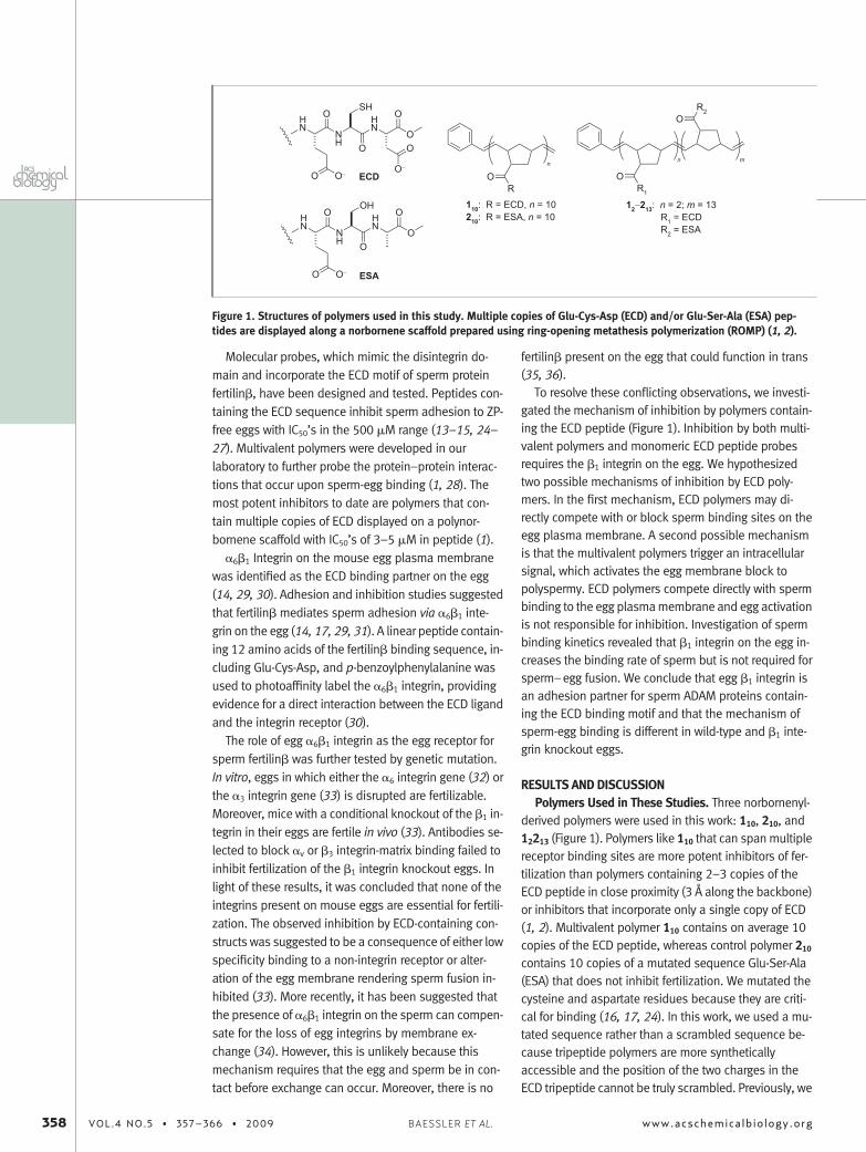
Molecular probes, which mimic the disintegrin do-main and incorporate the ECD motif of sperm proteinfertilin�, have been designed and tested. Peptides con-taining the ECD sequence inhibit sperm adhesion to ZP-free eggs with IC50’s in the 500 �M range (13–15, 24–27). Multivalent polymers were developed in ourlaboratory to further probe the protein–protein interac-tions that occur upon sperm-egg binding (1, 28). Themost potent inhibitors to date are polymers that con-tain multiple copies of ECD displayed on a polynor-bornene scaffold with IC50’s of 3–5 �M in peptide (1).
�6�1 Integrin on the mouse egg plasma membranewas identified as the ECD binding partner on the egg(14, 29, 30). Adhesion and inhibition studies suggestedthat fertilin� mediates sperm adhesion via �6�1 inte-grin on the egg (14, 17, 29, 31). A linear peptide contain-ing 12 amino acids of the fertilin� binding sequence, in-cluding Glu-Cys-Asp, and p-benzoylphenylalanine wasused to photoaffinity label the �6�1 integrin, providingevidence for a direct interaction between the ECD ligandand the integrin receptor (30).
The role of egg �6�1 integrin as the egg receptor forsperm fertilin� was further tested by genetic mutation.In vitro, eggs in which either the �6 integrin gene (32) orthe �3 integrin gene (33) is disrupted are fertilizable.Moreover, mice with a conditional knockout of the �1 in-tegrin in their eggs are fertile in vivo (33). Antibodies se-lected to block �v or �3 integrin-matrix binding failed toinhibit fertilization of the �1 integrin knockout eggs. Inlight of these results, it was concluded that none of theintegrins present on mouse eggs are essential for fertili-zation. The observed inhibition by ECD-containing con-structs was suggested to be a consequence of either lowspecificity binding to a non-integrin receptor or alter-ation of the egg membrane rendering sperm fusion in-hibited (33). More recently, it has been suggested thatthe presence of �6�1 integrin on the sperm can compen-sate for the loss of egg integrins by membrane ex-change (34). However, this is unlikely because thismechanism requires that the egg and sperm be in con-tact before exchange can occur. Moreover, there is no
fertilin� present on the egg that could function in trans(35, 36).
To resolve these conflicting observations, we investi-gated the mechanism of inhibition by polymers contain-ing the ECD peptide (Figure 1). Inhibition by both multi-valent polymers and monomeric ECD peptide probesrequires the �1 integrin on the egg. We hypothesizedtwo possible mechanisms of inhibition by ECD poly-mers. In the first mechanism, ECD polymers may di-rectly compete with or block sperm binding sites on theegg plasma membrane. A second possible mechanismis that the multivalent polymers trigger an intracellularsignal, which activates the egg membrane block topolyspermy. ECD polymers compete directly with spermbinding to the egg plasma membrane and egg activationis not responsible for inhibition. Investigation of spermbinding kinetics revealed that �1 integrin on the egg in-creases the binding rate of sperm but is not required forsperm–egg fusion. We conclude that egg �1 integrin isan adhesion partner for sperm ADAM proteins contain-ing the ECD binding motif and that the mechanism ofsperm-egg binding is different in wild-type and �1 inte-grin knockout eggs.
RESULTS AND DISCUSSIONPolymers Used in These Studies. Three norbornenyl-
derived polymers were used in this work: 110, 210, and12213 (Figure 1). Polymers like 110 that can span multiplereceptor binding sites are more potent inhibitors of fer-tilization than polymers containing 2–3 copies of theECD peptide in close proximity (3 Å along the backbone)or inhibitors that incorporate only a single copy of ECD(1, 2). Multivalent polymer 110 contains on average 10copies of the ECD peptide, whereas control polymer 210
contains 10 copies of a mutated sequence Glu-Ser-Ala(ESA) that does not inhibit fertilization. We mutated thecysteine and aspartate residues because they are criti-cal for binding (16, 17, 24). In this work, we used a mu-tated sequence rather than a scrambled sequence be-cause tripeptide polymers are more syntheticallyaccessible and the position of the two charges in theECD tripeptide cannot be truly scrambled. Previously, we
Figure 1. Structures of polymers used in this study. Multiple copies of Glu-Cys-Asp (ECD) and/or Glu-Ser-Ala (ESA) pep-tides are displayed along a norbornene scaffold prepared using ring-opening metathesis polymerization (ROMP) (1, 2).
358 VOL.4 NO.5 • 357–366 • 2009 www.acschemicalbiology.orgBAESSLER ET AL.
demonstrated that inhibition is sequence-dependentbecause a scrambled pentapeptide (Cys-Thr-Glu-Val-Asp) incorporated into a polymer does not inhibit fertili-zation, whereas the native sequence (Glu-Cys-Asp-Val-Thr) does (28). Polymer 12213, containing on average twoECD peptides at one terminus of the polymer, was usedas a low valency control as well as an aggregation con-trol. If supramolecular structures form in solution, the va-lency of a polymer would be higher than designed andinhibition might be due to the supramolecular structure.We found that polymer 12213 was no more effective aninhibitor than a monomeric ECD peptide (Supplemen-tary Figure 1). Thus, supramolecular aggregates are notresponsible for the inhibition observed in the experi-ments described below.
Inhibition of Fertilization by 110. We tested whetherthe �1 integrin is required for inhibition of fertilizationby ECD polymers. Eggs homozygous for the �1 integrinknockout allele (Cre��1f/f, KO), eggs heterozygous forthe �1 integrin knockout allele (Cre��1�/f, HET), andwild-type (Cre��1�/�, WT) eggs were obtained as pre-viously described (33). Immunofluorescence micros-copy with anti-�1 and anti-�6 integrin antibodies con-
firmed that the �1 integrin knockout eggs had no �1
integrin or �6 integrin on the plasma membrane (Supple-mentary Figure 2).
We chose to examine inhibition of fertilization usingZP-free eggs in order to isolate our observations to theegg plasma membrane. Previous research has shownthat ECD peptides are inhibitors of both ZP-intact andZP-free in vitro fertilization (13, 37). Thus, removal of theZP does not introduce an artifact into inhibition ofsperm-egg binding by these mimics. Moreover, we en-sured ample recovery time after removing the ZP so thategg fertilizability was not impaired (38).
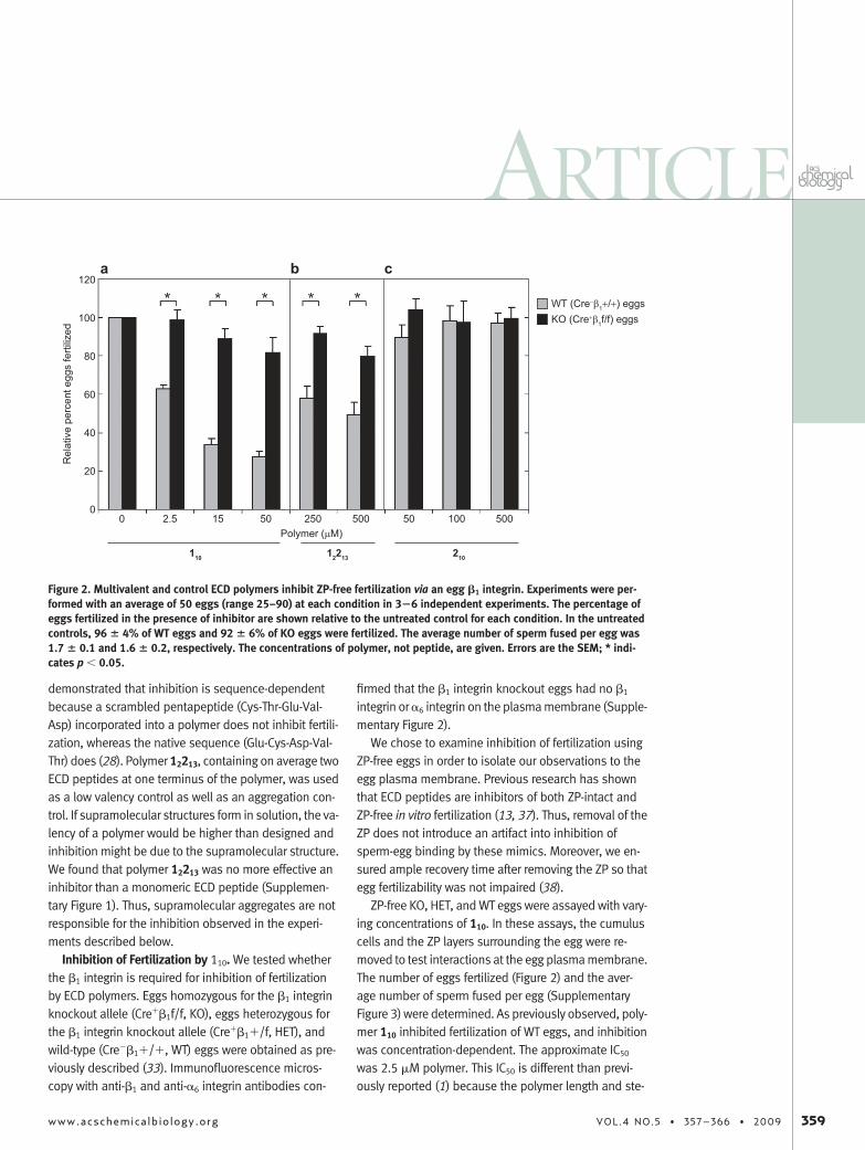
ZP-free KO, HET, and WT eggs were assayed with vary-ing concentrations of 110. In these assays, the cumuluscells and the ZP layers surrounding the egg were re-moved to test interactions at the egg plasma membrane.The number of eggs fertilized (Figure 2) and the aver-age number of sperm fused per egg (SupplementaryFigure 3) were determined. As previously observed, poly-mer 110 inhibited fertilization of WT eggs, and inhibitionwas concentration-dependent. The approximate IC50
was 2.5 �M polymer. This IC50 is different than previ-ously reported (1) because the polymer length and ste-
Figure 2. Multivalent and control ECD polymers inhibit ZP-free fertilization via an egg �1 integrin. Experiments were per-formed with an average of 50 eggs (range 25–90) at each condition in 3�6 independent experiments. The percentage ofeggs fertilized in the presence of inhibitor are shown relative to the untreated control for each condition. In the untreatedcontrols, 96 � 4% of WT eggs and 92 � 6% of KO eggs were fertilized. The average number of sperm fused per egg was1.7 � 0.1 and 1.6 � 0.2, respectively. The concentrations of polymer, not peptide, are given. Errors are the SEM; * indi-cates p � 0.05.
ARTICLE
www.acschemicalbiology.org VOL.4 NO.5 • 357–366 • 2009 359
reochemistry were different due to the use of a newerROMP precatalyst (2, 39, 40). Polymer 110 inhibited fer-tilization of KO eggs 19 � 6% at the highest concentra-tion used (50 �M in polymer, 500 �M in peptide), ascompared with 73 � 4% inhibition of WT egg fertiliza-tion at the same concentration. In experiments de-scribed below (Figure 5, panel b), when 1% DMSO wasincluded in the assay buffer with polymer 110, no inhibi-tion of KO fertilization was observed, whereas the inhi-bition of WT fertilization was unchanged. This result sug-gests that the small amount of inhibition detected inKO fertilization is due to non-specific hydrophobic bind-ing. Negative control polymer 210 containing ESA pep-tides was assayed and did not inhibit fertilization evenat 500 �M polymer (5 mM peptide, Figure 2, panel c).These data indicate that inhibition of fertilization by 110
is mediated by the �1 integrin on the egg membrane.Moreover, transfer of sperm �1 integrin to the egg mem-brane (34) cannot fully compensate for �1 integrindeletion.
Inhibition of Fertilization by 12213. We consideredthe possibility that the �1 integrin is required for bind-ing polymer inhibitor to the egg surface but that �1 inte-grin is not required for sperm binding. If the higher abun-dance �1 integrin were to act as an anchor for 110 andtether it to the egg surface, the avidity of terminal ECD li-gands binding to a second, lower abundance sperm re-ceptor would increase. In this scenario, inhibition by anon-avid, monovalent or low valency inhibitor thatblocked the second unknown receptor used by spermwould be equipotent in WT and KO fertilization. There-
fore we tested 12213, a low valency polymer, as an inhibi-tor of fertilization in WT and KO eggs (Figure 2, panel b).Polymer 12213 inhibited fertilization of WT eggs 100-fold less potently than polymer 110 as expected on thebasis of previous work. Polymer 12213 inhibits WT fertili-zation 51% at 500 �M, whereas only 20% inhibition isobserved in KO fertilization. This difference is statisti-cally significant ( p � 0.05) and indicates that ECD pep-tide binding to � 1 integrin on the WT egg blocks spermbinding. We observed that inhibition of HET fertilizationby polymer 110 is equipotent to inhibition of WT fertiliza-tion (Supplementary Figures 4 and 5). Therefore, theloss of inhibition is not due to differences in the ge-netic backgrounds of the mice, and thus �1 integrin-mediated avidity for a second sperm receptor is not re-sponsible for inhibition of WT fertilization.
Activation of Eggs by 110. We next sought to addresswhether inhibition occurs through egg activation. Dur-ing fertilization, egg activation triggers a complex se-quence of events, one of which is the establishment ofthe egg membrane’s block to polyspermic fertilization(6, 41). A downstream consequence of egg activationand intracellular calcium release from the ER is resump-tion of meiotic cell division and formation of the pronu-clei (42).
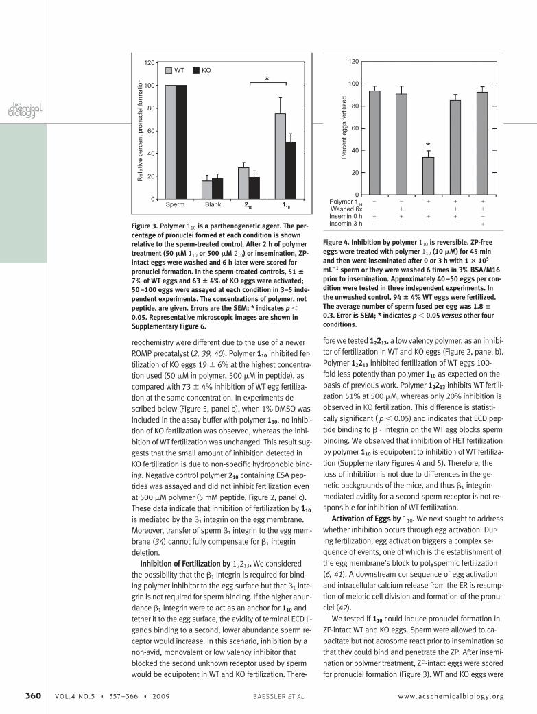
We tested if 110 could induce pronuclei formation inZP-intact WT and KO eggs. Sperm were allowed to ca-pacitate but not acrosome react prior to insemination sothat they could bind and penetrate the ZP. After insemi-nation or polymer treatment, ZP-intact eggs were scoredfor pronuclei formation (Figure 3). WT and KO eggs were
Figure 3. Polymer 110 is a parthenogenetic agent. The per-centage of pronuclei formed at each condition is shownrelative to the sperm-treated control. After 2 h of polymertreatment (50 �M 110 or 500 �M 210) or insemination, ZP-intact eggs were washed and 6 h later were scored forpronuclei formation. In the sperm-treated controls, 51 �7% of WT eggs and 63 � 4% of KO eggs were activated;50–100 eggs were assayed at each condition in 3–5 inde-pendent experiments. The concentrations of polymer, notpeptide, are given. Errors are the SEM; * indicates p �0.05. Representative microscopic images are shown inSupplementary Figure 6.
Figure 4. Inhibition by polymer 110 is reversible. ZP-freeeggs were treated with polymer 110 (10 �M) for 45 minand then were inseminated after 0 or 3 h with 1 � 105
mL�1 sperm or they were washed 6 times in 3% BSA/M16prior to insemination. Approximately 40–50 eggs per con-dition were tested in three independent experiments. Inthe unwashed control, 94 � 4% WT eggs were fertilized.The average number of sperm fused per egg was 1.8 �0.3. Error is SEM; * indicates p � 0.05 versus other fourconditions.
360 VOL.4 NO.5 • 357–366 • 2009 www.acschemicalbiology.orgBAESSLER ET AL.
both activated by 110, but less efficiently than sperm ac-tivate eggs. Polymer 110 appeared to activate more WTeggs than KO eggs, but the difference was not statisti-cally reliable. No significant activation was observedwith control polymer 210.
Next, we tested whether polymer 110 could inducecalcium oscillations. WT and KO eggs that were har-vested no later than 12 h after superovulation with hCGwere treated with polymer 110. Polymer 110 induced cal-cium oscillations in both WT and KO eggs, and the peakfrequencies, durations, and intensities were similar(Supplementary Figure 7). The control polymer 210 didnot induce oscillations in either egg type.
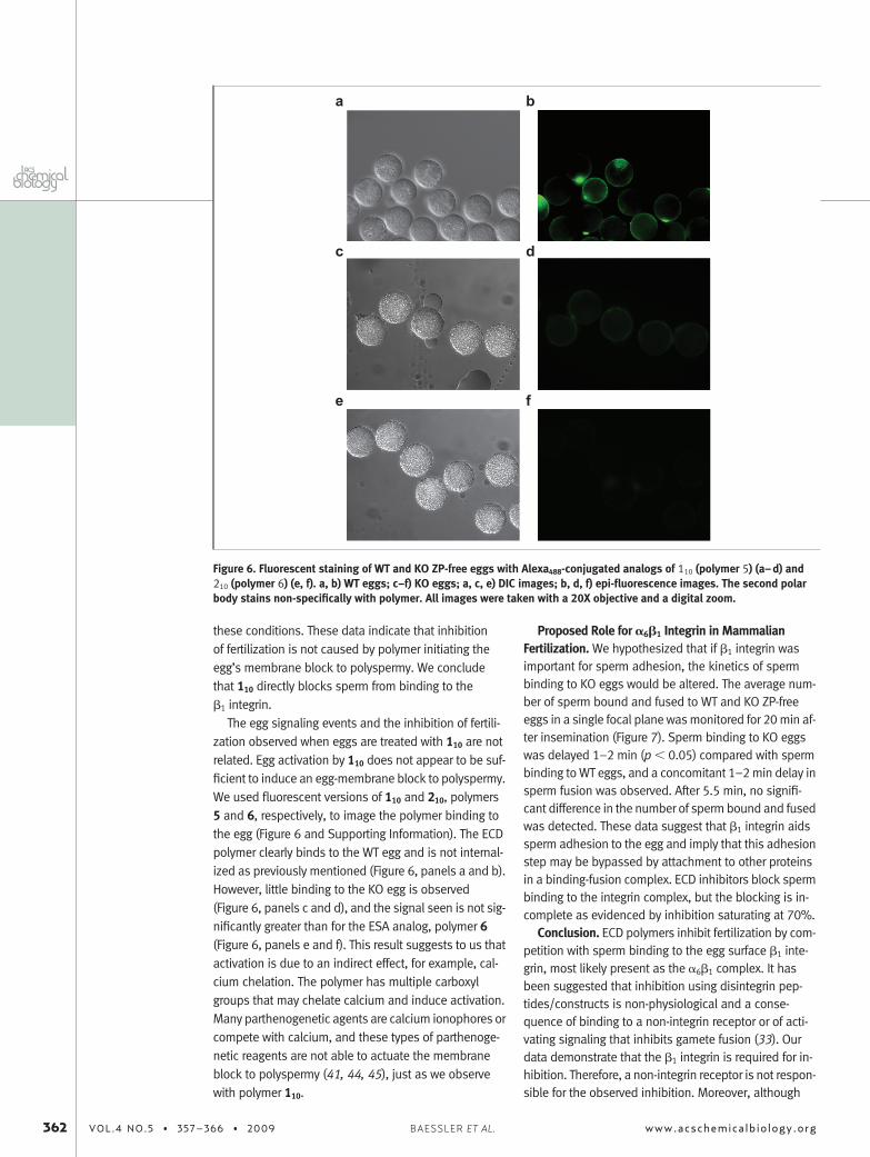
Activation versus Competition Model for Inhibition.If inhibition is due to activation of the egg membraneblock to polyspermy, the block is not expected to be re-versible (6, 41, 43). Therefore, we tested whether inhibi-tion by polymer 110 was reversible. A multivalent ECD flu-orescently tagged polymer was not internalized intoeggs (Figure 6), and after three washes the polymer wasnot detected on the surface of the egg (data not shown).Moreover, washing eggs six times does not affect eggpenetrability (Figure 4).
Eggs were treated with 110 and inseminated immedi-ately after washing or 3 h after washing to allow the eggmembrane block to reach a maximum (6, 43). Washingcompletely eliminated inhibition regardless of insemina-tion time. Reversible inhibition is consistent with a com-petitive binding mechanism and not an activationmechanism.
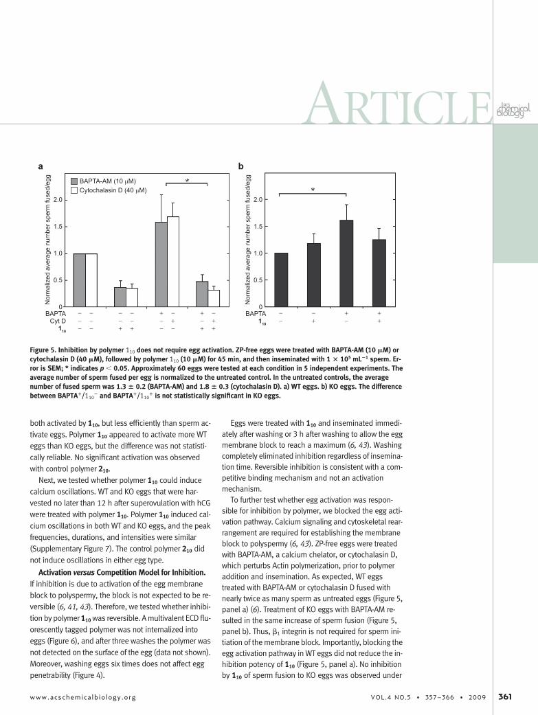
To further test whether egg activation was respon-sible for inhibition by polymer, we blocked the egg acti-vation pathway. Calcium signaling and cytoskeletal rear-rangement are required for establishing the membraneblock to polyspermy (6, 43). ZP-free eggs were treatedwith BAPTA-AM, a calcium chelator, or cytochalasin D,which perturbs Actin polymerization, prior to polymeraddition and insemination. As expected, WT eggstreated with BAPTA-AM or cytochalasin D fused withnearly twice as many sperm as untreated eggs (Figure 5,panel a) (6). Treatment of KO eggs with BAPTA-AM re-sulted in the same increase of sperm fusion (Figure 5,panel b). Thus, �1 integrin is not required for sperm ini-tiation of the membrane block. Importantly, blocking theegg activation pathway in WT eggs did not reduce the in-hibition potency of 110 (Figure 5, panel a). No inhibitionby 110 of sperm fusion to KO eggs was observed under
Figure 5. Inhibition by polymer 110 does not require egg activation. ZP-free eggs were treated with BAPTA-AM (10 �M) orcytochalasin D (40 �M), followed by polymer 110 (10 �M) for 45 min, and then inseminated with 1 � 105 mL�1 sperm. Er-ror is SEM; * indicates p � 0.05. Approximately 60 eggs were tested at each condition in 5 independent experiments. Theaverage number of sperm fused per egg is normalized to the untreated control. In the untreated controls, the averagenumber of fused sperm was 1.3 � 0.2 (BAPTA-AM) and 1.8 � 0.3 (cytochalasin D). a) WT eggs. b) KO eggs. The differencebetween BAPTA�/110
� and BAPTA�/110� is not statistically significant in KO eggs.
ARTICLE
www.acschemicalbiology.org VOL.4 NO.5 • 357–366 • 2009 361
these conditions. These data indicate that inhibitionof fertilization is not caused by polymer initiating theegg’s membrane block to polyspermy. We concludethat 110 directly blocks sperm from binding to the�1 integrin.
The egg signaling events and the inhibition of fertili-zation observed when eggs are treated with 110 are notrelated. Egg activation by 110 does not appear to be suf-ficient to induce an egg-membrane block to polyspermy.We used fluorescent versions of 110 and 210, polymers5 and 6, respectively, to image the polymer binding tothe egg (Figure 6 and Supporting Information). The ECDpolymer clearly binds to the WT egg and is not internal-ized as previously mentioned (Figure 6, panels a and b).However, little binding to the KO egg is observed(Figure 6, panels c and d), and the signal seen is not sig-nificantly greater than for the ESA analog, polymer 6(Figure 6, panels e and f). This result suggests to us thatactivation is due to an indirect effect, for example, cal-cium chelation. The polymer has multiple carboxylgroups that may chelate calcium and induce activation.Many parthenogenetic agents are calcium ionophores orcompete with calcium, and these types of parthenoge-netic reagents are not able to actuate the membraneblock to polyspermy (41, 44, 45), just as we observewith polymer 110.
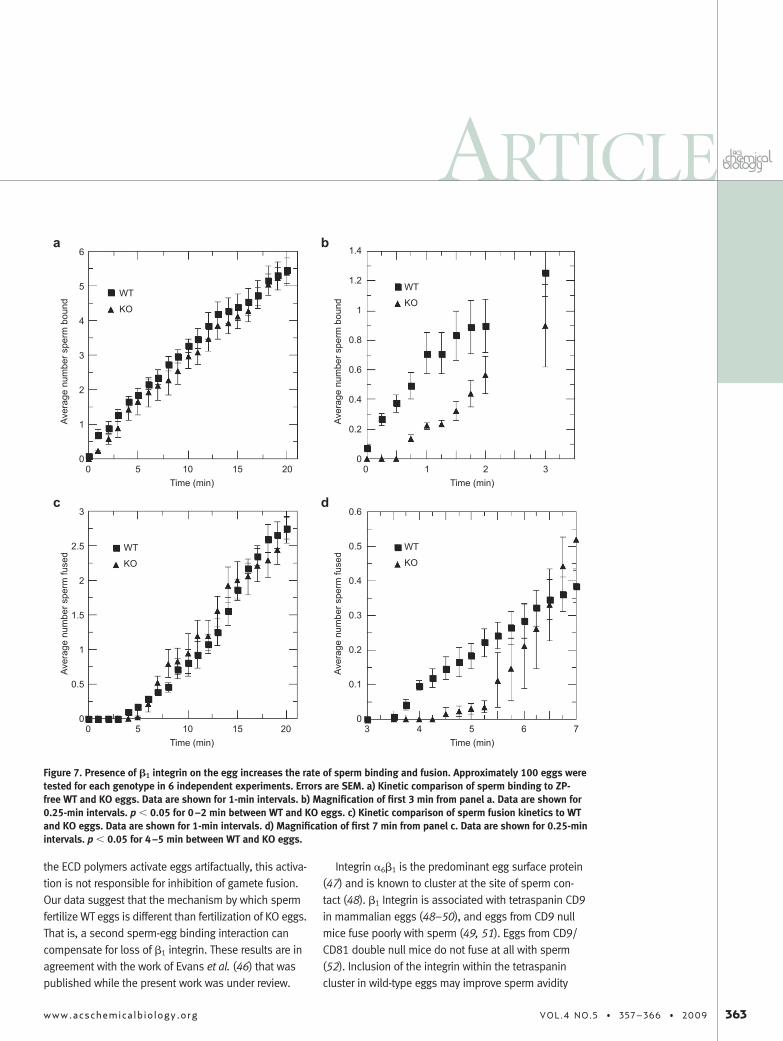
Proposed Role for �6�1 Integrin in MammalianFertilization. We hypothesized that if �1 integrin wasimportant for sperm adhesion, the kinetics of spermbinding to KO eggs would be altered. The average num-ber of sperm bound and fused to WT and KO ZP-freeeggs in a single focal plane was monitored for 20 min af-ter insemination (Figure 7). Sperm binding to KO eggswas delayed 1–2 min (p � 0.05) compared with spermbinding to WT eggs, and a concomitant 1–2 min delay insperm fusion was observed. After 5.5 min, no signifi-cant difference in the number of sperm bound and fusedwas detected. These data suggest that �1 integrin aidssperm adhesion to the egg and imply that this adhesionstep may be bypassed by attachment to other proteinsin a binding-fusion complex. ECD inhibitors block spermbinding to the integrin complex, but the blocking is in-complete as evidenced by inhibition saturating at 70%.
Conclusion. ECD polymers inhibit fertilization by com-petition with sperm binding to the egg surface �1 inte-grin, most likely present as the �6�1 complex. It hasbeen suggested that inhibition using disintegrin pep-tides/constructs is non-physiological and a conse-quence of binding to a non-integrin receptor or of acti-vating signaling that inhibits gamete fusion (33). Ourdata demonstrate that the �1 integrin is required for in-hibition. Therefore, a non-integrin receptor is not respon-sible for the observed inhibition. Moreover, although
Figure 6. Fluorescent staining of WT and KO ZP-free eggs with Alexa488-conjugated analogs of 110 (polymer 5) (a–d) and210 (polymer 6) (e, f). a, b) WT eggs; c–f) KO eggs; a, c, e) DIC images; b, d, f) epi-fluorescence images. The second polarbody stains non-specifically with polymer. All images were taken with a 20X objective and a digital zoom.
362 VOL.4 NO.5 • 357–366 • 2009 www.acschemicalbiology.orgBAESSLER ET AL.
the ECD polymers activate eggs artifactually, this activa-tion is not responsible for inhibition of gamete fusion.Our data suggest that the mechanism by which spermfertilize WT eggs is different than fertilization of KO eggs.That is, a second sperm-egg binding interaction cancompensate for loss of �1 integrin. These results are inagreement with the work of Evans et al. (46) that waspublished while the present work was under review.
Integrin �6�1 is the predominant egg surface protein(47) and is known to cluster at the site of sperm con-tact (48). �1 Integrin is associated with tetraspanin CD9in mammalian eggs (48–50), and eggs from CD9 nullmice fuse poorly with sperm (49, 51). Eggs from CD9/CD81 double null mice do not fuse at all with sperm(52). Inclusion of the integrin within the tetraspanincluster in wild-type eggs may improve sperm avidity
Figure 7. Presence of �1 integrin on the egg increases the rate of sperm binding and fusion. Approximately 100 eggs weretested for each genotype in 6 independent experiments. Errors are SEM. a) Kinetic comparison of sperm binding to ZP-free WT and KO eggs. Data are shown for 1-min intervals. b) Magnification of first 3 min from panel a. Data are shown for0.25-min intervals. p � 0.05 for 0–2 min between WT and KO eggs. c) Kinetic comparison of sperm fusion kinetics to WTand KO eggs. Data are shown for 1-min intervals. d) Magnification of first 7 min from panel c. Data are shown for 0.25-minintervals. p � 0.05 for 4–5 min between WT and KO eggs.
ARTICLE
www.acschemicalbiology.org VOL.4 NO.5 • 357–366 • 2009 363
for the egg surface. The incomplete blockage of spermbinding and fusion observed with ECD peptides andpolymers is consistent with the role of �1 integrin as anon-essential adhesion receptor (28). Therefore,�1 integrin KO eggs can bypass the sperm-integrinadhesion step, but as a consequence, sperm attach
and fuse to the egg plasma membrane more slowly.The reduced rate of binding does not impair fertilityunder laboratory mating conditions but may conferan evolutionary advantage in the wild that resultsin conservation of the integrin-fertilin� bindinginteraction.
METHODSGeneral Methods and Materials. All experiments performed
with mice were in accordance with the National Institutes ofHealth and United States Department of Agriculture guidelines,and the specific procedures performed were approved by theStony Brook University IACUC (protocol 0616). Mice containingthe floxed �1 integrin gene (53, 54) were provided by Ruth Glo-bus (NASA Ames Research Center) with permission from Rein-hardt Fassler (MPI, Martinsried). Transgenic mice expressing theCre recombinase under the control of the ZP3 promoter were ob-tained from Paul Primakoff (UC Davis) with permission fromJamie Marth (UC San Diego). Some mouse genotyping was per-formed by Transnetyx. All manipulations and incubations ofeggs were performed at 37 °C, 5% CO2 unless otherwise noted.Stock solutions of 1,2-bis(o-aminophenoxy)ethane-N,N,N=,N=-tetra-acetic acid acetoxymethyl ester (BAPTA-AM, Sigma) and cy-tochalasin D (Sigma) were prepared in dimethylsulfoxide(DMSO). Polymers were prepared with the [(H2IMes)(3-BrPyr)2Cl2RuACHPh] precatalyst (2, 55) instead of[(H2IMes)(PCy3)Cl2RuACHPh] as previously described (1).
Generation of Oocyte-Specific �1 Integrin Conditional KnockoutMice. Mice with the floxed �1 integrin gene and mice with theCre recombinase behind the ZP3 promoter were used to gener-ate oocyte-specific �1 integrin conditional knockout mice as pre-viously described by He et al. (33). PCR was used to genotypeprogeny to identify the ZP3-Cre transgene and the presence ofthe floxed �1 integrin gene. For ZP3 detection the following prim-ers were used:
Cre12: GGA CAT GTT CAG GGA TCG CCA GGC GCre13: GCA TAA CCA GTG AAA CAG CAT TGC TG
To detect the presence of the floxed �1 integrin gene the fol-lowing primers were used:
T56: AGG TGC CCT TCC CTC TAGAL26: TAA AAA GAC AGA ATA AAA CGCACL1: GTG AAG TAG GTG AAA GGT AAC
In Vitro Fertilization Assays. Sperm were isolated from thecauda epididymis and vas deferens of 8-month-old ICR retiredmale breeders (Taconic). Sperm were released from dissectedcauda and vas deferens into 3% BSA M16-modified Krebs-Ringer medium. Released sperm were incubated at 37 °C, 5%CO2 for 3 h in the same medium to allow them to capacitate andacrosome react. Eggs were collected from the oviducts of 8–10-week-old superovulated female ICR mice (Taconic) or C57 mu-tant progeny that were wild-type (Cre��1�/�), heterozygous(Cre��1�/f), or knockouts (Cre��1f/f) for the �1 allele. Mice weresuperovulated by injecting 5 IU PMSG (obtained through NHPP,NIDDK and Dr. A. F. Parlow), followed 48–52 h later by an injec-tion of 10 IU hCG. Then, 14–16 h after hCG injection, oviductswere removed from euthanized mice and were incubated in pre-warmed M16 medium with 0.5% BSA. Cumulus-egg complexeswere collected and transferred to 500-�L drops of medium con-taining 30 �g mL�1 hyaluronidase surrounded by mineral oil. Af-ter 5 min of incubation, cumulus-free metaphase II eggs werecollected, transferred first to an 80-�L drop of medium, and then
washed through six 40-�L drops of medium. Eggs were recov-ered for 1 h before treating with Tyrodes acid. Zona pellucidae(ZP) of metaphase II eggs were removed by incubating eggs in a100-�L Tyrodes acid drop for 1 min at RT followed by mechani-cal removal of the ZP through a pipet. ZP-free eggs were washedsix times with 0.5% BSA/M16 medium, recovered for 2 h in0.5% BSA/M16, and then loaded with Hoechst 33342 at 10 �gmL�1 for 30 min. Eggs were washed and placed in 100-�L dropsof 3% BSA/M16. At the same time, polymers were fully re-duced with 10 mM TCEP for 1–2 h, precipitated with 1 N HCl,washed with water, and then redissolved in water adjusted topH 7 with NH4OH. Polymer solution was added to the egg drop(no more than 5 �L of stock solution), and the eggs were incu-bated for 45 min prior to sperm addition. Eggs were inseminatedwith 1 � 105 sperm mL�1 for 45 min, washed in 3% BSA/M16,and mounted onto glass microscope slides. Sperm binding andfusion were scored by epi-fluorescence microscopy and DIC mi-croscopy (NIKON Eclipse 400, 40X, 0.75 NA objective). Fusionwas scored as the fluorescent labeling of sperm nuclei withHoechst 33342 present in the loaded eggs. The mean numberof sperm fused per egg (fertilization index, FI) and percentage ofeggs fertilized (fertilization rate, FR) were measured.
Immunofluorescence Microscopy. Cumulus and ZP layers wereremoved as described above, and eggs were allowed to recoverin 0.5% BSA/M16 for 2 h. Eggs were then treated with 30 �gmL�1 rat anti-�6 integrin mAb GoH3 (isotype IgG2a, MolecularProbes) or with 5 �g mL�1 rat anti-�1 mAb CD29 (isotype IgG2a,BD Pharmingen) for 45 min, washed in M16 for 10 min, andfixed with 4% paraformaldehyde. Then the eggs were stainedwith FITC (30 �g mL�1) or Alexa488-conjugated (5 �g mL�1) IgG2agoat anti-rat secondary antibody (Molecular Probes) for 45 min,washed, and mounted. Eggs were imaged on a Zeiss Axiovertwith a GFP/FITC filter and 0.55 NA, 20X objective. For polymer-labeled eggs, the recovered ZP-free oocytes were washed withfour 60-�L drops of 1% PVP/M16, placed in a 100-�L drop ofAlexa488-conjugated polymer solution in 1% PVP/M16, and incu-bated at 37 °C, 5% CO2 for 45 min. The polymer solutions wereprepared by diluting the stock polymer solutions with the buffer,and no more than 6 �L of stock solution was diluted. The con-centration of polymers in the 100-�L drop was 20 �M. Oocyteswere irradiated with UV light (max 350 nm, under 15 cm) at4 °C for 15 min. The photoaffinity labeled oocytes were gentlywashed twice through 300-�L drops of 1% PVP/M16 by shak-ing at 50 rpm for 10 min. After fixing the oocytes with a 100-�Ldrop of 4% paraformaldehyde in PBS at RT for 10 min, oocyteswere washed through four 60-�L drops of 0.5% BSA/M16,mounted, and imaged as described above.
Induction of Calcium Oscillations. Eggs were harvested nolater than 12 h after hCG injection. After ZP removal with Ty-rodes acid and recovery for 1.5–2 h, the ZP-free eggs were incu-bated for 30–40 min in 10 �M Fura-2AM, 0.025% PluronicF-127/0.05% BSA/M16. Eggs were washed, transferred to glass-bottom dishes (MatTek Corp.) that were pretreated with Cell-Tak (Sigma), and allowed to adhere for 10 min. Samples wereplaced on a microscope stage thermostatted at 37 °C. Polymer
364 VOL.4 NO.5 • 357–366 • 2009 www.acschemicalbiology.orgBAESSLER ET AL.
(30 �M) was added to egg samples directly on the microscopestage. The ratio of fluorescence emission at 510 nm with excita-tion at 340 and 380 nm was recorded using Carl Zeiss Axiovi-sion CD28 software.
Measurement of Pronuclei Formation. Eggs were harvested,and their cumulus cells were removed with hyaluronidase andallowed to recover for 1 h in 1.5% BSA/M16. Eggs were placedin 100-�L drops of the same buffer (covered with mineral oil),which contained either of the following: buffer only, capacitatedsperm (1 � 105 sperm mL�1), or polymers (110, 50 �M; 210,500 �M). After 2 h, all eggs were washed in parallel and incu-bated for another 6 h, at which time they were scored for pronu-clei formation by inspection under DIC optics. Prior to insemina-tion and after isolation from the cauda epididymis, sperm wereincubated in 1.5% BSA/M16-modified Krebs-Ringer medium37 °C, 5% CO2 for 1.5 h to allow capacitation without acrosomereaction.
Reversibility of Inhibition by Polymers. IVF inhibition assayswere performed as described above with the exception that theZP-free, polymer-treated (10 �M) eggs were either inseminatedwithout washing away polymer or were washed 6 times in 50-�Ldrops of 3% BSA/M16 prior to insemination. Capacitated andacrosome-reacted sperm were added to eggs at two time points:immediately after washing the eggs or 3 h after the wash. The fi-nal concentration of sperm was 1–5 � 105 sperm mL�1. Eggswere inseminated for 45 min and then washed with 3% BSA/M16. Eggs were mounted onto glass microscope slides, and FRand FI were scored as described above.
Inhibition of Egg Block to Polyspermy. ZP-free eggs wereloaded with Hoechst 33342 in 0.5% BSA/M16 as describedabove. Eggs were then treated with 10 �M BAPTA-AM or 40 �Mcytochalasin D for 60 min in 0.5% BSA/M16, 0.025% pluronicF-127. Control eggs were incubated in 1% DMSO. After 60 minof incubation with drug, BAPTA-AM treated eggs were washed 6times in 0.5% BSA/M16; cytochalasin D treated eggs were notwashed because actin perturbation induced by cytochalasin D isreversible, and the drug can be washed out (56). Drug-loadedeggs were treated with polymer (10 �M) as described above for45 min. Eggs were inseminated with 1 � 105 sperm mL�1 for45 min and washed with 3% BSA/M16, and the FI wasmeasured.
Sperm Binding Kinetics in �1 Knockout versus Wild-Type ZP-Free Eggs. After ZP removal with Tyrodes acid and 1.5 h recov-ery in 0.5% BSA/M16, eggs were transferred to glass bottomdishes pretreated with Cell-Tak and allowed to adhere for10 min. Samples were placed on a microscope stage thermo-statted at 37 °C and inseminated. The plane of focus was cen-tered on the equator of the egg. DIC and Hoechst 33342 imageswere recorded every 2 s for 20 min using Carl Zeiss AxiovisionCD28 Software. Images were scored for sperm bound and spermfused.
Acknowledgment: This research was supported by NationalInstitutes of Health grants R01HD38519 (N.S.S.), S10RR021008(N.S.S.), NYSTAR grant (FDP C040076, N.S.S.), and NSF grantCHE0131146 (N.M.R.). We thank J. Pazhayampallil for help withgenotyping.
Supporting Information Available: This material is availablefree of charge via the Internet at http://pubs.acs.org.
REFERENCES1. Baessler, K., Lee, Y., Roberts, K. S., Facompre, N., and Sampson,
N. S. (2006) Multivalent fertilin� oligopeptides: the dependence offertilization inhibition on length and density, Chem. Biol. 13, 251–259.
2. Lee, Y., and Sampson, N. S. (2009) Polymeric ADAM protein mim-ics interrogate mammalian sperm-egg binding, ChemBioChem 10,929–937.
3. Ducibella, T., Kurasawa, S., Rangarajan, S., Kopf, G. S., and Schultz,R. M. (1990) Precocious loss of cortical granules during mouse oo-cyte meiotic maturation and correlation with an egg-induced mod-ification of the zona pellucida, Dev. Biol. 137, 46–55.
4. Kline, D., and Kline, J. T. (1992) Repetitive calcium transients and therole of calcium in exocytosis and cell cycle activation in the mouseegg, Dev. Biol. 149, 80–89.
5. Jones, K. T. (2007) Intracellular calcium in the fertilization and devel-opment of mammalian eggs, Clin. Exp. Pharmacol. 34, 1084–1089.
6. McAvey, B. A., Wortzman, G. B., Williams, C. J., and Evans, J. P.(2002) Involvement of calcium signaling and the actin cytoskeletonin the membrane block to polyspermy in mouse eggs, Biol.Reprod. 67, 1342–1352.
7. Runft, L. L., Jaffe, L. A., and Mehlmann, L. M. (2002) Egg activationat fertilization: where it all begins, Dev. Biol. 245, 237–254.
8. Hoodbhoy, T., and Dean, J. (2004) Insights into the molecular basisof sperm-egg recognition in mammals, Reproduction 127, 417–422.
9. Seals, D. F., and Courtneidge, S. A. (2003) The ADAMs family of met-alloproteases: multidomain proteins with multiple functions, GenesDev. 17, 7–30.
10. Evans, J. P. (2001) Fertilin� and other ADAMs as integrin ligands: in-sights into cell adhesion and fertilization, Bioessays 23, 628–639.
11. Jia, L.-G., Wang, X.-M., and Fox, J. W. (1997) Function of disintegrin-like/cysteine-rich domains of atrolysin A. Inhibition of platelet aggre-gation by recombinant protein and peptide antagonists, J. Biol.Chem. 272, 13094.
12. McLane, M. A., Marcinkiewicz, C., Vijay-Kumar, S., Wierzbicka-Patynowski, I., and Niewiarowski, S. (1998) Viper venom disinte-grins and related molecules, Proc. Soc. Exp. Biol. Med. 219, 109–119.
13. Myles, D. G., Kimmel, L. H., Blobel, C. P., White, J. M., and Prima-koff, P. (1994) Identification of a binding site in the disintegrin do-main of fertilin required for sperm-egg fusion, Proc. Natl. Acad. Sci.U.S.A. 91, 4195–4198.
14. Almeida, E. A. C., Huovila, A. P. J., Sutherland, A. E., Stephens, L. E.,Calarco, P. G., Shaw, L. M., Mercurio, A. M., Sonnenberg, A., Prima-koff, P., Myles, D. G., and White, J. M. (1995) Mouse egg integrin�6�1 functions as a sperm receptor, Cell 81, 1095–1104.
15. Yuan, R., Primakoff, P., and Myles, D. G. (1997) A role for the disinte-grin domain of cyritestin, a sperm surface protein belonging to theADAM family, in mouse sperm-egg plasma membrane adhesionand fusion, J. Cell Biol. 137, 105–112.
16. Zhu, X., Bansal, N. P., and Evans, J. P. (2000) Identification of keyfunctional amino acids of the mouse fertilin� (ADAM2) disintegrinloop for cell-cell adhesion during fertilization, J. Biol. Chem. 275,7677–7683.
17. Bigler, D., Takahashi, Y., Chen, M. S., Almeida, E. A., Osbourne, L.,and White, J. M. (2000) Sequence-specific interaction between thedisintegrin domain of mouse ADAM 2 (fertilin�) and murine eggs.Role of the �(6) integrin subunit, J. Biol. Chem. 275, 11576–11584.
18. Primakoff, P., Hyatt, H., and Tredick-Kline, J. (1987) Identificationand purification of a sperm surface protein with a potential role insperm-egg membrane fusion, J. Cell Biol. 104, 141–149.
19. Cho, C., Bunch, D. O. D., Faure, J.-E., Goulding, E. H., Eddy, E. M., Pri-makoff, P., and Myles, D. G. (1998) Fertilization defects in spermfrom mice lacking fertilin�, Science 281, 1857–1859.
20. Nishimura, H., Cho, C., Branciforte, D. R., Myles, D. G., and Prima-koff, P. (2001) Analysis of loss of adhesive function in sperm lack-ing cyritestin or fertilin�, Dev. Biol. 233, 204–213.
ARTICLE
www.acschemicalbiology.org VOL.4 NO.5 • 357–366 • 2009 365
21. Kim, T., Oh, J., Woo, J.-M., Choi, E., Im, S. H., Yoo, Y. J., Kim, D. H.,Nishimura, H., and Cho, C. (2006) Expression and relationship ofmale reproductive ADAMs in mouse, Biol. Reprod. 74, 744–750.
22. Nishimura, H., Kim, E., Nakanishi, T., and Baba, T. (2004) Possiblefunction of the ADAM1a/ADAM2 Fertilin complex in the appear-ance of ADAM3 on the sperm surface, J. Biol.Chem. 279, 34957–34962.
23. Stein, K. K., Go, J. C., Primakoff, P., and Myles, D. G. (2005) Defectsin secretory pathway trafficking during sperm development inAdam2 knockout mice, Biol. Reprod. 73, 1032–1038.
24. Pyluck, A., Yuan, R., Jr., Primakoff, P., Myles, D. G., and Sampson,N. S. (1997) ECD peptides inhibit in vitro fertilization in mice, Bioorg.Med. Chem. Lett. 7, 1053–1058.
25. Gichuhi, P. M., Ford, W. C., and Hall, L. (1997) Evidence that pep-tides derived from the disintegrin domain of primate fertilin and con-taining the ECD motif block the binding of human spermatozoa tothe zona-free hamster oocyte, Int. J. Androl. 20, 165–170.
26. Bronson, R. A., Fusi, F. M., Calzi, F., Doldi, N., and Ferrari, A. (1999)Evidence that a functional fertilin-like ADAM plays a role in humansperm-oolemmal interactions, Mol. Hum. Reprod. 5, 433–440.
27. Gupta, S., Li, H., and Sampson, N. S. (2000) Characterization offertilin�-disintegrin binding specificity in sperm-egg adhesion,Bioorg. Med. Chem. 8, 723–729.
28. Roberts, S. K., Konkar, S., and Sampson, N. S. (2003) Comparisonof fertilin� peptide-substituted polymers and liposomes as inhibi-tors of in vitro fertilization, ChemBioChem 4, 1229–1231.
29. Evans, J. P., Kopf, G. S., and Schultz, R. M. (1997) Characterizationof the binding of recombinant mouse sperm fertilin � subunit tomouse eggs: evidence for adhesive activity via an egg �1 integrin-mediated interaction, Dev. Biol. 187, 79–93.
30. Chen, H., and Sampson, N. S. (1999) Mediation of sperm-egg fu-sion: evidence that mouse egg �6�1 integrin is the receptor forsperm fertilin�, Chem. Biol. 6, 1–10.
31. Tomczuk, M., Takahashi, Y., Huang, J., Murase, S., Mistretta, M.,Klaffky, E., Sutherland, A., Bolling, L., Coonrod, S., Marcinkiewicz,C., Sheppard, D., Stepp, M. A., and White, J. M. (2003) Role of multi-ple �1 integrins in cell adhesion to the disintegrin domains ofADAMs 2 and 3, Exp. Cell Res. 290, 68–81.
32. Miller, B. J., Georges-Labouesse, E., Primakoff, P., and Myles, D. G.(2000) Normal fertilization occurs with eggs lacking the integrin�6�1 and is CD9-dependent, J. Cell Biol. 149, 1289–1296.
33. He, Z.-Y., Brakebusch, C., Fassler, R., Kreidberg, J. A., Primakoff, P.,and Myles, D. G. (2003) None of the integrins known to be presenton the mouse egg or to be ADAM receptors are essential forsperm-egg binding and fusion, Dev. Biol. 254, 226–237.
34. Barraud-Lange, V., Naud-Barriant, N., Saffar, L., Gattegno, L., Ducot,B., Drillet, A. S., Bomsel, M., Wolf, J. P., and Ziyyat, A. (2007) �6�1Integrin expressed by sperm is determinant in mouse fertilization,BMC Dev. Biol. 7, 102.
35. Wolfsberg, T. G., Straight, P. D., Gerena, R. L., Huovila, A. P. J., Prima-koff, P., Myles, D. G., and White, J. M. (1995) ADAM, a widely distrib-uted and developmentally regulated gene family encoding mem-brane proteins with a disintegrin and metalloprotease domain, Dev.Biol. 169, 378–383.
36. Hooft van Huijsduijnen, R. (1998) ADAM 20 and 21: two novel hu-man testis-specific membrane metalloproteases with similarity tofertilin�, Gene 206, 273–282.
37. Evans, J. P., Schultz, R. M., and Kopf, G. S. (1998) Roles of the disin-tegrin domains of mouse fertilins � and � in fertilization, Biol.Reprod. 59, 145–152.
38. Takahashi, Y., Meno, C., Sato, E., and Toyoda, Y. (1995) Synchro-nous sperm penetration of zona-free mouse eggs in vitro, Biol.Reprod. 53, 424–430.
39. Love, J. A., Sanford, M. S., Day, M. W., and Grubbs, R. H. (2003) Syn-thesis, structure, and activity of enhanced initiators for olefin me-tathesis, J. Am. Chem. Soc. 125, 10103–10109.
40. Trnka, T. M., Morgan, J. P., Sanford, M. S., Wilhelm, T. E., Scholl, M.,Choi, T. L., Ding, S., Day, M. W., and Grubbs, R. H. (2003) Synthesisand activity of ruthenium alkylidene complexes coordinated withphosphine and N-heterocyclic carbene ligands, J. Am. Chem. Soc.125, 2546–2558.
41. Gardner, A. J., Williams, C. J., and Evans, J. P. (2007) Establishmentof the mammalian membrane block to polyspermy: evidence forcalcium-dependent and -independent regulation, Reproduction133, 383–393.
42. Ozil, J. P. (1998) Role of calcium oscillations in mammalian egg acti-vation: experimental approach, Biophys. Chem. 72, 141–152.
43. Wortzman-Show, G. B., Kurokawa, M., Fissore, R. A., and Evans, J. P.(2007) Calcium and sperm components in the establishment of themembrane block to polyspermy: studies of ICSI and activation withsperm factor, Mol. Hum. Reprod. 13, 557–565.
44. Sengoku, K., Tamate, K., Takaoka, Y., Horikawa, M., Goishi, K.,Okada, R., Tsuchiya, K., and Ishikawa, M. (1999) Requirement ofsperm-oocyte plasma membrane fusion for establishment of theplasma membrane block to polyspermy in human pronuclear oo-cytes, Mol. Reprod. Dev. 52, 183–188.
45. Horvath, P. M., Kellom, T., Caulfield, J., and Boldt, J. (1993) Mecha-nistic studies of the plasma membrane block to polyspermy inmouse eggs, Mol. Reprod. Dev. 34, 65–72.
46. Vjugina, U., Zhu, X., Oh, E., Bracero, N. J., and Evans, J. P. (2009) Re-duction of mouse egg surface integrin �9 subunit (ITGA9) reducesthe egg’s ability to support sperm-egg binding and fusion, Biol.Reprod. 80, 833–841.
47. Takahashi, Y., Yamakawa, N., Matsumoto, K., Toyoda, Y., Furukawa,K., and Sato, E. (2000) Analysis of the role of egg integrins in sperm-egg binding and fusion, Mol. Reprod. Dev. 56, 412–423.
48. Ziyyat, A., Rubinstein, E., Monier-Gavelle, F., Barraud, V., Kulski, O.,Prenant, M., Boucheix, C., Bomsel, M., and Wolf, J. P. (2006) CD9controls the formation of clusters that contain tetraspanins and theintegrin �6�1, which are involved in human and mouse gametefusion, J. Cell Sci. 119, 416–424.
49. Miyado, K., Yamada, G., Yamada, S., Hasuwa, H., Nakamura, Y., Ryu,F., Suzuki, K., Kosai, K., Inoue, K., Ogura, A., Okabe, M., andMekada, E. (2000) Requirement of CD9 on the egg plasma mem-brane for fertilization, Science 287, 321–324.
50. Takahashi, Y., Bigler, D., Ito, Y., and White, J. M. (2001) Sequence-specific interaction between the disintegrin domain of mouse ADAM3 and murine eggs: role of �1 integrin-associated proteins CD9,CD81, and CD98, Mol. Biol. Cell 12, 809–820.
51. Zhu, X., and Evans, J. P. (2002) Analysis of the roles of RGD-bindingintegrins, �(4)/�(9) integrins, �(6) integrins, and CD9 in the interac-tion of the fertilin � (ADAM2) disintegrin domain with the mouseegg membrane, Biol. Reprod. 66, 1193–1202.
52. Rubinstein, E., Ziyyat, A., Prenant, M., Wrobel, E., Wolf, J. P., Levy, S.,Le Naour, F., and Boucheix, C. (2006) Reduced fertility of femalemice lacking CD81, Dev. Biol. 290, 351–358.
53. Brakebusch, C., Grose, R., Quondamatteo, F., Ramirez, A., Jorcano,J. L., Pirro, A., Svensson, M., Herken, R., Sasaki, T., Timpl, R., Werner,S., and Fassler, R. (2000) Skin and hair follicle integrity is cruciallydependent on �1 integrin expression on keratinocytes, EMBO J. 19,3990–4003.
54. Potocnik, A. J., Brakebusch, C., and Fassler, R. (2000) Fetal and adulthematopoietic stem cells require �1 integrin function for colonizingfetal liver, spleen, and bone marrow, Immunity 12, 653–663.
55. Love, J. A., Morgan, J. P., Trnka, T. M., and Grubbs, R. H. (2002) Apractical and highly active ruthenium-based catalyst that effects thecross metathesis of acrylonitrile, Angew. Chem., Int. Ed. 41, 4035–4037.
56. Spector, I., Shochet, N. R., Blasberger, D., and Kashman, Y. (1989)Latrunculins–novel marine macrolides that disrupt microfilamentorganization and affect cell growth: I. Comparison with cytochala-sin D, Cell Motil. Cytoskeleton 13, 127–144.
366 VOL.4 NO.5 • 357–366 • 2009 www.acschemicalbiology.orgBAESSLER ET AL.