Oviducal sperm and fertilisation in poultry - hatchability.com
94
Oviducal sperm and fertilisation in poultry Murray R. Bakst Germplasm and Gamete Physiology Laboratory, Livestock and Poultry Sciences Institute, Beltsville Area, Agricultural Research Service, USDA, Building 262, Poultry Road, Beltsville, Maryland 20705, USA E-mail: [email protected] INTRODUCTION Wordsworth once recited, ‘‘The child is father to the man’’. In similar sense, the events that transpire in the avian oviduct and give rise to the fertilised ovum will impact the subsequent development of the embryo and hatchling. The oviduct is not only the organ responsible for the formation of the egg but it is intimately involved in such processes as sperm selec- tion, sperm storage, sperm transport, and ultimately fertilisation and early embryonic development. Whether sperm are present or not, every 25 hours or so, the ovulated yolk is gathered up by the infundi- bulum and begins its journey through the oviduct (Figure 1) where it accrues albumen in the magnum, the shell membranes in the isthmus, and the hard shell in the uterus [shell gland]. The most caudal oviducal segment, the vagina, serves as a conduit between the uterus and cloaca at oviposition. Oviposition marks the end of the daily ovulatory cycle with the next ovulation, generally within 45 minutes of oviposition, beginning the next ovulatory cycle. If the ovum is to be fertilised, it will take place in the infundibulum. Here sperm interacting with the ovum may have resided in the hen’s sperm-storage tubules (SST) located at the utero-vaginal junction (UVJ) (Figure 1) for a day, or weeks, following insemination. If fertilised, the developing embryo would have rapidly divided (see Fasenko, 2001) and at the time of oviposition, an embryo (blastoderm) of 60,000 cells (domestic fowl) or 30,000 cells (turkey) can be discerned on the surface of the ovum. In this chapter, our current knowledge of regarding the fate of sperm in the oviduct is briefly reviewed, particu- larly sperm storage at the distal end of the oviduct, and sperm:ovum interaction at the anterior end of the oviduct. SPERM SELECTION, STORAGE AND TRANSPORT Following sperm transfer into the vagina, which is accomplished by either natural mating or artificial insemination, sperm are selected for transport to the UVJ. This is either by some yet to be defined mechanism in the vagina, possibly immunological in nature (Wishart and Horrocks, 2000), or sperm succeed based on their own intrinsic mobility. This question of sperm competition is quite fascinating and has tremendous implications for commercial turkey breeding. For example, it is known that 50 – 70% of the progeny from hens inseminated with semen pooled form 12 – 15 toms will be derived from only one or two toms. Not only is this further limiting the gene pool for successive generations, but also it is not known if the toms producing the more ‘‘competitive sperm’’ have the most desirable phenotype. Located in the UVJ are the anatomical structures responsible for oviducal sperm storage, the SSTs (Figures 2, 3, 4 and 5). These are located predomi- nately in the apex of the UVJ mucosal folds and are generally located at the end of short fine folds or groves lined with cilia. Unlike the tubular glands found in the distal infundibulum, magnum, isthmus and uterus, the SSTs are not true glands. Histologically, the columnar cells forming the SST show little evidence of secretory activity (Figures 3, AVIAN BIOLOGY RESEARCH 2 (1/2), 2009 1–5 doi: 10.3184/175815509X430390 Figure 1 This turkey oviduct has a hard-shell egg in the uterus. The arrow denotes the utero-vaginal junction (UVJ), the location of the sperm-storage tubules (SST). The funnel-like fimbriated region of the infundibulum is evident (white arrow), as are the coiled magnum and isthmus segments.
-
Upload
khangminh22 -
Category
Documents
-
view
2 -
download
0
Transcript of Oviducal sperm and fertilisation in poultry - hatchability.com

Oviducal sperm and fertilisation in poultry
Murray R. Bakst
Germplasm and Gamete Physiology Laboratory, Livestock and Poultry Sciences Institute,Beltsville Area, Agricultural Research Service, USDA, Building 262, Poultry Road, Beltsville,Maryland 20705, USA E-mail: [email protected]
INTRODUCTION
Wordsworth once recited, ‘‘The child is father to theman’’. In similar sense, the events that transpire in theavian oviduct and give rise to the fertilised ovum willimpact the subsequent development of the embryoand hatchling. The oviduct is not only the organresponsible for the formation of the egg but it isintimately involved in such processes as sperm selec-tion, sperm storage, sperm transport, and ultimatelyfertilisation and early embryonic development.Whether sperm are present or not, every 25 hours or
so, the ovulated yolk is gathered up by the infundi-bulum and begins its journey through the oviduct(Figure 1) where it accrues albumen in the magnum,the shell membranes in the isthmus, and the hard shellin the uterus [shell gland]. The most caudal oviducalsegment, the vagina, serves as a conduit between theuterus and cloaca at oviposition. Oviposition marksthe end of the daily ovulatory cycle with the nextovulation, generally within 45 minutes of oviposition,beginning the next ovulatory cycle.
If the ovum is to be fertilised, it will take place in theinfundibulum. Here sperm interacting with the ovummay have resided in the hen’s sperm-storage tubules(SST) located at the utero-vaginal junction (UVJ)(Figure 1) for a day, or weeks, following insemination.If fertilised, the developing embryo would haverapidly divided (see Fasenko, 2001) and at the timeof oviposition, an embryo (blastoderm) of 60,000 cells(domestic fowl) or 30,000 cells (turkey) can bediscerned on the surface of the ovum. In thischapter, our current knowledge of regarding the fateof sperm in the oviduct is briefly reviewed, particu-larly sperm storage at the distal end of the oviduct,and sperm:ovum interaction at the anterior end of theoviduct.
SPERM SELECTION, STORAGE AND TRANSPORT
Following sperm transfer into the vagina, which isaccomplished by either natural mating or artificialinsemination, sperm are selected for transport to theUVJ. This is either by some yet to be definedmechanism in the vagina, possibly immunological innature (Wishart and Horrocks, 2000), or spermsucceed based on their own intrinsic mobility. Thisquestion of sperm competition is quite fascinating andhas tremendous implications for commercial turkeybreeding. For example, it is known that 50–70% ofthe progeny from hens inseminated with semenpooled form 12–15 toms will be derived from onlyone or two toms. Not only is this further limiting thegene pool for successive generations, but also it is notknown if the toms producing the more ‘‘competitivesperm’’ have the most desirable phenotype.Located in the UVJ are the anatomical structures
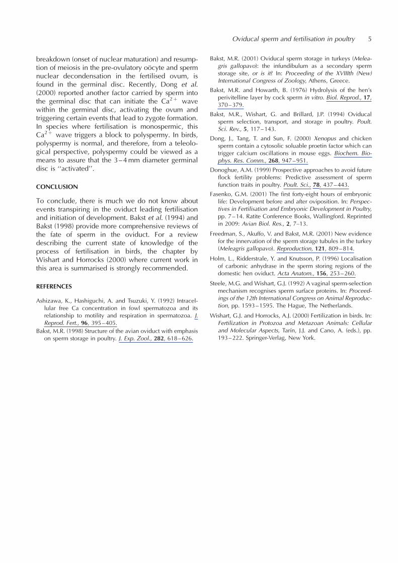
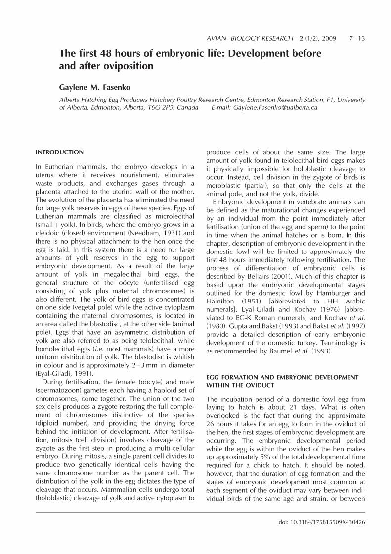
responsible for oviducal sperm storage, the SSTs(Figures 2, 3, 4 and 5). These are located predomi-nately in the apex of the UVJ mucosal folds and aregenerally located at the end of short fine folds orgroves lined with cilia. Unlike the tubular glandsfound in the distal infundibulum, magnum, isthmusand uterus, the SSTs are not true glands.Histologically, the columnar cells forming the SSTshow little evidence of secretory activity (Figures 3,
AVIAN BIOLOGY RESEARCH 2 (1/2), 2009 1–5
doi: 10.3184/175815509X430390
Figure 1 This turkey oviduct has a hard-shell egg in the uterus.The arrow denotes the utero-vaginal junction (UVJ), the
location of the sperm-storage tubules (SST). The funnel-likefimbriated region of the infundibulum is evident (white arrow),
as are the coiled magnum and isthmus segments.
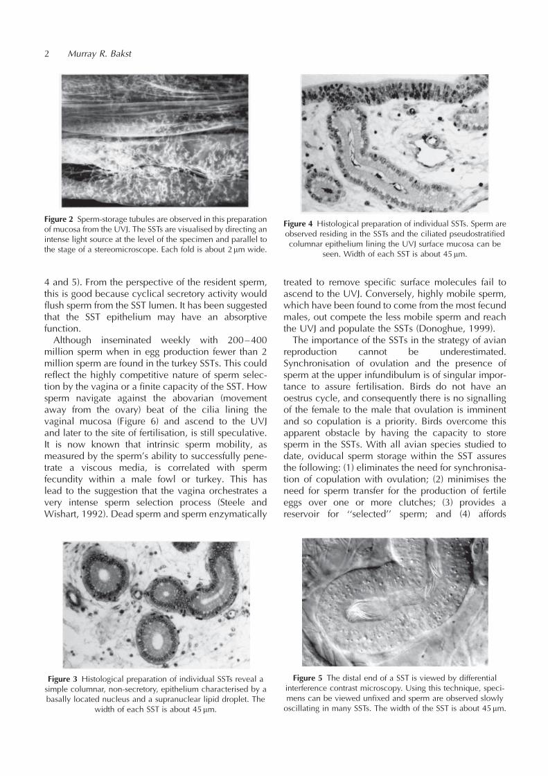
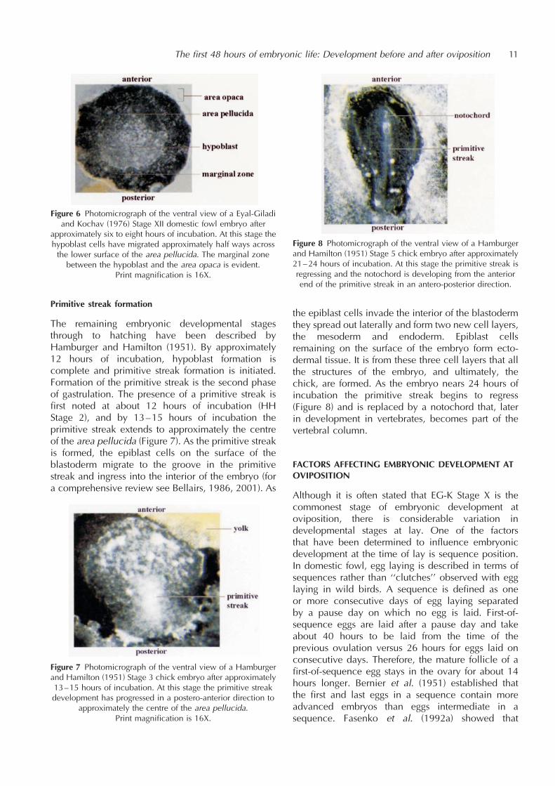
4 and 5). From the perspective of the resident sperm,this is good because cyclical secretory activity wouldflush sperm from the SST lumen. It has been suggestedthat the SST epithelium may have an absorptivefunction.Although inseminated weekly with 200–400
million sperm when in egg production fewer than 2million sperm are found in the turkey SSTs. This couldreflect the highly competitive nature of sperm selec-tion by the vagina or a finite capacity of the SST. Howsperm navigate against the abovarian (movementaway from the ovary) beat of the cilia lining thevaginal mucosa (Figure 6) and ascend to the UVJand later to the site of fertilisation, is still speculative.It is now known that intrinsic sperm mobility, asmeasured by the sperm’s ability to successfully pene-trate a viscous media, is correlated with spermfecundity within a male fowl or turkey. This haslead to the suggestion that the vagina orchestrates avery intense sperm selection process (Steele andWishart, 1992). Dead sperm and sperm enzymatically
treated to remove specific surface molecules fail toascend to the UVJ. Conversely, highly mobile sperm,which have been found to come from the most fecundmales, out compete the less mobile sperm and reachthe UVJ and populate the SSTs (Donoghue, 1999).The importance of the SSTs in the strategy of avian
reproduction cannot be underestimated.Synchronisation of ovulation and the presence ofsperm at the upper infundibulum is of singular impor-tance to assure fertilisation. Birds do not have anoestrus cycle, and consequently there is no signallingof the female to the male that ovulation is imminentand so copulation is a priority. Birds overcome thisapparent obstacle by having the capacity to storesperm in the SSTs. With all avian species studied todate, oviducal sperm storage within the SST assuresthe following: (1) eliminates the need for synchronisa-tion of copulation with ovulation; (2) minimises theneed for sperm transfer for the production of fertileeggs over one or more clutches; (3) provides areservoir for ‘‘selected’’ sperm; and (4) affords
2 Murray R. Bakst
Figure 2 Sperm-storage tubules are observed in this preparationof mucosa from the UVJ. The SSTs are visualised by directing anintense light source at the level of the specimen and parallel tothe stage of a stereomicroscope. Each fold is about 2mm wide.
Figure 3 Histological preparation of individual SSTs reveal asimple columnar, non-secretory, epithelium characterised by abasally located nucleus and a supranuclear lipid droplet. The
width of each SST is about 45 mm.
Figure 4 Histological preparation of individual SSTs. Sperm areobserved residing in the SSTs and the ciliated pseudostratifiedcolumnar epithelium lining the UVJ surface mucosa can be
seen. Width of each SST is about 45 mm.
Figure 5 The distal end of a SST is viewed by differentialinterference contrast microscopy. Using this technique, speci-mens can be viewed unfixed and sperm are observed slowlyoscillating in many SSTs. The width of the SST is about 45 mm.

protection to sperm during the daily ovulatory cycle.For many years there has been considerable researcheffort to attempt to understand how sperm survivewithin the SST. It is assumed that sperm residing in theSST are metabolically quiescent, motility suppressed,and that the plasmalemma and acrosomal membranesare stabilised. Whether resident sperm release signal-ling molecules to the surrounding SST epithelial cells(paracrine signaling) is also unknown. Possibly carbo-hydrate and lipid materials are transported to residentsperm. Carbonic anhydrase concentration, as well aszinc concentration in and around the SST epithelium,varies between the turkey, fowl, and quail (Holm et al.,1996).The actual mechanisms of sperm release from the
SST have been speculative and include a passiveescape of resident sperm while the hen is in eggproduction, to a squeezing of the sperm when theegg mass passes through the UVJ. Whatever themechanism is, it has to be assumed that the quiescentsperm in the SST are activated upon leaving the SST,or when in the vicinity or contact the ovum.Recently published observations (Freedman et al.,
2001) revealed that the SSTs are innervated and thatactin is present in the apical cytoplasm (terminal web)of the SST epithelial cells. The observation that nervefibres are associated with individual SST suggests thepossibility that a neural mechanism may be involvedin the release of sperm from the SST. The actin-richterminal web in the SST epithelium may be involvedin a contraction, or a series of contractions of the SST,thereby expelling sperm closest to the SST opening.Speculating further, cholinergic neurotransmittersliberated from nerve terminals adjacent to SST mayalter resident sperm plasmalemma permeability tocalcium ions (Ca2þ ). Given the crucial role of intra-cellular Ca2þ in avian sperm motility (Ashizawa et al.,1992), cholinergic induced influx of Ca2þ may reac-
tivate motility of some sperm cells leading to theirrapid escape from the SSTs. It has been proposedabove that there is a reversible suppression of spermmotility and metabolism, as well as a stabilisation ofthe sperm plasmalemma and acrosomal enzymesduring residency in the SST, an inactive or ‘‘decapa-citated’’ state (Bakst et al., 1994). An acetylcholine-induced influx of Ca2þ into the SST lumen mayrepresent a form of sperm activation (capacitation?).To fertilise a nearly daily succession of ova, sperm
are slowly but continuously released from the SST andascend to the site of fertilisation, the infundibulum,which is the most anterior segment of the oviduct.Here the sperm may accumulate in very smallnumbers. Given the attributes of the UVJ SSTs (seeabove), and the paucity of sperm at the infundibulumat any one time, it can be debated whether (Bakst,2001) whether the infundibulum should be consid-ered a secondary sperm storage site.To conclude here, an understanding of the biolo-
gical mechanisms regulating oviducal sperm transportand storage will provide the foundation for improve-ments in the efficiency of poultry breeding and aid inthe development of improved technologies for thepropagation of other domestic and feral birds andthe preservation of their germplasm.
SPERM: OOCYTE INTERACTION
One should not begin a discussion on fertilisationwithout briefly describing gamete structure and thefunction of the organelles in the process of fertilisa-tion. Briefly, galliform sperm are elongated and widest(0.6 mm) at the distal head region (Figures 6 and 7).The head consists of an elongated nucleus cappedwith an acrosome. The acrosome contains hydrolyticenzymes that when released at the time of the acro-some reaction, digest a path through the peri-vitellinelayer (PL), the acellular investment around the hen’sovum. The tail of the sperm consists of pairedcentrioles from which the flagellar portion of the tailis derived and the midpiece, a segment containing anarray of mitochondria. Surrounding the sperm is theplasmalemma.The megalecithal ovum (large yolky type; see
Fasenko, 2001) at ovulation consists of a vastvegetal region, characterised by yellow yolk, andthe 3–4mm diameter germinal disc. The germinaldisc contains while yolk spheres and cell organelles.In addition to organelles, the oolemma (the plasma-lemma of the ovum) in the germinal disc has a densearray of microvilli which project into the peri-vitellinespace between the underside of the PL and the ovum(Figure 8). Microvilli and the intact oolemma areunique to the germinal disc as the oolemma is
Oviducal sperm and fertilisation in poultry 3
Figure 6 This scanning electron micrograph shows sperm onthe cilia lining the vaginal mucosa. Note that the orientation ofthe sperm heads is in the opposite direction of the cilia beat.

discontinuous in the vegetal region and the yolk abutsdirectly to the underside of the PL.It is assumed that within a very short time window,
possibly minutes, one or dozens of sperm makecontact with the surface of the recently ovulatedovum, certainly before any secretions from the infun-dibulum begin to envelop the ovum and mask thesurface of the PL. The PL contains constitutive proteinsas well as glycoproteins that are involved in spermrecognition and initiation of the acrosome reaction.Sperm appear to bind to the PL (Figure 7) and mayundergo an acrosome reaction. In a process whichtranspires predominantly on the PL overlying thegerminal disc, sperm undergo the acrosome reaction,release one or more hydrolytic enzymes that digeststhe fibrous reticulum of the PL and transforms it into afibrillar mesh which sperm can penetrate (Figure 9). Ithas been suggested by Wishart and Horrocks (2000)that factors associated with the vegetal region (yellowyolk) may inhibit or some way deter sperm frompenetrating its overlying PL. Bakst and Howarth(1976) suggested that sperm receptors may be asso-ciated with the cytoplasmic extensions elaborated bythe oolemma at the germinal disc. Sperm in the peri-vitelline space appear to be engulfed by the microvilliand this intimate association leads to fusion of theinner acrosomal membrane and the oolemma.Subsequently the sperm are incorporated into thevitellus (the ovum). There appears to be no limit asto the number of sperm that can hydrolyse the PL asthere is no block to polyspermy other than theaccretion of oviducal secretions. Conversely, incor-poration of the sperm into the ovum by fusion with theoolemma may have a physiological limit.In the germinal disc, the sperm nucleus undergoes a
transformation from a highly condensed elongatedorganelle to a spherical pronucleus. While several
sperm nuclei may undergo this transformation onlyone aligns closely apposed to the female pronucleusin the centre of the germinal disc and completessyngamy, the reconstitution of the diploid number ofchromosomes, to form the zygote. Supernumerarysperm, those sperm not involved in syngamy, alsoundergo nuclear decondensation and some at leastform pronuclei within an hour following ovulation.Within 4 hours of ovulation centrally located apposedmale and female pronuclei, as well as some of themore peripherally located sperm pronuclei, undergomitosis initiating the first cleavage of the zygote.Little is known of the events that trigger sperm
nuclear decondensation and the onset of embryogen-esis. Most likely maturation-promoting factor (MPF),which is found in mammalian and amphibian oocytesand is thought to bring about both germinal vesicle
4 Murray R. Bakst
Figure 7 A scanning electron micrograph reveals a cluster ofsperm and some single sperm on the surface of the peri-vitellinelayer (PL). The PL was isolated from a recently ovulated ovum
and co-cultured with sperm before fixation.
Figure 8 The dense array of microvilli observed in this scanningelectron micrograph is restricted to the germinal disc region.The microvilli, which are about 1.5 mm in height, are thought toassist in the incorporation of the sperm into the vitellus (ovum).
White yolk spheres are shown at bottom of the picture.
Figure 9 A histological radial section of the germinal discshowing areas of the peri-vitelline layer (PL) that have been
hydrolysed by sperm (arrows). Dots are yolk spheres. The intactPL is about 2 mm thick.
breakdown (onset of nuclear maturation) and resump-tion of meiosis in the pre-ovulatory oocyte and spermnuclear decondensation in the fertilised ovum, isfound in the germinal disc. Recently, Dong et al.(2000) reported another factor carried by sperm intothe germinal disc that can initiate the Ca2þ wavewithin the germinal disc, activating the ovum andtriggering certain events that lead to zygote formation.In species where fertilisation is monospermic, thisCa2þ wave triggers a block to polyspermy. In birds,polyspermy is normal, and therefore, from a teleolo-gical perspective, polyspermy could be viewed as ameans to assure that the 3–4mm diameter germinaldisc is ‘‘activated’’.
CONCLUSION
To conclude, there is much we do not know aboutevents transpiring in the oviduct leading fertilisationand initiation of development. Bakst et al. (1994) andBakst (1998) provide more comprehensive reviews ofthe fate of sperm in the oviduct. For a reviewdescribing the current state of knowledge of theprocess of fertilisation in birds, the chapter byWishart and Horrocks (2000) where current work inthis area is summarised is strongly recommended.
REFERENCES
Ashizawa, K., Hashiguchi, A. and Tsuzuki, Y. (1992) Intracel-lular free Ca concentration in fowl spermatozoa and itsrelationship to motility and respiration in spermatozoa. J.Reprod. Fert., 96, 395–405.
Bakst, M.R. (1998) Structure of the avian oviduct with emphasison sperm storage in poultry. J. Exp. Zool., 282, 618–626.
Bakst, M.R. (2001) Oviducal sperm storage in turkeys (Melea-gris gallopavo): the infundibulum as a secondary spermstorage site, or is it? In: Proceeding of the XVIIIth (New)International Congress of Zoology, Athens, Greece.
Bakst, M.R. and Howarth, B. (1976) Hydrolysis of the hen’sperivitelline layer by cock sperm in vitro. Biol. Reprod., 17,370–379.
Bakst, M.R., Wishart, G. and Brillard, J.P. (1994) Oviducalsperm selection, transport, and storage in poultry. Poult.Sci. Rev., 5, 117–143.
Dong, J., Tang, T. and Sun, F. (2000) Xenopus and chickensperm contain a cytosolic soluable proetin factor which cantrigger calcium oscillations in mouse eggs. Biochem. Bio-phys. Res. Comm., 268, 947–951.
Donoghue, A.M. (1999) Prospective approaches to avoid futureflock fertility problems: Predictive assessment of spermfunction traits in poultry. Poult. Sci., 78, 437–443.
Fasenko, G.M. (2001) The first forty-eight hours of embryoniclife: Development before and after oviposition. In: Perspec-tives in Fertilisation and Embryonic Development in Poultry,pp. 7–14. Ratite Conference Books, Wallingford. Reprintedin 2009: Avian Biol. Res., 2, 7–13.
Freedman, S., Akuffo, V. and Bakst, M.R. (2001) New evidencefor the innervation of the sperm storage tubules in the turkey(Meleagris gallopavo). Reproduction, 121, 809–814.
Holm, L., Ridderstrale, Y. and Knutsson, P. (1996) Localisationof carbonic anhydrase in the sperm storing regions of thedomestic hen oviduct. Acta Anatom., 156, 253–260.
Steele, M.G. and Wishart, G.J. (1992) A vaginal sperm-selectionmechanism recognises sperm surface proteins. In: Proceed-ings of the 12th International Congress on Animal Reproduc-tion, pp. 1593–1595. The Hague, The Netherlands.
Wishart, G.J. and Horrocks, A.J. (2000) Fertilization in birds. In:Fertilization in Protozoa and Metazoan Animals: Cellularand Molecular Aspects, Tarın, J.J. and Cano, A. (eds.), pp.193–222. Springer-Verlag, New York.
Oviducal sperm and fertilisation in poultry 5
The first 48 hours of embryonic life: Development beforeand after oviposition
Gaylene M. Fasenko
Alberta Hatching Egg Producers Hatchery Poultry Research Centre, Edmonton Research Station, F1, Universityof Alberta, Edmonton, Alberta, T6G 2P5, Canada E-mail: [email protected]
INTRODUCTION
In Eutherian mammals, the embryo develops in auterus where it receives nourishment, eliminateswaste products, and exchanges gases through aplacenta attached to the uterine wall of the mother.The evolution of the placenta has eliminated the needfor large yolk reserves in eggs of these species. Eggs ofEutherian mammals are classified as microlecithal(smallþ yolk). In birds, where the embryo grows in acleidoic (closed) environment (Needham, 1931) andthere is no physical attachment to the hen once theegg is laid. In this system there is a need for largeamounts of yolk reserves in the egg to supportembryonic development. As a result of the largeamount of yolk in megalecithal bird eggs, thegeneral structure of the oocyte (unfertilised eggconsisting of yolk plus maternal chromosomes) isalso different. The yolk of bird eggs is concentratedon one side (vegetal pole) while the active cytoplasmcontaining the maternal chromosomes, is located inan area called the blastodisc, at the other side (animalpole). Eggs that have an asymmetric distribution ofyolk are also referred to as being telolecithal, whilehomolecithal eggs (i.e. most mammals) have a moreuniform distribution of yolk. The blastodisc is whitishin colour and is approximately 2–3mm in diameter(Eyal-Giladi, 1991).During fertilisation, the female (oocyte) and male
(spermatozoon) gametes each having a haploid set ofchromosomes, come together. The union of the twosex cells produces a zygote restoring the full comple-ment of chromosomes distinctive of the species(diploid number), and providing the driving forcebehind the initiation of development. After fertilisa-tion, mitosis (cell division) involves cleavage of thezygote as the first step in producing a multi-cellularembryo. During mitosis, a single parent cell divides toproduce two genetically identical cells having thesame chromosome number as the parent cell. Thedistribution of the yolk in the egg dictates the type ofcleavage that occurs. Mammalian cells undergo total(holoblastic) cleavage of yolk and active cytoplasm to
produce cells of about the same size. The largeamount of yolk found in telolecithal bird eggs makesit physically impossible for holoblastic cleavage tooccur. Instead, cell division in the zygote of birds ismeroblastic (partial), so that only the cells at theanimal pole, and not the yolk, divide.Embryonic development in vertebrate animals can
be defined as the maturational changes experiencedby an individual from the point immediately afterfertilisation (union of the egg and sperm) to the pointin time when the animal hatches or is born. In thischapter, description of embryonic development in thedomestic fowl will be limited to approximately thefirst 48 hours immediately following fertilisation. Theprocess of differentiation of embryonic cells isdescribed by Bellairs (2001). Much of this chapter isbased upon the embryonic developmental stagesoutlined for the domestic fowl by Hamburger andHamilton (1951) [abbreviated to HH Arabicnumerals], Eyal-Giladi and Kochav (1976) [abbre-viated to EG-K Roman numerals] and Kochav et al.(1980). Gupta and Bakst (1993) and Bakst et al. (1997)provide a detailed description of early embryonicdevelopment of the domestic turkey. Terminology isas recommended by Baumel et al. (1993).
EGG FORMATION AND EMBRYONIC DEVELOPMENTWITHIN THE OVIDUCT
The incubation period of a domestic fowl egg fromlaying to hatch is about 21 days. What is oftenoverlooked is the fact that during the approximate26 hours it takes for an egg to form in the oviduct ofthe hen, the first stages of embryonic development areoccurring. The embryonic developmental periodwhile the egg is within the oviduct of the hen makesup approximately 5% of the total developmental timerequired for a chick to hatch. It should be noted,however, that the duration of egg formation and thestages of embryonic development most common ateach segment of the oviduct may vary between indi-vidual birds of the same age and strain, or between
AVIAN BIOLOGY RESEARCH 2 (1/2), 2009 7–13
doi: 10.3184/175815509X430426

birds of varying strains and ages. These processes aresummarised here.
Fertilisation and extrusion of the second polar body
In birds, egg formation occurs as the yolk passesthrough five portions of the oviduct (infundibulum,magnum, isthmus, uterus [shell gland], and vagina)and coincides with early developmental processes ofthe embryo. After ovulation into the body cavity, theyolk (secondary oocyte) enters the first portion of theoviduct (the infundibulum). Fertilisation takes place,assuming sperm are present, during the 15 to 30minutes the oocyte stays in the infundibulum (seeBakst, 2001). Within an hour after fertilisa-tion of the oocyte, the second polar body(discarded nucleus) is extruded from theoocyte thereby completing the secondmeiotic division (Olsen and Fraps, 1944).During meiotic division a single parent cellproduces four genetically identical cellshaving half the chromosome number ofthe parent cell. The fertilised yolk (oocyte)is now called an ovum. Meiosis iscompleted during the two to three hourmovement of the ovum through themagnum, where albumen proteins aredeposited.
Cleavage of embryonic cells(EG-K Stages I to VI)
The inner and outer shell membranes aredeposited during the 60 to 75 minute
interval that the ovum takes to travelthrough the isthmus. While the ovum isentering the uterus, approximately three tofive hours after fertilisation, the first clea-vage furrow develops (Olsen, 1942).Cleavage of the zygote (the fertilised singlecell) involves repeated mitotic divisions toproduce an embryo with thousands ofgenetically identical cells. As mentionedpreviously, the large amount of yolkcontained in the ovum of telolecithal eggsmakes it impossible for division of the yolkto occur and therefore the cleavage isrestricted to the active cytoplasm locatedat the animal pole (Figure 1).Shell deposition occurs in the uterus
[shell gland] over the next 18–21 hours. Itis likely that the second cleavage (Figure2A) through to the sixth cleavages occurduring the first two hours that the ovum is inthe uterus (Eyal-Giladi, 1991). At the 8-cell
stage of development (Figure 2B) the blastomeres arenot completely surrounded by the cell membrane,and the cell contents are therefore still in contactwith the yolk (Figure 2C); blastomeres of this natureare described as ‘‘open cells’’ (Bellairs et al., 1978).Only after two hours in the uterus do the central cellsseparate from the yolk when the deep ends of thecleavage furrows gradually merge together horizon-tally to form a fluid filled sub-blastodermic cavity(Figure 2C; Eyal-Giladi, 1984). The formation of thesub-blastodermic cavity is complete approximately 6hours later (Kochav et al. 1980). At this point hori-zontal as well as vertical cell cleavage occurs (Eyal-Giladi, 1991).
8 Gaylene M. Fasenko
Figure 1 Diagrammatic representation of the first mitotic division in adomestic fowl embryo. (a) View of entire ovum showing the embryo
(embryo diameter is approximately 1y15 to 1y20 of the diameter of theovum). (b) Enlargement of the animal pole area of the ovum showing a viewof the entire embryo with the first cleavage furrow producing two cells(blastomeres). (c) Cross sectional view of the first cleavage furrow. Thecleavage furrow does not extend down through the entire depth of the
embryo, thus the two blastomeres are not entirely delineated from the yolk.The cytoplasm of the cells and the yolk mix in an area termed the periblast
(Bellairs et al., 1978). The peripheral periblast is called the marginalperiblast, while the periblast beneath the embryo is termed the
sub-germinal periblast.Diagram adapted from Barrett et al. (1985) and Etches (1996).
(a) (c)(b)
Figure 2 Diagrammatic representation of the second and third mitoticdivisions in a domestic fowl embryo. (a) Enlargement of the animal pole ofthe ovum showing a view of the entire embryo and the first (I) and second(II) cleavage furrows (4-cell stage). (b) Enlargement of the animal pole
showing a view of the entire embryo and the first, second, and third (III)cleavage furrows (8-cell stage). (c) Longitudinal cross sectional view of an
8-cell embryo. At this stage of development the cytoplasm of theblastomeres is still in contact with the yolk and the blastomeres aredescribed as ‘‘open’’ cells. The arrows indicate the gradual horizontal
merging of the deep ends of the cleavage furrows. These isolated spaceseventually combine to form the fluid filled sub-blastodermic cavity
(Eyal-Giladi, 1984).Diagram adapted from Romanoff (1960) and Bellairs et al. (1978).
(a) (b) (c)

Formation of the area pellucida (EG-K Stages VII-X)
Approximately 11 hours after the presence of the firstcleavage furrow, at the end of the cleavage period, theembryo, now called a blastoderm, is a circular discabout five to six cells thick in the centre but taperingout to one to two at the periphery (Figure 3A; Bellairs,1971). The formation of the area pellucida marks thefirst morphogenetic event in the development of thedomestic fowl embryo. This stage of development(EG-K VII) marks the initiation of cellular sheddingfrom the central area of the blastoderm, which occursover the next 8–9 hours while the ovum is still in theuterus. The shed cells fall into the anterior portion ofthe sub-blastodermic cavity and likely disintegratemerging with the yolk. The most ventrally locatedcells in the embryo next to the sub-blastodermiccavity continue to shed over the next 8–9 hourssuch that at the time the egg is laid, the blastodermconsists of a central area which is one cell layer thick,with a peripheral area that is several layers thick andis attached to the yolk (Figure 3D). The cell sheddingstarts at the future posterior side of the embryo andmoves towards the future anterior side (for a review ofaxis determination in the avian embryo see Eyal-Giladi, 1991; Khaner, 1993; and Bellairs, 2001). The
translucent central area of the blastoderm is called thearea pellucida, while the peripheral ‘‘ring’’ of cellsthat are still in contact with the yolk, is termed thearea opaca. It is the epiblast cells that compose thearea pellucida, which eventually form all the struc-tures in the embryo proper.
POST-OVIPOSITIONAL EMBRYONIC DEVELOPMENT
By the time the egg is laid, the embryo consists of40,000 to 60,000 cells. The most common stage ofembryonic development at oviposition is EG-K Stage X(Figure 4). The catalyst that initiates embryonic devel-opment once the egg is laid is the environmentaltemperature to which the egg is exposed. Artificialincubation of eggs in commercial incubators takesplace at temperatures between 37.0–38.0�C.Embryonic developmental rate may vary due to avariety of genetic and environmental factors,however, the approximate number of incubationhours required to reach the most prevalent stage ofdevelopment are shown based on an recent experi-ment conducted by the author.
The first 48 hours of embryonic life: Development before and after oviposition 9
Figure 3 Diagrammatic representation of the stages of embryonic development in the domestic fowl associated with the formation ofthe area pellucida. (a) Longitudinal cross sectional view through a blastoderm at Eyal-Giladi and Kochav (EG-K) (1976) Stage VII. Atthis stage cell shedding has begun at the future posterior end of the blastoderm. (b) Longitudinal cross sectional view through ablastoderm at EG-K Stage VIII. Cell shedding continues in a postero-anterior fashion and reaches the centre of the blastoderm.
(c) Longitudinal cross sectional view through a blastoderm at EG-K Stage IX. The blastoderm is between one to three cell layers thick.(d) Longitudinal cross sectional view through a blastoderm at EG-K Stage X. At this juncture all the blastodermal cells in the lowerlayer have been shed into the sub-blastodermic cavity. The result is a blastoderm that has a clearly demarcated central area that istranslucent (area pellucida) surrounded by an opaque ring of cells that are still attached to the yolk (area opaca). Diagrams adapted
from photomicrographs from Kochav et al. (1980).
(a) (b)
(c) (d)

Hypoblast formation (EG-K Stages XI-XIII)
Stage XI embryos are most prevalent between threeand six hours of incubation. At this stage of develop-
ment (EG-K XI) a sickle-shaped cluster of cells (Koller’ssickle) appears at the posterior side of the area opaca(Figure 5A). The origin of these cells has been a topicof debate (Stern, 1990; Eyal-Giladi, 1991, 1992). Thisevent marks the beginning of the first stage of gastrula-tion of the chick embryo, the formation of the hypo-blast and epiblast. Gastrulation is defined as theperiod during embryonic development when cellularmovements in the embryo transform the single celllayered embryo into an embryo with two or threedistinct layers of cells. Between six and eight hours ofincubation, the hypoblast cells have migratedapproximately half way across the lower surface ofthe area pellucida (EG-K Stage XII; Figure 5B andFigure 6). By nine to ten hours of incubation, theformation of the hypoblast is complete such that thehypoblast forms a continuous disc of cells on theunderside of the blastoderm (EG-K Stage XIII; Figure5C). Two to three hours later, the blastoderm hasformed a bridge of cells between the hypoblast andthe area opaca (EG-K Stage XIV). The sole goal of thehypoblast is to induce primitive streak formation inthe epiblast layer.
10 Gaylene M. Fasenko
Figure 4 Photomicrograph of an Eyal-Giladi and Kochav (1976)Stage X domestic fowl blastoderm showing a central translucentdisc, the area pellucida, surrounded by an opaque ring, the areaopaca, surrounding the disc. This is the most common stage of
embryonic development at the time of oviposition.Print magnification is 16X.
Figure 5 Diagrammatic representation of the stages of embryonic development in the domestic fowl associated with hypoblastformation. (a) Longitudinal cross sectional view through a blastoderm at Eyal-Giladi and Kochav [EG-K] (1976) Stage XI. At this stageclusters of hypoblast cells line approximately one third of the posterior portion of the blastoderm. (b) Longitudinal cross sectionalview through a blastoderm at EG-K Stage XII. Hypoblast formation continues in an anterior direction and now occupies half of theunder surface of the blastoderm. (c) Longitudinal cross sectional view through a blastoderm at EG-K Stage XIII. Hypoblast formationis complete and covers the central portion of the area pellucida. A hypoblast-free ring of area pellucida called the marginal zone
borders the hypoblast. Diagrams adapted from photomicrographs from Kochav et al. (1980).
(a) (b)
(c)
Primitive streak formation
The remaining embryonic developmental stagesthrough to hatching have been described byHamburger and Hamilton (1951). By approximately12 hours of incubation, hypoblast formation iscomplete and primitive streak formation is initiated.Formation of the primitive streak is the second phaseof gastrulation. The presence of a primitive streak isfirst noted at about 12 hours of incubation (HHStage 2), and by 13–15 hours of incubation theprimitive streak extends to approximately the centreof the area pellucida (Figure 7). As the primitive streakis formed, the epiblast cells on the surface of theblastoderm migrate to the groove in the primitivestreak and ingress into the interior of the embryo (fora comprehensive review see Bellairs, 1986, 2001). As
the epiblast cells invade the interior of the blastodermthey spread out laterally and form two new cell layers,the mesoderm and endoderm. Epiblast cellsremaining on the surface of the embryo form ecto-dermal tissue. It is from these three cell layers that allthe structures of the embryo, and ultimately, thechick, are formed. As the embryo nears 24 hours ofincubation the primitive streak begins to regress(Figure 8) and is replaced by a notochord that, laterin development in vertebrates, becomes part of thevertebral column.
FACTORS AFFECTING EMBRYONIC DEVELOPMENT ATOVIPOSITION
Although it is often stated that EG-K Stage X is thecommonest stage of embryonic development atoviposition, there is considerable variation indevelopmental stages at lay. One of the factorsthat have been determined to influence embryonicdevelopment at the time of lay is sequence position.In domestic fowl, egg laying is described in terms ofsequences rather than ‘‘clutches’’ observed with egglaying in wild birds. A sequence is defined as oneor more consecutive days of egg laying separatedby a pause day on which no egg is laid. First-of-sequence eggs are laid after a pause day and takeabout 40 hours to be laid from the time of theprevious ovulation versus 26 hours for eggs laid onconsecutive days. Therefore, the mature follicle of afirst-of-sequence egg stays in the ovary for about 14hours longer. Bernier et al. (1951) established thatthe first and last eggs in a sequence contain moreadvanced embryos than eggs intermediate in asequence. Fasenko et al. (1992a) showed that
The first 48 hours of embryonic life: Development before and after oviposition 11
Figure 6 Photomicrograph of the ventral view of a Eyal-Giladiand Kochav (1976) Stage XII domestic fowl embryo after
approximately six to eight hours of incubation. At this stage thehypoblast cells have migrated approximately half ways acrossthe lower surface of the area pellucida. The marginal zone
between the hypoblast and the area opaca is evident.Print magnification is 16X.
Figure 7 Photomicrograph of the ventral view of a Hamburgerand Hamilton (1951) Stage 3 chick embryo after approximately13–15 hours of incubation. At this stage the primitive streakdevelopment has progressed in a postero-anterior direction to
approximately the centre of the area pellucida.Print magnification is 16X.
Figure 8 Photomicrograph of the ventral view of a Hamburgerand Hamilton (1951) Stage 5 chick embryo after approximately21–24 hours of incubation. At this stage the primitive streak isregressing and the notochord is developing from the anteriorend of the primitive streak in an antero-posterior direction.

embryos of first-of-sequence eggs were significantlymore developed than embryos from subsequenteggs in a sequence and that embryonic viabilitywas lower in embryos from first-of-sequence eggs.The extended period that the follicle of a first-of-sequence egg remains in the ovary means that theremay be ageing effects on the follicle that contributeto an increase in development and a decrease inembryonic viability. It has also been established thatthat blastoderm area increases as birds age (Matherand Laughlin, 1979). This may be related tosequence position, as older hens lay shortersequences and thus have a higher percentage offirst of sequence eggs (Robinson et al., 1990, 1991).Mather and Laughlin (1979) hypothesised that theadvanced embryonic development in older birdscould also be due to the egg staying in theoviduct for a longer period of time, either becauseof a delayed rate of egg passage or because of alonger oviduct.In an effort to manage hatching egg numbers, fertile
hatching eggs are regularly stored at cool tempera-tures for usually a few days although storage canextend to a few weeks. It is usually assumed thatbelow a temperature of 20–21�C (physiological zero)embryonic development is arrested. Using the Eyal-Giladi and Kochav (1976) classifications of embryonicdevelopment, Fasenko et al. (1992b) confirmed thatembryonic development is arrested when hatchingeggs are held at 14�C. However, managementfactors such as frequency of egg collection andmethod of egg storage can influence embryonicdevelopment between oviposition and egg storageby increasing the amount of time required for theinternal egg temperature to cool below physiologicalzero (Fasenko et al., 1991).
FACTORS AFFECTING EMBRYONIC DEVELOPMENTDURING THE FIRST 24 HOURS OF INCUBATION
One of the major factors in the hatching egg industrythat negatively affects post-ovipositional embryonicdevelopment is storing eggs longer than one week. Itis well established that the amount of incubation timerequired for a chick to hatch is lengthened whenhatching eggs are stored for extended periods (Kosinand Konishi, 1973; Mather and Laughlin, 1976,1977). The hypotheses put forth to explain this werethat (1) the initiation of embryonic development isdelayed, andyor (2) that embryonic developmentproceeds at a slower rate, in embryos from eggsstored for long periods. Unpublished data recentlycollected by the author verified that eggs stored for 14days had embryos in which chronological age laggedbehind the biological age. A comprehensive exam-
ination of development during the first 24 hours by theauthor showed that not all embryos reacted in thesame manner to storage. Some embryos of 14 daystored eggs did not initiate development once incuba-tion temperatures were provided, while other embryosbegan development, but at a slower rate. It was alsoshown that the metabolism of embryos, as measuredindirectly by embryonic CO2 output, proceeds at aslower rate. Perhaps the most significant finding fromthe study was that the development of some embryosexposed to 14 days of storage was not affected. Thisfact may provide the basis for future studies aimed atgenetically selecting for factors that allow theseembryos to withstand the negative effects of eggstorage.Arora and Kosin (1968) provided an indication of
the negative physiological effects of long term storageon embryonic cell viability. In this study it was shownthat the number of embryonic cells with necroticnuclei increased as storage duration increased. Areduction in viable cell numbers may be the reasonbehind the observation made by Mather and Laughlin(1979) that there is shrinkage of the blastoderm wheneggs are stored for 7 or 14 days. In this study they alsoshowed that there was an increase in malformedembryos with storage, especially in eggs from youngand old birds. Recent research has established thatapoptosis (programmed cell death) increases whenembryos are stored for 14 days at 12�C (Bloom et al.,1998). It is the hypothesis of this author that long termexposure of avian embryos to storage below physio-logical zero may increase the number of necrotic(dead) or apoptotic cells relative to viable embryoniccells. This may inhibit initiation of embryonic devel-opment andyor impede normal development thusresulting in a higher percentage of embryonicabnormality and mortality, as there may be anoptimum number of viable embryonic cells requiredfor normal growth and development. Researchcurrently underway by the author and colleaguesaims to elucidate the relationship between long termstorage and the incidence of necrotic and apoptoticcells.
CONCLUSIONS
The objective of this chapter was to provide asummary of the stages of chick embryonic develop-ment during the 24 hours before and after oviposition,and to discuss some of the major factors affecting thisperiod of development. The information provided wasnot meant to be comprehensive, but was meant togive readers that are unfamiliar with early stages ofavian embryonic development an appreciation for thisarea of study. Although the development in the
12 Gaylene M. Fasenko
oviduct and during the first 24 hours of incubationonly make up about 9% of the total developmentalperiod of the embryo, these early stages are critical forsuccessful development of the embryo and hatchingof the chick. A general understanding of earlyembryonic development may also aid hatcherymanagers in problem solving fertility and hatchabilityissues in domestic fowl.
REFERENCES
Arora, K.L. and Kosin, I.L. (1968) The response of the earlychicken embryo to pre-incubation temperature as evidencedfrom its gross morphology and mitotic pattern. Physiol.Zool., 41, 104–112.
Bakst, M.R. (2001) Oviducal sperm and fertilisation in poultry.In: Perspectives in Fertilisation and Embryonic Developmentin Poultry, Baggott, G.K., Bakst, M.R., Bellairs, R., Christen-sen, V.L., Fasenko, G.M. and Starck, J.M. (eds.), pp. 1–6.Ratite Conference Books, Wallingford. Reprinted in 2009:Avian Biol. Res., 2, 1–5.
Bakst, M.R., Gupta, S.K. and Akuffo, V. (1997) Comparativedevelopment of the turkey and chicken embryo from clea-vage through hypoblast formation. Poult. Sci., 76, 83–90.
Baumel, J.J., King, A.S., Breazile, J.E., Evans, H.E. and VandenBerge, J.C. (1993) Handbook of Avian Anatomy: NominaAnatomica Avium, 2nd Edition. Nuttall Ornithological Club,Cambridge, Massachusetts.
Barrett, J.M., Abramoff, P., Kumaran, A.K. and Millington, W.F.(1985) Biology. Prentice-Hall, Englewood Cliffs, New Jersy.
Bellairs, R. (1971). Developmental Processes in Higher Verte-brates. Logos Press Ltd. Great Britain.
Bellairs, R. (1986). The primitive streak. Anat. Embryol., 174,1–14.
Bellairs, R. (2001) Some critical events in the differentiation ofthe avian embryo. In: Perspectives in Fertilisation andEmbryonic Development in Poultry, Baggott, G.K., Bakst,M.R., Bellairs, R., Christensen, V.L., Fasenko, G.M. andStarck, J.M. (eds.), pp. 15–22. Ratite Conference Books,Wallingford. Reprinted in 2009: Avian Biol. Res., 2, 15–20.
Bellairs, R., Lorenz, F.W. and Dunlap, T. (1978) Cleavage in thechick embryo. J. Embryol. Exp. Morph., 43, 55–69.
Bernier, P.E., Taylor, L.W. and Gunns, C.A. (1951) The relativeeffects of inbreeding and outbreeding on reproduction in thedomestic fowl. Hilgardia, 20, 529–628.
Bloom, S.E., Muscarella, D.E., Lee, M.Y. and Rachlinski, M.(1998) Cell death in the avian blastoderm: resistance tostress-induced apoptosis and expression of anti-apoptoticgenes. Cell Death Different., 5, 529–538.
Etches, R.J. (1996) Reproduction in Poultry. CAB International,Wallingford, UK.
Eyal-Giladi, H. (1984) The gradual establishment of cell com-mitments during the early stages of chick development. CellDifferent., 14, 245–255.
Eyal-Giladi, H. (1991) The early embryonic development of thechick, as an epigenetic process. Crit. Rev. Poult. Biol., 3,143–166.
Eyal-Giladi, H. (1992) The posterior section of the chicks areapellucida and its involvement in hypoblast and primitivestreak formation. Development, 116, 819–830.
Eyal-Giladi, H. and Kochav, S. (1976). From cleavage toprimitive streak formation: A complementary normal tableand a new look at the first stages of the development of thechick. I. General Morphology. Develop. Biol., 49, 321–337.
Fasenko, G.M., Robinson, F.E., Armstrong, J.G., Church, J.S. andHardin, R.T. (1991) Variability in preincubation embryodevelopment in domestic fowl. 1. Effects of nest holdingtime and method of egg storage. Poult. Sci., 70, 1876–1881.
Fasenko, G.M., Hardin, R.T., Robinson, F.E. and Wilson, J.L.(1992a) Relationship of hen age and egg sequence positionwith fertility, hatchability, viability, and preincubationembryonic development in broiler breeders. Poult. Sci., 71,1374–1383.
Fasenko, G.M., Robinson, F.E. and Hardin, R.T. (1992b) Varia-bility in preincubation embryonic development in domesticfowl. 2. Effects of duration of egg storage period. Poult. Sci.,71, 2129–2132.
Gupta, S.K. and Bakst, M.R. (1993) Turkey embryo staging fromcleavage throughhypoblast formation. J.Morph.,217, 313–325.
Hamburger, V. and Hamilton, H.L. (1951) A series of normalstages in the development of the chick embryo. J. Morph.,88, 49–92. Reprinted (1992) Develop. Dynamics, 195,231–272.
Khaner, O. (1993) Axis determination in the avian embryo. Curr.Top. Develop. Biol., 28, 155–180.
Kochav, S., Ginsburg, M. and Eyal-Giladi, H. (1980) Fromcleavage to primitive streak formation: A complementarynormal table and a new look at the first stages of thedevelopment of the chick. II. Microscopic Anatomy andCell Population Dynamics. Develop. Biol., 79, 296–308.
Kosin, I.L. and Konishi, T. (1973) Preincubation storage condi-tions and their effect on the subsequent livability of chickenembryos: exogenous CO2, plastic bags and extending theholding periods as factors. Poult. Sci., 52, 296–302.
Mather, C.M. and Laughlin, K.F. (1976) Storage of hatchingeggs: The effect on total incubation period. Brit. Poult. Sci.,17, 471–479.
Mather, C.M. and Laughlin, K.F. (1976) Storage of hatchingeggs: The effect on early embryonic development. Brit.Poult. Sci. (1977), 18, 597–603.
Mather, C.M. and Laughlin, K.F. (1979) Storage of hatchingeggs. The interaction between parental age and earlyembryonic development. Brit. Poult. Sci., 20, 595–604.
Needham, J. (1931) Chemical Embryology. Cambridge.
Olsen, M.W. (1942) Maturation, fertilization and early cleavagein the hen’s egg. J. Morph., 70, 513–533.
Olsen, M.W. and Fraps, R.M. (1944) Maturation, fertilizationand early cleavage of the egg of the domestic turkey. J.Morph., 74, 297–309.
Robinson, F.E., Hardin, R.T. and Robblee, A.R. (1990) Repro-ductive senescence in domestic fowl: Effects on egg produc-tion, sequence length and inter-sequence pause length. Brit.Poult. Sci., 31, 871–879.
Robinson, F.E., Robinson, N.A. and Scott, T.A. (1991) Repro-ductive performance, growth rate and body composition offull-fed versus feed-restricted broiler breeder hens. Can. J.Anim. Sci., 71, 549–556.
Romanoff, A.L. (1960) The Avian Embryo. MacMillan, NewYork.
Stern, C.D. (1990) The marginal zone and its contribution to thehypoblast and primitive streak of the embryo. Develop., 109,667–682.
The first 48 hours of embryonic life: Development before and after oviposition 13
Some critical events in the differentiation of the avianembryo
Ruth Bellairs
Department of Anatomy and Developmental Biology, University College London, Gower Street,London, WC1E 6BT, UK E-mail: [email protected]
INTRODUCTION
The term ‘‘differentiation’’ covers all those complexchanges that occur in a developing embryo and areconcerned with the conversion of a simple group ofcells into a complex individual with organs andtissues. Differentiation is not the same as growth,which is usually defined as an increase in bodymass, though the two are associated. If there weregrowth but no differentiation the embryo would neverbecome more than a large ball of cells. There couldnot be differentiation without growth as there wouldnot then be enough cells to form each organ.Differentiation is essential for a normal embryo to
develop and consists of a continuous sequence ofevents. Some of these are especially critical, however,and if they do not take place correctly the embryobecomes malformed or fails to develop at all. Thisreview briefly covers the basic principles of thesecritical events in development.
ESTABLISHMENT OF THE BODY AXES (POLARITY)
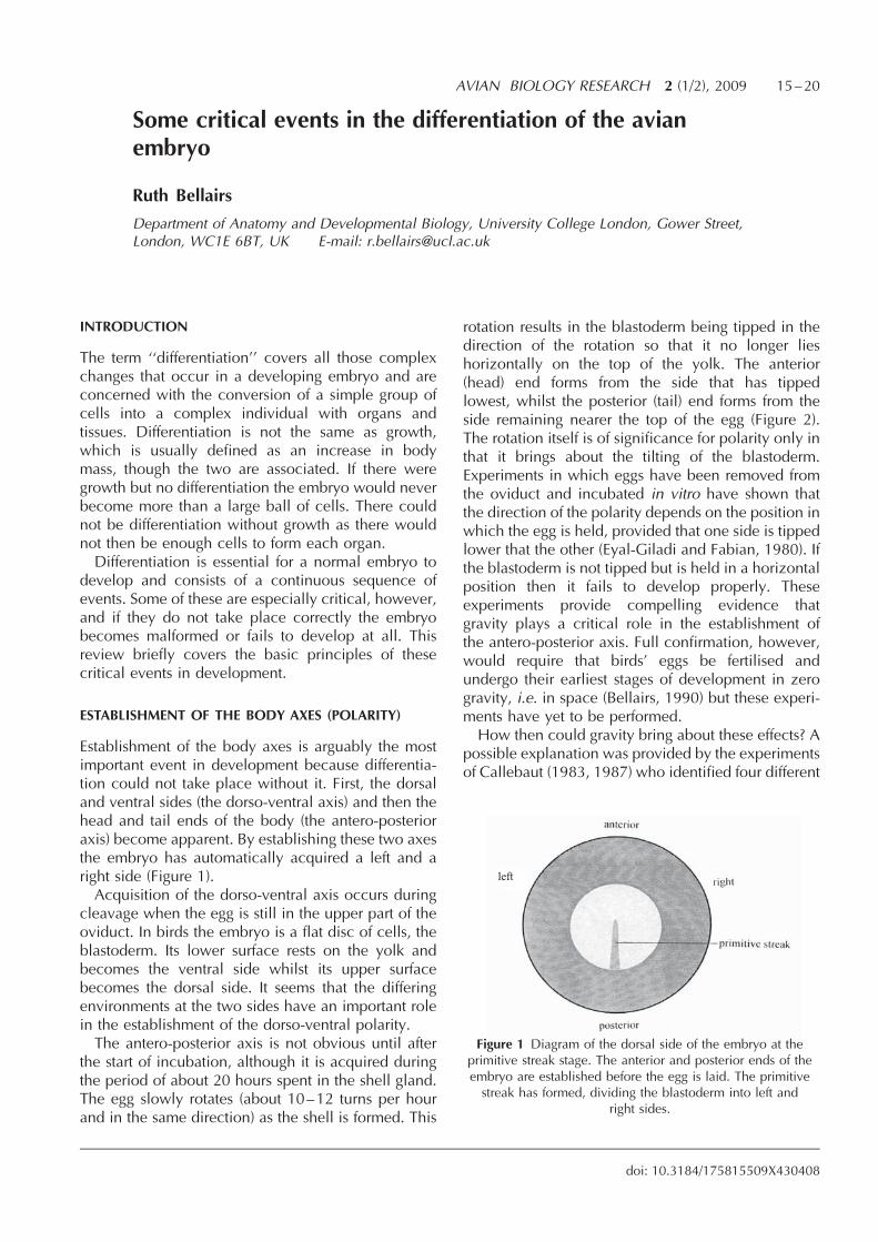
Establishment of the body axes is arguably the mostimportant event in development because differentia-tion could not take place without it. First, the dorsaland ventral sides (the dorso-ventral axis) and then thehead and tail ends of the body (the antero-posterioraxis) become apparent. By establishing these two axesthe embryo has automatically acquired a left and aright side (Figure 1).Acquisition of the dorso-ventral axis occurs during
cleavage when the egg is still in the upper part of theoviduct. In birds the embryo is a flat disc of cells, theblastoderm. Its lower surface rests on the yolk andbecomes the ventral side whilst its upper surfacebecomes the dorsal side. It seems that the differingenvironments at the two sides have an important rolein the establishment of the dorso-ventral polarity.The antero-posterior axis is not obvious until after
the start of incubation, although it is acquired duringthe period of about 20 hours spent in the shell gland.The egg slowly rotates (about 10–12 turns per hourand in the same direction) as the shell is formed. This
rotation results in the blastoderm being tipped in thedirection of the rotation so that it no longer lieshorizontally on the top of the yolk. The anterior(head) end forms from the side that has tippedlowest, whilst the posterior (tail) end forms from theside remaining nearer the top of the egg (Figure 2).The rotation itself is of significance for polarity only inthat it brings about the tilting of the blastoderm.Experiments in which eggs have been removed fromthe oviduct and incubated in vitro have shown thatthe direction of the polarity depends on the position inwhich the egg is held, provided that one side is tippedlower that the other (Eyal-Giladi and Fabian, 1980). Ifthe blastoderm is not tipped but is held in a horizontalposition then it fails to develop properly. Theseexperiments provide compelling evidence thatgravity plays a critical role in the establishment ofthe antero-posterior axis. Full confirmation, however,would require that birds’ eggs be fertilised andundergo their earliest stages of development in zerogravity, i.e. in space (Bellairs, 1990) but these experi-ments have yet to be performed.How then could gravity bring about these effects? A
possible explanation was provided by the experimentsof Callebaut (1983, 1987) who identified four different
AVIAN BIOLOGY RESEARCH 2 (1/2), 2009 15–20
doi: 10.3184/175815509X430408
Figure 1 Diagram of the dorsal side of the embryo at theprimitive streak stage. The anterior and posterior ends of theembryo are established before the egg is laid. The primitivestreak has formed, dividing the blastoderm into left and
right sides.
left

components of the yolky cytoplasm of the quailoocyte: a��; b��; g�� and d��ooplasms. During therotation of the egg, these ooplasms become shiftedso that the different parts of the blastoderm becomeunderlain by different ooplasms. In particular,b��ooplasm becomes located in certain cells (endo-phyll cells) beneath the region that will become thefuture posterior end of the embryo (Callebaut et al.,1998).The antero-posterior axis is not, however, perma-
nently fixed at this stage. It can be experimentallychanged after laying; if the blastoderm of a new-laidegg is bisected before incubation it may develop twoaxes which do not necessarily possess the sameorientation (Veini and Bellairs, 1983). This means
that one of them, at least, must have changed itspotential orientation. Once incubation begins, thisability is rapidly lost.Shortly after the start of incubation a range of genes
concerned with polarity start to become active (e.g.Hox genes) and apparently interpret the provisionallyestablished antero-posterior axis. Their domainsoverlap one another both in the antero-posterior andthe dorso-ventral axes and help to establish thedifferences between the different regions.
GASTRULATION AND THE FORMATION OF THEPRIMITIVE STREAK
Gastrulation and formation of the primitive streakbegins shortly after the start of incubation and ischaracterised by extensive migrations of cells. Weknow about them because many experiments havebeen carried out in which groups of cells have beenmarked in various ways, usually by applying dyes tothem, and then followed through succeeding stages ofdevelopment, often with time-lapse cinematography.Maps have been prepared of the patterns of migration.(e.g. Figure 3) and they consist principally of an initialforward movement in the midline. At the beginning ofgastrulation the region where the embryo will formhas upper and lower layers. Experiments have shownthat if the lower layer (the hypoblast) is removed, themigrations in the upper layer (the epiblast) fail to takeplace, so the lower layer seems to be responsible forprovoking these movements. By the end of gastrula-tion the embryo is three layered. The original lowerlayer (the hypoblast) has migrated away from thecentral region and its cells will eventually give riseto germ cells, which in turn will become sperm orova. Two new layers, the endoderm and the meso-derm have formed from the upper layer, which is nowknown as the ectoderm. We, therefore, now have athree-layered blastoderm, composed of ectoderm,mesoderm and endoderm.The endoderm and mesoderm have left the ecto-
derm by migrating through a region called the primi-tive streak (Figure 4). The primitive streak is atransitory structure formed by cells that have movedtoward the midline and piled up there. Eventually itdisappears as the cells leave this area and passbeneath the ectoderm, migrating out laterally.Much of the recent work on gastrulation has centred
on the mechanisms by which the orderly sequence ofcell migrations is controlled. There is a geneticelement, but an important role is played by compo-nents of the extra-cellular materials, such as fibro-nectin and hyaluronic acid, which affect cell adhesionand migration.
16 Ruth Bellairs
Figure 2 Diagram to illustrate the mode of establishment of theantero-posterior axis. (a) With the rotation of the egg in the shellgland, the young embryo becomes tipped to one side of theyolk. The anterior region subsequently develops from the endthat has tipped lowest. (b) Diagram to show the arrangements ofthe four different types of yolky cytoplasm, a; b; g; d, in a quailoocyte. (c) Diagram to show that the yolky cytoplasm comes tolie at the posterior end of the embryo and so may play a critical
role in the establishment of the antero-posterior axis.(b) and (c) after Callebaut (1987).
(a)
(b)
(c)
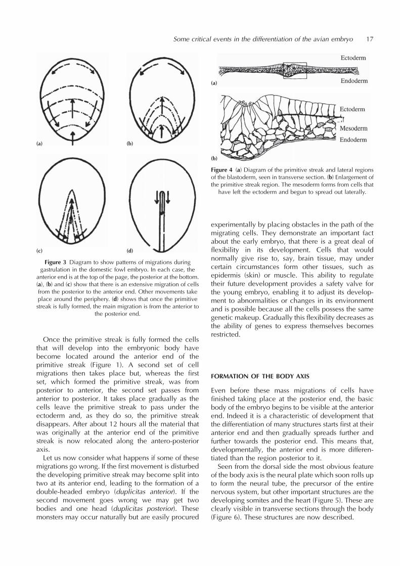
Once the primitive streak is fully formed the cellsthat will develop into the embryonic body havebecome located around the anterior end of theprimitive streak (Figure 1). A second set of cellmigrations then takes place but, whereas the firstset, which formed the primitive streak, was fromposterior to anterior, the second set passes fromanterior to posterior. It takes place gradually as thecells leave the primitive streak to pass under theectoderm and, as they do so, the primitive streakdisappears. After about 12 hours all the material thatwas originally at the anterior end of the primitivestreak is now relocated along the antero-posterioraxis.Let us now consider what happens if some of these
migrations go wrong. If the first movement is disturbedthe developing primitive streak may become split intotwo at its anterior end, leading to the formation of adouble-headed embryo (duplicitas anterior). If thesecond movement goes wrong we may get twobodies and one head (duplicitas posterior). Thesemonsters may occur naturally but are easily procured
experimentally by placing obstacles in the path of themigrating cells. They demonstrate an important factabout the early embryo, that there is a great deal offlexibility in its development. Cells that wouldnormally give rise to, say, brain tissue, may undercertain circumstances form other tissues, such asepidermis (skin) or muscle. This ability to regulatetheir future development provides a safety valve forthe young embryo, enabling it to adjust its develop-ment to abnormalities or changes in its environmentand is possible because all the cells possess the samegenetic makeup. Gradually this flexibility decreases asthe ability of genes to express themselves becomesrestricted.
FORMATION OF THE BODY AXIS
Even before these mass migrations of cells havefinished taking place at the posterior end, the basicbody of the embryo begins to be visible at the anteriorend. Indeed it is a characteristic of development thatthe differentiation of many structures starts first at theiranterior end and then gradually spreads further andfurther towards the posterior end. This means that,developmentally, the anterior end is more differen-tiated than the region posterior to it.Seen from the dorsal side the most obvious feature
of the body axis is the neural plate which soon rolls upto form the neural tube, the precursor of the entirenervous system, but other important structures are thedeveloping somites and the heart (Figure 5). These areclearly visible in transverse sections through the body(Figure 6). These structures are now described.
Some critical events in the differentiation of the avian embryo 17
Figure 3 Diagram to show patterns of migrations duringgastrulation in the domestic fowl embryo. In each case, the
anterior end is at the top of the page, the posterior at the bottom.(a), (b) and (c) show that there is an extensive migration of cellsfrom the posterior to the anterior end. Other movements takeplace around the periphery. (d) shows that once the primitivestreak is fully formed, the main migration is from the anterior to
the posterior end.
(a) (b)
(c) (d)
Figure 4 (a) Diagram of the primitive streak and lateral regionsof the blastoderm, seen in transverse section. (b) Enlargement ofthe primitive streak region. The mesoderm forms from cells that
have left the ectoderm and begun to spread out laterally.
(a)
(b)

FORMATION OF THE SOMITES
The notochord and somites (Figure 6) are structuresthat are present only in the early embryo because inthe later stages of differentiation they becomeconverted into other tissues. The notochord is a rodthat extends down the midline of the developing bodyand eventually becomes part of the intervertebraldiscs of the vertebral column. The neural tube formsabove it and eventually develops into the brain andspinal cord. The somites are derived from mesodermand lie as paired blocks on either side of the noto-chord and are the first segmented structures to form inthe embryo, laying the foundation for all the othersegmented structures that develop, e.g. vertebrae and
ribs, cranial and spinal nerves, vertebralarteries and skeletal muscles. Each somiteacquires three components: the dermatomewhich forms the dermis of the skin; themyotome, which gives rise to many of themuscles of the trunk and limbs; and thesclerotome, which forms the cartilage ofthe vertebrae and ribs.The first somites appear at about stage 7
(domestic fowl embryos are usuallydescribed according to an agreed stage ofdevelopment, rather than the length of timethey have been incubated [see Hamburgerand Hamilton, 1951]) and the others arelaid down pair by pair further and furthertowards the posterior. The somites arisefrom cells that initially lie in the ectodermaround the anterior end of the primitivestreak at about stage 3þ . During the laterstages of gastrulation they leave the uppersurface and come to lie as two blocks oftissue, one on either side of the midline. Thesomites will form from these are thesegmental plates (Figure 6).
There is much we do not understand about thefactors responsible for the breaking up of the contin-uous block of tissue, the segmental plate, into indi-vidual segments. Currently, a promising clue is thatthere are ‘‘oscillating genes’’ involved, that switch onand off, causing the somites to break off at regularintervals from the continuous blocks of mesoderm(Dale and Pourquie, 1990), but other factors includethe increasing adhesion of cells to one another.
FORMATION OF THE NERVOUS SYSTEM
The brain and nervous system develop first as theneural plate in the ectoderm that then rolls up into theneural tube (Figure 6). Its formation has alwaysexcited interest because it develops as a result ofinfluences from the underlying mesoderm, a processcalled neural induction. The anterior end of the neuraltube swells into a brain, and this in turn becomesmodified into its different regions. The eyes form byoutgrowths from the most anterior part, the forebrain,and all the nerves grow out from the neural tube andpass to the organs and tissues that they innervate.Many of these processes take place only after an
induction from a neighbouring tissue. The mechan-isms involved have resisted analysis for many years,though there are certain substances that appear to berelevant. They include fibroblast growth factor, activinand retinoic acid.
18 Ruth Bellairs
Figure 5 Diagram of the embryo at (a) stage 7 (about 24 hours incubation)and (b) stage 8 (about 27 hours incubation), seen from the dorsal side.Hensen’s node is the region of the primitive streak that will give rise to
notochord.
(a) (b)
Figure 6 Diagram of embryo at stage 11 (about 40 hoursincubation). (a) viewed from the ventral side. (b) Section
through the somite region. (c) Section through the segmentalplate region.
(a)
(b)
(c)
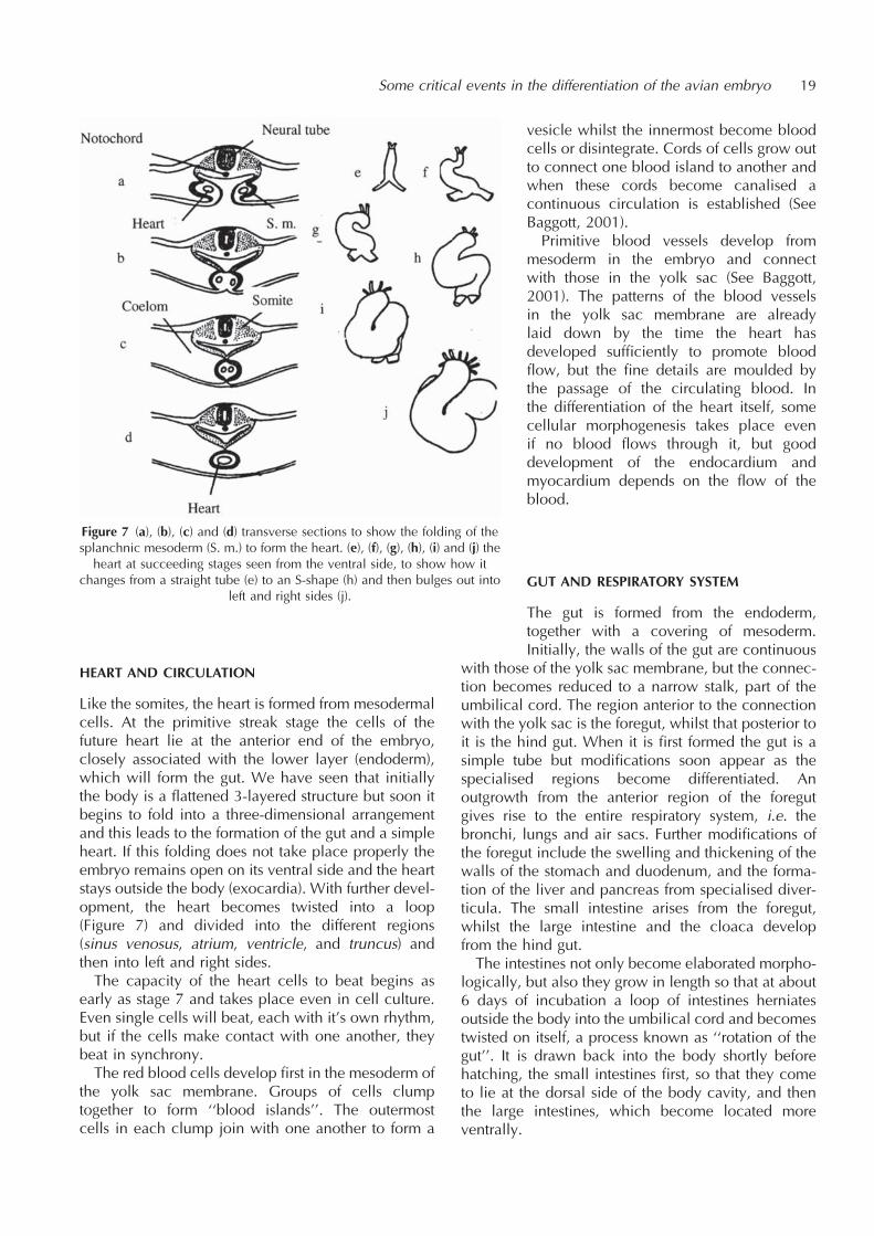
HEART AND CIRCULATION
Like the somites, the heart is formed from mesodermalcells. At the primitive streak stage the cells of thefuture heart lie at the anterior end of the embryo,closely associated with the lower layer (endoderm),which will form the gut. We have seen that initiallythe body is a flattened 3-layered structure but soon itbegins to fold into a three-dimensional arrangementand this leads to the formation of the gut and a simpleheart. If this folding does not take place properly theembryo remains open on its ventral side and the heartstays outside the body (exocardia). With further devel-opment, the heart becomes twisted into a loop(Figure 7) and divided into the different regions(sinus venosus, atrium, ventricle, and truncus) andthen into left and right sides.The capacity of the heart cells to beat begins as
early as stage 7 and takes place even in cell culture.Even single cells will beat, each with it’s own rhythm,but if the cells make contact with one another, theybeat in synchrony.The red blood cells develop first in the mesoderm of
the yolk sac membrane. Groups of cells clumptogether to form ‘‘blood islands’’. The outermostcells in each clump join with one another to form a
vesicle whilst the innermost become bloodcells or disintegrate. Cords of cells grow outto connect one blood island to another andwhen these cords become canalised acontinuous circulation is established (SeeBaggott, 2001).Primitive blood vessels develop from
mesoderm in the embryo and connectwith those in the yolk sac (See Baggott,2001). The patterns of the blood vesselsin the yolk sac membrane are alreadylaid down by the time the heart hasdeveloped sufficiently to promote bloodflow, but the fine details are moulded bythe passage of the circulating blood. Inthe differentiation of the heart itself, somecellular morphogenesis takes place evenif no blood flows through it, but gooddevelopment of the endocardium andmyocardium depends on the flow of theblood.
GUT AND RESPIRATORY SYSTEM
The gut is formed from the endoderm,together with a covering of mesoderm.Initially, the walls of the gut are continuous
with those of the yolk sac membrane, but the connec-tion becomes reduced to a narrow stalk, part of theumbilical cord. The region anterior to the connectionwith the yolk sac is the foregut, whilst that posterior toit is the hind gut. When it is first formed the gut is asimple tube but modifications soon appear as thespecialised regions become differentiated. Anoutgrowth from the anterior region of the foregutgives rise to the entire respiratory system, i.e. thebronchi, lungs and air sacs. Further modifications ofthe foregut include the swelling and thickening of thewalls of the stomach and duodenum, and the forma-tion of the liver and pancreas from specialised diver-ticula. The small intestine arises from the foregut,whilst the large intestine and the cloaca developfrom the hind gut.The intestines not only become elaborated morpho-
logically, but also they grow in length so that at about6 days of incubation a loop of intestines herniatesoutside the body into the umbilical cord and becomestwisted on itself, a process known as ‘‘rotation of thegut’’. It is drawn back into the body shortly beforehatching, the small intestines first, so that they cometo lie at the dorsal side of the body cavity, and thenthe large intestines, which become located moreventrally.
Some critical events in the differentiation of the avian embryo 19
Figure 7 (a), (b), (c) and (d) transverse sections to show the folding of thesplanchnic mesoderm (S. m.) to form the heart. (e), (f), (g), (h), (i) and (j) the
heart at succeeding stages seen from the ventral side, to show how itchanges from a straight tube (e) to an S-shape (h) and then bulges out into
left and right sides (j).

FACTORS INVOLVED IN DIFFERENTIATION
Many interacting processes are involved in differentia-tion but in this brief resume I have touched on a fewonly, viz. the establishment of polarity, cell migration,cell and tissue interactions and the switching on andoff of genes. The interplay of these and other factors,such as the appropriate incubation conditions, makethe difference between the formation of a normal oran abnormal embryo, and frequently of the survival ordeath of the individual.
REFERENCES
Baggott, G.K. (2001) Development of extra-embryonic mem-branes and fluid compartments. In: Perspectives in Fertilisa-tion and Embryonic Development in Poultry, pp. 23–29.Ratite Conference Books, Wallingford. Reprinted in 2008:Avian Biol. Res., 1, 21–26
Bellairs, R. (1990) The role of gravity in bird development.European Space Agency Publication SP-1123. [8-10 rueMario-Nikis, 75738, Paris, Cedes, France.]
Callebaut, M. (1983) The constituent oocytal layers of the aviangerm and the origin of the primordial germ cell yolk. Arch.d’Anatomie Micro. Morphol. Exp., 72, 199–214.
Callebaut, M. (1987) Ooplasmic localization and segregation inquail germs: fate of the four ooplasms. Archives de Biologie(Bruxelles), 98, 441–473.
Callebaut, M., van Nueten, E., van Nassauw, L., Bortier, H. andHarrison, F. (1998) Only the endophyll-Rauber’s sicklecomplex and not cells derived from the caudal marginalzone induce a primitive streak in the upper layer of avianblastoderms. Reprod. Nutr. Develop., 38, 449–463.
Dale, K.J. and Pourquie, O. (2000) A clock-work somite.BioEssays, 22, 72–83.
Eyal-Giladi, H. and Fabian, B.C. (1980) Axis determination inuterine chick blastodiscs under changing spatial positionsduring the sensitive period of polarity. Develop. Biol., 77,228–232.
Hamburger, V. and Hamilton, H.L. (1951) A series of normalstages in the development of the chick embryo. J. Morphol.,88, 49–92. Reprinted (1992) Develop. Dynamics, 195,231–272.
Veini, M. and Bellairs, R. (1983) Experimental analysis ofcontrol mechanisms in somite segmentation in avianembryos. I Reduction of material at the blastula stage inCoturnix coturnix japonica. J. Embryol. Exp. Morphol., 14,1–14.
20 Ruth Bellairs
Development of extra-embryonic membranes and fluidcompartments
Glenn K. Baggott
Department of Biology, School of Biological and Chemical Sciences, Birkbeck College, Universityof London, Malet Street, London, WC1E 7HX, UK E-mail: [email protected]
INTRODUCTION
Seventy years ago the Cambridge embryologist JosephNeedham coined the term ‘‘cleidoic’’ to describe thespecial characteristics of the avian egg (Needham,1931). He pointed out that the avian egg was essen-tially closed because nearly all the materials neededfor the development of the embryo are containedwithin the shell. Sufficient water, nutrients andenergy (in this case fats) for tissue growth and main-tenance are provided. Only oxygen (and heat) isrequired from the environment. Whereas Needham’sperceptive analysis of the nature of the avian egg hasbeen vindicated, the implications of this egg designfor the fashioning of embryonic tissue from thefertilised ovum are still to be fully explored.One of the primary difficulties is in thesupply of water to the growing mass ofembryonic and extra-embryonic tissues.The latter consist of membranes and fluidcompartments that develop outside of thebody of the true embryo and fulfil a pivotalrole in normal development. This reviewdescribes the importance of water in devel-opment of the extra-embryonic compart-ments of the egg in order to describe howthere is a resolution of the water problem.
DEVELOPMENT OF THE EXTRA-EMBRYONICMEMBRANES
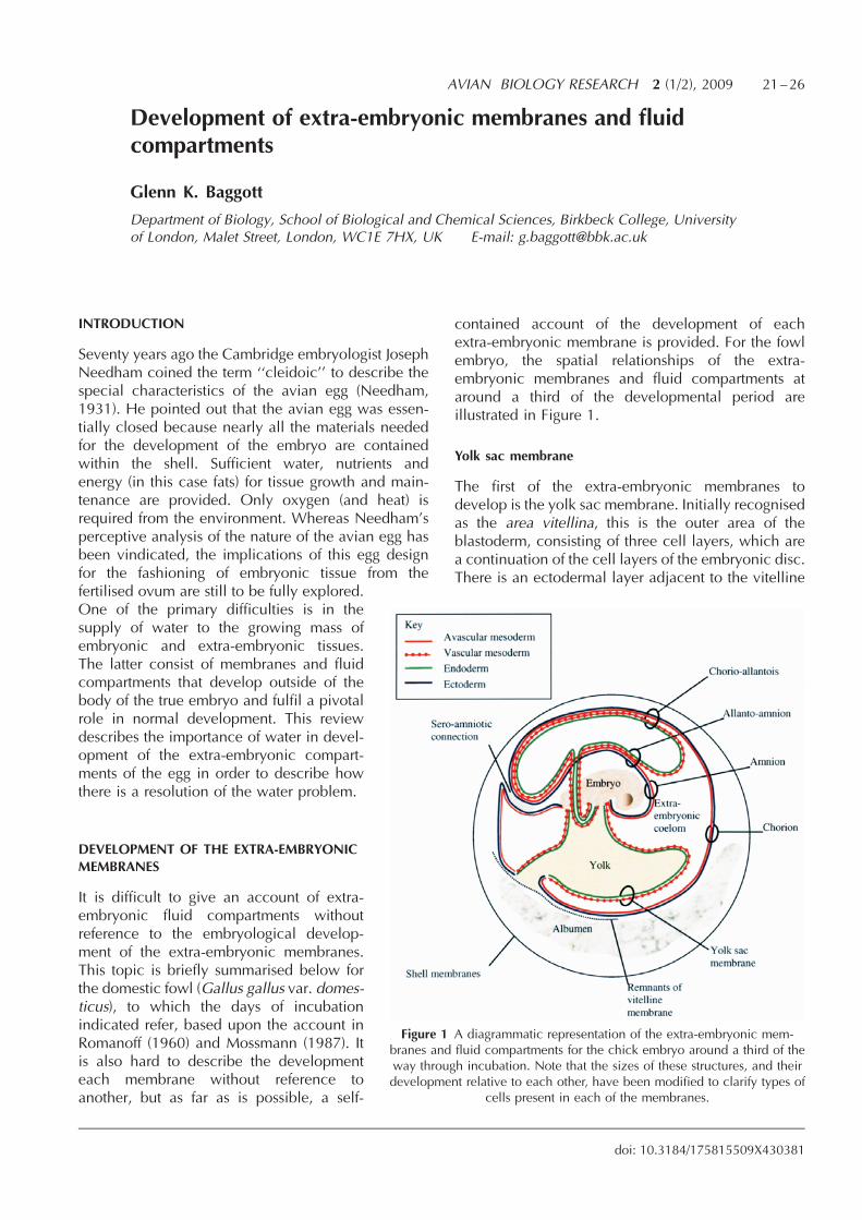
It is difficult to give an account of extra-embryonic fluid compartments withoutreference to the embryological develop-ment of the extra-embryonic membranes.This topic is briefly summarised below forthe domestic fowl (Gallus gallus var. domes-ticus), to which the days of incubationindicated refer, based upon the account inRomanoff (1960) and Mossmann (1987). Itis also hard to describe the developmenteach membrane without reference toanother, but as far as is possible, a self-
contained account of the development of eachextra-embryonic membrane is provided. For the fowlembryo, the spatial relationships of the extra-embryonic membranes and fluid compartments ataround a third of the developmental period areillustrated in Figure 1.
Yolk sac membrane
The first of the extra-embryonic membranes todevelop is the yolk sac membrane. Initially recognisedas the area vitellina, this is the outer area of theblastoderm, consisting of three cell layers, which area continuation of the cell layers of the embryonic disc.There is an ectodermal layer adjacent to the vitelline
AVIAN BIOLOGY RESEARCH 2 (1/2), 2009 21–26
doi: 10.3184/175815509X430381
Figure 1 A diagrammatic representation of the extra-embryonic mem-branes and fluid compartments for the chick embryo around a third of theway through incubation. Note that the sizes of these structures, and theirdevelopment relative to each other, have been modified to clarify types of
cells present in each of the membranes.
membranes, an endodermal layer adjacentto the yolk and in between a layer ofmesoderm cells. The mesoderm is splitinto two by a cavity, the extra-embryoniccoelom, and only the mesoderm next to theendoderm develops blood vessels (calledthe vascular mesoderm). It is these twocell layers that form the definitive wall ofthe yolk sac. The vitelline membranesenclose the yolk sac until day 4 of incuba-tion. Contact of embryonic tissue with thevitelline membranes alters their structure(Jensen, 1969), probably facilitating theirrupture when yolk sac volume increases atthis time. This causes the yolk to lose itsspherical shape and the embryonic-yolkstructure adopts the shape of the egg. Thetop of this structure is bounded by the yolksac membrane and the lower by the vitel-
line membrane, which slips down to the pole of theyolk sac opposite to the embryo. In this way thepartition of the yolk sac from the albumen (the so-called yolk sac umbilicus) is maintained and at this‘‘vegetal’’ pole, the yolk sac membrane remainsincomplete until day 17.The yolk sac membrane passes the equator of the
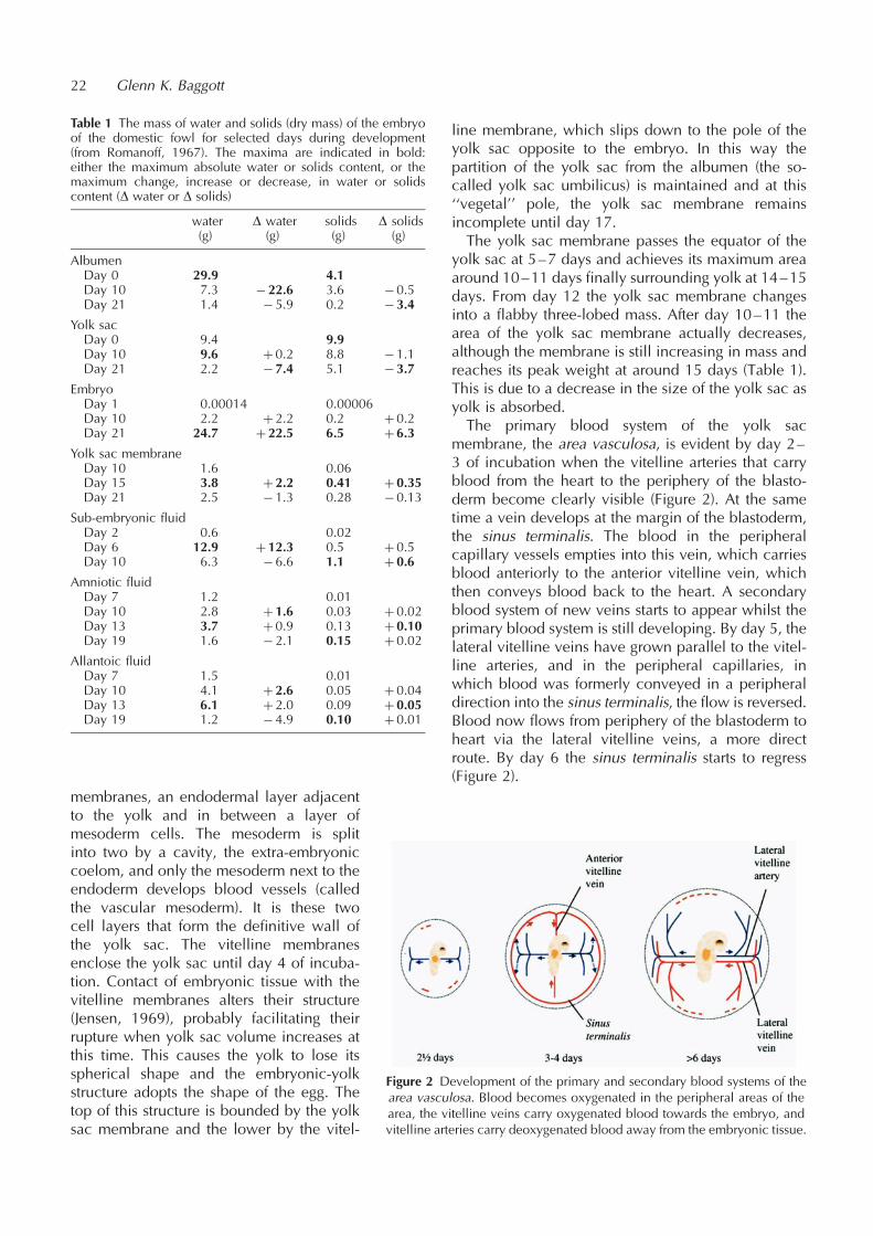
yolk sac at 5–7 days and achieves its maximum areaaround 10–11 days finally surrounding yolk at 14–15days. From day 12 the yolk sac membrane changesinto a flabby three-lobed mass. After day 10–11 thearea of the yolk sac membrane actually decreases,although the membrane is still increasing in mass andreaches its peak weight at around 15 days (Table 1).This is due to a decrease in the size of the yolk sac asyolk is absorbed.The primary blood system of the yolk sac
membrane, the area vasculosa, is evident by day 2–3 of incubation when the vitelline arteries that carryblood from the heart to the periphery of the blasto-derm become clearly visible (Figure 2). At the sametime a vein develops at the margin of the blastoderm,the sinus terminalis. The blood in the peripheralcapillary vessels empties into this vein, which carriesblood anteriorly to the anterior vitelline vein, whichthen conveys blood back to the heart. A secondaryblood system of new veins starts to appear whilst theprimary blood system is still developing. By day 5, thelateral vitelline veins have grown parallel to the vitel-line arteries, and in the peripheral capillaries, inwhich blood was formerly conveyed in a peripheraldirection into the sinus terminalis, the flow is reversed.Blood now flows from periphery of the blastoderm toheart via the lateral vitelline veins, a more directroute. By day 6 the sinus terminalis starts to regress(Figure 2).
22 Glenn K. Baggott
Figure 2 Development of the primary and secondary blood systems of thearea vasculosa. Blood becomes oxygenated in the peripheral areas of thearea, the vitelline veins carry oxygenated blood towards the embryo, andvitelline arteries carry deoxygenated blood away from the embryonic tissue.
Table 1 The mass of water and solids (dry mass) of the embryoof the domestic fowl for selected days during development(from Romanoff, 1967). The maxima are indicated in bold:either the maximum absolute water or solids content, or themaximum change, increase or decrease, in water or solidscontent (D water or D solids)
water D water solids D solids(g) (g) (g) (g)
AlbumenDay 0 29.9 4.1Day 10 7.3 � 22.6 3.6 � 0.5Day 21 1.4 � 5.9 0.2 � 3.4
Yolk sacDay 0 9.4 9.9Day 10 9.6 þ 0.2 8.8 � 1.1Day 21 2.2 � 7.4 5.1 � 3.7
EmbryoDay 1 0.00014 0.00006Day 10 2.2 þ 2.2 0.2 þ 0.2Day 21 24.7 þ 22.5 6.5 þ 6.3
Yolk sac membraneDay 10 1.6 0.06Day 15 3.8 þ 2.2 0.41 þ 0.35Day 21 2.5 � 1.3 0.28 � 0.13
Sub-embryonic fluidDay 2 0.6 0.02Day 6 12.9 þ 12.3 0.5 þ 0.5Day 10 6.3 � 6.6 1.1 þ 0.6
Amniotic fluidDay 7 1.2 0.01Day 10 2.8 þ 1.6 0.03 þ 0.02Day 13 3.7 þ 0.9 0.13 þ 0.10Day 19 1.6 � 2.1 0.15 þ 0.02
Allantoic fluidDay 7 1.5 0.01Day 10 4.1 þ 2.6 0.05 þ 0.04Day 13 6.1 þ 2.0 0.09 þ 0.05Day 19 1.2 � 4.9 0.10 þ 0.01

Amnion
The amnion is formed from a layer of ectoderm andunderlying avascular (does not develop blood vessels)mesoderm immediately adjacent to the embryo. Theflat tissue rises up to form folds over both the head andtail of the developing embryo and by day 4 ofincubation the folds fuse together over the embryoto enclose the amniotic sac. Consequently, the innerlayer of the amnion is ectoderm, and the outer one isavascular mesoderm. Muscle cells appear within thismesoderm so that the amnion becomes contractile byday 5, but the blood vessels of the amnion arise fromthe subsequent fusion of this avascular mesodermwith vascular mesoderm from the allantois. On day12 a duct develops between the albumen sac and theamniotic sac; this sero-amniotic connection (Figure 1)is the point where the head and tail folds of theamnion met on day 4 of incubation. This ductallows the movement of albumen proteins intoamniotic fluid where they are swallowed ending upin the yolk sac.
Chorion
The chorion develops from tissue continuous with theamnion and away from the embryo. It is comprised ofan outside layer of ectoderm adjacent to the innershell membrane and an inner avascular mesoderm,which lines the extra-embryonic coelom. Eventuallythis mesoderm fuses with vascular mesoderm ofallantois, so supplying the blood vessels and formingthe chorio-allantois. By day 11 the allantois hascompletely lined the chorion and the chorio-allantoiscovers 98% of the area of the eggshell membranes,thereby acting as the primary respiratory surface forthe embryo during the second half of incubation.
Allantois
At day 2 the allantois is a small bud of endodermalcells and by day 4 it is visible as a free sac growing outfrom the primitive hindgut of the embryo. Its inner sideis endoderm and the outer surface is vascular meso-derm. It expands into, and eventually fills, the extra-embryonic coelom, so that by day 6 its vascularmesoderm is fused both with the chorion, to formchorioallantois, and with the avascular mesoderm ofthe amnion. Hence, the allantois supplies the bloodsystem for both the chorio-allantois and the amnion.The allantois is a fluid-filled and is a repository forkidney excretions that first appear by day 5 and entervia the allantoic duct from the cloacal region of thehindgut.
THE WATER PROBLEM
Observations of many avian species have establishedthat with a total water loss during incubation of about20% of the initial egg mass, the water concentration ofthe hatchling (plus spare yolk) is very similar to theinitial concentration in the egg at lay (Ar, 1991). Thisis because fat provides the energy source forembryonic growth and maintenance, and for thissubstrate the amount of water consumed is replacedby almost exactly the same amount of water producedby metabolism (Ar and Rahn, 1980). Thus the designof the avian egg produces ‘‘constant hydration’’, suchthat the chick has an appropriate tissue water contentat hatching (Ar, 1991). So what then is the nature ofthe ‘‘water problem’’?At ovulation the ovum consists of a single cell
containing large amounts of yolk, a store of nutrientsand energy. In the upper part of the oviduct a secondprotein membrane is laid down over the primaryvitelline membrane that contains the fertilised ovum(Bellairs, 1991). Subsequently, within the oviduct,albumen is added around the fertilised ovum, to actas a source of water, and finally the shell membranesand the shell are added around the albumen prior tooviposition (Gilbert, 1971). By the time of lay theovum has undergone cell division to produce anembryo (Bellairs, 1991), which is physically separatedfrom the water in the albumen by the vitellinemembranes. The problem, then, is to ensure that thissource of water is made accessible to the growingmass of tissue. Resolution of this difficulty is crucial forgrowth of the embryo.At lay, 60% of the water in the egg is located in the
albumen, which itself comprises 88% water(Romanoff, 1967). The tissues of the embryo containabout 70% water (Romanoff, 1967), and water mustalso be supplied to the extra-embryonic tissues. Mostof the albumen water is transferred during the first halfof the incubation period (Figure 3): about 76% ofalbumen water has disappeared by day 10 of incuba-tion (Table 1). During this period the total amount ofsolids in the albumen hardly changes at all (Table 1),indicating that the water is removed preferentially. Bycontrast, at lay the yolk sac contains 49% water andthe water content of yolk changes by only 2% overthis 10 day period (Table 1).Over the whole period of incubation 28.6 g of water
disappears from albumen and 7.2 from the yolk;24.7 g appears in embryonic tissue and 2.5 g in theyolk sac membrane (Table 1). However, up to day 10of incubation the water content of the embryoincreases by only 2.2 gms (Table 1). Thus, the largestincrease in water content of the embryo is in thesecond half of incubation, but by 10 days 76% of
Development of extra-embryonic membranes and fluid compartments 23

the albumen water has been removed. So where has itgone, and how? The answers to these questions lie inthe formation and depletion of three extra-embryonicfluid compartments in the egg: the sub-embryonicfluid, amniotic fluid and allantoic fluid.
Sub-embryonic fluid (SEF)
Apparently, therefore, water does not move directlyto embryonic tissue from albumen. In fact, duringembryonic development water appears and disap-pears from a number of separate fluid compartments.The first new compartment becomes evident around2–3 days of incubation, when a fluid appears in theyolk sac beneath the embryo, the sub-embryonicfluid. Its water content reaches a maximum at day6 of incubation, when it is more than 95% water,and decreases in mass thereafter (Figure 3; Table 1).There is a ‘‘critical period’’ for the production of SEFbetween days 3–7 of incubation: if eggs are notturned during this period SEF volume is decreasedand the formation of other fluid compartments andembryonic growth is retarded (Deeming, 1989a,1989b). An absence of egg turning also reduces thegrowth of the area vasculosa over the yolk sac(Deeming, 1989c), and this is most likely the causeof the reduced amount of SEF found in unturnedeggs (Latter and Baggott, 2002). The endoderm cellsof the area vasculosa that face the yolk are specia-lised for the transport of water and sodium ions from
albumen to yolk sac (Babiker and Baggott,1995; Latter and Baggott, 2000), so withfewer cells less SEF will be produced. It isassumed that water from the SEF is distrib-uted to the growing tissue, embryonic andextra-embryonic, by the blood system(derived from the vascular mesoderm),although nothing is known about thisprocess. The transport of water into theyolk sac has an additional benefit: theyolk becomes lighter than albumen inwhich it is immersed and floats towardsthe upper surface of the egg, so placing thearea vasculosa just beneath the shellmembranes (Babiker and Baggott, 1992).As the area vasculosa is also a respiratoryorgan (Lomholt, 1984), this processimproves the access of the embryo to theair. In unturned eggs the yolk sac floats lessand sits deeper in the albumen (Babikerand Baggott, 1992), an outcome that maycontribute to the greater embryonicmortality of unturned eggs.
Amniotic fluid
Amniotic fluid appears later in development than SEFand achieves peak water content at day 13 of incuba-tion; solids accumulate slowly throughout develop-ment (Figure 3; Table 1). Amniotic fluid has a uniqueionic composition (high in chloride ions), which isresponsible for the inflow of water into the amnion(Faber et al., 1973). Amniotic fluid volume is, appar-ently, unaffected by the hydration state of the egg(Faber et al., 1973; Ar, 1991); it is thought that themain function of the fluid is mechanical protection ofthe embryo. An absence of egg turning substantiallydecreases the mass of amniotic fluid from days 12–18of incubation (Deeming, 1989a). This is thought to becaused by less albumen entering the amniotic fluidvia the sero-amniotic connection, probably due to thealtered spatial relationship between albumen, amnionand allantois in unturned eggs (Deeming, 1991).Additionally, by mid-incubation the albumenvolume of unturned eggs is greater than that ofturned eggs, which may also impede the transfer ofalbumen through the sero-amniotic connection(Deeming, 1991; Babiker and Baggott, 1992).
Allantoic fluid
Like amniotic fluid, the maximum allantoic fluidwater content is found on day 13 with largest increasein water content preceding this (Figure 3; Table 1).The source of this fluid is blood filtered by the
24 Glenn K. Baggott
Figure 3 The pattern of changes in the mass of the embryo, yolk andalbumen, and in the volume of the fluid compartments, of the developing
fowl egg. Data from Romanoff (1967).

embryonic kidney. As for amniotic fluid, a lack of eggturning decreases the mass of this fluid compartment,but for allantoic fluid this reduction was detectableonly on day 12 (Deeming, 1989a). Solids form only asmall proportion of the allantoic fluid (Table 1), butcan be important in determining its composition andproperties. Excretory nitrogen, as ammonia, urea anduric acid, are present in allantoic fluid at day 5, but itis predominantly the uric acid content of this fluid thatincreases during development (Fisher and Eakin,1957).It has been claimed that the rate at which water is
absorbed from allantoic fluid varies with the degreeof embryonic desiccation, thus producing a stablewater and ion content of the embryo and fluidcompartments (Hoyt, 1979). However, it is wellestablished that embryos subjected to higher waterlosses during incubation have a lower tissue watercontent and a smaller allantoic fluid volume (Vleck,1991). Also, investigators have repeatedly empha-sised the potential for alteration of allantoic fluidcomposition by ion and fluid transport across theallantoic membrane into the blood. However, aspointed out by Murphy (1997), changes in allantoicfluid volume can be explained adequately only ifurine inflow to the allantois, and fluid re-absorptionvia the allantoic membrane, are both measured.Certainly, ions (and water) are transported acrossthe membrane from allantoic fluid (Vleck, 1991),and this process, as well as a reduction in urinaryoutput from the kidney into the allantois, is mostprobably enhanced by the action of hormones(Murphy, 1997). However, the relative importanceof these two processes in determining the volume ofallantoic fluid remains uncertain. Moreover, theionic composition of the allantoic fluid is determinedby at least three factors: the capacity of the allantoicmembrane to reabsorb water and ions, the composi-tion of urine flowing in from the kidney, and theinteractions between excretory uric acid and ions inthe fluid.For example, by 15 day of incubation, calcium,
sodium and sulphate ions are all reabsorbed into theblood by kidney, so less are added to the allantoicfluid (Clark et al., 1993). However, uric acid in theallantoic fluid also can change the ion composition ofthe fluid, as solid uric acid sequesters sodium ions:under dry incubation conditions uric acid excretion isincreased, allantoic fluid contains more solid uric acidand more sodium ions are removed from the fluid, soincreasing the potential for water re-absorption intothe blood across the allantoic membrane (Bradfieldand Baggott, 1993a, 1993b). The solid uric acid (withfaecal material, meconium) is discarded in theeggshell at the time of hatching (Romanoff, 1967).
CONCLUSION
As is now clear, a number of fundamental aspectsconcerning the production of the fluid compartmentsof the avian egg remain to be explored. We are a littlecloser in understanding what factors, hormonal andenvironmental, influence the development of the areavasculosa, but still lack a complete account for arelatively simple environmental variable such as eggturning. We now know that both water inflow fromthe kidney and outflow, across allantoic membrane,determines allantoic fluid volume, but we are unsureof the relative importance of these processes, espe-cially with regard to the hydration status of theembryo. Even more fundamentally, we have littleunderstanding of the mechanisms whereby fluidmoves between SEF, amnion, allantois and embryo.From this long list, the role of the area vasculosa inmoving water from SEF to embryo would appear to bethe process most amenable to experimental analysis.
REFERENCES
Ar, A. (1991) Roles of water in avian eggs. In: Egg Incubation: ItsEffects on Embryonic Development in Birds and Reptiles,Deeming, D.C. and Ferguson, M.W.J. (eds.), pp. 229–243.Cambridge University Press, Cambridge.
Ar, A. and Rahn, H. (1980) Water in the avian egg: overallbudget of incubation. Amer. Zool., 20, 373–384.
Babiker, E.M. and Baggott, G.K. (1992) The effect of turningupon the sub-embryonic fluid and albumen of the Japanesequail. Brit. Poult. Sci., 33, 973–991.
Babiker, E.M. and Baggott, G.K. (1995) The role of ion transportin the formation of sub-embryonic fluid by the embryo of theJapanese quail. Brit. Poult. Sci., 36, 371–383.
Bellairs, R. (1991) Overview of early stages of avian andreptilian development. In: Egg Incubation: Its Effects onEmbryonic Development in Birds and Reptiles, Deeming,D.C. and Ferguson, M.W.J. (eds.), pp. 371–383. CambridgeUniversity Press, Cambridge.
Bradfield, P.M. and Baggott, G.K. (1993a) The effect of waterloss upon the urate, urea and ammonia content of the egg ofthe Japanese quail Coturnix coturnix japonica. Comp. Bio-chem. Physiol., 106A, 187–193.
Bradfield, P.M. and Baggott, G.K. (1993b) Allantoic fluid elec-trolytes and urate of embryonic Japanese quail subject todifferential water loss. Brit. Poult. Sci., 34, 597–612.
Clark, N.B., Feng, J.Q. and Murphy, M.J. (1993) Renal clearancemeasurements of electrolytes in embryonic chickens. J. Exp.Zool., 265, 107–111.
Deeming, D.C. (1989a) Characteristics of unturned eggs: criti-cal period, retarded embryonic growth and poor albumenutilisation. Brit. Poult. Sci., 30, 239–249.
Deeming, D.C. (1989b) Importance of sub-embryonic fluid andalbumen in the embryo’s response to turning of the eggduring incubation. Brit. Poult. Sci., 30, 591–606.
Deeming, D.C. (1989c) Failure to turn eggs during incubation:development of the area vasculosa and embryonic growth. J.Morph., 201, 179–186.
Development of extra-embryonic membranes and fluid compartments 25

Deeming, D.C. (1991) Reasons for the dichotomy in egg turningin birds and reptiles. In: Egg Incubation: Its Effects onEmbryonic Development in Birds and Reptiles, Deeming,D.C. and Ferguson, M.W.J. (eds.), pp. 307–323. CambridgeUniversity Press, Cambridge.
Faber, J.J., Gault, C.F., Green, T.J., Long, L.R. and Thornburg,K.L. (1973) Chloride and the generation of amniotic fluid inthe early embryo. J. Exp. Zool., 183, 343–352.
Fisher, J.R. and Eakin, R.E. (1957) Nitrogen excretion in devel-oping chick embryos. J. Embryol. Exp. Morph., 5, 215–224.
Gilbert, A.B. (1971) Egg albumen and its formation. In: Physiol-ogy and Biochemistry of the Domestic Fowl, Volume 3. Bell,D.J. and Freeman, B.M. (eds.), pp. 1291–1329. AcademicPress, London.
Hoyt. D.F. (1979) Osmoregulation by avian embryos: allantoisfunctions like a toad’s bladder. Physiol. Zool., 60, 9–17.
Jensen, C. (1969) Ultrastructural changes in the avian vitellinemembrane during embryonic development. J. Embryol. Exp.Morphol., 21, 467–484.
Latter, G.V. and Baggott, G.K. (2000) Ion and fluid transport bythe blastoderm of the Japanese quail. Brit. Poult. Sci., 41,S43–S44.
Latter, G.V. and Baggott, G.K. (2002) Role of carbon dioxideand ion transport in the formation of sub-embryonic fluid by
the blastoderm of the Japanese quail. Brit. Poult. Sci., 43,104–116.
Lomholt, J.P. (1984) A preliminary study of local oxygentensions inside the bird egg and gas exchange during theearly stages of embryonic development. In: Respiration andMetabolism of Embryonic Vertebrates, Seymour, R.S. (ed.),pp. 289–298. Dr W. Junk, Dordrecht.
Mossmann, H.W. (1987) Vertebrate Fetal Membranes:Comparative Ontogeny and Morphology; Evolution; Phylo-genetic Significance; Basic Functions; Research Oppor-tunities. Rutgers University Press, New Jersey.
Murphy, M.J. (1997) Endocrine control of hydromineral balancein bird embryos. Amer. Zool., 37, 546–552.
Needham, J. (1931) Chemical Embryology. Cambridge.
Romanoff, A.L. (1960) The Avian Embryo. The MacmillanCompany, New York.
Romanoff, A.L. (1967) Biochemistry of the Avian Embryo. JohnWiley, New York.
Vleck, D. (1991) Water economy and solute regulation ofreptilian and avian embryos. In: Egg Incubation: Its Effectson Embryonic Development in Birds and Reptiles. Deeming,D.C. and Ferguson, M.W.J. (eds.), pp. 245–259. CambridgeUniversity Press, Cambridge.
26 Glenn K. Baggott

Development during the first seven days post-hatching
Vern L. Christensen
Department of Poultry Science, North Carolina State University, Box 7608, Raleigh, North Carolina27695-7608, USA E-mail: [email protected]
INTRODUCTION
The neonate has basic needs that must be satisfied atthe time of hatching if survivability and maximumpotential are achieved. Some of these basic needs arefresh air, clean water, proper feed, and heat. Thedevelopmental state at hatching of the neonatediffers among all avian species (Nice, 1962). Thepost-hatching period for altricial neonates is morecritical than for precocial birds because they hatchin a less mature state. Differing amounts of maternalcare are therefore essential for each species (Nice,1962). Although embryonic growth among species isvery similar, no two physiological systems seem tomature at the same rate (Ricklefs and Starck, 1998). Inaddition, maturity may be a function of the eggconductance constant (Ar and Rahn, 1978), which isdetermined by egg mass, eggshell conductance (orfunctional properties) and the length of the incubationperiod (see Box 1 later), all of which may constrainneonatal maturity. When viewed energetically, thedifference between maturity types resides in thedifferent water concentrations in eggs and hatchlings,in the density of chemical potential energy in the drymatter of true hatchlings, and in the different amountsof energy transferred from the egg to the spare yolk (Aret al., 1987).On the basis of species comparisons of post-
hatching growth of all birds (Lilja, 1983; Ricklefs,1987), it has been suggested that the rate of growthafter hatching is at least partially determined by thepattern of organ growth. It appears that a high rate ofgrowth is correlated to early growth of ‘‘supplyorgans’’ (oesophagus, proventriculus, gizzard, intes-tines, heart and liver) at the expense of ‘‘demandorgans’’ (breast, wings, legs and feathers). Thesechanges begin very early in development (Lilja andOlsson, 1987), and Schmalhausen (1930) hypothe-sised that growth and organ function come intoconflict when growth occurs too slowly or too rapidly.Even under the most optimum conditions, a newly
hatched bird is not free from stress. This is impossiblebecause the absence of stress is death (Selye, 1951).However, hatchlings possess abilities to cope withstress via adrenal cortical hormones (Davis and
Siopes, 1989). Hatchlings have different blueprintsfor growth and maturation that must occur within apredetermined time frame. Many times this blueprintdoes not include adjustments that need to occur inmaturational and growth processes in an imperfectenvironment. The objective of this paper is to define a‘‘physiologically normal’’ hatchling and to describethe principles involved in the maturation and growthof several organ systems during the initial stages oflife.At least six physiological systems exist that require
maturation during the last week of incubation or, inthe case of altricial species, the initial days of lifeoutside the shell. These systems are: (1) the circulatorysystem (heart and blood); (2) the kidney and bodyfluids system; (3) the digestive system; (4) bodytemperature regulation; (5) the respiratory system;and (6) the immune system. These systems arediscussed here using published data to help clarifyimportant points.
CIRCULATORY SYSTEM
The heart and blood are among the first tissues todevelop and mature in the embryo (see Baggott,2001), and both function physiologically in theinitial stages of embryonic development (Wattersonand Sweeney, 1973; Tazawa and Whittow, 2000).The number of red blood cells increases with the ageof the embryo until hatching (Macpherson andDeamer, 1964; Bagley, 1987). This occurs by botherythropoiesis and reticulocyte recruitment andmaturation (Bagley, 1987). The processes for haemo-globin maturation and synthesis undergo similarpatterns (Isaacks et al., 1976). Post-hatching, erythro-cyte numbers have been reported to increase initiallyfor 3–4 days then decline to 10 days of age (Phelpset al., 1987a). Sexual dimorphism affects erythrocytenumbers because in males numbers of cells declinedmore rapidly than in females, and erythrocyte changeswere accompanied by declines in packed cellvolumes, cell sizes and haemoglobin concentrations(Phelps et al., 1987a).Heart growth has been reported to be more variable
than other organs during the first two weeks post-
AVIAN BIOLOGY RESEARCH 2 (1/2), 2009 27–33
doi: 10.3184/175815509X430417
hatching (Phelps et al., 1987a). Rates of heart growthpeak at about five to six days post-hatching thenplateau to a rate similar to body growth at about 10days. Spontaneous turkey cardiomyopathy (roundheart disease, cardiohepatic syndrome; Magwoodand Bray, 1962) and ascites in broiler chicks aremanifestations of conflicts between organ growthand function. These syndromes are characterised byan enlarged heart due to extensive dilation of theventricles (primarily the right ventricle initially),congestive heart failure and mortality. Julian et al.(1992) and Breeding et al. (1994) showed that rapidgrowth rates in connection with high protein dietscontribute to the problem. Lilja and Olsson (1987)hypothesised that rapidly growing poultry willdevelop supply tissues before demand tissues.Hearts in avian species are demand tissues because
they can neither recycle lactate nor can they createglucose from gluconeogenic amino acids. Thus, heartgrowth, heart energy metabolism and blood cellmaturation may be greatly influenced by the overallgrowth of the animal. These problems have beenshown to occur during embryogenesis and manifestthemselves throughout the life of the bird (Buyse et al.,1998).
KIDNEY AND BODY FLUIDS SYSTEM
Embryos create urea as nitrogenous waste that ispartitioned from the embryo and stored in liquidform in the allantoic fluid (Watterson and Sweeney,1973). Approximately half way through incubation,the system begins creating insoluble uric acid, as anitrogenous waste product. Water serving as thesolvent for urea is recycled from the allantoic sacinto the embryo via the chorio-allantoic circulation orthe hindgut (Romanoff, 1960; Baggott, 2001). Thismakes it very difficult to dehydrate the embryo itselfduring incubation (Ar, 1991). However, egg weightlosses of greater than 20% can result in physiologicalproblems for embryos (Davis and Ackerman, 1987;Ar, 1991). The embryo relies on this system to filternitrogenous waste until it hatches. After hatching a‘‘new’’ renal system develops to maintain body fluidbalances. Water is an essential nutrient for neonateswhile the system maintaining water balance matures(Doneen and Smith, 1982). Maturation of the renalclearance system requires a few days post-hatchingwhereas establishment and maintenance of acid-basebalance is completed during the final stages ofembryonic development (Goldstein and Skadhauge,2000).In birds, the renal system performs the additional
functions of gluconeogenesis and maintenance ofblood glucose concentrations (Fasenko, 1996). As
stress and difficulties in food acquisition are encoun-tered, the kidney becomes essential to neonatalsurvival (Donaldson and Christensen, 1994).Maturation of the kidney in the gluconeogenic senseoccurs whilst still in the egg but can be affected by thelength of the incubation period (Fasenko, 1996).Glucose in hatchlings increases significantly fromhatching until day five or six then may decreaseachieving constant values by about day nine(Phelps, 1985).
DIGESTIVE SYSTEM
The yolk sac contains all of the enzymes and absorp-tion mechanisms necessary for sustaining life in theshell and following yolk sac retraction into the bodybefore hatching, it maintains these capabilities for thefirst few days following hatching (Romanoff, 1960;Denbow, 2000). The yolk digestive system is basedprimarily on lipids (Donaldson and Christensen, 1994;Donaldson et al., 1994) but after hatching, theneonate must begin life on diet rich in carbohydrate(48%) and this basically requires a new metabolicprocess. Many intestinal mechanisms are required tobe established and mature during a very short timefrom hatching (circa 24 hours) until food is consumed.The yolk sac system is drawn into the body prior tohatching to continue yolk digestion after hatching.Some estimates indicate as much as 60% of the totalenergy of a neonate (Fan et al., 1997) is devoted tomaturation and growth of intestinal tissue in the firstfew days after hatching.Yolk assimilation in neonates is altered by feeding,
temperature extremes and type of feed but not water(Phelps, 1985). Gender also has no effect (Moran andReinhart, 1980). Yolk absorption is reported to becomplete after five to six days post-hatching. Yolk isutilised post-hatching by two mechanisms: (1) transferof yolk into blood in embryos and (2) anti-peristalticmovement of yolk from the distal end towards theproximal small intestine where it is absorbed by theintestine (Noy and Sklan, 1998).Intestinal maturation post-hatching has been
studied extensively (Sell et al., 1989; Fan et al.,1997; Uni et al., 1995a, 1995b, 1999; Suvarna,1999) because of its economic importance tocommercial poultry. Intestinal dissacharidases andglucose transport mechanisms are fully functional atabout 48 hours post-hatching and can adjust rapidlyto different types of food even at 72 hours post-hatching (Suvarna, 1999). In most domestic speciesof poultry, intestinal mass increases in parallel tonutrient intake (Noy and Sklan, 1996; Uni et al.,1999). Therefore, starvation or lack of intake ofvarious food types slows maturation of the intestine
28 Vern L. Christensen

(Noy et al., 1996). In genetic lines of domestic fowl(O’Sullivan et al., 1992; Uni et al., 1995a) and turkeys(Christensen, unpublished) selected for rapid growth,hatchlings used increased growth of the intestine,rather than improved intestinal function, to increasenutrient uptake for more rapid growth.One of the fastest growing organs during the first 10
days of the neonatal life is the pancreas (Phelps et al.,1987b). The exocrine pancreas is the source of manydigestive enzymes. Little is known about the functionof the pancreas at these stages although glucagon andinsulin from the endocrine pancreas are readily avail-able in the embryo and neonate (Christensen et al.,2001a).Appetite is controlled neurologically in precocial
species at hatchling (Noble et al., 1999). The principalneurochemicals involved are norepinephrine andepinephrine (noradrenalin and adrenalin respec-tively). Both neurotransmitters are functional athatching in poultry species (Denbow and Sheppard,1993). Manipulations of neurotransmitters by dietarytyrosine or tryptophan did not improve livability orgrowth of hatchlings but dietary carbohydrate did(Nicholson, 1992).
BODY TEMPERATURE REGULATION
The zone of thermoneutrality for newly hatched avianspecies is very narrow. Birds vary enormously in theirthermoregulatory capacities immediately afterhatching, and they have been classified according tothese and other capabilities (Nice, 1962). Precocialspecies are covered with down and are able torespond effectively to heat and to cold. By contrast,altricial birds are naked when hatched and have littleability to regulate their body temperature at airtemperatures below 35�C or above 40�C. Otherspecies are intermediate between precocial and altri-cial types (Dawson and Whittow, 2000). Most are notcomfortable until they are provided an environmentwithin this very narrow temperature range. During thelast half of the incubation period the fowl egg main-tains a temperature above that of the ambient airtemperature of 37–38�C. When hatchlings areexposed to room air at 28�C, a new equilibriumstate is reached in 5 hours (Tazawa and Rahn, 1987;Tazawa and Whittow, 2000). The difference betweenbody temperature and ambient temperature increasesfrom 6�C immediately after hatching to 10�C at fivedays post-hatching. Most altricial species require upto 10 days post-hatching to develop thermoregulatoryabilities (Dawson and Whittow, 2000).There are two types of thermoregulation available
to the neonate. The first is thermoregulation bybehavioural means (Dawson and Whittow, 2000).
Thermoregulatory behaviour involves movement ofthe entire bird, or part of the bird, such as a limb, inresponse to a change of either environmental or bodytemperatures, and it often requires conscious effort.This is demonstrated in cold temperatures when theneonate will reduce its surface area by ‘‘hunching’’ or‘‘huddling’’. Behavioural thermoregulation is avail-able to most avian species at hatching.Both peripheral temperature receptors and tempera-
ture-sensitive neurones in the central nervous systemare involved in neural thermoregulation, the secondmechanism in birds. Shivering is usually the responsein neurological thermoregulation. Neural thermoregu-lation is available to birds at differing times followinghatching. Some species, such as the domestic turkey,may require as long as four weeks to become fullymature and thermoregulate without supplementalheat (Dawson and Whittow, 2000). Brooding, theprocess of providing supplemental heat in domesticpoultry, simulates a similar response by the motherunder natural conditions.Thyroid function is associated with thermoregula-
tion because of its involvement with feather growthand basal metabolism and has been used as a basis todiscriminate between precocial and altricial neonates(McNichols and McNabb, 1988). Thyroid hormonelevels in precocial species peak prior to hatching andremain elevated during the first week after hatchingwhereas in altricial species they peak followinghatching. Weytjens et al. (1999) related plasma tri-iodothyronine concentrations to the onset of thermo-regulation in high and low body weight lines of fowl.Greater tri-iodothyronine levels indicated earlier ther-moregulatory abilities.
RESPIRATORY SYSTEM
The final days of embryonic life for avian species are atime of respiratory transition. Respiration evolves froma system totally dependent upon diffusion of gasesthrough the eggshell pores to one that is fully depen-dent upon convective forces for gas exchange in thelungs. Eggs are created with a functional property thatallows respiration to occur by simple diffusionthrough pores created in the shell by the hen.Amazingly, these pores determine precisely the devel-opmental time for the embryo (Ar and Rahn, 1978) aswell as the time of lung inflation. The timing of the‘‘plateau stage’’ in oxygen consumption determinesthe characteristic maturity of the respiratory system athatching. The functional properties of the shell arethought to initiate convective mechanisms that willsustain the animal throughout the remainder of its life.The fluids must be evacuated from the lungs, whichcan then become functional. Fluid evacuation and
Development during the first seven days post-hatching 29
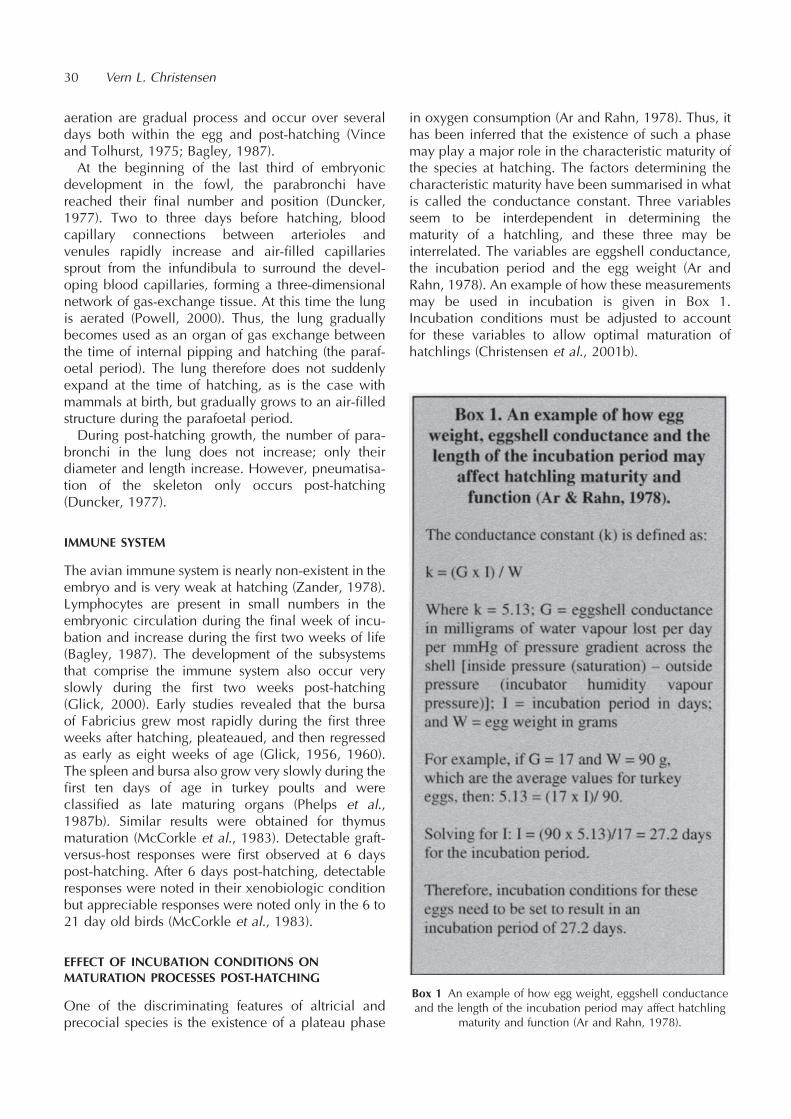
aeration are gradual process and occur over severaldays both within the egg and post-hatching (Vinceand Tolhurst, 1975; Bagley, 1987).At the beginning of the last third of embryonic
development in the fowl, the parabronchi havereached their final number and position (Duncker,1977). Two to three days before hatching, bloodcapillary connections between arterioles andvenules rapidly increase and air-filled capillariessprout from the infundibula to surround the devel-oping blood capillaries, forming a three-dimensionalnetwork of gas-exchange tissue. At this time the lungis aerated (Powell, 2000). Thus, the lung graduallybecomes used as an organ of gas exchange betweenthe time of internal pipping and hatching (the paraf-oetal period). The lung therefore does not suddenlyexpand at the time of hatching, as is the case withmammals at birth, but gradually grows to an air-filledstructure during the parafoetal period.During post-hatching growth, the number of para-
bronchi in the lung does not increase; only theirdiameter and length increase. However, pneumatisa-tion of the skeleton only occurs post-hatching(Duncker, 1977).
IMMUNE SYSTEM
The avian immune system is nearly non-existent in theembryo and is very weak at hatching (Zander, 1978).Lymphocytes are present in small numbers in theembryonic circulation during the final week of incu-bation and increase during the first two weeks of life(Bagley, 1987). The development of the subsystemsthat comprise the immune system also occur veryslowly during the first two weeks post-hatching(Glick, 2000). Early studies revealed that the bursaof Fabricius grew most rapidly during the first threeweeks after hatching, pleateaued, and then regressedas early as eight weeks of age (Glick, 1956, 1960).The spleen and bursa also grow very slowly during thefirst ten days of age in turkey poults and wereclassified as late maturing organs (Phelps et al.,1987b). Similar results were obtained for thymusmaturation (McCorkle et al., 1983). Detectable graft-versus-host responses were first observed at 6 dayspost-hatching. After 6 days post-hatching, detectableresponses were noted in their xenobiologic conditionbut appreciable responses were noted only in the 6 to21 day old birds (McCorkle et al., 1983).
EFFECT OF INCUBATION CONDITIONS ONMATURATION PROCESSES POST-HATCHING
One of the discriminating features of altricial andprecocial species is the existence of a plateau phase
in oxygen consumption (Ar and Rahn, 1978). Thus, ithas been inferred that the existence of such a phasemay play a major role in the characteristic maturity ofthe species at hatching. The factors determining thecharacteristic maturity have been summarised in whatis called the conductance constant. Three variablesseem to be interdependent in determining thematurity of a hatchling, and these three may beinterrelated. The variables are eggshell conductance,the incubation period and the egg weight (Ar andRahn, 1978). An example of how these measurementsmay be used in incubation is given in Box 1.Incubation conditions must be adjusted to accountfor these variables to allow optimal maturation ofhatchlings (Christensen et al., 2001b).
30 Vern L. Christensen
Box 1 An example of how egg weight, eggshell conductanceand the length of the incubation period may affect hatchling
maturity and function (Ar and Rahn, 1978).

If the timing of the plateau phase in oxygenconsumption is accelerated or decelerated by incuba-tion conditions or physical egg properties, then theembryo makes adjustments and may delay maturationand function. Examples of incubation conditions thatmay affect the plateau stage in oxygen consumptionare the incubation temperature or the ventilation ratesof the machines. When these conditions are imposedupon embryos in late development, the results can belong term (Buys et al., 1998; Weytjens et al., 1999). Athigh altitude, eggs are produced that naturally havelonger incubation periods because hens at high alti-tude create eggs with decreased eggshell conduc-tance values (Rahn et al., 1977). Thus, conditionsfor high altitude incubation must be adjusted topreserve optimal plateau phase conditions to resultin a mature hatchling.
CONCLUSIONS
The most striking feature of the initial days of lifeoutside the shell is the rate at which different physio-logical systems mature (Table 1). Some systemsbecome totally functional within a 24 hour periodbut others take considerably longer. As during thecontinued commercial development of differentstrains of poultry additional emphasis is placed onrapid growth, the ontogeny of some systems may becompromised (Lilja and Olsson, 1987). Managementstrategies need to be formulated to deal with suchsystems and simultaneously maintain the overallhealth of the neonate. Examples of tissue systemsthat need to mature rapidly in both size and functionpost-hatching are the heart, intestines and lungs.
REFERENCES
Ar, A. (1991) Egg water movements during incubation. In: AvianIncubation, Tullett, S.G. (ed.), pp. 157–173. Butterworths-Heinemann, London.
Ar, A., Arieli, B., Belinsky, A. and Yom-Tov, Y. (1987) Energy inavian eggs and hatchlings: utilization and transfer. J. Exper.Zool., Suppl. 1: 151–164.
Ar, A. and Rahn, H. (1978) Interdependence of gas conduc-tance, incubation length and weight of the avian egg. In:Respiratory Function in Birds, Adult and Embryonic, Piiper, J.(ed.), pp. 227–236. Springer-Verlag, Berlin-Heidelberg.
Baggott, G.K. (2001) Development of extra-embryonic mem-branes and fluid compartments. In: Perspectives in Fertilisa-tion and Embryonic Development in Poultry, pp. 23–29.Ratite Conference Books, Wallingford. Reprinted in 2009:Avian Biol. Res., 2, 21-26.
Bagley, L.G. (1987) Embryonic respiration and hatchability ofturkey eggs. Ph.D. dissertation, North Carolina State Uni-versity, Raleigh.
Breeding, S.W., McRee, W.A., Ficken, M.D. and Ferket, P.R.(1994) Effect of protein restriction during brooding on spon-taneous turkey myopathy. Avian Dis., 38, 366–370.
Buyse, N., Dewil, E., Gonzales, E. and Decuypere, E. (1998)Different CO2 levels during incubation interact with hatch-ing time and ascites susceptibility in two broiler linesselected for different growth rate. Avian Pathol., 27, 605–612.
Christensen, V.L., McMurtry, J.P., Donaldson, W.E. and Nestor,K.E. (2001a) Incubation temperature affects plasma insulin-like growth factors in embryos from genetically selectedlines of turkeys. Poult. Sci., 80, 949–954.
Christensen, V.L., Grimes, J.L. and Wineland, M.J. (2001b)Effects of turkey breeder hen age, strain, and length of theincubation period on survival of embryos and hatchlings. J.Appl. Poult. Res., 10, 5–15.
Davis, T.A. and Ackerman, R.A. (1987) Effects of increasedwater loss on growth and water content of the chick embryo.J. Exp. Zool., Suppl. 1: 357–364.
Davis, G.S. and Siopes, T.D. (1989) Ontogeny of daily rhyth-micity in plasma corticosterone and variation in sensitivity ofthe corticosterone response in turkey poults. Poult. Sci., 68,423–427.
Dawson, W.R. and Whittow, G.C. (2000) Regulation of bodytemperature. In: Avian Physiology, Whittow, G.C. (ed.), pp.344–390. Academic Press, San Diego.
Denbow, D.M. (2000) Gastrointestinal anatomy and physiol-ogy. In: Avian Physiology, Whittow, G.C. (ed.), pp. 299–326. Academic Press, San Diego.
Denbow, D.M. and Sheppard, B.J. (1993) Food and water intakeresponses of the domestic fowl to norepinephrine infusion atcircumscribed neural sites. Brain Res. Bull., 332, 121–128.
Donaldson, W.E. and Christensen, V.L. (1994) Dietary carbohy-drate effects on some plasma organic acids and aspects ofglucose metabolism in turkey poults. Comp. Biochem.Physiol., 109A, 423–440.
Development during the first seven days post-hatching 31
Table 1 Developmental rates of different physiological systems during the first seven days post-hatching in broilers and turkeys
System Growth Function
Circulatory system Rapid and early in embryogenesis Rapid and early
Kidney and body fluids system Rapid and early following hatching Slow until midway through embryogenesis to hatching
Digestive system Rapid at hatching Rapid at hatching
Body temperature control system Slow Depends upon species
Respiratory system Slow Slow
Immune system Slow Slow

Donaldson, W.E., Clark, J. and Christensen, V.L. (1994)Protein, lipid and glycogen stores in newly-hatchedturkey (Melagris gallopavo) poults as affected by post-hatch stressors and holding time. Comp. Biochem. Physiol.,107A, 559–562.
Doneen, B.A. and Smith, T.E. (1982) Ontogeny of endocrinecontrol of osmoregulation in chick embryo. II. Actions ofprolactin, arginine vasopressin, and aldosterone. Gen.Compar. Endocrinol., 48, 310–318.
Duncker, H.R. (1977) Development of the avian respiratory andcirculatory systems. In: Respiratory Function in Birds, Adultand Embryonic, Piiper, J. (ed.), pp. 261–273. Springer-Verlag, Berlin-Heidelberg.
Fan, Y.K., Croom, J., Christensen, V.L., Black, B.L., Bird, A.R.,Daniel, L.R., McBride, B.W. and Eisen, E.J. (1997) Jejunalglucose uptake and oxygen consumption in turkey poultsselected for rapid growth. Poult. Sci., 76, 1738–1745.
Fasenko, G.M. (1996) Factors influencing embryo and poultviability and growth in stored turkey eggs. Ph.D. dissertation,North Carolina State University, Raleigh.
Glick, B. (1956) Normal growth of the bursa of Fabricius inchickens. Poult. Sci., 35, 843–851.
Glick, B. (1960) Growth of the bursa of Fabricius and itsrelationship to the adrenal gland in the White Pekin duck,White Leghorn, outbred and inbred New Hampshire. Poult.Sci., 39, 130–139.
Glick, B. (2000), Immunophysiology. In: Avian Physiology,Whittow, G.C. (ed.), pp. 657–670. Academic Press, SanDiego.
Goldstein, D.L. and Skadhauge, E. (2000) Renal and extrarenalregulation of body fluid composition. In: Avian Physiology,Whittow, G.C. (ed.), pp. 265–297. Academic Press, SanDiego.
Isaacks, R.E., Harkness, D.R., Adler, J.L., Kim, C.Y., Goldman,P.H. and Roth, S. (1976) Studies on avian erythrocytemetabolism. 5. Relationship between the major phosphory-lated metabolic intermediates and whole blood oxygenaffinity in embryos and poults of the domestic turkey(Meleagris gallopavo). Poult. Sci., 55, 1788–1794.
Julian, R.J., Mirsalimi, S.M., Bagley, L.G. and Squires, E.J. (1992)Effect of hypoxia on spontaneous turkey cardiomyopathy(round-heart disease). Avian Dis., 36, 1043–1047.
Lilja, C. (1983) A comparative study of postnatal growth andorgan development in some species of birds. Growth, 47,317–339.
Lilja, C. and Olsson, U. (1987) Changes in embryonic devel-opment associated with long-term selection for high growthrate in Japanese quail. Growth, 51, 301–308.
Magwood, S.E. and Bray, D. (1962) Disease condition of turkeypoults characterized by enlarged and rounded hearts.Canad. J. Compar. Med., 26, 268–272.
MacPherson, C.R. and Deamer, J. (1964) Some observations onthe normal erythrocyte development in the chick embryo.Poult. Sci., 43, 223–228.
McCorkle, F.M., Simmons, D.G. and Luginbuhl, G.H. (1983)Graft-vs-host response in Alcaligenes faecalis-infected turkeypoults. Amer. J. Veter. Res., 44, 1141–1142.
McNichols, M.J. and McNabb, F.M.A. (1988) Development ofthyroid function and its pituitary control in embryonic andhatchling precocial Japanese quail and altricial Ring doves.Gen. Comp. Endocrin., 69, 109–118.
Moran, E.T. and Reinhart, B.S. (1980) Poult yolk sac amountand composition upon placement: Effect of breeder age, eggweight, sex and subsequent change with feeding on fasting.Poult. Sci., 59, 1521–1528.
Nice, M.M. (1962) Development of behavior in precocialbirds. Trans. Linn. Soc. New York, 8, 1–211.
Nicholson, L. (1992) Nutritional aspects of early poult mortality.MS thesis, North Carolina State University, Raleigh.
Noble, D.O., Nestor, K.E. and Polley, C.R. (1999) Factorsinfluencing early poult flip-overs in experimental popula-tions of turkeys. Poult. Sci., 78, 178–181.
Noy, Y. and Sklan, D. (1996) Uptake capacity in vitro forglucose and methionine and in situ for oleic acid in proximalsmall intestine of posthatch chick. Poult. Sci., 75, 998–1002.
Noy, Y. and Sklan, D. (1998) Yolk utilisation in the newlyhatched poult. Brit. Poult. Sci., 39, 446–451.
O’Sullivan, N.P., Dunnington, E.A., Larsen, A.S. and Siegel, P.B.(1992) Correlated responses in lines of chickens divergentlyselected for fifty-six-day body weight. 3. Digestive enzymes.Poult. Sci., 71, 610–617.
Phelps, P.V. (1985) The posthatch physiology of the turkeypoult. MS thesis, North Carolina State University,Raleigh.
Phelps, P.V., Edens, F.W. and Christensen, V.L. (1987a) Theposthatch physiology of the turkey poult. II. Hematology.Comp. Biochem. Physiol., 86A, 745–750.
Phelps, P.V., Edens, F.W. and Christensen, V.L. (1987b) Theposthatch physiology of the turkey poult. 1. Growth anddevelopment. Comp. Biochem. Physiol., 86A, 739–743.
Powell, F.L. (2000) Respiration. In: Avian Physiology, Whittow,G.C. (ed.), pp. 223–264. Academic Press, San Diego.
Rahn, H., Carey, C., Balmas, K., Bhatia, B. and Paganelli, C.(1977). Reduction of pore area of the avian eggshell as anadaptation to altitude. Proc. Nat. Acad. Sci., 74, 3095–3098.
Ricklefs, R.E. (1987) A comparative analysis of avian embryonicgrowth. J. Exp. Zool., 51, 309–324.
Ricklefs, R.E. and Starck, J.M. (1998) Embryonic growth anddevelopment. In: Avian Growth and Development, Evolu-tion within the Altricial-Precocial Spectrum, Starck, J.M. andRicklefs, R.E. (eds.), pp. 31–58. Oxford University Press,New York.
Romanoff, A.L. (1960) The Avian Embryo. The MacmillanCompany, New York.
Schmalhausen, L. (1930) Das wachstumgesetz als gesetz derprogressiven differenzierung. Wilhelm Rouxs Arch.Develop. Biol., 123, 153–178.
Sell, J.L., Angel, C.R., Piquer, R.L., Mallarino, E.G. and Batshan,H.A. (1991) Developmental patters of selected characteris-tics of the gastrointestinal tract of young turkeys. Poult. Sci.,70, 1200–1205.
Selye, H. (1951) The general adaptative syndrome. Ann. Rev.Med., 2, 327–342.
Suvarna, S.R. (1999) Ontogeny of glucose transport in turkeyintestines. PhD dissertation, North Carolina State University,Raleigh.
Tazawa, H. and Rahn, H. (1987) Temperature and metabolismof chick embryos and hatchlings after prolonged cooling. J.Exp. Zool., Suppl. 1: 105–109.
32 Vern L. Christensen
Tazawa, H. and Whittow, G. C. (2000) Incubation physiology.In: Avian Physiology G. C. Whittow (ed.), pp. 617–645.Academic Press, San Diego.
Uni, Z., Noy, Y. and Sklan, D. (1995a) Posthatch changes inmorphology and function of the small intestines in heavy-and light-strain chicks. Poult. Sci., 74, 1622–1629.
Uni, Z., Noy, Y. and Sklan, D. (1995b) Development of thesmall intestine in heavy and light strain chicks before andafter hatching. Brit. Poult. Sci., 36, 64–71.
Uni, Z., Noy, Y. and Sklan, D. (1999) Posthatch development ofsmall intestinal function in the poult. Poult. Sci., 78, 215–222.
Vince, M.A. and Tolhurst, B.E. (1975) The establishment of lung
ventilation in the avian embryo. The rate at which the lungsbecome aerated. Comp. Biochem. Physiol., 52, 331–337.
Watterson, R.L. and Sweeney, R.M. (1973) Embryology of thechick. In: Laboratory Studies of Chick, Pig, and FrogEmbryos, pp. 5–76. Burgess Publishing Company, Minnea-polis.
Weytjens, S., Meijerhof, R., Buyse, J. and Decuypere, E. (1999)Thermoregulation in chicks originating from breeder flocksof two different ages. J. Appl. Poult. Res., 8, 139–145.
Zander, D.V. (1978) Principles of disease prevention: diagnosisand control. In: Diseases of Poultry, Hofstad, M.S., Calnek,B.W., Helmboldt, C.F., Reid, W.M. and Yoder, H.W. (eds.),pp. 3–48. The Iowa State University Press, Ames.
Development during the first seven days post-hatching 33
Developmental plasticity during embryonic andpost-hatching growth of birds
J. Matthias Starck
Institute of Zoology and Evolutionary Biology, Friedrich-Schiller-Universitaet Jena, Erbertstrasse 1,D-07743 Jena, Germany
Present address: Department of Biology II, University of Munich (LMU), Großhaderner Straße 2,D-82152 Planegg-Martinsried, Germany E-mail: [email protected]
INTRODUCTION
Developmental plasticity is the ability of a developingorganism to respond to fluctuating environmentalconditions by invoking different developmentalprograms. Therefore, variable conditions duringdevelopment may result in different phenotypes ofthe adult. Temperature dependent sex determinationin reptiles (see review by Deeming and Ferguson,1991) is among the more spectacular cases of suchdevelopmental plasticity (or ‘‘developmental conver-sion’’) i.e. different genetic programs being read. Bycontrast, the more or less constant incubation condi-tions for bird eggs have meant that embryogenesis isquite unaffected by fluctuations of the environment(except for maternal effects, such as yolk contents ofeggs, or concentrations of hormones and immunoglo-bulins deposited in the yolk). However, during post-hatching growth birds may experience considerablefluctuations of their natural environment.Within the theoretical framework of developmental
plasticity it would be expected that growing birdsadjust their developmental patterns in response tochanges in environmental conditions. This mayoccur by invoking new genetic programs (develop-mental conversion) or by changing quantitativeexpression of existing genetic programs (develop-mental modulation). Availability of food, nutrientcomposition, and changes in ambient temperatureare the major sources of environmental variation fora young bird.Developmental plasticity and compensatory growth
of birds have been widely investigated (e.g. Schewand Ricklefs, 1998). In particular, laboratory studieshave investigated the effects of fluctuating food supplyon growth and intestinal function in the altricial nest-lings of the song thrush (Turdus philomelos) and theprecocial hatchlings of Japanese quail (Coturnixjaponica). Song-thrush nestlings were characterisedby a limited degree of plasticity of their develop-mental program which prohibited overfed nestlingsfrom significantly up-regulating their gut function to
accommodate increased food intake. This suggeststhat they were already growing at a rate close totheir physiological maximum. By contrast, young ofJapanese quail had a flexible developmental programand were able to interrupt growth for extendedperiods of post-hatching development.It is interesting to place the developmental plasticity
observed in birds in relationship to that reported inother sauropsids (i.e. reptiles and dinosaurs). Studieshave concentrated on investigating the bone micro-structure of Japanese quail growing under differentexperimental conditions. These observations werecompared with that of extant and extinct birds, anddinosaurs. The rate of bone formation is significantlyaffected by environmentalyexperimental conditions,skeletal element, and age. In the quail, the experi-mental conditions did not result in formation of linesof arrested growth. In contrast to extant birds, variableincidence of lines of arrested growth can be observedamong several dinosaur species, including fossil birds(from the Cretaceous), extant sauropsids (reptiles) aswell as non-mammalian synapsids, and some extantmammals. This suggests that developmental plasticitywas much common in the ancestral condition andthat the response of bone to environmental conditionswas more variable than what is observed in livingbirds. It is suggested that during the evolution of theornithurine birds (which died out at the end of theCretaceous Period), such high degree of develop-mental plasticity was reduced in modern birds inassociation with the shortened developmental time.
REFERENCES
Deeming, D.C. and Ferguson, M.W.J. (1991) Egg Incubation: ItsEffects on Embryonic Development in Birds and Reptiles.Cambridge University Press, Cambridge.
Schew, W.A. and Ricklefs, R.E. (1998) Developmental plasticity.In: Avian Growth and Development. Evolution within theAltricial-Precocial Spectrum. Starck, J.M. and Ricklefs, R.E.(eds.), pp. 288–304. Oxford University Press, New York.
AVIAN BIOLOGY RESEARCH 2 (1/2), 2009 35
doi: 10.3184/175815509X430435

Assessment and significance of fertility in commercialpoultry production
Graham J. Wishart
Avian Reproduction Group, University of Abertay Dundee, Bell Street, Dundee, DD1 1HG, UK
INTRODUCTION
Breeding efficiency in poultry is usually assessed interms of ’’percentage fertile eggs’’ laid followingartificial insemination (AI) or natural mating. Fertilityof eggs can be assessed ’’non-destructively’’ by exam-ining eggs that have failed to hatch or by candlingduring incubation, or ’’destructively’’ by opening asample of eggs to look for the presence of anembryo – either before or during incubation.All of these systems have particular disadvantages.
In the first two methods, any fertility or insemination’’problems’’ will be two or more weeks old by thetime that fertility data is obtained. In the second twomethods, there are the statistical problems associatedwith sampling as well as the loss of eggs. Additionally,assessing fertility by macroscopic analysis of un-incubated eggs can be unreliable.However, all of these methods have another
common disadvantage that is inherent in the measure-ment of fertility itself. It is that assessment of fertilityonly tells us what happened to one sperm, the onethat fertilised the egg, whilst we know that hundredsof millions of spermatozoa are transferred in an AIschedule or by natural mating. So, if we accept thatbreeding efficiency is simply the transfer of spermfrom the male to the female reproductive tracts (withor without AI), then the proportion of fertile eggs is nota particularly good measure of this.There are more direct ways of measuring successful
sperm transfer. Evidence of sperm that associate withthe peri-vitelline layers that surround the ovum at oraround the time of fertilisation in the upper oviductcan be found in laid eggs. In fact, evidence of up to athousand sperm per egg can be readily obtained toprovide an extended scale of breeding efficiencycompared to the binary ’’fertility’’, i.e. the egg isfertile or not. This chapter described the principles,use and application of some of the methods to detect‘sperm-in-eggs’.
SPERM IN THE FEMALE REPRODUCTIVE TRACT–HOWSPERM REACH THE OVUM
Inseminated sperm are deposited in the distal vagina(see Brillard, 2002), from where they are mostly
ejected into the cloaca. Around 1% of the insemi-nated dose of sperm (e.g. 1 million out of 100 million)enters the ’’sperm storage tubules’’ at the utero-vaginal junction. These tubules provide a reservoirfrom which sperm are released at a rate of around30% per day in chickens and 10% per day in turkeys.Released spermatozoa migrate up the hen’s oviduct tofertilise the newly ovulated ovum at the upper end ofthe oviduct. However, if the oviducal sperm reservoiris not replenished by further inseminations, then therewill eventually be insufficient sperm to fertilise theova and so the ’’fertile period’’ will come to an end.This occurs after about 2 weeks in chickens and 4–6weeks, or longer, in turkeys.
FERTILISATION–WHEN SPERM ENCOUNTER THEOVUM
Fertilisation takes place in the infundibulum, thefunnel-like opening of the oviduct adjacent to theovary (see Brillard, 2002). To enter the ovum, spermmust penetrate the inner peri-vitelline layer (IPVL) thatsurrounds the ovum and they do this by enzymatichydrolysis of the layer’s fibres to create small holes inthe IPVL. These ’’IPVL-holes’’ are most frequent in theIPVL over the germinal disc where over 1,000 can befound, although eggs are likely to be fertile if only 6IPVL-holes are present. If the numbers are lower than6, fertility is less likely and is zero if there are no IPVL-holes.Fertilisation has to take place in the period (~15
minutes) between ovulation and deposition of theouter peri-vitelline layer (OPVL), which is the firstoviducal secretion to surround the yolk. After fertilisa-tion, sperm that have not penetrated the ovum aretrapped in the OPVL. In the final accounting of IPVL-holes and OPVL-sperm, up to 50,000 sperm can beshown to interact with the peri-vitelline layers.The numbers of sperm interacting with the egg are
proportional to the numbers in the sperm storagetubules and so they are a measure of sperm transfer.Most importantly, they can be demonstrated in thelaid egg, which therefore represents a useful’’reporter’’ of oviducal sperm numbers and so ofsuccessful transfer of sperm into the hen’s reproduc-tive system.
AVIAN BIOLOGY RESEARCH 2 (1/2), 2009 37–40
doi: 10.3184/175815509X431795
METHODS FOR MEASURING OPVL-SPERM AND IPVL-HOLES
Whichever measurement is to be made or techniqueto be followed, the first steps involve removing thewhole yolk from an unincubated egg, washing it in asimple salt solution, and then cutting a 2 cm-squaredpiece of whole peri-vitelline layer from over thegerminal disc (Figure 1). This piece of layer is thenwashed free of yolk and spread on a microscope slide(Figure 2). For viewing OPVL-sperm the membraneshould be stained with a DNA-specific fluorochrome.OPVL-sperm nuclei can be seen using a fluorescentmicroscope at around 6400 magnification. IPVL-holes can be seen in glycoprotein-stained samplesby light microscopy at 640 magnification or, forunstained samples, at the same magnification usingdark-ground optics.OPVL-sperm require staining with potentially toxic
dyes and they must be viewed at high magnification,which takes a long time. So IPVL-holes measurementsare preferable and the author’s method of choice, onthe basis of simplicity, is to examine IPVL-holes inunstained samples. However, in all the followingwork, OPVL-sperm measurement can be applied inexactly the same way as that for IPVL-holes.IPVL-holes are most frequent in a broad ring that
corresponds to the region over the germinal disc
(Figure 3). We usually report the whole numbers ofholes in this region, although if the numbers are high,we only count the holes in a region of the area andcalculate the total number by proportion.
Application of measuring sperm-in-eggs in commercialbroiler-breeder flocks
We have measured the IPVL-holes in samples of 60eggs from broiler breeder flocks and have founddistributions similar to those shown in Figure 4. Thedistributions are skewed, more so in flocks with lowerfertility, where there are more eggs with either lownumbers, or no, IPVL-holes. What is remarkable aboutthese distributions is the range of sperm-in-eggs,which can range from zero to many hundreds. Thishighly differential distribution of sperm among thefemales represents the most significant contributionto flock infertility. It may be affected by femalereproductive physiology, but it is more likely toreflect mate choice. In fact, in all broiler breederflocks studied so far, infertile eggs can be shown byIPVL-hole analysis, to be laid by a separate populationof hens that do not appear to be involved in mating.
38 Graham J. Wishart
Figure 1 Cutting a small 4 cm2 piece of peri-vitelline layer fromaround the germinal disc.
Figure 2 Spreading the washed peri-vitelline layer on amicroscope slide.
Figure 3 The holes in the IPVL from over the germinal disc (theimage is ~2.5mm across).
Figure 4 Frequency distribution of the IPVL-holes from over thegerminal disc in samples of 60 eggs from three flocks of broilerbreeders. The fertility of all birds in the flocks was 87%, 84%
and 74% for flocks 1, 2 and 3, respectively.

Despite the large variation in IPVL-holes among eggs(and thus hens), the median IPVL-holes in samples ofbroiler eggs is highly correlated with overall flockfertility and so can be used to monitor flock fertilityand identify sub-fertile flocks.It is commonly found that the fertility of broiler
breeder flocks becomes reduced after 50 weeks ofage. It is often assumed that mating may decrease afterthat time due to lowered mating frequency or success,usually blamed on male ’’leg problems’’. Figure 5illustrates this by showing a typical pattern, withfertility dropping by around 10% between weeks 50and 55. However, the pattern of the median IPVL-holes of samples of eggs shows that real matingefficiency, in terms of the numbers of sperm passed
into eggs, follows a quite different pattern. In fact,oviducal sperm numbers (IPVL-holes) increase in eggsfrom hens of up to 38 weeks of age, after which thereis a steady decrease from around 250 IPVL-holes toonly 20 IPVL-holes at 55 weeks of age (Figure 5). Thusnot only does the greater quantitative range of theIPVL-holes assay show that the magnitude of theproblem is that of a 90%, rather than 10% reductionin mating success, but that also the reduction inmating efficiency actually occurs more than 10weeks before a significant drop in fertility is seen.This might suggest that the fertility loss in broilerbreeders is more likely to be the result of socialchanges in mating systems, rather than of infirmityin ageing birds.
Application of measuring sperm-in-eggs in commercialturkey breeder flocks
The distribution of OPVL-holes in commercial turkeyflocks is similar to that found in broiler breeders,although the range is less since artificial inseminationnegates the added variable of mate choice. However,in the same way as for broiler breeders, the medianIPVL-holes of samples of 60 eggs is related to flockfertility. When comparing IPVL-holes in eggs fromturkey flocks, it is important to take samples on thesame number of days after artificial insemination,since this makes a significant difference in thenumbers of IPVL-holes (Figure 6). Examining theIPVL-holes in eggs take before and after inseminationcan be used to assess the efficiency of AI.
Assessment and significance of fertility in commercial poultry production 39
Figure 5 Weekly flock fertility (a) and mean IPVL-holes of a sample of 60 eggs (b) during the life of a broiler breeder flock.
Figure 6 Number of IPVL-holes over the germinal disc (GD)from samples of 10 turkey eggs taken daily before and after
artificial insemination.
(a) (b)
Other applications of measuring sperm in eggs to assessbreeding efficiency
In many research applications, measuring sperm-in-eggs has proved to be a much more sensitive measureof breeding efficiency than the proportion of fertileeggs. Thus, measuring sperm-in-eggs, often underconditions where there was no apparent differencein fertility, has highlighted differences in spermquality, nutritional regimes, environmental conditionsand male to female ratios. Thus these techniques havesuperseded the traditional ’’fertility trial’.
CONCLUSIONS
Measuring sperm-in-eggs provides a useful alternativeto measuring the proportion of fertile eggs laid bychickens and turkeys. It has found application inanalysis of various breeding systems and on both aresearch and a commercial scale. It’s value lies in itsgreater quantitative range and sensitivity and moredirect relevance to breeding efficiency, in terms ofnumbers of sperm transferred from males to females. Italso provides a more immediate analysis, avoiding thewait for incubated eggs, and can even identify areduction in breeding efficiency before any infertileeggs are seen. In research, it has replaced the ’’fertilitytrial’’ as the best means of experimentally highlightingthe differences in fertilising ability between samples ofsemen or in breeding efficiency under differentenvironmental or nutritional regimens. On a commer-cial scale, it can be used for monitoring flock per-
formance and provides the only means by whichmating and AI efficiency can be properly analysed.
FURTHER READING
Reviews by Bakst et al. (1994), Wishart (1995),Wishart and Staines (1999), Wishart et al. (2001)and Brillard (2002) should be consulted for furtherinformation and details of original articles onmeasuring sperm-in-eggs by Bramwell, Brillard,Donoghue, Wishart and their colleagues.
REFERENCES
Bakst, M.R., Brillard, J.-P. and Wishart, G.J. (1994) Spermselection, transport and storage in the avian oviduct. Poult.Sci. Rev., 5, 143–177.
Brillard, J.-P. (2002) Practical aspects of fertility in poultry. In:Practical Aspects of Commercial Incubation in Poultry,Deeming, D.C. (ed.), pp. 7–10. Ratite Conference Books,Lincoln. Reprinted in 2009. Avian Biol. Res., 2, 41–46.
Wishart, G.J. (1995) New approaches to evaluating male andfemale fertility. In: Proceedings of the 1st InternationalSymposium of AI in Poultry, Bakst, M.R. and Wishart, G.J.(eds.), pp. 207–223. Poultry Science Association, Savoy,Illinois.
Wishart, G.J. and Staines, H.J. (1999). Measuring sperm:egginteraction to assess breeding efficiency in chickens andturkeys. Poult. Sci., 87, 428–436.
Wishart, G.J., Staines, H.J. and Hazary, R.C. (2001) Evaluationof Fertility: Biological Basis and Practical Application.World’s Poult. Sci. J., 57, 309–314.
40 Graham J. Wishart
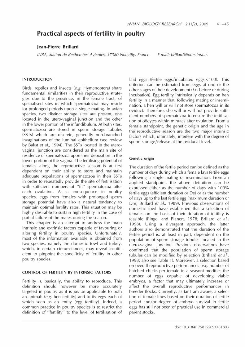
Practical aspects of fertility in poultry
Jean-Pierre Brillard
INRA, Station de Recherches Avicoles, 37380-Nouzilly, France E-mail: [email protected].
INTRODUCTION
Birds, reptiles and insects (e.g. Hymenoptera) sharefundamental similarities in their reproductive strate-gies due to the presence, in the female tract, ofspecialised sites in which spermatozoa may residefor prolonged periods upon a single mating. In avianspecies, two distinct storage sites are present, onelocated in the utero-vaginal junction and the otherin the lower portion of the infundibulum. At both sites,spermatozoa are stored in sperm storage tubules(SSTs) which are discrete, generally non-branchedinvaginations of the luminal epithelium (see reviewby Bakst et al., 1994). The SSTs located in the utero-vaginal junction are considered as the main site ofresidence of spermatozoa upon their deposition in thelower portion of the vagina. The fertilising potential offemales along the reproductive season is at firstdependent on their ability to store and maintainadequate populations of spermatozoa in their SSTsin order to repeatedly provide the site of fertilisationwith sufficient numbers of ‘‘fit’’ spermatozoa aftereach ovulation. As a consequence in poultryspecies, eggs from females with prolonged spermstorage potential have also a natural tendency tomaintain optimal fertility rates. This situation may behighly desirable to sustain high fertility in the case ofpartial failure of the males during the season.This chapter is an attempt to address the main
intrinsic and extrinsic factors capable of favouring oraltering fertility in poultry species. Unfortunately,most of the information available is obtained fromtwo species, namely the domestic fowl and turkey,which, in certain circumstances, may reveal insuffi-cient to pinpoint the specificity of fertility in otherpoultry species.
CONTROL OF FERTILITY BY INTRINSIC FACTORS
Fertility is, basically, the ability to reproduce. Thisdefinition should however be more accuratelytargeted in poultry as it is per se applicable to bothan animal: (e.g. hen fertility) and to its eggs each ofwhich seen as an entity (egg fertility). Indeed, acommon practice in poultry species is to restrict thedefinition of ‘‘fertility’’ to the level of fertilisation of
laid eggs (fertile eggsyincubated eggs6100). Thiscriterion can be estimated from eggs at one or theother stages of their development (i.e. before or duringincubation). Egg fertility intrinsically depends on henfertility in a manner that, following mating or insemi-nation, a hen will or will not store spermatozoa in itsoviduct. Therefore, she will or will not provide suffi-cient numbers of spermatozoa to ensure the fertilisa-tion of oocytes within minutes after ovulation. From afemale standpoint, the genetic origin and the age inthe reproductive season are the two major intrinsicfactors which, ultimately, interfere with the degree ofsperm storageyrelease at the oviducal level.
Genetic origin
The duration of the fertile period can be defined as thenumber of days during which a female lays fertile eggsfollowing a single mating or insemination. From anapplied standpoint, the above definition can beexpressed either as the number of days with 100%fertile eggs (efficient duration or De) or as the numberof days up to the last fertile egg (maximum duration orDm; Brillard et al., 1989). Previous observations ofdomestic fowl have established that a selection offemales on the basis of their duration of fertility isfeasible (Pingel and Planert, 1978; Brillard et al.,1998). Using a divergent approach, the latterauthors also demonstrated that the duration of thefertile period is, at least in part, dependent on thepopulation of sperm storage tubules located in theutero-vaginal junction. Previous observations haveconfirmed that the population of sperm storagetubules can be modified by selection (Brillard et al.,1998; also see Table 1). Moreover, a selection basedon overall reproductive performances (e.g. number ofhatched chicks per female in a season) modifies thenumber of eggs capable of developing viableembryos, a factor that may ultimately increase oraffect the overall reproductive performances inbreeder flocks. Currently, as far I am aware, a selec-tion of female lines based on their duration of fertileperiod andyor degree of embryo survival in fertileeggs has still not been of practical use in commercialparent stocks.
AVIAN BIOLOGY RESEARCH 2 (1/2), 2009 41–45
doi: 10.3184/175815509X431803
Seasonal variations of fertility
It has long been established that poultry speciesundertake more or less marked variations of theirreproductive performances over the reproductiveseason, a consequence of their physiological ageand of photoperiod. For example, the physiologicalstatus of the hen at the onset of the reproductiveseason can play a major role on subsequent perfor-mances of fertility. Brillard and Bakst (1990) demon-strated that turkey hens inseminated just prior to theonset of the laying season store higher populations ofsperm compared to hens inseminated just (equates toone day) after the onset of lay. The rationale for such ahigher sperm storage efficiency in pre-laying hens hasstill not been established. Hypothetically, the absenceof oviducal contractions in pre-laying hens mayfacilitate the migration of spermatozoa up to thestorage sites. Notwithstanding the origin of suchdifferences, it appears that high fertility rates obtainedearly in season will generally sustain persistent highfertility performances over the entire season. Bycontrast, low fertility performances at the onset ofthe season generally result in persistently poor fertilityperformances over the season. Whether or not suchdifferences can be attributed to a more functionalrelease of spermatozoa in hens having sperm storagetubules primarily filled with large populations of ‘‘fit’’spermatozoa also remains to be established.A general feature of fertility in fowl and turkey hens
is its progressive increase over the 3–5 first weeks ofthe reproductive period (up to peak of lay) followedby a plateau maintained up to 2y3–3y4 of this period.Following this plateau, fertility progressively declineswith age, a consequence of a faster release of sper-matozoa from the SSTs rather than impaired storage ofspermatozoa in the oviduct (Brillard, 1993). In orderto persistently obtain pre-cited profiles of fertilityrather than profiles with successive, temporary dropsrevealing the existence of impaired sperm storage, anadequate management of the hens with regards tofilling of the SSTs is necessary. Over the past years,research to assess the population of sperm present atthe fertilisation sites has provided a series of toolswhich, at many instances, may reveal useful to point
out the existence of defective sperm deposition(Wishart and Staines, 1995) or low sperm quality(Donoghue et al., 1995). These tools are eitherbased on the measuring the population of sperma-tozoa present in the peri-vitelline membrane (Wishart,1987; Brillard and Antoine, 1990) or on the number ofholes hydrolysed by spermatozoa in this membrane(Robertson et al., 1997). Each of these techniques maybe of practical interest in order to early detect thecauses of impaired fertility over the reproductiveseason in breeder flocks or in parental lines (seeWishart, 2002).
CONTROL OF FERTILITY BY EXTRINSIC FACTORS
Any factor impairing the initial sperm storage in theSSTs will also affect the population of ’’fit’’ sperma-tozoa at the infundibular level as these two popula-tions are closely linked (Brillard and Antoine, 1990).From a practical standpoint, a number of factors mayimpair sperm fertility such as defective mating beha-viour (in the fowl), or, in artificially inseminatedflocks, any factors capable of influencing the quantityand quality of sperm migration (e.g. sperm dose, invitro storage of semen, timing of insemination withoviposition).
Alterations of sperm migration
Several early reports described a ’’late, non reversibledecline of fertility’’ occurring in some but not allbreeder of turkey hens (Harper and Arscott, 1969;van Krey and Leighton, 1970; Sexton, 1977). Such asituation is with a very few exceptions, no longerobservable at the present times, an indication that thereproductive potential of turkey breeder hens or theirmanagement, or both, have been significantly modi-fied over the past decades. Whether or not the causesof a late seasonal decline of fertility have an intrinsicor extrinsic origin is questionable. Meanwhile, someevidence exists that both local infections (reversibleby antibiotic treatment) andyor inadequate use ofartificial insemination (AI) technology may havebeen responsible for the presence of local inflamma-
42 Jean-Pierre Brillard
Table 1 Some reproductive traits in females from domestic fowl selected during five generations on the basis of their high (H) orlow (L) potential to produce hatched chicks1
Line Maximum (SD) duration FyI% HyF% FyI% HyF% SSTs6103yhenof fertility (days) (2–8 days post-AI) (9–15 days post-AI) (SD)
H-line 15.1 (0.5) 94.8 89.2 89.4 82.2 2957 (140)
L-line 10.7 (0.6) 70.2 69.9 36.4 49.1 2241 (121)
1Differences were significant between lines for all parameters tested (P50.05). F¼ fertile; I¼ incubated; SST¼ sperm storage tubules.

tions in the vaginal wall. Such inflammations maydurably impair the migration of spermatozoa to theirstorage sites, which results in variable reductions infertility. The fact that in most circumstances, turkeyhens used for a second laying period had recoverednormal fertility levels supports the hypothesis of aninflammatory problem disappearing after the suppres-sion of its causal factor, i.e. repeated inseminationsinadequately performed.
Sexual behaviour
Despite the extensive use of natural mating condi-tions to produce day-old chicks in the fowl, thenumber of studies dedicated to the behaviouralaspects of this method of reproduction have beenrelatively few. It should be noted that fertility traitsare negatively correlated with growth rate, whichmay result in increased problems of fertility inbreeder facilities with low quality management.In my experience, in a majority of situations, leg
problems (including foot pad and articulation infec-tions) are often associated with an insufficient controlof body growth in males or females or both, shouldbe considered as responsible for the persistence oflow fertility during the second half of the layingseason. By contrast, delayed sexual maturity of themales may affect sexual behaviour in young flocks.Such a situation may have several nutritional causes,including excessive feed restriction during thegrowing period, andyor insufficient balance inmineralsyvitamins (e.g. biotin, B-complex vitamins).Another problem often affecting sexual behaviour
in young and ageing flocks is the incidence ofinappropriate light stimulation of the males. Indeed,the reproductive season in avian (including poultry)species is highly dependent on their photoperiodicenvironment. Thus, males subjected to ’’long’’ orincreasing photoperiods (410–12 hours) during thefirst 12–14 weeks of life will develop precocious butnon-persistent testicular growth. This will result inthe need for an early (540–45 weeks) and riskyreplacement of most of these males before the end ofthe reproductive season.
Artificial insemination: sperm quantity and quality
In contrast to sexual behaviour, the rationale forsuccessful use of artificial insemination procedureshas been extensively studied in fowl and turkeybreeders. (see review by Bakst and Brillard, 1995).Among factors known to influence the subsequentfertility performances, the initial quantity and qualityof spermatozoa at the time of insemination exert amajor role. This is because they directly influence
the degree of selection exerted by the hen onsubsequent sperm populations stored in theoviduct. A series of experiments conducted overthe past years have revealed that: (1) following asingle insemination, a maximum of approximately1% of the initial population of spermatozoa depos-ited intravaginally reaches the storage sites both inthe fowl and turkey hen (Brillard and Bakst, 1990;Brillard, 1993). (2) The quantity of spermatozoapresent in the infundibulum at the time of fertilisa-tion, i.e. during the 8–9 minutes following ovulation(Wishart and Fairweather, 1998) can be estimated infreshly laid eggs. This is either directly from theirpopulation trapped in the peri-vitelline membrane(Wishart, 1987) or indirectly from the number ofnumber of holes created by the hydrolysis of theinner peri-vitelline layer (Wishart and Wood, 1994).The number of peri-vitelline sperm is itself highlycorrelated with the actual population of spermatozoastored in the utero-vaginal junction (Brillard, 1993).From a practical standpoint, both techniques are
appropriate to rapidly detect whether or not afertility problem can originate from a lack ofsperm storage efficiency. If such a problem isconfirmed, several quality tests are now availableto pinpoint if whether or not the problem isqualitative or quantitative (see Bakst and Cecil,1997).
Timing of insemination with oviposition
A series of experiments conducted both in the chickenand in the turkey have demonstrated that most sper-matozoa deposited within 1–3 hours prior to or justafter oviposition are eliminated by the vaginalcontractions involved in the process of oviposition(Brillard et al., 1987; Brillard and Bakst, 1990). Forexample in the fowl hen, the relative efficiency ofsperm storage around the time of oviposition can bereduced as much as 30–40 times that observed inhens inseminated well apart (44 hours) from oviposi-tion.From a practical standpoint, the period of insemina-
tion in a given breeder house should be carefullyscheduled in order to minimise the risks of performinga majority of inseminations at or around the time oflay. Chicken breeder hens at peak lay most of theireggs between the 5th and the 8th hour following theonset of the photoperiod (model: 16L:8D) whileturkey breeder hens at peak would lay mainly fromthe 7th to the 9th hour of lay (model: 14L:10D). Bothspecies would have their laying period delayed about2 hours in ageing flocks.
Practical aspects of fertility in poultry 43
AI techniqueyAI crew
Fertility problems in breeder flocks should be consid-ered as originating, in a majority of cases, from’’human’’ rather than from technical problems.Among factors often encountered are: (1) Lack oftraining of AI crews (e.g. replacement of employeesduring vacations). (2) Inappropriate number of peoplein the crew. For example, large flocks of hens shouldbe inseminated with appropriate numbers of techni-cians to reduce the risks of AI performed during thelaying period (see above). (3) Inadequate use of adiluent.Sperm diluents are common practice to facilitate
the transportation and short term storage of semenprior to insemination. However, their optimal use issubjected to some simple rules which, if notadequately performed, may severely impair spermsurvival. Among mistakes most often observed are:(1) The absence of diluent at the time of semencollection. (2) The absence of agitation of the collec-tion tube between two semen collections (verycommon). In this case, the technician is convincedthat semen storage conditions are optimal which istruly the case only for one portion of the firstejaculate. (3) Inadequate temperature of semenstorage. A general rule should be: The longer thesperm to be stored, the cooler the semen should bewith temperatures ranging between 10–4�Calthough lower temperatures (particularly 0�C)should be avoided.
CONCLUSIONS
Unfortunately this short review of the main factorsinvolved in the practical aspects of fertility in poultryspecies cannot be exhaustive. Thus, some aspects ofmating behaviour in relation to heat stress have notbeen treated here. Indeed, the focus has been onpractical rather than generic questions of fertility,but a number of problems remain unanswered. In alarge majority of cases, questions relevant to AItechnology and, to a lesser extent, to interactionsbetween nutrition and behaviour, can be solved ifsufficient information on the history of the flock hasbeen recorded and is rapidly available to decisionmakers. However, in other occasions, an appropriateanswer to a fertility problem may not be availableimmediately.Such a situation is quite frequently observed when a
lack of recovery of fertility is observable upon partialreplacement of the males at the onset of the secondhalf of the reproductive season (i.e. around 43–45weeks of age in the fowl or 45–50 weeks of age in
turkey breeder flocks). At this time, the partial replace-ment of males is performed to boost the fertility of aflock over the remaining weeks of its season. Onmany occasions, the causes for such a decliningfertilising potential in males are either an inadequatelight stimulation or a too severe or, even, too liberalfeed restriction (see above). In this latter case, anacceptable solution is unlikely as the flock is undernatural mating (e.g. chickens) and the introduction ofnew males has created a major problem of socialbehaviour within the flock. Alternatively, the hens areartificially inseminated and the recovery of fertility hasbeen unsuccessful because the problem was notdirectly related to a question of sperm availabilitybut, rather, to a question of AI management. In bothcases, the only acceptable solution becomes the earlyelimination of the flock.
REFERENCES
Bakst, M.R., Brillard, J.-P. and Wishart, G.J. (1994) Spermselection, transport and storage in the avian oviduct. Poult.Sci. Rev., 5, 143–177.
Bakst, M.R. and Cecil, H.C. (1997) Techniques for SemenEvaluation, Semen Storage, and Fertility Determination.Poultry Science Association Publishers, Savoy, Illinois.
Brillard, J.-P. (1993) Sperm storage and transport followingnatural mating and artificial insemination. Poult. Sci., 72,923–928.
Brillard, J.-P. and Antoine, H. (1990) Storage of sperm in theutero-vaginal junction and its incidence on the numbers ofspermatozoa present in the perivitelline layer of hen’s eggs.Brit. Poult. Sci., 31, 701–710.
Brillard, J.-P. and Bakst, M.R. (1990) Quantification of sperma-tozoa in the sperm storage tubules of turkey hens and therelation to sperm numbers in the perivitelline layer of eggs.Biol. Reprod., 43, 271–275.
Brillard, J.-P., Beaumont, C. and Scheller, M.F. (1998) Physiolo-gical responses of hens divergently selected on the numberof chicks obtained from a single insemination. J. Reprod.Fert., 114, 111–117.
Brillard, J.-P., Galut, O. and Nys, Y. (1987). Possible causes ofsubfertility in hens following insemination near the time ofoviposition. Brit. Poult. Sci., 28, 307–318.
Brillard, J.-P., McDaniel, G.R., De Reviers, M. and Drane, J.W.(1989) Expression of several traits of fertility in young and olddwarf broiler breeder hens inseminated with duplicate dosesof semen. Poult. Sci., 68, 558–563.
Donoghue, A.M., Bakst, M.R., Holsberger, D.R. and Donoghue.D.J. (1995) Effect of semen storage on the number ofspermatozoa in the perivitelline layer of laid turkey eggs. J.Reprod. Fert., 105, 221–225.
Harper, J.A. and Arscott, G.H. (1969) Seasonal decline infertility of turkey eggs. Poult. Sci., 48, 2109–2113.
Pingel, H. and Planert, C. (1978) Untersuchung uber diemoglichkeiten der verlangerung der befruchtungspersistenz.Proc. 16th World Poult. Congr., Rio de Janeiro, 2, 116–125.
44 Jean-Pierre Brillard
Robertson, L., Brown, H.L., Staines, H.J. and Wishart, G.J.(1997) Characterisation and application of an avian invitro spermatozoa-egg interaction assay using the innerperivitelline layer from laid chicken eggs. J. Reprod. Fert.,110, 205–211.
Sexton, T.J. (1977) Relationship between number of spermsinseminated and fertility of turkey hens at various stages ofproduction. Poult. Sci., 56, 1054–1056.
van Krey, H.P. and Leighton, A.T. (1970) Sperm gland popula-tions, oviduct homogenates and late season declines infertility. Poult. Sci., 49, 1447.
Wishart, G.J. (1987) Regulation of the length of the fertile periodin the domestic fowl by numbers of oviductal spermatozoaas those trapped in laid eggs. J. Reprod. Fert., 80, 493–498.
Wishart, G.J. (2002) Assessment and significance of fertility incommercial poultry production. In: Practical Aspects ofCommercial Incubation in Poultry, Deeming, D.C. (ed.),pp. 1–6. Ratite Conference Books, Lincoln. Reprinted in2009: Avian Biol. Res., 2, 37–40.
Wishart, G.J and Fairweather, J. (1998) The chicken ‘‘fertiliza-tion window’’. Avian Poult. Biol. Rev., 9, 124.
Wishart, G.J. and Staines, H.J. (1995) Assessing the breedingefficiency of broiler breeder flocks by measuring spermtransfer into laid eggs. Brit. Poult. Sci., 36, 317–323.
Wishart, G.J. and Wood, L.K. (1994) Evaluation of tetrazoliumreduction assays for assessing poultry sperm metabolism andpredicting fertilizing ability. Proc. European Poult. Conf., pp.310–311.
Practical aspects of fertility in poultry 45

Shell formation and function and its role in incubation
Nick H.C. Sparks
Avian Science Research Centre, Scottish Agricultural College, Auchincuive, Ayr, KA6 5HW, UKE-mail: [email protected]..
INTRODUCTION
Poultry embryos develop within an egg that will bothprotect them during the incubation process and, withthe exception of oxygen and heat energy, provide allthat the embryo requires to allowing develop into aviable hatchling. The importance of the contributionmade by the shell to these needs is difficult to over-estimate because it provides physical protection andis a source of calcium for the embryo. An under-standing of how the shell carries out its variousfunctions should be underpinned by a basic knowl-edge of how the shell is formed. Flawed or incompleteshell formation is often the cause of the shell failing toperform the functions required of it during incubation.
SHELL FORMATION
Shell formation takes place in the oviduct (Figure 1).This organ and its associated (left) ovary, forms thereproductive structure of the domestic fowl. Theoviduct may be divided into a number of functionalregions viz: the infundibulum, magnum, isthmus,tubular shell gland, shell gland pouch and thevagina. While shell mineralisation begins in thetubular shell gland, factors that affect shell formationare influenced during albumen deposition in themagnum, and during shell membrane formation inthe isthmus.
Once the two fibrous shell membranes (Figure 2)have formed around the albumen (from which theyare separated by the so-called limiting membrane) theegg moves into the tubular shell gland where shellmineralisation begins at discrete sites on the outersurface of the outer shell membrane. These sites,sometimes known as mammilary cores, are chemi-cally and structurally modified causing the calciumsalts that are released into the lumen of the tubularshell gland to mineralise around the organic, shellmembrane-derived, core (Figure 3). This processfirmly anchors the shell to the shell membranes. Theegg then moves to the shell gland pouch where thebulk of the mineralisation process takes place, accom-panied during the early stages by a process known asplumping.Plumping is the process by which primarily water
moves into the albumen and causes the flaccid shellmembranes to become turgid. The mammilary cores,spatially separated as result of plumping, continue tobe mineralised and become encased in a sphere ofradiating calcium carbonate (Figure 4). Mineralisationcontinues until contact is made with neighbouringspheres. At this point the cone or mammilary layer iscomplete. The flow of calcium carbonate to thehemisphere that faces the shell membranes is nowdisrupted and shell formation moves into a newphase– the formation of the palisade layer.The palisade layer is frequently represented in
schematic diagrams as a series of columns, each
AVIAN BIOLOGY RESEARCH 2 (1/2), 2009 47–53
doi: 10.3184/175815509X431786
Figure 1 Diagram showing the functional regions of the hen’s oviduct.

emanating from a mammilary core. The outer regionof the palisade layer tends to be less dense than theinner region, giving this part of the shell its alternativename, the spongy layer. A thin surface crystal layercaps the palisade layer. The crystals in the surfacecrystal layer are distinct from those in the palisadelayer, almost certainly reflecting the changing envir-onment within the lumen of the shell gland as shellformation draws to a close.The palisade layer, which makes up the bulk of the
shell, consists of 95% calcium carbonate and 5%organic material. While constituting a proportionatelyminor component, this organic matrix plays an impor-tant role in conferring structural integrity upon theshell. Shell membranes are composed primarily oftype X collagen, which inhibits mineralisation, whilethe nucleation sites contain mammillan (a mammilarykeratin sulphate proteoglycan) and the palisade layercontains dermatan sulphate proteoglycan (Arias andFernandez, 2001).It is proposed that the matrix controls crystal
morphology, orientation and growth (Nys et al.,1999, 2001; Lavelin et al., 2000; Gautron et al.,2001). Given this it is predictable and has been
demonstrated, that disruption of the organic phase inthe albumen, shell membrane or shell may result inabnormal shell development. Board and Sparks(1991) noted that disrupting the cross-links withinthe membrane fibres (e.g. copper deficient diets orexposure to lathrogen-like compounds in the feed)will result in abnormal, typically wrinkled in appear-ance, shell formation. Similarly, disease conditionsthat affect plumping or albumen viscosity, andhence the tensioning of the shell membranes, willresult in shells that have a wrinkled or corrugatedappearance (Figure 5). This highlights the importanceof the organic component of the bird’s nutrition inrespect of shell formation, something that is some-times overlooked when considering shell qualityproblems.It is also now increasingly recognised that exposing
the laying bird to environmental stress (e.g. unusuallevels or types of noise, unusual visual stimuli, alteredhousing) can affect the mineralisation process and
48 Nick H.C. Sparks
Figure 2 Cross section through the shellmembranesof ahenseggshowing the fine inner, and the thicker outer, membrane fibres.
Bar marker 100mm.
Figure 3 Cross-section of a de-mineralised shell showing theclose-knit membrane fibres, organic core (towards centre of
micrograph) and organic shell matrix.Bar marker 10 mm.
Figure 4 Mineralised mammilary core. Bar marker 10 mm.
Figure 5 Corrugated shell resulting from watery albumen.
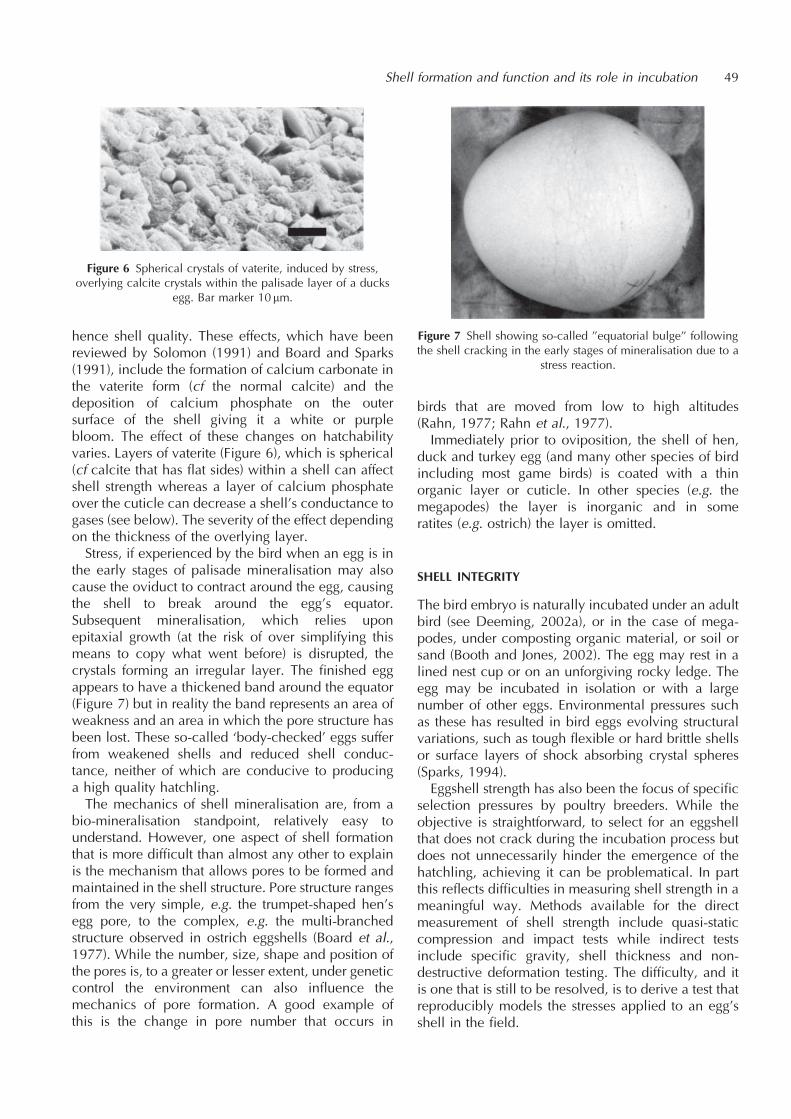
hence shell quality. These effects, which have beenreviewed by Solomon (1991) and Board and Sparks(1991), include the formation of calcium carbonate inthe vaterite form (cf the normal calcite) and thedeposition of calcium phosphate on the outersurface of the shell giving it a white or purplebloom. The effect of these changes on hatchabilityvaries. Layers of vaterite (Figure 6), which is spherical(cf calcite that has flat sides) within a shell can affectshell strength whereas a layer of calcium phosphateover the cuticle can decrease a shell’s conductance togases (see below). The severity of the effect dependingon the thickness of the overlying layer.Stress, if experienced by the bird when an egg is in
the early stages of palisade mineralisation may alsocause the oviduct to contract around the egg, causingthe shell to break around the egg’s equator.Subsequent mineralisation, which relies uponepitaxial growth (at the risk of over simplifying thismeans to copy what went before) is disrupted, thecrystals forming an irregular layer. The finished eggappears to have a thickened band around the equator(Figure 7) but in reality the band represents an area ofweakness and an area in which the pore structure hasbeen lost. These so-called ‘body-checked’ eggs sufferfrom weakened shells and reduced shell conduc-tance, neither of which are conducive to producinga high quality hatchling.The mechanics of shell mineralisation are, from a
bio-mineralisation standpoint, relatively easy tounderstand. However, one aspect of shell formationthat is more difficult than almost any other to explainis the mechanism that allows pores to be formed andmaintained in the shell structure. Pore structure rangesfrom the very simple, e.g. the trumpet-shaped hen’segg pore, to the complex, e.g. the multi-branchedstructure observed in ostrich eggshells (Board et al.,1977). While the number, size, shape and position ofthe pores is, to a greater or lesser extent, under geneticcontrol the environment can also influence themechanics of pore formation. A good example ofthis is the change in pore number that occurs in
birds that are moved from low to high altitudes(Rahn, 1977; Rahn et al., 1977).Immediately prior to oviposition, the shell of hen,
duck and turkey egg (and many other species of birdincluding most game birds) is coated with a thinorganic layer or cuticle. In other species (e.g. themegapodes) the layer is inorganic and in someratites (e.g. ostrich) the layer is omitted.
SHELL INTEGRITY
The bird embryo is naturally incubated under an adultbird (see Deeming, 2002a), or in the case of mega-podes, under composting organic material, or soil orsand (Booth and Jones, 2002). The egg may rest in alined nest cup or on an unforgiving rocky ledge. Theegg may be incubated in isolation or with a largenumber of other eggs. Environmental pressures suchas these has resulted in bird eggs evolving structuralvariations, such as tough flexible or hard brittle shellsor surface layers of shock absorbing crystal spheres(Sparks, 1994).Eggshell strength has also been the focus of specific
selection pressures by poultry breeders. While theobjective is straightforward, to select for an eggshellthat does not crack during the incubation process butdoes not unnecessarily hinder the emergence of thehatchling, achieving it can be problematical. In partthis reflects difficulties in measuring shell strength in ameaningful way. Methods available for the directmeasurement of shell strength include quasi-staticcompression and impact tests while indirect testsinclude specific gravity, shell thickness and non-destructive deformation testing. The difficulty, and itis one that is still to be resolved, is to derive a test thatreproducibly models the stresses applied to an egg’sshell in the field.
Shell formation and function and its role in incubation 49
Figure 6 Spherical crystals of vaterite, induced by stress,overlying calcite crystals within the palisade layer of a ducks
egg. Bar marker 10 mm.
Figure 7 Shell showing so-called ’’equatorial bulge’’ followingthe shell cracking in the early stages of mineralisation due to a
stress reaction.

While the effective thickness of the shell (i.e. shellthickness excluding the cuticular and mammilarylayers) is important in minimising crack propagation,the density of calcite columns and the arrangementof the columns are also important. Shell strength isimproved when the column density is high and thenumber of aligned columns is low (Solomon, 1991).If the shell breaks to the extent that the contents
leak out, then the embryo’s development will almostcertainly be arrested due to disruption of the extra-embryonic membranes. Should the shell be crackedbut the underlying membranes remain intact thedevelopment of the embryo may be affected eitherdue to infection of the egg contents by micro-organisms or excessive water loss from the eggcausing dehydration of the embryo.
MOVEMENT OF GASES ACROSS THE SHELL
Birds’ eggshells are required to fulfil two conflictingfunctions. A shell needs to prevent microbialcontaminants penetrating the shell and reachingthe egg contents while being sufficiently porousfor respiratory gases to diffuse through the shellwithout subjecting the embryo to hypoxia orsimilar.Gases move across the shell through the pores by
diffusion, the rate of diffusion being determined bythe effective pore area, the length of the diffusionpathway (e.g. shell thickness or length of the porecanal and associated overlying shell cover) and theconcentration difference of the gas across the shell. Itis notable that there is no correlation between therate at which gases may move from one side of theshell to the other and the rate that liquids orcontaminants may travel across the shell (Sparksand Board, 1984; Berrang et al., 1998). For thefowl’s egg this is a reflection of the role of thecuticle which acts as a barrier to the movement ofliquids and solids across the shell while having asmall or negligible impact on the movement of gases(Deeming, 1987).The shell needs to allow the passage of oxygen,
carbon dioxide and water in concentrations that varyduring the incubation process. Thus, as the embryogrows, the amounts of oxygen and carbon dioxidethat need to be exchanged across the shell increaseduring incubation. By contrast, with the exception ofthe first 24 h or so, the amount of water that needs todiffuse across the shell can be relatively constantthroughout the incubation period (The reasons forthis are discussed by Ar and Deeming, 2002).However, given that shell conductance is fixed atoviposition, the requirement to have a shell that canmeet the embryo’s peak requirement for oxygen,
while not being so porous that it allows theembryo to dehydrate during the incubation process,requires shell conductance to be under strict control.The mechanism that controls pore formation alsoneeds to be flexible in so far as the requirementswill change during a normal production cycle. Thusas egg weight increases the volume to surface arearatio of the egg changes requiring shell conductanceto change. However, as was illustrated when Frenchand Tullett (1991) reviewed this aspect of shellconductance, the mechanisms for achieving this arenot identical either between or within a species.Responses in shell conductance have also beennoted in response to moving birds from low tohigh altitude sites. The conductance of the eggschanging, although not always in the same direction,in response to the reduced gaseous concentrations atthe higher altitudes (Wangensteen et al., 1974; Rahnet al., 1977; Monge et al., 2000).There are occasions, however, more often linked
to the influence of breeding programmes thanaberrations occurring in wild population, whenthe eggshell does not meet the requirements ofthe embryo. Thus breeding programmes can resultin similar lines having significantly different shellconductances (French and Tullett, 1991; Sparks,unpublished observations). Reducing the age atwhich the parent bird starts to produce eggs canalso cause result in eggs being laid which have asub-optimal shell conductance value (French andTullett, 1991). Probably the most common exampleof this occurs in ducks where the removal of thecuticle from Pekin-type (cf Muscovy) duck eggsdoes, unlike the equivocal evidence for chickens(Deeming, 1987; Shafey, 2002), increase shellconductance and, as a consequence, improveshatchability. While removing the cuticle fromduck (Deeming, 1987) and turkey eggs(Christensen and Bagley, 1984) significantlyincreases shell conductivity, as noted above thisdoes not apply to all species and in these circum-stances other strategies have been used. Forexample it has been reported (Meir and Ar, 1996)that drilling holes through the shell in the latterhalf of the incubation period has improved thehatchability of early laid goose eggs.While species and breeding may influence shell
conductance, in commercial production the impact ofabnormal shells arguably has the biggest day-to dayimpact on production. For example, the rate of waterloss from either thin-shelled eggs (i.e. eggs laid beforemineralisation is complete), or eggs with crackedshells, will lose water at a faster rate than predictedcausing the embryo to be at best dehydrated when ithatches and at worst to die in ovo.
50 Nick H.C. Sparks

MOVEMENT OF LIQUID AND SOLID CONTAMINANTSACROSS THE SHELL
Eggs have evolved to be incubated in a range ofenvironments. Those like poultry that may be laid inmuddy, damp conditions have developed a surfacelayer or cuticle (Figure 8) that restricts the passage ofwater or mud into the mouth of the pore canal whileallowing gaseous diffusion to continue relativelyunimpeded (Sparks, 1994). Other species, such asthe ostrich, which incubates its eggs in dry environ-ments, produce eggs that have uncapped pores.The dual role of the cuticle in preventing the pores
from being blocked by, for example, mud or faecalmaterial and in restricting the movement of liquidwater through the shell is critical to the survival ofthe embryo. If the mouth of the pore were to becomeblocked then the conductance of the shell would bereduced in proportion to the surface area of the shellaffected. As mentioned above and elsewhere in thisbook, this can adversely affect the embryo in anumber of ways. Restricting the movement of liquidwater is important not only because the efficacy of awater logged pore is significantly reduced butbecause, with the exception of fungi, micro-organismstend not to penetrate the shell in the absence of water(Sparks, 1994; see also Berrang et al., 1999). It isnotable that bacterial contamination of hatching eggsposes a significant problem for producers who breedostriches in the UK (Deeming, 1997). This is aproblem due in significant part to the absence of acuticle or cover on an egg that has evolved to be laidin a dry environment.The structure of the cuticle deposited on eggs will
change as the bird ages and, on a particular egg, asthe storage time increases. However the cuticle is atits most vulnerable to challenge immediatelyfollowing oviposition (Figure 9) when it has still todry (Sparks and Board, 1985). In this respect, it isdifficult to overemphasise the importance of nest-boxhygiene in preventing internal contamination of thehatching egg.
While, in some respects, the cuticle on poultry eggsfaces fewer challenges in the commercial productionenvironment with its clean nest boxes, fumigation andartificial incubation those challenges that it does facecan be more severe than those that would be found inthe wild. For example, the movement of cooled eggsout of storage can cause condensation (sweating) toform on the shell creating good conditions for bacteriato move through the shell. Another challenge faced bythe cuticle is egg washing or sanitising. If carried outcorrectly, egg washing should pose little of no chal-lenge to the egg but if carried out incorrectly wash-water can be readily moved through the shell carryingcontaminants with it. This problem can becompounded by the likelihood of washing degradingthe cuticle reducing its efficacy significantly (Sparks,1994). In theory, the removal of the cuticle should notincrease the incidence of bacterial contamination ofhatching eggs that are handled with due regard to therisks. This is the case, for example, with duck eggswhere the cuticle may be removed deliberately toimprove shell conductance–but it does remove oneof the most effective barriers to bacteria if the eggshould be challenged subsequently.This section has focussed on the role of the cuticle
but this is predicated on the understanding that,should the shell be malformed (e.g. thinner thannormal) or cracked, then the influence of the cuticleis negligible, contaminants being relatively free tomove from the outer to the inner surface of acracked shell.
ION RESERVOIR
The developing embryo requires calcium for a rangeof biochemical processes however, proportionately,it is skeletal development that is the mostdemanding, requiring some 125mg of calcium.While some calcium is contained within the yolk,
Shell formation and function and its role in incubation 51
Figure 8 Cross-section of cuticle forming a loose plug in theneck of a pore canal in a hens eggshell. Bar marker 10 mm.
Figure 9 Bacterial growth underlying section of shellcontaminated (with faecal material) within minutes of
oviposition compared with no growth in section underlyingshell challenged several minutes post-oviposition.
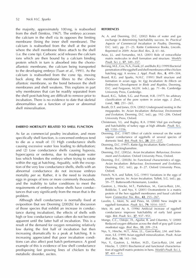
the majority, approximately 100mg, is reabsorbedfrom the shell (Simkiss, 1967). The embryo accessesthe calcium in the shell via its opposes the limitingmembrane (lining the inner shell membrane). Thecalcium is reabsorbed from the shell at the pointwhere the shell membrane fibres attach to the shell(i.e. the cone tip). Carbonic acid releases the calciumions which are then bound by a calcium bindingprotein which in turn is absorbed into the chorio-allantoic membrane, the calcium being transportedto the developing embryo (Tuan et al., 1991). As thecalcium is reabsorbed from the cone tip, movingback along the membrane fibres to the chorio-allantoic membrane, so the bond between the shellmembranes and shell weakens. This explains in partwhy membranes that can be readily separated fromthe shell post-hatching are difficult to remove prior toincubation. There is no evidence to date that skeletalabnormalities are a function of poor or abnormalshell mineralisation.
EMBRYO MORTALITY RELATED TO SHELL FUNCTION
As far as commercial poultry incubation, and morespecifically shell function, is concerned embryos tendto die as a result of: (1) High conductance shellscausing excessive water loss leading to dehydration;and (2) Low conductance shells causing hypoxia,hypercapnia or oedema, due to inadequate waterloss which hinders the embryo when trying to rotatewithin the egg at hatching. Arguably, with the excep-tion of the very low conductance shells, shells with anabnormal conductance do not increase embryomortality per se. Rather, it is the need to incubateeggs in groups of tens or more commonly thousands,and the inability to tailor conditions to meet therequirements of embryos whose shells have conduc-tances that vary significantly from the mean that is theproblem.Although shell conductance is normally fixed at
oviposition (but see Deeming [2002b] for discussionof those species that exhibit an increase in conduc-tance during incubation), the effects of shells withhigh or low conductance values often do not becomeapparent until the latter half of incubation. This is aresult of the demand for oxygen remaining relativelylow during the first half of incubation but thenincreasing dramatically to a peak at hatching. It isincreasing appreciated that poor incubation condi-tions can also affect post hatch performance. A goodexample of this is evidence of low shell conductancepredisposing fast growing lines of chicken to themetabolic disorder, ascites.
REFERENCES
Ar, A. and Deeming, D.C. (2002) Roles of water and gasexchange in determining hatchability success. In: PracticalAspects of Commercial Incubation in Poultry, Deeming,D.C. (ed.), pp. 21–25. Ratite Conference Books, Lincoln.Reprinted in 2009: Avian Biol. Res., 2, 61–66.
Arias, J.L. and Fernandez, M.S. (2001) Role of extracellularmatrix molecules in shell formation and structure. World’sPoult. Sci. J., 57, 349–357.
Berrang,M.E.,Cox,N.A., Frank, J.F. andBuhr,R.J. (1999)Bacterialpenetrationof theeggshell andshellmembranesof thechickenhatching egg: A review. J. Appl. Poult. Res., 8, 499–504.
Board, R.G. and Sparks, N.H.C. (1991) Shell structure andformation in avian eggs. In: Egg Incubation: Its Effects onEmbryonic Development in Birds and Reptiles, Deeming,D.C. and Ferguson, M.J.W. (eds.), pp. 71–86. CambridgeUniversity Press, Cambridge.
Board, R.G., Tullett, S.G. and Perrott, H.R. (1977) An arbitraryclassification of the pore system in avian eggs. J. Zool.,Lond., 182, 251–265.
Booth, D.T. and Jones, D.N. (2002) Underground nesting in themegapodes. In: Avian Incubation: Behaviour, Environmentand Evolution, Deeming, D.C. (ed.), pp. 192–206. OxfordUniversity Press, Oxford.
Christensen, V.L. and Bagley, R.A. (1984) Vital gas exchangeand hatchability of turkey eggs at high altitude. Poult. Sci.,63, 1350–1356.
Deeming, D.C. (1987) Effect of cuticle removal on the watervapour conductance of eggshells of several species ofdomestic bird. Brit. Poult. Sci., 28, 231–237.
Deeming, D.C. (1997). Ratite Egg Incubation. Ratite ConferenceBooks, Buckinghamshire.
Deeming, D.C. (2002a). Avian Incubation: Behaviour, Environ-ment and Evolution. Oxford University Press, Oxford.
Deeming, D.C. (2002b). In: Functional characteristics of eggs.Avian Incubation: Behaviour, Environment and Evolution,Deeming, D.C. (ed.), pp. 8–27. Oxford University Press,Oxford.
French, N.A. and Tullett, S.G. (1991) Variations in the eggs ofpoultry species. In: Avian Incubation, Tullett, S.G. (ed.), pp.59–77. Butterworth-Heinemann, London.
Gautron, J., Hincke, M.T., Panheleux, M., Garcia-Ruiz, J.M.,Boldicke, T. and Nys, Y. (2001) Ovotransferrin is a matrixprotein of the hen eggshell membranes and basal calcifiedlayer. Connective Tiss. Res., 42, 255–267.
Lavelin, I., Meiri, N. and Pines, M. (2000) New insight ineggshell formation. Poult. Sci., 79, 1014–1017.
Meir, M. and Ar, A. (1996) Artificial increase of eggshellconductance improves hatchability of early laid gooseeggs. Brit. Poult. Sci., 37, 937–951.
Monge, C.C., Ostojic, H., Aguilar, R. and Cifuentes, V. (2000)Reduced oxygen diffusion across the shell of gray gull (Larusmodestus) eggs. Biol. Res., 33, 209–214.
Nys, Y., Hinche, M.T., Arias, J.L., Garcia-Ruiz, J.M. and Solo-mon, S.E. (1999) Avian eggshell mineralization. Poult. AvianBiol. Rev., 10, 143–166.
Nys, Y., Gautron, J., McKee, M.D., Garcia-Ruiz, J.M. andHincke, T. (2001) Biochemical and functional characterisa-tion of eggshell matrix proteins in hens. World’s Poult. Sci. J.,57, 401–413.
52 Nick H.C. Sparks

Rahn, H. (1977) Adaptation of the avian embryo to altitude. In:Respiratory Adaptations, Capillary Exchange and ReflexMechanisms, Paintal, A.S. and Gill-Kumar, P. (eds.), pp.94–105. University of Dehli, Dehli.
Rahn, H., Carey, C., Balmas, K., Bhatia, B. and Paganelli, C.V.(1977) Reduction of pore area of the avian eggshell as anadaptation to altitude. Proc. Nat. Acad. Sci., 74, 3095–3098.
Shafey, T.M. (2002) Eggshell conductance, embryonic growth,hatchability and embryonic mortality of broiler breedereggs dipped into ascorbic acid solution. Brit. Poult. Sci.,43, 135–140.
Simkiss, K. (1967). Calcium in Reproductive Physiology.Publishing Corporation, New York.
Solomon, S.E. (1991) Egg and Eggshell Quality. Wolfe Publish-ing Ltd, London.
Sparks, N.H.C. (1994) Shell accessory materials: structure andfunction. In: Microbiology of the Avian Egg, Board, R.G. andFuller, R. (eds.), pp. 25–42. Chapman and Hall, London.
Sparks, N.H.C. and Board, R.G. (1984) Cuticle, shell porosityand water uptake through hens’ egg shells. Brit. Poult. Sci.,25, 267–276.
Sparks, N.H.C. and Board, R.G. (1985) Bacterial penetration ofthe recently oviposited shell of hens’ eggs. Aust. Vet. J., 62,169–170.
Tuan, R.S., Non, O., Akins, R.E. and Koide, M. (1991) Experi-mental studies on cultured, shell-less fowl embryos: calciumtransport, skeletal development and cardio-vascular func-tions. In: Egg Incubation: Its Effects on Embryonic Develop-ment in Birds and Reptiles, Deeming, D.C. and Ferguson,M.J.W. (eds.), pp. 419–433. Cambridge University Press,Cambridge.
Wangensteen, O.D., Rahn, H., Burton, R.R. and Smith, A.H.(1974) Respiratory gas exchange of high altitude adaptedchick embryos. Resp. Physiol., 21, 61–70.
Shell formation and function and its role in incubation 53

The critical importance of incubation temperature
Nick A. French
Aviagen Turkeys Ltd, Chowley Five, Chowley Oak Business Park, Tattenhall, Cheshire, CH3 9GA, UK.E-mail: [email protected]
INTRODUCTION
Incubation is defined as the process of applying heatto eggs and control of this process is critical forsuccessful hatching. Most birds apply heat to theireggs by sitting on them, although there are somenotable exceptions such as the megapodes, whichuse heat from rotting vegetation or volcanic activity.However, it is the purpose of this chapter to discussthe third method of incubation as used by man forpoultry and other avian species, artificial incubation.Before proceeding incubation temperature must first
be defined. For most hatchery managers it is thetemperature that they set their machines to operateat and is shown on the outside of the incubator.However, for the developing embryo it is the tempera-ture it experiences inside the egg that is the incubationtemperature and as this is what determines the devel-opment of the embryo this must be considered the realincubation temperature. Machine operating tempera-ture and embryo temperature are by no means neces-sarily the same thing (French, 1997).
RELATIONSHIP BETWEEN INCUBATOR AND EGGTEMPERATURE
Measurements of air temperature around the eggswithin incubators have shown that, depending onthe design of the machine, temperatures can differbetween 0.4 and 3.0�C from the set temperature(Kaltofen, 1969; Mauldin and Buhr, 1995; French,1997). Similarly, studies have shown that as incuba-tion progresses the internal egg temperature changesfrom being slightly cooler than the surrounding air inthe first half of incubation to being hotter than thesurrounding air in the second half of incubation(Tazawa and Nakagawa, 1985; French, 1997).Understanding why these temperature differencesoccur between egg and incubator requires an under-standing of how heat is transferred within the system.The temperature experienced by an embryo during
incubation depends on: (1) the metabolic heatproduction of the embryo itself which in turn isdependent on the size of the embryo, (2) the slightcooling effect of water lost from the egg during
incubation, (3) the temperature of the incubator and(4) the ability of the heat to transfer from embryo toincubator air. Several authors have produced thermalenergetic models describing how these four factorsinteract to determine the temperature within the eggduring artificial incubation (Kashkin, 1961; Sotherlandet al., 1987; Meijerhof and van Beek, 1993; French,1997).While incubators are designed with heating and
cooling systems to control the temperature within themachine, the eggs within the machine also have animportant effect on temperature. Embryos at the startof incubation are very small and so generate verylittle metabolic heat and so incubation heat needs tobe supplied to the embryo by the incubator. Indeed,the internal egg temperature at this stage is slightlycooler than the incubator air as there is a slightevaporative cooling effect due to water being lostfrom the egg (Figure 1). At the end of incubation, theembryo is large and generates a significant amountof metabolic heat, approximately 130mW in achicken egg (Romijin and Lokhorst, 1960), thatmust be removed from the egg. In a study oftemperatures within a turkey tunnel incubator,French (2001) showed that there was a strongcorrelation between the estimated total metabolicheat production of the eggs within the machineand the air temperature around the eggs. The totalmetabolic heat production of the eggs was depen-dent of the size of the egg and their fertility so that aseither egg mass or fertility increased so did thetemperature within the machine.How effectively heat is transferred from the egg to
the surrounding incubator air and control system ismainly determined by the rate of airflow over the eggs(Sotherland et al., 1987; Owen, 1991). The greater theairflow over the egg the more efficiently heat istransferred either to or from the egg. Variations inrates of airflow are a major reason for temperaturevariations within incubators. Monitoring temperatureswithin commercial incubators have found variationsof up to 1.2�C within machines (French, 2002) andreducing this temperature variation requires a moreuniform airflow over the eggs in all locations withinthe incubator.
AVIAN BIOLOGY RESEARCH 2 (1/2), 2009 55–59
doi: 10.3184/175815509X431812

Other factors that can influence the uniformity oftemperature within a machine are the heater andcooler units, evaporative cooling from humidifiersand cool air entering the machine through theventilation system. A well-designed incubator willensure that the localised air heating or coolingcaused by these elements will not effect the eggsby ensuring good mixing of the air before it reachesthe eggs.Where multi-stage incubation is used, i.e. incu-
bating eggs at different stages of incubation togetherwithin the same machine, there are some additionalconsiderations to ensure uniform temperatures. Multi-stage incubators rely on the metabolism of theembryos at the end of incubation to heat the eggs atthe start of incubation. For this to work successfully itis important to ensure that there are equal numbers ofembryos at each stage of incubation and differentstages of incubation are equally spaced apart. Majorhatch problems can occur if there are too many eggsat either the end or the start of incubation within themachine. Multi-stage incubators also rely on goodairflow amongst the eggs to ensure efficient heattransfer between the eggs.
INCUBATION TEMPERATURE TOLERANCE OFPOULTRY EMBRYOS
Many studies have investigated the optimum incuba-tion temperature for poultry species but most have
reported the effects of incubator operating tempera-ture rather than embryo temperature on hatchingsuccess. As the operating and embryo temperaturesare not necessarily the same and the difference willdepend on incubator design, it is very difficult tosummarise results between different studies.Optimum operating temperatures for poultry speciesappear to be between 37–38�C and deviations fromthis optimum can have a major impact on hatchingsuccess (Wilson, 1991). For example, French (1994)showed that increasing the incubation temperature ofturkey eggs from 37.5 to 38.5�C resulted in a 30–60%decline in hatchability.Recent studies have also shown that incubation
temperature will not only affect hatching success butalso post hatching performance (e.g. Lourens andMiddelkoop, 2000; Gladys et al., 2000; Hulet et al.,2000). It is therefore important to consider all theeffects when determining the correct incubationtemperature for poultry species (Decuypere andMichels, 1992).Rather than using a single optimum incubation
temperature, it would be better to define a tempera-ture range over which incubation will be successful.Ideally this would be the temperature at the embryolevel, although it may be more practical to use thetemperature of the air immediately surrounding theegg. The degree of temperature tolerance will dependon whether the temperature is high or low, the lengthof time applied and the stage of embryo development.
56 Nick A. French
Figure 1 The relationship between egg temperature (– ) and incubator air temperature (– - – ) in an artificial incubator.

So what happens as incubation temperaturechanges away from the optimal temperature rangefor successful development? The first effect of high orlow incubation temperature is to alter the rate ofembryo development resulting in an altered time ofhatch (French, 1997). High incubation temperatureswill advance the hatch whereas low incubationtemperatures will delay hatch time and this can be auseful early indicator for the hatchery manager thattemperature is sub-optimal. However, it should benoted that if incubation temperature continues toincrease further it can also slow down development(Romanoff, 1935).If incubation temperature further increases above
the optimum, then the next effect would be to alter theembryo’s development so that post-hatch perfor-mance is affected. A further increase in temperaturewould result in a delayed mortality: French (2000)showed that incubating turkey eggs at 38.0�C in thesecond week of incubation resulted in an increase inmortality in the fourth week of incubation. Immediateembryo death occurs when the internal egg tempera-ture reaches 46.5�C (Ono et al., 1994).The effects of decreasing incubation temperature
are less dramatic. Embryos are able to tolerate quitelong periods of low incubation temperature withoutadverse effects. Lancaster and Jones (1988) showedthat broiler embryos were able to tolerate cooling to21�C for 24 hours after day 13 of incubation withoutany adverse effect on hatchability. Indeed embryoswere able to tolerate 22�C for up to 48 hours on day16 of incubation without any adverse effect onhatch, although exposure for more than 30 hoursincreased the incidence of hatched chicks withdown abnormalities. These experiments werecarried out at temperatures below 27�C, the tempera-ture at which embryonic development stops (Wilson,1991). Prolonged incubation at temperaturesbetween 27–35�C are more of a problem and willcause abnormal development of the embryo (Wilson,1991). Immediate embryonic death at low tempera-tures will only occur if internal egg temperaturedrops below freezing and allows ice crystals toform inside the egg (Lundy, 1969).The effects of temperature on embryo develop-
ment are summarised in Table 1. The temperatureranges shown are not exact and may vary betweenpoultry species, eggs of different sizes (French,1997) and stage of embryo development. Studiesin both chickens (Romanoff et al., 1938; Morganand Tucker, 1967; Moreng and Shaffner, 1951;Ande and Wilson, 1981) and turkeys (French,2000) have found that the tolerance of embryos tohigh incubation temperature varies with the stage ofincubation.
IDENTIFICATION OF INCUBATION TEMPERATUREPROBLEMS
In commercial hatcheries the rapid identification andcorrection of incubation temperature problems isessential for maximising hatchability. There are twotechniques that are routinely used in the hatchery toensure that the correct incubation temperature isbeing used: (1) monitoring hatch time and (2)measuring incubation temperature within incubator.As noted above, an early indication that incubation
temperature may be sub-optimal is an alteration inhatch time. Hatching early can indicate high incuba-tion temperatures whereas late hatching can indicatelow or very high incubation temperatures. It should benoted that other factors can also influence the lengthof the incubation period, such as length of pre-incubation egg storage and age of breeder flock,and so a change of incubation time does not necessa-rily indicate a change of incubation temperature.Monitoring hatch time can be done in several ways,
assessing chick quality (thin chicks–hatched early, fatchicks–hatched late), counting how many chickshave hatched 18 hours before take-off and byweighing chicks. The simplest method to check thatthe chicks are hatching at the right time is to weigh thechicks and express this weight as a percentage of thefresh egg weight (chick yield). If hatch time is correctthen chicks yield should be between 66–68% whenthey are removed from the hatcher. If the weight is toolow, it is an indicator that the chicks have beenhatched a long time before removal from thehatcher. If the weight is too high it indicates that thehatch is late. Chick yield will also be affected by how
The critical importance of incubation temperature 57
Table 1 Typical effects of temperature on development of theavian embryo– temperature ranges shown are estimates basedon available evidence
IncubationTemperature
(�C)
Effect on Embryo
47 Immediate mortality39–47 Mortality dependent on length and timing of
exposure38–40 Altered rate of development and post hatch
performance37–38 Normal embryo development35–37 Altered rate of development and post hatch
performance27–35 Mortality dependent on length and timing of
exposure14–27 Embryo development stops, no adverse effects
of holding for 24 hours in embryos in last thirdof incubation
0–14 Mortality dependent on length and timing ofexposure
50 Immediate mortality

much water is lost from the egg during incubationalthough the effect is relatively small in comparison tothe effect of changing hatch time.Recently it has been suggested that measuring chick
length at hatch is an indicator of normal or abnormalembryo growth and could be a useful technique foridentifying high incubation temperature problems(Hill, 2001). Further studies are required to determinehow chick length is affected by incubation tempera-ture but if a relationship is demonstrated then thiscould be a very useful tool for the hatchery manager.Measuring temperatures within incubators can also
be very useful for solving hatch problems. This can bedone with thermisters attached to data-loggers so thattemperatures can be recorded at several locationswithin an incubator throughout the incubationcycle. There is a wide range of data-logging equip-ment available on the market but whatever system ischosen it is important that the thermister should beable to read to 0.1�C as a minimum and to be asaccurate as possible.Measuring internal egg temperatures would be the
ideal situation but practically difficult to achievewithin commercial incubators. Measuring eggshellsurface temperature is less problematic as it does notrequire placing a thermister within the egg and hasbeen shown to be similar to measuring internaltemperature (Sotherland et al., 1987). Measuring airtemperature in amongst the eggs within the incubatorwill not be as close to internal egg temperature asmeasuring shell surface temperature but can still be auseful method of checking temperature within themachine.
THE FUTURE
Whilst the importance of temperature for thesuccessful incubation of poultry eggs has been longunderstood it is only in recent years that the cleardistinction between egg temperature and incubatortemperature been made. An improved understandingof how egg temperatures with incubators arecontrolled has resulted in improvements in incubatordesign resulting in less within machine temperaturevariation. What are now required are guidelines as towhat are the maximum and minimum egg tempera-tures that the poultry embryo can tolerate withoutadverse effect on hatch or post-hatch performance.The temperature tolerance of embryos may not onlyvary between poultry species but also between breedsor types within species (Decuypere and Michels,1992). Determining embryo tolerance to temperaturewill be essential if hatchery managers are to maximisehatchability.
REFERENCES
Ande, T.B. and Wilson, H.R. (1981) Hatchability of chickenembryos exposed to acute high temperature stress at variousages. Poult. Sci., 60, 1561–1566.
Decuypere, E. and Michels, H. (1992) Incubation temperatureas a management tool: A review. World’s Poult. Sci. J., 48,28–38.
French, N.A. (1994) Effect of incubation temperature on thegross pathology of turkey embryos. Brit. Poult. Sci., 35,363–371.
French, N.A. (1997) Modelling incubation temperature: theeffect of incubator design, embryonic development andegg size. Poult. Sci., 76, 124–133.
French, N.A. (2000) Effect of short periods of high incubationtemperature on hatchability and incidence of embryopathology of turkey eggs. Brit. Poult. Sci., 41, 377–382.
French, N.A. (2001) Temperatures in a tunnel incubator–a longterm study. Avian Poult. Biol. Rev., 12, 184–186.
French, N.A. (2002). Managing the Incubation environment incommercial hatcheries to meet the requirements of theembryo. Avian Poult. Biol. Rev., 13, 179–185.
Gladys, G.E., Hill, D., Meijerhof, R., Saleh, T.M. and Hulet,R.M. (2000) Effect of embryo temperature and age of breederflock on broiler post-hatch performance. Poult. Sci., 79(Supplement 1), S179.
Hill, D. (2001) Chick length uniformity profiles as a fieldmeasurement of chick quality? Avian Poult. Biol. Rev., 12,188.
Hulet, R.M., Gladys, G., Hill, D. and Meijerhof, R. (2000)Embryonic temperature effects on post-hatch performancein broilers. Avian Poult. Biol. Rev., 11, 298–299.
Kaltofen, K.S. (1969) The effect of air movements on thehatchability and weight loss of chicken eggs during artificialincubation. In: The Fertility and Hatchability of the Hen’sEgg, Carter, T.C. and Freeman, B.M. (eds.), pp. 177–190.Oliver and Boyd, Edinburgh.
Kashkin, V.V. (1961) Heat exchange of bird eggs during incuba-tion. Biophysica, 6, 57–63.
Lancaster, F.M. and Jones, D.R. (1988) Cooling of broilerhatching eggs during incubation. Brit. Poult. Sci., 29, 597–604.
Lourens, S., and Middelkoop, J. H. (2000) Embryo temperatureaffects hatchability and grow-out performance of broilers.Avian Poult. Biol. Rev., 11, 299–301.
Lundy, H. (1969) A review of the effects of temperature,humidity, turning and gaseous environment in the incu-bator on the hatchability of the hen’s egg. In: The Fertilityand Hatchability of the Hen’s Egg, Carter, T.C. andFreeman, B.M. (eds.), pp 143–176. Oliver and Boyd,Edinburgh.
Mauldin, J.M. and Buhr, R.J. (1994) What is really happening inyour incubator? Int. Hatch. Pract., 9(5), 19–22.
Meijerhof, R. and van Beek, G. (1993) Mathematical modellingof temperature and moisture loss of hatching eggs. J. Theor.Biol., 165, 27–41.
Moreng, R.E. and Shaffner, C.S. (1951) Lethal internal tempera-tures for the chicken, from fertile egg to mature bird. Poult.Sci., 30, 255–256.
Morgan, W. and Tucker, W.L. (1967) Influence of thermalincubation stresses. Poult. Sci., 46, 1172–1176.
58 Nick A. French

Ono, H., Hou, P.-C.L. and Tazawa, H. (1994) Responses ofdeveloping chicken embryos to acute changes in ambienttemperature: Noninvasive study of heart rate. Israel J. Zool.,40, 467–480.
Owen, J. (1991). Principles and problems of incubator design.In: Avian Incubation, Tullett, S.G. (ed.), pp. 205–224. Butter-worth-Heinemann, London.
Romanoff, A.L. (1935) Influence of incubation temperature onthe hatchability of eggs, post-natal growth and survival ofturkeys. J. Agric. Sci., 25, 318–325.
Romanoff, A.L., Smith, L.L. and Sullivan, R.A. (1938) Bio-chemistry and biophysics of the developing hen’s egg. III.Influence of temperature. Cornell Agric. Exp. Stat. Bull.,216, 1–42.
Romijn, C. and Lokhorst, W. (1960) Foetal heat production inthe fowl. J. Physiol., 150, 239–249.
Sotherland, P.R., Spotila, J.R. and Paganelli, C.V. (1987) Avianeggs: Barriers to the exchange of heat and mass. J. Exp.Zool., Supplement 1, 81–86.
Tazawa, H. and Nakagawa, S. (1985) Response of eggtemperature, heart rate and blood pressure in the chickembryo to hypothermal stress. J. Comp. Physiol., B155,195–200.
Wilson, H.R. (1991) Physiological requirements of the devel-oping embryo: temperature and turning. In: Avian Incuba-tion, Tullett, S.G. (ed.), pp. 145–156. Butterworth-Heinemann, London.
The critical importance of incubation temperature 59

Roles of water and gas exchange in determininghatchability success
Amos Ara* and D. Charles Deemingb,c
aDepartment of Zoology, Tel-Aviv University, Ramat-Aviv 69978, Israel*E-mail: [email protected] Consulting and Research, 9 Eagle Drive, Welton, Lincoln, LN2 3LP, UKcPresent address: Department of Biological Sciences, University of Lincoln, Riseholme Park,Lincoln, LN2 2LG, UK
INTRODUCTION
Among egg-laying terrestrial vertebrates, birds areunique in that they lay only one egg at a time(related to the demands of flight), and that the eggsare almost fully exposed to the atmosphere. Birdembryos require relatively high temperatures fortheir relatively fast development and thus for theirshort incubation. These can only be provided bycontact incubation with birds and good thermalinsulation. As a result, avian eggs generally developin low humidity conditions and their embryos face thedanger of desiccation. The embryo is partiallyprotected from desiccation by the eggshell and alsoby the bird maintaining a microenvironment in thenest. The latter has not only a temperature but also ahumidity higher then the ambient conditions (Thereare, however, cases where ambient temperature ishigher then optimal nest temperature. In these casesthe bird cools its eggs). If normal development is tooccur the eggshell must be permeable to oxygen and
carbon dioxide and water is still lost through the shellin the form of vapour from the moment the egg is laid.Avian embryos have evolved to cope with this set ofconflicting conditions and successful development isdue to the interaction between the bird, its nest andthe eggs (Deeming, 2002).Artificial incubation has either to mimic this inter-
action or to generate equivalent solutions byproviding an appropriate humidity to ensure anoptimal rate of water loss from the eggs, the supplyof oxygen and the elimination of carbon dioxide. Theprinciples on which these are based may be the sameas in nature but the mechanisms by which they arecontrolled can differ greatly.
GAS DIFFUSION THROUGH THE EGGSHELL
Movement of water vapour, oxygen and carbondioxide across the eggshell is achieved by diffusionthrough pores within the calcitic eggshell (Figure 1).
AVIAN BIOLOGY RESEARCH 2 (1/2), 2009 61–66
doi: 10.3184/175815509X431830
Figure 1 Scanning electron micrograph of an avian eggshell (left) with the arrow indicating a pore (courtesy of Glenn Baggott).An illustration (right) showing the partial pressure differences across the eggshell (from Rahn et al., 1979).
Diffusion through the long narrow pores makes gasexchange independent of airflow around the egg anddependent only on the partial pressure differenceacross the shell. However, laterally, under the shell,within the shell membranes from and to the inner poreorifice, the exchange of gases is limited (Visschedijket al., 1988) and therefore a certain density of pores inthe shell must be provided to supply the blood in thechorio-allantoic membrane with sufficient oxygen.This dictates a link between diffusive water loss fromeggshell pores and respiratory gas exchange.Gas exchange across the eggshell is dictated by
Fick’s law:
Mx ¼ Gx 6ðPex � PixÞ
where x is the gas involved (H2O, O2 or CO2), M isthe rate of gas exchange, G is the conductance of theshell to the gas, Pe is the partial pressure of the gasoutside the egg, and Pi is the partial pressure of the gasunder the shell. For water vapour, PeH2O is 48 Torr at37.5�C (the air under the shell is considered to besaturated with water vapour, i.e. 100% relativehumidity at egg temperature which, in turn, is consid-ered to be equal to the incubator temperaturealthough small deviations from this scheme doexist.) and the rate of water vapour loss (MH2O)from a egg is controlled by the combination ofwater vapour conductance (GH2O) and the humidityof the air outside of the shell (PiH2O).In reality, for many bird species, GH2O is fixed at
laying and so MH2O can only be adjusted by chan-ging the humidity in the air around the egg if incuba-tion temperature (and thus PiH2O) is constant. In theirnatural habitat birds choose to build their nests inspecific locations and with specific construction char-acteristics and at a given season. The presence of thebird on eggs in a nest is sufficient to modify humidityin relation to the ambient and nest humidity, watervapour conductance and fractional mass loss as watervapour are intimately linked.Fractional mass loss values for bird eggs from a
variety of bird species averages at 0.15. Actually, this15% of the initial egg mass is a calculated loss for theentire incubation period based on average daily massloss (Rahn and Ar, 1974). Additional water (circa 4–5%) is lost between external pipping and hatching.The range of average values observed in birds gener-ally lay between 10–22% and in nests individual eggscan lose any value in this range without apparent illeffect. This makes sense because a requirement for anarrow range of mass loss would prove to be far fromadvantageous. Probability of hatching drops for lowrates of loss (510%) due to problems with exchangeof respiratory gases in particular at hatch (see below)
and losses higher than 22% cause problems withdehydration of the egg contents.It is interesting to note at this point that water
vapour and oxygen, which exchange through thesame pores, are also linked by the fact that the endproducts of aerobic metabolism are metabolic waterproduction and carbon dioxide (and energy).Metabolism utilises solids stored in the egg, which,together with the water produced, would haveincreased water concentration in the egg to an intol-erable level if no water loss would have occurred.Under normal conditions the water evaporation/waterproduction/solid elimination ratios are such that thewater concentration of the hatchling is almost iden-tical to that of the fresh egg.A batch of eggs under artificial incubation would be
normally composed of eggs from a large number ofindividual hens and so there is variation in the valuesfor GH2O, which are typically distributed around amean value. When dealing with thousands of eggswithin a setter, commercial incubation needs to setthe humidity to match the eggshell characteristics ofthe average egg of a particular batch so that it attainsthe appropriate mass loss (e.g. 15% to externalpipping). In this way the majority of the eggs will liewithin the 10–22% mass loss range and stand a goodchance of hatching normally. Significant departuresfrom this average mass loss using inappropriate incu-bator humidity will cause a significant increases inembryonic mortality (Meir and Ar, 1987).
REGULATION OF WATER LOSS AND HATCHABILITY
In commercial poultry production egg laying within aflock persists for many weeks and is associated withchanges in egg composition as well as increases inegg size and eggshell conductance (French andTullett, 1991). Changes in size and water vapourconductance over a 10-week laying season are alsoreported for commercial geese and pheasant eggs(Meir and Ar, 1996; Deeming, 2000). Astute artificialincubation procedures should take into account allthese interacting variables, in order to maximisehatchability, by monitoring and controlling incubatortemperature, humidity and respiratory gas levels, inaccordance to the stage of embryonic development.Several studies have shown that monitoring mass lossof eggs and subsequent adjustment of the humidityregimen can have beneficial effects.Tullett (1981) demonstrated that the water vapour
conductance of turkey eggs changed as flock ageincreased. As a result the humidity required toensure a standard amount of mass loss has tochange as the flock gets older. Several other studieson turkeys (Meir et al., 1984; Meir and Ar, 1987),
62 Amos Ar and D. Charles Deeming
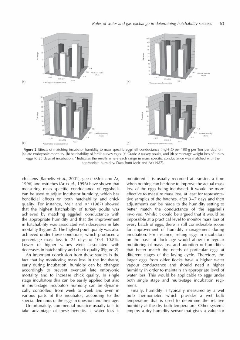
chickens (Bamelis et al., 2001), geese (Meir and Ar,1996) and ostriches (Ar et al., 1996) have shown thatmeasuring mass specific conductance of eggshellscan be used to adjust incubator humidity, which hasbeneficial effects on both hatchability and chickquality. For instance, Meir and Ar (1987) showedthat the highest hatchability of turkey poults wasachieved by matching eggshell conductance withthe appropriate humidity and that the improvementin hatchability was associated with decreases in latemortality (Figure 2). The highest poult quality was alsoachieved under these conditions, which produced apercentage mass loss to 25 days of 10.4–10.8%.Lower or higher values were associated withdecreases in hatchability and chick quality (Figure 2).An important conclusion from these studies is the
fact that by monitoring mass loss in the incubator,early during incubation, humidity can be changedaccordingly to prevent eventual late embryonicmortality and to increase chick quality. In singlestage incubators this can be easily applied but alsoin multi-stage incubators humidity can be dynami-cally controlled, from week to week and even invarious parts of the incubator, according to thespecial demands of the eggs in question and their age.Unfortunately, commercial practice usually fails to
take advantage of these benefits. If water loss is
monitored it is usually recorded at transfer, a timewhen nothing can be done to improve the actual massloss of the eggs being incubated. It would be moreeffective to measure mass loss, at least for representa-tive samples of the batches, after 3–7 days and thenadjustments can be made to the humidity setting tobetter match the conductance of the eggshellsinvolved. Whilst it could be argued that it would beimpossible at a practical level to monitor mass loss ofevery batch of eggs, there is still considerable scopefor improvement of humidity management duringincubation. For instance, setting eggs in incubatorson the basis of flock age would allow for regularmonitoring of mass loss and adoption of humiditiesthat better match the needs of particular eggs atdifferent stages of the laying cycle. Therefore, thelarger eggs from older flocks have a higher watervapour conductance and should need a higherhumidity in order to maintain an appropriate level ofwater loss. This would be applicable to eggs underboth single stage and multi-stage incubation regi-mens.Finally, humidity is typically measured by a wet
bulb thermometer, which provides a wet bulbtemperature that is used to determine the relativehumidity at the dry bulb temperature. Other systemsemploy a dry humidity sensor that gives a value for
Roles of water and gas exchange in determining hatchability success 63
Figure 2 Effects of matching incubator humidity to mass specific eggshell conductance (mgH2O per 100-g per Torr per day) on(a) late embryonic mortality, (b) hatchability of fertile turkey eggs, (c) Grade A turkey poults, and (d) percentage weight loss of turkey
eggs to 25 days of incubation. * Indicates the results where each range in mass specific conductance was matched with theappropriate humidity. Data from Meir and Ar (1987).
(a) (b)
(c) (d)

percentage Relative Humidity (%RH) directly.However, in terms of monitoring mass loss andadjusting humidity it is better to use measures ofvapour pressure for water (Absolute Humidity)vapour measured in Torr (1 Torr¼ 1mmHg¼7.5 kPa). Relationships between %RH, absolutewater content and vapour pressure are shown inFigure 3.
EMBRONIC WATER BALANCE
The fractional water content of the egg contents isequal to the fractional water content of the hatchling(~73% for poultry species). The water balance of theegg during incubation is a function of the initial waterfraction of the contents, the rate of water lost throughdiffusion across the eggshell and the amount of waterproduced by metabolism. Normal respirationproduces water as a waste product and as a resultthe total water content of the egg remains constantduring the second half of development. This is despiteof some increase in the rate of water vapour loss dueto increasing egg temperature as its metabolic activityincreases. Metabolic water production accounts for10% of the total water content of the hatchling and iscritical in the overall water balance and in main-taining the osmolarity of the egg contents (Ar, 1991).Another link between embryonic respiration and
egg water content emerges towards hatching: incuba-tion water loss creates the air space under the rigideggshell. Sufficient water must be lost to create an air
space volume adequate for complete inflation of therespiratory air sacs of the hatchling during the periodbetween internal and external pipping (Ar, 1991).Furthermore, the water content of the egg compart-
ments provides the embryo with heat inertia, thuspreventing fast changes in embryonic metabolismdue to temperature changes. As the embryo develops,water is shifted among compartments within andaround it, each of which has an optimal volume andionic composition at a given stage of development.This requires the maintenance of the appropriatewater content and ionic composition at each stage(Ar, 1991; Baggott, 2001).Metabolic rate and egg water vapour pressure
comply in the same way to changes in egg tempera-ture. The eggshell, carbon dioxide and water interactto supply dissolved calcium to the developingembryo. Many studies have shown that imbalancein any of the above-discussed parameters will reducehatchability success, and optimising them willincrease it. It becomes clear that appropriate incuba-tion atmosphere has to comply not only with tempera-ture and humidity demands of the developing embryobut at the same time with appropriate exchange of itsrespiratory gases.
RESPIRATORY GAS EXCHANGE
The eggshell acts a mediating barrier for the diffusionof oxygen into, and carbon dioxide out, of the egg viathe pores. Shell conductance values are normally
64 Amos Ar and D. Charles Deeming
Figure 3 Relationships between relative humidity (%), wet bulb temperature (�C), vapour pressure (Torr) and absolute moisturecontent (kg per kg dry air) at a dry bulb temperature of 37.5�C. The vapour pressure under the eggshell (100%RH) is also shown.
matched to the nesting environment of the bird and soeggs will usually incubate successfully within only anarrow range of humidities. The conductance of theeggshell is also matched to the respiratory exchangeof the embryo but problems arise with low conduc-tance eggshells. In average and above-average valuesfor conductance, the embryo is free to exchangeoxygen and carbon dioxide without restriction,while water vapour loss can be controlled by settingthe appropriate incubator humidity. This is becausethe eggshell conductance does not act to limit thepotential rate of oxygen consumption or carbondioxide production. However, in eggshells withlower than average conductance the eggshell limitsthe maximum gas exchange value and forces theembryo to conform to the maximum rate of diffusionthat it will physically allow. It should be mentionedhere that while the shell comprises the only resistanceto diffusion for water vapour and most of this resis-tance for carbon dioxide, about one third to one halfof the resistance for oxygen diffusion lies in thechorio-allantoic membrane (Rahn et al., 1979). Foreggs at the lower end of the range of eggshellconductance, this can mean a reduction in oxygenconsumption as early as day 15 in the fowl (Tullettand Deeming, 1982), and retention of carbon dioxide.This can affect rates of embryonic growth (Burton andTullett, 1983), presumably causing longer incubationperiods.Shell-induced hypoxia and hypercapnia are reasons
why embryos in low porosity eggshells can fail tohatch even if the incubator humidity is lowered tofacilitate the appropriate rate of mass loss. Increasingmass loss across a low conductance eggshell will notimproves rates of oxygen diffusion. In practice, shellinduced hypoxia may not kill many embryos in ovobut it can be a contributing factor in increasing thespread of hatch events such as internal and externalpipping (Visschedijk, 1968) and lowering the qualityof some chicks.Provision of oxygen to developing embryos means
that ventilation of the setters and in particularhatchers, where all the embryos are of the sameage, at their maximal development and active inhatching hence, consuming oxygen at a high rate,should be sufficient and should be supplied by a bodyof air that is itself being continuously replenished. Theimportance of hatchery ventilation is described else-where (Mauldin and MacKinnon, 2002).
IN CONCLUSION
From the point of view of improving hatchability andchick quality there is much that can be done inmonitoring and modifying humidity to improve hatch-
ability and chick quality even without any furtherresearch. So far, 6000 years of domestication (andselection) of the hen has not yielded a uniform eggwith fixed shell porosity and fixed composition. Acertain variability in these properties seems to be abuilt in repertoire of the egg for species survival innature. It looks like that even intense selection in thisdirection (of which we are not aware) would not yieldgood results because of the multiple genetic andenvironmental interacting factors involved.Theoretically, a possibility exists of ’’helping
nature’’ and treating eggs artificially pre-incubationsuch as to make them all uniform, of the same shellconductance and thus all could be incubated underthe same conditions. It all depends on the so-called’’cost benefit ratio’’. This goes also to the otherpossibility, to the amount of technology incubatorsand hatchers producers would be willing to invest intheir products. Incubating and hatching machines canbe made to monitor and deal with changing propertiesof egg batches and non-conservative variable incuba-tion conditions. Depending on the future marketdemands there a hope to see advancement in oneor more of these avenues.
REFERENCES
Ar, A. (1991) Egg water movements during incubation. In: AvianIncubation, Tullett, S.G. (ed.), pp. 157–173. Butterworth-Heinemann, London.
Ar, A., Meir, M., Aisik, N. and Campi, D. (1996) Standard valuesand ranges of ostrich egg parameters as a basis for properartificial incubation. In: Improving our Understanding ofRatites in a Farming Environment, Deeming, D.C. (ed.), pp.144–146. Ratite Conference Books, Oxfordshire.
Baggott, G.K. (2001) Development of extra-embryonic mem-branes and fluid compartments. In: Perspectives in Fertilisa-tion and Embryonic Development in Poultry, Baggott, G.K.,Bakst, M.R., Bellairs, R., Christensen, V.L., Fasenko, G.M.and Starck, J.M. (eds.), pp. 23–30. Ratite Conference Books,Lincoln. Reprinted in 2009: Avian Biol. Res., 2, 21–26.
Bamelis, F., Tona, K., De Baerdemaeker, J. and Decuypere, E.(2001) Increasing chicken eggs hatchability by matchingincubator humidity to eggshell conductance. Avian Poult.Biol. Rev., 12, 169.
Burton, F.G. and Tullett, S.G. (1983) A comparison of the effectsof eggshell porosity on the respiration and growth of domes-tic fowl, duck and turkey embryos. Comp. Biochem. Phy-siol., 75A, 167–174.
Deeming, D.C. (2000). Principles of Artificial Incubation forGame Birds. Ratite Conference Books, Oxfordshire.
Deeming, D.C. (2002). Functional characteristics of eggs. In:Avian Incubation: Behaviour, Environment and Evolution,Deeming, D.C. (ed.), pp. 28–42. Oxford University Press,Oxford.
Roles of water and gas exchange in determining hatchability success 65
French, N.A. and Tullett, S.G. (1991) Variations in the eggs ofpoultry species. In: Avian Incubation, Tullett, S.G. (ed.), pp.59–77. Butterworth-Heinemann, London.
Mauldin, J.M. and MacKinnon, I.R. (2002) Hatchery ventilationand environmental control. In: Practical Aspects of Com-mercial Incubation in Poultry, Deeming, D.C. (ed.), pp. 47–52. Ratite Conference Books, Lincoln. Reprinted in 2009:Avian Biol. Res., 2, 87–91.
Meir, M. and Ar, A. (1987) Improving turkey poult quality bycorrecting incubator humidity to match eggshell conduc-tance. Brit. Poult. Sci., 28, 337–342.
Meir, M. and Ar, A. (1996) Artificial increase of eggshellconductance improves hatchability of early laid gooseeggs. Brit. Poult. Sci., 37, 937–951.
Meir, M., Nir, A. and Ar, A. (1984) Increasing hatchability ofturkey eggs by matching incubator humidity to shell con-ductance of individual eggs. Poult. Sci., 63, 1469–1496.
Rahn, H. and Ar, A. (1974) The avian egg: incubation time andwater loss. Condor, 76, 147–152.
Rahn, H., Paganelli, C.V. and Ar, A. (1979) How bird eggsbreathe. Sci. Amer., 240, 46–55.
Tullett, S.G. (1981) Theoretical and practical aspects of egg shellporosity. Turkeys, 29, 24–28.
Tullett, S.G. and Deeming, D.C. (1982) The relationshipbetween eggshell porosity and oxygen consumption in thedomestic fowl. Comp. Biochem. Physiol., 72A, 529–533.
Visschedijk, A.H.J. (1968). The air space and embryonic respira-tion 2. The times of pipping and hatching as influenced byan artificially changed permeability of the shell over the airspace. Brit. Poult. Sci., 9, 185–196.
Visschedijk, A.H.J., Girard, H. and Ar, A. (1988) Gas diffusion inthe shell membranes of the hen’s egg: lateral diffusion in situ.J. Comp. Physiol., B158, 567–574.
66 Amos Ar and D. Charles Deeming

The role of egg turning during incubation
D. Charles Deeming
Hatchery Consulting and Research, 9 Eagle Drive, Welton, Lincoln, LN2 3LP, UK
Present address: Department of Biological Sciences, University of Lincoln, Riseholme Park,Lincoln, LN2 2LG, UK E-mail: [email protected]
INTRODUCTION
Egg turning is a crucial aspect of normal incubationunder a bird or in a machine. With some exceptionsall birds turn their eggs and commercial incubatorsemploy systems which move whole trays of eggstypically through 90� once every hour in the setter(usually 45� either side of the horizontal). Aftertransfer into the hatcher the eggs do not need to beturned. It has long been understood that failure to turneggs during incubation lowers hatchability and chickquality. This chapter investigates the reason why eggturning is so important during incubation and providesinformation on the kinds of problems encounteredwhen there is a problem with turning during artificialincubation.
EXPLANATIONS FOR EGG TURNING
Observations of incubating birds led the AncientEgyptians to realise that turning of eggs was necessaryduring incubation. In the oven incubators they usedthe eggs were turned by hand 2–3 times per dayduring incubation. The Egyptians did not, however,appear to understand why eggs needed to be turned.Over the years reasons for egg turning have
included the need for re-distribution of heat to breakdown temperature gradients within a clutch of eggs(de Reaumur, 1751). This has been discounted largelybecause in artificial incubators temperature gradientsare abolished yet turning is necessary.The commonest explanation for egg turning is that it
prevents adhesion of the embryo to the inner shellmembrane early in development. Whilst this problemdoes occur in poultry eggs, most of the depression inhatchability is associated with late mortality, withdead embryos being characterised by small size,poor growth of the chorio-allantois and residualalbumen in the bottom of the egg (Tullett andDeeming, 1987). Research over the past 20 yearshas shown that many of these problems are associatedwith physiological effects of the lack of egg turning(see reviews by Deeming, 1991, 2002; and Baggottet al., 2002).
PHYSIOLOGICAL EFFECTS OF EGG TURNING
Studies into the effects of turning on embryonicdevelopment have largely been based on not turningeggs during incubation. Most of the work has beencarried out in the domestic fowl (Gallus gallus) andthe Japanese quail (Coturnix coturnix japonica). Thiswork has revealed that lack of turning affects a varietyof aspects of embryonic development includingmembrane growth, fluid transport, embryonicgrowth and hatchability.An important aspect is the need to turn eggs during
a critical period in the first third of incubation. Turningduring days 3–7 in the fowl embryo (New, 1957) isquite sufficient for normal hatchability and growtheven if there is no other turning during incubation(Table 1; Deeming, 1989d). Similarly, turning at alltimes other than this critical period of 3–7 dayssignificantly prevents normal development anddepresses hatchability (Table 1). The effects ofturning during the critical period are further illustratedby the depression in growth of the area vasculosa(Figure 1).During this time there is massive transfer of water
from the albumen into the yolk sac to form the sub-embryonic fluid (SEF) and there is rapid growth of thearea vasculosa of the yolk sac membrane (Baggott
AVIAN BIOLOGY RESEARCH 2 (1/2), 2009 67–71
doi: 10.3184/175815509X431849
Table 1 Effects of turning during the critical period (3–7 days)for the fowl embryo on hatchability and embryonic growth at16 days of incubation (Deeming, 1989d)
Days turned Hatchability Embryo mass at(%) 16 days (g)
0–21 78.6 18.51
0–7 86.7 17.53–7 76.9 17.03–21 73.3 16.9
Mean 78.9 17.5
7–21 64.3 16.50–3 64.3 15.60–3 and 7–21 42.9 15.20 50.0 14.5
Mean 55.4 15.5
1Eggs were turned to 16 days.

et al., 2002). Failure to turn eggs causes a reduction inthe mass and solid content of SEF and there is a slowerrate of growth of the area vasculosa.Later in development the spatial arrangements
between the embryo, yolk and albumen are differentin unturned eggs. The embryo tends to sink down intothe yolk with the albumen lying below in the bottomof the egg. This prevents the normal transfer ofalbumen proteins into the amniotic fluid via thesero-amniotic connection (SAC; Figure 2). Thisprocess involves the ectoderm tissue of the SAC(where the head and tail folds of the amnion meetduring the first days of development) breaking downexposing a link between the dehydrated albumenproteins and the amniotic fluid. The proteins movethrough into the fluid and are then swallowed with theamniotic fluid, entering the alimentary tract andmoving through into the yolk sac via the yolk sacstalk.Volumes of allantoic and amniotic fluids are
reduced by lack of turning (Deeming, 1989a)because of the reduced amount of SEF. Furthermore,the amount of protein in amniotic fluid is significantlylower in unturned eggs (Deeming, 1991). The rate ofembryonic growth is slower in unturned eggs fromday 12 of incubation onwards, which adverselyaffects their oxygen consumption (Tazawa, 1980).However, Deeming (1991) showed that incubationin an atmosphere of 40% oxygen does not alleviatethe symptoms of failure to turn the eggs.Deeming (1989c) investigated whether SEF or
albumen was more critical in affecting the physiologyof unturned embryos. Experimental removal of SEF inturned eggs led to a reduced volume of allantoic fluidbut did not affect either the amniotic fluid or the rateof embryonic growth. By contrast, removal ofalbumen at 3 days of incubation in turned eggsproduced similar effects observed for unturned eggsin the volume of allantoic and amniotic fluids. Lack of
albumen proved to be more important than a reduc-tion in SEF in determining rates of embryonic growth.This evidence suggested that utilisation of albumenwas ultimately the primary embryonic processaffected by turning. However, SEF is formed fromalbumen and the effects of turning on its quantityand composition have proved to be a very useful toolin the study of SEF formation.
TURNING AND SEF FORMATION
Deeming et al. (1987) first demonstrated the effects oflack of turning on SEF formation and suggested that’’unstirred layers’’ were generated in the albumen.These supposedly prevented normal transfer ofsodium ions from the albumen across the area vascu-losa and into the yolk. Water and chloride moveddown the electro-osmotic gradient to form SEF.During the 1990s Baggott, Latter and Babiker tookup the challenge of investigating this concept furtherand showed that ’’unstirred layers’’ did exist althoughnot in the form envisaged by Deeming et al. (1987).
68 D. Charles Deeming
Figure 1 Effects of turning during the critical period (3–7 days)on the growth of the area vasculosa at 7 days in the fowl
embryo. Values are means plus SE.Data from Deeming (1989b).
Figure 2 Scanning electron micrographs of the sero-amnioticconnection (SAC) in a fowl egg. (a) A cast (membranes havebeen removed) of the SAC in solidified albumen proteins. Notethe crescent shape and irregular oval holes. (b) Cast of the
solidified albumen proteins protruding through the SAC. Arrowshows the membrane lying below the albumen. In both pictures
scale bar¼ 100 mm.From Deeming (1989d).
(a)
/
(b)

Careful analysis has revealed that theconcentration of sodium ions was higherin the albumen towards the shell than itwas towards the yolk and that this differ-ence was seen in unincubated eggs. Inincubated eggs this sodium gradientpersisted but was smaller in turned eggs.Therefore, the embryo does not generate theunstirred layers but rather turning duringincubation, and perhaps during storage,breaks down diffusion gradients forsodium ions that already exist in thealbumen (Latter and Baggott, 1996;Baggott et al., 2002).Work with Japanese quail eggs has
revealed the process by which SEF isformed from albumen. Space prevents afull description of this process but seeLatter and Baggott (2001) and Baggott et al.(2002) for further information.
A HYPOTHESIS TO EXPLAIN EGG TURNING
Deeming (1991) used the results of previous studies tosuggest that egg turning had a crucial role in theutilisation of albumen by the embryo. Short incuba-tion periods require that the water reservoir in thealbumen is moved efficiently into the yolk. Turningdestroys unstirred layers within the albumen therebypreventing diffusion limitations to SEF formation. Thehigh protein content is a valuable resource for theembryo and so turning ensures that there is completetransfer into the amniotic fluid. In the alligator, wherethe albumen is thin and incubation periods are long,turning the normally unturned egg has no physiolo-gical effect (Deeming and Ferguson, 1991).Deeming (1991) concluded that complete utilisa-
tion of albumen (fluids and proteins) was dependentupon egg turning. This hypothesis led to the sugges-tion that the albumen content of eggs will be corre-lated with the turning frequency during incubation.Therefore, eggs from altricial species, which have ahigh albumen content, should be turned morefrequently than eggs from precocial species, whichhave a relatively larger yolk and less albumen.
EGG TURNING BY BIRDS IN NESTS
Although anecdotal observations lent some support,this hypothesis has recently been tested by examiningthe literature for reports of the rate of turning (turns perhour) during natural incubation (Deeming, 2002). Theresults of this review has provided 82 reports from 61species from 14 orders of birds and showed that there
was considerable variation in the turning ratesbetween different groups of bird.Kiwis, megapodes and palm swifts do not turn their
eggs during incubation. In precocial and semi-preco-cial species rates of egg turning were between 0.75–1.50 turns per hour compared with rates of 2–12 turnsper hour in semi-altricial and altricial birds. Turningfrequency showed a significant (p50:01) positivecorrelation with relative albumen content of eggs(Figure 3). There were also significant (p50:01) nega-tive correlations for turning frequency with both initialegg mass and incubation period. The hypothesis thategg turning is needed to ensure full utilisation of waterand proteins in albumen is strongly supported by thisdata.It is likely that the archosaurian ancestors of birds
did not turn their eggs. During the initial stages of theevolution of contact incubation any accidental move-ment of eggs may have proved to have a selectiveadvantage. An increased rate of transfer of water fromthe albumen to the yolk may have allowed a short-ening of the incubation period, which in turn wouldhave reduced the risk of predation on the incubatingbirds. Further selection for more albumen rich eggs,which produce altricial young, meant that the rate atwhich eggs were turned had to increase in order toensure maximal formation of sub-embryonic fluidfrom the water in the albumen. Again as egg massdecreased then incubation periods could be reducedminimising the time spent by the birds sitting on eggs.Another interesting possibility for the evolution of
egg turning lies in the results that show the density ofblood vessels in the chorio-allantoic membrane isaffected by exposure to a brood patch (Ar and Sidis,2002). Application of an artificial brood patch to theshell of fowl eggs from 24 hours reduced the density
The role of egg turning during incubation 69
Figure 3 Correlation between average percentage albumen content of theegg and turns per hour for different groups of birds based on hatchling
maturity. The black circle represents kiwis and megapodes, black squaresare precocial species, the grey square represents semi-precocial gulls and
the white squares indicate semi-altricial and altricial species.

of blood vessels compared with uncovered areas ofeggshell. This effect was enhanced by 48 hours underthe brood patch.
TURNING AND COMMERCIAL INCUBATION
Turning during commercial incubation of poultry eggsusually continues up to the day of transfer–18 days inthe domestic fowl. Recently there have been studiesinto the effects of stopping turning earlier than attransfer.Wilson and Wilmering (1988) showed that cessa-
tion of turning after day 10 of incubation decreaseshatchability but stopping turning after 13 days hadvariable results. Stopping turning after 16 days had nosignificant effect on hatchability. Similarly, Lourensand Deeming (1999) showed that stopping turning at15 days of incubation had no adverse effect onhatchability or pattern of embryonic mortality.However, even though there was a lowering in eggtemperature, due to better air flow over the stationaryhorizontal egg trays, there was no beneficial effect onhatchability.By contrast, Tona et al. (2001) showed that stopping
turning at days 15, 16 or 17 (i.e. eggs were transferredto stationary hatcher baskets) depressed hatchabilityrelative to stopping turning at transfer at 18 days.There was also a pronounced strain effect andyoung and old flocks were also adversely affectedby stopping turning at days earlier than 18 days.However, if turning is associated with the utilisation
of albumen then stopping turning should have littleeffect on embryonic development. This is becausemost of the albumen proteins have moved into theamniotic fluid by day 15 of incubation in the domesticfowl (Romanoff, 1967; Deeming, 1989a). By contrast,stopping turning at 12 days may have a significanteffect on albumen utilisation because this is the timethat the sero-amniotic connection opens (Hirota,1891; Deeming, 1991). It is unclear why Tona et al.(2001) demonstrated the opposite effect but perhaps itrelated to other environmental conditions the eggsexperienced in the hatcher baskets rather than lack ofturning per se.At present there is no evidence to suggest that
stopping turning after 15 days of incubation in thedomestic fowl has any deleterious effects on develop-ment or hatchability.Egg turning has also been recently studied in the
large eggs of the ostrich (van Schalkwyk et al., 2000).In terms of the effect of angle of turn this species,resembles poultry but orientation of eggs is alsocritical. Eggs set in a horizontal position for 2–3weeks before being positioned vertically with the airspace upwards have a much higher hatchability than
eggs set vertically from the start of incubation or thosethat remain horizontal during incubation. This timeperiod for turning in the horizontal position approx-imates to the critical period of fowl eggs.To date many designs of small-scale incubators
have utilised rates of turning based on poultryspecies (one turn per hour). In light of the resultsfrom the study on turning in nests rates of turningmay need to be changed to accommodate the higherrates of turning required by eggs from semi-altricialand altricial species. This could assist in improving thesuccess rate of artificial incubation in conservationprogrammes.
REFERENCES
Ar, A. and Sidis, Y. (2002) Nest micro-climate during incuba-tion. In: Avian Incubation: Behaviour, Environment andEvolution, Deeming, D.C. (ed.), pp. 143–160. Oxford Uni-versity Press, Oxford.
Baggott, G.K., Deeming, D.C. and Latter, G.V. (2002) Electrolyteand water balance of the early avian embryo: effects of eggturning. Avian Poult. Biol. Rev., 13, 105–119.
Deeming, D.C. (1989a) Characteristics of unturned eggs: Acritical period, retarded embryonic growth and poor albu-men utilisation. Brit. Poult. Sci., 30, 239–249.
Deeming, D.C. (1989b) Failure to turn eggs during incubation:development of the area vasculosa and embryo growth. J.Morph., 201, 179–185.
Deeming, D.C. (1989c) Importance of sub-embryonic fluid andalbumen in the embryo’s response to turning of the eggduring incubation. Brit. Poult. Sci., 30, 591–606.
Deeming, D.C. (1989d) An Investigation into the PhysiologicalBasis for Egg Turning during Incubation in Birds. Unpub-lished PhD Thesis, University of Reading.
Deeming, D.C. (1991) Reasons for the dichotomy in the needfor egg turning during incubation in birds and reptiles. In:Egg Incubation: Its Effects on Embryonic Development inBirds and Reptiles, Deeming, D.C. and Ferguson, M.W.J.(eds.), pp. 307–323. Cambridge University Press, Cam-bridge.
Deeming, D.C. (2002) Patterns and significance of egg turning.In Avian Incubation: Behaviour, Environment and Evolution,Deeming, D.C. (ed.), pp. 161–178. Oxford University Press,Oxford.
Deeming, D.C. and Ferguson, M.W.J. (1991) Egg turning has noeffect on growth and development of embryos of Alligatormississippiensis. Acta Zool., 72, 125–128.
Deeming, D.C., Rowlett, K. and Simkiss, K. (1987) Physicalinfluences on embryo development. J. Exp. Zool., Suppl. 1,341–345.
Hirota, S. (1894) On the sero-amniotic connection and thefoetal membranes in the chick. J. College Sci., Univ.Tokyo, 6, 337–397.
Latter, G.V. and Baggott, G.K. (1996) Effect of egg turning andfertility upon the sodium concentration of albumen of theJapanese quail. Brit. Poult. Sci., 37, 301–308.
70 D. Charles Deeming
Lourens, S. and Deeming, D.C. (1999) Effect van niet meerkeren na twee weken broeden. Praktijkonderzoek, 99y1,20–24. (In Dutch).
New, D.A.T. (1957) A critical period for the turning of hens’eggs. J. Embryol. Exp. Morphol., 5, 293–299.
de Reamur, R.A.F. (1751). Konst om Tamme-vogelen van Aller-handesoort in alle Jaartyden Uittebroeijen en Opiebrengenzp door ’t Middle van Mest als van’t Gewone Vuur, Vol. 1,pp. 1–384. Pieter de Hondt, s’Gravenhage.
Romanoff, A.L. (1967) Biochemistry of the Avian Embryo. JohnWiley and Sons, New York.
Tazawa, H. (1980) Adverse effect of failure to turn the avian eggon the embryo oxygen exchange. Resp. Physiol., 41, 137–142.
Tona, K., Decuypere, E. and Coucke, W. (2001). Effects of strain,hen age and transferring eggs from turning to stationary traysafter 15 to 18 days of incubation. Brit. Poult. Sci., 42, 663–667.
Tullett, S.G. and Deeming, D.C. (1987) Failure to turn eggsduring incubation, The effects on embryo weight, develop-ment of the chorioallantois and absorption of albumen. Brit.Poult. Sci., 28, 239–249.
van Schalkwyk, S.J., Cloete, S.W.P., Brown, C.R. and Brand, Z.(2000) Hatching success of ostrich eggs in relation to setting,turning and angle of rotation. Brit. Poult. Sci., 41, 46–52.
Wilson, H.R. and Wilmering, R.F. (1988) Hatchability asaffected by egg turning in high density plastic egg flatsduring the last half of incubation. Poult. Sci., 67, 685–688.
The role of egg turning during incubation 71
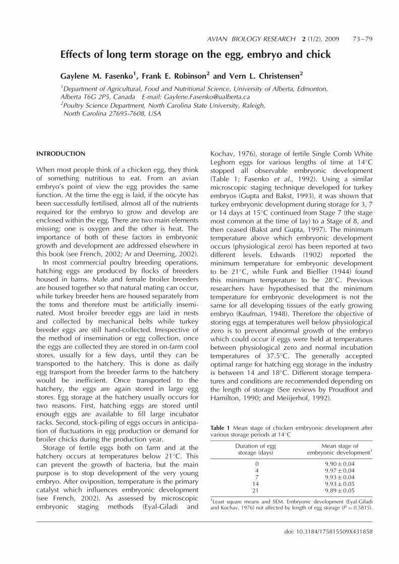
Effects of long term storage on the egg, embryo and chick
Gaylene M. Fasenko1, Frank E. Robinson2 and Vern L. Christensen2
1Department of Agricultural, Food and Nutritional Science, University of Alberta, Edmonton,Alberta T6G 2P5, Canada E-mail: [email protected] Science Department, North Carolina State University, Raleigh,North Carolina 27695-7608, USA
INTRODUCTION
When most people think of a chicken egg, they thinkof something nutritious to eat. From an avianembryo’s point of view the egg provides the samefunction. At the time the egg is laid, if the oocyte hasbeen successfully fertilised, almost all of the nutrientsrequired for the embryo to grow and develop areenclosed within the egg. There are two main elementsmissing; one is oxygen and the other is heat. Theimportance of both of these factors in embryonicgrowth and development are addressed elsewhere inthis book (see French, 2002; Ar and Deeming, 2002).In most commercial poultry breeding operations,
hatching eggs are produced by flocks of breedershoused in barns. Male and female broiler breedersare housed together so that natural mating can occur,while turkey breeder hens are housed separately fromthe toms and therefore must be artificially insemi-nated. Most broiler breeder eggs are laid in nestsand collected by mechanical belts while turkeybreeder eggs are still hand-collected. Irrespective ofthe method of insemination or egg collection, oncethe eggs are collected they are stored in on-farm coolstores, usually for a few days, until they can betransported to the hatchery. This is done as dailyegg transport from the breeder farms to the hatcherywould be inefficient. Once transported to thehatchery, the eggs are again stored in large eggstores. Egg storage at the hatchery usually occurs fortwo reasons. First, hatching eggs are stored untilenough eggs are available to fill large incubatorracks. Second, stock-piling of eggs occurs in anticipa-tion of fluctuations in egg production or demand forbroiler chicks during the production year.Storage of fertile eggs both on farm and at the
hatchery occurs at temperatures below 21�C. Thiscan prevent the growth of bacteria, but the mainpurpose is to stop development of the very youngembryo. After oviposition, temperature is the primarycatalyst which influences embryonic development(see French, 2002). As assessed by microscopicembryonic staging methods (Eyal-Giladi and
Kochav, 1976), storage of fertile Single Comb WhiteLeghorn eggs for various lengths of time at 14�Cstopped all observable embryonic development(Table 1; Fasenko et al., 1992). Using a similarmicroscopic staging technique developed for turkeyembryos (Gupta and Bakst, 1993), it was shown thatturkey embryonic development during storage for 3, 7or 14 days at 15�C continued from Stage 7 (the stagemost common at the time of lay) to a Stage of 8, andthen ceased (Bakst and Gupta, 1997). The minimumtemperature above which embryonic developmentoccurs (physiological zero) has been reported at twodifferent levels. Edwards (1902) reported theminimum temperature for embryonic developmentto be 21�C, while Funk and Biellier (1944) foundthis minimum temperature to be 28�C. Previousresearchers have hypothesised that the minimumtemperature for embryonic development is not thesame for all developing tissues of the early growingembryo (Kaufman, 1948). Therefore the objective ofstoring eggs at temperatures well below physiologicalzero is to prevent abnormal growth of the embryowhich could occur if eggs were held at temperaturesbetween physiological zero and normal incubationtemperatures of 37.5�C. The generally acceptedoptimal range for hatching egg storage in the industryis between 14 and 18�C. Different storage tempera-tures and conditions are recommended depending onthe length of storage (See reviews by Proudfoot andHamilton, 1990; and Meiijerhof, 1992).
AVIAN BIOLOGY RESEARCH 2 (1/2), 2009 73–79
doi: 10.3184/175815509X431858
Table 1 Mean stage of chicken embryonic development aftervarious storage periods at 14�C
Duration of egg Mean stage ofstorage (days) embryonic development1
0 9.90� 0.044 9.97� 0.047 9.93� 0.04
14 9.93� 0.0521 9.89� 0.05
1Least square means and SEM. Embryonic development (Eyal-Giladiand Kochav, 1976) not affected by length of egg storage (P ¼ 0:5815).

Hatching eggs can be successfully stored for up toseven days with little or no effects on hatchability.However, when fertile eggs are stored for more thanone week, embryonic abnormalities and mortalityincreases, which causes a decline in hatchability.Mayes and Takballi (1984) and Meiijerhof (1992)have reviewed the relevant literature on this topic.In addition to this, the quality of chicks hatching fromlong-term stored eggs is reduced (Becker, 1960;Merrit, 1964; Whitehead et al., 1985). Many researchprojects have been devoted to the subject of long-termstorage of fertile eggs by the primary author and theresults are described in more detail below.
EFFECTS OF LONG-TERM EGG STORAGE
Egg quality
It was observed that the yolk membranes (peri-vitel-line complex) of eggs stored for 14 days were extre-mely weak and ruptured very easily (Fasenko,
unpublished data). This observation had been notedby previous investigators (Fromm, 1964; Britton,1973). The incidence of yolk mottling is higher inlong-term stored eggs (Fasenko, personal observa-tions; Britton, 1973). Yolk mottling describes achange in the coloration of the yolk, giving it apatchy appearance. The mechanisms involved inyolk mottling are unknown, but water movementfrom the albumen into the yolk is associated withthe mottling. From these observations a hypothesiswas formed that the thickness of the peri-vitellinelayers in 14 day versus 1 day stored eggs would bereduced due to water movement into the yolk and asubsequent distension and weakening of the peri-vitelline complex. To test this hypothesis, fertileturkey eggs were stored for 1 or 14 days, the peri-vitelline complexes were sampled at the circumfer-ence area of the yolk (vertically between the animaland vegetal poles, and horizontally between the twochalazae), and prepared for transmission electronmicroscopy (Fasenko, 1996). The thicknesses of the
74 Gaylene M. Fasenko, Frank E. Robinson and Vern L. Christensen
Table 2 Average thickness of the peri-vitelline complex and the layers of which it is composed for eggs stored for 1 or 14 days
Duration of Egg no. Thickness (mm)egg storage
PCþ PC� IPL OPL OPLden. OPLdif.
1 day 1 22.0 11.2 5.2 16.6 6.0 10.52 21.8 11 5.0 16.5 6.0 10.33 51.0þ 8 4.0 46.0þ 4.0 41.9
14 days 1 33.3 8.6 3.4 32.8 5.2 24.42 9.3 7.6 3.2 6.1 4.4 1.73 24.9 11.5 3.9 20.9 7.6 11.6
PCþ ¼ inner peri-vitelline layer (IPL)þouter peri-vitelline layer (OPL); PC� ¼ inner peri-vitelline layer (IPL)þouter peri-vitelline dense area(OPLden.); OPL¼outer peri-vitelline diffuse area (OPLdif.)þouter peri-vitelline dense area (OPLden.).
Figure 1 Comparison of photomicrographs of six peri-vitelline complexes from eggs stored for 1 or 14 days. (Print magnification3000X; scale: 30mm¼ 10 mm.)

inner peri-vitelline layer, and of the dense area of theouter peri-vitelline layer (Figure 1) were reduced dueto storage (Table 2). In addition the density of theinner and outer peri-vitelline dense layers decreasedin eggs stored for 14 versus 1 day (Figure 1; Fasenko,1996).It is known that the peri-vitelline complex plays an
integral role in normal early avian embryonic devel-opment. New (1959) found that contact between theinner surface of the inner peri-vitelline layer of theperi-vitelline complex and the outer edges of theblastoderm is necessary for the blastoderm toexpand properly. In long-term stored eggs, the ‘‘adhe-siveness’’ of the embryo to the inner peri-vitellinelayer is often observed to be greatly reduced in eggsstored for long periods (Fasenko, personal observa-tions). There appears to be a special affinity betweenthe cells of the early embryo and the peri-vitellinecomplex. Hence, because of this intimate relation-ship, it is logical to hypothesise that any alteration inthe structure of the peri-vitelline complex may influ-ence early embryonic development. The fact that theperi-vitelline complex structure is modified duringlong-term storage may provide a partial answer as towhy embryos of stored eggs are more likely to die inearly embryogenesis.
Embryonic viability
Previous researchers studying embryonic mortality inturkeys (Arora and Kosin, 1966) and chickens (Matherand Laughlin, 1976, 1977, 1979; Reinhart andHurnik, 1976; Fasenko et al., 1992) have shown thatthe incidence of abnormal and dead embryos(Figure 2) significantly increased as storage waslengthened. Arora and Kosin (1968) established thatthe number of embryonic cells with necrotic nucleiincreased as the length of the storage period
increased. This reduction in viable embryonic cellsmay be the reason behind the observations ofblastoderm shrinkage when eggs are stored for 7 or14 days (Mather and Laughlin, 1979).A study by Bloom et al. (1998) showed that egg
storage for 14 days at 12�C increases the number ofapoptotic cells (cells programmed to die). It issuggested that long-term exposure of avian embryosto storage below physiological zero may increase thenumber of necrotic (dead) cells or may induceunscheduled apoptosis, thus increasing the ratio ofnon-viable to viable embryonic cells. This mayexplain why there is a higher percentage of abnormalor dead embryos occuring in long-term stored eggs asthere may be an optimum number of viableembryonic cells required for initiation of normalgrowth and development. Research currentlyunderway by the principle author and colleagues isaiming to clarify the relationship between long termstorage and the incidence of necrotic and apoptoticcells.
Embryonic development
It is well known that incubation time required for achick to hatch is extended when hatching eggs arestored for long periods (Kosin and Konishi, 1973;Mather and Laughlin, 1976, 1977). Researchershave suggested two possible reasons for the increasedincubation period needed. The first reason may bethat embryonic development in stored eggs does notbegin immediately after normal incubation tempera-tures are provided (Arora and Kosin, 1966). Thesecond is that embryonic development in embryosfrom stored eggs proceeds at a slower rate throughoutthe first periods of incubation. Mather and Laughlin(1977) showed that embryonic growth in embryos
Effects of long term storage on the egg, embryo and chick 75
Figure 2 Photomicrographs of an Eyal-Giladi and Kochav (1976) Stage X domestic fowl blastoderm (embryo) (a) and anabnormalydead blastoderm (b). Both embryos were sampled prior to incubation. Print magnification is 16X.
(a) (b)

from eggs stored for 14 days was about 12.2 hoursbehind that of embryos from non-stored eggs.It has been verified that embryos of eggs stored for
14 days were at a less developed biological age thaneggs stored for 4 days even though the chronologicalage (amount of incubation time) in both groups wasequal (Fasenko, unpublished data; Figure 3). In thisstudy, at three hours of incubation there was nosignificant differences in development between theembryos from 14 versus 4 day stored eggs, but as earlyas six hours of incubation the development of theembryos from 14 day stored eggs began to lag behind(Table 3). Further examination of the embryonicdevelopment during the first 12 hours of incubationshowed that not all embryos react in the same mannerto long term storage. Even after 12 hours of incubationsome embryos of 14 day stored eggs still had notinitiated development and were at Stage X, which isthe most common stage of embryonic development atthe time of lay (Table 3). Other embryos began
development, but at a slower rate andonly reached Stages XIII or XIV, while thedevelopment of other embryos appearedunaffected by the 14 day storage period,and these embryos managed to reach stagesof development that embryos from their 4day stored counterparts attained (Table 3).
Embryonic metabolism
In another study it was shown that themetabolism of embryos from 15 day storedeggs, as measured indirectly by embryonicCO2 output, proceeds at a slower rate thanembryos from 4 day stored eggs (Fasenko,unpublished data; Figure 4). This indicatesthat not only do the embryos from long-termstored eggs lag behind in development, but
their metabolism is somehow altered as well.Evidence for this comes from recent studies inturkeys (Fasenko, 1996) and in broiler breeders(Christensen et al., 2001). In turkeys, embryos from14 day stored eggs relied more upon gluconeogenesisduring pipping and hatching than did embryos from 4day stored eggs (Fasenko, 1996). Long-term eggstorage may change the biological quality of anembryo such that its metabolism is not as efficientas an embryo from a short-term stored egg.Christensen et al. (2001) showed that the carbohy-drate metabolism of a line of broiler breeders that wasresistant to the deleterious effects of storage wascompared to a broiler breeder line susceptible toembryonic mortality due to storage. Embryos fromthe line resistant to storage maintained largerglycogen reserves in the muscle and heart tissuesthan did embryos from the line susceptible tostorage. This fact may provide an advantage in thatadequate carbohydrate reserves in the storage resis-
76 Gaylene M. Fasenko, Frank E. Robinson and Vern L. Christensen
Figure 3 An embryo from an egg stored for 4 days (a) versus and embryofrom an egg stored for 14 days (b). Both eggs were incubated for 96 hourspost-storage. The biological age of the embryo from the 14 day stored egg
lags behind the chronological age by approximately 10 hours.
(a) (b)
Table 3 Comparison of the numbers of embryos at each stage of embryonic development for each egg storage length at 3(P¼ 0.3450), 6 (P¼ 0.0043), and 12 hours (P¼ 0.0031) of incubation
Stages of embryonic development (as you go from left to right embryonicdevelopment is more advanced)
Incubation Storage length(hours) (days) n1 EGK2 IX EGK X EGK XI EGK XII EGK XIII EGK XIV HH3 2 HH 3 HH 3þ
3 4 12 0 6 4 2 0 0 0 0 03 14 12 0 9 3 0 0 0 0 0 0
6 4 18 0 1 12 5 0 0 0 0 06 14 16 1 7 8 0 0 0 0 0 0
12 4 19 0 0 0 0 1 8 6 2 212 14 19 0 5 0 0 7 5 2 0 0
1n¼ total number of embryos staged for development.2Eyal-Giladi and Kochav (1976) stages of development.3Hamburger and Hamilton (1951)stages of development.

tant embryos are maintained and available for growthand function of demand tissues.
Hatchability and chick quality
There are negative effects of long-term egg storage onembryonic metabolism and viability and so it is notsurprising that egg storage has been determined toreduce hatchability (See reviews by Mayes andTakeballi, 1984; and Meijerhof, 1992). In a recentstudy on broiler breeders it was hypothesised thateach two days of egg storage would produce aconcomitant reduction in hatchability (Fasenko,unpublished data). The results, however, showedthat egg storage less than 8 days does not significantly
affect hatchability, but hatchability does significantlydecline after 8, 12, and 16 days of storage (Figure 5).Previous researchers have observed negative effects
of storage on broiler body weights (Becker, 1960),mortality (Merritt, 1964) and chick quality asmeasured by red blood cell counts and navel condi-tion (Whitehead et al., 1985). A recent study showedthat the percentage of culled chicks due to unhealednavels, deformities, and general signs of weaknesswas significantly higher in chicks from eggs stored for14 days versus eggs stored for 4 days (Fasenko,unpublished data). In this study the body weight ofchicks at placement was also 1.4 g lighter in the 14versus the 4 day stored eggs, however, by 3 weeks ofage this difference was erased. Irrespective of this,chicks with greater placement body weights may bebetter able to withstand environmental or diseasechallenges placed upon them.
MANAGEMENT METHODS TO REDUCE THE NEGATIVEEFFECTS OF LONG-TERM EGG STORAGE
In both turkeys and chickens research on incubationof eggs prior to long-term storage has proven fruitfulwith respect to improving hatchability. Fasenko et al.(2001a) found that hatchability of turkey breeder eggswas reduced from 70.9% in 4 day stored eggs to64.4% in 14 day stored eggs. When eggs destined tobe stored for 14 days were incubated for 12 hoursprior to storage, the hatchability significantlyimproved back to the same level (70.9%) as theeggs stored for 4 days. After 12 hours of incubation,the development of the turkey embryo at the time ofstorage had completed hypoblast formation.
Effects of long term storage on the egg, embryo and chick 77
Figure 4 Carbon dioxide production of incubating eggs storedfor 4 and 15 days before Incubation.
Figure 5 Hatchability of broiler breeder eggs after storage for 0, 2, 4, 6, 8, 10, 12, 14, or 16 days.
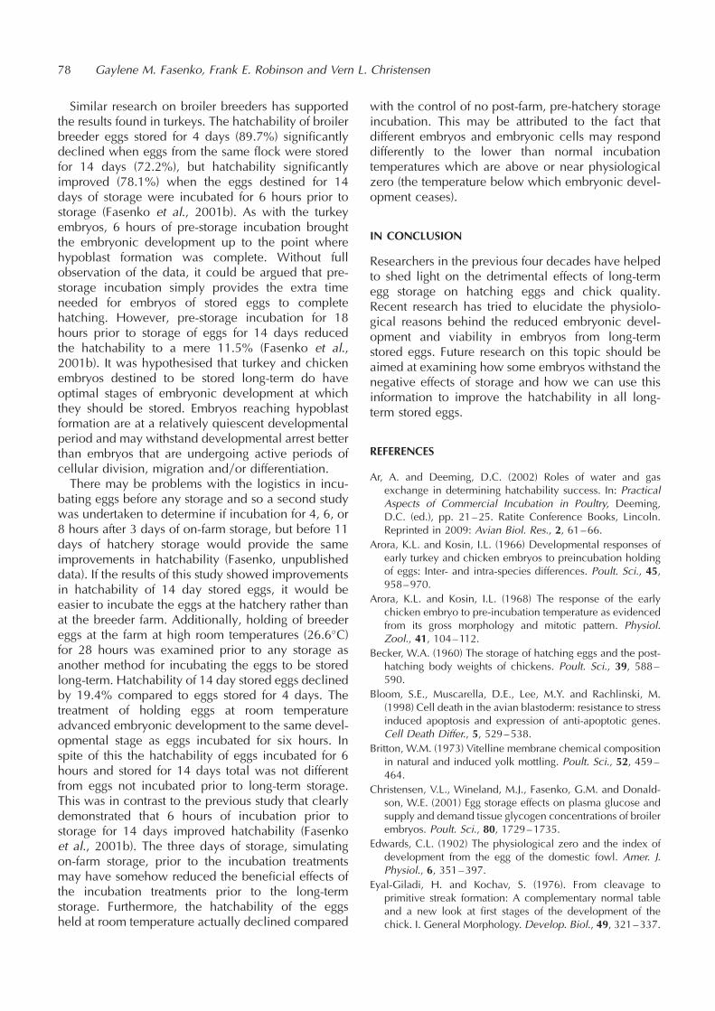
Similar research on broiler breeders has supportedthe results found in turkeys. The hatchability of broilerbreeder eggs stored for 4 days (89.7%) significantlydeclined when eggs from the same flock were storedfor 14 days (72.2%), but hatchability significantlyimproved (78.1%) when the eggs destined for 14days of storage were incubated for 6 hours prior tostorage (Fasenko et al., 2001b). As with the turkeyembryos, 6 hours of pre-storage incubation broughtthe embryonic development up to the point wherehypoblast formation was complete. Without fullobservation of the data, it could be argued that pre-storage incubation simply provides the extra timeneeded for embryos of stored eggs to completehatching. However, pre-storage incubation for 18hours prior to storage of eggs for 14 days reducedthe hatchability to a mere 11.5% (Fasenko et al.,2001b). It was hypothesised that turkey and chickenembryos destined to be stored long-term do haveoptimal stages of embryonic development at whichthey should be stored. Embryos reaching hypoblastformation are at a relatively quiescent developmentalperiod and may withstand developmental arrest betterthan embryos that are undergoing active periods ofcellular division, migration andyor differentiation.There may be problems with the logistics in incu-
bating eggs before any storage and so a second studywas undertaken to determine if incubation for 4, 6, or8 hours after 3 days of on-farm storage, but before 11days of hatchery storage would provide the sameimprovements in hatchability (Fasenko, unpublisheddata). If the results of this study showed improvementsin hatchability of 14 day stored eggs, it would beeasier to incubate the eggs at the hatchery rather thanat the breeder farm. Additionally, holding of breedereggs at the farm at high room temperatures (26.6�C)for 28 hours was examined prior to any storage asanother method for incubating the eggs to be storedlong-term. Hatchability of 14 day stored eggs declinedby 19.4% compared to eggs stored for 4 days. Thetreatment of holding eggs at room temperatureadvanced embryonic development to the same devel-opmental stage as eggs incubated for six hours. Inspite of this the hatchability of eggs incubated for 6hours and stored for 14 days total was not differentfrom eggs not incubated prior to long-term storage.This was in contrast to the previous study that clearlydemonstrated that 6 hours of incubation prior tostorage for 14 days improved hatchability (Fasenkoet al., 2001b). The three days of storage, simulatingon-farm storage, prior to the incubation treatmentsmay have somehow reduced the beneficial effects ofthe incubation treatments prior to the long-termstorage. Furthermore, the hatchability of the eggsheld at room temperature actually declined compared
with the control of no post-farm, pre-hatchery storageincubation. This may be attributed to the fact thatdifferent embryos and embryonic cells may responddifferently to the lower than normal incubationtemperatures which are above or near physiologicalzero (the temperature below which embryonic devel-opment ceases).
IN CONCLUSION
Researchers in the previous four decades have helpedto shed light on the detrimental effects of long-termegg storage on hatching eggs and chick quality.Recent research has tried to elucidate the physiolo-gical reasons behind the reduced embryonic devel-opment and viability in embryos from long-termstored eggs. Future research on this topic should beaimed at examining how some embryos withstand thenegative effects of storage and how we can use thisinformation to improve the hatchability in all long-term stored eggs.
REFERENCES
Ar, A. and Deeming, D.C. (2002) Roles of water and gasexchange in determining hatchability success. In: PracticalAspects of Commercial Incubation in Poultry, Deeming,D.C. (ed.), pp. 21–25. Ratite Conference Books, Lincoln.Reprinted in 2009: Avian Biol. Res., 2, 61–66.
Arora, K.L. and Kosin, I.L. (1966) Developmental responses ofearly turkey and chicken embryos to preincubation holdingof eggs: Inter- and intra-species differences. Poult. Sci., 45,958–970.
Arora, K.L. and Kosin, I.L. (1968) The response of the earlychicken embryo to pre-incubation temperature as evidencedfrom its gross morphology and mitotic pattern. Physiol.Zool., 41, 104–112.
Becker, W.A. (1960) The storage of hatching eggs and the post-hatching body weights of chickens. Poult. Sci., 39, 588–590.
Bloom, S.E., Muscarella, D.E., Lee, M.Y. and Rachlinski, M.(1998) Cell death in the avian blastoderm: resistance to stressinduced apoptosis and expression of anti-apoptotic genes.Cell Death Differ., 5, 529–538.
Britton, W.M. (1973) Vitelline membrane chemical compositionin natural and induced yolk mottling. Poult. Sci., 52, 459–464.
Christensen, V.L., Wineland, M.J., Fasenko, G.M. and Donald-son, W.E. (2001) Egg storage effects on plasma glucose andsupply and demand tissue glycogen concentrations of broilerembryos. Poult. Sci., 80, 1729–1735.
Edwards, C.L. (1902) The physiological zero and the index ofdevelopment from the egg of the domestic fowl. Amer. J.Physiol., 6, 351–397.
Eyal-Giladi, H. and Kochav, S. (1976). From cleavage toprimitive streak formation: A complementary normal tableand a new look at first stages of the development of thechick. I. General Morphology. Develop. Biol., 49, 321–337.
78 Gaylene M. Fasenko, Frank E. Robinson and Vern L. Christensen

Fasenko, G.M. (1996). Factors influencing embryo and poultviability and growth during long term storage of turkey eggs.Ph.D. Thesis. North Carolina State University, Raleigh, NorthCarolina.
Fasenko, G.M., Christensen, V.L., Wineland, M.J. and Petitte,J.N. (2001a) Examining the effects of prestorage incubationof turkey breeder eggs on embryonic development andhatchability of eggs stored for four or fourteen days. Poult.Sci., 80, 132–138.
Fasenko, G.M., Robinson, F.E., Hardin, R.T. and Wilson, J.L.(1992) Variability in preincubation embryonic developmentin domestic fowl. 2. Effects of duration of egg storage period.Poult. Sci., 71, 2129–2132.
Fasenko, G.M., Robinson, F.E., Whelan, A.I., Kremeniuk, K.M.and Walker, J.A. (2001b) Prestorage incubation of long-termstored broiler breeder eggs: 1. Effects on hatchability. Poult.Sci., 80, 1406–1411.
French, N.A. (2002) The critical importance of incubationtemperature. In: Practical Aspects of Commercial Incubationin Poultry, Deeming, D.C. (ed.), pp. 17–20. Ratite Confer-ence Books, Lincoln. Reprinted in 2009: Avian Biol. Res., 2,55–59.
Fromm, D. (1964) Strength, distribution, weight, and somehistological aspects of the vitelline membrane of the hen’segg yolk. Poult. Sci., 43, 1240–1246.
Funk, E.M. and Biellier, H.V. (1944). The minimum temperaturefor embryonic development in the domestic fowl (Gallusdomesticus). Poult. Sci., 23, 538–540.
Gupta, S.K. and Bakst M.R. (1993) Turkey embryo staging fromcleavage through hypoblast formation. J. Morph., 217, 313–325.
Hamburger, V. and Hamilton, H.L. (1951) A series of normalstages in the development of the chick embryo. J. Morph.,88, 49–92.
Kaufman, L. (1948) The effect of certain thermic factors on themorphogenesis of fowl embryos. In: Official Report of theEighth Worlds’ Poultry Congress. World’s Poultry Science
Association, Baelum, J. and Nielsen, A. (eds.), pp. 351–356.Nielsen and Lydich, Copenhagen.
Kosin, I.L. and Konishi, T. (1973) Pre-incubation storage condi-tions and their effect on the subsequent livibility of chickenembryos: exogenous CO2, plastic bags and extended hold-ing periods as factors. Poult. Sci., 52, 296–302.
Mather, C.M. and Laughlin, K.F. (1976) Storage of hatchingeggs: The effect on total incubation period. Brit. Poult. Sci.,17, 471–479.
Mather, C.M. and Laughlin, K.F. (1977) Storage of hatchingeggs: The effect on early embryonic development. Brit.Poult. Sci., 18, 597–603.
Mather, C.M. and Laughlin, K.F. (1979) Storage of hatchingeggs: the interaction between parental age and earlyembryonic development. Brit. Poult. Sci., 20, 595–604.
Mayes, F.J. and Takeballi, M.A. (1984) Storage of the eggs of thefowl (Gallus domesticus) before incubation: a review.World’s Poult. Sci. J., 40, 131–140.
Meijerhof, R. (1992) Pre-incubation holding of hatching eggs.World’s Poult. Sci. J., 48, 57–68.
Merritt, E.S. (1964) Pre-incubation storage effects on subsequentperformance of chickens. Brit. Poult. Sci., 5, 67–73.
New, D.A.T. (1959) The adhesive properties and expansion ofthe chick blastoderm. J. Embryol. Exp. Morphol., 7, 146–164.
Proudfoot, F.G. and Hamilton, R.M.G. (1990) Care of HatchingEggs Before Incubation. Publication 1573yE, Communica-tions Branch, Agriculture Canada, Ottawa, Ontario, Canada.
Reinhart, B.S. and Hurnik, J.F. (1976) The effect of temperatureand storage time during the pre-incubation period. 1. Theinfluence of storage changes on hatchability and first tendays chick performance. Poult. Sci., 55, 1632–1640.
Whitehead, C.C., Maxwell, M.H., Pearson, R.A. and Herron,K.M. (1985) Influence of egg storage on hatchability,embryonic development, and vitamin status in hatchingbroiler chicks. Brit. Poult. Sci., 26, 221–228.
Effects of long term storage on the egg, embryo and chick 79
Principles and practice of incubator design
Ron Meijerhof
Hybro bv, P.O. Box 30, 5830 AA Boxmeer, The Netherlands
INTRODUCTION
To be able to incubate with success, commercialincubators have to provide an optimal climate forthe developing embryo. To understand what are thecrucial factors in this optimal climate, we have to lookinto the process of embryo development. Thesefactors have to be facilitated in the incubationprocess. In this respect, we can learn from naturalincubation (see Deeming, 2002a). After all, the bird isthe most experienced in how to incubate, and if theyfind certain parts of the process of particular impor-tance, they probably have a good reason for that.From a biological point of view incubation, the
process of turning an egg into a living healthy dayold bird, is of a complexity far beyond imagination,and nothing less than a miracle. However, to be ableto understand the concepts of incubator design, wecan simplify the model of the process enormously.
WHAT HAPPENS DURING INCUBATION
During incubation, the embryo builds up its bodyfrom the contents of the egg using energy obtainedby burning yolk fat with oxygen. This process alsoproduces waste products: carbon dioxide, water andheat energy. To incubate successfully in a machine,environmental conditions have to be created to facil-itate this process. Heat has to be provided and theheat produced by the eggs has to be removed to keepthe embryo at the correct temperature. We have toventilate to provide the eggs with the required oxygenand remove the carbon dioxide produced. Any excesswater lost from the egg that increases relativehumidity above that required by the eggs has to beremoved, which is also done by ventilation. Turningthe egg is crucial for the developing embryo, espe-cially during the first part of incubation (see Deeming,2002d). Turning also influences the air velocity overthe egg, and with that heat transfer.Temperature is the most crucial climatic condition
during incubation. Besides turning, egg temperature isthe only process that the bird in nature controlsaccurately. Slight differences in temperature canhave dramatic effects on embryo development andincubation results.
Normal embryonic development is highly depen-dent on temperature. This is why all commercialincubators put so much effort in controlling andmaintaining a constant temperature throughout themachine. However, it can be questioned if creating aconstant air temperature by itself is enough forcreating optimal temperature conditions. After all,the embryo itself only experiences the temperatureinside the shell, the so-called embryo temperature.Controlling the air temperature by itself assumes thatembryo temperature is equal or at least highly corre-lated with air temperature. Although this seems logicalat first sight, this assumption is in fact not correct. Theembryo temperature is a result of the heat productioninside the egg (metabolic heat production) and thetransport of heat through the egg content and to andfrom the eggshell. This transport of heat is a functionof several mechanisms, of which air temperature isjust one.During the development of the embryo, metabolic
heat is produced, resulting in an increased tempera-ture in the shell. About 40 years ago, this metabolicheat was determined at approx 0.14 watt per 60 g eggat 18 days of incubation (Romijn and Lokhorst, 1961).This figure is still used in the design of incubators.However, it has to be questioned whether this figure isstill correct (Deeming, 2002b). After all, genetic selec-tion has resulted in an enormous increase in growth inthe broilers and it is likely that this difference ingrowth rate will have already had an influence onrates of embryonic growth. Recent experiments (Huletand Meijerhof, 2001a) have shown that heat produc-tion of eggs from modern broilers is substantiallyhigher than reported in 1960. If the heat transfer ofthe eggs is left unchanged this increased heat produc-tion will result in a higher embryo temperature.Interestingly, my field experience indicates that themetabolism of the embryo goes down if the embryotemperature is too high. This results in a less devel-oped chick (Romanoff et al., 1938), but also in areduction of the heat production during incubation.
HEAT TRANSFER
At the start of the incubation process, to create thecorrect temperature inside the shell for the embryo to
AVIAN BIOLOGY RESEARCH 2 (1/2), 2009 81–86
doi: 10.3184/175815509X431867
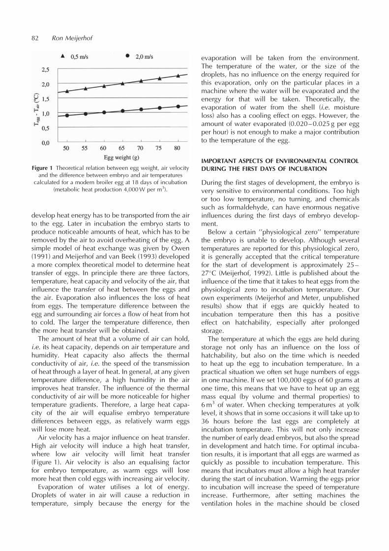
develop heat energy has to be transported from the airto the egg. Later in incubation the embryo starts toproduce noticeable amounts of heat, which has to beremoved by the air to avoid overheating of the egg. Asimple model of heat exchange was given by Owen(1991) and Meijerhof and van Beek (1993) developeda more complex theoretical model to determine heattransfer of eggs. In principle there are three factors,temperature, heat capacity and velocity of the air, thatinfluence the transfer of heat between the eggs andthe air. Evaporation also influences the loss of heatfrom eggs. The temperature difference between theegg and surrounding air forces a flow of heat from hotto cold. The larger the temperature difference, thenthe more heat transfer will be obtained.The amount of heat that a volume of air can hold,
i.e. its heat capacity, depends on air temperature andhumidity. Heat capacity also affects the thermalconductivity of air, i.e. the speed of the transmissionof heat through a layer of heat. In general, at any giventemperature difference, a high humidity in the airimproves heat transfer. The influence of the thermalconductivity of air will be more noticeable for highertemperature gradients. Therefore, a large heat capa-city of the air will equalise embryo temperaturedifferences between eggs, as relatively warm eggswill lose more heat.Air velocity has a major influence on heat transfer.
High air velocity will induce a high heat transfer,where low air velocity will limit heat transfer(Figure 1). Air velocity is also an equalising factorfor embryo temperature, as warm eggs will losemore heat then cold eggs with increasing air velocity.Evaporation of water utilises a lot of energy.
Droplets of water in air will cause a reduction intemperature, simply because the energy for the
evaporation will be taken from the environment.The temperature of the water, or the size of thedroplets, has no influence on the energy required forthis evaporation, only on the particular places in amachine where the water will be evaporated and theenergy for that will be taken. Theoretically, theevaporation of water from the shell (i.e. moistureloss) also has a cooling effect on eggs. However, theamount of water evaporated (0.020–0.025 g per eggper hour) is not enough to make a major contributionto the temperature of the egg.
IMPORTANT ASPECTS OF ENVIRONMENTAL CONTROLDURING THE FIRST DAYS OF INCUBATION
During the first stages of development, the embryo isvery sensitive to environmental conditions. Too highor too low temperature, no turning, and chemicalssuch as formaldehyde, can have enormous negativeinfluences during the first days of embryo develop-ment.Below a certain ’’physiological zero’’ temperature
the embryo is unable to develop. Although severaltemperatures are reported for this physiological zero,it is generally accepted that the critical temperaturefor the start of development is approximately 25–27�C (Meijerhof, 1992). Little is published about theinfluence of the time that it takes to heat eggs from thephysiological zero to incubation temperature. Ourown experiments (Meijerhof and Meter, unpublishedresults) show that if eggs are quickly heated toincubation temperature then this has a positiveeffect on hatchability, especially after prolongedstorage.The temperature at which the eggs are held during
storage not only has an influence on the loss ofhatchability, but also on the time which is neededto heat up the egg to incubation temperature. In apractical situation we often set huge numbers of eggsin one machine. If we set 100,000 eggs of 60 grams atone time, this means that we have to heat up an eggmass equal (by volume and thermal properties) to6m3 of water. When checking temperatures at yolklevel, it shows that in some occasions it will take up to36 hours before the last eggs are completely atincubation temperature. This will not only increasethe number of early dead embryos, but also the spreadin development and hatch time. For optimal incuba-tion results, it is important that all eggs are warmed asquickly as possible to incubation temperature. Thismeans that incubators must allow a high heat transferduring the start of incubation. Warming the eggs priorto incubation will increase the speed of temperatureincrease. Furthermore, after setting machines theventilation holes in the machine should be closed
82 Ron Meijerhof
Figure 1 Theoretical relation between egg weight, air velocityand the difference between embryo and air temperatures
calculated for a modern broiler egg at 18 days of incubation(metabolic heat production 4,000W per m3).

and the humidity turned off to prevent energy beingused to evaporate water during this time.
IMPORTANT ASPECTS DURING THE LAST DAYS OFINCUBATION
Temperature
During the latter stages of incubation, metabolic heatproduction causes embryo temperature to rise signifi-cantly above air temperature. Due to differences inheat transfer conditions and additional effects ofevaporation of water, huge differences in embryotemperature between eggs are often found at differentspots in the machine. A spread of 1.5�C is quitecommon, and differences up to 2.5�C can also befound, even though the air temperature is at the setpoint. These differences are not due to differences incontrolled air temperature in different parts of thecabinet, but are due to variations in air velocity andrates of evaporation of water.Embryo temperature at the end of the incubation
process has a large influence on hatch results andchick quality. We know that relatively small changesin embryo temperature can have a large effect onembryo development. In my opinion, a range of 0.2�Cin the spread of embryo temperatures within a cabinetshould be the goal for incubator manufacturers toachieve in their machines.
Moisture loss
It is well known that moisture loss during incubationinfluences hatchability and chick quality, and that atotal moisture loss of 12–14% of the initial egg weightshould be achieved at external pipping. (Meijerhof,1992). Ar and Rahn (1980) found that for a wide rangeof species an average moisture loss of 15% at externalpipping. This value was in line with maintaining waterbalance within the egg. In general, losing not enoughmoisture seems to be more detrimental than losing toomuch moisture. Romanoff (1930) suggested that alimited respiratory gas exchange will contribute tothis fact and low shell porosities have been shownto limit oxygen consumption in poultry species (Tullettand Deeming, 1982; Burton and Tullett, 1983). Tullett(1981) observed in commercial hatcheries that turkeyeggs had good hatchability values despite a relativelywide range in moisture loss.I believe that the fact that in nature birds are not
really able to control moisture losses confirms thisobservation (but see Ar and Deeming, 2002). Hence ifhatchability is very sensitive to rates moisture loss,birds or eggs would have a mechanism to controlmoisture loss. I think moisture loss becomes important
only once embryo temperature pattern is correct. Itmakes no sense to focus on moisture loss as a majorfactor if the embryo temperature is not wellcontrolled.One of the practical problems with moisture loss is
that it coincides with heat loss. To increase moistureloss from an egg, relative humidity of the air has to bereduced. This will mean a reduction of the heatcapacity and heat conductivity of the air, and there-fore, will limit heat transfer and lead to an increasedembryo temperature. If moisture is not needed for heattransfer, the optimal incubation settings for relativehumidity might be lower than currently used allowingmore moisture loss. This would be in line with theobservation of Ar and Rahn (1980).
Carbon dioxide
During the development of the embryo, oxygen andcarbon dioxide have to pass continuously through theshell and membranes. Although Taylor and Kreutziger(1965, 1966, 1969) and Taylor et al. (1956, 1971)have shown that embryos are rather insensitive to highCO2 levels at different stages of incubation, it iscommon practice to limit carbon dioxide levels insetters and hatchers to a certain set point to avoiddamage to the developing embryo. For setters amaximum set point of 0.6–0.7% is commonly used,for hatchers this is often limited to 0.4–0.5% CO2. Atthe end of the incubation process, carbon dioxidelevels in the air cell are 6–10 times higher than in theoutside air, due to metabolic activity in the shell andlimited permeability through the shell (Visschedijk,1987). The same author reported that a certain level ofcarbon dioxide in the air cell is a stimulus to theembryo to hatch.Based on this, it is often assumed that a certain
minimum level of carbon dioxide has to be obtainedin setter and especially in the hatcher. Although theinfluence of carbon dioxide in the air cell wasdemonstrated in experiments, it can be questionedwhether this influence is that great in a commercialsituation. Besides the fact that a bird doesn’t seem tofeel any need to control carbon dioxide, also thecommon use of in ovo injection techniques seem toindicate that this influence is limited. In large parts ofthe world the majority of the broiler eggs are injectedat 18 days of incubation to administer Marek’svaccine, and the presence of a relatively large holein the air cell, preventing the build up of carbondioxide, doesn’t seem to have a negative influenceon hatchability.Also carbon dioxide levels in setters and hatchers
can be related to maintenance of embryo tempera-ture. To obtain low levels of carbon dioxide, the
Principles and practice of incubator design 83
amount of ventilation in the machine has to be high.This means that the machines take in large amounts ofrelative cold and dry air that has to be heated andmoisturised, which will increase the spread in embryotemperature.
Oxygen and high altitude incubation
Under normal conditions at sea level, oxygen is not alimiting factor for incubation, especially in multi-stagemachines. However, at high altitude, incubationbecomes more difficult due to the limited amount ofoxygen molecules per unit volume of air (Visschedijkand Rahn, 1981). Whilst this is well known there isalso an effect on embryo temperature. At altitude theoxygen content of the air is reduced due to the lowerpressure. This does not only hold for oxygen contentbut also for all the other gases, including watervapour. This means that at high altitude the moisturecontent of the air is reduced and with that the heattransfer of the eggs. This explains why incubators athigh altitude show much more variability in embryotemperature than at sea level, although machinesettings are identical.
WHAT WILL INCUBATORS LOOK LIKE IN THEFUTURE?
Although the design of incubators has not basicallychanged over the last 40 years (Deeming, 2002c), Ithink that we have to reconsider the basic principlesof the design. Incubators have worked very well overthe last decades, but a continuous selection on broilergrowth and meat yield puts more pressure on theincubation process.
Size
Over the years, setting capacity of machines has beenincreased to reduce the cost per egg set. It has beenstated (Deeming, 2002c) that this tendency has anegative influence on hatchability, and that theseproblems could be reduced by bringing down thenumber of eggs set in a machine in order to improvethe flow of air over the eggs. This by itself is true, but isnot necessary the only or even best solution. In thefield we are often able to improve results by settinglimited number of eggs to improve air flow. I amconvinced that improving the design of machines,paying more respect to heat transfer then to airtemperature, will improve the results, even withincreased setting capacity. In fact, I think we canincrease the density of eggs in future machines evenmore if we take the laws of physics into account in thedesign.
Design
The design of incubators must focus primarily oncontrolling embryo temperature. The machines haveto provide a uniform embryo temperature for all eggs,and have the capacity to control this embryo tempera-ture. Other systems, like turning, ventilating, andhumidifying, should not interfere with embryotemperature, at least not in an uncontrollable way.A maximum spread in embryo temperature of 0.3�C(0.5–0.6�F) between individual eggs in the machinemust be a target.Given the importance of air velocity on heat
transfer, a big variation in air velocity is not accep-table as it will give a big variation in embryo tempera-ture. This means that incubators must be designed toprovide a forced and controlled air velocity. This canbest be achieved when the basic principles of alaminar air flow are used (Owen, 1991; Meijerhofand van Beek, 1993).A laminar air flow is heated up or cooled down
when it passes over the eggs, depending on heattransfer conditions. This limits the number of eggsthat air can pass before it has to be reconditionedagain, to meet the goals on spread in embryo tempera-ture. This number of eggs can be estimated by using arather simple model (Owen, 1991) or a more detailedapproach (Meijerhof and van Beek, 1993) anddepends on heat production, air velocity and heatcapacity of air. However, when the calculations aremade, the number of eggs that can be passed beforereconditioning is necessary is surprisingly low (lessthen 30 eggs, depending on climatic characteristics ofthe air used). When air velocity drops below a certainpoint, the air flow will change from laminar toturbulent. As turbulent air flows are more difficult tocontrol and tend to show high variation in velocity, aminimum air velocity is required to maintain thelaminar flow.In any design, the humidifier should not interfere
with embryo temperature. Energy for evaporationshould not be provided by the eggs, but the watermust be evaporated completely before the airreaches the eggs. The typical design with thehumidifier within the cabinet and close to theeggs is unlikely to achieve this, unless steamhumidification is used (which is unlikely nowa-days). With any other type of humidification theair has to be conditioned to the correct watercontent outside the egg cabinet. If the heat transferby air velocity and temperature difference isoptimal, I expect it is possible to incubate atlower relative humidity than commonly used nowa-days, resulting in a reduced negative effect ofhumidifiers inside the machine.
84 Ron Meijerhof
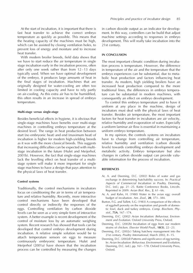
At the start of incubation, it is important that there isfast heat transfer to achieve the correct embryotemperature as quickly as possible. This means thatthe heating capacity of the machines must be high,which can be assisted by closing ventilation holes, toprevent loss of energy and moisture and to increaseheat transfer.With modern broiler breeds, field data shows that
we have to start reduce the air temperature in singlestage incubation early in the incubation process, oftenafter only one week rather than from 12–13 daystypically used. When we have optimal developmentof the embryo, it produces large amounts of heat inthe final stages of incubation. Machines that areoriginally designed for water-cooling are often toolimited in cooling capacity and have to rely partlyon air-cooling. As this extra air has to be humidified,this often results in an increase in spread of embryotemperature.
Multi-stage versus single-stage
Besides beneficial effects in hygiene, it is obvious thatsingle-stage machines have benefits over multi-stagemachines in controlling embryo temperature at thedesired level. The range in heat production betweenstart (no embryonic heat) and end (maximum heat) ofincubation is higher for modern high-yielding breedsas it was with the more classical breeds. This suggeststhat increasing difficulties can be expected with multi-stage incubation in the future (Hulet and Meijerhof,2001b). However, the fact that single-stage machineslack the levelling effect on heat transfer of a multi-stage system will make it more important for singlestage machines to have a design that pays attention tothe physical laws of heat transfer.
Control systems
Traditionally, the control mechanisms in incubatorsfocus on conditioning the air in terms of air tempera-ture and relative humidity. Recently, more interactivecontrol mechanisms have been developed thatcontrol directly or indirectly the responses of theeggs. Controlling ventilation by carbon dioxidelevels can be seen as a very simple form of interactivesystem. A better example is recent development of thecontrol of moisture loss by an automatic weighingsystem. Recent research has showed that tools can bedeveloped that control embryo development duringincubation. A relative simple solution would be toattach temperature sensors on eggs to measurecontinuously embryonic temperature. Hulet andMeijerhof (2001a) have shown that the incubationprocess can be controlled by measuring the changes
in carbon dioxide output as an indicator for develop-ment. In this way, controllers can be build that adjustmachine settings according to responses in embryodevelopment. This will really take incubation into the21st century.
IN CONCLUSION
The most important climatic condition during incuba-tion process is temperature. However, the differencein temperature of the air and the temperature that theembryo experiences can be substantial, due to meta-bolic heat production and factors influencing heattransfer. As modern, high yielding broilers have anincreased heat production compared to the moretraditional lines, the differences in embryo tempera-ture can be substantial in modern machines, withconsequently an effect on embryo development.To control this embryo temperature and to have it
uniform at any place in the machine, design ofincubators must deal with the physical laws of heattransfer. Besides air temperature, the most importantfactors for heat transfer in incubators are air velocity,relative humidity and evaporation of water. Especiallya uniform laminar air flow is essential in maintaining auniform embryo temperature.In my opinion, the controls systems on incubators
have to change from controlling air temperature,relative humidity and ventilation (carbon dioxidelevels) towards controlling embryo development andmetabolic activity. Embryo temperature but alsochanges in carbon dioxide output can provide valu-able information for the process of incubation.
REFERENCES
Ar, A. and Deeming, D.C. (2002) Roles of water and gasexchange in determining hatchability success. In: PracticalAspects of Commercial Incubation in Poultry, Deeming,D.C. (ed.), pp. 21–25. Ratite Conference Books, Lincoln.Reprinted in 2009: Avian Biol. Res., 2, 61–66.
Ar, A. and Rahn, H. (1980) Water in the avian egg: overallbudget of incubation. Am. Zool., 20, 373–384.
Burton, F.G. and Tullett, S.G. (1983) A comparison of the effectsof eggshell porosity on the respiration and growth of domes-tic fowl, duck and turkey embryos. Comp. Biochem. Phy-siol., 75A, 167–174.
Deeming, D.C. (2002) Avian Incubation: Behaviour, Environ-ment and Evolution. Oxford University Press, Oxford.
Deeming, D.C. (2002b) Incubation of eggs from our modernstrains of chicken. Elsevier World Poult., 18(3), 22–23.
Deeming, D.C. (2002c) Taking hatchery management into the21st century. Poultry International, March 2002, 8–15.
Deeming, D.C. (2002d) Patterns and significance of egg turning.In: Avian Incubation: Behaviour, Environment and Evolution,Deeming, D.C. (ed.), pp. 161–178. Oxford University Press,Oxford.
Principles and practice of incubator design 85

Hulet, R.M. and Meijerhof, R. (2001a) Real time incubationtemperature control and heat production of broiler eggs.Poult. Sci., 80 (Suppl. 1), 528.
Hulet, R.M. and Meijerhof, R. (2001b) Multi- or single-stageincubation for high-yielding broilers? Poult. Sci., 80 (Suppl.1), S156.
Meijerhof, R. (1992) Pre-incubation holding of hatching eggs.World’s Poult. Sci. J., 48, 57–68.
Meijerhof, R. and van Beek, G. (1993) Mathematical modellingof temperature and moisture loss of hatching eggs. J. Theoret.Biol., 165, 27–41.
Owen, J. (1991). Principles and problems of incubator design.In: Avian Incubation, Tullett, S.G. (ed.), pp. 205–224. Butter-worth-Heinemann, London.
Romanoff, A.L. (1930) Biochemistry and biophysics of thedeveloping hen’s egg. I. Influence of humidity.Mem. CornellAgr. Exp. Stat., 132, 1–27.
Romanoff, A.L., Smith, L.L. and Sullivan, R.A. (1938) Biochem-istry and biophysics of the developing hen’s egg. III. Influenceof temperature. Cornell Agric. Exp. Stat. Bull., 216, 1–42.
Romijn, C. and Lokhurst, W. (1961) Some aspects of energymetabolism in birds. In: 2nd Symposium on Energy Meta-bolism, Brouer, E. and van Es, A.J.H. (eds.), pp. 49–58.European Association of Animal Production publication No.10. Wageningen.
Taylor, L.W. and Kreutziger, G.O. (1965) The gaseous environ-ment of the chick embryo in relation to its development andhatchability. 2. Effect of carbon dioxide and oxygen levelsduring the period of the fifth through the eighth days ofincubation. Poult. Sci., 44, 98–106.
Taylor, L.W. and Kreutziger, G.O. (1966) The gaseous environ-ment of the chick embryo in relation to its development andhatchability. 3. Effect of carbon dioxide and oxygen levelsduring the period of the ninth through the twelfth days ofincubation. Poult. Sci., 45, 867–884.
Taylor, L.W. and Kreutziger, G.O. (1969) The gaseous environ-ment of the chick embryo in relation to its development andhatchability. 4. Effect of carbon dioxide and oxygen levelsduring the period of the thirteenth through the sixteenth daysof incubation. Poult. Sci., 48, 871–877.
Taylor, L.W., Kreutziger, G.O. and Abercrombie, G.L. (1971)The gaseous environment of the chick embryo in relation toits development and hatchability. 5. Effect of carbon dioxideand oxygen levels during the terminal days of incubation.Poult. Sci., 50, 66–78.
Taylor, L.W., Sjodin, R.A. and Gunns, C.A. (1956) The gaseousenvironment of the chick embryo in relation to its develop-ment and hatchability. 1. Effect of carbon dioxide andoxygen levels during the first four days of incubation uponhatchability. Poult. Sci., 35, 1206–1215.
Tullett, S.G. (1981) Theoretical and practical aspects of egg shellporosity. Turkeys, 29, 24–28.
Tullett, S.G. and Deeming, D.C. (1982) The relationshipbetween eggshell porosity and oxygen consumption in thedomestic fowl. Comp. Biochem. Physiol., 72A, 529–533.
Visschedijk, A.H.J. (1987) Air space: the embryonic medium.J. Exp. Zool., Suppl. 1, 193–201.
Visschedijk, A.H.J. and Rahn, H. (1981). Incubation of chickeneggs at altitude: theoretical consideration of optimal gascomposition. Brit. Poult. Sci., 22, 451–460.
86 Ron Meijerhof

Hatchery ventilation and environmental control
Joseph M. Mauldin1 and Ian R. MacKinnon2
1Poultry Science Department, The University of Georgia, Athens, Georgia, USAE-mail: [email protected] Incubator co. Inc., 30 High Ridge Court, Cambridge, Ontaria, N1R 7L3, Canada
INTRODUCTION
Ventilation and the maintenance of proper environ-mental control within the hatchery have always beencritical to good hatchability and chick quality.However, in the last 10 years proper ventilation andenvironmental control have become more importantthan ever before. The poultry industry has entered anera where egg size and egg density are restricting thenormal airflow patterns within incubators (Deeming,1996). The resulting failure to remove heat, moisture,and carbon dioxide away from the developingembryos is indeed a challenge. Incubation specialistshave found it necessary to initiate a different set ofparameters to reduce the effects of this problem. Thischapter intends to outline the principles of ventilationand environmental control and to describe the currentsuccess achieved in some commercial hatcheries.This will be done from an incubation specialist’spoint of view.
SOME GENERAL PRINCIPLES
The first goal of ventilation is to bring fresh air into thehatchery to replace stale and possibly contaminatedair. An operation that hatches a million chicks perweek will have four or five million embryos in onebuilding that must receive oxygen and eliminatecarbon dioxide on a continuous basis. Secondly, thehatchery and ventilation system must be designed sothat the fresh air does not accumulate contaminationon its way to the developing embryos. The environ-ment of each room within the hatchery must beproperly conditioned with temperature and humidityto achieve optimum results.There are three important parameters to providing
good hatchery ventilation: temperature, humidity, andair volume. Heating and cooling control air tempera-ture; humidifiers and air conditioning controlhumidity; and air volume is controlled by fans anddampers. There are also other factors that influencecontrol of the hatchery environment and these areoutlined below.
Climate
It is important to consider climate in designing andoperating a ventilation system. Climatic conditionsdictate the type of ventilation equipment needed,the type of hatchery construction, and the amount ofinsulation required. Climates with distinct seasonsrequire a full range of ventilation equipment tocontrol temperature, humidity, and airflow. In everyclimate, ventilation equipment should be designed tohandle all anticipated conditions. In the southeasternUSA, summers are very warm and humid. Although alarge percentage of hatcheries in this area useevaporative coolers they would be better served byair conditioning, especially in the setter and hatcherrooms. Evaporative coolers add moisture to the airand thereby reduce the temperature. This cooling‘‘effect’’ is due to the conversion of heat energyfrom one form to another, namely converting sensibleheat to latent heat. The water absorbs the heat ofvaporisation from the surrounding air, which in turnsreduces the temperature of this air. This process worksvery well in dry climates, as there is a need to reducetemperature and increase humidity at the same time inthese climates. In warm and humid climates, tempera-ture and humidity both need to be reduced and airconditioning is the best method to achieve this result.
Humidification methods
Most locations will have some period of the yearwhen the air is too cold. During these times heatingand humidification will be necessary. There areseveral common methods to humidify air in hatch-eries and these can be grouped into two maincategories; methods that add heat with the moistureand methods that do not. The methods that do not addheat with the moisture are the water spray nozzle andcentrifugal discharge type atomisers. Those that addheat with the moisture are steam systems. Newmodular type steam generation systems have createdrenewed interest in introducing humidity using steam.There are several advantages and disadvantages whencomparing humidification that the user should be fully
AVIAN BIOLOGY RESEARCH 2 (1/2), 2009 87–91
doi: 10.3184/175815509X431876
aware of before investing. The most common methodin the industry is to use a spray, mist or centrifugaltype device. This creates fine droplets by mechanicalseparation and heat energy from the surrounding air isabsorbed to turn the droplets into vapour form. It isessential to have sufficient heating capacity in theroom heating system to supply this heat of vaporisa-tion during the cold weather season as well as heatingthe incoming fresh air.
Heat recovery
Heat Recovery systems to capture waste heat fromincubators have been attempted in various locationsaround the world with, at best, mixed results. While itmay be argued that this exhaust is a waste of energy, ithas proven more difficult to implement a simple andeffective system to recover this energy. In mostinstallations observed by the authors, the efficiencyof the heat recovery was more than negated by theenergy used to operate the system, therefore creating anet loss in energy.
Insulation
The use of insulation is vital to cost saving in anumber of ways. The size and cost of the heating,ventilation, and air conditioning units are smallerwhen insulation is used. MacKinnon (2002) providedan example of cost savings in the size of air condi-tioning equipment needed in a 23 cm thick brick wallhatchery housing 10 tunnel-ventilated incubators.Comparisons of estimated equipment cost weremade between no insulation, 5 cm, and 10 cm ofinsulation (Table 1). In addition to equipment costsavings, the added insulation insured significantsavings in energy expenditure.
Biosecurity
One of the most basic principles of ventilation thathatcheries employ is to bring the fresh air into thehatchery from the roof, filter it, and to exhaust air fromthe sidewalls. In many hatcheries it is not possible to
follow this principle in every room environment dueto older design, construction, and building additions.Air that is exhausted through the roof has the potentialof re-entering the hatchery through the roof ventilationequipment and is a biosecurity hazard.One must view hatchery ventilation as an essential
part of the whole biosecurity program. The hatcheryshould be constructed away from sources of contam-ination or dust. For example, there are many hatch-eries that are located in the vicinity of the companyfeed mill. As a result, the hatchery must clean andsanitise filters many times a week to reduce thepotential for contamination. Other dust producingenterprises that should be avoided include processingplants, lumber yards, and live animal farms.As another biosecurity precaution the ‘‘clean’’ and
‘‘dirty’’ areas within the hatchery may be isolatedfrom each other by controlling the pressure withineach hatchery room. The vaccine preparation roomand setter rooms are examples of clean areas andmust have positive pressure to avoid the entrance ofcontamination. Negative pressure in the dirty rooms,such as the chick take-off room, will also help achievebiosecurity. A frequent mistake is that setter andhatcher room doors are left open for several hourseach day for ease of flow of workers and equipment.This results in loss of air pressure control and subse-quently, hatchability performance.
Hatchery room environments
Environmental control within each of the varioushatchery room environments may have significantinfluence on hatchability and chick quality.Components of a properly conditioned room environ-ment include temperature, humidity, and pressure.Table 2 provides acceptable ranges of these condi-tions for the most important hatchery rooms.The egg storage room shows a wide range in
temperature recommendations. This range accountsfor variable lengths of egg storage. For hatcheries ofintegrated poultry companies, the egg storage time isgenerally less than a week. For this situation, it is bestto store eggs at the upper limit of the temperature
88 Joseph M. Mauldin and Ian R. MacKinnon
Table 1 Comparison of air conditioning equipment needed with and without insulation for a building holding 10 tunnel-typeincubators (ambient air 40�C dry bulb, 24.5�C wet bulb)
Material U value British thermal units (Btu) per h Tons Estimated cost Savings
No insulation 0.25 438,488 37 US$73,081(£47,149)
5 cm insulation 0.10 369,188 31 US$61,531 US$11,550(£39,697) (£7,450)
10 cm insulation 0.05 346,088 29 US$57,681 US$15,400(£37,214) (£9,935)
U value¼overall coefficient of heat transmission, Btu per hour per ft2 per �F; Tons¼ ton of refrigeration effect where 1 ton¼ 12,000 Btu per hour.

range. Storing at the cooler end may result in conden-sation (sweating) on the eggs when they are broughtout of storage, making them susceptible to microbialpenetration. Primary breeder hatcheries typically uselonger storage times, frequently 10 days or longer sothat sufficient quantities of eggs from breeding stockare incubated at the same time. In this situation, eggquality can be maintained by reducing the tempera-ture of storage to the lower range of recommendedtemperature. For extremely long storage (more thantwo weeks), other management practices should beconsidered to preserve egg quality. Placing trays ofeggs in plastic bags and infusing with 95% nitrogenand 5% oxygen will help to maintain egg qualityunder long storage conditions. Storing the eggs withthe small end up will reduce the egg moisture lossduring storage. Some hatcheries that store eggs for along period turn the eggs similar to when turning inthe setter to prevent adhesions to the shellmembranes. However, Deeming (2000) suggests thatturning renews the albumen over the early embryohelping to maintain its viability. To the knowledge ofthe authors, there has been no research to support thispractice, however, no apparent damage to the eggsoccurs because of turning during storage.
SETTER AND HATCHER ROOM ENVIRONMENTALCONTROL
Proper environmental control is critical in the setterand hatcher rooms. An incubator will successfullyincubate eggs even when it is placed outdoors.However, in this situation, it will neither operateefficiently nor economically, and the performance inchick quality and hatchability may be lower. Foroptimum performance, the incubators need to beenclosed in a room where there is plenty of fresh airthat has been preconditioned with temperature andhumidity, with a slightly positive air pressurecompared with adjacent rooms or hallways. Thecontent of the air with regard to temperature,humidity, oxygen, and carbon dioxide is important
for the incubators to work at their best in these rooms(see Table 2). Temperature, humidity, and pressurecontrol insure that the machines perform at theiroptimum. For example, when a setter room is toocool, the incubator will provide additional heat toachieve the correct incubation temperature.However, heating the air with electric heating coilsin the setter will cost about three times as much aswhen the room air is properly heated with a gasfurnace before it enters the incubator. Furthermore,incubator dampers will close, reducing air exchange,and may result in elevated carbon dioxide levels.Similar non-optimum results will occur when theroom air is too dry. Incubators respond to dry air byadding mist to elevate humidity. The mist results inevaporative cooling, closing of dampers, and eleva-tion of CO2.The location of the heater and cooler vents are
important to provide patterns of airflow that willfacilitate machine performance. Air outlets shouldbe located near the ceiling and have their louvrespositioned so that the air is thrown into the roomhorizontally. When the horizontally moving air fromone outlet meets the air from another outlet comingin the opposite direction, the airstreams will beforced downward and eventually into a cyclicpattern of flow. These patterns insure propermixing of the air so that uniform temperature andhumidity conditions are maintained throughout theroom. The incorrect placement of air vents, pointeddownward or at a slant, will not result in good flowpatterns or optimum mixing. More serious mistakesare made when the vents throw air directly over theair intakes of the incubators. An example of this typeof mistake is illustrated in Figure 1. Here the freshair from the vent is directed exactly into the intakeof an incubator. During the winter the vents deliverheated air with very little humidity. Humidity isprovided with humidifiers and is uniform in the aironly after proper mixing. The incubator in this photowill have to work inefficiently and with reducedeffectiveness.
Hatchery ventilation and environmental control 89
Table 2 Recommended environmental conditions for selected hatchery rooms
Room Temperature range(�C)
Optimal temperature(�C)
Humidity(%RH)
Pressure(kPa)
Depends onEgg storage 13–19 length of storage 75–80 NeutralSetter and hatcher 24–29 26.6 75–80 1.2–3.7
Clean 22–26 24 Non added 6.2–8.7Chick take-off and wash 24–27 25 Non added � 1.2– � 3.7
Chick holding 24–27 25 55 Neutral to outside(slightly negative)

Thermostat and humidistat location
The placement of thermostats and humidistats iscritical to provide the correct ambient conditions inthe setter and hatcher rooms. These environmentalcontrol devices work much better when they areplaced in the airflow of the room and not in a‘‘dead’’ spot. A common, but incorrect, location forthermostats and humidistats has been the setter orhatcher room end wall. When they are flush mountedon the end wall, it is unlikely that they will correctly‘‘read’’ the room conditions and the corrections theymake will be arbitrary. Mauldin (2001) provided adramatic example of incorrect thermostat placementin a hatcher room. Figure 2A shows the temperaturefor a hatcher room during a 14-hour period. Thethermostat was located in a ‘‘dead’’ air spot andmade arbitrary corrections and as a result, wildfluctuations in temperature were noted. Figure 2Bprovides temperature recordings in the same roomafter the thermostat was correctly placed in the airstream. Note the dramatic difference in temperaturecontrol after correct thermostat placement. In theseexamples, moving the thermostat further into the roomsignificantly improved room conditions, and subse-quently machine performance.
MICROENVIRONMENTS IN INCUBATORS
Even assuming general conditions are correct in thesetter and hatcher rooms, micro-environmental condi-tions may occur within the incubators that lowerperformance. These may be due to poor machinemaintenance or increasing egg size. Air, like water,flows in the path of least resistance. It is easier for airto pass through poor door seals or warped, open baffledoors than for it to flow through the mass of eggs in an
incubator. Mauldin and Buhr (1994) demonstrated theeffects on temperature within masses of eggs due to aminor machine maintenance problem. Data-loggingthermometers were placed in several locations withintwo setters. One setter had a curtain with two brokensnaps and the other incubator had no apparentmaintenance problem.High temperature spikes were observed in the
incubator with the broken curtain snaps while theother machine showed much more uniform tempera-ture around the eggs. Small tasks in incubator main-tenance, such as removing lime-scale formations fromfan blades, make the incubators work far more effi-ciently.One of the most significant advances in modern
incubation has been the development of the plasticegg tray (or flat). Plastic trays were responsible for anincrease in hatchability for several reasons includingfewer cracks, increased egg density within themachine, and improved airflow around the eggs.However, a similar restriction of airflow due to largeegg size as was seen in times of metal trays is nowbeing observed. Some poultry personnel areconvinced that the observed increase in embryonicmortality is due to recent intensified genetic selectionfor high meat yielding birds. Their belief is that thecurrent embryo is in some ways different than theembryo of one decade past and has different incuba-tion requirements. Deeming (2002) and Meijerhof(2002) have supported this view. It is commonlyseen in commercial broiler hatchers that late stageembryo temperatures exceed 38.5�C. Embryo over-
90 Joseph M. Mauldin and Ian R. MacKinnon
Figure 1 An example of an incorrect location of air vent.
Figure 2 The temperature of a hatcher room before (a) and after(b) the correct placement of the thermostat.
(a)
(b)
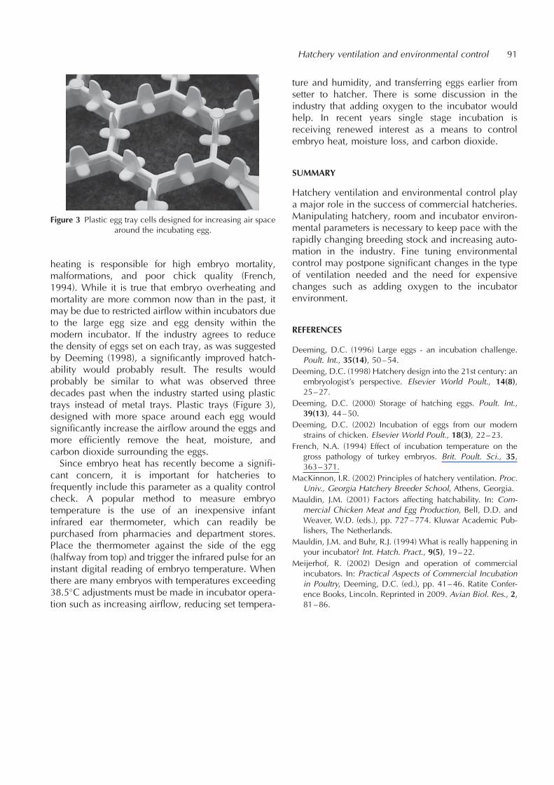
heating is responsible for high embryo mortality,malformations, and poor chick quality (French,1994). While it is true that embryo overheating andmortality are more common now than in the past, itmay be due to restricted airflow within incubators dueto the large egg size and egg density within themodern incubator. If the industry agrees to reducethe density of eggs set on each tray, as was suggestedby Deeming (1998), a significantly improved hatch-ability would probably result. The results wouldprobably be similar to what was observed threedecades past when the industry started using plastictrays instead of metal trays. Plastic trays (Figure 3),designed with more space around each egg wouldsignificantly increase the airflow around the eggs andmore efficiently remove the heat, moisture, andcarbon dioxide surrounding the eggs.Since embryo heat has recently become a signifi-
cant concern, it is important for hatcheries tofrequently include this parameter as a quality controlcheck. A popular method to measure embryotemperature is the use of an inexpensive infantinfrared ear thermometer, which can readily bepurchased from pharmacies and department stores.Place the thermometer against the side of the egg(halfway from top) and trigger the infrared pulse for aninstant digital reading of embryo temperature. Whenthere are many embryos with temperatures exceeding38.5�C adjustments must be made in incubator opera-tion such as increasing airflow, reducing set tempera-
ture and humidity, and transferring eggs earlier fromsetter to hatcher. There is some discussion in theindustry that adding oxygen to the incubator wouldhelp. In recent years single stage incubation isreceiving renewed interest as a means to controlembryo heat, moisture loss, and carbon dioxide.
SUMMARY
Hatchery ventilation and environmental control playa major role in the success of commercial hatcheries.Manipulating hatchery, room and incubator environ-mental parameters is necessary to keep pace with therapidly changing breeding stock and increasing auto-mation in the industry. Fine tuning environmentalcontrol may postpone significant changes in the typeof ventilation needed and the need for expensivechanges such as adding oxygen to the incubatorenvironment.
REFERENCES
Deeming, D.C. (1996) Large eggs - an incubation challenge.Poult. Int., 35(14), 50–54.
Deeming, D.C. (1998) Hatchery design into the 21st century: anembryologist’s perspective. Elsevier World Poult., 14(8),25–27.
Deeming, D.C. (2000) Storage of hatching eggs. Poult. Int.,39(13), 44–50.
Deeming, D.C. (2002) Incubation of eggs from our modernstrains of chicken. Elsevier World Poult., 18(3), 22–23.
French, N.A. (1994) Effect of incubation temperature on thegross pathology of turkey embryos. Brit. Poult. Sci., 35,363–371.
MacKinnon, I.R. (2002) Principles of hatchery ventilation. Proc.Univ., Georgia Hatchery Breeder School, Athens, Georgia.
Mauldin, J.M. (2001) Factors affecting hatchability. In: Com-mercial Chicken Meat and Egg Production, Bell, D.D. andWeaver, W.D. (eds.), pp. 727–774. Kluwar Academic Pub-lishers, The Netherlands.
Mauldin, J.M. and Buhr, R.J. (1994) What is really happening inyour incubator? Int. Hatch. Pract., 9(5), 19–22.
Meijerhof, R. (2002) Design and operation of commercialincubators. In: Practical Aspects of Commercial Incubationin Poultry, Deeming, D.C. (ed.), pp. 41–46. Ratite Confer-ence Books, Lincoln. Reprinted in 2009. Avian Biol. Res., 2,81–86.
Hatchery ventilation and environmental control 91
Figure 3 Plastic egg tray cells designed for increasing air spacearound the incubating egg.
Hygiene and microbiological control in hatcheries
Paul F. McMullin
Poultry Health Services, Poultry Health Centre, Main Site Lane, Dalton, Thirsk,North Yorkshire, YO7 3JA, UK E-mail: [email protected]
INTRODUCTION
The development of artificial incubation is probablythe technological advance that has had the greatestimpact on the structure of the poultry industry. Sinceits inception it has been recognised that the hatcheryprovides some benefits in hygiene by forming aphysical separation between the generations but italso introduces risks that may be summarised thus:
Firstly, the hatchery is a node of connectionbetween multiple breeding farms, receiving eggsand dispatching trays, trolleys etc., to the farms andcan, therefore, facilitate movement of infectionsbetween different breeding farms.
Secondly and similarly the hatchery can be node ofconnection between different customer farms andfacilitate movement of infections between them.
Thirdly, either breeding or customer farms canintroduce infections into the hatchery through themovement of vehicles, people and equipment.Some such infections can persist and multiply inthe hatchery environment.
Fourthly, the close proximity of large numbers ofeggs and chicks from different parent sourcesfacilitates transfer of infections among them.
Finally, hatchery staff can introduce further infec-tions in handling eggs and chicks and also becomeinfected with infections of zoonotic significance.
The major tools used in controlling these risks aredisinfectants and sanitisers. The principles and prac-tices governing their use is covered in detail else-where (Lister, 2002). However, given the range ofrisks involved, this subject is much broader thandisinfectants.For instance, full consideration needs to be given to
the key role of hatching egg hygiene prior to deliveryat the hatchery. This is influenced by the health of theparent flock, both with respect to its ability to produceeggs of normal internal and shell quality, and to itsstatus with respect to infections which may be trans-mitted vertically through the egg (Mauldin, 1983).Numbers of floor eggs, nest box hygiene, method ofcleaning and on-farm storage and sanitation can all
significantly alter the microbial burden coming intothe hatchery. Exposure of eggs to moisture by what-ever means (e.g. fogging, condensation, etc.) canconsiderably increase microbial risks by facilitatingbacterial movement through the egg shell. Washing ofhatching eggs, if practised, needs to be very carefullycontrolled with respect to water quality, temperatureand sanitiser strength. If these aspects are neglectedthen egg washing is capable of doing more harm thangood.It must also be recognised that chick quality is an
amalgam of physical quality and microbiologicalquality. Problems of chick development relating todisease or malnutrition of the parents, or poor orvariable physical parameters in incubation will tendto increase susceptibility to the effects of microbialcontamination, for instance by allowing tissue inva-sion from poorly healed navels.
MICROBIAL TARGETS FOR HYGIENE
A broad range of micro-organisms may be transmittedthrough the hatchery and many can reside andmultiply therein. Most are bacteria commonly asso-ciated with the farm environment (Pseudomonas sp.,Proteus sp., Escherichia coli, Staphylococci,Enterococci). Currently the fowl-specific Salmonellaspecies (S. pullorum and S. gallinarum) and the 2major serotypes of human health significance (S.enteritidis and S. typhimurium) are well controlledin Europe and some other countries. A range of otherSalmonellae are occasionally transmitted on thesurfaces and on shell membranes of hatching eggs(and more rarely in the egg contents). Some of these(e.g. Salmonella senftenberg) are particularly prone tobecome resident in the hatchery environment.The role of the hatchery in transmitting anaerobic
organisms such as Clostridium perfringens is unknownbut clostridial enteritis can certainly be seen in chicksas young as 4–5 days of age. Mycoplasma synoviaeand M. gallisepticum cannot reliably be controlled byegg and hatchery hygiene as the organisms surviveinside the egg and embryo. The main role of hygienewith these infections is to control cross-contaminationwhen a hatchery is hatching both positive and
AVIAN BIOLOGY RESEARCH 2 (1/2), 2009 93–97
doi: 10.3184/175815509X431885
negative chicks. Of the fungi, Aspergillus fumigatus isby far the most important.Brief mention should also be made of viruses.
Hatcheries were identified to be of significance inthe Newcastle Disease (ND) outbreaks in NorthernIreland in 1998 and in Italy in 2001. Since this virus islethal for embryos it is suggested that the viruspersisted on shell membranes or the shell and infectedthe next generation after pipping. Many viruses aremore resistant than ND virus so it seems likely thatpoor eggshell surface sanitation could be a factor inthe transmission of other viral infections. However,unlike bacteria they do not multiply until they actuallyinduce an infection in an embryo or newly-hatchedchick.
PLANNING FOR HYGIENE IN HATCHERY DESIGNAND OPERATION
It is well recognised by the industry that progenyflocks derived from a single parent flock haveimproved health status and productivity as comparedto those derived from multiple parent flocks. Theexact mechanism of this effect is not known but itmay well be related to the mixing of micro-organismsfrom different flocks. The pressure in some countriesto reduce use of antibiotics in agriculture, both as‘‘growth promoters’’ and as therapeutic agents, maywell exacerbate this difference.Planning egg movements and sets to facilitate the
composition of sets to the required progeny flockcomposition may be regarded as the basis for goodhatchery hygiene. The microbiological risks high-lighted above will be further magnified if there areregular movements of eggs between different hatch-eries, so this should be minimised. Eggs are usuallysanitised by fumigation on arrival at the hatchery evenif they have been previously sanitised on farm. Goodplanning facilitates sanitation of the eggs and oftransport vehicles. Planning of hatchery layout andwork practices also had a major impact on hygiene.Wherever possible, people, eggs, chicks and equip-ment should move through the hatchery in a consis-tent fashion (from egg reception to chick dispatch).Where necessary, the risks associated with ‘‘counter-flow’’ events may be controlled by appropriate atten-tion to equipment hygiene, protective clothing, foot-wear and hands. Education of hatchery staff as to thereasons for plans and procedures should improvecompliance and also result in a better outcomewhen staff have to respond to contingencies notenvisaged in the planning process.
SANITATION AND DISINFECTION PROCEDURES
Hatchery hygiene programmes may be divided intothe following general areas: (1) Sanitation of eggs andbuggies arriving from farms (usually by fumigation).(2) Cleaning and disinfection of single-stage settersand all hatchers between batches. (3) Periodiccleaning and disinfection of multi-stage setters andall areas not directly associated with the hatch. (4)Rigorous cleaning and disinfection of areas andequipment associated with the hatch. (5) Washingand disinfection of setter trays, egg buggies, hatchertrays, chick boxes.In each application the key aspects are effective
removal of organic material usually with the aid ofdetergents, followed by application of disinfectant atthe required concentration and under physicalconditions to optimise efficacy. Considerationneeds to be given to possible adverse interactionsamong different chemicals. For most purposes it ispreferable that the product used has some residualactivity. The exception relates to vaccination equip-ment (see below). In all such applications it isimportant to work in a consistent fashion and takecare not to re-contaminate disinfected surfaces.Particular attention should be given to surfaceswhich come in contact with large numbers ofeggs (e.g. automated transfer systems). Drains needto be regularly cleared of debris, washed clean anddisinfected.Equipment used for the application of vaccines,
especially those administered by injection, requirespecial care. Vents on Marek’s Disease vaccinebottles are best fitted with filters to avoid contamina-tion during vaccination. All vaccination equipmentneeds to be effectively sanitised or replaced betweendifferent hatches. For injection equipment this isusually achieved by heat treatment or use of dispo-sable components. For spray vaccination systems it isusual to use chlorine-based sanitisers which do nothave residual properties. Even so they must be thor-oughly rinsed to avoid contaminating the next batchof vaccine with the sanitiser.Hygiene is also a major consideration where
different chicks must be vaccinated with differinglive vaccines by spray. Care should be taken toseparate the batches both in the hatchery and transitas contamination of chicks with low doses of livevaccines can result in adverse effects through ‘‘rollingvaccinal reaction’’. This occurs when some chicks arecontaminated with a dose of vaccine which is insuffi-cient to induce adequate immunity. Such chicksreplicate up the virus and it successively infectsother in-contact chicks over a prolonged (e.g. 2–3week) period.
94 Paul F. McMullin

Where in ovo vaccination is practised in a hatcherythe equipment manufacturer’s recommendations mustbe followed to the letter. General hatchery hygienemust also be of a high standard given that eggs arepunctured and left unsealed at transfer. Aspergillus isa special hazard in this circumstance.
MONITORING HYGIENE IN HATCHERIES
The Assured Chicken Production scheme in theUnited Kingdom requires that participating hatcheriesmaintain a ‘‘Health Plan’’ in relation to any medicinesor vaccines used in chicks produced in the hatchery.It is helpful if the health plan also describes thesanitation programme and system for monitoring itsefficacy and these have a major impact on the healthstatus of chicks produced. Such plans are especiallyuseful for demonstrating compliance with the require-ments of customers, regulators, retailers, and assur-ance schemes.
The backbone of the monitoring programme shouldbe simple checks of physical cleanliness (e.g. byvisual inspection and use of check lists), stockcontrol and usage of disinfectants, concentration ofactive ingredients, temperatures, etc.. This should besupplemented with appropriate microbiologicaltesting carried out in a consistent fashion to facilitatethe recognition of trends. It is recommended to carryout full veterinary audits associated with microbiolo-gical sampling at 2–4 month intervals according tothe requirements of the hatchery and to arrangeintercalated testing by hatchery staff. Some authoritiesrecommend that this testing be done on non-hatchdays but this would prevent the identification ofsignificant incidents of recontamination which occurduring normal operation.
Hygiene testing
Microbiological sampling should be conducted in thenormal flow of eggs, typically testing 30–40 surfaces(each of an area of approximately 16 cm2) with use ofthe single moistened swab to provide a score for TotalViable Count (TVC), coliforms, and yeast and moulds.Surfaces sampled should include eggs, surfaces whichcome in contact with eggs andyor chicks, andcleaning equipment. A scoring system is shown inTable 1.Most sanitised surfaces should achieve a score of
0 to 2 (most of the 2’s will be at the lower end ofthe range). Figure 1 shows the average scoresobtained from various areas of hatcheries over a 6
Hygiene and microbiological control in hatcheries 95
Table 1 Scoring system where each swab is applied to a thirdof a 90mm petri dish. Total viable count and coliforms areread after 24 hours incubation, yeast and mould after 48 hoursincubation, all at 37�C
Score Colonies Status
0 or � 0 Very clean1 or þ 1–10 Slightly contaminated2 or þ þ 11–100 Moderately contaminated3 or þ þ þ More than 100 Heavily contaminated4 or þ þ þ þ Confluent Very heavily contaminated
Figure 1 Mean scores, based on the scheme showed in Table 1, from each area tested and representing all hatchery hygiene swabsreceived over a 5 year period.
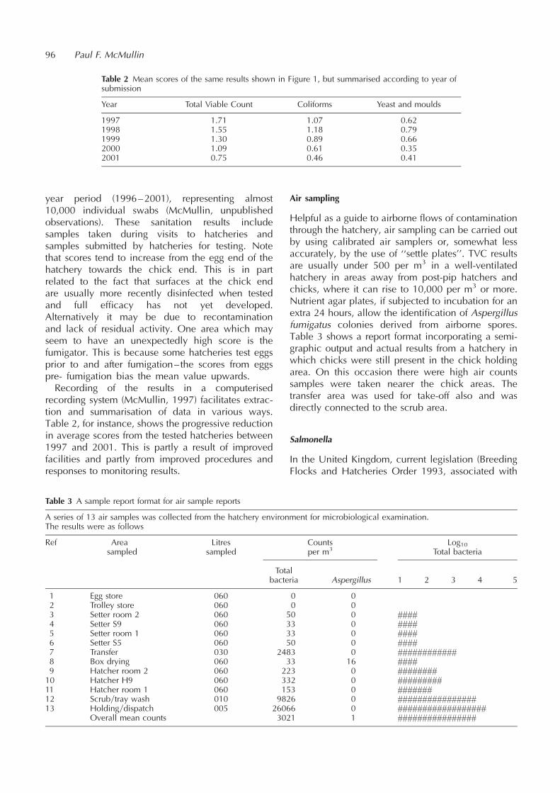
year period (1996–2001), representing almost10,000 individual swabs (McMullin, unpublishedobservations). These sanitation results includesamples taken during visits to hatcheries andsamples submitted by hatcheries for testing. Notethat scores tend to increase from the egg end of thehatchery towards the chick end. This is in partrelated to the fact that surfaces at the chick endare usually more recently disinfected when testedand full efficacy has not yet developed.Alternatively it may be due to recontaminationand lack of residual activity. One area which mayseem to have an unexpectedly high score is thefumigator. This is because some hatcheries test eggsprior to and after fumigation– the scores from eggspre- fumigation bias the mean value upwards.Recording of the results in a computerised
recording system (McMullin, 1997) facilitates extrac-tion and summarisation of data in various ways.Table 2, for instance, shows the progressive reductionin average scores from the tested hatcheries between1997 and 2001. This is partly a result of improvedfacilities and partly from improved procedures andresponses to monitoring results.
Air sampling
Helpful as a guide to airborne flows of contaminationthrough the hatchery, air sampling can be carried outby using calibrated air samplers or, somewhat lessaccurately, by the use of ‘‘settle plates’’. TVC resultsare usually under 500 per m3 in a well-ventilatedhatchery in areas away from post-pip hatchers andchicks, where it can rise to 10,000 per m3 or more.Nutrient agar plates, if subjected to incubation for anextra 24 hours, allow the identification of Aspergillusfumigatus colonies derived from airborne spores.Table 3 shows a report format incorporating a semi-graphic output and actual results from a hatchery inwhich chicks were still present in the chick holdingarea. On this occasion there were high air countssamples were taken nearer the chick areas. Thetransfer area was used for take-off also and wasdirectly connected to the scrub area.
Salmonella
In the United Kingdom, current legislation (BreedingFlocks and Hatcheries Order 1993, associated with
96 Paul F. McMullin
Table 2 Mean scores of the same results shown in Figure 1, but summarised according to year ofsubmission
Year Total Viable Count Coliforms Yeast and moulds
1997 1.71 1.07 0.621998 1.55 1.18 0.791999 1.30 0.89 0.662000 1.09 0.61 0.352001 0.75 0.46 0.41
Table 3 A sample report format for air sample reports
A series of 13 air samples was collected from the hatchery environment for microbiological examination.The results were as follows
Ref Area Litres Counts Log10sampled sampled per m3 Total bacteria
Totalbacteria Aspergillus 1 2 3 4 5
1 Egg store 060 0 02 Trolley store 060 0 03 Setter room 2 060 50 0 ####4 Setter S9 060 33 0 ####5 Setter room 1 060 33 0 ####6 Setter S5 060 50 0 ####7 Transfer 030 2483 0 ############8 Box drying 060 33 16 ####9 Hatcher room 2 060 223 0 ########10 Hatcher H9 060 332 0 #########11 Hatcher room 1 060 153 0 #######12 Scrubytray wash 010 9826 0 ################13 Holdingydispatch 005 26066 0 ##################
Overall mean counts 3021 1 ################

the Zoonosis directive 92-117) places the onus on thehatchery to monitor the Salmonella status of breedingflocks. For broilers this requires the submission ofgroups of dead-in-shell andyor cull chicks from eachparent flock every 2 weeks. Samples are tested byprescribed methods by enrichment culture. This isreasonably sensitive but not very specific. Hence, apositive isolate from progeny can sometimes beactually due to transmission from another parentflock, because of cross-contamination of progeny orsample in the hatchery. When a positive is obtainedall parent flocks hatching on the same day should beconsidered suspect until proven to the contrary. TheZoonosis Directive is currently being re-drafted. Itappears that no major changes in actual hatcherymonitoring are envisaged however the introductionof differential actions for ‘‘Salmonellas of humanhealth significance’’ and targets for Salmonellacontrol may well affect the way we do things.The hatchery environment should also be routi-
nely checked for Salmonella. Whilst it may bepossible to identify Salmonella on hygiene swabsit is more usual to submit each group of such swabsfor enrichment testing. Suspect areas should betested by the use of a large pad swab. Hatcherfloors are a good sample source for this purpose.Perhaps the most sensitive approach to the assess-ment of breeder and hatchery Salmonella status is totest a composite of 5 chick papers from boxescollected when the chicks are tipped. This isbecause it is possible to use more sensitive labora-tory techniques for such non-statutory testing andalso the extra time delay before sample collectionand large numbers of chicks represented allowsready detection of infection. Positive results shouldonly be considered indicative because it is possiblefor such papers to be contaminated on the broilerfarm. Identification of the bacterial serotype willusually help confirm the likely significance of anysuch positive isolates.
Aspergillus
Settle plates of Sabauraud agar are commonly used tocheck for mould contamination. Aspergillus is some-times associated with accumulated fluff in ventilationducts, but can also be associated with contaminationof hatching eggs entering the hatchery. The fungusgrows profusely in the air space of such eggs. Thismay be checked by opening the blunt end ofunhatched eggs in the hatch debris. It is importantto take precautions to avoid the dissemination ofspores from such eggs and their inhalation by theoperator.
SUMMARY
This paper briefly reviews the key issues relating to thepractice and monitoring of hatchery hygiene. Theactivities described are best incorporated in ahatchery health plan and specific operating proce-dures. Much useful information is available fromequipment manufacturers and suppliers of disinfec-tants. Each hatchery should have access to a poultryveterinarian to help them apply these principles in away tailored to their own business and to take intoaccount the health status of supply and progenyflocks.
REFERENCES
Lister, S.A. (2002) Hygiene and disinfection. in poultry manage-ment. In: Poultry Diseases 5th Edition, Jordan, F., Pattison,M., Alexander, D. and Faragher, T. (eds.), pp. 52–54. W.B.Saunders, New York.
Mauldin, J.M. (1983) A Hatchery and Breeder Flock SanitationGuide University of Georgia Bulletin 888. See also http://www.ces.uga.edu/pubcd/b888-w.html.
McMullin, P.F. (1997) Poultry Health Monitoring–Benefits ofComputerised Systems. Proceedings of WPSA Symposium‘‘Transfer of Knowledge in Poultry Production’’, 17–19November Utrecht. See also http://www.poultry-health.-com/library/vetco- mputing/wpsa97co.htm.
Hygiene and microbiological control in hatcheries 97
Chick transport and welfare
Malcolm A. Mitchell
Roslin Institute, Roslin, Midlothian, EH25 9PS, UK
Present address: Roslin Welfare Consultants, 12 Charlton Grove, Roslin,Midlothian, EH25 9NX, UK E-mail: [email protected]
INTRODUCTION
’’Welfare is top of the agenda for, politicians, foodretailers, consumers and farmers alike. Its importanceis such that all sectors of the poultry industry aresubject to a plethora of welfare regulation embroiledin Legislation, Codes of Recommendation andIndustry Codes of Practice.’’ (Cruickshank, 2000).Despite regional economic pressures world-wide
poultry production continues to expand. In the year2000 the number of broiler chick placings in theEuropean Union was approximately 4.3 billion, forlayer chicks 290 million and for turkey poults 245million. The corresponding production figures for theUK were 809 million broiler chicks, 32.5 million layerchicks and 27 million poults (Poultry World, 2001).The placement of broiler breeder chicks in the EUduring the same period was approximately 30 millionwith the UK contributing about 7 million to this total(Poultry Bulletin, 2002). All these birds are transferredfrom hatcheries to their sites of production or rearingwithin 1–3 days of their hatching. Chicks are trans-ported mainly by road or in the case of breeder birdsby air and road to these destinations. It is widelyrecognised that the husbandry of the birds duringthis period and the conditions under which they aremaintained immediately prior to and after placementare vital in determining subsequent performance andhealth status (e.g. Decuypere et al., 2001; Langhout,2001).Transport of all livestock, including poultry, is
regarded as a major source of stress and reducedwelfare and a major cause of these problems is thethermal micro-environment in transit (Mitchell andKettlewell, 1998; Cockram and Mitchell, 1999;Mitchell et al., 2000, 2001; Hunter et al., 2001;Kettlewell and Mitchell, 2001a, 2001b; Nilipour,2002). Many other factors may also contribute totransport stress such as handling, feed and waterwithdrawal, vibration, space restrictions upon beha-viour, noise and pollutants (Mitchell and Kettlewell1998; Mitchell et al., 2000). A number of previousstudies have addressed some aspects of the transporta-tion environments of day-old chicks but with a
primary focus of minimising in transit losses andmaximising subsequent performance. Transportconditions for day-old chicks have been reported asinfluencing subsequent incidence of ascites and’’sudden death syndrome’’ (Maxwell and Robertson,1998). It may clearly be argued that assessing the wellbeing of chicks during a journey or appropriateexperimental simulations by analysis of environ-mental variables and the concomitant physiologicaland metabolic responses, will facilitate identificationand definition of optimal transport conditions andpractices. These will be consistent with the impositionof minimal physiological stress and as such willensure optimum welfare and productivity in transitand during the immediate post-transport period andlater development and growth phases. This approachmay be termed ’’physiological stress modelling’’(Mitchell and Kettlewell, 1998; Mitchell et al., 2000,2001). It is the purpose of this chapter to examine theexisting knowledge and scientific advances relating tothe micro-environments and conditions encounteredduring the transportation of day-old chicks and toconsider relevant associated topics and issuesincluding welfare assessment and legislation.
WELFARE ASSESSMENT
In order to assess the welfare of an animal under aparticular set of conditions or during a specificprocedure two obvious questions must be addressed.What do we mean by ’’welfare’’ and what are theappropriate and objective scientific methods that canbe most usefully applied to the assessment? Theseapparently simple questions have challenged theminds of scientists from multifarious disciplines,moral and ethical philosophers, farmers, veterinarysurgeons and all those concerned with animal wellbeing for many years. The topic has been discussedwidely (e.g. Broom, 1991) and it is beyond the scopeof the present review to detail all the views andarguments offered other than to present some selectedpoints that are directly relevant to the topic of chicktransport. Two working definitions of welfare are ’’thestate of an individual in relation to its environment’’
AVIAN BIOLOGY RESEARCH 2 (1/2), 2009 99–105
doi: 10.3184/175815509X431894

(Broom, 1991). This definition is employed inconjunction with measures of the individual’s abilityto cope with environmental challenge where difficultyin coping or failure to cope are regarded as indicatorsof poor welfare. Some proposed indicators of poorwelfare are reduced life expectancy, impaired growth,impaired reproduction, body or tissue damage,disease, immuno-suppression, increased adreno-cortical activity, behavioural anomalies and self-narcotisation (Broom, 1991). In particular, it isproposed that a reduction in an individual’s controlover its interactions with its environment can lead toreduced welfare.A complementary definition of welfare (or the
welfare state of a sentient animal) is ’’the capacity tosustain physical fitness and avoid mental suffering’’(Webster, 1998). Numerous methodologies have beenproposed for the measurement of these conditions butfrequently the experimental design and objectiveshave been flawed (Webster, 1998) and have focusedupon the demonstration of stress with the purpose of’’proving’’ a preconceived prejudice. The analysis ofstress responses can, however, contribute to theassessment of welfare status if employed objectively.Thus whilst it is recognised that no stress does not
necessarily equate to good welfare it may beproposed that elevated levels of stress associatedwith disruption of physiological homeostasis andhigh demands upon homeostatic effort are consistentwith reduced welfare. Physiological stress modellingmay thus allow definition of acceptable ranges andlimits for physiological stressors (Mitchell andKettlewell 1998; Mitchell et al., 2000, 2001). In turnthis facilitates identification of those environmentalchallenges that are outwith this envelope of ’’comfort’’and which therefore may impose conditions likely toinduce poor welfare. The objective should be theminimising of detrimental stress and the preventionor avoidance of suffering. Such an approach shouldbe supported by other studies including behaviouralanalyses aimed at asking animals ’’what matters tothem’’ and ’’how much it matters’’ (Dawkins, 1993;Webster, 2001). It must be recognised that assessment
of welfare is necessarily extremely complex andalways requires a multi-disciplinary approach(Webster, 1998). The integration of available scientificknowledge and understanding of good husbandrypractices has resulted in the ’’Five Freedoms’’(Table 1) proposed by the UK Farm Animal WelfareCouncil (1993).These freedoms ’’identify the elements that deter-
mine the animals’ perception of their welfare state anddefine the provisions necessary to promote that state’’(Webster, 2001). When considering the welfare statusof chicks in transit it is imperative to examine how theprocess will influence these freedoms and to identifywhere the major risks to the animals’ welfare may lie.Careful quantification of the environmental andprocedural challenges imposed upon the animalswill then allow the characterisation of the ’’bestpractice’’ to reduce unnecessary or excessive stressand optimisation of welfare.It may be proposed that the approaches to definition
and assessment of animal welfare presented hereinare still limited in relation to the implementation ofhigher welfare standards and that the topic should beviewed in a wider context. For example whilst anobjective scientific measure of a physiological stressresponse and its interpretation should be free frommoral judgements clearly it is impossible to divorcemoral and ethical standards from animal welfare inpractice. New challenges, which impinge upon allanimal production, including poultry, are arising inconcert with scientific developments.An example is the increasing understanding of fear
in animals (Jones, 1996), an issue which is of centralimportance in procedures such as transportation. Fearcan be reduced, and thus welfare improved, by arange of strategies such as environmental modifica-tion, improved human-animal interactions and nutri-tion (Jones, 1996). In addition selective breeding orgenetic modification might be employed to alteranimal behaviour or fear thus effectively reducingthe welfare risk. Such approaches raise ethical issues(Grandin and Deesing, 1998; Jones and Hocking,1999), however, often crystallised in questions such
100 Malcolm A. Mitchell
Table 1 Animal welfare: The five freedoms and provisions as proposed by the UK Farm Animal Welfare Council (1993)
Freedom from thirst, hunger and malnutrition By ready access to fresh water and a diet to maintain full health and vigour
Freedom from discomfort By providing a suitable environment including shelter and a comfortable restingarea
Freedom from pain, injury and disease By prevention or rapid diagnosis and treatment
Freedom to express normal behaviour By providing sufficient space, proper facilities and company of the animal’s ownkind
Freedom from fear and distress By ensuring conditions which avoid mental suffering

as ’’is it appropriate to modify the nature of an animalsphysiology and behaviour in order to accommodateimposed stress and abuse?’’ This debate will undoubt-edly continue but a more balanced and holisticapproach to the problems is recommended consistentwith the proposals of Webster (2001) where threedistinct but interdependent lines of reasoning havebeen integrated into a strategy for action for improvedwelfare: (1) A scientific understanding of the factorscontributing to the welfare state of farm animals. (2)An ethical understanding of how and why we shouldrespect the intrinsic value of these animals. (3) Aneconomic understanding of the factors that determinethe extrinsic value we actually do give to theseanimals. Adoption of such an approach shouldprovide a sound platform for improvements in thewelfare of livestock during production and handling,transport and slaughter procedures.
WELFARE LEGISLATION AND CHICK TRANSPORT
In the UK the legislation relating to the transport ofchicks is encompassed by the Welfare of Animals(Transport) Order 1997–Statutory Instrument 1480based upon Directive 95y29EC. The general provi-sions and requirements covering the transport ofchicks are set down in Schedules I and III. All thegeneral provisions relating to protection of animals intransit (prevention and avoidance of suffering), trans-port containers, fitness of animals to travel, sickanimals, duties of the transporter, framework ofcompetencies, special provisions for the transport ofanimals by air and animal transport certificates applyto the transportation of chicks. Other key specificissues are shown in Table 2. Noticeably absent fromthese regulations are specific prescriptions of thermalmicroenvironments. The general provisions state thatthe means of transport (e.g. vehicles) and receptaclesshall be constructed, maintained and operated so as toprotect animals against inclement weather, adverse
sea conditions, marked fluctuations in air pressure,excessive humidity, heat or cold. These provisions aresupported by the designated general ventilationrequirements. For other species (e.g. cattle, sheepand pigs) acceptable temperature ranges in transithave been defined (411y98 EC) and ventilationregimes and performance recommended (The Reportof the Scientific Committee on Animal Health andAnimal Welfare–Standards for the physical environ-ment of animal transport road vehicles EC 1999).More recently the Report of the Scientific Committeeon Animal Health and Animal Welfare ’’The welfareof animals during transport’’ (European Commission,2002) has reiterated the importance of the in transitthermal environment and ventilation regimes and theprevention of unacceptable heat and cold stress.Continuous monitoring and recording (mandatory) ofthe thermal environment in transport containers hasalso recommended. It may be confidently proposedthat similar legislation and codes of practice applyingto poultry transport including day-old chicks will beadopted in the future.
IN-TRANSIT THERMAL MICRO-ENVIRONMENT ANDWELFARE
The commercial chick transport environment maypotentially compromise at least four of the ’’five free-doms’’ but that the risk of thermal stress may be majorsource of welfare risk. Whilst commercial breedersand producers have long recognised the necessity tomaintain an appropriate thermal environment forchicks in transit (Tamlyn and Starr, 1987; Freij,1988; Laughlin, 1989; van der Hel and Henken,1990; Qureshi, 1991; van der Hel et al., 1991) theconditions employed have been largely defined byempirical means and have been based upon mini-misation of mortality rates during and following trans-port, and efficient productivity during the subsequentrapid growth phase. In current practice the recom-
Chick transport and welfare 101
Table 2 Specific regulations relating to the transport of poultry chicks as presented in the Welfare of Animals (Transport) Order1997
Space allowance 21–25 cm2 per chick for day-old chicks in transport containers
Feeding, watering, travelling timesand rest periods
During a journey suitable food and liquid should be available, in suitable quantities andat suitable intervals save in the case of a journey lasting less than 24 hours for chicks ofall species, provided that it is completed within 72 hours after hatching
Ventilation Means of transport and receptacles shall be constructed, maintained, operated andpositioned so as to provide appropriate ventilation and sufficient air space above theanimals to allow air to circulate properly
Segregation of birds Chicks shall be segregated from other poultry except their mothers or other chicks. Birdsshall not be transported in proximity to any animal which is likely to be hostile to themor the presence of which is likely to cause them unnecessary suffering

mended temperature for chick transport is 24–26�C(Ross Breeders, 1996; Meijerhof, 1997; Weeks andNichol, 2000) although only the breeder company’sadvice includes a recommendation of controlledhumidity (75%RH at 24�C).In many transported animals the physiological chal-
lenges presented by the thermal conditions arecompounded by extended periods without access tofood or water. It has long been thought that the day-old chick may be partially protected from suchstresses by the presence of energy and water reservesin the yolk sac. Older studies proposed that yolk storesin the newly hatched chick constitute 18% of totalbody weight and contain approximately 2 g of lipidand 2.5ml of water, which in the absence of exces-sive thermoregulatory demands represent energy (75–80 kJ) and water supplies sufficient for 3 days withoutfurther provision of food and water (MacLeod, 1980;Freeman, 1984). More recent studies have indicatedthat in modern day-old chicks high metabolic rate andrapid utilisation of resources in the first 24 hours post-hatch coupled to delays in transit and placementresult in poorer performance and health statusthroughout flock life (Tanaka and Xin, 1997a; Xinand Lee, 1997; Hackl and Kaleta, 1997; Viera andMoran, 1999; Bigot et al., 2001).Major causes of in-transit and post-transport
mortality and morbidity are dehydration and under-nutrition (Xin and Lee, 1997). A suggested strategy toreduce metabolic depletion during extended transportis the exploitation of the reduction in metabolic rate incrated chicks in the dark (Tanaka and Xin, 1997a).The quantity and rate of use of metabolic reserves bybasal metabolism, however, is clearly not the onlyfactor that will influence chick survival during trans-portation. The prevailing microenvironment mayimpose thermoregulatory and metabolic demandsupon the chicks that will require the rapid mobilisa-tion and utilisation of lipid or the evaporation ofwater. Neonatal chicks do not possess fully developedeffective homeothermic mechanisms (Lamoreux andHutt, 1939; Freeman, 1964; Dunnington and Siegel,1984) and consequently are vulnerable to the detri-mental effects of thermal loads and fatigue and dehy-dration. In the immediate post-hatch chick both bodytemperature and metabolic rate increase (Freeman,1964), however body temperature remains labileduring exposure to sub-optimal thermal environments(van der Hel et al., 1991). Thus, if transportationenvironments are unduly hot or cold then immaturityof thermoregulatory homeostasis, including inade-quacy of lipid mobilisation or efficient evaporativeheat loss during thermal polypnea may result instressful or life threatening hypothermia orhyperthermia. In addition the accelerated rates of
utilisation of energy and water reserves may result inpremature depletion. Freeman (1984) estimated thatreserves may be completely exhausted in as little as8–10 hours at a temperature of 40�C.It may therefore be proposed that in order to
simultaneously optimise survival, productivity andwelfare of the newly hatched chick in transit aneffective strategy would be to match the thermalcharacteristics of the microenvironment to the biolo-gical requirements of the birds. Some previous studieshave attempted to define thermoneutral or optimalenvironments for neonatal chicks on the basis ofmetabolic heat production and body temperatureresponses (Misson, 1976; Henken et al., 1989; Gateset al., 1989; van der Hel et al., 1991). Generally thesestudies did not measure other indicators of homeo-static effort that might better define the physiologicalimpact of the thermal microenvironment. Morerecently Xin and Harmon (1996) examined theeffects of a range of temperatures and humidities(20–35�C and 40–17%) upon day-old chicks bymeasuring metabolic rate and mortality. Theyconcluded that optimum or thermoneutral conditionsoccurred between 30–32�C. Xin (1997) also reportedthat chicks held at a constant 29�C do not exhibit adifferent mortality or body weight loss compared tobirds exposed to as much as a 16�C cycling tempera-ture around the same mean temperature.Studies in my laboratory, the subject of preliminary
reports (Mitchell et al., 1996a, 1996b) have employedphysiological stress modelling, measurement of meta-bolic rate and the concept of Apparent EquivalentTemperature or (AET) derived from the dry bulbtemperature, water vapour density and the psychro-metric constant (Mitchell and Kettlewell, 1998;Mitchell et al., 2000, 2001) to determine optimumtransport thermal environments for day-old chicks. Allmeasurements were performed on chicks in commer-cial transport containers in calorimeter chambershoused in controlled climate rooms. Temperatures of20–35�C accompanied by relative humidities of 50–65% and duration of exposure from 3–12 hours wereemployed. Metabolic heat productions ranged from7.8+0.3 to 8.7+0.9Wkg� 1 in close agreementwith previously published values (van der Hel et al.,1991; Tanaka and Xin, 1997a). On the basis ofminimal change in body temperature and minimalalterations in basal metabolic rate, hydration state,electrolyte balance, body weight loss and plasmametabolite concentrations an optimal temperature-humidity range of 24.5–25.0�C and 63–60%RH forthe transport of chicks at commercial stocking densitywas identified. It was emphasised that these physio-logically ideal conditions are very similar to thosecurrently employed by commercial breeders and
102 Malcolm A. Mitchell

producers. The studies also provided evidence that ifthe thermal microenvironment is appropriatelycontrolled then journey durations of at least 12hours are wholly acceptable.It is concluded that both productivity and welfare of
day-old chicks in transit can be maintained by carefulregulation of the temperature and water vapourdensity to these prescribed limits inside the transportcontainers. Further work on behavioural identificationof the preferred thermal conditions for chicks incommercial transport simulations will provide furtherrefinement and support to these strategies.
VEHICLE AND TRANSPORT CONTAINER VENTILATION
The ultimate determinants of the localised on-boardvehicle (chick transporter) microenvironment are theprevailing climatic conditions, the addition of heatand water vapour to the load space from all sourcesincluding the bio-load (chicks), and the ventilationrate and distribution. All these issues have beenextensively addressed in relation to the transport ofbroiler chickens at slaughter age (e.g. Hoxey et al.,1996; Kettlewell et al., 2000) but the correspondingcharacteristics of chick transporters have received lessdetailed study.Up to date measurements of heat and moisture
production of chicks are available for calculation ofvehicle ventilation requirements (Tanaka and Xin1997a; Mitchell et al., unpublished) but only thework of Quinn and Baker (1997) appears to haveexamined in detail the ventilation characteristics ofcommercial chick transporters. That work employedfull-scale experimental determination of ventilationpatterns and prediction of the same byComputational Fluid Dynamics. The important find-ings included the observation that the presence of theload of stacked chick boxes had a channelling effectupon the air flow through the load space withsignificant amounts of air by-passing the chick boxesand being re-circulated. The implications of thisventilation regimen for air flow in the chick containerswas seen in the temperature distributions with peaktemperatures occurring in the front central boxes andcooler air by-passing the load. In addition cooler airentered from beneath the vehicle in the fully loadedconfiguration and reduced flow through the load aswell as potentially introducing exhaust fumes in to theload space.It must be concluded that considerably more
research must address these issues and that thoseapproaches so successful in optimising mechanicalventilation regimens for vehicles carrying otherpoultry and red meat species (Kettlewell andMitchell, 2001a, 2001b; Kettlewell et al., 2001a,
2001b) should be applied in future to chick transpor-ters.
AIR TRANSPORT OF CHICKS
Ever increasing numbers of chicks are transported byair over long distances and may be subject to delaysand periods of holding in less than optimal conditionsconstituting significant welfare concerns. Schlenkerand Muller (1997) claim high mortalities occur in airtransit due to long periods of inanition and dehydra-tion and that these problems will be significant if thebirds are still in transit 48 hours after hatching and nofood or fluids are provided. Transport conditions alsoappear to be closely correlated with air transportwhen thermal conditions in the aircraft hold arepoorly controlled on long haul flights (Xin andRieger, 1994). Chick container temperatures fellrapidly by 7�C upon departure and increased by upto 10�C upon touchdown. Stressful elevated tempera-tures were observed during holding on the aircraftprior to take off and again upon landing.A further issue associated with aircraft hold condi-
tions is the reduced barometric pressure and thereduction in water vapour density observed in therecycled air. Recent unpublished studies (Mitchelland Kettlewell, 2002) have monitored temperaturesand humidities throughout the load space of bothBoeing 747 and MD 11 freighter aircraft carryinganimals on long haul flights. Accompanying varyingand heterogeneously distributed temperature profileswater vapour densities as low as 2 gm� 3 wereobserved. These conditions would encourage highrates of evaporation from both skin and respiratorytract and would result in potential dehydration andhypothermia in chicks under certain thermal loads.The design of chick containers and their configura-
tion in stacking has also been questioned (Tanaka andXin, 1997b). In adequate passive ventilation flowthrough the boxes when chicks are held in ware-houses prior to flights or during other holdingperiods in aircraft holds before take off and afterlanding quickly resulted in potential heat stress condi-tions even when external temperatures were onlymoderately warm. For all long distance transport ofchicks it has been recommended that water and feedshould be available in transit to reduce mortality andmaintain welfare and productivity (Xin and Lee, 1996;Xin, 1997; Tanaka and Xin, 1997a). Water may beprovided in the form of commercial hydration gels,e.g. Aqua-Jel� or Pacific Oasis�, which are simply cutin to slices and placed in each container or box.As with road chick transporters further research is
required to characterise the prevailing on-board envir-onments, the nature of the ventilation regimes and the
Chick transport and welfare 103
consequences for the birds. Only in this way canstrategies for improved conditions and welfare bedeveloped.
SUMMARY
The welfare of chicks of all poultry species duringtransportation by road or air is a matter for concern.Thermal conditions (high thermal loads or chilling)may represent one of the main sources of physiolo-gical stress and reduced welfare as well as mortalities,morbidity and production losses. Physiologicalmodelling combined with appropriate behaviouralanalysis can identify the optimum transport thermalenvironments for chicks. When these have beenestablished it is possible to re-examine limitations onjourney duration and feeding and watering strategies.The solutions to many of these problems lie in animproved understanding of ventilation flows andregimes, the design of transport containers and vehi-cles and the implementation of practical engineeringremedies. This applies to transport by road and air.Only by the application of sound basic science to thewelfare problems of chicks in transit can practicalimprovements in transporters, transport procedures,codes of practice, education of personnel andsupporting relevant legislation be formulated.
REFERENCES
Bigot, K., Tesseraud, S., Taouis, M. and Picard, M. (2001)Posthatch feeding and early development in broiler chicks.Prod. Animales, 14, 219–230.
Broom, D.M. (1991) Animal welfare–concepts and measure-ment. J. Anim. Sci., 69, 4167–4175.
Cockram, M.S. and Mitchell, M.A. (1999) Role of research inthe formulation of rules to protect the welfare of farmanimals during road transportation. In: Farm Animal Wel-fare–Who writes the rules? Russel, A.J.F., Morgan, C.A.,Savory, C.J., Appleby, M.C. and Lawrence, T.L.J. (eds.), pp43–64. British Society of Animal Science Occasional Pub-lication No 23, Edinburgh.
Cruickshank, G. (2000) Personal communication: paper pre-sented to Poultry World Industry Conference–WelfareGuideline Sponsors 2000–copies available from originalauthor upon request.
Dawkins, M. (1993). Through our Eyes Only? The Search ForAnimal Consciousness. Freeman, Oxford.
Decuypere, E., Tona, K., Bruggenan, V. and Bamelis, F. (2001)The day-old chick: a crucial hinge between breeders andbroilers. World’s Poult. Sci. J., 57, 127–138.
Dunnington, E.A. and Siegel, P.B. (1984) Thermoregulation innewly hatched chicks. Poult. Sci., 63, 1303–1313.
Freeman, B.M. (1964) The relationship between oxygen con-sumption, body temperature and surface area in the hatchingand young chick. Brit. Poult. Sci., 6, 67–72.
Freeman, B.M. (1984) Transportation of poultry. World’s Poult.Sci. J., 40, 19–30.
Freiji, M. (1988) Chicks need cool transport. Poult. Int. (Misset),4(3), 18–19.
Gates, R.S., Turner, L.W., Turner, G.M. and Tuttle, J.W. (1989)Measured heat production in chicks. Proc. Am. Soc. Agric.Engineers, Paper no. 894513.
Grandin, T. and Deesing, M.J. (1998) Genetics and animalwelfare, In: Genetic and Behaviour of Domestic Animals,Grandin, T. (ed.), pp. 319–341. Academic Press, San DiegoCalifornia.
Hackl, G. and Kaleta, E.F. (1997) Methods for the estimation ofthe age of broilers and effects of water and feed deprivationwithin the first days of life. Arch. Geflugel., 61, 280–286.
Henken, A.M., van der Hel, W., Hanssen, E.J.M., Scheele, C.W.and van Kampen, N.M. (1989). The effect of dry and wetbulb temperature on heat production of newly hatchedchicks. In: Energy Metabolism of Farm Animals, Verstegan,M.W.A. and Henken, A.M. (eds.), pp. 291–294. MartinusNijhoff, Netherlands.
Hoxey, R.P., Kettlewell, P.J., Meehan, A.M., Baker, C.J. andYang, X. (1996). An investigation of the aerodynamic andventilation characteristics of poultry transport vehicles: Part1 Full scale measurements. J. Agric. Eng. Res., 65, 77–83.
Hunter, R.R., Mitchell, M.A. and Matheu, C. (2001) Mortality ofbroiler chickens in transit –correlation with the thermalmicro-environment. In: Proceedings of the 6th InternationalLivestock Environment Symposium, Stowell, R.R., Bucklin,R. and Bottcher, R.W. (eds.), pp. 542–549. Louisville, Ken-tucky, U.S.A., 21st–23rd May 2001.
Jones, R.B. (1996) Fear and adaptability in poultry: Insights,implications and imperatives. Worlds Poult. Sci. J., 52, 131–174.
Jones, R.B. and Hocking, P.M. (1999) Genetic selection forpoultry behaviour: Big bad wolf or friend in need? Anim.Welf., 8, 343–359.
Kettlewell, P.J., Hampson, C.J., Green, N.R., Teer, N.J., Veale,B.M. and Mitchell, M.A. (2001a) Heat and moisture genera-tion of livestock during transportation. In: Proceedings of the6th International Livestock Environment Symposium, Stow-ell, R.R., Bucklin, R. and Bottcher, R.W. (eds.), pp. 519–526.Louisville, Kentucky, U.S.A., 21st–23rd May, 2001.
Kettlewell, P.J., Hoxey, R.P., Hampson, C.J., Green, N.R., Veale,B.M. and Mitchell, M.A. (2001b) Design and operation of aprototype mechanical ventilation system for livestock trans-port vehicles. J. Agric. Eng. Res., 79, 429–439.
Kettlewell, P.J., Hoxey, R.P. and Mitchell, M.A. (2000) Heatproduced by broiler chickens in a commercial transportvehicle. J. Agric. Eng. Res., 75, 315–326.
Kettlewell, P.J. and Mitchell, M.A. (2001a). Comfortable ride:Concept 2000 provides climate control during poultry trans-port. Res. Eng. Tech. Sust. World, 8, 13–14.
Kettlewell, P.J. and Mitchell, M.A. (2001b) Mechanical ventila-tion: Improving the welfare of broiler chickens in transit. J.Roy. Agric. Soc., 162, 175–184.
Lamoreux, W.F. and Hutt, F.B. (1939) Variability in bodytemperature in the normal chick. Poult. Sci., 18, 70–75.
Langhout, P.J. (2001) Feeding during the first week of life. Int.Poult. Prod., 9(1), 7–10.
Laughlin, K.F. (1989) Computerised customer care. Int. Hatch.Pract., 4, 43–45.
104 Malcolm A. Mitchell

Maxwell, M.H. and Robertson, G.W. (1998) UK survey ofbroiler ascites and sudden death syndrome in 1993. Brit.Poult. Sci., 39, 203–215.
MacLeod, M.G. (1980) The biology of survival: The effects ofday old travel in chicks. In: Airfreighting of Hatching Eggsand Poultry, pp. 3–5. West of Scotland College of Agricul-ture Ayr.
Meijerhof, R. (1997) The importance of egg and chick transpor-tation. World Poult., 13(11), 17–18.
Misson, B.H. (1976) The effects of temperature and relativehumidity on the thermoregulatory responses of grouped andisolated neonate chicks. J. Agric. Sci. (Cambridge), 86, 35–43.
Mitchell, M.A., Kettlewell, P.J., Carlisle, A.J., Hunter, R.R. andManning, T. (1996a) Defining the optimum thermal environ-ment for the transportation of one-day old chicks: physiolo-gical responses during transport simulations. Brit. Poult. Sci.,37, S89–S91.
Mitchell, M.A., Kettlewell, P.J., Carlisle, A.J., Hunter, R.R. andManning, T. (1996b) Optimo transporte de pollitos de 1 dia.Avicultura Prof., 14(3), 47.
Mitchell, M.A. and Kettlewell, P.J. (1998) Physiological stressand welfare of broiler chickens in transit: Solutions notproblems! Poult. Sci., 77, 1803–1814.
Mitchell, M.A., Carlisle, A.J., Hunter, R.R. and Kettlewell, P.J.(2000). The responses of birds to transportation. In: Proceed-ings of the World Poultry Congress, pp. 1–14. Montreal,Canada, 22nd–24th August 2000.
Mitchell, M.A., Kettlewell, P.J., Hunter, R.R. and Carlisle, A.J.(2001) Physiological stress response modelling–applicationto the broiler transport thermal environment. In: Proceedingsof the 6th International Livestock Environment Symposium,Stowell, R.R., Bucklin, R. and Bottcher, R.W. (eds.), pp.550–555. Louisville, Kentucky, U.S.A., 21st–23rd May2001.
Nilipour, A.H. (2002) Poultry in transit are a cause for concern.World Poult., 18(2), 30–33.
Poultry Bulletin (2002) Chicken market quarterly, Taylor, D.(ed.), p. 15. National Farmers Union, Newmarket, Suffolk,January 2002.
Poultry World (2001) EU facts and forecasts. Poult. World,September, 14–15.
Qureshi, A.A. (1991) Losses due to dehydrated broiler chicks.World Poult. (Misset), 7(4), 75–79.
Quinn, A.D. and Baker, C.J. (1997) An investigation of theventilation of a day-old chick transport vehicle. J. Wind Eng.Ind. Aerodyn., 67 and 68, 305–311.
Ross Breeders (1996). Producing Quality Broiler Meat. RossBroiler Management Manual.
Schlenker, G.R. and Muller, W. (1997) The transportation ofday-old chicks by aircraft with regard to animal welfare.Berl. Munch. Tier. Wochen., 110, 315–319.
Tamlyn, J. and Starr, J.R. (1986) Monitoring the environmentduring transportation of day olds. Int. Hatch. Pract., 6(1),11–15.
Tanaka, A. and Xin, H. (1997a) Energetics, mortality, and bodymass change of breeder chicks subjected to different post-hatch feed dosages. Trans. Am. Soc. Agric. Eng., 40, 1457–1461.
Tanaka, A. and Xin, H. (1997b) Effects of structural and stackingconfiguration of containers for transporting chicks in theirmicroenvironment. Trans. Am. Soc. Agric. Eng., 40, 777–782.
van der Hel, W. and Henken, A.M. (1990) Give newly hatchedchicks a safe flight. Poult. Int. (Misset), 6(1), 8–9.
van der Hel, W., Verstegen, M.W.A., Henken, A.M. andBrandsma, H.A. (1991) The upper critical temperature inneonatal chicks. Poult. Sci., 70, 1882–1887.
Vieira, S.L. and Moran, E.T.(1999) Effects of delayed placementand used litter on broiler yields. J. Appl. Poult. Res., 8, 75–81.
Webster, A.J.F. (1998) What use is science to animal welfare?Naturwissen., 85, 262–269.
Webster, A.J.F. (2001) Farm animal welfare: the five freedomsand the free market. Vet. J., 159, 1–9.
Weeks, C. and Nicol, C. (2000) Poultry handling and transport.In: Livestock Handling and Transport. 2nd Edition, Grandin,T. (ed.), pp. 363–384. CAB International, Wallingford.
Welfare Of Animals (Transport) Order (1997) Statutory Instru-ment 1997 No 1480, Ministry of Agriculture, Fisheries andFood, London.
Xin, H. (1997) Mortality and body weight of breeder chicks asinfluenced by air temperature fluctuations. J. Appl. Poult.Res., 6, 199–204.
Xin, H. and Harmon, J.D. (1996) Responses of group-housedneonatal chicks to post-hatch holding environment. Trans.Am. Soc. Agric. Eng., 39, 2249–2254.
Xin, H. and Lee, K. (1996) Use of aqua-Jel� and feed for nutrientsupply during long journey air transport of baby chicks.Trans. Am. Soc. Agric. Eng., 39, 1123–1126.
Xin, H. and Lee, K. (1997) Physiological evaluation of chickmorbidity during extended post-hatch holding. J. Appl.Poult. Sci., 6, 417–421.
Xin, H. and Rieger, S.R. (1995). Environment and mortalityassociated with remote air-transport of day-old chicks. Pro-ceedings of the American Society of Agricultural Engineers,1994 Winter Meeting Paper No. 944557.
Chick transport and welfare 105