An Expressed Sequence Tag collection from the male antennae of ...

10.1128/IAI.73.5.2665-2679.2005.
2005, 73(5):2665. DOI:Infect. Immun. Handfield, Phillip I. Tarr and Stephen B. CalderwoodSarracino, Ann Progulske-Fox, Jeffrey D. Hillman, MartinDodson, Bethany McManus, Bryan Krastins, David Manohar John, Indira T. Kudva, Robert W. Griffin, Allen W. Infection
HumanO157:H7 Proteins Expressed during Escherichia colifor Identification of
Use of In Vivo-Induced Antigen Technology
http://iai.asm.org/content/73/5/2665Updated information and services can be found at:
These include:
REFERENCEShttp://iai.asm.org/content/73/5/2665#ref-list-1at:
This article cites 58 articles, 37 of which can be accessed free
CONTENT ALERTS more»articles cite this article),
Receive: RSS Feeds, eTOCs, free email alerts (when new
http://journals.asm.org/site/misc/reprints.xhtmlInformation about commercial reprint orders: http://journals.asm.org/site/subscriptions/To subscribe to to another ASM Journal go to:
on January 17, 2014 by guesthttp://iai.asm
.org/D
ownloaded from
on January 17, 2014 by guest
http://iai.asm.org/
Dow
nloaded from

INFECTION AND IMMUNITY, May 2005, p. 2665–2679 Vol. 73, No. 50019-9567/05/$08.00�0 doi:10.1128/IAI.73.5.2665–2679.2005Copyright © 2005, American Society for Microbiology. All Rights Reserved.
Use of In Vivo-Induced Antigen Technology for Identification ofEscherichia coli O157:H7 Proteins Expressed during
Human InfectionManohar John,1,2† Indira T. Kudva,1,2†* Robert W. Griffin,1 Allen W. Dodson,1
Bethany McManus,1 Bryan Krastins,3 David Sarracino,3 Ann Progulske-Fox,4Jeffrey D. Hillman,4 Martin Handfield,4 Phillip I. Tarr,5
and Stephen B. Calderwood1,2,6
Division of Infectious Diseases, Massachusetts General Hospital, Boston, Massachusetts 021141; Department of Medicine,Harvard Medical School, Boston, Massachusetts 021152; Harvard Partners Center For Genetics and Genomics,
65 Landsdowne Street, Cambridge, Massachusetts 021393; College of Dentistry, Department of OralBiology, University of Florida, Gainesville, Florida 326104; Departments of Pediatrics and
Molecular Microbiology, Washington University School of Medicine, St. Louis,Missouri 631105; and Department of Microbiology and Molecular Genetics,
Harvard Medical School, Boston, Massachusetts 021156
Received 4 November 2004/Returned for modification 14 December 2004/Accepted 12 January 2005
Using in vivo-induced antigen technology (IVIAT), a modified immunoscreening technique that circumventsthe need for animal models, we directly identified immunogenic Escherichia coli O157:H7 (O157) proteinsexpressed either specifically during human infection but not during growth under standard laboratory con-ditions or at significantly higher levels in vivo than in vitro. IVIAT identified 223 O157 proteins expressedduring human infection, several of which were unique to this study. These in vivo-induced (ivi) proteins,encoded by ivi genes, mapped to the backbone, O islands (OIs), and pO157. Lack of in vitro expression ofO157-specific ivi proteins was confirmed by proteomic analysis of a mid-exponential-phase culture of E. coliO157 grown in LB broth. Because ivi proteins are expressed in response to specific cues during infection andmight help pathogens adapt to and counter hostile in vivo environments, those identified in this study arepotential targets for drug and vaccine development. Also, such proteins may be exploited as markers of O157infection in stool specimens.
Enterohemorrhagic Escherichia coli (EHEC) O157:H7 (O157)is a uniquely human pathogen that causes disease ranging fromacute, self-resolving watery diarrhea to hemorrhagic colitis andthe potentially fatal hemolytic-uremic syndrome (HUS). Cur-rently, no therapies are available to lessen the potential mor-bidity and mortality of this infection.
E. coli O157 is thought to have evolved from a strain ofenteropathogenic E. coli (EPEC) O55:H7 bearing the patho-genicity island termed the locus for enterocyte effacement(LEE), through the acquisition of bacteriophages encodingShiga toxins type 1 (stx1) and/or 2 (stx2), acquisition of a viru-lence plasmid (pO157), transition of somatic antigen O55 toO157, and loss of sorbitol fermentation and �-glucuronidaseactivity (21). HUS as a complication of E. coli O157 infectionhas been associated with the presence of the stx2 gene or itsvariant stx2c in the infecting E. coli O157 strain (21). In addi-tion, the characteristic attaching and effacing (A/E) lesionsproduced by this organism on the human colonic epitheliumare a result of proteins encoded on the LEE, including theadhesion molecule intimin-� (Eae), its receptor (Tir), the typeIII protein secretion system, which secretes a variety of LEE-encoded translocator proteins (EspA, EspB, and EspD) that
translocate effectors into host cells, and effector proteins (Tir,EspG, EspF, Map, and EspH) that modulate the host cellcytoskeleton (21). The type III secretion system translocatesTir into the host cell, with subsequent trafficking to the hostcell membrane. Intimin binding of Tir leads to host cell actinrearrangement and formation of A/E lesions. Other putativevirulence factors are encoded on pO157 and include an en-terohemolysin (Ehx), an immunomodulator (Lif), and a serineprotease (EspP) (21). Hence several factors may be involved inE. coli O157 pathogenesis, and research is ongoing to under-stand the complexity of this infection.
The sequenced E. coli O157 EDL933 genome shows thatalthough this organism shares 4.1 Mb of DNA (termed back-bone) with E. coli K-12, it has 1.34 Mb of DNA distributedamong 177 DNA segments termed O islands (OIs) that isabsent in K-12 (44). Of the genes found in these OIs, only 40%have been assigned a function and several remain to be char-acterized (44). Collective evidence indicates that intimin-�and the Shiga toxins act in concert with other, unidentifiedvirulence factors, encoded by both the OI and backbone se-quences, to cause the spectrum of E. coli O157 disease (21, 54).
To date, the main impediment to identifying a broader com-plement of virulence factors in this pathogen has been the lackof an animal model that mimics the spectrum of human dis-ease. Also, the potentially fatal sequelae that can follow E. coliO157 infection preclude human volunteer studies. We circum-vented these limitations and exploited the human immune
* Corresponding author. Mailing address: Division of InfectiousDiseases, Massachusetts General Hospital, Boston, MA 02114. Phone:(617) 726-5694. Fax: (617) 726-7416. E-mail: [email protected].
† Equal contribution.
2665
on January 17, 2014 by guesthttp://iai.asm
.org/D
ownloaded from

response following E. coli O157 infection to identify a panel ofmicrobial factors that might contribute to the pathogenicityof this organism. In particular, we used a modified immuno-screening technique called in vivo-induced antigen technology(IVIAT) (11), which enables identification of antigens ex-pressed specifically during infection but not during growth instandard laboratory media. The rationale was that such immu-nogenic O157 antigens, expressed in response to unique signalsencountered within the gastrointestinal tract, might contributeto pathogen adaptation and survival within the gut and hencemight play important roles in the virulence of this organism.Here we report the identification of O157 proteins that areexpressed during human infection. We expect the proteinsidentified to be potential targets for development of diagnos-tics, drugs, and vaccines.
MATERIALS AND METHODS
Recombinant DNA methods. Isolation of plasmid DNA, restriction digestions,and agarose gel electrophoresis were performed using standard procedures (48).All enzymes for restriction digestions, DNA modifications, and ligations werefrom New England Biolabs, Beverly, MA. DNA sequencing was performed at theDNA Sequencing Core Facility, Department of Molecular Biology, Massachu-setts General Hospital, using ABI Prism DiTerminator cycle sequencing withAmpliTaq DNA polymerase FS and an ABI 377 DNA sequencer (Perkin-ElmerApplied Biosystems Division, Foster City, CA). Oligonucleotides for PCR andsequencing were obtained from the DNA Synthesis Core Facility, Department ofMolecular Biology, Massachusetts General Hospital. Plasmids were electropo-rated into E. coli DH5� or BL21(DE3) using a Gene Pulser (Bio-Rad Labora-tories, Richmond, CA) as instructed by the manufacturer. Electroporation con-ditions were 2,500 V at 25-mF capacitance, producing time constants of 4.8 to4.9 ms.
Bacterial strains, plasmids, and growth conditions. An isolate of E. coli O157,from a patient who recovered from recent, clinically diagnosed HUS and con-tributed a serum sample to the pool of convalescent-phase sera for probing theexpression library, was used to construct the DNA expression library (see below).E. coli X21-Blue(pEB313) expressed an intracellular derivative of intimin-�,His6–intimin-� (35), from which the putative signal sequence of 34 amino acidshad been removed (a gift of Alison O’Brien, Uniformed Services University ofthe Health Sciences, Bethesda, MD.). E. coli DH5�(pCVD468, pREP4) express-ing a genetically engineered version of EspA, His6-EspA (22), was a gift fromJames B. Kaper, University of Maryland School of Medicine, Baltimore, MD.Bacterial strains were grown in vitro in Luria-Bertani (LB) medium and main-tained at �70°C in LB broth containing 15% glycerol. Kanamycin (Kan) andampicillin (Amp) were used at concentrations of 50 �g/ml and 100 �g/ml,respectively.
Patient and control sera. Convalescent-phase sera (approximately 500 �l/patient) were obtained from four patients who had recovered from HUS follow-ing E. coli O157 infection. The ages of the patients ranged from 2 to 10 years, andsera were collected on days 13 to 96 postillness. A serum sample from a healthypediatric patient was used as the control. All of the above serum samples werecollected at the Children’s Hospital and Regional Medical Center, Seattle, WA,for routine laboratory investigations, and only excess sera were used for thisstudy. The Institutional Review Board of the Children’s Hospital and RegionalMedical Center approved the use of these sera, which were stored at �70°C untiluse.
Assessment of reactivities of pooled, unadsorbed HUS convalescent-phase andhealthy control sera with immunogenic O157 proteins. Sera were assessed byexamining their reactivities via colony immunoblotting (described below) againstE. coli XL1-Blue(pEB313), expressing His6–intimin-�, plated onto LB-Ampplates (35) and against E. coli DH5�(pCVD468, pREP4), expressing His6-EspA,plated onto LB-Amp-Kan plates (22). Both EspA and intimin-� are expressedduring human infection and targeted by the immune response (22, 35).
Adsorption of HUS convalescent-phase and control sera. To compensate forvariations in immune responses of individual patients and identify the widestarray of O157 antigens, equal volumes of HUS convalescent-phase serum sam-ples from four patients were pooled and sequentially adsorbed against the E. coliO157 isolate recovered from one of the four patients (the same E. coli O157isolate used to generate the expression library).
The adsorption protocol has been described previously (12). Briefly, a protease
inhibitor cocktail formulated for bacterial cells and containing 4-(2-aminoethyl)benzenesulfonyl fluoride (AEBSF; 23 mM), EDTA (100 mM), bestatin (2 mM),pepstatin A (0.3 mM), and E-64 (0.3 mM) was prepared per the manufacturer’s(Sigma, St. Louis, MO) instructions and then added to intact cells and cell lysatesat a dilution of 1:10. Pooled HUS convalescent-phase sera were sequentiallyadsorbed against in vitro-grown (LB broth, 37°C) E. coli O157 whole cells, celllysates (prepared by three cycles of freezing and thawing, followed by sonica-tion), and heat-denatured cell lysates (12). Adsorbed sera were stored at �70°Cuntil further use.
Individual HUS convalescent-phase serum samples from each of the fourpatients and the control serum sample were adsorbed against whole cells, celllysates, and heat-denatured cell lysates of the in vitro-grown (LB-Kan broth,37°C) expression host, E. coli BL21(DE3) containing the native pET-30abcexpression plasmids, in a similar manner.
The efficiency of adsorption of pooled HUS convalescent-phase sera wasassessed using an enzyme immunoassay described previously (12) and detailedbelow. Adsorption efficiency was further evaluated by reacting sera with recom-binant clones expressing His6–intimin-� and His6-EspA via colony immunoblot-ting as described below.
Efficiency of adsorption of pooled HUS convalescent-phase sera. Microtiterwells were coated with 100 �l of a 1:2 dilution of a lysate of in vitro-grown E. coliO157 (the same isolate used to make the DNA expression library) in 50 mMcarbonate buffer (pH 9.6), prepared by three cycles of freezing and thawingfollowed by sonication. Following overnight incubation at room temperature,wells were washed with phosphate buffered saline (PBS) containing 0.05% ofTween 20 (PBS-T) and blocked with a 1% solution of bovine serum albumin.After a 1-h incubation at 37°C, wells were emptied, and 100 �l dilutions (1:200to 1:25,600) of sera, removed from the pool after each adsorption step, wereadded to wells. The wells were incubated for 1 h at 37°C and washed, after which100 �l of a 1:1,000 dilution of horseradish peroxidase-conjugated goat anti-human affinity-purified immunoglobulin G (IgG), reactive against all classes ofhuman immunoglobulins (ICN, Cappel, Aurora, OH), was added to the wells.Wells were incubated for 1 h at 37°C and washed with PBS-T. Reactions weredeveloped with a 1-mg/ml solution of 2,2�-azinobis(ethylbenzthiazolinesulfonicacid) (ABTS; Sigma, St. Louis, MO) with 0.1% H2O2 (Sigma). The opticaldensity at 405 nm (OD405) was determined kinetically with a Vmax microplatereader (Molecular Devices Corporation, Sunnyvale, Calif.). Plates were read for5 min at 19-s intervals, and the maximum slope for an OD change of 0.2 U wasdetermined as milli-OD units/min (20).
Construction of an inducible E. coli O157:H7 genomic DNA expression li-brary. Polymorphic amplified typing sequences, a powerful and user-friendlytyping methodology for bacterial pathogens that compares well with pulsed-fieldgel electrophoresis (24, 25), profiled the cognate E. coli O157 isolates from thefour HUS patients as heterogeneous. We therefore selected one isolate at ran-dom, purified genomic DNA, and generated the DNA expression library.
To generate the expression library, vector DNA was prepared by digesting withthe restriction enzyme BamHI (New England Biolabs, Beverly, MA). The vectorsused were the pET-30abc series of expression vectors (Novagen, Madison, WI),which permit the cloning of inserts in each of the three reading frames under thetranscriptional control of the T7 phage promoter. The restriction enzyme-di-gested plasmid DNA was gel purified using the QIAEX II gel extraction kit(QIAGEN, Valencia, CA) and then treated with shrimp alkaline phosphatase.Genomic DNA of an E. coli O157 isolate from one of the four HUS patients waspartially digested with the restriction enzyme Sau3AI. Following fractionation ona 1% agarose gel, DNA fragments ranging from ca. 0.5 to 1.5 kbp (insert DNA)were excised and purified using the QIAEX gel extraction kit (QIAGEN).Various ratios of insert and vector DNA were ligated and used to transformcompetent E. coli DH5� via electroporation according to standard protocols(48). Transformants were plated onto LB plates supplemented with 50 �g/ml ofKan (LB-Kan). After an overnight incubation at 37°C, growth was scraped off theplates and plasmid DNA was isolated using standard procedures (48) and usedto transform electrocompetent E. coli BL21(DE3) (Novagen), a general-purposeexpression host. To determine the percentage of transformants containing in-serts, the library was plated onto LB-Kan plates, and 100 colonies were randomlypicked and analyzed by colony PCR using vector-specific primers. More than90% of transformants contained inserts ranging from 0.2 kbp to 1.8 kbp.
Screening of the expression library and identification of clones expressingimmunogenic O157 proteins. The expression library was first screened withpooled unadsorbed HUS convalescent-phase sera as follows. An optimal dilutionof the library in E. coli BL21(DE3) was plated onto LB-Kan plates to yield ca.300 to 350 colonies per plate. After 5 h of incubation at 37°C, colonies were liftedusing a nitrocellulose filter and placed, colony side up, on fresh LB-Kan platescontaining 1 mM isopropyl-�-D-thiogalactoside. Plates were incubated overnight
2666 JOHN ET AL. INFECT. IMMUN.
on January 17, 2014 by guesthttp://iai.asm
.org/D
ownloaded from

at 30°C to induce expression of genes contained within cloned inserts. Colonieson plates were partially lysed by exposing them to chloroform vapors for 15 minin a candle jar. The filters were removed from the plates, air dried, and blockedusing 5% nonfat milk in PBS (pH 7.4) for 1 h at room temperature. After a rinsewith PBS-T, the filters were probed with a 1:500 dilution of pooled unadsorbedHUS convalescent-phase sera for 1 h at room temperature on a rocking platform.Filters were washed three times with PBS-T and incubated with a 1:5,000 dilutionof peroxidase-labeled goat IgG directed against the human gamma globulinfraction (ICN/Cappel). Following development with an ECL chemiluminescencekit (Amersham Pharmacia Biotech), reactive clones were identified by theirpositions on the reference plate (the original plate from which the colonies werelifted, which was also incubated overnight at 30°C). Each reactive clone identifiedin the primary screen was purified further, picked with a toothpick in a gridpattern such that each test clone alternated with E. coli BL21(DE3)(pET-30a),the negative control, and processed as described above.
Identification of O157 proteins expressed during human infection by usingIVIAT. To identify proteins expressed by E. coli O157 during human infection,the clones identified above were subjected to IVIAT using pooled adsorbed HUSconvalescent-phase sera. Clones were picked with a toothpick onto LB-Kan plates ina grid pattern and incubated for 5 h at 37°C. Processing of filters and screeningwere identical to those described above, except that a 1:100 dilution of pooledadsorbed HUS convalescent-phase sera was used as a probe. Following confir-mation by four additional rounds of screening, plasmid inserts were sequencedand encoded proteins were identified via BLAST against the genomic sequencesof E. coli O157:H7 strains EDL933 and Sakai. Proteins expressed from insertswithin positive clones were called in vivo-induced proteins (ivi proteins), and thegenes encoding them were referred to as ivi genes. Positive clones were furtherprobed with individual adsorbed HUS convalescent-phase serum samples fromeach of the four patients and the control serum sample, as described above.
Cellular localization of ivi proteins was predicted using the PSORT/PSORT-Bprogram (http://psort.nibb.ac.jp/). Hypothetical ivi proteins were assigned puta-tive functions using the Clusters of Orthologous Groups (COGs) database,available at http://www.ncbi.nlm.nih.gov/COG/. The online browser tool Pro-teome Navigator was used to compare proteins not assigned specific functions bythe COGs database against Prolinks, a database of protein functional linkagesderived from coevolution (2), available at http://dip.doe-mbi.ucla.edu/pronav.
Proteomic analysis of E. coli O157 grown in LB broth using ESI�LC-MS/MS.To determine whether ivi proteins were expressed when E. coli O157 was cul-tured under standard laboratory conditions, E. coli O157 grown in LB broth wassubjected to microcapillary high-performance liquid chromatography combinedwith electrospray ionization tandem mass spectrometry (ESI�LC-MS/MS) at theHarvard Partners Center for Genetics and Genomics, Cambridge, MA. The E.coli O157 whole cells for one-dimensional �LC-MS/MS analysis were preparedas follows. Mid-log-phase E. coli O157 (OD600, 0.7), cultured at 37°C in LBbroth, was pelleted via centrifugation at 4°C and washed twice in deionized water.Cells were aliquoted into 1.5-ml tarred centrifuge tubes, frozen at �80°C, andlyophilized to dryness under a high vacuum. The tubes were then weighed againand the total dried cell pellet weight determined. Dried E. coli O157 pellets (2mg) were dissolved in 200 �l of 6 M urea, 1% sodium dodecyl sulfate, 100 mMammonium bicarbonate, and 10 mM dithiothreitol (pH 8.5). Samples were vor-texed, and following incubation at 37°C for 1 h, 12 �l of 500 mM iodoacetamide,100 mM ammonium bicarbonate (pH 8.5) was added to each 200-�l sample. Thereaction was allowed to proceed at room temperature for 1 h in the dark. Al-kylation was quenched by the addition of 2 �l of 2 M dithiothreitol in 100 mMammonium bicarbonate, pH 8.5. Samples were then diluted eightfold with 5 mMCaCl2, mixed with 20 �g of Promega sequencing-grade trypsin, and incubated at37°C for 16 h. Following quenching with 2 �l of formic acid, samples were dilutedwith 2 ml of 0.1% formic acid and cleaned up using a Waters Oasis MCX car-tridge. Peptides were eluted with 6% ammonium hydroxide in 50% acetonitrile,frozen, and lyophilized. Samples were redissolved in 5% acetonitrile–0.1% formicacid/water and loaded onto a 96-well plate for mass spectrometry (MS) analysis.
For MS, samples were run on an LCQ DECA XP plus Proteome X worksta-tion from Thermo Finnigan. For each run, 85 �l of reconstituted sample wasinjected with a Surveyor Autosampler, while the separation was done on a250-�m (inner diameter) by 30-cm column packed with C18 medium running ata 2-�l-per-minute flow rate provided from a Surveyor MS pump with a flowsplitter, with a gradient of 5 to 72% water, 0.1% formic acid, and 5% acetonitrileover the course of 240 min (4 h). Two such runs were performed. Between eachset of samples, two standards of a 5 Angio mix of peptides (Michrom BioRe-sources) were run to ascertain column performance and observe any potentialcarryover. The LCQ was run in a top five configuration, with one MS scan andfive MS/MS scans. Dynamic exclusion was set to 1, with a limit of 30 seconds.Peptide identifications were made using SEQUEST (Thermo Finnigan) through
the Bioworks Browser 3.1. Sequential database searches were made using the E.coli O157:H7 strain EDL933 FASTA database from the European Bioinformat-ics Institute (http://www.ebi.ac.uk/newt/display) of the European Molecular Bi-ology Laboratory using differential carbamidomethyl-modified cysteines and ox-idized methionines. A yeast protein database was spiked in to provide noise anddetermine the validity of the peptide hits. In this fashion, known and theoreticalprotein hits can be found without compromising the statistical relevance of allthe data (43). Peptide score cutoff values were chosen at cross-correlation values(Xcorr) of 1.8 for singly charged ions, 2.5 for doubly charged ions, and 3.0 fortriply charged ions, along with deltaCN values of 0.1 and cross-correlation nor-malized values of 1. The Xcorr chosen for each peptide ensured a high confi-dence match for the different charge states, while the delta rank scoring prelim-inary cutoff ensured the uniqueness of the peptide hit; the RSP value of 1ensured that the peptide matched the top hit in the preliminary scoring.
RESULTS AND DISCUSSION
Pooled unadsorbed HUS convalescent-phase sera reactedspecifically with previously identified immunogenic O157 pro-teins. Pooled unadsorbed HUS convalescent-phase sera re-acted strongly and specifically with E. coli XL1-Blue(pEB313)(35) and E. coli DH5�(pCVD468, pREP4) (22), expressingHis6–intimin-� and His6-EspA, respectively, in contrast toE. coli BL21(DE3) expressing recombinant Vibrio choleraePilA, an irrelevant control protein (Fig. 1A). This suggestedthat the pool of sera possessed sufficient reactivity for prob-ing the expression library. On the other hand, the unabsorbedhealthy-control serum did not react with either of these pro-teins (data not shown).
Adsorption of pooled HUS convalescent-phase sera as perthe IVIAT protocol resulted in selective depletion of antibodiesagainst O157 antigens expressed in vitro. Adsorption effi-ciency was determined by examining the reactivities of serumaliquots from pooled HUS convalescent-phase sera after eachadsorption step with lysates of in vitro-grown E. coli O157.There was a sharp decline in the reactivities of sera against thelysates of in vitro-grown E. coli O157 following the first ad-sorption step compared to those of the unadsorbed sera, indi-cating efficient depletion of antibodies against in vitro-ex-pressed O157 proteins (Fig. 2).
Although the adsorbed sera continued to react with theclone expressing His6–intimin-�, it did not react with the cloneexpressing His6-EspA (Fig. 1B). Since both are reportedly ex-pressed during human infection (22, 29), as well as during invitro growth (36), we anticipated reactivity with both might beeliminated by adsorption of the sera. We attribute the residualserum reactivity with intimin-� to relatively weak in vitro ex-pression of this protein, which may be insufficient to adsorbaway all of the anti-intimin-� antibodies generated in response
FIG. 1. Reactivities of pooled unadsorbed (A) and adsorbed (B)HUS sera against two known E. coli O157 virulence proteins, inti-min and EspA, and an unrelated negative-control protein, PilA, fromV. cholerae.
VOL. 73, 2005 O157 GENES INDUCED DURING HUMAN INFECTION 2667
on January 17, 2014 by guesthttp://iai.asm
.org/D
ownloaded from

to significantly higher expression of this adhesin within thegastrointestinal tract.
Screening of an E. coli O157 genomic expression library.Primary screening of ca. 50,000 clones of an E. coli O157 ge-nomic expression library, using pooled unadsorbed HUS con-valescent-phase sera, yielded 918 reactive clones. IVIAT ofthese clones using pooled adsorbed HUS convalescent-phasesera identified 223 persistently reactive clones containing uniqueinserts as determined from nonredundant databases.
ivi proteins included previously identified E. coli O157 vir-ulence-related proteins. IVIAT identified four proteins previ-ously reported to have a putative role in E. coli O157 virulence(1) (Table 1). (i) Intimin-� is a LEE-encoded outer membraneadhesin that acts in concert with other LEE-encoded proteinsto generate the A/E lesion (21) and to effect binding to hostnucleolin (50) to tether the bacterium to the enterocyte. (ii)QseA, a backbone-encoded LysR-type quorum-sensing E. colitranscriptional regulator, is part of the regulatory cascade thatcontrols expression of O157 virulence factors via quorum sens-ing (52). QseA is also present in other gastrointestinal patho-gens such as EPEC (QseA) and V. cholerae (AphB). Followingactivation by the furanone AI-2, QseA activates transcriptionof Ler, the positive activator of the LEE operon, and there-by influences expression of putative virulence factors fromthe LEE. A qseA mutant is impaired in the secretion of LEE-encoded proteins via the type III secretion system, also encod-ed on the LEE (53). (iii) TagA, a pO157-encoded inner mem-brane lipoprotein (41), is a protein of unknown function, but aputative role in E. coli O157 virulence has been suggestedbecause of its presence in a diverse collection of E. coli O157
strains (41) and the fact that a homolog in V. cholerae is reg-ulated by ToxR, a transcriptional regulator that governs expres-sion of several V. cholerae virulence factors (13). (iv) MsbB2, apO157-encoded inner membrane acyltransferase, facilitatesthe synthesis of hexaacyl lipid A, the form with maximal bio-logical activity (23). MsbB2 reportedly functions to suppressminor modifications of lipid A. Acting in conjunction withMsbB1, another homologous acyltransferase expressed fromthe chromosome, MsbB2 facilitates the synthesis of lipid A ofmaximal biological activity, which interacts optimally with thehost immune system to evoke an immune response to LPS(23). Strongly supporting a role for MsbB2 in E. coli O157virulence is the fact that LPS reportedly acts synergisticallywith the Shiga toxins, especially Stx2, in the pathogenesis ofHUS (19). Further support for a likely role in E. coli O157virulence is suggested by observations that MsbB2 contributesto the virulence of related pathogens such as Shigella flexneriand septicemic E. coli O18:K1:H7 strain H16 and that it influ-ences the expression of virulence-related surface structures indiverse pathogens (23).
ivi proteins expressed from the E. coli O157 backbone. Atotal of 181 ivi proteins of diverse functional classes wereexpressed from the backbone (Table 2). Those involved in bio-synthesis and metabolism (51.38%) may have functions essen-tial for bacterial growth in vivo, a feature imperative for bac-terial pathogenicity. Also, consistent with the anaerobic gutenvironment, IVIAT identified glycolytic enzymes, hydroge-nases involved in fermentation of carbon compounds, analcohol dehydrogenase, and reductases (including two crypticnitrate reductases) involved in energy generation from carbo-hydrates via anaerobic respiration and fermentation (42).These results were expected and consistent with those of otherstudies of diverse organisms using techniques such as transcrip-tional profiling (59), in vivo expression technology (15, 33), re-combinase in vivo expression technology (4), signature-taggedmutagenesis (14, 37, 38), selective capture of transcribed se-quences (SCOTS) (7), and IVIAT of other organisms (5).
Transport ivi proteins (18.78%) included diverse ABC-typetransporters and phosphotransferase systems (PTS), per-meases, transport proteins involved in the transport of diversemolecules, iron uptake proteins such as FhuA (a transporter offerrichrome [Fe3�] and a receptor for phages and colicins),CirA, an iron-regulated receptor for uptake and transport ofcolicin I, uncharacterized transport proteins, and, in particular,an anaerobically induced permease, TdcC, expressed from thetdc operon, which encodes the transport and degradation ofL-threonine and L-serine. Inactivation of the activator, TdcA,
FIG. 2. Enzyme immunoassay reactivities of sera with lysates of invitro-grown E. coli O157 after each step in sequential adsorption.OD405 values were corrected for background and for dilution duringadsorption.
TABLE 1. ivi proteins with reported roles in O157 pathogenesis
IVIATclone
Insert localization onEDL933 genome Gene/protein/functiona Bacterial-cell
localizationbContig/plasmidaccession no.
No. of reactive serac
HUS sera Control serum
H-310 OI 148 eae/intimin/attaching and effacing Outer membrane AE005595 4 0H-13, -75 pO157 msbB2/MsbB/lipid A acyltransferase Inner membrane AF074613 4 0H-124 pO157 L7031/probable toxR-regulated lipoprotein TagA Inner membrane AF074613 4 0H-314 Backbone qseA/transcriptional regulator, LysR type;
quorum-sensing E. coli regulator ACytoplasm AE005552 4 0
a Putative functions of hypothetical proteins determined from the COGs database when available.b Predicted by the PSORT/PSORT-B program.c Sera from four patients with HUS and one healthy person were tested individually.
2668 JOHN ET AL. INFECT. IMMUN.
on January 17, 2014 by guesthttp://iai.asm
.org/D
ownloaded from

TABLE 2. Backbone-encoded O157 ivi proteins
General functionand IVIAT
cloneGene/protein/specific functiona Bacterial-cell
localizationb
Contigaccession
no.
No. ofreactive serac
HUSsera
Controlserum
Macromoleculesynthesis
H-5, -20 topA/DNA topoisomerase type I, omega protein/DNA replication, repair,restriction/modification
Cytoplasm AE005379 4 0
H-9 dsbA/protein disulfide isomerase I, essential for cytochrome c synthesis andformate-dependent reduction/protein translation and modification
Periplasm AE005616 4 0
H-22, -23 crcB/putative protein involved in chromosome condensation Inner membrane AE005242 4 0H-37 purK/phosphoribosylaminoimidazole carboxylase (AIR carboxylase), CO2-
fixing subunit/purine ribonucleotide biosynthesisCytoplasm AE005233 4 0
H-40 purD/phosphoribosylglycinamide synthetase (GAR synthetase)/purineribonucleotide biosynthesis
Cytoplasm AE005632 4 0
H-42 apbA/thiamine biosynthesis, alternate pyrimidine biosynthesis Inner membrane AE005221 4 0H-110 recR/DNA replication, repair, restriction/modification Cytoplasm AE005487 4 0H-121 cysD/ATP:sulfate adenylytransferase, subunit 2 Cytoplasm AE005502 4 0H-145, -183 glgP/glycogen phosphorylase/polysaccharide modification Cytoplasm AE005566 4 0H-211 mutH/methyl-directed mismatch repair/DNA replication, repair, restriction/
modificationCytoplasm AE005512 4 1
H-230 Z2851/putative Rad3-related DNA helicase Cytoplasm AE005403 1 0H-245, -260 dfp/flavoprotein affecting synthesis of DNA and pantothenate metabolism/
DNA replication, repair, restriction/modificationInner membrane AE005591 4 0
H-313 mutS/methyl-directed mismatch repair/DNA replication, repair, restriction/modification
Cytoplasm AE005501 4 0
Small-moleculesynthesis
H-11 ggt/gamma-glutamyltranspeptidase/biosynthesis of cofactors, carriers:thioredoxin, glutaredoxin, glutathione
Periplasm AE005568 4 1
H-60 Z3637/putative thiamine pyrophosphate-requiring enzyme—isoleucine,leucine, valine biosynthesis
Cytoplasm AE005468 4 0
H-71 dapD/2,3,4,5-tetrahydropyridine-2-carboxylate N-succinyl transferase/aminoacid biosynthesis: lysine
Cytoplasm AE005192 2 1
H-116, -137 glyA/serine hydroxymethyltransferase/amino acid biosynthesis: glycine Inner membrane AE005485 4 0H-117 accC/acetyl-CoA carboxylase, biotin carboxylase subunit/fatty acid and
phosphatidic acid biosynthesisCytoplasm AE005553 4 0
H-131, -133 hemK/putative protoporphyrinogen oxidase/biosynthesis of cofactors: heme,porphyrin
Cytoplasm AE005338 4 0
H-132 alaS/alanyl-tRNA synthetase/tRNA modification Inner membrane AE005498 4 0H-164 folE/GTP cyclohydrase/biosynthesis of cofactors, carriers: folic acid Cytoplasm AE005447 4 0H-174 nadE/NAD synthetase/biosynthesis of cofactors, carriers: pyridine Cytoplasm AE005397 4 0H-177 hisS/histidine-tRNA synthetase/tRNA modification Inner membrane AE005480 4 1H-194 Z2789/putative thiosulfate sulfur-transferase Periplasm AE005398 4 0H-227 cstC/acetylornithine delta-aminotransferase/amino acid biosynthesis: arginine Inner membrane AE005397 4 0H-267 lysS/lysine-tRNA synthetase/tRNA modification Cytoplasm AE005519 4 0H-279, -280, -282,
-283, -308, -312proB/gamma-glutamate kinase/amino acid biosynthesis: proline Inner membrane AE005202 4 0
Macromoleculedegradation
H-79 hycI/protease/processing of C-terminal end of the large subunit ofhydrogenase 3
Cytoplasm AE005500 2 0
H-99 Z5946/putative restriction endonuclease S subunits Cytoplasm AE005666 2 0H-100, -253 Z3649/putative cellulase M and related protein Cytoplasm AE005469 4 0H-140, -154, -215,
-218, -219, -222dcp/dipeptidyl carboxypeptidase II/degradation of proteins, peptides,
glycopeptidesCytoplasm AE005351 4 0
H-173 Z1305/putative ATP-dependent protease Inner membrane AE005285 2 0H-185 malS/alpha-amylase/degradation of polysaccharides Periplasm AE005584 4 0H-188 uvrB/excision nuclease subunit B/degradation of DNA Cytoplasm AE005259 4 0H-236 Z2427/putative metal-dependent amidase/aminoacylase/carboxypeptidase Cytoplasm AE005372 4 0H-242 exo/5�–3� exonuclease/degradation of DNA Inner membrane AE005508 4 0H-304, -307, -309 endA/endonuclease I/degradation of DNA Periplasm AE005525 4 0
Small-moleculedegradation
H-7 fadA/thiolase I/degradation of fatty acids Cytoplasm AE005615 4 0H-8 rnt/RNase T/degrades tRNA Cytoplasm AE005388 4 0H-46, -67 xylA/D-xylose isomerase/degradation of carbon compounds Cytoplasm AE005583 4 0H-58 Z0666/putative dihydroorotase and related cyclic amidohydrolases Cytoplasm AE005232 4 0H-74 hflB/integral membrane peptidase/degrades sigma 32 Inner membrane AE005546 4 0
Continued on following page
VOL. 73, 2005 O157 GENES INDUCED DURING HUMAN INFECTION 2669
on January 17, 2014 by guesthttp://iai.asm
.org/D
ownloaded from

TABLE 2—Continued
General functionand IVIAT
cloneGene/protein/specific functiona Bacterial-cell
localizationb
Contigaccession
no.
No. ofreactive serac
HUSsera
Controlserum
H-91, -93 malY/enzyme that blocks biosynthesis or degrades endogenous Mal inducer,probably aminotransferase/degradation of carbon compounds
Cytoplasm AE005386 4 0
H-104 galK/galactokinase/degradation of carbon compounds Inner membrane AE005253 4 0H-166 celF/phosphor-beta-glucosidase/degradation of carbon compounds Cytoplasm AE005396 4 0H-175 treF/trehalase/degradation of carbon compounds Inner membrane AE005577 4 0H-178, -241 yciA/putative acyl-CoA hydrolase Cytoplasm AE005342 2 0H-301 malZ/maltodextrin glucosidase/degradation of carbon compounds Inner membrane AE005219 4 0
Energy/metabolismH-3 yhaF/putative 2,4,-dihydroxyhept-2-ene-1,7-dioic acid aldolase Cytoplasm AE005541 4 0H-16 Z2579/putative dimethyl sulfoxide reductases Inner membrane AE005382 4 0H-19 Z2779/putative arginine/ornithine N-succinyl transferase beta subunit Cytoplasm AE005397 4 0H-29 fba/fructose-bisphosphate aldolase, class II/glycolysis Cytoplasm AE005522 4 0H-35 Z2723/putative oxidoreductase Cytoplasm AE005392 4 0H-39 hycG/hydrogenase/carbon fermentation Cytoplasm AE005500 4 0H-49, -50, -52,
-53, -134adhP/alcohol dehydrogenase/anaerobic respiration Cytoplasm AE005357 4 0
H-54 ycaH/putative tetraacyldisaccharide-1-P4�-kinase Cytoplasm AE005281 4 1H-72 narW/cryptic nitrate reductase 2, delta subunit/anaerobic respiration Cytoplasm AE005359 4 0H-85 dmsC/dimethyl sulfoxide reductase, subunit C/anaerobic respiration Inner membrane AE005279 4 0H-88 ccmH/possible subunit of heme lyase/electron transport Inner membrane AE005452 4 1H-103, -165 ygjL/putative NADPH dehydrogenase Inner membrane AE005538 4 0H-105 nrfE/formate-dependent nitrite reductases/anaerobic respiration Inner membrane AE005640 4 0H-108 phoA/alkaline phosphatase/central intermediary metabolism Periplasm AE005217 4 1H-111 yhdJ/putative methyltransferase Cytoplasm AE005554 4 0H-114, -142 visC/putative 2-polyprenyl-6-methoxyphenol hydroxylase and related
FAD-dependent oxidoreductasesInner membrane AE005521 4 0
H-118 citF/citrate lyase alpha chain/central intermediary metabolism Inner membrane AE005241 4 0H-122 yleB/putative 2-polyprenyl-6-methoxyphenol hydroxylase and related
FAD-dependent oxidoreductasesInner membrane AE005245 4 0
H-138 citE/citrate lyase beta chain/central intermediary metabolism Inner membrane AE005241 4 0H-147 ygfZ/putative aminomethyltransferase related to GcvT Cytoplasm AE005520 4 0H-149 nrdB/ribonucleoside diphosphate reductase, beta subunit, B2/central
intermediary metabolismCytoplasm AE005456 4 0
H-153 agaV/N-acetylgalactosamine-specific IIB component 2 (EIIB-AGA)/centralintermediary metabolism
Inner membrane AE005542 4 0
H-160, -264 Z4220/putative dehydrogenase Inner membrane AE005518 4 0H-167 nuoL/NADH dehydrogenase I, chain L/anaerobic respiration Inner membrane AE005460 4 1H-171, -306 hybD/probable processing element for hydrogenase-2/anaerobic respiration Inner membrane AE005529 4 0H-186 nuoM/NADH dehydrogenase I, chain M/anaerobic respiration Inner membrane AE005459 4 1H-205 Z3775/putative dehydrogenase Outer membrane AE005480 4 0H-216 Z4018/putative flavodoxin Cytoplasm AE005499 4 0H-217, -221, -223 Z5951/putative GTPase (G3E family) Cytoplasm AE005666 4 0H-224 yhjL/putative oxidoreductase Cytoplasm AE005579 4 0H-233, -288 narY/cryptic nitrate reductase 2, beta subunit/anaerobic respiration Cytoplasm AE005359 4 0H-237 solA/putative sarcosine oxidase-like protein Periplasm AE005316 2 0H-239 yidS/putative dehydrogenase (flavoprotein) Cytoplasm AE005600 4 0H-244 ugd/UDP-glucose-6-dehydrogenase/central intermediary metabolism Inner membrane AE005428 4 0H-254 yliI/putative dehydrogenase Periplasm AE005265 4 0H-258 Z3401/putative oxidoreductase Inner membrane AE005446 4 0H-270, -276 epd/D-erythrose-4-phosphate dehydrogenase/central intermediary metabolism Cytoplasm AE005523 4 0H-277 ybjT/putative dTDP-glucose enzyme Inner membrane AE005268 4 0H-287 yraL/putative methyltransferase Cytoplasm AE005543 4 0H-292 Z3734/putative metalloprotease Inner membrane AE005477 4 0H-295 yeaA/putative peptide methionine sulfoxide reductases Cytoplasm AE005401 4 0H-300 wecB/UDP-N-acetyl glucosamine-2-epimerase/central intermediary metabolism Cytoplasm AE005610 4 0H-305 tktA/transketolase 1 isoenzyme/central intermediary metabolism Cytoplasm AE005524 4 0H-315 pflD/formate acetyltransferase 2/anaerobic respiration Cytoplasm AE005626 4 0
RegulatoryH-1 basS/BasS/sensor protein for BasR, involved in macromolecule synthesis Inner membrane AE005644 4 0H-14 phoQ/PhoQ/sensor protein; global regulation Inner membrane AE005328 4 0H-24 mopB/GroES chaperone/folding and ushering proteins Cytoplasm AE005648 2 0H-27 pqiA/paraquat-inducible protein A/not characterized Inner membrane AE005285 4 1H-45, -87, -290,
-293xylR/XylR/regulates the xyl operon, involved in xylose utilization Cytoplasm AE005583 4 0
H-81, -83 ydeW/putative transcriptional regulator Inner membrane AE005353 4 0H-82 glnG/GlnG/response regulator for gln operon, involved in glutamine
biosynthesis; interacts with sensor GlnLCytoplasm AE005617 4 0
Continued on following page
2670 JOHN ET AL. INFECT. IMMUN.
on January 17, 2014 by guesthttp://iai.asm
.org/D
ownloaded from

TABLE 2—Continued
General functionand IVIAT
cloneGene/protein/specific functiona Bacterial-cell
localizationb
Contigaccession
no.
No. ofreactive serac
HUSsera
Controlserum
H-86 rcsF/RcsF/regulates the rcs regulon, involved in colanic acid synthesis;interacts with rcsB
Outer membrane AE005195 4 0
H-94 yieP/putative transcriptional regulator Cytoplasm AE005607 4 1H-120, -148 glnL/GlnL/histidine protein kinase sensor for the GlnG regulator; glutamine
biosynthesisInner membrane AE0055617 4 1
H-127 yifB/putative 2-component regulator Periplasm AE005608 4 1H-141 uidR/UidR/regulates the uid operon, involved in beta-glucuronidase synthesis Cytoplasm AE005385 4 0H-159 prpR/PrpR/regulates the prp operon, involved in propionate catabolism Cytoplasm AE005212 4 1H-192 yfeR/putative transcriptional regulator, LysR type Cytoplasm AE005471 4 0H-193 uvrY/putative 2-component regulator Cytoplasm AE005414 4 0H-195 ybcZ/putative 2-component sensor Inner membrane AE005236 4 0H-201 yeiL/putative transcriptional regulator Inner membrane AE005448 4 0H-256 yifA/putative transcriptional regulator Cytoplasm AE005607 4 0H-262 fucR/FucR/positive regulator of the fuc operon Cytoplasm AE005508 4 0H-274 fliS/FliS/regulates flagellar biosynthesis Cytoplasm AE005415 4 0H-278 hydH/HydH/sensor kinase for HydG, hydrogenase 3 activity Inner membrane AE005632 4 0H-297 narX/NarX/nitrate/nitrate sensor, histidine protein kinase, acts on NarL
regulatorInner membrane AE005339 2 0
H-303 yhjC/putative transcriptional regulator, LysR type Inner membrane AE005577 4 0H-311 Z2724/putative Arac-type regulator Cytoplasm AE005392 4 0
TransportH-4, -289 ascF/AscF; PTS system enzyme II/transport of small molecules: arbutin,
salicin, cellobioseInner membrane AE005500 4 0
H-10 ybhS/putative ABC-type multidrug transport system, permease component Inner membrane AE005260 4 1H-12 wzxC/putative export protein; protein, peptide secretion Inner membrane AE005430 4 0H-15 �flhB/export of flagellar proteins Inner membrane AE005410 4 0H-28 yohM/putative ABC-type uncharacterized transport system, permease component Inner membrane AE005437 4 0H-31 tdcC/TdcC; anaerobically induced permease/transport of amino acids:
L-threonine, L-serineInner membrane AE005540 4 0
H-34 malF/MalF; permease/transport of small molecules: maltose Inner membrane AE005636 4 0H-44, -200, -213,
-250, -251, -286yegT/putative nucleoside permease Inner membrane AE005436 4 0
H-47, -66, -266 ycbQ/putative chaperone Outer membrane AE005284 4 0H-59 yedE/putative transport system permease protein Inner membrane AE005415 4 0H-73 Z4150/putative transport protein Inner membrane AE005512 4 0H-76 yhfC/putative transport protein Inner membrane AE005559 4 0H-92 yaeM/putative ATP-binding component of transport system Inner membrane AE005193 4 0H-101 Z2654/putative chaperone distantly related to HSP70-fold metalloprotease Cytoplasm AE005387 4 0H-109, -168, -169,
-172, -182, -184livG/LivG; ATP-binding component of high-affinity branched-chain amino
acid transport system/transport of amino acids, aminesCytoplasm AE005569 4 0
H-115 ugpC/UgpC; ATP-binding component of glycerol-3-phosphate transport sys-tem/transport of small molecules: carbohydrates, organic acids, alcohols
Cytoplasm AE005568 4 0
H-125, -275 ycdG/putative xanthine/uracil permease Inner membrane AE005300 4 0H-126 kefC/KefC/transport of cations: K� efflux antiporter, glutathione regulated Inner membrane AE005181 4 0H-143 Z3262/putative ADP-ribosylglycohydrolase Inner membrane AE005436 4 0H-163 yhfM/putative amino acid/amine transport protein Inner membrane AE005560 2 0H-176 Z5178/putative PTS component; transport of carbohydrates, organic acids,
alcoholsInner membrane AE005600 4 0
H-187 wzb/putative tyrosine phosphatase Inner membrane AE005432 4 0H-197 kgtP/KgtP; alpha-ketoglutarate permease/transport of small molecules:
carbohydrates, organic acids, alcoholsInner membrane AE005489 4 0
H-202, -261 fhuA/FhuA/transport of ferrichrome (Fe3�) and antibiotics, acts as areceptor for phages and colicins
Outer membrane AE005191 4 0
H-206 nagE/NagE; PTS system N-acetylglucosamine-specific enzyme IIABC/transport of amino acids, amines
Inner membrane AE005246 4 0
H-225 sgaB/putative PTS, galactitol-specific IIB component Cytoplasm AE005652 4 0H-226 Z2786/putative ABC-type uncharacterized transport system, periplasmic
componentPeriplasm AE005398 4 0
H-228 yaaU/putative transport protein Inner membrane AE005181 4 0H-240 cycA/CycA; permease/transport of amino acids and amines: D-alanine,
D-serine, glycineInner membrane AE005653 4 0
H-252 oppB/OppB; oligopeptide transport permease protein/transport of largemolecules: proteins, peptides
Inner membrane AE005342 4 0
H-271 Z5839/putative ATP-binding component of ABC-type transport system Inner membrane AE005655 4 0H-273 Z3589/putative transporting ATPase Cytoplasm AE005464 4 0H-296 araG/AraG; ATP-binding component of high-affinity L-arabinose transport
system/transport of small molecules: L-arabinoseInner membrane AE005411 2 1
H-316 Z2605/putative arginine/ornithine antiporter Inner membrane AE005384 4 0
Continued on following page
VOL. 73, 2005 O157 GENES INDUCED DURING HUMAN INFECTION 2671
on January 17, 2014 by guesthttp://iai.asm
.org/D
ownloaded from

results in hyperadherence of E. coli O157 to cultured epithelialcells due to derepression of OmpA expression (54).
Regulatory proteins (13.81%) comprised, among others,QseA, a LysR-type transcriptional activator (described above)(52), and several two-component regulatory systems that gov-ern the virulence of diverse pathogens (17). Some of these arefunctionally interlinked, as evidenced by earlier reports (8, 10,16, 58) and the Prolinks database, suggesting that IVIAT iden-tified proteins that sense and integrate diverse environmentalsignals (such as anaerobiosis, cation limitation, acid, and ex-cess, toxic levels of extracytoplasmic Fe3�) and help E. coliO157 mount a coordinated cellular adaptive response to counterthe hostile host environment. The two-component regulatorysystems IVIAT identified were (i) the sensor molecule, NarX,of the NarL-NarX system, which in the absence of oxygen
responds to nitrate or nitrite and acts via NarL, the responseregulator that activates expression of enzymes involved in ni-trate respiration and represses enzymes involved in respirationof other electron acceptors (27, 46); (ii) the sensor kinasecomponent, PhoQ, of the PhoP-PhoQ system, which respondsto extracytoplasmic levels of Mg2� and Ca2� (involved in theadaptation to Mg2� limitation) (8) and to Zn2� excess (10);(iii) the sensor component, BasS, of the BasR-BasS system,which governs the response to excess extracytoplasmic Fe3�
(58) and mild acid pH (51); (iv) the sensor protein, GlnL, ofthe GlnG-GlnL system, which responds to low ammonia con-centrations and stimulates ammonia assimilation (40); and (v)HydH, the sensor for HydG, which primarily responds to highperiplasmic Zn2� and Pb2� concentrations and nonspecificallyactivates the expression of hydrogenase 3, an enzyme involved
TABLE 2—Continued
General functionand IVIAT
cloneGene/protein/specific functiona Bacterial-cell
localizationb
Contigaccession
no.
No. ofreactive serac
HUSsera
Controlserum
Environmental adapta-tion (chemotaxis,motility, attach-ment, cell division)
H-30 trg/Trg; methyl-accepting chemotaxis protein III, ribose sensor receptor/regulator; chemotaxis and motility
Inner membrane AE005363 4 0
H-36 osmY/OsmY; hyperosmotically inducible protein/osmotic adaptation Periplasm AE005668 4 0H-41, -130, -152, -265 yjbB/putative alpha-helix protein Inner membrane AE005634 4 0H-55 ftsI/peptidoglycan synthetase/septum formation Inner membrane AE005185 4 0H-62, -63, -64, -65 mepA/murein D,D-endopeptidase/cell envelope synthesis Periplasm AE005464 4 0H-77 sfmH/SfmH/fimbrial assembly Inner membrane AE005234 4 0H-95 yaeH/putative structural protein Cytoplasm AE005192 4 0H-96 cirA/CirA/receptor for iron-regulated colicin I Outer mem-
braneAE005447 4 0
H-150 yhjD/putative membrane protein Inner membrane AE005577 4 1H-203 Z3481/putative membrane protein Inner membrane AE005454 4 0H-234 mltB/membrane-bound lytic murein transglycosylase B/murein sacculus
synthesisOuter mem-
braneAE005498 3 0
H-246 slt/soluble lytic murein transglycosylase/murein sacculus synthesis Inner membrane AE005670 4 0H-268 tsr/methyl-accepting chemotaxis protein I, serine sensor receptor/regulator,
chemotaxis and motilityInner membrane AE005667 4 0
H-281 mdaA/MdaA/modulator of drug activity A Cytoplasm AE005266 4 0H-284 yibP/putative membrane protein Inner membrane AE005588 4 0H-302 phoE/PhoE/outer membrane porin protein E Outer mem-
braneAE005202 4 0
Phage relatedH-209 Z1782/unknown, encoded within CP-933N Inner membrane AE005323 4 0H-238 hflX/GTP-binding subunit of protease specific for lambda cII repressor/
degradation of proteins, peptides, glycopeptidesCytoplasm AE005650 2 0
UnknownH-43, -208, -291 yfiH/uncharacterized conserved protein Cytoplasm AE005490 4 0H-57 ydbD/unknown Periplasm AE005365 4 0H-68, -69 ychA/uncharacterized conserved protein Cytoplasm AE005338 4 0H-80 yfbB/putative enzyme Inner membrane AE005458 2 0H-97 ybfE/unknown Cytoplasm AE005246 4 1H-157 yacC/unknown Cytoplasm AE005188 4 0H-170 Z4888/unknown Inner membrane AE005573 4 0H-179 yciI/unknown Inner membrane AE005342 4 0H-190 yjjX/unknown Cytoplasm AE005670 2 0H-198 yaaH/unknown Inner membrane AE005177 4 0H-212 ybiM/unknown Inner membrane AE005261 4 1H-243 Z2619/unknown Outer mem-
braneAE005385 4 0
a Based on homology to the sequenced E. coli O157 strain EDL933 genome. Putative functions of hypothetical proteins were determined from the COGs databasewhen available.
b Predicted by the PSORT/PSORT-B program.c Sera from four patients with HUS and one healthy person were tested individually.
2672 JOHN ET AL. INFECT. IMMUN.
on January 17, 2014 by guesthttp://iai.asm
.org/D
ownloaded from

in hydrogen production during fermentation (28). IVIAT alsoidentified an outer membrane lipoprotein, RcsF, that trans-duces a signal in response to glucose and zinc to the RcsC/YojN/RcsB/RcsA phosphorelay system, which in turn controlsthe rcs regulon (target genes), encoding enzymes for the col-anic acid exopolysaccharide capsule (10), an acid-adaptive re-sponse that protects E. coli O157 from environmental stressessuch as acid and heat (34). Other ivi proteins that were part ofthis functional group and likely to impact in vivo survival ofE. coli O157 were (i) PrpR, a regulator of the prp operon in-volved in the catabolism of propionate, a short-chain fatty acid(SCFA) that can be detrimental to E. coli O157 at high con-centrations (exposure to SCFA is a stress condition, and ca-tabolism may serve to decrease the concentration of this SCFA[26, 45]); (ii) FucR, a positive regulator of the fuc operon en-coding enzymes for metabolism of L-fucose, a component ofboth mucus and glycans on enterocytes (42) (following exper-imental inoculation of mice, E. coli O157 reportedly is foundattached to both mucus and enterocytes [in contrast to non-pathogenic E. coli, which is found in mucus alone] [39] andmay utilize L-fucose as one nutrient source to multiply andoutcompete other flora to establish infection); (iii) FliS andFliT, which along with FliD negatively regulate the export ofthe anti-sigma factor, FlgM, to prevent expression of the flagel-lar regulon (which may promote in vivo survival, since over-production of flagella is deleterious to bacterial growth) (61);and (iv) paraquat-inducible protein A (PqiA), an inner mem-brane protein of uncharacterized function.
ivi proteins functioning in environmental adaptation (8.84%)included methyl-accepting chemotaxis proteins (MCPs), a pro-tein that was part of the adaptive response to hyperosmolarity,a colicin expressed in response to iron-limiting conditions, amodulator of drug activity, and two proteins of the PhoB regu-lon expressed as part of the adaptive response to phosphatelimitation. Specifically, IVIAT identified two MCPs, namely,Trg, a receptor for the periplasmic ribose and galactose bind-ing proteins, and Tsr, the serine sensor receptor, both of whichare regulators of chemotaxis and motility (40). Interestingly,MCPs were also highly expressed during human infection withV. cholerae as identified by IVIAT (12). IVIAT also identifiedOsmY, a periplasmic protein of unknown function that is in-duced in response to hyperosmolarity (60). This protein isexpressed as part of the Rcs regulon, which includes genesencoding the synthesis of the exopolysaccharide colanic acidcapsule (see above) and possibly functions in the transport ofan alternative osmolyte (10). One of the ivi proteins in thissubgroup was CirA, an iron-regulated receptor for colicin ho-mologous to a siderophore iron uptake system, which is alsoexpressed under iron-limiting conditions by other pathogenssuch as Salmonella enterica serovar Typhimurium (16). Inter-estingly, as in previous studies (16, 58), IVIAT identified pro-teins expressed in response to iron limitation (FhuA andCirA), as well as those expressed in response to extracytoplas-mic iron excess (BasS of the BasR-BasS two-component sys-tem; see above). Other ivi proteins included MdaA, a modu-lator of drug activity, and two proteins that are part of the Phoregulon and are expressed as part of the adaptive response tolimiting phosphate in the environment, namely, PhoE, an outermembrane porin functioning in the transport of various anions,and a periplasmic phosphate ester hydrolase, PhoA, involved
in the degradation of nontransportable organophosphates (40).The PhoB regulon is required for colonization of the rabbitsmall intestine by V. cholerae (55) and regulates hilA and in-vasion genes in Salmonella serovar Typhimurium (31). A pos-sible role in E. coli O157 virulence is also suggested by the factthat in vivo expression of PhoB in an avian-pathogenic E. coli(APEC) strain during experimental infection of chickens wasidentified by SCOTS (7).
Phage-related proteins (1.11%) included an inner mem-brane protein of unknown function and a nonspecific proteasethat degrades the lambda repressor cII. Proteins of unknownfunction (6.08%) rounded off ivi proteins expressed from thebackbone. Collectively, these results suggested that definedbackbone ivi proteins not only support pathogenicity by facil-itating in vivo survival but also regulate and indirectly comple-ment pathogen-specific virulence factors.
ivi proteins expressed from OIs. The 37 ivi proteins ex-pressed from OIs included 13 phage-related proteins (Table3). Because phage proteins include both Stx1 and Stx2 inEHEC, and because they also include proteins that influenceevery stage of infection of mammalian hosts by diverse patho-gens (56), they are potential virulence factors and warrantfurther evaluation. Although IVIAT did not identify eitherStx1 or Stx2 (both are also produced during in vitro growth inLB broth) (47), it identified two homologous ivi proteins ofunknown function, one expressed from each of the phages thatencode Stx1 and Stx2. These two ivi proteins are homologousto ivi proteins expressed from several other cryptic prophages(Table 3).
Particularly interesting was the fact that certain OIs (OIs 36and 71) containing cryptic prophages expressing ivi proteinsalso expressed nonphage, non-LEE (Nle) effectors (proteinsencoded outside the LEE but secreted via the type III secre-tion apparatus encoded on the LEE) (6). The presence of Nlehomologs, such as NleA and NleF (OI 71) and NleB, NleC,and NleD (OI 36), in related pathogens and the requirement ofNleA for full virulence of Citrobacter rodentium, a pathogen ofmice, suggest a probable role for phage proteins expressedfrom these OIs in E. coli O157 virulence (9).
IVIAT identified 24 clones whose inserts encoded proteinsexpressed from OI sequences that are not part of phage ge-nomes (Table 3). These included intimin-�, expressed from theLEE (OI 148); WbdP, a cytoplasmic glycosyltransferase (OI84) involved in the synthesis of the O-polysaccharide antigen(57); WaaD, a putative periplasmic glycosyltransferase (OI145) involved in biosynthesis of the oligosaccharide core ofLPS; and numerous ivi proteins with putative or unknownfunctions (Table 3). The identification of enzymes involved inboth O-antigen and LPS core biosynthesis by IVIAT was ex-pected, because LPS is a broadly recognized virulence deter-minant of pathogenic gram-negative bacteria, and transcriptsof genes encoding such enzymes were identified during infec-tion of chickens with APEC by using SCOTS (7). Other non-phage ivi proteins that could impact E. coli O157 virulenceincluded a putative arylsulfatase (OI 40) involved in the scav-enging of sulfate and implicated in the ability of E. coli K1 toinvade brain microvascular endothelial cells (18) and a puta-tive recombinant hot spot A (RhsA) protein (OI 30) thatreportedly contributes to genomic plasticity (30). Transcripts
VOL. 73, 2005 O157 GENES INDUCED DURING HUMAN INFECTION 2673
on January 17, 2014 by guesthttp://iai.asm
.org/D
ownloaded from

of a gene encoding RhsH, a similar protein, were also detectedduring APEC infection of chickens by SCOTS (7).
Many nonphage ivi proteins were expressed from seven ofthe nine large OIs (�15 kb) that reportedly encode putativevirulence factors (Table 4) (44). Besides intimin-� from OI148, IVIAT also identified a putative acyl coenzyme A (CoA)synthetase (fatty acid:CoA ligase), expressed from OI 138,
which catalyzes the formation of fatty acyl-CoA, a substrate forphospholipid biosynthesis and enzymes of �-oxidation, and isinvolved in diverse functions such as protein transport, proteinacylation, enzyme activation, cell signaling, and control of tran-scription (40); a putative inner membrane ABC-type transportpermease expressed from OI 47 and functioning in cell wallbiogenesis; a putative inner membrane ABC-type bacteriocin/
TABLE 3. OI- and pO157-encoded O157 ivi proteins
IVIAT clone
Insertlocalizationon EDL933
genome
Gene/protein/functiona Bacterial-celllocalizationb
Contig/plasmidaccession no.
No. ofreactive serac
HUSsera
Controlserum
Assigned function:H-285
OI 84 wbdP/glycosyltransferase/cell surface polysaccharides and antigensynthesis
Cytoplasmic AE005429 4 0
Putative functionH-6 OI 47 Z1554/putative ABC transport, lipoprotein release, permease
component/cell wall biogenesisInner membrane AE005305 4 0
H-17 OI 28 Z0634/putative cytoplasmic membrane, ABC-type bacteriocin/lantibiotic exporter/defense mechanism
Inner membrane AE005229 4 0
H-25 OI 108d Z3936/putative transposase, IS30 family Cytoplasm AE005493 4 0H-33 OI 43 Z1214/predicted esterase of the alpha-beta hydrolase superfamily Cytoplasm AE005276 4 0H-48, -51, -89, -90 OI 172 Z5901/putative helicase with a unique C-terminal domain including
metal-binding cysteine clusterCytoplasm AE005661 4 0
H-78 OI 9 Z0348/putative arabinose efflux permease/carbohydrate transport Inner membrane AE005205 2 0H-84 OI 17 Z0419/putative ABC-type transport permease Inner membrane AE005211 4 0H-98 OI 145 waaD/putative lipopolysaccharide biosynthesis enzyme (glycosyl-
transferase)Periplasm AE005590 4 0
H-102 OI 138 Z4856/putative acyl-CoA synthetase/AMP-(fatty) acid ligase Inner membrane AE005571 4 0H-113 pO157 L7004/putative hemolysin expression-modulating protein—
hypothetical proteinCytoplasm, inner
membraneAF074613 4 1
H-129 OI 157 Z5415/predicted permease Inner membrane AE005619 2 0H-135 OI 30 Z0705/putative RhsA/Rhs family protein Inner membrane AE005236 4 0H-136 pO157 L7091/putative nickase Cytoplasm AF074613 4 0H-151 OI 40 Z1089/putative arylsufatase A Cytoplasm AE005267 4 1H-231 OI 172 Z5888/predicted transcriptional regulator Cytoplasm AE005659 4 0H-263 OI 139 Z4882/uncharacterized protein encoded in hypervariable junctions
of pilus gene clustersCytoplasm AE005572 4 0
H-298 pO157 sopA/putative plasmid partitioning protein A Inner membrane AF074613 4 0
Phage relatedH-18 OI 52 Z1930/putative hydrolase or acyltransferase (alpha/beta hydrolase
family), encoded within CP-933XInner membrane AE005334 4 1
H-38, -56 OI 52 Z1883/putative DNA-packaging protein encoded within CP-933X Cytoplasm AE005330 4 0H-70 OI 36 Z0964/putative DNA-packaging protein encoded within CP-933K Cytoplasm AE005256 4 1H-106 OI 45d Z1433/unknown, encoded within BP-933W Cytoplasm AE005295 4 0H-112 OI 52 Z1882/putative phage DNA-packaging protein, NU1 subunit of
terminase, encoded in CP-933XCytoplasm AE005330 4 0
H-128 OI 52 Z1888/putative capsid protein of prophage CP-933X Cytoplasm AE005330 4 0H-139 OI 57 Z2085/putative exonuclease VIII, encoded within CP-933O Cytoplasm AE005346 4 0H-158 OI 93 Z3327/unknown, encoded within CP-933V Cytoplasm AE005441 4 0H-191 OI 71d Z6060/putative Q anti-terminator encoded within CP-933P Cytoplasm AE006460 3 0H-196 OI 93d Z3334/unknown, encoded within CP-933V Cytoplasm AE005442 4 1H-247 OI 57d Z2100/unknown, encoded within CP-933O Inner membrane AE005347 4 0H-255, -257 OI 36 Z0975/putative tail component of CP-933K Inner membrane AE005257 4 0H-272 OI 79d Z3097/putative peptidase—putative head-tail connector protein
encoded within CP-933UInner membrane,
cytoplasmAE005420 4 0
UnknownH-2 OI 172 Z5897/unknown Inner membrane AE005660 4 0H-21 OI 7 Z0251/unknown (uncharacterized protein conserved in bacteria) Cytoplasm AE005198 4 0H-107 OI 142 Z5002/unknown (uncharacterized protein conserved in bacteria) Cytoplasm AE005584 4 0H-119 OI 89 Z3271/unknown Inner membrane AE005436 4 0H-123 OI 89 Z3269/unknown Cytoplasm AE005436 4 0H-146 OI 140 Z4912/unknown Cytoplasm AE005576 3 0H-162 OI 102 Z3616/unknown Cytoplasm AE005466 4 0H-269 OI 48d Z1606/unknown Inner membrane AE005309 4 0
a Putative functions of hypothetical proteins determined from the COGs database when available.b Predicted by the PSORT/PSORT-B program.c Sera from four patients with HUS and one healthy person were tested individually.d Homologous proteins also found on other OIs.
2674 JOHN ET AL. INFECT. IMMUN.
on January 17, 2014 by guesthttp://iai.asm
.org/D
ownloaded from

lantibiotic exporter expressed from OI 28 that functions in theexport of large molecules such as proteins and peptides and ishomologous to ATP-binding proteins of ABC transporters andtoxin secretion systems of several pathogens, including Pseudo-monas putida, Salmonella enterica serovar Typhi, and V. chol-erae; a cytoplasmic esterase of the �-� hydrolase superfamilyexpressed from OI 43; a conserved cytoplasmic protein ofunknown function expressed from OI 7; and an inner mem-brane protein of unknown function expressed from OI 48.Interestingly, IVIAT did not identify clones expressing puta-tive virulence factors encoded on OI 115 and OI 122 (Table 4).Perhaps these proteins are expressed equally in vitro and invivo, resulting in the removal of corresponding reactive anti-bodies during adsorption, or these proteins are not immuno-genic, or antibodies to these proteins are short-lived.
ivi proteins expressed from pO157. ivi proteins expressedfrom pO157 (Table 3) included the previously discussed TagAand MsbB2; SopA, an ATPase that accurately partitions low-copy-number F plasmids into daughter cells (also identifiedduring APEC infection of chickens) (7); a putative nickaseassociated with plasmid maintenance; and a putative hemoly-sin expression-modulating protein, a homolog (90% amino
acid identity) of the E. coli regulator Hha (3), which complexeswith the nucleoid-associated universal regulator protein H-NSand governs expression of the hly operon in response tochanges in temperature and osmolarity (32). In addition, Hhahas also been shown to repress the LEE-encoded regulator(Ler) in E. coli O157, thereby causing reduced expression ofthe esp operon encoding the LEE translocator proteins EspA,EspB, and EspD (49).
A majority of clones expressing ivi proteins reacted specif-ically and broadly with HUS convalescent-phase serum sam-ples from individual patients. ivi proteins for practical ap-plications, such as the development of diagnostic markers,vaccines, and drugs, should ideally be expressed strongly dur-ing infection and evoke robust immune responses broadly inpatients with E. coli O157 disease. The majority of the 223positive clones identified earlier using pooled adsorbed HUSconvalescent-phase sera reacted with each of the four individ-ual serum samples that made up the pool but not with a controlserum sample taken from a healthy pediatric patient (Tables 1,2, and 3). However, 15 ivi proteins expressed from the back-bone and four ivi proteins expressed from OIs reacted differ-entially with individual patient serum samples. We are cur-
TABLE 4. ivi proteins expressed from OIs encoding putative virulence factors
OI Putative virulence factor ivi protein/function
OI 7 Macrophage toxin and a chaperone Z0251/unknownOI 28 An RTX-toxin-like exoprotein and transport system Z0634/putative cytoplasmic membrane ABC-type bacteriocin/lantibiotic
exporterOI 43 Urease gene cluster Z1214/a predicted esteraseOI 47 Adhesin and polyketide or fatty acid biosynthesis system Z1554/putative ABC-type transporterOI 48 Urease gene cluster Z1606/unknownOI 115 TTSSa and secreted proteins similar to Samonella-Shigella
Inv-Spa host cell invasion proteinsNone identified
OI 122 Two toxins and a PagC-like virulence factor None identifiedOI 138 Fatty acid biosynthesis system Z4856/putative acyl-CoA synthetase/fatty acid ligaseOI 148 LEE proteins Eae/�-intimin/attaching and effacing
a TTSS, type III secretion system.
TABLE 5. O157 backbone ivi proteins expressed in vitro as identified by proteomic analysis
Protein no. O157 ivi protein
Result with 6 M urea and1% SDS denaturing
solutiona % Proteinabundance
Run 1 Run 2
1 Z4279 transketolase 1 isozyme (tktA) � � 0.492 Z5747 GroES, 10-kDa chaperone binds to Hsp60 (mopB) � � 0.323 Z4263 fructose-bisphosphate aldolase, class II (fba) � � 0.284 Z4540 degrades sigma 32, integral membrane peptidase, cell division (hflB) � � 0.175 Z4616 acetyl-CoA carboxylase, biotin carboxylase subunit (accC) � � 0.146 Z4228 lysine tRNA synthetase, constitutive suppressor of ColE1 (lysS) � � 0.137 Z0176 2,3,4,5-tetrahydropyridine-2-carboxylate N-succinyltransferase (dapD) � � 0.118 Z3999 alanyl-tRNA synthetase (alaS) � � 0.119 Z2789 putative thiosulfate sulfur transferase � � 0.1010 Z3827 serine hydroxymethyltransferase (glyA) � � 0.0911 Z5977 hyperosmotically inducible periplasmic protein (osmY) � � 0.0812 Z3777 histidine tRNA synthetase (hisS) � � 0.0713 Z5955 methyl-accepting chemotaxis protein I, serine sensor receptor (tsr) � � 0.0614 Z3491 ribonucleoside diphosphate reductase 1, beta subunit, B2 (nrdB) � � 0.0615 Z4043 methyl-directed mismatch repair (mutS) � � 0.0516 Z4478 ORF, hypothetical protein (yhaF) � � 0.0517 Z0998 DNA repair excision nuclease subunit B (uvrB) � � 0.0518 Z5392 protein disulfide isomerase I, essential for cytochrome (dsbA) � � 0.04
a Two 4-h runs were done using this denaturing condition. Each yielded more than 300 proteins.
VOL. 73, 2005 O157 GENES INDUCED DURING HUMAN INFECTION 2675
on January 17, 2014 by guesthttp://iai.asm
.org/D
ownloaded from

rently investigating via PCR whether the failure of individualpatients to respond to a particular ivi protein is attributable toheterogeneity of cognate isolates. Also, 22 backbone (Table 2)and 2 E. coli O157-specific (Table 3) ivi proteins reacted withthe control serum. We speculate that this may be due to cross-reacting antibodies in the control sera or to the presence ofpreexisting antibodies to O157 proteins from prior, unrecog-
nized infection. Studies are ongoing to compare the reactivitiesof sera from healthy individuals of different age groups to theivi antigens.
ivi proteins expressed from OIs and pO157 were not amongthe 300 O157 proteins most highly expressed during in vitrogrowth. The central premise of IVIAT is that the proteinsidentified are expressed specifically during infection but not
FIG. 3. Graphical representation of the locations of ivi genes on the chromosome of E. coli O157 strain EDL933. Outer and inner circles showthe positions of ivi genes on the backbone and OIs, respectively. The black numbers on the outside of the inner circle refer to individual OIs (44).Individual OI genes, phage-associated genes, the LEE (encoding intimin-�), CP-933V (encoding Stx1), and BP-933W (encoding Stx2) are alsoshown. OI groups, I, II, and III are indicated (see the text). The innermost circle shows the scale in base pairs. This figure was created usingGenvision software from DNASTAR.
2676 JOHN ET AL. INFECT. IMMUN.
on January 17, 2014 by guesthttp://iai.asm
.org/D
ownloaded from
during growth under standard laboratory conditions. Proteo-mic analysis using ESI�LC-MS/MS confirmed that none of the37 ivi proteins expressed from OIs and none of the 5 expressedfrom pO157 were among the 300 O157 proteins most highlyexpressed during growth in LB broth (data not shown). Thiswas not entirely expected, because some of the E. coli O157-specific proteins are reportedly expressed, at least to somedegree, in vitro (36). We speculate that, owing to low-levelexpression of such proteins during in vitro culture, an MS runof longer duration would be required for their identification.
In contrast, 18 of 181 backbone ivi proteins were expressedsufficiently in LB broth for detection by proteomic analysis(Table 5). Of these 18 backbone ivi proteins expressed in vitro,12 were identified by ESI�LC-MS/MS during the course ofboth runs; 6 of which were weakly expressed at a percentprotein abundance ranging from 0.09 to 0.04%, and the re-maining 6 were expressed in only one run (Table 5). We hy-pothesize that these 18 proteins are expressed at higher levelsduring human infection than during growth in LB broth andattribute their identification by IVIAT to the fact that low-levelprotein expression during in vitro growth may not effectivelydeplete HUS convalescent-phase sera of antibodies againstthese ivi proteins during absorption.
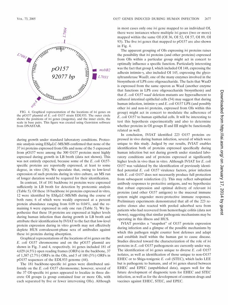
Graphical representations of the locations of ivi genes on theE. coli O157 chromosome and on the pO157 plasmid areshown in Fig. 3 and 4, respectively. ivi genes included 181 of4,029 (4.5%) open reading frames (ORFs) in the backbone, 37of 1,387 (2.7%) ORFs in the OIs, and 5 of 100 (5%) ORFs inpO157 sequences of the EDL933 genome (44).
The 181 backbone-specific ivi genes were distributed uni-formly on the E. coli O157 chromosome; however, several ofthe 37 OI-specific ivi genes appeared to localize in three dis-crete OI groups (a group contained four or more OIs, witheach separated by five or fewer intervening OIs). Although
in most cases only one ivi gene mapped to an individual OI,there were instances where multiple ivi genes (two or more)mapped within the same OI (OI 36, OI 52, OI 57, OI 89, OI93). The five ivi genes that mapped to pO157 are also shownin Fig. 4.
The apparent grouping of OIs expressing ivi proteins raisesthe possibility that ivi proteins (and other proteins) expressedfrom OIs within a particular group might act in concert tooptimally influence a specific function. Particularly interestingwas the fact that group I, which included OI 148, expressing theadhesin intimin-�, also included OI 145, expressing the glyco-syltransferase WaaD, one of the many enzymes involved in thebiosynthesis of LPS core oligosaccharide. The facts that WaaDis expressed from the same operon as WaaI (another enzymethat functions in LPS core oligosaccharide biosynthesis) andthat E. coli O157 waaI deletion mutants are hyperadherent tocultured intestinal epithelial cells (54) may suggest that duringhuman infection, intimin-� and E. coli O157 LPS (and possiblyother ivi and non-ivi proteins, expressed from OIs within thisgroup) might act in concert to modulate the adherence ofE. coli O157 to human epithelial cells. It will be interesting totest this hypothesis experimentally and also to determinewhether proteins in OI groups II and III might be functionallyrelated as well.
In conclusion, IVIAT identified 223 O157 proteins ex-pressed in vivo during human infection, several of which wereunique to this study. Judged by our results, IVIAT enablesidentification both of proteins expressed specifically duringhuman infection but not during growth under standard labo-ratory conditions and of proteins expressed at significantlyhigher levels in vivo than in vitro. Although IVIAT for E. coliO157 was validated by the identification of previously identi-fied potential E. coli O157 virulence factors, prior infectionwith E. coli O157 does not necessarily produce full protectionfrom subsequent reinfection (1). This may reflect suboptimalantibody responses to protective antigens, and we hypothesizethat robust expression and optimal delivery of relevant iviproteins (and other O157 antigens) to the mucosal immunesystem might engender more-protective immune responses.Preliminary experiments demonstrated that all of the 223 re-active clones also reacted with pooled adsorbed sera frompatients who had recovered from hemorrhagic colitis (data notshown), suggesting that similar pathogenic mechanisms may beoperating in this illness and HUS.
IVIAT provides a “snapshot” of O157 protein expressionduring infection and a glimpse of the possible mechanisms bywhich this pathogen might counter host defenses and adaptand establish itself within the human gut to cause disease.Studies directed toward the characterization of the role of iviproteins in E. coli O157 pathogenesis are currently under way.The identification of ivi genes unique to diverse E. coli O157isolates, as well as identification of those unique to non-O157EHEC or to Shiga-toxigenic E. coli (STEC), which lacks LEEbut is pathogenic to humans, and of ivi genes shared betweenEHEC and EPEC (unpublished data), augurs well for thefuture development of diagnostic tests for EHEC and STECinfection, as well as for the development of common drugs andvaccines against EHEC, STEC, and EPEC.
FIG. 4. Graphical representation of the locations of ivi genes onthe pO157 plasmid of E. coli O157 strain EDL933. The outer circleshows the positions of ivi genes (magenta), and the inner circle, thescale in base pairs. This figure was created using Genvision softwarefrom DNASTAR.
VOL. 73, 2005 O157 GENES INDUCED DURING HUMAN INFECTION 2677
on January 17, 2014 by guesthttp://iai.asm
.org/D
ownloaded from

ACKNOWLEDGMENTS
This work was supported by grants R03 AI53700-01 and R21AI055963 (to I.T.K.), R01 DE13523 (to M.H.), and R01 DK52081 andR01 AI47499 (to P.I.T.) from the National Institutes of Health.
REFERENCES
1. Besser, R. E., P. M. Griffin, and L. Slutsker. 1999. Escherichia coli O157:H7gastroenteritis and the hemolytic uremic syndrome: an emerging infectiousdisease. Annu. Rev. Med. 50:355–367.
2. Bowers, P. M., M. Pellegrini, M. J. Thompson, J. Fierro, T. O. Yeates, andD. Eisenberg. 2004. Prolinks: a database of protein functional linkages de-rived from coevolution. Genome Biol. 5:R35.
3. Burland, V., Y. Shao, N. T. Perna, G. Plunkett, H. J. Sofia, and F. R.Blattner. 1998. The complete DNA sequence and analysis of the largevirulence plasmid of Escherichia coli O157:H7. Nucleic Acids Res. 26:4196–4204.
4. Camilli, A., and J. J. Mekalanos. 1995. Use of recombinase gene fusions toidentify Vibrio cholerae genes induced during infection. Mol. Microbiol. 18:671–683.
5. Cao, S. L., A. Progulske-Fox, J. D. Hillman, and M. Handfield. 2004. In vivoinduced antigenic determinants of Actinobacillus actinomycetemcomitans.FEMS Microbiol. Lett. 237:97–103.
6. Deng, W., J. L. Puente, S. Gruenheid, Y. Li, B. A. Vallance, A. Vazquez, J.Barba, J. A. Ibarra, P. O’Donnell, P. Metalnikov, K. Ashman, S. Lee, D.Goode, T. Pawson, and B. B. Finlay. 2004. Dissecting virulence: systematicand functional analyses of a pathogenicity island. Proc. Natl. Acad. Sci. USA101:3597–3602.
7. Dozois, C. M., F. Daigle, and R. Curtiss III. 2003. Identification of pathogen-specific and conserved genes expressed in vivo by an avian pathogenic Esch-erichia coli strain. Proc. Natl. Acad. Sci. USA 100:247–252.
8. Groisman, E. A. 2001. The pleiotropic two-component regulatory systemPhoP-PhoQ. J. Bacteriol. 183:1835–1842.
9. Gruenheid, S., I. Sekirov, N. A. Thomas, W. Deng, P. O’Donnell, D. Goode,Y. Li, E. A. Frey, N. F. Brown, P. Metalnikov, T. Pawson, K. Ashman, andB. B. Finlay. 2004. Identification and characterization of NleA, a non-LEE-encoded type III translocated virulence factor of enterohaemorrhagic Esch-erichia coli O157:H7. Mol. Microbiol. 51:1233–1249.
10. Hagiwara, D., M. Sugiura, T. Oshima, H. Mori, H. Aiba, T. Yamashino, andT. Mizuno. 2003. Genome-wide analyses revealing a signaling network of theRcsC-YojN-RcsB phosphorelay system in Escherichia coli. J. Bacteriol. 185:5735–5746.
11. Handfield, M., L. J. Brady, A. Progulske-Fox, and J. D. Hillman. 2000.IVIAT: a novel method to identify microbial genes expressed specificallyduring human infections. Trends Microbiol. 8:336–339.
12. Hang, L., M. John, M. Asaduzzaman, E. A. Bridges, C. Vanderspurt, T. J.Kirn, R. K. Taylor, J. D. Hillman, A. Progulske-Fox, M. Handfield, E. T.Ryan, and S. B. Calderwood. 2003. Use of in vivo-induced antigen technol-ogy (IVIAT) to identify genes uniquely expressed during human infectionwith Vibrio cholerae. Proc. Natl. Acad. Sci. USA 100:8508–8513.
13. Harkey, C. W., K. D. Everiss, and K. M. Peterson. 1995. Isolation andcharacterization of a Vibrio cholerae gene (tagA) that encodes a ToxR-regulated lipoprotein. Gene 153:81–84.
14. Hava, D. L., and A. Camilli. 2002. Large-scale identification of serotype 4Streptococcus pneumoniae virulence factors. Mol. Microbiol. 45:1389–1405.
15. Heithoff, D. M., C. P. Conner, and M. J. Mahan. 1997. Dissecting the biologyof a pathogen during infection. Trends Microbiol. 5:509–513.
16. Heithoff, D. M., C. P. Conner, U. Hentschel, F. Govantes, P. C. Hanna, andM. J. Mahan. 1999. Coordinate intracellular expression of Salmonella genesinduced during infection. J. Bacteriol. 181:799–807.
17. Hoch, J., and T. Silhavy. 1995. Two-component signal transduction. ASMPress, Washington, D.C.
18. Hoffman, J. A., J. L. Badger, Y. Zhang, S. H. Huang, and K. S. Kim. 2000.Escherichia coli K1 aslA contributes to invasion of brain microvascular en-dothelial cells in vitro and in vivo. Infect. Immun. 68:5062–5067.
19. Ikeda, M., S. Ito, and M. Honda. 2004. Hemolytic uremic syndrome inducedby lipopolysaccharide and Shiga-like toxin. Pediatr. Nephrol. 19:485–489.
20. John, M., T. I. Crean, S. B. Calderwood, and E. T. Ryan. 2000. In vitro andin vivo analyses of constitutive and in vivo-induced promoters in attenuatedvaccine and vector strains of Vibrio cholerae. Infect. Immun. 68:1171–1175.
21. Kaper, J. B., J. P. Nataro, and H. L. Mobley. 2004. Pathogenic Escherichiacoli. Nat. Rev. Microbiol. 2:123–140.
22. Karpman, D., Z. D. Bekassy, A. C. Sjogren, M. S. Dubois, M. A. Karmali, M.Mascarenhas, K. G. Jarvis, L. J. Gansheroff, A. D. O’Brien, G. S. Arbus, andJ. B. Kaper. 2002. Antibodies to intimin and Escherichia coli secreted pro-teins A and B in patients with enterohemorrhagic Escherichia coli infections.Pediatr. Nephrol. 17:201–211.
23. Kim, S. H., W. Jia, R. E. Bishop, and C. Gyles. 2004. An msbB homologuecarried in plasmid pO157 encodes an acyltransferase involved in lipid Abiosynthesis in Escherichia coli O157:H7. Infect. Immun. 72:1174–1180.
24. Kudva, I. T., P. S. Evans, N. T. Perna, T. J. Barrett, G. J. DeCastro, F. M.Ausubel, F. R. Blattner, and S. B. Calderwood. 2002. Polymorphic amplified
typing sequences provide a novel approach to Escherichia coli O157:H7strain typing. J. Clin. Microbiol. 40:1152–1159.
25. Kudva, I. T., R. W. Griffin, M. Murray, M. John, N. T. Perna, T. J. Barrett,and S. B. Calderwood. 2004. Insertions, deletions, and single-nucleotidepolymorphisms at rare restriction enzyme sites enhance discriminatorypower of polymorphic amplified typing sequences, a novel strain typingsystem for Escherichia coli O157:H7. J. Clin. Microbiol. 42:2388–2397.
26. Kwon, Y. M., and S. C. Ricke. 1998. Induction of acid resistance of Salmo-nella typhimurium by exposure to short-chain fatty acids. Appl. Environ.Microbiol. 64:3458–3463.
27. Lee, A. I., A. Delgado, and R. P. Gunsalus. 1999. Signal-dependent phos-phorylation of the membrane-bound NarX two-component sensor-transmit-ter protein of Escherichia coli: nitrate elicits a superior anion ligand responsecompared to nitrite. J. Bacteriol. 181:5309–5316.
28. Leonhartsberger, S., A. Huber, F. Lottspeich, and A. Bock. 2001. ThehydH/G genes from Escherichia coli code for a zinc and lead responsivetwo-component regulatory system. J. Mol. Biol. 307:93–105.
29. Li, Y., E. Frey, A. M. R. Mackenzie, and B. B. Finlay. 2000. Human responseto Escherichia coli O157:H7 infection: antibodies to secreted virulence fac-tors. Infect. Immun. 68:5090–5095.
30. Lin, R. J., M. Capage, and C. W. Hill. 1984. A repetitive DNA sequence, rhs,responsible for duplications within the Escherichia coli K-12 chromosome.J. Mol. Biol. 177:1–18.
31. Lucas, R. L., C. P. Lostroh, C. C. DiRusso, M. P. Spector, B. L. Wanner, andC. A. Lee. 2000. Multiple factors independently regulate hilA and invasiongene expression in Salmonella enterica serovar Typhimurium. J. Bacteriol.182:1872–1882.
32. Madrid, C., J. M. Nieto, S. Paytubi, M. Falconi, C. O. Gualerzi, and A.Juarez. 2002. Temperature- and H-NS-dependent regulation of a plasmid-encoded virulence operon expressing Escherichia coli hemolysin. J. Bacteriol.184:5058–5066.
33. Mahan, M. J., D. M. Heithoff, R. L. Sinsheimer, and D. A. Low. 2000.Assessment of bacterial pathogenesis by analysis of gene expression in thehost. Annu. Rev. Genet. 34:139–164.
34. Mao, Y., M. P. Doyle, and J. Chen. 2001. Insertion mutagenesis of wcareduces acid and heat tolerance of enterohemorrhagic Escherichia coli O157:H7. J. Bacteriol. 183:3811–3815.
35. McKee, M. L., and A. D. O’Brien. 1996. Truncated enterohemorrhagic Esch-erichia coli (EHEC) O157:H7 intimin (EaeA) fusion proteins promote ad-herence of EHEC strains to HEp-2 cells. Infect. Immun. 64:2225–2233.
36. McNally, A., A. J. Roe, S. Simpson, F. M. Thomson-Carter, D. E. E. Hoey, C.Currie, T. Chakraborty, D. G. E. Smith, and D. L. Gally. 2001. Differencesin levels of secreted locus of enterocyte effacement proteins between humandisease-associated and bovine Escherichia coli O157. Infect. Immun. 69:5107–5114.
37. Mei, J., F. Nourbakhsh, C. Ford, and D. Holden. 1997. Identification ofStaphylococcus aureus virulence genes in a murine model of bacteremia usingsignature-tagged mutagenesis. Mol. Microbiol. 26:399–407.
38. Merrell, D. S., D. L. Hava, and A. Camilli. 2002. Identification of novelfactors involved in colonization and acid tolerance of Vibrio cholerae. Mol.Microbiol. 43:1471–1491.
39. Miranda, R. L., T. Conway, M. P. Leatham, D. E. Chang, W. E. Norris, J. H.Allen, S. J. Stevenson, D. C. Laux, and P. S. Cohen. 2004. Glycolytic andgluconeogenic growth of Escherichia coli O157:H7 (EDL933) and E. coliK-12 (MG1655) in the mouse intestine. Infect. Immun. 72:1666–1676.
40. Neidhardt, F. C., R. Curtiss III, J. L. Ingraham, E. C. C. Lin, K. B. Low, B.Magasanik, W. S. Reznikoff, M. Riley, M. Schaechter, and H. E. Umbarger.1996. Escherichia coli and Salmonella: molecular and cellular biology. Amer-ican Society for Microbiology, Washington, D.C.
41. Paton, A. W., and J. C. Paton. 2002. Reactivity of convalescent-phase he-molytic-uremic syndrome patient sera with the megaplasmid-encoded TagAprotein of Shiga-toxigenic Escherichia coli O157. J. Clin. Microbiol. 40:1395–1399.
42. Peekhaus, N., and T. Conway. 1998. What’s for dinner? Entner-Doudoroffmetabolism in Escherichia coli. J. Bacteriol. 180:3495–3502.
43. Peng, J., J. E. Elias, C. C. Thoreen, L. J. Licklider, and S. P. Gygi. 2003.Evaluation of multidimensional chromatography coupled with tandem massspectrometry (LC/LC-MS/MS) for large-scale protein analysis: the yeastproteome. J. Proteome Res. 2:43–50.
44. Perna, N. T., G. Plunkett, V. Burland, B. Mau, J. D. Glasner, D. J. Rose,G. F. Mayhew, P. S. Evans, J. Gregor, H. A. Kirkpatrick, G. Posfai, J.Hackett, S. Klink, A. Boutin, Y. Shao, L. Miller, E. J. Grotbeck, N. W. Davis,A. Lim, E. T. Dimalanta, K. D. Potamousis, J. Apodaca, T. S. Ananthara-man, J. Lin, G. Yen, D. C. Schwartz, R. A. Welch, and F. R. Blattner. 2001.Genome sequence of enterohaemorrhagic Escherichia coli O157:H7. Nature409:529–533.
45. Polen, T., D. Rittmann, V. F. Wendisch, and H. Sahm. 2003. DNA microar-ray analyses of the long-term adaptive response of Escherichia coli to acetateand propionate. Appl. Environ. Microbiol. 69:1759–1774.
46. Rabin, R. S., and V. Stewart. 1992. Either of two functionally redundantsensor proteins, NarX and NarQ, is sufficient for nitrate regulation in Esch-erichia coli K-12. Proc. Natl. Acad. Sci. USA 89:8419–8423.
2678 JOHN ET AL. INFECT. IMMUN.
on January 17, 2014 by guesthttp://iai.asm
.org/D
ownloaded from

47. Ritchie, J. M., P. L. Wagner, D. W. K. Acheson, and M. K. Waldor. 2003.Comparison of Shiga toxin production by hemolytic-uremic syndrome-asso-ciated and bovine-associated Shiga toxin-producing Escherichia coli isolates.Appl. Environ. Microbiol. 69:1059–1066.
48. Sambrook, J., and D. W. Russell. 2001. Molecular cloning: a laboratorymanual, 3rd ed. Cold Spring Harbor Laboratory Press, Cold Spring Harbor,N.Y.
49. Sharma, V. K., and R. L. Zuerner. 2004. Role of hha and ler in transcrip-tional regulation of the esp operon of enterohemorrhagic Escherichia coliO157:H7. J. Bacteriol. 186:7290–7301.
50. Sinclair, J. F., and A. D. O’Brien. 2002. Cell surface-localized nucleolin is aeukaryotic receptor for the adhesin intimin-� of enterohemorrhagic Esche-richia coli O157:H7. J. Biol. Chem. 277:2876–2885.
51. Soncini, F. C., and E. A. Groisman. 1996. Two-component regulatory sys-tems can interact to process multiple environmental signals. J. Bacteriol. 178:6796–6801.
52. Sperandio, V., A. G. Torres, and J. B. Kaper. 2002. Quorum sensing Esch-erichia coli regulators B and C (QseBC): a novel two-component regulatorysystem involved in the regulation of flagella and motility by quorum sensingin E. coli. Mol. Microbiol. 43:809–821.
53. Sperandio, V., C. C. Li, and J. B. Kaper. 2002. Quorum-sensing Escherichiacoli regulator A: a regulator of the LysR family involved in the regulation ofthe locus of enterocyte effacement pathogenicity island in enterohemor-rhagic E. coli. Infect. Immun. 70:3085–3093.
54. Torres, A. G., and J. B. Kaper. 2003. Multiple elements controlling adher-ence of enterohemorrhagic Escherichia coli O157:H7 to HeLa cells. Infect.Immun. 71:4985–4995.
55. von Kruger, W. M. A., S. Humphreys, and J. M. Ketley. 1999. A role for thePhoBR regulatory system homologue in the Vibrio cholerae phosphate-limitation response and intestinal colonization. Microbiology 145:2463–2475.
56. Wagner, P. L., and M. K. Waldor. 2002. Bacteriophage control of bacterialvirulence. Infect. Immun. 70:3985–3993.
57. Wang, L., and P. R. Reeves. 1998. Organization of Escherichia coli O157 Oantigen gene cluster and identification of its specific genes. Infect. Immun.66:3545–3551.
58. Wosten, M. M., L. F. Kox, S. Chamnongpol, F. C. Soncini, and E. A.Groisman. 2000. A signal transduction system that responds to extracellulariron. Cell 103:113–125.
59. Xu, Q., M. Dziejman, and J. J. Mekalanos. 2003. Determination of thetranscriptome of Vibrio cholerae during intraintestinal growth and midexpo-nential phase in vitro. Proc. Natl. Acad. Sci. USA 100:1286–1291.
60. Yim, H. H., and M. Villarejo. 1992. osmY, a new hyperosmotically induciblegene, encodes a periplasmic protein in Escherichia coli. J. Bacteriol. 174:3637–3644.
61. Yokoseki, T., T. Iino, and K. Kutsukake. 1996. Negative regulation by fliD,fliS, and fliT of the export of the flagellum-specific anti-sigma factor, FlgM,in Salmonella typhimurium. J. Bacteriol. 178:899–901.
Editor: A. D. O’Brien
VOL. 73, 2005 O157 GENES INDUCED DURING HUMAN INFECTION 2679
on January 17, 2014 by guesthttp://iai.asm
.org/D
ownloaded from
Copyright © 2022 FDOKUMEN