Upper body contributions to power generation during rapid, overhand throwing in humans
11
The Journal of Experimental Biology © 2014. Published by The Company of Biologists Ltd | The Journal of Experimental Biology (2014) 217, 2139-2149 doi:10.1242/jeb.103275 2139 ABSTRACT High-speed and accurate throwing is a distinctive human behavior. Achieving fast projectile speeds during throwing requires a combination of elastic energy storage at the shoulder, as well as the transfer of kinetic energy from proximal body segments to distal segments. However, the biomechanical bases of these mechanisms are not completely understood. We used inverse dynamics analyses of kinematic data from 20 baseball players fitted with four different braces that inhibit specific motions to test a model of power generation at key joints during the throwing motion. We found that most of the work produced during throwing is generated at the hips, and much of this work (combined with smaller contributions from the pectoralis major) is used to load elastic elements in the shoulder and power the rapid acceleration of the projectile. Despite rapid angular velocities at the elbow and wrist, the restrictions confirm that much of the power generated to produce these distal movements comes from larger proximal segments, such as the shoulder and torso. Wrist hyperextension enhances performance only modestly. Together, our data also suggest that heavy reliance on elastic energy storage may help explain some common throwing injuries and can provide further insight into the evolution of the upper body and when our ancestors first developed the ability to produce high-speed throws. KEY WORDS: Throwing, Biomechanics, Elastic energy storage, Kinetic chain, Human evolution INTRODUCTION The human forelimb is derived relative to other hominoids and to earlier hominins (Larson, 1993; Larson, 2007). One reason the human shoulder may be so different is selection for humans’ unique ability to throw objects overhand with both accuracy and high velocity. Today, most high-speed throwing occurs during sports, but in the past throwing was probably crucial for hunting, defense against predators, and aggressive interactions. Regardless of their purpose, high-speed overhand throws are produced using a stereotypic, whip-like motion involving the whole body. The throw begins with movement of the legs and progresses quickly up the trunk and arm, ending with rapid movement of the throwing hand as the projectile is released. There has been considerable inquiry into how this complex motion generates high projectile velocities, and which joints and joint-specific angular motions are primarily responsible (Dillman et al., 1993; Fleisig et al., 1995; Hirashima et al., 2007; Hirashima et al., 2008; Hong et al., RESEARCH ARTICLE 1 Center for the Advanced Study of Hominid Paleobiology, The George Washington University, 2110 G Street NW, Washington, DC 20052, USA. 2 Department of Human Evolutionary Biology, Harvard University, 11 Divinity Avenue, Cambridge, MA 02138, USA. *Author for correspondence ([email protected]) Received 31 January 2014; Accepted 17 March 2014 2001; Pappas et al., 1985; Putnam, 1993). Previous work has shown that large angular velocities of torso rotation, shoulder internal rotation, elbow extension and wrist flexion all occur at the moment of release and significantly contribute to projectile speed (Fig. 1) (Fleisig et al., 1995; Fleisig et al., 1996; Hirashima et al., 2007; Pappas et al., 1985). This study focuses on how these large angular velocities are produced in the upper body. Angular movements are produced when torques act across joints, generating mechanical work and power. Muscles are the source of most torques and are thus key contributors to joint power production and angular velocity. As expected, electromyography (EMG) patterns of muscle activity during throwing show sequential activation of muscles mirroring the progression of the throwing motion (Hirashima et al., 2002). However, muscle activation patterns alone cannot fully explain how throwing power is generated. For example, an individual with a paralyzed triceps brachii can still achieve rapid elbow extension during throwing, indicating that the triceps does not power rapid elbow extension on its own (Roberts, 1971). In addition, although EMG recordings of shoulder internal rotator muscles indicate high activity during internal rotation (DiGiovine et al., 1992; Gowan et al., 1987), experimental data on shoulder power show that these muscles only generate approximately 50% of the power for this rapid motion (Roach et al., 2013). A further problem with using EMG to evaluate the roles of muscles in generating torques in the upper body is the lack of any simple relationship between EMG intensity and muscle force production (Bell, 1993; Gans, 1992). Previous research has suggested that additional sources of torque to power large angular velocities during throwing come from movements generated in adjacent, connected body segments, which can be transferred from joint to joint via a ‘kinetic chain’ (Atwater, 1979; Fleisig et al., 1996; Hirashima et al., 2008; Hore et al., 2005; Ben Kibler and Sciascia, 2004). These interaction torques can result directly from muscular actions at other joints or from velocity- dependent, centrifugal or Coriolis forces (Hirashima et al., 2008). Mathematical decomposition of throwing kinematics using equations of motion has shown that high angular velocities observed at the elbow and wrist joints at release are largely due to these interaction torques (Feltner, 1989; Hirashima et al., 2007; Hirashima et al., 2003; Hong et al., 2001; Putnam, 1993). Induced acceleration analyses of these interaction torques further show that elbow extension during throwing is driven primarily by velocity-dependent forces generated by torso rotation and shoulder internal rotation (Hirashima et al., 2008). The same study also found that wrist flexion during throwing is mostly driven by velocity-dependent forces generated by elbow extension. These data strongly support the hypothesis that power generated at more proximal joints (such as the hips, torso and shoulder) is transferred to the throwing arm, producing very rapid, ‘whip-like’ accelerations of the arm and hand (Alexander, 1991; Atwater, 1979; Feltner, 1989; Putnam, 1993). However, like all kinetic analyses, these studies estimated only the Upper body contributions to power generation during rapid, overhand throwing in humans Neil T. Roach 1,2, * and Daniel E. Lieberman 2
Transcript of Upper body contributions to power generation during rapid, overhand throwing in humans
The
Jour
nal o
f Exp
erim
enta
l Bio
logy
© 2014. Published by The Company of Biologists Ltd | The Journal of Experimental Biology (2014) 217, 2139-2149 doi:10.1242/jeb.103275
2139
ABSTRACTHigh-speed and accurate throwing is a distinctive human behavior.Achieving fast projectile speeds during throwing requires acombination of elastic energy storage at the shoulder, as well as thetransfer of kinetic energy from proximal body segments to distalsegments. However, the biomechanical bases of these mechanismsare not completely understood. We used inverse dynamics analysesof kinematic data from 20 baseball players fitted with four differentbraces that inhibit specific motions to test a model of powergeneration at key joints during the throwing motion. We found thatmost of the work produced during throwing is generated at the hips,and much of this work (combined with smaller contributions from thepectoralis major) is used to load elastic elements in the shoulder andpower the rapid acceleration of the projectile. Despite rapid angularvelocities at the elbow and wrist, the restrictions confirm that much ofthe power generated to produce these distal movements comes fromlarger proximal segments, such as the shoulder and torso. Wristhyperextension enhances performance only modestly. Together, ourdata also suggest that heavy reliance on elastic energy storage mayhelp explain some common throwing injuries and can provide furtherinsight into the evolution of the upper body and when our ancestorsfirst developed the ability to produce high-speed throws.
KEY WORDS: Throwing, Biomechanics, Elastic energy storage,Kinetic chain, Human evolution
INTRODUCTIONThe human forelimb is derived relative to other hominoids and toearlier hominins (Larson, 1993; Larson, 2007). One reason thehuman shoulder may be so different is selection for humans’ uniqueability to throw objects overhand with both accuracy and highvelocity. Today, most high-speed throwing occurs during sports, butin the past throwing was probably crucial for hunting, defenseagainst predators, and aggressive interactions. Regardless of theirpurpose, high-speed overhand throws are produced using astereotypic, whip-like motion involving the whole body.
The throw begins with movement of the legs and progressesquickly up the trunk and arm, ending with rapid movement of thethrowing hand as the projectile is released. There has beenconsiderable inquiry into how this complex motion generates highprojectile velocities, and which joints and joint-specific angularmotions are primarily responsible (Dillman et al., 1993; Fleisig etal., 1995; Hirashima et al., 2007; Hirashima et al., 2008; Hong et al.,
RESEARCH ARTICLE
1Center for the Advanced Study of Hominid Paleobiology, The GeorgeWashington University, 2110 G Street NW, Washington, DC 20052, USA.2Department of Human Evolutionary Biology, Harvard University, 11 DivinityAvenue, Cambridge, MA 02138, USA.
*Author for correspondence ([email protected])
Received 31 January 2014; Accepted 17 March 2014
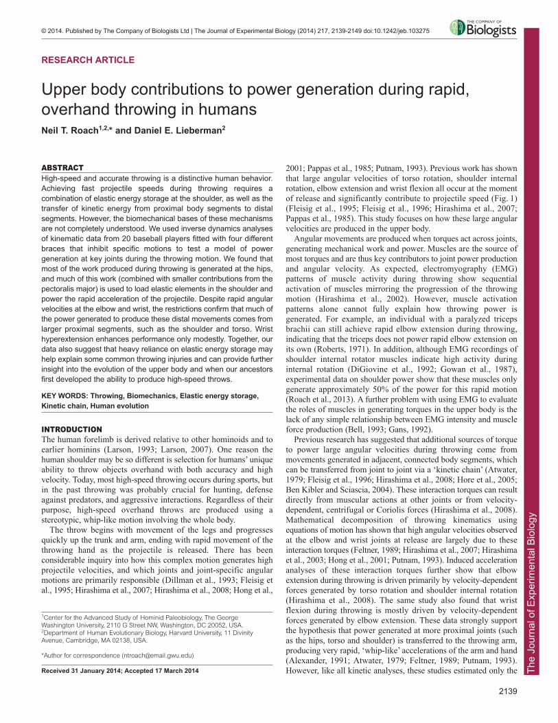
2001; Pappas et al., 1985; Putnam, 1993). Previous work has shownthat large angular velocities of torso rotation, shoulder internalrotation, elbow extension and wrist flexion all occur at the momentof release and significantly contribute to projectile speed (Fig. 1)(Fleisig et al., 1995; Fleisig et al., 1996; Hirashima et al., 2007;Pappas et al., 1985). This study focuses on how these large angularvelocities are produced in the upper body.
Angular movements are produced when torques act across joints,generating mechanical work and power. Muscles are the source ofmost torques and are thus key contributors to joint power productionand angular velocity. As expected, electromyography (EMG)patterns of muscle activity during throwing show sequentialactivation of muscles mirroring the progression of the throwingmotion (Hirashima et al., 2002). However, muscle activationpatterns alone cannot fully explain how throwing power isgenerated. For example, an individual with a paralyzed tricepsbrachii can still achieve rapid elbow extension during throwing,indicating that the triceps does not power rapid elbow extension onits own (Roberts, 1971). In addition, although EMG recordings ofshoulder internal rotator muscles indicate high activity duringinternal rotation (DiGiovine et al., 1992; Gowan et al., 1987),experimental data on shoulder power show that these muscles onlygenerate approximately 50% of the power for this rapid motion(Roach et al., 2013). A further problem with using EMG to evaluatethe roles of muscles in generating torques in the upper body is thelack of any simple relationship between EMG intensity and muscleforce production (Bell, 1993; Gans, 1992).
Previous research has suggested that additional sources of torqueto power large angular velocities during throwing come frommovements generated in adjacent, connected body segments, whichcan be transferred from joint to joint via a ‘kinetic chain’ (Atwater,1979; Fleisig et al., 1996; Hirashima et al., 2008; Hore et al., 2005;Ben Kibler and Sciascia, 2004). These interaction torques can resultdirectly from muscular actions at other joints or from velocity-dependent, centrifugal or Coriolis forces (Hirashima et al., 2008).Mathematical decomposition of throwing kinematics usingequations of motion has shown that high angular velocities observedat the elbow and wrist joints at release are largely due to theseinteraction torques (Feltner, 1989; Hirashima et al., 2007; Hirashimaet al., 2003; Hong et al., 2001; Putnam, 1993). Induced accelerationanalyses of these interaction torques further show that elbowextension during throwing is driven primarily by velocity-dependentforces generated by torso rotation and shoulder internal rotation(Hirashima et al., 2008). The same study also found that wristflexion during throwing is mostly driven by velocity-dependentforces generated by elbow extension. These data strongly supportthe hypothesis that power generated at more proximal joints (suchas the hips, torso and shoulder) is transferred to the throwing arm,producing very rapid, ‘whip-like’ accelerations of the arm and hand(Alexander, 1991; Atwater, 1979; Feltner, 1989; Putnam, 1993).However, like all kinetic analyses, these studies estimated only the
Upper body contributions to power generation during rapid,overhand throwing in humansNeil T. Roach1,2,* and Daniel E. Lieberman2
The
Jour
nal o
f Exp
erim
enta
l Bio
logy
2140
contributions of joint rotational motions (i.e. shoulderinternal/external rotation) without measuring how much energy wasproduced by particular muscles or by elastic energy storagemechanisms.
Several studies have suggested that the storage and release ofelastic energy also plays a role in power enhancement duringthrowing. Wilk et al. (Wilk et al., 1993) argued that throwingathletes enhance torque production by ‘pre-stretching’ numerousthrowing muscles just prior to their activation, resulting in elasticenergy storage in the muscle itself. In addition, Roach et al. (Roachet al., 2013) found that the posture of the ‘cocked’ arm (externallyrotated) just prior to the rapid shoulder internal rotation motionpassively stretches elastic elements crossing the shoulder. Bycomparing actual power production during throwing to modeledmaximum power values for all muscles potentially involved, theyinferred that passive stretching results in significant amounts ofelastic energy being stored and released, powering the rapid internalrotation of the humerus that follows. Although power amplificationis well known and well studied in the human lower limb (e.g.Anderson and Pandy, 1993; Komi and Bosco, 1978), understandingthe workings of such amplification mechanisms in humans iscomplicated by the difficulty of collecting direct strainmeasurements (Finni et al., 2000; Komi et al., 1987; Lewis et al.,1982; Pourcelot et al., 2005) and the inability to use invasivemethodologies, such as sonomicrometry. Accordingly, the presentstudy combines inverse dynamics analysis with experimentalperturbations of the throwing motion using therapeutic braces. Theserestrictions allow us to non-invasively examine how muscular force,kinetic transfer and elastic energy storage interact and power thethrowing motion.
Here, we test how the upper body contributes to power generationduring throwing by experimentally manipulating four key motionshypothesized to be responsible for projectile speed (torso rotation,shoulder internal rotation, elbow extension and wrist flexion; Fig. 2).We used therapeutic braces that limit joint range of motion (ROM)or modify joint position to induce variation in throwing kinematics.Note that these braces are not intended to fully remove thesemovements, as this would make throwing difficult or impossible.They are instead designed to induce modest perturbations that help
illuminate the effects each motion has on overall performance andupon the other critical motions in the upper body. Further, theserestrictions allowed us to test the effects of morphological variationsassociated with performance in a study group where all individualshave comparable, high skill levels. The braces used also served torecreate evolutionarily relevant chimpanzee-like and hominin-likejoint positions and ROM, as apes throw poorly and with verydifferent kinematics (Goodall, 1964; Goodall, 1986; Kohler, 1925;Sugiyama and Koman, 1979) and intermediate morphologies arefound solely in extinct fossil taxa.
HypothesesHypothesis 1: the hip rotator muscles generate the majority of thetorso rotation power during throwingPrevious studies have shown that hip rotation is an importantcomponent of the throwing motion that correlates with throwingperformance (Matsuo et al., 2001; Stodden et al., 2001; Wight et al.,2004). We propose that by stabilizing the torso relative to the pelvisthrough bilateral contraction of the intrinsic spinal rotator muscles,the large hip rotators power the rapid rotation of the torso and pelvistogether. Both the medial hip rotators (gluteus medius and minimus,and tensor fasciae latae) and the lateral hip rotators (gluteusmaximus, quadratus femoris, both gemelli, both obturators,piriformis and sartorius) can be active at the same time during thethrowing motion at the contralateral and ipsilateral hips,respectively. Additional contributions from the large hip flexor andextensor muscles in the thighs may also increase pelvic rotationpower as the body is vaulted over the legs during the throw. Wetherefore predict that by limiting intervertebral rotation with a brace,most torso/pelvic rotation will occur at the hip. This shouldsignificantly reduce torso rotation angular velocity, torque, powerand work. These reductions, however, are expected to be modestbecause we are limiting rotational motion only between thevertebrae and not at the hips.
Hypothesis 2: torso rotation primarily powers the storage of elasticenergy at the shoulderRoach et al. (Roach et al., 2013) proposed that by abducting theshoulder and flexing the elbow as torso rotation reaches its peak
RESEARCH ARTICLE The Journal of Experimental Biology (2014) doi:10.1242/jeb.103275
Arm-cocking phase Acceleration phase
End of stride Maximum external rotation Release
Shoulder external rotation Shoulder internal rotation
Elbow flexion Elbow extensionShoulder extension Shoulder flexion
Wrist extension
Torso rotation
Fig. 1. The arm-cocking and acceleration phases of the throw.Arm cocking begins with the contralateral foot touching the ground atthe end of a large stride in the target direction. During the cockingphase, the humerus externally rotates until it reaches maximumexternal rotation. Following maximum external rotation, the very briefacceleration phase occurs in which the humerus rapidly internallyrotates, the elbow extends and the wrist is flexed. Acceleration endswith the release of the projectile. Light gray bars indicate the relativetiming of ‘cocking’ motions, while the dark gray bars indicate thetiming of ‘acceleration’ motions.

The
Jour
nal o
f Exp
erim
enta
l Bio
logy
angular velocity, the forelimb’s mass is positioned away from theshoulder, increasing its moment of inertia, and causing the forelimbto lag behind the accelerating torso. This lag causes further externalrotation of the shoulder beyond the active ROM achieved by theexternal rotator muscles and into the passive range (Miyashita et al.,2008a; Miyashita et al., 2008b; Roach et al., 2013). We thereforepredict that using a brace to limit torso rotation will reduce theamount of negative work done at the shoulder during the cockingphase. This reduction in shoulder rotation work should furtherreduce shoulder rotation angular velocity, torque, power and workduring acceleration.
Hypothesis 3: the pectoralis major also powers the storage of elasticenergy at the shoulderWe propose that when the forelimb is maximally cocked (Dillmanet al., 1993), muscular action by the largest horizontal flexor of theshoulder, the p. major, will also generate substantial torque around the same shoulder axis as the torso rotation torque, causingthe forelimb’s mass to lag, thus stretching elastic elements thatcross the shoulder. We therefore predict that using a brace toabduct the shoulder should cause a suboptimal realignment bycranially rotating the p. major’s major line of action. We expectthat this will significantly reduce the amount of negative workdone at the shoulder during cocking, and thus reduce shoulderrotation angular velocity, torque, power and work duringacceleration.
Hypothesis 4: rapid elbow extension and wrist flexion at the end ofthe throw are largely generated passivelyPrevious research has suggested that the work and power necessaryto achieve rapid angular movements in the elbow and wrist arederived from the kinetic transfer of power generated at moreproximal joints (Feltner, 1989; Hirashima et al., 2007; Hirashima etal., 2003; Hirashima et al., 2008; Putnam, 1993). We thereforepredict that braces restricting more active proximal joints (torso andshoulder) will cause significant decreases in angular velocity inmore passive distal joints (elbow and wrist).
Hypothesis 5: wrist hyperextension allows release to occur later inthe throwing motion, enabling the shoulder internal rotation andelbow extension motions to attain higher angular velocitiesKinematic data on the overhand throwing motion have shown thatwhile the arm is cocking, the wrist slowly hyperextends and thenrapidly flexes as the arm accelerates towards the target (Fleisig etal., 1995; Hirashima et al., 2007). This wrist hyperextension couldenable other more proximal joint actions (e.g. elbow flexion) tocontinue to accelerate prior to release and still achieve an accuraterelease trajectory. We therefore predict that when a brace is used torestrict wrist hyperextension, there will be a significant decrease inthe duration of the final acceleration phase of the throw, as well assignificant reductions in wrist flexion, elbow extension, andshoulder internal rotation angular velocities and work.
RESULTSNormal unrestrictedDuring normal throwing, the maximum ball speed was27.7±3.8 m s−1 (mean ± s.d. of all subject means), with the ballstriking the target on average 0.3±0.2 m from the center or bullseye.While considerable variation exists between subjects in overallperformance and the timing and duration of the throwing phases,there was little variation in kinematic patterns across subjects(Fig. 3). The angular velocity of torso rotation peaked(848±160 deg s−1) during the latter half of the cocking phase, beforeproducing an opposing braking torque during the brief accelerationphase. This opposing torque resulted in a short period of powerabsorption during acceleration, generating net negative work(−74±44 J). During the cocking phase at the shoulder, the humeruswas externally rotated while simultaneously generating a largeopposing internal rotation torque. This resulted in a sustained periodof negative work (−201±70 J) at the shoulder. As the accelerationphase began with the initiation of internal shoulder rotation, theshoulder rotation torque and angular velocity became in-phase,resulting in very high angular velocities (4290±1127 deg s−1) andpeak power (11,838±4170 W). The elbow continued to flex as thecocking phase began and as a moderate flexion torque was
2141
RESEARCH ARTICLE The Journal of Experimental Biology (2014) doi:10.1242/jeb.103275
Towards target (+)
Away from target (–)
Flexion (+)
Extension (–)
External (–) Internal (+)
Extension (–)
Flexion (+)
A
C D
B Fig. 2. The four critical upper body motions examined in thisstudy. (A) Torso rotation; (B) shoulder rotation; (C) elbowflexion/extension; (D) wrist flexion/extension. The + and – sensesin the diagram show the convention used to describe thedirectionality of the angular velocities and torques. Note: the torsorotation senses are dependent upon handedness, as thedescription is relative to the throwing side.

The
Jour
nal o
f Exp
erim
enta
l Bio
logy
2142
produced. However, during the last quarter of the cocking phase theelbow began to extend rapidly (–2434±552 deg s−1), peakingmidway through the acceleration phase. During this rapid extension,the opposing flexion moment at the elbow intensified, resulting inlarge amounts of negative work (−246±63 J). Finally, at the wrist, asustained extension occurred until the very end of the cocking phase,when the wrist began to rapidly flex, continuing through theacceleration phase. A small flexion moment was produced (peak:7±3 Nm) at the end of the cocking phase, which then dropped tonear zero and again increased during the acceleration phase. Thisoscillating power generated a small, net positive amount of work(1±2 J) during the acceleration phase.
Torso restrictionThe torso restriction brace provided near-complete restriction ofintervertebral rotation between the clavicles and the pelvis, allowingonly the hip rotators to rotate the trunk. Given the design of the torsobrace, no sham condition was possible. Contrary to expectations, therewere no significant reductions in torso rotation peak power, angularvelocity or torque (Table 1). However, there was a significant drop intorso rotation work during cocking (−11±18%, P=0.005). Asexpected, there were significant reductions in shoulder rotation power(−14±20%, P=0.012), and work during the cocking phase (−8±11%,P=0.004). A reduction also occurred in elbow extension peak angularvelocity (−6±7%, P=0.001). No significant reductions in angularvelocity, torque, power or work were measured at the wrist. Ashypothesized, when the torso restriction brace was applied, maximumball speed dropped moderately (−5±6%, P<0.001).
Clavicle restrictionThe clavicle brace cranially rotated the scapula 7±4 deg and alsolimited scapular protraction. The clavicle sham produced no
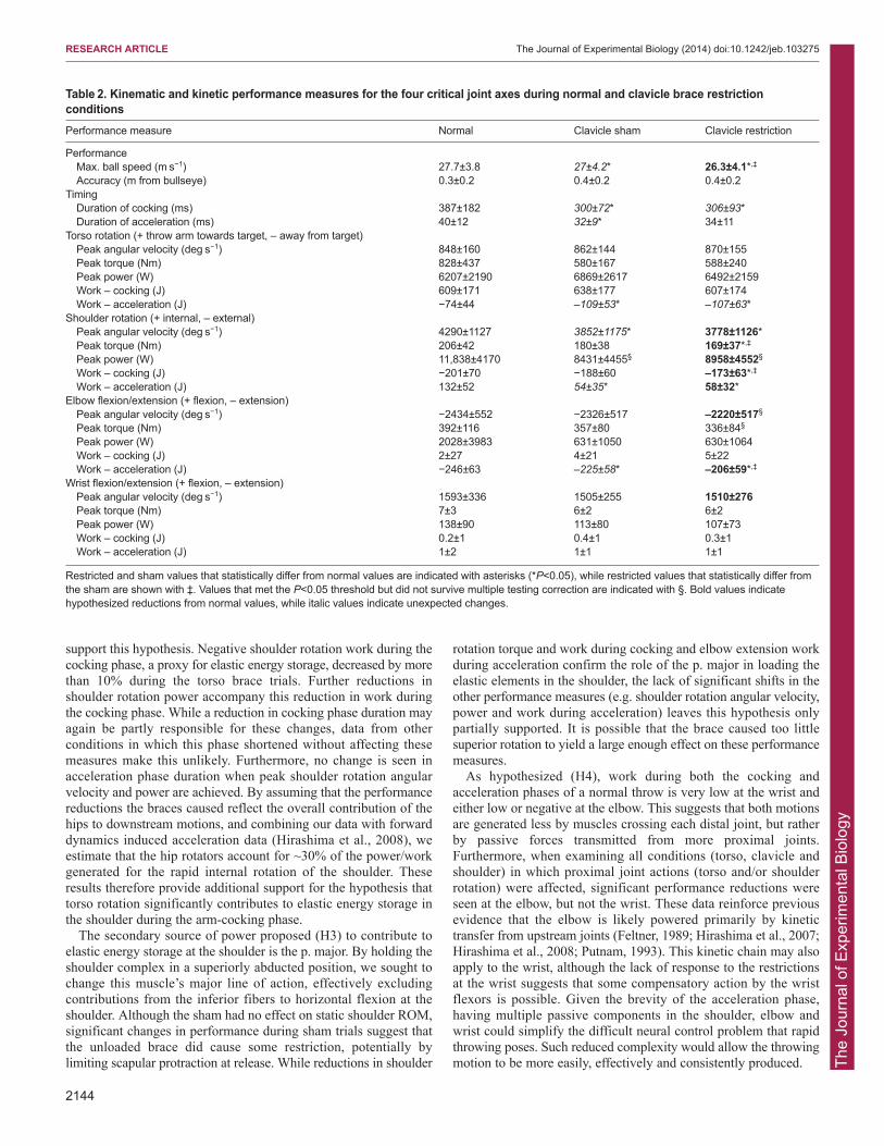
significant effects on joint motion relative to the unbraced condition.However, data from the sham trials (Table 2) indicate someperturbation of the normal throwing motion as evident fromreductions in: mean maximum ball speed (−3±5%, P=0.029), phaseduration (cocking −12±27%, P=0.028; acceleration −16±27%,P=0.010), torso rotation work acceleration (−42±42%, P<0.001),elbow extension work acceleration (−10±17%, P<0.001), shoulderrotation work acceleration (−56±25%, P<0.001) and shoulderrotation angular velocity (−11±12%, P<0.001). When the restrictionwas added, maximum ball speed dropped by a further 3±6%(P=0.031). Reductions in shoulder rotation peak torque (−13±31%,P<0.001) and work during cocking (−9±10%, P=0.002) wererecorded relative to normal values. Elbow work during accelerationdropped by 8±13% (P<0.001) relative to the reduced sham values.No statistically significant changes were observed in wristperformance.
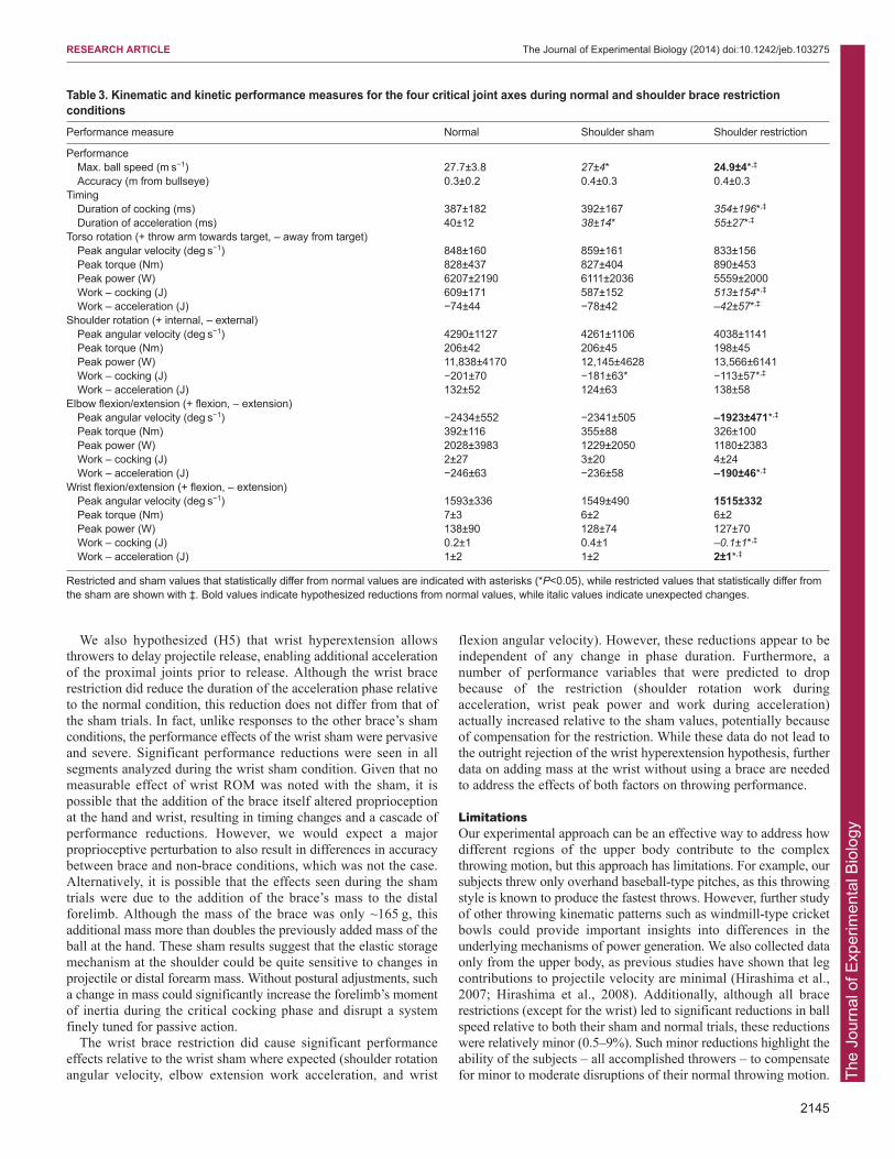
Shoulder restrictionThe shoulder brace reduced shoulder external rotation by 24±9 deg.However, the shoulder brace also restricted external rotation by11±7 deg in the sham condition, much like wearing a tight jacket.During the sham trials, there were also slight reductions in meanmaximum ball speed (−3±5%, P=0.026) and shoulder rotation workduring the cocking phase (−9±13%, P<0.001), and a slightshortening of the duration of the acceleration phase (6±10%,P=0.045; Table 3). For each significant change in the shamcondition, a further significant reduction occurred when therestriction was employed. The restricted trials caused an 8±6%reduction in maximum ball speed relative to the sham trials(P<0.001). The duration of the cocking phase of the restricted trialsshortened (−9±14%, P=0.032), while the acceleration phaselengthened (38±56%, P=0.013). Reductions in torso rotation work
RESEARCH ARTICLE The Journal of Experimental Biology (2014) doi:10.1242/jeb.103275
Angular velocity (deg s–1) Torque (Nm) Power (W) Work (J)
Wris
tE
lbow
Sho
ulde
rTo
rso
0
0
200
400
600
800
STR MER REL
0
200
400
600
C A
–800
–400
0
400
800
STR MER REL
–4000
0
4000
8000
STR MER REL
–200
0
200
C A
2000
4000
STR MER REL
0
100
200
STR MER REL
0
4000
8000
12,000
STR MER REL–4000
–300
–200
–100
0
C A–3000
–2000
–1000
0
STR MER REL 0
100
200
300
400
STR MER REL
–12,000
–8000
–4000
0
STR MER REL
0
1000
2000
STR MER REL–2
0
2
4
6
STR MER REL–100
0
100
200
STR MER REL
0
1
2
C A
Internal rotation (+)
External rotation (–)
Extension (–) Flexion (+)
Flexion (+)
Extension (–)
Towards target (+)
Away from target (–)
50% 50% 50%
50% 50% 50%
50% 50% 50%
50% 50% 50%
Fig. 3. Joint kinematic and kinetic data fromthe four critical joint axes during normalthrowing. Mean data of all individual subjectmeans are shown as bold lines, while 95%confidence intervals are shown as dashedlines. Differences in phase timing betweenindividuals were standardized by interpolatingeach phase 1000-fold and subsequently down-sampling each throw to a standard length. Thelength ratio of the two phases was keptproportional to the mean normal, unrestrictedphase duration ratio. The arm-cocking phase[between stride (STR) and maximum externalrotation (MER)] is shown in the white field andlabeled C in the work plots, while theacceleration phase [between MER and release(REL)] is shown in a gray field and labeled A inthe work plots. Note: peak values shown herediffer from mean peak values reported inTables 1–4. This results from each subject’speak performance occurring at slightly differenttimes relative to the normalized phases of thethrow.
The
Jour
nal o
f Exp
erim
enta
l Bio
logy
were recorded for both phases (cocking −14±20%, P=0.006;acceleration −41±57%, P<0.001). Although shoulder rotation workduring the cocking phase dropped significantly (−45±17%,P<0.001) from normal values, no further significant reductionsoccurred in shoulder rotation peak angular velocity, torque, poweror work during acceleration. As predicted, there were significantreductions in elbow extension angular velocity (−21±10%, P<0.001)and elbow work during acceleration (−20±21%, P<0.001). Wristflexion peak angular velocity remained unchanged, but a significantreduction in wrist work during the cocking phase (P=0.020) wasfollowed by a significant increase in wrist work during theacceleration phase (P=0.003).
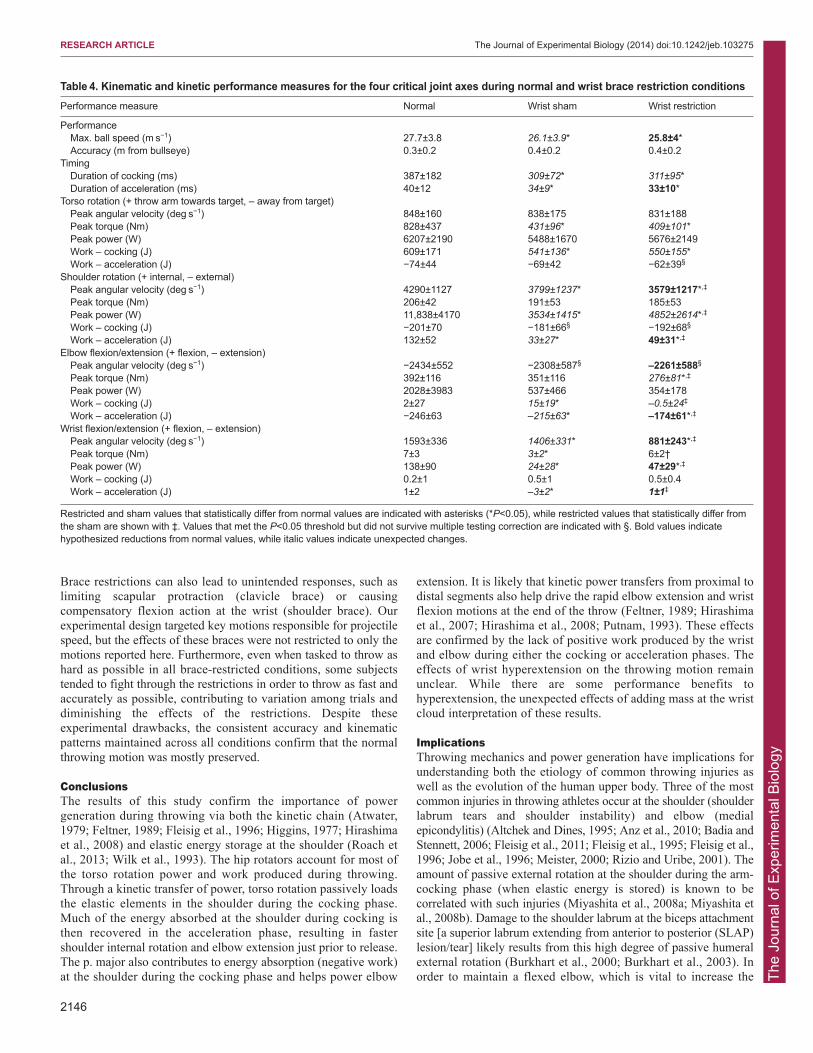
Wrist restrictionThe wrist brace reduced wrist extension ROM by 62±7 deg, butthere was no significant reduction in the sham condition. However,the wrist restriction sham trials showed numerous significantreductions in the measured performance variables (Table 4).Although the restricted condition produced significant reductions inball speed and phase duration from normal values, these reductionswere not significantly different from the sham condition. Restrictedshoulder rotation peak angular velocity dropped by 6±8% (P<0.001)from sham conditions, while shoulder rotation peak power(P<0.001) and work during acceleration values (P<0.001) bothdropped relative to normal and increased relative to sham trials.Restricted elbow work during acceleration dropped by 19±14%(P<0.001) from sham levels. The wrist flexion peak angular velocitydropped by 34±37% (P<0.001) relative to the sham trials, while
wrist flexion/extension peak power (P<0.001) and work duringacceleration (P<0.001) increased relative to the sham trials.
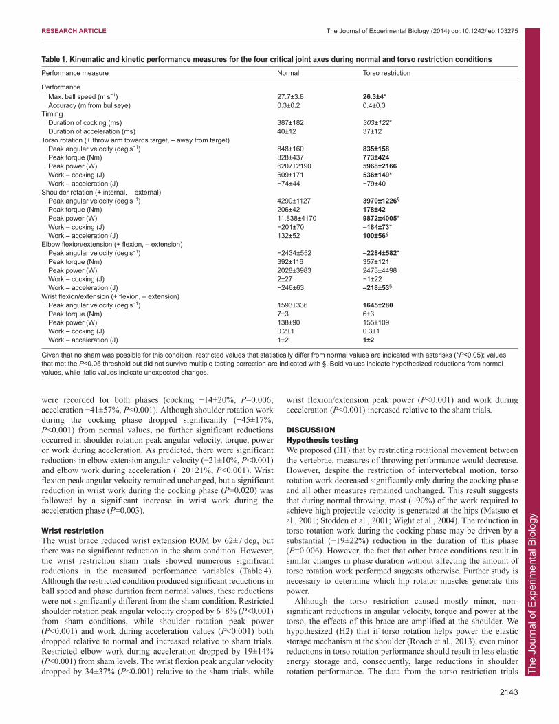
DISCUSSIONHypothesis testingWe proposed (H1) that by restricting rotational movement betweenthe vertebrae, measures of throwing performance would decrease.However, despite the restriction of intervertebral motion, torsorotation work decreased significantly only during the cocking phaseand all other measures remained unchanged. This result suggeststhat during normal throwing, most (~90%) of the work required toachieve high projectile velocity is generated at the hips (Matsuo etal., 2001; Stodden et al., 2001; Wight et al., 2004). The reduction intorso rotation work during the cocking phase may be driven by asubstantial (−19±22%) reduction in the duration of this phase(P=0.006). However, the fact that other brace conditions result insimilar changes in phase duration without affecting the amount oftorso rotation work performed suggests otherwise. Further study isnecessary to determine which hip rotator muscles generate thispower.
Although the torso restriction caused mostly minor, non-significant reductions in angular velocity, torque and power at thetorso, the effects of this brace are amplified at the shoulder. Wehypothesized (H2) that if torso rotation helps power the elasticstorage mechanism at the shoulder (Roach et al., 2013), even minorreductions in torso rotation performance should result in less elasticenergy storage and, consequently, large reductions in shoulderrotation performance. The data from the torso restriction trials
2143
RESEARCH ARTICLE The Journal of Experimental Biology (2014) doi:10.1242/jeb.103275
Table 1. Kinematic and kinetic performance measures for the four critical joint axes during normal and torso restriction conditions Performance measure Normal Torso restriction
PerformanceMax. ball speed (m s−1) 27.7±3.8 26.3±4*Accuracy (m from bullseye) 0.3±0.2 0.4±0.3
TimingDuration of cocking (ms) 387±182 303±122*Duration of acceleration (ms) 40±12 37±12
Torso rotation (+ throw arm towards target, – away from target) Peak angular velocity (deg s−1) 848±160 835±158 Peak torque (Nm) 828±437 773±424 Peak power (W) 6207±2190 5968±2166 Work – cocking (J) 609±171 536±149*Work – acceleration (J) −74±44 −79±40
Shoulder rotation (+ internal, – external)Peak angular velocity (deg s−1) 4290±1127 3970±1226§
Peak torque (Nm) 206±42 178±42 Peak power (W) 11,838±4170 9872±4005*Work – cocking (J) −201±70 –184±73*Work – acceleration (J) 132±52 100±56§
Elbow flexion/extension (+ flexion, – extension) Peak angular velocity (deg s−1) −2434±552 –2284±582*Peak torque (Nm) 392±116 357±121 Peak power (W) 2028±3983 2473±4498 Work – cocking (J) 2±27 −1±22 Work – acceleration (J) −246±63 –218±53§
Wrist flexion/extension (+ flexion, – extension) Peak angular velocity (deg s−1) 1593±336 1645±280 Peak torque (Nm) 7±3 6±3 Peak power (W) 138±90 155±109 Work – cocking (J) 0.2±1 0.3±1 Work – acceleration (J) 1±2 1±2
Given that no sham was possible for this condition, restricted values that statistically differ from normal values are indicated with asterisks (*P<0.05); valuesthat met the P<0.05 threshold but did not survive multiple testing correction are indicated with §. Bold values indicate hypothesized reductions from normalvalues, while italic values indicate unexpected changes.
The
Jour
nal o
f Exp
erim
enta
l Bio
logy
2144
support this hypothesis. Negative shoulder rotation work during thecocking phase, a proxy for elastic energy storage, decreased by morethan 10% during the torso brace trials. Further reductions inshoulder rotation power accompany this reduction in work duringthe cocking phase. While a reduction in cocking phase duration mayagain be partly responsible for these changes, data from otherconditions in which this phase shortened without affecting thesemeasures make this unlikely. Furthermore, no change is seen inacceleration phase duration when peak shoulder rotation angularvelocity and power are achieved. By assuming that the performancereductions the braces caused reflect the overall contribution of thehips to downstream motions, and combining our data with forwarddynamics induced acceleration data (Hirashima et al., 2008), weestimate that the hip rotators account for ~30% of the power/workgenerated for the rapid internal rotation of the shoulder. Theseresults therefore provide additional support for the hypothesis thattorso rotation significantly contributes to elastic energy storage inthe shoulder during the arm-cocking phase.
The secondary source of power proposed (H3) to contribute toelastic energy storage at the shoulder is the p. major. By holding theshoulder complex in a superiorly abducted position, we sought tochange this muscle’s major line of action, effectively excludingcontributions from the inferior fibers to horizontal flexion at theshoulder. Although the sham had no effect on static shoulder ROM,significant changes in performance during sham trials suggest thatthe unloaded brace did cause some restriction, potentially bylimiting scapular protraction at release. While reductions in shoulder
rotation torque and work during cocking and elbow extension workduring acceleration confirm the role of the p. major in loading theelastic elements in the shoulder, the lack of significant shifts in theother performance measures (e.g. shoulder rotation angular velocity,power and work during acceleration) leaves this hypothesis onlypartially supported. It is possible that the brace caused too littlesuperior rotation to yield a large enough effect on these performancemeasures.
As hypothesized (H4), work during both the cocking andacceleration phases of a normal throw is very low at the wrist andeither low or negative at the elbow. This suggests that both motionsare generated less by muscles crossing each distal joint, but ratherby passive forces transmitted from more proximal joints.Furthermore, when examining all conditions (torso, clavicle andshoulder) in which proximal joint actions (torso and/or shoulderrotation) were affected, significant performance reductions wereseen at the elbow, but not the wrist. These data reinforce previousevidence that the elbow is likely powered primarily by kinetictransfer from upstream joints (Feltner, 1989; Hirashima et al., 2007;Hirashima et al., 2008; Putnam, 1993). This kinetic chain may alsoapply to the wrist, although the lack of response to the restrictionsat the wrist suggests that some compensatory action by the wristflexors is possible. Given the brevity of the acceleration phase,having multiple passive components in the shoulder, elbow andwrist could simplify the difficult neural control problem that rapidthrowing poses. Such reduced complexity would allow the throwingmotion to be more easily, effectively and consistently produced.
RESEARCH ARTICLE The Journal of Experimental Biology (2014) doi:10.1242/jeb.103275
Table 2. Kinematic and kinetic performance measures for the four critical joint axes during normal and clavicle brace restrictionconditionsPerformance measure Normal Clavicle sham Clavicle restriction
Performance Max. ball speed (m s−1) 27.7±3.8 27±4.2* 26.3±4.1*,‡
Accuracy (m from bullseye) 0.3±0.2 0.4±0.2 0.4±0.2 Timing
Duration of cocking (ms) 387±182 300±72* 306±93*Duration of acceleration (ms) 40±12 32±9* 34±11
Torso rotation (+ throw arm towards target, – away from target)Peak angular velocity (deg s−1) 848±160 862±144 870±155 Peak torque (Nm) 828±437 580±167 588±240 Peak power (W) 6207±2190 6869±2617 6492±2159 Work – cocking (J) 609±171 638±177 607±174 Work – acceleration (J) −74±44 –109±53* –107±63*
Shoulder rotation (+ internal, – external) Peak angular velocity (deg s−1) 4290±1127 3852±1175* 3778±1126*Peak torque (Nm) 206±42 180±38 169±37*,‡
Peak power (W) 11,838±4170 8431±4455§ 8958±4552§
Work – cocking (J) −201±70 −188±60 –173±63*,‡
Work – acceleration (J) 132±52 54±35* 58±32*Elbow flexion/extension (+ flexion, – extension)
Peak angular velocity (deg s−1) −2434±552 −2326±517 –2220±517§
Peak torque (Nm) 392±116 357±80 336±84§
Peak power (W) 2028±3983 631±1050 630±1064 Work – cocking (J) 2±27 4±21 5±22 Work – acceleration (J) −246±63 –225±58* –206±59*,‡
Wrist flexion/extension (+ flexion, – extension) Peak angular velocity (deg s−1) 1593±336 1505±255 1510±276 Peak torque (Nm) 7±3 6±2 6±2 Peak power (W) 138±90 113±80 107±73 Work – cocking (J) 0.2±1 0.4±1 0.3±1 Work – acceleration (J) 1±2 1±1 1±1
Restricted and sham values that statistically differ from normal values are indicated with asterisks (*P<0.05), while restricted values that statistically differ fromthe sham are shown with ‡. Values that met the P<0.05 threshold but did not survive multiple testing correction are indicated with §. Bold values indicatehypothesized reductions from normal values, while italic values indicate unexpected changes.
The
Jour
nal o
f Exp
erim
enta
l Bio
logy
We also hypothesized (H5) that wrist hyperextension allowsthrowers to delay projectile release, enabling additional accelerationof the proximal joints prior to release. Although the wrist bracerestriction did reduce the duration of the acceleration phase relativeto the normal condition, this reduction does not differ from that ofthe sham trials. In fact, unlike responses to the other brace’s shamconditions, the performance effects of the wrist sham were pervasiveand severe. Significant performance reductions were seen in allsegments analyzed during the wrist sham condition. Given that nomeasurable effect of wrist ROM was noted with the sham, it ispossible that the addition of the brace itself altered proprioceptionat the hand and wrist, resulting in timing changes and a cascade ofperformance reductions. However, we would expect a majorproprioceptive perturbation to also result in differences in accuracybetween brace and non-brace conditions, which was not the case.Alternatively, it is possible that the effects seen during the shamtrials were due to the addition of the brace’s mass to the distalforelimb. Although the mass of the brace was only ~165 g, thisadditional mass more than doubles the previously added mass of theball at the hand. These sham results suggest that the elastic storagemechanism at the shoulder could be quite sensitive to changes inprojectile or distal forearm mass. Without postural adjustments, sucha change in mass could significantly increase the forelimb’s momentof inertia during the critical cocking phase and disrupt a systemfinely tuned for passive action.
The wrist brace restriction did cause significant performanceeffects relative to the wrist sham where expected (shoulder rotationangular velocity, elbow extension work acceleration, and wrist
flexion angular velocity). However, these reductions appear to beindependent of any change in phase duration. Furthermore, anumber of performance variables that were predicted to dropbecause of the restriction (shoulder rotation work duringacceleration, wrist peak power and work during acceleration)actually increased relative to the sham values, potentially becauseof compensation for the restriction. While these data do not lead tothe outright rejection of the wrist hyperextension hypothesis, furtherdata on adding mass at the wrist without using a brace are neededto address the effects of both factors on throwing performance.
Limitations Our experimental approach can be an effective way to address howdifferent regions of the upper body contribute to the complexthrowing motion, but this approach has limitations. For example, oursubjects threw only overhand baseball-type pitches, as this throwingstyle is known to produce the fastest throws. However, further studyof other throwing kinematic patterns such as windmill-type cricketbowls could provide important insights into differences in theunderlying mechanisms of power generation. We also collected dataonly from the upper body, as previous studies have shown that legcontributions to projectile velocity are minimal (Hirashima et al.,2007; Hirashima et al., 2008). Additionally, although all bracerestrictions (except for the wrist) led to significant reductions in ballspeed relative to both their sham and normal trials, these reductionswere relatively minor (0.5–9%). Such minor reductions highlight theability of the subjects – all accomplished throwers – to compensatefor minor to moderate disruptions of their normal throwing motion.
2145
RESEARCH ARTICLE The Journal of Experimental Biology (2014) doi:10.1242/jeb.103275
Table 3. Kinematic and kinetic performance measures for the four critical joint axes during normal and shoulder brace restrictionconditionsPerformance measure Normal Shoulder sham Shoulder restriction
PerformanceMax. ball speed (m s−1) 27.7±3.8 27±4* 24.9±4*,‡
Accuracy (m from bullseye) 0.3±0.2 0.4±0.3 0.4±0.3Timing
Duration of cocking (ms) 387±182 392±167 354±196*,‡
Duration of acceleration (ms) 40±12 38±14* 55±27*,‡
Torso rotation (+ throw arm towards target, – away from target)Peak angular velocity (deg s−1) 848±160 859±161 833±156Peak torque (Nm) 828±437 827±404 890±453Peak power (W) 6207±2190 6111±2036 5559±2000Work – cocking (J) 609±171 587±152 513±154*,‡
Work – acceleration (J) −74±44 −78±42 –42±57*,‡
Shoulder rotation (+ internal, – external)Peak angular velocity (deg s−1) 4290±1127 4261±1106 4038±1141Peak torque (Nm) 206±42 206±45 198±45Peak power (W) 11,838±4170 12,145±4628 13,566±6141Work – cocking (J) −201±70 −181±63* −113±57*,‡
Work – acceleration (J) 132±52 124±63 138±58Elbow flexion/extension (+ flexion, – extension)
Peak angular velocity (deg s−1) −2434±552 −2341±505 –1923±471*,‡
Peak torque (Nm) 392±116 355±88 326±100Peak power (W) 2028±3983 1229±2050 1180±2383Work – cocking (J) 2±27 3±20 4±24Work – acceleration (J) −246±63 −236±58 –190±46*,‡
Wrist flexion/extension (+ flexion, – extension)Peak angular velocity (deg s−1) 1593±336 1549±490 1515±332Peak torque (Nm) 7±3 6±2 6±2Peak power (W) 138±90 128±74 127±70Work – cocking (J) 0.2±1 0.4±1 –0.1±1*,‡
Work – acceleration (J) 1±2 1±2 2±1*,‡
Restricted and sham values that statistically differ from normal values are indicated with asterisks (*P<0.05), while restricted values that statistically differ fromthe sham are shown with ‡. Bold values indicate hypothesized reductions from normal values, while italic values indicate unexpected changes.
The
Jour
nal o
f Exp
erim
enta
l Bio
logy
2146
Brace restrictions can also lead to unintended responses, such aslimiting scapular protraction (clavicle brace) or causingcompensatory flexion action at the wrist (shoulder brace). Ourexperimental design targeted key motions responsible for projectilespeed, but the effects of these braces were not restricted to only themotions reported here. Furthermore, even when tasked to throw ashard as possible in all brace-restricted conditions, some subjectstended to fight through the restrictions in order to throw as fast andaccurately as possible, contributing to variation among trials anddiminishing the effects of the restrictions. Despite theseexperimental drawbacks, the consistent accuracy and kinematicpatterns maintained across all conditions confirm that the normalthrowing motion was mostly preserved.
Conclusions The results of this study confirm the importance of powergeneration during throwing via both the kinetic chain (Atwater,1979; Feltner, 1989; Fleisig et al., 1996; Higgins, 1977; Hirashimaet al., 2008) and elastic energy storage at the shoulder (Roach etal., 2013; Wilk et al., 1993). The hip rotators account for most ofthe torso rotation power and work produced during throwing.Through a kinetic transfer of power, torso rotation passively loadsthe elastic elements in the shoulder during the cocking phase.Much of the energy absorbed at the shoulder during cocking isthen recovered in the acceleration phase, resulting in fastershoulder internal rotation and elbow extension just prior to release.The p. major also contributes to energy absorption (negative work)at the shoulder during the cocking phase and helps power elbow
extension. It is likely that kinetic power transfers from proximal todistal segments also help drive the rapid elbow extension and wristflexion motions at the end of the throw (Feltner, 1989; Hirashimaet al., 2007; Hirashima et al., 2008; Putnam, 1993). These effectsare confirmed by the lack of positive work produced by the wristand elbow during either the cocking or acceleration phases. Theeffects of wrist hyperextension on the throwing motion remainunclear. While there are some performance benefits tohyperextension, the unexpected effects of adding mass at the wristcloud interpretation of these results.
ImplicationsThrowing mechanics and power generation have implications forunderstanding both the etiology of common throwing injuries aswell as the evolution of the human upper body. Three of the mostcommon injuries in throwing athletes occur at the shoulder (shoulderlabrum tears and shoulder instability) and elbow (medialepicondylitis) (Altchek and Dines, 1995; Anz et al., 2010; Badia andStennett, 2006; Fleisig et al., 2011; Fleisig et al., 1995; Fleisig et al.,1996; Jobe et al., 1996; Meister, 2000; Rizio and Uribe, 2001). Theamount of passive external rotation at the shoulder during the arm-cocking phase (when elastic energy is stored) is known to becorrelated with such injuries (Miyashita et al., 2008a; Miyashita etal., 2008b). Damage to the shoulder labrum at the biceps attachmentsite [a superior labrum extending from anterior to posterior (SLAP)lesion/tear] likely results from this high degree of passive humeralexternal rotation (Burkhart et al., 2000; Burkhart et al., 2003). Inorder to maintain a flexed elbow, which is vital to increase the
RESEARCH ARTICLE The Journal of Experimental Biology (2014) doi:10.1242/jeb.103275
Table 4. Kinematic and kinetic performance measures for the four critical joint axes during normal and wrist brace restriction conditions Performance measure Normal Wrist sham Wrist restriction
Performance Max. ball speed (m s−1) 27.7±3.8 26.1±3.9* 25.8±4*Accuracy (m from bullseye) 0.3±0.2 0.4±0.2 0.4±0.2
TimingDuration of cocking (ms) 387±182 309±72* 311±95*Duration of acceleration (ms) 40±12 34±9* 33±10*
Torso rotation (+ throw arm towards target, – away from target)Peak angular velocity (deg s−1) 848±160 838±175 831±188Peak torque (Nm) 828±437 431±96* 409±101*Peak power (W) 6207±2190 5488±1670 5676±2149Work – cocking (J) 609±171 541±136* 550±155*Work – acceleration (J) −74±44 −69±42 −62±39§
Shoulder rotation (+ internal, – external)Peak angular velocity (deg s−1) 4290±1127 3799±1237* 3579±1217*,‡
Peak torque (Nm) 206±42 191±53 185±53Peak power (W) 11,838±4170 3534±1415* 4852±2614*,‡
Work – cocking (J) −201±70 −181±66§ −192±68§
Work – acceleration (J) 132±52 33±27* 49±31*,‡
Elbow flexion/extension (+ flexion, – extension)Peak angular velocity (deg s−1) −2434±552 −2308±587§ –2261±588§
Peak torque (Nm) 392±116 351±116 276±81*,‡
Peak power (W) 2028±3983 537±466 354±178Work – cocking (J) 2±27 15±19* –0.5±24‡
Work – acceleration (J) −246±63 –215±63* –174±61*,‡
Wrist flexion/extension (+ flexion, – extension)Peak angular velocity (deg s−1) 1593±336 1406±331* 881±243*,‡
Peak torque (Nm) 7±3 3±2* 6±2†Peak power (W) 138±90 24±28* 47±29*,‡
Work – cocking (J) 0.2±1 0.5±1 0.5±0.4Work – acceleration (J) 1±2 –3±2* 1±1‡
Restricted and sham values that statistically differ from normal values are indicated with asterisks (*P<0.05), while restricted values that statistically differ fromthe sham are shown with ‡. Values that met the P<0.05 threshold but did not survive multiple testing correction are indicated with §. Bold values indicatehypothesized reductions from normal values, while italic values indicate unexpected changes.
The
Jour
nal o
f Exp
erim
enta
l Bio
logy
forelimb’s moment of inertia for elastic energy storage (Roach et al.,2013), the biceps are activated during cocking (Hirashima et al.,2002). However, the combination of biceps flexion and passiveexternal rotation of the humerus causes high concentrations of stressat the biceps’ tendinous insertion on the labrum. The flexed elbowposition also results in very high valgus torques at the elbow duringboth the arm-cocking and early acceleration phases (Fleisig et al.,1995; Loftice et al., 2004; Sabick et al., 2004; Werner et al., 1993).This high torque, which is aligned differently from the plane of thejoint, stretches the ulnar collateral ligament that stabilizes the elbow,potentially leading to painful inflammation or tearing from manyrepeated throws (Fleisig et al., 1995; Fleisig et al., 1996; Rizio andUribe, 2001). Similarly, shoulder instability and the problems it cancause [e.g. anterior ligament laxity, increased likelihood ofdislocation (Meister, 2000)] may be driven by use of the capsularligaments to store elastic energy. While these injuries directly resultfrom forelimb positioning necessary to maximize elastic energystorage, it may be possible to protect against such injuries bylimiting the frequency of high-speed throwing (Feltner and Dapena,1986; Fleisig et al., 2011; Fleisig et al., 1996; Gainor et al., 1980;Rizio and Uribe, 2001; Sabick et al., 2004) or wearing mildlyrestrictive garments (such as a compression shirt) to prevent overrotation.
Finally, morphological shifts known to occur in hominins mayaffect the performance of each of the motions targeted in this study.The torso brace restriction, which mimics the reduced torso mobilityof apes (Bramble and Lieberman, 2004), shows that torso rotationis vital to generating elastic energy storage in the shoulder. Similarly,the clavicle brace restriction highlights how a more ape-likecranially oriented shoulder complex reduces elastic energy storageand throwing performance. The torso restriction data also implicatethe hip rotators in powering much of the torso rotation motion andthus helping to power many of the rapid motions of the forelimb.These data suggest that the reorganization of the pelvis and hipmusculature seen in Australopithecus (e.g. Lovejoy, 1988; Stern andSusman, 1983) and especially the expansion of the gluteus maximusin Homo erectus (Lieberman et al., 2006; Marzke et al., 1988; Stern,1972) may have substantially improved throwing performance.Additionally, the hypothesis that adding mass to the distal forelimb(e.g. via the wrist brace) perturbs the elastic energy storagemechanism, reducing throwing performance, bears furtherinvestigation. If supported, selection for throwing would provide afunctional explanation for the relative shortening of the forearm that
occurred in the genus Homo (e.g. Richmond et al., 2002; Ruff andWalker, 1993; White, 2002). In sum, these data support thehypothesis that H. erectus could have produced fast, accurate throwsand may have used this ability to hunt, protect themselves andexpand into new habitats both in and out of Africa nearly 2 millionyears ago (Roach, 2012; Roach et al., 2013).
MATERIALS AND METHODSSubjectsData were collected from 21 male subjects (ages 19–23) who fulfilledseveral predetermined inclusion criteria designed to exclude poor throwers.First, they needed to be able to hit a 1×1 m square target from 10 m away at22.4 m s−1 (50 mph) or higher within five tries. In addition, at least one throwacross all conditions had to reach 22.4 m s−1 or higher. One subject wasexcluded because his kinematic patterns were more than two standarddeviations away from mean performance measures in all brace conditions.Female subjects were excluded from the study as CT imaging of relevantskeletal traits was deemed a heightened breast cancer risk by the HarvardUniversity and Massachusetts General Hospital Human SubjectsCommittees.
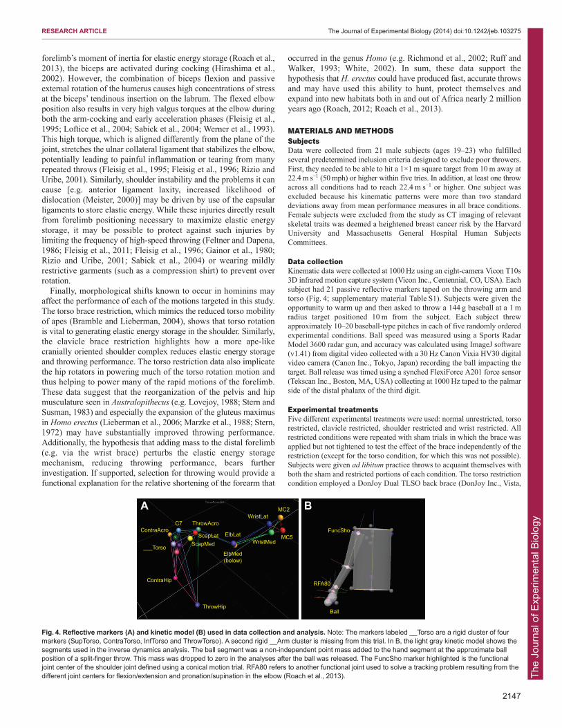
Data collectionKinematic data were collected at 1000 Hz using an eight-camera Vicon T10s3D infrared motion capture system (Vicon Inc., Centennial, CO, USA). Eachsubject had 21 passive reflective markers taped on the throwing arm andtorso (Fig. 4; supplementary material Table S1). Subjects were given theopportunity to warm up and then asked to throw a 144 g baseball at a 1 mradius target positioned 10 m from the subject. Each subject threwapproximately 10–20 baseball-type pitches in each of five randomly orderedexperimental conditions. Ball speed was measured using a Sports RadarModel 3600 radar gun, and accuracy was calculated using ImageJ software(v1.41) from digital video collected with a 30 Hz Canon Vixia HV30 digitalvideo camera (Canon Inc., Tokyo, Japan) recording the ball impacting thetarget. Ball release was timed using a synched FlexiForce A201 force sensor(Tekscan Inc., Boston, MA, USA) collecting at 1000 Hz taped to the palmarside of the distal phalanx of the third digit.
Experimental treatments Five different experimental treatments were used: normal unrestricted, torsorestricted, clavicle restricted, shoulder restricted and wrist restricted. Allrestricted conditions were repeated with sham trials in which the brace wasapplied but not tightened to test the effect of the brace independently of therestriction (except for the torso condition, for which this was not possible).Subjects were given ad libitum practice throws to acquaint themselves withboth the sham and restricted portions of each condition. The torso restrictioncondition employed a DonJoy Dual TLSO back brace (DonJoy Inc., Vista,
2147
RESEARCH ARTICLE The Journal of Experimental Biology (2014) doi:10.1242/jeb.103275
BA
ThrowHip
ContraHip
MC2
MC5WristMed
WristLat
ElbLat
ElbMed(below)
C7 ThrowAcroContraAcro
ScapMedScapLat
___Torso
FuncSho
Ball
RFA80
Fig. 4. Reflective markers (A) and kinetic model (B) used in data collection and analysis. Note: The markers labeled __Torso are a rigid cluster of fourmarkers (SupTorso, ContraTorso, InfTorso and ThrowTorso). A second rigid __Arm cluster is missing from this trial. In B, the light gray kinetic model shows thesegments used in the inverse dynamics analysis. The ball segment was a non-independent point mass added to the hand segment at the approximate ballposition of a split-finger throw. This mass was dropped to zero in the analyses after the ball was released. The FuncSho marker highlighted is the functionaljoint center of the shoulder joint defined using a conical motion trial. RFA80 refers to another functional joint used to solve a tracking problem resulting from thedifferent joint centers for flexion/extension and pronation/supination in the elbow (Roach et al., 2013).
The
Jour
nal o
f Exp
erim
enta
l Bio
logy
2148
CA, USA) to limit intervertebral motion from the T2/T3 vertebrae to thesacrum. This brace fastens rigid plastic plates to both the dorsal and ventralsides of the torso and fixes a steel bar against the clavicles to restrictrotational motion in the torso. The clavicle restriction condition used aDonJoy Clavicle Posture Support brace to hold the shoulder in a superiorlyrotated, ‘shrugged’ posture. This brace runs two straps over the shouldersand under the armpits, joining in the back. The subject was requested toshrug his shoulders as the brace was tightened, preventing relaxation. Forthe shoulder restriction condition, a DonJoy Shoulder Stabilizer brace wasused to limit external rotational ROM at the shoulder. This brace is a tight,elastic vest with a single, elastic half-sleeve on the throwing arm. To restrictexternal rotation of the humerus, the arm was internally rotated and a Velcrostrap was affixed from the ventral side of the vest to the dorsal side of thesleeve. The wrist restriction condition employed an Allsport Dynamics IMCWrist brace (Allsport Dynamics Inc., Nacogdoches, TX, USA) with the0 deg extension stop either employed (restricted) or absent (sham). Whenthe extension stop was screwed into place, the plastic sleeve contacts adorsal hand plate and prevents the wrist from hyperextending.
AnalysisMarker data were identified using Vicon Nexus software (v1.7.1) andexported to C-Motion Visual3D software (v4) for analysis. A Butterworthsecond-order low-pass filter with a cut-off frequency of 25 Hz was appliedafter a residual analysis of the data was conducted (Roach et al., 2013;Winter, 1991). Marker gaps of up to 100 frames were interpolated. JointEuler angles were calculated and inverse dynamics analyses performedusing mass distribution ratios from Dempster (Dempster, 1955). Jointangular velocities, moments and power were calculated using each joint’sinstantaneous axis of rotation. The Cardan sequence of rotations at each jointwas XYZ. Joint work was calculated in MATLAB (v7.11.0) using the trapzfunction.
StatisticsStatistical inquiry was limited to the arm-cocking and acceleration phasesas positive projectile velocity is generated only during these phases (Fig. 1).Individual subject means from a number of performance measures werecompared across experimental conditions using repeated-measures ANOVAor MANOVA when variances were not equal (Mauchley’s sphericity chi-square, P<0.05). Corrections for multiple comparisons were applied usingthe Holm–Bonferroni method with each experimental condition treated as afamily and a maximum alpha threshold of <0.05 (Holm, 1979). Post hocmatched pairs t-tests were used to determine which condition (sham and/orrestricted) accounted for the significance in multivariate tests. Because thetorso restriction condition did not have a sham, all reported statistics for thiscondition are matched pairs t-tests.
AcknowledgementsWe thank Mike Rainbow, Madhu Venkadesan, Leia Stirling, Richard Wrangham,Andrew Biewener, Susan Larson, Matt Vincent, George Karas, Randi Griffin, GulusEmre, Wes Gordon, Adanma Ekeledo and Amanda Lobell for their invaluable help;our subjects for generously giving their time; the Wyss Institute Motion Capture labfor the use of their equipment and facilities; and two anonymous reviewers, whosethoughtful comments vastly improved this paper.
Competing interestsThe authors declare no competing financial interests.
Author contributionsN.T.R. and D.E.L. designed the study, N.T.R. collected and analyzed the data,N.T.R. wrote the paper, and N.T.R. and D.E.L. edited the manuscript.
FundingThis research was funded by the National Science Foundation (BCS-0961943),the American School for Prehistoric Research, and the Harvard UniversityDepartment of Human Evolutionary Biology.
Supplementary materialSupplementary material available online athttp://jeb.biologists.org/lookup/suppl/doi:10.1242/jeb.103275/-/DC1
ReferencesAlexander, R. M. (1991). Optimum timing of muscle activation for simple models of
throwing. J. Theor. Biol. 150, 349-372. Altchek, D. W. and Dines, D. M. (1995). Shoulder injuries in the throwing athlete. J.
Am. Acad. Orthop. Surg. 3, 159-165.Anderson, F. C. and Pandy, M. G. (1993). Storage and utilization of elastic strain
energy during jumping. J. Biomech. 26, 1413-1427. Anz, A. W., Bushnell, B. D., Griffin, L. P., Noonan, T. J., Torry, M. R. and Hawkins,
R. J. (2010). Correlation of torque and elbow injury in professional baseball pitchers.Am. J. Sports Med. 38, 1368-1374.
Atwater, A. E. (1979). Biomechanics of overarm throwing movements and of throwinginjuries. Exerc. Sport Sci. Rev. 7, 43-86.
Badia, A. and Stennett, C. (2006). Sports-related injuries of the elbow. J. Hand Ther.19, 206-227.
Bell, D. G. (1993). The influence of air temperature on the EMG/force relationship ofthe quadriceps. Eur. J. Appl. Physiol. Occup. Physiol. 67, 256-260.
Ben Kibler, W. and Sciascia, A. (2004). Kinetic chain contributions to elbow functionand dysfunction in sports. Clin. Sports Med. 23, 545-552, viii.
Bramble, D. M. and Lieberman, D. E. (2004). Endurance running and the evolution ofHomo. Nature 432, 345-352.
Burkhart, S. S., Morgan, C. D. and Kibler, W. B. (2000). Shoulder injuries inoverhead athletes. The ‘dead arm’ revisited. Clin. Sports Med. 19, 125-158.
Burkhart, S. S., Morgan, C. D. and Kibler, W. B. (2003). The disabled throwingshoulder: spectrum of pathology Part I: pathoanatomy and biomechanics.Arthroscopy 19, 404-420.
Dempster, W. T. (1955). Space Requirements for the Seated Operator. Wright-Patterson Air Force Base, OH: Wright Air Development Center.
DiGiovine, N. M., Jobe, F. W., Pink, M. and Perry, J. (1992). An electromyographicanalysis of the upper extremity in pitching. J. Shoulder Elbow Surg. 1, 15-25.
Dillman, C. J., Fleisig, G. S. and Andrews, J. R. (1993). Biomechanics of pitchingwith emphasis upon shoulder kinematics. J. Orthop. Sports Phys. Ther. 18, 402-408.
Feltner, M. E. (1989). 3-dimensional interactions in a 2-segment kinetic chain. Part II.Application to the throwing arm in baseball pitching. Int. J. Sport Biomech. 5, 403-419.
Feltner, M. and Dapena, J. (1986). Dynamics of the shoulder and elbow joints of thethrowing arm during a baseball pitch. Int. J. Sport Biomech. 2, 235-259.
Finni, T., Komi, P. V. and Lepola, V. (2000). In vivo human triceps surae andquadriceps femoris muscle function in a squat jump and counter movement jump.Eur. J. Appl. Physiol. 83, 416-426.
Fleisig, G. S., Andrews, J. R., Dillman, C. J. and Escamilla, R. F. (1995). Kinetics ofbaseball pitching with implications about injury mechanisms. Am. J. Sports Med. 23,233-239.
Fleisig, G. S., Barrentine, S. W., Escamilla, R. F. and Andrews, J. R. (1996).Biomechanics of overhand throwing with implications for injuries. Sports Med. 21,421-437.
Fleisig, G. S., Andrews, J. R., Cutter, G. R., Weber, A., Loftice, J., McMichael, C.,Hassell, N. and Lyman, S. (2011). Risk of serious injury for young baseball pitchers:a 10-year prospective study. Am. J. Sports Med. 39, 253-257.
Gainor, B. J., Piotrowski, G., Puhl, J., Allen, W. C. and Hagen, R. (1980). The throw:biomechanics and acute injury. Am. J. Sports Med. 8, 114-118.
Gans, C. (1992). Electromyography. In Biomechanics – Structures and Systems: APractical Approach (ed. A. A. Biewener), pp. 123-147. Oxford: Oxford UniversityPress.
Goodall, J. (1964). Tool-using and aimed throwing in a community of free-livingchimpanzees. Nature 201, 1264-1266.
Goodall, J. (1986). The Chimpanzees of Gombe: Patterns of Behavior. Cambridge:Harvard University Press.
Gowan, I. D., Jobe, F. W., Tibone, J. E., Perry, J. and Moynes, D. R. (1987). Acomparative electromyographic analysis of the shoulder during pitching. Professionalversus amateur pitchers. Am. J. Sports Med. 15, 586-590.
Higgins, J. R. (1977). Human Movement: an Integrated Approach. St Louis, MO: Mosby.Hirashima, M., Kadota, H., Sakurai, S., Kudo, K. and Ohtsuki, T. (2002). Sequential
muscle activity and its functional role in the upper extremity and trunk duringoverarm throwing. J. Sports Sci. 20, 301-310.
Hirashima, M., Ohgane, K., Kudo, K., Hase, K. and Ohtsuki, T. (2003).Counteractive relationship between the interaction torque and muscle torque at thewrist is predestined in ball-throwing. J. Neurophysiol. 90, 1449-1463.
Hirashima, M., Kudo, K., Watarai, K. and Ohtsuki, T. (2007). Control of 3D limbdynamics in unconstrained overarm throws of different speeds performed by skilledbaseball players. J. Neurophysiol. 97, 680-691.
Hirashima, M., Yamane, K., Nakamura, Y. and Ohtsuki, T. (2008). Kinetic chain ofoverarm throwing in terms of joint rotations revealed by induced accelerationanalysis. J. Biomech. 41, 2874-2883.
Holm, S. (1979). A simple sequentially rejective multiple test procedure. Scand. J. Stat.6, 65-70.
Hong, D. A., Cheung, T. K. and Roberts, E. M. (2001). A three-dimensional, six-segment chain analysis of forceful overarm throwing. J. Electromyogr. Kinesiol. 11,95-112.
Hore, J., O’Brien, M. and Watts, S. (2005). Control of joint rotations in overarm throwsof different speeds made by dominant and nondominant arms. J. Neurophysiol. 94,3975-3986.
Jobe, C. M., Pink, M. M., Jobe, F. W. and Shaffer, B. (1996). Anterior shoulderinstability, impingement, and rotator cuff tear: theories and concepts. In OperativeTechniques in Upper Extremity Sports Injuries (ed. F. W. Jobe), pp. 164-176. StLouis, MO: Mosby.
RESEARCH ARTICLE The Journal of Experimental Biology (2014) doi:10.1242/jeb.103275
The
Jour
nal o
f Exp
erim
enta
l Bio
logy
Kohler, W. (1925). The Mentality of Apes. New York: Harcourt and Brace.Komi, P. V. and Bosco, C. (1978). Utilization of stored elastic energy in leg extensor
muscles by men and women. Med. Sci. Sports 10, 261-265.Komi, P. V., Salonen, M., Järvinen, M. and Kokko, O. (1987). In vivo registration of
Achilles tendon forces in man. I. Methodological development. Int. J. Sports Med. 8Suppl. 1, 3-8.
Larson, S. G. (1993). Functional morphology of the shoulder in primates. InPostcranial Adaptation in Non-human Primates (ed. D. L. Gebo), pp. 45-69. Chicago,IL: Northern Illinois University Press.
Larson, S. G. (2007). Evolutionary transformation of the hominin shoulder. Evol.Anthropol. 16, 172-187.
Lewis, J. L., Lew, W. D. and Schmidt, J. (1982). A note on the application andevaluation of the buckle transducer for the knee ligament force measurement. J.Biomech. Eng. 104, 125-128.
Lieberman, D. E., Raichlen, D. A., Pontzer, H., Bramble, D. M. and Cutright-Smith,E. (2006). The human gluteus maximus and its role in running. J. Exp. Biol. 209,2143-2155.
Loftice, J., Fleisig, G. S., Zheng, N. and Andrews, J. R. (2004). Biomechanics of theelbow in sports. Clin. Sports Med. 23, 519-530, vii-viii.
Lovejoy, C. O. (1988). Evolution of human walking. Sci. Am. 259, 118-125. Marzke, M. W., Longhill, J. M. and Rasmussen, S. A. (1988). Gluteus maximus
muscle function and the origin of hominid bipedality. Am. J. Phys. Anthropol. 77,519-528.
Matsuo, T., Escamilla, R. F., Fleisig, G. S., Barrentine, S. W. and Andrews, J. R.(2001). Comparison of kinematic and temporal patterns between different pitchvelocity groups. J. Appl. Biomech. 17, 1-13.
Meister, K. (2000). Injuries to the shoulder in the throwing athlete. Part one:Biomechanics/pathophysiology/classification of injury. Am. J. Sports Med. 28, 265-275.
Miyashita, K., Urabe, Y., Kobayashi, H., Yokoe, K., Koshida, S., Kawamura, M. andIda, K. (2008a). Relationship between maximum shoulder external rotation angleduring throwing and physical variables. J. Sports Sci. Med. 7, 47-53.
Miyashita, K., Urabe, Y., Kobayashi, H., Yokoe, K., Koshida, S., Kawamura, M. andIda, K. (2008b). The role of shoulder maximum external rotation during throwing forelbow injury prevention in baseball players. J. Sports Sci. Med. 7, 223-228.
Pappas, A. M., Zawacki, R. M. and Sullivan, T. J. (1985). Biomechanics of baseballpitching. A preliminary report. Am. J. Sports Med. 13, 216-222.
Pourcelot, P., Defontaine, M., Ravary, B., Lemâtre, M. and Crevier-Denoix, N.(2005). A non-invasive method of tendon force measurement. J. Biomech. 38, 2124-2129.
Putnam, C. A. (1993). Sequential motions of body segments in striking and throwingskills: descriptions and explanations. J. Biomech. 26 Suppl. 1, 125-135.
Richmond, B. G., Aiello, L. C. and Wood, B. A. (2002). Early hominin limbproportions. J. Hum. Evol. 43, 529-548.
Rizio, L. and Uribe, J. W. (2001). Overuse injuries of the upper extremity in baseball.Clin. Sports Med. 20, 453-468.
Roach, N. T. (2012). The Biomechanics and Evolution of High-Speed Throwing. PhDthesis, Department of Anthropology, Harvard University, Cambridge, MA, USA.
Roach, N. T., Venkadesan, M., Rainbow, M. J. and Lieberman, D. E. (2013). Elasticenergy storage in the shoulder and the evolution of high-speed throwing in Homo.Nature 498, 483-486.
Roberts, E. M. (1971). Cinematography in biomedical investigation. In Selected Topicsin Biomedical Invesitgation. Proceedings of the C.I.C. Symposium on Biomechanics(ed. J. M. Cooper), pp. 71-81. Chicago, IL: The Athletic Institute.
Ruff, C. and Walker, A. (1993). Body size and body shape. In The NariokotomeHomoe Erectus Skeleton (ed. A. Walker and R. E. Leakey), pp. 234-272.Cambridge, MA: Harvard University Press.
Sabick, M. B., Torry, M. R., Lawton, R. L. and Hawkins, R. J. (2004). Valgus torquein youth baseball pitchers: A biomechanical study. J. Shoulder Elbow Surg. 13, 349-355.
Stern, J. T., Jr (1972). Anatomical and functional specializations of the human gluteusmaximus. Am. J. Phys. Anthropol. 36, 315-339.
Stern, J. T., Jr and Susman, R. L. (1983). The locomotor anatomy of Australopithecusafarensis. Am. J. Phys. Anthropol. 60, 279-317.
Stodden, D. F., Fleisig, G. S., McLean, S. P., Lyman, S. L. and Andrews, J. R.(2001). Relationship of pelvis and upper torso kinematics to pitched baseballvelocity. J. Appl. Biomech. 17, 164-172.
Sugiyama, Y. and Koman, J. (1979). Tool-using and making behavior in wildchimpanzees at Bossou, Guinea. Primates 20, 513-524.
Werner, S. L., Fleisig, G. S., Dillman, C. J. and Andrews, J. R. (1993). Biomechanicsof the elbow during baseball pitching. J. Orthop. Sports Phys. Ther. 17, 274-278.
White, T. D. (2002). Earliest hominids. In The Primate Fossil Record (ed. W. C.Hartwig), pp. 407-417. Cambridge, UK: Cambridge University Press.
Wight, J., Richards, J. and Hall, S. (2004). Influence of pelvis rotation styles onbaseball pitching mechanics. Sports Biomech. 3, 67-84.
Wilk, K. E., Voight, M. L., Keirns, M. A., Gambetta, V., Andrews, J. R. and Dillman,C. J. (1993). Stretch-shortening drills for the upper extremities: theory and clinicalapplication. J. Orthop. Sports Phys. Ther. 17, 225-239.
Winter, D. A. (1991). The Biomechanics of Motor Control of Human Gait: Normal,Elderly and Pathological. Waterloo, ON: University of Waterloo Press.
2149
RESEARCH ARTICLE The Journal of Experimental Biology (2014) doi:10.1242/jeb.103275