Unwinding of synthetic replication and recombination substrates by Srs2
10
DNA Repair 11 (2012) 789–798 Contents lists available at SciVerse ScienceDirect DNA Repair jo u rn al hom epa ge: www.elsevier.com/locate/dnarepair Unwinding of synthetic replication and recombination substrates by Srs2 Victoria Marini a , Lumir Krejci a,b,c,∗ a Department of Biology, Masaryk University, Kamenice 5/A7, 625 00 Brno, Czech Republic b National Centre for Biomolecular Research, Masaryk University, Kamenice 5/A4, 625 00 Brno, Czech Republic c International Clinical Research Center, Center for Biomolecular and Cellular Engineering, St. Anne’s University Hospital Brno, Brno, Czech Republic a r t i c l e i n f o Article history: Received 17 February 2012 Received in revised form 14 May 2012 Accepted 17 May 2012 Available online 24 August 2012 Keywords: DNA repair Recombination Srs2 Replication Helicase a b s t r a c t The budding yeast Srs2 protein possesses 3 to 5 DNA helicase activity and channels untimely recombina- tion to post-replication repair by removing Rad51 from ssDNA. However, it also promotes recombination via a synthesis-dependent strand-annealing pathway (SDSA). Furthermore, at the replication fork, Srs2 is required for fork progression and prevents the instability of trinucleotide repeats. To better understand the multiple roles of the Srs2 helicase during these processes, we analysed the ability of Srs2 to bind and unwind various DNA substrates that mimic structures present during DNA replication and recombina- tion. While leading or lagging strands were efficiently unwound, the presence of ssDNA binding protein RPA presented an obstacle for Srs2 translocation. We also tested the preferred directionality of unwinding of various substrates and studied the effect of Rad51 and Mre11 proteins on Srs2 helicase activity. These biochemical results help us understand the possible role of Srs2 in the processing of stalled or blocked replication forks as a part of post-replication repair as well as homologous recombination (HR). © 2012 Elsevier B.V. All rights reserved. 1. Introduction The process of homologous recombination (HR) is widespread in nature, as it is present in organisms from bacteria to humans. It contributes to genetic diversity and is also essential for main- taining the integrity of the genome through facilitating the repair of DNA double-strand breaks (DSBs) and restarting stalled repli- cation forks. DSBs are caused by a vast number of endogenous and exogenous agents, including genotoxic chemicals and ionising radi- ation or the replication of a damaged template [1]. If not repaired, the DSBs can lead to aneuploidy, genetic aberrations or cell death [2–4]. DSB repair by HR is initiated by the resection of 5 ends on each side of the DSB, leading to the formation of 3 single-stranded tails covered by the single-strand binding protein RPA. Through the activity of Rad52, the RPA protein is replaced by Rad51, forming the presynaptic filament [5]. This filament, together with the help of Rad54 protein, engages in a homology search and strand invasion (D-loop formation) followed by DNA synthesis [5]. At this point, the recombination repair mechanism can proceed via two alternative subpathways. The first, known as the general DSB repair pathway (DSBR), is characterised by the capture of the second 3 tail and a consequent new round of DNA synthesis and ligation, resulting in the formation of a double Holliday junction (dHJ). This structure can ∗ Corresponding author at: Department of Biology, Masaryk University, Kamenice 5/A7, Brno 625 00, Czech Republic. Tel.: +420 549493767; fax: +420 549492556. E-mail address: [email protected] (L. Krejci). then be either dissolved by the action of the Sgs1/Top3 complex to produce noncrossover products or resolved to produce either noncrossover or crossover products [1]. In the alternative pathway (SDSA), the second 3 tail is annealed to the invading strand as it is released from the D-loop structure after its extension by DNA synthesis [1,6]. Several helicases have been implicated in the repair of stalled replication forks [1,7]. Of these helicases, Srs2 seems to play sev- eral important and sometimes opposing roles [7,8]. The SRS2 gene of Saccharomyces cerevisiae encodes a DNA helicase belonging to superfamily I and shows homology to the bacterial helicases Rep, PcrA and UvrD [9–11]. While the homologous helicase domain is located at the N-terminus, the C-terminal third of the protein bears no homology to the Escherichia coli Srs2 homologues and has been implicated in several protein interactions and regulation of mul- tiple processes [7]. The Srs2 protein has both ssDNA-dependent ATPase, DNA helicase activities in the 3 -5 direction, and is able to unwind various DNA substrates [12–14]. The SRS2 gene was first identified as a suppressor of radiation-sensitive mutations, i.e., the rad6 and rad18 mutants [9,15], and also as hyperecombination gene 5 [16,17]. Moreover, the deletion of SRS2 confers lethality or strong growth defects when combined with the deletion of sev- eral genes involved in DNA repair [7]. This synthetic lethality as well as the suppression of the UV sensitivity of post replication repair (PRR) mutants and the hyperecombination phenotype are all alleviated by the deletion of genes from the RAD52 epistasis group [16,18–21]. This suggests that Srs2 has a role in prevent- ing undesirable homologous recombination by channelling repair into a non-recombinogenic pathway [18]. The anti-recombinase 1568-7864/$ – see front matter © 2012 Elsevier B.V. All rights reserved. http://dx.doi.org/10.1016/j.dnarep.2012.05.007
-
Upload
independent -
Category
Documents
-
view
3 -
download
0
Transcript of Unwinding of synthetic replication and recombination substrates by Srs2

U
Va
b
c
a
ARRAA
KDRSRH
1
iItoceatd
etapR(rs(ct
5
1h
DNA Repair 11 (2012) 789– 798
Contents lists available at SciVerse ScienceDirect
DNA Repair
jo u rn al hom epa ge: www.elsev ier .com/ locate /dnarepai r
nwinding of synthetic replication and recombination substrates by Srs2
ictoria Marinia, Lumir Krejci a,b,c,∗
Department of Biology, Masaryk University, Kamenice 5/A7, 625 00 Brno, Czech RepublicNational Centre for Biomolecular Research, Masaryk University, Kamenice 5/A4, 625 00 Brno, Czech RepublicInternational Clinical Research Center, Center for Biomolecular and Cellular Engineering, St. Anne’s University Hospital Brno, Brno, Czech Republic
r t i c l e i n f o
rticle history:eceived 17 February 2012eceived in revised form 14 May 2012ccepted 17 May 2012vailable online 24 August 2012
a b s t r a c t
The budding yeast Srs2 protein possesses 3′ to 5′ DNA helicase activity and channels untimely recombina-tion to post-replication repair by removing Rad51 from ssDNA. However, it also promotes recombinationvia a synthesis-dependent strand-annealing pathway (SDSA). Furthermore, at the replication fork, Srs2 isrequired for fork progression and prevents the instability of trinucleotide repeats. To better understandthe multiple roles of the Srs2 helicase during these processes, we analysed the ability of Srs2 to bind and
eywords:NA repairecombinationrs2eplication
unwind various DNA substrates that mimic structures present during DNA replication and recombina-tion. While leading or lagging strands were efficiently unwound, the presence of ssDNA binding proteinRPA presented an obstacle for Srs2 translocation. We also tested the preferred directionality of unwindingof various substrates and studied the effect of Rad51 and Mre11 proteins on Srs2 helicase activity. Thesebiochemical results help us understand the possible role of Srs2 in the processing of stalled or blocked
of po
elicase replication forks as a part. Introduction
The process of homologous recombination (HR) is widespreadn nature, as it is present in organisms from bacteria to humans.t contributes to genetic diversity and is also essential for main-aining the integrity of the genome through facilitating the repairf DNA double-strand breaks (DSBs) and restarting stalled repli-ation forks. DSBs are caused by a vast number of endogenous andxogenous agents, including genotoxic chemicals and ionising radi-tion or the replication of a damaged template [1]. If not repaired,he DSBs can lead to aneuploidy, genetic aberrations or celleath [2–4].
DSB repair by HR is initiated by the resection of 5′ ends onach side of the DSB, leading to the formation of 3′ single-strandedails covered by the single-strand binding protein RPA. Through thectivity of Rad52, the RPA protein is replaced by Rad51, forming theresynaptic filament [5]. This filament, together with the help ofad54 protein, engages in a homology search and strand invasionD-loop formation) followed by DNA synthesis [5]. At this point, theecombination repair mechanism can proceed via two alternativeubpathways. The first, known as the general DSB repair pathway
DSBR), is characterised by the capture of the second 3′ tail and aonsequent new round of DNA synthesis and ligation, resulting inhe formation of a double Holliday junction (dHJ). This structure can∗ Corresponding author at: Department of Biology, Masaryk University, Kamenice/A7, Brno 625 00, Czech Republic. Tel.: +420 549493767; fax: +420 549492556.
E-mail address: [email protected] (L. Krejci).
568-7864/$ – see front matter © 2012 Elsevier B.V. All rights reserved.ttp://dx.doi.org/10.1016/j.dnarep.2012.05.007
st-replication repair as well as homologous recombination (HR).© 2012 Elsevier B.V. All rights reserved.
then be either dissolved by the action of the Sgs1/Top3 complexto produce noncrossover products or resolved to produce eithernoncrossover or crossover products [1]. In the alternative pathway(SDSA), the second 3′ tail is annealed to the invading strand as itis released from the D-loop structure after its extension by DNAsynthesis [1,6].
Several helicases have been implicated in the repair of stalledreplication forks [1,7]. Of these helicases, Srs2 seems to play sev-eral important and sometimes opposing roles [7,8]. The SRS2 geneof Saccharomyces cerevisiae encodes a DNA helicase belonging tosuperfamily I and shows homology to the bacterial helicases Rep,PcrA and UvrD [9–11]. While the homologous helicase domain islocated at the N-terminus, the C-terminal third of the protein bearsno homology to the Escherichia coli Srs2 homologues and has beenimplicated in several protein interactions and regulation of mul-tiple processes [7]. The Srs2 protein has both ssDNA-dependentATPase, DNA helicase activities in the 3′-5′ direction, and is ableto unwind various DNA substrates [12–14]. The SRS2 gene wasfirst identified as a suppressor of radiation-sensitive mutations, i.e.,the rad6 and rad18 mutants [9,15], and also as hyperecombinationgene 5 [16,17]. Moreover, the deletion of SRS2 confers lethality orstrong growth defects when combined with the deletion of sev-eral genes involved in DNA repair [7]. This synthetic lethality aswell as the suppression of the UV sensitivity of post replicationrepair (PRR) mutants and the hyperecombination phenotype are
all alleviated by the deletion of genes from the RAD52 epistasisgroup [16,18–21]. This suggests that Srs2 has a role in prevent-ing undesirable homologous recombination by channelling repairinto a non-recombinogenic pathway [18]. The anti-recombinase
7 A Rep
fR[ttahhtItaato
opcSctapDprraaHisarrolgtmthSSsaita
2
2
fccp0chbD
90 V. Marini, L. Krejci / DN
unction of Srs2 was confirmed in vitro by direct inhibition ofad51-mediated reactions including strand exchange and D-loop22,23]. Additional experiments then demonstrated ability of Srs2o dismantle the Rad51 presynaptic filament [22,23] and influencehe formation of Rad51 foci in vivo [24]. This anti-recombinationctivity requires translocase activity that is fully dependent on ATPydrolysis [25]. Moreover, it was found that Srs2 stimulates ATPydrolysis within the Rad51 filament through a physical interac-ion with Rad51, which leads to the release of Rad51 from DNA [26].n addition, an Srs2 mutant that has lost its ability to interact withhe Rad51 protein is unable to disrupt the Rad51 nucleoprotein fil-ment [26,27]. In turn, Rad51 was shown to stimulate Srs2 helicasectivity [14]. Srs2 was also shown to antagonise the ability of Rad52o mediate Rad51 filament formation by directly competing for theverlapping interaction site [28].
Srs2 was also shown to be necessary for the efficient repairf double-strand breaks through homologous recombination byromoting the SDSA pathway, thus preventing the formation ofrossover products during mitosis [2,29]. The phosphorylation ofrs2 was recently suggested to play an important role in this pro-ess [30]. One scenario suggested that Srs2 can directly dissolvehe D-loop structure; however, Mph1 has been shown to be moredept at doing so [14,31,32]. In an alternative scenario, Srs2 mightrevent second end capture or collaborate with nucleases to cleave-loops, DNA tails or other intermediates after re-annealing a dis-laced extended strand. Furthermore, Srs2 localises not only toepair centres but also to replication forks [24,33]. This recruitmentelies on an interaction with sumoylated PCNA through SUMO-nd PCNA-interaction motifs [24,33–36]. However, the role of Srs2t the replication fork does not seem to be the prevention ofR only. Additionally, the replication checkpoint was shown to
nhibit the recruitment of the Rad52 mediator, which is respon-ible for loading the Rad51 recombinase. The Rad51 foci thatre formed in the absence of Rad52 do not represent functionalecombination filaments [24]. These data indicate an additionalole for Srs2 at the replication fork. Indeed, the overproductionf either Srs2 or its helicase-dead mutant results in a syntheticethal phenotype in combination with many replication-associatedenes [37]. Furthermore, Srs2 was found to be important forhe prevention of fragility and instability of trinucleotide repeats,
ost likely by facilitating replication through the hairpin struc-ures, as it was shown to preferentially unwind CTG and CGGairpins [38–41]. To better understand the multiple roles of thers2 helicase during these processes, we analysed the ability ofrs2 to bind and unwind various DNA substrates that mimic thetructures present during DNA replication and recombination. Inddition, we tested the preferred directionality of the unwind-ng of various replication fork-mimicking substrates and studiedhe effect of the RPA, Rad51, and Mre11 proteins on Srs2 helicasectivity.
. Materials and methods
.1. Purification of wt and �C forms of Srs2
For expression of wt Srs2 and Srs2�C, corresponding to theragment 1-898 and containing a His9 affinity tag [26,33], BL21ells carrying the plasmids (pET11d) were grown in 2xTY mediaontaining 0.1 mg/ml ampicillin at 37 ◦C to an A600 of 1. The tem-erature was shifted to 16 ◦C and the cultures were induced with.1 mM isopropyl-1-thio-�-d-galactopyranoside overnight. Purifi-
ation was performed as described previously [27]. Cells werearvested by centrifugation and the pellet resuspended in lysisuffer C (50 mM Tris pH 7.5, 10% sucrose, 10 mM EDTA, 1 mMTT, 0.01% NP40, protease inhibitors: 2 �g/ml aprotinin, 5 �g/mlair 11 (2012) 789– 798
benzamidine hydrochloride, 10 �M chymostatin, 10 �M leupeptinhemisulphate, 1 �M pepstatin A) containing 600 mM KCl and lysedby sonication. All consequent purification steps were performed at4 ◦C. The clarified lysate was subjected to precipitation with ammo-nium sulphate (208 mg/ml). The precipitate was diluted with bufferK (20 mM K2HPO4 pH 7.5, 10% glycerol, 0.5 M EDTA) to a conductiv-ity corresponding to buffer K containing 150 mM KCl. The solutionwas then loaded onto a 7 ml Q Sepharose column and the result-ing flow was immediately loaded onto a 7 ml SP sepharose column(GE Healthcare Life Sciences). The SP column was developed with a70 ml gradient of 150–1000 mM KCl. The fractions containing Srs2were mixed with Ni-NTA agarose (Qiagen) overnight. The proteinswere eluted in a step-gradient using 50, 150, 300 and 500 mMimidazole in buffer K. Srs2 was mainly eluted at 150–300 mMimidazole. The fractions containing Srs2 were loaded on a 1 mlhydroxyapatite column (BioRad). Elution was done with a 10 mllinear gradient from 50 to 700 mM KH2PO4 in buffer K. Srs2 elutedat about 350 mM KH2PO4. Peak fractions were pooled, loaded onto a1 ml Mono S column (GE Healthcare Life Sciences), and eluted with a10 ml gradient from 75 to 1000 mM KCl in buffer K. Individual frac-tions were concentrated on Centricon 30,000 MWCO (Millipore)and the protein stored at −80 ◦C.
2.2. Synthetic DNA substrates
Synthetic oligonucleotides were purchased from VBC Biotech(Austria). The sequences and the structures are shown in Table 1.Some of oligonucleotides were end modified by a fluorescent dye(fluorescein or CY5). All substrates were prepared as describedin Matulova et al. [42]. In brief, the equimolar amounts of thecorresponding oligonucleotides were mixed in hybridizing buffer(50 mM Tris pH 7.5, 100 mM NaCl, 10 mM MgCl2), heated at 85 ◦Cfor 3 min and cooled slowly to room temperature to anneal. Thesubstrates were then purified by HPLC using a 1 ml Mono Q column(GE Healthcare Life Sciences) and eluted in 20 ml gradient of 10 mMTris buffer containing up to 1 M NaCl. Purity of each substrate wasalso checked on non-denaturing PAGE. Fractions were then con-centrated on Vivaspin Concentrator 5000 MWCO and washed withbuffer W (25 mM Tris pH 7.5, 3 mM MgCl2). Concentrations weredetermined using absorbance at 260 nm and corresponding molarextinction coefficients, taking into account the extinction coef-ficients of the used fluorophores (εfluorescein,260 = 13,700 l/mol cm,εCy5,260 = 10,000 l/mol cm).
2.3. DNA binding assay
Fluorescently labelled DNA substrate (3 nM) was incubated at30 ◦C with the indicated quantities of Srs2 in buffer E (30 mM TrispH 7.5, 1 mM DTT, 0.1 mg/ml BSA and 100 mM KCl) for 10 min. Thereaction mixtures were put on ice followed by addition of load-ing buffer (60% glycerol, 10 mM Tris pH 7.5, 60 mM EDTA, addedin a sample:buffer ratio of 5:1) and the samples were resolvedby electrophoresis on a 7% native PAGE in 0.5 × TBE buffer. Gelswere scanned using FLA-9000 Starion (Fujifilm) and quantified byMultiGauge software (Fujifilm).
2.4. Helicase assays
Fluorescently labelled DNA substrate (3 nM) was incubated at30 ◦C with the indicated quantities of Srs2 in buffer H (30 mMTris pH 7.5, 1 mM DTT, 0.1 mg/ml BSA, 100 mM KCl, 20 mM crea-tine phosphate, 20 �g/ml creatine kinase, 2.4 mM MgCl2, and 2 mM
ATP) for 10 min. The reaction was stopped by addition of 2% SDSand 0.5 mg/ml proteinase K and incubated at 37 ◦C for 3 min. Afteradding loading buffer to the samples the reaction products wereresolved by electrophoresis on a 12% native PAGE in TBE buffer. Gels
V. Marini, L. Krejci / DNA Repair 11 (2012) 789– 798 791
Table 1DNA substrates used in this study.
T tes.E eacho scein o
wMR2was5
2
3b
he list of sequences of oligonucleotides used to make individual synthetic substraach DNA substrate is made from the oligonucleotides indicated by the number onligonucleotide. The asterisks denote end modification by a fluorescent dye (fluore
ere scanned using FLA-9000 Starion (Fujifilm) and quantified byultiGauge software (Fujifilm). For the assays in the presence of
PA, the DNA substrate was preincubated with 190 nM of RPA for min at 25 ◦C. For testing the effect of Rad51, the DNA substrateas preincubated with Rad51 (30, 50, 100, and 200 nM) for 5 min
t 30 ◦C. In the case of the assay in the presence of Mre11, the DNAubstrate was preincubated with Mre11 (10, 25, 50, and 250 nM) for
min on ice, before starting the reaction by the addition of wtSrs2.
.5. Branch migration assays
Fluorescently labelled DNA substrate (6 nM) was incubated at0 ◦C with the indicated quantities of Srs2�C, Rad5 or Mph1 inuffer D (25 mM Tris pH 7.5, 1 mM DTT, 0.1 mg/ml BSA, 50 mM KCl,
representative schematic. The number is positioned at the 5′ end of its respectiver Cy5).
7.5 mM creatine phosphate, 11.25 �g/ml creatine kinase, 2.5 mMMgCl2 and 2.5 mM ATP) for 15 min. The reaction was stopped by anaddition of SDS to 0.2% and proteinase K to 0.5 mg/ml followed byincubation at 30 ◦C for 3 min. For the reactions in the presence ofRPA, DNA was incubated with RPA for 5 min on ice before additionof the helicase. After the addition of loading buffer, the reactionproducts were resolved by electrophoresis on a 12% native PAGE inTBE buffer. Gels were scanned using the image scanner FLA-9000Starion (Fujifilm) and quantified by MultiGauge software (Fujifilm).
2.6. ATPase assay
Srs2�C (100 nM) was incubated with ssDNA (15 �Mnucleotides), 1 mM ATP and 4 nCi/�L of [�-32P] ATP at 30 ◦C
7 A Repair 11 (2012) 789– 798
i0yRTawalptw
2
tS30mapatpAt3w
3
3
Htogwtsht(aeFscha
oiiFbosotor
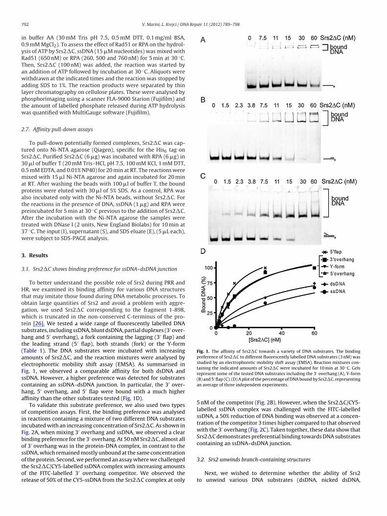
Fig. 1. The affinity of Srs2�C towards a variety of DNA substrates. The bindingpreference of Srs2�C to different fluorescently labelled DNA substrates (3 nM) wasstudied by an electrophoretic mobility shift assay (EMSA). Reaction mixtures con-taining the indicated amounts of Srs2�C were incubated for 10 min at 30 ◦C. Gels
′
92 V. Marini, L. Krejci / DN
n buffer AA (30 mM Tris pH 7.5, 0.5 mM DTT, 0.1 mg/ml BSA,.9 mM MgCl2). To assess the effect of Rad51 or RPA on the hydrol-sis of ATP by Srs2�C, ssDNA (15 �M nucleotides) was mixed withad51 (650 nM) or RPA (260, 500 and 760 nM) for 5 min at 30 ◦C.hen, Srs2�C (100 nM) was added, the reaction was started byn addition of ATP followed by incubation at 30 ◦C. Aliquots wereithdrawn at the indicated times and the reaction was stopped by
dding SDS to 1%. The reaction products were separated by thinayer chromatography on cellulose plates. These were analysed byhosphorimaging using a scanner FLA-9000 Starion (Fujifilm) andhe amount of labelled phosphate released during ATP hydrolysisas quantified with MultiGauge software (Fujifilm).
.7. Affinity pull-down assays
To pull-down potentially formed complexes, Srs2�C was cap-ured onto Ni-NTA agarose (Qiagen), specific for the His6 tag onrs2�C. Purified Srs2�C (6 �g) was incubated with RPA (6 �g) in0 �l of buffer T (20 mM Tris–HCl, pH 7.5, 100 mM KCl, 1 mM DTT,.5 mM EDTA, and 0.01% NP40) for 20 min at RT. The reactions wereixed with 15 �l Ni-NTA agarose and again incubated for 20 min
t RT. After washing the beads with 100 �l of buffer T, the boundroteins were eluted with 30 �l of 5% SDS. As a control, RPA waslso incubated only with the Ni-NTA beads, without Srs2�C. Forhe reactions in the presence of DNA, ssDNA (1 �g) and RPA werereincubated for 5 min at 30 ◦C previous to the addition of Srs2�C.fter the incubation with the Ni-NTA agarose the samples were
reated with DNase I (2 units, New England Biolabs) for 10 min at7 ◦C. The input (I), supernatant (S), and SDS eluate (E), (5 �L each),ere subject to SDS-PAGE analysis.
. Results
.1. Srs2�C shows binding preference for ssDNA–dsDNA junction
To better understand the possible role of Srs2 during PRR andR, we examined its binding affinity for various DNA structures
hat may imitate those found during DNA metabolic processes. Tobtain large quantities of Srs2 and avoid a problem with aggre-ation, we used Srs2�C corresponding to the fragment 1-898,hich is truncated in the non-conserved C-terminus of the pro-
ein [26]. We tested a wide range of fluorescently labelled DNAubstrates, including ssDNA, blunt dsDNA, partial duplexes (3′ over-ang and 5′ overhang), a fork containing the lagging (3′ flap) andhe leading strand (5′ flap), both strands (fork) or the Y-formTable 1). The DNA substrates were incubated with increasingmounts of Srs2�C, and the reaction mixtures were analysed bylectrophoretic mobility shift assay (EMSA). As summarised inig. 1, we observed a comparable affinity for both dsDNA andsDNA. However, a higher preference was detected for substratesontaining an ssDNA–dsDNA junction. In particular, the 3′ over-ang, 5′ overhang, and 5′ flap were bound with a much higherffinity than the other substrates tested (Fig. 1D).
To validate this substrate preference, we also used two typesf competition assays. First, the binding preference was analysedn reactions containing a mixture of two different DNA substratesncubated with an increasing concentration of Srs2�C. As shown inig. 2A, when mixing 3′ overhang and ssDNA, we observed a clearinding preference for the 3′ overhang. At 50 nM Srs2�C, almost allf 3′ overhang was in the protein-DNA complex, in contrast to thesDNA, which remained mostly unbound at the same concentration
f the protein. Second, we performed an assay where we challengedhe Srs2�C/CY5-labelled ssDNA complex with increasing amountsf the FITC-labelled 3′ overhang competitor. We observed theelease of 50% of the CY5-ssDNA from the Srs2�C complex at onlyrepresent some of the tested DNA substrates including the 3 overhang (A), Y-form(B) and 5′ flap (C). (D) A plot of the percentage of DNA bound by Srs2�C, representingan average of three independent experiments.
5 nM of the competitor (Fig. 2B). However, when the Srs2�C/CY5-labelled ssDNA complex was challenged with the FITC-labelledssDNA, a 50% reduction of DNA binding was observed at a concen-tration of the competitor 3 times higher compared to that observedwith the 3′ overhang (Fig. 2C). Taken together, these data show thatSrs2�C demonstrates preferential binding towards DNA substratescontaining an ssDNA–dsDNA junction.
3.2. Srs2 unwinds branch-containing structures
Next, we wished to determine whether the ability of Srs2to unwind various DNA substrates (dsDNA, nicked dsDNA,

V. Marini, L. Krejci / DNA Rep
Fig. 2. DNA binding preference using competition assay. (A) Reaction mixtures con-taining FITC-labelled ssDNA (3 nM) and 3′ overhang (3 nM) were incubated togetherwith the indicated amounts of Srs2�C for 10 min at 30 ◦C and analysed by EMSA.Alternatively, the reaction mixtures containing CY5-labelled ssDNA and 150 nMSrs2�C were challenged with increasing amounts of FITC-3′ overhang (B) or FITC-ssDNA (C) followed by incubation for 10 min at 30 ◦C and then analysed. CY5-labelledsubstrate is shown in red and FITC-labelled substrates in green.
3joa1esoYbeiorcs
effect of the presence of the Rad51 and Mre11 proteins. Inter-estingly, neither Rad51 nor Mre11 had any effect on the ability
′ overhang, 5′ overhang, 3′ flap, fork, Y-form, and Hollidayunctions) correlates with the observed DNA binding affinities. Flu-rescently labelled DNA substrates were incubated with increasingmounts of Srs2�C (Fig. 3A) or full-length Srs2 (Supplementary Fig.) followed by the resolution of the products by native PAGE. Asxpected, the highest efficiency of unwinding among all of the sub-trates was observed for the 3′ overhang (Fig. 3A). The second groupf substrates with comparable unwinding included the fork, 3′ flap,-form, and 5′ overhang. The blunt end duplex was also unwound,ut only at much higher concentrations of Srs2�C (Fig. 3A). Inter-stingly, the introduction of a nick in the dsDNA substrate resultedn a significant increase in the unwinding, almost reaching the levelf other ss/dsDNA junction-containing structures (Fig. 3A). Theseesults indicate that Srs2 is able to unwind a broad range of branch-
′
ontaining DNA substrates and is most efficient with a 3 overhangubstrate.air 11 (2012) 789– 798 793
3.3. Unwinding of forks with lagging and/or leading strand bySrs2�C
The above data show that Srs2 unwinds substrates mimickingboth the lagging and leading forks. Together with the presence ofSrs2 at replication forks, this prompted us to test the directionalityof Srs2 during the unwinding of structures resembling replicationforks. To address this, we used a DNA substrate containing twofluorescent dyes (3′ flap with FITC and CY5 labels at oligo1 and 4respectively, Table 1, Fig. 3B and C). The incubation of increasingamounts of Srs2�C with such a substrate led to the generationof all possible intermediates with no obvious preferred initiationof unwinding (Fig. 3B). Among the products, we noticed that 3′
overhang is not accumulated but further unwound into ssDNA,reflecting the highest unwinding affinity for this substrate (Fig. 3A).We also performed a time-dependence experiment at a low Srs2�Cconcentration to identify the initial products of unwinding (Fig. 3C).In this experiment, in only 1 min, we observed the first products offlap unwinding, 3′ overhang and Y-form DNA (Fig. 3C). Both ssDNAproducts were observed simultaneously 2.5 min after the start ofthe reaction (Fig. 3C). Moreover, testing with the dual-labelled forksubstrate (fork with FITC and CY5 labels at oligo1 and 3, respec-tively, Table 1) also showed the concomitant appearance of all of theintermediates at various protein concentrations (SupplementaryFig. 2), indicating that Srs2�C is able to unwind both the leadingand lagging strand as well as the parental duplex without preferreddirectionality.
3.4. RPA inhibits the unwinding of Srs2
The ability of Srs2 to unwind an obstructing DNA strand onboth the leading and lagging strand template led us to hypothesisethat the presence of a single-strand DNA binding protein (such asRPA) or Srs2-interacting partners might influence the unwinding.Therefore, we performed a standard helicase assay with Srs2�Con several DNA substrates pre-coated with RPA. In the presence ofRPA, we observed a significant inhibition of the unwinding of the3′ overhang and 3′ flap substrates (Fig. 4A and B). Only the unwind-ing of the fork DNA substrate showed a minimal response to thepresence of RPA, reflecting the absence of ssDNA in the structure(Fig. 4C). Comparable results were also obtained with full-lengthSrs2 (Supplementary Fig. 1).
Furthermore, the inhibitory effect of RPA on Srs2 was also mon-itored using its ssDNA-dependent ATPase activity. The ssDNA waspre-coated with RPA followed by incubation with Srs2�C andradiolabelled ATP. As shown in supplementary Fig. 3A, subsatu-rating ratios of RPA versus ssDNA (1 RPA molecule: 2 binding sites)resulted in marked inhibition of the ATPase activity. An additionalincrease in the RPA concentration resulted in a 50% reduction ofSrs2�C ATPase activity, indicating that RPA represents an obstruc-tion to Srs2 translocation. No further inhibition was observed whenan excess of RPA in relation to ssDNA was used (SupplementaryFig. 3A). To test whether this inhibition is due to direct physicalinteraction between Srs2�C and RPA, we used a pull-down assay.Purified Srs2�C was incubated with RPA and then immobilised onNi-NTA beads. However, under these conditions, we were not ableto detect a direct interaction between Srs2�C and RPA. In addition,pre-incubation of RPA with ssDNA did not result in a detectableinteraction between these two proteins (Supplementary Fig. 3B). Todetermine whether other DNA- or Srs2-interacting proteins couldalso present an obstacle to or affect Srs2 activity, we tested the
of wtSrs2 to unwind DNA substrates or Srs2�C to hydrolyse ATP(Supplementary Fig. 4), indicating that only RPA binding to ssDNA is

794 V. Marini, L. Krejci / DNA Repair 11 (2012) 789– 798
Fig. 3. Srs2 is able to unwind more strands from fork-like structures. (A) A plot of the unwinding of the different substrates relative to the increasing concentration ofSrs2�C. The data represent the average from at least three experiments, and curves were obtained from nonlinear regression using GraphPad Prism software. (B) The abilityof Srs2�C to preferentially unwind strands from fork-like structures was tested by mixing 3′ flap (3 nM) labelled with both CY5 (in red) and FITC (in green) in buffer H withthe indicated amounts of Srs2 and incubating for 10 min at 30 ◦C. The reactions were resolved using native polyacrylamide electrophoresis and analysed. The plot indicatest sing SS en) ina ing FIT
aa
3
paSlt
he ratio of the different DNA intermediates (containing FITC) as a function of increars2�C was incubated with 3 nM 3′ flap labelled with CY5 (in red) and FITC (in grend analysed. The plot shows the ratio of the different DNA intermediates (contain
ble to reduce both the helicase and the ssDNA-dependent ATPasectivities of Srs2�C.
.5. Srs2 helicase activity on recombination intermediates
Genetic studies indicate that Srs2 promotes SDSA [29,43]. Thisrompted us to test its ability to unwind recombination intermedi-
tes, such as HJs or D-loop structures. Increasing concentrations ofrs2 were only partially able to unwind HJ-like structures (Fig. 5A,anes 1–5). In contrast, the introduction of a nick into the HJ struc-ure resulted in almost complete unwinding at 1.5 �M of Srs2,rs2 concentration. (C) The time-dependence of the helicase activity in which 15 nM buffer H at 30 ◦C. At the indicated times, the individual aliquots were withdrawnC dye) over time.
indicating that the presence of a discontinuity in duplex DNA sig-nificantly increases the unwinding by Srs2 (Fig. 5A, lanes 6–10).Similarly, the synthetic D-loop structure also appeared to be avery good substrate for Srs2 helicase activity, showing preferen-tial unwinding of the short oligonucleotide on the invading strandor the invading strand itself (Supplementary Fig. 5).
Other DNA repair translocases have been shown to possess
an intrinsic branch migration activity [44,45]. To test whetherSrs2 has such an ability, we designed a fluorescently labelledmobile fork (Table 1) and tested the ability of Srs2 to branchmigrate this substrate. Incubation of Srs2 with such DNA structures
V. Marini, L. Krejci / DNA Repair 11 (2012) 789– 798 795
Fig. 4. Effect of RPA on the helicase activity of Srs2�C. The assay for helicase activity was performed as described in the experimental procedures, except the FITC-labelledDNA substrate was preincubated for 2 min at 25 ◦C either in the absence (left) or presence (right) of RPA (190 nM). The DNA substrates analysed represent 3′ overhang (A),3′ flap (B) and fork (C) structures. The plots represent the formation of ssDNA.
Fig. 5. Helicase versus branch migration activity on rigid and mobile DNA structures. (A) Helicase activity of Srs2�C on HJ with or without a nick. Reaction mixtures containingFITC-labelled DNA (3 nM) were incubated with the indicated amount of Srs2�C for 10 min at 30 ◦C and then analysed. (B) The ability of Srs2 to branch migrate a fork substratecontaining partial homology. The substrate (6 nM, labelled with CY5 (red) and FITC (green)) was incubated for 15 min at 30 ◦C with the indicated amounts of Srs2�C (lanes1–8), Rad5 (lanes 9–10) or Mph1 (lanes 11–14). For samples in lane 5–8, 10 and 13–14, the DNA substrate was preincubated with 100 nM RPA for 5 min at 4 ◦C.

7 A Rep
pbtntbrUpdhatrln
4
raapimtwfDcatasSaSc[
drswwttosrtdefldbib
[epli
96 V. Marini, L. Krejci / DN
roduced both longer and shorter duplexes, the two products ofranch migration (Fig. 5B, lanes 1–4). Similarly, Srs2 was also ableo generate the expected reaction products from mobile HJ andHJ (Supplementary Fig. 6). However, it could also be possible thathese products are the results of Srs2 helicase activity followedy the reannealing of unwound oligonucleotides. To prevent sucheannealing, we performed the reaction in the presence of RPA.nder these conditions, we did not observe any short duplex DNAroducts; instead, 5′ overhang and short single-strand DNA wereetected, indicating that these species represent the products of aelicase activity (Fig. 5B, lanes 5–8). Indeed, in the case of the Rad5nd Mph1 proteins, a previously described branch migration pro-eins [44,45], the presence of RPA did not have any effect on theesulting reaction products (Fig. 5B, compare lanes 9 and 10 andanes 11–14). These data indicate that Srs2 possesses a helicase buto branch migration activity.
. Discussion
Previous studies have suggested that Srs2 functions by acting ateplication forks as well as at recombination intermediates formeds a result of replication stalling or DSB repair (reviewed in Marinind Krejci [7]). In this study, we wanted to further corroborate onrevious studies [13,14] and characterise in details the DNA bind-
ng and helicase activities of the Srs2 protein to shed light on itsultiple roles during DNA replication/recombination. Initially, we
ested the ability of Srs2�C to bind various DNA substrates thatould imitate those naturally occurring in vivo, including different
ork structures, 3′ or 5′ overhangs, and single- and double-strandNA. The highest binding preference was observed for structuresontaining ss/ds DNA junctions (Fig. 1). This specificity is in angreement with the competition assays demonstrating that a struc-ure containing a ss/dsDNA junction is bound with much higherffinity compared to ssDNA and is able efficiently outcompetesDNA from the protein–DNA complex (Fig. 2). We conclude thatrs2�C has a specific preference for an ss/dsDNA junction, without
preference for structures with either a 3′ or 5′ tail. The bacterialrs2 homologue, UvrD, shows similar affinities. However, it has alear preference for binding substrates containing a 3′ extension46].
An examination of whether the binding mode provides a foun-ation for the ability of Srs2�C to unwind various DNA substratesevealed similar unwinding efficiencies for the majority of sub-trates, including the fork, 3′ flap, 5′ flap, and Y-form. Interestingly,ithout exhibiting any specific binding preference, the 3′ overhangas unwound the most efficiently. The potential structural feature
hat causes the preferred unwinding of the 3′ overhang remainso be determined. It cannot be explained simply by the generationf competing intermediates in the case of more complicated DNAubstrates, as similar substrates (5′ overhang and Y-form) showeduced unwinding. It is tempting to speculate that the interac-ion of Srs2�C with single-strand DNA at the 3′ end of DNA couldirectly promote unwinding in the 3′ to 5′ direction [13]. How-ver, a similar unwinding preference was not observed for the 3′
ap. However, despite the comparable affinity of Srs2�C towardssDNA, it unwinds the blunt duplex DNA poorly (Fig. 3). This maye related to the fact that the ability of Srs2 to initiate unwind-
ng from discontinuities within the DNA was also observed for itsacterial homologue UvrD [47,48].
Srs2 was shown to associate with replication forks in vivo24,33,34], and our data demonstrate that it can directly target and
fficiently unwind various fork substrates. Therefore, we tested thereferred polarity of fork unwinding dictated by the presence of aeading or lagging strand to help understand its biological role dur-ng DNA replication. In experiments using dual labelled substrates,
air 11 (2012) 789– 798
we did not observe any single primary product, but rather foundthe simultaneous generation of the individual products that arethe result of the unwinding of the 3′ flap as well as fork substrates(Fig. 3B and Supplementary Fig. 2). Our time-course experimentrevealed the same unwinding rates for the individual interme-diates (Fig. 3C). Srs2�C thus unwinds both leading and laggingstrands as well as the parental duplex without any obvious pref-erence, suggesting that it could act on diverse stalled replicationforks to promote appropriate repair. This could help to explain theability of Srs2 to unwind various hairpin-forming structures andthe observed role of Srs2 in preventing fragility and instability inCAG/CTG repeats [38,41,49]. Furthermore, the helicase activity ofSrs2 was shown to be necessary for replication to proceed past thehairpin [38].
Srs2�C appears to bind fork structures at the branch point andtranslocate in all three directions with no preference while keepingto the 3′ to 5′ direction of translocation. This is different from otherhomologous helicases, including Rep, PriA and UvrD, which showpreferred unwinding of the lagging strand DNA within the 3′ flapstructure [47,50]. The specificity could be dictated by another DNA-binding domain outside the truncated version of Srs2 used in thisstudy. However, we did not observe any difference in unwindingusing full-length Srs2 (Supplementary Fig. 1).
The ability of Srs2 to dissociate fork structures in all threedirections indicates that its initial orientation of binding is notdetermined by an asymmetry of the junction. Thus additional fac-tors might be needed in vivo to ensure that Srs2 binds in a waythat is productive for fork unwinding. Therefore, we tested aneffect of the single-strand binding protein RPA on Srs2 helicaseactivity. While the presence of a lagging or leading strand did nothave a significant effect on unwinding, coating ssDNA regions withRPA resulted in a suppression of Srs2 helicase activity (Fig. 4 andSupplementary Fig. 1). Correspondingly, ATPase activity was alsoinhibited by the presence of saturating amounts of RPA. This is incontrast to previously published data in which RPA was able tostimulate the unwinding of long DNA substrates by Srs2 [13]. How-ever, in the case of large DNA substrates, this could reflect a roleof RPA to sequester unwound ssDNA, prevent re-annealing and/orsuppress the formation of secondary structures of DNA. Our datashow that RPA presents an obstacle for the translocation of Srs2along ssDNA, indicating that it could inhibit unscheduled unwind-ing of the replication fork in vivo. This could be explained by thehigher affinity of RPA towards ssDNA (Supplementary Fig. 7). Inaddition, Srs2 cannot alleviate this inhibition via a direct interactionwith RPA, as we were not able to detect it under our experi-mental conditions. Even the pre-incubation of RPA with ssDNA,which was shown to be necessary for the interaction betweenRPA and Rad52 [51], did not result in any detectable interactionbetween Srs2�C and RPA (Supplementary Fig. 3B). In contrast,testing Srs2-interacting proteins, including Rad51 and Mre11, didnot show any inhibitory effect on Srs2 helicase and ATPase activ-ities (Supplementary Fig. 4). However, in previous work, Rad51was shown to stimulate the Srs2-mediated unwinding of longerDNA substrates [14], which might again correspond with the abil-ity of the Rad51 filament to either remove secondary structuresor sequester unwound ssDNA. This explanation is also in agree-ment with the ability of Srs2 to activate the ATPase activity ofRad51 followed by the destabilisation of Rad51 association withDNA [26] and is not accompanied by increased Srs2 ATPase activ-ity (Supplementary Fig. 4). Our data would suggest that, due to thedirect protein interaction between Srs2 and Rad51 or Mre11, bothproteins do not present an obstacle for Srs2 translocation and could
even serve for Srs2 targeting to the recombination intermediates.Our data point to the ability of Srs2 to act directly on replica-tion forks. Indeed, a recent study indicated that overexpressionof Srs2 primarily affects the progression of DNA replication, as it
A Rep
cmsmlgt
nTbpioDrcsspDatNompma
opnhBhotspweS
C
A
RSdPCba(
A
fj
[
[
[
[
[
[
[
[
[
[
[
[
[
[
[
[
[
[
V. Marini, L. Krejci / DN
auses the activation of the replication checkpoint [37]. Further-ore, the deletion of many key replication components leads to
ynthetic lethality when Srs2 is overexpressed. The helicase-deadutants induce a delay in S-phase progression and show enhanced
ethality in combination with genes involved in DNA repair, sug-esting that the helicase activity impedes the proper processing ofhe appropriate DNA repair pathway [37].
Srs2 has been shown not only to block inappropriate recombi-ation [22,23] but also to promote the SDSA pathway of HR [29].herefore, we tested the ability of Srs2 to act on various recom-ination intermediates, including HJs and D-loops. As observedreviously [14], Srs2 is unable to unwind efficiently a HJ. Interest-
ngly, the introduction of a nick resulted in a dramatic stimulationf the HJ unwinding. This is in contrast to a previous study byupaigne et al. [14] and could not be simply explained by the
equirement for an ssDNA region as we also observed high effi-iency in unwinding the fork structure. This rather argues fortructural features of branched DNA substrates or presence ofs/dsDNA junction to initiate the disruption. This is further sup-orted by the observation that the introduction of a nick into duplexNA also resulted in the dramatic stimulation of the unwindingctivity compared to dsDNA (Fig. 3A). In agreement, similar ini-iation of unwinding was observed for UvrD homologue [47,48].ext, we directly assessed whether Srs2�C can branch migrate HJr catalyse fork regression. However, in contrast to other branchigrating enzymes (such as Rad5 and Mph1), Srs2�C does not
rocess recombination/replication intermediates through branchigration or fork regression but rather solely through its helicase
ctivity.In summary, our data help us to understand the possible role
f Srs2 in processing stalled or blocked replication forks as part ofostreplication repair as well as HR. Interestingly, while Srs2 doesot have a direct mammalian homologue, several human helicasesave preserved its anti-recombinase function, including RECQL5,LM and FANCJ [52–54]. Furthermore, studies of the human FBH1elicase have shown that it can regulate the recombination insteadf Srs2 in yeast [55], suggesting that it can substitute for most ofhe Srs2 activities. Just recently another protein, called PARI, wasuggested to be a mammalian Srs2 functional homologue that sup-resses inappropriate recombination at replication forks [56]. Itill also be interesting to see how other interacting proteins influ-
nce Srs2 to promote their activity at the replication fork or in theDSA pathway of homologous recombination.
onflict of interest
The authors declare that there are no conflicts of interest.
cknowledgements
We would like to thank Lajos Haracska for providing us withad5 protein. This work was supported by Wellcome Internationalenior Research Fellowship (WT076476), Czech Science Foun-ation grants (GACR 301/09/317, GACR 203/09/H046 and GACR207/12/2323), Ministry of Education, Youth and Sports of thezech Republic grants (ME 10048, Mendel Centre for education iniology, biomedicine and bioinformatics – CZ.1.07/2.3.00/09.0186)nd European Regional Development Fund – Project FNUSA-ICRCCZ.1.05/1.1.00/02.0123).
ppendix A. Supplementary data
Supplementary data associated with this article can beound, in the online version, at http://dx.doi.org/10.1016/.dnarep.2012.05.007.
[
[
air 11 (2012) 789– 798 797
References
[1] B.O. Krogh, L.S. Symington, Recombination proteins in yeast, Annu. Rev. Genet.38 (2004) 233–271.
[2] Y. Aylon, B. Liefshitz, G. Bitan-Banin, M. Kupiec, Molecular dissection of mitoticrecombination in the yeast Saccharomyces cerevisiae, Mol. Cell. Biol. 23 (2003)1403–1417.
[3] M.A. Macris, P. Sung, Multifaceted role of the Saccharomyces cerevisiae Srs2 heli-case in homologous recombination regulation, Biochem. Soc. Trans. 33 (2005)1447–1450.
[4] O.D. Scharer, Chemistry and biology of DNA repair, Angew. Chem. Int. Ed. Engl.42 (2003) 2946–2974.
[5] P. Sung, L. Krejci, S. Van Komen, M.G. Sehorn, Rad51 recombinase and recom-bination mediators, J. Biol. Chem. 278 (2003) 42729–42732.
[6] P. Sung, H. Klein, Mechanism of homologous recombination: mediators andhelicases take on regulatory functions, Nat. Rev. Mol. Cell Biol. 7 (2006)739–750.
[7] V. Marini, L. Krejci, Srs2: the odd-job man in DNA repair, DNA Repair (Amst) 9(2010) 268–275.
[8] T.M. Ashton, I.D. Hickson, Yeast as a model system to study RecQ helicasefunction, DNA Repair (Amst) 9 (2010) 303–314.
[9] A. Aboussekhra, R. Chanet, Z. Zgaga, C. Cassier-Chauvat, M. Heude, F. Fabre,RADH, a gene of Saccharomyces cerevisiae encoding a putative DNA helicaseinvolved in DNA repair. Characteristics of radH mutants and sequence of thegene, Nucleic Acids Res. 17 (1989) 7211–7219.
10] A.E. Gorbalenya, E.V. Koonin, A.P. Donchenko, V.M. Blinov, A novel superfam-ily of nucleoside triphosphate-binding motif containing proteins which areprobably involved in duplex unwinding in DNA and RNA replication and recom-bination, FEBS Lett. 235 (1988) 16–24.
11] M.C. Hall, S.W. Matson, Helicase motifs: the engine that powers DNA unwind-ing, Mol. Microbiol. 34 (1999) 867–877.
12] L. Rong, H.L. Klein, Purification and characterization of the SRS2 DNA helicaseof the yeast Saccharomyces cerevisiae, J. Biol. Chem. 268 (1993) 1252–1259.
13] S. Van Komen, M.S. Reddy, L. Krejci, H. Klein, P. Sung, ATPase and DNA helicaseactivities of the Saccharomyces cerevisiae anti-recombinase Srs2, J. Biol. Chem.278 (2003) 44331–44337.
14] P. Dupaigne, C. Le Breton, F. Fabre, S. Gangloff, E. Le Cam, X. Veaute, TheSrs2 helicase activity is stimulated by Rad51 filaments on dsDNA: implica-tions for crossover incidence during mitotic recombination, Mol. Cell 29 (2008)243–254.
15] C.W. Lawrence, R.B. Christensen, Metabolic suppressors of trimethoprim andultraviolet light sensitivities of Saccharomyces cerevisiae rad6 mutants, J. Bac-teriol. 139 (1979) 866–876.
16] A. Aguilera, H.L. Klein, Genetic control of intrachromosomal recombination inSaccharomyces cerevisiae. I. Isolation and genetic characterization of hyper-recombination mutations, Genetics 119 (1988) 779–790.
17] L. Rong, F. Palladino, A. Aguilera, H.L. Klein, The hyper-gene conversion hpr5-1 mutation of Saccharomyces cerevisiae is an allele of the SRS2/RADH gene,Genetics 127 (1991) 75–85.
18] F. Fabre, A. Chan, W.D. Heyer, S. Gangloff, Alternate pathways involvingSgs1/Top3, Mus81/Mms4, and Srs2 prevent formation of toxic recombinationintermediates from single-stranded gaps created by DNA replication, Proc. Natl.Acad. Sci. U. S. A. 99 (2002) 16887–16892.
19] S. Gangloff, C. Soustelle, F. Fabre, Homologous recombination is responsible forcell death in the absence of the Sgs1 and Srs2 helicases, Nat. Genet. 25 (2000)192–194.
20] H.L. Klein, Mutations in recombinational repair and in checkpoint control genessuppress the lethal combination of srs2Delta with other DNA repair genes inSaccharomyces cerevisiae, Genetics 157 (2001) 557–565.
21] R.H. Schiestl, S. Prakash, L. Prakash, The SRS2 suppressor of rad6 mutations ofSaccharomyces cerevisiae acts by channeling DNA lesions into the RAD52 DNArepair pathway, Genetics 124 (1990) 817–831.
22] L. Krejci, S. Van Komen, Y. Li, J. Villemain, M.S. Reddy, H. Klein, T. Ellenberger, P.Sung, DNA helicase Srs2 disrupts the Rad51 presynaptic filament, Nature 423(2003) 305–309.
23] X. Veaute, J. Jeusset, C. Soustelle, S.C. Kowalczykowski, E. Le Cam, F. Fabre,The Srs2 helicase prevents recombination by disrupting Rad51 nucleoproteinfilaments, Nature 423 (2003) 309–312.
24] R.C. Burgess, M. Lisby, V. Altmannova, L. Krejci, P. Sung, R. Rothstein, Localiza-tion of recombination proteins and Srs2 reveals anti-recombinase function invivo, J. Cell Biol. 185 (2009) 969–981.
25] L. Krejci, M. Macris, Y. Li, S. Van Komen, J. Villemain, T. Ellenberger, H. Klein, P.Sung, Role of ATP hydrolysis in the antirecombinase function of Saccharomycescerevisiae Srs2 protein, J. Biol. Chem. 279 (2004) 23193–23199.
26] E. Antony, E.J. Tomko, Q. Xiao, L. Krejci, T.M. Lohman, T. Ellenberger, Srs2disassembles Rad51 filaments by a protein–protein interaction triggeringATP turnover and dissociation of Rad51 from DNA, Mol. Cell 35 (2009)105–115.
27] S. Colavito, M. Macris-Kiss, C. Seong, O. Gleeson, E.C. Greene, H.L. Klein, L.Krejci, P. Sung, Functional significance of the Rad51-Srs2 complex in Rad51presynaptic filament disruption, Nucleic Acids Res. 37 (2009) 6754–6764.
28] C. Seong, S. Colavito, Y. Kwon, P. Sung, L. Krejci, Regulation of Rad51 recombi-nase presynaptic filament assembly via interactions with the Rad52 mediatorand the Srs2 anti-recombinase, J. Biol. Chem. 284 (2009) 24363–24371.
29] G. Ira, A. Malkova, G. Liberi, M. Foiani, J.E. Haber, Srs2, Sgs1-Top3 suppresscrossovers during double-strand break repair in yeast, Cell 115 (2003) 401–411.

7 A Rep
[
[
[
[
[
[
[
[
[
[
[
[
[
[
[
[
[
[
[
[
[
[
[
[
[
[
98 V. Marini, L. Krejci / DN
30] M. Saponaro, D. Callahan, X. Zheng, L. Krejci, J.E. Haber, H.L. Klein, G. Liberi,Cdk1 targets Srs2 to complete synthesis-dependent strand annealing and topromote recombinational repair, PLoS Genet. 6 (2010) e1000858.
31] R. Prakash, D. Satory, E. Dray, A. Papusha, J. Scheller, W. Kramer, L. Krejci, H.Klein, J.E. Haber, P. Sung, G. Ira, Yeast Mph1 helicase dissociates Rad51-madeD-loops: implications for crossover control in mitotic recombination, GenesDev. 23 (2009) 67–79.
32] M. Sebesta, P. Burkovics, L. Haracska, L. Krejci, Reconstitution of DNA repairsynthesis in vitro and the role of polymerase and helicase activities, DNA Repair(Amst) 10 (2011) 567–576.
33] E. Papouli, S. Chen, A.A. Davies, D. Huttner, L. Krejci, P. Sung, H.D. Ulrich,Crosstalk between SUMO and ubiquitin on PCNA is mediated by recruitmentof the helicase Srs2p, Mol. Cell 19 (2005) 123–133.
34] B. Pfander, G.L. Moldovan, M. Sacher, C. Hoege, S. Jentsch, SUMO-modifiedPCNA recruits Srs2 to prevent recombination during S phase, Nature 436 (2005)428–433.
35] A.A. Armstrong, F. Mohideen, C.D. Lima, Recognition of SUMO-modified PCNArequires tandem receptor motifs in Srs2, Nature 483 (2012) 59–63.
36] P. Kolesar, P. Sarangi, V. Altmannova, X. Zhao, L. Krejci, Dual roles of the SUMO-interacting motif in the regulation of Srs2 sumoylation, Nucleic Acids Res.(2012), http://dx.doi.org/10.1093/nar/gks484.
37] A.M. Leon Ortiz, R.J. Reid, J.C. Dittmar, R. Rothstein, A. Nicolas, Srs2 overex-pression reveals a helicase-independent role at replication forks that requiresdiverse cell functions, DNA Repair (Amst) 10 (2011) 506–517.
38] R.P. Anand, K.A. Shah, H. Niu, P. Sung, S.M. Mirkin, C.H. Freudenreich, Over-coming natural replication barriers: differential helicase requirements, NucleicAcids Res. 40 (2011) 1091–1105.
39] S. Bhattacharyya, R.S. Lahue, Saccharomyces cerevisiae Srs2 DNA helicase selec-tively blocks expansions of trinucleotide repeats, Mol. Cell. Biol. 24 (2004)7324–7330.
40] S. Bhattacharyya, R.S. Lahue, Srs2 helicase of Saccharomyces cerevisiae selec-tively unwinds triplet repeat DNA, J. Biol. Chem. 280 (2005) 33311–33317.
41] A. Dhar, R.S. Lahue, Rapid unwinding of triplet repeat hairpins by Srs2 helicaseof Saccharomyces cerevisiae, Nucleic Acids Res. 36 (2008) 3366–3373.
42] P. Matulova, V. Marini, R.C. Burgess, A. Sisakova, Y. Kwon, R. Rothstein, P.
Sung, L. Krejci, Cooperativity of Mus81.Mms4 with Rad54 in the resolutionof recombination and replication intermediates, J. Biol. Chem. 284 (2009)7733–7745.43] T. Robert, D. Dervins, F. Fabre, S. Gangloff, Mrc1 and Srs2 are major actors inthe regulation of spontaneous crossover, EMBO J. 25 (2006) 2837–2846.
[
air 11 (2012) 789– 798
44] A. Blastyak, L. Pinter, I. Unk, L. Prakash, S. Prakash, L. Haracska, Yeast Rad5protein required for postreplication repair has a DNA helicase activity specificfor replication fork regression, Mol. Cell 28 (2007) 167–175.
45] X.F. Zheng, R. Prakash, D. Saro, S. Longerich, H. Niu, P. Sung, Processing of DNAstructures via DNA unwinding and branch migration by the S. cerevisiae Mph1protein, DNA Repair (Amst) 10 (2011) 1034–1043.
46] R.C. Heller, K.J. Marians, Non-replicative helicases at the replication fork, DNARepair (Amst) 6 (2007) 945–952.
47] C.J. Cadman, S.W. Matson, P. McGlynn, Unwinding of forked DNA structures byUvrD, J. Mol. Biol. 362 (2006) 18–25.
48] G.T. Runyon, D.G. Bear, T.M. Lohman, Escherichia coli helicase II (UvrD) proteininitiates DNA unwinding at nicks and blunt ends, Proc. Natl. Acad. Sci. U. S. A.87 (1990) 6383–6387.
49] A. Kerrest, R.P. Anand, R. Sundararajan, R. Bermejo, G. Liberi, B. Dujon, C.H.Freudenreich, G.F. Richard, SRS2 and SGS1 prevent chromosomal breaks andstabilize triplet repeats by restraining recombination, Nat. Struct. Mol. Biol. 16(2009) 159–167.
50] R.C. Heller, K.J. Marians, Unwinding of the nascent lagging strand by Rep andPriA enables the direct restart of stalled replication forks, J. Biol. Chem. 280(2005) 34143–34151.
51] C. Seong, M.G. Sehorn, I. Plate, I. Shi, B. Song, P. Chi, U. Mortensen, P. Sung, L.Krejci, Molecular anatomy of the recombination mediator function of Saccha-romyces cerevisiae Rad52, J. Biol. Chem. 283 (2008) 12166–12174.
52] D.V. Bugreev, X. Yu, E.H. Egelman, A.V. Mazin, Novel pro- and anti-recombination activities of the Bloom’s syndrome helicase, Genes Dev. 21(2007) 3085–3094.
53] Y. Hu, S. Raynard, M.G. Sehorn, X. Lu, W. Bussen, L. Zheng, J.M. Stark, E.L. Barnes,P. Chi, P. Janscak, M. Jasin, H. Vogel, P. Sung, G. Luo, RECQL5/Recql5 helicaseregulates homologous recombination and suppresses tumor formation via dis-ruption of Rad51 presynaptic filaments, Genes Dev. 21 (2007) 3073–3084.
54] J.A. Sommers, N. Rawtani, R. Gupta, D.V. Bugreev, A.V. Mazin, S.B. Cantor, R.M.Brosh Jr., FANCJ uses its motor ATPase to destabilize protein–DNA complexes,unwind triplexes, and inhibit RAD51 strand exchange, J. Biol. Chem. 284 (2009)7505–7517.
55] I. Chiolo, M. Saponaro, A. Baryshnikova, J.H. Kim, Y.S. Seo, G. Liberi, The human F-
Box DNA helicase FBH1 faces Saccharomyces cerevisiae Srs2 and postreplicationrepair pathway roles, Mol. Cell. Biol. 27 (2007) 7439–7450.56] G.L. Moldovan, D. Dejsuphong, M.I. Petalcorin, K. Hofmann, S. Takeda, S.J.Boulton, A.D. D’Andrea, Inhibition of homologous recombination by the PCNA-interacting protein PARI, Mol. Cell 45 (2012) 75–86.