Diagnosis, Assessment, and Treatment of Non-Pulmonary Arterial Hypertension Pulmonary Hypertension
www.elsevier.com/locate/ejctsEuropean Journal of Cardio-thoracic Surgery 31 (2007) 298—304
Unveiling gender differences in demand ischemia: a study in arat model of genetic hypertension
Bruno K. Podesser b, Mohit Jain a, Soeun Ngoy a, Carl S. Apstein a,�, Franz R. Eberli c,*
aCardiac Muscle Research Laboratory, Whittaker Cardiovascular Institute, Department of Medicine, Boston University School of Medicine, MA, USAb Ludwig Boltzmann Cluster for Cardiovascular Research, c/o Allgemeines Krankenhaus Wien, Medizinische Universitat Wien, Austria
cDepartment of Cardiology, Universitatsspital Zurich, Ramistraße 100, 8091 Zurich, Switzerland
Received 14 June 2006; received in revised form 29 October 2006; accepted 30 October 2006; Available online 18 December 2006
Abstract
Objective: Female gender is associated with reduced tolerance against acute ischemic events and a higher degree of left ventricularhypertrophy under chronic pressure overload. We tested whether female and male rats with left ventricular hypertrophy present the samesusceptibility to demand ischemia. Methods: Hearts from hypertrophied female and male salt-resistant and salt-sensitive Dahl rats (n = 8 pergroup) underwent 30 min of demand ischemia induced by rapid pacing (7 Hz) and an 85% reduction of basal coronary blood flow, followed by30 min of reperfusion on an isovolumic red cell perfused Langendorff model. Results: In female hearts, high-salt diet induced a pronouncedhypertrophy of the septum (2.38 � 0.09 vs 2.17 � 0.08 mm; p < 0.01), whereas male hearts showed the greatest increase in the anterior/posterior wall of the left ventricle (LV) (3.19 � 0.22 vs 2.01 � 0.16 mm; p < 0.05) compared with salt-resistant controls. At baseline, LV-developed pressure/g LV was significantly higher in female than male hearts (200 � 13 and 196 � 14 vs 161 � 10 and 152 � 15 mmHg g�1;p < 0.01), independent of hypertrophy, indicating greater contractility in females. During ischemia, LV-developed pressure decreased in allgroups; at the end of reperfusion, hypertrophied female andmale hearts showed higher developed pressures independent of gender (148 � 3 and130 � 8 vs 100 � 7 and 85 � 6 mmHg; p < 0.01). In contrast, diastolic pressure wasmore pronounced in female than in male hypertrophied heartsduring ischemia and reperfusion (24 � 3 vs 12 � 2 mmHg; p < 0.01). Conlusions: In the pressure overload model of the Dahl salt-sensitive rat,female gender is associated with a more pronounced concentric hypertrophy, whereas male hearts develop a more eccentric type of remodeling.Although present at baseline, after ischemia/reperfusion systolic function is gender-independent but more determined by hypertrophy. Incontrast, diastolic function is gender-dependent and aggravated by hypertrophy, leading to pronounced diastolic dysfunction. We can concludethat in the malignant setting of demand ischemia/reperfusion gender differences in hypertrophied hearts are unmasked: female hypertrophiedhearts are more susceptible to ischemia/reperfusion than males. To determine whether in female hypertensive patients with acute coronarysyndromes, diastolic dysfunction could contribute to the worse clinical course, further experimental and clinical studies are needed.# 2007 European Association for Cardio-Thoracic Surgery. Published by Elsevier B.V. All rights reserved.
Keywords: Gender; Hypertrophy; Ischemia; Reperfusion; Diastolic function
1. Introduction
Cardiac hypertrophy and, in particular, left ventricularhypertrophy (LVH) is an adaptive response to hypertension.By increasing ventricular wall thickness, hypertrophydistributes a pressure overload over a greater myocardialcross-sectional area such that systolic wall stress, fibershortening, and stroke volume can remain normal despite theincreased load [1].
In experimental studies, female gender is associatedwithmore concentric hypertrophy under pressure overload [2,3],
* Corresponding author. Tel.: +41 1 2552216; fax: +41 1 2554401.E-mail address: [email protected] (F.R. Eberli).
� In memoriam Carl S. Apstein, our great teacher, mentor and friend, whopassed away in autumn 2005.
1010-7940/$ — see front matter # 2007 European Association for Cardio-Thoracic Sdoi:10.1016/j.ejcts.2006.10.041
after myocardial infarction (MI) [4] or post-MI withhypertension [5] compared with males. In clinical studies,hypertension in premenopausal women is associated withincreased concentric hypertrophy and contractile perfor-mance compared withmen [6]. Similarly, in postmenopausalwomen with systolic hypertension and aortic stenosis [7],the pattern of hypertrophy is concentric, too. Thisconcentric pattern designates both an increase in leftventricular mass index and an increase in relative wallthickness, and results in a normalization of wall stress. Incontrast, the pattern seen in men with equivalent disease ismore consistent with a cardiomyopathic eccentric response[7].
Female gender is also associated with greater suscept-ibility to acute ischemic syndromes [8]. Data from the USNational Registry of Myocardial Infarction clearly indicatethat there is a gender-based difference in mortality: among
urgery. Published by Elsevier B.V. All rights reserved.

B.K. Podesser et al. / European Journal of Cardio-thoracic Surgery 31 (2007) 298—304 299
patients with less than 50 years of age, mortality rate forwomen is twice that for men [9]. Similarly, in the GUSTO IIbstudy women had more complications than men duringhospitalization and a higher mortality rate [10]. Thesedifferences seem unrelated to the presence of estrogen,since estrogen has been found to be protective of ischemia—reperfusion (I/R) injury [11].
In pressure overload hypertrophy, I/R injury is increased,whereby diastolic dysfunction is predominant [12,13]. Theextent of ischemic diastolic and systolic dysfunction isdependent on the type of hypertrophy [14]. To this day, it isunclear whether hypertension-induced hypertrophy leadsto similar gender differences as other models of pressureoverload with respect to development of LVH. In addition,nothing is known about the susceptibility of hypertrophiedfemale and male hearts toward I/R injury. We hypothesizedthat in hypertension-induced hypertrophy, females wouldhave increased concentric hypertrophy that increases thesusceptibility to ischemia—reperfusion injury. Using amodel of genetic hypertension we therefore investigatedthe influence of gender (1) on the development and extentof compensated LVH and (2) on the vulnerability to I/Rinjury.
2. Methods
2.1. Animal model
The Dahl salt-sensitive (DS) rat is an established experi-mental model of hypertrophy [5,14]. DS rats develophypertension with low renin and aldosterone levels andcompensated LVH. The Dahl salt-resistant (DR) rat developsneither hypertension nor hypertrophy and serves as age- andsex-matched control group.
Inbred DS (male n = 8, female n = 8) and DR (male n = 8,female n = 8) rats obtained from Harlan Sprague Dawley werehoused, one rat per cage, in the Animal Facility of BostonUniversity Medical Center as per the Guide for the Care andUse of Laboratory Animals (NIH Publication No.85-23, revised1996). As described previously [5,14], rats arrived at 8—9weeks of age and were fed with low-salt diet (0.12% NaCl) for1 week for acclimatization. The rats were then switched tohigh-salt diet (7.8% NaCl) and water ad libidum for 4 weeks.Systemic blood pressure was measured by tail-cuff method at30 8C.
2.2. Isolated heart preparation
After 4 weeks of high-salt diet hemodynamic changes, leftventricle (LV) remodeling, and tolerance to ischemia—reperfusion injury were studied in the isolated isovolumicallybeating (balloon in LV) heart preparation perfused with redblood cells as previously described [12]. Briefly, rats wereinjected intraperitoneally with sodium pentobarbital (1 ml,15 mg ml�1) and heparinized (300 IU) via the right iliacalvein. The chest was opened rapidly, the heart excised, placedin iced saline, weighed, the aortic cannula inserted andperfusion started within 10 s retrogradely via the aortic root.All coronary venous effluents were collected by a cannula inthe ligated pulmonary artery and measured by timed
collection. Two pacing electrodes were placed on theepicardial surface and the hearts were paced according tothe study protocol (Grass Instruments, Model 59, Quincy, MA).A fluid-filled latex balloon connected, to a Statham P23Dbpressure transducer (Statham Instruments, Hato Rey, PuertoRico) by a short, stiff polyethylene tubing was then placedinto the left ventricle through the mitral valve and fixed.Coronary perfusion pressure was measured via a StathamP23Db pressure transducer connected to the aortic cannula.Coronary pressures, LV pressure, and its first derivative wererecorded on a multichannel recorder (Gould Cleveland,Ohio).
2.3. Perfusion system and perfusate
The perfusion system has been described in detail [12].Briefly, it consisted of a venous reservoir, a variable flowpump, an oxygenator, a water-jacketed arterial reservoir,and a filter of 20 mm pore size. The red blood cell perfusateconsisted of bovine red blood cells at a final hematocrite of40% in Krebs—Henseleit buffer (in mM): NaCl 118, KCl 4.7,CaCl2 2.0, KH2PO4 1.2, MgSO4 1.2, NaHCO3 26.6, glucose 5.5,lactate 1.0, palmitic acid 0.4 (as a source for free fatty acidmetabolism), and 40 g l�1 bovine serum albumin. Gentamicin(0.02 g l�1) was added to retard bacterial growths. Theperfusate was equilibrated with 20% O2, 3% CO2, and 77% N2
at 37 8C and pH 7.4.
2.4. Perfusion protocol
During baseline hearts were initially perfused at 80 mmHgfor the nonhypertrophied and at 100 mmHg for thehypertrophied hearts. This ensured similar coronary bloodflow per LV mass (Table 2). Before the onset of ischemia (seebelow), the perfusion mode was switched to constant flowsuch that changes in perfusion pressure now indicatedchanges in vascular resistance. At the end of baselineperfusion, LV balloon volume was increased until a LVEDP of10 mmHg. Thereafter, LV balloon volume was held constant.Under these conditions, changes in LVEDP reflect changes indiastolic chamber stiffness. After the demand ischemiaprotocol, hearts were switched back to preischemic baselineconditions (5 Hz) and reperfused for 30 min under thesestable conditions.
2.4.1. Demand ischemia protocolHearts were subjected to 30 min of low-flow ischemia at
15% of their initial coronary blood flow. To simulate demandischemia, pacing rate was increased from 5 Hz to 7 Hz after5 min of ischemia and reduced to 5 Hz for the last 5 min ofischemia. Dahl salt-sensitive rats are characterized by anisoform-specific modulation of the Na+/K+-ATPase thatimpairs sodium excretion from the myocytes [15]. Wehypothesized that tachycardia, therefore, would furtherimpair ischemic diastolic function secondary to a sodiumoverload of the myocytes.
Hemodynamic measurements were performed accordingto a fixed schedule and were paralleled by sampling ofarterial and venous (coronary sinus) perfusate to calculatecumulative lactate production (mmol min�1 g�1 LV mass), asdescribed recently [12].

B.K. Podesser et al. / European Journal of Cardio-thoracic Surgery 31 (2007) 298—304300
Fig. 1. Changes in body weight and arterial blood pressure were monitored atbaseline, 2 weeks after high-salt diet and 2 weeks later, before sacrifice. Inpanel A, differences between male and female rats can be seen already atbaseline (**p < 0.01). Whereas male rats continuously increase in body weight,female rats grow slower. At the time of sacrifice, this difference in body weightis even more pronounced (***p < 0.001). Hypertrophy does not influencechanges in body weight. Panel B shows the constant increase in arterial systolicpressure in DS compared with DR rats measured by tail cuff. Already after 2weeks, hypertension is detectable and becomes even more prominent after 4weeks of high-salt diet in bothmale and female rats (#p < 0.05 and ##p < 0.01),
2.4.2. Pressure—volume curvesPrior to ischemia and at the end of reperfusion, diastolic
pressure—volume relationship was determined over a rangeof balloon filling volumes. Using an airtight Hamiltonsyringe containing saline, left ventricular balloon volumewas increased by 0.02 ml increments up to an LVEDP of40 mmHg. The diastolic pressures generated for givenvolumes were used to describe a pressure—volume relation-ship by an exponential curve and equation ( p = b � ekV)according to a model of Fletcher et al. [16]. Individuals withan R2 value greater than 0.95 were included. The formulawas solved for given pressures and a final exponentialpressure—volume relationship for each individual wasdetermined.
2.5. Fixation and histologic preparation
Following the postischemic pressure—volume experi-ments, hearts were arrested in diastole through an infusionof 1 ml high concentration KCl, flushed with 2 ml of saline,perfused at 50 mmHg with 200 ml of 10% buffered formalinacetate (Fisher Scientific), and fixed at a LVEDP of 5 mmHg.After fixation, hearts were processed for histologic examina-tion, cut into five sections of equal thickness, and stainedwith hematoxylin and eosin. Morphometry was performed onthe mid-papillary muscle section of the heart using a digitalimaging and computer-aided quantification (Sigma Scan Pro4.0).
2.6. Statistical analysis
Results were given as mean � SEM. Comparison of animalcharacteristics were analyzed with a one-factor analysis ofvariance and post-HOC tests (Tukey HSD and Fisher PLSD).Comparison of pressure—volume relationship betweenexperimental groups was performed by two-factor analysisof variance, and comparison of repeated hemodynamicmeasurements was performed by two-factor analysis ofvariance for repeated measurements. If overall analysis ofvariance indicated a significant difference of groups orinteraction, values at specific time points were examined bythe method of least significant differences (Fisher PLSD). Aprobability of p < 0.05 was considered to be significant. Twosymbols were used to identify gender-specific (*) andhypertrophy-specific (#) differences.
independent of gender.
3. Results
3.1. Animal characteristics
Fig. 1 shows body weight (Fig. 1A) and systolic bloodpressures (Fig. 1B) before 2 weeks and 4 weeks after theonset of high-salt diet. Body weight was smaller in femalescompared with males. After 2 weeks of high-salt diet, femaleand male DS rats had developed hypertension compared withDR rats ( p < 0.05) that became even more pronounced after4 weeks of high-salt diet ( p < 0.01, Fig. 1B). Hypertension-induced LV hypertrophy, as assessed by an increased LV/bodyweight ratio, was more pronounced in female than malehearts (4.2 � 0.2 vs 3.7 � 0.1; p < 0.05; Table 1).
In addition, the high-salt diet in female hearts induced apronounced hypertrophy of the septum (Table 1 and Fig. 2A),whereas male hearts showed the greatest increase in theanterior, posterior, and freewall of the LV (Table 1 andFig. 2A).Compensated LVH was also observed by a leftward shift of thediastolic pressure/volume curve in the DS groups. However,female hearts were positioned significantly left to therespective male hearts, indicating smaller LV cavities and,therefore, amore concentric hypertrophy (p < 0.05; Fig. 3A).
3.2. Coronary blood flow
At baseline, coronary blood flow/g LV was similar in allgroups (Table 2). Accordingly, coronary blood flow/LV mass

B.K. Podesser et al. / European Journal of Cardio-thoracic Surgery 31 (2007) 298—304 301
Table 1Animal characteristics
THW (g) LV (g) LV/BW Septum (mm) A/P wall (mm) Free wall (mm) Septum/radius Tibia (mm)
mDR 1.45 � 0.06 1.07 � 0.07 2.9 � 0.1 1.84 � 0.12 2.01 � 0.16 1.73 � 0.16 0.51 � 0.05 4.1 � 0.1mDS 1.59 � 0.06 1.22 � 0.07 3.7 � 0.1## 2.17 � 0.08# 3.19 � 0.22# 2.63 � 0.16# 0.84 � 0.07## 4.3 � 0.1fDR 0.95 � 0.05** 0.76 � 0.04 ** 3.3 � 0.2 1.69 � 0.08 2.18 � 0.11 1.98 � 0.14 0.55 � 0.04 3.8 � 0.1 **
fDS 1.29 � 0.02##, ** 1.00 � 0.03##, ** 4.2 � 0.2##, * 2.38 � 0.09## 2.84 � 0.26# 2.49 � 0.12# 0.96 � 0.09## 3.9 � 0.1 **
mDR andmDS,male DR and DS; fDR and fDS, female DR and DS; THW, total heart weight; LV, left ventricle; LV/BW, left ventricle/body weight ratio; A/Pwall, anterior/posterior wall.
* p < 0.05.** p < 0.01 indicates differences between gender.# p < 0.05.## p < 0.01 indicates differences between hypertrophied and nonhypertrophied hearts of the same gender.
was identical during demand ischemia and reperfusion.Coronary perfusion pressure was significantly increased inhypertrophied hearts, indicating an increased coronaryresistance during ischemia ( p < 0.01). During reperfusioncoronary perfusion pressure and resistance were normalizedin all groups except in female hypertrophied hearts, where itincreased slightly but not significantly (Table 2).
3.3. Systolic function
LV-developed pressure (systolic—diastolic LV pressure) wassimilar in nonhypertrophied female and male hearts and
Fig. 2. Representative prints of female (panel A) and male (panel B) DS and DRrats at mid-papillary level, stained with hematoxylin-eosin. Whereas infemales the hypertrophic response has its maximum in the septum (panelA), male hearts show the most pronounced increase in the anterior, posterior,and free wall of the LV (panel B).
similarly increased in hypertrophied male and female hearts(Fig. 4). When developed pressure was normalized for LVmass (mmHg g�1), female hearts developed higher pressuresthan male hearts, independent of hypertrophy, suggesting ahigher contractility in female hearts (200 � 13 and 196 � 14vs 161 � 10 and 152 � 15 mmHg g�1, female DS and DR vsmale DS and DR; p < 0.01). During demand ischemia, LV-developed pressure decreased to the same extent in allgroups and remained stable throughout ischemia. Duringreperfusion, hypertrophied male and female hearts hadhigher LV-developed pressures than nonhypertrophied hearts(148 � 3 and 130 � 8 vs 100 � 7 and 85 � 6 mmHg; p < 0.01),independent of gender. Maximal positive dp/dt (mmHg�1)paralleled LV-developed pressure (data not shown).
Fig. 3. Before ischemia (panel A) end-diastolic pressure—volume relationshipof female hearts are positioned left of male hearts (*p < 0.05), indicatingsmaller LV cavity size and more concentric hypertrophy in hypertensive hearts.After ischemia (panel B) all curves have shifted to the left, indicating greatermyocardial stiffness. Female hearts are still positioned left of male hearts(*p < 0.05).
B.K. Podesser et al. / European Journal of Cardio-thoracic Surgery 31 (2007) 298—304302
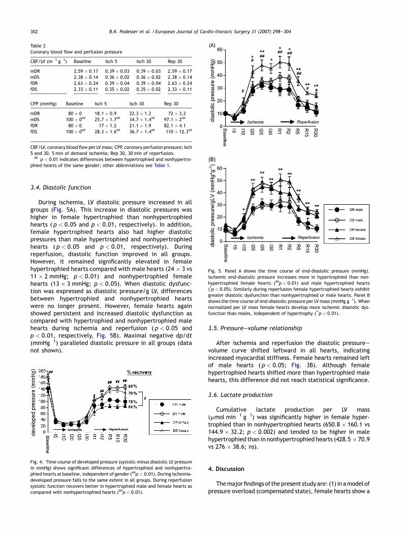
Table 2Coronary blood flow and perfusion pressure
CBF/LV (m�1 g�1) Baseline Isch 5 Isch 30 Rep 30
mDR 2.59 � 0.17 0.39 � 0.03 0.39 � 0.03 2.59 � 0.17mDS 2.38 � 0.14 0.36 � 0.02 0.36 � 0.02 2.38 � 0.14fDR 2.63 � 0.24 0.39 � 0.04 0.39 � 0.04 2.63 � 0.24fDS 2.33 � 0.11 0.35 � 0.02 0.35 � 0.02 2.33 � 0.11
CPP (mmHg) Baseline Isch 5 Isch 30 Rep 30
mDR 80 � 0 18.1 � 0.9 22.3 � 1.2 72 � 3.2mDS 100 � 0## 25.7 � 1.7## 34.7 � 1.4## 97.1 � 2##
fDR 80 � 0 17 � 1.2 21.1 � 1.9 82.1 � 4.1fDS 100 � 0## 28.3 � 1.6## 36.7 � 1.4## 110 � 12.3##
CBF/LV, coronary blood flow per LVmass; CPP, coronary perfusion pressure; Isch5 and 30, 5 min of demand ischemia; Rep 30, 30 min of reperfusion.
## p < 0.01 indicates differences between hypertrophied and nonhypertro-phied hearts of the same gender; other abbreviations see Table 1.
Fig. 5. Panel A shows the time course of end-diastolic pressure (mmHg).Ischemic end-diastolic pressure increases more in hypertrophied than non-hypertrophied female hearts (##p < 0.01) and male hypertrophied hearts(*p < 0.05). Similarly during reperfusion female hypertrophied hearts exhibitgreater diastolic dysfunction than nonhypertrophied or male hearts. Panel Bshows the time course of end-diastolic pressure per LV mass (mmHg g�1). Whennormalized per LV mass female hearts develop more ischemic diastolic dys-function than males, independent of hypertrophy (**p < 0.01).
3.4. Diastolic function
During ischemia, LV diastolic pressure increased in allgroups (Fig. 5A). This increase in diastolic pressures washigher in female hypertrophied than nonhypertrophiedhearts ( p < 0.05 and p < 0.01, respectively). In addition,female hypertrophied hearts also had higher diastolicpressures than male hypertrophied and nonhypertrophiedhearts ( p < 0.05 and p < 0.01, respectively). Duringreperfusion, diastolic function improved in all groups.However, it remained significantly elevated in femalehypertrophied hearts compared with male hearts (24 � 3 vs11 � 2 mmHg; p < 0.01) and nonhypertrophied femalehearts (13 � 3 mmHg; p < 0.05). When diastolic dysfunc-tion was expressed as diastolic pressure/g LV, differencesbetween hypertrophied and nonhypertrophied heartswere no longer present. However, female hearts againshowed persistent and increased diastolic dysfunction ascompared with hypertrophied and nonhypertrophied malehearts during ischemia and reperfusion ( p < 0.05 andp < 0.01, respectively, Fig. 5B). Maximal negative dp/dt(mmHg�1) paralleled diastolic pressure in all groups (datanot shown).
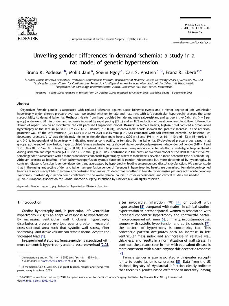
Fig. 4. Time course of developed pressure (systolic minus diastolic LV pressurein mmHg) shows significant differences of hypertrophied and nonhypertro-phied hearts at baseline, independent of gender (##p < 0.01). During ischemia-developed pressure falls to the same extent in all groups. During reperfusionsystolic function recovers better in hypertrophied male and female hearts ascompared with nonhypertrophied hearts (##p < 0.01).
3.5. Pressure—volume relationship
After ischemia and reperfusion the diastolic pressure—volume curve shifted leftward in all hearts, indicatingincreased myocardial stiffness. Female hearts remained leftof male hearts ( p < 0.05; Fig. 3B). Although femalehypertrophied hearts shifted more than hypertrophied malehearts, this difference did not reach statistical significance.
3.6. Lactate production
Cumulative lactate production per LV mass(mmol min�1 g�1) was significantly higher in female hyper-trophied than in nonhypertrophied hearts (650.8 � 160.1 vs144.9 � 32.2; p < 0.002) and tended to be higher in malehypertrophied than in nonhypertrophied hearts (428.5 � 70.9vs 276 � 38.6; ns).
4. Discussion
Themajorfindingsof thepresent studyare: (1) in amodelofpressure overload (compensated state), female hearts show a

B.K. Podesser et al. / European Journal of Cardio-thoracic Surgery 31 (2007) 298—304 303
more concentric hypertrophy and a higher contractility thanmale hearts. (2) During lowflow ischemia in females, thismorepronounced concentric hypertrophy results in increasedsusceptibility to ischemia—reperfusion injury.
4.1. Gender differences in LVH
Our study is in accordance with other experimentalstudies in hypertensive hearts and different models ofpressure overload hypertrophy that showed increasedconcentric hypertrophy with a predominant hypertrophy ofthe septum in females compared with males after aorticbanding [2,17] and post-MI [4]. Similarly, clinical studies inpre- and postmenopausal women showed that hypertensionand aortic stenosis were associated with increased con-centric hypertrophy [7]. The small concentric hypertrophiedchambers result in an increased contractile function duringbaseline and conversely might be responsible for theincreased diastolic dysfunction during ischemia in femalehearts (see below). The mechanisms that contribute to thisunique remodeling of the LV in pressure- and hypertension-induced LVH in females are not entirely known. Differences inhormonal status, i.e. an increased estrogen level or a lack oftestosterone might conceivably contribute. Estrogen hasbeen shown to influence cardiac hypertrophy via itsvasodilative NO-mediated properties [18]. With respect todirect influences of estrogen on the myocardium, it has beenshown that estrogen regulates myocardial hypertrophy viainfluences on MAP-kinase-phosphatase-1 (MAPK), atrialnatriuretic peptide (ANP) [6] or a modulation of the AT-1receptor [19]. In a model of aortic constriction in ovar-iectomized mice, 17ß-estradiol significantly decreasedhypertrophy relative to the placebo-treated mice. No effectof 17ß-estradiol was detected in sham-operated animals nordid 17ß-estradiol reduce cardiac fibrosis in the hypertrophiedhearts in pressure-overloaded mice. Therefore, estrogenmay prevent cardiac hypertrophy and a lack of estrogen mayplay a role in the development of LV hypertrophy late in life[20]. Similarly, estrogen and its regulatory effects on MAP-kinase-phosphatase-1 could also be the reason for delayedmortality and hypertrophy in female mice [21].
4.2. Gender difference in I/R injury in normotensive rats
We found no difference in the susceptibility to I/R innormotensivemale and female rats. This is in agreement withstudies in wild-type mice where no gender difference wasfound in isolated hearts undergoing an I/R protocol [17,22].Therefore, physiological levels of estrogen in normotensiveanimals seem not to exert any protective or deleteriouseffect. In humans, female gender is associated with worseprognosis in acute coronary syndromes [8,9]. Our datasuggest that differences in co-morbidity and other factors(e.g. more left ventricular hypertrophy, smaller coronary sizeetc.) might contribute greatly to this difference.
4.3. Gender differences in I/R injury in hypertrophiedhearts
We and others have repeatedly shown that pressureoverload hypertrophy increases susceptibility to I/R injury,
whereby diastolic dysfunction is predominant [12,23,24]. Inthe present experiments we chose a demand ischemia modelto increase the sodium load via the Na/Ca-exchanger in themyocytes through tachycardia. Since the Dahl rat has amutation of the alpa1-Na, K-ATPase sodium excretion fromthe myocytes could be impaired.
Surprisingly, no increase in diastolic dysfunction was foundin the male hypertrophied versus nonhypertrophied hearts inthis study. This is in agreement with previous studies in thismodel, where we did not find an increased susceptibility inmale hypertensive animals after 4 weeks of high-salt diet[14]. Left ventricular hypertrophy per se seems not in allcases deleterious, rather the increased susceptibility ofhypertrophied hearts is dependent on the stimulus forhypertrophy and the stage of compensation of the hyper-trophied heart [14].
In contrast, hypertrophied females showed increaseddiastolic dysfunction compared with nonhypertrophiedfemales and compared with male hypertrophied hearts.Differences in blood flow could not contribute to this finding,since coronary blood flow before and after demand ischemiawas similar in all groups. During reperfusion increasedcoronary perfusion pressure in female hypertensive hearts,however, indicates an increased vasoconstriction in theseanimals.
Female hypertensive hearts also had the highest lactateproduction. In this model of demand ischemia with residualflow, a mixed aerobic and anaerobic metabolism exists. Theincreased lactate production indicates greater areas ofsevere underperfusion where metabolism is mainly anaero-bic. The heterogeneous metabolism is a known phenomenonof global ischemia in the rat heart. Areas of aerobicconditions with near normal pH exist next to areas of severeunderperfusion and acidosis [14]. It is reasonable tospeculate that in female hypertrophied hearts with increasedconcentric hypertrophy increased diastolic dysfunctiondeveloped during demand ischemia mainly because of thisgeometric difference. The increased diastolic pressureresulted in (subendocardial) areas of severe underperfusionwith increased glycolysis and accelerated depletion of high-energy phosphates that resulted in the poorer recovery ofthese hearts.
It is important to note that in our experiments, theperfusate contained physiological concentrations of glucose,lactate and free fatty acids, the metabolites usuallyconsumed by the heart. This was deemed important sincethe response to ischemia is highly dependent on theconcentrations of the metabolic substrates [25], which mightbe even more important in female hearts.
In summary, the present study demonstrates thathypertrophy is significantly higher in female than malehypertensive rats. Furthermore, female hearts show sig-nificantly higher LV contractile performance per LV mass. Inaddition to these findings, the present study for the first timedocuments that during demand ischemia this more pro-nounced concentric hypertrophy results in no difference insystolic function but an increase in diastolic dysfunction andlactate production in female hypertrophied hearts comparedwith female nonhypertrophied and male hearts. To deter-mine whether in female hypertensive patients with acutecoronary syndromes diastolic dysfunction could contribute to

B.K. Podesser et al. / European Journal of Cardio-thoracic Surgery 31 (2007) 298—304304
the worse clinical course, further experimental and clinicalstudies are needed.
Acknowledgments
This study was supported by The National Heart, Lung, andBlood Institute Grant HL 55993 (CSA) and the American HeartAssociation Grant-in Aid 96-015220 (FRE). BKP was gratefullysupported by a scholarship from the Max Kade Foundation,100 Church Street, New York, NY and the Ludwig BoltzmannGesellschaft, Operngasse 6, Vienna, Austria.
References
[1] Grossman W. Cardiac hypertrophy: useful adaptation or pathologic pro-cess? Am J Med 1980;69:576—84.
[2] Douglas PS, Katz SE, Weinberg EO, Chen MH, Bishop SP, Lorell BH.Hypertrophic remodeling: gender differences in the early response toleft ventricular pressure overload. J Am Coll Cardiol 1998;32:1118—25.
[3] Wallen WJ, Cserti C, Belanger MP, Wittnich C. Gender-differences inmyocardial adaptation to afterload in normotensive and hypertensiverats. Hypertension 2000;36:774—9.
[4] Smith PJ, Ornatsky O, Stewart DJ, Picard P, Dawood F, Wen WH, Liu PP,Webb DJ, Monge JC. Effects of estrogen replacement on infarct size,cardiac remodeling, and the endothelin system after myocardial infarc-tion in ovariectomized rats. Circulation 2000;102:2983—9.
[5] Jain M, Liao R, Podesser BK, Ngoy S, Apstein CS, Eberli FR. Influenceof gender on the response to hemodynamic overload aftermyocardial infarction. Am J Physiol Heart Circ Physiol 2002;283:H2544—50.
[6] Garavaglia GE, Messerli FH, Schmieder RE, Nunez BD, Oren S. Sexdifferences in cardiac adaptation to essential hypertension. Eur HeartJ 1989;10:1110—4.
[7] Aurigemma GP, Gaasch WH. Gender differences in older patients withpressure-overload hypertrophy of the left ventricle. Cardiology 1995;86:310—7.
[8] Mendelson MA, Hendel RC. Myocardial infarction in women. Cardiology1995;86:272—85.
[9] Vaccarino V, Parsons L, Every NR, Barron HV, Krumholz HM. Sex-baseddifferences in early mortality after myocardial infarction. National Reg-istry of Myocardial Infarction 2 Participants. N Engl J Med 1999;341:217—25.
[10] Hochman JS, Tamis JE, Thompson TD, Weaver WD, White HD, Van de WerfF, Aylward P, Topol EJ, Califf RM. Sex, clinical presentation, and outcomein patients with acute coronary syndromes. Global use of strategies toopen occluded coronary arteries in acute coronary syndromes IIb inves-tigators. N Engl J Med 1999;341:226—32.
[11] Lee TM, Su SF, Tsai CC, Lee YT, Tsai CH. Cardioprotective effects of 17beta-estradiol produced by activation ofmitochondrial ATP-sensitiveK(+)channels in canine hearts. J Mol Cell Cardiol 2000;32:1147—58.
[12] Eberli FR, Apstein CS, Ngoy S, Lorell BH. Exacerbation of left ventricularischemic diastolic dysfunction by pressure-overload hypertrophy. Mod-ification by specific inhibition of cardiac angiotensin converting enzyme.Circ Res 1992;70:931—43.
[13] Wexler LF, Lorell BH, Momomura S, Weinberg EO, Ingwall JS, Apstein CS.Enhanced sensitivity to hypoxia-induced diastolic dysfunction in pres-sure-overload left ventricular hypertrophy in the rat: role of high-energyphosphate depletion. Circ Res 1988;62:766—75.
[14] Nagata K, Liao R, Eberli FR, Satoh N, Chevalier B, Apstein CS, Suter TM.Early changes in excitation-contraction coupling: transition from com-pensated hypertrophy to failure in Dahl salt-sensitive rat myocytes.Cardiovasc Res 1998;37:467—77.
[15] Herrera VL, Chobanian AV, Ruiz-Opazo N. Isoform-specific modulation ofNa+, K+-ATPase alpha-subunit gene expression in hypertension. Science1988;241:221—3.
[16] Fletcher PJ, Pfeffer JM, Pfeffer MA, Braunwald E. Left ventriculardiastolic pressure—volume relations in rats with healed myocardialinfarction. Effects on systolic function. Circ Res 1981;49:618—26.
[17] Cross HR, Murphy E, Koch WJ, Steenbergen C. Male and female miceoverexpressing the beta(2)-adrenergic receptor exhibit differences inischemia/reperfusion injury: role of nitric oxide. Cardiovasc Res2002;53:662—71.
[18] Nuedling S, Kahlert S, Loebbert K, Doevendans PA, Meyer R, Vetter H,Grohe C. 17 Beta-estradiol stimulates expression of endothelial andinducible NO synthase in rat myocardium in-vitro and in-vivo. CardiovascRes 1999;43:666—74.
[19] Nickenig G, Baumer AT, Grohe C, Kahlert S, Strehlow K, Rosenkranz S,Stablein A, Beckers F, Smits JF, Daemen MJ, Vetter H, Bohm M. Estrogenmodulates AT1 receptor gene expression in vitro and in vivo. Circulation1998;97:2197—201.
[20] van Eickels M, Grohe C, Cleutjens JP, Janssen BJ, Wellens HJ, DoevendansPA. 17beta-estradiol attenuates the development of pressure-overloadhypertrophy. Circulation 2001;104:1419—23.
[21] Dash R, Schmidt AG, Pathak A, Gerst MJ, Biniakiewicz D, Kadambi VJ, HoitBD, Abraham WT, Kranias EG. Differential regulation of p38 mitogen-activated protein kinase mediates gender-dependent catecholamine-induced hypertrophy. Cardiovasc Res 2003;57:704—14.
[22] Cross HR, Lu L, Steenbergen C, Philipson KD, Murphy E. Overexpression ofthe cardiac Na+/Ca2+ exchanger increases susceptibility to ischemia/reperfusion injury in male, but not female, transgenic mice. Circ Res1998;83:1215—23.
[23] Lorell BH, Grice WN, Apstein CS. Influence of hypertension with minimalhypertrophy on diastolic function during demand ischemia. Hypertension1989;13:361—70.
[24] Saupe KW, Lim CC, Ingwall JS, Apstein CS, Eberli FR. Comparison of heartswith 2 types of pressure-overload left ventricular hypertrophy. Hyperten-sion 2000;35:1167—72.
[25] Taegtmeyer H, Goodwin GW, Doenst T, Frazier OH. Substrate metabolismas a determinant for postischemic functional recovery of the heart. Am JCardiol 1997;80:3A—10A.
Copyright © 2022 FDOKUMEN