Untitled - Los Angeles City Clerk
663
-
Upload
khangminh22 -
Category
Documents
-
view
0 -
download
0
Transcript of Untitled - Los Angeles City Clerk


NAME: Martin L. Pall DATE: Sept. 2013 [email protected] Phone: 503-232-3883 EDUCATION Institutions, degrees, dates Organization and Location Degree Date Johns Hopkins University B.A. 1962 Baltimore, MD California Institute of Technology, Ph.D. 1968 Pasadena, CA EXPERIENCE Positions and dates Reed College, Portland, OR 1967-72 Assistant Professor of Biology Department of Botany, Indiana May-June 1971 Visiting Research Associate University, Bloomington, IN Program in Genetics and Depart- 9/16/72- Assistant Professor of ment of Chemistry, Washington 9/156/75 Genetics and Chemistry State University, Pullman, WA Program in Genetics and Program 4/12/73 Elected to Graduate Faculty in Biochemistry/Biophysics, Wash- in Genetics and Biochemistry ington State University, Pullman,WA 9/16/75- Associate Professor of Genetics 1/31/81 and Biochemistry Program in Genetics, Washington 9/16/78- Acting Chairman, Program State University, Pullman, WA 9/15/79 in Genetics Department of Physiology, Yale 9/16/79- Visiting Associate Professor University, New Haven, CT 9/15/80 (during professional leave) Departments of Genetics and 2/1/81- Associate Professor of Genetics Cell Biology and Biochemistry/ 9/15/83 and Cell Biology and Biophysics, Washington State Biochemistry University, Pullman, WA 9/16/83- Professor of Genetics and 8/15/96 Cell Biology and Biochemistry Department of Pharmacology, 5/15/86 Adjunct Professor of University of California, 12/31/86 Pharmacology San Francisco, Ca (professional leave)
Washington State University, 8/16/94- Coordinator of Sciences Vancouver, WA 5/16/96 Department of Biochemistry/Biophysics 8/16/96-99 Professor of Biochemistry and and Basic Medical Sciences Program Basic Medical Sciences School of Molecular Biosciences 8/16/99- Professor of Biochemistry and Basic Medical Sciences 8/15/08 and Basic Medical Sciences Program Professor Emeritus of Biochemistry 8/15/08- and Basic Medical Sciences, WSU and Research Director, The Tenth Paradigm Research Group, Portland, OR CURRENT WEB SITE: thetenthparadigm.org HONOR SOCIETIES Phi Beta Kappa Alpha Epsilon Delta Sigma Xi Member of Panel of Advisors of Environmental Law Centre in London Clinician of the Month, Healthcomm International (October, 1999), Member Scientific Advisory Board of Ariston Pharmaceuticals PROFESSIONAL SOCIETIES AND OTHER HONORS American Society of Biochemistry and Molecular Biology International Association for Chronic Fatigue Syndrome RECENT HONORS REGARDING ENVIRONMENTAL MEDICINE:
1. 2005: Pall was appointed to the Advisory Board of the Environmental Law Centre in London. 2. May 2008: Pall was the only person from outside of Europe, invited to address a special
session of the European Union Parliament (“Council of Nations”) as an “Expert” in Environmental & Health.
3. 2009: Pall was chosen from all scientists in the world to write an authoritative review on Multiple Chemical Sensitivity for General and Applied Toxicology, 3rd Edition.
4. April 2010: Pall was appointed as “life time, honorary ambassador and member of the Scientific Advisory Board of the International Society for Applied Preventative Medicine (I-GAP).”
5. May 2010: Pall was the meeting honoree at the Fundacion Alborada meeting in Spain, only the second time such an honoree had been chosen.
6. September 2010: Pall was the only person from outside of Europe invited to address a special meeting at the National Institute of Health in Rome on Environmental Medicine.
7. 2012 and onward. Pall was chosen to be a founding faculty member of the new Environmental Medicine Faculty in Italy. He started in that function in March 2013.
8. 2013: Chosen to be the Jonathan Forman award recipient, the highest award given by the American Academy for Environmental Medicine.
9. Publication #89 was honored by inclusion at the “Global Medical Discovery” as one of the top medical publications of 2013.

10. One of two keynote speakers for the Building Biology Conference, (International Institute for Building Biology & Ecology) Bainbridge Island, WA 2015 and voted the favorite speaker at the meeting by meeting participants.
Publications from 2001: 59. Pall, M. L. 2001. Cobalamin used in chronic fatigue syndrome therapy is a nitric oxide scavenger. Journal of Chronic Fatigue Syndr 8(2):39-44. 60. Pall M. L, Satterlee J. D., 2001. Elevated nitric oxide/peroxynitrite mechanism for the common
etiology of multiple chemical sensitivity, chronic fatigue syndrome and posttraumatic stress disorder. Annals of the New York Academy of Sciences 933:323-329.
61. Pall M. L. 2001. Common etiology of posttraumatic stress disorder, fibromylagia, chronic fatigue syndrome and multiple chemical sensitivity via elevated nitric oxide/peroxynitrite. Med Hypoth 57:139-145.
62. Pall M. L. 2002. The levels of nitric oxide synthase product, citrulline, are elevated in the sera of chronic fatigue syndrome patients. J Chronic Fatigue Syndr 10(3/4):37-41. 63. Pall M. L. 2002. NMDA sensitization and stimulation by peroxynitrite, nitric oxide and organic solvents as the mechanism of chemical sensitivity in multiple chemical sensitivity. FASEB J. 16:1407-1417. 64. Pall, M. L. 2002 Chronic fatigue syndrome/myalgia encephalomyelitis. Br J Gen Pract 52:762. 65. Smirnova I.V., Pall M.L. 2003 Elevated levels of protein carbonyls in sera of chronic fatigue syndrome patients. Mol Cell Biochem 248:93-95. 66. Pall M. L. 2003. Elevated nitric oxide/peroxynitrite theory of multiple chemical sensitivity: Central role of N-methyl-D-aspartate receptors in the sensitivity mechanism. Environ Health Perspect 111:1461-1464. 67. Pall M. L. 2003 Long delayed sequelae of organophosphate exposure. Arch Env Health 58:605. 68. Pall M. L. 2004 The simple truth about multiple chemical sensitivity. Environ Health Perspect 112:A266-A267. 69. Pall M. L., Anderson J. H. 2004 The vanilloid receptor as a putative target of diverse chemicals in multiple chemical sensitivity. Arch Environ Health 59:363-375. 70. Pall M. L. 2005 Chronic fatigue syndrome and nitric oxide: giving credit where credit is due. Med Hypoth 65:631-633. 71. Pall M. L. 2005 Multiple chemical sensitivity: towards the end of controversy. Townsend Let Doctors Patients Aug/Sept 2005:52-56. 72. Pall M. L. 2006 The NO/ONOO- Cycle as the Cause of Fibromyalgia and Related Illnesses: Etiology, Explanation and Effective Therapy. In, New Research in Fibromyalgia, Nova Science Publishers, Hauppauge, NY, pp 39-61. 73. Pall M. L., Bedient S. A. 2007 The NO/ONOO- Cycle as the Etiologic Mechanism of Tinnitus, Int Tinnitus J 13:99-104. 74. Pall M. L. 2007 “Explaining ‘Unexplained Illness’: Disease Paradigm for Chronic Fatigue Syndrome, Multiple Chemical Sensitivity, Fibromyalgia, Post-Traumatic Stress Disorder, Gulf War Syndrome and Others”, 16 Chapter book, Harrington Park (Haworth) Press. 75. Pall ML. 2007 Nitric oxide synthase partial uncoupling as a key switching mechanism for the NO/ONOO- cycle. Med Hypotheses 69:821-825. 76. Pall M. L. 2008 Post-radiation syndrome as a NO/ONOO- cycle, chronic fatigue syndrome-like disease. Med Hypoth 78:537-541. 77. Pall M. L. 2008 Does sauna therapy used to treat multiple chemical sensitivity and other diseases act by increasing availability of tetrahydrobiopterin? Med Hypoth 73:610-613. 78. Pall M. L. 2009 Multiple chemical sensitivity: Toxicological questions and mechanisms. In General and Applied Toxicology, 3rd Edition, John Wiley & Sons, pp. 2303-2352.

79. Pall M. L. 2010 The NO/ONOO- Vicious Cycle Mechanism as the Cause of Chronic Fatigue Syndrome/Myalgic Encephalomyelitis, In Chronic Fatigue Syndrome: Symptoms, Causes and Prevention, Edita Svoboda and Kristof Zelenjcik, eds., Nova Publishers, pp 27-56. 80. Pall M. L. 2010 How Can We Cure NO/ONOO- Cycle Diseases? Approaches to curing chronic fatigue syndrome/myalgic encephalomyelitis, fibromyalgia, multiple chemical sensitivity, Gulf War syndrome and possibly many others, Townsend Letter for Doctors and Patients, Feb/Mar 2010:75-84. 81. Pall M.L. 2010 High-dose therapy for ascorbate, niacin, folate and B-12: Pauling was right but for the wrong reason. J Orthomolecular Med 25(3):148-156. 82. Pall M. L. 2010 Multiple chemical sensitivity is a response to chemicals acting as toxicants via excessive NMDA activity. J Psychosomatic Res 69: 327-328. 83. Pall, M.L. Teufelskreis NO/ONOO--Zyklus, oxidaver Stress, mitochondriale, inflammatorische und neurologische Dysfunktion. Umwelt Medizin Gesellshaft 2010, 23, 281-293. 84. Hoeck A. D., Pall M. L. 2011 Will vitamin D supplementation ameliorate diseases characterized by chronic inflammation and fatigue? Med Hypotheses 76: 208-213. 85. Carruthers B. M. et al (including Pall M. L.) 2011 Myalgic enchephalomyelitis: International consensus criteria. J Intern Med 270: 327-338. 86. Pall ML 2012 Chronic fatigue syndrome and fibromyalgia are NO/ONOO- cycle diseases, not glutathione depletion/methylation cycle block diseases: an analysis of the Nathan/Van Konyneburg data. Towsend Lett April 2012:75-79. 87. Pall ML. 2013 Pulmonary hypertension is a probable NO/ONOO- cycle disease: A review. ISRN Hypertension 2013: Article ID 742418, 27 pages. 88. Pall ML. 2012 Two fragrance chemicals may act as toxicants via TRPA1 stimulation rather than via direct mitochondrial action. Toxicol In Vitro. 2012 Sep 25. pii: S0887-2333(12)00265-2. doi: 10.1016/j.tiv.2012.09.011. [Epub ahead of print] 89. Pall ML. 2013 Electromagnetic fields act via activation of voltage-gated calcium channels to produce beneficial or adverse effects. J Cell Mol Med 17:958-965. 90. Auhdya T., Pall M.L., Green J. 2013 A Study of Sauna Therapy in Myalgic Encephalomyelitis/Chronic Fatigue Syndrome Patients Shows Sauna Action via Raised Tetrahydrobiopterin and Three Predictions of the NO/ONOO- Cycle. Townsend Lett 364:60-64. 91. Pall ML. 2013. The NO/ONOO- Cycle as the Central Cause of Heart Failure. Int J Mol Sci Nov 13;14(11):22274-330. doi: 10.3390/ijms141122274. 92. Pall ML. 2014 Electromagnetic field activation of voltage-gated calcium channels: role in therapeutic effects. Electromagn Biol Med. 2014 Apr 8. 93. Pall ML. 2014 Is Open-angle Glaucoma Caused by the NO/ONOO(-) Cycle Acting at Two Locations in the Eye? Med Hypothesis Discov Innov Ophthalmol 4:1-2. 94. Pall ML. 2014 Microwave electromagnetic fields act by activating voltage-gated calcium channels: Why the current international safety standards do not predict biological hazard. Recent Res Devel Mol Cell Biol, 7(2014): 0-00 ISBN: 978-81-308-0553. 95. Pall ML, Levine S. 2015 Nrf2, a master regulator of detoxification and also antioxidant, anti- inflammatory and other cytoprotective mechanisms, is raised by health promoting factors. Acta Physiologica Sinica, February 25, 2015, 67(1): 1–18. 96. Pall ML. 2015 Scientific evidence contradicts findings and assumptions of Canadian Safety Panel 6: microwaves act through voltage-gated calcium channel activation to induce biological impacts at non-thermal levels, supporting a paradigm shift for microwave/lower frequency electromagnetic field action. Rev Environ Health 30:99-116. 97. Pall ML, Levine S. 2015 Nrf2 is a master regulator of cytoprotective responses including antioxidant, anti-inflammatory, detoxification, improved mitochondrial function and autophagy: Nrf2 activity is raised by many health-promoting factors. Townsend Lett, October 2015 : 51-54.

98. Pall ML. 2015 Microwave frequency electromagnetic fields (EMFs) produce widespread neuropsychiatric effects including depression. J Chem Neuroanatomy, 2015 Aug 21. pii: S0891-0618(15)00059-9. doi: 10.1016/j.jchemneu.2015.08.001. [Epub ahead of print] 99. Pall ML. 2015 How to approach the challenge of minimizing non-thermal health effects of microwave radiation from electrical devices. International Journal of Innovative Research in Engineering & Management (IJIREM) ISSN: 2350-0557, Volume-2, Issue -5, September 2015; 71-76.

!!!
Attachment!1!

NAVAL MEDICAL RESEARCH INSTITUTE
7
""125.-1504 REOTN..2
k • .S ---.o 11 C,ý-- .. .t.
" * * • . ... . . . .
_***-:. *\ -
- •-BIBLIOGRAPHY OF REPORTED BIOLOGICAL PHENOMENA ('EFFECTS') AND CLINICAL
SMANIFESTATIONS ATTRIBUTED TO MICROWJAVE AND RADIO-FREQUENCY RADIATIONI
S•-:RESEZARCH REPORT
KF:•,,,12,:."524. 015-0004B RE•PORTp EsDNO. 2 'i
•-•: NATIONAL TECHNICAL!_• INFORMATION SERVICE
[ 4(

Fmm -W- mrm im. r- w
BIBLIOGPHY OF REPORTED BIOLOGICAL PHENOMENA ('EFFECTS') AND CLINICAL
MANIFESTATIONS ATTRIBUrED TO MICROWAVE AND RADIO-FREQUENCY RADIATION
Zorach R. Glaser, Ph.D.LT, MSC, USNR
Research 'Report
Project MF12.524.015-00043,Report No. 2
Naval Medical Research InstituteNational Naval Medical Center
Bethesda, Maryland 20014, U.S.A.
4 October 1971
Second Printing, with Revisions,Corrections, and Additions: 20 April 1972
(Supersede3 AD No. 734391)
I I

I' ABSTRACTMore than 2000 references on the biological responses to radio
frequency and microwave radiation, published up to June 1971, areincluded in the bibliography.* Particular attention has been paid tothe effects on man of non-ionizing radiation at these frequencies.The citations are arranged alphabetically by author, and contain as
much information as possible so as to assure effective retrieval of
the original documents. An outline of the effects whicb have been
attributed to radio frequency and microwave radiation is also part of
the report.
*Three supplementary listings bring the number of citations to morei than 2300.
SKej Words
Biological EffectsNon-Ionizing RadiationRadar HazardsRadio Frequency Radiation
Microwave RadiationHealth HazardsBibliographyElectromagnetic Radiation Injury
The comments upon and criticisms of the literature made in this report,
and the recommendations and inferences suggested, are those of the
author, and do not necessarily reflect the views of the Navy Department
or of the Naval Service.
2

Secunt. Classification
DOCUMENT CONTROL DATA - R & D~c~f:, CIII, *t I IdCSf l ti 1 of UlU. 4,,dy 0, abs'rct anJ Indexing ano-.,* -in -ist I entered Alin :$ýe zoverz I t.9por i~ 1.s t~d
C'I-%tTIG AC ,VITY C. •rpofare*utho) 1Z*. REPORT SECURITY CO$AS•IFICA•.O,%
N AVAL UEDICAL RESEARCH INSTITUTE UNClASSIFIED.,TIUNAL NAVAL MEDICAL CENTER 2ib. GROUP
BETHSDA, -AW'IAND 20014I ALPORT TITISE
BIBLIUGMPHY OF ;;-ZOXTED BIOLOGICAL PHENOMENA ('EFFECTS') AND CLINICAL:ŽANIFESTATIONS ATTRIBUTED TO MICROW&VE AND RADIO-FREQUENCY- RADIATION
4. -ESC• •PTIVE NOTES ()r.pe ofirepor: and inclusive dates)
Medicai research interim report, bibliographic (Current to April 1972) j"AU T.ORCSI (First name. middie initial. last name)
Zorach R. GLASLR, Ph.D. IL-T, MSC, USN
REPORT ,ATE Revised 2O April 1972 Va. TOTA NO. OF PAGES Th. No. OF REPS
(4. Occm.er 1971, Original) 2,311454 CONTRACT OR GRANT NO. - • ORIGINATOR•S REPORT NUMSERteu)
b. ,,oJEC- No J F12.524.015-0004B, Report No. 2, Ke~ise
C. 1 Ob. OT#4ERk REPORT ftOMS (Any other num~bers that may be assignedthis gep@fl)
d. .
Z *ISTRI8b..T:CN STATEMENT A
ITHiIS LOWUMENT HALS BEEN APPROVED FOR PUBLIC RELEASE AND SALE; ITS DISTRIBUTION ISUNLIMITED.
-II:b.PPLEAENTAR~. NOTES 12-. SPONSORING MILI TARY AC TI VIT Y BUEUOMDIIEADSRRY(V) jWASHINGTON, D.C. 20390 1
:3 AUJS TACT-
More than 2300 relerences on the biological responses to radio trequency andmicrowave radiation, published up t April 1972, are included in this bibliographyot the world iiterature. Particular attention has been paid to the effects on mano: non-ionizing radiation at these ' requencies. The citations are arrangedalphabetically by author, and contain as much information as possible so as toassure eltective retrieval of the original documents. Soviet and East Europeanf literature is included in detail. An outline of the effects which have beenattributed tu radio trequency and microwave radiation is included as Chapter 1.The revised report (which supersedes DDC report AD#734391) is updated with theinclusion oi three supplementary listings, and has incorporated many correctionsand adoitions to the original 2100 citations.
I
" ov .,473 UNCIASSIFIED• Security Classification

UNCLASSIFIEDSecurity Chtssifii.tion
%-IN. A LINK BKEY WORDS- - --
biooica- eieiects
Non-iouizing radiation IRadar hazards I
Radio Irequency radiation 4
:Aicrowave radiar iton , Illeair. hazards |
Siblio6raphy II iEiec:r~a6netic radiation injury I
i-aCau.'amn adverse etlectsI2I I j
I I
I * I
I -!
2 2 ' t41
' !
I I I I2 " 2
I
* I'
I I
I I! J
2 I•II I
II I I

TABLE OF CONTENTS
PAGE
Abstract 2
Table of Contents 3
Foreword 4
Acknowledgments 5
Chapter 1, Outline of Reported Biological Phenomena ('Effects') 7and Some Clinical Hanifestations Attributed toMicrovave and Radio-Frequency Radiation
Chapter 2, Bibliography, Alphabetical Listing 12
Unsigned Reports and Articles 83
Addenda, Alphabetical by Author 87
Addenda, Unsigned Reports and Articles 89
Fir3t Suppleeuntary Listing (5 October 1971) 91
Appendix A, Accession Numbers and Sources 92
Second Supplementary Listing (21 November 1971) 93
Third Supplementary Listing (17 April 1972) 95
34
2:a

Foreword
It is the hope of the author that this bibliography will provide guid-ance to the diffuse and conflicting literature on the biological responsesto electromagnetic radiation at radio- and microwave-frequencies, withparticular reference to the effects of concern to man. Such guidance isneeded in the formulation and appraisal of criteria and limits of humanexposure to "non-ionizing" radiation, and in the planning and conduct offuture research.
The original plans were to categorize and key the literature cita-tions to the "outline of biological and clinical effects" (Chapter 1).This proved to be a much more dif"icult and time-consuming task thananticipated, and was actually completed only for about 400 papers. Thus,the letter-number combinations given in square brackets for some of the"A" through "C" citations refer to the outline. INV] indicates the cita-tion was "not verified".
The standard format used throughout the bibliography is: author,(date), journal, volume, (issue): page, "title". The authors are alpha-betized, and in chronological order. Multiple authora 'xe also alphabeti-cally ordered according to the second, third, etc., author. Inclusivepagination is given where possible, as is the original language of thecitation. Report accession and translation numbers (some of which arecited in Appendix A), and alternate sources are listed when known. Thetitle of books is underlined. When the title of the report was not avail-able (or not given), a short (one line) description of the paper is listedwhenever possible. Reports in which the name of the author was not givenare listed chronologically using the format, "title", reference, source,(date). In many cases the citation was obtained from secondary (andtertiary) sources. For this reason it was impossible to put every citationinto a consistent format.
In a few cases, papers have been cited which were presented atsymposia or meetings devoted to the present topic, even when the reporttitle suggests that it does not pertain directly to the topic. This hasbeen done to show the wide range of items considered relevant (at leastat the time of the meeting, and by the organizing chairman) in past years.An example is "electroanesthesia".
A few citations of marginal and/or peripheral relationship have alsobeen included so that the reader may judge the applicability to his indi-vidual research needs. Examples are reports dealing with the biologicaleffects of static and alternating magnetic fields, experimental techniquesusing radio frequency and microwave radiation (e.g., electron spin reso-nance, and nuclear magnetic resonance spectroscopy), and microwave expo-sure limits, regulations, and standards.
References for a few limited-distribu ion government reports areavailable upon request.
4

The author welcomes information which will correct errors and omis-sions (both of which no doubt exist). Copies of new papers would begreatly appreciated, and would encourage updating and revising the biblio-graphy periodically.
ACKNOWLEDGMENTS
The assistance and support received during the preparation of thisbibliography have been considerable, and I am happy to acknowledge my in-debtedness and gratitude. Drs. John Keesey and Dennis Heffner, former andpresent Heads of the Biophysics Division, and Dr. Seymour Friess, Directorof the Environmental Biosciences Department of the Naval Medical ResearchInstitute, permitted me the opportunitj to work on the bibliography, andoffered frequent encouragement.
Acknowledgment is also due to many friends and associates for their
helpful suggestions, comments, and loans and/or gifts of reports or othermaterial, which have been invaluable in the course of the work. "r. GlennHeimer of the Naval Ship Engineering Center contributed an extensive collec-tion of government reports and dccuments, many of which had not previouslybeen cited in the open literature.
- Special help in tracing and in the acquisition of relevant papers hasbeen received from the librarians and staff members of the NMRI library:Mrs. Thelma Robinson, Mrs. Ernestine Gendlemen, Mrs. Eleanor Capps, andMiss Deborah Grove. Their diligence and resourcefulness in tracing andobtaining copies of a large number of papers and reports, often in spiteof Incomplete and/or inaccurate citations given in other sources, enabledme to include many relevant items in the bibliography.
Mr. Christopher Dodge of the Scientific and Technical Center, Depart-sent of the Navy, provided much of the Soviet Bloc literature, linguisticand other technical assistance, and in addition offered valuable commentsand encouragement throughout the preparation of this report. Especiallynoteworthy were the corrections and improremeuts suggested by Chris follow-ing his reading of the entire manuscript.
Helpful also in locating some of the Soviet literature was Mr. E. S.Serebrennikov, of the Science and Technology Division, The Library ofCongress.
Credit is due Mrs. Ann& Woke (of this Institute) for translating manyof the German papers; to Dr. Emilio Weiss, who translated from the Italian,and to Mrs. Edith Pugh who typed many "first drafts"; also to Mrs. RhodaGlaser for her help in many aspects of the work.
Mrs. Fannie Epstein Aeserves special mention for her outstandingeditorial assistance, and especially for the heroic typing, organization,and checking of the entire report.
_ ~5
o !--- z- -'-- --.----

T- .aut line of Reported Biological Phenomena ('Effects') andClinical eal1festations Attributed to Microwave and Radio-Frequency IRadiatior, is patterneh after that given by R. Murray, .et al., in anarticle entitled, "Has-o safe are alcrowaves", which appeared in Non-Ionizing Radiation 1(1):7-8 (1969). Some of the "effects" were listedin the r#.ort by S. F. Cleary and W. T. Ham, Jr., entitled, "Considera-tions in the evaluation of the biological effects on exposure to micro-wave radiation!, (3ackground document, Port 1, 1969, for the Task Forceon Research Planving in Environmental Health, Subtask Force on PhysicalFactors in the Environment). The discusbion and suggestions offered byByron McLees,, Edward Finch, Lewis Gersbman, and Christopher Dodge relat-ing to the Outline are also gratefully acknowledged.
Prepare-r *.-n of the bibliography was supported by the Bureau ofMedicine A,;,, -.irgery, Department of the Navy, under vork unit HF12.524.0105-OO'V.
AII
74
61
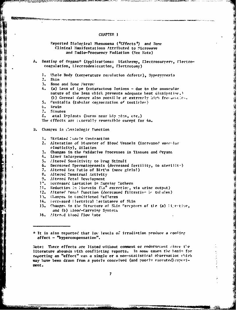
CIHAPTER I
Peported Diolorical Phenomena (*Effects') and SomeClinical Manifestations Attributed to !Iicrowave
and P.adlo-Frequency Padiation (See Note)
A. Heating of Organs* (Applications: Diathermy, rlectrosurrerv, Flectro-coagulation, I.lecrrodesicoation, lectrotomy)
1. M.ole Body (temnerature repulation defect), !Hyperpyreyla2. Skin3. Bone and Bone . tarro:4. (a) Lens of Lye (cataractous lesions - due to the avascular
nature of the lens vhich prevents adequate heat dissipat i,w.I(M) Corneal damane also poosil le at extrei.ely 0It fro- u4-i. 'c.
5. e'enitalia (tubular dreg.?neration of testicleF)C.. hrain7. SinusesS. etal Inplants (burns near hip ?ins, erc.)The effects are :tneralPy reversible except for 4a.
B. Chianes in liv, o.ogic .function
I. Striated :ttscte Contraction2. Alteration of DWi-meter of Blood Vessels (increasec' va.tcr-lar
elasticity), DilationS. Changes in the Oxidative Processes in Tissues and ArranL4. Liver Lnlargement5. A.ltered Sensitivity to Drug Stitruli6. Decreased Spernato.enesis (decreased fertility, to steril.)7. Altered Sex Patio of Blrths (more girls!)8. Altered ,!enstrual Activity9. Altered Fetal Devwlopment
I. iAecreasee Lactation in :ursing ":others'.l. Reduction in i'iuresis (',a+ excretio:,, via urine output)!2. Altered !'era!! function (decreased filtratir," ::- tul alef)13. U:hanf-es in (onditioned P:eflexes14. iecreased .;lectrical .eristance of Skin15. Changes in tbU Structure of Skin re.cptorn of tie (a) i.-ti~e,
and (b) i-loot'-Carryinr Systcr.-:16. Alterrd mIood Fnow !:ate
* It is also reported that leov levels of irradiation produce a coolingeffect - "hypercor.pensation".
Note: Thece effects are listed without comment or endorse.:ent :Aince. t-eliterature abounds with conflicting reports. In sonte cases the !:asi. f~nrreporting an "effect" was a single or a non-statistical observaticr, i'"ichmay have been drawn from a poorly conceived (and poorly executed)xpevri-fnente
7

17. Alterations In the Siocurrents (KW?) of the Cerebral Cortex(in animals)
18. Changes In the Rate of Clearance of Tagged Ions from Tissue19. Reversible Structural Changes In the Cerebral Cortex and the
Diencephalon20. Electrocardiographic (EKG) Changes21. Alterations In Sensitivity to Light, Sound, and Olfactory
StImuli22. Functional (a) and Pathological (b) Changes in the Eyes:
(a) decrease in size of blind spot, altered color recogPltion,changes In Intraocular pressure, lacriaation, trembling cf eye-lids; (b) less opacity and coagulation, altered tissue respira-tion, and altered reduction-oxidation processes23. Myocardial Necrosis
24. Hemorrhage in Lungs, Liver, Gut, and Brain J At Fatal Levels25. Generalized Degeneration of all Body Tissue of Radiation26. Loss of Anatomical Parts27. Death28. Dehydration29. Altered Rate of Calcification of Certain Tissue
C. Central Nervous System Effects
1. Headaches2. Inomnia3. Restlessness (Awake and During Sleep)4. Electroencephalographic (EW) Changes5. Cranial Nerve Disorders6. Pyramidal Tract Lesions7. Condittoned Reflex Disorders8. Vagominetic Action of the Heart; Sympaticouminetic Action9. Seizures, Convulsions
D. Autonomic Nervous System Effects
1. Neuro-vegetative Disorders (e.g., alteration of heart rhythm)2. Fatigue3. Structural Alterations Lu the Synapses of the Vague Nerve4. Stimalatin of Parasympathetic Nervous System (Bradycardia),
and lnhbition of the Sympathetic Nervous System
E. Peripheral Nervous System Effects
Effects on Locowtor Nerves

F. Psychological Disorders ("Human Behavioral Studies") - the so-called"Psychophysiologic (and Psychosomatic) Responses"
I. Neurasthenia- (general "bad" feeling)2. Depression3. Impotence4. Anxiety5. Lack of Concentration6. Hypochondria7. Dizziness,S. Hal lucinations9. Sleepiness
10. InsomniaII. Increased Irritabiiity12. ')ecreased Appetite13. Loss of !:er-ory14. Scalp SensarionsY 15. Tncreasee Fatirabilty 41r16. Chest rainA17. Tremor of the lianus
G. Behavioral (Cnanres (Aninal Studies)
Peflexive, Operant, tvoldance, and DIscrinination Belaviors
l. Mood Disorders(V = in vivo)(v = in vitrt.'
Changes in:1. Bloo.' anC Bone *:arrcn%2. Pha~oryt.:r (polrVorphs) and Bactericidal "unctiornr f . ,v)S3. llemol.-s.s .at* (increase), (a shortene,! lifeaz~: of , "•. St-dir.enta-ion ;ate (increase), (due to ,-e. ;n sp, t " I
levels or ar:ount of fitrino-er. (?))"5. ::uriber of L.rythroc'tes (Cecrease), also nneher of 1.yr- c, tt'.
Bl. ood CtluccFe foncentration (increase)7. Llood 11istanine rontentZ. Cholesterol and Lipids9. Garma (also u and 0) Gl.obulin, and Total Protein Concentration
10. Number of Eosinophils A11. Albumin/Globulin ratio (decrease)12. PHemopoiesis (rate of formation of blood corpuscles)
13. Leul-openia (increase in number of x0.ito cells), ancl Leu1-oc-,orosis14. * .et iculocytosis
1. Vascular Disorders
1. Thrombosis2. Hypertension
9
-4
2 ii

JS Enzyme and Other Biochemical Changes
h Changes in activity of:1. Cholinesterase (V,v)2. Phosphatase (v)3. Transaminase (v)4. Amylase (v)5. Carboxydismutase
6. Protein Denaturation7. Toxin, Fungus, and Virus Inactivation (at high radiation dose
levels), Bacteriostatic Effect8. Tissue Cultures Killed9. Alteration In iRate of Coll Division
10. Increased Concentration of RVA in Lymphocytes, and DecreasedConcentration in Brain,. Liver, and Spleen
11. Changes in Pyruvic Acid, Lactic Acid, and Creatinine LxereJionsS12. Change in Concentration of Glycogen in Liver (Hlyperglycevia)
13. Alteration in Concentration of 17- Ketosterolds in Urine
SK. Metabolic Disorders
Glycosuria (sugar in uriTe; related with blood sugar?)2. Increase in Urinary Phenol (derivatives? DOPA?)3. Alteration of rate of ietabolic Enzymatic ProcessesS4. Altered Carbohydrate Metabolism
L. Gastro-Intestinal Disorders
1. Anorexia (loss of appetite)2. Epigastric Pain3. Constipation4. Altered Secretion of Stomach "Digestive Juices"
It. Endocrine Gland Changes
1. Altered Pituitary runction2. Ilyperthyroidism3. Thyroid Enlargement4. Increased Uptake of Radioactive Iodine by Thyroid Gland5. Altered Adrenal Cortex Activity6. Decreased Corticosteroids in Blood
S7. Decreased Glucocorticoidal Activity8. 1lypogonadism (usually decreased testosterone production)
IN. Histological Changes
1. Changes in Tubular Epithelium of TesticlesS2. Gross Changes
10

0. Genetic and Chromoscrtal Changes A
1. Chromosome Aberrations (e.g., linear shortening, pseudochiasm,diploid structures, amitotic division, bridging, "sticky"chromosomes, irregularities in chromosomal env-zlope)
2. Mutations =3. Mongolism4. Somatic Alterations (changes in cell not involving nucleus or
chromosomes, cellular transformation)5. Neoplastic Diseases (e.g*, tumors)
P. Pearl Chain Effect (Intracellular orientation of subcellular part cles,and orientation of cellular and other (non-biologlc) particles)
Also, orientation of animals, birds, and fish in electromagnetic
fields
Q. Miscellaneous Effects
1. Sparking between dental fillings2. Peculiar metallic taste in mouth3. Changes in Optical Activity of Colloidal Solutions4. Treatment for Syphilis, Poliomyelitis, Skin Diseases5. Loss of flair6. Brittleness of flair7. Sensations of Buzzing Vibrations, Pulsations, and Ticklinp About
the Head and Ears8. Copious Perspiration, Salivation, and Protrusion of Tonpue9. Changes in the Operation of Implanted Cardiac Pacemakers
10. Changes in Circadian PMythms
4
I
I
II I

CHAPTI.P 2 '•
BIBLIOGRPHE Y OF REPORTED ItOLOGICAL PHENOMENA ('VEH 'S) AND LI•CALMANIFESTATIONS ATTRIBUTED 'TO MICROWAVE AND RADIO-FREQUENCY RAD)IATION
1. AARONSON, T. (1970) Environment 12(4):2-10, "Mystery" (A good review articlel
2. ABRAMSON, E. I., BELL, 1., REJAL, a., TUCI, S.. SURNEFT, C., 4 FLEISCHER, C. 3. (1960)1Amer. J. of Physical Med. 39:67-95,"Change, in blood flow. oxygen, uptake, and tissue temperatures produced by therapeutic physical agent", 1n. Effect of-shortvsve Ifdiathermy" (A2, Bt2, 5t3, 8161
3. ABRAMSON, D. I., HARRIS, A. 3., BEACONSFIELD, P., & SCIHOEDER. J. H. (1951) Arch. of Physical Ned. 38:369-376, "ChangesIn peripheral blood flow prodi-ced by shortwave diathermy" (I) (816, 121
4. ABRIKOSOV, I. A. (1954) Dissertation, Moscow, "The Iplae UHF Field in Experimental and Clinical Prictice" WNv)
S. ABRIKOSOV, I. A. (1955) Theses of Reports of the Scientific Session of the State St. Rae., Inst. of Physiotherapy. Moscow,pp. 28-29, "The Action of a Pulsed Electric UHF Field on the Organism" (NV)
6. ADDIbGTON. C. H.. FISCHER, F. P., NEUBAUER, R. A., OSIORNI. C.. SA,)EEs Y. T., & SWARTZ.1 G. (1958) Proc. 2nd Trn-serviceConf. on Biological Effects of Microwave Energy (PattiasbalE, G. C., & Benghart, F. U., eds.) 2:189-201, "Review of the Moeeat University of Buffalo - Studies of the biological effects of 200 megacycles- I. Electrical'facilities and instrumentation;II. Ophthalnological studies"
7. ADDINGTON. C. H., NEUBAUER, R. A.. OSBOR, C., bSARTZ, G., FISCHER, F. P., & SAiNEES. Y. T. (059) Prec. 3rd Tri-sarviceConf. on Biological Effects of Microwave Radiating Equipments (Snsakind, C., ed.) 3-1-9, "Siological effects of microwaveenergy at 200 megacycles upon the eyes of selected mmsels" (A4, B22)
8. ADOIGTOi., C. H.. OSIORN, C., SIARTZ, G.. FISCHER., F. P.. , SARYZES, . T. (1959) Proc. 3rd Trn-service Conf. on Ito-logical Effects of Microwave Radiating Equipments (Suaskind, C., ed.) 3:10-14, "Thermal effects of 200 megacycles (cw) irradia-tion as related to shape, location, and orientation in the field"
9. ADDIN•ONr, C. H., OSBORN, C., SWARTZ, G., FISCHER, F. P., NEUBAUER. R. A:., & SARKEES. Y. T. (1961) Proc. 4thi Trn-serviceConf. on the B iogcal Effects of Microwave Radiation, Vol. 1 (Peyton, M. F.. ed.) pp. 177-186, "Biological effects of micro-wave energy at 200 mc
10. ADLER, E., & MAGORA, A. (1955) Amer. J, of Physical Med. 34:521-, "kxperiments on the relation between shortwave Irradi-E sation and the pituitary cortical adrenal system" IMl)
11. AFA.A'YEV, B. G., (1968) Voyeuno-Meditsiaskiy Zh.' (1):73-74, "The functional condition of the adrenal cortex in shipspecialists who are subjected to the action of a super-high frequency L4 field" ' [4S)
12. •K•OY0VGLOU. C. (1964) Nature (London) L02(4931):452-, "Effect of a mgnetic field on carbaordiasstase" 1,,13. ALBRECHT, W. (1935) Arch. of Physical Therapy 16:634 only, -(Abate. fromi Zeitschrift fur Gesamte E.enimentale Med. 93:816-,
F (Jun 1934)), "Development and form of shortwave thermal zones in an star body" (A) ,
t 14. AMElSEYENO, N. YU. (1956) In: Materialy po evolyuts'.tnoy fiziologll. Situpzi- (Materials on evolutionary physiology.Symposium), Moscow, Leningrad, .1:7-, (Title not give) (A UIF fleld evoked change.% In wmcle fuvctiom of frol
9 15. ALE.•V, A. H., VELINTSEVA, V. R., & DTXUMAGALIEVE, M. (1961) Zdravookhraneaiye Karakhstana (Public Healtih of Kazakhstan)(4):.2573, (JPRS 9713), "Effect of a VHF-MF field on the course of exper/mental echinococcus" Is, j!
16. ALLAM, D. S. (1969) J. Microwave Power i(2):108-114. "Conference Report: Radio and microwave radiations, applications,and potential hazards"
1,. AI•M. H. (1951) (In German) Berliner Nedizinische Verlaganstalt G.m~b.lt, Setlen, 174 pages. introduction to MicrowaveThera"y
18. ALTAXASHEVA, V. P., & ILVYASM1IEICH, M4. 1. (1934) Biulleten Gosudsrstvenoogo Tsentral'sago Institute Sechenova (Bull. ofthe State Central Institute of Secbenova) (4-5), "The effects of he action of -h.r- th norp.oim v and e phyicaland chemical behavior of the blood of the rabblt"
19. ALTMA., C. (1969) Zoologischel Anzelger. Germany, 3(Suppl):416-43G, (In Germa) "The physiological effect of electricfields on animals"
20. AMER, N. (1956) Prot. Institute of Radio Magimetes 4I4.2-, "An observation on the detection by the ear of microwavesignals" 1Q7121. A.!'RIYAS•FiVA, N. V. (1937) Is: The biological Action of VHF-HF-Ultrashort Waves (Supslgv, P. S., & Freekel, C. L., edt.),
f All Union Institute of Experimental Mediclue, 'oscow, pp. 373-379, "Occupational bhaard of VHF-HF and the preventive measures"
22. A•.Yl:I, w. M., & itITSJITSOYA-lJSSUI, H. V. (1961) J. of Neuropathology and Psychiatry tmeni S.S. Korsakov 6118):1122-1128. Idigh frequeocy currents -in the treatmeat of Poliomyelitis in adults" (Q4) J23. A)IE, A., SAITO. M.. SALATI, 0. N., SOVAN, H. P. (1961) Proc. 4th Trn-service C*of. am the giolo ical Effects of "
ificrewave rAdiation Vol. I (Peyton. M. F.. ed.! pp. 153-176, "Relative microwave absorption cross sections of biologicals IgnhfIc~
Z2. XNE., A., SAITO. 1I., SALATI, 0. M.. 4 SCHIAK. V. P. (1962), Wiv. of Penaa. Rpt. No. 62-13, 125 pag.,(AD 284811), "Penetration and thermal dissipatiom of microwaves in tissues" (A"
25. •;•L.. A., SALATI, 0. X., & SaS.•.n. H. P. (1961) Dinst of the Ath .nternat. Conf. on .iedical Electronics, kiologicalEffects of 'icruiaves I (Ahersal Aspects. (Frtotr. P. L.. ed.) Plenum Prcs, lieu York, p. l5)-."Relative microwave absorptiomcross section of amaki"
a

26. AN1NE. A.. 4 SCHIWAS. HI. P. (1963) (From: Ph.D. Dissertation of A. Anne* Univ. of rennai., "Scattering and absnirption ofmicrowaves by dissipative dielectric objects: The biological significan~e and hazzards to mankind"
27. ANTNJ:OV, C. S. (1964) Voprosy Kurortologi. FIzioterapli, I Loechebnoy Fizicheskoy Kul'tury (Problem$ in Hlealth, ResortScience, Physaitnerapy and Medical Physical culture) Vloscov, __(6):513-518. (JP'.LS 293854)0 "ComnbinedI treatment of puutulousskin diseases with ultra--high frequency electric field and staphylotoctal anti-phagin electrophoresis" 132, 116. 328.* 11~2. HI10. 113, 1114, J.16 Q41
28. .%RONGVA, S. B. (1955) Theses of Reports, Sc, Session of the State Sdi. Res. last. of Physiotherapy. Moscow. "Comparative
action of a pulse and continuous UHF field on the arterial pressure" 1516. 121 (NV)
29. ASANOVA, T. P.. et al. (1963) Materials of the Scd. Session Concerned with the Work of the Institute of IndustrialHygieneand Occupt tonal Diseases for 1962-1962. Leningrad, pp. 52-54, "The problem of the effect of high voltage Industrialfrequency electric frequency field on the~ organism of workers" (gy)30. ASA3NOVA. T. P.. & RAXOV, A. N. (1966) Cigiena Truda I Professionslnye Zabolevanlya, USSR. __(5):50-53, "The health of -vorkcrs exposed to high voltage (400 to 500 1(V) electtic fields" (MO
31. ASCIIOFI, J1. (1969) Aerospace lied. 10(8):844-849, "flesynchronization and revs/nchrouizatiun of huamn eireadian rhythms" [QIG]
32. ASTAST4, P. P. (1937) In: The Biological Action of VMF-HF-hlltrashort Waves, (Kzapalov. P. S. & Freakel, G. L., eds.), AllUnion. Institute of Experime~rsfl Medicine, Moscov, (Title not given] INV)
33. ATANELISHVILLI, E. V. (1065) Soobahcheaiya Akademia nauk Cruzinzkoi SSI1 37(2).,453-458, "chianger In the functional stateof the CS In patients with resected stomachs daring vernous physiotherapeuti-ca procedures" Ia. C] (%V)
34. AUSTIN. G. N.. 4 HORVATHI. S. M. (1949) Amer. .1. Medical Sdt. 218:115-, "Production of convulsions =~ rat.. ty e..curet toCulstranign trequency electrical currfnr* (radar)* ICeDi
35. 1.AILM. G. V.. 4 HMogATlI, S. M. (1954) Awn. J. of Physical Med. 33:1141-l49I "Production cf convulsions in rats by highfrequency electrical currents" (A6. C91
36. RABAIRANOV. r. V. (1948) Sbornik Voproey Ekspmrimesta3./Piioterapii (Tashkent) l0:9)5-, "Influence of various dosages ofelectrical fields of URll on the isolated rabbit's heart" (120, Dil (XV)37. BABITSKII, E.. L. (1966) Vraehabsoe Del. 1:143-, *Ultra high frequency therapy of patients with peptic ulcer" (MV)
38., BACIH, S. A. (1965) Fesderation Proceadisga. Supp. #14., S22-, "Biol-4gical sensitivity to radio-frequency and sicrowaveenergy" (1(9, .14, J361
39. BACH. S. A. (1961) Digest of the 4th Interest. Conf. on Medical Electronics, Biological Effects of flicrowaves I (AthermuLAspects, (Fropmer, P. L.. ed.) "Chage5. In macromlecules produced by alternating electrical fieldsw(4 6
40. BACH, S. A., BALDWIN, M., 4 LEVIS, S. A. (1959) Pro". 3ZI Iri-service Coof. an Biological Effects of Microwave Radiating 3*Eq'aip tau Manekind. C.. ed.) 2:82-93, "Same effects of ultrahigh frequency energy on primate cerebr~al activity" [Cl
41. BACH, S. A., BROUNEL, A. S., LUZ10, A. J., & SPOEOL, Z. S. (1960) V. S. Army Medical Us. Lab.. Ft. Knox, Ky.. ProgressRpt. CSCP).D 16 July 1959 to June 1960, pp. 12-16, (AD 239186), "Biomedical effects of microwave radiation" (II9. iJ642. BACH, S. A., 1.02210, A. J., 4 BWlWlEL, A. S. (1961) Proc. 4th Tri-service Coof. cn the Biological Ef facts of MicrowaveRadiation, Vol. 1 (feytom, M. T., ed.) pp. 117-133, "Effects of ratio, frequency energy on humn gamm glbu0n
43. BACII, S. A. LUZZI0 A. 3., 4 BSNOWFLL. A. S. (1961) 3. of Medical Electronics 1(1):9-14, *"Efects of B? energy on humaas globulin" (R9)
44. BACH, S. A., 4 ROSENBAUM, 3. C. (1965) Ia: U. S. Army Medical Res. Lab. Progress Rpt. (AD 470368). pp. 31-32. "Waiofrequency effects cm seayms systm" .e S45l. ACBE, A.1'S A rc1 ofsyia hrpy 16:6454650, "A selective heat production by altrashort (Hertziaa) umvea"
46, * ADEMOCI, A. W. (1945) British Medical J. 2:601-603, Desacent of the testes Is rotation to tesiueature" [Al
47. BAGS? R. B. (iPSO) prepared by Bell Telephonem Labs., 1. 1., NI. T.. Case 027675-2. (AD 244137). "Improved NIKE-VERCUIZ -personnel safety - microwave raditleeim" Nrs for Mie48. BAIL!?, P. (1959) Aiatioma Week :29-30 (Mey 4), (Q~)"High istoneity radiation produces Convulsions. death in WOW Ay(Ali 227, C3, £9]
49. BAIIIIE, R. D. (1970) Ia: Proc. of the "Sinlogical Effects sad Yealth Isplicatioss of Microwave Saliation" Symposeium(Cleary. S..P., ad.)-* Bar, of Red. WeAlth. Div. of Rio. Effects Noept. go. 70-2, pp. 59-6S,, 01he~mal and soetbermal cataract*-goememi by micraoaves" (Alec: Noem-lomisia SAd. LOAgS9-10! (1t00))50. VAI1IE.14,I. D., BEAM0, A. C., & PAL, D. K. (1970) Ia: Proc. of the "Biological Effects and Btelth lplicatiose of flier.-mae, ladiatiou" Symposium. (Cleary S. T.. ad.). Inr. of Sad, Reacth. Div. of aie. Effet'es Sept. go. 70-2. pp. b549, "Thediseipatim of micreenag es best In the eye" (Also: loo-lonizift Red. 1&(4):164-168 (1970)S
SI. SIZER, V. It., VIANT, D. T_, 4 TANDoam, 0. (1956) J. of Economic Entomology 49(1):33-37. "Some effects of microuave' s Awcerianin sects which infect what ani f lest"52. SALMON11, a. a., COSMAM, P. C., Jr., JSn, 3. L., &0=~G, L., & IAIDEICN, D. L.. (1961) U. S. "av, lure". of ShipsA
-contract with Midwest mea. test., Kesss city, ft., Interim Rpt 11. Oct. 1960; Rpt. #2 (AD 5427612), 20 June 1961,Serwey of radio frequency; radiation hazars (art 92. P. 1
.Zx3
S3. BALDWIN. K. S., UCH. S. A.. & LEM.S S. A. (1960) Neurology LM')s178-1870 "Effects of raidio-frequency oemeg on primate
cerebral activity" 103, C4, C9, F9154. RALUINA, A.?P. (1965) Bulletin of Experimetal Biology sad Haed. AO(12):1385-1386. "Experimetal Injury to the eye withUN? electromagnetic: field" IA4. 3222155. B&AMUIMA, A. P., A KOMWIKOVA, T. L. (1969) Cigisan Truda I Profesoionalnaye Zabolevaniya USSR 13(4:57-S8. "Patboblotolosi-cal alterations in the eyes of rabbits exposed to SHY-tIN? radiation" (ad. 5223
56. RARAiiSKI. S. (1964) Military Lost. of Aviation Ned. (5) pp.-. "hIatochemical Investigations on the microwave effect on the
57. BARANSKI. S., CZEKALMIIIKI L., CZERSKI. P.. A MADUCB, S. (1963) Revue de medecine seronautique (Paris) 2:108-111. "Experl-
58. DARAIISKI. S.. 4CZERSKI. P. (1966) Lekarz Vaoakouw (Poland) (10):903..909. (In Polish) "Investigation of the behaviorofta crpuseuar blo onstetitulefnts if mpcrsons exiseaveoelectromagnetic radiato
60. BRMKS., A EDELWILN. Z. (1967) ACTA Physiologia Polonica 18(4):517-532 (423-436 USg. Traels.). "Electroenceplislo-graphical and morphological Inveatigation upon the Influence of microwave* on the central nervous system"
61. SAMER, D. Z. (1962) Institute of Radio Enginser* Trens., on Aiomedical Electronics 9(21:77-80. 'The reaction of 1.in.ousbscterias to microwave rei~ation exposures In the frequency range of 2608.7 to 3082.3 He- IM6
62. BARLOW. HI. K. (1962) Institute of Rasdio Ragineers Trans. on Inatrumentstiae 1-2:257-, "Mi1crowave power smasuremesta"
63. BAXWRRYT, M. F. (ad.) (1964, Vol. 1) (1969, Vol. 2) Pleai Press. Now York, Biological Effects of magnetic Tields
64. hEONUhl, V. A., 6 TIHOFEEA. K. F. (1958) Zaahchira ot deystviya elektrou, poley I elektr. toak v prom. Leningad.pp. 4&8-59. 'Ile effect of high and ultrahigh frequency Ul? on the organism of amn and animal" (MV)65. DAROWIKO, V. A., 4 TIHOPEEA, K. F. (1959) pitiologicheskiy Zh. SSSR Sechenov 45:184-188, "Effects of high frequency electro-magnetic fields on the conditioned reflex activity and certain unconditioned functions of animals and amn" (113, C71 (XV)66. SJUVON. C. I.. & IDARA?. A. A. (1958) J. of the Amer. Medical Assoc. 168(9):1194-11"9 (Also U. S. Navy Medical lime Letter
2 34f7):35-40. 1959), "Medical considerations of exposure to microwaves (rdarF)'" (A, at C, ?, H. J. K)
67. NARROW. C. I.. A SARA??, A. A. (19S$) Proc. 2nd Tri-service Conf. on Biological Effects of Microwave Eoergy (Pattiahall,F- .. Bagnat.P. 11.. eds.) 1.:112-117. "Medical considerations ofexposure to microwaves (radar)" (A. be C. F9 N, 5, KI68. NARROW, C. I., LMV, A. A., A BALI??, A. A. (19;~5) J. of Aviation Med. 26:442-452, (Also Institute of Radio Engineers Trans.on Medical Electronics, FQIE-4:4 only. Feb. 1956) (AD 063851). "Physical evalluation of personnel exposed to microwave emoati~me"(ad, C.?F. h,1, J. C.K Ll
69. SARTON.ICU., V., & XLIlMOVA-DEUTCM0A, E. (1964) Casopia Lekaru Cesicyci CZ 103(l):26-30, (AmD Tranal. U3-64-95, AD 140106).(Also In: BIoouicAl Effects of Microwaves, AID P-05-63. Sept. 1965. pp. 13-14. "Effect of centimeter waves on bases biocebmistry")."Some biochemical chan;e -I* worers exposed to centimeter waves"7:
E 70. BASS, D. I.. KIEEKA . C. I., QUIM, R., HRlSCIIE, A., a HEMMERI!I A. ff. (1955) M~edicine (Analytical Reviewa of Gem. Ned.,r earology. and Pediatrics) 34:2323-380, "Nechanim of acclimatization to heat In man"71. DASSErE, N. L., ECUZ, N& A., JOHNSON. 1. C.. A SIM'ARD, A. P. (1971) IEEE Trans. no Microwave Theory sad Techaiques (SpecialIssue on Bilogincal Effects of Microwaves) MIT-19(2)197-204. "New techniques for Implementing microwave biological-exposnre systome72. BAMER. J., 4 GUnA, G. (1960) Urolegic sad Cutaeneos Review li(l):6M-66. -"me effect of diathermy on testicular functione"
73. galS, R.. 4 VLEHMIG, J. D. (1959) Proc. 3rd Tri-service Conf. on Biological Effects of Microwave Radiating Equients;I(Suekada" C., ad.) 3:291-313. "Biologic effect of microwave radiation with lUmited body beating
74. DAVID, C. V., & ZVOLMIOV, YU, A. (1962) In: Summries of reports, Questions of the Siological Effect of a SKI-lW Electra-magnetic Field. Kiroy Order of Lemim Miltar medical Academy. Leniagrad. pp. 3-4. 'The character of binelectric reacti-m of therabbit cerseral cortex during the Influence of a SIW-00 field"
75. SAVRO, 6. V.. & D.OSO, YU, A. (1L6) Gigleme Trade I 301. Deyst. Electron. Poley nadiochastot (Trudy. Inst. of ImndutrialfivUneue A Occupational Blooes"e. Aced. Medical Sci., meeP-1. 0cc' patisel bygieme a biological effects of RIT fields, V. 106-, [Title
met Siven) ±76. BAZEIT. H. C. (1949) In: P~ytblos of Beet Pstulatiou amd the Science of Clothing, (Vewburgh, ad.). II. a. Seauder.Philadelphia, Pa. o pp. 109-191. "The regulation of body aertes
77. BEISCHER. D. S. (1962) Naval School of Aviatiou Ned., ad NASA Rpt. "Survival of amimele In sagnetic fields of 120,000
78. BEISaIK, 0. E. (1964) In: Bi1ological Effects of !!Kei Fields, VsI. 1. (Bernothy. P4 F.. ad.), Plenem Pres Now York.Oaapt. 11. pp. 201-. "Survival of animals in magnetic fialds of 140,000 fe"
79. BEISaIER, 0. E.. &4IAET, .G.5. (1970) Nevel Aeroepace Medical Institute Apt XNAII-1iOS, "Crowth of Stawbylococcus serves in -a Maui magnetic field onvirommot"
S0. BEISaIER, 0. 1.. 4 UNEPTO. J. C.. JR. (1964) Nava School of Aviation Red. and UISA Rpt, "Inf luence of strong megeetcfields am the electrecardiopine of squirrel meekey (SeinrIL sciuraus)"
14

81. BEISCM*R, to. 4. & O N JIEP , .C,o JR. (1966) %MAIa Aerospace Medical Institute (and NASA) apt WAI-972. "The electro-encephalogram of the squirrel eonkey (Siaimir aciure!!L) in a very high magnetic field"
82. BEISCHER. D. a., & MtiLLRs E. V. It (1962) ~assearch apt. Bureau of Ned. 6 Sara. (Nay). *Expoeure of amL to low Intaenitymagnetic ficeld"
3 3. ALISCME, 0. a., NILUAR, 5. P.. 11, 4 ERIOMMI J. C.. JR. (1967) Naval Aerospae Medical Institute (sad NASA) Apt Mo. 1018.AD 0662672. "Exposers, of mnto low Iatensaity magnetic fields in a coil aysers.
86. IUA1RI. N. V. (1941) Fiuiologt~chasaiy Zh. SSSR 30(2):173-, *The affect of ultrashort wvaes on the ref lex excitability of
85, AEKKER, D. a., & MOCENDOICS, M. 1. (1948) In: Biological and Therapeutic ffect of aMgei il n tityProiVibratoston pp. 93-. "The effect of a w4ýicetfc field on osmotic processes In Mgei il n tityProi
66. 31.01G,.H. S.. 4 HATCH. T. F. (1955) Neatiag, Piping and Air Coaditiosing 27.(S):129-136, "Index for evaluating beat
$tieas* in terms af resulting physiological strains"81. BELITSKII. 3. M., 4 K03W, K. ý, (1959) rat Sommries of reports, labor Hygiene and the 31*logical Effect of Radio FreqmuecyElectromagnetic Wave,, Moscow. (Title met givwlo
v88. 2ELITSKII. A. M., 4 ENRINE, K. C. (1960) Trndy YIi Cigyera Trada I Froftabolobai~ USSR. (1):107-117, (Also In: TheBiological Action of Ultrahihfe ni, (Letavet. A. A.. 4. Gordon. Z. V.. eda.). Acad. of i'e. SdA., USSR. Moacy Zit. 126,71.1962. pp. 10-l22);-Frotaction from radiation in work with SHF-UHF Generatuts"
89. BELL, R. L.., SICK. A. F., HERVIN, L. L., 4 GRAY, L. 3. (1969) Goddard Space Flight Center. Gresabelt, Maryland.apt -205-69-05.O "Wecrow~sve radiation - It. potential health hazarda and their control"
90. =1., V. R., 6 FERGUSONI, D. (1931) U. S. Navy Medical Bulletin 29:525-551. "Effecta of super-1igth frequency radio currenton health of men exposed under service conditions" (Also Arch. of Phyaical Therapy _(12):pp.-. (1932))
91. BELOVA, S. F. (1957) In: Summries of faporta. Part 2, Moscow. Jubilece Sci. Session of the Institute of Labor Hygiene 4Occupstiotwl Dinessea, dedicated to the 40th ~Aiv. of the Crest October Socialistic Revolution. pp. 66-. "State of the organof sight IA persona subjected to the Influence of altrahigh ffequency fields"
92. BELOVA, S. F. (ý.950) In: Physical Factors of the External Environment. Moecow. pp. l8.."?be state of the visual orywin perasfns exposed to superbighfruecfidi
93. BELOVA, S. F. (1960) Trudy Nit Gigyame Tiuda I Proftabolbikey~i. __(1):86-S9. (Abatr. In: The biological Action of Utalb*Frequencies, (Latavet. A. A., 4 Gordon. Z. V.. eds.). Aced, of Med. Set.. USSR, Moscow, (5125 124.71. pp. 89-93, 1962)), waa.e
In the elostotoomemtric curv In rabbt. under the influence of SHF-tNlF"
94. BELOVA, B. F. (1%62) In: The Biological Actiam of Ultrahigh Frequeacies. (Laetvet, -- A. 6 Gordoa. Z. V.. e"a.). Moscow.* JFRS 12471. pp. 36-38. 1962):'Wia-1--@w--ei W Nem the orga of eight-
95. gELOVA. S. F. (1964) Trudy Mi GIgyema Trada I Proftabolelteya. USSR. __(2):119-121. "Results of slant organ eamisaaticuin worker. associated with NP-I.? genarators (150-600KC)"
v96. IELOVA, S. F. (1966) Trudy Sit Cigyema Trade I Profzaholobsiya. USSR. __(2):140-143. "Foactional state of the visualanalyzer nader the action of microuavea"
97. BEMOA. S. F. (1960) In: Nacmia~oee'kyImetitut Cigiema Truda I Provzabolevantya. Truey (1): 36-31L (Abott. in:The Biological Action of Ultrahig rguais (Letevet, A. A., & Gordon, Z. V., eds.). Acad, of he-I. ScI., USSR. 40scow. (312
1261, 962),(AITD at. P-65-17 (iY T' effect of UHF on the ae" W
98. SELOVA, S. P., G GORDON, Z. V. (1956) Bellezin Ez~rkenltal Biology 4 Med. 41:327-330, 'The effect of centimeter waveson the eye"
"9. BENEDICT, W. L., MaILT, L.., KERI CH, .. V., 4 VAKIM. N. J. (1951) Amr. J. of ophuthalmology. Series 3. -U-.1301-. "Theeffects of microwave diathermy am the eye of a rabbit"
100. BENTO. I., FUSY, F.. 4 IBM.Z M. (1965) Kiserletee Orvoetudonmay 1(5):656-58, "Ef fact of shortwave irradiatioe of theliver on the eltmiation of bromsuIpihalete from the blocS"
101. BEREZKITKATA. A. N. (1968) Claiams Truda I Profeoscaiol'aye Zabolecaniya. Mioscow. USSR. 12(9)-33-37. *Sow. fadicatoreof the fecuedity In female mica irradiated with 10 ca waves"102. SEM, A. 1. (ad.) (1960) Gosemergoirdlat, Moscow, Proc. Moscow Cenf. Jae. 1959. 392 paes (see especially paop f-1. 77. 92.&123) (to Bosism). (Ahetr. In: The Stalexical Effects of Electromagnetic Fields - Amnotated Bibliography, AID tept. ?4-7
Ap.165). Electronics Is Nedici --
103. BERGMAN, W. (1965) Tras1. (from Germs) by Tech. LIb. Meo. Serv.. Ford Peter Co., Copyright by author, The Effect ofmicrowaves ontecentral Nervosu system
104. BERLIN. L. 3., 4 P VS.E V.?F. (1942) (AD #4=015). "Hitstological clianges in skin following hemoplaety to burte ofAIrradiated rabbits._
* 10. BERLINER. M. L. (195) ANA Arch. of Ophtualinlogy. Anual Reviews. 45(2).1.96-213. "Cornea sad meters"
106. ITNmw, L. S KEPINGU. 4. (1959) Isinatrial Med. 4 Surgery 28:535-538. "Ef fects of enviromwantzi tvageratura, md airvnluin excesageaso sutvisa3 of Mat empoeed to microwave radietatio of 26.000 seacycles"A
107. atKFSD 1. C.. 4 WREMKING, 3. D. (1965) Diget of 6th Intereat. Coof. on M~edical Electronics and Siolsocal Eogismearig.(jut Y.. Ch.) p. 112 only, "Neuronal atimladtise by pulsed magnetic fields tit aniaAL madi one

108. BIERMAN. V5. (193.) Amer. J. of 3bdical Scelsce 187:545-552. *The affect of byperpyrezi.. induced by radiation upm. thelaukocyts count"
109. BIEMAN. 15. (1948) Arch. of Phyaical mad. 29.408415. "Present status of fae.e therapy"
11. 1113,II, naaiu . .,& EDIOM . (93)Arh.o Pyela thrpy 1:2-2,Pavr thrapyi plconditions: Rsesulta of axperfiasatal aiclinical studios*
Ill. BILOURIYTS'ET, V. S. (1966) Piziologicheyy fla. 12(l)z70-78, (AmD apt 67-3. Jan. 1967). "Clsangee in. the tigraid substanceof neurons uinder the effect of radio waves"
W0 112. 511.019101S'M, V. S. (1968) Pizilogicheyy Th. 14(3):376-381, (Uk?. with English summry). "lioxphological chasea isthe sciatic serve of dogs affected with SUP electrsusgestic field.113. 31W!3AUZ. L., ChOSOP, C. M., SWMEOD A. 11., MOSEV!UAL. S. V.. scuma-Z U.. &22? HA. H,.m (1965, 1966) In frogresaRpt.si. 28, AD 476288, Apr. 1965 - Sept. 1965; Progresa apt. No. 29, AD) 4C821303 Oct 1965 - Mar. 1966. Sumary of CurrentReasearch in the Microwave Research Institute Prograw. Polytech. Iost., Brooklyn, N5. Y., "Effects of microwaveradiation on the eye*
114. AIIIEUAI, L., C2050P, C. H.. WSUUf. S. V5., 4 2*11, ff. (1969) IEM Tran. on Biomedical Engineering 315-716(1)s7-14. "Effect of microwaves as the eye"
115. DIRE153AUN5, L., KAPLAN. I.. R0SEUIU*AL, S. V., SCUMIDT, 15. 4A~r M*1?. H. (1967) in: Progress Rpt No. 32. AD 662885 (me8-16938).Mar. 1966 - Sept. 1961. A Sumry of Current Research Is the microwave Research Institute Programs. Polytech. lost..Brooklyn. N5. I.. pp. 50-51. "If fecta of microwave radiaticn on the eye" [of the rabbit]
116. 11RUMAUH, L., WISEXTRAL, S., AUna, I., ISIAt, V.. SC41DT, 8., 4 ZAJET, M. (1968) Paper presestod at msaltin of . ?p. 48-, "Effect of microwaves on the rabbit eye
Z5l11. 3112aAlm. C., 4 nRAuNE, J. (1949) J. of Applied ftyaice 20:817-, "Measurement of the Dialectric constant and loss ofs olids and liquid. by a cavity perturbation msed"o
118. BLACKSMITH, P.. A MAC, IL S. (1945) Air Porce Cambidge Res. labs.. Hmancao Field, Mass.. AD 625163. 'Onm easurieg theradar cross sections of ducka sod chickens"
119. ILACOIDOVA, L. A., SELEWWA. H. C.. 4 ZAOUGIIZ0 T. H. (1962) giulleten besperimestal'aoi 3iologii I Mditaisy, Moecow.55:8-13, (AD 294524, 1tD-tTr-4-1482/112) "Chasses Is electrical activity of tim Alienceplaalic area sad cortex of the rabbit**cerebral hesiapbfere under the effect of bitaqioral diathermy
1;120. 3.'EDEN. L.. IEUISNALKI, S., P31, F- U., 3*33, 1. H., A, NEMPED, 9. X. (1968) J. of Cardiovascular Surgery (Toriao)2:49-53. "Environetal hazards associated with a radio, frequency pacemaker"
t121. BLDIKOYA. T. P., 30MIN0V, 0. V.. 4 IAXOYLEVA. M. 1. (1947) Zh. Evolys~oshinoy Wobialhmi I PIziol. 3(2):178-181g "Effectof so".rbig frequency elettroegmatic fiald on the pulse rate of chick emryos"
122. &WICS. S. (1956) Institute, of Radio RegSteer Trame. on Medical Electrosica PC!IE4:35-37 (from Symposium on Physiologicand Pathologic Effects of Microwaves. Erumae P. It., Ch.. Sept. 1955). "Parama-Zoettc remsonece methods io biological researc"
In L.N12-3Z.LUDVA, P. A., KURIWVA. L. 1L. & IE]SWOVA, W. A. (1953) Th. Uevrop t.ýCikhittJ orsaawv R3(10):790-, "The effect of
ahortwave diathermy so the function of the visasl analyzer"
124. 30M. C.. G PONFE. N. J. (1962) Zlsktromiacbe Radechaw 16(11':517-516. (in Cerm) "The effect of UP7-radiatios *aliving orgaslam
125. 30030A* , V.9,A, KRAYWUUIU, I.V, (1945) Is: Konica, Maks Mocwfp 6- "h aea e ofbi as an apparatusfor the perceptio, of as electric field"
126. SOTTEA, ff. (1940) RAoes des Corp"s de Smete des Anos 1-637-452. (In Prench) 'Biological effects of radar waye
3127. DDITEAU. U. (1943) L~e NMdscIm do Reserve 1:1-9. UIa Preach) "Biological action of radar waven"
12L. SOLSHIK.lN, 1. 0. (1959) Ia: Stmovries of reports. Labor Wgiesne aod Biological Effect of Radio frequency ElectromagneticWaves, Moscow. "Results of shielding of certain Riede of MF-LP geaerators"
129. 803011, U. (1935) Arch. of 1hyalcal therap" A.:263-267. "Radiotherapy combined with diathermy end galvanization istafmatile paralysis: vordier nmetod130. BOUREOIS. A. E.. JR. (1967) Ph.D. Thesis (10 Experimenal paychology). Baylor Dingy.. 117 pag-s "The effect of micro-wave exposure up". the ruditazy threshold of buns"
131. IOwiLL, C. a. (1940) Britiah Comunications and Electrosics 7:363-345. "Are radar radiatioe. dangerous? A earvey ofposeible hazards"
122. DoA15, a. a. (1970) is: Prot. of the "Bielogical Effects sod Wealth Implications of Microwave Rediatios" Symposeium,(Cleary. S. V., ed.) amr. of Red. UssIth. Kv. of afo. Effects. Dept. No. 70-2, p". 204-2119, "Qustifyis hazardous microinvsfields: practical coosdanatioee
133. 3011, C. A. (1947) S3ipy Ice S~mift, Scbsol Of Mod. 4 Dentiatry. Mmiv. of Rochester. (sempbliaebd report), (Dec.),"Attkerual. biological effects of uicromvWs
13.4- D3MUG). t. D. (1943) Ia: ene.eesa d the *moral feutrol of System Puectioss Dader Normal sd PatholoaicalConditions Testay dskladow. Iv.fekvk eea etrsof the effect Of seemg Of 'electromageetIc oecilla-tiocs of varied frequescy a"d lsIt..tv on the quality of istezoceptive ref lese"

135. BOYENKO. 1. D. (1964) tn: Sams Probless of Physiological Biophysics, Voronozh. lsd-vo Voronezh. tiniv., pp. 7-21,"Electromagnetic field as a atisalus"
136. SOYENto. 1. D.. 4 SHAKRCELDYAN, P. C. (1968) Fiziologicheskiy Zh. * Sechenova. USSR 54(8) :937-941, "The role of ref lezo-genice-vagcolar tooes In blood coagulatiom champes during the action of slectroessgnetic oscillations an the organism"137. BOYLE. A., COOK, H. F., &BUCHANAN, T. J. (1950) British J1. of Physical Mod. 13:1-9. "Effects of microwaves, prelimi-nary investigations"t
138. BOTIZ, A.. COOK, H. 1., 4 WOOLY, D. L. (1952) Annals of physical Mad. 1:3-16. "Further investigation into the effectsof microwaves"
139. BOYSEN, J. (1953) AMA Arch. of Industrial Hygiene & Occupational Med. .1(6):-516-525. "Hyperthernic and pathologiceffects of electromagnetic radiation (350 me:)"
140. BoYSEN, J. E. (1961) Proc. 4th Tri-aervics Coef. on the giolo~icasl Effects of Microwave Radiation, Vol. 1 (Peyton.M. F.. e d.) pp. 309-317. "A review of unmountued biological haza-rd operationaul pr~i.f--
141. SOYSiN, J. E. (1962) J. of Occupational Mad. 1(4):192-194. "U. S. Air Force experience with~ microwave exposure"
142. B021K, L., & GRIJIEROVA. J. (1967) Pracowni Lakarstvi, Prague. 19(6):249-251. "The Influence of electromagnetic gaess
143. BRADLEY, F. J. (1969) Coof. on Federal-State Iupl mtation of P.L. 90-602, (Mat. 1969). Montgomery, Ala.. (Miller. .3. W..Gerusky, T. K.. Co-chi.) U.S. Dept. of HEW. P.H.S.. S.R.H. Rpt. #ORO 69-4, "Review of current standards for electronic
14.BRANUT, A. A. (1963) Coeudlarstvamuoe lzdatel'stvo Fisiko-MetematIcbesbov Literatury. Moscow,, Research on Dielectrics atSuehs requencies
V 145. BRATKOVSKIY, ft. YE. (1937) In: The 3iological, Action of VHF-HW-Ultrashort !aves, (Kupalov. P. S.* & Prenkel, G. L.. ads.)All Union Institute of Experimantal PiaIcliae -Mocowpp. 227Z-21*.'"zh ýinfluence of an ultrahigh frequency electric field asoxidation processes and nitrogen matabolism" _
146. UXEIflaIESER, L- F. (1935) Arch. of Physical Therapy 16t$94-598. "Analysis of selective effects of shortwave therapy"147. 31EIIMIESER. C. 3.. & HISSIN, J. S. (1935) Arch. of Physical Therapy 16:228-234, "Comparative analysis of heat production:Physical analysis of high frequecy, radio frequencyt and conductive heat"148. BRENGS, R., JR., & RCUDLI, P., (1969) U. S. ay.(July), (unpublished report), "Preliminary notes on the Nav's I.Fhazards (RADUAZ) progrin"
V 149. BRODY. S. 1. (1953) Aviation Med. 240378-333. "The operat-onal hazard of microwave radiation"
15. BRODY. S. 1. (1956) lastatute of Lail. tagimeers Trans. on Medical Electronics PGME-:S$-9 (from Symposium on Physiologicand Pathologic Effects of Microwaves, Knoesm, F. H.. Chum., Sept. 1955), "Military aspects oif the biological effects of microwave
151. &MNM, F. A., JR3. (1971) Ia: Ceef. am "Oriestatim: Sensory Basis" (Adler, H. E.,c ad.) 4 ConE. Chairman), Am. .3. V.Acad, of Sciences 184:224-241, "Soe asriestationel influences of non-visual, terrestrial electromagnetic fields"152. BROWN, 1j. H., HOYLER, C. N., & BIAWIMX, It. A. (1947) D. Van Nostrand Co..* Inc.. New York, 384 pages. M~eandApplication of Radio E~ac Hesting153. BROWN. G. a.. 4 NOBRIsom W. C. (1954) food Technology 8*361-366 (Also IRE Trans. on Medical EleLtronics PCM-4:16 only.(1955). (Abstr. from Symposium on Physiologic and Pathologic Effect, of Microwaves, Irusen, F. H., Chom.. Sept. 1955)7 "Asexploration of the effects of strong 17 fields om aicro-orgaaisas In aqueous solutions"
154. 330113. W. S., JR. (1952) Lockheed Aircraft Corp.. Burbank, Calif.. (ipt SDR-1072, AD -199all. "Physiological hazard ofnon-ionizing radiation"
155. BR33iOME, G. 0.. LUWAU, J. F., HcMIUAN, J. A., 303351011. V. C., 4 GUY. A. W. (1963) Annals of Physical Ned. 7(4):121-1)2 4 p. 139. Temperature distributions as pwoduced by microwaves In specimens under therapeutic conditions"
156. MRAN. ft. X. (1966) Science 163(3739):897-.89 "Ietrogtade annegsa: effects of hand)inS and microwave radiation"
157. BUDZA, 9. (1959) Biological ihetracts.R V4 t. 67M#L, 2(113):353-363. "Biological effects of electromagnetic radiationwithin the scope of ca &wavs1$8. BUCM&MA2. A, I.. ff111 2. C., 4 KRBASNAAR. J. 3. (1961) Mrt force System Coanmd OD 265279),166 pages. (see especiallyP. 93). "Aionedical effect$ ot eXp.4;ureT to electromagnetic radiation. Part It. Bioemdical effect$ an the eye hoce expoesure tomicrowaves and iontizin radiatioes"
159. BUDK . L. 1. (1964) Is: Sbae qatinns n! Etgor sad Biophysics. Trudy Otdoeleaiya. Voronezh. lad-vo Voronesh Uaiv.,pp. 31-, "Dlynamics of carbohydrate metabl,. I as ia isolated liver of white rats on eale are to electromagnetic fields of dif ferentfrequencies"; sad pp. 73-, "Chane Is blood Ceia0ebydrat@ contest idue to the action of aelctromsgmetic radiation of audio- andradio-frequency rangesa of oranse"
E5 160. AUD00, L. N.. 4 KOSTfM, A. 0l. (1964) Ia: Smea Probleu of "1ysoloM end siot Ics, Trudy Otdelesiya, VoroaeAb, lad-waVoronezh. Univ., pp. 21-25, "The effect of certal* prtio"s f th lctgei spectrum on the sorption of alkaline stainby the organs of white rata'

161. BURSA, L. G. (1950) Tr.Paemakogo Goeudarstveono Wed. lat. (24-25):pp.? "The effects of magnetic fields, electricfields, I'-VWIU fields, Ond utraviolet radiation on the reproduction of y %at"
162. BURGESS, J. S. (1957) Proc. let Tnr-service Coof. on Biological Haza.Js of Microwave Radiation (Pattiehall, Z. G.. ad.),1:32-34, "Hi powr" microwave facilitiee"
163. BURHAN, A. S. (1959) Proc. 3rd Tri-earvi•c Cosf. am Biological Effects of fticrQc ve Radiating gBaqsim ts (SaOWkind, Co.ad.) 3:124-135. "Som recent developments in pulsed eseWr sleep"164. BURNER, A. M. (Chairman) (1968) Symposium on Microwave Pover. Internet. Microwave Paver Institute* Boston, Mass.,(Transcript 4 Supplementary Haterial) San Francisco Press. Inc., Biological Effects of Microwaves: Future Research Directionm
165. BURNER. A. M. (Moderator), TELLES, X., MICHAELSON, S. J., FIEU, A., ALPEN, E., CARPI9TEN. g. L., SUSSEIVD, C.. & LKlLu,3. N. (1970) In: Proc. of the "Biological Effects -n -jtlth Implications of Microwave Iodistios" Symposmi, (Cleary* S. P.ood.), Buar. of Rad. Health, Div. of BIo. Effects, Rept. go. 70-2, pp. 248-262, *Panel discussion I: Future need" In reserchan the biological effects of microwave and IF radiation"
[ 166. BUR. H.. & MAUR0., A. (1949) Yale J. of Biology and tied. 21:455-, "Electrostatic fields and the sciatic nerve in the frog"S167. •SCO, R., & aOKIGNUAXI, L. (1967) I~tivsto di Me~cin/a Aeronutic* a Spatial* (Raomi) 30:469-528. "Cormat knowledge TO-Larding the effects of radar waves an livin orgnzs7 and the protective equipment iuvolvo Part . General principles•* of the physiological effects"; pp. 718-7.57. "Part it"
168. BUTEKI, T. 1. (1959) In: Summaries of reports. L&Lor Hygiene and the fiological Effect of Radio Frequency Electro-magnetic Waves, Moscow, "Sanitary hygienic working conuitions and the health ofindividuals exposed simltsaeoualy to i-rays sod centimeter waves-
169. BUTtIN1, T. K., VOKONTSOVA, A. S.. CIRSAYA, I3. N.. DUBIVSKATA, L. I.. & KLYACHINA, I. Z. (1959) In: Summaries ofreports, Labor Hygiene and the Biological Effect of Radio Frequency Electromagnetic Waves; pages?; title ?170. STALKO, V. K., & SAICNIXOVA, M. A. (1964) Trudy NiU Cgiyena Trade i Profzabolevaniya, USSR, (2):137-139. -"Hme bis-chemical blood Indices under the action of centimeter meve"
171. BICHKOV, M. S. (1957) Trudy Voyen. Neditsink Akad. i Kirov,. USSR. 73:58-77. "Changes of electric activity of the cortexof the large hemispheres Is animals exposed to SNF-UHF electromagnetic fields"
172. iYCMV M. S. (1959) In: Srmmea of reports. Labor Hygiece and the Biological Effect of Radio Frequency Electro-magnetic WavJff-K9 only, "Electropbhyaological characteristic of the biological effect of microwave electrcmagnetic fieldsof various par~ameter"
173. BYCOW•, K. S. (1962) In: Summriee of reports, Questions of the Biological Effect of a SNF-F Electromagnetic FieldKirov Order of Lenin Military Medical Academy. Leningrad; pp. 6-8 & 8-9. "on the mechanism of action of a SNF-URF electro-magnetic fitld"
174. BYOIRO, 1. S. (1967) In: Abstracts of reports of the All Union Conference on Neztrocyberetice. Rostovw-o-Dom, pp. 17-18."Neurophysiological characteristics of the specific effects of radiowaves in the SMF-KmF range"
175. BYCH•O•, I. S., & OtREVA, Z. E. (1960) Trans. Leningrad Obshchest. Isp'tatel. Prirod. 71:178-, "Ibe effect of radio-wavs, in the SF rge on a frog serve- tcle pr*paration"176. BICKOV, M. S.. & SYIO•AIEVSKAYAt V. A. (1962) In: Somiaries of reports, Queations of the Biological Effect of a SI-DIElectromagnetic Field. ittov Order of Lenin Military Medical Academy. Leningrad. pp. 9-11. "Oets on the cam-tbermal effectof SHF-URF fields on the chounergic syst•m of as organism"-
177. BYMUM. J. A. (1966) Ph.D. Dissertation, Baylor Univ., 103 pars, "The effects of UIr fields on retention in a verballearning tak"
178. CAFFARAITO, T. MT. (1946) La Ginecologis 12(9):237-249, "Leakocytis vagistrtces following shortwave therapy"
179. CAIDEION, A. P. (1953) Ohio State Univ. Research Foundation, Rpt 478-18, (AD 19536). "The cotation of radistiem andscattered electromagnetic fields"
180. CAPPEULI, L. (Editor) (1935) Boek (is 2 volmes) Bologn (Papers in Emlish. Freach. CGeman. or Italisn). 1330 peart.ProL. of First Internatlosal Congres of Electro-Rdilto-iology. Sept. 1934. Venice
181. CARD, a. 3. (1957) Trae. of the Natio•al Saf-ty Congreme 8:8-12, "The -azard of radio tranmtters sad their cerret-t• o"-
192. CARLETON, R. A., SESSIONS. . W., & GRAEMTIUER, J. S. (1964) J. of the Ar. Medical Assoc. 190(l0):938-90. "Enviroe-mental influence an implantable cardiac pacemakers
183. CARLEY, W. S., 4 STVlIL. L. G. (1961) Unpblished (aemort to Bureau of Ships, * SM, from Jamaky Bailey, Diwvsion ofAtl•ntic Research, Washington. D. C.). "Cacule"tis of hazardous zomes of electroumsaetic radiatiom"
184. CAoR•N, S. A., LAMIENCE. J. C., 4 RICKETS. C. 3. (1968) British J. of Industrial Med. (Part I) 25:223-228; (Part 11)229-234; (Part III) ibid. 27:72-76 (1970). "Effect of micitowvee at I-band on guidse pig skis Is tissjculture. Part 1. Micro-wve apparatus for eqineisitissue sad the effect of the radiation on skin respiratien. Part II. Effect of the radiattion onskin biochemistry. Part 111. Effect of pused micreuse on skis rempirr.tion asd biochemistr"
INS. CARPRrIIV. C. N.. & 30A4 It. A. (1930) -r. J. of Sy1li•s 14:35-365, "The !effect of est produced by am ultral1ipfrequency oscillator on experimental syphilis Is rabbits-
186. CARPEbTER, C. H., 4 PFC, A. B. (1930) Science 71(184):450-452, "Production of fever in m by short radio waves"
18
187. CARPENTER, ft. L. (1957) Tufts Usiv., Medford, Mess., Infornal Progress Report to U. S. Air Frorc* on the "CUWAslatveef fects ot 12.3 caradiation os the eyes of rabbits"
1I8. CARP2TU f. L. (1958) Proc. 2nd Tni-service Cuif, on biological Effects of Microwave Etargy (Pattiehall, Z. G..4lanshart, IF. W.. odt.) 2:146-168. "bylaew of the work conducted at Tufts Univ. (USA? sponsored); experluental. radiation
cataracts Induced by vicrowave radiation"
189. CARPERIER, Rt. L. (1959) Digest of Tech, Papers, Proc. of the 12th Annual Coof, oc. Electrical Techniques In Medicine andBlalogy (Schvan, H. P.. 0mm.), Pub. Lewis Riinser, New York, p. 52 only, "Opacities In tue lens of the eye experiinentallyinduced by exposure to microwave radiation"
190. CARPEIIIER, 1. L. (1959) Proc. 3rd Tl-service Cosf. on, the biological Effects of Microwave Radiating Equipments*(Mankind, C.. ad.) 3.279-290 (RADC-IK-59140, AD 234788). "Studies on the effects of 2450 megacycie radiation on the eyeof the rabbit"191. CARPENTER, A. L. (1962) Kept. RADC-lTW62-131 (AD 275840), (Also In Senate Hearings, pp. 991-1049). "An eiperizentalstudy of the biological effects of microwave radiation In relation to the eye"
192. CARPEIITE, R. L. (1965) Digest of 6g1, interest. Cosf, on Medical Electronica and Biological Engineering. pp. 573-574."Suppression of differentiation In living tiesues exposed to microwave radiatione
193. CARPENTER. L. L. (1970) In: Proc. of the *Biological Effects and Health Implications of Microwave Radiativne Symportaft.(Cleary, S. F., ad.) Vun, of Rad. Health, Div. of 31o. Effects, Kept. go. 70-2, pp. 76-81. "t.-perinent&Ial ucro-.ve cataract,a review"
194. CARPENTER, 3.. L. (0mm.) (1971) "Microwave" Session of the Internet. Conf, on Non-lonizzing Radiation Safety. 29-31 Mar.,Cincinnati. Ohio. sposeored by Medical Canter of V. of Cincinnati
195. CARPENTER, ft. L., KIDIES, D. K.. 4 VAR MaRSMEN, C. A. (1959) I,, Investigators' tConf. on Biological Ef fects of Elec-tronic Radiating Equipuents Meant, G. H.. Ch.) RADC-Tit-5-67, pp. 12-15. (AD 214693). "Rteport on work in progress at Tufts
196. CARPENTER, ft. L., SMIDE. D. K., 4 VAN WEMiEN, C. A. (1960) Institute of Radio Enginifers ITran. or. Medical Electronics,ME-IM3):52-157. "Opacities in the leso of the eye eqpenisntslly Induced by erposure to microwave radiation"197. CARPENTER, L. L., KIDIME, D. K., 4 VAN UNBUIN, C. A. (1960) From Proc. of 3rd Internet. Cant, on Medical Electronics,,Part 3, London, pp. 401-406. (Also in Senate Mteaings, pp. 982-M9), "Biological effects of microwave radiation with particular
198. CAIPENTM R. L. I., RID==, D. I., G VAN LNOMU, C. A. (1961) Digest of Internet. Cool, on Medical Electronics. Bi..logical Effects of Microwaves I sAhri ). (Frousr. P. L.. ad.) Pleonw Press, New York. pp. 196-, "Compariseonoabsorption by unormal and phatou esyes epoeed to cataractogenic doses of microwave radiationi at 2450 ac and 10.050 ac"
199. CARPEMIR, R. L.. at al. (1941) Digest of Isternet. Coof, on Medical Electronits, Bioleui1al ffets of MirwvsI(Athernal ~) Fon.P. 1.. ed.), Plemon Press, New York, Mew York, 26:5-, "Mhe effect on the ;abilt eye of micrtmaveradiatio, at ashond regions-
200. CARPNTER, L. L., BIJWLE, 0. L., VAN 3338823, C., MUA3CAAS. C. P., 4 PREEMAY, V. M. (1959) leer. J3. of Ophthalmology47:94 only. (Abstract of paper presented at asetiag of Eastern Section of Assoc. fat Research In Ophthalmology, Inc.,Nov. 1958, at Mew York Vaiv.). mExperimestsl radiation cataracts induced by microwave radiation"
201. CARPnIRr, L. L.. & CLARK. V. A. (1944) It: Eavirooematal Biology, Altman, P. L.. & tDittue.r, D. C. (,eds.), Federationf__ of r. Soc. for Experimontal. Biology, Bethesda. 1W.. -(AD 64689-0), pp. 131-139, 'Responses to radio frequency radiation"
202. CARPENTE16Rf. L.. G LIPSIWE E. H. (1971) ISM Trans. as Microwave Theory and Techniques (Special Isse* on BiologicalEffects of Microwave) WIT-TMlA2:73-l7S. "Evidence for sonthermal effects of microwave radiation: aboormel development ofIrradiated insect aO
203. CaRPEWTER. ft. L.. 4 TAX SEN C. A. (1948) J. oI Microwave Power 3(1):3-19. (AD 468619). "The action of adroavradilattoo on the eye
204. CARPDEXITR L. L., S VAR 0168823. C. A. (1968) Proc. of Mearings before CGemittee of Conugreass. U. S. Senate, 90th Congress,on Radiation Contfol for Menlth sad Saf~ty Act Of 1967, Part 2, Serial No. 90-49, pp. 95"-71, -the action of microwave radla-tion on the eye
S 205. CARSTENiEM, E. L. (1942) Arap Nopett. Ft. Detrick Tech. fat". MS-23, (AD 293493), 9 pages. "Internal conductivity ofoeacerichia coli"
206. CASSIAIS, 0., G AU31310, D. (1944) Mlnerv Amestesiologica (Torin). 32:261-A44, (In Italian) "Som mestovegetative re-spouees to the action of electromagnetic fields In use
S 207. CASSLAW, 0., CaK[A, Q., 4 TUNOuE, S. (1967) Mioery. hoeatesiologica (Torino), L33:326-329, (in Italiaft) "Actioin ofelectromagnetic fields on the Slyceadc level f unoreal wad diabetic *objects"
208. CASTALDI, L. (1934) Abstracts of the 1st Internet. Coogreas of Electro-radio-biology (C*pplli, L., ed., bologma. Italy).Pp 277-335. (In Italian with Englsh smmasy),'holalgical effect of hihf requefcy waves"
209. CAVALLADD, L. (1934) Abetracts of the lot Intersat. Congers. of Zlectro-radio-bioloigy (Cappellif I... ed.. Rologme, Italy).pp. 341-350. (In Italiem with English samny) "biapersiont of radio frequency waves In protein system"F210. CAZZAMALLI. 7. (1925) ftevrolozics 6-.193-. (AD 0273787). 'The effects of radar on the t1xn body"

211. CIEZAMALLI* F. (1960) In: 11 Cezvelle Radiantet, (In Italian)s (U. S. Army Engr, Res, & Dev. Lab. Trasel. T11695)9(AD 422217. 42 page translated rprt), pp. 125-152. -On a cerebro-paychic radiation phenomenon (cerebto-payt' 'c radiation ref lax)as a means of psychophysical exploration"
212. CAZZANALLI. F. (1960) In: 11 Cervelle R&adiat., (In Italian), (U. S. Army Engr. Res. & Dew. Lab. Trawsl. T-1696),(AD 422218. 40 page translated repoWT. pp 15ý3-194, "Electromagnetic phenomena uhf. i radiates from the human brain during Intemsepsychosensoria activity from dreamty. hallucinatory AMd telepsychic states"
213. CHALOV. V. G. (1968) Voenno-Neditsaiukii ZU. _(5):24-26, "The effect of a SHP-UHF field on the furctional condition of theotorhinolaryngolegical. orgamss214. CHIRKOY, M. N. (1964) In: Some Questions of Physiology and Biophysics, Trudy Otdoleniys. Voronezh* lzd-vo Voronezh. Univ..pp. 25-31. (In Russian), "The effect of the energy of electromagnetic radiation of the acoustic spectrum on catalasa activity In
21,CHIZHEMEOA9 Rl. A. (1966) Bil~leten Eksperimental'noi Biologil. I .4editsiny, Moscow 61(6):11-15, "Changes in the EIG ofSfo~~rmatiins duigteatonviet cionstant mag rati bioetlid"nte abtdrn xpsr oettomgei il,
218. CHIZEENKOVA, R. A. (1967) 2h. Vyashel Nervnoil Deiatel'nosti, .Iscow 1. P.):10B3-1Mosco L(2)n313s21.a(I Rusingls abstact)"ElecatricalInEE reat onse ofa rabbit's cerbra corstenx antc il n to various eld .H- lctromagnetic fields"
"Bacgrund and8) evop. n07,Sveuro Racivity ,'rin thbisaiorotextasi ofe rabbit folloing exposure to telactionofagSeP-H fields"
219. CHIZUEMMOA9 , A. (1969) En. Vysshei Nervaoi, Deiatelenosti Pxvlov USSR L9(3):495-501. (In Russian, English summary),
220. CHRISTIANSON, C. (1963) Presentation: Naval Naterial lab. Program Summary. "Radiation hazards body protection devices"
221. CKRISTIANSON, C., & 11MMIKI, A. (1966) Naval Applied Sci. lAb. Tech. Mem ,no. 3 (Jan 1967). grooklyn; (AD 645096); (Alaonprestnted at 4th Annual Navy-wide Workshop in the Biological sciences. Nav. Ned. Rss. Unit 04. Creat Lakes, Ill.. Oct. 1966)."Electromagnetic radiation hazards In the Navy"
222. CHRISTIE, R. V.. & LOOKIS. A. L. (1929) J. Experimental 1ed. 49:303-321, "The relation of frequency to the physiologicaleffects of ultra-high frequency currenta"223. aUJENICYIN, B. A. (1965) Viaenno-MeditstnakIi En.. Moscow (Military Medical Journal), _(7):25-29, "The effect of S9EP-UNFelectromagnetic radiation on the Immunoblological properties of the organisu"
224. OHIMKWVIN, B.. A., CRACHEY, D. N.. 4 LIKINA, 1. V. (1966) Biulleten Eksperimental'noil Aiologil I Neditainy, MoscowI61(6):53-55, "The detection of C- and Car reactive protein In the blood serum during exposure of the organism to 537-MH electro-
magnetic waves225. CIECIURI,, L., KARASEK. N., PAWLINKOUSU. N., & MNIECXI, L. (1969) Folia Morphologics (Warszasa) 28(3):343-351, (In PolishIwith English suimary) "The Influence of microwavee radiation on the ultrastructure of the pineal glandi of white rats"
226. CIECIEJRA, L.. & NINECKI, L. (1962) Leaer: Vojakowy. Poland 1!(6):519-530. (In Polish. French summary). "Pathologicalchanges In the testes of rats subjected to single or repeated doses of microwave* (S hand)"
227. CIECIURA, L., & MINECKI, 1. L. (1966) Medycyna Pracy 17:507-514. "Ristopathological changes in the testes of rats exposedto the action of microwave radiation In hyperthermal condftion"
228. CIGHOLINI. P. (1947) Minerva Medic~ne 38:284-28.5. (In Italian) "Dosimetry in shortwave therapy"
229. CIGIOL~iN. P., 4 OLIVIERI. . (1936) Rev. do Phvsiotherap. _(3):212-. "The action of high frequency electromagnetic waves
230, CIHITAN, 0. (1951) Ciorneledi Science NedIche (Venezia) A6:138-140, "Effect of shortwave irrodistii'n on bacteria"
231. CLARM, J. V. (1950) Proc. of the Inetitute of Radio Engineers 33(9):2028-1032, "Effects of Intense nicrowave radiationon living organieme"
232. CLARK, J. W., VIME. H. M., & SALISBUR. W. W. (1949) Electronics 22:66-. "Exposure to microwaves: recent experiments on
233. CUARK, L. A. (1959) Proc. 3rd Tni-service Goof, on ioalogir.al Effects of Microwave Radiating Fiquipments (Surskind. C.. ed.)I3:239-243. "Eye study survey W
234. CLARK, W. B. (1952) Trans, of thtAe r. Aced. of Ophtrhalmo~logy 56:.600-607, "Microwave diathermy in ophtha;lmoloY:clinical evaluation"
235. CLAY A.F 3.) (1970) Symposium held at Medical College of Virginia. Richmond,1-9Sp.96,,.S ps fHelh .
Educstion, end Welfare, Public Health Service, Burea" of Radiological Health, DIVISion of Biological Effects, Rept. No. 70-2,(PB 193-898). Prcedns Biological Effects and Health Implications of Nicrowave Radiation; Also: "Chairman's Remarks` and"Introductory Coamst
236. CLEARY, S. F. (1970) Aver. Industrial Xyglene Assoc. J. 31:52-59. "Coaiaderations in the evaluation of the biologicaleffects of expoture to microwave radiation"
237. CLEARY, S. F.. 4 R AN, V. T.. JR. (1969) Task force on Research Planniag in Environmental He-alth Science, Subteak Farceon Physical Factorr In the Envirnoment (background document), (unpublished), Part 1. "Considerations in the evaluationof the biological effects of exposure to microwuave radiation"I
20
- -~ ~ -- v

238. CLEARY, S. P., & PAVEX , B. S. (1966) Arch. -f Enviroomeal Health 12:23-29, "Lenticular changes in microwaveworkers" 0(A6"-2036)239. CLEARY, S. F.. PASTERWACK, S. S., & BEEBE . W. (1965) Arch. of Environmental Health ll:179-182./"Catsract incidencein radar workers"
240. CLEARY, S. P., PASTEMC, I.. & EISENBUD, N., Inst'tute of Environntal Medicine. N. Y. Univ. Hed. Center, Report, *N-lationship of environmental factors to lenticular changes In 1crowave workers"241. CLOSE, P. & BEISCiER, D. E. (1962) Naval School of .WMetion Mdicine, SlMED and NASA Report. "Experiments with Dmoso-Pl& melatnoaster in magnetic fields"
242. COGAN, D. C. (1950) J. Amr. Medicul Aesopi. •42(3):145-151. "Lesions of the eye from radiation energy"
243. COCAN, D. G. (1959) AMA Arch. of IndustrIal lelt.a 20:293-, "Ocular effects of radiation"
244. COGAN, D. G., PRICKER, S. J., LUBIN, H., DONALDSON, D. D.. A HARDY, H. (1958) AMA Arch. of Industrial Health 18(4):299-302,"Cataracts and ultr&-high-frequency radiation"
245. COLSOI, C.. at al_. (1970) lulletin de Is Classe d2s Sciences, Academie Royale de Belgique 58(9):960-, 4 983-, "Action ofelectromagnetic radiatioUs on protein*. Parts I & 11"
246. CONSTAi , P. C., JR. (1963) Institute of Electrical snd Electronics Engineers Student J. 1(l):36-, "Biological aspects ofRF radiation"
247. CONSTAMN, P. C., JR. (1967) Digest of the 7th Internat. Conf. on Medical and Biological Engineering. (Jacobson. B., a.e.).Stockholm, 7:349 only, "Hearing EK waves*
248. CONSTANT, P. C., JR.. ASHLEY, V. H., SALIJIN, B. I., MARTIN. E. J. JR.. & RICE, R. F. (1960) Midwest Research Institute,Kansas City. No., Report to Navy (Jaws 1960), "Survey of radic frequency radiation hazards"
249. CONSTANT, P. C., JR., & MARTINI, E. J. (1963) IEE Tran. on Radio Frequency Interference 5(l):56-76 (als&o eport tonavy from Midwest Research Institute, Kansas City, Ho.). 'The Navy radiation hazards (RADHAZ) program on the formalationof standards"
250. COOK, H. F. (W351) British J. of Applied "lhyics 2:295-300, "The dielectria behavior of somw types of hman tissuesat microwave frequencies"
251. COOK, H. F. (1952) J. of Physiology T8..-le, he pain threshold for microwave and infrared radiation"
252. COOK, H. P. (1952) British J. of Applied Physics 3:33-40, "Microvaves in medical and biological research"
253. COOK, H. F. (1952) British J. of Applied Physics 3:245-248, "A physical Investigation of heat production in humantissue w.hen exposed to microwaves"254. COOK, H. F. (1952) SMitisb J. of Applied Physics 3:241-255, "A comparison of dielectric behavior of pure water andhuman blood at microjwv frequencies"
255. COOK, H. F., 4 MOULE, A. (1952) British J. of Applied Physics 3:1-6. "Clinical picture of the chronic effect of electro-magnetic microwave radiation"
256. COOPER, R (1946) J. of the Institute of Electronic Engineers 93(3):69-, "The electrical pr.petties of salt-water solutionsover the frequency renge 1-4003 Mc"
237. COOPER, T.. JILLINCK. H.. PIMAKATT, T., & RICKARZSOV, A. W. (1965) Experientia 21:28-29, "The effect at pyrLdoxineand pyridoWal on the circulatory izapoomes of rats to microwave irradiation"
2S8. COOPER, T., PIMAAlTT, T., JFIINEK, M. & RICARDSON. A. V. (19..) Aerospace Ned. L3(7):794-798, "Effects of adreas-lectcomy. vagotomy, and ganglionic Llockade on the circulatory systeam response to microwave hyperthermia"
259. COPSON, D. A. (1967) Digest of the 7th Islazrnt. Cotf. on redical and Biological Engineering, (Jacobson, B.. ed.),Stockholm. p. 404 only, "Atbhreic and thermic absorption processes with sicrowaves from I -- to 30 cm"260. COPSOl, D. A. (1956) Institute of Radio Engineer, Trans. on Medical Electronics, PCHE-4:27-35 (from Symposium onPhysiologic and Pathologlc Effects of Microwaves, Uuaen, F. B.. Cm., Sept. 1955), "Microwave energy in food procedures"
261. O 1PSON, 0. A. (191) Digest of the 1961 Intermat. Conf. on Medical Electronics, Biological Effects of Microwaves, I(Atheronl to!)* (Fromm, P. L., ,a.), Plasa rms, No York. pp. 228-, "Theory of thermal dissipation of aicrovm-ewem*M,and ulcrowmve efine@rlIW
262. CO0SWI, 0. A. (1962) Mitcrowave Ntisa, AviL PFb. Co., Inc.. Westport, Coam., -sp. Chapt. 19, "The radiation biololy ofilcrowaves"
263. CDPSON, V. A., NEUMAN, B. R., & RUADY, A. L. (1955) J. of Agricultural/Ffood Chesistry _(S):424-427. "browning methodsIn microwave cocking"
264. COSIC. V., KRAMER, M., 4 GALA. A. (1963) Vojameanit ftegl M0(3):119-126. "Effects of radar Installations on the hu~manbody
265. COULTER, J. S., 4 CARTER, 1. A. (1936) 3. of the Amt. Medical Assoc. 106:2063-2066, "Mesting of human tissues by shortwave diathermy" 3
266. COULTER, J. S., & OSBORNE, S. L. (1936) Arch. of Physical Therapy 17:135-139, "Shortwave diathermy: a comparative studyS In pelvic beatin2
S~21

267. CRAPUCaEnTES, P. W. (1970) In: Proc. of the "Biological Effects and Health Implications of MicroWave Radiation" SyMpoeSuIM(Cleary, S. F.. ed.), Bur. of &ad. HleltL., Div. of BIo. Effects, Rept. No. 70-2, pp. 210-216. "Microwave leakage instrumentatio"
268. CUSTIN, T. G. (1961) Proc. of the Institute of Radio Engineers 49:1574 only, "Microvave radiation hazards"
269. CUTTER, R. S. (1958) (compiler) National Library of Medicine, uashiugton, D. C.. (unpublished report), "Biologicaleffects of non-ionizing radiation on humans and higher animals; selected reference* I.n English 1916-1957"
270. DADVARIAH, A. N. (1959) Pf-c. 3rd Trn-service Conf. on Biological EffeA;-.s of Microwave Radiating Equipments (Suaskind,-. ad,) _3:271-278. "A microwave medical safcsy program in an Industrial electronics facility"
211. DAILEN, A. W. (1960) Disesdtation Abstracts 21(6) -,612-, "`fects of irradiation of the head region of dogs with2%5-1 mc microwaves"
272. DAILY, L. E., (1943) U. S. Navy Medical Bulletin 41:1052-1056, "A clinical study of the results of exposure of1.boiatory personnel t* radar and high frequency radio"-"
273. DAILY. L., JR.. MAXIM, K. C., HERIICK. . F., PA.XIIILL, E. H.. & BENEDICT, W. L. (1950) Amer. J. of Ophthalmology 13:12AI-1254. "The effects of •icrowave diathermy on the eye: an experimental study"
274. DAILY, L., JR., MAXIM, K. C., HERRICK, J. F., FARKHILL, E., & BENEDICT, W. L. (1952) Amer. a. of Ophthalmology 35:1001-1017, "The effects of microwave diatheray on the eye of the rabbit"
275. DAILY, L., JR., ZELLER, E. A., MAXIM. K. G., HERRICK, J. F., & BENED!CT, W. L. (1951) Amer. J. of Ophthalmology 34,1301-1306, "Influence of aicrovaves on certain enzyme systems in the lens of the eye"
276. DAILY, L., JR., MAKIM, K. C.. HERRICK, J. F.. & PARKHILL, F. M. (1948) Amer. J. of Physiology 1255.432 only, (AlsoInstitute of Radio Engineers Trans. on Medical Electronics, PGQE-4:25-26 (1956); (from Symposium on Physiologic and PathologicEffects of Microwaves, Kru•en, F. H.. Chm., Sept. 1955), "The effects of Microwave diathermy on the eye"
277. DANIELS, R. G., & GOLDSTEIN, B. (1965) Federation Proceedings Supplement .r;4, S-27-, *Lsers and messrs - healthhazardo and their control"
278. DWARSONVAL, A. '.932) Arch. of Physical Therapy 13:715-717, "Therapeutic applications of high frequency currents"
279. D'ARSONVAL, A. (1934) Abstracts of the 1st Internet. Congress of Electro-Radlo-Biology, (Cappelli. L.. ed.), Bologna.Italy. pp. .11-114, "Biological effects of high frequency fields"
280. D'ARSONVALv A., & CHARBIN, A. (1896) Comptes Rendue Societe de Biologie 48:121-123, (In French) "The action of electricityon bacterial toxins"
281. DAVIS. H. (19S8) Proc. 2nd fri-service Conf. on Biological Effects of Microwave Energy (Pattishall, E. G., & Banghart,F. W., ads.) 2:19-32, "Discussion of long range research and development plans in the Air Force"
282. DAVIS, R. T., ELAM, C. B., & McDOWELL, A. (19 ) Report, School of Aviation Med., Randolph Air Force Base, (AD 204696)."Latent effects of chronic whole body irradiation oT-onkeys vith mixed source radiation"283. DAVIS, T. P. (1959) Digest of Tech. Papers, Proc. of the 12th Annual Conf. on Electrical Techniques in Medicine andBiology (Schwan, H. P., Cho,), pp. 9G-91, "The temperature response of skin exposed to penetrating and non-penetratingradiation"
284. DAVIS, T. R. A., & MAYER, J. (1954) Amer. 1. of Physiology 178.283-287, "Uses of high frequency electromagnetic wavesin týe study of thermogenesis"
285. DAY. C. c. (1955) British J. of Physical Med. 18:14-16, "The subjective effects of general irradiation"
286. DAYTON. W. P. (1961) Ground Electronics Engineering Installation Agency, Crlffiss Air Force Base, Rpt.GEETA TR-61-1.(AD 253671). "Microwave radiation effects program"
287. DEBRONS, A. (1958) Proc. 2nd Ttt-service Conf. on Biological Effects of Microwave Energy (Pettishall, E. G., & Banghart,F. V., ads.) 21sO5-111, "Human engineering applications as related to personnel protection"
288. DeCNOLNOKY, T. (1935) Arch. of Phyical Therapy 16:587-594, "Shortwave therapy in pyogenic skin infection"
289. DEICHMAW, V. B. (1959) Prot. 3rd Tri-service Conf. on Biological Effects of Microwave Radiating Equipments (Suaskind,C., ed.) 2:72-74, "Results of (pathological) studies of microwave radiation"
pp.?290. DEICHMANN, V. 3. (1961) Biochemical Pharmacology (1):/"Introducing the irradiation cycle rate in Microwave radiationexposures -
291. DEIW•IAN, W. B. (1966) Arch. of Toxicology 2L22-35, "Biological e.fects of Microwave radiation of 24,000 Megacycles"
292. DEICIODANN, V. 8., & BERIAL., E. (1963) Univ. of Miami, (AD 400345). "Chronic exposure of dogs to Microwave radiation of24,000 megacycles and a power density of 20 mulsq cm "
293. DEICZ-ANS, W. I.. BERIWL, E., STEPHENS, F., & LAJDEEN, K. (1963) J. of Occupational Medicine 5(9):418-425, "Effectson dogs of chronic exposure to microwave radiation"
294. DEICHUMN. W. B.. KEFLINGER, N., & ERERAL, E. (1959) Prot. 3rd Tri-service Conf. on Biological Effects of •icrowaveRadiating Equipments (Susskind, C.. ed.) 3:77-81, (Alec. Industrial Med. & Surgery 28(5):212-213 (1959), and RADC-T*-59-302, jAD 228987). "Relation of interrupted pulsed microwaves to biological hazards"
22
I

295. DZICHKANdN, WI. B., KEPLINGER, M..* & BENIM. E. (1959) Proc. 3rd Tri-service Conf. on D0iological Effect. of MicrowaveRadiating Equipment* (Suaskind, C.. ad.) .1:62-70. (Also, Industrial Med. & Surgery L8(12):535-.). (Rome Air Development Garret.LADC-TH-59-303* AD 228993), - Effects of enwrom ental temperature and air volume exchange on survival of rate exPoae tomicrowave radiation of 2,400 megacycles"
296. DEICHKAjOS, W. B., MALE. 3.. a LAJIDEEN, X. (1%62) Report, UnIv. of Miami. RADC-TDR-62-192. (AD 278022), 16 pages."Effecta of microwave radioktion of 10 and 20 me/ce (24,000 megacycles)"
297. DEICIOWIN, W. B.. KIALE, J.3. 4 LANDEEN. K. (1964) Toxic Applied Pharmacology 6k(l:71-77, "Effect of microwave on thebemopoletic system of the rat"
298. DEICHDWIN, W. B., & STEPHENS. F. H., JR. (1961) Digest of the 1961 Internat. Conf. of Medical Electronics, BiologicalFjfecta Of Microwaves. I (Athermal Aset (Frammr. P. L.. ad.) Plenum Press, New York, pp. 191-, (Also. Industrial iedicineend surgery 12:264-1191)), "rFactoras that influence the biological effect, of microwave radiation"
299. DEICHMWIN, WI. B., 6 STEVENS, F. H., JRg. (1961) industrial Med. & Surgery 30:*221 only, "Microwave radiation of 10 mI/cs3
- and factors that Influence biological effect* at various power denaities"
300. DEIQEKANN. W. B., STEPHENS, E. HI., JR., KEPLfLGU, M.. 4 LAMPE. K. E. (1959) J3. of Occupational Ned. 1(7):369-381."Acute affects of microwave radiation on experimental animala (24,000 aegacyclea)"
(?AFV, G.H., UROAMER, B., A FINMUTYf, D. Z.)301. DEICHMAKN, W. B., at al. /(1959) Section In: Microwave Radiation Research, Univ. of Miami Annual Report, RADC-fl6-59-220,(AD 232925), pp. 11-14, m1iyperpVrexia - microwave versua infr ard";pp. 14-15, "Comparative ainaitivity of head, lumbar, andabdominal reginc to microwave radiation"; pp. 15-18. "Comparative rises of temperature In various organs"; pp. 19-25. "Theeffect of single and repeated microwave exjposure on the formed element& in the blood of rate"; p. 25 only, "Skin cancerstudy"; pp. 26-28. "Chronic microwave studies"; pp. 29-.I2, "Observations on the effects of radar upon the emryonic heart"
302. DUEWIMAWA, W. a.. at al. (1960) Section in: Microwave Radiation Rasearch, Univ. of Miami Annual Report, RADC-TR-61-429- (AD 256500), pp. 4-10, *r0ganW temperature studies" pp. 11-24. "The effect of-microwave radiation of 10 rn/sq. cm. In the
treatment of acute leukeia of the rat"; pp. 25-46. "Chronic, intermittent, exposure of experimenal animals to microwaveradition"; pp. 36-41, "Chronic exposure of Beagle dog* to microwave radiation of 20 mi/sq. cm."
303. DELGADO, 3. N. a. (1969) ?resented at the Hazards and Utility of 4icrowaves and Radiowaves Seminar, (Keller, J1., Cb. I,11-12 Dec., Boston. "Effects of radio-frequency on the central nervous systeo"
304. DeLHERY, C. P., DERESEN, W. L., & NARMAN, T. 1. (1959) Naval Material Lab., Brooklyn, APSUP-114. (AD 220576). "Re-search on the thermal conductivity and diatbermancy of Albino rat akin"
305. DFLHERY C. P., DERKSEN, W. L., 4 MONAHIAN, T. 1. (1959) Digest of Technical Papers, Proc. of the 12th Annual Coni. onElectrical Tecluiques in Medicine and Biology (Schwan, H. P., Gb..), p. 92 only, "Some thermal and optical properties of ratakin"
306. De LOOR, 0. P. (1968) 3. of Microwave Power.1(2):67-73, "Dielectric properti-a of heterogeneous mixtures containingwaster"
307. Do WEO, A. (1951) La Scalpel L04(21):591-59$, (In French) "Influence of high frequency radiowaves on 'hypercholeatarinemas'"
303. DeMINCO. A. P., (1961) Proc. 4th Tri-service Gonf, on Biological Effects of Microwave Radiation, Vol, 1 (Peyton, H.F., ad.) pp. 33-46. (Also, 14DC-TR-60-185. Nov. 1960), "Generation and detection of pulsed x-rays from microwave sources"
309. DEIXIDGOGLYAN, G. C. (1953) Problemy Fiziologicbeakoi Optiki (Akademlii nauk SSR). Moscow. _;S):203-, "Photopotentialof the retina and its variation under the action of SUE-:UHP fields"
- 310. DENIER, .(1933) Arch. of Electron. In Medicine 41:273-276, (In f rench)"Biological action of high frequency ultrasbortradio waves of 80 cm"*
311. DESSAUER. F. (1934) Abstracts of the Ist Internet. Congress of Electro-Radio-Sioli:.gy, (Cappelli. L.. ed.,. Bologna.Italy, pp. 336-340 (In German. English summry). "Reference concerning electrical waves and biological phenomena"
312. DIAS. J. P. (1965) J. of the lnternat. College of Surgeons 43:505-, "Eye disease from natural "i man-made radiation"
313. DII4KWH, H. (1966e) ¶fthrmedixin 4(617):123-131. "Health damage caused by microwaves, especially radar waves"
314. DODGE. C. H. (1965) ATD Bulletin (Library of Goug-es) 1(2).-33-33. "The influence of microwaves on the functional con-dition of the nerve" (Trencl, of Kameaskiy (1964), citaa.&on 033. this Bibliography)
315. DODCE. C. M. (1965) Foreign Science bulletin (Library of Congress).L(2):7-19. "Biological and medical aspects of micro-wavesI See also citation numbrs 1931 and 1932. this Bibliography I316. DODGE, C. H. (1966) Unpublished repor , SioscItaces Div., U. S. Naval Observatory. `Wsehington. D. C., "Clinical andhygienic aspectz. of exposure to electromagnetic fields (a review of the Soviet and Eastern European litersaure)" (Exeanded In citation
0327)
317. DOGC. R. (1970) lin Proe. of the "biological Eff- and Health Implications of Microwave Radiation" Symposium.(Cleary S. F.. ad.). ar. of W~. Health, Div. of Rio. Eftee .. Kept. No. 70-2. pp. 140-149, "Clinical sand hyagIenc aspectsof exposure to electromagnetic fields"
318. DODGE, C. M.. 4 KASSEL, 5. (1966) ATD Report (Library of Congress) 066-133. "Soviet research on the neural effect.ot microwaves"
U19. DOLATFAMSI, A., LEIWO, J., NROZ44LILEWSKA, -. 4 Vocusa, Z. (1964) Polish Medical J3. 138(3):1156-1163, "Studies on theeffect of microwaesO emitted by radar devices oa the testicles and epididymidas of the rabbitw-
320. DOLt*:, L. A. (1959) In look, SWaarles of reports, Labor Hygiene and the Biological Effect of Rs#!o FrequencyElectromagnetic Naves, Moscow. pp. 44-45 !Title not gisma*
23
321. DOLIXA, A. (1961) hxkhiV fur patholosli 23(l):51-57. "Morphological changes In the central nervous system followingthe action of centimeter waves on the oraiesm. (An expirivental Investisation)"
322. DOWDERO, R. L. (1958) MeLical Hewn Letter 31(2):22-, (Abstracted from Proc. 1st Tri-service GCof. on Biological Hazards Xof Microwave Radiation (PattLahall, E. C., ed.) 1:115-118, (1957)). "Determination of power de-.it- at microwave iroqueceso
323. DOOLEY, 2. S., CILLENWATER, J. Y., & FROHLICH, 9. D. (1963) U. S. Army Medical Research Lab., Port Kmox. Bpt. S65,(AD U11221), 23 pages, "Altered remopreasor response-pattern to eadotoxin radiated with radio-frequency enser
324. DOUGHERTY, J. D., CALDWELL, J. C., HOWE, W. M., i CLARK, V. A. (1965) Aroepace Med. 36-:66-471. "Evaluation ofan alleged case of radiation induced cataract at a radar itie"
325. DROGICiINA, E. A. (1960) In: The Biol_ icl_ Action of Ultrabtlg Preuen•.ctes, (Letavet, A. A., & Gordon, 2. V., ads.),Moscow, (JPRS U12471, pp. 22-24., (1962)T asat f 0 UBloligitheakns Vozdelstvii Sverkhvyaokikh Cusatot, Moscow, Acad.of Mod. Sci.. USSR, 1960, pp. 29-31); (Also in &oo Effects of Microwaves, "Effect of chronic exposure to WI o thehuman orga•lsm", pp. 7-8, ATD P-65-68. Sept. 1965)m7'"heclnlcal apeftts of chronic influence of SHFIU• oan the human organiem"
326. DROGICHINA. E. A.. 6 SADCIIIKVA, M. A., (1963) Abstracts of Conf. on Industrial Hygiene and the Biological Action ofRadio Frequency Electromagnetic Fields. Institute of Idustnrial Hygiene and Occupational Diseases, Acd. Med. Sci.. ""ocow';29 pages
327. DROGICtINA, E. A., 6 SADCaIKOVA, 1. A. (1964) Trudy Si Cigites TIrda I Profzabolevaniys, USSR. (2):105-109, "Clinicalsaydromes during the action of various radio fr"uncy ranges"
328. DROGICHINA, E. A., & SADCHIKOVA, M. S. (1965) C1iyena Truda I Professional*nyye Zabowevaniya (Labor ftylene andOccupational Diseases) 9(1):17-21 (JPRS 929694, TT:65-30791), "Clinical syndrome. arising under the effect of various radiofreq;wecy bands"
329. DROGICHIIA, E. A., SADCHIKOVA, M. A., & GIM2URG, D. A. (1962) In: Sumsaries of reports, Questions of the BiologicalEffect of a SHF-UHF Electromagnetic Field. Kirov Order of Lenin Military Medical Academy. Leningrad, p. 22 orly, "Clinical _--symptoms of acute phases of continuous action of centimeter waves"
330. DROGICUINA, E. A.. SADOIIKOVA, 1. A., GIWZB'.RG. D. A., & COULINA, V. A. (1962) Gigiena Truda I ProfeasionalayeZabolevanlya, USSR, 6(l):28-31, (JPRS 13157), "Certain clinical manifestations from chronic exposure to centimeter waves"
331. DRDGICHIMA, E. A., SADCHIKOVA, 1. X., SNI.GOVA, C. V., KOICBALovSKAyA, W. 1., 6 CLOTOVA, K. V. (1966) Gigsems Iruja iProfesaional'nye Zabolevanlya l0(7):13-170 (JPRS 38663. LC-ATD-66-424, AD 644360), "The problvm of autonomic (vegetative)and cardiovascular disorders during the chrosic action of SHP electromagnetic fields"332. DIRZ, V. A., 4 HADIYEVSKII, V. M. (1956) Biophysics 11:724-731 (In English), (Biofizika 11(4):631-637), "Effect ofconstant magnetic and low-frequency electromsagetic field7son the hydration capacity of surviving tissues"
S 333. DUIMEL, J. (1959) Peasse M4ed. 67_(4):151-, (In Frean) "Biological effects of ultrahigh frequency radio waves"
334. DUHAXSKIIT YU. D. (1966) (Oef.?) ATD-66-92, "Hygienic evaluation of radio frequency electromagnetic waves"
335. DUMANSKIY, YU. D. (1967) Vestnik Akadeaii KedMitouskih Honk USSR. 22(8):47-52, (AID 68-105-108-9, Soviet diaobiologa(June 1968)1 (AD 671436), "Hygienic evaluation of radio frequency electromagnetic fields in populated areh"
336. UtDYU. YU, P. (1957) Trudy Voy HMd. ekad. I Kirov, USSR, 73:20 only. (Title not given)
337. DZYA•IIDAVA, S. I., & KXULI, YA. T. (1967) Akademila Savuk ISSR, Minsk. Vesti Seryya Biyalahichuykh Reak (2):64.46,(Abstr. in AID Rpt 68-105-108-9, Soviet tadiobioloe . p. 73 only, (June 1961), AD 671136), 'Effects of ultrahi frequency exposureon the amount of glycolysis-fttermdiste products in yeast cells"
338. EAIIN, S. X. (L964) Doctoral t'.asertation, Baylor Univ., "Behavioral effects of stleulati4o by URP radio fields"
339. LAWIM, S. K., 4 THOMPSON, W. D. (1962) Psychological Reports 11:192 only. "Effects of microwave radiation on the activitylevel of rate"
340. ECKER, H. A.. ZIMMER, R. P., 4 CAMP, 1. V. (1969) Ceorgia Institute of Tech-zlogy. Tech. note #1. "Preliminary in-vestigation of the use of eLtcttvomsg*tic radiation in differential hypothermia"
341. EDEIEN, Z. (1968) Act& Physiologica Polo•ica 19(0):897-906. (In Polish with English summary) "An attempt to assess thefunctional state of the cerebral synapses In rabbits exposed to chronic Irradiation with microwaves"
3342. 2DEIAI.N, Z., 4 BWAISMI, S. (1966) Lekarz Vojo•awy, Poland, _(9):781-786, (in Polish), (NASA TT-F-l0-612, Jan. 1967).S"Investigation of the effects of irradiation on the nervous system of personnel working with microwave fields"
343. EDWEJN14, Z., & HADUCH, S. (1962) Acts h"ysiologica Polonica 13(3):431-"35, (In Polish), (Physiological Polonica 13(3):371-374, (1963). English tranal.), "Electroencephalograpbic studies in persons exposed to microwaves"
314. EDEN. W. M. (1970) Paper presented at 4th Annual Midyear Topical Symposium, Health Physics Sot.. Electronic ProductRadiation and the Nealth P aIM cis, Loulsville. Ky., 28-30 Jan.; tar. of Rad'stion Nealth, Div. of Electconlc Product uept.NU. 70-26, pp. 159-i7Z, incroave oven repair: hazard evaluation"
315. nNUNDS, F. E. (1961) Proc. 4th Tri-oervice Coaf. on the Biological Effects of Microwave Radiation, Vol. 1 (Peyton,M4. F., ed.) p. 327-, "Naval exposure eavirousmet"
366. EGAN, V. G. (1957) Electrica& Engineering 76:126-, "Eye protection in radar fields"367. EISENBUD, M. (1961) Annual Progess Report to the Comission on Environmental Hyiene of the Armed Forces Epidemio-logical Board. (WD 431067L), "Exposure of radar workers to microwaves"
_24

348-, EL'VAADV, A. L.. KNOODOV. IlU. A. (1964) lb. Obshcbi 1iolo11l 15(3):224-229. "the effect of a permaeant sagneticfield on the motor activity of birds*
349. ELDER. It. L. (1971) in: Proc. "Bliological Effects of %(oO-Ioalsing Radiation" Symposium. (Rosenthal. S. V.. Ch.).Now York. 22-25 War.. "Iatroduction-developmat of regulatory programs under the Radiation control for Health and SafetyI
350. ELEA1AWVA. M. P. (1940) Noskovskaia oblastaaia kinika- fizicheekikh metodow lechenila. Trudy (Moscow) !4:177-, "4Caagein protein metabolism uinder the influence of IUF fields"351. EUSEVY. V. V. (1964) in: The Biological Action of Radia Fteguency Electroagnetic VasNocw . 94-, "Method of
Irrd~ainganialsin xpaiss~ta Inestgatonsofthe action of radio-frequency electromagnetic uavsM5. ELISMA, H. 1. (1937) Shornik Dm1o. delstwil VHF. (Ccqail~taUo Of Biological Effects Of OXW Radiation). (In RWSia).
"21paeGlycaee..c reaction In rabbit, to the action of electrical field of i"353. ELY. T. S. (1958) Proc. 2nd Tni-service Conf. on Biological Effects of Microwave Energy (Patttiahall. E. C.. & meaghart,F. V.. eds.) 2:.97-104 (AD) #131477). "Field trial of Richardson microwave dosimeter"
354 EL. T S.(1959) Digs~t of Technical papers. ?roc. of 12th Annual Gonf. on Electrical Techniques In Mledicine sandBiology (Schwian. H1. P., Chu.), Lewis Winner. pub.. New York. "Review of some recent research on the whole body effects ofmicrowaves"
355. ELY. T. S.. & COLIUW, D. E. (1957) Proc. of lot Tel-service Cond. on Biological Hazards of Microwave Radiation(Pattishall. E. C.. ed.) 1:64-75. (Also Naval Medicsl Research Institute Research Rpt 15, 77-138. (1957); (with weaton J. 2.).]aIEEE Trama. on Rio-Medical Engineering, 31E-ll(4):123-137. (1964)g sad laet, of Radio Engineers Trans. os Ned. Electronics.PGHE-4. 38-43 (1956)). "Heating characteristics of laborstory animals exposed to ten ce ~tinetor microwaves - Smary" J
356. DGURRECUT, R. W., 4 MUNOUD, W. W. (1961) Proc. 4th Tni-service Conf. on the Biological Effects of Microwave Radiation.Vol. I (Peyton, H. F., ad.) pp. 55-70. "Some engineering aapects of microwave radiation hazards'
357. ENGLAND, T. S. (1950) Nature L66(4220):4.80-4 "Dielectric properties of the homen body for wave-lengths In the 1-10 CaUsAW"'358. ENGLAND. T. S., & SHARPIES, N. A. (1949) Nature 163(4143):487-468, "Dielectric properties of the human body 1. themicrowave region of the spectnin"
359. ENGLE. J1. P.. HERRICK. .3. F., WAKIM, K. C., CRINDLAY, 3. H., & KRSEI, F. H. (1950) Arch. of Phyical Ned. 31:453-461,"The effects of microwaves on bone and bone marrow, and on adjacent tisaues"
360. EPSTEIN. V., COOK, H. (1951) british J. of Cancer 3:244-, "The effect. of microwaves on the 'Reme *-I* faul starcmavirus"
361. ERICKSON, E. E., &KWISEY, R. A. (1969) L.ou'isna& State Univ.. Baton Rouge, Tech, apt 92, (AD 685644), "A study of thefeasibility of stimulating neurona by electromagnetic waves"
362. ERRURA, .3. (1939) ACTA 11.1. Internationalls contra cancrum (Paris) 1:195-203. (In French) "Colloidal solutions sad high363. ERSHOVA, L.. K., 4 DUMAKSKII. YU. D. (1969) Fiziologichnyy lb. (Kiev) 15(6):777-780. (In tar. with English summary)."Cortical biapotentiasl In rabbita unider the effect of low Intenaity electr~omgetic field. With radio frequency vaveer
364. ESAY, A., et al. (1936) Naturv saenschaf ten 24:520-, "Temerature measurements of biological tissue layers at ft.--quencies of 2.7 Tl-0 ft to 1.2 x 10 Wa
=365. ESSNAN, L.. W ISE. C. (1950) Arch. of Physical Med. 31:502-507, "Local effect. of microwave radiation on tissues Inthe Albino rat"
366. cisfl H. S.. PUDM.12 R. N., 4 ClSH, 1. (1947) Arch. of Physical Med. 28:333-344, "Injuriousa effect. of tissues con-
367. ErIucNER, N. J. (1963) Loe Alamo Sci. Lab., USAEC Health and Safety Information. Iasue 171 (Sept.), "Microwave hazard.
EVERDINCEP (See Van Everdingen)
-368. tACO. E. T. (1966) Midwest Research Institute. Kansas City. Mo., Final apt. (March 1905 to Augut 1966) to the Nava-ship SYStu Comnd. "Evaluation of radio-frequency protective clothin and measuri Ingsmarmments"
activity under the Influence of the Microwave electric field"; (Ailso. Federation Proc. 22. Trama. Supp. pp. T301-T305 (1963),
(in English). "Absorptive activity of stomach and Intestine under the influence of a UHF electric field")370. FAYTEL'DE&C-3U81. V. R. (1962) Akadeaiia aauk SSSI, Kiev Depovidi (10):1367-1370. (In Ruassau% "The ef fect of centi-meter-band radio wavsa on the absorption Of amino atids, chlorides, and water In the stomach a t i tetine
3'i. FAITEL'&EM4G-LAUK, V. R. (1963) Diulleten Eksperimental'noo 3iologil I. Mediteany (Moekva) 56(8):70-74. (In Russian);(Also, awemcal Abstracts 59:14367-S). "Effect of long-wavc diathermy on the sbsorption by the stomch and Intestine"
372. FAITEL'SERG-SLAJIK. V. R. (1964) 2iulleten Ekspetlmentallnoi fliologli I. Meditsaiy, (Hooekv) 57(l);45-48. (Io Russian);(Abotr. in The Biological Effects Of Electm eaec tld - nnt td IyLrab.ADRp.-617Ar.95)"fec
of ighfreuecy ave o cetimterwaelegthonthe absorptive actvt of the stomach and intestine"
25

373. FAITELU'EAG-WLAMK. V. 1. (1945) Patologichbeekiti Fziologiia I nsperinental'naia Terapila (Noekv&) !(4):90 only, (Is Amisas)t"dhasmg is absorptive and aecrecotry functione of the stomach affated by experimental ulcers from expoeure of the orgaaimnto hib frequency pwstcal agentse :374. FtIT'E1tIG-5JLAKK, V. IL (195) 1'ininlogIchemkIL Zk. SSSR Sedbemaova R1(3):372-377, (In Rhussia) "Variation in mechaisnmof gastric and intestinal absorptive activity spot exposure to SHF-MF radlovaves (in the centimeter range)" :
375. FAITL'SENG-MLANK, V. L. (1965) AN WU RSt Dopovida Akad. Set. (I):13-U6, (In Russian); (Abstr. to ioln ical Effectsof Microwave, ATP-F-65-68. pp. 56-58). "Role of the CUS and autonomic nervous system In the mechanism of the action of; -F506on gastrointestinal absorptlon"
376. FEW1Ot , N. YE., & SPASSUIY, A. A. (1946) Elaktroeomay 0brabotks Materiolov 5:5S-62. (Abut. in ATD Rpt 68-10-10M-9,Soviet Radiobiology, p. 74 only (Ju~ 1948). AD 671436). "Method of expoesaS the acthve'lectrical field of living irganism"m
377. rEUS, 0. U., CRAY. 0. S., & SAXD£RS, M. (1970) Presented before the Now York Academy of Szleacea. Nov. 1970 at theSymposium on *Effect of Controlled Elctrmgnetic Energy -on iolgical Syste.., 7 page•, "Selective effect of electromagneticenergy on viruses"
378. TEIM, J. E. (1969) Canadian Medical Aesso. 100:251-254, "Effect of pulsed electZlsoagnetic energy..iapunle) onexperimental hematomem"
379. tERREIFA, J., & CAtDA]ELUJ J. (1957) Cane Practice Mediclie 34:262-, "LlUpopbgin granulosa of abdominal well dot to :
380. FERRIS. S. C.. JR. (1946) New England J. of Mad. 275:1100-1105. "Environmental hazards: electftgnetlc radiatiQn"
381. FVXHM, Z. L.. RICHARDS0, A. .$, 4 3U1 S, N. K. (1949) Arch. of Physical Hed. 30:14-169, "Effects of implanted uetalsaon tissue hyperthetnia produced by microvea"382. FIDEL'OMA, F. M.. & RASINA, G. YA. (1967) Giosena Truds I Profesaioual'n•ye Zabolevantia (Moekva) (8):56-57#, (ATD aept.68-105-108-9 Soviet Radiobiolosy, pp. 74-75 (June 1968); AD 671436). Mygianic evaluation of intensity 1evela for H? electro-magnetic fields at Chelyabinsk Industrial plants. and the mema of -r.otection against the fields"
383. FICAR, S. (1963) Caskosloveneka Pysiologie (Prfia) 12(5):U16 only. (In Czach).(AIT Rept. U-64-110 (English abst.). 7 pages.AD 623253), *Effect of a strong electrommgetic field on vaomotor activit•t"
384. '7I•". H. (1955) Ceneral Electric Lab., *102 Data Folder DVSSCL-278, "Bibliograpkical abstract of biological effectsof electromgnetic radiation"
385. FIJLWMSMN, S., & AMTh, E. M. (1948) In: Copenuim of unt • to the Aesce Environent, , S), pp. 1-22,"Electrical current"
386. FISCHER, F. P., NEULAER, R. A., & SARKEES, 1. T. (1959) In: Investigators' Coaf. on liological Effects of ElectronicRadiating Equipments, (Kna•f, G. M.. Cb,.), pp. 19-25 (for Partm , It. 6 1I), Part I. "Studies on the biological effects of200 aregcycles"; Par It, Osborn, C. L., (title not -give), and Part Ill, Addington, C.. "Ophthalmological studies"
387. FISCHER, F. P., EIAIIR, R. A., SARKWS, 1. T., AMDIUCYOI, C. U., OS S8. C.. & SWIATZ-,G. (1959) Proc. 3c6 Tnt-service Conf. on Biological Effects of Microwave Radiating Equipments (Suaskind. C., ed.) 3.-15-21, "Electrical Instrumenotatinof bio-electric hazards at 200 me. and the development of a minlature hazard mert"e
388. FISMER, L. 1. (194) Voproey Kurertologii Fizioterapii, i Lechebaoy Fizicheoskoy Valtury (Problem of Health-Reo.r !Science, Physiotherapy, and Therapeutic Physi•al Culture), 29(2):149-154, (OTS-"-3LS00; 31 25121. pp. 9-16), "Use of SFUP-therapy in acute nephritis"
389. FIXOT, a. S., & 8055, -. (1956) ipt, School of Aviation Medicine, U. S. Air Force (March), "Ocular findings on electronics-= personcel"
390. FLAX H. J.J., MILLER. L N., & UWSVATE, S. M. (1949) Arch. of Physical Ned. 30:630-637, "Alteration. in perip~heral ciirc.-lotion and tissue temperature following local application of abort wmae diathety?- a
391. FLEMING, H. (19U ) Electrical Engineering 3():18-21, Effect of high frequency field• on ••cro-ortanism (bactera)" M
392. FDIMING, J., JR., PI016, L., AWS, R., JR.,& McAJEE, R. (1941) Proc. 4th Trn-service Coof. on ths Biological Effects*of Microwave Radiation, Vol. 1 (Peyto.o •. FP. ad.). pp. 2219449, "Microwave radiation In relAtion to biological syst andneural activity-
393. FOPAJ•V. P. N. (1946) Klinicheekaya Meditisa "(4):18-22, (JSRS 36301; T1-66-32733), "Features peculiar to hqmeldyamicnin persons working In coaditioas of protracted eicttý stic high frequency radiation"-394. FOFANOV, P. N. (196) Probl. Esdakrisologii I Goimoterapit. Moscow 12(5):16-17, (In Russian), (ORS-39265). "Onfunctional changes of the thyroid gland in peteos exposed to the Offact of microweve Irradiation (preliminary report)'
395. FOYANOV. P. N. (196) Sovetskaya editaimsa 31(9):107-110. (In waiaa)p "Clinical picture of continuou action of SHF-WEFelectromgnetic radiation on mUse
396. FOFAXOV, P. U. (1949) KIrdiologiya 1(4):124-126, (JS3 48481. July 1969). "modynamic changes tn individuals votkis"under microwave irradiation"
397. FOLIS, a. X., JR. (I14) Amer. J. of Physiology 147:281-283,.(Also, Proc. 4th Tni-service Coaf, on the BiologicalEffects of Microwave Radiation, Vol. 1 (Paytos, M. F., ;W.) pp. 229-249,1 "Studies on the biological effects of high frequWacyradio waves (radar)" 1941),
398. FORT4UAYTM, E. (1968) Report., ATU 68-105-10 ; N6$,33037; AD -•1434.. "SwavIt tWatobiology.
S . . .. • ! I • i-•' • • 'i .- ... = • -•.r --;••--• =-•26

399. PlAtNUEL, G. (193?) Archives Bas Sciences Riolosique 47(3):2lS-132. (Arkh. Biol. Huak). (In Euslia) *A oumayT Of O•.studies In the electric field of ultra-high frequency" -f400. n;UK. V. A.-"(1957) Is: Pree. of Jubilee Scientific Ssasion of Institute of Labor uiss ,d Occuptioa Dies eases.. of • •Academy of Medical sciences of the USSR. NMocov. pp. 71-. "Measuremt of electric and magnetic components of a higb-froque-cyfield 1n timeImmediate vicinity of radiation sourcea (in the induction zone) in the range 100 kilt - 300 MIz"
401. lRIOM9S V. A. (1958) In: Protection from the Action of Electromagnetic Fields and Ilactric Current In ladusti Leningrad•. 64-. "Measuremnt of electric and agnestic comoents of a high-frequency field in the frequency range 100 kit to 3 Mg, madthe deeign of equipmet*
402. flAMS, V. A. (1959) In: Smmaries of reportse Labor ftliene And the Biological Effl.ct of Radio Frequency 2lectro-"naretic Maves, Hoscow, e Depenence on the irequescy of the absorptien of energy by a human au ea ilectroamgnetic field"
403. FRANKE. V. A.. at al. (1962) Circulation Iesearch 10:870-, "Study of c!.om-fruipc cponents in electrocardiogram8 bypomer spectrus analyaiih--
404. lRAMM, V. A., & USHINSKAYA, 0. (1962) Arbeitooeoonoulk nod Arbeltsmchutz •abor Economy snd Occupational Safety) 6(0):65-71. (In erasm) "Personnel safety problem confronting operators of (HF ad V•f) radio frequency equipment"
OS. FPRA=-IMAI•MTSKIY. D. A. (1961) Naunka i Zizn' (7):88-90. (Abstr. in: The Biolical Effecta of Electromsgetic 1pields -fAnnotated Iihlioarb. AID Rapt. -65-17 (Apr. 1965)1. " Ibanrsation by physics melectroagetic--" [tee of electroma•getic• iend 31bio021-ua studies)
406a FMA.iK-KAHENErSKIY, D. A. (1961) Doklady Akad. Sci. USSR 136(2):476-478, (In Russian), (Also Transl. In Soviet PhysicsDoklady .(In English) (:91-92 (1961)), "Plansa effects in semiconductors, and the biological effect of radiovaves*
407. FPANKLL, P. (ad.) (190) Hicrowavas 8(91:13-14, "Micrommve safe expoeure level scrtislased; p. 14. 0Low level micrmosvsstop fregs' heats"; (1M69) p. 16, 'Inmka7 mathe denied Is 11 big-taste at Seanders"
408. FRASEA, A., FREY, A. H. (1968) Biophysical J. 8(6):731-734, "Electromagnetic mission at micron wavelengths fromactive nerves"
4 409. FRENCKEL, G. L. (1941) Arkh. Biologii Rank. Archives Des Sciences Biologique$ 61(11:147-156, "Urgent problema of highftequescy therapy and their experimental accomplishment"
410. FREIKEL', L. L (1937) In: All Union Institute for Experimental Medicine, Hoscow, pp. 115-137, also p. 410, "Soemcharacteristics of. the biological effect of VHF-HF"
411. FRE•KEL', G. L. (1939) The Electrical (UHF-VHF-HF) Field (%trashort Waves In BLiolo and EmnerInenral Medicine, Vols. Iand I1; Vols. III and IV (194O) (Elektrichaskaye pole ul'tr avysokoy chastoty (ulltrakorotkiye volny" v biolotti i eksperiaental'noymeditsinee.edgiz, Moscow. Leningrad)
412. FRE•KEL', G L., & KL'ALOV. P. S. (1937)oSee Kupalov and Frenkel' (1937)
413. FREY. A. H. (1961) Aerospace .ed. 32(12):1140-1142, "Auditory system response to radio frequency energy: technical note"
414. FREY; A. H. (1961) Presented at Aerospace Medical Assoc. Meeting, April, (Also at Ath Internet. Conf. on M•edical Elec-.tronies, 20 July, Cornell Univ., Ithaca, N.Y.), "Auditory system response to modulated radio frequency energy"
415. FREY, A. H. (1961) In: Digest of the 1961 Internat. Conf. on Medical Electronics, 4th, (Fromwer, P. L., ed.). p. 158 only." "Human auditory system response to ncdulated radio frequency enery-"
416. FREY, A. H. (1962) J. of Applied Physiology 17(4):689-692, "iusan auditory system response to modulated electromagneticenergy"417. FREY. A. H. (1963) Amer. J. of medical Electronics 2(11:28-31, "Some effects on human subjects of ultra-high frequencyradiation"--
418. FWY, A. H. (1963) Naval Research Reviews 16:1-, "Human response to very-low-frequency (VLF) electroagnetic energy"
419. FREY, A. H. (1965) Psychological Bulletin 63(5):322-337 (Also Rpt. 064-01. Institute for Research, State College, Pa.,S :(47 pages),* AD 9606961), "flhaioral biophysics" -
420. FREY, A. H. (1967) J. of Applied Physiology 23(6):984-988. (AD 678943), "Brain stem evoked responses associated withiou-intensity pulsed IRF energy"
421. FREY. A. II. (1970) In: Proc. of the "Biological Effects and Health Implications of Microwave Radiation" Symposius,(Cleary, S. F., ed.), Bur. of Iad. Health, Div. of Bic. Effects, Rept. No. 70-2, ppa 134-139, (AD 698195; N70-20352),'.ffectsof microwave and radio frequency energy on the central nervous system"
422. FREY, A. H., & SEIFERT, E. (1968) Life Sciences 7 (part 11):505-512, (AD 678%2). "Pulse modulated UHF energy illuminationSof the heart associated with change in heart rtter
423, TRICKER, S. J. (1957) Proc. 1st Tri-service Coof. on Biolorical Hazards of :Iicrovave Radiation (Pattishall, E. C., ad.)-1:77-78, "Biologically meaningful units of RF measurement and dosimetry development"
424. F*IKCMt, S. J. (1957) Proc. 1st Tri-service Conf. on Biological Hazards of .Microwave Radiation (tsttishall, E G., .ad)1(Appendix C):104-103, "Summary of results of =F radiation hazard experiments at Lincoln Laboratory, HI T
425. FRICR. S. J. ('Iderator) (1957) Proc. lst Tri-service Coof. on Biological Hazards of .Itcrowave Radiation (Pattishall,E. G., *d.) 1:79-88, "Microuave exposure discussion"
27
-1
"---..... . - . . . ..

426. FRIEND, A. W., JR. (1970) Report, Moore School of Electrical Engineering, Univ. of Pennsylvania, "Some research resultsconcerning the effects of AC electric fields and pulses on the Ciant Amoeba, Chaos chaos"
427. FRIN, A. Wo. JR. (1970) (A Report proposal for a course at Univ. of Pennsylvania, May), "An investigation of motionof living cells and related electrical, mechanical, and optical phenomena, using giant amoeibse and the techniques of micro-circuitry"
F 428. FIRLOVA, L. T. (1963) Gigiena Truda I Profeaaional'nye Zaholevaniya (Moskva) (Labor Hygiene and Occupational Disease)_(2):27-29, (PKS 19068. pp. 6-9. OTS 63-21756, N64-11858). "Hygienic evaluation of the working conditions in work with high-frequency currents"
429. FRUHER, P. L., (ed.) (1961) Digest of the 1961 Internet. Conf. oel Medical Electronics. Plenaum Press. New York,Biological Effects of Microwaves, I (Athermal Aspects)
430. FUCHS. G. (1952) Wiener IledizLnische Wochenschrift 102:583-5G8. (In German) "The combined shortwave and x-ray therapyof malignant tumors"
v431. FUKALOVA. P. P. (1964) Trudy Nil Gigsa" Tluda i Profzabolehnlya.USSR, (2):78-79, (3PMS .4.963) "The effect of shortand ultrashort waves on body temperature, and the survival rate of experisental animals"In:432. FUKALOVA, P. P. (1964)/Biological Effects of Radio Frequency Electromagnetic Fields, Inst. of Industrial Hygiene andOccupational Diseases, Academy of lied. Sci., USSR. (Trudy Iiii Gigiyena Truda i Profzabolevaniy, Moscow, USSA, (2):144-148)(In Russian) "Sensitivity of olfactory and visual analyzers in individuals exposed to continuously generated short andultrashort waves"
433. PUKALOVA, P. P. (1964) In: Biological Effects of Radio Frequency Electromagnetic Fields, Inst. of Industrial Hygiene andOccupational Diseases, Academy of fled. Sci., USSR, Moscow (Trudy Nil Gigyena Truda I Profzabolevaniy (2):158-163) (Trana.in: The Biological Action of Radio Frequency Electromagnetic Fields), "Hygiene characteristics of working conditions withsources of shortwave and ultrashort waves at radio and television stations"(34. FUKALOVA, P. P. (1966) Gigiena i Sanitariya, USSR, 31(2):306-308, (Tr 66-51160/4-6, in English), "Effectiveness of pro-tection against shortuave and ultrashortvave electromagnetic fields at radio and TV stations"
435. FUKALOVA, P. P., & SUWROVA, YE. 1. (1962) In: Sumaries of reports, Questions of the Biological Lffect of a SHF-UHFElectromagnetlc Field. Kirov Order of Lenin Military Medical Academy, Leningrad, pp. 57-58. "Changes in the functional condi-tion of some analyzers (sense receptors?) in persons exposed to SHF-UHF fields"
436. PUKALOVA, P. P., TOLGSKAYA, H. S., NIKOCOSYAII, S. V., KITSOVSIAYA, 1. A., & ZEHI'A, I. N. (196h) Gigiena Truda i Pro-fessional'nye Zabolevaniis (Moskva) USSR, 10(7):5-9, (ATh tpt 66-126, JPMS 38,663 (16 Nov. 1966), AD 644537). "Research dataon the standardization of electromagnetic fields in the short and ultrashort wave ranges"
437. FUREDI, A. A., & OADM, 1. (1964) Biochimica et Biophysics Acts 79:1-8, "Effects of high-freqzency electric fields on thieliving cell: I. Behavior of human erythrocytes in high-frequency electric fields and its relation to their age"
438. FUREDI, A. A., & VALENTINE, R. C. (1962) Biochimica et Biophysics Acts 56:33-42. "Factors Involved in the orientation ofmicroscopic particles in suspensions influenced by radio-frequency fields"
439. FURMAN, S., PARKER, B., KRAUTHAMER, M., & ESCHER, D. J. V. (1968) Annals of Thoracic Surgery 6(l):90-95, "The influenceof an electromagnetic environment on the performance of artificial cardiac pacemakers"
440. GALE, C. K. (1935) Arch. of Physical Therapy 16:271-277, "Penetrative and selective heat effects of short and ultrashortwaves. (An experimental study with unicellular organism and with electrolytes)"
441. CAPEYEV, P. 1. (1957) Trudy Voenno-editsinskoi akademii Krasnoi Armii Imeni S. M. Klrova 73:152-, "The effect of 53F-UHFfields on sight organs"
442. CATEV, S. (1965) Voenmo Meditsinski delo 20(3):30-35, (In Russian) "Treatment of tenovaginitis with microwave (radar)and hydrocortisone phonophoresis"
443. CEL'FOI, I.A. (1964) In: Biological Effects of Radio Frequency Electromagnetic Fit) inst. of Industrial Hyýiene andOccupational Diseases, Academy of Hed. Sci., USSR, Moscow, pp. 68-69, "The effect of 10 - low-intensit electromagnetic waveson the histamine content in the blood of animals"
44. CEL'PO1., I. A., FEDONOVA, V. I., & PATUSHIN6KII, r. 1. (1965) Cigiena Truda I Professlonal'nve Zabolevaniia (?.skva)USSR, 9(5):28-33, (In Russia)((PRS 31877, English sumtary), "Effect cf VHF-HF therapy on connective tissue proteins of thtlungs in expe&nental silicosis"
445. CEL'FON (1960) Trudy Siii Glgyena Truda i Profzabolevaniy, LSSR (1):46-49, (In tussian),(Also an article with similartitle: ibid. (2):133-136 , (1964); (Also in Biological Action of UHF, Letavet, A. A., & Gordon, Z. V., (eds.), Academy of MedicalSciences USSR, .Hoscow, (OTS 62-'9175), (JPRS 1271, pp. -42-46), "Protein fractions and histamine of the blood under the influenceof SHF-UNF and H? radio waves"
446. C•DBITSKIY, YE. V. (19f!) In: Smmaries of reports, Questions of the Biological Effect of a SHF-UHr Electromagnetic Field.Kirov Order of Lenin ;ilitarv Medical Academy, Leningrad, pp. 14-15, "Haterial on the clinical aspe~ts of chronic microwaveeffects"
447. CE•BTSKIY, S. V. (1963) Honvedorwos _(2)(Apr-Jun):ll4-llS, "Some problem in the area of the biological effects of hlght-frequency electromagnetic field:"
448. CEMBITSKIY, YE.. V., LOLESNIK,, V. A., & MALYSHEY, V. t'. (1969) Voyenno-Meditsinsklv Zh. (Militarv Medical J.) .J5):21-23,"Changes in the blood systes -luring chronic exposure to a superhigh-frequency field"
2$

449. GENTILE, W. (1934) (In Italian with English summary) Abstracts of the 1st Internar. Congress of Electro-radio-biology.Cappelli, L., (ed.). Bologna, Italy. pp. 356-359, "Induced humn radiation" 41
450. CEINS]ACK. U. 159) Radio Electronics (?):29-, "Lethal radio waves"
451. GERSTEN, J. u., lAilIn. K. C.. HERRICK. J. F., & KR.SEN, F. H. (1949) Arch. of Physical Med. 30:7-25, "The effect of Micro-wave diatheray on the peripheral circulation and on tissue temperature in adn"
452. GERSTEN, 3. W., WArIM, K. C., & KRUSEN, F. H. (1950) Arch. of Physical Med. 31:281-286, "A method for decreasing reflec-Stion of miLcrowave* by tissue"
453. GHETII, B. (1934) (In Italian with English susmary) Abstracts of the let Internet. Congress of Electro-radio-biology,Cappelli, L.. (ed.), Bologna, Italy. pp. 360-366, *Beport on tests to determine the possible Influence of very short electro-magnetic waves (2-3 a) on seed germination and plant development"
454. GIESE, A. C, (1947) Quarterly F&'rlew of Biology 2214):253-283, "Radiations and cell division"
455. GILL, S. J. (1959) Univ. of Colorado, Progress Rpt. to Office of Naval Research (Nov.), (AD 229625). "Hatmetic suscep-tibility of single biological cells"456. GILILE, E. (1944) Comptes Rendus 123:546-547, (In French) "Lethal effects of ultrashort ones on microor i " -457. CILLES, E. (1944 Comptes Rendus 123:565-567, (In French) ' %ungicidal and bacterici.dal effects of ultrashat waves are a Iconsequence of selective thermal acticeunder certain conditions"
458. CINZIURG, D. A., & SADCHIKOVA, H. A. (1964) Trudy nil Gigyena Truda I Profzaboleanly. USSR. (2):126-132, (Abstr. In:The Blological Action of Radio Frequency Electromagnetic Fields, Moscow), "Changes of the electroencephalogram under continuousaction of radio waves
'.59. GIOXI, F. A., & UINTERGERBER, A. B. (1967) Biomedical Sciences Instrumentation 3:291-308, "Remote physiological monltor-ing using a itcrowave interferometer"
460. GLASER, Z. R., & HEIHER, G. H. (1971) Institute of Electrical snd Electronics Engineers, Trans. on V-crowave Theory andTechniques, (Special Issue on the Biological Effects of lHrovaves), K1T-19(2)*232-238. "Determination and elimination of
= hazardous microwave fields aboard Nlaval ships"
461. CLEZER, D. YA. (1936) Fiziologicheskiy Zh., SSSR 20:5-, (Abdtr. in: "The Biological Effects of Elec:romag•ctLc Fields -Annotated bLio , ATD Rpt P-65-17 (Apr. 1%5)) ITitle not given] -
462. CLEZEf, D. YA. (1937) In: .Xaterials of the Leningrad Conference on VHF-HF Waves, Leningrad, pp. 5-18, (Title not given]
463. GLLZER, V. YA. (1940) Referaty rabot uchrezhdeniy otdeleniya biologLcheskikh nauk za, (Abstracts of Studies by the Depart-sent of Biological Sciences for 1940), Moscow - Leningrad. pp. 318-. (Abstr. in: The Biological Effects of Electrosaintic Fields -
Annotated Bibliography, ATD Rpt Pý-65-17, Apr. 1965) [Irradiation of the heads of dogs with 7.7 a _electromsagnizic.vesJ464. GLEZ£X, D. YA. (1940) Nauchnyi Institut isenl P. F. Lesgaft. Lenlurad Izvestlia, 22:5-146, fIn Russian with Germansummary pp. 142-146) (Abstr. in: The Biological Effects of Electromagnetic Fields - Annotated Blibljorraphy, ATD apt P-65-17,1965), "Ultra short waves and their offect o4 the organs of the circulatory sytm
465. GLEZER, D. YA. (1940) Referaty nauchno-issledovatel'skolo uchrezhd, OBMAN SSSR, Leningrad, "The effect of Ultra abort s wasan the higher nervous activity"
S 466. GLOTZ. H. C. (1951) Archly fur Physikal-sche Therapie 3:45-50, "The increase in fluid production during ultrashort wawv___ irradiation of the head"
467. GOFF. L. G. (1957) Prot. Tni-service Conf. on Biological Hazards of microwave Radiation (Pattlshall. E. C.. ed.) 1:p. 76 only, "Remarks at microwave conference" (Pertinent to Navy's program of microwave research)
468. GOGIBEDASHVILI, V. G. (1954) Gosudarstvennyy nauchno-i$sledovatel'skLy Intitut Kurortologii i Fizioterapii, ReferativnyySbornik Trudov, Thilisis, 22:151-178, (Abstr. in: ahe Biological Effects of Electrmagetic .- ds - Annotated Biblloaraphy flATD Rpt P-65-17, Apr. 1965), "Concerning the participation of the nervous system in the tec a "of UL1F actionon the secretory function of the stomach"
469. GOGIBUDASH1ILI, V. C. (1954) Goeudarstvennyy nmuchno-isslrdovatel'skiy Institut Kurortolopil I Fizioterapil, ReferstivnyvSbornik Trudov, Tvilisti, 21:176-, (Abstr. in: The Biological Effects of Electromagnetic Fields - Annotated Bibliogra hIATD Rpt P-65-17, A -. 19653: "Influence of UHF fields on the secretory function of the t•-ch
470. GOLDBLITH, S. A. & ANG. D. I. (1967) Applied Microbiology 15:1371-1375, "Effect of microwave on Escherichia col sandbacillus subtilis"
471. GOLIIAN, 1). E. (1960) .IRI Lecture & Review Series, No. 60-6, 1959-1964, (Sept.) pp. 247-255. (Also AD 252582) "Short `Wave electromagnetic radiation as a hazard to personnel"
4&72. GOLE'DZERG, A. D., YEVSTIFEYEVA, H. I., CLAZUNOVA, TE. I., LY2MKOVA, A. YA., & OSTRIArOVA, A. N. (1965) Voprosy Kuror-tologii, Fimioterspil, I Lechebnoy Fizicheskoy Kal'tury (Problems In Health Resort Science, Physiotherapy and Medical PhysicalCulture) "I.ccow, 30(11:45-47 (JM.S 029914, pp. 9-13, TT 65-30903), "Experience with microwave therapy"
473. GOLISCUEVA. K. P. U1937) In: Problem of Metrics and Dosimetry o Ultrahf Frequency Ln Biology a Md _edicine, Moscow,pp. 63-74 __474. GOLISC K'VA, K. P. (1937) Archiv Des Sciences Biologiques (Arkh. Biol. Nauk), 7(31:133-140, (In Russian with Englishq lry).(Abstr. In: The B Ioocl Efcts oA Electromagetic Fields - Anntated Bibliography* AT/) Report P-65-17) "E~xperimental•study o3 the thermal efeect of he feldI;nd pp.141-145•,Eperi•ental otudy an the tiet181
effect of the electrIcal ultrabith frequency field, IlI"
29:

=4
475. COLISWHEVA, K. P. (1939) Arkhiv patolugicheskoy anatomil I patologicheskoy fiziologil 5:5-, (Abatr. in; The BioloticalEffects of Electromagnetic Fields - Annotated Bibliorspbhy, ATD Rpt P-65-17, Apr.) [Title not given). jirradiation of rabbitsat UHF fi9eldi
476. GOLISCHEVA, K. P. (1941) Arkhiv Patologicheskoi Anatomil, Moscow, 7(2):119-122, (In Russl.an (Abstr. in: The BiolosialEffects of Electromagnetic Fields - Annotated Bibliography. ATD Rpt P-65-17, Apr.), "The effect of the electric field ofultrahigh frequency upon the temperature reaction and glycogen contents in denervated liver in c•ts"
477. GOLISQIEVA, K. P.. & ANDRIYASHEVA, N. M. (1937) In: The Biological Action of Ultrahih F Waves, Frenkel', C. L.,& Kupalov, P. S., (eda.), All Union Institute for Experimental !edicine, H7-c•'.', pp. 309-324, The effect of ultrahigh fre-quency on embryonic development of white nice"
478. GOLISCUEVA, K. P., & CAJLPERIN, S. 1. (1941) Biulleten' Eksperimental'noy Biologil I Medttsiny 12(0-6): (Abstr. in:The Biological Effects of Electromagnetic Fields - Annotatej Bib! lonraphy, ATD Rpt P-65-17) [Title not given)
479. CONCHAUOVA, N. N., KARAIYSHEV. V. B., & HAKSIDIDO, N. V. (1966) Gigiena Truda i Professlonal'nye Zabolevanlia (•oskva)USSR, 10(7):l0-13 (JPRS #38663, ATD 3~t 66-125), "Occupvtional hygiene problems in working with ultrashort-wave transmittersused in TV and radio broadcasting"
480. GONCHAXUK, E. :N., & PIVOVAROY, N. A. (1964) 3rd All Union Conf. on Racio Electronics, Tezisy Dokladov, Moscow•, "Theeffect of LW-VHF electromagnetic field on the motor reactions of mn"
481. CRDORN. U. A. (1948) Science 1O8(2817):710-, "Sensitivity of the homing pigeon to the magnetic field of the earth"
482. GORIm.N, Z. V. (195?) Zh. Gigiyens Epideuiologii .ikrobiologili I amunologii, Prague (I):399-404, "Problems of laborhygiene during work with centimeter wave generators" I483. GORDON, Z. V. (M955) Gigiyena i Sanitarlya _(12):16-19, (Abstr. in !jofrgcalt Effects of Microwaves, AT! F-05-68., Sept.,1965, pp. 24a-26, eistitled "Effects of centimeter waves on the development of rats )" t-ain data on the action of centimeterwaves"
A
484. GORron., Z. V. (1957) Gigiyena Truda i Professional'nye Zabolevanlia (Moskva) (6):14-18. "Certain problems of laborhygiene related to the influence of a UHF field"
485. GORDON, Z. V. (1957) In: Summries of reports, Part 2, Jubilee Scientific Session of Inst. of Labor Hygiene & Occupa-
tional Diseases Dedicated to 40th Anniv. of the Great October Socialistic Revolution, Moscow. (Title not given)
486. GORDON, Z. V. (1960) !auchno-issledovatel'skiy institut gigiema i profzabolevanlya_(1):22-25 (Abstr. in: BiologicalEffects of *icrowaves, ATO P-65-68, Sept. 1965, pp. 21-22,; also in: Letavet, A. A., & Gordon, Z. V., (eds.) (1960). pp. .18-21(JPRS 12471) The Biological Action of Ultrahigh Frequencies), "Hygienic evaluation of the working conditions in the viAmity ofUHIF generator?'
487. CORDON, Z. V. (1960) Trudy Hii Gigima Truda i Profzabolpniya, USSR, (1):65-68 (also in: The Biological Action ofUltrahigh Frequencies, Letavet, A. A., & Gordon, Z. V., (eds.), Moscow, .1RS"12471, pp. 64-67), Investigation of the bloodpressure in rats (bloodless methnd) under the influence of SlIF-UHF"
488. CORDON, Z. V. (1960) Trudy Nil Giglena Truds i Profnabolevaniya, USSR, (1):5-7, (Abstr. in Biological Effects ofMicrowaves. ATD-P-65-b6, pp. 71-72; also, abstr. in: The Biological Action of Ultrah!ih Frequencies, Letavet, A. A., &Gordon, Z. V., (eds.), Academy of Medical Sciences, USSR, Moscow, p. TTJ'PRS7 12471, 0T5 62-1917¶), "The problem of thebiological action of LIMP"
489. GORDON. 2. V. (1960) In: hical Factors of the Environment, Letavet, A. A., (ed.), p. 135-t
490. GOPJ.UO, Z. V. (1960) Vestnik Akademil ;Iedibinskikh nauk SSSP., :Nsokva, .5(4):82-86. All Union Scientific Conf. on Problemsof Industrial hygiene and the Biological Action of Electromagnetic Waves
"-21. roRwo... Z. V. (1962) In: Susries of reports, Questions of the Biological Effect of a SHF-URF hlectromagnetic Field.Kirov Order of Lenin Military :Sedical Academy, Leningrad, pp. 15-16, "Cert& a features of the biological effect of microwavesof various ranges"
492. CORDON. Z. V. (1964) In: Biological Action of Radio Frequency Electromagnetic Fields, Institute of Industrial Hygieneand Occupational Diseases, Academy of !tedical Science, USSR, 1oscow, (Trudy lil Gigiyena Truda I Profzabolevaniy. USSR, (2):pp. 57-60),"T1e effect of microwaves on blood pressure level in test animals"
493. ZORDON. 2. V. (19b4) In: Akademiya meditsinsklkh nauk, Vestnik, 19(7):42-49. (Abstr. in: The iological Effects of2hicrowaves, ATD-P-65-68, pp. 90-92; also, Herald of the Academy of Medical Sciences USSR, JPRS 2"•32 IT 66-52 Ot.-1964,pp. 61-71), "Problem of industrial hygiene and the biological effect produced by radio waves of various bands"
494. GORDON, Z. V. (1964) In: Biological Action of Radio Freuenc Electromagnetic fields, Institute of Industrial Hygieneand occupational Diseases, Academy of .nedicsl Science, USSR. Moscow, (Trudy Sti Cigiye•iruda i Profzabolevaniy, USSR, (27:3-9)"Results of a comprehensive study of the biological effects of radio frequency electromagnetic waves and the outlook for-furtherrcsearch"
495. GORDON, Z. V. (1966) Gigiena Truds i Profeasional'mye ?.holevaniys (Moskva), 10(10):3-6. (IPMS %9820), "Electromagneticradto frequency fields as a health factor"
496. GORDON, Z, V. (1966) (Book Review. in Foreign Science Bulletin 3(1):46-50, Jan. 1967), Biological Effects of .icrowaves:Prob~le of industrial hygiene and the biological effects of ultrahighfrequency electromag.neti waves, flda--erel'tvr-99Mfmiss".Leningrad Otdelenie, 164 pages. [Transl. by Israel Program for Scientific Translations, Ltd., Pub. by Nat. Aeronautics & SpaceAMain., and mat. Sci. Foundation (TT-70-50087; NASA TT-F-633), (1970). Biological Effects of M icrowaves in occpational Nxie"e)
497. GORDON, Z. V., & LITSEIY', B. H. (1959) In: Summaries of reports, Labor Hygiene and the Biolorical Fffect of Radio Fre-quency Electromagnetic Waves, Moscow, pp. 7-8
30

498. CORDJON, Z. V., & EI•SEEV, oV. V. (1964) In: Th Biological Action of Ra__•Lr•un• Electromagnetic Fields, YMocow.pp. 151-, Meaus of protection from SHY rId~atioi ad their ffect-v•n ''s "499. GORDON, Z. V., KITSOVSXAYA, I. A., OLWSKAYA. H. S., 6 LETAVET, A. A. (1961) Digest of Internat. Conf. on Medical Elec-tronics, in: Biological Effects of Microwaves (Atherual Aspects) I, (Fromer, P. L., ed.) Plenum Press, New York, PP. 153-SO0. GORDION, Z. V., LOIRAOVA, YE. A., KITSOVSKAYA, 1. A., 4 TOGSKAYA, H. S. (1963) Medical Electronic* and Biological Engineer-tng L(l):67-69 (Presented at 4th Internat. Cool. oa Medical Electronics, New York, July 1961). "Biological effect of microwavesof loU LnteusityO
501. COVDON, Z. V., LOBMAOVA, TE. A., KITSOVSXAIA. I. A.. & rOLCSKAIA, M. S. (1969) Blulleten Eksperimental'noy BiolojliIetitstny 68(7):37-39, (In Russian with English summry), "Experimental studies of the biological effect of electromagneticWaves with wavelengths of about a millimeter"
502. GOkVOI, Z. V.. LOAIMOVA, YE. A.. & TOLCS•AYA. H. S. (1955) Cigieno i Sanitarlys USSR, (12)=16-18. "Some data on the (bio)effects of microwaves"
503. GORDON. Z. V., LORANOVA, YE. A., KITSOVSKAYA, 1. A.. NIKOCoSYAII. S. V., A TOHMAKAYA. M. S. (1962) In: Summaries of report&,Second All Union Conf. on the Application of Electronics in Biology and Medicine, (Moscow#N, iteir), p. 20-, "Data on thebiological effect of adicwaves of various frequencLes"
504.. CORvON, 2. V., & LOWANOVA, YE. A. (1960) Trudy S1it Gigieme Truda I ProfzabolIniyasSS1, (1):59-bO. (Abstr. In: The bio-loical Action of Ultrahigh !Lt-ciessLetAvet, A. A., & Gordon, Z. V., (eds.). JPRS 12471, pp. 57-59). "Temperature tea-ctil"n_ _of iRL under khe intluence at W•-Unp-
50S. GORDON, Z. V.. & PRESM4, A. S. (1955) GiSisna I Sanitarlya USSR, J12):16-18, "Certain data on the action of centimeterraves (experimental Investigation)"
506. GORDON, Z. V., & FRESNA, A. S. (1956) Bureau of Technical Information, Ministry of the Radio Enoin.erlng Industry, !bscowp14-, Preventative and Protective Measures in WMzk with Generators of Centimeter-Waves507. GORDON. z. v.. raSAYA, .t. s.. & ALEKsANOovA, L. S. (1963) Abstr. of the Conf. on Industrial Hygiene and the BiologicalAction of Radio Frequency Electromagnetic Fields, Institute of IndLutrial Hygiene and Occupational Diseases, Academy of MedicalSciences, Moecow, p. 23-, (Abstr. in: The BelLa .al Effects of Electromagnetic Fields - Annotated Bibliogreahy, ATV Cpt.P-65-17, Apr. 1965)508. GORDON, Z. V., & YELISEYEV, V. V. (1964) Trudy 916i CiSIema Truda I Profzabolleni3USSPR (2):151-157 (ATD abstr., JPRS34,963), "Devices for protection against SHP-liF radiation and their effectiveness"509. GORDON, Z. V., et al. (1957) In: Summaries of reports. Part 2- Jubilee Scientific Session of the institute of Labor Hygieneand Occupational Uieas-esDedic"ed to the 40th Anniv. of the Great October Socialistic Revolution, Moscow, "Horphologicalchanges in animals under the action of ultrahigh frequencies"510. GORDON. Z. V., et al. (1963) Biol. £ Medicts. Flektroika (6):72-, "On the biological action of microwaves of variousfrequencies"
511. GORE, I., & ISAACSON, N. H. (1949) Amer. J. of Pathology 25-1029-1046, -The pathology of hyperpyrexia: observations atautopsy in 17 cases of fever therapy"512. GORODETSKAYA, S. F. (1960) Fiziologicheskly Zh. Akad. naok DUX SSSR. 6(5):622-628, "The effect of cent ieter-band radiowaves on heatogftec organs, reproduction, and the higher nervous activity'r'513. GOX)DETSKAYA, S. F. (1961) Fiziologicbeskiy Zh. Mkad. nauk UIR SSSF, 7(5):672-674. "The effect of 3 ca radiowaves on thefunctional condition of the adrenal cortex"514. GORODEISKAYA, S. F. (1962) Fiziologicheskly Rh. Aked. nauk UKR SSSR, 8(3):390-396, (Also. FTD-TT-62-1361/1+2. AD 9292205),"Morphological changes in internal organs when the organism is exposed to the effect of centimeter waves"515. GORODETSKAYA, S. F. (1962) In: Summries of reports. Questions of the Biological Effect of a SHF-UHF ElectromarneticField. Kirov Order of Leinm Military Medical Academy. Leningrad, "The effect of SHF-UF oan reproductive organs"516. CDROUETSVAYA, S. F. (1963) Fiziologicheskiy Rh. Akad. nauk UXX SSSR, 9(3):394-395, (Also, JPRS 21200. 01M 63-31815, andN63-22588), "Tbe effect of centimeter radio waves on mouse fertility"
517. GOOETSKAYA, S. F. (1964) Fiziologicheskiy Zh. Akad. nauk UKX SSRS, 10(4):494-500 (Abstr. in: The jZ2o4cs1 Effectsof Electr c yields.- A tated Biblio1•ph, AID Rept. P-65-17, Apr. 1965), (Also, S 26990"T6-3346);Effect of aSUF-U F field and ccnvectioal• e•at on the etrual cycles of nice"
518. GOMODETSKAYA. S. F. (1964) In: Problem of the i rsica and Mode of Action of Radiation, (Also, JPRS 34963). pp. 70-74,"Characteristics of the biological effectcof :W W-ur dio waves on animalsw
519. GOROUESKAYA, S. F. (1964) In: B3ogtca1 Action of Ultrasound and SHl-W Electroms ic Oscillations, Gorodetskiy. A.A.,(ed.), Academy of Sciences, Institute of F!;siolomgy tai A. 6. Bogmlets. Kiev, UKR SSR, (JjS 30860, Abstr. in: BiologicalEffects of microwaves. ATfl-?-65..8 (196%5), pp. 53-54, and H65-28706), pp. 80-91, "Fffect of a SNfm-UKF electromametic fieldon the reproductiou, peripheral blood composition, conditioned reflex activity, and the morphnlogy of the internal organs ofwhite alce" •
520. GORODETSKAYA, S. F., & XXWVA, It. (1966) FiXiologicheskiy Zh. Akad. nauk =XX SSSR, 12(2):246-253, "Changes in soxs func-tional and biochemical indices in the testicles of animals exposed to 3 co radluwaves"521. GOROD•"KIY, A. A. (ed.) tl964) Academy of sciences, Institute of Physiol. imeni, A. A. tofowlets, Kiev, 1.1:'. Sst, 120 paws.(JPES 308W0, IT-65-31380, and li65-28700), Biological Action of Ultrasound and Sumper j Frequency Electromagnetic Oscillations
_ 31

522. GORODETSKIY, A. A., YEV•OKIR, I. R., KOLESNI, V. H., 4 SHEVKO, C. N. (19671 Fiziologicheskiy Zh. 13(2):230-233, [Title?)
523. GORSIENINA. T. 1. (1963) Materially Teoreticheskoy I klinicheskoy meditsiny (Tomak), (2):pp.? "Early mrphological changmeafter exposure to experimental electromagnetic fields"
524. GORSHENIMA, T. 1. (1964) Materials of the 1st Scientific Conf. of the Central Scientific Research LAb. (Tomak), "changesin the lungs induced by alternating electromagnetic fields"
525. GORSKI. S.. XWASNIEWSKA-BLASZ_.ZYK, M., 4 HACKIEWICZ, S. (1967)9 Polski typodnik lekarski, Uazwt, 22:940-943, "Isotopeevaluation of the effect of microwaves on capillary circulation in muscles of the extremities"
526. GRAHAM, G. 0. (1935) Arch. of Physica: Therapy 16:741-742, "Desiccation of hemorrhoids"
527. GRANBERRY, W. H., & JANES. J. H. (1963) J. of Bone and Joint Surgery 45A:773-777. "The lack of effect of microwavediathermy on bone of the groving dog"
528. GANOVSKAYA, R. H. (1961) Leningrad Obshc;:estva Yestestvoispytateley 72 (l):pp.? "The problem of electromagnetic brainfields"
529. GRANT, E. H. (1969) Non-Ionizing Radiation 1(2):77-79, "Fundamental physical concepts un-4erlying absorption of microwaveenergy by biological material"
530. CRANLT, E. H.. KEElE. S. E.. & fAKASHIA, S. (1968) J. of Physical Chemistry 72:4.73-, "The dielectric behavior of aqueoussolutions of bovine set= albumin from radiovave to microwave frequencies"
531. GRAY, 0. S. (1970) Feb. 10, U. S. Patent Office, Pat. f3,494.,722, "Method and apparatus for sterilizing [using uicrowaveradiation & heat & pressure)"; Pats. 03.494,723, and g3.494,724, "Method and apparatus for controlling microorganisms and enzymes"532. GRAY, 0. S.. 6 SANDERS, Ii. (1970) Paper presented to Section of Environmental Sciences of the New York Academy ofSciences, (4 Nov.), 7 pages, "Effect of controlled electromagnetic energy (microwave] on biological oystems"
533. GhZ3SBE/INAA. (1962) In: Sumaries of reports, Questions of the Biological Effect of a SHiF-UHF ElectromagneticField. Kirov Order of Lenin Military Medical Academy. Leningrad. p. 17-. "The effect of SHF-UHF fields in the decimeterand meter wave ranges on the motor evacuator function of the gastrointestinal tract in dogs and guinea pigs"
534. GRIFFIN, D. R., McCUE. J. . G.. & GRINNELL. A. D. (1962) Rept., Harvard Univ. Cambridge. Hass. (11D 29f•493). "The resistanceof bats to Jamming"
535. GR1GO'IAN. D. G. (1969) Voprosy Kurortologii Fizlaterapii i Lechebnoi Fizicheskci Kultury (Probleus in Health ResortSci.. Physiotherapy, & Hedical Physical Culture). Moscow* 34(6):510-513, (- Russian) "Fixaminstion of proteins in the brainand blood serum of animals which have been exposed to microwave radiation"
536. GRIGOR'YEVA, T. A. (1937) liologicheskoye deystviye UVCh. Sinpozium. (Biological effect of ultrahigh frequencies.Syoposium), Moscow. pp. 137-. (Abstr. in: The Bioloxical Effects of Electromagnetic Fields - Annotated Bibliography. ATD PIeptP-65-17, Apr. 1965), [Title aot givas, (Irradiatloam of sciatic merve of cat)
537, c.RISBARG, A. G. (1959) Kazanskiy Hed. Zh. Navy USSR, 40(4):59-61 and/or 63-65ý '(JPRS 2802), "VIIF-HF therapy in certainaffections of the peripheral nervous system"
538. GRISIIIllA. K. F. (1958) Biofiz•ka 33(3):358-362 (Pergamon Press Transl.). "The importance of certain procedures in thelocal response of tissues to centimeter waves"
539. GRISHINA, K. F., & KOMAROVA, A. A. (1963) Leningrad (Transl. of some sections are in JPRS 21725, O15 63-41061), 320 pages,Techniques and 1%ethods of Conductin. Physiotherapeutic Procedures
540. GRIS1HEO, P. I. (1959) Fiziologicheskiy Zh. Akad. nauk btR SSR, 5(l):31-38, (Abstr. in Biological Abstracts•, No. 33058,1964), "The effect of an ultrahigh electromagnetic field on the reflex activity of the spinal cord with differing Ca and K con-centration"
541. GROAG, P. (1937) ACTA of Ist Internat. Congress of Shortwaves. Vienna. "Shortu'ave therapy; a specific heat therapy"542. GROAG., P., & TOhIEPG. V. (1933) Wiener Klinische Wochenschrift 46(30):929-935. (In German), "Concerning shortwave therapy";ibid., 46(31):964-969, "Concerning shortwave therapy"; ibid,, 47, 9, (1932), "Biological effects of shortwave therapy"
543. GROSS, E. (1969) Science News (25 Oct.) 96(17):382-,, "•icrovaves and health effects"GO W. M. V., see citation 02062
544. GROSSE, G., LINDNER, G., & SCHNEIDER, P. (1969) Zeltschrift fuer Mikroskopisch-anatowische Forschung, Germany, 80(2):260-268, (In German with English etmsry)k "The influence of electric fields on in vitro cultured nerve cells"
545. GRUTZ.ER, P., & HEIDEEAIN. R. (1878) Archives fur die Gesaste Physiologic 16:1-59. (In German). ITitle?]
546. .RUZOEV, A. D. (1965) Btofiika 10:1091-, "The orientation of microscopic particles in electric fields"547. GRYlAIDI, B., MEGIBOW, R. S., & BIERJUW, W. (1950) Arch. of Physical Ned. 31:629-631, "The effect of shortwave diathermyupon the . . . circulation as determined by microplethysmography"
548. GUEHLBEICEZ., m. (1945) atslovet lIed. Acts 12..173-183. "Changes in renal function produced by shortwave irradiation of thekidneys"
$49. GULYAYEV, P. I. (1940) Trans. of Ist Conf. on Applied Probles of Shortwaves and Microwaves la .edicine, viedtiz, (Abstt.In: The Biological Effects of lFectro.mýnetIc fields - Annotated Bibliography, ATD Rpt.!%-65-17. 11)65), -•axi=um energy absorp-tion'. in a high frequency electromagnetic field'
32

_ ___ ___ _- 7V
550. CULTAYEY, P. 1. (1967) Proc. of Symposiu on "Lysis and Biology, Moscow, pp. 19-, "The electroaurarram. The electric
field of organisms as a amw biological connection"
___ ~551. GVLYAYEV. P. I.. ZABGTIB, V. P.. & SHLIPPENIUiH, N. VA. (1967) Paper read to the Leningrad Society of Naturalists. Febru-ary 13. *The electroauwgrWem The electric field in the air arotind excited tissues"
Vý 552. GaM. S. A.. COULD. T.C.,& ANDERSON. W. A. _. (16)_ _.4hTisevc of nteBolgclEfcsO
Rdiatodr n. perons I(eytosedf.. to * Slpp.H e -cro.(Alneti field.mn for prloe petr&1, (959 tiec n irwv aito
"558 effec A. m.(91 EE rn.o icrowave Theoryio (2 nd0 M eei)e Seia su on Bioogce Effectsrl-i-yie of ticroraats
559. GUY, A. A.. GO LESWI, T. C.# (167 ADiRSeNt of the 7th (9)ebrnat.orf. Invesdicaliond 10iolo314.cAls EnIn: eeri(scoan. UjA. (19.9, Stctionm p.: 396ronly, Radetrination o2fta) ele fectrmgei heatingopatte rns ian otn tissuesogy tandrfuncticn ofrtude
560. GUNTER.A R.. et 1. (1958)V Arc. of ZRIA I.thlolg N.4., & k V Som effct (1of. liul ten v E pcurrentso o inloye itissue ditaAnA-555.ow 58(8)6V.6H. (1962r) In:Th Summareis of fepotsQetosfteBilgclEet ofa H- Electromagnetic Fields. noae ilornhNl etF6-7195;alov Ordetr oLein Mil ffy ectd o ic roavs Academy.8 1965grad pp. 20-21.) "Some 26ob725s of7 the.92 adutencfpepeto 1966,pp. U. 31-. W (19-65) Sensitivity of o th Trabbi cenral nervou syte Poktokk a contnuu 1(non12-2lsed utRaSih fre64)quencyeleict-dsresIPesnexoetoSF4elcrmagnetic field" o rlne pro~o ie
561 G. VZIOA. W. (1971 IEEITransI. onH., wv Thor andV.I (94 Trcniudy (Spciiyeal Isrudao Bi Polzog~icalU E 5fec ., ofMicowaves(Abtr. in:2514 The Anaction of e ai lectromagnetic filsIdcenbooicaldtsus, byc) "Themeffehct ofcotuinuos SonF-quivalentr
552. HA*AS ., W aLEHIE . (1971) StETas.o iroahvenTheorypdiechniq3-15s (Inperman) "Isuonvesiolgiatin Efctcrnof theinfluaesc)oshort electrical uvesron thegrothl ofied bacteriatiebaigpten"u oarcaglraetr oreI ietCO
tact-ith 61-379-1, d biol77ica "tiols clefsuoe" n aelcrmgetcwv
__ 559. GAUYU, A., W..&EHANN. J. F.(1967RSKI, P. (f9th) In: Imnteroble. oof. Sunersoica and Bilgia Enineinht (Jrour so.n.11,ed) S~itocghm H, F. 3%d) ognly. "Dtressio po. electrmagneic inleatnce ofauterahIghfeueanc raiossues on themgri htudianZorgpanism Sdes
565. HACCISOV, Z. M.. BHANAWN, 7. V. M.,NX.1 , & ZAK.ED V. 1. (1951 aue176764), "slltiantio pkproteintal hydratio bI Mdiletric~measurement ab.to mic l Efcsrowaves frqunces
566. ~ ~ ~ tia HAL-.A,&SBEE,1.A 16)Ac.o -phthalolog 7852, pRlaiv bursting PStrnt 26.f5 rab41bi.Ot. sc6rs
567. 31LL, 11. 4..78) 'snitvt oAfFEL (1927 J i of the n. edical sysoem to17- a "Atiuos udnofneperisen)utral h rqeat y sletroe" -
568. HAI.PHEN, A.MZNN,1 ., 4 ZAKAI V.J . (193364h.o)hyia Thrapy 146971 "A nyea frorm PoftDarlonaizeat~n; ther _ho2t:2Gves,569st. AIn:LSWI Te (1957)ca Prtoc. of RadericPea EleftronmaioloicalHardoft Microwav "e eadiatio on(at:inuous, SH. ., edira"Fugntuesicfelsowa the raditiolnehzrdos"Sstm
570. HAAERI, W., & HANDY, 1AK. 9. (1961) Stedberstio rocleeig 40:3-5.(In l):401-, "Icrnvestheations cofcn the hansin"luenceeo571.t eI~lecrica waes on theY grwt of (1961) rigetola- ent of nMdclEetois ilgclEfcso rwvsti563 (Athera AS..cs (FrASamer., P. L, Ed.) Pan. Pr60 essx 16 orsk. (Ar. 192 rg"eatn), Poflands*i h62:1915 sicroansi NASA- li
572.T 61W3L9-, AD 2.(17077), "BiProc.ioflthefeciologcal anff ects and eathIpicwatios"o irwv aito"Smoim
(Cleary, S., F. Fd., (aus, of ga. H realth piv. of 4u fleni..tabg frffects, Radt. v1.7-,p.1611Cluar ffcs of h hmirav
573. IIAHNEIWI G.... (1967)AN TJ.&PAeroDpace B.d (179-51) NCangsuroedi u67insrv8 "sdiumo poas~ an thdrcaltion bydelcrtrion
inamicemenpose ato omicogaenfeousencetos nt" tes56. AL. .. 4SCLEEL W . 197)Arh.o OhtalolgyL.51- "eltiebustngstent o rbbt33lr
afe crougr and diathermy" Q-

574. RAtDIEAX* L. J. (1970) Microwaves 9(2):p. 17 and p. 24 (Feb.), "Microwave oven leakage: Federal regulation. some
575. HARDY. J. D. (1961) Anport to CUR from Uaiv. of Peana., Moore School of Electrical Engineering, (4 pages), (AD 61S472)0"Physiological effects of heating the skin with microwave and infrared radiation: final report"
576. HARDY, J. D. (ed.) (1966) Thetnal Problsm In Anepace Medicine, The Advisory Croup for Aerospace Res. & Develop.,NATO, Technivision Services, Maideshetd, England tincluding scr-ro-wave radiation effectsa, (Aba. No. 069-25051)
577. VARVY, J. D., & MUR!•ARUTD, D. (1958 or later) ref.?, "Responses of man to high Intensity thermal radiation"
578. HARRISON, P. G. (1935) Arch. of Physical Therapy 16:393-397. "Electrosutrgery in urology"
579. MARTE. C. (1949) Chromosome 3():O440-447, "Notation activity through ultrashort waves"
580. HARTMAN, F. W. (1937) J. of the Amer. Medical Assoc. 109:2116-2121, "Lesions of the brain following fever therapy:etiology and pathogenesis"
581. HARTMAN, F. W. (1958) Proc. 2nd Tri-service Coef. on the Biological Effecta of Microwave Energy (Pattishall, E. G.. &Danghart, F. W., eds.1 2-54-70, (AD 131477, ARDC-TR-58-54), "•he pathology of hyperpyrexia"
582. HARMHAN, F. W. (1959) La presse medical 67:151-. (TA French), "Biological effects of ultresbort electromagnetic radio waves"
53. HARThUTH. Z. (1954) Zh. Naturforech. 96:257-, (In German). "The electrical characteristics of biological substances atWavelengths Of about 1/10 meter"
584. HARVEY, A. P. (1963) Licrowave Engineering, Academic Press, Neu York
585. HAYWOOD. A. L. (1960) Wright Air Development Technical Rpt #60-51, (Oct. 1960), "Radzir radiation hazards in the nearfield of aperture antennas"
586. HEALER, J. (1970) In= Proc. of the "Biological Effects and Health Implications of Microwave Radiation" Symposium,(Cleary, S. F., ed.), Bur. of Red. Health, Div. of bio. Effects. Kept. No. 70-2, pp. 90-97. "Review of studies of peopleoccupationally exposed to radio frequency radiation"
587. HEALER, J., & POLLACK, H. (1967) Allied Research Assoc., Inc. (Concord, Hass.), Final Report No. ARA 348-1. "Review ofinformation on hazards to personnel from high frequency electromagnetic radiation"
588. HEALER, J., & SNILEY, R. (1967) Allied Research Asaoc., Inc. (Concord. Mass.), Rept. No. ARA 319-3-1 (38 pages), *Mlio-b_.graphy on biological effects of microwave radiation - a sampling of the world literature"
589. HEALER J.. & SMILEY, R. (1968) Allied Research Assoc.. Inc. (Concord, Mass.), Rept. Yo. ARA 376-1. "Some biologicaleffects of radio-frequency radiation"
590. HEALER, J., 6 SMILEY. L. (1969) Allied Research Assoc., Inc. (Concord, Mass.), Smmary Rept. Mo. ARA 9061P, in threevolumes. (AD 704712), "Bibliography on biological effects of radio-frequency electromagnetic fields"
591. HEARN. C. E. (1965) Thesis, Brylor Univ., 77 pages; and HEARN, C. E., & THOMPSON, W. D. (1968), In preparation (?),"Effects of UHF radio fields on visual acuity and critical flicker fusion 1i the Albino rat"
592. HEARON. J. Z. (1964) (Part of Ely and Goldman's (1964) report entitled "Heating characteristics of laboratory animalsexposed to 10 ca microwaves"), IEEE Trans. on Biozedical Engineering , BKE-l(4):135-137, "Some mathematical considerations"
593. EENIUS, P., ODE'LAD, E., & URASI1ROM, L. (1966) Current Therapy Research 8:317-321, "Some preliminary Investigationson the therapeutic effect of pulsed short waves in intermittent claudication"
594. HEDVIG, P., & ZETTA1, C. (1969) The Chemical Rubber Pub. Co.. Cleveland, Ohio (Tranel. from Hungarian). 462 pages.MLcrowave Study of Chemical Structures and Reactions I
595. HEIMER. G. (1966) Unpublished Report (Naval Ship Engineering Center, Washington, D. C.), *"May radio frequency radla-tion hazards program"
596. BETHEI, G. M. (1967) (Classified) "Report of shiptoard (USS DECATUR (D0G-31)) electromagnetic radiation hazard measure-menta" (U)
tV~nt rj Iss.ue).597. HEIER, G. M. (1970) Fathom (Surface Ship & Submarine Safety Review); U. S. Navy Safety Center, pp. 58-60t/ SbI.pboard• • itF bu.-v hazards"
598. HEIMER, C.. 4 REAST, D. (19.69) Naval Ship Engineering Center, Washington. D. C., "Report of IF burn Investigation
(on the) USS WICHITA (A00-1)"
59". HELM(ER, C., 4 HOWAR.D. K. '2961) Safety faview 18(4):11-. "Nav radio frequency radiation hazards program"'
600. HEINLE, R., & PHELPS. R. (1933) Aver. J. of Physiology 104-349-, "The effects of short rad-o-waves on perfused catshearts"
601. HEINMETS, F., & HERSHMAN, A. (1961) Physical Mied. and Biology 5:271-, "Consideration of the effects produced by super-Imposed electric and magnetic fields in biological system and electrolytes"602. HELLER, J. H. (1959) Proc. of the 12th Annual Conf. on Electrical Tectniques in Ned. and Biology, Digest of Tech. P•pers, I(Levis Winner, pub., New York. Nov.). p. 56 only, "The effect of electromagnutic fields on uni-cellul8r organisms"
603. iLLER, .J. 9. (1959) Radio Electronics (6):b-, "Effect of high-frequency electromagnetic fie'ds on micro-organisme"
34

___ __ ____It-7 7
604. UIELU J. H. (1963) V. S. Pat. 3,095,359. "Nigh-frequency treatment of biological metter"
605. 22112R, J. H. (1969) Presented at the Hazards and Utility of Microwaves and Radioveves Seailar, (Huller, J., 033.).11-12 Dec.. Boston. "Chairman's remarks"; "Area of national aad Industrial concern - noxious sand beneficial"; and *Yetureresearch requirements"
606. HULLER. J. H. (1970) Is: Proc. of the *biological Effects sad Nealth Implications of Microwave Radiatina" Sympoelem,(Cleary, S. V., .6.), Bar. of Bad. Bealth, Div. of Rio. Effects, %apt. so. 70-2, pp. 116-121, "Cellular effects of microwav
607. HELIX!, J. 3.. 4 MICKZY, C. u. (1961) Digest of the 4th Internest. Con!. on Medical Electronics (July), p. 152 only,"Mao-thermal effects of radio frequency in biological system"
608. MuLLE, 3. HL, 4 TE IRA-PINTO. A. A. (1958) BeticUlo-Rodothelial. System Bulletin 4:10-11, "?uther Investigation Intoradio frequency effects which appear to be active on the reticulo-endothelial system in 46ble-body Irradiations"
609. HE1LMR, J-.H., & TEIIEIRA-PINIG, A. A. (1959) Nature 183(4665):905-906. "A ome physical method of creating chromosomalaberration""
610. HENMtEf, E. (1959) Prot. of the 12th Annual Coaf. an Electrical Techniques In Ned. and Biology. Digest of Tech. Papelds,Cewis Winner, pub.. Mew York, 10-12 Movw), p. 37 only. "Some observations regarding temperature sensations due to microwaveirradistiod"
611. HENDLER, Z. (1965) In: Thrarml Problems In a Medicine, (Vardy, 3. D.9 ad.). The Advisory Group for Aerospac A-Rs~arc & eveopmntNAT, Midebea, Egladp. 149-162 -Cutaneous receptor response to microwave irradlation"
612. HEEDLE, Z., 1HARDY, J. D. (1960) Institute of Radio Engineers 7(3):143-152. (Presented at 12th Annual Coaf. on Elsec-trical Techniques In Nead. sod Biology. Nov. 1957, Philadelphia, Pa.), winfrared and microwave effects on skin heating and tempera-
tuesensation"_ _ _ __
613. HEMD=E, I., & HADY, J. D. (1961) see citation Now. 570 & 571; Incorrectly listed under RAM.ER
614. SMELER, NZ., HARDY, 3. D.. 43iATIM.D, . (1963) In: Tmeaue- Its Measurement and Control in Science sad Indusntry3. Part 3, Chart. 21$ Reinhold Pub. Co.* New York, P. 211-230,S -kIn heating znd temperature senaio produ ced byjlnrso amIcrowav irradiation"
615. IMlE?, C. C., TAMS, N., RAIL, A. R., WATTS, H. M., & CINPULLONE, R. (1970) in: Proc. of the "Biological, Effect* sodHealth Implications of Microwave Radiation" Sympoeiw,4 (Cleary. S. P., ed.), bur, of Bad. Health, Div. of Bin. Effects,, Rept.go. 70-2. pp. 66-69. 'Studies of biological hazards from high-power F heand tranxsmitters"
616. HENIQUES. P. C.. JR. (1947) Arch. of Pathology 43:489-502. "Studies of thermal Injury: V. The predictability sad thesignificance of therselly-indeced raw processes leadQi to Irreversible epidermel Injury"617. REMRICK, J. F. (1952) presented at Institute of Radio Engineers National convention, New York, "Application of microwavesin1 physical medicine"
618. HERRI1CK, J. (1954) Prot. 2nd Tni-service Cost. on the Biological Effect. of microwave Energy (Pattishall. E.G., 4Bangbart. P. V.. eds.)l 2:88-96, (Also, Digest of Technical Papers, 12th Annual Coatf. on Electrical Techniques In Mtedicineand Biology (Schwan . P-., Chu.). (1959), Leafs Ilinner, Pub., New York, p. 60 only), "Pearl fchs&. CfirzwtiOn"
619. HUZRICK, J. P., JELATIS, D. C.. 6 LEI, C. M. (1950) Federation Proceedings 9:60-, 'bielectric properties of tissues im-portent In microuwav diathermy"
620. HERRICK, J. P., 4 - 0SE, P. a. (IM5) Paper presented at Amr. Institute of Llectrical Engineers Sumet Meeting, Mlasea-polio, Min-., Juse, (Also, Electrical. Engineering A.-239-244, (1953)). "Certain physiologic and pathologic effect. of microwaves"621. UNBCd, .. F., 4&SK. P. H. (1956) Institute of Radio Engineers Trans. on Meadical Elactronics. PCIIE-4:l0-12 (sadSymposium on Physiologic and Pathologic Effects of Nicrowaves (Krusen. P. H.. Cha.). Mayo Clinic, Sept. 1-95-5-) ~obieus whichare challenging Investigators in sedicisa"
622. HERRICK, J. P., MARTIE, C., FR5U ., 4 VA~tH, K. (1950) Miedical Physcs :2 (Vol. or V.?). "Physical medicine: microwavediathermy
623. DETHERINIIGIU, A. (1957) Proc. of let Tni-service Coof, on Biological. Hazards of Microwav Radiation (Pattishall, L. C..e.)1:1-4, "Introduction to biological effect. of microwave radiation conference"
624. SIGAS!, K. (1950) Monograph Series of the Research Institute of Applied Electricity, Hokkaido Univ., Sapporo. Japan, 1:7-.19,"Physical prinIciples Of ultra-abort wave therapy sand other high frequency applications"(In Polish), (M6840352).625. RIGIEB, 3., 4 NARAXSXA. W. (1967) Wisdomsecl, Lekarskle 2 0 :14 35-1 4 38./"Examinatious of the genital organs: and studies of
the menstrual cycle Is wines ilorking In the field of aicrowai7rsddition"626. HILL, T. (1958) J. of the Amer. Cheical Society _($):2142-, "Some possible biological effects of an electric field actinJon nucleic acids or proteins"
627, IES 3 .$ N (19sgflgtet Univ. Of Ilae, COllege Of Medicise OF Rapt. 41(657)-113), "Maffet of 3, 10, sand 12 caradiatioe apse the aviasculu ballaw viscera of dsW
428. MIM~, 8LK. I, Du, C. J.. 4 TMtAMH, J. D. (148S) Proc. of the Society of Eaphenimetal gilo adMdcis 9323M,"Teticlardegmertvtom a. a result of aicrowev radiation"
35

41WE 629. axUCH, Y. C. (1952) EASE Gonf. an industrial Healtha, Cincinnati. Ohio, April, "Kicrowave cataracts"a
630. HIRSCH, 1. C. (1956) Institute of Radio Fagimeerm Trams. on Medical Electronics, PCNE-4:22-24 (and Sympoesium on Physio-logic and Pathologic Effects of Microwuaves. (aromse, F. U.. 0in.) wayo Clinic. Sept. 1955), ..Ie use of biological siamlastar In eatimstlag the dose of microwave esergy'631, HIRSCH. 1. 0. (1970) Paper preseated at 4th Amasl Midyear Topical Symposiums Health Physics Soc. * Electronic ProductRadiation and the HelhPhI~t Louisville, Ky.. 28-30 Jan.; Bureau of Radiation Health. Div. of Ee ~~dcsHpr632. w), a. s.. Gur. A. w.. sIGEMAmI, L- Aý. & xZINMw, j. F. (1971) IEEE Trams. on Microwave Theory and Techniques (SpecialIssue on biological Effects of Microwaves) HIT-19(2):224-23l, "Microwave hesting of sianlared hum limbs by aperture sources
633. 3)DGE, D. N. (ad.) (1963) Report of U. s. Dept. of Health, EducAtion, and Wielfare, Public Health Service, Consmer Pro-tection and Environmetal Health Service, tewiroemental Coatrol Advin., Bureau of Radiological Health, Rockville, Md.. SumryReport Jan. - Dec., "Radiation bin-effect,"
01 634. iOEFT. L. 0. (1965) Aerospace Medicine 36(7):621-622, (AMRL 71-64-127, AD 624036), "Microwave heating. a study of thecritical exposure variables; for uan and experimentt&alusanias
635. HOLZER, W. (1934) (In Geraman with nuaglsh Summary) Abstracts of the lot Internet. Congress of Electro-1tadio-Blology,(Capeli.11, # ad., Solos-a, Italy), pp. 367-368. "A spatial model for the thermic effects of electrical vibrations In tbersw636. HOPKINS, A. L. (196M) Annals of the New York Academy of science 85 (vol?. page?). *Rsdio frequency apectroscopy ofIfrozen biological Material: dielectric heating and the study of bound water"637. NRMN, C. (1965) Autosax. Automat. 9:5-, (1* Italian) "me. passive electrical characteristics of biological systems"
638. HORIIOWSKI. J. (1965) Polski Tygodfik taekrski (Warasaw) M0.1906-1907, "Case of skin burns by microwaves"639. 3)R3)WSKI, J., MARKS, E., & CHNWRZO, E. (1966) Madycyna Pracy 17:.213-217, "Studies nas the pathogenic effect of micro-waves in men"
640. BORTEN. E. (1947) Iliniache Vochenachrift 24.-25(25/26):392-396, (in German). "The effect of electromagnetic short waveexposure of the midbrain on the vegetative fnainoa of mlan"
" "64. HORVATH, S. M., MILLER, R. N., 811u7T, B. L. (1948) Amer. .1. of hedical Sciencea 216:430-436, "Heating of humn tissuesby microwave radiation"by microwaves"
"63. HOSVIEW, M. S. (1970) Proe. 3rd Annual National Conf. of the Neuro-Electric Society, "ate nervous syateL and electric Wcurrents", (Vulfaohn. N. L., 6 Sances, A.. Jr., eds.). (23-25 Mar., Las Vegas, Pleano Press, New York), pp. 8hc 1,"lectro-
644. HOWLAND, J. W.. & NICRAMLON, S. N. (1959) Digest of Technical Papers, Proc. 12th Annual Conf. on Electrical Techniques
In Medicine and Biology, 10-12 Nov., (Winner, L., Pub.), New York, p. 40 only, "Biological effects of pulsed electromagnetic(2880 Me) Irradiation""55. HOWLAND, 3. U., & MICHAELSON, S. (1959) Proc. 3rd fri-service Couf. on Biological Effects of Microwave Radiating Equp-sants, (Sueskind, C.. ed.). 3:A91-238, (RADC-70-59-99, AD 212110), "Studies an the biological effects of microwave irradiatineof the dog and rabbit"
"64. HAILAIID. J. W.. & MICHAELSON, S. (1964) Industrial Mad. and Surgery L33:500-t "Ibe effect of microwave on the biological- ~response to Ionizing radiation"
"67. HOWLAND. J. W., MICHAELSON, S. N., THOMSON, Rt. A. Z., & NEMRNaIM H, (1962) Rapt.. Usiv. of Rochester, RADC-fl342-102,(AD 274338), "The effects of microwaves on the response to Ionizing radiation""64. HOWJLAND, J. V.., THOWM0, R. A, E., & MICHAILSON, S. M. (1961) Proc. 4th Tri-service Conf, on the 1iolouical Effectsof Microwave Radiation, Vol. 1 (Peyton, N. F.. ad.) pp. 261-284, "Biomedical aspects of microwave Irradiation of anows
649.' HUANER,_l (1950) Nunchener Y~dizinische liocenschrift 92(37/38):1546 only, (In Cearme), "Bedside ultrasbort waft tresa~ms"650. HBLL A., TIZARD. N., & LEDEN U. (1947) British J. of Physical Med. 10:177-184, "Preliminary studies on the heallogand circulatory effects of Microwaves (radar)"
651. HURlT, A. G. (1969) Non-ionizing lad. 1(3):105-112, "Non-ionizing radiation. physical relationship between- typical Moore"and buman targets"I652. H117T, B., NOQIE, J., COtONNA, P., 6 HORVATH, S. (1952) Amer. 3. of Physical Ned. 31:422-428, "Influence of microewaeirradiation on body temerature in dog and msen653. HUTTON, C. C. (1962) Secret Report. AD 332916, "Biological effects of microwAves; an ASTIA report biblography"
654. HUZL, F., KLDINKVA-DEUTSCHOVA, E.. JA*OYA, J1., NAINEROVA. 4~., SAI.CHANA, Z., SCHWARTZOA, a., SUCHAJNuVA, L.,STrO*A, 3. (1966) Pracovni Lekarstvi, Prague, M8(3):100-108, (ATD Abstr. A66-81307), "Examnuation of workers In the Vest
Bohemia Region exposed to electromagnetic waves one meter and longer' <655. XYDE, A. S., & FRIEDMAN., 3. J. (1968) In: Thermal Problem in Aerospc eiie (Hardy, J. D., ed.), The AdvisoryGroup for Aeroepace Research 4 Development, NATO. Technivision Services,"0 "ade 3-Fagland. pp. 163-175.I"Scse effects ofacute and chronic microwave irradiation of mice" (Abstr. A69-20678),36 .

656. TAIDVILEA, M1. 1. (1968) Biulletem ftsperi~tlsl'OY 1iolofil Meditainy 66(9):9-11. (in Russian with laglih seary),"The study of of ferest ia1pulSAati in poe:-gamaglia'ic symaethetic fibers under the influence of a super-aigh frequency eloctro-magnetic field" (Also cited as 01822. this Bibliography, as 1AKOVLEVA
657. ZAIDYLEVA, H. 1- (1968) A. Evoliutsiounnoi tiokhhimi I PiziologUl (Akademlas Rank sSSiR), Moscow, 4(S):437-442, (isRe0ui89 with Eaglieb suary), "The effect of ultrathigh frequency electromegnetic fieolds on reguletion of the heart rate andrespiration In birds"
658. ZZOYLIVA. N. I.. SMLIPrn, T. P., & TSWTKDYA, 1. P. (1968) Vyssahe Vervool Daystel'nooti, imei I p Pavlovao USSR18(6):973-978. (In Rosion with English abstract), *On coaditioned cardiac ref lexesa nd the functional and morpholo~icidstatie
ofthe cortical neuroms andr the action of electromegnetic fields of superhig- frequenciea" (Also cited as 41824)659. ZISMIEKO, M1. 1. (1966) Piziologicheakil Th. (Kiev) 12:377-381, "Effect of microwaves on the absorptive capacity of theknee joint under the effect of atropine end carbocholInO--(Also cited as 01831, t~his Bibliography, as YATSMVZO)660. WdSEUKO, 31. 1. (1946) Vopromy turortologii, Pizinterapli i lachebnoy Fizicbeakoy Kuii'tury (rroblems In Eteith ResortSience. Physiotherapy. and Medical Physicel Culture), Moecow, 31:"6-449, `ThO asborption capacity of the knee joint follow-
Ing sevrerancts Of the femoral end sciatic ervea, end under the effect of microwaves"
661. IRERALL, A. S. (1959) Proc. 3rd Tni-service Cool, on Biological kffect. of microwave Radiating EqupmnOts (Suuskind, C..ed.). 3:136-160, "benm body as en inconstant beat source and its relation to clothe* Inaulation: 1. Descriptive moedls ofheat source, 2. Experimental investigation into the dynamics of the source"
662. WIN., B. I., & KOROLEY, V. 0. (1964) Voprosy Kurortologii, Piziorerapii I Lechebnoy Pizicheskoy Kulttury (Problems inHealth Reort Science, Physiotherapy, end Medical Physical Culture), Moscow, 29(2):172-, (JPRS 25121, pp. 20-21; OTS-6431500), "Treatment of pedal hypethydrosis with a UHF fild"
"63. ULINGEU, K. H1. (1970) Im: Proc. of the *3islogical Effects and Health Isplications of Microwave Rediation" Symposium,(Cleary, S. P., ad.), Ban, of Red. Health, Div. of Rio. Effects. Rapt. No. 70w,*. pp. 112-115, "Molecular mechaniasm for micro-
wave absorption In biological systems-
664. IMTO, C. 3., & SEARLE, G. V. (1958) Pmo. 2nd fri-service Coof. on Biological Effects of Microwave Energy (Pattishall,L 0., & Beaghart, F. V., edo.) 1.-242-253, "Roview, of the work conducted at State Univ. of loam"
"65. WCT, C. 3.' & StABLE C. V. (1959) Tm: Imuestigacos Conf. on Siological, Effects of Electronic Radiating Squipsesmtaheld at Patrick Air Porm Base, florida, 14-15 Jaw. (RADC-IR-59-67, Proj. 5545. pp. 3-5; AD #214693). "Report from Statethiv. of lows, Dept. of Physilo00
666. IMTO, C. J.. & StABLE, C. V. (1962) Report, RADC IDS-62-358. AD 287160, 188 pages,"Reviev of work conducted at Statethiv. of Iowa"; "Studies on organisms exposed to 2450 mc: cv microwave irradiation"
667. IMIG, C. J., THMISOU, J. D., 4 BfINES, U. M1. (1948) Proc. of the Society for Experimental Biology and Medicine 69(2)t38Z-386, "Testicular degeneration as a result of microwave irradiation"666. INCALLS, C. 2. (1966) Report from Interference Comeultants, Inc. (Preprint of paper. New York J. of Med. 67*2992-2997 (1967)), "The steaation of hearing In electromagnetic fields"
"69. INMAN, L. A. (1970) NASA, lieroahe Spaes Plight Canter, Huntsville, Ale., (M70-33065, NASA,-7*-X-6523),"R.V radiatios-hazards to spaesetation personnel"
670. 1*1501*, N. A. (1968) Vesatmk Akademl Seauk 5551 _(lO):63-71. (In Russian), *Expenim&Wa techniques of sabmillmtetrwave ..masrmenMts"
671. ISMAILOV, E. SR. (1966) Vastaik Laminejadskogo Veiveraiteta Sortiia iologiila 1(9):147-149. "Effect of microwaves anOpelina ranartvi"
672. IVAJIOV, A. 1. (1962) Tm: Summaies; of reports, Questions of the Biological Effect of a SHW-VK Electrneeagnetic Ifiel.Kirov Order of Lenin Military Medical Academy, Lemingrad. pp. 24-26, "Changes of pbegocytIc activity end mobility of meutsophil. under the Influence of microwave fields"
673. 111109, V. I.. at &1. (1957) Tm: Samorase of reports, Part 2, Jobilee Scientific Session of the Institute of LaborHygiene and Occupationa-l Dnemee. Dedicated to the 40th Aoniv. of the Creat October Socialistic Revolution, Moscow, pp. 52-53,"*Biochemical, changes In the blood uinder the chronic Winflence of radiation"
674. lWAl, Y. (1945) Editor, biosat of the 6th Internat. Cool. on Medical Electronics end Biological Emaineerina, (Tokyo, Z22-27 A--.) (Chairman of Program and Publication of the rgaizingd Committee), komars Printing CO-* Tokyo675. lEAR, C., & MORETTI, P. (1933) Riforma Medica 49:1611-, gn Italian), "On the biological action of short te*c~mr*Vegneicwaves; Mote 7. Action on enzyme
676. JACKON. A. S. (1935) Arch. of Physical Maerapy 16:342-344, "Physical therapy in general Surgery"
* 677. JACKSON, U. (1946) Trans. of the Faraday Society A2A:91-. "The representation of dielectric properties end the principlesdewrlying their mesaumwatsf at 'entimeter mavdemseths-
678. JAO3SON, B. (1947) Editor, Organstzin C~mmttee for the 7th Internat. Conf. on Medical and Biological Engineering,Stockholm, 14-19 Aug.
679. JACOBSON, B. S., PIMUSNITZ, S. B., G 1:SKW C. (1959) institute of Radio Engineers Trans. on Medical Electronics:p.?, "Invastigatiss of thermal balsm Is inmamle by sme of microwave radiation"
610. JADDISO, A. S.. & SUSKIMD, C. 3. (1958) Proc. 2sd Tri-servico Conf. on Biological Effects of Microwave leergy(Pattiahkall, 3. 0., 4 Isaghart, I. V., ed@.) .1:234-241, *Review of the work conducted at Wnv. of Califormia; Effects of micro-wave Irradiation as intereel temprature sad viability In mica"
37

[- • -• ,, _____. • ,- ... ..
661. JACORSEX, V. ',., & 9060!, K. (1931) Arch. of Pathology 11:744-759. "Morphological changes in animal tissu due toheating by UHF oecillators0
682. JAM, D. E., LEACH, W. H., HILLS, W. A., MOORE, R. T., i SHORE, N. L. (1968) Radiation At*-Effects, (Hodge, D. U., a4.)*Report, U. S. Dept. of Health, Education and Welfare. ureanu of Radiological Health, pp. 89-s 'Effects of microwave ralIs-tion on Chinese hamster" 93,
683. JASKI, T. (1960) Radio Electronics 9:43-.5, "Radio waves acd life"
694. JASKY, T., & SUSSIN, C. (1961) Science 133(3451):443-447. "Eloctromgnetic radiation as a tool in the Life ScimSCesw685. JOHNSON, U., KINDSVATTER, V. N., & SHAM, C. C. (1959) U. S. Armed Forces Medical J. 10(5):513-523, "Raliation hazards Aaboard a guided missile cruiser"
686. JOLT, R. (1968) International Electronique 23:9-17. "VHF electromagnetic radiation hazards from radar antennas"
687. JOLT, 1. (1969) In: Association Pour It Developpement des Sciences et Techniques de L'envirm~uent. French Coof. onEnviroomental Studies Ecole Vationiae Superieure de L'Aeronautique. Paris. France. Proc. 31 March to 1 April, "The electro-magnetic eavironenut, biological eftectssd. possible danger of radar antenna radiation"
"688. JONAS, H. (1941) Thesis (B.S.), Univ. of California (Berkeley' (Dissertation Abstr.), 148 pages, "Some Effects of Very
Rig.. Radio Frequency Irradiation on the Getmiration saw Metabolism of Certain Small Seeds"
689. JONES, I. A. (1966) Th-ais. Baylor Univ., Texas, "Human Detection of UHF Energy"
690. JUNC, R. V. (1935) Arch. of Physical Therapy 16*397-404, "Immunologic studies in hyperpyrexia"
S691. JUSTESEN, D. R., & UING, N. W. (1970) In: Proc. of the "Biological Effects and Health Implications of Microwave Radio-tion" Symposium, (Cleary, S. F.. ed.), but. of Rad. Health, Div. of Bic. Effects, Rapt. No. 70-2, pp. 154-179, "BehavioralMeffects of low level ficrowave irrediatios in the closed-space situation"
692. JUSTESEN, D. R., PEIDLETON, 1. D., 4 PORTER, P. t. (1961) Psychological Reports 9:99-102s "Effects of hyperthermia onactivity and learning"
693. KACHKOVSKII, M. A. (1952) (In Russian), Eksperimental'nye i Klinicheskie issledovaniia, Leningrad, Raspublikanskii naaadmo-
iisledovatel'skii kozhuo-venerologicheskli institut 9:78-84, (Abstr. in: The Biological Effects of Electroagnetic Fields -)"Reactivity of skin vess-els and its variation under the influence
of UZIF fields" --
694. KADO, R. T., & ADAI, V. R. (1965) Digest of 6th Internat. Conf. on Medical Electronics and Biological Engineering(Ivai, To. ed.), pp. 551-552, "Method for the measurement of impedance changev in brain tissue"
695. iZAIX-4T, H. (1959) Canadian Medical Assoc. J. 81:575-582, "Physiological hazard of microwave radiation: A survey ofpublished literature" AA
616. KALL. A. R., & AITrS, H. H. (1968) Ark Electronics Corp., Vil:w Grove, Pa., Report to U. S. Information Agency. 180pages, "Final technical report on research projects to study radiation hazards caused by high power, high frequency tieldsm
697. KALYADA, ;. V. (1959) In: Sumaries of reports, Labor Hygiene and the Biological Effect of Radio Frequency Electro-magnetic Waves, Moscow, "hyusical hygienic characteristic of microwave radiation conditions in mooring teats"
698. KALTADA, T. V. (1964) In: Proc. Scientific Session Devoted to the 40th Anniv. of the Scientific Research Institute.Labor Hygiene and Occupational Diseaseas Leningrad. (Abatr. No. 14P162 in JPRS 34.588), pp. 66-67, "Temperature sensitIvityand functional nobility of thersoreceptors under the effect of ultrahigh frequency radiation"
699. KALYADA, U. V..* K=~KOV'SXAYA, YE. L., & OSIPOW, YE. A. (1959) In- Summaries of reports, Labor Hygiene and the BiologicalEffect of Radio Frequency Electromagnetic Waves, Moscow, p. 35 only, (Abstr. in: The Biological Effects of Electromametc AFields - Annotated RbilIo&raphy, ATD Rept.P1-65-11. Apr. 1965), "Physiological shifts In work with high frequency electro-magnetic field."
700. RAMAT, G. P. (1968) Radiation Ito-Effects Report (Hodge, D. H., ed.), U. S. Dept. of Health. Education, and Welfare,Uxeau of Radoloigcal Realth°, 9--8 - ctof X-ray radiation and microwave radiatin in vitro and In viv oan huma d rat"gam& globulins" 99.
701. AMAT, G. P., & JAMES, D. E. (1969) (Complers) Unpublished report of Bureau of Radiological Health. U. S. Dept- OfHealth, Education, and Welfare, "Effect of roaio-frequency energy an biological macromolecules"
702. KAHAT, C. P., & JAMS, D. 9. (1970) In: Proc. of the "Blologi..al Effects and Health Implications of Microwave Radis-
tioa" Sy•poisum, (Cleary. S. P.. ed.), bu. of lad. Health, Div. of Rio. Effects, Rapt. fo. 70-2, pp. 104-111. "Studiws ofthe effects of 24150 M microwaves on hoe iogl obulin G"
703. VA.W4STFY, YU. 1. (1964) Biofizika 9(6):695-700, (in Ruasian). (Biophyslcs 9:758-764, in loglish). (Also ATO-T-6S-39*,1965; (AD 465383); (AbItr. in: Riological Effects of icrotwves: Cplation of Abstracts. IT 1•-65-68, pp. 47-52, 1965),""he inflUence of ,irowaves on the functional condition of the ncrve (Trenal. by DODGE. C'"., citation 9314)
704. KLUE•SMI. ID. 1. (1968) Tttes. Moscow Society of Naturalists 28:164-172. "Effect of microwave* on the kinetics ofelectric parameters of a nerve impulse"
705. IrAPEL.VICHl, VU. TA. (1942) giulleten Eksperiantal'noi Biologli I Mreditsiq (14oslvs) 3(4):S5-569 (Abstr. In% ThsBioloical Effects &f Electromsagetic Felld - Anotsted Biblotoaphy, AID Rpt P-65-17, Apr. I165), "The effect of s onheart eXcitahility"'"
384
706. KAPITANEKO, A. N. (1964) Voyeano-fdtsinskiy Th. (10):19-23, (Abate. In: Biologica&. Effects of Nicrowaves2 Cam-tailAton of Abstracts. AID P-65-68. p. 10 only. Sept. 196Z, *Clinical and therapeutical aspects of WYý)7'EIiiii1 ýifwent[ ~ tione anC1beraputic measures during chronic exposure to Ut?"
707. KAPLAN. 1. T., IYLAT, V., ZARZT, N. K., 5XIMENAUN, L., 6 WSENTUAL, S. ii. (1969) Final Resport 019 to Advanced Researchprojects Agency (ARPA), Dept. of Defense. 30 Nov., '!bsence of heart -.at* effects to rabbits during low-level microwaveIrradiation"
708. KARDASMIE, V. L., & CERSANITA. L. C. (1968) Dokiedy AkadeiLL Sci. Beak USSR. 160(3):730-733. "Some new data as the bi.--I logical effect* of microwaves"709. KAJELIN, 0. V.. 4 NISHIKA, 1. N. (13966) Cigiyana i sanItariys _(5):46-51. (,IPES 36.461). "Some protective measures formedical personnel and patients during the operation of SHY physiotherapy apparatus-
710. KARlDMZHMAOV, A. (1959) Nad. Th. Urbekietana _Q):32 only. "The effect of ULF field and diatherwy on the permeabilityof blood capillaries"
711. KAY. C., & SONAj. H. (1957) CircrUation 5:439-, "Capacitive properties of body tissues"
712. KAWH3SKlY, B. 3. (1962) riyev, (ftf.?) lZiological effects of radio cmiemmication"
713. KZKCHEEV, K. KM. (1941) Problemy Fiziologicheskoi Optiki. (Akademi~a Nauk SSSIP). _(l):77 only. (SAN-TT-R-880-0367.967-39546. AD 653949), -Determination of achromatic visual thresholds In amn following exposure to ultreabort, ultraviolet$
ad roentgen wave$"
714. KCMEYEV, K. XH., AIIISIMOV, A. I., & DIDENO, N. YE. (1941) Pizioterapiy& (2.3.4):44- "Change of sensitivity of thevisual brain centers under the action of SHP and UK? electric fields"
W1r. KELLY, I. (1962) Research Report 963-27. Univ. of California, Berkelty, "Electromagnetic effects on the nervous system"
716. KMM. C. R~.. PAUL., W. D., 6 HIME, H. N. (1948) Arch. of Physical Ned. 29.12-179 "Studies conceroing *!ffect of deep
tissue heat on blood flow" _
717. KEPUINMER N. L.. (1958) Proc. 2nd Tni-service Couf. on Biological Effects of hicrowave Energy (Pattishall, E. G.,Beoghart. F. H., ads.) 2:215-233. (ABDC-!R-53-54. AD 131477), *Review of the work conducted at Univ. of Viami [Describese
the orientation of rate lila waveguide at 24500 lMz
718. KEPLIUGER, N. L.. & BBIVAL, Z. (1959) Industrial Ned. and Surgery 28:212-218, wRelation of Interrupted pulsed micro-waves to biological ha...rd"
719. KEFLINGER, M. L., & LA1PE, K. T. (1959) J. of occupational Ned. _(1)-369-381, "Acute effects of microwave radiation onIexperimental animals (24,000 megacycles)"720. KEBELUAM K., LIMD, R. S.. 6 DALLY, D. (1970) Presented before the New York Academy of Sciences. Nov. 1970. at theSympoeium an "Effect of Controlled ElectroemmgnetIclMcrowave) Energy on Biological Sysetm". 16 pages. "Icerobiological aspectsof electromagnetic energy In combination with, other physical factors"
'721. KERUVA, N. 1. f1964) Akademiya nank Ukralnekoy SSL, Institut fizilotoii. Biologicheekoye deystviyt ulltrazvnkaIsverkhvyso-kochastotoykh elektramsegatuyllh kolebaniy (Biological Effect of Ultrasound and Superhigh Electromagetic Oacili*-tions). Gorodetakiy, A4. A. (ad.). Kiev, Neukows dusa. ".. 108-118. LIPUS 30860. 1065-28708). (Abetr. In Biological Effectsof Microwaves: Cc~l~atiou of Abstracts AID P45-580 1965, pp. 54-56, "Effect of SEP on polynuclease activity and nucleicacid content"), "Te influencei of SEP on polynuclesse activity and the content of nacleit acl.4
722. KEV0RKIW, A. (1948) Institute of liork Hygiene of Professional Diseases, Academy of Medcal Sciences USSR. (110scow);(Tranel. OIS 59-21098); Cigiyems i easitariya 31:2-0;(bsr1i:NilMia Effects of Xicrowaves: CAMilation of AbsttactsAID P5-6S-,4 Su-pt. 1965, pp. 2-3. "Industrial hygiene aspects of pulse URI"; n abstr. in: Te ilo ialffects of Elactro-
mateFields - Annotated 31l-grpm AID Report P-65-11, Apr. 1965), "Work with UHF pulse generatorsgam th pinitaview of inItstrIal.~ I __ ___ __ _ _ __ _ __ _
723. KNARCHEMM. N. S. (1939) Shonnik esteti. Instytut fizin3loi. Usiversitet. Doepropetrovak. (2)771. (Abstr. In:The Bioouicsl Effects of Ele.zrromagnstic Fields - Annoated WIb~linraphy. AID Rpt.P-65-17, Apr. 1965), "The biological effect
of UP and VHF fields* on the higer nervou system of birds'724. EHAZAN, G. L., 6 COCANWJVA. N. N. (1959) In: Sumeries of reports. Labor Hygitne and the Bioloaical Effect of RadioFrequency Llectromagetic Waves, Moscow. P. 53 only, "The experimental effects of fields of different Creqsencies end differentcomponents of an electromagnetic field on the asisel orginiian
725. BRAZAN. c. L.. CMICLWV14A X. N.. 6 PETWSIYT V. S. (1958) Cigiyean trade I Professionsl'aykh Zabolevanlyn Ai1):9-16.WPAS L-1474D. IT-59-11443), (AIbstr. In: Biological Effects of Nictowavee: Comilation of Abstracts. AID P-65-68. Sept 1965, pp. 5-6.
"Industrial hygiene aspects of high frequency curremtsiie &Zabe-t?, Is: The 3iolo1 cai Eff-ects of Elect oantic Fields -"Annotated fibliograuily. AID Rpt P465-17. 1565), "Same problem of industrial hygiene in working with high frequency currentse726. UHAZAN, G. L.. PL'MIAIVA, V. C., 6 AMATOVSK&A.A V. S. '1957) Theses of reports to the Jubilee Session of the Institute ofLabor Hygiene sad Occopatiosa Diseases, Academy of Medical Science. USSR, Dedicated to the 40th Ansiv. of the Great October Socia-Ist Rewlurios, Part 2. Moscow, pp. 62-70. *Problems of labor hygiene and the state of health of workers vith high frequency currents"
727. P1*2516, G. L., PISUMOVA4 V. C., 4 AXAYOVSK&A, V. S. (1960) In: Physical Factors of the EnVIro&Met, Letavet, A.A., (ed.),Ipp. 152-161. "Problem of labor hygiene ad occupational pathology during iVork with MON freq ecy equipmenet"
728. KIUZEN, 1. N1. (1940) schoeldvte'kyklinicheskiy inatitut. Trudy, Moscow *blast'. 4:2S-, (Abetr. in: The4iol*XIcal Effects of Elect!0Stic fields - Anntated Bibliography AID Xpt.P-65-17. 1965) (Irradiati~a of nerveIsmYe prepare-Eions with UNY ýý IM
3,0

- -- - - -- ~ ~ - Mt
729. INLYSTOVA. 1. P. (1962) Voprosy Okhrany Ksterinskaya I Ddtstvs 7(3):47-52, (Jl'RS 13?35, TI-62-24743), "The ef fect Of 4ultrabigh frequency electrlc field on the change of reactivity of children in treating sepais ia~tbe newborn"
730. KIIOLDOY, YU. A. (1959) in: Summaries of reports. Labor Hygiene and the Biological Effect of Radio Ftequgocl Electra-magnetic Waves, Moscow, p. 58 only, Title?
731. KHWM0DOV. YU. A. (1962) Materials of the All Union Sci. Conf. on Exi,. Physiology, Moscow. pp. 39"-402, "The role of disseatAreceptors In the electrical reaction of cerebral cortax In a rabbit elposed to liT-VHF fields"
732. KWKLODOV. YUV. A. (1962) In: Sumaries of reports. Questions of the Biological Effect of & Sildl-WI ElectcainanEtIe yield-.7Kirov Order of Lenin Military Miedical Academy, Leningrad. p. 58 only. "The effect of a pulsed SliT-WI? field ozn the electricalI ~activity of the cortex of a normal and an iaolA~ted rabbit brain"
733. IKWLODOY. YU. A. (1962) Priroda, USSR. _(4):104-1l05. (Jl'RS 26990. F06Tp-r-62-110 7 -1. AD 284123, ann Abstr. in: BiloMcfl :!Effects of Microwaves: Comailation of Abstracts. AI -65-68. 1965). "The effect of an electromagnetic field on the central nervoussystem
734. KIIOLODOV, YU. A. (1963) 20th Conf. on the Problems of H1*?, Nervous Activity, Moscow, Leningrad, AN 55511, pp. 953-, .I"Certain features of the physiological effect of electrimagnetic fields as 'evidenced by tht conditioned re~flex and EEG methods"
735. KHOLODOV, YU. A. (1963) in: Nervous M'echaniama of Conditioned-R~eflex Actitity 1:datel'atvo Aksh. Naul', Hoscow. pp. 28'7-. Z
" The role of the main divisions of the brain of fish ia the elaboration of el1ecrric defense conditioned ref lexes to different
73i6.i KODV. YU. A. (1963) Electrophysiology of the Nervous Syaten. Postov-ou-Don, pp. 418-. *The effect of an electromagnetic'
field on the EEG of an isolated rabbit brain"]
737. KHOLO)DOV, YU. A. (1963) siulleten Eksperimental'nai Biologii I Meditsiny (Maokta), jk(9):42-46. (Bulletin of ExperlimtalBiolegy and Medicine 56:969-972, (1963) In English). "Chauxtsi In the electric*! activity ef the rabbit ceebral cortex duringexposure to a UHF-HF electromagnetic field: Part 2: The direct action of the UHF-HF field on the central nervous system" (SeaKhoiodav & Tanson (1962) for Part 1).
738. (iiOLODOV. YU. A. (1964) in: Biological Effects of Halnetic Fields, Barnothy, H. Y'. (ad.).,Vol. 1, Plenum Press. Now York.Chapt. 10, pp. 196-ZW0. (also, Privoda 4:157#-405. (19762). asbtr. in: Biologicsl Effects of Microwaves: Coplto of AbstractstAYD-P-65-68, 1965, pp. 76-77. "Review of the effect of EMP's on the central nervous system", also JPZS 26990; FTD-IT-62i-IA0-iAD 284123), *Effects of electromagnetic fields on the C'entral nervous system"
?39. YJIOLOIIOV, TV. A. (1964) Biulleten Eksperiftentallnoi Biologil I 4editsiny (Poskva) 157(2):9&-102. (N64-18972, JPXS 24301);(Abstr. in: The Biological Effects of Electromignetit Fields - Annotated Bibliography. AID ipt. 1-65-17, 1965; also abstr. In:
ca fects of Microwaves: (Ilation of Abstracts, AID P-65-68. 1965, pp. 42-44, "Effect of UHF on brain bioelectricity"),"iiIie u of a VHlE-li electromagnetic tield on the electrical activity of an isolated strip of cerebral cortex"
740. KHOLODOV. YU. A. (1965) In: Bim~* Gasta-Rapoport, .M. G. & Takobi, V. E. (ads.), Flauks Publ. House. Moscow. (1666-24170,
JP111 35125, TT-66-31562). pp. 278-M289. A magnetic field as a stimnlus" _ _ _ _ _ _ _ _
741. Ci0WIJOV. YIV. A. (1966) (in Ruasine) MAcd. S;iences, USSR, Lust. of Higher Nervous activity 4 NeurophysiJology. Hauk&
Izdatel'stvo. Morcu, 283 pages, (Full Tranal. WAA r5T-F-461,, June 1967,; Partial 'ran,3l. 'PBS 37.102. No.6-35763, TT-66-!3353511).The Influence of Electrosagnetic and Maxnetic Fields on the Central Nervous System742. KHOLODOV. YU. A., 4 A331HEDOV, K. B. (1962) Biologiya Belejo Horys. Tr Beloe..rs-Koy 6iostantsil. MGU, Mascow. Wodtel'stvoHCIJ l)26, "The effect of certain physical factors on the sensitivity of fish to a eonmtant electric currant"
743. KHOLODOV. YU. A..* LWXANO~VA. S. N.. & CH12HENKOVA. R. A. (1967) In: Current Problem inElectrophysiolofv of nhel CS.a Mascaw, pp. 273-380. "Electrophysiological analysis of 015 reaction to electromagneti -fiel.i z
744. KHoLODOV. YU. A., & NOVITSKIY. YU. 1. (1966) Vestuik Akad. 11auk SSSR, _(12):87-89. (All) FSB 1(3), p.? 1967). "Biclogicaleffects of magnetic fields"
745. KNOLODUV, YU. A., & %TEREVKINA. G. L. (1962) Binlo~x;sa Belogo Marys. Tr Delomorskoy BiostentsIi ICU. Mascaw. lmdatel'StweMGU, .(1):248-. "The effect of a constant magnetic fiellI en conditioned reflexed saltwster fish"
746. KIIOLoDov, YUL. A., & TASMS, Z. A. (1962) Biuller&n Eksperizieptallnet Biologii I ?Ieditsiny (Manly.a). 4l)s1,(arr In: The Biological Effects of _Aectramagmetic Fields - Annotated llbllozhv, ATD-P-65-17. 1965; also bsi tr. in- Manlog
Ef fe-cts of Microwaves: oMpilation of Abstracts, ATD-P-65-68. 1965, pp. . 1-42, "Effect of UHF an the brain bloelectricity, ofintact rabbits ), Change in the electrical activity of rabbits cerebral cortex resultinIC from exposure to a VRF-HF electra-
Hmagnetic field. Part 1. The effect of a VHF-HF field on the electroencephalogram of intact rabbits"
747. KIIOLODOV. TU. A..* & ZENINA. 1. N. (1964) Trudy Nl Cigycrna Truda I Prof zabole~dyakno SSSR. (2):Y33-38. -the effectof caffeine on EEG reaction during the action of pulsed Sll-L'W' field on the incact and !solated brain of a rabbit"
748. KHOWDO-V. TV. A.. 4 ZHVKCVSKIY. V. D. (1967) Pervyy zoditsinakiy Inatitut, Moscow. Trudy. 50:300-307. (Diagnosti madtreatuent of tumrs). (Abatin. in AT:) 68-105-103-9. Soviet Radlobioloev Jue16,p. -77; Ali 671436), "Effect of magmeticA
fields on development of neoplasez"
749. KIIV-IDIZE. M. Aý. DUMIAt'ZL S. I.. & SURGUIADZZE. T. Di. (1955) lh: 3lr-lkas (Bionizs). USSR. Gasae-Rapoport. N1.C..
4TAkobi, V. L_ (eds.), Nouls Pub. Home. Moscow; pp. 1-.474., (JPUS 35,125)_. -_ the bioelectrmgauelic field-
750. HOWLES, G. TA.. IOCV.TSP.lT ft. V., & K=0. A. P. 1092) HaterIslIr All Union Scl. Conf. Exp. Physiotherapy. Msa*a. P9.? 1'be affect of pulsed low frequency field am the blood preesure. res~trat ion and electric brain prccesses"
751. KIEN. K. (1947) Arch. of Physical Ned. 28:345-347. "Effect of the presence of metals In tissues subjected to diatbeimtreatment"
752. KIRENIIEVtX, K. 0.. BRAVER, 1., 4 & AXIE, C. (194&9) Naturwissenschsftrn 36:27-28. (in German) -Coacerhaga the effect ofmeter waves on the trout'i of plants"

1; - -. .... ... ._ ___
753. KING, D. 0, (1965) Boston Tech. Pub., Cambridge, 327 patss, Measurements at Centimeter Mavelength
754. iIG, G. Rt., HAMBURGE, A. C., PAISA, F., EIELER, S. J., & CARLETON, R. A. (1970) J. ot the Amer. HPdical Assoc.212(7)M1213 only, "Effect of microvave oven on Implanted cardiac pacemaker"755. KING, N, W. (1969) Dissertation Abstracts Interost., a. 30(6):28935-28943. "The effects of low level microwave ir~ra-diation upon reflexive, operant. and discrimination behaviors of the rat"
756. KUNG, N. v.. JUSTESjd, 0. R., & CLARKE, R. L. (1971) Science 172:398-401, "Behavioral sensitivity to microwvae irradiation"
757.' KINOSITA, H. (1963) J. of the Faculty of Science, Tokyo Univ., 4:137-, "Electrical etimulatiop of paramecium"
758. KINOSITAJ H. (1964) J. of the Faculty of Science, Tokyo Univ., 7:1-, "Electrical potentials "nd ciliary reaponse In Osali"- Ir.l -l:knak* f . k~pnt K.759. XIHOSHITA, J. H..(1966) DoCumenta Oohthaimologi:a. Netherlands, 20:91-103., "Biochemtcal changes In microvave cataracts"
760. KIRCHEV, K. K. (1937) Moskovskala oblastnaia kliaika fizicheskikh metodov leccheniti. Trudy. Moscow, 3:217-, "In-fluence of UHF electrical fields (6.5 m) on the blood vessels of tOe Isolated rabb!tla heart"
761. KIRCUIEV, K. K. (1937) Trudy 111 vaes. alesda fizioterap., Kiev, pp. 245-, "On the problem of the Influence of ultrashott-waves on blood vessels fa the rabbit"
(EFIJSW, _.. & CUERHAv,762. KIRCHEV, K. K., et al./(1962) Proc. of the 5th Internat. Biochenical Congress, Section 14-28, "Biochemical changesin the muscles and blood of white rats due to microvaves"
763. KIT59'¶SKAYA. 1. A.. (1959) In: Smmaries of reports, Labor Hygiene and the Biological Effect of Radio Frequency Elec-tromagnetic'aves, 4oscov, "Chruians in the higher nervous activity of rats exposed to chronic effects of radio frequency (centi-meter) waves"
764. KITOVSKATA, I. A. (1960) Trudy Nii Giglyena Truae i ProfzaboldsnIyaA.H SSR, (l):75-80 (Also in: The Iol 1Action of S I ' 1 ce•cies, Letavet, A. A., & Gordon, Z. V., (eds.), (1960). HMocow, JPRS 12471, pp.-75.U62 .str. In:The -Bologlcal EfecL. of Slectromasetic Fields - Annotated Bibliography, ATO Rpt.?-65-17, Apr. 1965), "Investigation of theinterrelationships between the basic neural processes in rats under the influence of SHF-UHF of various intensities"
:765. KITSOVSKAYA, 1. A. (1964) GCiLena Truda i Professional'nye Zabolevaniya (Moskva) 8(63:14-19. (JPRS 31047. *65-28357,Tr-65-31545). "The effect of centimeter waves of varying intensity on the blood and hemZoietic organs of white - its"
766. KITS0•1SKAYA, I. A. (1964) Trudy Hi Ciglyena Truda I Prof l ZboleaIyaAHN SSSR, (2):39-42, (In: The Biological Action ofUltrahigh Frequcncies, Letavet, A. A., & Gordon, Z. V., (eds.), (1960). mobcou. JPRS 12471), "Compsartive evaluation of theaction of microwaves of various wavelengths on the nervou. system of rats susceptible to sound stimlus"767. KLASCIUS, A. F. (1971) Jet Propulsion Lab. Rept. (8 pages), [Evaluation of the Navy's "Microwave radiation protectivesuit" (Also: Amer. Indust. Hygiene Assoc. J. ( ):771-774 (Nov. 1971)
768. XLIISA-DZTSCbOuV, £_ (1963) In: Traml. of tzechaslovskian Neurology", 26(3):18&-119, (FTD-TT-64-267, pp. 22-.Aug; 1964; AD 1450604); "Effect of (microweve) radiation an humEEC
769. KLIMKOVA-DISJ.W-Sa1OA, X., & ATM, B. (1963) Electroencephaloegraphy and Clinical •'rophyasology 15(l):170 only, (Abstr.17 of .Ieeting of Czech EEC Commision. MAM KRALOVFCtech, June 1962), "The influence of a high frequency electrom•gneticfield on the human EEC"
770. KLD*DVA-DEhITSC1HOVA, E.. & RITH, B. (1963) International Archiv Geverbepathol Geverbehyg 20(1):I-10, "The effect ofelectromagnetic ,rves on the nervous system - am electroencephalographic study"771. E.ItKOVA-DEUTSCHOVA, E., & ROT, B. (1963) chekhoslovatskee •editsaiskoe Obozrenifi 9:254-, fThe effect of radiation on
the human encephalogram" -
772. RUNG, P. H. (1935) Arch. of Physical Therapy 16:8-95, "Results of short wave and ultra-,hort wave therapy (radiatberay)"
773. KNAUF, G. IM. (1957) Proc. lt Trn-service Coof. on Biological Hazards of .1Lcrowave Radiation (Pattiahall. E. G., ad.)1:3.-46, Pr6gram for the investigation of the biological effects of electromgnetic radiation at the ftme Air DevelopmentCenter"; Mlso, Appendix A, pp. 89-93, "Investigation of the biological effects of electromagnetic radiation: status report"
774. KNAEU. C. H. (1958) Proc. Tri-service Coenf. on Biological Wffects of Microwavae Energy (Pat:ishall, E. C.. & bnaohart.F. W., eds.) 2:.U8, "Outline and purpose of meeting"; Also, pp. 49-53, (An 131477, July 1958). "New concepts in personnel pro-tection"; also, pp. 124-125, "Reviev of the biological effects program (abstract)775. IKNUN, G. .I. (1958) AMA Arch. of Industrial Health 17:48-S2, (Presented at 106th Anmnual ANA meting. New Tork City,Jame 1957). "The biological effects of microwave radiation on Air Force personnel"; and Ibid. 17.383-388, "Industrial medicalproblems in an electronic research center"
776. KNALF, G. M. (1959) (Chairaan% Technical Report, Investigators' Conf. on 3iologtical Eifects of Electronic RadiatingEquipments (held at Patrick Air Force Base, Florida. Jan.)., (RADC-T-59-67. AD 214693, July 1959, 4S pates
777. KXNCF. C. H. (1959) DIeast of Tech. Parers. Proc. of the 12th A•nual Conf. on Electrical Techniques In Medicineand Biology (Schuan, M. P., ed.). p. 34 only. "Biological effects of microwave ra.iaiou: A research progress report"
7 8. 134MF, G. .. (1961V Prot. 4th Trn-service Conf. on the Biological Effects of V.crovwve Radiation, Vol. 1 (•eytoa, X. F..ed.1 pp. 9-12, 'Chairman's remarks"
779. KNA•T, G. M. (1960) her. J. of Public Health 50(3):364-367. "cr•.ave exposure ane sissle propellants as occula-tional bealth Probleme"
41-

780. KWAF, C. M. (1960) Aerospace Ned. 31(3):225-228. "Mhe blo-effects of radar energy"
781. VNZAUF. C. M.., 4 SPENCER, J. L. (1957) Proc. lIt Tri-service Coof, on Biological Hazards of Microwave Radiation(Pattishall. E. G.. ed.) 1(Appendix 5):94-1030 (AD 115603, R.WC-TR-58-S1), "Bibliography of biological effectr of raditofrequency energies, 194G-71957"
782. O~AIJE, H. (1940) Minerva Medics 31:322-323, 01bormal sensitivity of testes and speresttozoa
783. OlICKEnBOCKER, G. C., KOUWEUINDVIU, 8.9 4. BARKES, Hi. C. (1967) IEEE Trans. an Power Apparatus and Sstems 866(4):*9-OS Exposure of nice to a strong AC electric field: An experimental xtudy"
764. KNORRE, K. C. (1959) In: Summaries of reports, Labor Hygiene and the Biological Effect of Radio Frequency Electra'-magnetic Waves. Mloscow, p. 22 only, Title?
785. K)4OERE. K. G. (1960) Trudy Nil Ciglyeba Truda I Profzabolq~mi~aA)O SSSR, l:l-1 (Also In: The Bloloaca1 Actionof Ultrahiah Freqiuencies, Letavet. A. A.. & Cordon, Z. V.. (eds.). Moscow, .JPPS 512471, (%62.11902. iff-627-19175). "Parametersof SFL'HF fZilds -deternining the hygienic evaluation of working conditions and the problems of their measurenent"
78.KNOIRE, K. G. (1963) leferativnyy fla., Elaktronika £ Vyey pricneniye. (3):11-21. (Also in: The Booia cinoUltrahigh Fr~equencies, Letavet. A. A., & Cordo4. Z. V. (eds.), Moscow. JPRtS 12471, pp. 5-17). "Parameters of 4N felsN deter-oining the hygienic evaluation of working conditions and the problems of their measurement"
787. KNORRE, K. C., & BELIISKIT, B. M. (1959) In: Sumaries of reports, Labor Hygiene and the Biological Effect of RadioFrcquency Electromagnetic Waves, Moscow. p. 36 only. Title?
788. KYdORKE9 K. G., 4 CORDM, Z. V. (1960) In: Elektronika V Meditsine. Berg, A. I., (ad.), Moscow - Leningrad. pp. 374-382,"Methods of measuring SHF-UHF field ps iters which determine the hygienic estimate of labor conditions during work with
IVO generators"
V87VUSN A.. & SCHAIBIE, P. J. (1929) Abstr. of Communications to the XIllIth Internst. Physiological Congress,held In Boston, Aug., pp. 147-143, "diemical changes In the body resulting from exposure to U11F field. 1. Blood chemical find-ings in the dog. 11. Acid base balance In the plasma of dogs"
790. KOBAK, D. (1935) Arch. of Physical Therapy 16:171-173. (Editorial), "Priority In short wave therapy; Also. ibid.16:430-431, (Editorial), "lirologic electrosurgerytt
791. KOaIERCA I.. o. (1940) Universitet. Instytut fiziologii, Sbornik statti, Dnepropetrovsk. (3): pagal."The effect ofSHF-WAF fields on spinal cord functions"
7'i2. KOGAN., A. B.. &TIUHaOVA, NI. A. (1965) Biofizika L0D(2):292-296. "Me effect of a constant mapietic field on the =anv-sent of paramecia"
793. KOIWA, M. (1939) Tohoku .1. of Experimental Medicine 37:202-215, (in German) "Influence of short wave Irradiation onthe glonrulary filtration and the tcbular resorption in the normal and In the denerwed kidney"
794. KOT)iAMOVICH, N. P. (1941) Fizicterapia, Moskva, 3-4:47-49, (in Rossisa), (Abstr. In: The Biological Effects of Electro-magnetic fields - Annotated libliorraphy, A=D Npt.P-657-17. Apr. 1965). Title? [Irradiation o dogs with UHF radiatio10-
795. KOLESNIII, F. a., & MALYSUEP, V. M. (1967) Voenno-meditsanskiy Zh. (USSR Military Medical J.) _(2)*28-29. ACSI 32103),PP "Someoclature of disorders caused by electromagnetic waves of ultrahigh frequency"
V:7%6. KOLFSI'IK, F. A., & HALYSHEY, V. M. (1967) Voyenno-seditsioskdy 22.. (USSR Military medical 3.) jis):21-23. (Abastr. In:Sovict Rsdiobiology. AID 656-105-108-9, June 1968, pp. 77-78; AD 5671436), "The problem of clinical obzervation of Injuriescaused Sy SHP electromagnetic fields"
797. VOLESNIK. F. A., MALYSHEV. V. M., 4 MIMHSEV. B. F. (1961) Voyeave-meditisinskiy 72.. (USSR Military Medical J.) (7):3ea.(btr n oit aibo~ AID 68-105-108-9, Junc 1968, pp. 78-79; AUl 671436), "Disturbances of the endocrinesystem by chronic aitlof~ a sper-bigh-frequency microwave fiel.O"
-A 98. KODLESNIKOY, V. Mi. (1969) Irvestiwa Vys*4ziJ@ Uchebey~rh Zavedeniy, Priboroatroyemiyt. Roms., 12(7):G'-12. (3P1tS 49239),"Iew measurement techniques In stud-sing the effect of aupertaigh frequenc-y fields on biological sutrects"
79". ROLIld. A. (1959) Proc. of the Ist .4atieaal Biophyeics Coal., 1-12S-137. "Sorting of vacromolecules and micro-organiesmby means of electromagnetic and electrokinetic:peo wn
800. KOLIN.I A. (1968) Physics Today _:39-50(oy) "magnetic fields In biology"
p801. KOLIN. A. (1969) Final report, May 196W - Aug. 1960. Univ. of Los Aogeles, Calif. (DOUR 233-(64). '-R 136-505), "Electra-magnetic separation of biological paiticles"
X 80Z. K=%RL'KVA. L.. A. (1967) fo9prosy Kurortologli Fiaioterapil 1 Lechebnioi Fizitheskoi Kulturi (I):'5-13. -'Macbsniss ofact~on of ,auperbigh frequency magnetic fields (icdrowaves)"
803. KOUCUSLOYKAYA, 14. M., KMIAA, S. V.. 4 GLCITOVA. K. V. (1966) Truy Nil Ciglyeas Truds I Proftaboleamiy AhON S551,f ~~(Z):114-116, (Abstr. in: Tbe biological Actios of Eltrahtth rmqtve, . .,& oron LV. (eds.), Moscow,
JpXS 12471), "Conditlon of the cardiovsscular system uamer the act'on of radio waves oi various ran".s"
804. KIco;,-; P. :-., FRAME, V. A., et &1. (1960) In: tiectroniks V. tieditaine, Berg, A. I.. (ed.), Moscow, Leningrad,(vi-IT-63-1200, AD 600581), pp. 383-392. "Electronics and indwstrial safety"
805. wK0?.SL s. (1966) Roport. 4 pages. "behaiviors) eftb-cts of ultrah!gb frequency radio waves: asat racts"~

806. 1O0R3 ., S. F. (1970) In: Proc. of the "Biological Effects and Health Implications of Microwave Radiation" SyMositm,(Cleus.-y S. F., ed.). Sur. of sad. Health, Div. of Bio. Lftects, Rept. No. 70-2, pp. 180-184, "Behavioral effects of loWintensity UHF radiation"
80?. KORBEL, S. F., 4 FINE, H. L. (1967) Psychonomic Science 9(9):527-528, "Effects of low intensity UMF radio fields asa function of frequency"
808. KORBEL, S., & TH4OMPSOU, W. D. (1965) Psychological Reports 17:595-602. "Behavioral effects of stimulation by UiF radiofields"
809. KORENEVA, L. C., & GAIDUK, V. 1. (1970) Doklady Akad. NHak, USSR, 193(2):465-468, "Resonance effects in hemglobin re-sulting from irradiatimn with SHF electromagnetic waves are, in principle, possible"
810. KOIWER, H. J. (1967) Zentraiblatt fur Arbeitamedizin und Arbeitsschutz (Frankfort am Main), L7:(12 pages), "Potentialradiation hazard in radar Installations"
811. KORSUN, G. S., & MIXUAYLOV. G. V. (1956) Voyenno-neditsinskoy Zn. (9):32-36 ( Abstr, in: Biolosical Effect• of Micro-waves: Coapilation of Abstracts, ArT-P-65-68, Sept. 1965, pp. 4-5, "Clinical examination of rader-set oper-- T LSo abstr.in: The oo i Effects of Electrosagnetic Fields - Annotated Bibliography, ATD-P-65-17. Apr. 1965), "Some problem concern-ins the physiological and clinical evaluation of people working on UHF generators"
812. KORTELING, G. J.. & BACH, S. A. (1964) Report No. 548, U. S. Army Medical Research Laboratory, Ft. Knox, Kentucky,(AD "3679). 1/. pages. "Activity changes in alpha-amylase solutions follcwing their exposure to radio-frequency energy"
813. KOSIERADZTI, K. (1936) Biochenische Zeltschrift 287:265-, "Investigations on the effect of shortvave radiation on enzyme;Report No. 1. Studies on diastase" 13
814. ZOSLOV, S. (1969) Presented at the Hazards and Utility of Microsaves mad Radlowaves Seminar, (Heller. J., Chm.), 11-12Dec., Boston. "The U. S. - Soviet radiation gap"
815. KOSMAN, A. J., OSBOr.VE. S. L.. & IVY, A. C. (1948) Arch. of Physical Med. 29:559-562, "Importance of current from andfrequency in electrical stimulation of muscles"
816. KOTIlF., F., KOZM, D., ,UBICEK, W., & OLSON, N. (1949) Arch. of Physical Med. 30:431-437, "Deep circulatory response toshort vr.e diathermy and microwave diathermy in smn"
817. KOWENHOVEN. W. B., LA!•GORTHY, 0. R., SINGCERALD, M. L., & KNICKERBOMUR, G. C. (1967) IEEE Trans. on Power Apparatus andSystem 86(4):506-511, "Medical evaluation of men working in AC electric fields"
818. KOVACS, R. (1935) Arch. of Physical Therapy 16:743-744, "Vacuum type wave generator of faradic and galvanic current"
819. KOVACS, R. (1951) Annals of Western Med. and Surgery (Los AnLeles) 5:199-200, "Radar and ultrasound in therapy"
120. KOWLOWSKI. B. (1967) Klinika Oczna. Acts Ophthalsologica Polonica (Warszawa), 37:413-418, "Effect of electromnetican- molecular radiation"
821. KOZEMO, C. (1942) Biulleten Eksperimental'nol Biologii I Meditainy, Moscow, 13(3-4):57-59, (Abstr. L: The BiologicalEffects of Electromagnetic Fields - Annotrted Bibliography, ATD Rept P-65-17, Apr. 1965), (In Russian) "Effect of UHF on thefunction of denervated kidneys in thee -
822. KRAMZR, C. (1951) Die Vogelvarte 16(2):55-59, (NRC-TT-1162, N65-28590), "Experiments on the perception of ultrashortwaves by birds"
823. KRAM•-ERGENR, V. (1936) Hochfrequenztechnik und Elektrocastik, Jahrbuch der Drahtlosen Telegraphie und Telephbone45:126-133, (In German) "Non-thernic effects of alternating electrical fields on colloids"
824. KRAS•HY-ERGEN. W. (1937) Hochfrequenztechnik und Elektroakustik, Jahrbuch der Drahtlosen Telegraphie und Telephonie49:195-199, (In German) "Field effects with very short waves; spontaneous alternating fields"
825. KRES3, J. S. (1968) I'RDL-TR-68-104, Sept. (AD 677924), "Analysis of the radiation-induced los of testes weight in term Iof stem cell survival"
826. KRICHAGIN, V. I. (1962) In: Sumaries of Reports, Questions of the Biological Effect of a SHF-JEF Electromagnetic Field.Kirov Order of Lenin Military Academy, Leningrad, "Practical points In standardization of microwave radiation fields"827. XROTOV, A. V.. GAYSINSKI, B. YE.. KAL'KAYEV, 14. Z., & ML114NA. L. A. (1967) NeditoLinkays Tekhbika (4):S2-54,(Abstr. in:ATD 68-105-108-9 Soviet Radiobiologv, June 1968, p. 79 only; AD 671436), "Application of an ult~r-blhb-frequencymag~netic field In radizuIlt3w-
828. KRUSE%, F. H. (1935) J. of the Amer. Medical Assoc. 104:1237-1239, "Short wave diathermy: preliminary report"
829. KRUSEN. F. H. (1950) Proc. of the Royal Society of Med. 43:641-658, "Medical applications of microwave diatharay:laboratory and clinical studies"
830. KRUSEN. F. H. (1951) Arch. of PhysicrA .ed. 32:695-698, "New microwave diathermy director for heating large regiog.Of the human body"
831. 1KUSEN, F. H. (1956) tn.titute of Radio Engineers Trans. on Medical Electronics, POI-4:3-4, (Prom Symposium onPhysiologic and Pathologic Effects of Microwaves. Kronen, F. H., Chu., Sept. 1955), "AJdress of welcome. Session I, Problemwhich are challenging Investigators"
832. K13351, F. 't., HERRICK, J.. LtrXN, V.. & MAKIN, K. (1947) Proc. of Staff Meeting of the Mayo Clinic 22:209-234,"preliminary report of experimental studies of heating effect of microwaves (radar) in living tissues"
43
,•_ j

833. XU0STANOV, L., GOShEV, .. (1966) Voenmo Meditsinsko Delo (4):41-46, "The peripheral blood charte.eristice of personnelexposed to a superhigh frequency electromagnetic field"
834. X•YLOV, V., & SOLOVEY, A. P. (1961) Statg Sci. Tech. Pub. House, Hoscow, 17 pages, (FTD-TT-62-339/1+244, Nov, 1962;AD 292611). Safety Measures Recommended for Work on Radio-Frequenc•y Generator Installations
835. KU'AKOVA, V. V. (1964) Trudy NU CIgyene Truda t Profzabo1.eAniyaA.'.%t SSR, (Biological Effects of Radio Frequency Electro-magnetic Fields, lost. of Industrial Hygiene and Occupational Diseases, Academy of Medical ScI., USSR), Moscow, _(2):70-74,"The effect of microwaves in the centimeter and decimeter range on thc general and specialized patterns of appetite in animals"
836. KULAKOVA, V. V. (1966) In: Konferentaly• molodykh nauchnykh rabotnikov (Report su-atiles, Conf. of Young Scientificworkers), Moscow, Tezisy dokladov, pp. 73-74, (Abstr. in: ATD 68-105-108-9 Soviet Radiobiolory. June 1968, p. 80 only, AD 671436)."Methods for investigating electrolyte requirements and their content in blood and urine In studying the biological effects ofu•ctw~rves*
837. KULII, J. J. (1963) Final Report Federal Aviatiun Agency (No. 10-64-1), (AD 435491), "Microwave radiation hazard toaircraft transiting radio and radar beam"
838. KULIOVSKAYA, YE. L. (1961) In: Materials of the Scientific Session Concerned with the Results of Work Conducted by theLeningrad Institute of Industrial Hygiene and Occupational Diseases for 19S9-1960, Leningrad. "The problem of microwave radia-tion of ship crews of the civil ocean fleet"
839. KULIKOVSKAYA, YE. L. (1962) In: Smmaries of reports. Questions of the Biological Effect of an SHF-UHF ElectromagneticField. Kirov Order of Lenin Military Medical Academy, Leningrad. "Effects of high frequency electromagnetic fields (medium andshort vave lengths) on Havy ships creus-
840. KULIKOVS)AYA. YE. L. (1963) Cigiyena Truda I Professional'nyye Zabolevaniya (Labor Hygiene and Occupittonal Diseases),Moscw, (2):24-27, (JPRS 19,068, OTS 63-21756, Hay 1963, pp. 1-5), (In hoslan), "Ultra-high frequency electromagnetic vaves onthe decks of merchant ships"
841. KULIMO¶SKAYA, YE. L. (1968) Cigiena Truda I Professional'nye Zabolevanila (Yzskva) (5):22-28, "Shielding radio operatorson sea-going vessels from MF-LF radiation"
842. KULIKOVSKAYA, YE. L. (1970) lid-vo "Sudostroyeniye", Leningrad, 152 pages, (JPRS 52622, MSar. 1971), (In RussLan), (Zasbehitaoat Deystvyly RadiovoLn) Protection from the Effect of Radio Waves (in the zaritfw. Industry)843. KULLS, YE. T. (1965) In: Eaers on the Physicochemical Basis of Autoregulatlon in Cells, Moscow, pp, 26-, "Conceutra-
tion and rsdio-frequency dependence of autoregulation of functions of unicellular oraaniss- (paramecia)"DEIDOVA, SI., & KASIMIEO, V.I.
844. KULIZ. TE. T.,1(1968) Blofizika 13(l):81-85, "Dpendence of the phagocytic function of paramecia on the frequency andintensity of the electromagnetic fiedlr
845. KJLI., YE. T., & MOROZOV, YE. 1. (1ý6&) Doklady Akadeali Scl. SSR, 8(5):329-331. "The effect of decimeter vavelengthradiation on the physiological fnctions of one-celled organism"
846. IWLI:N, YE. T., & MOROZOV, YE. I. (1965) Vestnik Akademil Nauk BSSR, Ser. Biologich, Nack -(4):91-, "Some features ofthe effect of electromagnetic fields of the SUF range on the phalncytic function of paramecia"
847. KUPALOV, P. S., & FRENKEL. G. L., (UEs.), (1937) (In Rusian), All Union Inst. of Experimental Mediclne, Moscow, 471 pages,The Biological Action of VHF-HF-Ultrashort waves
848. KUSSEL, C. (1949) Olhthalmologlca (Basel), 177:299-, "Late form of electrical cataract case"
849. KUWSAAYASHI1, S., LAIONGE, T. ., 4 LARES, H. M. (1967) Report (10 pages), June-Dec., (NASA, CR-91523). (%68-13316),"Mech.inisas for the effects of electric and magnetic fields on biological systm"
850. KYLE:;, A. 4., et al. (1964) J. of the Amer. Dietetic Assoc. 45:139-145. "Microvave and conventional cooking of meat"
851. K'..nTSEL'. A. A., 4 KA3I1LOV. V. 1. (1947) Elilicheskays Mditsina, Moscow. (24), "The pr. -lea concerning the effectof electromagnetic fields on the blood coagulation rate"
852. LACEY, B. A., W1)0rE, H. I., & McLELLA, H. E. (1965) J. of Applied Bacteriology 2R:331-335. "Effects of microkwaecookery on the bacterial zount of food"
853. LWFOND, C. (1959) Missiles and Rockets _(?):20-, (14 Dec.) "Microav 'hazards' are exaggerated"
854. LAWRI. E. (1952) Canadian 3. of Physioloay 30:663-, "Dielectric propertits of some solid proteins at wavelength$ of1.7 a and 3.2 cm"
855. LAIRD, E., 4 FERGUSIN. 9. (1949) CamAdlis 3. of R"earch. A, 27:218-230, "Dielectric properties of same animal tissues atueter and centimeter se lengths"
856. LA;G, 0., & kOLLER*, G. (1956) Zenth. Arbeitsmed. Arbeitaschutz 6:13-, (In German) "Protective measures for workingspaces in high frequency Installations"
857. LAWSMA21, M. 1;. (1965) Trans., Scientific Cona., Central Science Lab. TtSK, (2):360-362, "The effect of an alternat-ing magnetic field on the phagocytic function of the reticulo endothelial system in experimentation"
858. LAKIN., C. R. (1957) ?roc. lst TrI-service Conf. on Biological Hazards of Microrave L-Aiation (Pattl*ball, E. _. ed.)1:47-51, "Hazards of electromagnetic radiation to ordnance"
859. LaROahE, L. P., ZlAETL, H. H., & RAIU•. A. F. (1970) Arch. of Enviromental Health 20:350-355, "An operational safetyprogrm for ophthalmic hazards of aicruwav"

860. LAVRMIUEA, B. 1., 4 FELIRORV. a. G. (1937) Shoraik Rio. Dasitwil. UHF, Moscow. pp. 145-. (hbstr. Is: Biological EffectOf Ultrahigh Frequescies Symposium. Moscow; also Abetr. In: The Biological Effects of Electromagnetic Fields - AnnotatedBibliography, AID RpL P465-17. Apr. 1965). "Obsaervations on lie ynspoes under the action of UNY on tii1-g's~i-6ar
tissue culture. Part 1. Microwave apparatus for eaposing tissue and the effect ot radiation on akin respiratiom"
862. LAWRENCE. J. C. (1969) Noo-Ionizing Radiation l(2):SO-84, "Effect of pulsed microwaves at I-hand an skin metabolism"
863. LAIMENCZ. L. C. (1969) Electronics World R2(4):25-28. "Electronics a"i the living plant" f864. UNRIMS L.. SIENS. B., KOSWa, P., STAFFMA. L.. 4 OSIORKE. 34. (1948) Arch. of Physical Med. 29:12-, Title?w6. aves" V P. P. (1935) Klialcheskala, laditatna, )Iaskva, 1L3(11h:1583-1590, "Theory of the action of short ead ultrashort
866. 1 211.L J1. A. (196W) Health Physic 16:525-. "Radiation Control for Health and Safety Act of 1968"
867. LEARY. F. (1959) Electrouics 32(8):49-53. "Researching microwave health hazards"
868. LEAVY, 1. II. (1935) Arch. of Physical Therapy 16:145-149. *"Phscal therapy in chronic diseases: With special refer-ence to peripheral vascular disease and ulceratiooses'diatheruyl869. LEXEDINSII, A. V. (1937) In: Materials of the Leningrad Coat, on UtF-9P Waves. Leningrad, pp. 45-54, "The physiologi-cal mechanits involved In the action of VHP-HF an the organism of animals and man"870. LfEWWIYSI, A. V. (1940) Pertoye soveshchaiye pa voproeso primeaeniya XV I 1KV v meditsine. Trudy. (Trams. of thelot Coat, on problem of the applications of shortwaves and ultraahort waves In medicine) Medgiz. pp. 321-129. Mum str. i:-The Biologica fects of Electromagnetic fields A tnotted Bilogrpy AID XpL P.65-17, Apr. 1965). Title? Wiscusesw theuiposure of honses to UHF electromagnetic fil~dsl871. LEEM. U. M.. HERRICK, 3. F., 14RKm. LG., 4 RUSEN, F. Hi. (1947) British 3. of Physical Maed. 10.177-184, "?reliaissrystudies on the heating and circulatory effects of aicrowaves - 'Radar'"872. LzEHMAM. J. P., CUT. k. W.. 3CWIS200 V. C., RRUMER, C. D., & BELL, J1. W. (1962) Arch. of Physical Md5. 43:.69-76."*Comparison of relative heating patterns produced in tissues by exposure to microwave energy at frequencies of 2,450 sad 9W0megacycles"
873. LEHMARN, J. P., et al. (1964) Arch. of Phrsical lied. 45555-563. "Modification of beating patterns produted by micro- Iwaves at the frequencies oF 2456 sand 900 MC by tuysiolosic factors In the hrimn"
874. LZIIES, F. L., 4 SORMIIJA, L. A. (1961) B'mlleten Ekapenisntal'noil Dialogii I liediteiny (Moekvs) 52(12):47-50,(Bulletin of Eqraprmental Biology med Fied. H2(12),.1337-1390. 1961). (PTD-TT-62-277. AD 281169), (In Bmtasis'i "The effec= ofsicrowave on the hormonal activity of the adrenal cortex!"J
875. Lino. I., VAI6IDISX, I._ & WOCWA, Z. (1966) %olski Tygoduik Isbarski 39(21).1475-1477, "Studies of the effects ofmicrowaves of low power flux density on the testiclet of rabbits"
87C, LEVXAN, 1. (1931) Arch. of physical Therapy 12:1 3-s "The heating effect of short radic-waves"877. LEsSCE, P.. HERRICK, 3.. & UUSM, P. (1950) Arch. of Physical Maed. a1.637-695, "Imsperatues produced In hose marrow,bone, and adjacent tissues by diathermy: experimsntal st~ dy"
878. LEU MH0VC. A. V. (1937) Fiziologichockip Zh. SSSR. vacbenov. 22(3.4).-377-365, (Abetr. I--: The ~lgclEfcsoElectronsgei Fed- notd AIRpt. 1 -65-17, he7. 1965). "The problem of nerve Sz'icati2f
LRNIAN (See citati1on39) M879. LETAVET, A. A., 4 00UDOU, Z. V., (Fde.) (1960) lustitu-e of Labor Hygiene and Occupational Diseases, Acad, of Radical Oscience, USSR, Moscow, 142 pages, (JFRS 12.471* 1962), (Aheti -in: The Biological Effecta of Electromagnetic Fields - Antated 4Bibliography. AID Apt. P-65-17, Apr. 1965). The Biological Act, -in of IUltrahith Frequracies
880. (LETAVET, A. A., & CRUZ. V. . 74 (1960) In: The 1iok'gILcal Action of Ultrabigh Frequencies, Le&taVtt, LA.A., Caries. Z. V.,(eds.). pp. 123-125. (JPXS 12471. 1962); (Abstr. In.- The 8io1uoa-a1 Eff-ects of lec-tromagnetic _ llds - Annotated 11AID Rpt F-65-17, Apr. 1965), -becomedscilows for conducting pre. iminary and periodic medical e- ina"tloes of workers usiag UHfsources -O
881. LEVITINA, V. A. (14P64) lullates Fkaperosatal'aoi Biologi -. aditeiny (lieekvs). MS(7).67-69. (Ahetr. In: Booia
Effects of Microwaves: Ccowilation of Abstracts, 1965. p. 4" only. 'Effect of pulsed URF on cardiac Thythm") (Also abet?. is:The'Biologial Effects of M Mct Wnei Fielde - Annotated Bible -Mý, AID Apt. "S6-17. her. 1965). "Effect of microwravs
882. LIIIA .A. (1966) Author's abstr. of COmildate's DISSertati 0. Moscow, "Am iaveatigatiou of the sootherual aftis.
883. IXVITIZA, 9. A. (1964) Biulleteft V9spenimatal'aoo siologii. I nedi slay (Maokva). E2(12):6-6&66. "Uonthermal action of ~microwaeus on the cardiac rhythu of the frog
8"4. LIBBER L. Ni. (1970) Biosciesce 10 (21):-1169-1170. "Extremely low fro% aency electroempagtic radiatioft biological reseaorch"
as$. LICHT. S. a. (Ed.) (195. Physial Medicine Library. Vol. 2); (1947. %,1. 4. 2ad tiltion), L- Licht. pub., New levee, Crm.*Vol. 2, Therapeutic seat med CoLd; Vol. 4, The1raputic: Electricity !nd Ultrm 1tolet Radiation
U86. LICff, S. 3I. (1967) Ciapt. I In: Theaputc lectritci and Ultraviolet Radlesotis Pbs, c Medicine Lthrary Vol. 4, ZedEdition; L- Licht, pub., New Heven. Cama., pp. I-T * itor a elelctrotberapl
__ __ ___S
887. LIC11IER, I., BORRIE, J., A MILLER, V. M. (1965) British Medical J. 1(5449).1513-1518, "Radio-frequency boarada with cardiacpacemaker."
US8. LinuiAV, S. I.. & CORN. C. (1945) Air Surgeons Bulletin 2:448-449, "Effects of radar emanations on the hematopolatic system"
889. LIERESNI P. (1934) Abstr. of the lst Internet. Congress of Electro-Rsdio-Biology, (Lietuni Cappelli. ad.. Dologna. Italy).pp, 369-392# (In German with Enlish Summary), *Biological effects of Hertzian shortwaves"
890. LIEIkSXY, P. (1935) Urban sand Schwartenberg, pub..* Vienna. (Book Review In%: Arch. of Physical Therapy 1L6:306 only, 1935),Short sand Ultrashort Wavea in Biology and TheropZ
891. LIZIESNY. P. (1938) Arch. of Physical Therapy 1&736-740, "Athernic short wave therapy"
892. LEDW, S. (1962) V. Y. State .1. of Medicine 62(19):3075-3085. "Radiation cataractogenesis" (losizing and non-inniziagradiation) (0.it of place, should follow citation f8-78)
893. LIKMU3I4AN, B. V. (1933) Dyall. Goawderatvensogo Teentral'nog Instuta iseni sechenovs, (Bull, of the State CentralInstitute of Sechenova), 8(10): "The effect on attendiog personnel of work with high frequency electromagnetic equipment"
594. LD=ERKA)I, V. B., IG8DINA, M. A., LINCRENKO, V. M., 4 ORL0V. L. M. (193C) Sevastopol'. coeuadarstvennyy TasntralnyyNauchno-iseledovatel'skiy Institute Zicheakikh Metodov Lecheniys. Izvestiya 3(3. 4):pp. ?, (Abstr. In: TheRIEfet
of Eectomanetc Feld - nnoate ilioray, ATD Rept P-65-l7, Apr. 17965) "me therapeutic use ofahortwaves-895. LIZDWIN. A., at a1. (1964) Zeitschrift fur Gesamte Inner. Hedizin und Wite Gtenzgebiete, Leipzig, 19.705-711t "EffectOf ahort Waves on some fWctionu Of the liver"896. LIXDQUIS2T. I. J. ( ? ) Reference?, 20 pages. "Short wave diathermy"
897. LIWDUIST, ft. J. ( ? ) Rteference?. 19 pages. "Galvantear
898. TIMME, C. A., LOUNSBERRY, W., & COLDSCHKIM, V. (1962) J. of Urology !!(2):303-311, "Effects of microwaves on normaltissues"
899. LION. K. S. (1947) Arch. of Physical Ned. 28-.344-347, ""be effect of the presence of meters :in tisaues subjected todiathermy treatment"
900. LIVANO, M. 5. (1944) Academy of Medical Sciences. USSR. (Biol.) (6), "Cerebral cortex electrical reactivity curve. forma and snimal under normal and pathological conditions"
"90. LrVAN0V. M. W. (1960) biulleten Eksperimental~noi 3iologii I K-aditsimy (Moo~kv) L9.(5):47"-481 "Influence of electro-magnetic fields on the electrical activizy of ret cerebral corteme"
902. LIVAOW,. H. N., TSYIN, A. B., GRIGOKIEV. YU. C.. KRUSHCHEV, V. G., STEPAMOV. S. K.. & AMAn'YEV V. m. (1960) siallatesEkaperlmantsl'noi 3iologii I Meditsiay (Moskva) i9(5):63-67. "The effect of electromagnetic fields on the bioelectric activityof cerebral cortex in rabbits"
903. !.IVE100, A. L. (1959) Novoeci Meditsisakoi Tekitniki. USSR. (1)-31-44. (JIRtS 9409), "The uae of SHV-UU electromagneticfields In medicine"
904. LIVENSON, A. L. (1960) (In Russian) In: Electronics In Medicine (Electronik V Meditain), A. 1. Berg. (ed.)v Moscow*Leningrad. pp. 233-238. (Abate. In: The W W _zEffect* of Electromagnetic Fields - Annotated Biblio'graphy. AID Upt P-65-17,Apr. 1965). "High frequency therapeutic apparatus-
905. LIVESSON. A. F.. (1960) Meditisivasky* C4*eta Xavy USSR _(5):57-63. "The use of microwave* In physiotherapy (The Latch 58Apparatus)"
906. LIVERNS.M A. Rt. (1962) Proc. of the 2nd All-Ualo. Conf, on the Use of Radigelectronica In Biology and Medicine, Maecow.pp. 15-, "ee..matric methods in microwave therapy"
907. LIVUOISM, A. R. (1963) Trudy Vsesoyunog Naauchno-Issledovatel'sknoo Inatituta MedItsInsklkh Iastramsetov Oboradovsnii.1:12-, "Doslsatric mathodsi centimeter and decimeter-wave therapy"
"90. LIVENSCU, A. ft. (1963) MeditsInakys Promyshlennost, USSR Med. Indistry. _Ml):10-17. (3M 231677. 364-14920)."bahsimtry methods In microwave and decimetar wave thtr~py"
909. LIVENS0, A. ft. (1964 Voproey Eurortoloaii Fizioterspli I Lechebsoi Fiticebtkoi Kulturi J$) :450-. "Questions ofoccupational hygien relating to the operation of equipment for microwave therapy
910. LIVERS(E, A. ft. (1964) MeditaS~kasy Prvoyahlennoat, USSR Med. Industry, M8(6):14-20. (3PWS 26191. 11-64-1450,964--23092). (Abstr. In: The BiologIcal Effects of Electromaaaetic yields - Annotated Siblorh, AID Rpt "S4-17. Apr. 196).(Also In: Biologilcal Effect of Hicrt v. coplto o btats, AID ?-65-68, 1965." ..8-0, Eetialpresobiological tissue in the microwave rainge; Fart I-
911. LIVUSOK. A. ft. (1964) Mediteaiskays Promtyshleanos:. MSR Meadical Indostry. 18(l):10-17. (JPRS 26429, IT-64-41487),(ADbetr. in: The Biological Effects of Electrowspetic Fielda - Annotated Biblioirahy, AID ftpt P.45-17, Apr. 1945). "Electrical-parameters of biological tissue In Ni-i~icrowave range; Part 2, Methods of gauging electrical parameters of biological tiwssue
912. LIVUSON, A. L. (1966) Meditsinskays Promyshleanout. USSR Medical Industry. (10):17-24, (Trawsl. by Trawal. Div..FOreigm Technology Div.. WI-AIB, Chio. Documnt # FrD-4T-23-232-68. Kay 1948), (IaRmassam) "Determinatiom of the coefficientof reflection for valtilayered systems of biological tissues In thie microwave range"
913. LIVERSN A. ft., & FREW$, A. A. (1966) Haditsinskaya Promyshlennoetg USSRt Medical Industry, LO(4):18-24, (JPRS 34332,July 1966). "On the problem of dosimetry of the esergy of decimetric waves"
46
- ~ -- ~= A

914. LIVENSON. L. R., 4 GAllILIV, V. L. (1964) Section is: Recant Development* in Medical Instrummnts. State Sci. laet. $ceATech. Info.. Moecow. (IllS 25587. T-1-64-31859. V64-30396), "An apparatus for synchronized treatment of biological objects withmodulated microwaves (Sinkbroimpulg)"
915.. LIMSITS. V. V. (1947) Akedemiya nauk SSSR, Fisiologicheekiy Inetitut. Trudy. 2:64-, (Abatr. in: The Ito&E! affect*.-of Electomageic Fieldn - Annotated Bilorpy AID Rpt P-65-17, Apr. 1965).* "Visual adaptation to darknssI a mederthe a ti
of SKY-DRF fields 4wa-ihe occipital regionr
917. LIVSHITS. U. V. (1954) Dissertation, Moecou, (Abstv. In: The Bilgia Effects of Etlectrmgei Fields - AnootatedMilo& 7,AID Rpt 1465-17, Apr. 1965),. *Theaffect of an u~i~ir-gh frequency electric fieldlc~u anranzjdiation on the
917. LIUSZITS, N. W. (1957) lofizika 2(3)z387-389. (In Russian). (Biophysics 2(3)%372-374. 1957. (In English)). (Abstr. in:The Biological Effects of Electromagnetic Fields - Annotated Bili AID 4t P-65-17. Apr. 1965); (Also Abstr. in-Biological Effects of microwaves: compilation of Abstracts, AID 1-65R6P 95 p. 68 only. 'Review of the role of the nervoussystem in reactions toiiY ai. role of the neruu system In reactions to tiHF electromagnetic fields"
918. L1VSUITS. N. N. (1957) siofizika 2(2):97-208, (Abstr. In: The j~ ogfq Effects of Electromaneic Fields - AnnotatedBibliose4aby, AID apt 1-45-17, Apr. 1965), -Conditioned reflex actlvi lidjuner lol ifluecof a VBF-HF field uponcertain zones of the cerebral cortex"
919. LIVSHZIS, 1. V. (1957) Doklady Akademil Yank SSSR 112:1145-1147. (Abstr. in:Ao Bilogcl Eaffects of Microwaves: ERmlation of Abstracts, AID F-65-68, 1965. pp. 27-28, "Effects of tiHY on cunditioned refe Ictiviti). (Also abstr. In: The Rioloical Effects of - t icFields - Annotated Inbl~ah AID Rpt 1-65-17, Apr. 1965), "Conditioned reflex activity ofdogs during exposure tmothe cerebellum to VHF-WF fields-
also 426-436,920. LIMSITS. V. V. (1958) Biofixika 3(4):409-421,I(Abstr. In Biological Effects of Microwaves: Cmilation of Absrcs
-AID 1-65-6". 1965. pp. 70-71, "levies of the effect of UH? fields on the functions of the nervous systeW*), (Also. =bt.in:The 311lia Effects of Ma i Fields, ATD apt P-65-17, Apr. 1965), "The effect of an ultrahigh-frequency field onthe functions of the nervou ~sy-iii921. WIAJIVA, YE. A. (1959) Gig. Bio1. Deystviye, Moscow. (In: Sumries of Reports. Labor Nygiene and the Biological Effectof Radio Frequency Electromasgnetic Have*, Moscow, pp. 46-47), 'Changes of the conditioned reflex activity in animals (ratsand rabbits) under continuous exposure to centimeter waves"
v
922. WIAOSA,~ YE. A. (1940) Trudy NLi Clgiyena Trada A ProfzsbolaiminAMN10 SSSR, -(1):61-64. (Abstr. In: Billia affectsIof Microwaves: Coplto of Abstracts, AID 1665-68, 1965, pp. 30-31. "Survival and development of eamls in WN fieldsv),(Alfeo rabtr. in The Rio -i~ Action of Mlrh Lr~ec esItavet, A. A,O & Gordon, Z. V., (eds.), Moscow. JYES 12471,pp. 60-63). "Survival and delmatof siaIM&M posed to various intensities and durations of pulsed SIIF-UWI
* 923. WIBANOVA, YE. A. (1964) Trudy Nii Gigiyeaa Treda i ProfzaboletuipeANI SSSR. J2):13-19. (Abstr. In: The BioliwcalAction of Radio Klectrn!S~l Pialde, Lost. of Industrial flygiene & Occupational Diseases, MAcd. of Had. Sciences,USSR, Moecow). -Canges In conditioned reflex activity of animals exposed to various ranges of microwavos"
924. LOSIOVA. YE. A. (1964) Trudy *ff Gigyesa Truda A ProfzaboleAniyeh3I SSSR, _(2):73-77, (Also in: TheBilgclAtoof Radio Electromagnetic Fields, Institute of Industrial Hygiene and Occupational Disease*. Academy of Medical Science,USSR, MMeow,49Study of temperature reaction of animals to the effects of microwaves of various wave ranges"
925. L NWIA.OA YE. A. (1966)/Gigien Trada I lrofeasdonal'nye Zabol~evaniya (hoehva) USSR. 10(l0):7-12. (I'M 39820). "Effectof chronic exposure to pulsed sand nonpulsed 10 cm wave on the conditioned reflex activity of whaite rats'926. LOBAISOVA, YE. A. (1968)Icigiena Trada I ?rofeasional'nye Zabolevanlys (Moskva). USSR, (11):23-279 "The probtem ofestablishing standards for periodic microwave radiation exposure: An experimental study"-
V 2
927. LOBAMOA, YE. A., & CORION, Z. V. (1960) Trud Mu igyena lIn i rafzabIe ni AWNt 55!,SSR-, (loinThe Action of Ultrahipt n~unla Letavet, A. A., & Gordon, Z. V.. (eds.). Moscow, 1960. JPRS 12471 (1962).
pp. ~~04 i:TeBoo ica ffects of Electromagnetic Fields - Annotated Bllo~t AID apt P-65-17, Apr.1965), "Investigation of the olfactor sensitivity In persons Subjected t~o the inlecvfsFtr
928. VDBANOVA, YE. A.. 6 TOLGSKAYA, PL. S. (1960) Trudy Nii Gigiyema Trada i lrofzaboldani~ AWN SSSR, _(I)-69-74. (In Russian).(Abstr. In: The I cal& L Atoof Ultrahigh Frequencies,- Letavet, A. A., 4 Gordon, Z. V., (eds.). Moecow. JPRS 12471. (OTS-62-19175-1-816) A8! so abtr. in: The a ffcts of Electromagnetic Fields - Annotated Bibliography, AID aptP-65-17. Apr. 1965). also, (Abstr. In* Bi1ologimcal Effets off Microwav7es: C~iato of Abstrats. AID P-65-68. Sept. 1965,pp. 31-32. "affect of UN on nervous activiSty sod inter-neurou oncin s) 'hneIn the higher nervous activity sad 7inter-neuron connection& In the cerebral cortex of animals un~dr the influence of SHF-431"929. LOGA, S.. & ZACIU, R. (1964) Fisiologia Normals Patologica L20395-402, "Determnatioen o! the electric praetrs---s off bin- -logical systems at microwave frequencies"
930. W1SH K. A. IA. (1963) In: Aviation mad Medicine, Parin, V. V., (ad.). Academy of Med. Scestces, USSR, Moscow.pp. 292-295. (Tranel. In: VMS IT-F-223, 165-W137M. )7'ii hygiene and occupational pathology Involved In the Mork with centi-Imater wave generator* in the Civil Air Mlet"931. LOSNAK, A. IA. (196) Gigiena I Sanitaiiya USSR, -(6):13-22. (Abstin. In AID Press, Special Ifene "Biinedicel Microwaveleseareb": Vol. 4 (43) ff. 9-10; Teusmal. is: CIS!I TT-W651033, 4-6; also 3115 31280. and 065-292"6). "The effect of climaticconditions during chronic Irradiation with SE-WY energy"
932. LOSHAK, A. IA. (1966) In: Problema of Meedift cin, Mosecow. pp. 262-263, (AID Rept. 66-116). "The problem of the combimedbiological affect of 1-ray and UN? ; iraistifom
933. LOSOAK. A. IA. (1968) Gigiena Trode I Professional'nye Zabolevaniya (ftekvs) USSR, JS):15-16, "Radio frequescy ivra-- diatiom from aircraft cinMUicatiao Systems as a health hazard"
934. LOSNAA, A. TA., & MAR'YECURKIN. YE. F. (1964) Gfgiyens i Senitariys, UMR. (7).39-44. (Fin TT-65-34511 and 4, AD 618635.365-32289). (Abatr, in: Biological Effects of Mirwvs Cmlto of Abstracts, AID P-65-0., Sept. 1965, pp. 21-22.
- ~ Ioring cnditins arund Cvil Ar fletradar stations-)- (Also abatr, In: The Biological Effects of Electmntcies-Annotated Bibliography. AID apt P-65-17. Apr. 1965), "Evalustion of working conditions of civilan airport radar inegalatioms"
47

935. LUBIN. N.. CURTIS, G. Vi., DUDLEY, H. R., BIRD, L. E., •ALEY, P. F., COGANl, 0. G., £ FRICKER, S. J. (1960) AMA Archivesof Industrial Health 21( ):555-558, "Effects of ultrahigh frequency radiation on animals"
936. LUDFORD, J. F. (Report). (unpublishead Issuing Agency?), 17 pages, "Status of the field of biological effects of radio-frequency radiation"937. LUDWIG, F., & lIES, J. (1944) Nanatschr. F. Gebrutsch Gymak 118:291-298, "Influence of short electromagnetic waves onembryonic development"
938. LUKYANOVA. S. N. (1967) Zh. Vyshel Nervno' Deyatelsnnsti meal I Pavlov, USSR, 17(4):722-729, "The effect of a paera-nent magnetic field on the bloelectric activity of various brain formations in rabbit"
939. LUZZIO. A. J. (1965) In: U. S. Army Med. Research Lab. Progreus Report. pp. 37-38, (A) - J368), "Immuv mechanism-[Athermal biological effect of RI energies]"
940. LYALINA, 0. V. (1937) In: All Union Inst. for Experimental Medicine, Moscow, "Hyperglycemic reaction to ultrahigh fre-quencies in connection with dosimetry"941. LYSIXA. G. G. (1965) Gigiena I Sanitasiya, USSR, (6):95-96. (ATD Press, Special Issue "Biomedical Microwave Research",Vol. 4(43), pp. 4-5 (Aug. 1965); also Is: CMSTI Tr-66-51033/4-6), "Changes in the morphological composition of blood under the
influence of SHF-UHF"(or LYSCOV?)
942. LYSTSOV, V. M-4 F&K-KAXEMETSKI, D. A., & SHCHEDRI•A, M. V. (1965) Biofizika 10:105-109. (In Russian), (Biophysics10:114-119, 1965. In English). "Effect of centimeter radi•waves on vegetative cells. spores, and transforming DNA"
943. LYUDKOVSLAYA. R. G., & LEKSEYENKO N. YU. (1956) Materials on Evolutionary Physiology. Symposium. Moscow-Leningrad.1!183-, (Abstr. in: The Biological Effects of lectgMg etic Fields - Annotated Bibliography. AID Rept P-65-17. Apr. 1965). Titlenot given (Deals with exposure of muscle to UHF radiationF
944. LYUTOV, A. I. (1964) In: Some Problems of Physiological Blophysics. Voronezh, Izd-vo Voronezh, Univ.. pp. 92-98, "Dynamicsof excitability and eff.ciency of spinal cord motor neurons during brain incisions at various levels, and the action of soundand RF electromagnetic oscillations upon the CNS"
945. McAFEE. R. 0. (1959) Proc. 3rd Tti-service Coof. on Biological Effects of Microwave Radiating Equipments (Suoskind, C..ed.) 3:314-331, "Neurophysiological effects of microwave irradiation"
946. HcAFEE, R. D. (1961) Amer. J. of Physiology 200(2):192-194, "Neurophysiological effect of 3 cm microwave radiation"
947. MzAFEE, R. D. (1963) Amer. J. of Physiology 203(2):374-378, "Physiological effects of thermlde and microwave stimulationof peripheral nerves"
%8. McAFEE, R. D. (1963) Biomedical Sciences, Instrumentation 1:167-170, "Microwave stimulation of the sympathetic nervoussystem
949. NcAFU, R. D. (1970) In: ?roc. of the "Biological Effects and Health Implications of Microwave And!-:ic-" Symposiu,(Cleary. S. F., ed.), Bur. of Med. Health, Div. of Bin. Effects, Rept. No. 70-2, pp. 150-153, "The neL.tkL e 1 .--rroasl responseto microwave stimulation of peripheral nerves"
950. McAFEE, R. D., BE&GER, C., & PIZZOLATO, P. (1960) Proc. 4th Trn-service Conf. on tbe Blologi. !f .. cts of Microwave
Radiation, Vol. 1. (Peyton, M. F.. ed.) pp. 251-260, "Neurological effect of 3 ca microwave irradi,
951. HcELHANIY, J. H.. & STALNAER, R. (1968) J. of Biomechanlcs 1:47-52. "Electric fields and bone loss of disuse"
952. McILUAIN. H. (1953) Blochem. J. 55:618-624. "Glucose level, metabolism and response to electrical Impulse in cerebraltissues from am and laboratory animsaL-
953. McLAUGHLIN, J. T. (1957) California Medicine 86(5):336-339. "Tisasv- destruction and death from microwave radiation (radar)"954. McLEES. B. D., FINCH, E. D., & ALBRIGHT, M. L. (1971) Naval Medical Research Institute Report (Rept. No. 1 on Project1F12.524.015-O001B), "An exmination of regenerating hepatic tissue following in vivo exposure to RF radiation" (Aso: J. Applied
rhysiology 32(1):77-45 (1972))955. McLEES, B. D., & FINCH, E. D. (1971) Naval Medical Research atitute Report (Rept. No. 2, on Project HFl2.524.015-0001B),"Bibliography on the hazards of artificial cardiac pacemaker exposure to radio frequency fields and electric shock"
956. McLEES. B. D., 4 FINCH, E. D. (1971) Naval Medical Research Institute Report (Kapt. No. 3 on Project MF12.524.015-4OOIB)."Analysis of the Ph7siologic Effects of Ilictwave Radiation" (Also: see citation 92086, this Bibliography)
957. McNALLY, E. M., & Br.CHIMOL, P. (1968) mer. Heart J. (Part I) 75:pp? (Her.); (Part 11) 75:679-695, (May), "Medical andphysiological considerations in the use of artificial cardiac pacing""-parts I and UI
958. McNALLY, J. a., NUHN, A. S., CICHON, J. V.. 4 RIaIAADSW, A. W. (1962) Federation Proceedings 21(2):1-255, "Microwaveeffects on glucose absorption and transfer in the rat"
959. CACHABELI, N. YE., 1BMUTIYA, V. A., 4 CNINCHALADZE. J. J. (1957) Gigiena I Sanitariya 22(l):81-83, (In Russian). "Horkingconditions and the state of health of workers soayed in radio frequency Installations"
960. MACHLE, W., & LANDEI, L. (1959) Proc. 3rd Tri-service Goof. on Biological Effects of Microwave Radiating Equipseats(Sunskind. C., ed.) 3:71 only. "The effect of repeated microwave exposures on the formed elements in the blood of rats"
961. MACKAY, R. S. (1960) Inst. of Radio Engineers Tram. ME-7:111-113, "Som electrical and radiation hazards in the laboratory"
962. KACLEOD, J., • N0TCHKISS, N. S. (1941) E-docrinology 28:780-784, "The effect of hyperpyrexia upon spermatozoa counts in man"
48

963. MACHURRBX, L. C. (1958) Proc. 2nd Tni-saervie Coaf. on Biological Effects of Microwave Energy (Pattishall, Z. 6., &Reaghan,. F. W., "ad.) 2:79-47. (AD 131477), "Microwave radiation hazards problems in the U. S. Army"
964, KAOWLAI, L. C., DEE=, E. J., a DUGUID, a. H. (1958) U. S. Army Environenutal Hygiene Agency, Tech. Pub., "Healthhazards associated with microwave radistion"
965. HADSOU, R. A.. CORDARO, J. T., KILLER, R. L., A VOELKER, G. X. (1970) USAF School of Aerospace Medicine Rapt. SAM-TR-10-87,"Effects of microwaves on bacteria in frozen foods"
ata j966. PW(AROV, P. 0. (1967) Vestnik Leningradekogo Univeraitat/ Seriys, BialogiL, USSR, (21):150-15., "an the resonance andselective abs~rption of microwaves by the Flagellate Opalins r~aartai"
967. HAKIMN.V C. A., 4 KRYUKOVA. 1. M. (1956) Biafixika 1:201-205. (In Russian) "Study of the mechanism of heat and was* ex-change In seeds of plants grown with heat provided by an if electrical fieold"
968. HALAXHGV, A. M., MAXSIMW, A. S., & NEFEDOP. YU. TA. (1965) In: Bioafta (SIOUICS), CAAZR-RAICIORT, M. C., & Yakobi. V. E.,(eds*.) Ranka Pub. House, Moscow, pp. 377-381, (JI'RS 35125, Apr. 1966). 9 on the electromagnetic hypothesis of biological comimui.-cation"
969. HALANIDV, A. N., ROMAN0V, 1. 14. SHIMu~ , YUi. V., & UL'YAWIV, M. YU. (1965) In: Itoniks (BRIOIC). Coste- Rapoport, H. G-9.,& Yakobi, V. E., (ads.), Mnaks Pub. House, Moscow. pp. 302-305. (AID Tronol. V66-24173; JPRS 35125: TT-66-31562), "BiologicalIndication of a SUP-UHF electromagnetic field"
970. H.LAXHOV, A. N., SMIRNOY, YUI. V., & ULO'IANOV, M. YU. (1963) Materials of the 3rd Povolzhakays Conf. of Physiologista. Aio-chemists, and Pharmacologists, Gor'kiy, "The SHY-UHF electromagnetic field as a signal factor In the conditioned reflex of white snice"
971. MALLARD, J. R., & LaVN, D. C. (1967) Nature (tondon) 213:28-30, "Dielectric absorption of microwaves In hunman tissues"
972. MALLARD, .1. R., 4 UHTIT1INGRAM, T. A. (1968) Nature (London) 218(5139):366-367. "Dielectric absorption of microwave* Inhuman tissues"
973. MAKDELTSVAYC. YU. B. (1962) Neditsinakaya Radiologira 7(S)*100-101, (SF35 15553), The second all union conference on theuse of radio-electronics In biology and medicine
973. HANDLER. F. H. (1934) Abstr. of the 1st Internet. Congress on Electra-Radio-Biology, Cappelli. L.. (ed.), Bologna, Italy*pp. 543-552, "Some &ape-ers of combined radiation therapy"
974. HANSFIEL, P. B. (1966) Amer. J1. o.: redical Electronics 5:61465, "on interference signals and pacemakers"
975. MAIITASHIM. ID. A. (1967) Cigiyena Truda I Professional'ayyo Zabolevaniys _jb):47-49. (AL 6714.36). (Abstr. In: SovietRadiobiology, LTD 68-105-108-9. pp. 80-81, June 1968), "Disturbance of aromatic sminu-scid exchange products excreted wIN urineto persons exposed to the action of HP and UHF electromagnetic waves"
976. HMAE, H. (1959) Pracovni Lekarsti, Prague, IL:401-403, (In Czech.) "Protective measures against the effects Of centimeter* radiation on the human organiame
977. MARRA. K. (1963) Prscovui LekarstvI. Prague, 11(6):236-242. (In Czech.). (AD 460316, VTD 11-64-898, N65-35916, AD 61646S-.&64-90014.also LTD Rept. 65-56, July 1965); (Abatr. In: The R~lwa Effects of Electros..getic Fields - Annotated !ibi,1a1 h, ATD Rept. -
P-65-17. Apr. 1965), "Certain experimental observations of the effect of a high frequency electromagnetic fidinva and in vitro"- -
978. HARMA. K. (1963) Pracavni Lekarstvi, Prague, 14.3187-393, (in Czech.) "Biological effects of rf electromagnetic waves"
979. HARMA. K. (1963) Final Report of the Institute of Industrial Hygiene and Occupational Diseases, Prague, (In Czech.)."Complex theory of the mechanism of the affects of electro=egaotIc fields on the organism"
"0g. MARRA. K. (1967) U. S. Govt. lea. & Dev. Reports. 25 pages (AD 642029) (Sumnry of Unclassified Report). "Biological Aeffects of Uhig-frequency electromagnetic waves" f(rans1. of Item 9978 (above)) ;
981. KARRA, K4 (1970) In: Proc. of the "Biological Effects and Health Implications of Microwave Radiat ion" Symposium, (Cleary,S. F., ad.), Bur. of Rad. Health. Div. of Rio. Effects, Rapt. No. 70-2. pp. 153-196. "Mauimum admissible values of EP and UHFelectromagnetic radiation at work placcs in -tecboolovAWia
982). MARMA, K.. 6 MUSIL, J. (1962) Slahoproudy obtor 7:409-413. (in Czech.) "Nleasurement of the power density at centimeterwavelengths for health purposess"
983. HURL,* K.. MUSILO J.. 4 TUHA, H. (1963) Pracoyni Lskaracvi, Prague, 11(9):387-393. (In Czech.); (LTD Rept. 66-92; AD 642029),(Abstr. In: The Biological Effects of Meetld - Annotated Bibliography, LTD Apt P4-0-17 Apr. 1965). "Biologicaleffects of high frequency olectromagnatc a
984. MARRA, K.. MSIL, 5., a TUBA, i. (1968) (in Eat.,State Health Publishing Momse, Probe. Czechoslovakia. 138 pages,"(Trnal. SON 911302-13-7, Pub, by San Franclmo Prvss, Inc. . 1971). Electromanetic fields and the Liin Environment
985. 1HAIRS, Z., & Mromr~lSU, 5. (1968) Neurlogia I eauruchirurgia Polska .1(1):25-29. (In Polis" u-Ith English sursy)"Clinical obse-vations concerning the effect of microwaves on the nervous system"
986. MARKS, J., CARTER, E. T., SCARPELLI, D. c., & USER, .1. (1961) Ohio State Medical J. 57(3):274-279. "Microwave radia-tion to the anterior mdiasatin~m of the dog: 1. Riatologic and electrocardiographic observaitiana"; pp. 1132-1135. "II. Thermal.cardiovascular, respiratory, and blood enzymeaboeervatiose"
987. MARRIOTT, 1. A. (1964) Medical Service .3. of Canada 10:546-.$52, "Three cases of apparent chemical burns of the handsfollowing contact with a magaetron tuba"
988. MARSHALL, R. (1963) Trrdex - 0 and M. 1l(2):pp.? "Safety notes on microwave generation hazards"
'9

989. MA•lIN. 3. 3., CONSTANT, P. C., JONaS, B. L., FARGO, E. T., & CARTWRIGHT, E. C. (1962) n Iapor-t on Bureau of Shipe(Navy) Contract #Nobs-77142 (June) by Midwest Research Institute, Kansas City, No., "Survey of vadio frequency radiation haezrds"
990. MARTIN, Go, & ERIIKSO, D. (1950) J. of the Amer. Medical Assoc. 142:27-30, "Medical diathermy"
"991. MARTIN, G., & HERRICK, J. (1955) J. of the Amer. Medical Assoc. 159:1286-1287, "Further evaluation of heating by micro-waves and by infrared tadiatiom as used clinically"
"992. MARTIN, G., RAE, J., JR., & KRUSEN, F. (1950) Southern Medical J. 43:518-524, "Medical possibilities of microwave
- 993. MASOERO. P., et al. (1965) Mtinerva Pediatrica 17:1133-, (In Italian) "Preliminary Report: Influence of electrostaticfields and of 'activstled' water on weight increase"
994-. MASON, J. F. (1959) Electronics :34-35. (Dec. 1), (Also In: Digest of Tech. Papers, Proc. of the 12th Annual Coaf.an Electrical Techniques in Medicine Zd Biology (Schwan, Hi. P., Chu.)), "Germ-g•s electronic Iradiationi detectors"995. MATUZOV, N. I. (1959) Biulleten Eksperliental'noi Biologii i Mediteiny (Hoskvs) _8(7)i27-30, "Changes in the excitationof the optic analyzer in ams by microwaves"
996. MAY. L., KAHLE, A. B., & ACOSTA, 1. P. (1970) J. of Membrane Biology 2:192-200, "The effect of electric fields on braincephalin and lecithin film"
:•-•=- 97. MAYER. 0. (1954) Science Newsletter 4&7:296-, "Effect of radar waves studied by Amy and Navy*
998. MAZURKIEWICS, J. (1968) Lekarz Wojsko•y (3):165-170, (ATD 68-129), "Classification of the harmful effect of microwaveson ma"
;•:o 999. MEADE, K. (1959) The Engineer's Digest (U. S. Coast Guard Pub.) CC-133, 0118, (Sept.-Oc..), p. 42, "Radio frequencTradiation hazards"
1000. MEAHL, H. R. (1956) Institute of Radio Engineers Trans. on Medical Electronics, PGQE-4:16 only, (Abstr. from Symposium onPhysiologic and Pathologic Effect* of microwaves (Krusen, F. H., Cha.), 23-24 Sept. 1955, Mayo Clinic). "Protective measuresfor microwave radiation hazards: 750 to 30,000 Me"
1001. MEAHL, H. R. (1961) Proc. 4th Tl-servlce Conf. on the B g Effects of Microwave Radiation, Vol. 1, (P"yton.M. F., ed.) pp. 15-22, "Basic problem in measuring RF field stren-th
1002. MEAHL. H. R. (1961) Digest of the Internat. Conf. on Medical Electronics, Biol2lical Effects of Microwaves I (AtherualAspects). (Frommer, P. L., ed.) pp. 229-, "Ion orb omntdirectional, fixed level, visual Indicator of rtadio frequency fieldstrength7
1003. MELLON, R. R., SZ•hANOWSKI, V. T., & HICKS, R. A. (1930) Science 72:174-175 (Aug. 15), "An effect of short electric waveson diphtheria toxin Independent of the heat factor"
1004. H0OSSIKINE, 1. (1948) Rev. Norrel 60:364-366, "Rapid modification of local tempersture following application of shortwaves and its clinical significance"
1005. mERJAIAN, S. V. G., 4 SCHWAN, H. P. (1966) ONR Tech. Rept. No. 42, and M.S. Thesis of S.V.G.M., Moore School of Elec-trical Engineering, Univ. of Pennsylvania, "Optimization study of an electrical method for the rapid thawing of frozen blood"
1006. K1RIMGEN, H. (1959) Digest of Tech, Papers, Proc. of the 12th Annual Conf. on Electrical Techniques in Medicine andBiology. (Schuan, H. P., Ch,.) p. 41 only, "Studies on the behavior of phantoms in electrom•gnetic (radar) fields"
1007. MEEMAC, H. (1961) Prto. 4th Ttn-service Conf. on the Biological Effects of Microwave Radiation, Vol. 1, (Ps•yt-n, N. F.,ed.) pp. 143-152, "Phantom experiments with microwaves at the University of Rochester"
1008. MEROLA, L. 0., 4 KINOSIITA, J. H, (1961) Proc. 4th Tri-service Conf. on the Biological Effects of Microwave Radiation,Vol. 1. (Peyton, M. P., ad.) pp. 285-291, "Chang"s In the ascorbic acid content in lenses of rabbit eyes exposed to microwaveradiatione
1009. MRREY, J. L. (1963), N) 415814, 11 pages, "Some biological aspects of microwave radiatio•"
1010. MERRIMAN, J. R.. VOL1WEST. M. J. 4 OSBORNE, S. L. (1934) Amer. J. of Medical Science 187-677-, Title?
1011. MEYERS, G. H., ?PARSONNE, V.. ZUCKER, I. 1. & LEWIN, G. (1968) Medical Research Engineering1--:13-16, "An experimentalradio-frequency carotid-sinus pacemaker"
1012. MIALE, J., & LANDEEM, K. (1964) Toxicology and Applied Pharmacology 6A01-77. "Effect of microwave radiation on the hemo-poletric sysem of the rat"
1013. MICHAELSON, S. M. (1958) Commnication at the 2nd Tri-service --nf. on biological Effects of Microwave Emergy, July 1958,Uiv. of Virginia. reported by Baldwin and Each, "Dogs turned toward the besm at 2800 Nqz"1014. MICXAEL.SC, S. M. (198) Report %59-25298, UR-49-1012, 28 pages, "The effect of 2800 MOxz elcrowaves on the eye of rabbits
and dogm"
1015. hIQCVMAV, S. M, (1969) J. of Microwave Power 4(2):114-119, "Microwave haz.rds evaluation: concepts and r.riterls"
1016. MICHAELSON. S. M. (1970) L-t ?roc. of the *Biological Effects and Health Implications of Microwave Radiation" Symposium,(Cleary, S. F., ed.). War. of lad. Health, Div. of &1o. Effects, Rept. Mo. 70-2, pp. 35-58. "Biological effects of microwaveexposue"
1017. MICRAELSOY, S. M. (1971) 1E3 Trans. on Microwave Theory ond Techniques (Special Issue on Biological Effects of Micro-waves) MTT-19(2):131-146, "The Tri-Service Program - A tribute to George i. fnauf, USAY (MC)"
so

101. MICHAELSON, S. H., & DODGE, C. H. (1968) 21st Annual Conf. on Engineering in Med. and Biology, 16-21 Noy., (Also, Rapt.SV69-25367, UR-49-976), "Soviet views on the biologic effects of microwaves"
1019. MICIUAELSON, S. H., & DODGE, C. H. (1971) Health Physics (in press), "Soviet views on the neural effects of microwaves"(Expanded in citation D2057)
1020. MICHAELSON. S., UHOLAND. J. W., & DUNDEDO, R. (1958) ?roc. 2nd Tri-service Conf. on Biological Effects of MicrowaveEnergy (Pattishall. E. G. & Baughart, P. V., ads.) 2:175-189, "Review of work conducted at University of Rochester (USAF spon-sored)"
1021. MICHAELSON. S. H., HOWLAND, J. W., THOMSON, R. A. E.. & HIERAGEN, H. (1959) Proc. 3rd Tri-service Conf. on BiologicalEffects of Microwave Radiating Equipments (Sueskind, C.. ed.) 3:161-190, "Comparison of responses to 2800 MOt and 200 Mst micro-waves or increased environmental temperature"
1022. MICUAELSON, S. H., THOMSON, R. A. Z., E1-TAMHII, H. Y., SETH, H. S., & (VLAND, J. W. (1964) Aerospace Hed. 35(3):814-829,I"The hematologic effects of microwave exposure" (tAbsi. -o. A64-60830)
1023. MICHAELSON, S. H., THOMSON, R. A. E., & HOILAND, J. W. (1959) Digest of Tech. Papers. 12th Annual Conf. on ElectricalTechniques in Medicine and Biology (Schwan, H. P., Chu.) pp. 38-39. "Characterization of the thermal response among animalsexposed to microwaves or incr-ased environmental temperature"
1024. MICHAELSON. S. H., THOMSON, R. A. E., 6 HOWLAND, 1. W. (1960) Proc. 3rd Internat. Conf. on Medical Electronics andBiological Engineering, pp. 399-400, "Biomedical aspects of microwave irradiation of masmals"
1025. MICHAELSON, S. H.. THOMSON, R. A. E., & HOMLAND, J. V. (1961) Amer. J. of Physiology 201(2):351-356. "Physiologic aspectsof microwave irradiation of mamals"
1026. MICHAELSON, S. H., THOMSON, R. A. E., 4 H X1AXD, J. V. (1962). Radiation Research 16(4):476-. "The potential influenceof microwaves on injury and recovery from ionizi-1 radiation"1027. HICHAELSON, S. M., THOMSON, it. A. E., & HOWLAND, J. W. (1965) Aerospace Med. 36:1059-1064, '..omparntive studies on 1285 and2800 MHz pulsed microwaves"t
1028. MICHAELSON, S. H., THOMSON, R. A. E., & HOWLA•D, 3. W. (1966) In: Proc. of the Symposium on Biomedical Engineering,(Sances, A., Jr., ed.), (held at Marquette Univ., Milwaukee). :1-215-218, "Microwaves in biomedical investigations"
1029. MICHAELSON, S. H., THOMSON, R. A. E., & HO-LAND, J. V. (1967) Rose Air Development Center Tech. Rapt. No. RhD1-TR-67-461, Sept., (AD 824242L;/ (Also in Senate Hearings), "Biologic effects of microwave exposure"; Final Rapt. 1958-1965 [Studies on
N68-36850; & X68-12450) bone marrow, thyroid functions & CNS]1030. MICHAELSON, S M.. TPOCSON, R. A. E., ERASAVACE, W. J., QUINLAN, W. J., & HOWLAND, J. W. (1961) Digest of the Internet.Conf. on Medical Electronics, Biological Effects of Microwaves I (Atherual Aspects) (r r, P. L., ed.) Plenum Press, NawYork, pp. 194-, "The biological effects of microwave irradi;ion"
1031. MICHAELSON, S. H., THOMSON, IR. A. E., ODLAND, L. T., HOWLAND, J. W. (1963) Aerospace Med. 34(2):,11-1169 "The influenceof microwaves on ionizing radiation exposure"S~(A66-32156),1032. MICHAELSON, S. H., THOMSON, R. A. L., & QUINLAN, W. J.. JR. (1967) Aerospace Med. M8(3):293-298/ "Effects of electro-magnetic radiations on physiologic responses"
1033. MICHAELSON, S.. at &l. (1961) Industrial Ned. an, Surgery 30:298-, "Tolerance of dogs to microwave t \posure under variousSconditions"1034. HICKEY, G. H. (1963) New York State J. of Med. 63(13):1935-1942, "Electromagnetism and Its effect on the organism"
1035. MICKEY, G. H. (1969) Presented at the Hazards and Utility of Microwaves and radlowaves Seminar. (Hcller, J., Chi.).11-12 Dec., Boston, "Effects of microwaves and radiowaves on plant and aninal cells; htuan genetic and somatic damage"
1036. MICKEY. G. H., 4 HELLER. J. H. (1964) / "Radio frequency treatment for breaking dormancy and controlling virus infectionsof phnts" Trans. of the Amer. Soc. of Agricultural Engineers 7(•):398-•l0
1037. MICKEY, . H.. & KOERTING, L. (1970) Newsletter of -he Environmental Mutagen Society, No. 3, pp. 25-26, "Chromosmebreakage in cultured Chinese hamster cells induced by radio-frequency treatment"
1038. HIKHAILOVA, R. 1. (1966) Stoaatologiia (Moskva) 45:49-53, "Experience with microwave therapy in stomatoloy"
1039. MILCZAREK, H.. ZALEJSKI, S., & MAZURKIEOICZ. J. (19671 PoIsKi Tygodnlk Lekarski. Poland, 22:1924-1927, "Changes in thenervomus system In Individuals working within ath range of ad Crowave radiation" -
"1040. MILITSIN, V. A. (1937) Trudy III vaes. S'91odafizioterep., (Monograph), Kiev, pp. 199-, (Abstr. in: The Biological Effectsof Electrom tic Fields - Annotated Blblfotjgph, AID Rapt. P-65-17. Apr. 1965). "Problems of using short- and ultra-short waves
1041. MILITSIN, V. A. (1938) Fizloteraplya, Moskva, (1): T•he first International congress on SNY-UIF radiation"
1042. HILITSIZA, V. A., 4 VOZNAYA, A. TS. (1957) Fizioterspiye, Moekva. (2):33-43, "he influence of chronic action of ultra-Shigh frequency (In weak doses) an the morphology of the blood. hematopol~etic, and reticulo-endotbelial system"
1043. MILLARD, J. B. (1955) Annals of Physical Ned. (2):248-252, "Changes in tissue clearance of radioactive sodium from skinand muscle during heating with nhortvave diathermy" -
1044. MILLER, J. W.. 6 GERUSKY. T. M. (Co-Chairme). (1769) Conf. on Federal-State Implementation of Public Low 90-M02."Radiation Control for Health 4 Safety Act of 196W", held in Ho.tpmery, Alabama, 24-28 Mar., U. S. Dept. of Health, Education.
0Zand Welfare; Public Health bervice; Brreau of Radiation Health, Rapt. #040 0-49 (Sept.)
1045. MILLS. W. A. (1969) Conf. on Federal-State Implementation of P.L. 90-602, "Radiation Control for Health 4 Safety Act of198", (Miller, J. W.. 6 Cerusky. T. M., Co-Ckin). held in Montgomery, Ala., U. S. Dept. of H. L W.. P. H. S.. Z. R. H., Rapt.#ORO 69-4, (Sept.). rp. 13-25, "lioeffects of non-Ionizing e:ectronic product radiation"

1046. MINECXI, L. (1961) Hadycyas P-aCy 122():329-335, (In Polish), (AD 271865), (FTr-.T-61-39011. Dec. 1961, pp. 1-8). `Ithhealth of person s ezposed to the effect of high frequency electromagnetic fields" ]1042?. MI.C=I, L. (1962) Rapt. of t:;e 6th Polish Conf. of Occupational Medicine. "The therual effect of microwave radiation";ond "Changes in activity of cholIneaterase in mict oubjected to single and repeted actio" of microwaves*
1048. MINECKI, L. (1964) Arhiv zL. ýIrijes• ead I toksiko'ogiju 15(l):47-55, (In r*lish), (Delivered before the lst Tugoslav toCongress of Occupational Medicine, N o Nv. 1963), "Critical'evalustion of maximum permissible levels of microwave radiation"
1049. MINECKI, L. (1964) Med. pracy R:307-315, (In Polish), "Effect of microwave radiation on the sight organs"
1050. HINECKI. L. (1964) Med. pracy 15:391-396, (In Polish), "Effect of an rf electromasgetic field on eabryonal development"
1051. MINECKI, L. (1965) Medycyna Pracy 16:300-304. "Clinical symptoms in workers exposed to the effect of high frequencyelectromagnetic fields" __
!1052. MIMECKI, L. (1966) Medycyna Pracy 17(2):134-136, "Critical evaluation of the health protetctlon of personnel occupationallyI exposed to high frequency electromagnetic radiations"
1053. t!INECKI. L. (1966) Warsaw, (In Polish), Electromaetic Radiation: Biological Effects "d Safeguarding of Health(public Health)
1054. MINECKI, L. (1967) Zdraw Publiczae/ (2):213-220, "High frequency electromagnetic fields, a new environmental hazard" A1055. MINECKI, L.. & BILSKI, R. (1961) Hedycyna Pracy 12(4):337-344, (In Polish). (AD 271865), (FTD-TT-61-380/1. Dec. 1961,pp. 9-15), "'Hitopathological changes in the Internal organs of nice exposed to the effect of nicrowaves (S-Band)"(4)1056. MINECKI. L.. OLUBEX, K., 4 ROMANIUK. A. (1962) Medycyna pracy 13A255-264, (in Polish). "Changes In the activity of con-ditioned reflexes of rate under the influence of the action of microwaves (S-baid): 1. Single exposure to microwaves"
1057. MINECKI. L.. & ROIANIUK, A. (1963) Medycyna Pracy 14:355-360, and 361-372. "Changes In conditioned reflexes of ratsunder the Influence of S-band microwaves (I, and 11)"
1058. HINTZ, H.. & HEIMER. G. (1965) IEEE Trans. on Electromagnetic Compatibility 7(2):179-183, "New techniques for microvave"radiation hazard monitoring"
1C59. MIRAHORIA?. L. (1934) (In French vith English S1-ry), Absrr. of the 1st Internat. Congress of Electro-Radlo-liology,(Cappelli, L., ed.) Bolosna, Italy, pp. 383-386, "The possibility of clinical diagnostic differentiation of mutations due toelectromagnetic enersy"
1060. MIRAULT, h. (1950) Praxis, Switzerland, 39:927-931, "Microwaves (radar) in electrotherapy"
1061. MILO., L. (1962) Revue do Medicine Aeronatique, Paris, 1 (4) :16-17. (in French), "Hematological modifications and clinicaldisorders cb-t-rved In persons exposed to radar waves"1062. IRO, L., LOUBIERE, R., & PFISTER, A. (1965) Re-ue de Medicine Aeronatique. Paris, 4:37-39, (In French) "Research onvisceral lesions observed in mice and rats exposed to ultrashort %aves: special study of the effects of these waves on the• reproduction of the animals"
1063. MIHO. L.. LOUBIERE. L.. & PFISTER, A. (1966) Revue de Medicine Aeaonatique, Paris, 5:9-13. "Morphological and meta-bollc changes ooserved experimentally under the influence of high frequency electroeagneti'c fields"1064. IRO. L., LOUBIERE. R., & PFISTER, A. (14967) in: Proc. of the 2nd Internst. Symposium in Basic Envtronwental Problem Iof Man in Space, (Pjurstedt. H., ed.), held in ýaris, June 1965,, SFringer Verlag, publisher. pp. 288-297, "Effects ofhigh. frequency electromagnetic fields on the uptake of sethionine S-35 by the spleen and liver of mice" (A65-26302)
1065. IRO, L., L.'UBIERE, R., & PFISTER, A. (1968) In: Thermal Probleu in Aerospace Hedicine, (Hardy. J. D.. ed.). TheAdvisory Group for Aerospace Research & Development, MNATO -aideh-ead, England, pp. 177-1683. "visceral lesions observed Innice and rate exposed to ultrashort waves: special atudy of the effLcrs of these waves on the reproduction of the animals"
1066. MI1D. L.. ATIAN, H.. ARNAUD, Y., 'TO,., 4 LOUBIERE, R. (_) Ref? "A note on the radio protection expricuced by Ibacteria axposed to ultrahiln frequency waves"
1067. HIRWTENKO, V. 1. (1962) Fiziologil Ab. Akademiya Nauk U=f SS3, 8(3).382-389, (AD 292205), (Ml TI-62-1361/i+2), "Inveitigatlng local thermal effect of electromagnetic (3 co) vaves on aninals"
1068. MIRUTENKO. V. 1. (1964) In: The Biological Action of Ultrasound and Super-high-Frequency Electrowmenetic Vibration$,Naukova Du~ma, Aaaemiya nauk Ukriso 553. Institut Fiztiolgli, Lepp 2-9 (Abstr. In BiMlgclEffects of Microuwaves:Compilation of Abstracts, AT 1P-6--68, pp. 92-93 (1965), "SHF 0.,ilsetry and nonthermal threshold deteinai Thet• al- __"effects of a CHF electrosataetic field on animals, ind soae problems of ShF-field dosimetry"
1069. MIRUTENKO. V. I. (1964) FizioloXii Zh. Ak '"iy, ý.auk UHR SSR 10(5)z641-64•, (JPRS 29375). (Abstr. In: The BiologicalEffects of Electroaaagnctic Fields - Annotated gibliogripbv, AID Rept. P-&S-i7, Apr. 1965), "Effect of binod circulation on thedistribution of heat. and the marnitude of the thermal effect during acti'n of a SUF-UHF electromagnetic field on animals"1070. MIRUTM-.O, V. I. (1965) (In Russian), In: Problems in !!Mvsics and the Mechanism of Action of Ionizinx Radlition, iev*,
SZdorovya, pp. 79-82, *Heat distribution in the organs and tissues of anino=l exposed to a UHF electromagnetic field"
1071. MISLHLIIO. L. 1. (1969) Biuletri Eksperimcntallnoi Blclogil I "aditsiny (Moskva) 68(7):56-58, (In Russian with EnglishStnmary), "The influence of 4n u-.,.4 ,alh frequency elen.tromeagetic field on the carbohydrrte metabolism in the brain of rats"
1072. XISHIN, V. V. (191.') Vsesoyurnoye fIziologitheskoy.t Ohsh.heatvo. Voronezhskoye Otdelenlye Nekotoryve Voprosy FiziologliI Biofiaiki, Trudy Otdeleu-yL. Izd-vo Vororeth Univ., pp. 40-46, "Chan.e of lability of the neuromuscular system under the in-fluence of electromagnetic oscillat•ons In V audio frequency range"1073. MITCHELL, J. P., L L1MB, G. N. (I960) l.oceedings of the Royal Sorlety of a-diclne 53 343-3b&, "Hazads of diathermy in
52

1074. )4TTLEKANY. 3. (1941) Digeat of Intemuat. Cof. Of Medical Electronic,. Biological Effects of MicrwavesI Aa mAspects), (frwer, P. L., ed.), Plieum Pres., Now York, pp. 193-, "Relationship between heat seneatiun•a hi f requecy powerabsorption "
1075. MOOGNDOVICH, H. R. (1937) Kiulleten Eksparisentl'snoi 3iologli I Weditsloy (Mookva) 4:246-, (Azytr. in: The i loEffects of Electrmagnetic FieNds - Annotated Bibl phy e Rpt. P-65-17, Apr. 1965), Title not given, [j(DiscAe chaefesin condctivity of nerves upo.. expsure to UIN
1076. MOLOLUEY, K. (19"6) Biullecen Ekapertmetel'noi Btlologii meditainy (Gieaen) 18:1-20, "Bactericidal effect of ultraehortwaves on xicroflora of metallic foreign bodies: experimental studies"
1077. OHA•T•4OVA, A. M., & SAOCHI(OVA, M. N. (1966) CigLyena Truds I Professiooal'nyye Zabolevaniya 10(7):18-21, (JPBS 38663;AXD kapt 66-123, Oct. 1966). "Nemdynamic Indices during the action of super-high frequency electromagnetic fields"
1078. MOORE. K. T. (1969) Presented at the Hazards & Utility of Microwaves & Radiowaves Seminar (Hallet, J., Cho.), Boston.,iU-1-2 Dec., "Government relations: problems and plem"
1079. . 04RE, W., JR. (1968) Report TSS-68-4, 25 pages, U.S. Dept. of Health, Education, and *elfare, Public Health Service,Consumer Protection & Environmental Health Service, Environmental Control Admin.. Bur. of Radiation Health. Rockville, Md.."*Biological aspects of microwave radiation: a review of hazards"
377.1080. MOOS, V. (1964) Aerospace Med. 35:374-1 "A preliminary report on the effects of electric fields on mice"1081. MOOSSIKWXS, A. (1948) Rev. Morrel 60:364-366, "Rapid mndification of lcal temperature following application of shortWaves and its clinicsl significance"
1082. MORELL14I, H., & INGAO), F. (19043) Abstr. only in: Zentralblatt fur die ensamte radiologie. p. 216 only, (In Gea),"Effect of short waves on the vegetative nervous system"
1083. MORESSI, V. J. (1964) Experimental Cell Research 33:240-253, "Mortality patterns of nouse sarcoma-180 cells tesultingfron direcc heating and chronic microwave irradiation"
1084. MDORG•N, . E. (1960) AMA Arch. of Industrial Health 21:570-573, (Alpo, Safety Maintenance :16-, July 1959), "Microwaveradiation hazards"
1085. MORIELL, R. H. (1959) Digest of Tech. Papers, 12th Annual Cmaf. on Electrical Techniques in Medicine and Biology (Schwan.N. P.. OCn., pp. 32-33, "Radio telemetry of ubole-nerve action potentials"
1086. MORTIlR, B., & OSBORNE, S. 1. (1935) J. of the mer. Medical Assoc. 1041:•13-•4B. "Tissue beating by short wavediathetrm: some bio1ogic observations"
108). MOSES. P. (1951) Medecine Aero•sauique, Paris, 6:143-144, "Recent investigations on the biologic effect of radar"
1088. NOSIIER, ., & ISSSMOP, G. (1960) C. r. seances sac. biol. filielcA associses 1:1016-1017. (In French), "on thehistological reactions following Irradiation of Intratissular metal pieces by microwav•if
1089. MOSKALNKO, YU. YE. (1958) Biofizika 3(5):619-626, "The use of SlF-UHF fielis in biological reearch"
1090. MOSKALLSKO, YU. YE. (1560) in: Electronika v edi"tine, ("eg, A. I., ed.) Moccrw, Leningrad. pp. 207-218, (Abstr. in:Thee Biolorical Effects of Electromaetic Fields - Annotated Bibliography, ATD Rept. P-65-17, Apr. 1965). "Clinical and biologicalapplication of SL'F-L.NF electrlmnezic fist-
1091. MOSRALDIW, J. E. (1961) Nov. med. techn. Ilskva, :79-88, (in Russian), "Some of the possible biophysical mechanimfor the inreractirn of the energy of an electromagnetic field with living structures"
1092. NOSKALYUK, A. 1. (1949) Avtore.,. and. Dissertation (Author's abstract of dissertation, Candidate), Leningrad, "latentrefle period as am Indicator of SN?-UHF field effect"
1093. OSKALYUK, A. I. (1957) Tr. VflLA (Report of Military Medical "Order of Lenin", Abad. load !; .• irov) 73:.t33-. "Effectof a SHP field on oxidation reduction processes in come rabbit tissues"
1094. MOSKWA, W., et &l. (1965) Koesou-Seri- A Biologta :277-284, (IJRS 33,500), "Rlophysical effects of a constaut magneticfield"
1095. IISIY I, P. L. (1936) Doepropetrovsk, aliveraitet. Rauchnyye Zapiaki 4:4-, (Abstr. in: The iolca Effects of Electro-magnetic Fields - Annotated ,ibliogirtyp, ATD Kept. P.-6-17, Apr. 1965), Title not given, [Discusses altere response in muclefollowing UHF exp"--ute]
1096. MOrIEL, G. (3950) Revue do Medecine 25:39-40, "Sioloilg effect of electomagnetic radiation (short wave) on isolated= ~cells"
1097. WK31, V. %19R1) Ophthalmolo.-ic (Baees) 121:41-43, "Uktra abort w.ve therapy followtng extra capsular cataract extraction" ;
1098. U.LER, H. (1949) Arch. of Pktysical Ned. 29.765-10, "3 grimo•tal lenticular opacities produced by microweve Irradiations"
1099. UJLLER. H. (1950) Amr. Scientist 38: 33-S9. "RodLiaon dminge co the gemtic material"
11UC. MRQORD, 1. W . (tL06) dell (Teleaphoe) Labs. Progress 9ept. 717, "Natards to persomaq-1 tear high power W tramittinngsacennas"
1101. WMOADR, W. V. (1%1) PFec. of the Institute of Radio Engineers L9(2):427-4&7, 'Son technical aspects of microwaveradiation •a•ar s"
1102. WJORD, V. W. (1969) Proc. of the leatitute of Electrical & r~tcraomica Enginesra 5 ):71-173. "Neat straw doe to IF •radlation" (Also.: Un-lonisisg Ba, 1(3). 113-119 (1969)) R_ -
53

1103. HWIFORDU. W o. (1970) In: :roc. of the "Biological Effects and Iesltholaqlications of Microwave A•d"atfe" ISymloImo(Cleary, S. F.. ed.). Bur. of Ra. .nelth, Div. of 11o. Effects, Rapt. go. 70-2, pp. 22-34, "*fet stress due to P iaidlatlnu"
1104. MUNGO, A. (1962) F*i/s Memdica (2):1.56-, (In Italian), "Badar: Technology, pathology. abd prevention"05 1~]105. WRSOV, A. F. (1966) Voemoo Meditsjs.i M. (oskva) (61:82-83, "The linseris• effect of an ultr, hft frequm-v field
on the hpoptYsal system: the cortex of the adrenal glands". - 3eLIG6. MURPHY, A. J-, PAUL, W. D., & HINES, H. M. (1950) Arch. of Physical Ned. 31•:151-156, "A comparative study of the tempers-ture chane* produced by v~rLotw thkertmsvtc sg-nts"U07. K-lHY. .. !1L, KIAUSt7, .. , JUSTESEW, D. R., 4 PENDLETON, R. R. (1967) Scientlf€1c Prtc., Amer. Psychiatric Assoc. 123(l):
201-032, *'3uIenced -oesrnis floving electroshock sad fibrille (microwave) induced convulsions"
1108. MUIR, L. (1965) •ature 206:467-. "Biophysics of plant growth in am electrostatic fAeld"
1101. MURRAY, J. L. (1963) M. S. Thesis, Dept. of Radiation Biology. Univ. of Rochester; School of Ned. and deniiy, Boches;ter.INe York, 12 pages, (AD 415814), "Some biological Aspects of Microvave Radiation"1110. MURRAY. R. J. (1959) Safety Manual, Sperry GyrOscope Go., "Microwave safety precautions"*
1111. MURRAY, R., ARRA1UI, J. D. R., CW4AJERS, J. H.; ELLIOTr. P. M., RE•C• , G. E., GILW3AT, P. I., HOLDEN, H., & 1UIRHEAD A.(1969) Non-lonizing Radiation l(l):7-8, "How safe are microvaves?"
1112. ZIUSIL. J., & HARRA, K. (1963) Final Report of the Institute of Industrial Hygiene and Occýptional Diseases. Prague, (InCzech), Measurement of If Field Iutensity in Work Areas Accord"Ag to the Guidelines Iskued b the Surgeon 'vneral
1113. MUSIL, J. (1964) Final Report of the Institute for Industrial Hygiene and Occupatitral Diseases, Prague, (In CzeeJa,Reflection and Absorption of Electromagnetic Enkerg Ln a Model of the o: :1114. MUSIL. J., & MAU•IA, . (1965) Czech. patent No. 115-714, "Uide-band device for measuring tne intensity of an electro-magnetic field for health purposes"
1113. M1ISIL, J. (1965) Final Report of the Institute for Industrial Hygiene and Occupational Diseases, Prague, ,•(n Czech.),The Effect of Clothing on the Absorption of U11 Energy In the Organism
1116. NUSIL, J. W165) Slaboproudy Obszor, Prague, 26(7):391-397, (In Czech.), "Wffect of the consritutinq of the body ou theabsorption of electromatnetic waves"
1117. MUSIL, J. (1965) Sdelovacl technika 13(4):145-146, (In Czech.), (ATD 68-129), "Nessurement of the intensity of anelectromagnetic field for hygienic purposesw-
1118. MUSIL. J. (1965) Final Report of the Institute of lunhstriual Hygiene dnd Occupqtional bixeases, Prague, (In Czech.)*Possibilities of U Simle Measurements of Pouer Density of Elecro nietc haves for BeAltS Eg2osi
1119. MUTH, E. (1927) Kolloid-Zeitschrlft 41:97-1,2, (In cetman), "Concerning the appeararce of the (string of) pearl chainformation of emulsion particle. under the effect of an alternating field"
1120. MUTSCHALL, V. E. (1969) Foreign Science Bulletin, Lib,:tyr of C,,nrez., 5(2):13-36. (AD 6692), "Biologicaleffects of highfrequency electromagnetic waves"
1121. MU`CSCJLL, V. E. (1969) Foreign Scier.ce Bulletin, Library .f Cnn.rc~s, S(6):18-55, (Ali 689769; N69-33390), "Response oi thenervous system to aicrowave radiation"
1122. .ADEL, A. B. (1961) General Electric Co., Techn.cal .:ilitary ra- ning, Santa Utbara, C•lif...iReport #LR 617T4F-29, 21 pages,
"Selected biologic effects of microwave radiation"
S1123. NAYA.'I[RA, H., OWK RA, It., & TA•WSA, K. (1938) Gann ijapapesa J. of Cancer Research) 32:294-300, "Short ana ultrashortSwaves, their effects on glycogen, Vitamin C, lutathione, calcilu and potassium contents, and on cyocUhrome oxidase reaction"S1124. NAGELSCHMIDT, F. (1935) Arch. of Physical Thprapy 16:457-4t3, "the condenser. fields an /sopynved.method of application"
(diathermy)
1125. •ALIVAVKO. G. T. (1939) Doepropetrovsk. Universitet. lnstitut FLz.ologli. SWiraik rabpt, 2:2-, (Abstr. in: 2hn 1iologlcal
1126. NEIDLIN•ER, it. U. (1971) IEEE Trans. on Microwave Theory and Techniques (SpeCial Issue on Biological Effects of Mireo-waved) .TT-19(2):2W-251, "Microwave catarzct"
1127. NEIFELD, H. (1935) Arch. of Physical Therapy 16:544-59, "Some effects of electric currents on boman respiratory movements"[DIathermayS~in::11.18. NELSON, U. J., JR., & SOLEM, D. L. (1969)/Bureau of Radiation Hetalth Rept. #OO 69-4, ,CAnf. on Federal-State Imples--:of Public Lao 90-602, (Miller. 3. V., & Geruky, T. N., co-chm.)), pp. 54-56. "Laser and i.jcrovave problems"
1179. IETREBA, M. 1. (1%3) In: Aviaticn & Spare .edicine, (Parin, V. V., cd.). Acrd. of Me.. USSR. , .sc-u, (NASA tranal.TT-F-228, pp. 321-324; R65-13739), m sianitary espect of the vorL•.e cenditions around S1ZV--1fF genrators"
1130. El!A,, H. F., & VLHE-I1., S. F. (1950) J. ef Urolrg7 63(2):I93-352, "Testi ular teamprature Ist ainN
1131. .;IEPOIMSKI, Wi., & S.ICLA, K. (1966) Polish Medical J. $:3)6-405. .Alvo, Patolo:ie Polska (Warsews) 16:129-139. 1•56),"Visceral pathomorphology of exper!mental animals b..bjected to the attion of 10.7 .1t0 electro.a netic fields" .

nw f-,
[ ~ ~ 1132. KumSET R. T.. et al. (1957-1961) Progress Reports (Tulane Univ.) on investigations of the Biological Effects of MicrowaveXIrlAiation: (1956. Atr 14-9246: 1958, AD 225409 and 225837; 1959, AD 214693, AD 230822, LADC-TR-59-67-215, and -311; 1960, AD 229023.AD 257198; 19619 RADC-Ta-61-65); (Also: Proc. 2nd Tri-service Conf. on Biological Effects of Kicrow"~ Energy (Pattishall, Et. C.,4 bangharto F. W,9 eds.,%(1958), pp. 202-214, "Review of the work ccdducted at Tulane University") and (Investigators' Coolf. on3iological Effects of Electronic Radiating Equipments. (Knauf, G. M., (clam.), RADC-TR-59-67. pp. 6-11), "Neural effect$ of Micro-
1133. WiKOGOSYAM, S. V. (1959) In- Summariea of reports, Labor Hyg ..ne and the Biological Effect of Radio Frequency Ulectr-magnetic waves, p. 51 only
1134. NLKOCOSYAII, S. V. (1960) Trudy Xil Ciglyena Trud. I Profzabo1~mniaAM.'N SSSR, (1):81-84. (Also in: The Biological Action]of Ultrabigh Frequencies, Letavet. A. A., & Cordon, Z. V., (eds.), Moscow. JPRZ 12471, pp. 83-88, "influence 0f U!!? on chbolinester-ama actvity in the blo serum and Erythrocytes"); (AJ'str. in: The Biological Effects of Electromagnetic Fields - Annotated Bibli-ography, AT!) Kapt. P-65-17, Apr. 1965); (Also. abstr. in: Biologia Effects of Microwaves: oplto fAsrcs I etP-65-68. Sept. 1965, pp. '33-34. "Effect of UHF on blood-sir-um cholireaterase c-i-vi-ty g), (in Russian), "Influence of SHF-IIIF onthe cholinesterase activity In the blood serum, and on the organs of animals"
1135. NIKOGOSTAN, S. V. (1962) In: Summaries of reports, questions of the Biological Effect of a SHF-UIIF Electromagnetic Field.Kirov Order of Lenin 11ilitary Medical Academy, Leningrad, "The effect of centimeter and decimeter waves on the content of proteinand prptein fractions In the blood serum of animals"1136. NIKOGOSYAN, S. V. (1964) Trudy 4i1 Cigiyen3 Truda I Prof zabo1efnilm AIM. SSSR. _(2):43-48, "A study of cholinesteraseactivity In th4q blood serum and organs of animals subjected to the chronic effects of microwaves"; ibid..* pp. 66-67. "Effects of10 cat waves on the content of nucleic acids In animal organs"; ibid., Issue 9, pp. 56-, "Effect of 10 ca waves on dmunt ofprotein fractiont in animai blood serum; (Also In: The Biological Action of Radio-Frequency Electromagnetic Fields, Institute ofIndustrial Hlygiene and occupational Diseaases Academy of Medical Sciences, USSR. Moscow)
£1137. NIKOGOSYAII, S. V. (1967) Biulletem Eksperimental'noi Biologii I Meditsiny (Moskva) 64(9)-56-58. (Abstr. in: Soviet Radio-bioloucy, AT!) 68-105-108-9; pp. 81-82. June 1968; AD 671436). "Changes In protein metabolism under chronic~ exposure to 1-0 c; lIow-Intensity waves"
1138. ND:OCOSYAII, S. V., & KITSOVSKAYA, 1. A. (1968) iGIZyena Truda I Professioual'nye Zabolevanlya (Moskva) (5):53-55$ "Changesin the activity of cholinesterase In the central nervous system of animals with differeat functional conditions under the actionof ýIow Intensity decimeter waves"
1139. NIXOLAEVA, E. N. (1953) Sborn. Eksp. Kiln. Neurolog. (Mlonograph), "On experimental basis of use of UHF currents in medicalpractice"
1140. HIKOLOVA-TIKIEVA, L. (1964) Voprosy Kurortologii, Fizioterapii I Lechebnoy Fizicheslany Kul'tury (Problems In Health ResortStd. Physiotherapy and ?Iedical Physical Culture), Moscow, 29(3) :239-242, (JPRS 26038; N64-27670). "Results of microwave treatmentof some diseases"
1141. NIKONOVA, K. V. (1960) Cigiena Truda I Pro: .sional'nye Zsbolavaniya (Moskva) (1):9-12. "The hygienic characteristic oflabor conditions during work with high frequency heating in the electrovacuam Industry"0
1142. NIKONOVA, K. V. (1960) In: Physical Factors of the Environment, Letavet. A. A., (ad.). pp. 163-170, "The problem of laborhygiene durinag work with high frequency generators in tChe electrovacuum, inus try"
1143. 2£IKONOVA, K. V. (1963) Kand (Candidate's)Dissertation. Moscow. (Abstr. in: The Biclogical Effects of ElectromgneticFields . Manotatd BIbliogrpy AT!) Rapt. P-65-17. Apr. 1965), *%9atenials on the hyienic assessment of high frequency electro-magnetic fils(eimand.long waves)"
1144, !41K02OVA6 K..V. (1964) Trudy Nli Cigiyana Trod. i ProfzaboUdaniysA£IN$ S53K, (2):49-5b, "Effects of high, frequency electro-magnetic ftelds on the functions of the nervous system"; ib~d., pp. 61-65, "Effects of high frequency electromagnetic fields onblood pressure and body temperatuare of experimental animaes~TjT (Also in: The Biculorical Action of Radio-Frequency FildsL% Instituttwof Industrial Hygiene and occupational Djiseases, Academy of Hedical Sciec, USSR, Wocow)
1145. NIKnKOVA. K. V., & FUKALOVA, P. P. (1962) Girtiena Truda I Professional'n~ye Zaholevantis (Hoskva). 6(3):8-13. MJRS 13920;N62-12615), "Hygicnic evaluation of working conditions ind the effectiveness of protective (safety) measures during the inductionheating of metal using high frequency tube generators"
1146. NIZHI4IK, 3. V. (1956) 7h. Obshchei Siologii, Moscow. 17(4):311-316. "Viability changes In sexual cells of male rabbits ;and nice under the action of VHF-HF fields"
1147. N(iVJc, J. & Cerny, V. (1963) Caaopis Lekaru Ceskych, Pragtue. 102.49ta-497, (In Czech.) "Influence of a pulsed electromagnetic-field on the human organism"4-
1148. MNLORI, N.. & TORRISI, S. (1930) Amer. J. of Physical Therapy. J9):130-. "A specific effect of high frequency electriccurrents on biological objects"; and Ibid, _(11):102-, "Ultra-high frequency electromagnetic w~ibations: their effects uponliving organisms"
1U49. EfWlOP, J. E. (1946) Nature 157(3976):51 only, (12 Jan.). "A specific effect of high-freqiency electric currents on bio-logical objects"
1150. O'BRWIE, C. R., RICHARDSON, A. J., 4 KAPLAN, H. M. (1971), (Tower International Technomedical Institute)IJ. of Life 1Sciences i10):1-3. "Histopathologic changes In rat liver following 2450 idtZ microwave radiation"
1151. OBROSOV, A. N. (1960) In: Elektroniki V. Maditisin, Berg, A. 1. (ed.), Moscow, pp. 197-206, "basic trends in the applatoof electronics In physiotherapy" lcto
1152. OBROSOV, A. N. (1963) Proc. of 1st sepublican, Cont. of Physiotherapist$ and Health-Resort Specialists of the UkrainianSSR, Kiev. pp. 238-, "A pulsed UHF field - a new therapeutic factor"
1153. OsiROSOV, A. N. (1967) 1,.. -%erapeutlc Electric'ty and Ultraviolet Radia8tion, Licht, S. H.. (ed.), E. Lichat, Publisher,New Haven, Conn., Z nd Editiun, (Vol. 4 of the Physical Medicine Library) Chaspt. 5. pp. 179-187, "Electrosleep therapy"J

1154. OBROSOV, A. N., & KUWrOV, A. (1966) tleditsinskaya Cazets, Xavy, USSR, p. 3 only, "VHF-HF pulse therapy"
1155. OI•OSOV, A. N., & SKURIEMINA, L. A. (1964) KlInicheakays Heditsina 42:(4):139-144, (JPRS 25235), "Experience in thetreatment of patients using microwaves"
1156. OZROSOV, A. N., SKU0RIHIXA, L. A., & SAFIULINA, S. N. (1963) Voprosy Kurortologii, Fizioterapii i Lechebnoy FizicheskcyKul1tury (Problems in Health Resort Science, Physiotherapy & Medical Physical Culture), Moscow. 28(3):223-229, (JMS 21067;N63 22435), "Effent of microwaves on the cardiovascular system of a healthy persod"
1157. OBROSOV, A. N., S YASNOCORODSKI, V. G. (1961) Digest of the Internat. Co, f. on Medical Electronics in Biology andEngineering, p. 156 only. "A new metnod of physiLal therapy: rulsed electric fields of ultrahigh frequency"
1158. ODINTSOV, YU. N. (1965) Trans. of the Sci. Conf. Central Sci. Lab., TWISK, No. 2, pp. 3e2-386, "The effect of an ACmagnetic field on some immnobiologicsl indices in experimental listerellosis"
1159. OLDENDORF, W. H. (1949) Proc. of the Society for Experimental Biology and fled. 72:432-434, "Focal neurological lesionsproduced by mizrowave irradiation"
W 1160. ONCLEY, J. L. (1942) Chemical Reviews 30:433-450, "The investigatioa of proteins by dielectric measurements"
1161. OPPEAN, R. (1966) The Health Worker, tucharest, :2-, (.PRS 36,639), "The biological effect of electrostatic and maneticS~ field:"S~In:
1162. ORLOVA, A. A. (1957)/Sumaries of reports, Part 2, Jubilee Scientific Session of the Institute of Labor Hygiene & Occu-pational Diseases Pedicatnd to the 40th Anniv. of the Great October Socialistic Revolution, Hoscow. p. 65 only, "The action ofultrahigh and hign frequency fields on the internal organs"
S1163. ORLPJVA, A. A. (19SQ) 1-.: S. rie, of reports, Labor Hygiene ann the Biological Effect of Padio Frequency EleatromagneticWaves, Noscow, pp. 25-26, (Abstr. it: The Biological Effects of Electromagnetic Fields - Annotated Bibliography, ATID Rept.P-65-17, Apr. 1965), "Clinical aspects of changes in the in:ernal organs dur'ng exposure to radlowraws of various frequencies"
1164. ORLOVA, A. A. (19C0) In: Physical Factors of the Environment., Letavet, A. A., (ed.), pp. 171-176, "The c tion of thecardiovascular system during exposure to SHF-traF Z4d ihtb frequency fields"
1165. ORLOVA. A. A. (1960) Trudy Nil Gigiyena Truda i ProfzabojI4knI.&AMO, SSSR, (l):36-40, (Also in: Tne Biological Actionof Ultrahigh Frequencies, Letavet, A. A., & Gordon. Z. V., (eds.). loscow, JPRS 12471. pp. 30-35); (Abstr. in: The 3ioloaicalEffects of Electromagnetic Fields - Annotated Bibliography, AID Lept. P-65-17, Apr. 1)65), "Clinical aspects ofcasnges in theinternal organs caused by exposure to me-
1166. OSBORNO, C. H. (1959) Technical Kept., Investigators' Conf. on the Biological Effects of Electronic Radiating Eq.uipents."pp. 20-N
1167. OS3014, R. R. (1943) "Lncet 2:277-, "Findings in 262 fatal accidents"
S1168. OSBORH;E, S. L.. & BELLENGER, J# (1950) British J. of Physical Med. 13:117-180, "Heating of human maxillary sinus bymicrowaves"1169. OSBORNE, S. 1.., & FREDERICK, J. N. (1948) J. of the Amer. Medical A&soc. 137(12):1037-1041, (Also, Quarterly Bull. North-western Univ. Medical School 23:222-228 (1949)), "Heating of human and animal tiss-ues by msans of high frequency current withwavelength of twelve centimeters (the .icrotherm)"
1170. OSbORNE, -. L., & HOLMQUEST, H. J. (1944) Clharles C. Thomas, (ftb.), Springfield. I11., 799 pages, Technic of Electro-therapy and its Physical and Physiological Basis
1171. OSIPOV, YU. A. (1952) Gigiena i Sanitariya, USSk, _(6):22-23, (Abstr. in: The Biological Effects of ElectrmageticFields - Annotated Bibliography, ATI Rept. P-65-17. Apr. 1965); (Abstr. in: Bio.ogical Effects of .lcrova.es: Compiation ofAbstracts -. P-65-8, Sept. 1965, pp. 3-4, "Biological effects of ultrahigh frequencies under" n dustrin conditions')."mei efft of VHF-HF under industrial conditions"
1172. OSIPOV, YU. A. (1952) Vrachebnse Delo nauchnyy seditsinskiy zh., Khsarkov,. (l1):1018-1020, "High frequency currents fromthe standpoint of occupational pathology"
1173. OSYPOV, YU. A. (1953) Sov-tskoe Zdravookhranenie Kirgizii (2):4-47, "Dispensary service offered workers engaged in workwith high frequency currents"
1174. OSIPOV, NU. A. (1953) Gigiyena I sanitaria 8:39-42. (In Russian) "Induction heating of metals by high-frequency currentsfrom the health point of view"
1175. CSIPOV, YU. A. (195..) Papers of the 2nd Leningrad Conf. on Industrial Use of 'luh Frequency Currents. Moscow, pp. 26-31,"Labor hygiene problem in the Industrial use of high frequency currents [fields)"
1176. OSIPOV, YU. A. (1953) Vrachebnoe Delo asucinyl muditsinskii zxh., Kharkov, (4):345-3A4, "Potential organic lesionsduring %,ork with high frequency currents"
1177. OSIPOV, TU. A. (1965) IzJ. .editsina Publinning House, Leningrad, 220 Fages, Occational ene and the Effect of Radio -Sg•c Electromagnetic Field. -n Workers; pp. 78-103, "Biological effect of radio frt*quency electromagnetic-flel' ; .ITlc14, •cupational hygiene and the health of workers exposed to radio frequ-ncy radiation"; and pp. 156-202, (JPRS 32725.
IT:65-33 2 1 3, .%v. 1965; and %66-11812). "Measures of protection, therapy, and pro.rhylaxia to be taken during work with radio-frequency oscillators" (r-scribes "Mierothermal Effects")
1178. GSIPOV, IU. A.. & •ALYADA, T. V. (1962) Sumaries of Reports, Questions of the Biological Effect .- f a SHF-UNF Electra-nagnetit Field. Firov order of Lenin Hilttary Medical A.cademy, Lenirgrad. "Results of an. experimntal study into the effects of
3lr1v intensity centimeter wavcs on nan"
1179. OSIFOV, YU. A., & EAVYADA, T. V. (1963) Cigiyena ! Sanitarlya (Hygiene and S'nitation), Moscow, (10):73-78, (JPRS 23287,Feb. 196 4; OTS G4-215947 & N64-15335), "Tceperature res.won•, of the skin dLring irradiation with nicrowaves of low intensity"
56

1181). 0S!'V, . Y11. A., KALYAUA, T. V.. & Kt-LIKOVSKAYA. Vh. L. (1961) Haterials of the srcentific Session oncrerned with theArsult.s of Work o:onductcd by thet Irningrld Inatitut. ot Industrial Hygiene and Occupational Diseases for 1959-1960, Leningrad.p. 24-. "rrobleam ot Industrial hyrl.ne in work with 'entiater radlOWave meariuring equipment"
& |1181. OSIPOY. YU. A., k.%IYAIIA. T. V.. & KULIKOVSKAYA, YU. L. (1962) (Gir.lenn I Sanitariya, Moacow, (b):81-86, (Ahstr. in: The5oloalo•.,1 Ef:.-cts of E1ectron~e,! ri Fields - Annotated dibliography, ATU Kept. r-65-17. Apr. 1965). (JPKS 15b44), "(ObservaZionson certain functional changes which occur in people exposed to irradiation with centimeter electromagnetic wa•es during work"
1192. .'SIPOV* YUi. A.. KVLIM'-VSLAYA. VE. L.. & r-ALYA1JA. 1. Y. (1962) Ciliena I Sanitarlyn, Moscow. Z7(2):190l-102. (JPKiS 13691).I *I "Conditions of SHF-UHF c1cctromagnetic field irradiation of those working on the tuning and testingiof rad.o engineering instru-mots
1183. OSIPOY. YU. A.. VOLF3VSKAMA, R. N., ASAOVA, T. P.. KULIKOVSKAYA, YE. L., KALYADA, T. Y.. 4 S1CULGLOVA A. V. (1963)Cigiena I Sanitariva. Moscow, 28(b):35-38, (JPRS 20872; Nb3-20696), "Concerfing the problem of tie combined effect of a MF-LFelectronmagnetic field and X-ra-Tirradiation under Industrial ccnditions"1184. L-rT, V. R.. RUSCH. D.. 4 RUIZ-BLAXCO. B. (1966) Arch. of Physical fnerapy (Leipzig) 18:1-17, "Experimental and clinicalstudies with dccimeter waves"
1185. OVEP.MUh. H. S. (1959) U. S. Naval Proving Ground Technical Memorandum No. W-3/59. Jan., "Microwave radiation hazards topers.onnel from Bureau of Ordnance (Navy) radar"
'186. OVERL•.1, Ii. S. (19bl) Proc. 4th Tri-service Conf. on the Biological Effects of microwave Radiation, Vol. 1, (Peyton, H.F., ed.) pp. 47-54, "Quick formulas for radar safe distances"
1187. PACAKOVA. L., & HYTHA. M. (1962) Prague, p. .119. (In Czech.). Verv Short Waves and Their ANpIcations1:'n Modern Technology
1188. PACELLI° M. (1959) Annali di Medicina Navale e Tropicale 64:533-, (In Italian) *aO the biological effects of -dcrovavesa
1189. PAFF, G. If., BOUCEK, R. J., 4 DEICHHANN, W. V. (1960) Anatomical Record 142(2):264-, (Also, Section In: Microwave Radia-tion Researc!. (1960), pp. 42-47; Univ. of Miami Annual Report. RADC-TR-61-42, AD 256500). "The effects of microwave Irradiationon the embryonic chick heart as revealed by electrocardiographic studies"
36Bt•OC I. J., NmIEAX, I. E., 3861190. PAFF. G.,14 DEICHAOY, V. V.. (1963) Anatomical Record 147:379-4 "The embryonic heart subjected to radar"1191. PALIYEV, B., & GOSltV. Y- (1966) V~oeo MedLtsbko D•ol l(t):2U-41. "EM changes occurrtng under the effects of a SiHF-LUP electromagnetic field"
1192. PALLAOIM, A. M., SPASSKAYA, 1. M., 6 YAIUBOVICH, R. S. (1959) In: Sumaries of reports, Labor Hygiene and the BiologicalEffect of Radio Frequency Electromagnetic Waves, Moscow, "On the health of women working around intermediate frequency generators"
1193. PALLADIN, A. M., SPASSKAYA. 1. H., & YAKUBOVICH, R. S. (1962) Akusherstvo i Ginyekologiya (Obstetrics and Gynecology)39(4):69-74, (In Rassian), "The effect of SHFo-UH on the specific functions of women working with generatore"
1194. PALMISAN.O, W. A., & PECZEIIK, A. (1966) Military Medicine 131:611-618, "Some considerations of microwave hazards exposurecrit:rla"
1195. PANOV, A. G.. PORTNOW, A. A., LoBZm, V. S.. 4 POLYAJ, V. P. (1966) Voenno Meditsinakii Zh. (Moskva), (12):12-15,*Diencephalic asthenic conditions"
1196. PA.KOV, A. G., 4 TYAGIX, NI. V. (1966) Voyenno Med. Zh. (Military Med. J.), USSR, _(9):13-16, "Symptomatology classifica-tion and expertise of SHF-UIIF after-effects on the human organism"
1197. PARIZIWJAD'E, SH. K. (1954) Thesis, Collected Abstr. of Papers from the Research Institute of Spa Therapy and Physio-therapy of the Georgian SSR 21:198-, (Abstr. in: The Biological Effects of Electromagnetic Fields - Annotated Bibliography,ATD Rept P-65-17. Apr. 1965). "The mechanism of action of HF-VHF electromagnetic fields on the organism7-
1198. PARIN;, (1963) Akadeiays Meditsinskikh Nauk SSSR, Moscow, (.4ASA-TTF 228), "Aviation and space medicine"
1199. PARIN, V. V.. & DAVYDOV, 1. N. (1940) In: ProBlems of Physiotherapy and the Science of Health Resorts, Collection.Sverdlovzk' pp. 178-181, "The influer.ce of a UHF field on experimental hyperteC•-]nio-
S 1200. PAPKER, B., FLR ,X S.. 4 E.SCHR, D. J. W/. (1969) Annals of the N. Y. Acad. of Science 16.7:823-, "Input signals topacemakers in a hospital environment"1201. PATTISUIALL, E. G. (ed.) (1937) Proc. (lst)Tri-service Conf. on Biological Hazards of Microwave Radiation, 1, (15-16July). (ARDC-rR-58-5l; AD 115603) S.ponqored 'v Air Rerearch I "eveiop-:.nt Co--nrq Hdqs.. i . S. Air rorve
1202. PATTI.;HALL, E. G., & BAGhART. V. W.. (eds.), (1958) Proc. 2nd Tri-service Conf. on Biological Effects of Microwave Energy,2, (8-10 July), Spomsored by Rome Air Dev. Center, Air Res. 4 Dev. Conand (Wnauf. C. M.. Chm.), 264 pages, (ARDS-TR-58-54&;
AD 131477)1203. PAULf, Ul., PACKER., L.. . SCHWAN, H. P. (1960) J. of Biophysics 6 Biochemical Cytology 1(4):589-, "Electrical propertiesof mitochordrial membranes"
1204. PAY.%E, 3. N. (1961) Proc. 4th Tri-service Conf. on the Biological Effects of Microwave Radiation. Vol. 1, Peyton, 4. F.,(ed.), pp. 319-325, "Smlilarli.ies and differences between the technical aspects of the HNry HERO (Hazards from ElectromagneticRedLatton to Ordnance) program for ordnance and the personnel hazard program"
1205. FNAKE, W. it. (1959) Ohio State Uaiv. Columbus. AF 33616o158, (AD 417869), "The interaction of electromagnetic Waves withsome natlaral surfaces"
1206. VEARLFAII• . ., 4 BALDWIN, M. (19), (ref ) pp. 157-166. "Experimental designs ln the study of biological effects duringradio frequency transmission"
1207. PELIS, L., JR. (1964) Industrial Medicine £ Surgery 33:866-868, "The hazards of low voltage radiation"
1208. PENNOCK, B. E., 4 SCHWAN. M. P. (1967), (Ph.D. Thesis). (ONR Tech. Rept. 041). (Electromedical Div., The Moore School ofElectrical Engineering. Univ. of lit., (Rept. 068-01))| (AD 655127). "The Measurement of the Complex Dielectric Conatant of froteinSolutiona at Ultrahigh Frequencies: Dielectric Properties of Hemoglobin Bound Water"
1209. PEREIRA, F. A. (1933) Comptea Rendus Acad. SCl. 197:1124-1125, (In French), "Oscillstory chemical mechanics: modificationof chemical reactions under the influence of wavegulde Zsclllator circuits"
1210. PEREIRA. F. A. (1935) Biochem. Z. 238:53-58, (In French), "On the effect of electromsgnetic avesa on enzyme system"
1211. PERVUSHIN. V. YU. (1957) Biulleten Eksperimental'noy biologii I Meditsiny (Moakva), 43(6):87-92, (Abstr. in BiologicalEffects of Microwaves: Comilation of Abstracts, ATD P-(5-68, (1965). pp. 26-27, "Chsues in-the cardiac nervous mechanism dueto SHip)7-. *anges In the cardi..: nervous mechanism during exposure to an SHF-UHF field"
1212. PERVUSHIN, V. YU., & TRIUJFOV, A. V. (1957) Trans. Milit. Med. Acad. ieni S. M. Kirov, (USSR) VMOLA, 73:141-151, "Horpo-logical changes In some organs of rabbits subjected to the action of a S1UF field"
1213. PETERS. W. J., JACKSON, R. U., IWANO, X., & GROSS, A. E. (1970) Presented before the New York Academy of Sciences, 4 Nov.at the Symposium entitled, "Effect of Controlled Electromagnetic Energy on Biological Systems". 11 pages, "The effect of micro-wave electromagnetic radiation on the growth of ma-aallan cells in tissue culture"
1214. PETROV. F. P. (1929) New Findings in the Reflexology and Physiology of the Nervous System, 3:pp?, Moscow, (In Russian),"The effect of electromagnetic fields on nerve stimulation"
'215. PETMOV, F. P. (1935) In: Phvslcochenical Bases of Higher Nervous Activity. Leningrad, pp. 97-, "Effect of an electro-magnetic field on icolated organs_
1216. PETROV, F. P. (1952) Trudy Instituta fiziologii imeni I. P. Pavlova. Akadeaiia nauk SSSR, Moskva. 1:369-376, "Effect ofa low-frequency electromagnetic field on higher nervous activity"
1217. PETROV, I. R. (Ed.). (1967) VNOLA Is. S. H. Kirov Publ. House, (USSR), Medical-Biological Problems of SHF-UHF Radiation(In s•og.)
1218. PETROV, I. R. (1968) Voyenno Med. Zh. (Mlittary Med. J.), USSR, _(5):21-24J "Factors involved in the etiology of Injuriesdue to SHF-UHF electromagnetic energy"
1219. PETROV, I. R., 4 SUB•OTA, A. G. (1964) Voyenno Med. lb. (Military Med. J.), USSR, _(9):26-31, "Mechanisam of the actionof StF-UHF electromagnetic radiation"
1220. PETROV, 1. R., & SUBMOTA, A. G. (1966) Voenno Meditainskii Zb., (2):16-21. (ATD Abstract (?) 1-9841, pp. 21-,),"Effect of electromagnetic radiations of superhigh frequency range upon the organism" (Review of the literature)
1221. PETROV. 1.., R TAROKHNO, N. Y. (1967) Voyeano Heditina•kIi Z., USSR Military Hed. Journal, _(7):26-30, (Abstr. In:Soviet Radiobiology. ATD 68-105-108-9, June 1968, pp. 83-14), "The combined effect on animal organisms of SHF-UHF electromagneticwaves, and breathing of a gns mixture with low oxygen content"
1222. PETROV, I. R., & YAROkHNO, N. Y. (1967) Voyenno-Meditsinsd.y Zh., USSR Military Med. Journal, _(4):20-21, (Abstr. in:Soviet Radfoblololy, ATD 68-105-108-4, June 1968, pp. 82-83), " ncreased resistance to SHF-UHF irradiation under conditions ofsystematic muscular activity"1223. PEIMO,. H. F. (ed.) (1961) Proc. 4th Trn-service Conf. on the Biological Effects of Microwave Radiatlon, Vol. 1, (Knauf,G. H., Chin.) held at New York Univ. Medical Center, 16-18 Aug. 1960, (Plenum Press)
1224. PEZZI, G. (1954) Annali di Medicina Navale • Tropicale 59:473-, "Radar waves In therapy"
1225. PFLOMM, E. (1931) Archiv fur Klinviche Chirurgle 166:251-305, (In Gerxan) "Experimental and clinical investigations con-cerning the effect of ultrashort electrical waves on inflausatlon"
1226. PICCARDI, G. (1959) Ricerca sci. 29:1252-1254, "The structure of water and the Influence of low-frequenc.! electromgneticfields"
1227. PICKET, J., & SCHRANK. A. (1965) Texas J. of Science 17:245-, "Responses of coleoptiles to magnetic and electric fields"
1228. PICKERS, B. A., 4 GOLDBERG, M. J. (1969) British Medical 1. 2:504-506, "Inhibition of a demand pacemaker and Interfereneewith monitoring equipment by radio-frequency transmissions"
1229. PIESLAK, W. (1967) Ochrona pracy, Warsaw, 22(8):22-24, (In Polish), (English .bstr. In Nuclear Science Abstr. 22(23):949597, 1968), "Protection from the effects of high frequency electromagnetic radiation"
1230. PINAKArI, T. L., COOPER, T., & RICHARDSON, A. W. (1963) Aerospace Med. 3M(6):497-499, "Effect of onabain or. the circula-tory response to microwave hyperthermia in rat"
1231. PINAKA7T, T. L., 4 RICHARDSON, A. W., (1963) Federation Proceedings 22(2):176-. "Effects of onsbain on the circulatoryresponse of the rat to microwave hypertheria"
1232. PINAKATT, T. Z.. RICHARDSOM. A. V., • COOPER, T. (1965) Archives Internationales de fharmacodynamie et de Theraple, Gand.Belgiom, 156(1):151-1b0, "The effect of digitoxin on the circulatory response of rats to microwave radiation"
1233. PINEDM , L. A., BAUS, R., McAFEE, R. D., 4 FLEMING, J. 0. (1962) Summary rept., Tulane Univ., New Orleans, La., 24 pages.(AD 277684; RADC-TOR-62-231), •'Mhe neural effects of microwave radiation"
1234. PL•.4£O, L.. SPEAR. V., & FLE4ING, J. (1961) In: Digest of Internat. Conf. on Medical Electronics, Biololical Effects ofMicrowaves, I (Atheriel As)ct.). Frommer, P. L., (ed.), p. 227 only. "Relationships involved in considering effects of micro-waves in the central nervous system"
S . . .. . , i i -t .. . • • •• •= _• = _ . . .. •58

1;35, PIONTKOVSKIY, 1. A. (1936) Na.uch Khoika GIFF, Moscow. (2), pp? "The effect of ultrashort waves on reflex excitability"
1216. PIONTKOVSKIY. 1. A., &. YA.'40SHEVSKAYA, R. K. (1944.) Moscow, (In Russian), Physical 4ethods of Frostbite Therapy4
1237. PIROVAIIO, A. (1934) In: Prot. of the let Internat. Congress of Electro..Radio-biology. (Cappelli. L., ed.) pp. 134-144.(In Italian with English Sumary), "Interaction of electromagnetic fields with biological materials"
1438. PISH, C. W., STOREY, W. H.. TRUIBY. F., a ROLUITZ, IW. (1959) USAF Report RADC-TR-59-81, (AD 216431). (Also In: Proc. 3rd TnI-s4rvice Conf. on Biological Effects of Microwave Rsdiating Equipments, Susskind, C., (ed.), pp. 251-270), and (In: Investigators*Conf. on Biologicil L.fects of Electronic Radiating Equipments. Knauf" G. ". " (chm.)" pp. 33-36), "A preliminary investigationof the applicatiotA of magnetic resonance absorption spectroscopy to the study of the effects of microwaves on biological
cr'erials"
12-,9. PISKUNOVA, V. C. (1957) Cigiena Truda £ Professionallaye Zabolevanips (Hoskv~.) _(6):27-30, (In Russian), "The health ofWO:keGr exposed to high frequency electromagnetic fields"
1240. PISKUNOVA, V. G. (1958) Shorn. Rabot I Avtoref Po Voprosam Gig. Tr.. Kharkov, pp. 144-146, (Also in: Papers of theScientific Sessions of the Institute on Questions of Industrial Hygiene in Mining, Chemical, and Machine Construction Industries.Khar'kov. (1956), pp. 45-46), "The health of workers exposed to high frequency elec~tromagnetic fields"
1241. PITENIN, 1. 1. (1962) In: Stumaries of reports, Questions of the Biological Effect of s SHF-UHF electromagnetic field.Kirov Order oE Lenin Military Hedical Academy, Leningrad, pp. 36-38, "Pathological and anatomical changes In anioal organs enditssues during the influence of a SHU-UHF electromagnetic field"
1242. PITENIN, I. V., & SUBBOTA, A. C. (1965) Blulleten Eksperiaental'noi Biologii i Meditsiny. Moskva, 60(9):55-59, "on thedevelopment of gastric ulcer In rabbits following irradiation of the epigastrium with ultrahigh frequencyradiation"
1243. PIVIVAROV, H. A. (1962) In: Summries of reports, Questions of the Biological Effect of a SHF-UHF Electromagnetic field.Kirov Order of Lenin Military Academy, Leningrad, "The effect of microwave fields of low intensity on some physiologic 'detectors'"
1244. PIZZOLATO, P., BERGER, C.. & McAM. R. D. (1961) Digest of the Internet. Conf. on Medical Electronics. BiologicalSEffects of Microwaves. I (Athermal Aspects), (Frommer, P. L., ed.)% Plenum Press, New York. pp. 196-, "Tissue injury from micro-
wave radiat Io
1245. PLEKHANOV, G. F. (1965) In: Bionika Caoe, Rapoport, X. G., & Yakobi, V. E., (eds.), Uuka Publ. House, Moscow, pp. 273-277,0(66-24170; JPRS 35125; Tl-66-3•l62T37"e _material on interpretation of information by living systems" 2
1246. PLEIOANOV. G. F., & V'EDYUSHKINA, V. V. (1966) Zh. Vysshk Nervnot Deyateltnosti imeni i p Pavlova, USSR, 16(l):34-37(N6'-26928),"Elaboration of a vascular conditioned reflex in man to a change in the intensity of an electromagnetic field ofhigh frequency (Effect of an Eff on human reflexes]"
1247. PLURIEN. C., SENTD(AC-ROU)WOU, IL, JOLT, R.. & DROUEF, J. (1966) Comptes Rendus des Seances de I& Societe iol., Paris.160:597-599, "Influence of electromagnetic radiation emitted by radar on the phagocytic function of cells in the reticulo endo- -
thelial system of mice"
"1248. POKORNY, J.. & JELINEK, V. (1967) Neoplasms 14(5):479-485, "Investigations of the effect of combined electromagneticfields on neoplastic malignancy growth - A contributi'-ion to the problen"
1249. POKOI4Y, J., & JELINEK, V. (1968) Casopis Lekaru Ceskych 107(16):474-482, "Mhe effect of coherent electromagnetic fieldon neoplastic malignant processes"
(4)1250. POL. V. (1962) Lekarz WoJskovy, Poland, 4=318-327. (AD 433135; FTDI-T?-63-1070), "Effect of microwaves emitted by radartransmitters on the origin of cataracts"
1251. POLLACK. H., & HEALER, J. (1967) Institute for Def,nusc Analysis. Research &. Engineering Support Div., (Internal ReportM o. N-451;/ "Review of information on hazards to personnel from high-frequency electromagnetic radiation"
IDA)BQ 67-6211),1252. PO.lArW.• A. V. (1940) In: Papers on the Use of Short- and Ultra-short Waves in Medicine, Medgiz, Moscow. pp. 90-."Action of UHF on nicro-organisms and on imamno-biologIcal processes
1253. PONUAREV, A. V.. & KAM.B;AROVA, 0. I. (1937) In: Biological Action of Ultrahigh Frequency Ultrashort Vaves, pp. 193-, ;"SS(Abstr. in: The Biological Effects of Electromagnetic Fields - Annotated Bibliography, ATh Rept. P-65-17, Apr. 1965). "Influenceof UHF on the nervous system In immunization reactions -"
S1254. POPOV, N. A., GUBAREV, F. A., VADINOVA. H. A., & HALEVANNAIA, J. T. (1940) Trudy State Sci. Res. Inst. Fizioterap.6:314-, (MOscow Gosudarstvenny nauchno-issledovatel'skii institut fizioterapii), (Abstr. in: The Biological Effects of Electro-
anetic Fields - Annotated Bibliography, ATD Rept. P-65-17, Apr. 1965), "on local action of diathermy and U1HF on the so-celledvegetative centers of the brain"
1255. POPOV. N. A., & MAMIKOV14IKOVA, YE. P. (1940) Biulleten Eksperinental'nol Ziologit i Meditsiny (!Ioskva) 6(l):pp?, (Abstr.in: The Biological Effects of Electromagnetic Fields - Annotated Bibliography, ATD Rept. P-65-17. Apr. 1965),--b1e problem ofthe effect of a high irequency electromagnetic field on the vegetative cerebral centers" [Discusses reduction of blood sugarlevel by irradiation of the head of dogs with UHF]
1256. POTTER . at. (1961) U. S. Naval Weapons Laboratory. Technical Memorandum No. W-2/61 (Jan.), "Proposed Naval weaponsdesign requirements to preclude hazards from environmental electromagnetic fields"
1257. POVZHITKOV, V. A., TYAGIN2, N. V., & GREESHECUNIKOVA. A. X. (1961) Biulleten Eksparinental'noi Biologlii editsiny,Moskva. 51(5):103-109t (Abstr. in: Biological Abstracts, 37, No. 12874 (1962)), "The influence of SliF pulsed electromagneticfield on conception and the course of pregnancy in white nice" 'in English Trml. a pp. 615-618 (1961),
1258. PMOELL, C. C. (1959) Amer. J. of Public Health 49:1-4, "Radiation hazards"
1259. POZOS, X. S.. RIC•ARDSSU, A. W., 4 KAPLAN, H. 4. (1969) Proc. of the'iological Effects and Health Implications 3f Micro-wave Rsdiatlon"Symposium., (Clear). S. F., ed.). Medical College of Vs., Richmond, 17-19 Sept., Bureau of Rsdiological *eslth/bDivision of biological Effects, Kept. No. 70-2, pp. 70-75. "Zion-unlform biophysical heating with aicrowaves"

1260. PRATt, C. B., £ SHEARD, C. (1935) Arch. of Physical Therapy 16:268-271, "Thermal changes produced In tissues by 1o5alspplicstions of raliothermy"
1261. PRATT, C. B., & SHEARD, C. (1935) Protoplasma 23:24-33, "The effects of intravenous injection into rabbits of rtrainsof streptococci which have been exposed to the high-frequency field"
1262. PRAUSNITZ, S., & SUSSKIND, C. (1953) Proc. 3rd Trn-service Coof. on Biological Effects of Microwave Radiating Equip-mnts, (Susakind, C., ed.) .:33-45, "Temperature regutlation In laboratory animals irradiated with 3-cm microvaves"
1263. PRAUSNITZ, S. 4 SUSSKI.D, C. (1962) In: "Nonthermal Effects of Microwave Radiation", Scientific Rept., Institute ofEngineering Research, Univ. of Calif., Berkeley, Series No. 60, Issue No. 478, (Also, Institute of Radio Engineers Trans. onB1o-Medical Electronics, BHE-9:104-108), "Effects of chronic microwave irradiation on nice"
1264. PRAUSSITZ, S.* SUSSKIND, C., & VOCELHUT, P. 0. (1961) Proc. 4th Tri-service Conf. on the Biological Effects of Micro-wave Radiation, Vol. 1, (Peyton, H. F., ed.),/"Longevity and cellular studies with microuaves"
pp. 135-1421265. PREHAi4, A. S. (1954) Cosenergoizdat, Moscow, Centimeter Waves
1266. PRESMA., A. S. (1954) In: Annotations of Scientific Works of the Academy of Medical Sciences of the USSR, Hoscow,
pp. 479-, "An irnstrument for measuring the intensity of Irradiation of 1U-centimeter waves In cndustrial conditions"
1267. PRESMA1;, A. S. (1956) CGxgiena I Sanitariya, USSR, (9):32-37, "The electromagnetic field as a hygienic factor"
1268. PRESMAN, A. S. (1956) Lspekhi S•vremennoy Biologii, USSR, (Progress of Modern Biology) 41(l):40-54, (Abstr. In: TheBiological Effects of Electromagnetic Fields - Annotated Bibliography, ATD rept. P-65-17. Apr. 1965), (OTS-59-21107), "P-hsicalaspects of the b'ologxcal action of centimeter waves-
1269. PRESHA•, A. S. (1956) Biulleten Eksperniental'nol Biologil i Meditsiny (!oskva) 43(2):51-54, "Temperature changes ofthe human skin irradiated with low intensity waves several centimeters in length"
1270. PRESMA., A. S. (1957) Biulleten Eksperimental'noi Biologii I Meditsiny, Moskvs. L3(2):51-54, "Change In the humanbody and skin temperature due to irradiation with low-intensity electromagnetic waves several centimeters in length"
1271. PRESMAN, A. S. (1957) Gigiena I Sanitariya, USSR, (1):29-35, (OTS-59-21101, U-38?5), "dethods of evaluation of theeffective energy of the electromagnetic field under industrial conditions"
1272. PRESMA24, A. S. (1957) Proc. of the Jubilee Scientific Session of the Institute of Labor Hygiene and OccupationalDiseases. Moscow, pp. 72-, "The hygienic evaluation of high-frequency electromagnetic fields"
1273. PRES4AN, A. S. (1958) Biofizika 2(3):335-338, (Abstr. in: Bilgcal Effects of Microwvevs: Compilation of Abstracts,ATD P-65-68, 1965, pp. 69-70, "Methods of irradiating animals with UHF fields"'T"7(Aso, Abstr. In: The Biological Effects ofElectiomagnetic Fields - Annotated Bibliography. ATID Rept. P-65-17, Apr. 1965), '.ethods of experimentally irradiatinl •mllanimals with centimeter waves"
1274. PR'S1W!, A. S. (1958) Cigiena I Sanitarlya, USSR, (1):21-27, "Method of prntection from the action of radio fre-quency electromagnetic fiel4ls under Industrial conditionsi
1275. P8551W, A. S. (1960) In: Physical Factors of the Environment, (Letavet, A. A., ed.), pp. 142-151, "A hygienic evalua-tion of high frequency electromagnetic fields"
1276. PRESMAN, A. S. (1960) In: Elektronika V Meditsine (Electronics in Medicine), Berg. A. I., (ed.), pp. 219-227, (Abstr.In: Biological Effects cf microwaves: Compilation of Abstracts, ATD P-65-63, pp. 72-74, "The use of microwaves for therapeuticand biological purpo-es", (Also, AbStrl in: oghe Biloical Effects of Electromagnetic Fields - Annotated Bibliography, AT!Rept. P-65-17, Apr. 1965), "Microwaves In physiotherapy and biological investigations"
1277. PRES•H•A, A. S. (1960) Novosti Meditsinskoi Tekhnikl, Moskva, (43:51-55, (Abstr. in: The Biological Effects of Electro-Smagnetic Fields - Annotated Bibliography, ATD Rept. P-65-17, Apr. 1965), "An experimental device for the dosed irraolation
of rabbits with microwaves in the 10 centimeter range"
1278. PRESHN, A. S. (1961) Biofizika 6(3):370-371, (In Russian), "Experimencal apparatus for microwave irradiation of proteinsolutions"
1279. PRESMW;, A. S. (1961) %auka I Zhizn' (7):88-89, (Abstr. in: The Biological Effects of Electromapnetic Fields - AnnotatedBibliography, ATO Rept.P-65-17, Apr. 1965), .14ore intricate methods of investigation are needed"
1280. PRESUIA,. A. S. (1962) In: Susiaries of reports. 2nd all Union Conf. on the Application of Radloelectronics In Biology and:edicine, •liteir, (Publisher?), pp. 21-, "Problems concerning the mechanism of the nonthermal action of microwaves"; and pp. 23-,".ethods of measured irradiation with microwaves in biological experiments"
1281. PREST., A. S. (1963) Biofizika 8(1):138-140, "Excitability in paramecium stimulated with DC and AC pulses"
1282. PRES:t,. A. S. (1963) Biofizika 8(2):258-260, (Qstr. In: The Biological Effects of Electronmanetic Fields - AnnotatedBibliography, AT! Rept. P-65-17, Apr. 0965), "Effect of microwaves on parameciu (Letters to the Editor)
1283. P.ES96A, A. S. (1963) Uspekhi Sovrrmennoy Siologli (Progress of Hodern Biology) 56(2):161-179, (Abstr. in: Siololical .Effects of Microwaves: Compilation of Abstracts, ATD) P-65-65 (1965). pp. 78-79, "Revielwof the mechanism of the biological effectof uicro-;es"). (JPRS 22580. Jan. 1964; 015 64-21190; ::64-12357). "Problem of the mechanism of the biological effect of micro-wavesl"
1284. PRES•&M, A. S. (1963) Biol. i Hed. Elektronika (5):56-, "A method of determining the excitation thresholds of the neuro-muscular apparatus of animals"; and ibid. (6):76-, "A 3ethod of comparative irradiation of protein solutions with microwaves amdinfrared rays"
1285. PRESA.;, A. S. (1964) Zarube-hnaya Radioeltktronika 3):63-, (Part 1), and (4):67-, (Part II). "Investigation of thebiological effect of microwaves"
_ I0

1286. PRESIAN. A. S. (1964) Biofizika 9(1):131-134, (In Russian), (Abstr. in: The Biological Effects of ElectromaneticFields - Annotated Bibliora 'M, ATD Rait. P-65-17, Apr. 1965), (Also abstr. in: Bo110_ic• l Effects of Microwaves: Comilationof Abstracts, ATD P-65-68, (1965), pp. 81-82, "The role of electromagnetic fields (EF) in living processes•, AD25857;A6-18516; PSC 381-T65-601). "The role of electromagnetic fields in livin3 processes"
1287. PRESMA2, A. S. (1965) Hauka i Zhizn' (6):82-88, (JPRS 31501; TT-65-31997; H65-31004), "Effect of electromagnetic radis-toeas on living organisms"
1288. PRESHAN, A. S. (1965) Uspekhi Fizicheskikb nauk, Moscow, 86(6):263-302. (In: Soviet Physics Uspekht 8(3):463-488; Amer.Inst. of Physics), (JPM S 33054; N66-12294; TT-65-33631), "The action of microwaves or living organisms and biologicalstructures"
1289. PR.SMA, A. S. (1966) Proc. of Symposium on Problems of Neurocybernetics, Moscow, pp. 41-, "Electrom-gneric fields inneurocybernet £cs"
1290. PRES.IN, A. S. (1966) Proc. of Conf. on the Effect of Magnetic Fields on Biological Objects, Moscow, pp. 59-, "Some generalmethodological questions of bioelectrosagntic investigations"
1291. PRESM!AN, A. S. (1967) In: questions of Bicnics. Hauka, Moscow, pp. 341-, "Electromagnetic fields and regulation processesin biology"
1292. PRESU•, A. S. (1967) Byslleten Moskovskogo Obshchestva Ispytateleti Pritrody otdel Biologicheskii, USSR, 52:149-, "Therole of electromagnetic fields in evolution and the vital activity of organisms"
1293. PRESiAN, A. S. (1967) Proc. of Sympoeium on Physics and Biology, Moscow*, pp. 13-, "The interaction of physics and biologyin the investigation of the biological effect of electromagnetic fields"
1294, PREStII, A. S. (1968) Izd-vo Nauka, Moscow. 287 pages. (English Transl. In: USSR Scl. Abstr., Ito-Medical Sciences62:49-52 (1968)), (In Russian), Electromagnetic Fields and Animate Nature (See also citation #1295)
1295. PRESHMAN, . S. (1970) (Translated from Russian by SIclair, F. L0 Bro.-n. F. A., Jr., (ed.), Plenum Pubi. Co., New York,332 pages. Electromagnetic Fields snd Life: Effects of Electronsoneric Fields on aiving Organi-sm ,(Transl. of citation #1294)
1296. PRESHAi, P. S., 4 KAHENSKIY, YU. 1. (1961) Biofizika 6(2):231-233, (In lusslAn). "Experimental apparatus for studyingthe excitability of neur•omuscular preparations during irradiation by aicrowaves"
1297. PRESMA24, A. S., KANENSEIYY. 13 ., & LEVIrINA, V. A. (1961) UspeWh Sovremennoy Biologii 51(l).82-103. (In Russian).(JPRS 9451). (Abstr. in: Biological Effects of Microwaves: Compilation of Abstracts. ATD P-65-68-(1965), pp. 74-ib, "-*tcvlewof the biological effects of aicroaw ?37"Biological effect of microwaves-
• 1298. PRESKAN, A. S., 6 LEVXTINA, N;. A. (1962) Part 1. Biulleten Eksperimentsllnoi Biologit 1 Neditsiny L3(1):41-44; Part It..
ibid., 53(2):39-43, (1962), (In Russian), (Part I. Bulletin of Experimental Biology 4 Ned. 52:36-39 (1962), Part II., ii.,53(2)±-pp.? (1963), "Nonthermai action of mdcroaves on cardiac rhythm: Cou ti waveson I. A St1udy of the action of continuous
microwaves; Comunication 11. The action of pulsed sicrowaves"); (Part 7: AD 288404; FID-'l-62-278-1, 2. & 4; Part l: AD283882); (Abstr. in: Biological Effects of Microwaves: Compilation of Abstracts, ATD P-65-68, (1965), Part i, pp. 38-39;Part IIP pp. 40-41, "Honthermal effect oC-Vpled micro1w.ae on manalan cardiac rhythm"); (Also abstr. in: The AlologicalEffects 1r Electromagnetic Filids - Annotated Bibliography, ATD Rept.P-65-17, Apr. 1965), "The nonthermal ffecitf* -ncrowavesan the systolic rhythm of sials. Report io. I, The effect of non-pulsed microwaves"; Report No.fl, "The effect of pulsedmicrowaves"
1299. PLES!L., A. S., & LEVITWA, N. A. (1962) R5a0obiologiya 2(1):170-171, (In Ruasan)A, R T-528, pp. 258-; TID-3912,
pp. 447-), "Influence of nonthermal microwave radiation on the survivability of gamma irradiated animals"
1300. 1ESR4AN, A. S., & RAPPEPOir, S. M. (1964) Biologicheskle N1-uki (fo-*merly Xauchnye boklady Vysahel bhkoly SiologicheskieNauki) USSR, (1):48-, "N1ew data on the existence of an excitsbl( system in paramecia. I. Reactions of paramecia to direct current
pulses"; ibid. (3):44-, "II. Reactions of paramecia to ac pulses'
1301. PRESHAN, A. S., & RAPPEPORT, S. M. (1965) Biulleten Eksperimentalnoi Biologii I Meditsiny (Moskva) 59(4):48-52, (In
Russia•); (In English. Bulletin of Experimental Biology and Medicine L9(?):pP.? (1965)), "Effect of microwaves on the ex-
citable (sensory) systems of paramecla"
1302. PRCOVAT 1T N. (1956) Vysshel Nervnot Deyatel'nosti iseni I p Pavlova, USSR, 6(6):846-854ý, (Also in Psychological
Abstracts 32(3), No. 2398 (195S)), "The effect of a continuous UHF electrical field on the higher nervous activity of dogs
under noraZ and pathological conditions"
1303. PULARICH, 11. K. 4 LAREIICE, J. L. (1964) Report, 77 pages. (AD A59956; RADC TDR-64-18), "ElectrO-stimulation techniques
of hearing"
1304. PUKH1OV. V. A. (1965) Pathologicheskaia Fiziologia i Eksterlsental'nala Teraptia (Moskva) 9(6):72-73, (JPIS 36,906),
"SMF-UHF electromagnetic 16ve effects on nice cause Induced changes of the functional state of t7e central nervous system"
1305. PSCHER, H. (1966) Spriner-Verlag, New York, 337 pages. Heating with Microwaves - Fundamentals, Components, and Circuit
Techniques
1306. QUON, K. C. (1960) U. S. Navy tedical News Letter 36(10):29-34 (18 N3ov.), (originally in: Industrial Med. 16 Surgery 29:
315-318 (July), "Hazards of microwave radiation"
1307. RAE, I., JR., HElRIcK, J. F., WAIh, K., 4 KRUSEN, F. (1949) Arch. of Physical M•d. 30:199-211, "A coarative study of
the temperatures produced by microwave and shortwave ditathery"
61
_4M

1308. RAE, J., JR., MARTIN, G., TREAHOR, W., & KRUSEN, F. (1950) Proc. of Staff Meetings, Mayo Clinic, 25:441-446, "Clinicalexperience with microwave diathermy"
1309. RAICHILSON, R. R., & EMERY. Z. (1951) Lockheed Aircraft Corp.. California. Rept. E£4 5217. "Deleterious effects of theradar beam"
1310. RAJEWSXY, V., & SCHWAN, H. (1948) NaturVissenschaften 10:315-, "The dielectric constant and conductivity of the bloodat ultra-high frequencies"
1311. RANDALL, 5. F., IMIC, C. J.. & HINES, H. H. (1952) Arch. of Physical Med. 33:73-81, "Effects of some physical therapieson blood flow"
1312. RASSADIN, A. H. (1965) Trans. Sci. Conf. of the Central Sci. Lab. Tomsk, _(2):357-359, "Dependence of morphologicalchanges in the kidneys on their functional load under the action of a 1u frequency electromagnetic field"
i313. RAWLS, 0. B. GRAYSTON, C. H., & McUONALD, B. H. (1959), (AMNTC-TN-59-4 (C)), (Classified) "P3 radiation hazards; AirForce Missile Test Center Ordnance - Bio-effects - Fuel"
1314. RAWLS, 0. B.. STILWELL, R. J., & IfcDOINAD, B. N. (1961) RCA Service Co. report, 103 pages. (UO-047832), (AD 260721; AFTICTR-61-14), "RF radiation hazards: fuel. ordnance, aod blo-effects"
1315. REILUBEIX,, G. L., HOCHISSI, A. A., & PEPPER. E. W. (1969) Proc. of the "Biological Effects and Health Implications ofiicrowave Radiation' Symposium, (Cleary, S. F., ed.), Iiedical College of Va., Richmond, 17-!9 Sept., Bureau of RadiologicalHleaith/Division of Biological Effects, Rept. No. 70-2. pp. 101-103, "Effects of microwaves on optical activity"
1316. REINS. 1). A., & WEISS, R. A. (196Q) Uork Order No. 523-003-10. Navy Clothing and Textile Researc; Unit, %stick, Hess.,"Physiological evaluation of effects on personnel wearing the microwave protective suit and over-gament"
1317. RtEINER, S. (1967) In: Therapeutic Clectricity mnd Ultraviolet Radiatioo, Licht, S. th, (ed.) 2nd edition, Ucht, L.,Publisher, New Haven, Conn.j (Vol. 4 of Physical "Iedicine Library), Lhnpr. 2, pp. 70-104, "Ins, -iaentation for electrotherapy"
1318. REITER, P. J. (1936) Zentralblatt fur die gesamte .Lurologie and Psyc!blatrie 155:382-40., (In Cerman) "The biologicaieffect of shortwaves on the brain and investigation of a therapy fe- ""onic brain diseases"
1319. rEI-Tlut, T. (1933) British J. of Physics 8:119-, "Some Investigations of short waves"
1320. REVIrLIO, G. N. (1934) Abstr. of the lst Internat. ýongress of Electro-Radio-Biology, Cappelli, L., (ed.), Bologna,Italy. pp. 387-395, (In Italian with English sua-ry),'on the topic of short wave diathermic generators"
1321. REVUTS'EYY. YE. L. (1964) Akademiya niuk UERSSR. Fiziologichnyy 7h. 10(5)*636-640, (JPFiS 27982; h:)6-1505), (Abstr. in:The BiolpgiEql Effects oi Electroanetic Fields - Annotated Bibliography, ATD Rapt. P-65-17, Apr. 1965). "The effect of high-iqiic S6 39 to-41, and 2375 ) electromagnetic oscillations on the aotor function of the human stomach"
1322. REVUTS'KYY, YE. L. (1965) Akadesiya nauk UKR SSR, Fizlo~ojichnvy Zh.. 11(3):380-384, (Abstr. only in ATD Press, SpecialIssue, "Biomedical Microwave Research". 4(43), Aag, 1965), "The effect of IW."VHF, and UHF radiation on the secretory and ex-cretory functions of the human stomach"
1323. REVUTS-KYY, YE. L., & EYDEL'IAN, F. H. (1961) Fiziologicinyy 2Ih. Akadeaiya nauk UR SSR, 1O0(3):379-383. (Abstr. inBiological Effects of Microuaves, ATD P-65-68, Sept. 1965, pp, 14-18, "Effects of meter and centimeter waves on human hemo-dynamics"), N64-31540; (Alsn, Biological Abstracts (Biophysics Section) 46:430. (196), 05407), "Effect of centimeter andmeter gaves on the content of biologically active substances in human blood"
1324. REYNOLDS, H. R. (1961) proc. 4th Trn-service Cor.f. on the Bioloic l1 Effects of Microwave Radiation, Vol. 1, (Peyton, A4. F.,ed.) pp. 71-84, 'Development of a garment for protection of personnel worSing in high-power RF environments"
1325# REYZIN. H. S.. & MOTSbYl. P. E. (1939) Dnepropetrovsk, Universitet, Institut Fiziologii Sbornik robot, 2:21-. (Abstr. in:The Biological Effects of Electromagnetic Fields - Annotated Bibliography, ATI Rept.P-65-17, Apr. 1965). (Title not gives),|')eals with induced changes in nerve upon UN? exposure]
1326. REZMIKOVA. L. (1937) Atologicheskuye deystviye UVCh. Simpozium. (Biological effect o( ultra-high frequencies. Symposium':Coscow, pp. 373-, (Abstr. in: The Biological Effects of Electromagetic Fields - Annotated Bibliography, NTD Rept. P-65-17,1965), (Title not given), (Discusse• bioehemical analysis of 1'117 irradiated tissuej
1327. KICCIO0:I, U. (1934) In: Abstr. of the 1st Internat. Congress of I'lectra-radio-biology, Cappelli, L., (ed.), Bologna, ItSA',
pp. 152-229. (In Italian with English summary). "On the Increase in grain production by thp preLiminarv electrical exposure ofthe seed"
1328. RIaiAI), W., & LOOCIIS. A. (192?) Proc. of the National Academy of Sciences 15:58 . Aelectr:c losses in electrolyte -
tions in hir.h frequency fields"
1329. RICLVX¶)SON, A. J. (1954) J. of Physical tied. 33(2):103-107, "Effect of microwave induced heating on the blood flow tnrouso..peripheral skeletal Muscles"
1330. 1-C1.A1SO0., A. V. (1755) British J. of Physical :Mcd. 18(7):143-. "Te effectiveness of Microwave d.athermy therapy As ahyperthernic agent uron vascularizcd and svascular tissue"
1331. RICILMMS(•, A. W. ((197) Proc. Ist Tri-service Conf. on Biological Hazards of ;icrowave Radiation, (Pattishallg E. G., ed.).1:109-110. "Abstract of report on patL;,logic effects of three centimeter Microwaves of low magnitude. and demonstration of dosi-meters to assay accumulated •icrowave energy"
1332. RIcHARDSON, A. W. (1958) Proc. 2nd Tri-servlc- Conf. on Biological Effects of !Ucrowave Enermy. (Pattishall, E. G., &Baghart, F. W., eda.), 2:169-174, "Review of the wurk conducted at University of St. Louis (US% sponsored)"
1333. P.ICIARDSON. A. W. (1959) Blood 14(11):1237-, "Blood coagulation changes due to electromagnetic microwave irradiations"
62

Er•=7 .. • • - -••-• •• ---• -~ - - -
1334. RICHARDSON, A. W. (1959) In: Investigators' Conf. on Biological Effects of Electronic Radiating Equipments, (Knauf, G.H., MChin.). (RADC-TR-59-67, pp. 37-41; AD 214693; Also? AD 131477), "Review of work conducted at St. Louis Univ. School of Medicine"
1335. RICHARDSON, A. W. (1959) Proc. 3rd Tri-service Conf. on Biological Effects of Microwave Radiating Equipments, (Sumskind. C.,ed.) 3:244-250, (RADC-Tg-59-140; AD 234788). "New microwave dosimetry and the physiologic need"
1336. RIdARDSOM, A. W. (1966) St. Louis Univ. School of Medicine, (NO0R-130402). (AD 484726L), "Studies concerned with thebiologic effects of microwave irradiations of different frequencies"
1337. RICHARDSON, A. W. (1968) Scientia (Milan) 103:447-453, (Abstr. in: Nuclear Science Abstracts 23(19):3978, 838860. 1969)t"Biologic effects of non-Imizing electronagnetic-r'diations"
1338. RICHARDSON, A. v., DUANE, T. D., & HINES, H. H. (1948) Arch. of Physical Hed. 29(12):76S-769, "Experimental lenticularopacities produced by microwave Irradiation*"
1339. RICHARDSON. A. W., D1i"NE, T. D., & HIMES, H. H. (1951) Amer. Medical Assoc. Arch. of Ophthalmology 45:382-386. "Experimentalcataract produced by three c itineter pulsed microwave Irradiations"
1340. RICHARDSON, A. W., DUG, C. J., FEUCHT, B. L., & HINES, H. H. (1950) AMA Arch. of Physical Med. 31:19-25. "The relation-ship between deep tissue temperature and blood flow during electromagnetic irradiation"
1341. RICHARDSO•, A. V., LOMAI, D. H., NICHOLS, J., & GREEN, H. D. (1952) Amer. J. of Ophthalmology 35:993-, "The role of energy.pupillary dimeter and allxan diabetes in the production of ocular damage by microwave irradiations"
1342. RICHARDSON. A. W., et &l. (1969?) From: Systemo Engineering and Consultant Corp., Tulsa, Oklahoma, "Hicrowave/radarradiation measuring instruet (advanced information)"
1343. RICHARDSON. P. D., 6 VHITEL.A, J. H. (1967) In: Digest of the 7th Internat. Conf. on Medical and Biological Engineering,(Jacobscn, B., ed.). Stockholm, p. 398 only, "The response of human skin to localized heat sources"
1344. RICHTER, U. R. (1964) U. S. Arsy Medical Research Laboratory Rept. 600, (AD 440272), 12 pages. "Effects of RF energy ontissue cultures"
1345. RIEKE, F. E. (1953) Industrial Medicine & Surgery 23.328-. "Unplanned radio wave diathermy at place of work"
1346. RIVIERE, H. R., PRIOrE, A., BERLUREA, F. (1964) Cowmtes Rendus acsd. sci. 259:4895-4897, (In French) "Effect of elec-tromagnetic fields on Implanted T-8 tumors in the rat"
1347. RIVIERE, H. R., PRIOR, A., 6 BERLUREAU. F. (1965) Comptes Rendus acad. sci. 260:2099-2102, (Also, Sesamne des HopitauxInfor•ations, Paris. 11±6-,) "Effect of electromanttic fieles on transplantable lypho-"blastic sarcoma in the rat". (In French)
1348. RIVIERE, H. R. PFIORE, A., & BERLUREAU, F. (1965) Semamne des Hopitaux Informations Paris, 11:3-, (In French),"Action ofelectromagnetic fields on skin graft of T-8 tumor in the rat"
1349. RIVIERE, H. R., PRIORE, A., 6 BERLUREAU, F. (1965) Comptes Rendus acad. sci. 260:2639-2643, (In French), "Regressionphenomenon observed on the skin grafts of lymphosarcoma in nice exposed to ultra-hilh frequency electromagnetic radiation"
1350. ROBERTS, A. 1. (1969) Nature (London) 223(5206):639 only, "Effect of electric fields on mice"
1351. ROBERTS, A. H. (1970) J. of Theoretical Biology 27(l):97-106, "Hotion of Paramecium in static electric and magnetic fields"
1352. ROBERTS, J. E., 6 COOK, H. F. (1952) British J. of Applied Physi-s 3:33-40, "ltcrowavea in medical and biological research"
1353. ROCK, 3. (1969) Medical Aspects of Human Sexuality 3(9):45 only, "Scrotal temperature and fertility"
1354. RODICHEVA, E. K., GITELZON, I. I., & TERSKuV, I. A. (1965) Trans. of Sci. Conf. Central Sci. Lab., TO!SK, _(2):319-322,(The Biological Effects of Electromagnetic Fields), "The effect of constant electric and alternating electromagnetic fields onthe biosynthesis of chlorella during continuous culture"
1355. ROFFO, A. E., JR. (1934) In: Abstr. of the Internat. Congress of Electro-radio-biology, Cappelli, L., (ed.), Bologna,Italy, pp. 230-242. (In French with English sumary), "Modification of electrocardiographic results produced by the applicationof high frequency electromagnetic fields"; ibid., pp. 396-414, "Relation of high frecuency electromagnetic fields on cellularmultiplication of in vitro tissue cultures"; and ibid., pp. 415-439, "Action of high frequency electromagnetic fields on photo-dynamics of colored materials In the heart of bacteria"
1356. ROGERS, S. J. (1969) Proc. of the "Biological Effects and Health Implications of Microwave Aadistion." Symposium, (Cleary,S. F., Ed.), Hledical College of Virginia, Richmond, 17-19 Sept. 1969, Bureau of Radiological Health/Division of Biological Effects.Rapt. No. 70-2, pp. 222-232, "Radio frequency radiation hazards to personnel at frequencies below 30 .fa"
1357. ROGOVAYA, T. Z., TROITSKIY, S. A., & LASHCHENKO, N. S. (1959) In: Sumaries of reports, Labor Hygiene and the diologicalEffect of Radio Frequency Electromagnetic Waves. Moscow, p. 34 only, "The state of health of workers having long contact withhigh frequency electromagnetic equilpmnt"
1358. ROHRSCHhEIDER, V. (1955) Monch, Hed. Vschr. 97:33-37, "Radiation damage and protection for the eye against radiation"
1359. ROLLIITZ, V. L. (1958) Proc. 2nd Tri- -vice Conf. on Biological Effects of Microw.ave Energy, (Pattishall. E. G., &Ranghart, F. W., eds.), 2:254-264, "Review o. twe work conducteo at Southwest Research Institute on the use of olectron par&-magnetic resonance to evaluate the chemical and/or physical changes in the lenses of eves Irradiated by microwaves (USAF sponsored)"
1360. ROLNICK, H. C. (1935) Arch. of Physical Therapy 16:391-393, "Status of electrosurgic.il prostatic resection"
1361. ROMI.', J. (1958) Proc. 2nd Tri-service Conf. on Biological Effects of Hicrovav- Energy, (Pattishall, E. G., & Banghart,F. W., eds.), 2:70-78, "Radio frequency hazards aboard naval ships"
63

1362. RONAN, J. (1959) The Engineer's Digest, CG-133, No. 118 (Sept.-Oct.), pp. 39-, "Calculating power densities in the vicinityof radar antennas"
S1363. ROHANOV, V. I. (1940) Trans. of the 1st Conf. on Problems in the Application of Shortvaves and Ultrashort Waves in Hedicine,Hedgiz, (Abstr. in: The iological Effects of Electromagnetic Fields - Annotated Ribliography, ATh Rept. P-65-17, Apr. 1965)."H'igh frequency field as a method af studying molecular struc-r--e --f
1364. RONALD, K. (1962) Canadian J. of Zoology 41(2):197-217, "The effects of physical stimuli on the larval stage of Tenanwoadecipins. III. Electromagnetic spectum galvana•taxis" I1365. ROSE, D. L., 6 HEAD, S. (1948) Arch. of Physical Miedicine 29:637-642, "Electrical tests of sensation" (Voltage-durationcurves of tactile sensation and pain)
1366z ROSENSTEIN, I1., BRILL, W. A., . SUO(ALTERs C. K. (1969) U. S. Dept. of Health, Education, and Welfare; Public HealthService; Consumer Protection & Environmental Health Services. Environmental Control Admin., Bureau of Radiological Health, Rock-ville, 21d., Rept. No. ULCS 69-1. "Padiation enposure overview: 'ticrowave ovens and the public"
1367. , OS1r2L%. S. W., D•.. 1 AU1, L., GROSOF, G. ui., 6 ZARET, kt. M. (1967) Digest of the 7th Internat. Canf. on iedical endBiological Engineering. (Jacobson, B., ed.), p. 399 only, "A study of the cataractogenic eftect of microwave radiation"S1368. ROSF•irAL, S. W. (!Haderator), FREY,., LEW.TER, F.,=. , R. R. EHN . SPHCJ,& MCALOS. (1969)Proc. of the "Biological Effects and Health Implications of Microwave Radiation" Symposium, (Cleary, S. P., ed.), Medical College
of Virginia, Richmond, 17-19 Sept., Bureau of Radiological Health/Division of Biological Effects, Rept. So. 70-2. pp. 233-247,"Panel Discussion I: Microwave teasurements method and standards for biological research and hazard surveys"1369. ROTH, E. 1. (Ed.) (1968) Compendium of Human Responses to the Aerospace Environment l(l):1-22, (Document 0 NASA CR-1205(l);N-69-12435), "Microwave radiation"; (Also, "Magnetic Fields", Section 4, pp. 1-7, N69-12438)-A
1370. ROTHMEIER. J. (1970) Proc. of the 3rd Annual National Conf. of the Neura-Electric Society, "The Nervous System and Elcc-tric Currents", (Wulfsobn, N. L., & Sances, A., Jr., eds.), 23-25 Mar., Las Vegas, Plenum Press, New York, pp. 57-69. "Effect ofmicrowave radiation on the frog sciatic nerve"
1371. ROYER, R., WAKIN, K., LEVESTEOR, S., 6 KRUSEN, F. (1950) Arch. of Physical Medicine 31:557-566, "Influence of microwavediathermy on swelling and trisuim resulting from adontectoay"
1372. ROZANOVA, 0. S. (1939) Fizioterapiya (2):pp.? "Significance of the frequency factor for the bloeffects of a HF-VHFelectric field"1373. ROZENBERG, P. A., & GELFON. I. A. (1966) Gigiena Truda i Professlonal'nye Zabolevaniia (Moskva) (5):-52-53, "The effectof VHF-HR therapy on the silicon content in the lungs and bifurcated lymph nodes during experimental silicosis"
1 1374. RUBIN, A., & E•DMAN, W. J. (1959) Amer. J. of rhys. Med. 38:219-220, *Microwave exposure of the human female pelvisduring early pregnancy and prior to conception"
1375. RUBIN, L., & VOROG'YEV, I. (1936) Kurortologii i Fi-ioterapli 1:11-, (Abstr. in: The Biological Effects of Electro-magnetic Fields - Annotated Bibliography, ATD Rept. P-65-17, 1965), (Title not given), (Deals with temperature rise and suppression'of nervous excitation of in vitro frog muscle)
1376. BUDGE, A. W., & KNOX, R. H. (1970) U. S. Dept. of Health, Education, and Welfare, Public Health Service Publication ao.BRH/IDEP 70-16, 69 pags (limited distribution), "Near-field instrumentation"
1377. RUTKOCSUI, A. & CHRISTIANSON, C. (1965) Progress Rept. 1, Naval Applied Science Lab., Brooklyn, "Development of a radiation"hzzard protective suit and RF measuring techniques"
1378. SACCHITELLI, G., & SACCHITELLI, F. (1956) Folia Medics, Naples, 39:1037-, (In Italian, "The action of radar microwaveson plasma lipases and serum amylase"
1379. SACCHITELLI, G., & SACCHITELLI, F. (1958) Foli3 Medics. Naples 41:345-, (In Italian), "On the behavior of blood glt.tathionefollowing irradiation with radar microwaves"
1380. S;CCHITELLI, F., & SACCUITELL!, C. (1960) Falia HMeccs Maples, 43:1219-1229, (In Italian), (FfTi-TT-65-1497/I+344, Jan.1767), "on the protection of personnel exposed to radar microwaves"
1381. SACC1IITELLI, F.. & SACCHITELLI, G. (1960) Minerva fizioterap. k:201-203, (In Italtan), "On the analgesic effect of radarmicrowaves on caisson disease"
1382. SADCHIKOVA, X. N. (1957) In: Sunmaries of reports, Part 2. Jubilee Scientific Session of the Institute :f Labor I'gieneand Occupational Diseases, Dedicated to the 40th Anniv. of the Great October Socialistic Rerctution. Hoscow. .Title rct given)v1383. SADCHIKOVA, H. N. (1960) Trudy Nit Gigiyena Truda I Profzaboldania AM•. SSR, l):32-35, (Abstr. in: The tiologicalEffects of Elettrocamnetic Fields - Annotated Bibliogranhy, ATn Pept. P-65-i;, 1965); (Also, Abstr. in: u:!n Rlo gical Actionof UHF, (Latavet, A. A., . Cordon, Z. V., ed.), Mocow, pp. 25-29, JPKS 12471), State of the nervous system n'er the influenceof SHF-UHF fields"1384. SADCHIKOVA, N. A. (1960) In: Physical Factors of the Environment, (Letavet, A. A., ed.). pp. 177-183, "State of the
nervous system under the influence of SHF-UHF fi elds
1385. SADCHIKOVA, H. N. (1964) Trudy Nii Clgiyena Truds i Profzabole'dtmAZO1 SSSR, (2):110-113, (Abstr. in: The Biological Actionof Radio-Fregenc-v E1actronaxnetic Fields, Moscow), "Clinical aspects of changes witTin the nervous system indud-ed-y the actilnat radio waves of various frequencies-
1386. SAl3NIKOVA. M. N., & ORLO"A, 4. A. (1958) Cigiena Truda i Professional'nye Zabolevaniya (OSKVA), 2(l)=16-22, (InRussian), (JPRS L14SlD; OTS-5-ll4,37), "Clinical picture of the chronic effect of electromagnetic centimeter waves"
64

1387. SADCHIKOVA, H. N. ORLOVA, A. A. (1960) Nauchno-issledovatel'skiy institut gigiyeny truae 1 profzabolevaaiy, Trudy (10):25-29, (Abstr. in: The Biological Action of UHF, (Letavet. A. A., & Gorton, Z. V., eds., Mescow, .PRS 12471); (Also, Abstr. in:The Bogical Effects of Microave cm--pilation of Abstracts, ATD P-6S-6U, (1965). p. 9 only, "Effect of UHF on the h.vi.a.
neroussysem', State f--th'enervo~us syste-m under- the Influence of UHF"~
1388. SAPONOV, YU. D., PROVOTOWV, V. H., YAKIHENXoV, L. I., & 6lBE, V. H. (1967) Biull.tien Elsperlsental'noy B3olo0l 1Heditsiny 64(9):111-113, (ATl) Ahstr. 3(6/54)), "Hethod of recording the magnetic field of a heart-magnetocardiography" ME
1389. SAITO, M., & SCHWANI, di. P. (1961) ?roc. 4th Trn-service Conf. an the Bioloical Effects of Microwave Radiation. Vol. 1,(Peyton, H. F., ed.) pp. 85-97, "The time constants of pearl-chain formation"
1390. SAITO, H., SCHWA., H. P., & SCHWARZ, G. (1966) Biophysical J. _65):313-327, "Response of nonspherical biological particlesto alternating electric fields"
1391. SAITO, H., SHER, L. C., & SCHWA, H. P. (1961) Digest of Internat. Conf. on Medical Electronics and Medical and BiologicalEngiueering j:154 only, "RF field-induced forces on microscopic particles"
1392. SALATI, 0. H. (1959) In: Investigators Conf. on Biological Effects of Electronic Radiating Equipments, (Knauf, C.,Chu.), Patrick Air Force Base, Florida, 14-15 Jan., PADC-TR-59-67, July 1959, pp. 26-30, (AD 214693), "Microwave absorptionmeasurements"
1393. SALATI, 0. H., ANNE, A., S CHWA, H. P. (1962) Electronic Industries, (Nov.) _(U):96-101,"Radio frequency radiationhazards" -
1394. SALATI, 0. M., 6 SCHWAN, H. P. (1959) Proc. 3rd fri-service Conf. on Biological Effects uf Microwave Radiating Equipments(Susskind, C., ed.)% 3:107-112, "A technique for relative absorption cross-section determination"
1395. SALEV, A. P. (1964) Voronezh, Isd-vo Voronezh. Univ., pp. 50-58, "The effect of the ene:gy of an electromagnetic field ofvarying frequency on the secretion of the salivary glands"
1396. SALISBURY, W. W., CLARK, J. W., & HIN•S, A. i. (1948) Collins Radio Co., (Report OCER-153, Rand - P-58), 14 pages, "Physio-logical damage due to microwaves"
1397. SALISBURY, V. W., CLARK, J. W., & HIN4ES, H. H. (1949) Electronics 22:66-67, "Exposure to microwaves"1398. SALOTTI, A.. & FIORENZI, ?, (1934) Proc. of the Ist Internat. Con.ress of Electro-Radio-Biology (Cappelli, L., ed.),bologna, Italy, pp. 440-444, (In Italian with English sumary), "Results of rese..rch on the influence of microwaves of wavelength60-70 cm on plants"
1399. SXCLCES, A., JR., 6 LARSON, S. J. (1965) Digest of 6th Intern3t. Conf. on Itedica! El.ctronles and Biological Engineering,(Iwai, Y., ed.) pp. 113-114, "Electrotonic solution of rectanpular electrical ancsLi esia currents applied to nodel neurons"
1400. SAIMLA, A. (1968) Honvedorvos, (Apr-Jun), (2):198-205* "Investieations on the relations betueen the biological effectsof ionizing radiation and electromagnetism; "Part 2: Joint effect of ionizing radiation and electromagnetism on the growth ofthe root of Vicia Fabea"
1401. SAREL, 11., et al. (1961) Zeitschrift fur die Sesaute ;ly.iene und lhre Ctenzewhicte (Berlin) 7:897-, (In German) "CIn-cerning the effect of electromagnetic radar waves (cm wavelength) on the nervons system of man"
1402. SAIN;SKA, A.. BIELSKI, J., & VALASZXOW.SI. A. (1967) Przeglad Lekarski, Cracow, 23:742-744. "Health conditions of workersat radio and television stations exposed to the high frequency electromagnetic field"
1403. SAZ0NOVA, T. YE. (1964) Vestaik Leningradskogo Universiteta.Seriya Biologii, USSR, 19(3):1099-116, "Effect of low frequencyelectromagnetic fields on the motor function of animals (Biol. Set. No. 1)"; and ibid., RS(15):82-86, "The effect of a highgradient low frequency electromagnetic field on the efficiency of an altered motor structure (Biol. Sr. 4o. 3)"
1404. SAZONOVA, T. YE. (1964) Author's Abstr. of Candidate's Dissertation, Leningrad. "Functional Changes in an Organism Due toWork in P Hig,.-Intensity Electric Field at Industrial Frequencies"
1405. SC.S1I, B. (1957) Radioterapia Radiobiologia Fisica Medics 12:135-. (In Italian) "Theraogenesxs by ultrasound and ultra-high frequency electromagnetic (radar) waves on organ',c, not-living tissues"
1406. SCHAEFER, H., & SCHWAN, H. (1943) Annalen Physlk 43:99-135, (In German), "Concerning the Question of selectije heating ofsmall particles In the ultrashort wave condenser field"1407. SCHAEFER, H., 6 SCHWAN, H. (1947) Strahlentherapie 77:123-130, "Concerning the question of selective overheating ofsingle cells in ',iological tissue by means of ultrashort wave currents"
1408. SCHAFFER, H. b. (1962) Report (Rand-P-2558-1), 38 pages, "The thermal response of small animals to microwave radiation"
1409. SCHAIBLE, 1. P., & KNUDSEN, A. (1929) Reported at 13th lrternat. Physiolopleal Conrrex,. "Chemical changes in the bodyresulting from exposure to ultra-high frequency fields"
Trmdy 13t='bwrsvbog Neditsimskoo lastituts,.,10. SCRHAS-h\LYA. P. 1. (19ý5) In:1),llection of Scientifir Worls of )3,arknv "edical Institute), 133 pp. 170-, "Mhe effect Y
of Sipr fields on microorganisms" MIn:
1411. SGICASTUAYA, P. 1. (1957)/Trudv 11rt,'kovto~o ".editsins'.oro Instituta, USS%., 15-" 9-, "The eI.ec't of electromagnetic waves _aof auperhigh frequency on microorganisms"
1412. SCHASTHAYA, P. 1. (1958) Trudy Khar'kovskovo .ieditsirskogn Inntituta, USSr. lfI:359-, "The effect of SeHF radiowaves on thecolon bacillus"
=I65

1413. SCHEMI, It S;, JEROI:E, Is. (1949) Amer. .1. of Otit'halriology L2:6,1-78. ( n:e. pt. 2), "Clectrucoanulat ion of tbv sedcra:reduction in ocul.,r volume and pathologic changes produced"
1414. Hcl i:RKS(.hWSY, J. 1.1. (192ti Public Vealti, Reports 41:1939-, "'lle ploysioloftical effects of currents of very high freq-..sncy(135.01.)0,000 to 8,a0Ol,00l cps)"-
1415. SCIILRESCIIL.VSKY, J3. W3. (1923) Public Health Reports 43(16):94~7-939, "The action of currents of very Isigha frequercy upontissue cells, A. Upon a transplantable mouse sarcoma"
1416. SCHLIEPHAUS, L.. (1935) Actinic Press, London, (Authorized En~lisfi tiransl. of 2nd and enlarged frerman edition), 238 pages,Short Wave Thaerapy - The 'Uedic~al Use of Electrical 1!11fh Frequencie-
1417. SCULILMAIIAX, 1%. (1950; British J1. of Physical Mledicine 13:145-152, "Supersonic and ultrashort waves"
1424. SCUWAR, IIAE (1 (948) Setuttgart, fur Getrmoshan) Shotubiaven 3T 313he(nrapy),T ertr epneceo h
1425. SCHUAIEPILU. (150 (1n6. Phys. 6:2. 5:16- 106 "Rsnn Enmthdforin thlie deemnton ofedn cod eagueationtinme"o usacsa
1420. SCHIfK.AS F., (193) .1. r (15)Brts. of Physical Ueddi32:44- "Elec9rica propertie of blorsod t ultaheiathem o~uncbloo"
1427. SCHWAI.*, C.AH A 0.(1953)DMS M &etcrf furMS 0.tufoscNn . (Tiie)8:3190) P l em resenebfr ur ewn Yofk elaemtricalScoencstantsthe coti~pl-rsis ettlned infec bolonialmterals"EetoantcEeg nBooia ytes,(o.5pns ieefc fedc
tofmusle tissueoatrolod requergncvieusesi uanbod
1429. SCHULTZ, F. V.PUGNR .(1955 Inttt of-G Radi En1ieers Tran. Pf the:nstitut (lof Rech. Enpt.- 46:476ni. ofMeaisurementa ofOffie ofadavr crssseaction 23 age) (ma6n" Aplcto fUi-ipdnemaurn ehius nbulyi14230. SCrIVAS, H. 1'. (1945) Irnztue, Lofcali Pefl ihneers,2e rans onfluentcsEectois 2f2-6 "Etiria prprte of F id
tise In imednC n 2e 'IfF Cso=raphy"rv
1431. SCHWAN, HI . (19__) Eeitctroeift fu Lat.. Horschn (Tubigen o B36f37 (Ienrca kieering,"Univ of P dnnylvnidenSurey of the rodielectharpicon chaacteii of blodaodw trquniesu"
14.25. SCHIWAN, Hi. (1956) An: handoo of23- "Resonaicel mathod fator thetearcinaCouncil, Wasinon reistace o., " substr ance prpatedcmetsure witvelterngtinhus et"bd is s
1423. SC.INAN, .4P. (1956) .1o h Amer. J.o Pyicaedic. 32:1so-. 16Elct-ic7l "Thpertieiyia ais of bloptlrhsical reduences
1437. SCIIWAN, 11 . (19537) einal ript. furo N niv. of Punna oTbngen): Contract (in.ul Grt)"1s54 to 30Jne of57 eletrcal eost ant15s)"Inf ozluen re ofclti-noaeti raiIo n biological materials
1435. SMIWN, HI 1. (1957) Proc. 1sit Tur i-sefrvic Cnf. oTbngn 93olo ):20-H25ard o In licrm'ave P itio electrical4 charat.,er.stics"o "usl tsueph at..low fequebasies"o nuy src)
1436. SCIIUA-4 H. P. (1957) In:stitutes of Raiologrica and Trdians frl4:75c, ,(Alsourench,.. Rep., 015 Unbsv, o. Pednsylvni, todeOffice oncf Naval Reserch, 23 147-2s9, (Tech 566p91) 2, .:piv.o of Pcnn) imeance me3s533), "i echniea rpn xc ftsues ine bitpysc
1437. SCIIN.C,% H1. P. (195ý5) Prottut 2 od T i-i- EngiceeConf orn .ilonia Medical Eetonis 3:3rvav -46,rp (Pazttx&:.lz proerie o. L ,
51431 Seda.) 2, P 19_ 4: Ar Icctr. Center, AiLar.5, Hop. 126-P.So Elctica~l47 L.'ilser: ing Uly. ofn Pennsylvani, "Survey~ of micro-wave absorption characteristics of body tissue"
1432. SC;trt'..', if . (1956) In: ualdProkrs Rpt onlgia Data Natonract Resiarc Cou encil Wasington4P, D.C,"Flropertcl properies~ia
1433. SCHWA.'x; If. P. (l195) PJo. of d te -serv eical to,.f. on -160:191197 "Tfet of ysica basi of physicatp'a~l meil. .4 aeht143. SCH., if.. P.3-5 (1)57)Fina C:R echna from -t. d4of thena or~. M of tract (I A l 19:354, t:o 30Juln~epne J97)I- arts, (M) 1t9cs1to
14351. SCA.P.A~ 1;. P. (1957'Prt In: Thraruic ervc, Conf. o i olo : ,dca Habzards 2f (Lii.-muav S.H. dia), LPatchtl., ui..h.. d,.!-
press H nc, oNework fhint. 3,7 20. MC-1, (A-.; tec. Ilp. 2 ra no.o rnj to3) "Eectrica prope3r)tie nss of tise nat:neruv
1437.. ScUVAR-, H, P. (1959) Pvoc. 3rd Ti-i-service Conf. on BilnogIcal Effects. of RiertwavEe raiiai (Vatt2piaelt (.. .. ski-nd,.at
1d-.) i:9-N.V, (RIt. F T. (1958) Anul: 23ogess8), L 'm orti11 contldractoniv oertainnna.n tn.* l d1o46M noertler o i"oia
=-ttcrial

1442. SC1NI4W, Hi. P. (Conf. Chs.) (1959) Digest nf Technical Papers, 12th &%nn. Coc:. on Electrical Techn.'ques in 14ed. & Biology.1ot Edition, 10-12 Nov., Sponsored by Xtatitute of it? a Engineere. Amer. Institute of E1ectrictl incLneers, and Instrument Societyof Amer., Wiuner, .., publisher, Nev York
1443. SCHWAX, !I. ?. (1959) Proc. of the Istitute of Radio Enginesrz 4/:A441-1855, "alternating curreat spectroscopy of bio-logical substances"
144-. SChVAAN. d. ?. (1960) In: IediLal ?Pysics, 3, (Glasscr, o.. ed.. The Year Book Publishers, Inc., Chicago, pp. 1-7, "Ab•orp-itou and energy tranoter of hllcrovaa sand ultrasound in tlsaues: ci'atactertistcs"1445. SCHV••. I. P. (1963) In: Phvsical Technius in Biol&gical Reasaot., (Hastuk, I. L., ed.), Academic Fress. "Iev York, from
Vol. 6, fatr 5 of "Elictrophysaological ethoda pp. 323-4.7 "Deter=inatlon of biolopical impedances"
14•6. SMANU, 1i. P. (1964) trinal PHept. (from ltdv. of Penma. under oi..R Contract, 1 1) 60b263), 13 pages, oNon-thermsl effectsof alternating electrical fit.Ids on biological structures"
1-.47. SCHWAN, ti. P. (13b.6) Iln Hicro•ave Poaer En inee-in&, (Okrest. E. C., cd.), Acadeuc Press, N. Y., 2:215-243, "Radiation,bio.ogy, medical app. aticra, anu; rad!stion hazards"
1448. SCHWAN, 3. P. (1969) J. of mon-lonizinp Radattion l(1)t2•-, "Effects of Microeave radiation on ti.sue - a survey of basicmc,!hanis-.j"
.- 19. SCHWAN. I. P. (1969) Froc. of the "31ological Effects gld Health I~li:ationS of Mhicrowave Radiation" Symposium (Cleary,f. F., ec.), "iedical College i( Virginia, Rlchuand, 17-19 Sept., Bureau of Radioloplcal lealth/llbvision of Biological Effeces.Rept. 11o. 7u-2, pp. "i-s, interaction of microwave and radio frequency radiation with biolog.ical systems"
1450. SCMIA., H. e'. (1970) Final Rept. on O1R Contrazt. iar. 1964 - Der. 1969, Univ. of Penna., "Non-therual effects ofaltercating electrical field4 on biological structuxes"
145l. SCHWAN., H. P., ANNE A., A. &IER. L. (196b) U. S. aval Air 1.n.ineering Co.nter, Philadelphia, PA., Acrosp.ace Crew. EquiptentLab., Re:t. # HAL-ACEL-534;rlleating of living tisssci [by aicrwave irradiation to determine threshold sensatioas of Warmth);
(AD Q79lM2L; & X66-16685) Final Rept. 1963-*nS"-452. SCHWAN, H. P., & ASTThE , E. L. (195. . (TranK. AIK" preprint Paper 53-137, Winter General .leeting, ElectricalTochniques in led. and Biology). AlEE Trans. 72:106-, "Application of electric and acoustic impedance ceasurinC techniquesto problems in diatherny"
1453. SC.UN,, h. h ., CSSTE.USE, E. L., & LI. K. (1953), (AII.X Technical Paper 53-206, 4IEE Suer Gener.al "'eering), Elec-'rical Techniques in ;:edicine and Biology, ALEE Tranu. 72:483-, "Heating of fat - muscle layers by electrosmgnetic and ultra-sonic diathertry"
1454. SCE-rA:, i1. P., ItSl• 3C2;, E. L., & LI, K. (1954) Electronics 27:172-175, "El;ctrIc an.d ultrasor]- deep heating diaz;...-"
1455. SGCMIX., if. N., CAILSTL.SE, F. L.. t LI, X. (1954) Arch. of Physical :ned. and 'ehah1litation 35:13-19, "Co.nparative LV41u.-ti-n of electronagnetic and ultrasonic diathermy"
1456. SarJrC:, II. P., & RAY, C. F'. (1957) Annals of the :;er lork Acade"y of Science 65((,):l;O( .-, 3, "The conductivlt! of livinptiSSues"
1457. S•..NA;., It. P., & LI, 1.. (1953) Proc. of the Institute of rtad;i, F•.in.-er$ 41(12):1735-174(., "•apacitv and conductivitv ofbody tissuei at ultrahigh frequencies"1458. SCIMi:, H. P., & LI. K. (1955) Trans. of the A,\EE (Co'mnicntionn and Llectronics) 1(,:603-607, "keasurenents of materialswith hiph dielectric constant and conductivity at ultrahigh frequencies"
14•-. SCHIWA%., It. P., 6 LI. i:. (1955) Electronic Engineering 74:64-. "'eesurement of materials at ultra-hir'! frequencies"
1460. SCiHWN., 11. P., & LI, K. (1955) Arch. of Physical lied. & Rehabilitation 36:363-37n. "Variations between measured and ba.-lot ically effective microwave diathermy dosage"1461. S fl.-•I P.. & Lt, 1.. (1950) Insti.tute of R~adio Engineers Trans. on Medical Elect, -ics PT-4:4S-4q, WAoo. Tech. f~ept.alt,, OUI:. Contract, Univ. of Penna.. a!) 80164; Also, presented at Symposium on "Physiolo•ic .1 Patholorlic Effects of .iicrovaies,:`1yo Clinic. Sept. 1955), "The mechanism of absorption of ultrahigh fmequencv electromagnetic eneray in tissues, as relateo tc.the proble= of t~lerance dosage"
1462. SCHU;,. H. P., 4 *..i :. (1956) Purc. of the Institute of Radio Lngineers 44(ll):1572-1541, (Also Tech. Kept. #19. -4nv. ,:Penna. on O;R Lontract, I) 122467),"HIazards due to total bcdy irradiation by rad'r"
14t.3. S"HIJ,&, H. P.. & LI. K. (1059) Prot. ;f the lot National B-op,::aqics Conf., Columbus, (Qluastler. H.. 4 lorovltz. it., eds.,Yale Unl •.s;%e *daven), pp. 355-356. "DielectriF properties of hzmoglohin at ultrahg:ý frequencies"
14&.. SCIW.;. It. P., & .ZACT-K, J. (1959). Proc. of the 1st .2tional Biophysics Coof., Columbus. (Ouastler. I.. & MoroVItX, H.,eds.. Yale Univ. Press, 4Me Haven), pp. 348-355, "Electrical relaxation phenomenon of biological cells and colloidal p•rtrclesat low frequencies"'
1465. SCHWAN, H. P., & PAUL,. It. (1959) .i•eset of Technical Papers, Proc. of the 12th Annual conf, on Electrical Techniques tn:L-dicine and Biology (Schvan, It. P., Chm.), p. S4 only. "Dielectric constant and conductivitv of the interior erythrocytes aacpearl chain formation in blood"
1466. SCH.Y:, Hi. P., 4 PAULY, H. (1)59) Proc. 3rd Tri-service Cont. on Bioltacal Effects of lilcromave Radiating Equipments(Suiskind, C., ed.1 3:113-123, "Electrical substitutes for humn tissue"
147. SOM-AM, H. P., PAULY. H.. TtISLg, J., • (CLAMER, I. (1958) First Annual Progress R•pt. to Air Force, Univ. of Penna."Effects of microwaves on tankind"
67I

1468. SCIWAN, H. P., & PIERSOL, C. M. (1953) Arch. of Physical Mad. 33234-, "Absorption of electromagnetic euergy In bodytissue; review and critical analysis"
1469. SC•WAN. H. P., & PIESOL, G. M. (1954) Amer. 1. of Physical ied. 33(6):371-404. "The:Absorption of Electromagnetic ftergyin Body Tissues: A Review and Critical Analysis, Part 1, liophysical Aspects"; Part 11. Aest. J. of Physical HeJ., Internet. IReview of Physical Mod., 34(3):425-448 (1955), (AD 83453,. "Physiological and clinical aspects - physiological ffects of micro-"wave diather.y*
1470. SCHWAN, H. P., SAITO, H., A SCHWAZ, C. (196b) liophysical Journal 6:313-, (Also, Tech. Rept. #49 of Univ. of I-snpsylv:nia),""I"Reapoae of non-spherical biological particles to alte•nting electric fields." -
1471. SCHAM., H. P., & SALATI, 0. H. (1959) Proc. 3rd Trn-service Vonf. on liolorical Effects of Microvave aiting EqIpclnts,--(Susxklnd. C., ed.) 3:107-, (Also: Rept. IiADC-TR-59-140), "A technique for relative *absorption cros-.ection determination"1472. SCHWA;. It. P., SALATI, 0. H., A.iIL. A., 6 SAITO, M. (if90) Univ. of Penna. Progress Rept to Air 'otrce, RADC-TI.60-2.58.(AD 241768), 77 pages. "Effects of microwaves on mankind"
1473. SaS", Hi. P.. SALATI, 0., FAuLY, H., ANNE, A., PERIIS. C. D.7 & [IWISIOH, J. (1958) Univ. of Penca. Kept. to Air yorce(EADC-Th-59-159, AD 217611), 42 pages, "Effects of microaavej. o* mankind"
1474. SaIAIX, H. P.. & SH.•R, L. D. (1967) Univ. of Penna. Pror.ess Rept. to OXR.(AD 656736), 8 pages, "Non thermal effects ofAlternating electric fields on biological structures"
• 1475. SCHW.•, H. P.. SHER, L. D.. & H£WANIAN. S. V. (1967) Prec. of the 20th Annual Conf. on Engineering In MSedicine and
Biology, (Ab3o, Univ. ef Pann. Tech. Rapt. 51), "Optimization study of an electrical method for the rapid thawing of frozen blood"
1476. SCUAIN, H. P., 4 SHIER, L. D. (1969) In: Dielectrophoretic and Eectrophoretic Deposition, (lehl. H. A., & Pickard, U. F.,eds.). The Electrochemical Society, Inc.. Now York; pp. 107-12b, "Electrostatic field-Induced fotces and their biological impli-cations
1477. SCQUAi, H. P., 4 SIER, L. D. (1969) J. of the Electrochemical Society: Reviews & ews 116(l):22C-, "Altemating-currentfield-Induced forces and their biological impliculons"
1478. SCUMAN, H. P., 4 SHEN. D. W. C. (1959) Digast of Technical Papers. Proc. of the 12th Annual Conf. on Electrical Technicoasin .I.dirtine and Biology, (Schwan. H. P., Clm.). Sponsored by the Institute of Radio :.uglneera, the Amer* Institute-cf ElectricalEngineers., " the Instrument Soc. of Amer., (Nov.), "Relaxation parameters of a suspension of membrane covered sllipsolds"
1479. SCHWAN, H. P., & VOGELIT, P. a. (1%8) in: Microwave Fultineerinl. '2, Academic Press, pp. 213-2•44, '1lcrowave biophysics'
1480. SCHWARTZ, It. V. (1966) Electronic Industries (June), pp. 88-96, "Precision microwave power measurements, a survey"1481. SChARTZKOPPF, . (1950) Die Vogelwante 11(3):194-196, (hMC Transl. TT-1161; 9465-28815), "On the question of the perceptionof ultras*sorzwaves by migratory birds"1482. SCIWiARZ, C. (1963) Jý of Chemical Physics 39(9):2387-2388. "General equation for tho manwelectrical enýrgy of a dieltctricbody in an alternating electrical field"1483. SCHWARZ, G., SAITO, H., & SCHWUA, H. P. (1966) J. of Chemical Physics 3(LI0)-3562-3569. (Univ. 9f Penna. Rept.), I(AD 631617), "On the onientatiov of norAphericil particles in an alteroating electrical field"
1484. SCOTT. J. (1971) MIicrowavc 1O(1):9-14, "Is today's standard for microwave radiation safe for humans"1485. SEAPL•, C., DoI ,REN. w. 1 G., cIlG C. J., Il•UHEX, C. C.i Th(OISO9, J. V., THOMAS, J. A., & 'IORESSI, U. J. (1961) Proc. 4th'Trn-service Comf. on the Biological Effects of Microwave Radiatiou, Vol. 1. (Peyton, H. F., ed.), pp. 187-199, "Effects of 2450 acmicrowaves in dogs, rats, and larvae of t.he -o a fruit fly'
1486. SEARLE, C. W., I1IG, C. J., 4 "jA3XI•, R U. (1959) Proc. 3rd Tri-service Conf. on biological Effects of Microwave RadiatingEquipments (Susskind, C., ed.) 3.5t .1., "Studies with 2450 .)c(cv) exposures to the head of dogs"
1487. SEDULACEK, I., & MACE. 0. (1966) Sbornk LekArsky, Prague, 68:28-35, "Attempt to analyze the substance responsible for thehigh freoquency Impedance of cerebral tissue" -a
1488. SEDUHOV, B. 1., 4 FRA.'Mg-K&HETSKII, D. A. (1963) Uspekhi Fizicheskikh/ ioscouw, 79(3):617-639. (Amer. Institute of PhysicsTranasl. 6(2):279-293 (1963)),"tDelectri.: conatant, of biological objects"
1489. de SEC.IN. L. (1947) Comptes Rendus Hebdomadaires des Seances de 1'Academie des Sciences 225:76-77, (In French). "Raversi-bility of lesions observed In small animals exposed to ultra high frequency radiation (w•s-elengdt-aof 21 ca)"
1490. de SE.UIN, L. (190?) Coeptes Rendus Lebdomadaires des Seances de lAcaieRie des Sciences 228:lY5-, (In French) "Laws ofheat distribution in tiasues of oiganfinn Irradiated wtith ultrahigh frequency electroaignetic fields"
14ql. de SECIKIL, I.. (1949) J. de sadiolole et d'Electrolotitc 30:458-46L, (In Frencd), "3lophysiral bnocr of theral-wutitc applicat,::.of microwaves"
1442. dc f:LGUiN9 L., 4 t*STEIL, C. (1947) CAWnptcs Rendua liebdoc~adaires des Stance-z de 1'Acaderi des Sciencef 224(21):16 b2-1663, (In P-ench), "*hf fect of tiltra hinh frequency uAves (wavelengths of 21 cns) on Aeipqeraturt of small labnratory animals,"1493. de SEGt;I:., L., 4 OSTELAIN, G. (1947) Comptes Rendus !lebdomadaires des Scancca de l'Acadenir des Sciences 224(26):1851'-
R15., (In French), ".•.atomic lesions obsecved in laboratory animals emmsed to ultrahirii frenuenev radiation (vauel-4.ect of 2. crm,"
1494. de SECUIP L..., LeFS'kE. J., k FOL=TIER. :1. (1949) J. de Radiolol.ie et c'Electre!orie V.):36f-•hi. (In VrCnch), "Specifl.action of nicrowaves on tissue cultures"
1495. de StErI:;, I.., et al. (1946) Corptes 2endus Iebda•-dalres des Seances de I'AcAdesie des Sciences 227:781-, (In Frenct),"In'reas4- in the grouthrat"e c. tissue cultures Irradiated with ultiehi h frequenct electromagnetic radliation (wavelength 21 ca)"
68

1496. SEIPEL, J. H., 6 MORROW, R. U. (1960) J. of the Wash. Academy of Sciences 50(6):1-4, "The magnetic field accoumPayingneuronal activity: a now method for the study of the nervous system"i497. SMNOV, A. 1. (196Z) lzd-vo Hnskovskogo Univetsitets, Moskva. pp. 1-254. (Abstr. in: The Btea. l Effects of Electra-agnetic Fields - Annotated Bibliography. ATD Rapt. P-65-17, Apr. 1965), "Theory of electromagnetitc ave
1498. SEHENOV, A. 1. (1965) Binuleten Eksperimental'noi Biolo7ti I Meditainy (Moskva) 60(7):64-66, (Abstr. in: ATD Press,Special Issue "Biomedical Microvave Resesearch" j(43):6-7 (196i)), "The influence of the SHF-UHF electromagnetic field on the tew-perature In rabbit femoral tissues"
1499. SE1NV, N. V. (1965) Biuileten ficspericental'noi Biologli i Meditsiny (Moskva) 60(4):17-19, (FTD Transl. Tr-65-31496;"& JPRS 30998; N65-28140), "Elialastion of hypotherami in dogs by means of high frequency currents":(Possibly Zenkevich?)
1500. SE•-OEVECH, A. I./(1959) Smmaries of reports. Labor Hygiene and the Biological Effect of Radio Frequency ElectromagneticWaves; Moscov, p. 6 only, (Title not given)
1501. SERCL, J., et &1. (1961) Sbornik Vedeckych Praci Lekarske Fakulty Karlovy University. Czechoslovakia. 4(4):427-440, (AlsoZ. &ea. Hyg. 7:897-9d7. (1961), (In German)). "On the effects of ca electromagnetic waves on the nervous systce of san; radar"
othermal G soutbemall1OZ. SETH, H. S., & MICHAELSON. S. (196') Aerospace Medicine 35(8):734-739, 'Microvave/hazards evaluation"
1503.. SETH. H. S. ICHAELSON. S. H. (1965) J. of Occupational edicine 7(9):439-442, "Microwave cataractogenesi"
1504. StTrER. L. k., SNAVELY. D. It.. SOLEM, D. L., 4 VAN WYE, R. F. (1969) U. S. Dept. of Health, Education, and Welfare. PublicHealth Service Publication No. 999-Ri-35 (April), 77 pages (limited distribution). (Also in: "Senate Hearings", pp. 1216-1296);"Regulations, standards, and guides for microvaves, ultra-violet radiation, and radiation from lasers and television receivers -an annotated bibliography"1505. SEVASNANOV, V. V. (1965) Voyenno heditsinskii Zh., USSR Military .ed. Jour. (7):21-25, "Measurement of SHF-UHF electro-magnetic radiation intensities and the proble= of their hygienic appraisal"
.1506. SEVAS*AXOV, V. V. (1969) Voyenno ileditslnskil Zh., USSR Military Med. Jour., -(l):54-55, "Visual recording technique usedii, the assessnent of SIF-UHF effects on an organism"
1507. S.APAR, H. K. (1961) Health Physics 5:155-159, "Significance of health physics evidence in the tria! of a case of radia-tion personal injury"
1508.. SHA.L.A, R. C. (1967) Nature 214:83-84. "Mechanism of characteristic behavior of cells in an alternating electric field"
"1509. SHAW, I. T & WI.ELE, J. (195n) J. of Applied Physics 21:956-, "Mcro-ave techniques for measur.nent of the dielectric con-stant of "fibers and :fils of high eolymers"
1510., SICHECLAVA, (1961) Glglena I Sanitariya, USSR, 28(5):18-22, MRPS 23898), "On the combined action ot a high frequencyelectrocagnetic field and x-ray in industry"
1511. SHCHERBAK, A. .E. (1933) Bit'lieten Gosudarstvennogo Tsentral'nogo tnstituto Sechenova (Bull. of the State Central Insti-tute of sechenova), _(2-3):pp.? "From the history of the scientific life of the Sechenov Institute"
1512. SHEIYAMWV, S. 1. (1955) Voyenno :Mcditsinski£ Zh., USSR Military Med. Jour., _(5):79-83, "Certain data of medical observa-tions In radio technical stations"
1513. SHEN. 0. W. C., & SCHH•A, H. P. (1959) Digest of Technical Papers. Proc. 12th Annual Coaf. on Electrical Techniques innedicine and Biology, (Schwan, H. P., Chm.), Nov., p. 55 only, "Relaxation parameters of a suspension of meubrane-coveredellipsoids"
1514. SULER. L. D. (1970) Paper presented at 4th Annual Midyear Topical Symposium, Health Physics Soc.. Electronic ProductRadiation and the health Physicixtv Jan.. Louisville, Ky., Bureau of Radiation Health, Div. of Electronic Products, Kept.No. 70-26, pp. 431-462, "Interaction of microwave and RF energy on biological material"
1515. SHER, L. 9. (1970) Medlcal Research Engineering 9(1):)2-16, "Symposium on biological effects and health Implications ofmicrowave radia'ion: a review"
1516. SHER. L. D., KRESCH, E., & SCHWAN, d. P. (1970) Biophysical Journal 10(10):970-979, "On the possiblity of noothemalbiological effects of pulsed electromagnetic radiation"
1.517. SHE., L. u., & SCHWAN. H. P. (1963) Ph.D. thesis of L.O.S.. and Tech. Rapt. 037 to OUR. the Moore School of EltectricalEngineering, Univ. of PeFma., (Abstr. in IEEE MlE-16:l, 1969), "Mechanical Effects of AC Fields on Partieles Dispersed In aLiquid; Biological Implications"
1518. SHER, L. 0.. ýSLNX%., H. P., AND MACZUK, J. (1905) Digest of 6th Internat. Coaf. on Medical Electronics and BiologicalEngineering, (Ivat, Y., ed.) Aug., pp. 547-548, "The electrical ilped-ace of frozen blood and applications to electricalmethods of thawing" o-i
1519. SEP.ESHEVSiAYA, L. (1966) Vestnik Oftalmoll (3):5-9. "Centimeter-band therapy of distrophy of the macula lutes anduVeitis"
1520. SHEVCNIK, F.. & VETIERL', V. (1965) Blofiaiks IO(3):"4I--46.(A1D (4-35) (Abstr. in: ATD Press, Special Issue, "BiomedicalMicrowave Research" 4(43):1-3. (1965)), "Complex dielectric pervitivity of solutions in the centimeter wave band"
1521. SHEVELoVA, A. B. (1939) Sbornik trudov lnstituta Fiziolo•il Jnepropetrovsk. Univerxsitet., 1937-1940, (Subserics of theUni•ersity's "Nauchnye Zapiski". monograph), 2:31-, "Influence of UHF field on heart action in the frog"
1522. SHETVAS. V. C., & ZW'AROV. F. A. (1968) Sled. Zh. Uzbek, (6):12-15, "Stolo.ical effetts of electrusagnetic fields; elec-tron mcroscopic research"
_I-
1523. SHEYVEMIlAN, B. YE. (1949) Probl*ry Piziologicheskoy Akustik-, USSR, 1:122-127, (Abatr. in: The Bioloical Effects ofElectromagnetic Fields - Annotated Bibliography, ATD Rept. P-65-17, Apr. 1965); (AAD 281129; FTD-TT-62-491 1+2)*E-ffect of theaction of e VHF-p Hfield of the aural sensitivity during application of electrodes in the zone of projection of the aural zone
E of the cortex (lamella of temporal bone)"
1524. SHIHKOVICH, I. S., & SHILYAYEV, V. G. (1959) Vestaik Oftelmol/, Moscow, 72(4):12-169 (Abatr. in Hamalian ye, A Liters.-ture Survey. b? Lazarus. H. 5., & Levedahl, a. H., TID-3912, DrIE-, U. S. Atomic Energy comission, Oak Ridge Te.n., 1962.pp. 447-). ;`*evelopaent of c.ztaract of both eyes as a result of brief exposures to high density SHF-UIHF electromagnetic fields"
1525. SHIRDRYAYEV. A. A. (1969) Voyenno-Heditainski Zh.,(USSR Military Ned. J.). (5):87-88, "Nomogram for determining radiiof radar sat danger zones"
1526. SHINN, D. H. (1958) Nature 182(4652):1792-1793, "Health hazards from powerful radio transmissions"
1527. SHIHNOARA, G. YE., & HORAVA, A. !1962) Inst. of Contemporary Russian Studies 4(3):7-8, "The biological action of ultra-high frequencies"1528. SHIP?, L. M. (196%5) J. of Occupational Medicine 7:423-430, "Electronics and medicine"
1529. SHLYAFER, T. P., & YAXOVLEVA, M. I. (1969) Fiziologicheskiy Zh., SSSH, 55(l):16-21. (In Russian with English aumary),"The effect of SHP-UHF electromagnetic fields on the pulsed activity of cerebro-cortical neurons"
1530. SMIELEV. V. P. (1964) In: Some Questions of Physiology and Dionhysica, Voroneih. pp. 89-, "The effect of an electro-magnetic field of the audio- and radio-frequency rangis on the reflex act~vi~y of the spinal cord"; and ibid., pp. 98-, "Thestate of electric activity of the brain due to action of electromagnetic vibtations of the audio- and radio-frequency range on theorganism"
1531. SILNEYVAS, V. 8., G ZUFAROV, K. A. (1968) A7D Press. Aerospace Technology Division. Library of Congress 7(10):4-5,(Summary in: USSR Science Abstracts (62):48-, (19681, "The biological effect of electromagnetic fields (electr-onicroscopicstudy)"
1532. SHORE, H., & LEACH, V. (1969) In: Coaf. on Federal-State Implementation of Public Law 90-602. (Hiller, J. V., & Gerusky,T. M.. Co-Ch•.). Bureau of Rad. Health Rept. OLD 69-4 ILD30 Studies on rats & hamsters; changes In protein synthesis; chromosomalstudies following exposure to electromagnetic radiation]
1533. SHTOL'TSER, V. &. (1958) Problemy Geuatologil I Perelivaniit Krovi. Moskva, 3(3):178-183. "Changes in the activity ofhemostatic blood preparations caused by the electromagnetic field"
1534. SHVAlTS, YA. 1. (1945) Frunze, Local and Reflected Changes Due to Localized Action of HF-VHF Field Upon CervicothoracicSegments of tne Spinal Cord
1535. SIWONS. H., & SORTO. E. (1967) Chas. C. Thomas, Publ., Springfield, 111.,. pp. 99-102, (see especially p. 100 for adiscusion of experimental effects on cardia pacemakers of various tye of RF/Imcrowaveldiathermy. etc. eq-•pamit 1. CardiacPacemakers
1536. SIM2,S. L. L., ROSMAN, A. J., & OSEOIiE, S. L. (1948) Arch. of hical medicine 29(12):759-764, "A comparative studyof short wave and microwave diatherMy on blood flow"
1537. SICtI, F. M., 6 BUINSTEIN. T. (1959) In: Annual Rapc. of Microwave Radiation Research, Univ. of Miami (AD 232925),"Effect of microwaves on mammalian cells grown in vitro"
1538. SICLEk, A. Z.. LILTENFEID. A. H., COHEN, B. H., & WESTLAKE, J. L- (1965) Bull. of Johns Hopkins Hospital 117(6):374-400,"Radiation exposure in parents of children with Mongolism (Dowas Syndrome)"
1539. SILVER, S. (1959) Proc. 3rd Tr-seervice Conf. an Biological Effects of Microwave Radiating Equipments. (Sussilnd, C.,ed.). 3:22-32. (RADC-TR-59-140; AD 234788). "Physical aspects of microwave radiation"
150. SILVERS, L. J. G. (1935) Arch. of Physical Therapy 16:671-673, "Control of pain and hemorrhage in electrosurtical tonsil-lectomy"
1541. SIM.ONS, A.. & DtERSO., V. (1953) Tale-Tech and Electronic Industrics _(7):pp.?. "Anochotc chambers for microwaves"1542. SIM•01, C. W., & AJDERSON, L. E. (1956) Presented at Oth Annual Meetinr of Flight Safety Foundation (luohes Aircraft Co.).(AD 144744). "Potential pro-md hazards of high performance radar"
1543. SIZI0ELLI, H.. 6 RIZZINI. V. (1951) .Jora*le Itallano di Oftalmologia 4(l):3-., (In Italan). "Action of microwaves an theeye (preliminary note)" (MAsc. in: Zemtralll. f.d. ges. Opbth. 59(7):344 (Jujy 1953))
1544. SIMATULINA, R. G. (1961) Blulleten Eksperinental'nol Biologii i '%editsiny ("oskva) 52(7):69-72, (In Russian). "Theeffect of ultrahigh frequency currents oan blood serum protein fractions"
1545. SINGATULLI.A. R. G. (19b1) Biulleten Eksperisental'uoi Iiolor.ii I .editsiny (!oskva) 5z(7):812-815. (Also. 3iolo iIalAbstracts 3R(5036). (1962)), "The effect of UHF currents on proteins in blood strum fractions-
1546. SI:ISI. L. (1954) Electroencephalogy & Clinical Neurophysiology I:S35-, "ETC [hunmaI after radar application"
1547. S.ACCS, G. A. (1971) Naval Research Laboratory tenoerrsdm Report 2218. 11 pages. "High frequency exposure chamber forradiobiolog•cal research"
M158. SkRItZII:NA. L. A. (1961) Voprosy Kurortoloaii. Fizioterapli i Lechebnoy Fizirheskoy Kul'tury (Froble=s in Health ResOrtScience. Physiotherapy 4 Medical Physical Culture), .ostow, (4):336-. "The therapeutic application of microves (SHP electro-mapetic fields)"
L549. SSUI3•IE.A. L. A. (1962) Novosti .ieditsinslylTekhniki. loskva. (3):9-. "Clinical and physiological base3 of microwavetherapy"
- - -_

1550. SLABOSPITSKIY, A. A. (1964) In: Biological Action of Ultrasound and SHF-UIF Electromagnetic Oscillations,(Gorodatskiy,A. A., ed.), Academy of Sciences, Institute of Physiology, imeni A. A. Bogomolets, Kiev, U.'R SSR, (JPRS 36060; N65-28707),pp. 92-107, "The problem of vicrowave lesions of the skin"
1551. SLABOSPITSKIY, A. A. (1964) In: Problems of the ' cs and Mode of Action of Radiation. Zdorovya Publ. House, Kiev.pp. 89-94. (Tranal. of abstr., Zh. ial-. (19, 7(Oct. 1965). Z;st. 19-P-373; JPFLS 34963), "MHorphological changes in the skin ofwhite rats when exposed to centimeter range radio waves"
1552. SLABOSPITSKIY, A. A. (1965) Fiziologicheskly Zh. SSSR, 11(2):225-231, "The mechanism of action of microwaves on the skin"
1552. SLAVISKIY, C. H. (1937) Sevastopol, (Abstr. in: The Biological Effects of Electromagnetic Fields - Annotated Bibliography.AID Rept. P-65-17, Apr. 1565). The Experiment.i Foundation of Short Wave Therapy
1554. SLAVSKIY, G. H., & BUH.AI, L. S. (1935) Bull. Gosudarstvennogo Tsentrallnogo liauchno-issledovatel'skogo Inst. imeniSechenova (6-7): "The problem concerning pathological anatomical changes occurring in the organs and tissues under total exposureto short waves"
1535. SLEPICKA, J., SLIVOVA, A., ZPPOC• N, 0., 6 ZAPLETALOVA. E. (1967) Pracovni Lekarstvi, Prague 19=5-11. "The effect ofelectromagnetic radiation in the meter wavelength on operators of short-wave radio transmitters"
1556. SLI.EY, D. H., & PALHISAIO, W. A. (1967); (AD 652708; N67-32384), 37 pages, M•icrowave hazards bibliography"
1557. SHILNOVA, H. I., 6 SADCHIKOVA, H. N. (1960) Nauchno-issledovatel'skiy Institut Giglyena Truda I Profzabolevanmi Trudy(l):50-51, (Also in: The Biological Action of Radio-Frequency (UHF) Electromagnetic Fields. (Letavet. A. A., & Cordon, Z. V.,
eds.). (JPRS 12471, (1962), pp. 47-4i9); (Abstr. in: Biological Effects of .Microwaves: Compilation of Abstracts, ATD P-65-68,Sept. 1965, pp. 18-19, "Effect of UHF on thyroid gland functions); (A-br. in: The Biological Effects of ElectromagneticFields - Annotated Bibliography, ATD Rept. P-65-17, Apr. 1965). "Determination of the functional activity of the thyroid glandby means of radioactivw iodine in workers exposed to URF fields"
1558. SHI.SOVA, H. I.. & SAUCHIKOVA. H. H. (1962) Saries of reports. Questions of the Biological Effect of a SHF-UIF Elet-tronagnetic Field. Kirov Order of Lenin Military Medical Academy, Leningrad, (Title not given)
1559. SMITH, E. E. (1928) U. S. Uavy Hedical Bulletin 26:479-502, "Beat stroke, a thermoregulatory incompetency"
1560. SMITH, G. C. (1950) British Medical J., No. 4668, (July 13-21), pp. 1466-1467, "Effects of diathermy currents on metalimplants in the body wall"
1561. SMITH, G. C. (1958) Medical J. of Australia 45:313-215, "Radiation hazards in industry"
1562. SHOLYA2OV, A. A. (1957) Sci. Work 1st Leningrad Military Naval Hospital, pp. 56-659 "The effect of high frequency pulsedfield on the vegetative nervous system"
1563. SiOJROVA, YE. 1. (1959) Sumsaries of reports. Labor Hygiene and the Biological Effect of Hadio Frequency ElectromagneticWaves, 4oscow, "Occupational hygiene problem In areas where M.F-LF currents are used"
1564. SMUlOVA, YE. 1. (1966) Clglena Truda i Professional'nye Zabolevan', a (.oskva) L04M):17-, (JPRS 35648; TT-66-32083),"Ileal.-: characteristics of conditions for medical personnel working with sources of radio frequency range electromagnetic fields"
1565. SMUROVA. YE. 1. (1967) Cigiena i Sanitarlya, USSR. 32(6):37-41, (Tt-67-51409-2); (Also Abstr. in: Soviet Radiobiology,n1AT 68-105-108-9, pp. 84-35) (AD 671436), "Changes in the ph-agocytic and bactericidal functions of the blood in animals exposedto radio frequency electromagnetic fields"
l5o6. S.XROVA, YL. I., ROGOVAYA, T. Z., TROITSKIY, A. S., LASHCHEM-D, N. S., & MEINIKOVA, N. D. (1962) CGiiena Truda I Pro-fessional'nye Zabolevaniya (Hoskva) 6(5):22-28, (In Russian), (JP.'S 14925, •62-14907), "Problem of occupational hygiene andhealth status of operatoss exposed to the effects of high frequency currents"
1567. SM'ROVA, YE. I., tOCOVAYA, T. Z., YAKIJU, 1. L., & TROITSKIY, S. A. (1964) Cigiena i Sanitarlya, USSR, (12):27-30,(Abstr. in: Biological Effects of Microwaves, ATD-P-65-68, pp. 11-12 (1965)), "Industrial hygiene and the health of techniciansservicing 60 - 90 kc generators"7
1568. S.UROVA, YE. I., ROGOVAYA, T. Z., YAKUB, I. L., & T1OITSKIY, S. A. (1966) Kazanskiy Meditsinskly Zh. 47(2):82-84,"Ceneral health ons working with HF, UHF, and VHF generators In physiotherapy machines"
t1569. S.•'fDE, S. U. (19M•D Annual. Summary Report., Johns Hopkins Univ. (AD 710005), June 1960 to M47 1970, IS pages, "Theeffeet of microwave irradiation on the turnover rate of serotonin and norepinephrine in Tat brain"
1570. SOBAKIN, M. A. (I'uS) Diges. of the 6th Internat. Conf. on Medical Electronics and Biological Engineering, (Ivai, Y.,ed.) p. 654 only, "Infra-red radiation from the body surface (radio epigastrica) as an index of the state of the stomachfun•tion"
1571. SuK.LNIKOV, n. 1. (1937) rr. III Vses. yezda fiaioterap.(Trans. of the Third All-Union Conf. of Physical Therapists).Kiev, pp. 206-, (Abstr. in: The BiologIcal Effects of Electromagnetic Fields - Annotated Bibliography. AT) Rept. P-65-17,Apr. 1965), "The character of biochemical dislocations in the organi;sm under the effect f HF and UHF waves-
1572. SOOLOGV, S. 0. (I67) Patologicheska"a fizlologiya I eksperiaental'naya teraplya L(3):69-70, (Abstr. in: SovietRadiobiolog, ATD b6-105-108-9, pp. 85-86, (AD 671436), "Anti-inflaxnator7 effect of a constant magnetic field"
v1573. SOKOLOV, V. V., & A1IYEVt41. H. "4. (1960) Trudy Nil Giiyena Trda I tProfzabolea.ysAM-" SSS (1):63-45, (Abstr. in:"The Biolo ical Actlon of UL1F, 4etvet, A. A., & Gordon. 2. V., eds.), pp. 39-"1, (Jins 12471); "Changes in the blood underthe Inflnce o• SHF-UHF on the organism"1574. SOKOLOV, V. V. & CHlLI:NA, .. A. (1964) Trudy Nil Gigiyena Troda t Prof zaboleniya A: SSSt _):122-12S, (Nbtr. in:The Biological action of Radio F Electromagnetic Fields, (Lctavet. A. A., & Gordon, 7. V., eds.), JPRS 12471 (1962);(JPIMS 363); "Peripheral -lood coumt ader the action of radio waves of various wavelengths an the organsiz
7' j

1575. SOKOLOV, V. V., Iat &. (1962) Stmaries of reports. Questions of the Biological Effect of a SHF-UHF Electroraagetic.. Field. Kirov Order of Lenin Military Medical Academy, Leningrad. p. 48 only, "The effect of centimeter waves of varying intensity
cn blood"
1576. SOLEHI D. L.. R••AK. D. G., .GORE, R. L., CRAWFORD, R. E., RECHW. 1. . J. L. (1968) U. S. Dept. of Health, Education,and Welfare. Public Health Service, Environmental Control Admn., Technical Service Branch Staff Rapt.. TSB No. 5. "Report ofpreliminary measuretents of electromagnetic radiation fields near microwave ovens" (Also: Mon-Ionizing lad. 1(2):88-94 (1%9))1577. SOLOVEV, N. A. (1962) In: Proc. of the 2ne &-Union Conf. on the Use of Radioelectronics in Biology and Medicine,ioscow, pp. 29-, "Differentiation of the action of alternating magnetic field and the emfs and currents induced by it Inliving organisms"
1578. SOLOWV'EV, .. A. (1963) Trudy Vaesoyuznogo nauctmo-Issledovatel'skogo Instituta Heditsinskikh InstrumentovAOborudovaniioUSSR, 3:120-. "Responses of the entire living organism to an electromagnetic field"
1579. SOLOVE•V, N. A. (1963) Doklady Akadesdi. Nauk SSSR 14_9:438-. "Mechanism. of the biological action of a pulsed electro-magnetic field"1580. SOLOVTSoVA, K. H. (1965) Fiziologicheskii Zh. Akad. Nauk UKR SSSR 11(4):489--503, "Effect of electromagnetic high-frequency oscillations on the functioning of the liver in persons with a normal or moderately pathological functional state ofthis organ"
1581. SaMIR. U. C., 6 Van G__RKE, H. E. (1964) Arnspace Med. 35(9):83-839, "Hearing sensations in electric fields"
1582. SOROKI.•A YE. 1. (1965) Voprosy Kuuortolol P-tioterap i Lechebnoy Fzicheskoy l'tury (Problems in Health ResortSci., Physiotherapy 6 Medical Physical Culture) '.. 1:40-45, (JPRS 29914, pp. 1-8; TT 65-30903 (1965)), "Experience in the useof nicro'ave therapy in patients suffering from sympathetic ganglionitis and radiculitis of the thoraco-cervical segment witha cardiac pain syndrome"
1583. SOUTHIORTH, 6. (1937) J. of Applied Physics 8:660-664. "New experimental methods applicable to ultrashort waves"
1584. StVrON, E., CRAY, K.. A PRESTON, T. (1970) British Heart 3. 32:626-632, "Electrical interference In non-competitivepacemakers"
1585. SPALA, H. (1961) Sbernik lekarsky 63:349-370, "Dosimetry of thermogenic effects of an rf field and its tolerable dose"in the rabbit" (In Czech.)
1586. SPALA, 11., RIEDE., 0., & KACL, J. (1962) Casopis Lekiru Ceskych 101:791-795. (In CzschJ •E•fect of the rf field onthe metabolism of bone tissue in the rabbit: Incorporation of ostheotro-pc radioisotopes"
1587. SPARKS. R. A. (1961) Digest of the lnternat. Conf. on Medical Electronics, Biological Effects of Microwaves I (AthermalAspects ). (Frommar, P. L., ed.) Plenum Press, New York, pp. 230-, "X-radiation hazards from high pier traveling wave tubes-
1588. SPASSKIY, V. A. (1956) Voyenno Meditainskii Zh. (USSR Military Med. J.) (9):25-28, (Abstr. In: The Biological Effectsof Electromagnetic Fields - Annotated Bibliography, ATD Rept. P-65-17, 1965), "•fte objectives of the study of work conditionsand hygienic facilities for the personnel of radar stations"
1559. SPECTOR, N. (1969) Medical College of Virginia Quarterly j(l):20-, "Thernodes and theories"
1590. SPEICHER, H. W. (1958) AMA Arch. 3f Indust-ý-al Health 17:546-555, "Some factors to be considered in a protection prcgramfor use of radiation sources"
1591. SPE-%;CER, J. L., 6 K•AUF, G. H. (1957) Proc. Ist Tri-service Conf. on Biological Hazards of Hicrouave Radiation,(Pattishall. E. C., ed.) 1:52-59, "Exposure of Air Force personnel to Ionizing radiation produced by radio frequency generators -summary"
1592. STAR•IKOVA. H. H. (1959) Sovetskaya Heditsina• (3):66-68, "The use of a new physical factor - The pulsed VHF-HF electricfield in cases of acute inflammstory infiltrates and lymphadenltes"
1593. STAR.NER, C. F., WHALEN, R. E., & MIcLNTOSH, H. D. (1964) Amer. J. of Caraiology 14:537-546. "Hazards of electric shockin cardiology"
1594. STEPHENS, F. H., JR. (1959) 1.1 eatigators* Conf. on Biological Effects of Electronic Radiating Equipments, (Knauf,G. :m., am.) pp. 42-45, (AD 214693). "Equipmmnt and methods employed in the eposure of experimental animals to microwsves at24,OU megacycles"
1595. STEPHERS, F. H., JR. (1961) Industrial Med. 4 Surgery 30:221-228, "Microwave radiation of 10 alcm and factors thatinfluence biological effects at various power densities"
1596. STt'HERS, F., & L•ADEEv, K. (1953) J. of Occupational Med. 5:418-"25, "Effects on dogs of chronic exposure to microwaveradiation"
1597. STEPIN. L. D. (1965) H.I.T. Press. Quantm Radio F Physics
1598. STIE8OCK, L. H. (1935) Arch. of Physical T-herapy L6:657-661, ".lte fundanentals and indications cf short wave therapy,fulguration and coagulation"
1599. STIrLLE1. G. K. (1967) In: Vol. 4. Thernapeutic Eeciity and Ultraviolet Radiation. Phvsical Medicine Library,(Licht, S. H., ed.), Licht, E., Pub.. New Haven, Cn.. Clinical electric stimulation
1600. ST•ONIA., H. E. (1969) Electronics (.ov. 24), _:110-, "Seeing in the dark !s aim of r-f holography"
1OiI. STODO.1.IK-BAL I4KA, W. (1967) sature 214:102-103, "Lyophoblasto~d transforoation of lympbocytes in vitro after microwaveIrradiation"
72
_____42

lks-
1602. STOLWIJK, I. A. J.. & HARLDY, J. D. (1965) Rept. No. DASA-1566. "Skin and subcutaneous temperature changes duringexposure to intense thermal radiation"
1603. STOPCZ'K. H.. & PIENIAA, H. (1968) Polish Arch. Hed. Hewn 41:773-782, (In Polish). "Diagnosis of the cause of stimula-tion disorders in patients with implanted heart stimulators with constant rhythm"
1604. STOWELL. R. E.. ARNOLD, E. A.. GOLDILATT, P. J., TAhASUIIA, S.. TRUMP, B. F., & YOUNG. D. E. (1960) Armed Forces Instituteof Pathology Annual Progress Rept, (AD 241314), (Also 1964 Progress Report), "Biological and biochemical effects of microwaves"
1605. STOWELL. R. E., ARNOLD, E. A., FAITH. G. C., GRIFFIN. J. L., & YOUNG, D. E. (1965) Arted Forces Institute of PathologyAnnual Progress Rept.. pp. 98-117, (AD 470416; RCS-NEDDH-288). "Biological and biochemical effects of sicrowaves and otherphysical agents"
1606. STRASSBURGER, A., & SCILIEPHAKE, E. (1935) Archiv fur Experimentelle Pathologie u. Phatsakol. 177:1-17, (In German) "Theinfluence of ultrashort waves on the heat regulation of rabbits"
1607. STRAUB, 9. D.. & LYNN. W. S.. JR. (1963) Federation Proc. 22, Abstr. No. 2763, p. 623 only, "Effects of oxidizing andreducing agents and A-C current on frog skin potential"(USSR Military X'edical J.)
1608. STYM.RA, 0. A. (1967) Voyenno Heditsinskly h./M_(7):36-38, (ATD Abstr. 8(6/51)), "Problem of radiation-p.enetic effects ofthe electronic-vacuum apparatus in radar stations"
1609. SURaOTA. A. G. (1957) Trudy Voyenno Meditskinskii AkadesLi I Kirov, USSR, 73:35-37 (Abstr. from Zh. Biol. No. 46203,1959), "The effect of SHF-UHF electromagnetic fields upon the higher nervous acti7-ity of dogs"; Ibid., pp. 78-83, (Abstr. fromZh. Biol. No. 59927, 1959), "Changes in respiration, pulse rate and general blood pressure during irradiation of animals withSHE-UHF ibid.. pp. 111-115. (Abatr. from Zh. Biol. No. 59926, 1959), "The effect of a SHF-UHF field on heart function and thelumen of -vessels"; ibid., pp. 127-132, (Abstr. from Zb. Biol. No. 59922, 1959), "Effect on the blood of animals of exposure toa qtrong SLIF-UHF fifeld'; ibid.. pp. 165-. "Some tissue reactions .'ue to local exposure to a SHu field"
1610. SUIBOTA. A. G. (1958) Biulleten Eksperimeatal'noi Biologii I Heditsiny M6(O0):55-61, "The effect of pulsedSHF-MHF electromagnetic fields on the higher nervous activity of dogs"
1611. SUBBOTA, A. G. (1959) In: Suiarie' of reports. Labor Hygiene and the Biological Effect of Radio Frequency ElectromagneticWaves, Moscow, (Title not given)
1612. SUBBOTA, A. G. (1962) In: Summaries of reports. Questions of the Biological Effect of a SHF-UHF Electromagnetic Field.Kirov Order of Lenin Military Hedical Academy, lenIngrad, pp. 49-51. "Some problenm of adjustment and accumulation undermultiple exposures to microwaves"
1613. SU:BBOTA, A. G., & CREBESI•JLL1KOVA, A. 4. (1907) In: iedical and Biological Problems of SHF Radiation. (Petrov, 1. iR.,ed.) (Title not given)
1614. SL,•.UITSrAYA F. H. (1933) Byull. Tsentr. Nii Fix. Heto4ov Lechenip In Secienov1(6-7):24A-, (Abstr. in: The Biological Effects
of Electromagnetic Fields - Annotated Bibliography. ATD Rept. P-65-17, i965). (Title not given)
1615. SUROVIEC, H. J. (1967) Arch. of Environmental Health 14:469"-72, (Also in Senate Hearings. pp. 1359-1302), *'icrowave ovenradiation hazards in food vending establishments"
1616. SUSSEIRD, C. (1958) Annual Scientific Rent. (1957-1958); (RADC TR-5'-298; AD 226735) Institute of Engineering Research,Univ. of Calif. (Berkeley), Set. No. 60, No. 21 . "Biological effects of nicrowave radiation"
1617. SUSSKI:NU, C. (1959) Annual Scientific Rept. (1958-1959); (RMDC 1T-S9-181; AD 227847), 45 pages, Inst. of Engineeringresearch, Univ. of Calif. (Derkesey), Ser. No. 60, No. 241, "Cellular and longevity effects of microwave radiation"
1618. SUSSKIND. C. (1959) In: Investigators' conf. on Biological Effects of Electronic Radiating Equipments (Knauf. G. H., *duu.(IUWC TR-59-67, p. 18 only; AD 214693), "Suary of the micro¶.-ve research performed at the Univ. of Calif."
1619. SUSSKI.%D, C.. (ed.) (1959); (MADC TR-59-140, Univ. of Calif., Berkeley, AD 234788) 335 pages, "Proc. of 3rd AnnualTri-servlce Conf. on Biological Effects of Microwave Radiating Equipmeuts"
1620. SUSSID. C., at al. (1960) Institute of Engineering Research, Univ. of Calif.. Berkeley. Series No. 60, No. 285,(RADC TR-60-122; AD 245534) 39 pages, "Microwave radiation as biological hazard and tool"
1621. SUSSkI.D, C. (4 Staff) (1961) Annual Scientific Rept. (1960-1961); (ILADC-T-61-205; AD 269385), Inst. of EngineeringResearch. Univ. of Calif., Berkeley, Series No. 60, No. 382, 28 pages, "Longevity study of the effetts of 3-cm microwave radia-tion C.. mice"
1622. SUSSKIND, C., (6 Staff) (1962); (RADC-T•-62-624) Univ. of Calif., Berkeley, Series No. 60, %o. 4B9, "iNonthermal effectof microwave radiation"
1623. SUSSKIND. C., 4 PKAUS.ITZ, S. (1959) Prot. af the 3rd Tri-service Conf. on Biological Effects of Hictouave RadiatingEquipments, (Suaskind. C., ad.) 3:33-, (RADC-Th-59-140). "Temperature regulation in laboratory animals irradiated with 3-ruMicrowaves"
1624. SUSSKIn.I, C.. 4 VOGELUT, P. 0. (1959) Proc. of the 3rd Tri-service GCof. on the Biological Effects of Hicrowave Radia-ting Equipments, (Susskind. C., ed.) 3:46-53. "Analytical and experlimntal investigation of unicellular organisms with 3-camicrowaves"
162a. SLSSKI"D, C., & VOGEUIUT, P. 0. (1959) di.st of Technical rapers, Proc. of the 12th Annual Conf. on Electrical Tech-niques in •edicine and Biolog (Schuan, H. P.. (Co.), p. S3 only, -Analytical and experimental investigatzon of unicellularorganisms under microwave irradiation"
73
I

1626. SUSSKID, C.. , VWGL.HUT, P. 0. (1961) Presented at the Conf. on microwave Measurement Techniques held by the Inst. ofElectrical Engineers In London, Sept., (Institute of Engineering Research, Univ. of Calif., Berkeley. Series No. 60, No. 4a9.1962, p. 19-); (Also in: Proc. of the Institute of Electrical Eugineers 109B, Suppl. 23:668-669, and 682-685 (1961)). "Cavityperturbation measurenent of the effects of microwave radiation on protei-a
1627. SUSSKE.%, C., 4 VOGEL•U1t, P. 0. (163) Annual Scientific Rept. No. 63-27 (1962-1963) Univ. of Calif., Berkeley. (AD 433659)"Blological uses of non-ionizing radiation"
1628. SUVOROVSKAYA, N. A. (1961) Patologicheskain Fliologllla I Fkpertnental'naia Teraplia 5(l):38-40. (JPRS 9314)."Invessitgtton of the effect of electromagnetic energy of centimeter waves on hemopolesais pathology"
1629. SVETLOVA, Z. P. (19b2) In: Sumearieb of reports. Questions of the Biological Effect of a SHF-UHF Electromagnetic Field.Kirov Order of Lenin Military !iedical Academy. Leningrad, pp. 43-44. "Changes in the symetrical conditioned and unconditioned re-flexes In dogs undue the influence of a SHIF-UHF field In the decimeter range"1630. SWANSO:N, J. R.. ROSE. V. E.. & POWER, C. H. (1970) Paper presented at 4th Annual Midyear Topical Synposinm, Health PhysicsE Soc., Electronic Product Radiation and the Health Physicist, Louisville. Ky., 28-30 Jan.; Bureau of Radiation Health, Div. ofElectroric Products Rept. No. 70-26.-j. 9"--110. Also: Ater. Industrial Hygiene Assoc. J. 31:623-629. (1970)), "A review ofinternational microwave exposure guides"
1631. SYCII, G. YA. (1940) Dnepropetrovsk. Universitet. Institut fiziologi. Sbornik rabot, 3:103-, (Abstr. in: The BiologicalEffects of Electronentic Field" - Annotated Bibllography, ATD Rept. P-65-17, 1965), [Title not given - Discusses alteration ofreflex tieA in frogs exposed to ultrahig-h frequ cy electromagnetic fieldsl
1632. SY.C•,\¥VSKAYA, V. A. (1962) In: Summaries of reports. Questions of the Biological Effect of a SHF-UIIF ElectromagneticField. Kirov Order of Lenin Military Medical Academy. Leningrad, pp. 52-53. "Some metabolic indices In the blood and urine ofindividuals following their exposure to SHF-UHF electromagnetic fields"
1633. SY:.AEVSKAYA, V. A., & ICIATYEVA, 0. S. (1962) -- t sries of reports. Questions of the Biological Effect of a SHF-UHF Electromagnetic Field. Kirov Order of Lenin Military fs. .al Academy, Leningrad, p. 52 only. [Title not given)1634. S¥N GAYEVSKA•A, V. A.. IGNTTEVA, 0. S., & PLESKEIA-SIH•EMO, G. F. (1962) In: Sumaries of reports. Questions of theBiological Effect of a SHF-UHF Electromagnetic Field. Kirov Order of Lenin Military Modical Academy, Leningrad [Title not gitveI1635. SYNCAYEVSKAYA, V. A. & PLESKENA-SINEWO, G. F. (1959) In: Summaries of reports. Labor Hylene and the Biological Effect
M of Radio Frequency Electromagnetic Waves, Moscow (Title not given)
1636. SYAYEVSKA•Y•, V. A., PLESKERA-SIXENKO, G. F., & IGNATYEVA, 0. S. (1962) In: Summaries of reports. Questions of theBiological Effect of SH-UHF Flectromagnetic Field. Kirov Order of Lenin Military Medical Academy., Leninp.rad, pp. 51-52, "Theeffect of microwave radiation In the meter and decimeter vaveranges on the endocrine regulation of carbohydrate metabolism andthe functional state of adrenal cortex in rabbits and dogs"
1637. SZACIlOAUICZ, L. A. (1967) Pediatria Polska 42:679-684, "Use of physical therapy In sequelse and late complications ofinfectious hepatitis (Botkin's Disease) in childrenr
1638. SZCZURtEK. i1. (1963) Przeglad lJoiskladov.ch, t•arsaw, (3):5-159 "Effect of microwaves on livingt organisms"
1639. SZY1A:;0WSKI, Ui. T., & HICKS, R. A. (1932) J. of Infectious Diseases S0(1):1-25, (Title?)
1640. TACCARI, E., CRESPZ,4.. 4 DAIOTTO, F. (1967) Rassgina di medicina sperimentale 14(4):158-167, "Experimental contribu-
tion to the study of the effects of microwaves on the ncscnteric east cells of the albino rat"
1641. Ti•JASHIIA, S. (1966) IEEE Trans. on B1o-Medical FogineerinC, It'F-13(1):25-31, "Studies on the effect of radln-frequenrvwaves on biological macromolecules"
1642. T.i4A\T, ;3., 4 MUtRASUGI, T. (1941) Bioklimstische Beiblatter 8:17-26, "Disturbance of the flocculation index in healthhunon blood serum: Cosno-terrestrial %.MpathY"
1643. i\LIX-.lIwf, I.. n., & I JTCIU,, J. (1959) Annual ':evort to Air Force of ::zcrowave Radiation Ht-seart, ,it the -ntv. ot "61j) 232925), pp. 57-78, "Effects of exposure to ticronave and infrared energy upon behaviot of rats"
S164. RAI.L::O, I. f., & X-IIL:;, -1. (1959) Proc. 3rd Tnl-service Conf. on Biological Effects of Air. rwave Aadiating Equ:p-ments, (Susskind, .. , ed.) 3, pp.? "Effect of microwaves on certain behmavior patterns of the rat"1645. TAAIWN, -1. G. (1961) Proc. 4th Tri-servlce Conf. on the ftologric=l -.ffect.t of ;crawave .nadlat:on, Vol. 1, (Peyton,S. ed.), np. J-6, "'adxo frequency environment"
i64r,. tcU.I;, .J. A. (1966) Sature 210:636 only, ('•av 7), "E:ffect of micrmwave radiation On t•irds"
1647. JAEH. •, J. A., " 24-ERO-SIEttA, C. (196.) .nd Canadian ;edtcine .and hiolo0 in nIrnine'rrmn, Conf.. tornntn. (0-I I Sept.:"::icrvwave.% vs. :4rds: A new approach to the bird problem in aviationu
1,4o. TAi..L., J. A., & R(rL'frl-SII-IA, C. (1969) Proc. of the "niological Effectt and Health mnlacationnn of fricruave iadial on"zu.*nsii.n, (Cleary. S. -.. ed.), :.edcal ColleLe of Va., !ichmaond, Va., 17-13 Sept.: (Bureau of 'adiolr.xcal Ilealth/livision of
Siolorical effects. Pept. No. 70-2, pp. 185-187), "Bird feathers as sensory detectors of microwave fields"
Io49. TNN;ER, J. A., IRtMLTRO-SIERIZA, C., & D\VIL, S. J. 11967) Nature 216:1139 only. (16 Dec.), ":-on-thrml effects of Ricro-wave radiation co birds"
1650. TANN::LR, I. A., Rf:ER.O-SIF.ZRA, C., 4 DAVIE. S. J. (1969) J. of ::icrowave Power &(2):122-128. "The effects of microwaveson birds: prelininary experiments"
1651. TAXEn., 1. A., RIIIERO-SIEPJtA, C.. & VILLA, F. (1969) Proc. of 8th Internat. Conf. on .Iedicine ann Biolocv in Engineer-Inc.; and 22nd Annual Conf. on Enrinecring in Isedicine and binlory, held in flica-o, 111., 21 July. "hiantes of muscle action inbirds exposed to a microwave field"
7'

1652- TABKhEVSKIY, L. A. (1964) ta: Proc. of Concluding Scientific Conf. of Kazan State University, Kama. pp. 30-, "OuCeaEt inhotosyunthetic carbomeitabolism as a nonspecific response to the action of electromsgoatic factors"
1.653. TARJAY, P. P., & MMD!! W. P.. JR. (1970) S. Amer. Medical Assoc. 114_(7)-1323 only. "Cardiac paemakers sand microwave
1654. TARJUSOY, S. V. (1938) Arkhld Biologichsskikk Usuk Moscow (Archives des Sciences Biologique) W2)pp.' (Abstr. lot ThW O alEffcts of It~f~ Fields - eAnnoate SObLlorp A-D Rapt P-65-17, Apr. 1965)7, fElectrocoaductivifty as- o of utem~ g ibav IM11lii f tisaUWe
1655. TATARXuOY v. v.. s. ram& GB. . L. (1939) Medgi*. Leningrad. he Introduction to the Std of Ultrahigh Frqec
1656. TAUSSIC, V. B. (1969) Amr. Scientist 37() :06-316. "Death from lightning and the possibility of living *again
1657. TAYLOR. F. J. V. FLOYD. C. P., & &MALIN=.U V. A. (1960) Proc. of the Internet. Conf. on Medical Electronics andBiological Engineering 1-393-398, "Same asects of the measurement of potentially hazardous electromagnetic fields"1658. TEUILIA-PIM.O A. A., CUERIR, J. L., 4 EZIK. J. H. (1959) Investigators Conf. on Biological Effects of ElectronicRadiating Equijeanta, held at Patrick An8, noe. 14-15 Jan. (ganef, G. M. amo.) RADC-IR-59-67, pp. 31-32. (AD 214693), "Bewievof work accomplished at the No Eagland Imetitute for Medical Research"
1659. TELIEIRA-PlINTO, A. A., NEJELSKI. L. L., CUTIZR, J. L., & KELER, J. H. (1960) Experimental Cell Research 20:548-564."The behavior of unicellular organic.. in an electromagnetic field"
1660. TEPLYAKOVA, V. L. (1965) Trans, of the Science Couf.. Ceurtral science laboratory. Toessit, (2):363-364, "Clinical andmorphological changes In the visual organ ise guise pigs uner short term exposure to alternating and constant mogneti c fields" a
1661. TEREVVTUVA, YE. V. (1945) Uauchm.-iasle aeskiys ratoty biologicheskikh nauk so 1945 g. Referaty. (Research ofthe division of biological sciences for 1945. Abstracts), (Izd-vo AN S551 pp. 347- (19467)# (Abstr. In: The Biological Effectsof ElectroanetcFelds - Annotated IU61 L. TD Rept. P-65-17, 196S). [Title not given; Discusses exposure of the bead
7fdogs to l--f UHW fie1 aT attemla Changes In conditioned reflex feeding effects wereo aer*-edI
1662. TENITE, M.* & LasA~l)Nt P. (1351) Bollattino Dell'Istituto Sieroterapico MIilanese. Italy. 30:134-ISO, "Effect ofmicrowaves on bacteria:. elactromagnetic avsof 3. 10, end 142 Co. wavelength on Eacherichia collir
166. THERIOT. F.P. (1953) Uapmbliahed omery of the "Conference on the Biological Effects of )ticrowave" hlucd at the Sa~alMedical Research Institute, Bethesda, VA., 29 April
1664. THOMA, J. A., 4 THONSCI, J. D. (1961) Federation Proceedings 20(1).401-, (Also. Dissertations Abatr. 22(5)-1696 (1961))" "The effect of microave, Irradiation on spermtoeasosis and on accessory sex organs In the male Albino rat"
1665. THOMM . L. 1- (1970) Presented at 4th Annul Midyear Topical Symposium, Health Physics Soc.,* Electronic Product Radia-tionk and the Health Pyicist. Uleivilla, Ky., 28-31) Jan., Bureau of Radiation Uealth, iii,. of Ele.:tronic Products Rept. 2015 2-C. pi. 461i7 46,-crowve hasards surveillane.so coatrol"
1666. THOMPSON, V. 0., 4 BOSMUI, A. Z. (1965) Priinte Behavior Lab.. Aeromdical Research Lab. Report. (Wright-tattersosAn. adio). (ARL-TR-65-20; AD 48945*:1 77 pages. "ftsecta of microwave expoeure on behavior and related phenomena" [I.*.. physi.-1I6?. 2301150, P. (1910) Procoediags. ftya3l Sac. (mUdsi) 82: 396-, "A physiological effect of an alternating magoe-4c fileld"
1668. 23015, K. A. L., MIUSELSCU, S. M., W JlAIS, S. V. (1960) Federation Proceedings 19l:1,"Leucocyte changes in 3normal dogs exposed to microwaves"2
1669. tS 015, V. A. L., MICHAELSON, S. M., NWCIALMD, J. V. (1963) Report, 10 peaes, (LADC-fDR-63-352. AD 424411), "?icro-wave nodification of m-ray lethality is mice"
1670. 2301150, a. A. Z., HIOIAZSO, S. M.. 4 OW1AIID J. 11. (196S) Radiation Research 24:631-635, "Hodification of x-irredia-tics lethality In mice by nicranveas (Radar)"
1671. 1110150, K. A. a., MICWELSON, S. M., 4 UWAW. J. V. (1966) Blood 18(2):257-162. *Leukocyte response fo~olouing sioul-tansous ioniting sad microwav (Radar) irradiation"
1672. 230M=0, L. A. Z., )1ICRAELSON, S. M.. 4 NUWUIID, J. V. (1967) Aerospace Medicine M8(3):252-255, "Mlicrowave radiation andits effect us response to *-radiatioa"
1673. THOOPE. H. (195) Tran. of the anhmricmn ads of OptbtabwloLv j6:596-599, 61icreamve diathermy in oph~halmology. Thevarious diathermy currents med in ophthalsology
1674. TAUG!!, U. V. (1958) Wal1letem Espeetits~aoi Uolugii I Meditainy (Moskva!, 11(8):963-966. "roe thermal effects ef UP Ielectromagnetic fields" (A dopliesee of TVMU (1356) 0t718)
1675. TtENWOA, N. A. (1948) Problems of Experimental Physiotherapy, Collection, Tashkent, pp. 113-119. "The problem of theaction Of as electromagnetic (UN?) field an the growth Of you ng mimes"
1676. TIME, S. U., & Elz-MK A. A. (1%68) MAstbasiology 2A:8458441, "Fibrillation resulting from pacemaker electrodes aodelsetrocautery during surgery"
1677. 1KACOWE50. YE, C., 4 PAIALKA, I. S. (1965) Trans. of Seleacs en Ca., Central Science Lab. Tomsk. pp. 338-341. "Changes inthe reactivity of levimeytes in the peripberal bleod of Albino nice simultaneously vactinated *painst anthrax under the action ofen alternating electromagnetic field"

1678. TEAl, V. K., P RIUOZUI, 1. 1. (195) last. 2iofIz. Akad. Baek Sssa scl. Sesion celebrating Achleveasate of SovietBiophysics in A61iculture, PP. 61-, *Pocul~isrities of the kinetics of tlectrical properties of the blood under the action ofUNF, Infrared rays. and high frequency fields on the organilsm1679. TOWGSKAYA* M. S. (1957) Kiealetest Ucpruntlilologil I HadItainy (inekva) 4L3(1):104-107, "Changes In the synapticformations during intamicatiem withb Occupational palsoe..1680. TOWCSKYA, Ii. S. (1959) Vopromy Kurortolopgii Vitioterspli I Lechabuoy Fizicheskoy Kul'tury (Problem in Itealth RtesortScd. Physiotherapy. 4 Medical physical Culture) (0):21-24. (Abstr. in: Biologicfl Effects of Microwaaves: Coplto ofAbstracts. Sept. 1965, pp. 20-29; ATD-P1-65-68), 91florphological changes in saimmls expse to 1') ca microwaves'16U1. TOLGSKAYA. H. S., 4 YMMkLOA, P. P. (1968) Cigienas Trode I Professional'nye Zabolevaniys C!oskva) (9):137-40. "'Iorpho-logical changes in experimental animals nader the action of electromagnetic fields In the 11 sand VHF ranges"1682. TOLCOSKAYA, %. S., & GORDON. Z. V. (1959) tn: Summries of reports. Labor Hygiene and the Biological Effect of P.2dia
1683. T0LGSIKAYA. :1. S., & COftM.J Z. V. (1960) Trudy Nii Cisiyen3 Truda I Profzsbolq~niuX11 SSSIR (0):99-103. (In Rlussian);(In: The fto cale Action of WVf, latavet, A. A., & Cordon. Z. V., (ede.), Moscowi, Academy of Medical sciences lJSSr, 1960.
pp. ~ ~ ~ ~ ~ ~ ~ ~ p .055-U-F---i;JR 271;(b . TInsTe BilgclEfcsof EetofantcFed - Annotated Bibliography.
of 5SF-UET radiation"
of heIns~tte f oba Hgla &Occupational Dsaef ctdtohe40th ni.othGraOcbeScalscRvlu
Ku~to y (roblms i Wealh mort si.. aysioheruy. &Medial " cal Cltur)(_():tr2.,in: t g in:BiolgicaEffets f Kcro~ Copiltenof Abstracts (1965). ATD-F-65468). "morphological changes In experimental animals under theaction of tea eetltir -ectrova ie Waves-~
V187. TOLOSKAA, M.SG1M .V .4 OWhIA. YE. A. (1960) In: Physical fcosof teEnvironment. Letavet, A. A. fed.)ITItle not gives)168.IGUYH. S. OM 2.V.&L8SAIOPA, IE. A. (1960) Trudi Nil igyaTrdI Profsabol~miimAHN SSSR l:0'd(In hussies); _______s: ____ialgic Action of (NW* letavetq , A.,, & Gordon, 2, V.. (eds.), Moecow: Academy of MidicalScieacts USSR 6102p.9-13 r 4-1175. JPRS 1471); (Also ahstr. in: NioluCal Effects of Microwsves: Compilation ofAbstracts, *Effect ofP emodad opld117a the organism', Sept. 1965, pp. 343;AID-F-65-68). "!iorpbolosical cbangs in ex-;perisan sal adi s ander the ionfluca of pulsd sad continuous waya SIIF-9W radiation"VI1689. T0ILSEAYA, 14. S., & XNINNIVA, L. V. (19%4) Trudy 211 Cigigena Tvuda I ?roftabo1disshRW naS (2):89-93. "Eistoloricchanges in the oa g of w.Aite rats under cautiouus" ealmosre to MF-IF electroweagntic fields"1690. 7OLOSEAVA, II. S., at &1 (195?) Ilaxiy behindo, Tobilsysoy Sessii lnstitut Gip. Tr. Prof. ?Abel. _(2):73-74, "M4orpboloricailchats. in sanimgl exposed to USW sam LW fields"1691. TOULS, W. E., & NORVAIN, W1. J. (1956) Trans. of Institute of Radio Inginsers Ont medical Electronics, PQIL-4:lI-IS.(See also Erratum in Trase. of lost. of 3adio Eaginerso PCHS-7:pp? 1956)); (presented at Symposium on Phyalulogic and I'athou'"i.Effects of MicromaveA, Irussa, r. H., (Ow.). Kay. Clintic, 2Y.;24 Sept,* 1955), "Energy densities of microwave radiatinr £vattm.."1692. TOMITCI, V. T. (1934) Abstract* of the Int Internet. Congress an FJlectro-Radio-slolory. (Cappelli. L.. e d.) Bologna.Italy, pp. 44S-451. (to German with Wugish none y) "The epecific bilolgical. effects Of short we"Ieensth electrical vnerri?.
1693. Tver3LT.C. V. T. (19.54) ti.c't of' Toebsiesi Paper. Proc. 12th Anuwal Cost, em Electrical Tecthoiques in Medicine andsiology. (Schema, X. P.. 0d.), Pp. 58-9, "Siesegtiwe actiose of aicrowavee"V1694. 705M, V. T. (1940) Proc. 204 Internet. Clef, em Medical Slectromics, paris, (1959), was. C. Thomas (Publisher).Springfield, Ill.. pp. 401-407, 9fltrassesc effects compared with microvave biological effects'1695. 11115180, V. T. (1940) In: Isetitute of Padio ro*ineers Internet. Conveotion Record. Part 9: Instrumentation. liedicalElectronics and Nuclear Science S3s"i00, "varied Vimes of Medical Electromicou, pp. "4-9, "Sialopieal microwave hazards"1696. 7116180, V. T. (1960) Prot. 4th Tri-servicet Gonf. oft the loftc Effects of MIicrowave Radiation. Vol. 1.3 (Npyton. .ed.) pp. 221-228, "Specific therm!l effects of high frequency fields
16%7. 7001116, V. T. (1941) Digest of Interest. Ceaf. an Medical 2lecronics, 318021cal Effects of Mi2crowaves, (Athermal
1698. T0SKN.I A. V. (1940) Us: ZM" so the Vse of Sbert Waves and Ultrashort Voss ist Medicine, Moscow, "The effect ofFVHFW electromagnetic fields ao
1499. MXIKNt, A. V. (1941) Sbern. "l~et. %PCe. Yerw. Sptst, LOmiograd, L3, (Abser. in: The0 VIolutica) Effects of Electro-!Mtic Fields - Ammetated 90blgeru'hy AID P.51.1965), "Iafluesc& of TAW exectrane~etntie felds em bosail metabeli-sam1700. IUoiPIUV I. V., & CAWAM6Y, 6. P. (1962) ta: Materials of the All Vales Sc. Goof.. Rap. Karortelo.Iup and Thysioi.""ecow. 'Snee sorphelogcal changes4 it euperiasetal animals subject to exposures of alteroating? electromagnet c fields of Indus-trial is".orter"
7G

171 TSIIV G, NINDV, V., &TO1V'.V, V. (1964) Voprosy Kurortologil Fizlote~apii I Lechebnoy Fiticheskoy Kul'tury (Problem InIfeath esot Sc., hysotheapy 6 edical Physical Culture) 29(2):154-155, (JPPS 25121. pp. 17-19 (1964); OTS-61.-31S00).
"txperience In tew treatment of puerperal sastitia with decimeter- waves"
- ~1702. TRESKUNOVA. A. S., & SLIZSKIY. G. N. (1962) In: Sumries of reports. Questions of the Biological Effo.ct of a SliP-MINElectromagnetic Field. KI.~oy Order of Lenin Military Medical Academy. Leningrad, pp. 53-54, "Data on t.he dispensary service offeredto !individuals expobed In their work to microwave fielda"
1703. TRIFONOV, YU. A., & L'Tim4, 1. A. (1966) Biofizika 11(0:646-652, (Biophysics 11:740-748 (1966), (in Enrlish)), "Inveoti-Ration of the mechanism of action of curre-nt on the L type cells of the retina"1704. TROYANSKIY, HI. P., KRUCLIKOV, It. I., XOP.NILOV, . It., PM..11VA-cOLUVE:Z0, L. B., & KALASIINIROVA, Z. S. (1967) Voyenno-iieditsinskiy Zh. USSR (Military tied. Jour.), (7):30-35, (g~str. in Soviet Radiabiology, 68-105-108-9, ATh Press, (June 1968):P. 87 only), "Sore results of an investiation of the state of health of-specialists working with StIF-Ull-F fenerators"
1705. TSOU. If., et al. (1962) National Medical Journal of Chinka _(7-12):531-533. "Observati-ns on the clinical effectivenessof microwatve therapiryW
1706. ThJAXKJ\A, L. Nt.. & DUBROVSKIY, 12 (1966) Biofiz1ia 11(4):653-658 (Biophysics 11:750-756 (1966), (In English). "Certainaspects of the perception by wnr of amplitude-oodulated sigals"
1707. TURLTC.1. S. TA. (1937) Comptes Fendus (Dolclady) de 1'Acad. des Sel. de~ l'USSS,* 17(1):19-22, (In Lniclish), (Abszr. in: ].'.TL' R'.pt. P-f'5-6d, Sept. 1965. Ciolopical Effects of :licrowaves, pp. 1-2, "I.f feet of centimeter waves on the ht'.nn centrzlnervous system!'); (Also, Abstr. in: The Bi-ol-ogical Effects of Electromagnetic rie.lds - Annotated Bibliopraphy. AT!) fept. P-bS-17.1965). "I~f fct of electromagnetic ce~n-ti~eter waves on the central nervous systen"
1708. ITEAI'J.Y~l S. YA. (1942) Hiulleten Ekaperinental'nol Biologli I Ileditsiny (i'oskva) _(4):63-. "lrradia1-ion of tile humanorganisan with 2-rm microwaves"
1709. 3t..I,.. J. (1962) Biased on a translation of The Biological Action of Ultrahigh Freauencies, I~etavet, A. A.,r L Cordon. 7. 1c.., Moscow (1960); V. S. Army !Icteriel Coanand, 7.11S L.iaison Officee, Bell Telephore Labs., llhippany, %. J1., 16 .Julr,64. razes, (AD 2785172), "The effects of radar on the human body (results of C~ussian studies on the subjeet)"
1710. mK::lIl:, J. J. (1962) Rept. No. P.5-TI-, -1 (AD) 273787), 1'. S. Army Ordnance Missile Comand, (Bell lo-lephone Labs.),21Nr,89 pages, "The effects of radar on the human body" (Useed on a tranal, of citation 0879, th isubliograpiy)
1711. TLRVIIIIL, Wi. J. (193S) Arch. of Physical Therapy 16,278-281, "Short wave therapy"
1712. TUTII, k. W., 6 JAI.EEY, .4 D. (1948) Arch. of Physical tied. 29:416-421, "Thbe construction, calibration, and uao ofthernocourles for measurinp body teneratur.i"
1713. TIXI, It. A., It U141ThA24, w. C. (1930) "Unpublished super-high fiequency data"
1714.. flYfll, N.. V. (1957) Trudy Voyenno-"editsinskaya Altademiys I Klirov (Leningrad) L'SSI', L3:9-19, (Abstr. In: TheIhiological Effects of Flectrocmapnetic Fields - Annotated 1:iblicpzraphy, AT!) Rept P-65-17* Apr. 196%) "Study of the thermalr effect of Stir-UIIF electromagnetic fields on vari-ous animals a.smnt the thermometric method"1715. TYI.CV., N. V. t1957) Trudy %Voyenno-M-editsinskcaya Alta-eujya I Kirov (Leningrad) ';ssP. 73:84-101, "I~leerrocardiotraft
chan~es in dogs affected by SlIP-UIIT electromagnetic fields"_I1716. TYACIN, N. V. (1957) Trudy Voyenno-Iteditsinskaya Alkademiya I Ktirov (Leninrrad) USSR, 73:116-126, (Abstr. from Mi. Blol.No. 59923 (1959)), "Changes in the blood of animals subjected to a SIIF-UHW field
1717. TY:.GIE', N. V. (1957) In: Sumaries of reports. Part 2, Jubilee Scientific Session of the Institute of Libor hYeieneI.Occunatio,.sl DIseatses Dedicated to the 40th Anrniv. of the Crest October Socialistic Revolution, M~oscowj, 11ttle not Riven)
963-966 ($7-),1718. TYAIXN1, %. V. (1954) biulleten Ekaperimental'noil Biolorii I editsiny (Mosktva) 4L6(8):I "The thermal action of a SlIPelectromagnetic field"
1719. 1-YAC.17, N1. V. (1959) In: Summaries of reports. lAbor Hygiene and the Biolorical Effect of Radio Frequpncy Electra- 3magnetic Waves, Whscow, "Some problem of occupational hazard caused by microwave electromagnetic fields"
rM 1720. TYAr.IN. %. V. (1960) Voyenno Iled. 7h_, (USSSR Milltarv lied. J.), (9):14 only. iTitle not riven)l
1721. TYArIS, N'. V. (1462) In: Sumariea of reports. fluestions of the biolottical If eet of a SlIP-UHF l.lectrosapnetic Vield.Kirov Order of terin Military Medical Academy. Leningrad, pp. S4-5c5, "The syndrome of the chronic effect of a microwave firel"'
172-7. ITACr.1~ N. V., &. tSPI.NSAVA.,. N. V. (1966f) 7).. Vervroratolotii, i I'sikthiatrii I Kore;akova 66(A67-S1f~~lunto162)tChantres in the nervous system and some other system of the organism under chronic exposure to SIlT-tI*l' raI;tiainn" 4
See also TUCIN
1723. tIC, %1., & SVACIA, 3. (19f6b) Ceskoslovennka 'Neurolorie 29(61):402-406, "EFr. shifts in personnel wor;.inr around centi-meter wave sources"
1724.. i-I-IClI, L.. & FEPIN, 1. (19$9) l'raccvni Lekarstvi, Prapue, LH:500-503, (in Czech.) "The effect of wo'r.ing. in high-:.owetr transmittinr. stations upon certain funetions of the ortanisw*
1725. t.SPL:.SIA~f`., N. V. (1959) In: War"s of the Scient ific Session Dievoted to lesuult% of IWorl, in 1057 hty ttu In%%. of lnd.,s-trial I-veiene and 11ccupational Dliseases. Leninrrad, pp. '--,"Clinical aspec:n of rthe contin,,ou.'. A,ýIoni nf stirfiiW rurrent.*
172h. 16I:SAY.7.'. 019s?) In: Suosart"~' report-s. .0-,'r Ilygiene .vd tthe r-iolts'Ie.%i Iffect of r~~i'r,.-.q,,n'x' lA.'*"'Waves. '%o p., 21 only, ITItle not givfn) I
172. NII.';Y;, .. . l91l)In 'lteial ' ti- i~n i,. e.san toocercd Vi*:- the let,,ltc; of Clo. 1(,,n1hwted by t- Jiixcninyr.su ln'lt , tct of htuhstria! lI,,rivne 1 ', V,,ra:-nictn,- ' "a or '"I-1 ~~ pp ,.nrd W'Io If. 1cls theol~str,,at ion "I v,-rkcrr er.roed to ele. :r'onr wave% the centi "Ie.? GUra,.*"
77

1728. VALFPI., et al. (1964) Cetifis. !'et.ocrol. 13:76-, (In Italian) "li~e '.esitivity of animal orpani%mo; t 'ir,. taa artablebtested with, reyclar watet, and physically 'ac tive' water"
1729. VALI1'*l,*. , S. Sl1I'l:%YJI, N,. V. (11)58) Vcwll:rddt. !:scow. F~adio 'lesuretiets at Sut'erlhip,, frenuervcaes
1730. VA] t.L'h:, I. J. (1`16b) Act,, Piheum. Scand. 12:291-?0)9. "The e'ffects too mcroave radiat ion m', the -Acid eilta i ent,. lot
Cie r'critioneal fluid and neriplheral blood of the rat"_I
1731. VAI.lt.L~. I. 1.J.(Jit't.) Lxp rirental tell retcarcl, 43:221-, "Ci-ant r3st cell-, - .' ian., dF,4-ni-r.it 'v. f.tir producedbyv microwave :zdlatiouiI ~~~1732. VA *.AI:, 1). A. (196r) r'eport. Ar~l-lo-*,58 pares, "Soviet research on ri. p~rath4Povl4'-2,-s * ultcdbipi, freoluencelectromagnetic fields"
1733 VA~ Ly lyr.:, L:. A. *:. (1938) t~eerlands Tijdselirift vaor Geneeskunde, Amsterdam, 82:284-. (In Dut ch) "Irrada ionwith ultrahigh frequency radio waves"
1734.. VAL. EU! 1:C.L%, W. A. C. (194.0) lederlands rijds-chrift Voor Ceneeskunde, Amsterdam, 64:437',-.lbli "lIolecular chanpesfollowing irradiation wlith, Hertzian waves of a frequency of 1875 merahertz"
ZbstT. 16:576-577. Abstr. 0 6356 (1942)), (In Putch) "Noleculialr and structural alterations due to irradiation with 10 ccliertrzian waves at 3000 M~tz frequency"
1736. VAN INWR)l;NC.Li. W.. A. G.. (1946) Revue Beige des Sciences P'edicales (Revue de l'athollogie et de Hede, ioe Lotperimentale)4L7(5):261-283, (in French) "Nolecular and structural changes produced by irradiation witli llertzian radio waves of It, and 10 cr(187:5 and 3OO0) milz). 1. %olecular transformation% (hepatic metabolism and problems of cancer)"A
1737. VUl. POOLt., G. Hcli. (1935) Arch. of Physical Therapy 16:634 only, (tAbstr. from Arch. of Otolarynrc'lory 2!J:152-. (1934),."luberct:1oszs of the larynx" (Used electrocautery for trea-tment i1738. VAN L-.-:LKSL', C. (1961) I'roc. 4th Tii-service Conf. on the Biological Effects of Microwave Radiation, Vo.l. 1, (Peyton,.1i. .1., ed.) pp. 201-219, (Also in: Investigaters' Conf. on Biological Effects of EectronicRaitn LupetsPtrkAl,(lnauf. C. H., Chiti.). P.ADC.TL.59-67, AD 214693, .iuly 1959, pp. 16-17). "The effect of 2450 mc radiatic;' on the development of t;.echick embryo"*
(A6"-2039),1739. VAV.'.LILRSMb, A. & cOCAN, F. C. (1965) Arch. of Environmental Health 1l(2)* 177-178. /(Also in Senate Hearingrs, tip. 912-'D74."Experimental microwave cataracts: age as a factor In Induction of cataracts In the rabbit"
1.740. VAN 0. t91i.l:5, C. A.. It COGAN, F. C. (1969) Proc. of the "Biological Effects and Health implications of Microwave Radia-tion" Symposiumi, (Cleary, S. F.. ed.), Bur., of Pad. Health, Div. of Bio. Effects, Rept. No. 70-2. pp. 122 only, "Effecta ofmicrowave radiatiou o lens epithelat cell* (suaiiry)-
174.1. Vx% WL-:ýT, J. (1952) Veneeskundipa Cids (Den Haar) 30:77-88, "Ultrashort wave pituitarsv irradiation"
1742. VAPII, 1. YL. (1964) Giriena I San~tariya, USSR, 29(l):28-33t (JPRtS 23898), "Concerning the occupational hazaurdst in wouiag, with medical VHF-HIF oscillators"
1743. VAPIN. 1. YI.. (1964) Voplrcosy Kurortoloeii, Fizioterapil I techebonty Fizicheskov Wu'tury (Problems of Health R~esortScience, Physiotherapy, and Therapeutic Physical Culture), Moscow, 29(2):183-190, (SPiS 25321, pp. 22-15; tiT5-64-315(hi). r..ill-Prussian congress of healthi-resort specialists and physiotherapists"1744.1. AbLbk' F. 1). (1937) Xoskovsl-aia oblastnala klinika fizicl.e.&Likh n~etodov lecheniva. Irudy., J, (.Abstr. in: rhichiolopical L~ffets- of Llectronagntic Fields - Annotated B2ibliopra',hv, AlID rept. P-65,-17. 1965), "Influe7nce cC for.Ictrfields ton the. isolaited frog heart"
1%~5. NAl'V,~. V. (1965) Trans. of the Sci. Conf. of the t-entral Sc& Tomsk (2):17'1'HIl, "Thie e'ffect of ~tati. an..AL. magnetic fields on the immunobiological reaction of the organist."
174.6. u.:IK, ;.., 1. Vi'S, J. (1958) J. of Applied Physiology UW(33435-444, "Comparison of thi, %titpulaitie" nf thec thermalsense organ by nicoroave and infrared radiation"
17..Vhl.lT1j.SI.j.Yi, A. G.., TOIflSIjYA. '!. S., &. rAvL(vvA, 1. v. (1966.) C-igicna Truda i l'rofesaional'nivo. ~alwlvva,,ijo., ('o'sfv.'-lO(-)):41-44, (i'.~39632, -,T-6.7-30281). "Chanlici; of nu. ici acids crontent, Induced boy .1'1 w~avis, in Ci. III % -if ralt% wit-..;'eri-vi-.tal zilitos5is"
174.1 VtRS:I. 1. A. (1965) In: 1. S. Army :;(dical r'esearch 1.ah. Progress Rept. pp. 11-5f6. Im - f-~,"i'ha-r radi.,-freq,,en~x- andl microwave radiation in maenals"
J71.9. 1-11:1, k . F.iAIT~i P.rr-max., v. r. (19681 Venik A~adorr~Iaz.ilo tIII:0.,"'f.?.iofo.vs"cnot,*nt of nucleit acid- in direstive orpans" A
1750. VI:!, T. I.. & KII,1mel~Cv, S. A. (194.0 The-orv and Practice of Physiother-apv, 1,illeCtInO ('OVowl (4):711-, "Thesurgar c,,-teznt in the W~ood under the action of a 1111 e'lectric field"
17,51. VlIA.1A.TACIJFI, E.. &. CRESt'!, %. (19I.4-6s5) *;edieinn Sperismentale, Turin, -44-. (In Itali~an) "hlistopatho.'h'r~i':tudv o.f abdloiinal organ% of animarls treated ",I th mi-rnvaves"
1752. VITTF, J1. (1965) Final Report of 7.F2 r~esearch and ;v.-lopenfirnt Center, Prague, (in Czech.), "Ift.uturocnent of Vr-efierfyem-issien in rE equipment f ror Sthe health aspe-t .ant! supgeat :on for ýafcty mawaurest"
1753. VLWIzl::OVAZ. A. (1959) :-rditsins'ta-ra Itaisiil',rva 4(7)*14-Sfi, "The effect of VHlf-li electric ficel- on Ore .-nruroto ifexperimental radiation sirlnevs in antimals"
78

175k4 VOCCIA, H. (1955) Annall dl Medicina Navale e Tropicale 60:658-, (In Italian) "On the causes of ocular fatigue in radar -
operators"
1755. VOCELHUT, P. 0. (1960) Proc. of the Internat. Conf. on tedlecal Electronics 3:409 only, "Hicrowaves as a tool in biological •reseLrch" -
1756. VOCELIUT, P. 0. (1960) In: 2rd Internat. Conf. on iedical Electronics, p. 52, "Study of enzymatic activity under theinfluence of 3-ca electromagnetic radiation"!
1757. vOEUiT, P. o. (1962) Electronics Research Laboratory Pept., Series No. 60, Issue (476), Univ. of Calif., Berkeley,(AD 40167), "The dielectric properties of water tnd their roke in enzyme-substrate interactions"
1758. VOGEIMLH, P. 0. (1968) J. of Microwave Power 3(3):143-147, "Microwave techniques in biophysical "easurementa"
1759. VOCUMM, P. 0. (1969) Proc. of th- "Biological Effects and Health Implications of Hicrowave Radiation" Symposium,(Cleary, S. F., ed.), Bur. of Rad. Health, Div. of Rio, Effects, Rept. No. 70-2, Richmond. Va., 17 Sept., pp. 98-100, "Interactionof microgve and radio frequency radiation with molecular systems"
1760. VOCl:l•l., J. II. (1958) Proc. of 2nd Tel-service Conf. on Biological Effects of Hicrowave Energy (Pattishall, E. G.,& Banghart, F. W1., eds.) 2:9-18, (AD 131477; RADC-TR-58-54), "Physical characteristics of microwaves as related to biologicaleffects"-
1761. VOGELNAII, J. 1. (1951) Proc. 3rd Trn-service Conf. on Biolo•.ical Effects of Microwave P.Radiativn Equipments (Suaskind,C.. ed.) 3:332-333, "Comments on papers delivered at Tri-selvice Conference on Biological Effects of microwave Radiation"
1762. VOGMA•C', J. 11. (1959) Digest of Technical Papers, rroc. of the 12th Annual Conf. on Eltctrical Techniques in Hedicineand blioloey, (Schwan, H. P., Chm.), p. 36 only. "Physical and electrical charactcristics of a microwave hazard"
1763. VOGCLU1,, J. ll. (I161) Proc. 4th Tri-Service Conf. on the BiologIcal Effects of Microwave Fadiation, Vol. 1, (SusekInd.C.. ed.) 2:23-31. ":icrowave instrunr-nation for the meas-zacrent of biological effect;"
1764. Vt•'.l1iA, J. .i, (1966) Proc. of the Syrzpositun on biorcdical Unrineering, (Sances. A., Ir., e'I.) "arV.uette tniv., " A1:204-210, "A comparative analysis of biolorical effects of microwave energy"
1765. VOGI.iA: , . U. (1969) Proc. of the 'Isolor.icnl Effects and Health I--plications of !icrowave ,adintion" .1.posiun,(weJary. S. r., ed.). Bur. of P.Rad. ialth, Mlyv. of lio. Effects, Rept. ::o. 70-2, pp. 7-12. "Physic.al charncteristics of ritr,-
%gave and other radio frequency radiation"
1766. VUt~V';., YL. 1'. (19.7) Candidates UIsse:tatlon, Leningrad, "Theranv vith tiac vur rnectri.al •ivid for Acute Infl-atot1Processes"
":767. V'Ol rFVSt.YA, r. :.. oSIPtrV, Y'U. A., YILYI;A, T. B.Z *EI.I' .K'V AYA, K. ., AS'::Jn... 1. I ., L S!(!li.':.t'\A, :,. ,. (I 1'l)cigiena i Sanltari-.a, MrSSI., 2L(3):10-23, (In tussian), (JPrS I%95) "f'n the caroined nction of PRE field ane X-radiation inin•dustr.""
176b. VOI.EoV. P., I r.OVtA, YE. 1. (1967) Ciplena I SanitarlyaS, ) -ssr, (Abstr. in Sovier 1.'dinhiolory,; 1l-) 6&-I 108-9 (June 1966) p. 88 only). "The effect of radio frenuency ].'-crronmanetit fields on pha ,s•. an(; the -ontr
of Infectuc.us inflammation In rats" (At"o: Hysieina & Sanitation 32:451-454 (1967). (In English))1769. V01. IM.LM, C. (1947) Acta Phisiol. Scandina'. 14 suppl.: 45-, [1itle?]
1770. VOSIXtIr.Mi, U. L. (1956) Institute of Ra-io Lapineers Trans. vn :*edical riectronies, P.I.?-4:5-7, (romt: spousium onlhysiolog.ic and ratholopic Effects of M.crowaves, Sept. 1955, .ayo (linic, 'rw.cn, r. is. (Ch-.), "Problems which are challen.;-investipators in indutry" Z-9>1 1771. VOSBUI.CH. R. L.. (1958) Proc. 2nd Tni-service Conf. on the fiological Effects of :1icro•ave Fnergny (PattIshall, U:. G., *lsanphart, F. W., eds.) 2:116-123, "RPeommended tolerance levels of microwave enerry: current views of the Ceneral rlectricCompany's health and hygiene service"
1772. VOSS, W. A. G. (1969) J. of Microwave Pow.er 1(21:120-121, "Exposure reference chart and notes on instruvents"
1773. VOSS, W. A. G. (1969) Proc. of the "Biological Effects and Health Iemplicaticns of Microwave radiation" Symposium, (Cleaur, .S. F.* ed.), Bur. ef Rad. Health, Div. of Zia. Effects, Dept. No. 70-2, pp. 217-221, "Microwave hazard control in design"
1774. vYL-OV. A. :., 4 LISICUKIA, Z. S. (1966) Gitiena Truda I Professor.al'nye 7abolevanlya (:Noskva) (5):39-43. "Character-istics of some clinical ard physiological changes in workers exposed to the action of dispersed, constant magnetic fields under -industrial an4 laboratory conditions"
1775. VUALEN, A. U., et al. (1961) In: Ouestione of Occupational Pathology. Moscow, pp. 169-, "The question of the effect ofconstant and variable magnetic fields or• the human orranise"
1776. WALOV_ A. Ni. (1967) Vestnik Akad. "-Neditinsk•tz lNauk 01N SSSR, (8):52-58. (Abstr. in: Soviet Radiobolopy, AID 68-101-108-9 (Uta-e p. 88 only), "matnetic fields as a fa•tor in an Industrial er.virazent"
1777. WACM. P. F. (1969) Proc. of the "Blolopical Effects and Health Implications of Microwave Radiation" Sy•postam, (Cleary.S. F., ed.), tiur. of Dad. Health, Div. of Bio. Iffects, Rept. No. 70-2, pp. 197-202, (.\Iso: (1970) Report: '$., Bouldar, Colo.,Eectromgnetics Div., NIS-TN-391, (N70-32534), "Ouantifying haardous microrave fields: analysis,"
1779. WSZC2I:, U. J., HMRY, F. H.. & DCAN, N. L. (1969) Neu England J. of .kd. 280(j).34-35, "Deactivation of a demandpacemaker by trsansurethral electrocautery"
1778. WACME, P. F,. & W4W, R. R. (1971) IEEE Trans. on Mlcrovave Theory rnd Techniques (Special Issue nf Btologicsl Effectsof ticrowaves) flT-19(2):178-187, "Quantifying hazardous electrom•agetie fields: scientific basis and pract!-.l considerations"
79°
- -~-~ -- ~-- ~- ~-~Elk

1780. MAKIM, K. G., GERSTEN. 1. V., HERRICK, 3. F., ELKINS, E. C., & KRUSE$, F. 8. (1948) Arch. of Physical Ktd. 2_(9)583-593,"The effects of diathermy on the flow of blond in the extremities (An experimental and clinieal study)"
1781. VAKI, X., HERRICK, J., & C ETsyi j. (1947) Proc. Central Society for Clinalce. Research 20:49-. (Also: J. LabsratoryClinical Medicine 32:1511-1512 (1947)), "Effects on blood flow: clinical and experimental stuole*1782. WVKIh, K.. Jn;. S , T."HI.LL, r.. BENuEDICT, V. (1948) Amer. J. of Physiol. 155±432-. (Also: Amer. J. of 0grthal.33:1241-1245. (1950)\ '•f•: of microwave alathermy on the eye"
1783. WAXIM, K. o., KbaICi. j. F.. MARTIN, G. M., 4 110511. F. H. (1949) J. of the Amer. Medical Asaoc. 139(15):93M992,S"Titerspeutic possibilitties of microwave@"
1784. 'aTHARDT , s. (1950) Medical Hygiene 8:182. 431, "Microwaves in physiotherapy"
P 1785. WARD, G. E. (1947) The Interne 13:347-351, and p. 379, (Augut). "Electrosurgery"
1786. VATAXI. H., HVASG, K. J., ASHIDA. K. (1966) Baochim. alophys. Acts 128:256-261, "Seniquinone formation of D-no acidcaidase by Irradiation"
1787. WEBS, S. J., 4 BOOTH, A. D. (1969) Mature 222(5199):1199-. (21 I=*s), "Absorption of microwaves by .icroortaniame"
1788. WEBN. S. J., 4 DODDS. D. D,. (1968) Mature 218(53..9):374-. (27 Apr.), "Inhibition of bacterial cell growth by 136 Ccmicrowaves"179. SEDLICX, L. T. (1967) Medical J. of Australia 2:1050-1051, "ne use of heat and cold in the treatment of sports Injuries"
1790. WEI. L. Y. (1969) Science 163:280-262, (19 Jan.), "Role of surface dipoles on axon mrmena"
1791. MUISS. A. (1935) Arch. of Physx'sl Therapy 16:.95-96, "-tM flasher sinusoidal machine"
1792. VEISS, M. H., & HUMORD, . W. (161) Health Physics 5:160-168. "Microwave radiation hazards"
1793. VEISSEMIERG, E. (1934) Abstracts of the lt Internet. Congress on Electro-radio-bLology. pp. 452-456, (Vi Germanwith Eng'lsh Summry). "Effects of distance on biological hazards to ame from radio waves"
1794. WESTIN. J. 8. (1968) J. of Occupational f5ed. 10(3):134-, "Microwave radiation and huma tolerance: a r•view"
1795. V•E'R, R. (1967) Zeltscbzift fur Vergleichen't Physiologie 56:111-128, "The influence of weak eleatrosapetic fieldson the cardiac rhythu of msen
1796. WEVER, 1. (1970) Life Sciences mad Space Research 8:177-187, "The effec:s of electric fields on Ciradion rhythmicityIn nea"
1797. HMALEN, R. E., STL'.3., C. F., & McINTOSH, H. D. (1964) Annals of the N.Y. Academy of Sci. 111:922-931, "Electricalhazards associated viah cardiac pacemsking"
1798. VILDEWRVACK, A.. 4 HAKIM, K. G. (1959) Arch. of Physical Med. 40:45-55, "Certain experimntal observations on a pulseddiathermy machine"
D4.9. WILKE, E.. & MULLER, K. (1933) Kolloid Z. 65:257-260. (In German), "Effect of electrical waves on colloids"
1800. WILKINS, D. J... & HELLE J. H. (196") J. of Chd-ical Physics 39(12):3401-3405, "Effect of radio-frequency fields onthe electrophoretic mobility of s.c colloids"
1801. WILLIAMS, C. (1955) Annual toeting of the Isnustrial Hygiene Foundstion, Mellon Inst., Pittsburgh, Pa.. 16-37 Nov."Industrial hygiene asects of microwaves"1832. WILLIAM. D). A., 4 YTIOIT, I. S. (1957) Prac. lot Tri-terict Coof. an Biological Hazards of Microwave Radiation(Pattisohal, E. ., ed.) 1:6-19. (AD 115603). "A smary of the SUMASAT program for research on the biomedical aspects of micro-
rave raftatlin1803. WILLIAIS. D. 1.. 4 PIXOT?. R. S. !1957) Medical News Letter (Navy) 30(10):35-. "3iolosical hazards of microwave
radlation"
1804. WILLIAMS, r. I.. MANM. J. P., NICHOLSON, V. J.. 4 ALDRICH. J. J. (1956) Institute of Radio Engineers Trans. on aMedical Electronics P02-4:17-22, (From: Symposim on Physiologic and Pathologic Effects of Mcroawves (Kraes, F. H.. C*h.)Sept. 1955); (Also, A.H.A. Arch. Ohthhal. 54:863-974 (1955), zd Report 55-94 of Air University, USA? School of Aviation Med.,Randolph AFP, Texs, Aug. 1955), (AD 3007237 "Biologic effects studies on microwave radiation: time an! power thr-,'holds forthe produngion of lens opacities b,- 12.3 em SacromsvteC
1805. WILLIAMS, D. S., & NICHOLSON. V. J. (19 ) Report (Classified): Air University. School of Aviation Medicine. U .AF,Randolph i1F3, Texas, "Biolo;ical effects statismo on xicrowa" radistion" Amk appraisal of A*e biological effects potential ofcurrent USA? I5O band groatd radar transmitters"
iO3. JILLIAMS, R. A., 4 CAPVENT•E•., H. (1.S7) Naval Metiral Research Institute Reo•it (by Ely, T. S-. 4 Cols-a, 0. E..Appaenix I of Heatitg charactoisIavcs of Laboratory alninia axranad to twa-c*stimter licrowaves". NMI Research Reports15-.12-137. 'Early lesion in dog testes due to sArowaves"
18)7. VILLIAMS, . 3., I _t al. (19_) Institute of Radio Engineers Tran. on Medical Electronics, Ref?, "An observation on thedeter.'on by the ear of micro t wav ig•15I
1808. VILMER, H. .. & .ILLER. H. H1. (1935) Arch. of Physical Thehapi 16.:574-677. "Physical therapy in allergic diseases
80
-SO

1609. WILSON, G. (1951) North Carolina Medical 3. 12(1):19-23. "Treatment of fibromitis in the neck and houalder vith ricro-thermy (radar)"
1810. UILTSCHIO, V. (1968) Zeltschrift fur Tierpaychologie 25:537-, (In Geraen), "A study of the influence of static magneticfields on the migratory orientation of the robin (EriLthacus rubecula)"
1812. WILTSCIHO, W.. & MERKE., F. W. (19661 Zoologischer Anzeiger Suppl. 29:362-, (In German),"Olentation and migratorybehavior of the robin In a static magnetic field"
1812. WIVAER, R. (1954) Raport: (ERD-CuRC-TH-55-118) Atomic Warfare Directkrste. Air Farce Cambridge Research Center, Ak:Research and Development Cosmend, "A survey and analysis of ultra-high-frequency measurement of dosimetry techniques"
1813. WINDLE, J.. & SHA., T. (1954) J. of Chemical Physics 22:1752-. "Dielectric properties of wool-water system at 3000and 9300 MHz"
1814. WINDLE. J.. & SHWA, Ir. (1956) J. of Chemical Physics 25:435-. "Dielectric properties of wool-vater system at 26,000 iii"
1815. VIZGO, W. (1958) Washington Dally News, p. 6 only* (Sept. 3). "Navy warns of strange antena"
1816. WISE, C. S. (1948) Arch. of Physical Med. 29:17-21. "Effect of diathermy on blood flow: plethysmographic studies"
1817. WISE. C. S., Castleman. ., & Watkins, A. L. (1949) J. of Bone & Joint Surgery. 31A(3):487-. "Effect of diathtrmy onbone growth in the Albino rat"
1818. WORDEN. R. E.. HERRCK, J. F., WAEIM. K. G.. 6 KRUSEN, F. H. (19468) Arch. of Physical Med. 29(12):751-758. "The heatihgeffects of aicrowaves with and without iachemisa
1219. IROMBLE. R. F. (Editor), (1968) Proc. of a Heeting to Discuss "Technical ConsIderations in the Measureme-t and Evaluationof Radiation Emissions fron HicroawAve Ovens", National Center for Radiological Health. U. S. Dept. of Health. Lducation. andWelfare. Public Health Service. Rockville. Maryland
1820. .tDFA, E.. & LEOPOLD, 1. H. (1957) Arch. of Ophthalmology 58:829-S49. "Experimental studies of the choroidal vessels:Vl: Observations on the effects of physical agents"
1821. YAKOVLEVA. N. 1. (1964) Section int Chapter 8 of Outline of the Evolution of Nervous Activity. Mediteins Publ. House.Leningrau, pp. 202-. "The functional state of the sympathetic-adrenal syn-cen during the action of microwave electromagneticfields.
1822. YAKOVLEVA. M. I. IS48) Biutleten Eksperimentai'noi blologii i Meditsiny (Moskvsý69(9):9-1l. "The study of efferentimpulsation in postganglIonic sympathetic fibers under the action of a SHIF-UlF electrocagnetic field" (Also cited as M656. thls
Biblio.)1823. YAKOVLEVA, M. 1. (1968) Zh. Vysahei Nervnoi Deyatel'nosti Imeni I Pavlovi, CSr, %-'.3):418-424, (JPILS 46632;N68-37285). "The effect of SHF-LWF electromagnetic fields of conditioned reflex control ;j' rdlac and respiratory activity"
1824. YAKOVLEVA, M. I., SHLTAFER, T. P.. & T-VEhIMVA, I. P. (1968) Zh. Vyashei Nerwoi 1e•r.teltn..sti lmeal i Pavlovay USSP.18(6):973-978, "Conditiouned cardiac reflexes and the functional and morphological status of coi . al neurons under the action ofSHF-UHF electromagnetic fields" (Also cited as 1658, this Biblio.)
1825. YAHALRA, 1., & CHICHIBV, S. (1967) Tohoku J. of Eyperireotal Ned. 93(3):249-259. "Superhigh frequency electric field and"crustatean ganglionic eischarges"
1826. YAO, K. T. S.. & 4JIL5, M. N. (1969) Proc. of the "Biological Effects and Health Implications of Microwave Radiation"Symposium, (Cleary. S. F.. ed.). Bar. of RadioloSical H•elthb, Division of Biological Effects, Rapt. No. 70-2, pp. 123-133. "Effectsof 2450 MHlz microwave radiation on cultivated rat kangarco cell*"
1827. YAS.%IJCOODSKIY, Y. (1959) Voprosy Kurortologii. Fizioterapii i Lechebnoy :."heskoy Kul'tury (Problems in HealthResor, Sci.. Phyiotherapy. & Medical Physical Culture). Moscow. (6):563-567, (JPRS 3139D), "Conference devotod to problems con-cerning the applicaticn of radicelectrouict, in medicine and tiology"
1828. YAS.OCOKODSKIY, V. G. (1960) In: £lektronika V Mlditaine (Electronics in Med;c'ne), Cosenergiadat, Leninlrad. pp. 228-232, (Abstr. in: ine Biological Effec., of Electromasnetic Fields - Ansotatcd 3ib5ii ,,h, ATD Repr. P-65-17. (Apr. 1965)),"SP4i.4 cstiuns for a high-frequency the,-%peutic apparatus; hygienic estimate of labor condtions during work with HF gene:stcrs"
1629. YASUICNI, H. (1952) J. Chei. Soc. of Japan (Pure Chem. Sec.) 73:64-"-645. *Effect of ultra-high-freqrs7y waves on thecrystallisation process of salts"
1830. YATSEIO, N. 1. (1965) Fiziolo-~ ..eskiyiAhkd. Sank LMR SS 11(0):516-119. "Effect of microwaves on the absorptivecapacity of the synovial membrane of the knee joint when the spinal cord has heci severed"
1831. YATS-F , H. I. (19%6) Fiziologicheskiy!Akad. Sauk UX SSR 12(3)2377-381, "Effect of microwaves on the absorptivecapazity of the knee jo'lt under the effect of atropine and carboc4oline" (Also cit-ad ab 1659. this Biblio.)
1832. YATSEKO, M.. 1. (1968) Fisiologicheski.Akad. •auk VEX SSR 1_.(2):261-264, "Effect of microwaves on the absorptivecapacity of the knee !oint under conditious where -.rtnalsn and amilazine have been Introduced into the orgeniam*
111. YATTEAU, L F. (1970) New England J. ef Ned. 28,1(26):1447-1448, "Radar-induced failure of a demand pacemaker"
1834. 11FINOV, V. V. (1942) Biulletan Eksper!smtal'oot tiologii I Meditsiny (Moslkva) 14(2):61-. (A4atr. in: The BioloalcalEffects of Zlectromagaetic Fields - Annotate* .luliography, ATD Rakpt. P-65-17 (Apr. 1965-)), (Title not given) *WL' fieldcauses drowlecesa in soft species of anm•asl)
183.. YELEAZAROVA, M. P. (1940) Klinlka Fiztelt' 'i Metodov L=cheniya. Tvudy, Hoscow obl;st'. (4):177-, (Abotr. in: TheBiological Effects of Icctrcmaasti: Fields - r t. ,_! Sihli.trep_., AYL tept. P-65-17 (Apr. 1955)). "Chrage in protein"--mtabo-lt%..drr the 1nfluence Of a 1 Z _TIe4
-- -----.---- :

1836. YVLISIMP, V. V. (1964) Trudy NIL11170 Cigyaa Tu I ?E~fzsboI#%miisAiM SSSR (2):94-104. *Method of Auiael Irradiationin the experibuntal study of the effects of radio frequency electromagnetic waves"
1637. TELISETEVA. M. 1. (1937) Biological Effect of UltrabigliFrequencies. Symposium. Moecow. pp. 261-. (Abortr. in: TheBiological Effects of Elctomgnetic Fields - Annotated .bbliograpb-,# ATh Rapt P-65-17 (Apr. 1965)).* "Glycenic reaction, ofrabbits to aUI j3
1838. YURKAXD, Y. V. (1969) Voyenno Haiitsnaskiy lb. ( USSR Military Med. J.), (3):42-44. "Developmental mechanism ofanthemo-vegetative disorders In case of chronic envsure to UHF fierida"
1839. YERHOLAYEY. YE. A. (1964) Voyenno-Keditainskly Zb. (USSR Military W~e. J3.)9 (9):22-26, (Abstr. In: BicologicalEffects of Microwaves, (XTD-P-65-68. (Sept. 1965). pp. 23-24, "Industrial Hygiene and Radiation boaimetry #xourd UiiF Sourcesa")."Eialiating, the danger of SUE-UHF an,! m-radis! -ori in the vicinity of radar otations"
1840. YERMOIOAYEV. 1. A., & KOVACH. R. 1. (1968) Voyeamn iaditainakiy lb., (USSR Military Ned. 1.). _(l:55-59, ."De theproblesm of the methods of eatimating Irradiation by SHE-UHF radiowavee"
i1641. YERMOIAYEV. YE. A., SUNWOA. A. C., 4 CHUKHWVIW, S. A. (1967) V.,yeuoo Iieditsinakiy, lb. (USSR KIl.Med..3.) _(7):..5-49.Z(ACSl J33146). "The degree of standtardization of microwave radiation in foreign armies -a literartuce review"
1842. YEVDOKIIOV, I. R. (1964) In: Biological Action of Ultrasound and Superh:.h Freoec t5 rousanatic Osiltions,Gorodetakiy. A. A..* Academy of Sciecs Institute of Physology. iniA. A. Rogomolets. Uev.- Ultraacousti paraeteac of
the blood in the d~nanica of acute radiation sickness"1841_ YOUMANIS, C. I., JR., ROURIANOFF, G., MALESUORTHl, D. C.. MARTIN, V. L.. & DERRICK. .1. R. (1969) Amer. .1. of Surgery1l8:93l-?:i7. "Electroshock therapy and caraiac pacesakare-
1844. lAM~IN. A. 1. (1965) In: Question* of Hlematology, Radiobiologt, and the ZiologicaL Acto of Magnetic Fielda, Tomsk,
pp. 323-, "The effect of magnetic end electric fielda on the rate and chemistry of -rnotosynth;-ia":845. ZAAORUL'KO, L. 1. (1948) Uapekiri sovremeruo- biologil 25:231-, (Abatr. in: The Biolog~ical Effects of1Elc..omagorticFields - Annotated B1iblokranby, ATD Kept. P-65-17 (Apr. 1965)3T. (Title not giverifF[irpocure of ~c-cipital rejioan& to UHii produces
iterationa ir the course of consecutive visual Images)
1846. ZAIIRAIIIIK, J. W., 4 CHEN1. C. S. (1967) Digest of the 7th.Internat. Cenf. on-Medical and Biological Engineering,(Jacobson. I.. ed.). Szockholm, p. 402 only, *Bacterial lethality predictions during heating based or. principles of similitude"
1847. ZAflZIIEVSKIV, YE. B.. 4 MALYSMEV V. M. (1964) Voyenno )editsinskiy lb. (USSR Military Hed. 3.). _(l0):13-19.(Abate. ACSI-17232). *The chronic effect of an SHE-UHF electromagnetlc field on the human organiam - review of literature"
1848. ZANLI4I A. (1943) Zentralblatt fur die Senaate radiologie 37:216 only. (Originally appeared in lied. Ital. 24:73-83,-(1943'; fin Italian)). Abstr. only, (in German). "%-I-rtwave therapy5 in the non-expectorant bronchopneumonia In chifldren"
it', . VJRET, Ii. M. (1959) Proc. 3rd Fri-Service Conf. on Biological Effects of Microwave Radiating. Equipments (Susskind.C.. 1.A. ý:334-335, "Comments on papers delivered 2.. Tri-Service Conference on biological Effects of Microwave Radlation"
Oý: .11, :1. X. (1962) Industrial Hygiene Review 5:11-, "The biological effebts of microwave radistmcon
18ý1. ZAK&%I K. M4. (1964) Report. 25 pages, (AD 608746; RADC TDRt-64-273). "An experimental study of the catoractogenic effectsFof sacrounave radiation"
1852. ZARET. K. M. (1965) In: Life in Spacecraft. Proc. of the 16th litternat. Astronautical Congress. Athens. (A67-313769*:Abstr. avaliaNle as A66-10793):'m0-ptb6lIsc effects associated with'ionizing and non-ionizing electronagnetic radiationi fie'ds"
1853. ZM'.ET, M. M4. (1965) Annual Progress Report (AD 615469). "Effects of electroiPagnetic radiation on biological systems"
r1854. ZARETs M. M. (1966) Annual Progress Report, Zaret Foundation Inc., Scarsdale, N. Y., 22-psges. (AD 635943). (Also.Progress Rept, for 1967, 5 potes, (AD 654447; n67-86176)), "Oscular effects of wicrouwae radisti.-i"
1855. Zt.RETI, H. M. (1967) Annual Frogrers Report, The Zaret Faonda'tion, Inc.. June 19'.6 tc' Key 1967, 10 pages. (AD 654523;N67-3S537). "Ophthalmic hazards of microwave sod laser environments"
1856. ZARET. M4. H4. (1969) Final Report on ARPA Project'. The Zaret Foundation. Inc.. (AD 856712), "Effects of low-levelmicrowave lrra!.atmon on heart rate In rabbits"
1357. ZARET, 14. Mi.. CLEARY. S. F., PA-STERNACK. B.. EI-EMBUU M4., & SCHMIDT1, 14. (1961) Rtport (ILADC TIN-61-226). 110 pags.(AD 266831). "Occurrence of leaticular Imperfections in the eyes of microwave workers and their association with environmntalfactors"
1858. URET. X3. NI.. CLE-ARY. S. F.. PASIERIIACK, fi., EISEMaD, Vt., & SCHMIDT, it. (1963) Institute of industrial Medicine,%. Y. Univ. Medical Center, Final Report (RADC-TT3-63-125). (AD 413294). 142 pages. "A studi of lenticular luperfectioris inttts- eyes of a sample of microwave workers and a a:ýtrol population"
1559. liRET, 14. M4.. & EISENSUD. Mi. (1961) Proc. 4th Tnl-Service- Gonf. oi the biological Effects of Microwave Radiation,9Vol. 1. (reyton, M. F.. ed.) pp. 29.)-308. "Preliminary results 9i studies of the lenticuar -effec-ts 7 microwae "'exposed
personnel"
1860. lARE!, M. M.. UAP:^N 1. T.. & W.Y A. M. (1969) Proc. ef the "Biological Sffects and Health Implications of MicrowaveIRadiation" Symposium. (Cleary, . F.. ed.) But. of Radiological Health. Division of Biological Effects. Rapt. %o. 70-2;, pp. 62-5&.
1861. lARET, M4. M.. MARTIN, C.. & LIM31, V. (l%65) Myf. "Investigation of haxar. due to exposure to microwave, radiationfields eacountered io Itaval operations"
82

1562. ZAE, M- M. at &1 (1964) Technical D.e-nt.ay IeOW-T No. RAhC-1bI-64-273. (AD 606746). 25 paps". "An experiesatal
study of thie catsaractlenlC effects tuf uictOUS' ro~diaticd!"
.1663. ZARZREVSKII, S. YA.. & KAMN 0. 'A' (1966) V_,ONdti*ky2.(US iiayKd .,(12):pp?. (ACSI 31642),
"Ta, nothaod of calculatin the protective mem is inradar station areso"
1864. 2DEC1. S. (1967) Lekar: Wljkowy (?olind) 0(2)jl24l129. (PTD IM2-50-7 AI Ar 20512) &AD450)"Eaeindutiofl sod rating Of the argin of Vision of petrsma exposed to uicrowato radiation w~ith particular .itcenctqp to the
V lenses of the eye*
1665. ZMEAL, Z. A., UARIDI K. C., RINICK. j. 1., & SEICUfCL. V. fIgcS) Amer. J. of Ophthaloologya3(9):.1301-. "Iafluenceof microwave on certain e"gyme sytm76 h es fteee
n".6 ja G*, WDMa.z Z. s.(99 ealth "hyIcs 2:73-80. *Some unuual x-radiation dosimetry problem associated
wIthradr Installations" v ~o~
1869. 3U~l1. W4 . (1967) Tray III hnOi55lyen Trat i8 T.vzblQast. AM SSS _(ich.32 (Abtodo Lcin:. The ,() ato
E51870. ZEIFlBVICH S.1,1101,1 4*IGAI~.K . VAO.P . PYO, D .(97 ~mdcal Enionern
k(3):177-179 (Tsalecrekccvsdsatitu f e. eh 1325.2 1967 (outial Hysem) andWi5 Occpatiotmlforsbiologicalem aod Wei&caliene.tr-:- ~ aton inld Getmee rsng ofT9 r- ssemduigoinl adaotime ad5t
(Abat. AIDu 14.A (5 1937);Work70o69 th). Tutlam Hod lostt iC * . of hatsoloodalan sreinathae 1 c-50hanges Indcertanora
conditions undedrin sme disraleapsuest"H letOAWtCfJ
1673. MIPKU., V. A. (1960) T.NusepOO-9etr dO t~ei1ei*~t lastt: Fixichgi. Shm toik ra ot. (3):3- (SUeR. inp.,.
moipological chffets of lctromenl teticeta Rely6 " S~ Stem 8ilo 3f, animal expose.t u1-65 apr. 1965)),"licaim eato
1871. ZUBOVM. a. p. WVAE. H. A.& PLEBSIKY 1. V.. (151A) LEIuee TV&sponMirioewaven Thory~i an Mgeditiny (Msks (el):llssue a
"Bioogea Effec fects ofByhCTowavscilt) n1(2:3825 theradi frequen electroagnetc neuosecngetmWV* If tSainptha" an dee on endo-
(Absr. ID 5 a1W) Articles ) (I ChronooglOder.c ofsr Pos bloe)o eumI h 0-50M e udrnra
167.openiong and durn sloftdiseaeshmd yterg. ttefrtMeigo h .1 .B Sc nen: fEeto
1873.iol0) AM:?~f. of90 thep let Itrns?. Calwugre. of t~ut Klclo-atlo-i~l5. Venice, robotll. (3):ed., Bologn. in al)W
PP. 82I S (1 934)ct o (En lish !m STranslation)5 17 ( pr % 5 ) N c an o o - ct o
1874.8 ZOA."ThSe "t sen9iti) Ato10 ele $cty Of~ Q hate's Aisrta.iof. Mhyscal. Therapy 1 o62f66 (19it 5)l SysteofPramein a
Sc iecroae Ne"setter dae?
1879. "Ultrashr S e. in (10 rmedifcine an bology" Sroc. of Vthelturalsino Coa30LU. rafct hoftw Slotudiges. iar fieeldisdasth
1860. ZUMAMK of th Metrice V TO. 1. med 8 Blulloti n Da~rment1 o Allol Me Ice MItcuv (1937) )
"18o2 effectfedlag5 of "*irs ols-illtons C Ifrstes radio fryequency uspecitrum on Posotlegisof the prothlme ofd the seaof
short sad L'ltrcanhArtiVers (In Cedicnelogical Ordesr,) (1940ossbl)
1683. O"Illog(oS led therag) petiefect of# ay mtgeti fitel Fist Hstricty- rodf a th S i. .BratiSo. laermt. Meaof (1948)
1684..lo) "edar ofd c tarac ots In.eres. MdCalgress of E(S):S28 (4 Oct0;g. 1952), .(Alto Sc. L.. abe. 4:33 a (1957)).
pp. 10-12. (19534)(EgihTetes
1686. "Cosacil v to Pscl. ectdic, sad d I abilizbist"Therchpy: "Leeal Therati y of 62-66 (1dical -item Eupet. .Aer.
Mcedica owlAesoc. date?)- 194
1879. o"Cratqe oft the bis oloical head oosy" r of mirwv aitiom Is.. tVraashinosf Univ S*r UStuiesom . C., art. 56-2d1z.
(Ni.1956)188. ~bemof1? Mrie A asm~ M Utrbla ýt~*! t atlw ndHaicto Kew(837
IM.Matrils f he enngrd onfrece m W-WWae .laiogad (137

1888. "Electromagnetic radiation hazards" (Claasihdoma Air Defense Center, Pro3. 4554. (Oct. 1956)
1889. 'Biomedical aspects of mlcrowave radiation", (Classified), School of Avietion Medicine, U. S. Air Forces Proj. 7783t(Mar. 1956)
1890. "Symposiau on Physiologic and Pathologic Effects of Kicrowaves", Institute of Radio Engineers Trans. on Medical Elec-
tronics PFQE-4, 52 pages. %Feb. 1956)
1891. "Radar death calls for caution". Electronics (business Edition), p. 26, (20 June 1957)
1892. "Health hazards; Information on aicrovave radiation (including ionizing radiation from electronic equipment)", Eaviron-mental and Occupational Health Information Letter No. 5B; Headquarters Air Material Command, Wright-Patterson A? Base, Ohio.(:o'i. 1957)
1893. Conference on Radio-Ftequency Iiazardc; Minutes, Sponsored by Navy Dept., Bureau of Ships, Electronics Div. (Code 960)9(Aug. 1957), (Also Minutes of 19:8 Conf.)
1894. "Bibliography of microwaves and their biological effects", Prepared in cooperation with the Directorate of TechnicalServices, Rome Air Defense Center; Appeadit E, p. 111-114. Proc. 1st Tri-Service Coof. on Biological Hazards of Microwave Radia-tion, (Pattishall, E. C., ed.) 1. (1957) (AF 1860011, AD 115603)
1895. "The biological effect of a SlF-UHF electrotagne.ic field, Trudy Voy. Ned. Akad. i Kirol. USSR, Leningrad (1957)
1696. "•.icro-,ave (radar) health hazards; health precautions for prevention of". Bureau of .edlcine and Surgery. Departmentof Navy, Bused Notice 6260, (1958)
1897. "Radar radiation hazards". Electronics (Business Edition) :15-, (April 18. 1958)
1898. "Hazards of micro ves electromagnetic radiation", New York Univ. College of Rngineering. N. 1., "(1958)
1899. "Control of potential hazsrds to health-from microeave energy%. Army Regulatio, (A.I.) No. 40-583, (Sept. 1958), Saper-
seded by kegulations of Sept. 1961)
1900. "Radio frequency hazards handbook ebur. of Aeronautics of U. S. Air Force, T. 0. 31-1-80. (Apr. 1958). (Revlsed Jan. 1959)
1901. 'Fazards of microwave electromagnetlc radiation", Report. N. Y. Univ. School of Eng. Scl. (AD 624221). 11958)
1902. "Health Hazards Information: Mlcrowave radiation", U. S. Aii Force Rept. AFP 160-613, •p. 1-10. (may 1958)
1903. "New biological effects of R-F radiation". tectroulcs 32:38-39. (1957). MFron Proc. of the 12th Annual Conf. on Elec-trical Tech. in Ned. and Biology)
1904. "lnvestigator'a Conference on Biological Effects of Electronic RWdiating Equipments", Tech. Report on Proj. 5545,
RADC-Ti-59-67, 45 pages, (AD 214693), (Jan. 195S)
1905. "Biutogical. Llnical, and Research Aspects of the New Bio-Electrtcal Approach to the Treatment of the Whole Patient",The Abraham J. Cinsberg Forodation, Invitational Sympositm, New York, (June 1959)
1906. "Medical considerations of exposure to microwaves (radar), Medical News Letter (Navy) 34(l):35-40, (Oct. 1959)
1907. "Radar hazards", NLational Safety News. Date Sheet 481. (1959)
190. "Blood coagulation changes due to electromagnetic microwave irradiations", Report, St. Louis Unlv., (DA-36039, SC-78122). (AD 229267). (1959)
1909. 'Labor hygiene and the biological effect of radio freqoLucy electromagnetic wave, smanrles of reports", Moscow (19S9)
1910. Digest of Technical papers of 12th Annual 'onf. on "Electrical Techniques in Medicc-ne and Biology". (Schwan, M. P.,
Caa.). Ros Air Development Center, x. Y., T1-59--2?. (Sponsored by Institute of Radio Engineers. AIEX. and Instrument Soc. ofAmria; Phila., Pa.), (riv. 1959)
1'11. "Biological effects of radio frequency radiatia2: biblio raphy" Prepared by Rom Air Develoent Center and Midwest
Research Inatitut,:, laosa City. Mo., (RADC T& 60205). (AD 2403). (1460)
1912. "ew 4angeroos are aicros - ', British Medical 3.. pp. 1420-1421, (1980)
1913. Discussion on Ultrasonics and Milcrowav Radiation (at 3rd Internat. Cofnf. on Hedictue & Electronics), Proc. of theInternet. Conf. on Med. Electr•oics in Medicine & Biol. togieterint 3:4S9-461, (1960);
1914. "Safety precautions relating to intense radio-frequency rajiatio•". Her Maeatyas Stationery office, London (1960)Reprinted in: Radlation Control f2r Health sad Safety Act of .967 ("To provide for the pretection of the pablic health frouradiation emissions"). Hearing* before the Cmittee on Commerce. United States Senate, 90th Congress. Second Session, Part 2,Serial No. 90-49, pp. 1571-1574, 1s, 1968)
1915. "Intetit standard definitions of termn related to radio frequency radiatiot harards", Prepre under Navy, Bureau ofShips. Contract with Midwest Research Irstitute, Contract Ni. NIO1S-77142, (Key 1961)
1916. "ElectroAagnetic radiation hazrds*. U. S. AUr Force a. 0. 31Z-10-4, (Oct. 1961), Superseded by: "Ground Electron c
Engineertnt - ;nstallation Aency Stardard". Tech. Vesusl. (1ay 1967)
1917. *Final report on biological effects-af R-F radiatton on mcrtowlecales". Helpar, Inc., Falls Church. Va., (AD 284373),",Aug. 1%2)
6'

1918. "Questions of the liological Effect of a SHF-UMF Electromagnetic Field, Summaries of leports". Klrov Order of LeninMllitary Medical Academy, Leningrad (1962)1919. Methods of Protection Against the Action of Elettroassnetic Fields with the Use of High-Freguencv Generators, Noscow,
(1962) (In Russian)
1920. "Bulletin on health hazards due to radar and similar installations and their prevention". Dusseldorf, (1962). (In Germn)
1921. In: The Siological Action of Ultrahigh Frequencies, Letavet, A. A., & Cordon, Z. V.. (Ed*.), (JPIS 12471), (362-11902).(Feb. 1962). camendatifous for condudting preliminary and periodic medical exasinaion of workers using LW sources", pp. 123-125;"Sauntary regulations in work with generators of centimeter waves", pp. 12(-130; "Instru'rions on the method nf measuring the powerflux density of LYIIF energy at working positions". Appendix. pp. 131-133; "Bibliography of biological effects of UBF". pp. 134-142
41922. "Microwave effects on the human body: bibliography% (AD/46950), (1962) [Not presently avail, from DDC; "wvithdrawn by coo-
trolling agency")1923. "The 'Hyfrecator' for electro-desiccation, fulguration. and coagulation", Symposium on Electrodesiccation and 3i-activeCoagulation; The Birtcher Corp., Los Angeles. 32 pages, (1963)
1924. "Neurological responses to external electromagnetic energy (A critique of available data and hypotheses)", Compilation ofMaterial Presented at the Conf. at the Brain Research lost., UCLA, (Adey. W. R., Chm.), 101 pages. (July 1963)
1925. Abstracts of the Conference on 'Industrial Hygiene and the Biological Action of Radio Frequency Electromagnetic Fields".Inst. of Indust. Hygiene and Occup. Diseases, Acad. of tied. Scl.. Moscow, (1963)
(In Russian).1926. Prote-..-ton Against the Action of ElectromAgnetlc Fields and Electric Current in Industry, Leningrad / (1963)
1927. "Soviets design clothing to protect workers from the effects of electric fields". Technical Digest (Czech) _(9):79-.(Sept. 1964)
1928. "Threshold limit values for toxic chemicals and certain electromagnetic radiation". U. S. Army Report (T5 MED-265).(April 1964)
1929. "Some biochemical changes in workers exposed to centimeter waves". Trans. of Soviet Bloc Sci. and Tech. Lit. (ATOP"6495; AD 460106). (1964)
193C. "Biological Effect of Ultrasound and UHF Electromagnetic Waves", Kiev, (1964), (Ir Russian)
1931. The Biological Effects of Electromanetic Fields - An Annotated Bibliographv" of Soviet-Bloc Literature, AerospaceTechnology Division. Library of Congress, ATD Rept. P-65-17, 75. pages, (AD 460705), (April 1965) [by DODGE, C. L.1
1932. Biological Effects of Microwaves: Compilation of Abstracts, (Survey of Soviet Scientific & Tech. Lit.), AerospaceTechnology Div.. Library of Congress. ATD kept. P-65-68. 98 pages. (AD 621648). (Sept. 1965) [by IODCE. C. H.)
1933. "Biomedical mlcrowave research", "Aerospace Technology Division Press. Library of Congress, 4(43):pp.', (August 1965)
.934. "Radlation hazards". California Public Health, (Berkeley), pp. 1-12, (1965)
1935. "A standard method of determining field intenslty and irradiation by electromagnetic waves In the IF and UIT bands forhealth purposes, preventive medical examinations of personnel and possibly of persons exposed to such radiation", Decree of theCzechoelovak Surgeon General, (1965). (In Czech.)
1936. "Control of hazards to health from microwave radiation", U. S. AM/lAir Force, TB-HED-270/AF'.-161-7. (Dec. 1965)
1937. "Effects of X-F energy on biological mscromtlecules, I1", by Melpar. Inc., Falls Church, Vs., for U. S. Army. FdAtwoodArsenal, Md.. (AD 618472). (1965)
1938. "Ground electromagnetic interference and radiation hazards", Air Force Regulation AFR-100-6, (Supersedes AFR-66-19 ofOct. 1961). (Dec. 1966)
1939. "Technical manual for radio frequency radiatien hazards", NAVSHIPS 0900-005-8000. Dept. of the Navy, Naval Ship SystemsCo.mmnd, (July 1966)
1940. "Sanitary regulations in work with sources of MF-LF and VHF-HP electromagnetic fields" (USSR No. 615-66), (19%6), 11 pages.
1941. "Safety level of electromagnetic radiation with respect to personnel". Report of V. S. of A. Standards Institute, Spon-sored by V. S. Navy and lst. of Electrical & Electronics Engilners. (tSAS C95.1). (Nov. 1966); Also IEEE Trs. on BiomedicalEcgineering. AME-IAQ):Pp.'. (1167)
1942. "UM electrmgnetic fields change behavior". Wadistion 90(20):389-412. (1966)
1943. 'Uff changcs behavior", -iaeuce News 90(20k394 .'.v.(1966)
1944. "Dog tests incrmas microwave concern. Technology Ueek, pp. 33-34. (1966)
1945. *"Electromic (IS) safety", Abatr. from 'Safety Precautions for Shore Activities; Dept. of the ravy. N&VSO P-2455. (June1967)
1946. "microwave equipment", Chapt. C, a. 25-. in: tl.ctrScal Guiede Caid.e f~r Research; Safety and tire Protection Tech-sical Bulletin #13. (Div. of Operational Safety, U. S. Arctic Energy Contis$Ion). (•ec. 1967)
1947. "Radiatiom hazards". Abl•tr. from: 'Electronics Installatlon and Maintenance book'. Dept. of the N AVSHIPS 0967-000-0106,(formerly 900,000.100). (Juse 1'-7)
65

1948. "The microvave oven - a benefit and a potential hazard", To Congressional Racord - Senate, (8 July 1968), pp. 8231-8234
1949. "Report of shipboard electromagnetic radiation hazard measurements (aboard the USS DECATUR (Dl'J-31)" (U), (CONFIDENTIAL).Naval Ship Systems Command, Dept. of the Navy, (March 1961)
1950. "Radiation Catrol for Health and Safety Act of 1967" (to provide for the protection of the public health from radiationemissions), Hlearings before the Committee on Commerce, U. S. Senate, 90th Congress, 2nd Sessions on S. 2067, S. 3211. andH.R. 10790, Part 1, 28, 29, 30 Aug. 1967; Part 2, 6-15 May 1968. Ser. No. 90-49; Government Printing Office (Referred to in thisbibliography as "Senate Hearings. 1967"), (1968)1951. "Evaluation of microwave radiation hazard measurement equipment and techniques". Georgia Institute of Technology ResearchProposal submitted to: National Center for Radiological Health. Department of Health, Education, an. Welfare. (Dec. 1968)
1952. "Effects of radar on the human body", (AD 278172), (1969)
1953. "Biological effects of low intensity radio-frequency radiations". (bibliography). Allied Research Associates, Inc.,Concord, Mass. Rept. No. ARA-8366, 204 pages, (1969)
1954. Report of Chief of Naval Research, Chief of Kaval Development (CNR-CMD) Technical Working Group on Biological Effects ofon-.*nizing Radiation. Department of the Navy, (Aug. 1969)
1955. .on-ionlzing radiation biomedical development project 43-XR, Development Plan (DP). Bureau of Medicine & Surgery, Dept.of the Navy. (For Official Use Only). (April 1970)
1956. "Microwave ovens can cook your goose", Prevention: The Magazine for Better Health 22(11):113-124. (Nov. 1970)
1957. "Voltage and violets for the insane", The World's Most Socialized Medicine (USSR), Life (Magazine) 68M(2):2-43, (23 Jan.1970)
1958. "Study shoew microwaves can ptodu-e cataracts", Industrial Research, p. 26 only. (Feb. 1971)
1959. "Survey of selected industrial applications of microwave energy", Bureau of Radiological Health, Division of El-ctrenicProducts; U. S. Department of Health. Education, and Welfare, Public Health Service Publication No. 5MWID'P, 70-10, 67 pages,(limited distribution), (May 1970)
1960. "Electronic product radiation and the health physicist", ?roc. of the 4th Annual Midyear Topical Symposium of the HealthPhysics Society, cosponsored by tne Health Physics Society and the Bureau of Radiological Health, U. S. Department of Health, Edu-*ation, and Welfare. Public Health Service, Bureau of Radiological Health Publication No. BPHI/DEP 70-26. (Limited distribution).464. pages, (Oct. 1970)
of NtStan-'d ni-:.1961. "Safety procedures for RF and mticraives Zeouin--ent)". Abstr.3 Electrical Safety Guide/"Crossatee Naval Aviation SafetyNewsletter, Dept. of the Say, NAVEXOS P-35, (7), p. 2 only. (1970)
1962. "Radarange Microwave Oven Radiation Standards, Testing and Quality Control". Prepared for the 4th Annual MidyearS,-spoaium of the Health Physics Soc., (Louisville, Ky . Jan. 1970). by Asins Refrigeration, Inc.
1963. "Microwave cooker hazards", New Scientist 4.5(688):293 only, (19 Feb. 1970)
1964. "National Electrical Safety Code", National Bureau of Standards Handbook 1-30
1965. "Shortvave diathermy molt instruction book", Model M.F.-49,(27.120 MlO). The Burdick Corp.. Milton, Miuc.
1966. "RADHAZ Instrumentation", (RF radiatiGon hazard), Genera) Electric, Light Military Electronics Department, Utica, N. T.
AAddenda i0110S
~N

Alphabetical AddendaHF
1967. ANDRAS, J. (1958) Sd*lovaci techaika6(g):331-334, (In Czech.)."Problem` of/interference from Industrial equipment"
1968. ARONOVA, S. B. (1961) Voprosy Karortololll, Fitioterapil I Lechebnoy Fihicheikoy Kul'tury (Problems in Health ResortScd., Physiotherapy & Medical Physical Culture), Moscow, 3:243-246, (In Russian),"On the problem of the mechanism of the actionof a pulsed UHF field on arterial pressure"
1969. AUE.SWALD, W. (1952) Wien Z. Nervenbleitkunde 4:273-281, (In German), "Temperature topographic studies of the problem ofthe effect of short waves passing through the midbraTn"
1970. AYRES, F. W., & McILWAIN, H. (1953) Blochem. J. 55:607-617, "Techniques in tissue metabolism: 2. Application of elec-trical impules* to separated tissues in aqueous media"
1971. BASSLT, C., & ANDREWl, L. (1965) Scientific American 213(4):18-25, "ElectricaJ effects In bone"
1972. BLNL4TATO. C., & DUMITRESKU-PAPACHADZHI, E. (1964) Rev. roemaione filio. 1:125-J33, (in Russian),"Changes in the fibri-nolytic activity of blood plasma under the Influence of UHIF radiation In the hypothalamic region in various age groups"
1973. BILITcH, H.. LAUs F. Y. K., & COSBY, R. S. (J967) Circulation 36(Suppl. 2):68-, "Demand pacemaker inhibition by radio-frequency"
1974. BOOTH, L. F. (1970) Naval Research Laboratory (NRL) hemo. Rapt. 2J78, "Review of microwave safety"
1975. BOTANI, B., FRANCIOSI, A., & LORENZINI, R. (1953) Boll. soc. msd. chir. Modena 53:11-14, "Biochemical effects of adrenalshort-wave therapy of patients with bronchial asthma"
1976. BOURGEOIS, A. E., JR. (1967) Ph.D. Thesis, Saylor Univ., (N168-23132). (University Microfilms, Order No. 67-2927), "TheEffect of Microwave Exposure upon the Auditory Threshold of Pumans"
1977. BRATKOVSKIY, R. E. (1938) FliIoterapiya 3:53-58, (In Russian) "On the effect of a UHF electrizat fielde on the oxidationprocesses of nitrogen exchangese in man"
1978. BRAUER, 1. (1950) Chromosome 3:483-509, (In German), "Experimental studies on the effect of meter waves of varL is fieldintensities on the vrowth of plants Fy division"
1979. BRAUN, I1., & THOM, G. (1956) Strahlentherspie 99:617-623, (In German) "Microwave studies cn experimental animals"
1980. SURCHELL, It. g. (1961) Circulation 24:161-, "Hidden hasards of cardiac pacemakers"
1981. CARtPENTEIR, R. L..(Chm.).(1971) "MIcrowave" Session of the Interest. Coof. on Mon-lonizing Radiation Safety, sponsoredby Medical Center of Univ. of Cincinnati, 29-31 har.
1982. COCOZZA, G., 1SIJO.1, A., & NUNZIATA, B., (1960) Pediatria rivista d'iglene med. a chir. dell'infantia 68(J):7-23, (InItalian) "Rematka on short-wave eubryopathy"
1963. COMPERE, A. (1935) C. r. seances soc. biol. filiales associseo 120:237-240, (In French) "Changes in blood compositionduring short-wave' treatment"
1984. CZERSKI. I , HtORNOVSKI, J., & SgZ ziYKWSKI, J. (1964) Ned. pracy "051-253. (Is Polish) "A case of microwave disoasv"
1985. DAILE'rSKIT, J., 4 VOROBEV, A. (1935) Pflugerf Arch. Gee. Physiol. 236t440-451, (In German) "On the long-range effectof electrical high-frequency currents os the nerves"
1986. DONLTSKAYA, 0. L. (19$9) Glglyena I sanitariya (9)t29-35, (In Russian) "Use of ultrasound and high-frequency currentsto counteract the carcinogenic effect of shale chambov tar"
1987. DIUVALL, E. (1971) Head (Data) Central, Inc., (1254 Jefferson Davis Highway. Arlington, Va., 22202). "zomputer storageof selected articles on the biological effects of electromagnetic radiation"
1988. FEIN, R. L. (1967) J. of the Amer. liadcal Assoc. 202s:10-103, "TranaurethraJ electrocautery procedures in patientswith cardiac pacemakers"
1989. FRANKE, V. A. (1960) In: Collection of Scientific Papers of the VCSPS Institutes of Industrial Safety. Leningrad. 3:36-45,(In Russian) "Calculation of the arpo nergy from an elecste--ieeic fld by meanofsiconductor models resemblingthe human body"
1990, FRANK, V. A. (1961) ino hith- guenc. glectrothermal Apparatus, Leningrad, pp. 138-144, (In Rsstan) '"roblems ofsafety when working with RF and UHF intaillations in industry"
1991, FREY, A. I4, (1971) IEEE Trans. on Microwave Theory and Techniques (Special Issue on Biological Effects of microwaves)Mrr-19(2):153-164. "Biological function as influenced by lov-powl r modulated !F energy"
1992. GRISHCHIHA, K. F. (1958) Siofiuika 30358-362, (In Russian) "Sig.lIflcance of certain methodological conditions in areaction to the local action of cetumeter--'ives"
1993. GRUSZECKI, L. (1964) Praneled lekareki, Cracow, 20:336-338, "Influence of microwaves radiated by a radar transtitteron the human and animal organism" (In Polish)
87

1994. GIZESIK, J.. EIMASZKA, F., 4 PARADOWSKI, z. (1960) Med. pracy 11:323-330. (In Polish) "Influence of a medium-frequency
electromsgnetic field on organ parenchyma and blood proteins in white mice"
1995. HARHSE. H. (1953) Arch. physik. Thersp. 5:331-335. (In German)."The lethal effect of meter waves on insects"
1996. HARHSEN. H. (1954) Arch. Hyg. 138:278. (In German) "On the 14ologic'- effect of ultra-short waves of low field strengthon rats
1997. HARVEY, A. F. (1960) Proc. of the Inst. of Electrical Engineers 107:557-566, "Industrial. biological, and medical sspectsof microwave radiation"
1998. HASCIIE. E. (1940) Katurvissenschaften 8:613, *The action of short waves on tissue"
1999. HASIK, J., & MIKOLAJCZYK, Z. (1960) Polski Tygodnlk 1ekarski 15:817-820, (In Polish). "Retention of sugar, cholesterol.and i1pids in the blood of diabetics under the influence of short waves"
2000. HIGASHI, K. (1948) Science (Japan) 18:467-468, "Denaturation of protein by ultra-short waves"
t 2001. HILDEBRAI,. F. (1941) Arch. esp. path. Pharmak. 197:148-160. (In German), "Histamine in the blood and tissue under theinfluence of short waves, diatheray, and fango mud packs"
2002. HINES, H. H., 4 RANDALL. J. E. (1952) Electronic Engineering 71:879-881, "Possible industrial hazards in the use ofmicrowave radiation"
(6)2003. HIRSCH, F. G.. S PARKER, 1. T. (1952) AMA Arch. of Industr. Health _ 512-5J7, "Bilateral lenticular opacities occurringin a technician operating a microwave generator" (Abstr. in: Ophth. Lit. -(7):913 (mar. 1954))
2004. HODUCH, S., BARANSKI, S., & CZERSKI, P. (1960) Act* physiol. pol. 11:717-719, "Effect of microwave radiations on thehuman organise"
2005. HIBE, Rt. (1961) Elektromedizin 6=193-209, (In German) 'The biological effect of microwaves"
2006. UBINER, R. (1962) Schveizer Maachinenmarkt 62:39-42. (In German) he effect of powerful radar beam
and 79,2007. JASKI, T. (1961) Electronics Horld 65(6):31-37; "Detecting microwave radiation hazards"
2008. KAPLAN, I. T., METLAY, W.. ZARET, M. M.. BI•IRHAIM, L., 4 OSENTHAL. S. W. (1971) IEEE Trans. on Microwave Theory andTechniques (Special Issue on Biological -effects of Microwaves) MIT-•9(2):168-173, "Absence of heart-rate effect& in rabbitsduring low-level microwave irradiation"
2009. KARBASHEV, V. L. (1957) Voprosy Kurortologii, Fizioterapii I Lechebnoy Fizicheskoy Kul'tury (Problem in Health ResortSci., Physiotherapy & Medical Physical Culture), Moscow, 22.37-41, (In Russian) -the effect of a pulsed ultrahigh-frequencyelectrical field on processes of biologicsl oxidation under conditions of normal and experimental hypertonicity"
2010. KHOLODOV, YU. A. (1966) In: Problem of Space Medicine, Moscow, pp. 378-379, (ATD Rept. 66-116), "Ihe biological effect ofmagnetic fields"
2011. D.1JDSON, A., 4 SCHAIBLE, P. F. (1931) Arch. of Path. 11:728-743, "Physiological and biochemical changes resulting fromzeposure to an ultrahigh-frequency fieid"
2012. KOHLER, F. P., & NACKINEY. C. C. (1965) J. of the Amer. Medical Assoc. 193:8%5-, "Cardiac pacemakers in electrosuxgery"
2013. ZRAFT, D., DMRICH, K., GUN-rliER. K., et al. t1967) Zentralbl. Chit. 92:Suppl:1799-. (In Gera "Studies on the physicalInfluence* on Implanted pacemakers"
2014. ERATEING, C. C. (1951) Biochem. J. 50:253-257, "Metabolic effects of electrical stlmulanion of 1" a nsa tissues in vitro"
2015. -ILIKOVSKIAYA, E. L., & OSIPOV, J. A. (1960) Gigiyena truda* 6:3-7, (In Russian) "Electromasgetic f -,. in work areaswhere high-frequency beating is employed"
2016. LEPESCHKIN, W. W. (1948) Biochem. Z. 3168:15-43, (In German) "Electrical short waves and serun proteins"
2017. LI, T-C. (1961) Chinese J. of Surgery (11):783-784. (JPRS 4&,4.37), "Study on tre -- eent of abscess and cellulitis withultra short waves"
2018. LICHTLEn, P. (1966) Schweiz Ned Wochenschr 96:867-, *Disturbances of cardiac pacemaker by radio frequency currents"
2019. McAFEE, R. D. (1971) IEEE Trans. on Microwave Yheory and Techniques (Special Issue am Biological Effects of Microwtves)4UT-19(2):2S1-252, "Aaaleptic effect of microwave irradiation on experimi.:..J animla"
2020. HALTSHEV, V. H., & K•LESNIK, F. A. (1968) Izd-vo "Neditsina', Leningrad, Effects of SHF Electromagnetic Fields on HamnHealth
"021. MAkiM, K. t1971) IEEE Trans. on Microwave Theory and Techniques (Special Issue on Biological Effects of Microwaves)fTT-19(2):165-168. "Microwave radiation safety standards in Eastern Eup"
2022. NILWT, W. C., & NICHAELSON, S. N. (1970) Health Physics 20:567-575, (Univ. of Rochester Rept. No. L'R-49-1314), "Bio-logical effects of microwave radiation"
• 88

2023. ROSEINSTEIN. H., BRILL, W. A., 4 SHiOWALTER, C. K. (1969) Report No. OCS 69-1. Bureau of Radiological iealth, Department
of Health, Education. and welfare. Rockville. Md.. "Radiation exposure overview - microwave ovens and the public"2024. ROSENDTHAL, S. W., (Chm.), (1971) "Biological Effects of Non-Ionizing Radiation". Session of the IEEE Internst. Conventionand Exposition, N. ¥., (22-25 Mar)
2025. SAMARAS. G. H., 1U1OFF, L. R., & ANDERSON, G. E. (1971) IEEE Trans. on Microwave Theory and Techniques (Special Issueon Biological Effects of Microwaves) !TT-19(2):245-247. "Prolongation of life during high-intunsity microwave exposures"
2026. SCIHAN, H. P. (1971) IEEE Trans. on Microwave Theory and Techniques (Special Issue on Biological Effects of Microwaves)MTT-19(2):146-152. "Interaction of microwave and radio frequency -adiation with l'i"loglcal syptess"
2027. SCHWAN, H. P. (1971) Proceedings of the "Biolotical Effects of Non-lonizlng Radiation" Symposium. IEEE Internat. Con-vention & Exposition, N. Y., (Rosenthal. S. W., chm.). (22-25 Mar). "Biological effects of microwave radiation"
2028. SHAPIRO. A. R., L•I•tIIRSKI, R. F.. • YURA, H. T. (1971) IEEE Trans. on Microwave Theory and Techniques (Special Issue onBiological Effects of Microwaves MTT-19(2):187-196. "Induced fields and heating within a cranial structure irradiat•d by anelectromagnetic plane wave"
2029. YAKIMFEO, 0. I. (1961) Vest. dern. vener. 35:33-36, (In Russian) "Treatment of certain neurotrophic ýin diseases withultraviolet radiation and high-frequency currents in small doses"
2030. ZARET, H. H. (1971) Proceedings of the "Biological Effects of Non- Ionizing Radiatiou" Symposium, IEEE Internst. Conven-tion and Exposition. N. Y., (Rosenthal, S. W.. chm.), (21-25 Mar). "Clinizl- aspects of non-lonizing radiation"
Unsigned Reports and Articles: Addenda
2C031. "Oven leakage of microwaves 'considerable'". U. S. Medicine 7(8):30 only. (Apr. 15. 1971)
2032. "Meter measures oven radiation", Microwaves _:18 only. (July 1971)
2033. "Heat (Piathermy) treatment may cause cataract". Science News Letter 98(19):368 only. (Nov. 7, 1970)
2034. "Deep heating Is held a danger to athletes", Hospital Tribune :20 only, (Feb. 8, 1971)
2035. "Non-thermal radiation effects investigated". Microwaves :10 only. (Nov. J970), (Report of discussions at 5th Internat.Syzposium of the Internat. Microwave Power Institute (IMPI). Scheveningen, The Netherlands. 6-9 Oct. 1970)
2036. "Did secret bea" produce rtuors - or brain tumors?". MedicJl World News 12(5):19 only. (197J) [Rare type of brain tumor(astrocytome) alleged to have been catsed by microwave radiation)
2037. 'Plane signals all in lady's head", Washington Star. (22 April 1971)
2038. "Microwave ovens", Look Magazine 35(4):18 only. (Feb. 23. 1971)
2039. "Microwave (food) sterilization", Washingtot. Science Trends _. (12 Apr. 19.1). (Studies reported by E. H. Kenyon. I S.Army Natick Laboratories. Natick. Mass. Available r, Rept. AD 715-853. from XTIS. U. S. Department of Comerze, Springfi..,.Va. 221E1)
2040. "Amy develeping radar for possible antl-riot %.eapon". Microwaves 10(4):18 only. (Apr. 1971)
2041. "Radar plane crews may have eye damage". Microwa•es 10(4):9 only. (Apr. 1971)S•20,2. "Electrou.guetic radiation experts study heart paem--kers". Study by Soc. of Automotive Engineers,. Ref?
2043. "Radittlon leakage, ovens", Washington Poet. p. H2, (8 Aug. 1971)
2044. "Radiation rumor may be probed". Electronics 44(1'j:.7 only, (2 Aug. 1971) [Rare type of brain tumor (astrocytoma) allegedto have been caused by microwave radiation]
2045. "Plans developing for national study of broadcasting 'hazards'", Washington Science Trenda XXVI(IJ):79-80. (12 July 197J)
2046. "Malfunction of heart pacemakers". U. S. Navy Medicine 36:25 only. (Nov. 1970)
2047. "Microwave cataract caset re-opens controversy". VWashingtcn Science Trends XXV(W):2S-27. (9 Nov. 1970)
2048. "Microwave conference", Proc. ef the European Microwave Conf. he!d in London Sept. 1969, 570 pages. (1969 EuropeanMicrowave Codf.. lEE Conf. Publication 58, Dept. S 100, Institute of Electrical Engineers. Savoy Place, London WC2R OBL. England) :42049. "Microwave tests kill monkeys". The Washington Post. p. 027. (Column by Jack Anderson), (31 July 1971)
2050. "(NF) Glow discharge lessens wool's shrinkage". Chem. & Engineering New :28 only, (3 May 1971)
2051. "A low field electron spin resonance study of the effect of radiation • n living animals", Final report on Project so.05-1927-01. Contract No. DA-49-146-XZ-560. Defense Atomic Support Agevcyq Wash.* D. C., DASA-19529 (AD 81630). (June *%7)
2052. "Electromagnetic waves speed up potato growth rate", Clue Wybrzeza. (Rumania). _:4-, (29 May 1966)
2053. "Electronic device for treating nervous system diseases", Nedely7. (Bulgaria), (7):8-. (5 Feb. 1%716
87 1

Radiation2054. Proceedings of the Department of Defense Electra-gapetic/Research Workshop, Sponsored by the Surea& of ediciine G AlSurgery, Dept. of the "AWJ, Vashington, D. C., 27-28 Jan. 1971. Conten:s:
MITCHELL, J. C., & GASS, A. B., pp. 1-14, "Hieaatological And biochemical results iron IF exposures at 10.5, 19.3, and16.6 IW*"FRAZER, J. W., pp. 15-32, "Empirical data on energy transfer models and application to primates"
PReINCE, J. E., pp. 33-49. "A possible cytologic aspect on RF radiation in subhuman primates"
GREE:M, F. M., pp. 50-79, "Design and calibration of E and H field probes for HF band application"IBISCHER, D. E., & L4O, V. R., pp. 80-96. "Naval Aerospace Medical Research Laborator.w microwave zacility"
BEISCHEIR, D. E., & GRISSEIT, J. 0., pp. 97-114, "Extremely low frequency radiation and man"- FRAZER, J. V., pp. 115-132, "Use of temperature sesor implants and radiometric technique to monitor animal temperatures
in RF fields"
MITCHELL, J. ,.. pp. 133-13S, "Modified exposure system for UF band RF radiation studies"
CASS, A. E., J1., pp. 139-146. *Prelimlnary study of 26.6 Mli radiation on the growth rate of young mice" A
MICKEY, G. V., pp. 147-164, "Genetic damage to cells and organisms exposed to RP irradiation"
MCLEES. .. D., r FINCH, E. D., pp. 165-174. "The effect of radio frequency Irradiation on biologically important macro-molecules"KcLEES, B. V., 4 FINCH, E. D., pp. 175-206, -no effects of radio frequency radiation on regenerating hepatic tissue"
FINCH, E. D.. pp. 207-235, "-L.perimental protocol for the irradiation of biological system with radio frequency elec-tromagnetic energy" and "An alternative to dielectric absorption: pulsed HMR determinations of the structure of 'bound'water and its Interaction with radio frequency electromagnetic radiation"
FINCH. E. D., HAMVOM, J. F., 4 HULLER, 3. V., pp. 236-242, "Self-diffusion of water in tissue"
GLASER. Z. R.. pp. 243-254. "Biological studies at microvave frequencies"
SILVEiO•4, C., pp. 255-267, "Follovup study of ri3r.: workers"
SCHUAR, H. P., & XIT3WS, . I., pp. 268-287, "Current microwave studies"
STEAB, K. D., pp. 288`Y.".•rellimsary results of non-ionizing radiation effects research"lilUT. E. L., & PHILLIPS, Rt. D., pp. 301-327, "Effects of microwave radiation on physiclogItal behavioral factors andCUS excitability in laboratory anmlals"
SHAIP, J. C., pp. 328-334, "Thyaldlue as 3 uptake following low level microwave exposure".JUSTESEM. D. F., pp. 335-349, "Behavioral sensitivity to microwave irradiation"
BRIZZEE, K. I., JUSTESEN, D. R., KRIEGEL, It., & SHARP, J. C., pp. 350-352. "Cytokinetic effects of ulcrowave irradiation"
LIZZEE, r. R. JISTEN, D. 1t., & KIMG, 3. IW., pp. 353-364, "Microwaves and density of brain cells"
DuVALL, E., pp. 365-370, "Status of v2rld literature base"
90i
jai
5 October 1971(Revised 15 April 1972)*(CITATIONS 12055 through 02072)
FIR%1 SUPPLEI*.NARY LISTIN:G*
to
Bibliography of 1'eportcd biological Phenomena ('Effects') and Clinical Hanifestations Attributed to .icrowave and Radio-Frequency Radiation; :;avi; Medical Research Institute Research, Report No. 2 on Project 11F12.524.015-0004C, dated 4 October1971. by Zorach R. Glaser. (AD 0734391)
2070. 1L'LI, E. 0., & PAY, T. L. (1970) In: fladi• i.:..*ffets Summary Report, Hlodge, 1). *..o (ed.), lan-ilec 1969, Div. ofBiological I ffeets, bur. Ead. health, D11031, (Rept. Z.?.....'•-1U, pp. 188-189. "Genetics of orosophila .elanopaster exposed to2450 M.:z microw'ave radiation"
2055. CoUMLLY, L. C. (1969) In: Biological Effects of 'L.onetic I'ields, Vol. 2, pp. 29-51, Plenum Pres3. "Lffects of near-zeromagnetic fields upon biole..ical syster-s"
2J56. hr ~il .,. I.. A. (ed.) (1971) 'llcrowaves 10(:;):9-12, (Aur.)* ":iicrewave inif-irr" helps FAA foil hij.,cl.ers"
2064. hi!;!:rlhto s. (1969) Amer. .1. of Prhysiol. 217:403-410, "UMidto-frequency-curret. -nd direct-current lesions in the ventro-t.edis1 hvpottilrai al"_
2065. IhL.::, 0. A., ',ClIIII'IR, L. I:. (1970) Exlperientia 26:992-994, "The effvctb of non-tlrermr•l r.adio frequency radiationoi n"]tT'i| lytmphocyteb in vitro"
2066. JOLI.LS, :,., Z, &ARRISO:;, R:. (1970) StrahlentLerapie 139:716-723, "Studies of thz influence of wavelenuth rn aioloricaleffects. liut. and dose differentials nt radiation action sites in the skin"
2069. ::cLAUCC.LIN, J. R. (1962) t:estern .lcdicine 3:126-132, (April), "health hazards from microwave radiation"
2068. MCCIOLI, J. T. (1971) ?ioenvironmental 'afety :Newsktter, pp. 3-5, (4th Ouarter), "RF health nazards and monitoringmeters -- Recent :;otis"
2057. ,IIaIAELSO,':. S. M., I. DODGE. C. II. (1971) iiealth rhvsics 21:108-Ill, "Soviet views on the biological effects of micro-waves -- An analysis"2072. 1AnRO, L., DELTOUR, G.. PFISTEP., A., & KAISrP-, P.. (1970) Revue de M.edecne Aeronautique et Spatiale, :.o. 33, pp. 7-i
(in French), "Difficulties involved in describinp the dangtro,'s zones for perionnel working near radar antenna-"
2053. ':271OM)i W. U:. (1971) Presentation at Mleeting of !!. V. Acad. of Sci., 6 Oct., "Radio-freauency radiation hazards"
2067. OLIVER, R. l1970) Phys. fed. 3iol. 15:217-, "health physics in relation to the use of non-ionizin" radiations"
2059. POULL, C. If., & ROSE, V. E. (1970) Amer. Industrial Ilyriene Assoc. J. 31:358-361 (Ilay-June), "lfealth surveillance ofnicrowave hazards"
2071. TIiHOAS, A., COUC.ET, P., & PAREILLEWX, A. (1970), Frernch Patent No. 2,036,49i, (fho. 69.07475), "Procedure and techniquesfor destruction of ticro-organizns in aqueous medlum'" [using low frequency (45 to 5000 lIz) alternating electroasnetic currents)I
2060. h1:B1r, S. J., & BOOMI, A. D. (1971) Science 174(4004):72-74. (1 Oct.), "Microwave absorpt-on by normal and tumor cells"
2061. "And now, microwave pollution - An expose of the damage .rought to humans by radar, electronic ovens, and TV trans-mission," In: Moneysworth .agazine (In: Issue to be published, Fall 1971), (110 West 40th Street, New York, N. Y. 10018)
2062. "Biolorical Effects of Electromagnetic radiation - A Bibliography," Behavioral Radiology Lab., Ualte Reed ArMy InStit"teof Research. h'ash., T. C., 250 pares, (1971) (by HI. II. Cro'de
2063. "Technical manual for radio-frequency radiation hazards," especiaily Appendix A. entitled, "Biolorical effects of rfradiation;" First Revision of ,ZA'VSIIIPS 09(3-005-8000 Hanual (Ref 01939, this bibliography), Dept. of the .avv, Naval Shipincinntring Center (July 1971)
AMotc: Items in this list have been alphabetized but the orig.inal numbering has been retained.
91

.PPELjIl A
ACCESS ION :,U. BhKRd
Pr•tix ulRc;por , Agency Assigning Number
AAD- Defense Documentation center (DDC), formerly Armed ServicesTechnical Information Agency (ASTIA)
JPRS- Joint Publications Research Service
LC-ATD- Libiary of Congress - Aerospace Technology Division
PB- National Technical Information Service (NTIS), U. S. Dept.of Commerce •
RADC-TR- Rome Air Development Center, Griffiss Air Force Base, N. Y.
OTS- Office of Technical Services, U. S. Dept. of Commerce
DA- Department of the Army
SC- Sandia Laboratory, Albuquerque, New Mexico,
ACSI- Assistant Chief of Staff for Intelligence (Army, Washington,D. C.)
CR- National Aeronautics and Space Administration
i- Scientific & Technical Aerospace Reports (STAR) of NASA
A- International Aerospace Abstracts of Amer. Inst. of Aerownautics & Astronautics
SX- Classified NASA document (avail. from NASA Sci. & Tech. Info.Facility, P. 0. Box 33, College Park, lid., 20740)
NLL- Nationai Lending Library for Science & Technology, BostonSpa, England
92
Q i: Vd

:77777777v7777
r21 November 1971
SECOND SUPPLEMEVTARli LISTING
to
Bibliography of Reported Biological Phenomena ("Effects"), and Clinical Itanifestationt. Attributed to Microwave and Raaio-Frequency Radiation; Naval Medical Research Instituite Research Report No. 2 on Project MF]2.524-0004B, dated 4 October 1971,
* by Zorach R. Glaser.
2073. BARANSKI, S. (1971) Aerospace Mediciae 42(11):1196-1199, "Effect of chronic microwave irradiation on the blooa formingsybtem of Cuinea pigs and rabbits"
2074. BOSISIO, R. C., 6 BJARIMAKUR, N. (1969) J3. of Microwave Power 4:190-, (Abbtr. In: Non-ionizinp Rau. )(4):193 only, (19700."Hicrowave protection of plants"
Z075. BOSISIO, R. G.. BARTHAXUR, N., & SPOGNER. 3. (1970) 3. of Ilicrowave Power 5:47-53. (Abstr. in: Non-ionizing Rad. 1(4):A193 only. (1970)), "Microwave protection of a field crop against cold"
2076. BREYSSE, P. A. (1969) 3. Microwave Power 4:25-29, (Abstr. In: Non-ionizing Rad. 1(2):102. 103, (1969), Ab..tract 043)."IMicrowave uses on the Campus; a study of environmental hazards"A
2077. CLEARY, S. F. (1970) Critical Reviewsin Enr.ironmental Control I (Chemical Rubber Co.):257-.104. (Abstr. In: Non-ionizingRed. 1(4):194 only, (1970)), 'Biological effects of microwave and racio frequency radiation"
2078. DAVIS, F. S., WAYLANID, J. R.. & MERICLE, M. G. (1971) science 173:535-537, (6 Aug.), "Ultrahigh-frequency electromanneti..fields for weed control: Pitytotoxicity and selectivity"
2079. IXIBREV, B., et &1. (196?) Works of the Scientific Research Institute of Labour Protection Aan Occupational hlisease, (Sofia,Bulgaria). 17:31-40, (Abstr. In: Non-ionizing Red. 2(01:43 only. 1971)), "Hiph freqt-ency electrom~ignetic waves ano (r- ittion oft
2080. ILA'HIl, .4. A. K., BOENER, W. It., & TONG, S. C. (1970) 3. of Microwave Power 5:44-46, (Abstr. in: Non-ionizing Ksd. 14), 313 only. (1970)), "Microwave Irradiation of potato-waste water"A
2081. IIAIIID, M. A. K., &. BOULANGER, R. 3. (1969) J1. Microwave Power 4:11-18, (Abstr. in: Non-ionizing Red. 1(2):102 only, (1969).Abstract 040), "New method for cont~rol of moisture and insect infeatationa of grain by vicrowave power"
2082. HEYDENRLIGII, A. (196?, beitsmeaizin - Sozialm-dizin - Arbeitshygiene, *;ttuupart),j.28O-2$4, (Abstr. in: ZNon-ionixinpRau .1(1):44 only, (1971)), "liaoiation-induced eye lesions,"
2083*. LIZUICA, IC. (196?) Report (AD 667729) Avail, from 006 Clearing House, "Photographing microwave fielus"3
E_ 2084. JANE.S, D. E., LEACH, W. M., HILLS, W. A., MOUOE, R. T., & SHORE, H. L. (1969) Non-ionizing Rad. 113):l25-130. "Effects.of 2450 M~z microwaves on protein synthesis and on ciromobomss In Chinefe hamsters"2C.85. JOLY, R.. et a1. (196?) Revue des corps de jante dea armees, (Paris), 10.235-259, (Abstr. in: Non-ionizing Ran. 2(11:43
only, ('971)), "Possible biological and physiopatholoxical effects of v.h.f. electromag~netic raoiationt, from radar serials"2086. HcLEES, B. D., & FINCH1, E. D. (1972) In: Advances in Biological and Medical Physics, 14, Acacemic Pressý. N. Y., "Analyai.of the reported physiologic effects of =icrowave radiatl'Jn"
2087. MAY, IC. N. (1969) J. Microwave Power 4:54-59. (Abstr. in: Nun-ionizing Rod. 1(3):151 only, (11969), Abstract F67), Appli-
cations of microwave energy it :-!~paration of poultry convenience foods" ~2088. MICIIAELSON, S. It. (1970) Non-ionizing Red. 1(4):169-176, "Pathiophybiologica) asp4ects Of microwave irraciation, Patt IThermal effects."; Part 2, ibid. (1971) 2(l):27-3r. "Critical analysi*, of the literature"S12089. PAZDLROVA, J. (1968) Pracovni Lek. 20:10-, "Effects of electromsgnetic radiation of the ormer of centimeter ano meterwaves on human's l-ealth"
2090. PULAX, ll., SEIIVUS, V., & SCHUBERTOVA. J. (196?) Vojenske zdravotnicke listy (Prague), 380 ):7-9. (Abstr. in: N..n-ionizin;'Pau. 1(4):194 only, (1070)). "IMazaroa associated witzh microwaves, and preventive examinations of ra~ar specialistt,"
2091. PLISUL.L, L. W., & WOLFF. W. F. (196?) 3. of t0o Arturican Soc. of Safety Enrineors 146(6):12-15, (Abair. loi: Non-JonizineRau. 2(l):43 only, (1971)). "Tunea In or turnea on - r.f. raciation stuay"
2092. ROGLRS, S. J., i4 KI,.C, R. S. (1970) Non-ionizingR Red. 1(4):178-189. "Radio hazaros in tte m.f.Ih.f. hand"
2093. :_;OSL.. V. v., (tLLIN, G. A., PMMWLL, C. it., & IlomN, it. G. 0196q) Amer. Incustrial Hlyriens Assoc. 1. 13:037-. "L--;nluafltriann control of ckpobures In repairine microwave Oven,"'
2094. SixLL:;5l.Y. B., NEDBAL, J., & ZAKOVA, L. (19s?) Pra,.ovni lekarstwi 2n:363-36n. (Al'sbtr. in: Non-ionizin,. ran. l(3)152-i..,(1969)), (Also CIS abstract 562-1969). "State of Ittaltl, of workerb erposed_, to raolofrecquency raujation In 11noubtrGIl saz't-ontz, at i~mno"
2095. TAKAHAShtI, K.. VASISII2II, R. C., I. COT,. W. A. (1965) J. Zticrowave Power 4:64-F7, (bt.In: ~on-tonizinp Rad. 1(3):151only, (1969), hbstract 069), "Uniform polymer distribution in paper saturated wiTth polymecr ,olution, -Ia microwave power"
2096. TERRILL, J. G. (1970) Archives of Environmental Health, 19:265-271, (Abstr. In: :Non-innf71ng ROO. 1(4):195 Unly, (l'k70J,."Microwaves, lasers and X-rays - adverse react Ions due to occuptional exiwosurtus"
7097. URS.AIN, W.. m. (1969) ... Microwave Power 4:5"l5, (Abatr. in- Non-lonizinr ROn. 1(3):151 only, (1969), Abstract t68).' "Some thourhts an tne problems of microwrave hez.ting ann food Processing"
93I

2098. ".on-ionizing radiation - an introduction", Non-ionizlng Rad. 1(1):5-6, (1969)
2099. "Biological injuries and effects'!. Rept.. Bur. of Md. Health/DEP 70-3, Dept. of Health. Education and, Welfsre. (Abstro
in: %on-ionizing Rad. 2(1):41 only. (1971))
2100. "Con.,mer hazard": Why they happen and how they can be fixed, El-ctronics, 3 August 1970.,pp. 54-:7, (Abstr. In: %kn-
ionizing Rad. 2(l):44 only, (1971))
2101. "Microwave Oven Safety". in: Hobpitai Adminitration Notes. No. 41, Bureau of Medicine 6 Surgery, Depart•ent of the
N*avy. p. 7 only. Oct. 1971.Iia
I
I
94
--- -- -.....- -----,,, - -- I -

17 April 1972
T1IR) SUPPLEMNTARY LISTING
to
Bibliography of Reported Bliological Phenomena ('Effects') and Clinical Itanifestations Attributed to Microvave and Radio-Frequency ladiation; Naval iedical Research Institute research Report Nto. 2 on Project HF12.524.015-0004B, dated 4 October1971, by Zorach R. Closer. (AD 0734391)
2102. ALLIS, J. W., & JJAMS, D. E. (1970) In: Radiation Dio-Effects Stvarv R , 1tolg.e, 9. M., (ed.), for Jan-Dec 1970#Div. of kiologiral Effects, Bur. Rad. Health, 01'1. (Rept. No. BI'1!IDBE 70-7). pp. 131-136, "Ultraviolet spectral chances inbovine serum albumin after irradiation with microwaves at 2.45 ClMe"
2103. BAMIALD, t-. R. (1970) tlehrredizinische .lonateschrift 14:249-257 (In Cerman), (Abstr. A.71-12845), "Effects, pre-
cautionary measures, and medico-military aspects involved in Ra.dlinp microwaves"
"2104. BA'IM., K. F.. & FULLER, Wi. H. (1965) unpub. data cited in: OLISE', C. NI.. DRA•r., C. L.. & •tNCH, S. L., J. of iivrowavePoYel 1:45-56. "Nor-& biolorical effects of mierowave enerry"
2105. MUiLlNIUM, IM. T., & EDUAiLDS, W. P. (1970) In: Radiation Bio-Effects Staarr RepMort, llodee U. 1., (ed.). Jan-nec 1969,Div. of Biological Effects, Bur. Rod. Health, MIn, (Rapt. 3.'io. D 70-1), pp. 83-94, - Cerebral effects of radio frequency enercN"
2106. r.-RTSL%'II:,. . U., ILIM, A. V., K.IVJ3.K V. V i., r.(I.sEII, S. S., i. LriTS.Il, I.. A. (I1)71) Voennao-Meditsinskil Zhurnal:39-41 (In Puss.), (Abstr. 'A70-20489), "results of dynamic observation of persons workins In the rerion of influence of a
"-crovave field" fStudy of behavior and blond chenistry (including proteins))
2107. X~lz.111=. U. 11. (1969) r~eport (7 popgea), r.S. *)eat. Health. Education, ut llelfaret Pidilic Health Service, Cnnson~er Pro-tection & Environ. Health Service, E-,iton. Control %dmt., rur. of r.ad. l'ealth, "Biolorical eifects of radio- and low-frequencyeicctronaxnetjc radiation" (Prelimlnary b.raft)
2108. BELOVA. S. E. (1962) In: The Effects of Radar on the loan Body (Results of Cussian 5tudies on the Subject), Turner. J. ..J(ed.). pp. 43-48, (AfD 8278172), lfe" erffe-ts of microwave irradiation on the eye"
2109. BER1ZI'ESIZAJA, D. 1. (1940) Vesta. Oftal. 16:466-470 (In Phss.), (Abstr. in: Zentralbl. f.d. sea. Ophth. 47(1):21 (Sept16, 1941)), "The effects of diatheray on the ant-ior part of the eye"
S 2110. BEUCIUAT, L. R., FOX. K. I., LrMEUMC , . V., & LTJSTEr, r. it. (1969), (Abstr. fA69-80726), "Procedure for evaluatlin theeffects of 2,450 .OIS ictrowaves upon Streptococcus faecalis and Saccharomyces cerevisiae"
2111. MEYri, F. C.., PAY, T. I.., & iUINw, P. T., Jr. (1970) In: Radiation Bio-Effects Somarv Report, liede, fl. M., (ed.),for Jan-Dec 1970, Div. of Blological F~fects, Bur. Rad. Health, TIUM, (-wt. o. N1W/VBE 70-7), pp. 248-250, "Oevelopmental and 4penetic testing of Dr %tila with 2,450 -ft microwave radiation"
2112. BIELICKtI, Z., SAR•NSKI, S. CZERS.I, P., & RIWDUCH, S. (1%4) Rev. Mled. Aero. (Paris) 2:106-107 (Feb-ftr). (In Fr.)."mAalysis of difficulties of occupational activity in personnel exposed to aicrometric wave irradiation"
2113. 3IP.RAIJIM. L., KA"lAl, 1. T.. "LAY, V., W. IROSE AL, S. V., SCWIDT, U•*. ZAZET., M. .M. (1969) J. of Ilicrowave Power4_:232-243, "Effect of microwaves on rabbit ey"
.2114. .LAWODATIN, Ta. A. (1960) Is: Sbornik Robot KI!ni1kiElian=ykh Role!nel, Gorkil, pp. 19-25, (In Russ.), (Abstr. in: Abstr.'of Soviet .ed. 1(5):745-746 (Nay, 1961 effect of evclodisthoeru oay lation on the eye of rabbits"
* 2115. DW.GS6 i. Vt, " SHEPPARD, A. P. (1971) Ph.D. Dissertation, Georgia Inst. of Technology., Atlanta, nissertation Abstr.:7663. (134 james), "Determination of the effects of electrocarnetic ecergies on the henatologlc Syst*" a
2116. !"CU, K. F., SH•EPARD, A. P., & CLARK, A. J. (1972) Health Physics 22(3):217-224, "Effects of 2450 .•z microwaveradiation on human blood coagulation processes"
2117. II0UCHAT, J.. & MARSOL, C. (1967) Arch. Ophthalsol. (Paris) 27(6):593-596 (In Fr.), "Bilateral capsular cataracts fromra~ar"
Zi18. "w"'FE., S. (1970) In: Radiation Rio-Tffects r Report, Hodge, . '1., (ed.). Jan-Dec 1969, niv. of BeiologicalEffects, Bur. Rad. Tiealth, PEVma (mt. so. DuE 70-f), pp. iN1-177, "The reversal of mitotic effects of Coleuld in culturesof human peripheral lymphocytes"
2119. M•.Z. I-. 1 .iltv.I , E. (1970) In: Proc. of Hlntgarlis had. of Sei.. & Scd. Soc. for Telecommication, Collaq. onnicrowave ConIcaiIon, 4th, bodapeSt (Apr. 21-24, 1970), "Effect of micrvwave fields on hinlotical structures" [:osbsauer.'ektrut of submolecular chances of oxy-beantlobin in &niral blood exposed to microuave irradistion!
2120. r.!r:, P. F. (1965) Brit. CoW'. S rlectronics 12-20-23. (Abstr. -,Af'S-14417), "`'ea'urin- intense 17 raliation" |Incluair-radiation leffects on. hsanARS-
r.2121. LLC'-wr, .1. F. (1971) Pennsylvania Trianle (A Univ. of "eons. Undereraduote fu-lication 39(2OW1 (C'-ov), "L.lectroe.arr.. _-
rearoase In bone"2122. A•.b, f.. A., LASWX, J.. & RELLY, C. (1970), to: Radiation .Ro-Effects S• .•.y !l. un.d', t.. :.. (ed.), for .Jav-:a'-
1970. .1tv. of pItolt.-Ical rffectsw. !'Ar. "ad. hea~th, TUIIX, (rept.- o. W.101117 V V-7). r. lf51-!- 1. Vpt'ni resnonse of trats follovinr Z43.. '1:ht ?'lrnwave irradiation;" rnwl (wit;. MIS, .)P. M-66-.I~ "I fr":Ir'. Wf '-'.'s" haton..irtic roloan-fominr unitu fol!m:lnr Injurv tv 24O '1.- nirr-.oive Irradiation'
2123. nVMIST1.E, ri. (19561 Flin. bhl. Auteah. 129(3):V16-•-. 'In rer.). (Abstr. in: Feetralbl. f. d. -es. -phth. L1(2):Ir(Juiv 1957)),'cRa-ults of irradtatin" the evex %itX "lcrnuea"v_

2124. flUPliM. A. Ht. (1969). iEE nternat. Conf. 2n Communication, Vol- 69C29-GOt, (iu,.e 9-11). pp. 32-1 through 32-6, "Aiologiceffects of radio and microwaves: present knowledge: future directions"
2125. BYEOIKOV, :h. S. (1961) Tr Lening fbshchestva Yestestvoispytateley 62(1):11N-, "Thte effect of an SUP electrical field onstrychnine poisonine in white mice"*
2126. CALIMELL, J. C.. CLARr. V. .B.. DOUCIFTY, J. D., & IHOE, W. 'I. (1965) Aerospace M1edicine 36:466-47! (Rept. #S65-81073).Evaluation of an alleged case of radiation-ifiduced cataract at a radar site"
2127. CARAIAZZA. F. (1932) Atti Cone. Soc. (ftal. Ital. 11:264-274 (In Ital.). (Nbstr. in: Zentralbl. f. d. ges. Ophtb. 31(2):Elý 71-72 (1934)), "Experi•ental research on adhesive choriore-tinitis due to diathermy coagulation of the diasclera"
2128. CARU\AZZA, F. (1933) Boll. Ocol. 12:1357-1426, (In Ital.), (Abstr. in: 7.entralbl. f. d. ges. ophth. 31(11):658 (1934)),"Adhesive choriorctinitis after diascleral and transcleral dlathcrmocosgulation"
2129. CARILOfTI, H., R•IOLI). J., S ROLA&IM, H. (1936) Rev. oto-neuro-oftal. 14(4):260-268, (In Fr.), (Abetr. in: 7entralbl. f. d.ges. Ophth. 36(1112)1644 (1936)), "The effects of short waves of very hih f-requeney on the superficial circulation o" the ocular -•globe, the retina, and the optic aerve"
2130. CAEISON., H. L. (1961) Report (!:67-25893, NASA-CR R3925,. *SR-36008027), "Dielectric constant of vacetation at 8.5 G!lt
2131. CARROLL, D. I. 6 LOFEZ. A. (1969), J. of Ptood Science 34:320-324, "Lethality at radiofrequency energy upon microorgantirsin liquid, buffered, and alcoholic food systeste" -i2132. CARSON. R. W.. & I.HIS. W. E. (1970) Naval IUeapons Lab. (Dahigren, Va.). Tech. Rept. TR-2481. "Electrical Impedance ofthe human body for 11F (2-30 .liz) band, (Initial results)"
2133. CEPE1O-.RitCI., G., & GCE.AS-CESPEFjES, L. (1933) Rev. Cubana Oto-neuro-oftal. 2:199-208 (July/Aug). (In Span.), (Abstr. in:Zentralbl. f. d. ges. -phth. 230(9):488 (1934)), "The action of medical diathermy on7-the normal and patholoric eye"
2134. C•IASM L. R. (19M?) Ph.D. Dissertation, Baylor Vniversity, "The effects of visible light and microwave radiation anendocrine organs in the rat"
2135. CHEREOVA, L. K.. (1965) Electronic Treatment of :Iaterial (3):89-96, (1.:6-36597), "An the role of electrical and matoetirS fields in the vital activities of biolog.ical systeme"
2136. COCA.. 0. I.. (1958) In: Svstenic Ohthalzolw. Part Vl, Chapt. 4, sec. IV, pp. 637-643 (Sor.by, A., ed.), 2a4 edition,London: Butterworth & Co., Ltd., "E' iant ener Effects on eye of various foras of radiation- including r-f, nicrouaves, etc.l
2137. COGAR, D. C.. DOALDSO, D. D., & REESE, A. $. (1952) M.,A Arch. of Ophthalbol. 47,.55-70, "Clinical and pathologicalcharacteristics of radiation cataract" fig
"213S. CMUM2, I. P.. 6 LILIM.FEWM, A. H. (1970). Annals of the N. Y. Academy of Science 171, Art. 2:320-327, -be epidenio-logical study of conpolisn in laltitore"
2139. COWI-!, L., & fLMLIC.l, E. A. (1971) Naval Pc-.carch Laboratory Rept. P7306, 11 pp. (AD 87906L), "'*uclear resonanctabsorption as a diagnostic and Investigative technique" lincluding a di-cussion of the interactien of ethrt-vaveleeth lectr.-Smatnetlc radiation with matter]
214q. WKcdI, H. (193S) Ann. Ottaln. e Clic. Ocul. 66(10):721-739, (In 1tal.), (.N.str. in: 7antrall.1. 1. d. tc-4. 'sthth. 1.3(AU)349 (June 27. 1"39)), "The effects of short-wave irrad-•ation (short waves of 30 m and ultrashort waved of 6 t-) on the circula-tion of the ocular fundus"
2141. nIAINM•O, F., ct al. (19621) PoliclInico 49:270-. (In ltal.), "Study of tlycositic fractions In t:ue -lelotal -uncle o:erecrirtental aninala treated with nicromaven"
21"Q. .AM-I.;;, J. (1971) Fn.Iernl (owulncation ronias. Rept. 1'n. 7144, "VUVl1TV -adlation hazards an.0 saferv uldellnea"
111.3. .II.:S. R, (19 )) In: Frequoncy Technoloay 7(10):1--40, "Fo-e side effects of E1": (-ilectronarnetic cormatl'litv!"
2144..W . S .. S .3., R L.(-SI:RA, C., TA2T6..3. A., &. VIL.A, F. (19019 In: Proceedine., llorld Cn'f. Mn Dimd razards toAircraft. Nat. res. omuncil. tlueen's tUniv. rinrston, 'ntariot Can., Pp. 215-221, (Abstr. vWA-IS993). "4lcrownvca - a rictentialsolution to the bird hazard vroblen in aviation"W -
2145. D•GINIH. I. I. (1970) VoMno-.edltsiu&kli 71,u•ral :42-43. (Ia Russ.), (Abstr. PA71-2n519), "Influence of a fticrUWAVCfield on the henepoictIc systcn"
2146. --V.-E LrMF.. U. S. (19261 Lancet 1:1137-1140. 1148-1191, 1250-156, "The pathuilovical action of Iirht upon the eveI|includinr very lons wavelength "light"I
2147. -MVACH -o, U. A. (1970). Voeonn-.Meditsinski rZhurnal 3M-37, (In •9•u.), (.Nbstr. -Al-293S), "Effect of Plectro-magnetic microwave radiation -n the fnstrtionl state of the s"vocarditi" Ih-'nan studiers
2145. -MAC:rq.• N. A. (1971) r-igirtna .. I Profr-srni l nyyr Zbolevanlya, Moscow. (7):51-52, (in 11 R 51213A & :170-39438). "G-a;.e in thyroid function [usir I in hurmln after caranic cxrosure to vicrm.ave irradiation"
2149. iAnl::, S. Yt.. & TIKOI, W. 0. (1965) rsych|oio. •e•pt. 17:1595-M2, "Cebavioral effects of stitulatio hj MT radio fields"
2150. E55IHH81. rX L.. ~ ART J. (1971) Ifralut b PyIts 21(3)--457-46'.. "A survey of residential And comrcial wicrowave ovenfsin Ofrane County. Califo.-na1"
t151. ZIHL'cr Eq. R. 11., 4 RUR. C. It. (196t), Amer. .1. of bthals.Ol" fT S (Atr. CA6-60371), "C1taract *e"*%dutyto microtrave radiatiop"

2152. LItOLf. C. W. (1968), Ph.D. Disseration, V. of Rochester (Disnertation Abstr. 29(5):15688 (Nov)), "re low frequencydielectric dispersions of microorganisms" e
2153. ELY, 7. S. (1971) In Letters to the Editor section of J. Amer. Mled. Assoc. 217(10):1394 only, "•Iicrowave death" [quotesSection of an Armed Forces Inst. of Pathology rept. which discounted a report (citation 0993, this Siblio.) of a human death
__ allegedly induced by radar]
2154. flhCLý ), T. (1949) Nature 163(4143):487-, "Dielectric properties of the human hady in the microwave region of the Spectrum"
2155. FABIAN. F. U.. & 4CRAW, I. T. (1933) J. of Infectious Diseases 55:76-88, "Influence of hith-frequency displacement cur- 3rents on bacteria"
2156. FVMA.Y, J. iH., & POWELL. C. H. (1967) Amer. Industrial Htygiene Assoc. J-. 28(4):335-342. "Field neasurement of ultraviolet.infrared, and microwave enxergies"
2157. FI*-t1!LK, h., 4 613"LE., U. (1964) Truppenptaxis (Tactics, Technique. and Trainint for Officers a! the .ilitary). Kept.N;o. lOpp. 757-.'5b (Abý,-AOOSP,), "Axe radar waves • rosto zn?"
2158. FISH!E. L. J. (L CAIPEtEiR, K. L.), (1969) Ph.D. Dissertation, Tufts Lniv. (University Microfilms. Inc., No. 70-18,002)o."Peak "crsu- average poaer in stcrowave induction of lenticular cataracts"
2159. FPEY. A. L.. & EICUT. E. E., I11 (1971) randomline, Inc., (Willou Crave, Pa.). Rept., 63 paces, "On the nature of electro-sensing in the fish"
216u. [XLV, j., & Iflts, C. (1972) Spectrum. Inst. of Electrical & Electronics Fnineers, Inc., 9(3):41-47. "lat''s £eadfor microwaves" tincludinr research on health hazards]
•16. -•;•.•),A. W.. Jr. (1921) ;Iav~l .'l-dical Peý. lost. (Bethesda. *%d.). (Aesearch Pe-pt. Vo. 4 on Proiect "l.t.)••~•,"Lo- frequencies, motile cells. mcasureaonts, and models: Part I. The effi-.ts of lo" Lkrenuencv electric fiels or. 1.A9)iIta.".o e5ltheir uses a% tools for studyinr cellular structure"
21G2. aLri. C. r. (1960) Office of !Haval -esvarch (Lo.don) 5e., pages (AD0' 24E(714., on ti; "rourt'. tnnvl lri-'-ervic,Conference an the tiolorical Sfects of licroave --tdiation"
2163. C-NU-114': :1. F.. FOLVI.• B. L., W.OYV. V. V., KRICHW.IV, V. I., & "4ITIFMEM, V. 1. (1956) %loe-nmed• -7. (9).25-31, "UbrýI V cond-tions for radar set onerators and the possible nreventivc cmsures ar"iost rotnra| fatleur anJ C7- f.ti-oc"
wave over. on facial radiodernatiti$s*
2165. cuttmr 2. L I~l C. -4. (1912) flioenvirunnnental Safety S(l):IO-l5, (Jan), "Veeriaio ln elininatinn ofhazardous !-Icrovave fields ab-oare. •aval ships"
S 2tt. C L.'W Iv., & SAmIICIIAWA U. Ii. (19741) Cieiyena Truda i Profcsional'n'vrc ZA.1levaniva. 4ozcrot', (7):24-fl. (In -2SrS 51233, V:70-394f5), "Develo•cwnt and clinlcal course of cardiovascular chan-cs after chronic ennosuro lof h'rmAnsl tomicrouave irradiation"
2167. ClltIl.I-l, S. A. (1964) Advances in Food Research 15:277-301, "Basic princioles of micraves and recent ce-jeilo-ents"
216R. r4OLDLI-l, S. A. (1%79) . of the iser. Tietet. Assoc. 51:233-237, "tossible applications to fooe of ionIzirn andnonionizinr radiations"
2169. COv)iflI, S. A., & MILLER, J1. S. (1962) Interim Report GLASKA-t-46, -E63-11540). "Crowth and development of Plant% In can-pensatel gravitational, agmettic. and electrical fields"
2170. C0x%;:, Z. V. (1970), In: Eronomics & Phsical Environmental Factors, (Vol. 21 of the occusational Safety and Health;Scrles), internat. Labour Office, ieneva, pW. 159-172. (In Fr.), "Ocupational health aspects of radio-frequencv electrcsagneniradiation"
S2171. GCWA10ET.%L#, S. F.. LISI.-A, C. G., & k.wOpr*T, M. R. (1969). riziolorlcthil 7huroal 1:•-1,(In tkrain.). (Absts.rA7O-l8730), "'Ifopoictic condition due to the action of radio waves" trabbits and sice)
2172. CRrLELL, C. C., & fl•C•CUIM, 0. (1967) (Abstr. VA*7-2623). 25 pmtes, "fllcular bindin h the cell surface; ProgressSreport" ISpectral analyses of microwave absoption is protein solutions, water, an oruanic solvents bv nolecular bondint tocell surface)
2173. lAINES, C. F., JR., & PU61T, T. (1952) Seatitn snd Ventilating, (oves•her). V0-?. "Intustrial heat expasures., evalua- ISti00 and control"
2174. HIXM. .1. r.. (196?) Raport: Space 01lO09 Laboratory, Uain Research Institute. Univ. of Calif., Los ."ngeles. (5-16! •--( ACR or M•1 l-92700-04. AP•9&381387), 'Effects of low level, low frequency slectric fields on han reaction tiWe"
2175. IAVASI, 0. (1938) Actr Soc. 4thals. Jap- 12=1747-17$5, (In lap., with Car. suem , (Abstr. in: Zentralbl. f. d. esOpbth. R2(12):591 (nar 21, 1939)), "fspeiamntal I vestigatiom on the effect Of ultrasK-art waves on the eye. Repot I. ft feet
Son the viscosity and the refractive ildex of the aq s and the vitreous Rumor"* 21.. A HAY.-,. -, (9i$) Acts Soc. Onbthslu. Jap. &3(7):1727-17fl, (In J•a. with t'ar. semer on op. 101-102), (Abtr. In:
Zenrtalbl. f. d. tea. 0tth. 0(2):25 (S"t 30, I1T)), "Experinmetal iwveetirntion on the Influence of uitrashort wares on ••eeye. Report It. The atfluetct of the temperature an eye tissues"
-t fl I-;Ut, g. (19I1) Iecttoned. :14-I-ng- (trais. as ADO C467"5-L), "The hinlorrl effects of microwaves
21701T Lil1, :H,. 4 RASAIJ, t. (1952) Elect. tasime-r. 7: sn , " siblt ;fustris! hazards In the use of microwave ra•isaten"
_ _

2179. HIRSCH, F. G. (1970) Lovelace Foundation for Medical Fducatlon and icsearch, Aliuquerque, H. I. 17 pages, "nicroVavecataracts - A case report reevaluation"
2180. HODGE, 0. HI. (ad.) (1970) for Jan-Dec 1969. Div. of Biological ;ffecrs, Bur. Rad. Itealth, BPEIW (Rept. No. !ThE 70-i).(t1TIS Rept. 'oo. P8-190-1i0). 213 pages, Radiation Bio-Effeets Susmarv eprr
2181. IO)DGE, D. .I. (ad.) (1970) for Jan-Dec 1970, Div. of Bliological Effects. Bur. Had. Health, MIT' (Part. 'o. 38R/ID01E 70-7).267 pares, Padiation Blio-Fffects Sucrarv Peport
2182. HOOD. 0. r.. KEESHISM[,• J. M. 1..* 11TH-, N:. F. as., PODOIAr, C:., IthFIM.N, A. A., & MLK., 5. % 1972) Aerospace '!ed.43(3):314-322, "Anti-niackinp effirts and cardiac pace-akers - Report of a clinical study" fusio• an external electrOamineticfi-eld (at 239 !llz) fron a weapons detector]
2183. IUW•.HM.'SMI 3.. & WR.Y-!. E. (1968) H:eurolorieal i neurochirurpia Pols1a 2:25-29, (In Pat.j, (Abstr. AfP-rI•2&). "('1inicaIobservations cencerninsn the effect of microwaves -,n the nervouti systu"
2184. M •inn, i, . (1172) Presented at: Aerospace "tdical Assoc., 43rd Ann. -retintne. P-I1 l*a, lRi Harbour, Fin., •H1•nun responsesto nicrowave irradiation - A revicu of and evaltation of phblishe:! reports"
21H5. F..-...; .1 .. 1 :'lhMn . ". (119661) Flood 23:157-162, (Abstr. .A66-1239%), "Luýocvte response follovine sitoul-taneaus ioni-inr and nicrovave (radar) irradiation"
2186. 3:ii.Zil. 3. L.. HICH.t.l.•7N 5. M.., & --•• 'a. A. r. (1965) Uerosract 4edicine 36:1059-1064. "Comarative studies on212 2d-01 !Iclsec puied aic:avev ee" s
2187. IF.WA, UI. (19616) Nippon Acta Radiol. 2b:284-288, (A67-8109&), "Studies on biolerical effects of microwave radfitiun(second report). Investipation of shleldin. effect of concrete, Lauan. and 2lass aRainst microwave radiation"
2188. INCLIS. L. P. (1969) In: Record, llth Flectra•.manetic Conpatibility Sy•posiu•. Inst. of TJectrical and FlectronicsEn•ircers, A•burv Park. ::. I.. pp. 7-11. (Abstr. -A69-2216), "The coo•patilitv of ran in the nicrok-ave environment" nhuanresponses; thermal I. nonthermal effects, eye damire, & infornation aterartel
2189. INGLIS, L. P. (1970) in: IEME Record of Internat. Svopos. en Electromaxnetic Computibllitys Anaheim. Calif.* op. 168-172.(Abstr. PA71-39442). * "1Uv the double standard? - critical rcview of Russiana worl on the hazards of rn-crovave radiation"
2190. I DnI. D.., RS S.. Ev.R!H.. R., L:!F.St1.wI!, F., M.MlG !-, o. D.- R. Erx-iv., 8. .. (1970) 1•!:-r Trans. on 'lwletics.:.CT6(2):321-322, "Stimulation of cardiac muscle by a tizc-varying magnetic field"
2191. JACMS, S. r., ThO•MLEY, H. J., & 1A13,ICE, r. (1159) Prot. of the Soc. for Applied Bacteriolony _(2):161-169, "The
survival of bacteria in hia,-frqucsnCy electric fi-Ida'
2192. Mat!".~ A. M.. LALL. H-. J., & UEISOU,. S. 0. (1967) Ann. of the Entonol. Soc. of Afer. 6-3:80-892, "'Sorphololticalaboornalities re-ultinl, from radio-frequency treatment of larvae of Tena ria nmolitor"
2193. r A. ' n., RALL. I1. J., , SýMySO.. I.. L. (l9b7) Ann. of the 1-ntomol. Soc. of mer. _-i:1195-1199. *'ýerabollis In theYellow =calworn, Tenebrio nlitor (cc'leotera: Tunerlrionidae). following exp-sure to radiofrequeincy electric fied.s-
2194. KA"Mt. A. M.. NFISON, S. 0.. h STLMI1ON L. E. (1967) Ann. of the Fantool. Soc. of .ifer. 60:885-989, "lIortality andinternal basting in radio-frequency--rrcatcd larvae of Tenebrio nolitar"
2195. Xku-r, C. P. (1970) nP: Raiiation ic--Effects Sumnarv Report, Dodge* h. 4., (ad.), Jan-Dec 1969. Div. of RioloftlalEffects. nor. Pad. Pealth. D•W• (Rept. :'o. DZY 70-1), pp. 10f-110. "Studies on the biological and phvsico-chctical propertiesof 2.•50 I!iz microwave irradiated human i•==nolobu1ln r (Ir.r)"
2196. W.vi.t, C. P. t1970) In: Radiation Rio-Effects fmrv Padrt, e 1De . H.# (ed.) for Ian-Dee 1970, Div. of CiolosticalEffects. Bur. Rad. Health, MDE1. (Rept. !:o. 8M|1D/E 70-7). pp. 137-141. Sane preliminary observations on autoir=-n responsein rats eposed to 2451) Ms- microwaves"; pp. 142-146, "Absence of itnmworlolulin arercerates in human olasma warntel •ith 2.50
:Utz microwaves"; and (uith IA-LSEY, J. t.) pp. 146-153. "Enzyme inactivation in vitro with 21SO =i;z tilerovave•"
2197. F.l, .( JAN It. E. (1970) In: Radiation rio-l'ffetn Summar P.ort. HPod. . P. 'i., (ed.). !an-her 1949. "iv.ot !tiolo.1--.al Effects, Gort. -ed. Ucalth, Rai: (eaft. "c. 1t1 7vs-), pp. 95-145, "Stuelr- r-i the effect rf Z4'-- ;.hr ,dero-Averor human Irr~ua.'pobulln r-
:.198. F it. 8. (195") .Min. 1l. Au-erh. 9.:il•0 :uyih-c), (In eHr.), (Abstr. in: lentrAl! . 1. d. -q-. 'nI-t.. 1i():I,27 "•(193)),~ 1'jretrwexlnriattonn on the off cT of rshort uwaves en thc eve"
219. •. .I:X.. P i•.., & F.ELiFS*, S. .1. (!•79) ":on-Inn=ine -';diatlon 1(1.):178--189, (Wsstr. 1A71- •, "•i..io hnrar.is (t. hota'-••.ins tu- I;.F. .. %*i
'20a. I 1-Z., I . •711.) ;n: Radiation Sb-Effects R Ienmrt, Podee, 1). (e., fd.). .•ow jan-lre I", .•iv. of M!.=-,-tic• lUzfect%. ra... Mid. Un-"" m ilho.pw. "0. 1r-'lI) n.!S4 eauwnof aý--- IftW..'-rrr~h1.oici
ol . -cal. of ci. :effet oan or .irlv -hru.-=-i fr.
serum in the rant,. ot I--. le'ei-c'
2201. s£|-il;'.', .. ;.. .-.- . '). (pii:w '.,:. ..f lerri.cal L r--etr-Men T*T. n cn. -rs rao--. on -, - -19(1):-.- 5 - "l.'-at• rereratet! in con-..cn" .spheres•t- h lecrtr-onarntic wves-- and hiol"Ira•l i-v licatloess
lpg

2204. KVUP. C. U., &. EIKAIN.LER, R. 0. (1968) Amer. J. of Ophthal. 66:866-869, (169-i0371), "1rataract secondary to nicrowaveradiation"
2205. LA.-S, H. :1. (1970) Final •-port on NZASA rrant :21L 39-M0-015, June 1967 - Sept. 197'), (N71-12313 to !!71-12324),(LR-1115B2). 83 pages. Drexel Univ., Ohemistry Dept.. Philadelphia, Pa., "%echanisas for the effect of electric and agineticfields on biolorical systems" (collection of papers by LABES, al .)
2206. LsEY, J. (1970) In: P-adiation Bio-Effects Summary Re'.ort, lHodge, 0. I., (ed.). for Jan-nec 1970, Div. of Biol•oicalEffects. Bur. Rad. Health, DHi.). (•ept. Bo. 31W/IlE 70-7), p. 167 only. "Lethal dose of 2450 Milz ticrowave irradiation atvarious pow.vr densities in the Sprague-hawley rat (A prelimintry report)"
2207. LASa-Y. J., DA-ES, D., &6 1171S, ii. (1970) In: Radiation So-Effects Surmary Rp *ort. iodre. D. %.. (ed.), for Jan-Dec1970, Div. of Biological Effects, Bur. Pad. Health, DIM, (D.ept. 1o. BIH/Db. 70-7), pp. 167-173, "Progress report on 2450 +IzIrradiation of pregnant rats and the effect on the fetus"
2208. LATTES. R. C., & BRECIER, S. (1970) In: RLadiation io-Effects Summrv Fenort, Hodze. D. 4,. (ed.), for Jan-Dec 1970,Div. of Biolog:cal Effects, Bur. Rad. Health, D01151, (Rept. N1o. iPP/DBE 70-7),"p. 2-29-232, ""icrowave Irradiation of peripheralleukocyte cultures without average temperature rise of cultu:e nediu-"
S 2209. IAZARUS, 11. D., & IL3EVEDAUL. B. Ht. (1962) U, S, APtomi rzergy Commission, P~ept. No. TID-3912 (Viol. & .4ed.), Esp. section10. (Microwaves. pp. 431-451)t Effects of Radiation on the alan Eye.: A Literature Survey
S• 2210. LEYTM. F. L., & SKL711 A, L. A. (1961) BLull. Eksp. Biol. •ed. 52(12):47-50, "The effect of microwaves on theS• ~~hormonli activity of the adrenal cOrtex" - •
2211 LIU-711.P. 193) Boloy ad Teray, oscow. "Short and ultrashort waves"__ 2212. v. LUCOSSY. C. (1942) Kiln. Mbl. Augeoh. 108:319-328 (Hay/June), (In Cer.), "Effect of diathermy on the eye"
2213. LVIWFF, L., & LOVERS, C. (1960) Klon. .Mbl. Augxeh. 137:232-238, (In Cer.), (Abstr. in: Zentralbl. f. d. ges. Ophth.M1(5):29S (:lr 1961)), "The sclera after non-erforating electro-coagulation"
2214. lMc•ac -•ER, P. J. (1970), (Ahbte. 071-14482; AD 712694), "A brief survey of literature relating to the influence of lmointensity Cicreeaves on nervous function"
2215. !UcCRErOR, V. J. (1970) The Rand Corp. Rept. P"398, "A direct mechanism for the influence of microwave radiation onneuroelectric potentials"
2216. 'IWAJLA, K. (1968) Polish Medical J. VII:989-994, "Investigations on the effect of microwaves on the eye"
2217. -!ARIHM-I, V. U1. (1972) J. of the lber. Inst. of Homeopathy 65(1):7-20, ("to be cont'd in June 172 issue"). "The minima,nan, and bioupanetiso: Some contemporary concepts" ("interesting" (?-) reading] =
221R. "IEZE0OYA, V., & SrIf.., V. (1970) Pracovni lekarstvi A2M1):1-%, "Evaluation of Important factors influencinr U.C findiotsin persons with a long-term exposure to electronagnetic radiation in the reter wave band" 3221S *WZE-;u-)VA. V., SYNiW-, V., & VOIAVEA. J. (197a)) Pracovni lekarstvi 21(l):5-7, "The effect of tht electronarnetic radiationin meter wave band on the EEC frequency spectrun of exposed Pi sons-2220. !IICOAZ-SON. S. U. (1969) Presented at Ind. N-urol. r(onrr., Prague, (Abstr. &4,706-1245e), "ierovave standards - a
co-4karative •nalysi" (bhetween U. S. & Russia of quantification of biolovical rentnonresl
2221. "11 .u-st•:, S. !!. (1971?) Arerican Industrial Ity-iene A-xoc. J. 12:33S-345, "Dlonedical asnects of ilcrowave exosure"
2222. iIa.L'-, S. 1., & S11, H. S. (1965) J. of Occupational !*dicine 7:A19-442, (f-%str. #A6U-2 MET), icrovavecataractog en-ris"
2223. -IILIUW, V. C. (1972) Presented at: Aerospace "edical I-*sc., O3rd Ann. *rcetinr, *-Il "w, h*aI Harbour, rV3.,"::eroendocrine effects of mlcromave radiation"
2224. :ii!.oiw, U. C., . ?IIaL13O...., S. T. (1972) Aerospace '"ed. L3(1):67-75, "•icrm.oave cataractonenesis: A critical reviewof the literature" Q
2225. 11LhtoY,, U. C., & MICUREISON, S. H. (1972) IsternAt. J. of Envwironetal Studies (in Ntes., Surinr 1972), "The- silcrcwave controversy"
2226. '12.1 .. L F. (1970) In: RadiAtion lRio-rfects Somarv _ flodse, HD. 4., (ed.), for Jan-Dec 1970, Div. of RiololicaiEffects. Bur. Pad. Health, 1•, 15i;t. No.. UutF 70-7), pp. 50-52, "Biolo.ical effects of diathermy"
2227. UILLS, I. F., i SECAL, P. (1970) Div. of Biological Fffects, 8r.. Rad. Ifealth, M4V, (Rept. no. u'.NiMs 20 SS a.."R•diation Incidents -eristry report 1970" jIppro. IS of the total nuer of incidents reort.ed (133) isovlved microwaveandlor radio fycquc•cy equiple t)
S3+ 21228. -I1L1. L. (1959) tedycyns Pracy 10(1:57-6., (In Pol.), "•iy ienic inmmrtance of electrical currents of hith andultrahith frequencies"
2229. :NI1H5.I, L. (1%1) l•edIcyna Pracy (o4,0lid) 12:337-31A, (UD-Tt-61-3fh)), "The health of persons exposed to the effectof hlah frequency electromagnetic fields"
2230. IitIt.NAHFF. A. (1927) Reve Ce. Itopbt. 51.:97-119, (In Fr.), "Diathermy in ophthalsmlovy"
_4 - A

-~~~ý -__ - ý7=-i -
2231. HIIUIMEO, V. 1. (1964) In: Problem of the Biophysics and Mechanism of Action of Iioinix Radiation. Miev, Zdorov'ya,pp. 79-82, "lteat distribution in the organs a tissues of animals exposed to UHF electroasgtie field"
2232. N201. C. C., & CASiHIN, J. L. (1970) Aerospace .1ed. Res. Lab., U.right-l'atterson AFB, Rept. AMRL-TIt-65-32, "ZiomagAetIcresponse of simple biological systems and the Implications for lona. duration space misuionsO [results indicated no significant -effect on the two bioloAic system studied)
2233. .M98RUM, A., & CASHILMA, H. (1927) J. d'Opht. l•ed. Franc. 16:136 (April), (In Yr.), "Diathermy in ophthalmology"
2234. IOhCREIFF, V. F., COULTER, J. S., & HOLJIUET, it. J. (1932) Amer. J. of Ophth. 15(3):194-205. (Aestr. in: Ucntralbl.f. d. ges. Ophth. 27(7):406-407 (1932)), 'Experimental studies in diathermy applied to the eye and orbit. A. Thermal efiectof diathermy"
2235. !OflCREIlF, V. F., COULTER,, I. S., & H0LOIFST, H. J. (1933) Amer. J. of ophth. 16(3):193-199, (Abstr. in: Zentralbl.f. d. pea. Ophth. 29(6):347 (1933)). "Experimental studies in diathermy applied to the eye and orbit. D. comparison ofthermal effects of dlather=., infrared radiation, and an electric heating pad"
2236. IMUSIL. J. (1970) Ceskoslovcnska by Iena 15 9-10):315-320. (In Czech.), "Values of field intensity in the surroundinrsof high frequency industrial generatoru"
2237. .lSt.-., S. 0. (1960) Farm, 1..ich, & Hone ouart., .No. 132, pp. 15-16, (Smer), "Nlew ways to control Insects" lincludincuts of r-f radiation)
2238. ?:OVITSI1Y, Yu. I., COMEDO, Z. V., P11-MI. A. S., & t101o003M, Yu. A. G971), (Transl. fron rwus.), "IASA TT-V-14,t21,Eadio Frequencies and !Micro6-avcs: Harnetic and Flectric.l Fields
2239. rIS!;, C.. (1965) Food rmrincerinq 37:51-54., -icrnvaves Inhibit bread Vold"
2240. our-:, C. %, 11paC, f. I... & rx.t0Ci -. L.. (lI6f,() J. af tcrovavo Ptwv'r 1:45--56, "frac biolov.ical ef bertan of nicrtc.vvcenerr 0$"
2241.1 .4 •1, J. -. (19717) Rzytiaeoit Co. report. (,Ahstr. ?A72-1I.32), "romtprison of potential device interferenceand bi' ogical exposure hazarjs in microwave leakape fields"
2242. rASt! :1. (1934) Studi Sassar., .,ec. 2. 12:;S7-812, (In ital.), (.bstr. in: Zentralbl. f. d. -es. .Oath. 2401):137 (1V).":ese.arch on th, pns.ibiiitý of producine. a cnt3_ract bv trar.g-scleral dint:cr•.y"
2243. I.X'Atu.1 J. (19C1) .racovni lchnaratvi 21(lO):&-7-J.57, (In C-ech.), (Trannl. Ity A. ';aropi, (0.. h- r. f. lirsch),Lnvelanrd Fourn. for !ed. rducation and Res., Alj'wierouc), "Fffects of electrs. nctic radiation of the order of centireterand ticter u•.vlen'tth on hucan's health"
2244. P1?-..T.r.i, s. (19fi6) -veivena Truda i Vrofesanonal'n•ye 7 ahnlevaniva, :iescow, (7):15-21, (VTD-66-123, ":(.7-14373),I "fl~eodynan-ie indict-, durinr the action of superhirh frequency eleetrowoa.netic fiedsd
2245. rtI..3w, 1. P. (1961) Tr-nsl. (from E"Ns.) of citation '1218 (this Hiblio.), ("ept. Uo. :'70-3044• , .7L.-Tranal-2629-(WJ22.Sl)), ".vetiolo-.y of ultra-hith frequency exposure" Icombined effects of ,icromave radiation and rarifled a4twsphere anitwunization reactionn of hunan org4nises|
2246. PI.O\V, I. K., (ed.), (1970) (In Puss.), "N.editsina" press, •enintrad, (.A.A Transl. •o. Tr-F-708, (197i)), Influenceof microtave P•diadtion on the Arganisn of "Lan and Animals
2247. PLITAS, P. S. (1935) Sovet. %lest. Oftal. 7(4):"42-447. (In Rluss.), (A1bstr. In: Zentralbi. f. .1. rea. Vphth. 26(1):23-24, and An. .1. of Opht,. 19(51:449 (Fv 1Q36))1 "oification of the visual organ under the Influence of ultrashort radioWaves"
2248. -n-Ul, 1.. A. (KOLIN, A.). (1970) Ph.n. Oissertation, P- of Calif., 145 pp. (N71-36494), "Studics on nametie fieldexposures of Drosophila neian-.ater and Pelvetia fastininta"
2249. 1•'USCl- A..Tl, CG. (1935) holl. Ocul. 14:383-445. (In Ital.). (Abstr. in: 7Aentralbl. f. d. pea. Ophth. liM:177-179))"Lesions due to the diathermic coanulation of tWe vitreous humor"
-2250. PIJ•TEZtEY, I., & OSBORIE, S. L. (1939) Arch. Ophth. (Chicago) 22(21:211-227, (Abstr. in: Zentralbi. f. d. res. Ophth.4L5(3):148 (Apr 30. 1940)), "Te perature changes and changes in calibeF.of retinal blood ve•sels after short wave diathermy"
2251. RAFAILA, F., Wt'..M:JAN, I., PR.IMA H., PoPFf:.O, 1'., ROENTA, A., & TMtAUIScO, D. (1970) In: F row.4•cl and -sicalEnvironmental Factors, (Vol. 21 of the lk-capational Safety and Health Series), laternat. Labour Office, Seneva. (Is Fr.),os'- 175-177, "Researchem concerninp changes in the orranism In jersrmtel employed in radar installations"
M222. RI•.RE, !f. t;. (1971) .MtIBMPIS, Rut. of gadd. Health, (Pub. :o. N:IMTFJL 71-1), 38 patre, "Survey of diathermy eu|imentuse in Pinellas Countv. Florida"
2253.: - 1: II-.. R . (19721 V:. S. .4edicie 1(5): pp. 3 & 23 (Y.at 1, 1). escribes aork of D. Z. Justesen on cats and vice), "•ticreinhibit tunor induction"
2254. •IE WITf-1t, R. %. (1953) UIS. Armed Forces :'WdJ.. 4_I:71-72, "1cular fatigue in the radar operator"
2255. ROatE. (196") Food Processting. and .ret ut 27:r84-, "Improved flavor of pasteurized. products 1eowed frith nicrwav.- radiation)-
2256. Mst-., V. L., ..rLI::. C. A. 4. & PMTULL, C. 1!. (1070) In: Lri'onrmics nm Prhsical EDviroomntal Factors, (Vol. 21 of theOccuration.l afets and rcaltI. Series), Internat. 14bonir Office, Ceoa, pp. 17V-155, Ian control of e"POsutesin terairinr nicrmnvc ovns"
Jun1

S 22.57. ROSE, V. E..LL, r , c It, La.•.'-., H. E., 61 A.!:SO". J. R. (1970) In: Frgoonoics and Ph Environmental Factors.
___(_ol. 21 of the Occupational -nfety and Health Series), Internet. Labour Office, Geneva. pp. 186-. "A review of U. S. microwave
exposure criteria"
____ 2258. ROMMWh. ., S. V. (1970) in: Proc. of lHunerian Acad. of Sce., & Sci. Soc. for Telecommunlcation, tulloq. on -icroaveC
S~ Communication. 4th, rudapest, (Aer. 21-24, 1970), (Ab A7t0-4AJT7-3), "Safety standards and biolo-Ical effects of microwave
radiation"Rn 20()10-0 ."Hprnds mirwv
____ 2259. PROSEithAL, D. S.. & SEI.I::c, S. C. (1968) J. of the Aier. 'edical Assoc. 205(4):105-108, liypOon•di5n ter nicra'r3Veradiation"
2260. R1u.mLmA, P. 5., & rL!).. r. 1.. (1971) UbDIhME/PIUS. Dur. of !%sd. Pe•.lth (Pub. !:a. UPtP;/OEP 71-S). ?S r% wves, •LIectronarnet i C
ra-liation interferenec with cardiac racenakeCs"
2261. -USn., F. 1. L AýL!i.I.l., :. r. (1971) Genetic nycholozr ?bnorraphs 84:177-24.3. "Etomnaneti'. phenomena: vk~e 1--plication
for the bl.havioral and neurophyefo•nical sciences"
2262. 5.EJ.I, 'v., & ORTFEI. E. (1968) A.er. J. of Physical !)-dicinc 47:225-234. (069-!01171, ""or-r'or.-l effert of
nicrouave radiation in vitro or perito:ueal tast cells of the rat"2263. 1--:: .1 L.. (1933) P4blic 5ealth P•-prts 48:81.-(8 (July). "•leatinp effect of very hich frerusncy condenser
fields on orcanic fluids and tissues"
2264. SCMXLEIPIl:, 1-. (1939) Dissertation. Frankfurt a. M.. 18 pares, (In Cer.). (Abstr. in: 7entralbl. f. d. res. Othth. 46(11:
336 (Feb 15, 1941)). "Results of histological studies usinnr short wave radiation"
2265. SCIMi:IT., M:. J., SW.IMT, 1. r., & ROBISON, C. A. (1971) Science 173:1142-1143 (17 Sert), "Cydic adenosine nonophoephate
in brain areas: Hirrowa.e irradiation as a vtans of tissue fixation"
2266. SC.AE::, 1. P. (1952) Abstr. in Federation Proceedinns 11:142 only, "Electrical properties of blood at ultrahich
f requencies"
2267. SC•HVAI, 1!. P. (1965) Technical Progress Report (AD) 1615661. N65-28329), "Non-thermal effects of alternatine electrical
fields on biolotical structures"
2268. SCi!UM. H. P. (1971) r.aval ,eapons Lab. (Dah~lren. Va.), Tech. Rapt. TR-2713, "Hazards from exooeure to electrical
fields and potentials"
2269. . '1113O0 L. (194.4) Bureau of .ied. (U. S. Wavy) News Letter 3(1O):30-31, *Radar operation not harmful to the eyes" -
2270. SHIV11Y, J. .1. (1970) In: radiation Bio-Effects Summary Ro, Hlodpe, D. M., (ed.), for Jan-Dec 1970, niv. of ti0-
log.ical Effects. Bur. rad. •-ealth,"i-I , (ept. ppo. lIhi7 ), PP. 201-203, "A pilot study of effects of microwave exposure
on ontogenesls" (usian 2 - 3 day old do"js
2271. SICE&HS, S.. & FliEDEMUV•, J. S. (1954) A.d.A. Arch. of Ophth. S2(l):46-57, (Abstr. in: Ophth. Lit. 3(3):356 (.liar M5)),
":*.itotic and wound healing activities of the corneal eplthelium. Effect of sensory denervation-
2272. SILVSmImi, C. (1970) In: Madiation Mo-Effects • Resor, 11dze, D..i., (ed.), for Jan-Dec 1969, siv. of 1io0.
logical Effects, Lur. Rind. Health, DHEW, (Rept. tNo. D-E 70-1), p. 22 only, "Parental radiation exposurc and 'kam's syndrome
(mong~olism)"
2273. SILVEMMM, C. (1970) In* Radiation Aio-Effects of ner D . (ed.). for Jan-Dec 1070, Mv. =f ric-
log.ical ffects. Bur. Rid. Health- --- :9 (Rept.-- o.* 3t/DNE 70-7)- pp. 22-23, "Parental radiation exposu-e and -Wn's syndrome
(=ongolisn)"; and pp. 45-46, "Follow-up study of radar workers"
2274. Sl:.Y1.. l,., & RI-•1I11, V. (1952) Clorn. Ital. Oftal. 5(3):190-196 M.ay/June), (In ttal., with Fr., tns.. & rer.
sur=aries), (Abstr. in: Zentralbl. f. d. ges. Ophth. 59(1):55 (Mar 1953). and Ophth. Lit. 1(3): 263 (tec 1952)), "Furthcr con-
tribution to -he study of the effect of microwaves on the eye"2275. SL1,:LY, o. 11., & PA-IISAVO, W. A. (1967) my FAVironeltal lyiene Ancy Rept. (M67-32384. A-) 6.52,7 --1 icrowave
hazards biblioeraphy"
2276. SaW.1-M. E. (1951) Arch. of Physical .Iedicine 32:408-416, "Ths effect of microw.ave radiation on the ver;.-era• pulse-
volume. difital skin temperature, and disital blood Vlow in nzn"
2277. . .T ., &t!., mit ', h. (195S) Mcr. dtsch. ,)-htbal. r-es. S9:3f,1-36-3, (Oi rr.), (.Vbstr. in: "entralbl. f. d. ces.
Ophth. 6•(6):35P-359 (Oct 1955)), "Eve alterations in rabbits due to Picroaves and e-dd currents"
2275. -O~ .5,4toT1. .I("0 ~.Ids. yrirne Agnr. ! 161"* ,
national nicrow-ave crxponre ruides"
2279. L•.L':-n, ::. 1.. (1971) VSr!../'ES, Lur. of lad. !.ealti (ftb. :o. J/t•.'7 71-1), 13 p•nes, "'icro'svr --w-surtne.ts t ribd
new tvves ol detectors for evaluation of health haztrd."
2230. TAPIE, E L. (1W96) Pacific "Issile r.nre (Pt. -uýu, ralif.), rent. '-'-.'-r "('), "0 srndv -r- 1-
lularS : created N" lected ,i h-y'•-or rndst %ets"
:2--1. 7-.:. A. (14.11) Int: "dation ",nio-rftec t t fl, d-v , ., -' e r '! ,-lt.-r -'7 .- io'. it. "iorosi • -
Ff1 cctnS.. -ad r jlt ."C, f'ent. MUFFE:Ihi. 74-71. 'is. 75-47* a.1i freanunci V0Th "lrc *r s"te"r" m-. 'r-t inn int 'u"
and (Wit.. -.:1. ., r-. -.F-7", "Eai- lth A.,th-ur-v-
2.82 tST~- C V(1'~ litr C .deie15:il56 ~~ -186 0 1), 4the effect of rlcr'vw .,- r ion .1TI."'

2283. TI-•ipsor. V. D.. & Bltqc9.r)l,, A- r. 1l-71) In: riar•acolostical and r pionhvntcal Agenta and Behavio.r Furchsrott. r., te-.),Academic Press, !I. N.. pp. 65-913, ".eea-ionit:ing radi atioS
2284. TIU'x•MV F. 1). (1970) V.ý.'. -. teditclnskti •hurnal .. :44-46, (In Russ.). (Abstr. OA71-219S5). ":'mctional disturbance.of the gastrointestir.nal tract In tit-LinI subjects workior in a Microvave field"
2285. TOLCSK-%A.. n. S., L RWu•o Z. V. (1971) leditslna Pub. liase, Mosow, 135 pares, .in Ru•t..). "irphophysiolo•icalChanges iluring the Action of FadiA-Freeuency Mlectronmanetiv •avesI t 2286. V rLTux.i. 1. 3. (1967) Z. ?7ellf.'.sch. !ikroskop. Angot. 80:322-328, "(*Wervations on the fine structure of p.lant mastcells produced by nicrouyve radiation on the peritoneal fluid"
2287. 7'LTtwia;, L. J. (1968) A•er- J. of Phvsical 'Icairine 61:75-F3, "af fect of treatment with short •ave diathermy on thehistetam', content of various orn.ant"
2288. ".r. ZVI', I- . l., c. jO I!.LRN, S. K. (1970) 1. of the .¶ner. hietetic ,coc. 56:133-13S. "Iffect o0 electronic cookeryon t'i ,•i and rvi-of1avi:. !!I buffered solutions"
2289. .S. ;1. -(1%.. D-.rtation Abstr. 23(2):1114-1175, "'-h, effects of temperature, licht, an? III radio waves
upon the . vonic 'an.rzta'i.pauco-hia2290. V-.- .. A. (1912) Ar a phda. 83(l):99-113 % .nt). (It. •er.). "Some measurecmnts on the disthernmancy ot the human cy,Sball, its m%-aia, and th1 ý eyelid, in addition to observations of the bioloricsl effects of Infrared (radiation)"
2291. VON nLaa, C. (1947) Acta Physiologica Scandinavics 14, Supplement 45. pp. 1-75. "Selective responses to thernalstimulation Of namalian nerves"
2292. "lOmI_.. ., & S R, . B. (1959) An. J. of Ophth. 48(3)11:336-337, "Chances in corneal asti•natien obeorvedfollowlnr surface diaitherny to rabbit corneas"
2293. YAGI. L. (1970) Nippon Acts Radiol. (Jao.) 30:184-204. (In Jap., vith Eng. abstr., fi,. titles, and biblio.). "Localaplastic bone narrow induced by microwave irradiation in rabbits; especially hfrzolotical and histocheical studies"2294. V:a. .t - .. 6 MUZ.E*. :1. (1970) In: Radiation Rio-Effects Suaary P• , Hlodge, D. *I., (ed.). Jan-Dec 196&.Div. of Biolornical Effects, Bur. -ad. hIealth. WON (Pept -o. 5FT 75-1), p0. 165-137, "'.ffects of 2430 11z microwave radia-tion on cultivated rat kannaroo cells"
2295. "IM,. ja . rs lls, m. n. (1970) Ia: Padiation P.o-Effects Sumarv y eport, E!ndpe, ). .!.. (ed. ,..in-lla ItC . '..of Aliolortcat r t 'ects, Bur. !:ad. Health, miwP (Rapt. No. Rn-flE 70-7), op. 233-2-15. 'hortelity pattern~s of ac:crouzave- irra'-!-ated rat kanvmar-o cells in culture"
2296. ZAMI. I. (1969) 4Wth Annual Sci. :.eetinL of the Aerospace Asd. nnc., San Francisco, "Ophthalnac *mnzar-1n of -ie'A.and lar.er enviraonr'nts"
1297. ZI o•',t '.Cire-l. I[.4.. VOICU, A\.. %a-.:ATt I.VI, 1.• 1. r•,-IN-J, , 1. (19(.4) Cercet tlineol rizipitcr 1; 41,;-7-.",(AGB-r(,7781). "!!istocheclcal studies on som alterations of the animal org.anisz under the action of s'i, --waves"
(or Tuitologila)2298. ZPa C h.• A. & ;:AIVAIS, V. -. (197-1) ZvtoIoriaf.2(2):14(-l51, (In hens.), "•'-acti.ans of the -iroc ''-r- -' "f t -liver of V itre nice to the action of elcctrona-net~e fie•ld" Iflinar hoi!, ana a-rnarnncr of -tant ceIN, -4t )- .
2299. "l.r-ort-nt areas of electronic rescarchl; -opilation ef stotemnet I leaders in the field". (?..- 'iw5-' ). (I- 'f.
MO0. "' Nat nilovrical action of radio frcquestcy elprtrcam-aatir fields anal a'vrnetir fields.: ~'arr"ar' rein'?'" ne the '-o'~1r'~c.-'i rrrruerra.-u anaOther Field rfferatn. awt5ttlIninlor ('Arnittet', %a~ce 'cloen. '.a
2301. " .ele- n Rdia-Freo.,encv Nzanrd- ra'rol'cwa", "hica of 7Fain) 1perntions instruction ('P".M'I o'-11.)a!1968. ("T, 'ronulrat." policy_ nertaininp to the vaalutvorn if r-aIio frequency . rnAtl nroi'cas involvin' ..rainince, ner'-nr'.and veli;i:c -at.'r.ial., annd to assirn resnonsihi ivt in connection therewith•)
2302. ':..penrxe. rTact Te electr'•cenctic radlati.n risks." Electronics .:1-34, (..ur. 16, 1971)
2303. "' tud? --1 ana crnation rurrentlv available on eleetra!=rectir side iffects," 'ent. bv Interfere..ce torvsulra.t.. In,4Boston. f"-a 'cer ,' Trelco=tsiunications bhnarerornt, Office of Vnergtemyy Prajaaredris.e' Executive "'uti. -1 ?',:c 'bte u-,~t.Vol. 1. f4h .1 ( 0114-.1. "ct. 1968; V'0.l1.I, &N op. (':O1')rontaininr biblioncranthy and laist,.ri. !n'-unc tr., Sernt. :112304. "1 lectroenarnetisn to induce abortion? taperitent. sho-r xposure to nicrovave raiatinn can caut- rfsaorpl h,- ". ratfetuses," -&i'ai torl. :'.us. r. 487 only. uo.'aeibe ork of Z. I.. Brent) (April '1, 19711
23C5. "'M~ectrotsleWp held aid in depression, anxiety," V. S. 'ledicine 1(22): pp. 10 &od 1, (15 rtoy. 1971)
2306. "limb re-ceneration in r-rnals: Research Indicates that electricity stimulates partial reroet-'. nf vw-utatce: limbsof rat." Science M:eat 10:12-323 (S.ov. 13. 1971
2307. '.icroavz safety•• Circular 5o. 6.tn1. laiv. of badiol'eical Health, ?ur. of tnvironnental Healt!,. Illinois Dent.of Public fIkath (19711
2308. "Waar rodliatioo riles residents," Industrial Rteseartla, p. 2" only, (1'ar. 1972,
~o,

2309. "Radiation hazards." (including RF and microwave frequenciesn, fron interference T1dhno~loy EatftiierS' Matter, (R & BiEaterprises., ,B .ox 328. Plymouth Heeting, Pa.), pp. 102-104 (1972.
2310. "Effects of wicrovave irradiation - USSR." Rept. (JPRS 51218 & N70-391,84), conL'ining articles by Clotows & Sadchikovat Cand by Dyackenko (numbers 2166 and 2148, respectively, this Bibliopraphy), ftom Cigiyem. Truda i Profeaalonal'nyye Zabolevsniya.
Moscow, (1970) @
2311. "Annual Report on the Administration of the Radiation ControL for Health and Safety Act of 196r. 4eseage ftro thePresident of the United States Transmitting the Annual Rqprt on thz tdministration of the Radiation Control for Uealth andSafety Act of 1968 (Public Law 90-602), covering 1970. 92nd Congt~ss, ist Session, lHouse Document No. 92-113. U. S. GoveraNeitPrinting Office, llashir,.ton. D. C., 1971
I- I
I•mI
aa

!!!
Attachment!2!

Review
Microwave frequency electromagnetic fields (EMFs) producewidespread neuropsychiatric effects including depression
Martin L. Pall
Professor Emeritus of Biochemistry and Basic Medical Sciences, Washington State University, 638 NE 41st Avenue, Portland, OR 97232-3312, USA
Contents
1. Introduction . . . . . . . . . . . . . . . . . . . . . . . . . . . . . . . . . . . . . . . . . . . . . . . . . . . . . . . . . . . . . . . . . . . . . . . . . . . . . . . . . . . . . . . . . . . . . . . . . . . . . 0002. Microwave/lower frequency EMFs act to activate voltage-gated calcium channels . . . . . . . . . . . . . . . . . . . . . . . . . . . . . . . . . . . . . . . . . . . . . 0003. Genetic polymorphism studies . . . . . . . . . . . . . . . . . . . . . . . . . . . . . . . . . . . . . . . . . . . . . . . . . . . . . . . . . . . . . . . . . . . . . . . . . . . . . . . . . . . . . . 0004. Histological and functional changes in central nervous system (CNS) and peripheral nervous system (PNS) in animals exposed
to microwave EMFs . . . . . . . . . . . . . . . . . . . . . . . . . . . . . . . . . . . . . . . . . . . . . . . . . . . . . . . . . . . . . . . . . . . . . . . . . . . . . . . . . . . . . . . . . . . . . . . 0005. Older epidemiological reviews and other related studies . . . . . . . . . . . . . . . . . . . . . . . . . . . . . . . . . . . . . . . . . . . . . . . . . . . . . . . . . . . . . . . . . 000
Journal of Chemical Neuroanatomy xxx (2015) xxx–xxx
A R T I C L E I N F O
Article history:Received 13 April 2015Received in revised form 1 August 2015Accepted 9 August 2015Available online xxx
Keywords:Excessive calcium effectsOxidative/nitrosative stressLow-intensity microwave electromagneticfields
A B S T R A C T
Non-thermal microwave/lower frequency electromagnetic fields (EMFs) act via voltage-gated calciumchannel (VGCC) activation. Calcium channel blockers block EMF effects and several types of additionalevidence confirm this mechanism. Low intensity microwave EMFs have been proposed to produceneuropsychiatric effects, sometimes called microwave syndrome, and the focus of this review is whetherthese are indeed well documented and consistent with the known mechanism(s) of action of such EMFs.VGCCs occur in very high densities throughout the nervous system and have near universal roles inrelease of neurotransmitters and neuroendocrine hormones. Soviet and Western literature shows thatmuch of the impact of non-thermal microwave exposures in experimental animals occurs in the brainand peripheral nervous system, such that nervous system histology and function show diverse andsubstantial changes. These may be generated through roles of VGCC activation, producing excessiveneurotransmitter/neuroendocrine release as well as oxidative/nitrosative stress and other responses.Excessive VGCC activity has been shown from genetic polymorphism studies to have roles in producingneuropsychiatric changes in humans. Two U.S. government reports from the 1970s to 1980s provideevidence for many neuropsychiatric effects of non-thermal microwave EMFs, based on occupationalexposure studies. 18 more recent epidemiological studies, provide substantial evidence that microwaveEMFs from cell/mobile phone base stations, excessive cell/mobile phone usage and from wireless smartmeters can each produce similar patterns of neuropsychiatric effects, with several of these studiesshowing clear dose–response relationships. Lesser evidence from 6 additional studies suggests that shortwave, radio station, occupational and digital TV antenna exposures may produce similar neuropsychi-atric effects. Among the more commonly reported changes are sleep disturbance/insomnia, headache,depression/depressive symptoms, fatigue/tiredness, dysesthesia, concentration/attention dysfunction,memory changes, dizziness, irritability, loss of appetite/body weight, restlessness/anxiety, nausea, skinburning/tingling/dermographism and EEG changes. In summary, then, the mechanism of action ofmicrowave EMFs, the role of the VGCCs in the brain, the impact of non-thermal EMFs on the brain,extensive epidemiological studies performed over the past 50 years, and five criteria testing for causality,all collectively show that various non-thermal microwave EMF exposures produce diverseneuropsychiatric effects.! 2015 The Authors. Published by Elsevier B.V. This is an open access article under the CC BY license
(http://creativecommons.org/licenses/by/4.0/).
E-mail address: [email protected].
G Model
CHENEU-1334; No. of Pages 9
Please cite this article in press as: Pall, M.L., Microwave frequency electromagnetic fields (EMFs) produce widespread neuropsychiatriceffects including depression. J. Chem. Neuroanat. (2015), http://dx.doi.org/10.1016/j.jchemneu.2015.08.001
Contents lists available at ScienceDirect
Journal of Chemical Neuroanatomy
jo ur n al ho mep ag e: www .e lsev ier . c om / lo cate / jc h emn eu
http://dx.doi.org/10.1016/j.jchemneu.2015.08.0010891-0618/! 2015 The Authors. Published by Elsevier B.V. This is an open access article under the CC BY license (http://creativecommons.org/licenses/by/4.0/).

6. Specific epidemiological studies on neuropsychiatric effects of microwave EMFs . . . . . . . . . . . . . . . . . . . . . . . . . . . . . . . . . . . . . . . . . . . . . . 0007. Criteria for assessing causality in epidemiological studies . . . . . . . . . . . . . . . . . . . . . . . . . . . . . . . . . . . . . . . . . . . . . . . . . . . . . . . . . . . . . . . . 0008. Discussion and conclusions . . . . . . . . . . . . . . . . . . . . . . . . . . . . . . . . . . . . . . . . . . . . . . . . . . . . . . . . . . . . . . . . . . . . . . . . . . . . . . . . . . . . . . . . . 000
References . . . . . . . . . . . . . . . . . . . . . . . . . . . . . . . . . . . . . . . . . . . . . . . . . . . . . . . . . . . . . . . . . . . . . . . . . . . . . . . . . . . . . . . . . . . . . . . . . . . . . . 000
Chemicals having roles:
Calcium(2+)
Nitric oxide (NO)
Oxido nitrite (peroxynitrite)
1. Introduction
Microwave syndrome (Hocking, 2001; Johnson Liakouris,1998), a combination of various neuropsychiatric symptomsoriginally described in persons with occupational exposures tomicrowave frequency EMFs, has been disputed largely because ofthe lack of an apparent mechanism for generating these symptoms.It is reported to often include such symptoms as fatigue, headache,insomnia, dysesthesia (impaired sensation), irritability, lack ofconcentration and other symptoms (Hocking, 2001; JohnsonLiakouris, 1998). Similar but more extensive combinations ofsymptoms have been reported following occupational exposuresin two U.S. government reports from the 1970s/1980s (NavalMedical Research Institute Research Report, 1971; Raines, 1981)and following environmental exposures as described in two morerecent reviews (Khurana et al., 2010; Levitt and Lai, 2010).
The goal here is not just to review the epidemiology, however,but more importantly to consider the issue of possible physiologi-cal mechanism(s). Hennekens and Buring (1989), on p. 40 in theirtextbook Epidemiology in Medicine state ‘‘The belief in theexistence of a cause and effect relationship is enhanced if thereis a known or postulated biologic mechanism by which theexposure might reasonably alter risk of developing disease.’’ It is ofcritical importance therefore to assess possible biological mecha-nism before considering the epidemiological evidence.
Accordingly, this paper considers the mechanism by which lowintensity microwave EMFs impact the cells of our bodies, how thatmechanism may be predicted to impact the nervous system,evidence for such impact from experimental animal studies,genetic polymorphism evidence for that mechanism acting inhumans to produce neuropsychiatric effects and finally, theepidemiological evidence for such effects in human populationswith repeated low level microwave EMF exposure. Considerationof each of these types of evidence influences the overallinterpretation presented in this paper.
2. Microwave/lower frequency EMFs act to activate voltage-gated calcium channels
In 24 different studies reviewed earlier (Pall, 2013) and twoadditional studies (Li et al., 2014; Lisi et al., 2006), microwave andlower frequency low intensity EMF effects were blocked or greatlylowered by calcium channel blockers, agents thought to be specificfor blocking voltage-gated calcium channels (VGCCs). In these26 studies, a total of 5 distinct types of channel blockers were used,with each type having a distinct structure and binding to a distinctsite, such that it is essentially certain that these must be acting byblocking VGCCs, which is their only known common property. Ineach of these 26 studies, each of the responses studied, were
blocked or greatly lowered by calcium channel blockers, showingthat VGCC activation has roles in producing a wide variety of EMFeffects. There is a large literature on changes in calcium fluxes andin calcium signaling following microwave EMF exposure (partiallyreviewed in Walleczek, 1992; Adey, 1993); each of these, includingcalcium efflux changes, can be explained as being due to VGCCactivation, again suggesting a widespread role of VGCC activationin producing biological responses to EMFs. Pilla (2012) showedthat pulsed microwave field exposure, produced an almostinstantaneous increase in calcium/calmodulin-dependent nitricoxide (NO) signaling, providing strong evidence that these fieldscan produce an almost instantaneous VGCC activation. It is likely,that these EMFs act directly on the voltage sensor of the VGCCs toproduce VGCC activation (Pall, 2015) with the voltage sensor beingexquisitely sensitive to these EMFs because of its physicalproperties and location in the plasma membrane.
EMFs have been proposed to act to produce a wide variety ofresponses in the cell, via downstream effects of VGCC activation(Pall, 2013, 2014, 2015), including elevated intracellular calcium[Ca2+]i, excessive calcium and nitric oxide signaling and alsoexcessive peroxynitrite, free radicals and oxidative stress.
VGCC activation has been shown to have a universal or near-universal role in the release of neurotransmitters in the brain andalso in the release of hormones by neuroendocrine cells (Berridge,1998; Dunlap et al., 1995; Wheeler et al., 1994), with such releasebeing produced by calcium signaling. There are high densities ofdiverse VGCCs occurring in neurons throughout the nervoussystem. Both the high VGCC density and their function inneurotransmitter and neuroendocrine release throughout thenervous system suggests that the nervous system is likely to behighly sensitive to low intensity EMFs.
3. Genetic polymorphism studies
Genetic polymorphism studies are powerful tools for looking atthe roles of specific proteins in human populations. In Table 1, aseries of genetic polymorphism studies have been performed thatshow that an allele producing increased expression of the geneencoding the channel of the main L-type VGCC in the brain,produces diverse neuropsychiatric effects. These studies clearlyshow that excess L-type VGCC activity can cause neuropsychiatriceffects. They also predict, therefore, that increased VGCC activityproduced by microwave EMFs may be able to also producewidespread neuropsychiatric effects.
4. Histological and functional changes in central nervoussystem (CNS) and peripheral nervous system (PNS) in animalsexposed to microwave EMFs
The most extensive literature on histological and functionalchanges in animals is from the Soviet literature from the 1950s/1960s with additional Western literature from the same timeperiod. Both Soviet and non-Soviet literature were reviewed in anEnglish language Publication by Tolgskaya and Gordon (1973). Thispublication is, therefore, the main focus of this section. Thatpublication was divided into thermal and non-thermal exposurestudies, with the non-thermal studies which occupy the majorityof the text (pp. 53–137) being of sole interest here.
M.L. Pall / Journal of Chemical Neuroanatomy xxx (2015) xxx–xxx2
G Model
CHENEU-1334; No. of Pages 9
Please cite this article in press as: Pall, M.L., Microwave frequency electromagnetic fields (EMFs) produce widespread neuropsychiatriceffects including depression. J. Chem. Neuroanat. (2015), http://dx.doi.org/10.1016/j.jchemneu.2015.08.001

These were all derived from the Tolgskaya and Gordon (1973)review and page numbers listed are page numbers from thatdocument. All refer to changes produced by non-thermalexposures in the nervous system of experimental animals, withmost being in rats.
This discussion scrolls down through Table 2.The majority of the histological changes seen in these mostly
rodent studies, are seen in the nervous system, despite its beingless than 2% of the rodent cell mass. There are statements madethat the nervous system, both central and peripheral, is the mosthighly sensitive tissue to these non-thermal microwave and lowerfrequency EMFs. Following the nervous system in sensitivity arethe myocardium and the testis; myocardial cells are known to havevery high densities of VGCCs with especially high densities in thepacemaker cells and the testis is known to have high densitiesspecifically of the T-type VGCCs. Pulsed EMFs are more active inproducing histological changes in the brain than are non-pulsedfields, in two studies reviewed; there is a much larger literatureshowing that in most cases pulsed fields are more biologicallyactive (Pall, 2015; Pangopoulos et al., 2013; Belyaev, 2015).
A wide variety of brain and peripheral nervous system tissuesshow histological changes following non-thermal exposures.Among the important tissues impacted are the hypothalamusand pituitary gland, where both show similar patterns of changesin neuroendocrine activities. There Is an initial increase inneuroendocrine activity (this may be produced directly by VGCCstimulation of secretion), followed over time by ‘‘exhaustion’’ ofneuroendocrine activity (this may be produced by tissue damageproduced from long term intracellular calcium [Ca2+]i elevation).
There are widespread histological changes produced in neuro-nal and neuroendocrine tissues. These were repeatedly reported tobe largely reversible on cessation of EMF exposure. They become,however, irreversible when exposure is extended in time. There arechanges in EEG activity, which may be an easily measurablemonitor of neurological damage.
In a summary statement, Tolgskaya and Gordon (1973) state,‘‘This does not confirm the view, so widely held in the past amongSoviet investigators and still maintained to a large extent even atthe present time in the West, that the action of microwaves isentirely thermal.’’
While there were many studies of brain impact of non-thermalEMFs performed in the 1950s/60s that make the informationcontent of Tolgskaya and Gordon (1973) quite high, there is also asubstantial recent literature on brain effects of non-thermalmicrowave EMF exposures (see, for example: Ammari et al.,2008a,b; Bas et al., 2009; Brillaud et al., 2007; Carballo-Quintaset al., 2011; Eberhardt et al., 2008; Dasdag et al., 2009, 2012;
Grafstrom et al., 2008; Kumlin et al., 2007; Lopez-Martın et al.,2006; Mausset-Bonnefont et al., 2004; Odaci et al., 2008; Ragbetliet al., 2010; Salford et al., 2003; Sonmez et al., 2010).
5. Older epidemiological reviews and other related studies
Two U.S. Government reports each listed many apparentneuropsychiatric effects of microwave/radiofrequency EMFs anda third recognized the role of non-thermal effects on our bodies,but had only a little consideration of neuropsychiatric effects.
The earliest to these was a Naval Medical Research Institute(NMRI) Research Report (1971) which listed 40 apparent neuro-psychiatric changes produced by non-thermal exposures includ-ing: 5 central/peripheral nervous system (NS) changes, 9 CNSeffects, 4 autonomic system effects, 17 psychological disorders,4 behavioral changes and 2 misc. effects. This NMRI report alsoprovided a supplementary document listing over 2300 citationsdocumenting these and other effects of microwave exposures inhumans and in animals.
The Raines (1981) NASA report reviewed extensive literaturebased on occupational exposures to non-thermal microwave EMFs,with that literature coming from U.S., Western European andEastern European studies. There are no obvious differences in theliterature coming from these different regions. Based on multiplestudies, Raines (1981) reports 19 neuropsychiatric effects to beassociated with occupational microwave/radiofrequency EMFs.
The Bolen (1994) report put out by the Rome Laboratory of theU.S. Air Force, acknowledged the role of non-thermal effects ofmicrowave EMFs on humans. This report states in the Conclusionsection that ‘‘Experimental evidence has shown that exposure tolow intensity radiation can have a profound effect on biologicalprocesses. The nonthermal effects of RF/MW radiation exposureare becoming important measures of biological interaction of EMfields.’’ Clearly Bolen (1994) rejects the claim that only thermaleffects occur. Bolen (1994) discusses a specific non-thermalneuropsychiatric effect, where anesthetized animals are awakenedwhen the head is irradiated with microwave EMFs. This suggests asimilar mechanism to that acting in humans where such EMFsproduce insomnia (see below).
6. Specific epidemiological studies on neuropsychiatric effectsof microwave EMFs
There are 26 different epidemiological studies described inTable 3. Although 4 of these only studied a single neuropsychiatriceffect, 22 of these each provide substantial evidence for the patterndescribed in the earlier U.S. reports, that a wide range of
Table 1Influence of genetic polymorphism of the CACNA1C in producing diverse neuropsychiatric effects.
Citation Genetic polymorphism Changes produced by allele of gene
Bhat et al. (2012) Polymorphism producing Increased expression ofCACNA1C L-type VGCC subunit
Review: The polymorphism Is associated with increasedsusceptibility to bipolar disorder, ‘‘depression, schizophrenia, autismspectrum disorders, as well as changes in brain function and structurein control subjects who have no diagnosable psychiatric illness.’’
Bigos et al. (2010) Polymorphism producing Increased expression ofCACNA1C L-type VGCC subunit
Associated with increases in both bipolar disorder and schizophrenia
Krug et al. (2010) Polymorphism producing increased expression of CACNA1CL-type VGCC subunit
Negatively influences language production on a semantic level
Krug et al. (2014) Polymorphism producing increased expression of CACNA1CL-type VGCC subunit
Influences episodic memory and retrieval
Soeiro-de-Souzaet al. (2012)
Polymorphism producing increased expression of CACNA1CL-type VGCC subunit
Produces impaired facial emotion recognition
Tesli et al. (2013) Polymorphism producing increased expression of CACNA1CL-type VGCC subunit
Produces increased activation of the amygdala during emotionalprocessing
Thimm et al. (2011) Polymorphism producing increased expression of CACNA1CL-type VGCC subunit
Associated with attention deficits including alerting, orienting andexecutive control of attention
M.L. Pall / Journal of Chemical Neuroanatomy xxx (2015) xxx–xxx 3
G Model
CHENEU-1334; No. of Pages 9
Please cite this article in press as: Pall, M.L., Microwave frequency electromagnetic fields (EMFs) produce widespread neuropsychiatriceffects including depression. J. Chem. Neuroanat. (2015), http://dx.doi.org/10.1016/j.jchemneu.2015.08.001

neuropsychiatric effects are produced by exposure to various non-thermal microwave frequency EMFs. Perhaps the most importantof these 26 is the Santini et al. (2003) study of people living nearcell phone base stations.
There are three recent studies on the generation of headacheduring or shortly following long mobile phone calls (listed underChu et al., 2011 in Table 3). The timing of development of theseheadaches and the finding that they occur on the ipsilateral side ofthe head, the side receiving much higher EMF exposure during thecall, both argue strongly that these headaches are caused by thelong mobile phone calls. Such causality was concluded earlier byFrey (1998) based on earlier studies and is now still more stronglydocumented.
7. Criteria for assessing causality in epidemiological studies
It is important to consider the different criteria that allow one tojudge whether a cause and effect relationship is justified by thestudies listed in Table 3 and the individual studies cited in Raines(1981). There are five such criteria that should be considered in
making that judgment (see pp. 39–43 in Hennekens and Buring,1989):
Strength of Association: Is there a strong correlation betweenexposure and the neuropsychiatric symptoms? There clearly is forseveral studies cited in Raines (1981). One example is the Dwyerand Leeper (1978) study (see Table 3) where there is a largeincrease in symptoms and where that increase is greater withlonger occupational exposure. Another example is the Lerner(1980) study of 1300 microwave workers, where workers withrelatively low exposure levels had an approximate doubling ofneurological complaints and where those with substantially higherexposure levels had an approximate tripling of neurologicalcomplaints over controls. Sadcikova (1974) found that 7 of8 neuropsychiatric symptoms studied, showed a statisticallysignificant rise in prevalence with longer occupational exposure(see Table 3). Sadcikova (1974), also found that microwaveworkers had increases of 3 to over 10-fold in: feeling of heavinessin the head; tiredness; irritability; sleepiness; partial loss ofmemory; and skin sensitivity. There is also a strong associationwhere important new exposures occur – this is clearly the casewith all of the studies of people living near cell/mobile phone base
Table 2Histological and functional changes in brain function in animals following exposure to non-thermal microwave EMFs.
Observations including page numbers Comment from Author
The majority of the histological changes seen following non-thermal exposures,occurred in the nervous system, despite its being only about 2% of the tissuemass in rodents; this suggests that the nervous system is highly sensitive tosuch exposures. Elsewhere (pp. 129, 136), it is suggested that the nervoussystem is the most sensitive tissue, followed by the heart and the testis, amongall of the tissues of the body. The most severe histological changes producedby these non-thermal EMF exposures occur in the nervous system (pp. 136).
High CNS sensitivity to EMFs is predicted by the high density ofVGCCs that occur in neurons throughout the nervous system,plus the VGCC role in neurotransmitter and neuroendocrinerelease.
Pulsed fields were more active than non-pulsed fields in producing histologicalchanges (pp. 71, 97).
Pulsed fields have often been found to be more biologicallyactive than are non-pulsed fields in many different studies frommany countries (Pall, 2015; Pangopoulos et al., 2013;Belyaev, 2015).
Nervous system regions impacted by non-thermal microwave and lower frequencyfields include: cortex, diencephalon including the hypothalamus and thalamus,hippocampus, autonomic ganglia, sensory fibers, pituitary gland includingneurohypophysis.
Neuroendocrine changes seem to undergo change over increased time of exposure.Neurosecretion in the hypothalamus and in the pituitary each go through a complexsequence over time, where EMF exposure initially produces increased hormonesecretion but where over time, the neurosecretory cells become ‘‘exhausted’’, leadingto lowered secretion and in some cases cell death (pp. 77–96).
Elevated [Ca2+]i stimulates hormone secretion. However whensuch elevated [Ca2+]i occurs over extended time periods it ishighly damaging to the cell, leading in some cases to apoptosis;thus this time course of action should not be surprising.
Histological changes include boutons/argyrophilia, smaller neurons, vacuole formationin neuroendocrine cells, bead-like thickening along dendrites (pp. 66, 70, 71, 73, 97, 98,100, 111, 115–117, 121–125). Spines near the ends of dendrites become deformed andwith still more sessions of irradiation, disappeared entirely (p. 70). Sensory neurons,following exposures, developed changes characteristic of irritation, with ‘‘markedtortuosity of the nerve fibers.’’ Many histological changes are seen in the hypothalamiccells (pp. 87–92) as their neuroendocrine function becomes impacted. Histologicalchanges were found even with exposures that produced no apparent functional changes.
Many histological and functional changes are reported to initially be reversible, followingcessation of exposure, but progressively become irreversible with longer exposure.(pp. 64, 72, 74). Paralleling the development of irreversibility, it is found that ‘‘Repeatedexposure leads to gradual increase in severity of observed changes.’’ . . . including‘‘increasingly severe disturbance of conditioned reflex activity in the animals, changes inresponses of animals particularly sensitive to acoustic stimulation. . ..’’ (p. 104).
If this is also true in humans, then claims that there cannot benon-thermal effects, claims which act to prolong exposures,may be causing irreversible damage to many humans.
EEG changes (pp. 55, 60, 102), including seizure activity following sensory provocation. Lai (1997) has an extensive review of EEG changes in animalsfollowing non-thermal microwave EMF exposures
Neurodegeneration is reported in a number of places in this review (pp. 72, 83, 117).Synaptic connections in regions of the brain are disrupted (pp. 65–74, 97, 113, 121, 136),
and at the extreme, some neurons are completely asynaptic (p. 73).Synaptic connections are known to be disrupted in autism;could this suggest that autism may be generated by EMFexposure? No doubt, we need much more evidence on this.
‘‘after prolonged and repeated irradiation with low-intensity centimeter waves, with noelevation of the body temperature and when the animal’s condition remainedsatisfactory, changes were nevertheless found in the sensory fibers of the skin and viscerain the form of irritation phenomena. These findings concur with the view in the literaturethat the receptor system as a whole and, in particular its preterminal portions are highlysensitive.’’ p. 76. This description is similar to what is reported to occur in electromagnetichypersensitivity (EHS). Other such studies are described and include cumulative changesover time, that may also explain changes reported in EHS (pp. 75, 99, 100, 104).
One wonders whether almost 60 years ago, the Sovietliterature may have already described a possible animal modelfor EHS. None is known to exist today, and because of that, EHSstudies are severely constrained. Clearly one needs to beskeptical about this interpretation, but it is of great importancethat this be further studied.
M.L. Pall / Journal of Chemical Neuroanatomy xxx (2015) xxx–xxx4
G Model
CHENEU-1334; No. of Pages 9
Please cite this article in press as: Pall, M.L., Microwave frequency electromagnetic fields (EMFs) produce widespread neuropsychiatriceffects including depression. J. Chem. Neuroanat. (2015), http://dx.doi.org/10.1016/j.jchemneu.2015.08.001

Table 3Neuropsychiatric symptoms apparently produced by exposure to various electromagnetic fields.
Citation EMF exposure Apparent neuropsychiatric symptoms
Abdel-Rassoul et al. (2007) Living near mobile phone basestation
Significant increases in neuropsychiatric complaints included: headache, memorychanges, dizziness, tremors, depressive symptoms, sleep disturbance; attributed toeffects of EMFs on the human nervous system.
Al-Khlaiwi and Meo (2004) Mobile phone use Higher prevalence of fatigue, headache, dizziness, tension and sleep disturbance;the authors conclude that mobile phone use is a risk factor for developing thesesymptoms.
Altpeter et al. (2000) Short-wave broadcasting tower,ranging from 6.1 to 21.8 MHz
Sleep disruption shown to occur, correlated with exposures and apparent increaseover time; short term suppression of melatonin shown, based on melatoninincreases during a 3 day period when the tower was turned off.
Bortkiewicz et al. (2004) Living near cell phone base stationEMFs
Sleep disturbance, irritability, depression, blurred vision, concentration difficulties,nausea, lack of appetite, headache, vertigo.
Bortkiewicz et al. (2012) Living near mobile phone basestations
Dose response relationships for sleep disturbance, irritability, depression, blurredvision, concentration difficulties, nausea, lack of appetite.
Chu et al. (2011), alsoChia et al. (2000), Oftedalet al. (2000)
Mobile phone use Headache during prolonged mobile phone use or within an hour following such use,with pain occurring on the ipsilateral side of the head; similar observationsobtained in each of the 3 studies in column 1; see also Frey (1998).
Conrad (2013) Smart meter EMF exposure 14 common new symptoms (both severe and moderate) among those exposed andsymptomatic, 13 apparent neuropsychiatric: Insomnia, tinnitus, pressure in thehead, concentration difficulty, headaches, memory problems, agitation, dizziness,fatigue, skin tingling/burning, involuntary muscle contractions, eye/visionproblems, numbness; These ranged in prevalence from 63% to 19% of thoseexperiencing symptoms, such that most symptomatic people experienced multiplesymptoms.
Dasdag et al. (1992) People working in MWbroadcasting or at a televisiontransmitter station
These groups suffered from headache, fatigue, irritability, stress, sleepiness, loss ofappetite, loss of hearing.
Dwyer and Leeper (1978) People working in radiofrequencyEMFs
Headache, eyestrain, dizziness, disturbed sleep, daytime sleepiness, moodiness,mental depression, memory impairment, muscle and/or cardiac pain, breathingdifficulties, increased perspiration, difficulty with sex life.
Eger and Jahn (2010) Living near mobile phone basestation
Neuropsychiatric symptoms, with most showing dose–response relationships:depression; headache; cerebral symptoms; dizziness; disorders of optical andacoustic sensory systems; sleep disturbance; skin changes; with the exception ofdizziness, all of these had p < 0.001.
Johnson Liakouris (1998) Study of personnel in U.S. embassyin Moscow exposed to microwaveEMFs
Statistically significant increases in neurological (peripheral nerves and ganglia),dermographism (skin responses), irritability, depression, loss of appetite,concentration difficulties, peripheral ganglia and nerve dysfunction.
Khan (2008) Excessive mobile phone use Complaints of headache, fatigue, impaired concentration, memory disturbance,sleeplessness, hearing problems.
Kolodynskii and Kolodinska(1996)
Children living near a RadioLocation Station, Latvia
Memory dysfunction, attention dysfunction, lowered motor function, slowedreaction time, lowered neuromuscular endurance.
Lamech (2014) Exposure to wireless smart meterradiation in Victoria, Australia
The most frequent symptoms to develop after smart meter radiation exposure wereinsomnia, headache, tinnitus, fatigue, cognitive disturbances, dysesthesias(abnormal sensation), dizziness.
Navarro et al. (2003) Living near cell phone base station Statistically significant dose response relationships for fatigue, irritability,headache, nausea, loss of appetite, sleep disorder, depressive tendency, feeling ofdiscomfort, difficulty of concentration, loss of memory, visual disorder & dizziness.
Oberfeld et al. (2004) Living near cell phone base station Statistically significant dose–response relationships for headache, fatigue,irritability, loss of appetite, visual disorder, nausea, sleeping disorders, dizziness,poor concentration, memory loss.
Oto et al. (1994) Occupational exposure of25 workers to either UHF televisionbroadcasting (10) or to 1062 kHzmedium wave broadcasting (15)
10 neuropsychiatric changes were assessed, all showing statistically significantchanges compared with controls: Somatization*, obsessive compulsivity*,interpersonal sensitivity, depression, anxiety*, hostility*, phobic anxiety*, paranoidideation, psychoticism*, sleeping disturbance.*p < 0.001.
Sadcikova (1974) Occupational exposure tomicrowave radiation, including at<.07 mW/cm2
Heaviness in head*, fatigue*, irritability*, sleepiness, memory loss*, cardiac pain*,dermographism (skin sensitivity)*, hyperhidrosis** significant increase with time of exposure.
Salama and Abou El Naga(2004)
High cell (mobile) phone use Most common effects were headache, ear ache, sense of fatigue, sleep disturbance,concentration difficulty, face burning sensation. The first three of these had veryhigh statistical significance for correlation with extent of cell phone use.
Santini et al. (2003) Living near cell phone base stations Each of the following neuropsychiatric symptoms showed statistical significantdose–response relationships: nausea, loss of appetite, visual disturbance,irritability, depressive tendencies, lowered libido, headache, sleep disturbance,feeling of discomfort, fatigue.
Schuz et al. (2009) Mobile phone use Found a small, statistically significant increase in migraine and vertigo. Also foundan apparent lowered occurrence of Alzheimer’s, other dementia, Parkinson’s andepilepsy – these latter were interpreted as being due to perhaps early symptoms ofthe developing diseases lowering probability of acquiring a mobile phone.
Soderqvist et al. (2008) Use of mobile phone amongadolescents
Increased mobile phone use was associated with increases in tiredness, stress,headache, anxiety, concentration difficulties and sleep disturbances.
Thomee et al. (2011) High mobile phone use High mobile phone use was associated with statistically significant rises in stressand sleep disturbance, with somewhat weaker association with depression.
Waldmann-Selsam et al.(2009)
Digital TV signaling Constant headaches, pressure in head, drowsiness, sleep problems, tightness inchest, shortness of breadth, depressive mood, total apathy, loss of empathy, burningskin, inner burning, leg weakness, pain in limbs, stabbing pain in various organs,weight increase.
M.L. Pall / Journal of Chemical Neuroanatomy xxx (2015) xxx–xxx 5
G Model
CHENEU-1334; No. of Pages 9
Please cite this article in press as: Pall, M.L., Microwave frequency electromagnetic fields (EMFs) produce widespread neuropsychiatriceffects including depression. J. Chem. Neuroanat. (2015), http://dx.doi.org/10.1016/j.jchemneu.2015.08.001

stations, listed in Table 3 and also with the two studies of peoplewho become exposed to radiation from smart meters. The studieslisted in Table 3 under Chu et al. (2011) (see also Chia et al., 2000;Oftedal et al., 2000) are of a special type. Here people making verylong (over 1 h) cell/mobile phone calls develop headaches an houror more following the initiation of the long call. So these occurwithin a specific time range following initiation of these long calls,such that headache would only occur very infrequently in that timeframe by chance. So here again, there is a strong association. Whilethere is no question that many of these studies show high strengthof association, it is also clear that it is becoming progressively moredifficult to do these studies. As exposures become almost universalin countries around the world, it is getting difficult if notimpossible to find good negative controls. There may be a similarproblem in doing animal studies, such that it may be necessary toraise animals in Faraday cages in order to avoid exposures thatwould otherwise occur as a consequence of our near ubiquitousEMFs.
Biological credibility is extremely strong here, with three aspectsof the biology predicting that these low intensity fields causewidespread neuropsychiatric effects. This was discussed above andis reconsidered in the following section.
Consistency within the different epidemiological studies andwith other types of studies. The epidemiological studies listed inTable 3 and also those showing neuropsychiatric effects that werecited in Raines (1981) have been performed in many differentcountries with different cultures. They have been performed inmultiple countries in Western Europe, Eastern Europe, the MiddleEast and in East Asia, as well as in the U.S. and Australia. They are,therefore, not limited to one or two cultural contexts. This isdeemed, therefore, an important indicator of causality. We alsohave a surprising consistency of apparent neuropsychiatric effectsof different fields, including various occupational exposures andexposures to cell/mobile phone base stations, exposure to thephones themselves, exposure to smart meter pulses, and otherEMFs (see Table 3). Pulsation patterns, frequencies and exactintensities may produce various biological responses (Pall, 2015;Pangopoulos et al., 2013; Belyaev, 2015) so it is a bit surprising thatwe have as much consistency as we do have across different typesof exposures. We also have consistency with the biology discussedin the previous section. Because elevated VGCC activity producedby genetic polymorphism (Table 1) produces diverse neuropsy-chiatric effects, it is not surprising that elevation of VGCC activityproduced by microwave EMF exposure apparently also producesdiverse neuropsychiatric effects. Similarly because non-thermalEMF exposures produce widespread changes in brain structure andfunction in animals (Tolgskaya and Gordon, 1973), it is notsurprising that the neuropsychiatric symptoms, which areproduced as a consequence of brain dysfunction are producedby such EMFs.
Time sequence: It is clear that the all of these effects followexposure in the various studies that have been published. In somestudies, it is also clear that longer occupational exposure timesproduce increased symptom prevalence. These include Dwyer andLeeper (1978) and Baranski and Edelwejn (1975). These observa-tions all support a causal relationship between exposure to EMFand the development of neuropsychiatric symptoms.
Dose–response relationship: It is assumed, here, that biologicaleffects have a positive correlation with the intensity of theapparent causal stressor. This is not necessarily true of EMF effects,because it has been shown that there are ‘‘window effects’’ wherespecific intensities have larger biological effects, than do eitherlower or higher intensities (Pall, 2015; Pangopoulos et al., 2013;Belyaev, 2015). Nevertheless, where different intensities werestudied in these epidemiological studies, they do show the dose–response relationship assumed here including Altpeter et al.
(2000), Dwyer and Leeper (1978), Eger and Jahn (2010), Lerner(1980), Navarro et al. (2003), Oberfeld et al. (2004), Salama andAbou El Naga (2004), Santini et al. (2003) and Thomee et al.(2011). Thus these data do fit well to the assumed dose–responserelationship, found in most causal roles. The Altpeter et al. (2000)study showed a special type of evidence for causality: during a 3-day period when the broadcasting tower was turned off, themelatonin levels recovered to near-normal levels. The studies ofheadache occurrence on prolonged cell/mobile phone calls(typically well over one hour) listed under Chu et al. (2011) inTable 3 also suggest the assumed dose–response relationship (seealso Chia et al., 2000; Oftedal et al., 2000 and earlier citations listedin Frey, 1998). Because such headaches only occur with prolongedcell/mobile phone calls, these studies also provide evidence for adose–response relationship because low doses are ineffective.Furthermore these same studies provide evidence for such a dose–response relationship from another type of observation. Becausethe headaches occur predominantly on the ipsilateral side of thehead which receives much higher EMF exposure intensity, ratherthan on the contralateral side of the head, which receives muchlower intensities, this provides an additional type of evidence forthe predicted dose–response relationship.
While the evidence is convincing that the various neuropsychi-atric apparent consequences of microwave EMF exposure are in factcaused by such exposures, there may be somewhat morecontroversy about another EMF-neuropsychiatric linkage. Havaset al. (2010) have reported a similar list of neuropsychiatricsymptoms in electromagnetic hypersensitivity (EHS) patients. Theyfound that each of the following symptoms were common in EHS:poor short term memory; difficulty of concentration; eye problems;sleep disorder; feeling unwell; headache; dizziness; tinnitus;chronic fatigue; tremors; body pain; difficulty speaking; tinglingsensation in feet or hands; difficulty writing; difficulty walking;migraine. The similarity of these symptoms to the most commonlyfound symptoms following non-thermal microwave EMF exposures(Table 3), suggests that EHS is a genuine sensitivity to EMFs. In thebottom row in Table 2, sensitivities were found in rodent studiesfollowing non-thermal exposure that suggest a possible animalmodel for the study of EHS. Each of these EHS-related issues needs tobe followed up experimentally.
8. Discussion and conclusions
In the previous section, each of the five criteria for assessingwhether an epidemiological association is causal, were considered.Those five are (Hennekens and Buring, 1989): (1) strength ofassociation; (2) biological credibility; (3) consistency; (4) timesequence; (5) dose–response relationship. Each of these fiveprovide strong support for causality such that the combination ofall five provides compelling evidence for causality. Low-intensitymicrowave frequency EMFs do cause diverse neuropsychiatricsymptoms. While each of these five is important here, the one thatis most important is the criterion of biological credibility.
Three related sets of biological observations each predict thatlow-intensity microwave EMFs produce widespread neuropsychi-atric effects:
1. Such EMFs act via activation of VGCCs, acting through the VGCCvoltage sensor which is predicted to be exquisitely sensitive tothese EMFs (Pall, 2015). VGCCs occur in high densitiesthroughout the nervous system and have essential rolesthroughout the nervous system in releasing neurotransmittersand neuroendocrine hormones. These properties predict,therefore, that these low intensity non-thermal microwaveEMFs cause widespread changes in the nervous system, causing,in turn, diverse neuropsychiatric effects.
M.L. Pall / Journal of Chemical Neuroanatomy xxx (2015) xxx–xxx6
G Model
CHENEU-1334; No. of Pages 9
Please cite this article in press as: Pall, M.L., Microwave frequency electromagnetic fields (EMFs) produce widespread neuropsychiatriceffects including depression. J. Chem. Neuroanat. (2015), http://dx.doi.org/10.1016/j.jchemneu.2015.08.001

2. Elevated VGCC activity, produced by an allele of the CACNA1Cgene which encodes the channel of the main L-type VGCC in thebrain, produces various neuropsychiatric effects (Table 1). Thispredicts, that low intensity non-thermal microwave frequencyEMFs which also produce elevated L-type and other VGCCactivity, therefore produce widespread neuropsychiatric effects.
3. Studies reviewed in the Tolgskaya and Gordon, 1973 publication(Table 2) have shown that the cells of the mammalian nervoussystem show high sensitivity to various non-thermal microwaveand lower frequency EMFs, being apparently more sensitivethan any other organ in the body of rodents. These studiespredict that the human nervous system is likely to be similarlysensitive to these EMFs, predicting, therefore, widespreadneuropsychiatric effects in humans.
We not only have biological credibility but also moreimportantly, each of these distinct but interrelated biologicalconsiderations predicts that low-intensity, non-thermal micro-wave EMFs produce widespread neuropsychiatric effects. Thatcommon prediction is verified by extensive data summarized incitations provided by the Naval Medical Research InstituteResearch Report (June 1971), data provided by The Raines(1981) NASA report, and by 26 epidemiological studies summa-rized in Table 3.
The most commonly reported neuropsychiatric symptoms fromthese studies are summarized in Table 4.
A total of 22 different studies described in Table 3 were used fordata for this table, but not 4 others that only assessed a singleneuropsychiatric end point. The Altpeter study which onlyassessed sleep disturbance/melatonin depletion and the threestudies listed under Chu et al. which only assessed headacheoccurrence following long cell phone calls, listed in Table 3 werenot included. Because many of the studies only assessed from 3 to7 specific symptoms, it is not surprising that the numbers ofstudies reporting a specific symptom fall far below 22. Whereseveral symptom descriptions were included under one heading,such as dysesthesia, if a study had more than one of these symptomdescriptions, it was only counted once.
All the symptoms listed in Table 4 should be consideredestablished parts of microwave syndrome (Hocking, 2001; JohnsonLiakouris, 1998). Even if the statistical significance in each studywas of the lowest statistical significance (p < .05) one wouldexpect only 1 positive study to occur at random out of the22 studies included here. Because many individual symptoms werenot surveyed in many individual studies, the expectation is
substantially lower than that. Each of these, having shown positiveresults in 5 or more studies are highly unlikely, therefore, to haveoccurred by chance. Stong statistical significance is also seen forindividual neuropsychiatric effects reported to have p < 0.001 inthe Eger and Jahn (2010) and Oto et al. (1994) studies (see Table 3).
EEG changes may well be part of microwave syndrome, as well.While none of the studies described in Table 3 measured EEGs, sixstudies of human occupational exposure cited in the Raines (1981)showed EEG changes (Baranski and Edelwejn, 1975; Bise, 1978;Dumanskij and Shandala, 1974; Lerner, 1980; Sheppard andEisenbud, 1977). Murbach et al. (2014) cited 10 human studies insupport of their statement that ‘‘the most consistently reportedeffects (of mobile phone use) in various studies conducted bydifferent laboratories are changes in the electroencephalogram(EEG) power spectrum.’’ Three recent studies (Lustenberger et al.,2013; Schmid et al., 2012a,b) and several earlier studies cited inWagner et al. (1998) have each shown EEG changes in sleepinghumans exposed to non-thermal pulsed microwave fields. Tworecent studies showed EEG changes in persons exposed to Wi-Fifields (Maganioti et al., 2010; Papageorgiou et al., 2011). Lai (1997)described 8 animal studies showing changes in EEG patterns inanimals exposed to non-thermal EMFs and three additional animalstudies were described in Tolgskaya and Gordon (1973). With theexception of the 6 studies cited in the second sentence in thisparagraph, all of these are direct experimental studies which arenot, therefore, susceptible to the questions of causality that can beraised about epidemiological studies. It is the author’s view thatfuture studies should consider studying EEG changes as anobjectively measurable assessment of brain physiology and thatbefore and after increased exposure studies should be consideredwhen a new EMF source is to be introduced into humanpopulations. While such studies must be done carefully, giventhe complexity of EEGs, even very small numbers of individualsmay produce highly statistically significant results in welldesigned studies analyzed with paired t-tests.
One of the citations from the previous paragraph, Bise (1978)reviewed earlier studies of low level microwave frequencyexposures in humans and concluded that such EMFs producedthe following neuropsychiatric effects: headache, fatigue, irrita-bility, dizziness, loss of appetite, sleepiness, sweating, difficulty ofconcentration, memory loss, depression, emotional instability,dermographism, tremor, hallucinations and insomnia. The strongsimilarity of this list from 37 years ago and the list in Table 4 shouldbe noted. The Bise (1978) list is based on occupational exposurestudies whereas the current list in Table 4 is based primarily onEMF exposures from cell/mobile phone base stations, from heavycell phone usage and from smart meters, three types of exposuresthat did not exist in 1978. The strong similarity between the Bise(1978) list and the current one 37 years later alone produces acompelling argument that the 11 neuropsychiatric effects found onboth lists are caused by exposure to multiple types of low-intensitymicrowave EMFs.
The pattern of evidence is compelling in support of the earlierstatement of Levitt and Lai (2010) that ‘‘the primary questions nowinvolve specific exposure parameters, not the reality of complaintsor attempts to attribute such complaints to psychosomatic causes,malingering or beliefs in paranormal phenomena.’’
We can barely imagine how the combinations of neuropsychi-atric effects, including those in Table 4, will influence humanbehavior and social interactions, now that the majority of thehuman populations on earth are exposed to ever increasingintensities and diversity of microwave frequency EMFs. You mayrecall that three of the occupational exposure studies cited in(Raines, 1981 showed increasing prevalence of neuropsychiatricsymptoms with years of exposure to consistent patterns of EMFexposure intensities (Dwyer and Leeper, 1978; Sadcikova, 1974;
Table 4Commonly reported neuropsychiatric symptoms following microwave EMFexposure.
Symptom(s) Numbers of studiesreporting
Sleep disturbance/insomnia 17Headache 14Fatigue/tiredness 11Depression/depressive symptoms 10Dysesthesia (vision/hearing/olfactory
dysfunction)10
Concentration/attention/cognitivedysfunction
10
Dizziness/vertigo 9Memory changes 8Restlessness/tension/anxiety/stress/
agitation/feeling of discomfort8
Irritability 7Loss of appetite/body weight 6Skin tingling/burning/inflammation/
dermographism6
Nausea 5
M.L. Pall / Journal of Chemical Neuroanatomy xxx (2015) xxx–xxx 7
G Model
CHENEU-1334; No. of Pages 9
Please cite this article in press as: Pall, M.L., Microwave frequency electromagnetic fields (EMFs) produce widespread neuropsychiatriceffects including depression. J. Chem. Neuroanat. (2015), http://dx.doi.org/10.1016/j.jchemneu.2015.08.001

Baranski and Edelwejn, 1975). With ever increasing exposures inhuman populations, we have no idea what the consequences ofthese ever increasing exposures will be.
Conflict of interest
The author declares no conflict of interest.
References
Abdel-Rassoul, G., El-Fateh, O.A., Salem, M.A., Michael, A., Farahat, F.,El-Batanouny, M.A., Salem, E., 2007. Neurobehavioral effects amonginhabitants around mobile phone stations. Neurotoxicology 28, 434–440.
Adey, W.R., 1993. Biological effects of electromagnetic fields. J. Cell. Biochem. 51,410–416.
Al-Khlaiwi, T., Meo, S.A., 2004. Association of mobile phone radiation withfatigue, headache, dizziness, tension and sleep disturbance in Saudipopulation. Saudi Med. J. 25, 732–736.
Altpeter, E., Battaglia, M., Bader, A., Pluger, D., Minder, C.E., Abelin, T., 2000. TenYears Experience with Epidemiological Research in the Vicinity of the Short-Wave Broadcasting Area Schwarzenburg: What does the Story Tell Us? ,http://www.salzburg.gv.at/Proceedings_%2819%29_Altpeter.pdf.
Ammari, M., Brillaud, E., Gamez, C., Lecomte, A., Sakly, M., Abdelmelek, H.,de Seze, R., 2008a. Effect of a chronic GSM 900 MHz exposure on glia in therat brain. Biomed. Pharmacother. 62, 273–281.
Ammari, M., Lecomte, A., Sakly, M., Abdelmelek, H., de-Seze, R., 2008b. Exposureto GSM 900 MHz electromagnetic fields affects cerebral cytochrome coxidase activity. Toxicology 250, 70–74.
Baranski, S., Edelwejn, Z., 1975. Experimental morphologic andelectroencephalographic studies of microwave effects on the nervoussystem. Ann. N. Y. Acad. Sci. 47, 109–116.
Bas, O., Odaci, E., Kaplan, S., Acer, N., Ucok, K., Colakoglu, S., 2009. 900 MHzelectromagnetic field exposure affects qualitative and quantitative featuresof hippocampal pyramidal cells in the adult female rat. Brain Res. 1265,178–185.
Belyaev, I., 2015. Biophysical mechanisms for nonthermal microwave effects. In:Markov, Marko S. (Ed.), Electromagnetic Fields in Biology and Medicine. CRCPress, New York, pp. 49–67.
Berridge, M.J., 1998. Neuronal calcium signaling. Neuron 21, 13–26.Bhat, S., Dao, D.T., Terrillion, C.E., Arad, M., Smith, R.J., Soldatov, N.M., Gould,
T.D., 2012. CACNA1C (Cav1.2) in the pathophysiology of psychiatric disease.Prog. Neurobiol. 99, 1–14.
Bigos, K.L., Mattay, V.S., Callicott, J.H., Straub, R.E., Vakkalanka, R., Kolachana, B.,Hyde, T.M., Lipska, B.K., Kleinman, J.E., Weinberger, D.R., 2010. Geneticvariation in CACNA1C affects brain circuitries related to mental illness. Arch.Gen. Psychiatry 67, 939–945.
Bise, W., 1978. Low power radio-frequency and microwave effects on humanelectroencephalogram and behavior. Physiol. Chem. Phys. 10, 387–398.
Bolen, S.M., 1994. Radiofrequency/Microwave Radiation Biological Effects andSafety Standards: A Review. AD-A282 886, Rome Laboratory, U.S. Air ForceMaterial Command, Griffiss Air Force Base, New York.
Bortkiewicz, A., Zmyslony, M., Szyjkowska, A., Gadzicka, E., 2004. Subjectivesymptoms reported by people living in the vicinity of cellular phone basestations: review. Med. Pr. 55, 345–351.
Bortkiewicz, A., Gadzicka, E., Szyjkowska, A., Politanski, P., Mamrot, P., Szymczak,W., Zmyslony, M., 2012. Subjective complaints of people living near mobilephone base stations in Poland. Int. J. Occup. Med. Environ. Health 25, 31–40.
Brillaud, E., Piotrowski, A., de Seze, R., 2007. Effect of an acute 900 MHz GSMexposure on glia in the rat brain: a time-dependent study. Toxicology 238,23–33.
Carballo-Quintas, M1, Martınez-Silva, I., Cadarso-Suarez, C., Alvarez-Figueiras,M., Ares-Pena, F.J., Lopez-Martın, E., 2011. A study of neurotoxic biomarkers,c-fos and GFAP after acute exposure to GSM radiation at 900 MHz in thepicrotoxin model of rat brains. Neurotoxicology 32, 478–494.
Chia, S.E., Chia, H.P., Tan, J.S., 2000. Prevalence of headache among handheldcellular telephone users in Singapore: a community study. Environ. HealthPerspect. 108, 1059–1062.
Chu, M.K., Song, H.G., Kim, C., Lee, B.C., 2011, September. Clinical features ofheadache associated with mobile phone use: a cross-sectional study inuniversity students. BMC Neurol. 11, 115, http://dx.doi.org/10.1186/1471-2377-11-115.
Conrad, R.H., 2013. Smart Meter Health Effects Survey and Report, http://www.mainecoalitiontostopsmartmeters.org/wp-content/uploads/2013/02/Exhibit-D-Smart-Meter-Health-Effects-Report-w-AppendicesV3-1-9Reduced-Appendices.pdf.
Dasdag, S., Akdag, M.Z., Ulukaya, E., Uzunlar, A.K., Ocak, A.R., 2009. Effect ofmobile phone exposure on apoptotic glial cells and status of oxidative stressin rat brain. Electromagn. Biol. Med. 28, 342–354.
Dasdag, S., Akdag, M.Z., Kizil, G., Kizil, M., Cakir, D.U., Yokus, B., 2012. Effect of900 MHz radio frequency radiation on beta amyloid protein, protein carbonyl,and malondialdehyde in the brain. Electromagn. Biol. Med. 31, 67–74.
Dasdag, S., Balci, K., Celik, M.S., Batun, S., Kaplan, A., Bolaman, Z., Tekes, S.,Akdag, Z., 1992. Neurologic and biochemical findings and CD4/CD8 ratio in
people occupationally exposed to RF and microwave. Biotechnol. Biotechnol.Equip. 6 (4), 37–39.
Dumanskij, J.D., Shandala, M.G., 1974. The biologic action and hygienicsignificance of electromagnetic fields of super-high and ultrahighfrequencies in densely populated areas. In: Czerski, P., et al. (Eds.), Effectsand Health Hazards of Microwave Radiation, Proceedings of an InternationalSymposium, Warsaw15–18 October 1973.
Dunlap, K., Luebke, J.L., Turner, T.J., 1995. Exocytic Ca++ channels in themammalian central nervous system. Neuroscience 18, 89–98.
Dwyer, M.J., Leeper, D.B., 1978. A Current Literature Report on the CarcinogenicProperties of Ionizing and Nonionizing Radiation. DHEW Publication(NIOSH), , pp. 78–134.
Eberhardt, J.L., Persson, B.R., Brun, A.E., Salford, L.G., Malmgren, L.O., 2008.Blood–brain barrier permeability and nerve cell damage in rat brain 14 and28 days after exposure to microwaves from GSM mobile phones.Electromagn. Biol. Med. 27, 215–229.
Eger, H., Jahn, M., 2010. Specific symptoms and radiation from mobile basisstations in Selbitz, Bavaria, Germany: evidence for dose–effect relationship.Umw. – Med. Ges. 23, 130–139.
Frey, A.H., 1998. Headaches from cellular telephones: are they real and what arethe implications? Environ. Health Perspect. 106, 101–103.
Grafstrom, G., Nittby, H., Brun, A., Malmgren, L., Persson, B.R., Salford, L.G.,Eberhardt, J., 2008. Histopathological examinations of rat brains after long-term exposure to GSM-900 mobile phone radiation. Brain Res. Bull. 77,257–263.
Havas, M., Marrongelle, J., Pollner, B., Kelley, E., Rees, C.R.G., Tully, L., 2010.Provocation study using heart rate variability shows microwave radiationfrom 2.4 GHz phone affects autonomic nervous system. Eur. J. Oncol. Libr. 5,273–300.
Hennekens, C.H., Buring, J.E., 1989. Epidemiology in Medicine. Little Brown andCo., Boston.
Hocking, B., 2001. Microwave sickness: a reappraisal. Occup. Med. 51, 66–69.Johnson Liakouris, A.G., 1998. Radiofrequency (RF) sickness in the Lilienfeld
study: an effect of modulated microwaves? Arch. Environ. Health 53,226–228.
Khan, M.M., 2008. Adverse effects of excessive mobile phone use. Int. J. Occup.Med. Environ. Health 21, 289–293.
Khurana, V.G., Hardell, L., Everaert, J., Bortkiewicz, A., Carlberg, M., Ahonen, M.,2010. Epidemiological evidence for a health risk from mobile phone basestations. Int. J. Occup. Environ. Health 16, 263–267.
Kolodynskii, A.A., Kolodinska, V.V., 1996. Motor and psychological functions ofschool children living in the area of the Skrunda Radio Location Station inLatvia. Sci. Total Environ. 180, 87–93.
Krug, A., Nieratschker, V., Markov, V., Krach, S., Jansen, A., Zerres, K., Eggermann,T., Stocker, T., Shah, N.J., Treutlein, J., Muhleisen, T.W., Kircher, T., 2010.Neuroimage 49, 1831–1836.
Krug, A., Witt, S.H., Backes, H., Dietsche, B., Nieratschker, V., Shah, N.J., Nothen,M.M., Rietschel, M., Kircher, T., 2014. A genome-wide supported variant inCACNA1C influences hippocampal activation during episodic memoryencoding and retrieval. Eur. Arch. Psychiatry Clin. Neurosci. 264,103–110.
Kumlin, T., Iivonen, H., Miettinen, P., Juvonen, A., van Groen, T., Puranen, L.,Pitkaaho, R., Juutilainen, J., Tanila, H., 2007. Mobile phone radiation and thedeveloping brain: behavioral and morphological effects in juvenile rats.Radiat. Res. 168, 471–479.
Lai, H., 1997. Neurological effects of radiofrequency electromagnetic radiationrelating to wireless communication technology. In: Paper presented at theIBC-UK Conference: ‘‘Mobile Phones – Is There a Health Risk?’’. , http://www.papcruzin.com/radiofrequency/henry_lai1.htm.
Lamech, F., 2014. Self-reporting of symptom development from exposure toradiofrequency fields of wireless smart meters in Victoria, Australia: a caseseries. Altern. Ther. Health Med. 20, 28–39.
Lerner, E.J., 1980. RF radiation: biological effects. IEEE Spectr. 17 (December),51–59.
Levitt, B.B., Lai, H., 2010. Biological effects from exposure to electromagneticradiation emitted by cell towers base stations and other antenna arrays.Environ. Rev. 18, 369–395.
Li, Y., Yan, X., Liu, J., Li, L., Hu, X., Sun, H., Tian, J., 2014. Pulsed electromagneticfield enhances brain-derived neurotrophic factor expression through L-typevoltage-gated calcium channel- and Erk-dependent signaling pathways inneonatal rat dorsal root ganglion neurons. Neurochem. Int. 75, 96–104.
Lisi, A., Ledda, M., Rosola, E., Pozzi, D., D’Emilia, E., Giuliani, L., Foletti, A.,Modesti, A., Morris, S.J., Grimaldi, S., 2006. Extremely low frequencyelectromagnetic field exposure promotes differentiation of pituitarycorticotrope-derived AtT20 D16V cells. Bioelectromagnetics 27, 641–651.
Lopez-Martın, E., Relova-Quinteiro, J.L., Gallego-Gomez, R., Peleteiro-Fernandez,M., Jorge-Barreiro, F.J., Ares-Pena, F.J., 2006. GSM radiation triggers seizuresand increases cerebral c-Fos positivity in rats pretreated with subconvulsivedoses of picrotoxin. Neurosci. Lett. 398, 139–144.
Lustenberger, C., Murbach, M., Durr, R., Schmid, M.R., Kuster, N., Achermann, P.,Huber, R., 2013. Stimulation of the brain with radiofrequencyelectromagnetic field pulses affects sleep-dependent performanceimprovement. Brain Stimul. 6, 805–811.
Maganioti, A.E., Papageorgiou, C.C., Hountala, C.D., Kyprianou, M.A., Rabavilas,A.D., Papadimitriou, G.N., Capsalis, C.N., 2010. Wi-Fi electromagnetic fieldsexert gender related alterations of EEG. In: 6th International Workshop on
M.L. Pall / Journal of Chemical Neuroanatomy xxx (2015) xxx–xxx8
G Model
CHENEU-1334; No. of Pages 9
Please cite this article in press as: Pall, M.L., Microwave frequency electromagnetic fields (EMFs) produce widespread neuropsychiatriceffects including depression. J. Chem. Neuroanat. (2015), http://dx.doi.org/10.1016/j.jchemneu.2015.08.001

Biological Effects of Electromagnetic Fields, Bodrun, Turkey, October, http://www.istanbul.edu.tr/6internatwshopbioeffemf/.
Mausset-Bonnefont, A.L., Hirbec, H., Bonnefont, X., Privat, A., Vignon, J., de Seze, R.,2004. Acute exposure to GSM 900 MHz electromagnetic fields induces glialreactivity and biochemical modifications in the rat brain. Neurobiol. Dis. 17,445–454.
Murbach, M., Neufeld, E., Christopoulou, M., Achermann, P., Kuster, N., 2014.Modeling of EEG electrode artifacts and thermal ripples in humanradiofrequency exposure studies. Bioelectromagnetics 35, 273–283.
Naval Medical Research Institute Research Report, 1971, June. Biobliography ofReported Biological Phenomena (‘‘Effects’’) and Clinical ManifestationsAttributed to Microwave and Radio-Frequency Radiation. Report No. 2 Revised.
Navarro, G., Segure, J., Porteles, M., Perretta, Gomez, 2003. The microwavesyndrome: study in Spain. Electromag. Biol. Med. 22, 161–169.
Oberfeld, G., Navarro, A.E., Portoles, M., Maestu, C., Gomez-Perretta, C., 2004. Themicrowave syndrome: further aspects of a Spanish study, http://www.apdr.info/electrocontaminacion/Documentos/Investigacion/ESTUDOS%20EPIDEMIOLOXIDOS%20E%20ANTENAS/The%20Microwave%20Syndrome%20-%20Further%20Aspects%20of%20a%20Spanish%20Study.pdf.
Odaci, E., Bas, O., Kaplan, S., 2008. Effects of prenatal exposure to a 900 MHzelectromagnetic field on the dentate gyrus of rats: a stereological andhistopathological study. Brain Res. 1238, 224–249.
Oftedal, G., Wilen, J., Sandstrom, M., Mild, K.H., 2000. Symptoms experienced inconnection with mobile phone use. Occup. Med. (Lond.) 50, 237–245.
Oto, R., Akdag, Z., Dasdag, S., Celik, Y., 1994. Evaluation of psychologicparameters in people occupationally exposed to radiofrequencies andmicrowave. Biotechnol. Biotechnol. Equip. 8 (4), 71–74.
Pall, M.L., 2013. Electromagnetic fields act via activation of voltage-gatedcalcium channels to produce beneficial or adverse effects. J. Cell. Mol. Med.17, 958–965.
Pall, M.L., 2014. Electromagnetic field activation of voltage-gated calciumchannels: role in therapeutic effects. Electromagn. Biol. Med. 33, 251.
Pall, M.L., 2015. Review: scientific evidence contradicts findings andassumptions of Canadian safety panel 6: microwaves act through voltage-gated calcium channel activation to induce biological impacts a non-thermallevels, supporting a paradigm shift for microwave/lower frequencyelectromagnetic field action. Rev. Environ. Health 30, 99–116.
Pangopoulos, D.J., Johansson, O., Carlo, G.L., 2013. Evaluation of specificabsorption rate as a dosimetric quantity for electromagnetic fields bioeffects.PLOS ONE 8 (6), e62663.
Papageorgiou, C.C., Hountala, C.D., Maganioti, A.E., Kyprianou, M.A., Rabavilas,A.D., Papadimitriou, G.N., Capsalis, C.N., 2011. Effects of wi-fi signals on thep300 component of event-related potentials during an auditory hayling task.J. Integr. Neurosci. 10, 189–202.
Pilla, A.A., 2012. Electromagnetic fields instantaneously modulate nitric oxidesignaling in challenged biological systems. Biochem. Biophys. Res. Commun.426, 330–333.
Ragbetli, MC1, Aydinlioglu, A., Koyun, N., Ragbetli, C., Bektas, S., Ozdemir, S.,2010. The effect of mobile phone on the number of Purkinje cells: astereological study. Int. J. Radiat. Biol. 86, 548–554.
Raines, J.K., 1981, April. Electromagnetic Field Interactions with the HumanBody: Observed Effects and Theories. National Aeronautics and SpaceAdminstration, NASA CR 166661, Greenbelt, Maryland.
Sadcikova, M.N., 1974. Clinical manifestations of reactions to microwaveradiation in various occupational groups. In: Czerski, P. (Ed.), BiologicalEffects and Health Hazards of Microwave Radiation. Proceedings of theInternational Symposium, Warsaw 13–18 October 1973. Polish MedPublishers, Warsaw, pp. 261–267.
Salama, O.E., Abou El Naga, R.M., 2004. Cellular phones: are they detrimental? J.Egypt. Public Health Assoc. 79, 197–223.
Salford, L.G., Brun, A.E., Eberhardt, J.L., Malmgren, L., Persson, B.R., 2003. Nervecell damage in mammalian brain after exposure to microwaves from GSMmobile phones. Environ. Health Perspect. 111, 881–883.
Santini, R., Santini, P., Le Ruz, P., Danze, J.M., Seigne, M., 2003. Survey of peopleliving in the vicinity of cellular phone base stations. Electromagn. Biol. Med.22, 41–49.
Schmid, M.R., Loughran, S.P., Regel, S.J., Murbach, M., Bratic Grunauer, A.,Rusterholz, T., Bersagliere, A., Kuster, N., Achermann, P., 2012a. Sleep EEGalterations: effects of different pulse-modulated radio frequencyelectromagnetic fields. J. Sleep Res. 21, 50–58.
Schmid, M.R., Murbach, M., Lustenberger, C., Maire, M., Kuster, N., Achermann,P., Loughran, S.P., 2012b. Sleep EEG alterations: effects of pulsed magneticfields versus pulse-modulated radio frequency electromagnetic fields. J.Sleep Res. 21, 620–629.
Schuz, J., Waldemar, G., Olsen, J.H., Johansen, C., 2009. Risks for central nervoussystem diseases among mobile phone subscribers: a Danish retrospectivecohort study. PLoS ONE 4 (2), e4389, http://dx.doi.org/10.1371/journal.pone.0004389.
Sheppard, A.R., Eisenbud, M., 1977. Biological Effects of Electric and MagneticFields of Extremely Low Frequency. New York University Press, New York.
Soderqvist, F., Carlberg, M., Hardell, L., 2008. Use of wireless telephones andself-reported health symptoms: a population-based study among Swedishadolescents aged 15–19 years. Environ. Health 7 (May), 18, http://dx.doi.org/10.1186/1476-069X-7-18.
Soeiro-de-Souza, M.G., Otaduy, M.C., Dias, C.Z., Bio, D.S., Machado-Vieira, R.,Moreno, R.A., 2012. The impact of the CACNA1C risk allele on limbicstructures and facial emotions recognition in bipolar disorder subjects andhealthy controls. J. Affect. Disord. 141, 94–101.
Sonmez, O.F., Odaci, E., Bas, O., Kaplan, S., 2010. Purkinje cell number decreasesin the adult female rat cerebellum following exposure to 900 MHzelectromagnetic field. Brain Res. 1356, 95–101.
Tesli, M., Skatun, K.C., Ousdal, O.T., Brown, A.A., Thoresen, C., Agartz, I., Melle, I.,Djurovic, S., Jensen, J., Andreassen, O.A., 2013. CACNA1C risk variant andamygdala activity in bipolar disorder, schizophrenia and healthy controls.PLOS ONE 8 (2), e56970, http://dx.doi.org/10.1371/journal.pone.0056970.
Thimm, M., Kircher, T., Kellermann, T., Markov, V., Krach, S., Jansen, A., Zerres, K.,Eggermann, T., Stocker, T., Shah, N.J., Nothen, M.M., Rietschel, M., Witt, S.H.,Mathiak, K., Krug, A., 2011. Effects of a CACNA1C genotype on attentionnetworks in healthy individuals. Psychol. Med. 41, 1551–1561.
Thomee, S., Harenstam, A., Hagberg, M., 2011. Mobile phone use and stress,sleep disturbances, and symptoms of depression among young adults – aprospective cohort study. BMC Public Health 11 (January), 66, http://dx.doi.org/10.1186/1471-2458-11-66.
Tolgskaya, M.S., Gordon, Z.V., 1973. Pathological Effects of Radio Waves. (B.Haigh, Trans.)Consultants Bureau, New York/London, pp. 146.
Wagner, P., Roschke, J., Mann, K., Hiller, W., Frank, C., 1998. Human sleep underthe influence of pulsed radiofrequency electromagnetic fields: apolysomnographic study using standardized conditions. Bioelectromagnetics19, 199–202.
Waldmann-Selsam, C., Aschermann, C., Kern, M., 2009. Warning against adversehealth effects from the operation of digital broadcast television stations(DVB-T): letter from 3 German physicians to the U.S. President andCongress, http://www.stayontruth.com/warning-against-adverse-health-effects-digital/TV.php.
Walleczek, J., 1992. Electromagnetic field effects on cells of the immune system:the role of calcium signaling. FASEB J. 6, 3177–3185.
Wheeler, D.B., Randall, A., Tsien, R.W., 1994. Roles of N-type and Q-typechannels in supporting hippocampal synaptic transmission. Science 264,107–111.
M.L. Pall / Journal of Chemical Neuroanatomy xxx (2015) xxx–xxx 9
G Model
CHENEU-1334; No. of Pages 9
Please cite this article in press as: Pall, M.L., Microwave frequency electromagnetic fields (EMFs) produce widespread neuropsychiatriceffects including depression. J. Chem. Neuroanat. (2015), http://dx.doi.org/10.1016/j.jchemneu.2015.08.001

!!!
Attachment!3!


STUDIES IN SOVIET SCIENCE
PATHOLOGICAL EFFECTSOF RADIOWAVES M. s. Toigskaya and Z. V. Gordon Institute of Labor Hygiene and Occupational Diseases Academy of Medical Sciences of the USSR Moscow, USSR
Trans/ated from Russian by Basil Haigh
® CONSULTANTS BUREAU- NEW YORK-LONDON -1973

Marlya Sergeevna To/gskaya is head of the Laboratory of Pathomorphology of the Institute of Labor Hygiene and Occupational Diseases of the Academy of Medical Sciences of the USSR. Her main investigations have been devoted to the pathological anatomy of occupational diseases.
Zinalda Vasil'evna Gordon is head of the Laboratory of Radiofrequency Electro-magnetic Waves of the Institute of Labor Hygiene and Occupational Diseases of the Academy of Medical Sciences of the USSR. Her main investigations have been devoted to problems of labor hygiene and the biological effects of radiofrequency electromagnetic waves.
The original Russian text, published for the Academy of Medical Sciences of the USSR by Meditsina Press in Moscow in 1971, has been corrected by the authors for the present edition. This translation is published under an agree-ment with Mezhdunarodnaya Kniga, the Soviet book export agency.
MORFOFIZIOLOGICHESKIE IZMENENIYA PRI DEISTVII ELEKTROMAGNITNYKH VOLN RADIOCHASTOT
M. S. Tolgskaya and Z. V. Gordon
MOP<PO<PH3HOJlOfllQeCKHe H3MeHeHHfl rrpH )l.eiiCTBliH 3J1eKTpOMarHHTHbIX BOJIH pa)l.HOqaCTOT
MapHfl CepreeBHa TOJIrCKafl, 311HaH)l.a BaCHJlbeBHa fOP)l.OH
Library of Congress Catalog Card Number 72-94825 ISBN 978-1-4684-8421-2 ISBN 978-1-4684-8419-9(eBook) DOl1 0.1 007/978-1-4684-8419-9
©1973 Consultants Bureau, New York Softcover reprint of the hardcover 1 st edition 1973 A Division of Plenum Publishing Corporation 227West 17th Street, New York, N.Y. 10011
United Kingdom edition published by Consultants Bureau, London A Division of Plenum Publishing Company, Ltd. Davis House (4th Floor), 8 Scrubs Lane, Harlesden, NW10 SSE, London, England
All rights reserved
No part of this publication may be reproduced in any form without written permission from the publisher
Contents
Introduction ... 0 • I) • I) 0 0 •• 0 0 0 0 I) 0 • 0 Q. 0 0 , I) • 0 0 0 0 0. 0 0 0 1 The present state of the problem • • • • • • • • • • • • • . • • .• 3 Experimental material and method ••• 0 00 • • • • • • • • • •• 8
SECTION 1. Physiological and Morphological Changes in Animals Exposed to Radio Waves of
High Intensity
Introduction . 0 • • • • • 0 0 0 0 • • • I) • • 0 • 0 0 II • 0 • 0 GO • • 0 0 • 0 GO 15 Chapter 1. Morphological and physiological changes following
exposure to high-intensity microwaves (centimeter, millimeter, and decimeter bands). • • • • • . . • . • • • • •• 19
Morphological changes following exposure to high-intensity centimeter waves •.•••••.....•••••••.. 0 • • • •• 19
Morphological changes following exposure to high-intensity millimeter waves •..•••••••••.•...••••.••.. 32
Morphological changes following exposure to high-intensity decimeter waves . 0 •••••••• Q 0 0 • 0 Q ••••• 0 ••• o. 35
Chapter 2. Morphological changes in animals following expo-sure to ultrashort waves (69.7, 155, and 191 MHz) of high intensity 0 0 • 0 0 GO 0 0 0 0 0 0 0 0 0 0 0 0 ••••• 0 0 Q 0 0 0 39
Chapter 3. Morphological changes in animals exposed to short and medium radio waves of high intensity. • • • • • • . • •• 41
Chapter 4. Comparison of morphological changes following exposure to radio waves of high intensity and of different frequencieso Q 0 0 0 0 0 Q 0 •• 0 0 ••• 0 0 0 II 0 0 ••• 0 •••• 0 43
Conclusion 0 0 0 0 • 0 0 0 0 0 • 0 0 0 0 0 0 0 •• 0 • 0 0 0 0 0 • 0 0 •• 0 0 0 49 v

vi
SECTION II, Physiological and Morphological Changes in Animals after Prolonged and Repeated Exposures to Low-Intensity Radio Waves of Differ-
ent Frequencies
Introduction 0 • 0 0 • • • 0 0 • • 0 • • • • 0 0 0 • • 0 0 0 0 0 I,) • • • 0 • 0 0 53 Chapter 1. Morphological changes following prolonged and
repeated low-intensity microwave irradiation. • • . • . •• 63 Morphological changes following prolonged and repeated ir-
radiation with low-intensity centimeter waves •••.•• o. 63 Morphological changes following repeated irradiation with
low-intensity millimeter waves .•••.•••••••. 0 • •• 97 Morphological changes following repeated irradiation with
low-intensity decimeter waves •••••••• 0 ••••••••• 100 Chapter 2. Morphological changes in animals following re-
peated low-intensity irradiation with ultrashort waves •• 107 Chapter 3. Morphological changes in animals following re-
peated irradiation with low-intensity short waves •.••• 111 Chapter 4. Morphological changes in animals following re-
peated irradiation with low-intensity medium waves .•• 113 Chapter 5. Comparison of morphological changes following
repeated exposure to low-intensity radio waves of dif-ferent frequencies (microwaves; ultrashort, short, and medium waves) .•.••••••••.••..•••••...•• c •• 119
Conclusion", II 0 0 0 0 0 0 iii iii g • 0 0 0 GOO 0 • II 0 0 0 0 • 0 0 0 GOO 0 0 0 133 Bibliography .•.•••••••.••••....•.•.••.•.•...•• 139

Intro du ctio n
A large region of the electromagnetic wave spectrum is oc-cupied by electromagnetic waves (or fields) in the radiofrequency band: high frequency (HF), very high frequency (VHF), and super-high frequency or microwave radiation.
The electromagnetic field spreads as electromagnetic waves (radio waves). The wave is formed at a distance greater than its length (lI.) from the source, in the wave zone where the electric and magnetic components of the electromagnetic field vary in phase (E == 377H) and the emission of energy is measured in terms of the power flux density (PFD) in watts (W/cm2), milliwatts (mW / cm2) ,
or microwatts (/lW/cm2)0 At a distance less than the wave-length from the source of radiation, i.e., in the zone of induction, the electric and magnetic components (E and H) do not vary in phase, and the emitted energy is evaluated either as the field in-tensity in volts per meter (V/m) or in amperes per meter (A/m). The energy decreases rapidly with increasing distance from the source (inversely proportional to the square or cube of distance).
Persons working with sources of HF and VHF radiation will eVidently be for the most part in the zone of induction, and the in-tensity of their irradiation may reach thousands of volts and tens of amperes per meter in the HF band (Nikonova, 1963) and hun-dreds of volts per meter in the VHF bands (Fukalova, 1964, 1968). So far as microwaves are concerned, persons working with micro-wave generators are as a rule in the wave zone. The intensity of irradiation for them may vary from several microwatts to several milliwatts per square centimeter (Gordon, 1958, 1966). The pres-
1

2 INTRODUCTION
ence of a wave zone or induction zone under industrial and exper-imental conditions determines the physical values used to assess the intensity of irradiation (the power flux density or the field in-tensity), the instruments used for measuring, and the experimental technique (irradiation from a distant source, in a cavity resonator, inductor, capacitor, and so on).
The physical parameters eVidently also determine the biolog-ical effects of the different frequency bands.
In recent decades various bands of radio waves have been extensively used in many branches of industry. The microwave band has been widely applied in the field of radar, radionavigation, radioastronomy, radiometeorology, radiocommunication, nuclear physics, and physiotherapy. The short-wave and ultrashort-wave band are used for physiotherapy, radiocommunication, broadcast-ing, television, and also for the heat treatment of dielectrics and for welding plasticized rubber. The long and medium waves are used for the heat treatment of metals in the vacuum-tube industry and in mechanical engineering.
In view of the wide use of radio waves in many branches of industry the study of their biological action on persons working with sources of radiation requires investigation. A matter of par-ticular importance is the action of radio waves of low intensity, which are most frequently encountered in industry. Another' inter-esting topic for study is the dynamics of development of patho-logical changes in the body as the result of prolonged exposure to low intensities of radiation in the various frequency bands, the pathogenesis of the lesions, and the mechanism of action of the radiation. Finally, it is essential to establish maximal allowable intensities of irradiation for workers exposed to the action of radiowaves. Earlier investigations have dealt mainly with the study of high intensities which cause overheating of the body, and it is only very recently that work has been published which showed that pro-longed exposure to low intensities gives rise to certain functional and morphological changes. Clinical and physiological investigations have shown that radio waves of low intensity, acting on the human body, can produce functional changes in the nervous system and, in particular, in its autonomic division, as well as changes in the cardiovascular system and neurohumoral disturbances. These changes have a definite clinical form, and in the overt stages of

INTRODUCTION
the disease it appears as an increasingly severe syndrome of autonomic asthenia, neurocirculatory dystonia, and features of diencephalic insufficiency (Drogichina and Sadchikova, 1968).
Experimental investigations on animals (physiological, bio-chemical, morphological, etc.) have demonstrated disturbances
3
of activity of the nervous and cardiovascular systems and of metab-olism in experimental animals irradiated with radio waves, and the character of the biological effects depends on the intensity and duration of exposure and on the frequency of the waves.
Until now there has been no monograph in either the Soviet or western literature to summarize the morphological and phys-iological results obtained by experimental studies of the biological effect of radio waves of different intensities.
In this book the authors survey the literature and describe the results of their own morphophysiological investigations, which have extended over many years and have been undertaken with the close collaboration of the Laboratories of Radiofrequency Elec-tromagnetic Waves and of PatholOgical Anatomy.
The object of these investigations was to discover the general principles governing the development of morphological changes in the organs and tissues of experimental animals exposed to radio waves, to compare them with the functional disturbances, to de- , termine the characteristic features of action of individual wave bands, and to compare the intensity and character of the mor-phological changes resulting from the action of various frequencies. Besides the study of the biological action of high intensities of radio waves, causing overheating, particular attention has been paid to the action of low intensities of waves in the various frequency bands, not giving rise to a thermal effect, for the reason that the chronic effect of such low intensities is of great importance to persons working under conditions of exposure to radio waves.
The Present State of the Problem
When considering the published material on morphological changes caused by exposure to radio waves it is advisable to ex-amine the available evidence from the standpoint of the general similarities and differences between the biolOgical effects char-acteristic of the different wave bands. The follOwing two situations

4 INTRODUCTION
can accordingly be distinguished: the action of high intensities of irradiation and the action of low intensities, not giving rise to an integral thermal effect.
Most published work is devoted to the morphological study of the organs and tissues of animals exposed to the action of short and, in particular, ultrashort waves (3-300 MHz). The investiga-tors themselves were mainly concerned with discovering whether such radiation could be used in physiotherapy as a method of deep heating. This naturally explains the high intensity of the irradia-tion used in investigations of the biological action of short and ultrashort waves. In the last decades these frequencies have been widely used also for radiocommunication, broadcasting, and televi-sion purposes, and also in branches of industry using heat treat-ment of dielectrics, welding of plasticized rubber, and so on. Unde! these conditions, despite the high power ratings of the sources of radiation, the workers using this radiation were exposed to the ac-tion as a rule of comparatively low intensities, not sufficient to raise the body temperature. The short and ultrashort wave bands are thus interesting as regards the action both of high and, in par-ticular, of low intensities of irradiation.
Microwaves in the 300-300,000 MHz wave band (millimeter, centimeter, and decimeter waves) have certain special properties from the physical and health aspects. They differ from radio waves in the longer part of the spectrum by the limited depth of their penetration into the body. For example, millimeter waves are ab-sorbed in the surface layer of the skin, centimeter waves at a depth of 2-4 cm, while decimeter waves penetrate deeper still. From the health point of view another significant difference is that pos-sibilities of exposure to microwave irradiation have increased, in particular with the mass production and exploitation of microwave generators for use in radar stations, for example. The potential health hazard of longer radio waves (medium, short, and ultrashort waves), by contrast with that of microwaves, occurs chiefly in con-nection with the use of sources of these waves. Microwaves are important as regards the action of both high and low intensities of irradiation. Finally, the biological action of the longest radio waves (long and medium waves of frequencies between 100 kHz and 3 MHz), despite their extensive use for heat treatment of metals in the vacu-um-tube and mechanical engineering industry, has only recently

INTRODUCTION 5
begun to be investigated. The work of Tolgskaya and Nikonova (1964) demonstrated for the first time histological changes in the organs and tissues of irradiated animals exposed to the action of the electrical and magnetic components of a high-frequency elec-tromagnetic field (medium wavelengths). However, the intensities of irradiation used experimentally (1800 Vim and 60 Aim) did not cause an increase in body temperature, and the effect produced was conventionally regarded as nonthermal.
The morphological picture of changes in the organs and tis-sues resulting from the action of short and ultrashort waves and microwaves of high intensity is largely similar in character.
In acute experiments the animals (mice, rats, rabbits) die quickly with marked evidence of hyperthermia, and in the opinion of most investigators this is the cause of death, and is indistin-guishable from the manifestations of ordinary hyperthermia.
Slavskii and Burnaz (1933) and Oettinger (1931), for instance, observed definite vascular disorders after a single exposure to short and ultrashort waves: congestion of the brain and internal organs and multiple petechial hemorrhages in the pleura, pericar-dium, and meninges. These degenerative changes eVidently had no chance to develop because of the rapid death of the animals and they were slight in degree. After repeated, brief exposures to , radiation of the same intensity, the vascular disorders and degen-erative changes were more severe in character.
Morphological changes accompanying the action of ultrashort and short waves of high intensities, as Zhukhin (1937), Shibkova (1937), and others have shown, are characterized not only by hyper-emia and hemorrhages in all the viscera and brain, but also by severe rigor mortis and degenerative changes in the liver cells, the epithelium of the convoluted renal tubules and the muscle fibers of the myocardium; degenerative changes also were found in synap-ses and in nerve cells in various parts of the central nervous sys-tem and autonomic ganglia. Acute swelling of the cytoplasm of nerve cells with vacuolation was particularly marked in the hypo-thalamic region (Shibkova, 1937; Liebesny, 1936).
Considerable changes are also observed in the gonads, notably degeneration of the epithelium of the seminiferous tubules which may progress to their necrosis (Oettinger, 1931; Schliephake,
6 INTRODUCTION
1932), and degenerative changes in the 00 cytes with atrophy of the follicles and cyst formation (Lotis, 1936; Gillerson and Voznaya, 1939).
Morphological investigations of the effects of microwaves until recently consisted prinCipally of studies of the action of cen-timeter waves, predominantly of high intensity and most frequently emitted continuously. No investigations in the millimeter band could be found in the accessible literature.
Investigations in the 3- to 10-centimeter and decimeter bands (wavelength 21, 40, and 82 cm) undertaken by Milyutina (1938), Seguin and Castelain (1947), Boysen (1953), Pervushin and Trium-fov (1957), Dolina (1959), Tolgskaya, Gordon, and Lobanova (1960), Gorodetskaya (1962), Pitenin (1962), and Minecki and Bilski (1961), in which acute experiments were conducted during exposure to high intensities of irradiation, frequently ending in death, revealed marked vascular disorders: congestion, perivascular edema, mul-tiple hemorrhages and numerous infarcts in the lungs, hemorrhages in the spleen, liver, and kidneys. Degenerative changes, just as after irradiation with short and ultrashort waves, eVidently had no chance to develop because of the rapid lethal issue.
During chronic exposure, mild congestion of the organs was accompanied by more marked degenerative changes, consisting of swelling of nerve cells in the brain, the appearance of vaquoles in the cytoplasm of individual cells of the hypothalamus, cloudy swelling of the epithelium of the convoluted renal tubules, fatty degeneration of the liver cells, and cloudy swelling and focal ho-mogenization of individual myocardial fibers.
A group of investigations was concerned with the action of high-intensity centimeter waves on the gonads (!mig et al., 1948; Gunn, Gould, Anderson, et al., 1961; Povzhitkov et al., 1961; Berez-nitskaya,1968a,b; Gorodetskaya, 1962,1963, 1964; Minecki,1967). With very high intensities of irradiation (250-400 mW/cm2) third degree burns on the skin of the scrotum and extensive hemorrhages into the testicular tissues were observed. On microscopic exam-ination, coagulative necrosis of the seminiferous tubules was ob-served. The seminiferous tubules 4 weeks after a single irradia-tion under these conditions contained no germinative epithelium, and the interstitial tissue contained numerous fibroblasts but few Leydig's cells (Gunn et al., 1961). Irradiation with radio waves can

INTRODUCTION
cause irreversible morphological changes in the gonads not only in high intensities, but also in intensities too low to cause body heating. From this point of view the work of Imig et al., who de-monstrated morphological changes in the testes which developed at a lower temperature than those caused by convection heating, is of considerable importance .
7
. Other interesting investigations were carried out by Um-mersen (1961), who found delay of cell differentiation in the tissues of the developing chick embryo exposed to irradiation of high in-tensity (400 mW/cm2).
The results of experimental morphological investigations of the organs and tissues of animals after a single exposure to irradiation by radio waves (short and ultrashort waves, microwaves) of high intensity thus demonstrate the uniform character of the changes observed. These morphological changes are characterized by marked vascular disorders (congestion, perivascular edema, multiple hemorrhages), which are eVidently attributable to the thermal effect produced by radio waves of different frequencies. Repeated irradiation leads to more moderate vascular disorders, combined with severe degeneration changes (shrinking and dark staining of the cortical nerve cells, swelling and vacuolation of the cytoplasm of nerve cells, espeCially in the hypothalamus, cloudy swelling of the epithelium and granular degeneration of the epi- , thelium of the convoluted renal tubules, fatty degeneration of the liver cells, damage to the germinative epithelium, and so on).
Morphological changes produced by the action of low-inten-sity radio waves have received little attention in the literature.
As has already been stated, Nikonova and Tolgskaya carried out the first investigations into the biological action of medium radio waves at intensities not sufficient to raise the body tempera-ture. Prolonged and repeated (10 months) irradiation of animals (albino rats) in an electric field of intensity 1800 W/m and in a magnetic field of intensity 50 A/m caused moderate histological changes in the animals' organs and tissues. These changes include moderate vascular disorders, changes indicative of irritation of the nervous system in the receptors and synapses, a proliferative reaction of the microglial cells in the brain, and initial degener-ative changes in nerve cells of the brain and the viscera.

8 INTRODUCTION
Investigations into the action of short and ultrashort waves of lower intensities (Slavskii and Burnaz, 1933; Militsina and Voz-naya, 1937; Rakhmanov, 1940) demonstrated morphological changes consisting of swelling of the vascular endothelium, especially in the capillaries of the liver, granular degeneration of the epithelium of the renal tubules, irritation phenomena in the spleen (mitoses and the appearance of plasma cells), stimulation of hematopoietic function, and so on. These findings suggest that the reticuloendo-thelial system participates in the general response of the body to the action of short and ultrashort waves. In response to irradia-tion with short and ultrashort waves of comparatively low inten-sities, these workers observed proliferative changes in various organs of the irradiated animals, proliferation of the vascular en-dothelium, and increase in the number of mitoses and of young cell forms in the bone marrow and spleen, and so on. Vorotilkin (1940) described proliferation of connective-tissue cells and lymphocytes in the liver, myocardium, and lungs of irradiated animals.
There are only isolated reports in the literature of mor-phological changes caused by microwave irradiation (decimeter, centimeter, and millimeter waves) of low intensity (Gordon, Loba-nova, and Tolgskaya, 1955; Pervushin, 1957; Tolgskaya, Gordon, and Lobanova, 1960; Tolgskaya and Gordon, 1960, 1964, 1968). As well as degenerative changes of slight severity, these workers ob-served a brisk proliferative reaction of the microglia in the brain and reticuloendothelial cells in the viscera. Low intensities of microwave irradiation were found to cause changes in the synapses of the brain and in the receptors of the skin and viscera.
Experimental Material and Method
Since the scope of the investigation covered a wide range of frequencies of radio waves, a number of different sources were used to irradiate the animals.
In the medium wave band animals were irradiated by a gen-erator with frequency 500 kHz to 1.5 MHz, giving an intensity of the electrical component of the field in the capacitor from 0 to 8000 V 1m and of the magnetic field in the solenoid from 0 to 160 Aim.
The animals were irradiated with short (14.88 MHz) and ultrashort (69.7, 155, and 191 MHz) waves in cavity resonators

INTRODUCTION
(Figs. 1 and 2). In its construction the cavity resonator consists of a metal box, the size of which is determined by the frequency characteristic of the generator.
In the microwave (decimeter, centimeter, and millimeter waves) band the energy generated was transmitted by horn-type rectangular or conical antennas (Figs. 3 and 4).
9
Whereas the animals were irradiated with medium, short, and ultrashort waves in closed containers (capacitor, solenoid, cavity resonator), microwave irradiation was given principally from a distance, and the intensity of irradiation was regulated either by changing the distance from the generator or by changing its output power (by means of a power control). In individual cases a contact method was used, so that the microwave energy absorbed in the part of the body irradiated could be estimated.
The animals were mainly irradiated in groups.
Fig. 1. Loading animals in a cavity resonator for irradiation with ultrashort waves (70 MHz).
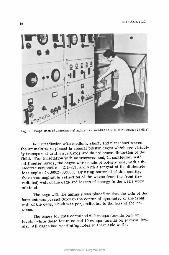
10 INTRODUCTION
!J • •• • I
• Fig. 2. Preparation of experimental animals for irradiation with short waves (15 MHz).
For irradiation with medium, short, and ultrashort waves the animals were placed in special plastic cages which are virtual-ly transparent to all wave bands and do not cause distortion of the field. For irradiation with microwaves and, in particular, with millimeter waves, the cages were made of polystyrene, with a di-electric constant e = 2.4-2.9, and with a tangent of the dielectric loss angle of 0.0002-0.0003. By using material of this quality, there was negligible reflection of the waves from the front (ir-radiated) wall of the cage and losses of energy in the walls were minimal.
The cage with the animals was placed so that the axis of the horn antenna passed through the center of symmetry of the front wall of the cage, which was perpendicular to the axis of the an-tenna.
The cages for rats contained 6-9 compartments on 2 or 3 levels, while those for mice had 10 compartments on several lev-els. All cages had ventilating holes in their side walls.
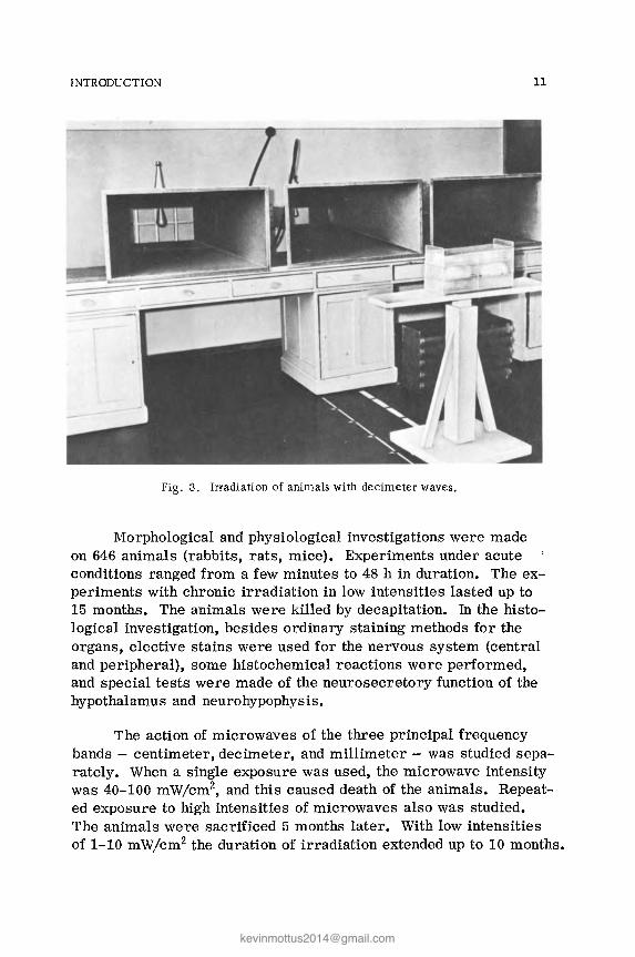
INTRODUCTION 11
Fig. 3. Irradiation of animals with decimeter waves.
Morphological and physiological investigations were made on 646 animals (rabbits, rats, mice). Experiments under acute conditions ranged from a few minutes to 48 h in duration. The ex-periments with chronic irradiation in low intensities lasted up to 15 months. The animals were killed by decapitation. In the histo-logical investigation, besides ordinary staining methods for the organs, elective stains were used for the nervous system (central and peripheral), some histochemical reactions were performed, and special tests were made of the neurosecretory function of the hypothalamus and neurohypophysis.
The action of microwaves of the three principal frequency bands - centimeter, decimeter, and millimeter - was studied sepa-rately. When a single exposure was used, the microwave intensity was 40-100 mW/cm2, and this caused death of the animals. Repeat-ed exposure to high intensities of microwaves also was studied. The animals were sacrificed 5 months later. With low intensities of 1-10 mW/cm2 the duration of irradiation extended up to 10 months.

12 INTRODUCTION
Fig. 4. Irradiation of animals with centimeter waves.
All investigations were carried out in greatest detail using microwave radiation (especially in the 10-cm band) 0 For compari-son, and to determine any special characteristics of their action, briefer studies under similar conditions using ultrashort-, short-, and medium-wave radiation are also reported.
Irradiation with ultrashort waves (191, 155, and 69 MHz) was also studied at high (350-5000 Vim) and low (20-150 Vim) inten-sities; short-wave radiation (14 MHz) was used in high (5000-9000 Vim) and low (2250 Vim) intensities. Irradiation of the experiment-al animals with medium waves was repeated over a period of 10 months in intensities of 1800 Vim and 50 Aim.

SECTION I
PHYSIOLOGICAL AND MORPHOLOGICAL CHANGES IN ANIMALS EXPOSED TO RADIO WAVES OF HIGH INTENSITY

Introduction
It was decided to describe the material relating to the action of high intensities of radio waves of all frequencies together. The reason for this decision was that the clinical picture of the hyper-thermia caused by high-intensity irradiation and the morphological changes are uniform in character.
The clinical picture of hyperthermia in animals irradiated by waves of high intensity is divided into five periods. The first period is that of the usual orienting reaction. In the second and third periods, even if the rat is anesthetized, it awakens. The slight erythema of the paws, tail, and ears in the second period changes into marked hyperemia; the arousal reaction is then fol-lowed by depression. In the fourth period the animal is recumbent, clonic convulsions and pareses are occasionally observed, and there is marked hyperemia of the paws, tail, ears, and tip of the nose and edema of the head and genitalia. Finally, in the fifth pe-riod the animals lie on their side and there is a copious discharge of blood-stained fluid from the nose and mouth. This period ends with death of the animals followed quickly by rigor mortis.
The duration of each period is determined by the frequency of the waves and the intenSity of the irradiation. In the case of short and ultrashort waves and microwaves the first two periods last from 1 to 50 min, the third from 2 to 20 min, and the fourth and fifth from 2 to 30 min.
During irradiation of animals, depending on the intensity of the radiation, changes in rectal temperature may occur in 3 phases (Michaelson et aI., 1961a,b; Howland et aI., 1961): a slight initial
15

16 SECTION I
rise of temperature, followed by a period of equilibrium, and final-ly a rapid rise of temperature leading to irreversible disturbance of thermoregulation and death of the animal. However, if the ani-mal is irradiated with microwaves of high intensity (> 100mW/cm2
for instance), the maximal rise of temperature occurs sooner and the animal dies more rapidly. In the investigations described be-low high intensities of irradiation were used and, as a rule, the rectal temperature of the animals was found to be increased by 2_6°C (to 42-45°C). The greatest rise of temperature was observed in the case of irradiation by 10-cm waves.
Comparison of the level of elevation of the body temperature during irradiation with waves of equal intensity, for instance with the shorter microwaves and infrared rays, i.e., in both cases by radiation which is largely absorbed in the skin, shows that the microwaves heat the skin more rapidly and to a high temperature, and death ensues much quicker (Deichman et al., 1959). It thus appears that the thermal effect is determined not only by the quan-tity of acting energy, and that even in the case of microwaves of high intensity, their biological action is not determined entirely by their thermal effect. This conclusion is very important, becase later, when the biological action of low intensities of irradiation (not raising the body temperature) is described, the importance of low (nonheating) intensities of irradiation in the biological effects produced by them will also be demonstrated. C
Intensities of irradiation giving rise to severe hyperthermia terminating in death differ for waves of different frequencies.
The way in which the survival period of the animals depends on the wavelength and intensity of irradiation is shown in Table 1.
Since the intensities of irradiation by waves of different fre-quencies are expressed in different units, namely, field intensity or power flux density (volts per meter, milliwatts per square cen-timeter), the results of the authors' investigations (Gordon and Lobanova, 1960; Nikonova, 1964; Fukalova, 1964) are given in ener-gy density units (ergs per cubic centimeter). With identical inten-sities of irradiation by microwaves (100 mW/cm2 or 33 '10- 6
erg/cm3) animals die quickest from the action of 10-cm waves (15 min). With equal intensities of irradiation by ultrashort and short

INTRODUCTION
TABLE 1. Survival of Animals after Irradiation by Radio Waves of Different Frequencies
Intensity of radiation Time (in min) and percent of animals
Waveband dying
Field inten-r Energy I sHy and pm density in 50% 100%
ergs· cm3
Medium (500 kHz) 8000V/m 2830 10-5 Nil Short. 5000 " 1 100· 10-0 100 (14.88 MHz) 9000 " 3564 10-6 10 Ultrashort . 5000 " 1100· 10-'; 5 69.7 MHz. 2000 " 176,10-6 100-120 130-200 155 " 700 " 21.5·IO-G 100-120 130-200 191 " 350 .. .5.410-6 100-150 160-200 Microwaves Decimeter. 100 mW/cm2 33.10-6 60 Centimeter 10 cm 100 .. 33,10-6 15 60
3 " 100 " 33·IO-G 110 Millimeter. 100 " 33.10-6 180
17
waves (5000 Vim or 1100.10-6 erg/cm3) animals exposed to ultra-short waves die within 5 min and those exposed to short waves af-ter 100 min. In the medium-wave bands irradiation at 8000 V /m did not cause death of the animals for many hours, whereas irradia-tion in the short-wave band at 9000 Vim caused rapid death (10 min) of all the animals (100%).
Hence, with a decrease in wavelength the energy density (ED) causing death of the animals decreases down to the microwave range and then rises slightly. Whereas medium waves with ED = 2830· 10- 6 do not cause death of animals over a period of many hours, short and ultrashort waves with much lower ED values (176.10- 6
to 5.4.10- 6 erg/cm3) lead to death of 50% of the irradiated animals at about the same times (after 100-120 min).
In the case of microwaves with equal ED, 50% of the animals die after times varying from 15 to 180 min. The 10-cm waves have a particularly rapid action and the clinical picture of the hyper-thermia is very severe.
Let us consider the morphological changes in the organs of four groups of animals exposed to irradiation by waves of different frequencies (microwaves, ultrashort, short, and medium waves).

Chapter 1
Morphological and Physiological Changes Following Exposure to
High-Intensity Microwaves (Centimeter, Millimeter, and Decimeter Bands)
Morphological Changes Following Exposure to High-Intensity Centimeter Waves
Morphological changes were investigated after a single ex-posure to 3-cm and 10-cm waves of high and medium intensities, causing phenomena of hyperthermia in animals.
The results showed that exposure to 3- and 10-cm waves is accompanied by functional and morphological changes.
The material can be divided into groups depending on the in-tensity of irradiation and the period of exposure. Since the mor-phological changes in the animals within each group were identical, the results will be described for groups as a whole.
G r 0 u p 1. After a single (25-40 min) exposure of animals to 3-cm and 10-cm waves of high intensity (40-100 mW/cm2),
severe clinical manifestations of hyperthermia terminating indeath were observed.
Autopsy revealed severe vascular disorders, in the form of hyperemia and multiple hemorrhages in the brain and meninges, the peritoneum, the pleura, and the pericardiumo Rigor mortis was severe. Microscopic examination revealed vascular disorders
19

20 SECTION I
in the nervous system and viscera, in the form of hyperemia, peri-vascular edema, and multiple small hemorrhages in the brain, myocardium (Fig. 5b,c), pleura, epicardium, and intestinal mucosa.
The vascular disorders were accompanied in the brain by edema of the oligodendroglia, with the appearance of drainage forms (Fig. 6b,c), acute swelling of nerve cells in different parts of the brain with solitary vacuoles in the cytoplasm of individual neurons (Fig. 7b), by homogenization and sometimes by fatty degeneration of single fibers of the myocardium (Fig. Sb,c), and by changes in the testicles, in which albuminous masses were deposited in the lumen of individual tubules and, in some cases, necrosis of single seminiferous tubules was observed (Fig. 9b). In every case the content of ribonucleoproteins was reduced in the epidermis and in its derivatives in the upper layers of the skin. Cloudy swelling was observed in the epithelium of the convoluted renal tubules and there was fatty degeneration of the liver cells. There was a total absence of proliferative response of the microglia in the brain and of the reticuloendothelial elements in the liver. This protective, adaptive response is suppressed by irradiation with high-intensity radio waves of various frequencies.
G r 0 u p 2. The animals were irradiated once (for 30 min) with pulsed IO-cm waves with an intensity of 20 mW/cm2• There were no deaths among the animals, which were sacrificed tmme-diately after the end of irradiation showing slight signs of hyper-thermia. Autopsy showed that the vascular disorders were not as severe as in the preceding group. Well-marked rigor mortis was found.
Histological examination showed acute swelling of nerve cells in various parts of the brain, with solitary vacuoles in the cytoplasm, swelling and homogenization of the muscle fibers of the myocar-dium, and initial degenerative changes in the form of cloudy swell-ing of individual hepatocytes and epithelial cells of the convoluted renal tubules, against the background of vascular disorders. There was no proliferative reaction of the microglia in the brain and no proliferation of reticuloendothelial elements in the liver.
After a single exposure of the animals to pulsed 3-cm waves for 30 min at an intensity of 20 mW/cm2, as a rule no evidence of hyperthermia was observed. None of the animals died. All were sacrificed immediately after the end of irradiation.

CHAPTER 1 21
. .. ", --, -, ..... ... .
,, - ...
• " .. • .. · -J: · • . .. I
• • •
Fig. 5. Comparative characteristics of vascular disorders after single exposure to high-intensity irradiation with waves of different frequencies. Hematoxylin-eosin: a) mul-ti pIe hemorrhages in stroma of testis following exposure to millimeter wa ves, 400 x; b) multiple hemorrhages in myocardium after exposure to centimeter waves, 200 x; c) multiple hemorrhages in brain after exposure to decimeter waves, 200 x; d) multiple hemorrhages in lumen of alveoli following irradiation by short waves, 160 x.
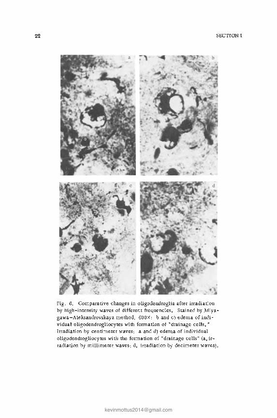
22 SECTION I
Fig. 6. Comparative changes in oligodendroglia after irradiation by high-intensity waves of different frequencies. Stained by Miya-gawa-Aleksandrovskaya method, 600X: b and c) edema of indi-vidual ollgodendrogliocytes with formation of "drainage cells." Irradiation by centimeter waves: a and d) edema of individual oligodendrogliocytes with the formation of "drainage cells" Ca, ir-radiation by millimeter waves; d, irradiation by decimeter waves).

Fig.
7.
Com
para
tive
eff
ects
on
nerv
e ce
lls
of t
he h
ypot
hala
mic
reg
ion
of i
rrad
iati
on b
y hi
gh-i
nten
sity
wav
es o
f di
ffer
ent
fre-
quen
cies
. S
tain
ed b
y N
issl
's m
etho
d:
a) v
acuo
lati
on o
f hy
poth
alam
ic n
erve
cel
ls a
fter
irr
adia
tion
wit
h m
illi
met
er w
aves
, 63
0 X
;
b) v
acuo
lati
on o
f hy
poth
alam
ic n
erve
cel
ls a
fter
irr
adia
[ion
wit
h ce
ntim
eter
wav
es,
570
X;
c) v
acuo
lati
on o
f hy
poth
alam
ic n
erve
ce
lls
afte
r ir
radi
a tio
n w
ith
deci
met
er w
a ves
, 63
0 X
; d)
vac
uola
tion
of
nerv
e ce
lls
and
swel
ling
of
thei
r cy
topl
asm
aft
er i
rrad
ia ti
on
wit
h sh
ort
wav
es,
320
X.
()
>-' "" c.:>

24 SECTION I
Fig. 8. ComparatiVe changes in muscle fibers of the myocardium after irradiation by high-intensity radio waves of different frequencies: a) un-
evenness of staining of muscle fibers after irradiation with millimeter waves. Hematoxylin-eosin, 330x; b) well marked unevenness of stain-ing and homogenization of myocardial muscle fibers after irradiation wi th centimeter waves. Hematoxylin-eosin, 330X; c) fatty degeneration of myocardial muscle fibers after irradiation with centimeter waves. Sudan III, 660 X; d) homogenization and unevenness of swelling of myocardial muscle fibers after irradiation with decimeter waves. Hematoxylin-eosin, 330 X.
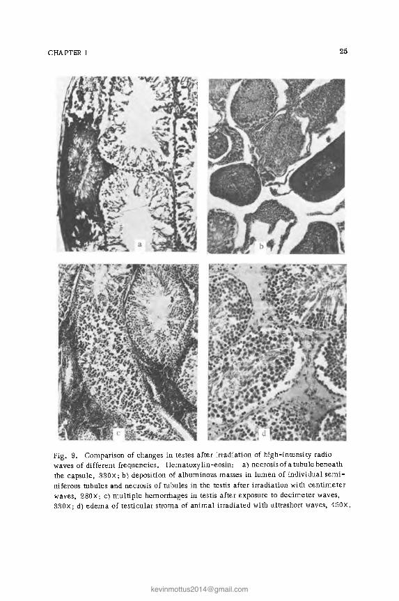
CHAPTER 1 25
Fig. 9. Comparison of changes in testes after irradiation of high-intensity radio waves of different frequencies. Hematoxylin-eosin: a) necrosis of a tubule beneath the capsule, 330x; b) deposition of albuminous masses in lumen of individual semi-niferous tubules and necrosis of tubules in the testis after irradiation with centimeter waves, 280X; c) multiple hemorrhages in testis after exposure to decimeter waves, 330X; d) edema of testicular stroma of animal irradiated with ultrashort waves, 450x.

26 SECTION I
Histological examination revealed slight hyperemia, perivas-cular edema in the brain, and swelling of the cytoplasm of the nerve cells with single tiny vacuoles, congestion and swelling of individual muscle fibers of the myocardium, with unevenness of their staining, and also congestion and slight cloudy swelling of the cytoplasm of the hepatocytes and epithelial cells of the convoluted renal tubules. Hyperemia and edema were present in the intestine, spleen, lungs, and testes. Comparison of the effects of pulsed 3-cm and 10-cm waves demonstrates that the 10-cm waves produce the more marked changes.
G r 0 u p 3. Morphological changes in the viscera and ner-vous system were studied in 75 albino rats exposed repeatedly (75 sessions) to pulsed and continuous irradiation with 10-cm waves and pulsed 3-cm waves of high intensity (40 mW/cm2) but with a very short duration of irradiation (5-10 min).
Severe clinical manifestations of hyperthermia were observed in the animals only in the first sessions of irradiation. Later they tolerated the irradiation satisfactorily, their body temperature re-turned to normal, and the gain in weight of the young experimental animals was the same as in the controls. The animals were de-capitated after 75 session::; of irradiation. At autopsy a varied degree of hyperemia was found. Rigor mortis was not so evident as in the preceding groups. The liver was flabby and yellow in color. The kidney tissue was dull, with a slightly swollen cortical layer.
Histological examination of the organs and tissues of the animals sacrificed immediately after repeated irradiation revealed moderate vascular disorders in the brain and viscera. Degenerative changes, in the form of shrinking and dark staining of individual cortical neurons and swelling of the cytoplasm with lysis of the tigroid substance and the appearance of small vacuoles in individ-ual cells of the hypothalamus, were observed in the brain cells. Swelling, unevenness of staining, and homogenization were found in individual groups of myocardial fibers, occasionally they showed fatty degeneration, and there was slight fatty degeneration also of individual hepatocytes. A brisk proliferative response of the mi-croglia was present in the brain (Fig. l4a) and proliferation of the reticuloendothelial elements in the liver (Fig. l4b), i.e., a brisk proliferative reaction of the reticuloendothelial elements partic-

CHAPTER 1 27
ipating in both local and general protective and adaptive reactions of the body. In all irradiated animals the degenerative changes in the nervous system and viscera described above were focal in character and cells of normal configuration could always be seen side by side with pathologically changed cells.
Although the irradiated animals had slight degenerative changes in the viscera and nervous system, together with ill-defined vascular disorders, they remained apparently healthy dur-ing their period of survival, reflecting the well-developed com-pensatory powers of the body as a whole.
The next step was to investigate morphological changes in particularly sensitive structures of the nervous system. For this purpose, changes were studied in the receptors of the skin and interoceptors in various receptive fields of the viscera in 25 ani-mals (rabbits and rats) after irradiation with lO-cm waves of high intensity.
Changes in the skin receptors were particularly interesting because during exposure of the living body to radio waves the skin is the first barrier which they meet. Because of its rich supply of afferent nerve endings the skin is a powderful receptive field, whose response to the action of radio waves largely determines the response of the body as a whole. It was no less interesting to determine changes in the interoceptors of the viscera for, accordi'ng to published observations, the interoceptors of the viscera are very sensitive to various harmful agents, even of minimal character.
In recent years Lavrent'ev's school has amassed an extensive material on the morphology and pathology of the sensory innervation of the viscera. They have shown that sensory fibers participating in the innervation of the viscera arise from neUrons of the spinal and, sometimes, of the cranial ganglia.
Interoceptors of the viscera help to maintain the relative constancy of the internal medium which is a condition of the nor-mal existence of the organism. If, however, stimulation is exces-sive, pathological changes may arise. Chernigovskii states that resonance arising in the nervous system due to stimulation of re-ceptive fields embraces very many physiological processes. In other words, stimulation arising from interoceptive fields disturbs the activity of the somatic muscles, the smooth muscles, the res-

28 SECTION I
piratory, circulatory, and hematopoietic systems, and the work of the kidneys and adrenals, and it upsets metabolism.
According to Chernigovskii, this wide irradiation is a char-acteristic feature of stimulation from interoceptors in general, and from vascular interoceptors in particular.
Many different workers have studied changes in the interocep-tor system of the viscera in diseases and during exposure to harm-ful factors. They have studied changes in the interoceptor system of several reflexogenic zones: 1) the gastrointestinal tract, 2) the periendomyocardium, 3) the arch of the aorta, carotid sinus, and walls of large blood vessels. Investigations have been made in patients with cancer, peptic ulcer, intestinal obstruction, hyper-tension, diphtheria, tuberculosis, and uremia, and subjects exposed to ultrahigh-frequency and x-ray irradiation and to experimental anoxia (Dolgo-Saburov; Kupriyanov; Lavrent'ev; Plechkova). Mor-phological changes thus revealed in receptors of the various re-ceptive fields were basically similar and consisted essentially of irritation phenomena and degenerative changes expressed to dif-ferent degrees.
Changes in the interoceptors resulting from exposure to superhigh-frequency radiation were studied by Pervushin (1957), who showed that reversible and irreversible changes take place in the interoceptors of the heart under these conditions.
According to Pervushin, superhigh-frequency irradiation acts primarily on afferent fibers of the spinal ganglia, and changes in the peripheral components of the autonomic nervous system are slight in degree.
In the present investigation no encapsulated or free nerve endings were found. Mainly thin afferent (sensory) nerve fibrils of the skin and viscera were studied. The fact that these fibrils belonged to sensory (afferent) fibers in every case was proved by their origin from myelinated nerve trunks (traced by the study of serial sections). The object of the investigation was to show changes in afferent nerve fibrils of the skin and viscera during irradiation, and whether these were "boutons terminaux," preterminals, or larger sensory nerve fibrils was of no fundamental importance.
To begin with the receptor system of the viscera and skin was studied in 10 healthy control animals from which pieces of the

CHAPTER 1 29
receptive (reflexogenic) zones were taken: from the skin of the ear and thigh, the urinary bladder, esophagus, stomach, intestine, atrium, and arch of the aorta.
Examination of all the material showed, more so in some cases than in others, thin, delicate myelinated nerve fibers enter-ing the smooth muscles or connective tissue of an internal organ or the epithelium of the skin, dividing into small twigs branching in different directions and having the appearance of delicate, thin fibrils. Sensory nerve fibrils were then investigated in the various receptive fields of the viscera and skin of animals irradiated with centimeter waves of different intensities.
At the same time, nerve cells of the spinal sensory ganglia (cervical, thoracic, lumbar, and sacral), the autonomic ganglia (solar plexus, ganglia of the vagus nerve), and of the hypothalamic region were investigated in the same animals.
Considerable changes were found in the sensory nerve fibers in the receptive fields of animals sacrificed immediately after a single exposure to irradiation with pulsed 10-cm waves of high intensity, ranging from 40 to 100 mW/cm2, for 15 min. Since the changes in the nerve fibers of the different receptive fields were more or less identical as a manifestation of the nonspecific re-sponse of the body, and the only differences were in the degree of their intensity, they will be described collectively. First, in all c
the viscera and skin investigated, nerve fibrils were more numer-ous than in the control animals, a result which can evidently be explained by the increased argyrophilia of the nerve fibrils of these receptive fields and their better impregnation with silver. In-creased argyrophilia of the afferent nerve fibrils is evidence of their irritation. Frequently strongly argyrophiliC, black, swollen fibrils with irregular swellings, pools of axoplasm, and even frag-mentation were seen (Figs. 10c and lla). The thin sensory nerve fibrils were on the whole affected to an equal extent in all the re-ceptive fields investigated, but comparison of the changes in the sensory nerve fibrils of the viscera and skin revealed that those in the skin were most severely affected, the lesion involving a larger number of nerve fibrils and the changes being more severe in character, sometimes involving fragmentation of the fibrils.
Swelling of the cytoplasm and lysis of the tigroid substance in the center of the cell, with ectopia of the nucleus, were found

30 SECTION I
Fig. 10. Comparison of changes in sensory nerve fibers of receptive fields of the skin after irradiation by high-intensity waves of different frequencies. Bielschowsky-Gros. a) Thickened myelinated cutaneous nerve fibers, with increased argyrophilia, breaking up into fragments after irradiation with millimeter waves, 400 x; b) axons of myelinat-ed cutaneous nerve fibers (large myelinated trunks) are strongly argyrophilic and have irregular swellings, with fragmentation in some places, after irradiation with millimeter waves, 350 X; c) very marked argyrophilia, irregular swellings, tortuosity of cutaneous nerve fibers after irradiation with centimeter waves, 400 x; d) delicate network of thin, unchanged cutaneous fibers after irradiation with decimeter waves, 400 x; e) increased argyrophilia and irregular swellings of cutaneous nerve fibers after irradiation by short waves, 400 x.

CHPATER 1 31
Fig. 11. Comparison of changes in sensory nerve fibers of viscera after irradiation by high -intensi ty waves of different frequencies. Bielschowsky-Gros. a) Very marked argyrophilia with irregular swellings of a myelinated nerve fiber of the atrial myo-cardium after irradiation with centimeter waves, 440X; b) side by side with thin, delicate fibers, a strongly argyrophilic fiber, breaking up in some places into frag-ments, is seen in the urinary bladder after irradiation by decimeter waves, 600X; c) very marked argyrophilia, irregular thickening of sensory nerve fibers in the myocardium after irradiation by ultrashort waves, 440X
in the nerve cells of the spinal sensory ganglia. Some cells were seen with swollen cytoplasm containing tiny vacuoles. Signs of karyocytolysis and death of individual neurons were observed. The nerve cells of the autonomic ganglia were only slightly altered.
With all intensities of irradiation the worst affected struc-tures were the small sensory fibrils, i.e., the preterminal por-tions of the receptor system, and the larger afferent fibrils, in agreement with the findings of Dolgo-Saburov, Kupriyanov, and Pervushin, who regard the preterminal portions of the afferent fibers as the most vulnerable.
The changes discovered in the sensory nerve fibers of the receptive fields of the viscera and skin following exposure to cen-timeter waves are not specific. Similar changes have been found

32 SECTION I
by other workers (Lavrent'ev, Plechkova, Falin, Vyropaev, Gusei-nov, Rakhmatullin, Alekseev, etc.) in various diseases and after exposure to various harmful agents.
As was mentioned above, after irradiation with centimeter waves of high intensity the damage of the sensory fibrils of the skin was particularly severe and the degenerative changes in those fibrils progressed as far as fragmentation of individual fibrils. The reason for this may be that the skin is the first barrier in the path of the radio waves, and part of the energy which falls on it is absorbed by it.
Parallel with the changes in the sensory nerve fibers of the skin, after irradiation with centimeter waves histochemical changes were studied in the skin itself. The content of ribonucleoprotein (RNP) in the skin of the irradiated animals was determined by Brachet's reaction. The results showed a decrease in the RNP content in the epidermis and its derivatives and, in particular, in the surface layers of the skin (Fig. l3b).
When the morphological changes observed in animals exposed to pulsed and continuous 10-cm waves were compared, they were found to be more severe after exposure to the pulsed waves.
Comparison of the morphological changes observed after exposure to 10-cm and 3-cm waves of high intensity showed that they were more severe after exposure to the 10-cm waves.
Morphological Changes Following Exposure to High-Intensity Millimeter Waves
Half of the animals receiving a single exposure to millimeter waves with an intensity of 100 mW/cm2 for 180 min died 3 h after the end of irradiation with evidence of marked hyperthermia. The other half of the animals were sacrificed with a similar clinical picture. At autopsy severe congestion and small hemorrhages were found in the brain and viscera. Rigor mortis was very pro-nounced.
On histological investigation of the organs of these animals, marked vascular disturbances were present, eVidently associated with the hyperthermia. They consisted of severe hyperemia of the viscera and brain, perivascular edema, and multiple small hemorrhages in the brain, myocardium, liver, kidneys, mucous

CHAPTER 1 33
membrane and submucosa of the intestine and pancreas, beneath the capsule and the stroma of the testes (Fig. 5a), and in the lumen of the alveoli. Acute swelling and a few vacuoles also were found in the hypothalamic nerve cells (Figs. 7a), together with edema of the oligodendroglia with the formation of "drainage" cells (Fig. 6a), irregular staining and homogenization of myocardial muscle fibers (Figs. Sa), edema of the testicular stroma, necrosis and homogenization of solitary seminiferous tubules, particularly those located beneath the capsule (Fig. 9a), swelling of the epidermis of the skin, and edema and hyperemia of the dermis.
Histochemical investigation showed a decrease in the RNP content in the cells of many visceral organs and ofthe nervous system. However, these changes were most conspicuous in those organs which normally have a high RNP content in the cytoplasm of their cells. These include: nerve cells, the glandular epithelium of the gastrOintestinal tract and pancreas, the epithelium of the bronchi and trachea, cells of the secondary follicles of the spleen, the endothelium of the capillaries, the ependyma of the ventricles of the brain, and the epidermis of the skin and its derivatives. The changes in RNP content varied from slight pallor of the cytoplasm to its complete transparency. Cells of the epidermis of the skin and its derivatives were particularly severely affected (Fig. 13a).
The content of deoxyribonucleoproteins (DNP) in the viscera, was more stable. Only very slight changes were found in the DNP content in the cell nuclei of tissues affected by cloudy swelling (hepatocytes, epithelium of the convoluted renal tubules, sper-matogenic epithelium of the testes) and in the hypothalamic nerve cells with signs of karyocytolysis.
A special feature of the changes in RNP content in the cells of the epidermis and its derivatives in the skin should be em-phasized. The changes in the derivatives of the epidermis dimin-ished in intensity toward the deep layers of the skin.
Changes in the sensory nerve fibers of the receptive fields of the skin were very severe. They took the form of argyrophilia and the appearance of beads and pools of axoplasm, or in some cases, fragmentation of the cutaneous nerve fibrils (Fig. lOa). In-creased argyrophilia and irregular swellings were found even in fibers of the large myelinated trunks in the dermis (Fig. lOb). Nerve fibrils of the receptive fields of the viscera (myocardium,

34 SECTION I
aorta, esophagus, intestine, stomach) were much less severe and signs of irritation were found. If the animals survived for a long period after irradiation, cloudy swelling was observed in the epi-thelium of some convoluted renal tubules and hepatocytes, and in a few cases there was fatty degeneration of the hepatocytes and the epithelium of a few convoluted renal tubules.
Animals exposed to a single irradiation with millimeter waves in an intensity of 140 mW/cm2 for 15 min did not die. Signs of hyperthermia were observed, but they were less marked than in the preceding group. In all the Viscera, the skin, and the ner-vous system, histological examination revealed vascular disorders less severe than in the preceding group, but still of considerable intensity. Acute swelling of the nerve cells was found in the brain, especially in the hypothalamus, with the appearance of soli-tary vacuoles in their cytoplasm and occasionally with signs of karyocytolysis. Unevenness of staining and homogenization of the myocardial muscle fibers, edema of the stroma and homogeniza-tion of individual seminiferous tubules (necrobiosis), subcapsular hemorrhages in these organs, cloudy swelling in the epithelium of the convoluted renal tubules and, to a lesser degree, in the hepatocytes, and slight thickening of the alveolar septa through hyperemia and edema were found. After irradiation in an inten-sity of 40 mW/cm2, changes also were found in the sensory nerve fibers of the cutaneous receptive fields. The fibers were strongly argyrophilic and had irregular swellings and pools of axoplasm on them. The sensory fibrils of the viscera were relatively un-changed. Nerve cells of the spinal sensory ganglia, just as after irradiation with 10-cm waves in an intensity of 100 mW/cm2,
showed swelling of the cytoplasm in individual cases, with lysis ofthe tigroid substance in the center of the cell and ectopia of the nucleus. Phenomena of karyocytolysis with death of individual neurons (Fig. 12) were evident. Since millimeter waves are absorbed by the skin, the changes in the nerve cells of the spinal ganglia can be attri-buted only to reflex mechanisms.
Histochemical tests showed a decrease in the RNP content, especially in the cells of the epidermis and its derivatives in the skin. The decrease in RNP content in the viscera was much less marked.
The DNP content in the cell nuclei of the viscera was un-changed, but in the nuclei of the epidermis it was considerably

CHAPTER 1
Fig. 12. Swelling of cytoplasm with tigrolysis and ectopia of the nucleus in some cells, and with evidence of karyocytolysis and death of the neurons in other cells in a spinal ganglion after irradiation with millimeter waves in high intensity. Nissl's stain, 450x.
reduced. A decrease in the DNP content was also observed in foci of cloudy swelling in the hepatocytes and epithelium of the convoluted renal tubules and also in the nerve cells of the hypo-thalamic region showing signs of karyocytolysis.
Morphological Changes Following Exposure to High-Intensity Decimeter Waves
35
The animals were irradiated with decimeter waves in an intensity of 100 mW/cm2 for 60 min; 50% of the animals died with manifestations of hyperthermia.
Macroscopic examination revealed clearly defined features of hyperthermia in all the animals, in the form of vascular dis-orders (very marked congestion of the viscera and brain) and rigor mortis. Microscopically, evidence of perivascular edema and multiple small hemorrhages were found in the brain (Fig. 5c) and also in the liver, lungs, myocardium, and stroma of the testes (Fig. 9c).
Against the background of the vascular disorders, acute swelling of the brain neurons was observed, together with the ap-pearance of solitary large vacuoles in the hypothalamic nerve cells (Fig. 7c), edema of the oligodendroglia (Fig. 6d), and swelling and

36 SECTION I
homogenization and unevenness of staining, and sometimes fatty degeneration of the myocardial muscle fibers (Fig. 8d). Slight cloudy swelling was seen in the hepatocytes and the epithelium of the convoluted renal tubules.
Changes in the sensory nerve fibers in various receptive fields of the viscera (myocardium, aorta, esophagus, intestine, stomach, urinary bladder, etc.) were very marked. The changes consisted of argyrophilia of the nerve fibrils, tortuosity, the ap-pearance of beads and pools of axoplasm, and sometimes fragmen-tation of individual nerve fibers. However, no damage was found to the sensory nerve fibers of the skin. They were thin and delicate, and they had no swellings or pools of axoplasm (Fig. 10d).
Histochemical tests showed a decrease in the RNP content in the cytoplasm of cells of the viscera and nervous system, more conspicuous in organs normally rich in RNP (nerve cells, glandular epithelium of the gastrointestinal tract and pancreas, bronchial epithelium). The RNP content in the skin was not reduced (Fig. 13c).
Considerable vascular disorders were found in the testes, in the form of edema of the stroma and small hemorrhages beneath the capsule and in the stroma of the organ.
* * * Comparison of the morphological changes found in the ani-
mals after exposure to microwaves of all frequencies in high in-tensity shows that these changes were uniform in character, i.e., they were due mainly to the thermal action of the radiation.
High intensities of irradiation with microwaves of different frequencies caused death of the animals with clinical features of severe hyperthermia. The morphological changes in the tissues and organs of the irradiated animals consisted of severe vascular disorders, acute swelling and vacuolation of the cytoplasm of the nerve cells in various parts of the brain, and unevenness of stain-ing, homogenization, and fatty degeneration of the myocardial fibers. However, despite the fact that the thermal effect masked all the finer changes, this does not mean that all the changes can be ascribed to the thermal effects alone. Exposure to microwaves gives rise to some phenomena of a special kind. For example, despite the high intensity of irradiation with decimeter waves, the sensory

CHAPTER 1 37
"
..... . " I
(.
Fig. 13. Comparison of histochemical changes in the skin (RNP content) after irradiation by waves of different frequencies in high intensity. Brachet's reaction: a) sharp decrease in RNP content in epidermis and its derivatives in top layers of skin with integrity of RNP in epidermal derivatives in deeper layers of the skin after irradiation with millimeter waves. 120x; b) decrease in RNP content in epidermis and its derivatives in upper layers of skin with integrity of RNP in epidermal derivatives in deeper layers of the skin after irradiation with centimeter waves. 200X; c) normal content of RNP in epidermis of skin and its deriv-atives after irradiation with decimeter waves. 160x.
nerve fibrils of the skin were unchanged. Yet, at the same time, they were severely affected by exposure to centimeter and, in par-ticular, to millimeter waves. This difference can be explained by the fact that millimeter waves are absorbed by the skin, and most of their effects are observed on the skin and its sensory nerve fibers (the other changes in the viscera are eVidently reflex in character). This was also confirmed by the clearly defined changes in the nerve cells of the spinal sensory ganglia, which showed evi-dence of irritation (swelling, central tigrolysis of the cytoplasm, and ectopia of the nuclei). Although after exposure to microwaves of various frequencies and of high intensity the differences between the morphological pictures were thus apparently masked, it must be noted that the lO-cm waves gave rise to the severest effects, for death took place earlier, the picture of hyperthermia was more marked, and the vascular disorders were more extensive. Second place in order of severity of effects was occupied by the decimeter waves, and third place by millimeter waves.
The intensity of the biological action of microwaves is deter· mined by the intensity and frequency of the waves and the duration of exposure to them.

38 SECTION I
Without dwelling in detail on the biological effects of high-intensity irradiation (measured in hundreds of milliwatts per square centimeter), it will merely be noted that such intensities lead to severe hyperthermia and, if the exposure is long enough, to death. The morphological changes in the tissues and organs of the animals consisted of severe vascular disturbances, accompanied by less marked degenerative changes in the nervous system and viscera, presumably on account of the earlier death of the animals. No proliferative reaction of the microglia was observed in the brain or of the reticuloendothelial elements in the viscera. This protective and adaptive reaction was eVidently inhibited by the effects of the high-intensity microwaves. This pattern is characteristic of the whole band of microwaves and is due to their considerable heating effect.
Millimeter waves, which are absorbed completely in the skin and act on its sensory nerve fibers, irritating them and sending an intensive flow of impulses along the afferent fibers to the spinal ganglia and on into the brain, have a lower thermal threshold. How-ever, if the irradiation is of high intensity (40 mW/cm2) the dynam-ics of the temperature response is such that the integral heating effect is more marked in the case of centimeter waves. These waves, which are absorbed only partially in the skin and which penetrate deeper, evoke a well-defined temperature response to irradiation of high intensity eVidently as a result of their direct action on the tissues and on the thermoregulatory centers.
So far as decimeter waves are concerned, they are virtually unabsorbed in the skin and penetrate deeper into the body, where they evoke a thermal effect if the PFD levels are high.

Chapter 2
Morphological Changes in Animals Following Exposure to Ultrashort Waves (69.7, ISS, and 191 MHz) of High Intensity
After a single exposure to ultrashort waves (69.7 MHz, in-tensities 5000 and 2000 Vim) the animals died with well marked features of hyperthermia. At autopsy severe hyperemia and small hemorrhages were found in the brain and viscera, with well-devel-oped rigor mortis. Microscopic investigations shown marked vas-cular disturbances in the brain and viscera, with hyperemia and perivascular edema in the lungs, brain, and liver, edema of the myocardial and testicular stroma, and multiple perivascular hem-orrhages in the brain, lungs, liver, kidneys, myocardium, and testes. Because death occurred quickly (5 min after irradiation in an in-tensity of 5000 Vim), degenerative changes had not had time to develop in the viscera and they were slight in degree. Acute swell-ing of the cytoplasm was observed in the nerve cells in various parts of the brain. After irradiation with ultrashort waves in an intensity of 2000 Vim the animals survived rather longer (up to 3 h), and the picture of vascular disturbances as described above was accompanied by degenerative changes in the nerve cells of the brain and viscera which were rather more clearly defined, and consisted of swelling and vacuolation of the cytoplasm of nerve cells in the thalamus and hypothalamus. Marked argyrophilia and irregular swellings and pools of axoplasm were found in the sen-sory nerve fibers of the skin and sensory fibrils of the myocardium, intestine, urinary bladder, esophagus, and elsewhere (Fig. llc).
39

40 SECTION I
Homogenization and unevenness of staining of the myocardial muscle fibers, swelling of these fibers (with edema of the stroma), and cloudy swelling with, occasionally, fatty degeneration of the hepatocytes and epithelium of the convoluted tubules were found.
Animals irradiated with ultrashort waves with a frequency of 155 MHz and intensity of 1000 Vim and with a frequency of 191 MHz and intensities of 700 and 350 V 1m died 17-200 min later (depending on the intensity of the irradiation) with marked features of hyperthermia. Changes similar to those described above, but more severe, were found.
Hence, after exposure to high intensities of ultrashort waves (5000, 2000, and 1000 Vim) with frequencies of 69.7, 155, and 191 MHz, death quickly ensued; the animals showed marked evidence of hyperthermia and severe vascular disorders (hyperemia, peri-vascular edema, and multiple hemorrhages). The vascular dis-orders provided a background for ill-defined degenerative changes in the nerve cells of the brain, the muscle fibers of the myocardium, and other parenchymatous organs.
If the animals survived longer (up to 3 h), vascular distur-bances in the nervous system and internal organs were similar to those described above, and they were accompanied by more defi-nite degenerative changes in the nerve cells, and marked signs of degeneration of sensory nerve endings in the skin and viscera. Degenerative changes were found in the muscle fibers of the myo-cardium, the spermatogenic epithelium of the testes, the hepato-cytes, and the epithelium of the convoluted renal tubules.

Chapter 3
Morphological Changes in Animals Exposed to Short and Medium Radio Waves
of High Intensity
G r 0 u p 1. After a single exposure to irradiation with high-intensity (9000 and 5000 V 1m) short waves the animals died with signs of marked hyperthermia in 10-1000 min. At autopsy congestion of the brain and meninges and focal hemorrhages in the brain and meninges and in the serous membranes were found. Rigor mortis was severe.
Microscopic investigation revealed considerable vascular disturbances in the brain and all the viscera: congestion, stasis, perivascular edema, and multiple small or larger perivascular c
hemorrhages in the brain, kidneys, liver, and lungs. Hemorrhages into the lumen of the alveoli also were present in the lungs (Fig. 5d).
If death took place early, the degenerative changes in the nerve cells and viscera were slight in degree. If the animals sur-vived longer, the vascular disturbances were marked and were ac-companied by acute swelling of nerve cells in various parts of the brain, with vacuolation of the cytoplasm in the hypothalamic neu-rons (Fig. 7d).
The sensory nerve fibrils of the viscera and skin showed marked argyrophilia and irregular swelling in different receptive fields (Fig. 10e). Homogenization and unevenness of staining were found in the muscle fibers of the myocardium. Cloudy swelling and, sometimes, fatty degeneration affected individual groups of hepatocytes and epithelial cells of the convoluted renal tubules.
41

42 SECTION I
Consequently, after irradiation with short waves of high in-tensity (9000 and 5000 Vim), leading to death in 10 to 100 min with well-marked evidence of hyperthermia, acute and severe vascular disturbances can be detected morphologically in the nervous sys-tem and viscera and are accompanied by ill-defined degenerative changes in the nerve cells, the muscle fibers of the myocardium, and other internal organs. Evidence of irritation of cutaneous sen-sory nerves and of sensory fibrils of the viscera is clearly visible.
After irradiation with high intensities of short and ultrashort waves the differences between the morphological picture associated with the action of each particular frequency disappear. Everywhere vascular disturbances are predominant, with ill-defined degener-ative changes and irritation of sensory nerve fibers of the various receptive fields of the skin and viscera.
However, the impression is obtained that the changes are more severe and developed earlier in the case of irradiation with ultrashort waves. This correlates with the clinical evidence of earlier death after irradiation with ultrashort waves.
G r 0 u p 2. Irradiation with medium waves with an inten-sity of 8000 V 1m. With waves of this length it was impossible to obtain intensities sufficiently high (even in the case of long or re-peated exposures) to cause fatal hyperthermia, because the field voltages of up to 8000 Vim which were used were only on the threshold for producing a thermal effect. The effects of waves of these frequencies will accordingly be discussed in the section on the chronic action of low intensities not inducing a thermal effect.

Chapter 4
Comparison of Morphological Changes Following Exposure to Radio Waves of
High Intensity and of Different Frequencies
Morphological changes associated with exposure to micro-waves and ultrashort, short, and medium radio waves can be use-fully compared.
In the first series a morphological study was made of the organs and tissues of animals (305) irradiated with radio waves of high intensity and of various frequencies: a) microwaves with in-tensities of between 40 and 100 mW/cm
2
, b) ultrashort waves with' intensities of 5000 and 2000 Vim; c) short waves with intensities of 9000 and 5000 Vim. The animals of all groups died with marked signs of hyperthermia. At autopsy, rigor mortis was severe. The rectal temperature of the cadaver as a rule was very high, some-times reaching 40-44°C. The nasal mucosa were cyanosed, and the skin of the paws, ears, and tail strongly hyperemic. Marked con-gestion of the brain and meninges and petechial hemorrhages be-neath the serous membrane of the intestine, the pleura, and the peri-cardium, and in the gastric mucosa and the meninges and brain tis-sues were found. The brain tissue was congested and edematous. The liver was severely hyperemic, and sometimes had an icteric tinge. The spleen was severely congested, and dark liquid blood exuded from its cut surface. The kidneys were congested and flab-by and the capsule stripped easily.
43
44 SECTION I
Microscopic investigation showed considerable vascular changes in the nervous system and viscera: severe hyperemia, marked perivascular edema, and multiple small hemorrhages in the stroma of the testes, brain, myocardium, lungs, liver, kidney, and intestinal wall (Fig. 5a,b,c,d). Severe hyperemia and edema in the brain affected both the brain tissue and the meninges; stasis and perivascular and pericellular edema were evident. Changes in the oligodendroglia consisted of edema and the formation of drainage cells (as described by P. E. Snesarev), which are as-sociated with the presence of acute vascular disturbances accom-panied by cerebral edema and disturbances of water and mineral metabolism (Fig. 6a,b,c,d).
Damage to the central nervous system was the most con-spicuous feature. In sections stained by Nissl's method and by the histochemical reaction for ribonucleoproteins, changes were found in nerve cells in various parts of the brain, more especially in the hypothalamus. Acute swelling of the cytoplasm of the neurons was accompanied by tigrolysis and by a sharp decrease in the ribonucleoprotein content. In animals which survived longer, tiny vacuoles appeared in the cytoplasm of the swollen nerve cells, and their contest gave a negative reaction for lipids (Fig. 7a,b,c,d).
Similar changes (vascular disorders and degenerative changes in nerve cells) took place in the spinal cord and spinal sensory ganglia, and were much less marked in the autonomic ganglia.
After the central nervous system, the myocardium was next most severely affected. Microscopic examination of the heart muscle showed unevenness of staining of the myocardial muscle fibers: some fibers stained very intensely, homogeneously, and had lost their cross-striation, while others, on the other hand, were pale with a distinct cross-striation. Sometimes fatty degeneration of individual muscle fibrils was observed (Fig. 8a,b,c,d). Next in order of severity of damage were the testes. Congestion and edema of the interstitial tissue were clearly defined. Degeneration of the spermatogenic epithelium and, sometimes, necrosis of the tubules or the liberation of albuminous masses into the lumen were observed in individual seminiferous tubules, most frequently those situated beneath the capsule (Fig. 9a,b,c,d).
Congestion and perivascular edema were very evident in the lung tissue, and here and there small perivascular hemorrhages

CHAPTER 4 45
and hemorrhages into the lumen of the alveoli could be seen (Fig. 5d). Sometimes there was focal edema of the alveoli, associated with focal emphysema. The liver tissue was severely hyperemic and sometimes edematous; the structure of the hepatic columns was disturbed, and Desse's spaces were widened. The hepatocytes were swollen, with palely stained nuclei and granular cytoplasm. Side by side with these cells, others with darkly stained cytoplasm could be seen. In the kidneys severe hyperemia and perivascular edema were ac-companied by tiny perivascular hemorrhages and well-marked evi-dence of cloudy swelling in the epithelium of the convoluted tubules. Hyperemia in the spleen was severe, with obliteration of the pat-tern of the follicles.
It must be emphasized that after a single exposure to radio waves of high intensity no proliferative response of the microglia in the brain or of the reticuloendothelial elements in the liver was observed. This protective and adaptive response was evidently inhibited by the action of the high-intensity radio waves.
The differences between the morphological picture associated with the action of radio waves of different frequencies tended to be obliterated in the case of high-intensity irradiation. Vascular dis-turbances in the nervous system and viscera dominated the patho-logical picture.
Compared with the vascular disturbances, the degenerative C
changes in the nervous system and viscera were not so clearly defined. This was probably because they had not had time to de-velop because of the early death of the animals, and the usual mor-phological methods were unable to reveal them clearly. However, some morphological differences between the effects of individual frequencies of radio waves could be identified. For example, af-ter exposure to millimeter and centimeter waves necrosis of in-dividual seminiferous tubules located beneath the capsule was most frequently observed (Fig. 9a,b), while after exposure to decimeter and ultrashort waves the testicular damage was more uniformly distributed and vascular disturbances in the tissues of the testes were predominant (Fig. 9c,d).
More precise histological methods (elective staining of nerve tissue and investigation of receptors) and histochemical methods revealed more distinct changes in the nervous system and viscera. Sensory nerve fibers of various receptive fields (skin of the ear

46 SECTION I
and thigh, the myocardium, aorta, esophagus, intestine, stomach, and urinary bladder) showed definite signs of irritation as reflected by marked argyrophilia, irregular swellings and pools of axoplasm, or even fragmentation of the fibrils (Fig. 10a,b,c,d,e).
Characteristic differences between effects of different wave bands were also clearly revealed by a study of the lesions of the sensory nerve fibers in the different receptive fields. After exposure to centimeter and, in particular, to millimeter waves the severest changes took place in the cutaneous sensory nerve fibers (Fig. 10a,b,c), whereas after exposure to decimeter waves the cutaneous sensory nerve fibers were unchanged (Fig. 10d), but by contrast the sensory nerve fibrils of the viscera were most severe-ly damaged by the action of decimeter waves (Fig. llb).
Simultaneously with the sensory nerve fibers in the various receptive fields of the skin and internal organs, changes were studied in the spinal sensory ganglia, where the nerve cells showed central tigrolysis of the cytoplasm and ectopia of the nuclei. Some-times signs of karyocytolysis were present, with death of individual neurons (irradiation with centimeter, decimeter, and ultrashort waves). Changes were found in the neurons of the spinal sensory ganglia after irradiation with millimeter waves (Fig. 12). Since millimeter waves are absorbed in the skin, changes in the neurons of the spinal ganglia are evidently reflex in origin.
Histochemical investigations revealed a decrease in the con-tent of ribonucleoproteins in the cytoplasm of the cells of many internal organs and of the nervous system. The content of deoxy-ribonucleoproteins was more stable.
Histochemical changes were more severe in the skin (de-crease in the ribonucleoprotein content in the epidermis and its derivatives), especially after exposure to millimeter and centi-meter waves, but not after exposure to decimeter waves (Fig. 13a, b,c).
Comparison of the morphological changes found in animals exposed to radio waves of all these frequencies and of high inten-sity confirms, and this must be stressed repeatedly, that when high intensities of irradiation are given the morphological changes are virtually uniform in character and are associated primarily with the thermal effect.

CHAPTER 4 47
However, some characteristic differences can be found as a result of exposure to the different wavelengths, and these can be attributed to the characteristic action of the waves rather than to hyperthermia. For example, after exposure to millimeter waves the sensory nerve fibers of the skin are particularly severely af-fected, and they exhibit well-defined histochemical changes, where-as after exposure to decimeter waves the skin is almost unaffected. The skin is also damaged by the action of ultrashort and short waves.
Exposure to decimeter waves leads to the severest changes in the sensory nerve fibers of the viscera.
A group of animals was also exposed for a short time (5 min) to irradiation by high-intensity (40 mW/cm2) radio waves in the IO-cm band, which was repeated altogether 75 times.
The animals tolerated the first exposures to irradiation bad-ly. Their body temperature rose sharply, they developed erythema of the limbs and ears, and they lay on their sides. All these phe-nomena disappeared after 2-3 h. After the subsequent exposures the animals showed less marked effects. In animals sacrificed after 75 exposures (immediately after the last irradiation) vas-cular disturbances were found but they were only slight by com-parison with those in the groups of animals receiving lethal ir-radiation. The rigor mortis also was not so severe. However, microscopic investigation showed hyperemia and edema of the brain, the lungs, the interstitial tissue of the myocardium, the kid-neys, and liver. The vascular disturbances were accompanied by degenerative changes which were most marked in the nervous sys-tem, myocardium, and testes. Shrinking of the cortical nerve cells was accompanied by acute swelling of the cytoplasm of nerve cells in the thalamus and hypothalamus, with tigrolysis and vacuolation. Sensory nerve fibers in the skin and viscera showed considerable changes consisting of marked argyrophilia, and irregular beading of the individual fibrils or their complete fragmentation. In this group of animals, by contrast with the group exposed to acute lethal irradiation, a proliferative response of the microglia in the brain and of the reticuloendothelial elements of the viscera was observed (Fig. I4a,b). This is evidence of a protective and adap-tive response.
Accompanying the vascular disturbances, there were changes in the muscle fibers of the myocardium, the staining of which was

48 SECTION I
Fig. 14. Proliferative changes in the microglia of the brain and reticuloendo-thelial elements of the liver after irradiation by high-intensity waves: a) pro-liferation of microglia around the cerebral vessels after repeated irradiation with centimeter waves. Miyagawa-Aleksandrovskaya's stain, 325x; b) proliferation of reticuloendothelial elements of the liver after repeated irradiation with centi-meter waves of high intensity. Hematoxylin-eosin. 325x.
visibly uneven. Some fibers were dark, homogeneous, swollen, and without their cross-striation while others were pale and thin. Cloudy swelling and fatty degeneration of individual fibers were present.
Considerable changes were found in the testes. Individual tubules showed degenerative changes of the spermatogenic epi-thelium with desquamation of the epithelium into the lumen, and occasionally with necrosis of individual tubules. However, side by side with these changes in individual tubules, in most tubules of the testes evidence of spermatogenesis was clearly visible. These observations regarding testicular damage following repeated irradiation by radio waves do not agree fully with those described by other workers (Imig, Gunn, et at), who report inhibition of spermatogenesis in irradiated animals.

Conclusion
Comparison of the morphological changes arising after ex-posure not only to microwaves of various frequencies, but also to longer radio waves (ultrashort, short, and medium), shows that in the case of the acute action of high intensities of radio waves of whatever length, because of the early death of the animals and the sharp predominance of vascular disturbances, differences between the morphological changes in the organs and tissues of the irradi-ated animals are to a large extent obliterated. Nevertheless, it is possible to distinguish, when comparing the effects of high-inten-sity radio waves of different frequencies, that centimeter waves give rise to the most marked effects: the hypothermic manifesta-tions are particularly severe and death takes place sooner. Second place is occupied by decimeter waves, and third place by the short-est bands of ultrashort and short waves. Next follows the band of millimeter waves. The last place is occupied by medium waves, which, even in comparatively high intensities, do not cause hyper-thermia or death of the animals. The changes discovered in the animals are principally attributable to hyperthermia. However, although the thermal effect masks the finer changes, not all the changes discovered after exposure to high intensities of radio waves can be attributed entirely to the thermal effect.
Individual changes (changes in the sensory nerve fibers of the skin and viscera, histochemical changes in the skin) indicate that radio waves of different frequenCies, but of high intensity, dif-fer in their effects. For example, after exposure to decimeter waves the sensory nerve fibers of the skin are unchanged, and sen-sory nerve fibers of the viscera are more severely damaged, where-
49

50 SECTION I
as after exposure to millimeter waves the sensory nerve fibers of the skin suffer particular damage and the sensory nerve fibers of the viscera are only slightly affected. Sensory nerve fibers of the skin and viscera are about equally affected by irradiation with ultrashort, short, and centimeter waves.

SECTION II
PHYSIOLOG ICAL AND MORPHOLOG ICAL CHANGES IN ANIMALS AFTER PROLONGED AND REPEATED
EXPOSURES TO LOW-INTENSITY RADIO WAVES OF DIFFERENT FREQUENCIES ,
Introduction
In the preceding section it was shown that the effects of radio waves are predominantly thermal and that there are certain char-acteristic differences between the effects of irradiation by high-intensity radio waves of different frequencies.
However, both in industry, during high-frequency heat treat-ment of metals and dielectrics, and also in radio communic ation (broadcasting and television), radar, etc., despite the high output ratings of the transmitters used, persons working with them as a rule run the risk of exposure to irradiation at intensities too low to produce a thermal effect.
To avoid differences of interpretation, the term "thermal effect" will be taken to imply the presence of an integral thermal effect, defined as the temperature response of a human or animal subject.
As the authors showed in 1957 for centimeter waves, and later for other bands, intensities of irradiation not sufficient to induce a thermal effect are by no means inactive on the living organism.
Chronic exposure to low intensities often gives rise to func-tional changes which may vary in their severity. This conclusion has been reached after many years of experimental study of in-tegral parameters such as the ability of animals to tolerate irradia-tion, changes in the body weight, the temperature response, and so on, and reactions of systems which are sensitive to the energy of radio waves. These functional changes may affect the nervous and
53

54 SECTION II
cardiovascular system and metabolism, and they may be accom-panied by morphological changes.
The biological action of radio waves of different frequencies is on the whole consistent in direction.
The functions investigated and the character of the changes observed during long-term experiments on animals by Bereznit-skaya, Gordon, Zepina, Kitsovskaya, Lob anova , Nikogosyan, Tolg-skaya, and Fukalova are given in Table 2.
However, there are certain specific differences between the biological effects of different wave bands. These differences are due to differences in the severity, the time of appearance, and the character of the course of the animal's response to the various frequencies of waves. In the first place certain integral indices characterizing the response of the organism as a whole to radio waves of nonthermogenic intensities and of different frequencies will be described.
Investigations (Gordon, Lobanova, Nikonova, Fukalova) have shown that radio waves of different frequencies have different thresh-olds of their thermal effect. The highest intensities of irradia-tion not giving rise to a thermal effect are given in Table 3. Since the units of measurement of intensity of irradiation differ for waves of different lengths, the results are also given in the table ,as com-mon units of energy density (ergs/cm3).
It will be clear from Table 3 that as the wavelength shortens, the energy density values for which there is no increase in body temperature fall steadily, except in the ultrashort wave band. No explanation for this phenomenon can yet be given. It can only be assumed that the special behavior of the ultrashort wave band is due to a resonance effect in heterogeneous structures and macro-molecules (Bach et a!., 1961; Cook, 1952; Moskalenko, 1960; Franke, 1960).
Allowing for these maximal values associated with absence of an integral thermal effect, an experimental study was made of the chronic action of radio waves of different frequencies on ani-mals. The dynamics of body weight was used as the integral index of biological action of the radio waves.
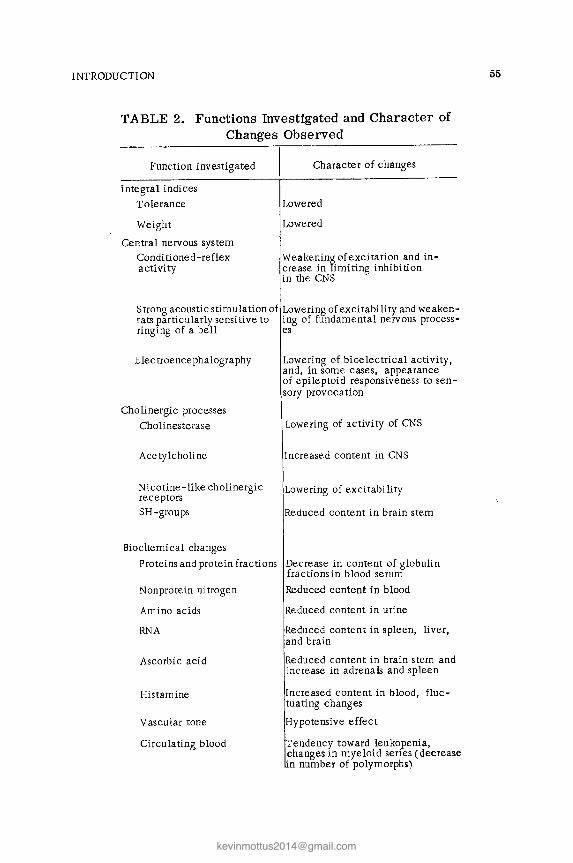
INTRODUCTION
TABLE 2. Functions Investigated and Character of Changes Observed
Function investigated
Integral indices Tolerance
Weight
Central nervous system Condi tioned -reflex activity
Character of changes
Lowered
Lowered
Weakening of excitation and in-crease in limiting inhibition in the CNS
Strong acoustic stimulation of Lowering of excitability and weaken-rats particularly sensitive to ing of fundamental nervous process-ringing of a bell es
E lectroence phalogra phy
Cholinergic processes Cholinesterase
Acetylcholine
Nicotine-like cholinergic receptors SH-groups
Biochemical changes
Lowering of bioelectrical activity, and, in some cases, appearance of epileptoid responsiveness to sen-sory provocation
Lowering of activity of CNS
Increased content in CNS
Lowering of excitabi li ty
Reduced content in brain stem
Proteins and protein fractions Decrease in content of globulin fractions in blood serum
Nonprotein nitrogen
Amino acids
RNA
Ascorbic acid
Histamine
Vascular tone
Circulating blood
Reduced content in blood
Reduced content in urine
Reduced content in spleen, liver, and brain
Reduced content in brain stem and increase in adrenals and spleen
Increased content in blood, fluc-tuating changes
Hypotensive effect
Tendency toward leukopenia, changes in myeloid series (decrease in number of polymorphs)
55

56 SECTION II
TABLE 2 (Continued)
Function investigated I Character of changes
Sexual function
Ovarian function
Fertility
Progeny
Eyes
Disturbance of estrous cycle
educed in irradiated females, ten-dency toward postmaturity, stillbirth
etardation in development, high postnatal mortality
etinal angiopathy, cataract
TABLE 3. Intensity of Irradiation Not Raising Body Temperature
Wave band
Medium (500 kHz) Short (14.7 MHz) Ultrashort ••.• 6\J.7 MHz .. 155 " 191 Microwaves:
decimeter ..••.... centimeter (3 and 10 cm). millimeter • • • • • . . .
I Threshold of non-I Energy.den-SHY, III thermal effect
Below 8 000 Vim 2 830· 10-6
22.10 " 224.10-6
150 " 50 " 20 "
0.995.10-6
0.11 .10-6
0.018.10-6
Above 40 mW/cm2 13.2 .10-6 -10 3.3 .10-6
7 2,31.10-6
There is reason to suppose that the deviation in weight of the irradiated animals (mice) is largely determined by the intensity of irradiation and, to some extent, also by the frequency of the waves. Low intensities of irradiation in the ultrashort wave band, according to some investigators, stimulate the animals' gain in weight, but the majority consider that the gain in weight is retarded (Golysheva and Andriyasheva, 1937; Skipin and Baranov, 1934; Glezer, 1937; Voznaya and Zherdin, 1937; Derevyagin, 1939; Tikhonova, 1948). As regards the superhigh-frequency band, only a few isolated papers have been published (Denier, 1933; Kutting, 1955; Gordon, Lobanova, and Tolgskaya, 1955; Lobanova, 1960). They indicate that prolonged exposure to an SHF field stimulates gain in weight. Finally, radia-

INTRODUCTION
tion in the medium wave band, according to Nikonova, causes no changes in the pattern of gain in weight by irradiated animals.
57
Changes in weight of animals exposed to radiation in different wave bands (from data obtained by Gordon, Lobanova, Nikonova, and Fukalova) are shown in Table 4. In every case intensities of ir-radiation not giving rise to an integral thermal effect (not causing an increase in the rectal temperature) were used.
The results given in this table show conclusively that any de-crease in the amount of weight gained by the experimental animals compared with controls and the degree of that difference are defi-nitely connected with wavelength.
Centimeter waves have the strongest action in depressing development of the animals (the increase in weight and the time when the increase begins to diminish compared with the control). With an increase in wavelength (decimeter, ultrashort, short waves) this inhibitory effect weakens. It is low at the extreme limits of the radio wave spectrum (millimeter and medium waves), but in the millimeter band the difference arises much later than in other bands (starting with the third month of irradiation) and the greatest decrease is observed after 6 months (when the weight is 45 g less than in the control series).
TABLE 4. Changes in Weight of Animals Exposed to Radio Waves of Different Lengths
....... Gain in weigh t of an-Intensi ty of
oOe imals, in Wave band if5.3 g (mean value)
irradiation 'S a.> c:: b.( • ....t irradi -I control .,-j c"t:l
if"'''' ated .at co ().,.-.I
Millimeter. 10 mW/cm2 3 65 75 Centimeter
3cm 10 .. 1 42 70 10 " 10 " 1.5 25 70
Decimeter 10 ..
2 95 120 Ultrashort
191 MHz. 20V/m 2.5 125 145 155 .. 50 .. 3.5 110 128 69.7 .. 150 .. 4 182 210
Short. 2250 .. 4 180 210 Medium . 1800 .. 10
\
50A/m 10

58 SECTION II
In the medium wave band the difference between the gain in weight of the irradiated and control animals was not statistically significant.
Consequently, the dynamics of the body weight, as an index of development of the animals, is to some extent dependent on the wave band of the radiation,
The effect of radio waves on vascular tone in man and ex-perimental animals has frequently been demonstrated.
Without discussing in detail the responses arising in man to radio waves of different frequencies, it will suffice to mention that in the high-, ultrahigh-, and superhigh-frequency bands a hypoten-sive effect has been described in workers with radio frequency generators by investigators who have observed mainly vagotonic responses, with a tendency toward hypotension (Parin and Davydov, 1940, 1959; Osipov, 1953; Abrikosov, 1958; Sadchikova and Orlova, 1958; Orlova, 1960; Obrosov and Yasnogordoskii, 1961; Smurova, 1962).
Experimental investigations on animals, undertaken at the Institute of Work Hygiene and Occupational Diseases, Academy of Medical &:iences of the USSR, to study the action of radio waves of different frequencies (Gordon, 1960, 1964; Nikonova, 1964; Fukalova, 1968) have shown that chronic exposure to radio waves of nonther-mogenic intensity causes a persistently low blood pressure. This is often preceded by a phase of raised pressure (Table 5).
Although a persistent decrease in blood pressure is a com-mon feature after exposure to radio waves of all frequencies, each individual wave band is associated with its own characteristic changes:
1. Absence of the first phase - elevation of the pressure -in animals irradiated with medium, 3-cm, and millimeter waves;
2. The rapid appearance of the first phase (first week of ir-radiation) in the case of irradiation with decimeter and 10-cm waves;
3, Early appearance of the second phase of lowering of pres-sure after irradiation in the ultrashort (69.7 MHz) and short wave bands in the 6th week of irradiation;

INTRODUCTION
TABLE 5. Changes in Blood Pressure After Irra-diation with Radio Waves
Wave band
Medium (50() kHz)
Short (14.B MHz) • Ultrashort
697 MHz . 155 191
Microwaves: decimeter .. centimeter • • .
10 cm .... 3 ..
millimeter " .
Intensity of c:: phase- o.J Second
. .. .3 o.J pressure '" madla tlOn 0..<;;;;:; 12 d o.J
+...I > en cu re ue - J-I U o.J IG o.J tion 0.. o.J Gl i5.. (weeks) t.;'<. '0
1 BODV/m Not significant 5A/m - ;j) 11.7
2250V/m 2 6 12
150 .. 2 6 17 50 .. 2 12 33 20 .. 4 12 29
10 mW/cm2 10 17
10 1-6 22 11 6 25
10 4 20
59
4. A considerable (20-33%) decrease in the blood pressure level, especially after irradiation by waves in the mil-limeter, 3-cm, and ultrashort (155 and 191 MHz) wave bands.
Great importance is attached to the hypothalamus in the mechanism of autonomic vascular disturbances.
Results obtained in the writers' laboratory indicate that the diencephalon is concerned in the response of the body to irradiation with radio waves. Zenina investigated brain potentials and showed that the effects of cardiazol, which evokes paroxysmal responses of diencephalic origin, are inhibited or totally suppressed by microwave irradiation. In this case it can be assumed that the action of the microwaves was to block particular areas of the dien-cephalon.
Disturbance of hypothalamic activity after exposure to radio waves has also been observed when the effects of microwaves on specialized forms of appetite and electrolyte metabolism were studied in irradiated rats. Hypothalamic nuclei form the higher center for the control of water and mineral metabolism. Kulakova (1968) found no correlation between the intensity of certain types

60 SECTION II
of salt appetite and the electrolyte composition in the body. This could possibly have been due to a disturbance of the mechanism controlling the distribution of salts and water between the cell and the internal medium.
Evidence of the important role of the hypothalamus is given by the clinical picture of the severe forms of disturbances arising after chronic exposure to radio waves, characterized by the dien-cephalic syndrome (Drogichina and Sadchikova, 1964). This syn-drome is manifested as paroxysmal states resulting from neuro-circulatory disorders. Distinct disturbances of cortical electrical activity are present.
As a vital part of the brain the hypothalamus plays an im-portant role in the integration of nervous and humoral processes. The hypothalamus can regulate, in particular, the activity of the cardiovascular system, the content of mediators such as acetyl-choline, cholinesterase, and histamine, and so on.
Recent investigations undertaken at the Institute of Work Hygiene and Occupational Diseases have shown that radio waves, especially microwaves, can affect certain cholinergic processes. Clinical observations show that chronic exposure to microwaves leads to functional disturbances of the nervous system. The most characteristic disturbance affecting the autonomic nervous system is a tendency for parasympathetic control over the cardiovascular system to predominate. An investigation of neurohumoral regula-tion (Nikogosyan) revealed an increase in the acetylcholine content in the CNS and a decrease in cholinesterase activity and in the content of SH-groups in the brain stem (Kitsovskaya).
Kitsovskaya (1968) showed that exposure to microwaves low-ers the excitability of nicotine-like cholinergic receptors, while the state of the muscarine-like cholinergiC receptors remains un-changed. Changes in the state of the former may be to some extent responsible for the disturbance of transmission of excitation in the CNS. The absence of change in the muscarine-like receptors is presumptive evidence of the selective action of microwaves on cholinergiC structures.
A very important aspect of the study of the mechanism of action of low-intenSity microwaves is the identification of those parts of the nervous system which are most intimately concerned.

INTRODUCTION
Interesting results from this standpoint were obtained by Kitsov-skaya (1968a), who used drugs which induce convulsions in rats
61
and which act on the eNS at different points (camphor, nikethamide, strychnine, nicotine, etc.). She showed that the 'motor cortex and the basal ganglia, certain structures of the mesencephalon and dien-cephalon, and the segmental system which controls the transmis-sion of impulses from efferent pathways to the motor units of the spinal cord are sensitive to irradiation. To this conclusion must be added the inhibitory action on nicotine-like cholinergic recep-tors already mentioned above.
It must be emphasized that the nervous system varies in its degree of sensitivity to microwave irradiation. Its response is most marked to millimeter radiation (as exemplified by the re-sponse of higher nervous activity of animals). However, the periph-eral nervous system and, in particular, the receptors of the skin, are highly sensitive to millimeter irradiation, even to a single dose,
The functional disturbances develop and attain their definitive state gradually 0
Depending on the physical parameters (wavelength and inten-sity of irradiation), on the duration of exposure, and on the initial functional state of the body, the course of the process may vary, having one or two phases; usually there are two phases, with initial stimulation and subsequent depression of the functions. Investiga'-tions have shown that morphological changes resulting from ex-posure to radio waves of different frequencies and of nonthermo-genic intensity also develop gradually.
The morphological changes following exposure to radio waves of low intensity are examined in detail below.

Chapter 1
Morphological Changes Following Prolonged and Repeated Low-Intensity
Microwave Irradiation
Morphological Changes Following Prolonged and Repeated Irradiation with Low-Intensity Centi-meter Waves
Morphological changes in the viscera and nervous system were studied in 74 albino rats exposed repeatedly for long periods to pulsed and continuous 10-cm waves and pulsed 3-cm waves of low intensity, depending on differences in the duration of irradia-: tion.
G r 0 u p 1. To analyze the initial disturbances resulting from the action of centimeter waves, it is necessary to examine in rather more detail the chronic effects of 10-cm waves of very low inten-sity on the animal organism. Such a study is rendered all the more important because these intensities may be encountered in industry.
Repeated exposure (35-40 sessions, each lasting 30 min) of albino rats to low-intensity 10-cm pulsed or continuous waves was studied. No signs of hyperthermia were observed. There were no external manifestations of the action of the centimeter waves. The animals appeared normal and continued to gain weight equally with the controls; they were clinically healthy.
The rats were sacrificed at various times after the end of ir-radiation: one batch immediately after irradiation, a second batch one week later, and a third batch 3 weeks after the end of irradiation.
63

64 SECTION II
No vascular disorders were found in animals sacrificed im-mediately after the last irradiation. Cloudy swelling of the cyto-plasm was found in individual cortical and hypothalamic neurons, with occasionally the appearance of solitary vacuoles in the cyto-plasm (especially of hypothalamic neurons), slight swelling, and uneven staining of single myocardial muscle fibers. Histochemical examination revealed a slight decrease in the ribonucleoprotein content in the skin and in several vi sceral organs normally rich in RNP (neurons, glandular epithelium of the gastrointestinal tract). The most marked decrease in RNP content was found in the epidermis and its derivatives in the surface layers of the skin. Individual animals showed swelling of the cytoplasm and slight cloudy swell-ing of individual hepatocytes and the epithelium of single convoluted renal tubules.
Not all these changes could be seen 3 weeks after the end of irradiation, i.e., some regression had taken place.
After repeated daily irradiation (34 sessions) for 30 min with 3-cm pulsed waves up to 10 mW/cm2 in intensity, no external mani-festations of the action of the radiation could be found. * In animals sacrificed immediately after the last irradiation the changes were even less marked than after irradiation with 10-cm waves. In ani-mals sacrificed 1 and 3 weeks after the end of irradiation no mor-phological changes could be seen in the internal organs or brain. Consequently, all the morphological changes mentioned above are reversible.
Hence, in animals irradiated repeatedly with 3- and 10-cm pulsed waves of low intensity (10 mW/cm2) and whose initial state was good, investigation with the usual morphological methods re-vealed initial, reversible, and very insignificant degenerative changes, mainly in the nervous system. These changes, according to P. E. Snesarev, are an early response of the nervous system to stimula-tion. They were very slight, reversible, and compensated, and the animal remained to all intents and purposes healthy.
In animals irradiated repeatedly with 10- and 3-cm waves not exceeding 10 mW/cm2 in intensity for up to 10 months no ex-
'Changes connected wi th the method of sacrifice (decapi tation) were allowed for. All changes described in the experimental animals are relative to a control.

CHAPTER 1 65
ternal manifestations of their action could be seen. At the same time, it was found that low intensities of 10-cm irradiation, although not giving a thermal effect, nevertheless give rise to certain physio-logical changes. For example, changes in the CNS expressed as an imbalance between the fundamental nervous processes (Kitsovskaya, 1960), disturbance of conditioned-reflex activity of the animals (Lobanova, 1960), and lowering of the blood pressure (Gordon, 1960, 1964) were found in these animals. The usual morphological of investigation, capable of revealing morphological changes of slight severity, failed to reveal any changes accompanying these physio-logical disorders.
It was therefore necessary to use more precise, elective neurohistological methods to investigate the nervous system, for example, to study changes in synapses and receptors of the CNS, with their very delicate responses to stimulation. It was therefore de-cided to investigate the higher nervous activity of irradiated ani-mals and to accompany this by a parallel study of synaptic activity in the cerebral cortex after exposure to centimeter waves.
Before describing the results of these investigations, the literature on interneuronal connections in the cerebral cortex must be briefly surveyed.
The morphological organization of interneuronal connections in nerve tissue is very clearly defined. In the human brain, acterized by the high level of development of the association areas, cortical connections achieve an extremely high level of differen-tiation and complexity (Sarkisov).
Sarkisov (1948) and Polyakov (1955) studied the complex or-ganization of cortical interneuronal connections. They found that three systems (efferent and afferent neurons and interneurons) in-teract and distribute the flow of nervous impulses over the cerebral cortex and constitute the material basis of the reflex arc. Trans-mission of the nervous impulse from one neuron to another can take place only through terminal branches of the axon of one neuron to the dendrites or bodies of the other neuron.
Synapses joining neurons and specialized for the reception of impulses or their transmission to other neurons have been stud-ied by Dolgo-Saburov (1956), Polyakov, Sarkisov, and Lavrent'ev. Other investigations (Snesarev, Lavrent'ev, Sarkisov, Polyakov)

66 SECTION II
have shown that there are two types of interneuronal synapses: axo-somatic, or direct, terminal connections in which the terminal branches of the axon of one neuron wind around the body of another neuron and form synaptic contacts, "boutons," knobs, and rings (Fig. 15) on it, and axo-dendritic synapses, "en passant," or col-lateral synapses, in which very fine branches of the axon make contact with branches of the dendrites. Contact takes place through very small spines carried by the dendrites (Fig. 16).
Fig. 15. Axo-somatic synapses consisting of terminal boutons on body of nerve cell (after Shabadash).

CHAPTER 1
Fig. 16. Diagram ofaxo-dendritic synapses in the cerebral cortex (after Cajal).
67
The scheme of interneuronal connections in the cortex as described by Polyakov is as follows. Axons arriving in the cortex from lower levels of the nervous system branch in the deep layers and form direct connections between one neuron and another, or branches ofaxons in the phylogenetic ally young, association layers near the surface of the cortex come into contact with large numbers of neuronal dendrites in different layers of the cortex, for in what-ever layer the efferent neuron lies, it most frequently sends its apical dendrite into the upper layers of the cortex for association
68 SECTION II
purposes (Fig. 17). Connections of this type constitute a delicate mechanism for controlling and modifying the functional state of many neurons depending on changes in the functional state of any one of them.
In the human cortex, because of the high development of the association connections, axo-dendritic, indirect synapses are of the utmost importance.
In the modern view (Sarkisov, Polyakov) spines on the den-drites of nerve cells, as cortical receptor systems, undergo changes before the presynaptic axonal branches and their systems of syn-apses and before the bodies of the nerve cells are correspondingly affected.
An investigation was accordingly carried out in order to study the initial morphological changes in cortical interneuronal connections after prolonged exposure to 10-cm waves of low in-tensity, to identify the stages of gradual development of changes in the cortical interneuronal connections depending on the inten-sity of irradiation and to correlate these with changes in condi-tioned reflexes, and also to study the reversibility of these changes.
Pieces of cortex taken from the motor and sensory areas were impregnated with silver by Golgi's method (some sections were stained by the Golgi-Bubenet method).
As the control, the structure of the interneuronal connections was studied in the cortex of 10 unirradiated healthy albino rats, killed by decapitation.
Examination under high power clearly shows that the apical and basal dendrites of a pyramidal neuron are abundantly supplied with small spines (Fig. 18a). Spines are particularly clearly visi-ble when examined with an immersion objective: they consist of pear-shaped projections of cytoplasm, lying perpendicularly to the long axis of the dendrite (Fig. 18b). Often they appear like small "boutons" on thin pedicles (Fig. 18c).
The largest number of spines was found on the apical den-drites of pyramidal and spindle cells, and on dendrites running from various layers of the cortex into the surface layers for as-sociation purposes, and branching there. They were fewer in num-ber on the basal dendrites, and totally absent on the bodies of the nerve cells.
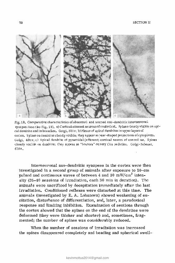
70 SECTION II
Fig.lB. Comparative characteristics of abnormal and normal axo -dendritic interneuronal synapses (see also Fig. 19). a) Cortical etterent neurons of controlrat. Spines clearly visible on api-cal dendrite and its branches. Golgi, 320x; b) Plexus of apical dendrites in upper layers of cortex. Spines on dendrites clearly visible; they appear as pear-sha ped projections of cytoplasm. Golgi, 4BOx; c) Apical dendrite of pyramidal (efferent) cortical neuron of control rat. Spines clearly visible on dendrite; they appear as "boutons' on very thin pedicles. Golgi-Bubenet, 650x.
Interneuronal rum-dendritic synapses in the cortex were then investigated in a second group of animals after exposure to 10-cm pulsed and continuous waves of between 4 and 10 mW/cm2 inten-sity (35-40 sessions of irradiation, each 30 min in duration). The animals were sacrificed by decapitation immediately after the last irradiation. Conditioned reflexes were disturbed at this time. The animals (investigated by E. A. Lobanova) showed weakening of ex-citation, disturbance of differentiation, and, later, a paradoxical response and limiting inhibition. Examination of sections through the cortex showed that the spines on the end of the dendrites were deformed (they were thicker and shorter) and, sometimes, frag-mented; the number of spines was considerably reduced.
When the number of sessions of irradiation was increased the spines disappeared completely and beading and spherical swell-
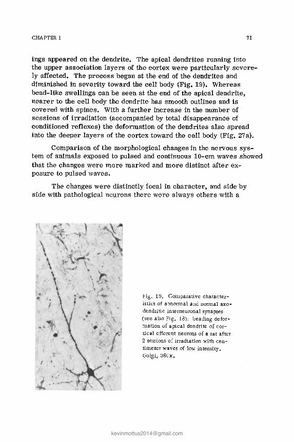
CHAPTER 1 71
ings appeared on the dendrite. The apical dendrites running into the upper association layers of the cortex were particularly severe-ly affected. The process began at the end of the dendrites and diminished in severity toward the cell body (Fig. 19). Whereas bead-like swellings can be seen at the end of the apical dendrite, nearer to the cell body the dendrite has smooth outlines and is covered with spines. With a further increase in the number of sessions of irradiation (accompanied by total disappearance of conditioned reflexes) the deformation of the dendrites also spread into the deeper layers of the cortex toward the cell body (Fig. 27a).
Comparison of the morphological changes in the nervous sys-tem of animals exposed to pulsed and continuous 10-cm waves showed that the changes were more marked and more distinct after ex-posure to pulsed waves.
The changes were distinctly focal in character, and side by side with pathological neurons there were always others with a
Fig. 19. Comparative character-istics of abnormal and normal axo-dendritic interneuronal synapses (see also Fig. 18) : beading defor-mation of apical dendrite of cor-tical efferent neurons of a rat after 2 sessions of irradiation with cen-timeter waves of low intensity. Golgi. 360x.

72 SECTION II
normal configuration of the dendrites. In brain tissue stained by Nissl's method the initial changes were found as cloudy swelling of the cytoplasm of individual neurons which, according to P. E. Snesarev, is an early, reversible response of nerve cells to various forms of irritation.
No significant changes except swelling of the cytoplasm of individual fibers in the myocardium, solitary hepatocytes, and epithelial cells of the convoluted renal tubules were found in the viscera of these animals,
All the morphological changes described above in the ner-vous system corresponded completely with the physiological changes detected on clinical examination as disturbances of conditioned-reflex activity.
In animals sacrificed 3-4 weeks after the end of irradiation, after complete recovery of conditioned reflexes no changes were found in the nervous system. All dendrites had smooth outlines and were covered with numerous spines.
The changes described are functional and reversible and dis-appear immediately after the end of irradiation side by side with recovery of the animals' conditioned-reflex activity.
The changes described above in axo-dendritic interneuronal synapses in the cortex are not specific for exposure to 10::"cm waves; similar changes in these synapses have also been described by Tolgskaya (1954) in animals exposed to various harmful chem-icals (arsenic, lead, aniline). They are a manifestation of the fine response of the nerve cells, which may disappear after the action of the harmful agent is terminated, or, conversely, if its action is prolonged, they may be followed by irreversible degenerative patho-logical changes.
In some rats exposed to the chronic effects of 10-cm waves of low intensity (not exceeding 10 mW / cm2) axo-somatic as well as axo-dendritic synapses were investigated in the cortex. The for-mer are less conspicuous and are more highly developed in the deep layers of the cortex. The stages of the changes in the axo-somatic loops are illustrated in Gibbs' scheme (Fig. 20).
In the control animals (sections stained by Caj aI's method) axo-somatic synapses are very clearly revealed on the motor neurons

CHAPTER 1 73
b c
g II
Fig. 20. Scheme showing successive stages of degenerative changes in "boutons terminaux" of the pericellular synaptic system at various times (after Gibbs): a) normal boutons; b) after 24 h; c) after 48 h; d) after 72 h; e) after 96 h; f, g, h) after 120 h; i) after 120 h (granular degeneration).
of the anterior horns of the spinal cord and they consist of axon endings of the ring or "bouton" type on the body of the nerve cells (Fig. 21a).
In the irradiated animals, on the motor neurons of the deep layers of the cortex or hypothalamic neurons these rings and bou-tons are thickened, their argyrophilia is increased, and the interior of the rings is filled so that they are converted into large "clubs" which are displaced from the bodies of the nerve cells (Figs. 21 and 28), i.e., asynapsia of the neurons occurs.
Consequently, axo-somatic interneuronal synapses, like axo-dendritic synapses, in the brain are extremely sensitive and soon begin to undergo changes during irradiation (before the neurons and cells of the viscera).

74 SECTION II
Fig. 21. Comparison of normal and pathological axo-somatic synapses in the an-terior horns of the spinal cord. Cajal; 600x: a) numerous terminal loops on body and dendrites of a motor neuron in the anterior horns of the spinal cord of a control rabbit; b) club-like swellings and increased argyrophilia of synaptic vesicles and their detachment from body of the nerve cell in the hypothalamus after irradiation with low-intensity centimeter waves.
The pathological process in the axo-somatic synapses is re-versible, and I month after the end of irradiation no sign of these changes can be found.
Evidently in response to stimulation by IO-cm waves of low intensity functional reversible changes develop in the synaptic struc-tures of the brain. These changes in the nerve cells, if irradiation is prolonged, may be converted into irreversible, degenerative pathological changes.
Besides changes in the brain synapses, morphological changes were also studied in other fine structures of the nervous system after exposure to IO-cm waves of low intensity (changes in the sen-sory nerve fibers of the skin and viscera).
As already mentioned, changes in the sensory nerve fibers of the cutaneous receptive fields are particularly interesting be-

CHAPTER 1 75
cause the skin is the first barrier in the way of these radio waves. Because of the very large number of afferent nerve endings which it contains the skin is a powerful receptive field. Waves of the centimeter band are partly absorbed by the skin and partly pene-trate into deeper tissues. Changes in the sensory nerve fibers of receptive fields of the viscera are equally interesting. According to published observations, sensory fibers of the viscera respond briskly even to minimal harmful factors.
It was therefore of the greatest importance to study afferent sensory nerve fibers which are so numerous in the tissues of the body and whose coordinated activity helps to maintain the constancy of the internal milieu of the organism. Investigation of sensory nerve fibers in the various receptive fields is an important part of the study of the mechanism of action of microwaves.
Thirty animals were exposed to repeated irradiation by cen-timeter waves of low intensity (not exceeding 10 mW/cm2) for 1 h daily and for 100-200 sessions. Changes characterized as irrita-tion phenomena were found in the sensory nerve fibers of the vis-cera and, in particular, of the skin. They consisted of increased argyrophilia, the appearance of swellings, thickenings, and pools of axoplasm, and marked tortuosity of the nerve fibers. In this group of animals, side by side with pathological nerve fibers there were always many which were unchanged, evidence of the able powers of compensation of the peripheral nervous system. The most severe changes were found in the sensory nerve fibers of the skin (Figs. 22 and 29c). Changes in sensory nerve fibrils in the receptive fields of the viscera were equally ill-defined and consisted of increased argyrophilia and the appearance of irregular bead-like thickenings along the course of the fiber (Fig. 30b).
Changes in the nerve cells of the sensory spinal ganglia, nerve cells of the thalamus and hypothalamus, cortical cells, and neurons of the autonomic ganglia (solar plexus, ganglion nodosum of the vagus nerve) also were studied. Swelling of the cytoplasm and tigrolysis in the center of the cell, with ectopia of the nuclei, were observed in the nerve cells of the sensory spinal ganglia. Individual cells showed signs of karyocytolysis and death of the neurons (Fig. 31b). In the hypothalamus the cytoplasm ofthe neurons was swollen and contained single vacuoles. The neurons of the autonomic ganglia were almost unchanged.
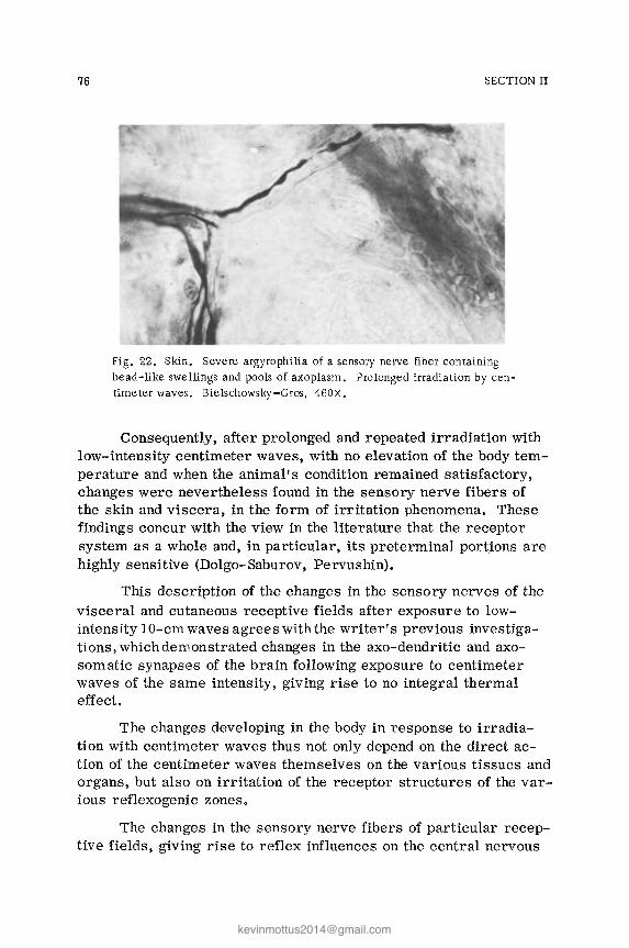
76 SECTION II
Fig. 22. Skin. Severe argyrophilia of a sensory nerve fiber containing bead-like swellings and pools of axoplasm. Prolonged irradiation by cen-timeter waves. Bielschowsky-Gros, 460X.
Consequently, after prolonged and repeated irradiation with low-intensity centimeter waves, with no elevation of the body tem-perature and when the animal's condition remained satisfactory, changes were nevertheless found in the sensory nerve fibers of the skin and viscera, in the form of irritation phenomena. These findings concur with the view in the literature that the receptor system as a whole and, in particular, its preterminal portJons are highly sensitive (Dolgo-Saburov, Pervushin).
This description of the changes in the sensory nerves of the visceral and cutaneous receptive fields after exposure to low-intensity lO-cm waves agrees with the writer's previous investiga-tions, which demonstrated changes in the axo-dendritic and axo-som atic synapses of the brain following exposure to centimeter waves of the same intensity, giving rise to no integral thermal effecL
The changes developing in the body in response to irradia-tion with centimeter waves thus not only depend on the direct ac-tion of the centimeter waves themselves on the various tissues and organs, but also on irritation of the receptor structures of the var-ious reflexogenic zones.
The changes in the sensory nerve fibers of particular recep-tive fields, giving rise to reflex influences on the central nervous

CHAPTER 1 77
system and, consequently, on the functional state of the circulatory and respiratory systems, together with changes in the hypothalamic region, could help to explain the fall in blood pressure and brady-cardia observed both clinically and experimentally during the ac-tion of low-intensity microwaves in the absence of any direct ther-mal effect.
Since in these experiments with chronic exposure to irradia-tion of low intensities early changes were found in the hypothalamic neurons it was decided to investigate the hypothalamic neuro-secretory function, more especially because clinicians and physiol-ogists have repeatedly found evidence of the important role played by the diencephalon and hypothalamus in responses to the action of low-intensity microwaves.
A characteristic feature of the response of the body to elec-tromagnetic radio waves is its predominantly vagotonic direction. Clinical and physiological investigations on man (Ginzburg and Sadchikova, 1964; Drogichina et aI., 1962) have shown that persons working with superhigh-frequency generators may develop autonomic or vascular disturbances in the mechanism of which an important role is played by the hypothalamus.
There is also experimental evidence of the important role of the diencephalo-hypothalamic region in responses to microwave action.
Evidence of changes in the regulating activity of the hypo-thalamus during exposure to radio waves is also given by the changes observed in the blood pressure of the irradiated animals. These changes were in two directions: in the first period of irradiation by low intensities the blood pressure was raised, but during sub-sequent irradiation it was persistently lowered.
It has recently been shown that the cerebral cortex regulates the various functions of the body, including its endocrine functions, through the pituitary gland.
The hypothalamus participates in: 1) activity of the cardiovas-cular system; 2) thermoregulation; 3) regulation of water, mineral, protein, carbohydrate, and lipid metabolism; 4) regulation of per-meability of blood vessels and membranes; 5) regulation of the functions of endocrine glands; 6) the autonomic basis for somatic functions; 7) regulation of the functions of the gastrointestinal

78 SECTION II
tract and leukopoiesis; 8) regulation of sleep and waking; 9) regula-tion of the constancy of the internal milieu of the organism; and 10) adaptive behavior, based on connections between the cortex and hypothalamus (cortico-subcortical relationships). Through the hypothalamus the central nervous system thus exerts both nervous and neurohumoral control over functions maintaining the dynamic constancy of the internal milieu of the body and responsible for non-specific internal and external adaptation of the organism to the environment.
The neurohypophysis is formed by the posterior lobe of the pituitary gland and partly by its infundibulum, while functionally it is connected with the anterior hypothalamic (supraoptic and para-ventricular) nuclei.
Axons and dendrites of the nerve cells of these nuclei par-ticipate in the structure of the posterior lobe of the pituitary gland. It has been shown in the last 2 decades that neurosecretion is formed in the neurons of the supraoptic and paraventricular nuclei and is transported via the axons of neurons (mainly the hypo-thalamo-hypophyseal tract) into the posterior lobe of the pituitary, where it is stored.
Movement of secretion along axons of nerve cells has been demonstrated histolOgically. Gomori-positive thickenings and swellings are clearly visible on axons, and at the ends of the axons these swellings assume the form of large club-shaped structures (Herring's bodies), which are particularly conspicuous in the pos-terior pituitary (Polenov, 1964), where the secretion accumulates around the vessels. Most workers are inclined to believe that the neurohypophysis is merely a reservoir where neurosecretion from the hypothalamus accumulates.
The opinion is held (Polenov, 1964; Voitkevich, 1969) that the secretions of the hypothalamic neurons, like any internal secre-tion, can enter the blood stream directly (hemocrinia), and this is particularly likely because the anterior hypothalamus has an ex-tremely rich blood supply. Many workers have described the special arrangement of the numerous capillaries in the anterior hypothal-amus: the capillaries appear to surround every nerve cell; often the capillaries do not contain glial membranes.
Neurosecretion can also enter the ventricles of the brain directly (hydrocephalocrinia).

CHAPTER 1 79
Investigations by Soviet and other workers have demonstrated the secretory function of neurons of the anterior hypothalamus both histologically and histochemically. The structural features dis-tinguishing cells of the anterior hypothalamic nuclei have been described: the characteristic arrangement of the tigroid sub-stance (at the periphery of the cell), the eccentric situation of the cell nuclei, and the presence of special Gomori-positive granules and vacuoles in the cytoplasm around the nucleus.
The vacuoles are optically empty or contain oxyphilic co1l6id. Vacuolation of the anterior hypothalamic neurons was observed by Tarakanov and Maiorova (1957) after injection of hypertonic sodium chloride solution and after x-ray irradiation.
According to Gerber (1967) vacuolation of the hypothalamic neurons is evidence of their intensive activity. The vacuoles may be numerous; sometimes they merge into one large vacuole. The vacuoles burst and expel their secretion; drops of colloid lie ex-tracellularly. In this case the cell has ragged, irregular outlines. This expulsion of large masses of colloid (lumps of cytoplasm) is regarded by some workers as holocrinia (Polenov, 1964). Mosinger (1950) considers that it may be physiological melting of the cell; this is followed by repair. Guillemin (1955, 1961) attributes vacuola-tion to overproduction of colloid and its expulsion outside the cell. Maiorova describes the complete cycle of secretion of the hypo- ' thalamic neurons from the beginning of accumulation to discharge, followed by a process of cell repair. She considers (Maiorova, 1964) that RNP of the cell cytoplasm has an important role in the production of colloid.
Vacuolation of anterior hypothalamic neurons is thus evident-ly, on the one hand, the highest stage of hypersecretion, while on the other hand it may indicate the onset of degenerative changes in the nerve cells, but in this case the cell nucleus must also be af-fected (Andersson and Jewell, 1957).
Electron-microscopic investigations (Scharrer and Bergmann, 1949; Mosinger 1950; Voitkevich, 1964) have confirmed the presence of hormonal granules in the cytoplasm of the anterior hypothalamic neurons, along the course of nerve fibers of the hypothalamo-hypo-physeal tract, and in the posterior pituitary. Other electron-micro-scopic studies have shown that granules of neurosecretion increase in volume and size as they move along the hypothalamo- hypophyseal

80 SECTION II
tract, indicating that synthesis of neurosecretion takes place pro-gressively as it moves toward the neurohypophysis.
The posterior pituitary contains cells known as pituicytes. These liberate the secretion from the posterior lobe of the pituitary gland and deliver it into the blood stream. Pituicytes do not par-ticipate in the synthesis of posterior pituitary hormones. Endings of neurosecretory fibers are in very close proximity to the pitui-cytes, but there is no neurosecretion in the pituicytes.
The nuclei of the anterior hypothalamus secrete a chemical substance which is very little different in principle from the hor-mones secreted by the posterior lobe of the pituitary.
Extract of the pituitary infundibulum contains substances with an antidiuretic action and with actions on the blood pressure and smooth muscle of the uterus. These substances are called antidiuretin, vasopressin, and oxytocin, respectively.
Guillemin and Rosenberg (1957) have shown that, besides these three hormones, the anterior hypothalamic nuclei also secrete a substance stimulating the release of ACTH. This factor is called corticotropin releasing factor (CRF).
Although in the opinion of some investigators the anterior pituitary (adenohypophysis), which produces gonadotropic, cor-ticotropic, thyrotropic, and adrenotropic hormones, has no direct nerve supply, it is nevertheless under hypothalamic control through the innervation of the portal blood vessels and the penetration of chemical mediators (hormonal products of the hypothalamus) into the portal vessels of the adenohypophysis.
The direction of the blood flow along the portal vessels has now been shown (Zhdanov, 1964) to be definitely from the hypo-thalamus to the pituitary. This direction explains why the chem-ical mediators of the hypothalamus can exert a stimulant action on the anterior pituitary (adenohypophysis), and can exert neuro-hormonal control over the production of the hormone ACTH (Por-ter, 1953, 1956; Bogdanovich, 1964). The hormone ACTH liber-ated by the adenohypophysis induces the secretion of hormones of the adrenal cortex. The most important of these are the ster-oid hormones, including the 17-hydroxycorticosteroids and aldo-sterone.

CHAPTER 1 81
Zubkova-Mikhailova (1964) found a marked decrease in the production of neurosecretory granules in the nuclei of the anterior hypothalamus and in the posterior lobe of the pituitary after in-jection of ACTH into the blood stream. Consequently, the neuro-secretion has a stimulating action on the production of adeno-hypophyseal hormones. All these facts suggest that a number of biologically important active substances are produced in the nuclei of the anterior hypothalamus and in the neurohypophysis, and that these substances regulate the activity of the adenohypophysis, and the other endocrine glands by the neurohumoral route (Polenov, 1964; Akmaev, 1960).
As a result of many investigations it has thus been shown that the chemical mediators which reach the adenohypophysis via the blood in the portal vessels can induce its secretory activity. This results in the liberation into the blood stream of ACTH, the prin-cipal hormone concerned in the general adaptation response of the organism to endogenous and exogenous demands.
It is now established beyond all doubt that the hypothalamus participates in every physiological and neurochemical process, plays a highly important role in maintaining the constancy of the internal milieu of the body, and is an important brain center con-trolling adaptation and nutrition (Orbeli).
Polenov (1964) considers that hypothalamic neurosecretory activity is of great importance in adaptation to changing environ-mental conditions, i.e., that the hypothalamic neurosecretion is im-portant primarily in connection with protective and compensatory responses of the body to constantly changing environmental con-ditions and to various harmful factors.
According to Polenov (1964) under conditions of stress the neurosecretion is the principal trigger mechanism with a gener-alized action on the various organs and endocrine glands.
Following the work of Selye on stress and the adaptation syn-drome and the investigations of Scharrer and Scharrer (1954b), Bergmann (1954), Mosinger (1950), Aleshin (1964), Voitkevich (1964), Maiorova (1962), and Polenov, Tarakanov, and Maiorova (1957), the importance of the neurosecretory system and the role of the hypothalamus as the trigger mechanism for the hormonal com-

82 SECTION II
ponent of adaptive responses aimed at the maintenance of homeo-stasis in the body were realized.
The work of Selye (1950) demonstrated the role of the pitu-itary-adrenal system in adaptive and defensive responses of the body to harmful factors.
In Selye's opinion adrenocortical steroid hormones, whose formation is activated by the ACTH of the adenohypophysis, play a decisive role in the adaptation syndrome. ACTH formation is itself regulated by the anterior hypothalamic nuclei. Consequently the anterior hypothalamus (the supraoptic and paraventricular nuclei) and neurohypophysis, along with the adenohypophysis and adrenals, participate in the adaptive mechanisms concerned in the body's response to harmful factors. Davydovskii (1961, 1962) points out that the secretory act of the adenohypophysis in response to stress (to alarm) is probably secondary to the act of neurosecretion in the hypothalamus. Kurtsin (1963) considers that the pituitary-adrenal adaptation system can be triggered from the cerebral cor-tex by a reflex of the following character: the stressor acts on receptors which send impulses along afferent fibers into the sub-cortical structures and then into the cortex; from the cortex im-pulses pass along efferent fibers to the hypothalamus, and from there by the neurohumoral route to the pituitary gland. The adeno-hypophysis secretes ACTH which reaches the adrenal by tpe hu-moral route, and induces the secretion of corticosteroid hormones which play the principal role in the adaptation reaction. Since the same stressors induce active neurosecretion of the hypothalamo-hypophyseal system and activity of the hypophyseo-adrenal system, Aleshin (1964), Voitkevich (1964), Polenov (1964), Maiorova (1962), Tarakanov (1957), and others consider that the production of Gomori-positive secretion in the hypothalamus (antidiuretic hormone, vaso-pressin, oxytocin, and CRF) regulates the secretion of ACTH during stress. Whatever the mechanism of their action, the role of the neurosecretory system and of the hypothalamus as the trigger mech-anism activating the hormonal component of adaptation responses aimed at maintaining homeostasis in the body have thus been conclusively proved by numerous authoritative investigations. The study of the intimate mechanisms whereby hypothalamic neu-rons exert their regulatory influence is of great general biological importance. It explains the pathogenesis of many neuroendocrine, autonomic, and other diseases (Galoyan, 1965).

CHAPTER 1 83
Many investigators have studied changes in the neurosecre-tory function during exposure to harmful factors.
Vladimirov (1964) found exhaustion of the neurosecretory system after strong nociceptive stimulation for 10 min. Zhukova (1964) extirpated the superior cervical sympathetic ganglion and observed an increase in the content of secretion in the nerve cells of the anterior hypothalamic nuclei (through blocking of the libera-tion of the neurosecretion); under these circumstances the blood pressure fell. Consequently, in response to reduced secretion of vasopressin the blood pressure fell. Conversely, in rabbits re-ceiving antithyroid preparations, desympathization was followed by elevation of the blood pressure and a decrease in neurosecre-tion in the hypothalamus as a result of its discharge into the blood stream in large quantities. Voitkevich (1964) observed a decrease in the content of secretion in the anterior hypothalamic nuclei dur-ing dehydration, which he regarded as the result of increased uti-lization of stored neurosecretory substance in connection with antidiuretic activity. Maiorova found an increase in the content of secretion in the anterior hypothalamic nuclei 2 h after hypophysec-tomy. Zubkova-Mikhailova (1964) found an increase in the content of secretion in the anterior hypothalamic nuclei and neurohypoph-ysis 3 h after x-ray irradiation. The content of neurosecretion was reduced 24 h after irradiation, and degeneration of the nerve cells began after 6-7 days.
Guillemin (1957) showed that the content of neurosecretion in the anterior hypothalamic nuclei is sharply reduced after in-jection of histamine into the blood stream. Meanwhile histamine stimulates ACTH formation, and at the same time changes take place in the ascorbic acid content in the adrenals (Galoyan, 1965).
Galoyan (1965) states that after injection of cadmium into the blood stream of animals the formation of neurosecretion is reduced in the cells of the anterior hypothalamus, and this is fol-lowed by emptying of the contents of the nerve cells, which then undergo degenerative changes.
According to Aleshin (1965) chlorpromazine lowers the blood pressure while neurosecretion is retained in the hypothalamic neurons, indicating the blocking of its liberation. These findings agree with the observations of Voitkevich (1964), who also ob-served blocking of the liberation of neurosecretion from the neuro-

84 SECTION II
hypophysis by large doses of chlorpromazine. Stasis of Gomori-positive secretion is observed under these circumstances in the hypothalamic nuclei.
Hence, the accumulation of granules and droplets of secre-tion in the hypothalamic neurons may be an indication of its in-creased synthesis and accumulation, and may also be the result of its deficient outpouring into the blood stream (blocking). No change or a decrease in the content of secretion in the hypothal-amic neurons and posterior pituitary indicate increased mobiliza-tion of secretion and its discharge into the blood stream, and also the cessation or inadequacy of its synthesis in the hypothalamus. In the latter case degenerative changes affect the neurons of the hypothalamic nuclei.
The results reviewed above can be summarized by saying that in response to acoustic, photic, and electrical stimulation and to whole-body x-ray irradiation in the first stage of stress the content of secretion in the hypothalamus and neurohypophysis is reduced (i.e., the neurohypophysis is emptied); under these condi-tions the ACTH concentration in the blood rises sharply.
During long-term experiments and on removal of the stimulus in an acute experiment, secretion gradually accumulates. In re-sponse to the action of an extremely strong stimulus (stressor) under acute experimental conditions or to inadequacy of the adap-tation mechanisms under chronic experimental conditions the neurosecretory neurons undergo exhaustion, vacuolation, and death. Dilated blood vessels and the network of pituicytes can be seen in the posterior lobe of the pituitary gland when deprived of its secre-tion.
In connection with the facts described above it is of great practical and theoretical interest to study processes taking place in the adaptation system during exposure of the body to toxic sub-stances in low concentrations.
The Soviet literature on industrial toxicology includes many papers on the important role of the nervous and endocrine systems in the mechanism of nonspecific responses (Lazarev, 1963; Spynu, (1959).
Kurlyandskii (1966) showed that in every case of prolonged action of toxic substances in low concentrations on experimental

CHAPTER 1 85
animals, without exception increased neurosecretion of the anterior hypothalamic nuclei, the formation of large quantities of neuro-secretion in the tissues of the hypothalamus and neurohypophysis, and the active liberation of this secretion into the blood stream are observed. These processes coincide in time with increased activity of hormone secretion in the adenohypophysis, i.e., with elevation of the blood ACTH level. These changes all indicate a state of stimulation of the hypothalamo-hypophyseo-adrenal sys-tem.
According to Kurlyandskii (1966), besides the disturbances listed above, changes also take place in other indices: the state of the descending activating system, arterial pressures, gas ex-change, and certain aspects of protein metabolism (espeCially 'Y -globulin metabolism).
With all these facts in mind it was decided to investigate the changes taking place in hypothalamic neurosecretory activity during prolonged (from 4 to 14 weeks) irradiation of animals (40 albino rats) with 10-cm waves of low intensity (not exceeding 10 mW/cm2).
As described above, in animals irradiated with such low in-tensities of radio waves the body temperature did not increase, the animals' condition remained good, and they increased in weight. However, 4 weeks after the start of irradiation the blood pressure began to rise, and after 8 weeks of irradiation it was 13% higher than the control. This increase was maintained unti112-14 weeks after irradiation. At the same time the ascorbic acid concen-tration in the adrenal tissues and spleen was increased. Recalling that Nikogosyan (1964) and Sangaevskaya (1962) had found an increase in the 17-ketosteroids in the urine and also of amino acids and non-protein nitrogen in the blood of animals irradiated for long periods with 10-cm waves of the same intensity, it could be considered that the ACTH concentration in the blood of the animals in the present series of experiments also was increased. All the functional changes described indicate the onset of compensatory and adaptive responses in the irradiated animals.
During continued irradiation the blood pressure started to fall and reached normal figures (corresponding to those in the control animals) 19-20 weeks after the beginning of irradiation. If irradiation was continued, the blood pressure fell sharply after

86 SECTION II
20-26 weeks, and by the 26th week this decrease had become per-manent in character. The decrease in pressure amounted to 14-15% of the control value.
Parallel with these functional changes in the animals, the neurosecretion in the anterior hypothalamic nuclei and in the pos-terior pituitary was investigated histologically and histochemically. The animals were killed by decapitation at different times after irradiation: the first group during the first 8 weeks of irradiation, the second group between 8 and 14 weeks, the third between 14 and 20 weeks, and the fourth between 2 0 and 26 weeks; the animals of the fifth group (unirradiated) were killed at the same times and acted as the control. Pieces of tissue containing the diencephalon (hypo-thalamus with the infundibulum) and pituitary were fixed in mer-curic chloride with formalin (9:1) and embedded in paraffin wax. Frontal serial sections were stained with aldehyde-fuchsin and meth-ylene blue by Gomori's method in Maiorova's modification. Sec-tions from this same piece of tissue were also stained by Nissl's method.
A study of sections through the paraventricular and supraop-tic nuclei in the hypothalamus of the control group showed that the anterior hypothalamic neurons were in different phases of the neurosecretory cycle. A large quantity of Gomori-positive granules could be seen in the cytoplasm of individual neur:ons. They were most frequently seen very close to the nucleus; the tigroid substance of the cell was displaced toward the periphery. In other cells a phase of discharge of secretion along the axons could be seen. Gomori-positive granules of secretion were clustered to-gether at the periphery of the cell, most frequently near the axon. In individual neurons nearly the whole of the cytoplasm was filled with small Gomori-positive granules of secretion. Sometimes larger Gomori-positive granules and droplets of secretion could be seen in the tissues around the cells and among the fibers of the supraoptic or paraventricular nucleus. Cells of a third group were in a resting state with perinuclear accumulation of small quantities of secretion, and cells of this type in the resting phase were more numerous. When stained by Nissl's method some cells contained tiny vacuoles with granules of secretion. Along the course of the nerve fibers of the hypothalamo-hypophyseal tract, here and there solitary Gomori-positive granules were visible among them. Similar granules were found in small numbers among the pituicyte

CHAPTER 1
fibers in the posterior lobe of the pituitary. Sometimes small granules in the posterior lobe of the pituitary merged into large collections (Herring's bodies).
87
In the animals of the first group, irradiated for under 8 weeks, a marked increase (compared with control) in secretory activity of neurons of the paraventricular and supraoptic nuclei of the an-terior hypothalamus was observed. Most cells were in the phase of accumulation or liberation of secretion, and every stage of in-creased accumulation of secretion in the hypothalamic neurons and its discharge into the pituitary could be observed in succession, An accumulation of large numbers of Gomori-positive granules could be seen in the perinuclear cytoplasm of many nerve cells, and sometimes the cytoplasm was entirely filled with these gran-ules and the nucleus was displaced toward the periphery (Fig. 23a), In sections stained by Nissl's method, a decrease in the quantity of tigroid substance and displacement of the nucleus could be seen clearly in these cells. The nucleus and nucleolus were swollen and active, with a clearly defined membrane, and many small vacuoles were clearly visible in the cytoplasm of the cell. These findings indicated activation of the neurosecretory activity of the hypothalamic neurons, Neither karyolysis nor death of the nerve cells was observed.
Sometimes many Gomori-positive secretory granules were ' collected near the axon of the nerve cell. Many Gomori-positive granules were located in the tissues among the nerve fibers of the paraventricular and supraoptic nuclei along the course of the axons. Sometimes a subependymal accumulation of Gomori-pos-itive granules could be seen (Fig. 23a), In sections stained by Nissl's method many neurons contained small vacuoles, as a rule located peripherally, Some vacuoles were optically empty while others contained Gomori-positive secretion at the periphery,
An extremely localized accumulation of granules and larger deposits of secretion could be seen in individual nerve cells inside the lumen of the large vacuoles, These vacuoles in some cells were ruptured and the secretion was distributed as large spherical drops in the tissues surrounding the cell and among the fibers (Fig. 23b), When stained by Nissl's method these cells had small vacuoles around the periphery of their body (Fig. 24), and some when stained with aldehyde-fuchsin and methylene blue gave a
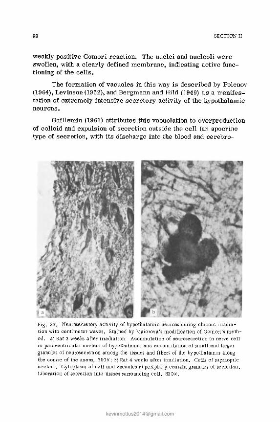
88 SECTION II
weakly positive Gomori reaction. The nuclei and nucleoli were swollen, with a clearly defined membrane, indicating active func-tioning of the cells 0
The formation of vacuoles in this way is described by Polenov (1964), Levinson (1952), and Bergmann and Hild (1949) as a manifes-tation of extremely intensive secretory activity of the hypothalamic neurons.
Guillemin (1961) attributes this vacuolation to overproduction of colloid and expulsion of secretion outside the cell (an apocrine type of secretion, with its discharge into the blood and cerebro-
Fig. 23. Neurosecretory activity of hypothalamic neurons during chronic irradia-tion with centimeter waves. Stained by Maiorova's modification of Gomori's meth-od. a) Rat 3 weeks after irradiation. Accumulation of neurosecretion in nerve cell in paraventricular nucleus of hypothalamus and accumulation of small and larger granules of neurosecretion among the tissues and fibers of the hypothalamus along the course of the axons, 350X; b) Rat 4 weeks after irradiation. Cells of supraoptic nucleus. Cytoplasm of cell and vacuoles at periphery contain granules of secretion. Liberation of secretion into tissues surrounding cell, 830x.

CHAPTER 1
Fig. 24. Swelling of cytoplasm and appear-ance of single vacuoles at periphery of nerve cell of supraoptic nucleus of hypothalamus in a rat 8 weeks after irradiation. Nissl's stain, 675X.
89
spinal fluid. In individual cells the vacuoles disappeared after ex-pulsion of the secretion and the normal quantity of tigroid substance was restored. Meanwhile, alongside the cell the expelled secretion could still be seen as large extracellular spherical droplets of colloid. The secretory cycle in the cell was beginning to repeat itself. Very occasionally death of single neurons and evidence of karyocytolysis with conversion of the neurons into cell ghosts could be seen. This was the result of excessive secretory activity of the nerve cell. Much secretion was found inside the neurons also. The cytoplasm of some neurons was literally packed with granules of secretion. These cells had lost all their tigroid substance. Many Gomori-positive granules were found between the fibers of the

90 SECTION II
hypothalamo-hypophyseal tract, where the granules of secretion appeared like Gomori-positive beads along the axons or as large masses and drops between the fibers of the tract (Fig. 25). As the granules and droplets of secretion moved along the hypo-thalamo-hypophyseal tract their number increased steadily, in-dicating increased synthesis of secretion along its course toward the neurohypophysis.
Many Gomori-positive granules and larger deposits were seen in the posterior pituitary along the course of the pituicyte fibers, indicating that the secretion is stored in the pituitary gland. The collections of secretion in the pituitary formed Herring's bodies. It was very obvious that some of the larger masses of secretion in the neurohypophysis had broken up into small gran-ules and these lay close to the blood vessels in the perivascular tissue. Sometimes drops of secretion were visible in the vessels.
Fig. 25. Neurosecretory activity of hypotha-lamic neurons during prolonged irradiation with centimeter waves of low intensity. Maiorova's modification of Gomori's stain. Rat 8 weeks after irradiation. Numerous granules of secre-tion forming large masses and drops along the course of fibers of the hypothalamo -hypophys-eal tract, 300 x.

CHAPTER 1
This indicated an increase in the absorption of secretion and its entry into the blood stream.
Consequently, in this group of animals increased formation and accumulation of neurosecretion and the beginning of its in-creased liberation into the blood stream were observed.
91
In the second group of animals, which were sacrificed be-tween the 8th and 14th weeks of irradiation, a completely different picture was seen in the anterior hypothalamic nuclei and pituitary gland.
The nerve cells of the anterior hypothalamus contained a small quantity of Gomori-positive secretion, especially toward the end of the period, i.e., the quantity of secretion in the anterior hypothalamic nerve cells was definitely reduced by comparison with the preceding period. Most neurons were in a resting state. In individual neurons there was no resynthesis of secretion, in-dicating exhaustion of the nerve cells as a result of their exces-sive activity in the preceding period. Few Gomori-positive gran-ules and deposits also were found in the neurohypophysis, i.e., there was increased absorption of neurosecretion into the blood. In fact, Gomori-positive granules and drops could actually be seen in the lumen of many vessels in the pituitary gland, and also in the perivascular tissue. Much Gomori-positive secretion was found in the axons of nerve cells in the form of Gomori-positive poolsj drops, and granules along the course of the hypothalamo-hypophyseal tract. Granules and drops of Gomori-positive secretion were pres-ent around and inside the vessels of the neurohypophysis and even of the hypothalamus, indicating increased absorption of secretion into the blood and cerebrospinal fluid. This coincided with eleva-tion of the blood pressure in the animals of this group and an in-crease in the ascorbic acid content of their adrenals. Consequently, the reduced content of neurosecretion in the cells of the anterior hypothalamic nuclei and neurohypophysis, together with its in-creased liberation into the blood stream, accompanied by elevation of the blood pressure and an increase in the ascorbic acid content in the adrenals of animals irradiated with 10-cm waves of low in-tensity, maybe manifestations of an adaptive response to irradia-tion of this type.
In the animals of the third group, irradiated for 14-20 weeks, marked accumulation of granules of neurosecretion was observed

92 SECTION II
in the cells of the anterior hypothalamic nuclei, with swelling of the nuclei and nucleoli, in which the membrane was clearly defined. Many neurons were in a resting state. The pattern of the neuro-secretory cycle was highly reminiscent of that in the control ani-mals. In individual cells of the anterior hypothalamic nuclei Gomori-positive granules of secretion were observed. Similar granules were found among the fibers of the hypothalamo-hypo-physeal tract, but they were far less numerous than in the first group. No large deposits or drops could be found among the fibers of the hypothalamo-hypophyseal tract. Only small Gomori-positive granules were seen (Fig. 26a). Gomori-positive granules and large deposits were visible in the posterior pituitary (the neurohypophysis),
, b
Fig. 26. Neurosecretory activity of hypothalamic neurons after prolonged irradiation by centimeter waves of low intensity. a) Rat after irradiation for 20 weeks. Small Gomori-positive granules among hypothalamic nerve fibers. Maiorova's modification of Gomori's stain, 340X; b) Rat after irradiation for 26 weeks. Marked vacuolation of some hypothalamic neurons. Loss of nuclear membrane, evidence of karyocytolysis in individual neurons. Nissl's stain, 680x.

CHAPTER 1 93
but in smaller quantities than in the previous group. At the same time, small granules of Gomori-positive secretion could be seen in the perivascular tissue of the neurohypophysis. These findings indicated intensive production of secretion and its absorption into the blood stream. Consequently, this group of animals is char-acterized by the formation (resynthesis) of neurosecretion. Its content in the cells of the hypothalamus and in the neurohypophysis was the same as in the control (unirradiated) animals. Absorption of secretion into the blood stream was observed, because Gomori-positive granules of secretion were found in the tissues around the vessels and in their lumen. The normal content of neurosecretion in the hypothalamus and neurohypophysis correspond to restoration of the normal blood pressure as observed in this group of animals. Here and there cells with tiny vacuoles were observed. There was hardly ever any considerable degree of vacuolation of the nerve cells or evidence of karyocytolysis (with the formation of cell ghosts and death of the nerve cells).
In the animals of the fourth group, sacrificed after irradiation for 20-26 weeks, a completely different picture was observed in the neurohypophysis and in the anterior hypothalamic nuclei. A sharp decrease in the content of neurosecretion was observed in the nerve cells and tracts (axons), and this was accompanied by a sharp decrease in the secretory function of the neurons of the anterior hypothalamic nuclei. Compared with the control group, ' in this case there was a very marked decrease in the intensity of secretion by the nerve cells. Only occasionally were tiny in-dividual Gomori-positive granules found in the cytoplasm of single cells. Very few Gomori-positive granules were visible in the hypothalamo-hypophyseal tract among the fibers. Many neurons had uneven, or notched, outlines (Fig. 26b). Because of the large number of vacuoles, the nuclei and nucleoli of the neurons were shrunken. Evidence of karyocytolysis was observed. Hardly any secretion was found in the tissues of the posterior lobe of the pituitary (in the neurohypophysis). Only here and there were single tiny Gomori-positive granules found in the tissue around the blood vessels. Neurosecretion was much less abundant than in the control (unirradiated animals). The sharp decrease in neurosecretory ac-tivity of the hypothalamo-hypophyseal system was accompanied by a decrease in size of some of the nerve cells, by marked vacuola-tion of most of the neurons, and by evidence of karyocytolysis of
94 SECTION II
the cells and total chromatolysis of individual neurons with the formation of cell ghosts. The nuclei and nucleoli were swollen and their outlines were indistinct or contracted, indicating death of the cells, In individual neurons marked vacuolation of the cells and notching of their outlines were accompanied by shrinking or lysis of the nucleus and nucleolus (with loss of the nuclear membrane), indicating degenerative changes in the hypothalamus, In this group of animals the blood pressure was consistently low.
The morphological findings in the animals of the fourth group, exposed to prolonged (up to 26 weeks) irradiation with low-inten-sity 10-cm waves can be summarized by saying that everything suggests exhaustion of the neurosecretory system as a whole, with little evidence of the formation and liberation of secretion and with no secretion present in the depots because it had all been absorbed previously into the blood stream. No fresh secretion was being supplied to the neurohypophysis, Clinically in these animals a conSistently low blood pressure was observed.
This description still does not mean that the process in the hypothalamus is irreversible. Only solitary neurons showed de-generative changes and most of them when stained by Nissl's method were little affected. Only a sharp decrease in secretory activity of the anterior hypothalamic neurons was observed, i.e., functional changes of a reversible character were present, c Some 4-8 weeks after the end of irradiation the neurosecretory activity was completely restored and corresponded to that observed in the control animals; the normal blood pressure also was restored,
It can be concluded from the results of this experiment that three phases depending on the duration of irradiation were observed in the animals' condition: a phase accompanied by elevation of the blood pressure, a phase of return to normal blood pressure, and a phase of conSistently lowered blood pressure.
Corresponding to these three phases, three phases of changes in hypothalamic neurosecretory function could be distinguished. The first, initial phase is subdivided into two periods: to begin with increased accumulation of neurosecretion in the cells of the supraoptic and paraventricular nuclei, followed by its increased liberation into the blood stream. In the second phase the formation and liberation of secretion return to normal and its content cor-respond to that in the control animals. In the third phase the for-

CHAPTER 1 95
mation of neurosecretion and its liberation into the blood stream are sharply reduced and neurosecretory activity is exhausted, with death of individual neurons; this corresponds to the phase of lowered blood pressure. These findings, together with others described in the literature and cited above, suggest that in the first period of the first phase the increase in accumulation of secretion in response to the action of low intensities of irradiation may be regarded as a phase of mild irritation with increased formation of secretion. During more prolonged exposure to the harmful factor, in the second period of the first phase there is an increased libera-tion of secretion into the blood stream - the alarm response in-duced by irradiation (Selye's alarm reaction or stress reaction is the term applied to mobilization of the organism to adapt it to the action of the pathogenic factor). This stage of mobilization shows that the hypothalamus and neurohypophysis, together with the adeno-hypophysis and adrenals, play an active part in adaptive mechanisms activated in the response to prolonged irradiation by radio waves.
A s a result of the action of these adaptive mechanisms, nor-mal processes are restored in the body (the normal blood pressure is restored and the content of neurosecretion corresponds to that in the control animals, but this return to normal is not lasting).
If exposure to irradiation continues still longer, a point is reached when these compensatory and adaptive mechanisms are insufficient and break down.
The possibility cannot be ruled out that secretory function of the hypothalamus is inhibited by the action of the harmful factor on the cerebral cortex, where inhibition arises and spreads to the hypothalamus. This can be postulated on the basis of results ob-tained by Romasenko (1967) and Gerber (1967), who found exhaustion of the hypothalamic secretory function in patients with schizophrenia with well-marked cortical inhibition and clinical manifestations of stupor.
In this period exhaustion of secretory activity of the hypo-thalamus and neurohypophysis was observed, and activity of the adenohypophysis and adrenals was reduced. However, this process is reversible and soon after the end of irradiation all the changes described above can be reversed and the normal state restored.
The question of changes in the neurosecretory function of the hypothalamus during prolonged irradiation with low-intensity

96 SECTION II
radio waves is of considerable interest and calls for further phys-iological and histochemical investigation of the sys tem as a whole (including the hypothalamus, the posterior neurohypophysis, the adenohypophysis, and the adrenals). The investigation now being described is both functional and morphological in character and it demonstrates the role of the functional state of the anterior hypothalamic secretory nuclei, in conjunction with the hypophyseo-adrenal system, in the adaptive mechanisms arising in response to prolonged irradiation with low-intensity microwaves in order to maintain homeostasis in the body.
The results obtained agree with those of investigations by Gorizontov (1969), who showed that under the influence of stress factors (electrical stimulation, fixation of animals, irradiation) reactivity changes in the various periods of the extremal state. In the initial period, there is what can be called a stage of mobiliza-tion (as Selye showed), adapting the organism to the action of the pathogenic factor. Next follows a stage of increased nonspecific resistance, and finally there is the stage of exhaustion (Gorizontov).
In animals exposed to even more prolonged irradiation (10-12 months) by 10-cm waves, in addition to the changes described above in the fine structures of the hypothalamus and neurohypoph-ysis, slight degenerative changes also are found in the brain, in the form of swelling of individual cortical cells, and shrinktng and dark staining of other neurons (Fig. 32a). Numerous vacuoles also were observed in the hypothalamic neurons with evidence of karyo-cytolysis and the formation of cell ghosts (Fig. 33b). Histochemical investigation of the skin revealed a decrease in the RNP content in its surface layers, in the epidermis, and in its derivatives (Fig. 34b). In addition, unevenness of staining was found, and sometimes homogenization of the myocardial muscle fibers (Fig. 35b).
In individual animals exposed to still longer irradiation (12-15 months) with 10-cm waves with an intensity of 10 mW/cm2, in addition to the reversible degenerative changes in the nervous system and changes in the myocardium described above there were ill-defined degenerative changes also in the testes: degeneration of the spermatogenic epithelium in certain tubules with desquamation of the spermatogenic epithelium into the lumen of the tubules and the formation of plugs (Fig. 36b).

CHAPTER 1 97
In some tubules, besides desquamation of the spermatogenic epithelium, giant cells indicating distorted regeneration were found. However, in the overwhelming majority of tubules in the testis spermatogenesis was normal and the animals were capable of fer-tilization (as tested by experiments on fertilization of females by irradiated males). Very slight degenerative changes also were found after irradiation for 12-15 months in the hepatocytes and the epithelial cells of the convoluted renal tubules: cloudy swelling of the cytoplasm with the appearance of single vacuoles. Regenerative changes were found in the hepatocytes (the appearance of mitoses and of polynuclear cells), and a proliferative response of the micro-glia in the brain (Fig. 37b) and of the reticuloendothelial elements in the liver (Fig. 38b) also was present, reflecting adaptive and defensive responses of the body. Despite the prolonged irradiation, clinically the animal remains healthy.
Comparison of the morphological changes found in animals after exposure to pulsed and continuous irradiation with 10-cm waves shows that the changes were more marked in the case of pulsed waves.
Morphological Changes Following Repeated Ir-radiation with Low-Intensity Millimeter Waves
Animals were exposed frequently (for 10 months) to irradia-tion with intensities up to 10 mW/cm2 for 1 h daily. '
The animal showed no clinical signs of hyperthermia, but the rectal temperature (as mentioned above) was raised by 0.3°C. The animals tolerated irradiation well. The irradiated animals gained in weight somewhat more slowly than the control animals and showed a slight hypotensive effect.
Morphological examination of this group of animals by the usual methods revealed virtually no degenerative changes in the viscera or nervous system.
However, by the use of delicate elective methods of inves-tigation changes were found in the cortical interneuronal synapses: disappearance of the spines on the apical dendrites of individual cortical neurons with the appearance of beads and irregular thick-enings on the dendrites.

98 SECTION II
Marked changes in the sensory nerve fibrils of the skin, consisting of increased argyrophilia and the appearance of bead-like thickenings and swellings, also were observed. Frequently the fibril was fragmented (Fig. 29a). Axons of larger myelinated cutaneous nerve trunks were affected by the process (Fig. 29b). Changes in the sensory nerve fibrils of the visceral receptive fields were almost completely absent (Fig. 30a). Meanwhile changes were found in the neurons of the spinal sensory ganglia, in the form of swelling of the cytoplasm with tigrolysis in the center and ectopia of the nuclei (Fig. 31a), indicating reversible changes in the ganglionic neurons as a result of reflex stimulation of the cuta-neous sensory nerve fibers. This can be postulated because, ac-cording to information in the literature, millimeter waves are absorbed in the skin and do not spread deeper into the body tissues.
During continued irradiation changes appeared in the cortical neurons: shrinking of individual groups of neurons with dark stain-ing of their cytoplasm. Swelling of the cytoplasm and the appear-ance of solitary vacuoles also were observed in certain hypo-thalamic neurons (Fig. 33a) with an accompanying decrease in the RNP content. Histochemical changes in the skin consisted of a decrease in the RNP content in the surface layers of the skin, the cells of the epidermis, and its derivatives (Fig. 34a).
Consequently, during exposure to low-intensity radio changes in the cortical interneuronal synapses, histochemical changes in the skin, and changes in the sensory nerve fibrils of the skin appeared sooner than the usual morphological changes, i.e., they appeared before any changes in the body were detectable by the usual histological methods. Swelling and unevenness of staining of the myocardial muscle fibers were discovered later (Fig. 35a).
The RNP content in the spermatogenic epithelium was slight-ly reduced. Vacuolation of the cytoplasm of the spermatogenic epithelial cells with pycnosis of their nuclei was found in some of the tubules, most frequently under the capsule of the testis. Oc-casionally necrosis of the tubules developed, particularly beneath the capsule (Fig. 36a). During prolonged irradiation, tubules with desquamation of the spermatogenic epithelium into the lumen of the tubules and the formation of albuminous deposits were found under the capsule.

CHAPTER 1 99
Slight cloudy swelling of individual hepatocytes and epithelial cells of the convoluted renal tubules was found later.
Hence, despite the ill-defined degenerative changes in the viscera and nervous system detectable by the usual morphological methods, more delicate methods of investigation revealed more definite changes in the nervous system of the animals of this group (especially in the sensory nerve fibers of the skin and the inter-neuronal synapses of the cerebral cortex). The changes were most marked in the skin, where histochemical disturbances, namely, a decrease in the RNP content, also were found.
Characteristic morphological changes for the animals of this group are slight proliferation of the microglia in the brain (mainly around the blood vessels; Fig. 37a) and of the reticulo-endothelial elements of the liver (Fig. 38a).
Consequently, the slight degenerative changes developing in the nervous system, viscera, and skin in response to prolonged stimulation by low-intensity millimeter waves are accompanied by proliferation of the microglia in the brain and reticuloendo-thelial elements of the liver. The proliferative changes reflect defensive and adaptive processes in the body.
Comparison of the morphological changes after prolonged exposure to repeated sessions of irradiation with lO-cm and mil-limeter waves indicates that the degenerative changes in the vis-cera are more severe in response to the action of the lO-cm waves while the histochemical changes in the skin and changes in the sensory nerve fibrils of the skin are more severe as a result of the action of millimeter waves.
The selective changes in the sensory nerve fibrils of the skin and the histochemical changes in the skin described above are eVidently due to the action of waves of this particular wave-length, for millimeter waves are absorbed in the surface layers of the skin and do not penetrate into deeper tissues. It can be pos-tulated that the degenerative and proliferative changes in the vis-cera and central nervous system during exposure to millimeter waves are brought about by nervous reflex mechanisms. This hypothesis is also confirmed by the irritation phenomena in the neurons of the spinal sensory ganglia.

100 SECTION II
Morphological Changes Following Repeated Ir-radiation with Low-Intensity Decimeter Waves
Animals were exposed to prolonged and repeated irradiation with decimeter waves up to 10 mW/cm2 in intensity for 60 min daily for 10 months (220 sessions). The animals tolerated the ir-radiation well and there were no signs of hyperthermia. However, changes were observed in their higher nervous activity, and these were particularly marked and appeared early in rats, which are sensitive to acoustic stimulation. The animals also showed a hypotensive effect and their gain in weight was less than that of the control animals.
Investigation of the animals of this group by ordinary mor-phological methods revealed practically no vascular disorders or degenerative changes in the viscera and nervous system. By the use of delicate elective neurohistological methods changes were found in the complex structures of the nervous system: the cortical interneuronal synapses and sensory nerve fibers of the visceral receptive fields. The changes consisted of disappearance of spines from the dendrites of individual cortical neurons and the appearance of beading and irregular thickenings on the dendrites (Fig. 27b). No changes were found in the sensory nerve fibers of the skin (Fig. 29d), but marked signs of irritation and degenerative changes were observed in the sensory nerve fibers of the receptive (reflexogenic) zones or the viscera (myocardium, aorta, esophagus, intestine, stomach, bladder; Fig. 30c). The nerve fibers showed increased argyrophilia and tortuosity, bead-like thickenings and pools of axoplasm appeared on them, and sometimes the fibers were broken up into fragments. Consequently, the sensory nerve fibers of the skin were not predominantly affected in this case.
During continued irradiation swelling of the cytoplasm of individual cells (with the appearance of vacuoles) of the nervous system was observed in the basal ganglia, and the changes were most severe in the hypothalamus (Fig. 33c).
Histochemical investigation revealed a decrease in the RNP content in the cytoplasm of cells in organs normally rich in RNP (bronchial epithelium, glandular epithelium of the gastrointestinal tract). The RNP content in the skin was not reduced (Fig. 34c). Swelling, irregularity of staining, and homogenization of individual

CHAPTER 1
Fig. 27. Com parison of changes in axo-dendritic interneuronal synapses in the cerebral cortex during prolonged irradiation with waves of different lengths and of low intensity. Stained by Golgi's method: a) severe bead-ing deformation of apical dendrites. Pr'olonged irradiation by centimeter waves, llOx; b) beading deformation of apical dendrites of cortical pyr-amidal neurons after prolonged irradiation by low-intensity decimeter waves 400x.
101
muscle fibers were observed in the myocardium (Fig. 35c). After longer irradiation some animals developed degenerative changes in the spermatogenic epithelium of individual tubules of the testes, with desquamation of the epithelium into the lumen of the tubules and the formation of casts (Fig. 36c). Giant cells, a manifestation of distorted regeneration, were found in other tubules. However, only individual tubules were affected and the great majority con-tinued to exhibit normal spermatogenesis. The animals remained capable of fertilization (as tested by fertilization of females by the irradiated males).

102 SECTION II
A slight degree of cloudy swelling was found in individual hepatocytes and epithelial cells of certain convoluted renal tubules. In the liver there was fatty degeneration of individual hepatocytes with the appearance of minute droplets of fat.
Against the background of the very slight degenerative changes in the brain described above focal and diffuse proliferation of micro-glial cells was observed, mainly around the blood vessels (Fig. 37c). Microglial processes showed the initial signs of degeneration. Slight hyperplasia of reticuloendothelial elements was observed in the liver, sometimes with the formation of submiliary histocytic nodules (Fig. 38c). The number of perivascular round cells in the lungs was greater than normal. It must be emphasized that besides the evidence of irritation in the fine structures of the nervous sys-tem (synapses, sensory nerve fibrils in the various visceral recep-tive fields) and the gradually increasing degenerative changes in the viscera and testes, there was also a gradual increase in the in-tensity of proliferation of the microglia in the brain and of the reticuloendothelial cells in the liver.
Comparison of the morphological changes found in animals exposed to the action of low-intensity microwaves of all frequencies clearly shows that irradiation whose intensity is too low to induce a thermogenic effect is nevertheless harmful in its action.
This does not confirm the view, so widely held in the past among Soviet investigators and still maintained to a large extent even at the preset time in the West, that the action of microwaves is entirely thermal.
In the present experiment the attention was concentrated chiefly on the chronic effect of low-intensity microwaves, on the detection of the earliest responses of the body, and the character and severity of these responses in relation to the wave band.
The central nervous system and, in particular, its higher levels are highly sensitive to the action of microwaves, which cause disturbances of conditioned-reflex activity (weakening of excitation and the development of limiting inhibition) accompanied by changes in the structure ofaxo-dendritic and axo-somatic synapses in the brain. Changes are also found in the sensory nerve fibrils of the skin and viscera.

CHAPTER 1 103
A decrease in excitability and weakening of inhibition were observed in irradiated rats, animals which are sensitive to acoustic stimulation.
Early responses of the eNS to microwave irradiation were detected by recording brain potentials, and by analysis of the rela-tionship between intensity of irradiation and latent period a curve reflecting the sensitivity of the rabbit eNS to the action of micro-waves of different frequencies could be plotted. This curve was found to resemble in its character the Hoorweg- Weiss curve of sensitivity for an electric current and the curve of Tsypin and Grigor'ev (1960) for ionizing radiation.
Lowering of the blood pressure is to a certain extent a specific response to microwave action. The intensity, the time of appearance, and the character of the vascular response depend on the wave band and intensity of irradiation.
Functional changes due to the action of microwaves of dif-ferent bands are similar in character but differ in intensity, in the time of their appearance, and sometimes in their course, notably in the initial period of long-term exposure. In this period (first phase) increased excitability of the nervous system, hyper-tension, and increased cholinesterase activity in the blood and or-gans are often observed. The second phase is characterized by , depression of the functional state of the eNS, especially of its higher levels, by a hypotensive effect, by reduced enzyme activity, and by marked morpholOgical changes in the eNS.
The intensity of the response varies with the frequency of the microwaves.
As the authors have shown, the intensity of the response of the eNS to low-intensity microwave irradiation is most fre-quently reduced with shortening of the wavelength, but at the same time the intensity of the vagotonic reactions is increased.
This general pattern can be connected with the depth of penetration and absorption of different types of microwaves in the tissues. The hypotensive effect, which is most marked in response to irradiation with millimeter waves, can be attributed mainly to the action of the radiation on cutaneous sensory nerve fibers and

104 SECTION II
reflex effects on vascular tone. Meanwhile the response of the nervous system, which is more severely affected by the action of centimeter and decimeter waves, can be attributed mainly to the direct effect of the radiation on the brain structures.
There is no question that the effects of low-intensity micro-wave irradiation are cumulative. Repeated exposure leads to a gradual increase in severity of the observed changes.
This conclusion is confirmed by the gradual development of increasingly severe disturbances of conditioned-reflex activity of the animals, changes in the responses of animals particularly sensitive to acoustic stimulation, and hemodynamic changes (hypo-tension, reduced cholinesterase activity in the blood and organs).
Functional changes observed after chronic exposure to low-intensity microwaves are reversible.
The character of generation of the microwave energy, whether it is pulsed or continuous, is not reflected in the ultimate effect of low-intensity microwave irradiation.
Parallel with the functional changes in the nervous system after prolonged exposure to low-intensity microwave irradiation reversible morphological changes in the axo-dendritic and axo-somatic synapses of the brain and changes in sensory nerve fibrils in various cutaneous and visceral receptive fields are found. Co-ordinated activity of the sensory nerve fibrils and their receptor endings in the receptive fields is a factor maintaining the constancy of the internal milieu of the organism. Later reversible changes are found in the neurosecretory activity of the hypothalamo-hypo-physeal system, which is responsible for adaptive reactions of the body to the harmful action of radio waves. The morphological evi-dence of functional changes in neurosecretory activity of the hypo-thalamus coincide with clinical manifestations of an initial rise and subsequent fall in the animals' blood pressure. During more prolonged irradiation degenerative changes arise in the myocardium: unevenness of staining and homogenization of individual muscle fi-bers.
During prolonged irradiation degenerative changes are found in the spermatogenic epithelium of individual tubules in the testes. However, spermatogenesis in most tubules is well marked and the animals are still capable of fertilizing females.

CHAPTER 1 105
Later, ill-defined degenerative changes develop in the liver and kidneys, where they are accompanied by regenerative changes, and proliferation of the microglia in the brain and of the reticulo-endothelial cells of the liver are manifestations of the animals' defensive and adaptive responses.

Chapter 2
Morphological Changes in Animals Following Repeated Low-Intensity Irradiation
with Ultrashort Waves
After prolonged (1.5 and 5 months) irradiation with ultra-short waves (USW) with a frequency of 69.7 MHz and of low inten-sity (150 Vim) the animals appeared well and showed no evidence of hyperthermia.
Microscopic investigation revealed ill-defined changes in the nervous system: increased argyrophilia, and irregular swell-ings and tortuosity of the sensory nerve fibrils of the skin and vis..': cera (Figs. 2ge and 30d). The lesions were not noticeably pre-dominant in the sensory nerve fibrils of the skin or viscera.
Cortical nerve cells were reduced in size and darkly stained (Fig. 32b), and hypothalamic neurons showed vacuolation and, frequently, evidence of karyocytolysis (Fig. 33d), while individual muscle fibers in the myocardium were unevenly stained and ho-mogenized. Some rats showed degenerative changes in the sper-matogenic epithelium of individual tubules of the testes, sometimes with the formation of giant cells indicating distorted regeneration (Fig. 36d).
Degenerative changes in the viscera were ill-defined: cloudy swelling and sometimes fatty degeneration of individual groups of hepatocytes and cloudy swelling of the epithelium of some con-voluted renal tubules. Together with slight degenerative changes in the viscera, there were proliferative changes in the form of
107

108 SECTION II
hyperplasia of the microglia in the brain and reticuloendothelial cells in the liver, sometimes with the formation of histiocytic nodules (Fig. 38d), and hyperplasia of perivascular round cells in the lungs, to form cuffs,
If the duration of irradiation was shorter, these changes were less severe,
During prolonged (5 months) and repeated irradiation of animals with pulsed or continuous radio waves with a frequency of 155 MHz and intensity 25-50 Vim and 191 MHz with intensity 10-20 Vim the animals' body temperature was not raised, Clinically, functional disturbances consisting of lowering of the blood pres-sure and changes in the electrical activity of the brain and con-ditioned-reflex activity were found,
Morphological investigation revealed shrinking of individual cerebral cortical neurons (Fig, 32c), slight vacuolation of the cytoplasm of cells in the hypothalamus, increased argyrophilia and irregular thickenings of the sensory nerve fibrils of the cutaneous and visceral receptive fields, and very slight degenerative changes in the myocardial muscle fibers.
Degener.ation of the spermatogenic epithelium of individual tubules of the testes, with the formation of giant cells by distorted regeneration, was slight in intensity (Fig, 36e), but was intact in most tubules and the animal retained its fertilizing ability, Slight degenerative changes in the nervous system and vis-cera (cloudy swelling of the hepatocytes and epithelial cells of the convoluted renal tubules) was accompanied by proliferation of microglia in the brain and of the reticuloendothelial cells in the liver.
All the degenerative and proliferative changes were more marked in animals irradiated with waves in the 191 MHz band and with an intensity of 20 Vim than in those irradiated with waves of 155 MHz and an intensity of 50 Vim,
The morphological changes following irradiation by USW in the 191 and 155 MHz bands were more marked than those observed after irradiation in the 69,7 MHz band, All the changes described above became more severe as the duration of irradiation was in-

CHAPTER 2
creased. However, after irradiation with USW ended, the mor-phological changes were largely reversed.
109
After prolonged irradiation of animals with USW in the 191, 155, and 69.7 MHz bands, with intensities not evoking a thermal effect (10-20 and 150 Vim), ill-defined degenerative changes were thus found in the nervous system (mainly in cells of the cortex and hypothalamus, sensory nerve fibrils of the skin and viscera), the muscle fibers of the myocardium, and parenchymatous cells of the testes, liver, and kidneys.
The degenerative changes increase in severity with an in-crease in the period of irradiation, and after irradiation ends they gradually undergo regression. This corresponds to the clinical evidence of restoration of disturbed functions.
Degenerative changes in the nervous system and viscera are accompanied by proliferation of the microglia in the brain and the reticuloendothelial cells in the liver, manifestations of defensive and adaptive responses.
The morphological changes are most severe after irradia-tion in the 191 MHz band, less severe after irrradiation in the 155 MHz band, and least severe of all after irradiation in the 69.7 MHz band.

Chapter 3
Morphological Changes in Animals Following Repeated Irradiation with
Low-Intensity Short Waves
During prolonged (5 months) and repeated irradiation with short waves of low intensity (2250 Vim) the animals showed no effects of hyperthermia. Their condition was good at the time of sacrifice. Microscopic investigation revealed ill-defined de-generative changes in the nervous system. Increased argyrophilia and irregular thickenings and pools of axoplasm were found in the sensory nerve fibrils of the skin and viscera (Fig. 30e). No evi-dence of fragmentation of the nerve fibrils was observed. Damage' to the nerve fibrils was not noticeably more severe in the skin or viscera. Shrinking and dark staining of individual groups of neurons (Fig. 32d) was found in the cortex, and the cytoplasm of hypothalamic neurons showed vacuolation, frequently with evidence of karyocy-tolysis (Fig. 33e). Slight degeneration of the myocardial muscle fibers was expressed as unevenness of staining. After more pro-longed irradiation, mild degenerative changes occurred in the sper-matogenic epithelium of individual tubules of the testes, sometimes with deposition of albuminous masses in their lumen. However, spermatogenesis was intact in most tubules. After a shorter pe-riod of irradiation (1.5 months) all these changes were less severe. Fatty degeneration of individual groups of hepatocytes and cloudy swelling of the epithelium of individual convoluted renal tubules were observed in the viscera and were accompanied by prolifera-tion of reticuloendothelial cells in the liver, sometimes with the
111

112 SECTION II
formation of histiocytic nodules (Fig. 38e), and by proliferation of perivascular round cells in the lungs.
Consequently, during prolonged and repeated irradiation with short waves of low intensities for periods of up to 5 months the changes observed included shrinking of the cortical cells, vacuola-tion of the cytoplasm of hypothalamic cells, and irritation phenom-ena and degenerative changes in the sensory nerve fibers of the cutaneous and visceral receptive fields. Pathological changes in the nervous system corresponded to clinical features observed in the animals (lowering of the blood pressure, reduced sensitivity of the rats to acoustic stimulation, and depression of the brain potentials).
Initial degenerative changes were found in the spermatogenic epithelium of individual tubules of the testes with preservation of spermatogenesis in the great majority of tubules, so that the ani-mals still remained capable of fertilization.
ill-defined degenerative changes were found in the viscera and were accompanied by proliferation of reticuloendothelial cells in the liver.
Comparison of the action of ultrashort and short waves showed that the pathological changes arising in response to both were similar in direction, although the intensity of all the <?hanges was greater after irradiation with ultrashort waves.

Chapter 4
Morphological Changes in Animals Following Repeated Irradiation with
Low-Intensity Medium Waves
Morphological changes were studied in animals exposed for 10 months to irradiation by radio waves in the medium wave band (electrical component 180 Vim, magnetic component 50 Aim).
Microscopic investigation revealed a background of very slight vascular disturbances against which moderately severe degenerative changes in the form of disruption of the axo-somatic synapses could be seen in the brain (Fig. 28b). Meanwhile, irrita-tion phenomena were found in the sensory nerve fibrils of the skin and viscera. Sensory fibers of the viscera were predominantly affected, and showed increased argyrophilia and irregular thicken-ing (Fig. 30f). The changes were much less marked in the sen-sory nerve fibrils of the skin (Fig. 29f). Shrinking of cortical nerve cells (Fig. 32e) and tortuosity of their apical dendrites were found, while in the hypothalamus the cytoplasm of the nerve cells was vacuolated and evidence of karyocytolysis could be seen in individual neurons (Fig. 33f).
Initial degenerative changes, in the form of unevenness of staining and homogenization, occurred in the myocardial muscle fibers.
Some animals showed ill-defined degenerative changes in the spermatogenic epithelium of individual tubules of the testes, with the appearance of giant cells indicating distorted regeneration
113

114 SECTION II
Fig. 28. Comparison of changes in interneuronal synapses in the hypothala-mus after prolonged irradiation by low-intensity waves of different frequen-cies. Cajal's stain, 600x: a) club-shaped thickenings and increased argyro-philia of synaptic boutons and their separation from bodies of nerve cells in the hypothalamus after prolonged irradiation by centimeter waves of low in-tensity; b) increased argyrophilia of boutons terminaux and thickening of synaptic loops with their partial separation from the body of a neuron after prolonged irradiation by medium waves (magnetic field).
(Fig. 36f). However, spermatogenesis in most tubules was well marked. The animals remained capable of fertilization during life as shown by tests involving fertilization of females.
Changes in the viscera consisted of ill-defined cloudy swell-ing or fatty degeneration of individual hepatocytes and cloudy swell-ing of the epithelium of individual renal convoluted tubules.
Together with mild degenerative changes in the nervous sys-tem and viscera, proliferation of the microglia in the brain and of the reticuloendothelial cells in the liver, with the formation of histiocytic nodules in the liver of some animals, was observed. Comparison of the morphological changes following irradiation by high-frequency magnetic and electric fields Rhowed more marked vascular disturbances and degenerative changes in the viscera fol-

Fig. 29. Comparison of changes in sensory fibers of cutaneous receptive fields after ir-radiation by low-intensity radio waves of different frequencies. Bielschowsky-Gross stain: a) intense argyrophilia and fragmentat ion of a cutaneous sensory nerve fiber af-ter prolonged irradiation by low-intensity millimeter waves, 440 x; b) axons of my-elinated nerve fibers in the skin have irregular swellings and thickenings and are strongly argyrophilic. Prolonged irradiation by millimeter waves of low intensity, 330 x; c) increased argyrophilia, irregUlar swellings, and tortuosity of a cutaneous nerve fiber after prolonged irradiation with centimeter waves of low intensity, 440 x; d) unchanged thin and delicate cutaneous sensory fibers after prolonged irradiation by decimeter waves of low intensity, 440 x; e) increased argyrophilia, irregular thick-enings and swellings of a cutaneous nerve fibers after prolonged irradiation by ultra-short waves of low intensity, 440 x; f) thin cutaneous nerve fibers with slight thick-enings in certain places after prolonged irradiation by medium waves (magnetic field), 440 x.

Fig. 30. Comparison of changes in sensory fibers of visceral receptive fields after prolonged irradiation by low-intensity radio waves of different frequencies. Bielschow-sky -Gross stain, 400 x; a) almost unchanged sensory fibers of the aorta after prolonged irradia tion by millimeter waves of low intensity; b) increased argyrophilia and irregu-lar thickenings of sensory fibers in the esophagus after prolonged irradiation by low-intensity centimeter waves; c) increased argyrophilia, irregular thickenings of sensory fibers in the myocardium after prolonged irradiation by low-intensity decimeter waves; d) intense argyrophilia and irregular thickenings of a myocardial sensory fiber after prolonged irradiation by low-intensity ultrashort waves; e) intense argyrophilia and ir-regular thickenings of a myocardial nerve fiber after prolonged irradiation by low-in-tensity short waves; f) intense argyrophilia and irregular thickenings of a myocardial nerve fiber (near the aorta) after prolonged irradiation by low-intensity medium waves.
CHAPTER 5 117
lowing irradiation in a high-frequency magnetic field. To sum up, after irradiation of albino rats for 10 months in a high-frequency electric field (1800 Vim) and a high-frequency magnetic field (50 Aim) moderately severe degenerative changes took place in the nervous system (especially in the cells of the cortex and hypo-thalamus and in the sensory nerve fibrils of the viscera). Degen-erative changes were found in the spermatogenic epithelium of the testes and moderately severe vascular disturbances and slight initial degenerative changes were found in the parenchymatous organs.
The degenerative changes in the nervous system and viscera were accompanied by proliferation of reticuloendothelial elements, reflecting defensive and adaptive processes.
Comparison of the morphological changes found after ir-radiation by low-intensity high-frequency electric and magnetic fields with the changes observed after irradiation by short and ultra-short waves and by microwaves reveals a common direction of the pathological changes after exposure to low intensities of these wave bands, but medium waves give rise to more severe morpho-logical changes than irradiation by short and ultrashort waves and microwaves.
Chapter 5
Comparison of Morphological Changes Following Repeated Exposure to
Low-Intensity Radio Waves of Different Frequencies (Microwaves; Ultrashort,
Short, and Medium Waves)
Investigations of the morphological changes after prolonged exposure to low-intensity radio waves (not inducing a thermal ef-fect) are particularly interesting because it is to these intensities of irradiation that industrial workers are exposed.
To study the action of microwaves, the animals were irradiat-ed repeatedly with microwaves of low intensity (10 mW/cm
2
) for 60 min daily for 5-15 months.
Experiments were also carried out in which animals were irradiated daily for 5 months with ultrashort waves with inten-sities of 150 Vim (69.7 MHz), 50-25 Vim (155 MHz), and 20-10 Vim (191 MHz). When the effects of daily exposure to short waves for 5 months were studied irradiation was given in an intensity of 2250 Vim, while in experiments lasting 10 months the animals were exposed to electrical and magnetic high-frequency fields with in-tensities of 1800 Vim and 50 Aim. Altogether 243 animals took part in the experiments. It is interesting to compare the physio-logical and morphological changes in the various organs and sys-tems following exposure to low intensities of radio waves.
119

120 SECTION II
The animals tolerated irradiation well and no evidence of hyperthermia was observed. However, the irradiated animals gained in weight more slowly than the controls, and certain functional changes were observed.
These were mainly uniform in pattern despite exposure to different frequencies of radio waves, although their severity was identical.
Functional changes can be observed at different levels of activity: subcellular or at the cells, organ, or system level.
Without going into details, it can be said that because of dif-ferences in the sensitivity of the various systems to radio waves it is impossible to speak of a single threshold of the biological response to irradiation.
However, it is clear that indices of responses of excitable systems, i.e., neurophysiological data, must be used as the basis for the definition of threshold responses.
Investigations in this direction have begun and will be de-scribed in subsequent publications. At this stage, however, it can be provisionally stated that threshold intensities of irradiation for the nervous system in the microwave range are of the order of several microwatts per square centimeter, while in the ultrashort-wave band they are of the order of several volts per meter.
Future investigations are required to obtain more accurate values of the thresholds in two situations:
(1) differential assessment of physiological (especially adap-tive) responses;
(2) a differential approach to thresholds of single and pro-longed exposure to radio waves.
Morphological examination of groups of animals irradiated with microwaves below 10 mW/cm2 in intensity and sacrificed after 4-10 months by decapitation, using the ordinary morphological methods, revealed no vascular disturbances or degenerative changes. However, more delicate histological and histochemical investiga-tions using elective methods of staining the nervous system showed initial functional changes affecting primarily the nervous system, cor-responding to phYSiological changes in the nervous system detect-

CHAPTER 5 121
able clinically, in animals exposed for long periods to low inten-sities not sufficient to induce a thermal effect. After prolonged ir-radiation with low intensities changes occurred principally in axo-dendritic synapses, the delicate receptor structures of the cortex. The changes consisted of coarsening and sometimes disappearance of spines of the apical dendrites and the appearance of bead-like swellings on the dendrites (Fig. 27a,b). These changes coincided with a disturbance of the animals' conditioned-reflex activity and were a manifestation of an early, reversible cortical response which disappeared, accompanied by recovery of the conditioned re-flexes, after irradiation had been discontinued.
The bead-like swellings on the apical dendrites of the cortical pyramidal neurons were equally prominent whatever the wavelength of the radio waves used.
After more prolonged irradiation disturbances also were found in the synapses at lower levels of the nervous system (in the basal ganglia, thalamus, and hypothalamus, where the changes were found in the axo-somatic synapses) as thickenings and coarsening of the boutons terminaux and their detachment from the bodies of the nerve cells (Fig. 28a,b). When irradiation was discontinued these changes disappeared and the synapses reverted to their nor-mal structure. Marked signs of irritation also were found very early in the sensory nerve fibrils of the skin (Fig. 29a,b,c,d,e,f) and Viscera, in the form of tortuosity, increased argyrophilia, and ir-regular swellings of the nerve fibrils (Fig. 30a,b,c,d,e,f). A defi-nite difference was seen in the action of waves of different frequen-Cies, i.e., after exposure to low intensities of irradiation, just as to high intensities, each wave band gives rise to its own character-istic pathological changes.
After exposure to centimeter and, in particular, to millimeter waves the most marked changes occurred in the sensory nerve fibrils of the skin, and changes in the corresponding structures of the viscera were less marked. After irradiation with decimeter waves, the sensory nerve fibrils of the skin remained intact but changes were particularly marked in the sensory nerve fibrils of the viscera. The action of a high-frequency field was similar. Af-ter exposure to ultrashort and short waves, the sensory nerve fibrils of the skin and viscera were equally affected (compare Figs. 29a,b,c,d,e,f and 30a,b,c,d,e,f).

122 SECTION II
The sensory nerve fibrils of the viscera were thus affected by waves of nearly all bands except millimeter waves. Sensory nerve fibrils of the skin were affected by all wave bands except decimeter and medium waves.
The disturbances in the sensory nerve fibrils of the skin and viscera were accompanied by reversible morphological changes in neurons of the spinal sensory ganglia, which are the first struc-tures to receive stimuli from the nerve fibrils of the skin and vis-cera (Fig. 3la,b). The discovery of these changes in neurons of the spinal ganglia after irradiation with millimeter waves confirms the view that the action of these waves is reflex in character, be-cause they are absorbed in the skin. However, by stimulating the sensory nerve fibrils of the skin they give rise to reflex stimula-tion of neurons in the spinal ganglia.
Fig . 31. Comparison of changes in nerve cells of spinal ganglion after irradiation of animals by low-intensity radio waves of different frequencies. Nissl's stain, 530X: a) central tigrolysis and swelling of cytoplasm with ectopia of nucleus after irradiation with millimeter waves; b) swelling and tigrolysis of cytoplasm, evidence of karyocytol-ysis, and death of individual neurons after irradiation with centimeter waves.

CHAPTER 5 123
After more prolonged irradiation definite changes were de-tected in the bodies of the cortical neurons, namely, shrinking and dark staining of individual neurons (Fig. 32a,b,c,d,e). Neurons with swollen cytoplasm containing tiny vacuoles were seen at lower levels in the brain, particularly in the thalamus, hypothalamus, and medulla. Neurons in a state of karyocytolysis also were found (Fig. 33a,b,c,d,e,f). This corresponds to the decrease in ribo-nucleoprotein content in the swollen nerve cells and, in particular, in neurons containing vacuoles. Changes in the hypothalamic neu-rons occurred simultaneously with disturbance of the neurose-cretory activity of the hypothalamo-hypophyseal system (compare Figs.23a,b; 25; 26). This shows that the hypothalamus and neuro-hypophysis, together with the adenohypophysis and adrenals, play an active part in the adaptive and compensatory mechanisms. The changes in the hypothalamic neurosecretory function described above were reflected clinically in raised blood pressure. Ex-haustion of the neurosecretory activity and a fall of blood pressure then occurred. These phenomena also disappeared after the end of irradiation. After prolonged irradiation at low intensities, his-tochemical tests revealed a decrease in the RNP content of the skin (Fig. 34a,b,c). Here again, differences were noted following the action of waves of different bands: after irradiation with mil-limeter waves the decrease in RNP content in the skin was par-ticularly marked, while after irradiation with centimeter waves , it was less marked, and after irradiation with decimeter waves the R NP content in the skin was unchanged.
All these findings indicate that the initial functional changes in the fine structures of the nervous system and in the protein metabolism of the cells can be detected by histochemical methods and by elective methods of investigation of the nervous system, whereas no changes are revealed by the ordinary morphological methods.
After even more prolonged irradiation by low-intensity waves of different frequencies changes appeared in the individual muscle fibers of the myocardium (Fig. 35a,b, c) which showed unevenness of staining: some fibers were pale while others were dark and homogenized, but their cross-striation was everywhere intact. These changes were reversible in character. Only occasionally could intensely stained fibrils which had lost their cross-striation

124 SECTION II
j
• I • -,
• • I '1
.:1 . /11 ,
J
1 . IJ. .
a " b a
• • , I! I
•
Fig. 32. Comparison of changes in cortical neurons in animals exposed
to prolonged low-intensity irradiation with radio waves of different fre-
quencies. Shrinkage and dark staining of cortical neurons. Nissl's stain:
a) irradiation with centimeter waves, 230x; b) with ultrashort waves
(69.7 MHz), 410X; c) with ultrashort waves (155 MHz), 460x; d) with
short waves, 460x; e) with medium waves (magnetic field), 280x.

CHAPTER 5
b
d
Fig. 33. Comparison of changes in neurons of the thalamus and thalamus after prolonged irradiation by low-intensity waves of different frequenc ies. Vacuolation of thalamic and hypothalamic neurons with evidence of karyocytolysis of individual neurons. Nissl's stain : a) ir-radiation with millimeter waves, 690X; b) with centimeter waves, 690x; c) with decimeter waves, 620X; d) with ultrashort waves (69.7 MHz), 620x; e) with short waves, 460x; f) with medium waves (electric field) , 690X.
125

126 SECTION II
Fig. -34. Comparison of histochemical changes in the skin after prolonged irradia-tion with low-intensity waves of different frequencies. Brachet's reaction: a) de-crease in RNP content in epidermis and its derivatives in the skin after prolonged irradiation with low-intensity millimeter waves 330x; b) decrease in RNP content in epidermis and its derivatives in the skin after prolonged irradiation with low-in-tensity centimeter waves, 330X; c) normal RNP content in epidermis and its deriv-atives after prolonged irradiation by low-intensity decimeter waves, 220x.

CHAPTER 5
'.
" c •
127
•
• ""
Fig. 35. Comparison of changes in myocardial muscle fibers after prolonged irradia-tion by low-intensity waves of different frequencies. Hematoxylin-eosin, 330x: a) uneven staining of myocardial muscle fibers after irradiation of animals with low-in-tensity millimeter waves; b) uneven staining and homogenization of individual myo-cardial muscle fibers after irradiation of animals with low-intensity centimeter waves; c) uneven staining and homogenization of individual myocardial muscle fibers after irradiation of the animals with low-intensity decimeter waves.

CHAPTER 5
Fig. 36. Comparison of changes in the testes after prolonged irradiation of an-imals by low-intensity waves of different frequencies. Hematoxylin-eosin. a) Necrosis of two tubules of the testis located beneath the capsule after prolonged irradiation by millimeter waves, 300 X. b) Degenerative changes in the sper-matogenic epithelium of a tubule of the testis. Desquamation of spermatogenic epithelium into lumen of tubules with the formation of a cast after irradiation
129
with centimeter waves, 350 X. c) DegeneratIve changes in the spermatogenic epithelium with desquamation into lumen of a tubule after irradiation with deci-meter waves, 350 x. d) Degenerative changes in spermatogenic epithelium with desquamation into lumen of a tubule. Formation of a giant cell reflecting dis-torted regeneration after irradiation with ultrashort waves, 600 x. e) Degenerative changes in spermatogenic epithelium with its desquamation into lumen of a Ulbule. Appearance of giant cells reflecting distorted regeneration after irradia-tion with ultrashort waves, 500X. f) Edema of stroma of a testis and desquama-tion of degenerated epithelium into lumen of a tubule. Formation of giant cells reflecting distorted regeneration after irradiation with medium waves (magnetic field), 400 x.
be found. Consequently, the severest changes were found in the nervous system, including neurons, synapses, and sensory nerve endings of the various receptive (reflexogenic) fields.
The structures in second place as regards the severity of the lesions are the myocardial muscle fibt;.rs, followed in third place by the testis, in which after very prolonged low-intensity ir-radiation with waves of all frequencies degenerative changes were found: swelling and degeneration of the spermatogenic epithelium in individual tubules with death of some cells and their desquama-tion into the lumen of the tubules, sometimes with devastation of the spermatogenic layers and the development of giant, multinuclear cells, indicating distorted regeneration (Fig. 36a,b,c,d,e,f). Waves of all frequencies have roughly the same action on the testis. The changes are slightly more severe after irradiation with decimeter and centimeter waves (in the microwave group), followed, in order of diminishing severity, by ultrashort, short, and medium waves and high-frequency fields. However, only some of the tubules of the testes were affected. In the great majority of tubules sper-matogenesis remained intact and the animal was still capable of fertilizing the female, as tests with the irradiated males showed.
After very prolonged irradiation, slight cloudy swelling and vacuolation of the cytoplasm were found in individual hepatocytes

130 SECTION II
Fig. 37. Comparison of microglial reaction in the brain after prolonged exposure to low-intensity irradiation by waves of different frequencies. Miyagawa-Alek-sandrovskaya stain, 330 X: a) hyperplasia of microglia in the brain after prolonged exposure to low-intensity irradiation by millimeter waves; b) hyperplasia of mi-croglia in the brain after prolonged exposure to low-intensity irradiation with cen-timeter waves; c) hyperplasia of microglia in the brain, commencing degeneration of microgliocytes after prolonged irradiation with low-intensity decimeter waves.

CHAPTER 5
Fig. 38. Comparison of proliferative reaction of reticuloendothelial cells in the liver of animals after prolonged irradiation by low-intensity waves of different frequency. Hematoxylin-eosin: a) irradiation with millimeter waves, 340X; b) irradiation with centimeter waves, 340x; c) irradiation with decimeter waves, 225X; d) irradiation with ultrashort waves, 275X; e) irradiation with short waves, 340 x.
131
132 SECTION II
and epithelial cells of certain convoluted renal tubules, In some animals (most frequently after exposure to decimeter waves) fatty degeneration of individual groups of hepatocytes, with deposition of fine droplets of fat in the cells, was observed, After prolonged irradiation at low intensities no vascular changes were found. Degenerative changes in the parenchymatous organs were minimal and were accompanied by regeneration (by the appearance of multi-nuclear cells and mitoses in the liver), and also by a well-marked proliferative reaction of the microglia in the brain, with slight degenerative changes in individual microgliocytes, in the case of exposure to decimeter waves (Fig, 37a,b,c), and by proliferation of the reticuloendothelial cells in the liver, sometimes with the formation of histiocytic nodules (Fig. 38a,b,c,d,e).
Consequently, in response to irradiation at low intensities, besides trivial and mainly reversible changes in the nervous sys-tem and parenchymatous organs, a definite proliferative reaction of the reticuloendothelial elements arises. This reaction reflects defensive and adaptive processes in the body in response to low-intensity irradiation.

Conclusion
It can be concluded from the foregoing facts that after ir-radiation with millimeter waves the most seriously affected struc-tures are the sensory nerve fibrils of the skin, the action being accom-panied by histochemical changes in the skin. Irradiation with cen-timeter waves gives rise to considerable histochemical changes and also to structural changes in the sensory nerve fibrils of the skin and viscera and in the interneuronal synapses of the cerebral cortex. Exposure to decimeter waves and to high-frequency radia-tion causes damage primarily to the sensory nerve fibrils of the viscera, unaccompanied by corresponding changes in the sensory nerve fibrils of the skin; these changes correspond to the more marked morphological changes in the viscera (in material examined by the usual morphological methods). After irradiation with ultra-short and short waves of low intensity the sensory nerve fibrils are equally affected in the skin and Viscera.
A likely hypothesis is that millimeter waves are absorbed in the surface layers of the skin, and that all the degenerative changes in the brain and viscera are reflex in origin, whereas decimeter waves, which can penetrate deeper, can affect the vis-cera and brain directly while leaving the skin intact. Centimeter waves, which are partly absorbed by the skin, can give rise to changes in it and also in the deeper tissues.
In response to the action of low- intensity radio waves of dif-ferent frequencies two phenomena can eVidently arise: (a) the wave can penetrate deeply and exert a direct action on the organs and tissues (this is particularly marked in the case of decimeter waves),
133

134 SECTION II
and (b) irritation of peripheral receptor structures giving rise to reflex effects on the viscera and nervous system (millimeter waves).
During the action of microwaves the animals are in a formed field and they are therefore exposed to a definite flux of electromag-netic energy, while during exposure to ultrashort and short waves and to high-frequency fields the animal is in an unformed electro-magnetic field, the intensity of which was indicated above.
The dynamics of evolution of the pathological process during exposure to low-intensity radio waves of different frequencies can be pictured as follows. Although low intensities do not raise the body temperature and produce hyperthermia, in the case of very prolonged exposure they can exert definite effects on the living or-ganism. Electromagnetic waves of different frequencies initially stimulate the sensory nerve fibrils of the cutaneous receptive fields (this applies in particular to millimeter and centimeter waves) or, as they penetrate more deeply, they stimulate the sensory nerve fibrils of visceral receptive fields. It is predominantly the neurons of the corresponding sensory spinal ganglia which respond to this stimulation. The stimulation next passes to the fine structures of the brain (interneuronal synapses of the cerebral cortex, constituting the cortical receptor apparatus), the stimulation in this case being either reflex (irradiation with millimeter waves) or direct, through penetration of the electromagnetic waves into the deeper structures of the body (decimeter and longer waves). Synapses at lower levels of the central nervous system and sensory nerve fibrils of the vis-cera then become involved in the process. These visceral fibrils constitute an adaptive apparatus which maintains the equilibrium between the internal medium of the body and the external environ-ment (the equilibrium on which the normal existence of the or-ganism is based).
As a result of excessive stimulation of the sensory nerve fibrils of the various visceral receptive fields pathological im-pulses are sent to the brain, where they induce considerable changes in its nerve cells: shrinking of the cortical cells, swelling and vacuolation of the cytoplasm of the hypothalamic neurons. The neurosecretory function of the hypothalamo-hypophyseal system is disturbed, and this, in turn, disturbs neuroendocrine correlation. This is undoubtedly an important factor in the development of still more marked changes in the nervous system and viscera.

CONCLUSION 135
The changes in the fine structure of the cerebral cortex ob-served in the course of this investigation correspond to clinical manifestations in the form of depression of the conditioned reflexes of the irradiated animals and a decrease in their sensitivity to acoustic stimulation. Changes in the neuroendocrine function of the hypothalamo-hypophyseal system correspond to clinical mani-festations reflected by a decrease in the blood pressure.
However, all the changes described above, whether struc-tural or functional, are reversible and disappear soon after the end of irradiation.
All the fine changes described above in the nervous system are found before changes detectable by the usual morphological methods, and they constitute the early response of the nervous sys-tem to the action of low-intensity radio waves of different frequen-cies.
Later in the course of irradiation more marked changes arise in the central nervous system and in the sensory nerve fibrils of the viscera, especially of the myocardium (from this point of view the results agree with those obtained by V. Yu. Pervushin), and they are accompanied by corresponding changes in the myocardial muscle fibers.
During more prolonged irradiation degenerative changes arise in the spermatogenic epithelium of individual tubules of the testes, although spermatogenesis in the great majority of tubules remains intact and the animals are still capable of carrying out fertilization (as tests with the irradiated males showed). Degen-erative changes in individual groups of cells in the liver and epi-thelium of the convoluted renal tubules appear later still.
All the degenerative changes in the parenchymatous organs are slight in degree and are accompanied by a proliferative reac-tion of the reticuloendothelial cells of the liver and of the microglia in the brain; this reaction is a reflection of protective and adaptive processes.
After prolonged irradiation with low-intensity radio waves, although the animal remains clinically healthy and no detectable thermogenic effect is observed, nevertheless certain reversible and irreversible morphological and physiological changes arise in the body.

136 SEcrION II
For instance, if the effects of radio waves of low intensity but of different frequencies are compared it will be noted that after prolonged and repeated irradiation (lasting up to 10 months) with low-intensity radio waves of the ultrashort, short, and medium bands no significant differences could be found in the severityof the changes in the sensory nerve fibrils of the skin and viscera. However, after irradiation with decimeter and high-frequency waves, the sensory nerve fibers of the cutaneous receptive fields are un-affected although the sensory nerve fibrils of the viscera show pathological changes. After irradiation with millimeter waves, on the other hand, the sensory nerve fibrils of the skin are particularly severely affected, while those of the visceral receptive fields are intact.
After exposure to ultrashort, short, and centimeter waves the sensory nerve fibrils of the viscera are affected equally.
The most severe changes are found in the nervous system and, in particular, in its fine structures: the cortical synapses, axo-somatic synapses of the thalamus and hypothalamus, and the sensory nerve fibrils of the cutaneous and visceral receptive fields. Cor-tical and hypothalamic neurons are particularly severely affected. Neurosecretory function in the hypothalamus is disturbed, and degenerative changes then develop in the hypothalamus, giving rise to vacuolation of its nerve cells; this coincides with a lowering of the animals' blood pressure.
In second place after the nervous system in order of severity of the lesions is the heart muscle.
The third place after exposure to radio waves of all bands (microwaves, ultrashort, short, and medium waves) is occupied by the changes in the testes. After very prolonged irradiation degenerative changes arise in the spermatogenic epithelium of individual tubules although in the great majority of tubules sper-matogenesis is unimpaired. This is reflected clinically in the ability of the animals to fertilize the female.
Degenerative changes in the remaining viscera are minimal. They are accompanied by regenerative and proliferative processes, namely, proliferation of microglia in the brain and of reticuloendo-thelial cells in the liver, and by the formation of perivascular cuffs of round cells in the lungs.

CONCLUSION 137
The morphological changes following exposure to radio waves of the whole range of frequencies are basically in the same direc-tion although the intensity of the changes diminishes as the wave-length increases.
In the microwave band the centimeter waves give rise to the most severe changes. Consequently, the most marked changes are found as a result of exposure to superhigh frequencies, followed by ultrashort and short waves, while the least severe effects are observed after exposure to electrical and magnetic high-frequency fields.

Bibliography
Akmaev, I. G., 1960 "The Adenohypophysis, Its Secretory Activity and Nervous Regulation," Author's Abstract of Candidate's Dissertation, Moscow.
Aleshin, B. V., 1964, in: Neurosecretory Elements and Their Role in the Organism [in Russian], Izd. AN SSSR.
Andersson, B. and Jewell, P., 1957, J. Endrocrinol., 15(3:332. Andersson, B. and Jewell, P. 1958, Acta Anat. (Basel), Vol. 35. Bach, S., Luzzio, A., and Brownell, A., 1961, in: Biological Effects
of Microwave Radiation, Plenum Press, New York, p. 117. Bergmann, W., 1954, Das Zwischenhirn-Hypophysensystem, Berlin. Bergmann, W., and Hild, W., 1949, Acta Anat. (Basel), 8:264. Bereznitskaya, A. N., 1968, in: Work Hygiene and Biological Ac-
tion of Radio Waves. Proceedings of the 3rd All-Union Symposium on June 24-28 [in Russian], Moscow, pp. 11 and 13.
Bogdanovich, N. K., 1964a, Arkh. Pat., 26:3. Bogdanovich, N. K., 1964b, Arkh. Pat., 26:8. Bovsen, 1. E., 1953, Arch. Indust. Hyg. Occup. Med., 7(6:516. Bykov, K. M., and Kurtsin, I. T., Cortico- Visceral Pathology [in
Russian], Leningrad (1960). Chernigovskii, V. N., 1941, "Investigations of the Receptors of
Certain Internal Organs," Dissertation, Leningrad. Chernigovskii, V. N., 1959, Vestn. Akad. Med. Nauk SSSR, 4:3. Cook, H. F., 1952, Brit. J. App!. Phys., 3:249. Davydovskii, I. V., 1961, General Human Pathology [in Russian],
Moscow. Davydovskii, I. V., 1962, The Problem of Causality (Etiology) in
Medicine [in Russian], Moscow.
139

140 BIBLIOGRAPHY
Deichmann, W., et al., 1959, J. Occup. Med., 1:369. Denier, A., 1933, Arch. d'Electr. Med., 41:273. Derevyagin, M. P., 1939, Fizioterapiya, 6;55. Dolgo-Saburov, B. A., 1956, The Neuron Theory-The Basis of
Modern Views on the Structure and Function of the Nervous System [in Russian], Leningrad.
Dolina, L. A., 1959, in; Work Hygiene and the Biological Action of Radio Waves [in Russian], Moscow, p. 44.
Drogichina, E. A., et al., 1962, Gig. Truda i Prof. Zabol., 1;28. Drogichina, E. A., and Sadchikova, M. N., 1964, "The Biological
Action of Radio Waves," Transactions of the Institute of Work Hygiene and Occupational Diseases, Academy of Medical Sciences of the USSR [in Russian], No.2, Moscow, p. 105.
Drogichina, E. A., and Sadchikova, M. N., 1968, in; Work Hygiene and the Biological Action of Radio Waves. Proceedings of the 3rd All- Union Symposium on June 24-28 [in Russian], Moscow, p. 42.
Falin, L. I., 1948, in; Morphology of the Sensory Innervation of the Viscera [in Russian], Izd. AMN SSSR, p. 126.
Franke, V. A., 1960, Collected Scientific Transactions of the In-stitute of Work Safety of the All- Union Central Committee of Trade Unions [in Russian], No.3, p. 36.
Fukalova, P. P., 1964, in; The Biological Action of Radio Waves. Transactions of the Institute of Work Hygiene and Ocpupation-al Diseases, Academy of Medical Sciences of the USSR [in Russian], Moscow, No.2, p. 78.
Fukalova, P. P., 1968, in: The Biological Action of Radio Waves. Transactions of the Institute of Work Hygiene and Occupation-al Diseases, Academy of Medical Sciences of the USSR [in Russian], No.3, Moscow, p. 101.
Galoyan, A. A., 1965, Some Problems in the Biochemistry of Hypothalamic Regulation [in Russian], Erevan.
Gerber, E. L., 1967, "Neurosecretory Activit yin Various Mental Diseases," Author's Abstract of Doctoral Dissertation.
Gillerson, A. V., and Voznaya, A. N., 1939, in; Problems in Ex-perimental Physiotherapy [in Russian], Moscow, p. 149.
Ginzburg, D. A., and Sadchikova, M. N., 1964, in; The Biological Action of Radio Waves. Transactions of the Institute of Work Hygiene and Occupational Diseases, Academy of Medical Sciences of the USSR, No.2, Moscow, p. 126.

BIBLIOGRAPHY 141
Glezer, A. Ya., 1937, Proceedings of the Leningrad Conference on Ultrahigh Frequency Radiation [in Russian], Leningrad, p. 5.
Golysheva, K. P., and Andriyasheva, M. N., 1937, in: Biological Action of Ultrahigh-Frequency Radiation [in Russian], Mos-cow, p. 309.
Gordon, Z. V., 1960, in: The Biological Action of Superhigh Fre-quencies. Proceedings of the Institute of Work Hygiene and Occupational Diseases, Academy of Medical Sciences of the USSR, No.1, Moscow, pp. 5-8, 22-25, 65-69.
Gordon, Z. V., 1964, in: Biological Action of Radio Waves. Tran-sactions of the Institute of Work Hygiene and Occupational Diseases, Academy of Medical Sciences of the USSR, No.2, Moscow, pp. 3-9, 57-60.
Gordon, Z. V., 1966, in: Problems in Work Hygiene and the Bio-logical Action of Electromagnetic Fi.elds of Superhigh Fre-quencies [in Russian], Meditsina.
Gordon, Z. V., Lobanova, E. A., and Tolgskaya, M. S., 1955, Gig. i San., 12:16.
Gorizontov, P. D., 1969, Abstracts of Proceedings of the 27th Ses-sion of the General Assembly of the Academy of Medical Sciences of the USSR on the Problem of "Current Endocri-nologyand Hormone Biochemistry" [in Russian], Moscow.
Gorodetskaya, S. F., 1962, Fiziol. Zh. (Ukr.), 8(3:390. Gorodetskaya, S. F., 1963, Fiziol. Zh. (Ukr.), 9:394. Gorodetskaya, S. F., 1964, in: The Biological Action of Ultra-
sound and Superhigh-Frequency Electromagnetic Waves [in Russian], Kiev, Naukova Dumka, p. 80.
Grashchenkov, N. I. (Editor), 1963, The Physiology and Pathology of the Diencephalic Region of the Brain [in Russian], Moscow, p. 5.
Grashchenkov, N. I., 1964, in: The Hypothalamus, Its Role in Physiology and Pathology [in Russian], Moscow.
Guillemin, R., 1961, Acta Neurovegetativa (Vienna), 23. Guillemin, R., and Rosenberg, B., 1955, Endocrinology, Vol. 57. Gunn, S., Gould, T., and Anderson, W., 1961, in: Biological Effects
of Microwave Radiation, Plenum Press, New York, p. 99. Gunn, S., et al., 1961, Lab. Invest., 10:301. Guseinov, D. Yu., 1963, Arkh. Pat., No.2, 45. Howland, I., et al., 1961, in: Biological Effects of Microwave
Radiation, Plenum Press, New York, p. 261.

142 BIBLIOGRA PHY
Imig, C. L., Thomson, J. D., and Hines, H. M., 1948, Proc. Soc. Exp. BioI. (New York, 69(2:382.
Kitsovskaya, 1. A., 1960, The Biological Effects of Super high Fre-quencies. Transactions of the Institute of Work Hygiene and Occupational Diseases, Academy of Medical Sciences of the USSR, No.1, Moscow, p. 75.
Kitsovskaya, 1. A., 1968a, in: Work Hygiene and the Biological Action of Radio Waves. Proceedings of the 3rd A 11-Union Symposium on June 24-28, Moscow, p. 71.
Kitsovskaya, 1. A., 1968b, in: The Biological Action of Radio Waves. Transactions of the Institute of Work Hygiene and Occupation-al Diseases, Academy of Medical Sciences of the USSR [in Russian], No.3, Moscow, po 81.
Kulakova, Vo Yo, 1968, in: The Biological Action of Radio Waves. Transactions of the Institute of Work Hygiene and Occupation-al Diseases, Academy of Medical Sciences of the USSR lin Russian], No.3, Moscow, p. 112.
Kupriyanov, V. V., 1955, Material on the Experimental Morphology of Vascular Receptors [in Russian], Leningrad.
Kurlyandskii, B. A., 1966, Abstracts of Papers Read at a Toxico-logical Laboratory [in Russian].
Kurtsin, I. R. 1963, in: Cortico- Visceral Relationships and Hor-monal Regulation [in Russian], Khar'kov.
Kutting, H., 1955, Med. Klin., 30:1262. Lavrent'ev, B. I., 1934, Proceedings of the 1st Histological Con-
ference [in Russian], Moscow. Lavrent'ev, B. I. 1936, Fiziol. Zh. SSSR, 21(5-6:858. Lavrent'ev, B. I., 1948, in: Morphology of the Sensory Innervation
of the Internal Organs [in Russian], Izd. Akad. Med. Nauk SSSR.
Lazarev, N. V., 1963, Harmful Substances in Industry, Chapters I, II.
Levinson, A. B., 1952, Dokl. Akad. Nauk SSSR, 83(5:745. Liebesny, P., 1936, Short and Ultrashort Waves. Biological Action
[Russian translation], Moscow-Leningrad, po 221. Lobanova, E. A., 1960, in: The Biological Action of Radio Waves.
Transactions of the Institute of Work Hygiene and Occupation-al Diseases, Academy of Medical Sciences of the USSR [in Russian], No.1, Moscow, p. 61.
Lotis, V. M., 1936, Akush. i Gin., 10:1240.

BIBLI OGRA PHY 143
Maiorova, V. F., 1962, Probl. Endokrinol., Vol. 5. Maiorova, V. F., and Tarakanov, E. I., 1964, Neurosecretory Ele-
ments and Their Role in the Organism [in Russian], Izd. AN SSSR.
Michaelson, S. M., et aI., 1961a, Indust. Med. Surg., 30:298. Michaelson, S. M., et a!., 1961b, Digest Internat. Congr. Med.
Electronics, 26:4. Militsin, V. A., and Voznaya, A. I., 1937, Fizioterapiya, 2:33. Milyutina, E. V., 1933, in: Data on the Biological Character of
Ultrahigh-Frequency Radiation [in Russian], Gor'kii, p. 45. Minecki, L., 1967, Promieniowanie Elektromagnetyczne wielkiey
Czestotliwosci, Warsaw. Minecki, L., and Bilski, P., 1961, Med. Pracy, 12(4):337. Mosinger, M., 1950, Schweiz. Arch. Neurol. Psych., Vol. 65, Nos. 1/20 Moskalenko, Yu. E., 1960, in: Electronics in Medicine [in Russian],
Moscow- Leningrad, p. 207. Mumford, W., 1961, Proc. IRE, 49:427. Nikogosyan, S. V., 1964, in: The Biological Action of Radio Waves.
Transactions of the Institute of Work Hygiene and Occupation-al Diseases, Academy of Medical Sciences of the USSR [in Russian], No.2, Moscow, p. 43.
Nikonova, K. V., 1964, in: Biological Action of Radio Waves. Trans-actions of the Institute of Work Hygiene and Occupational Diseases, Academy of Medical Sciences of the USSR [in Russian], No.2, Moscow, pp. 49 and 61.
Obrosov, A. N., and Yasnogorodskii, V. G., 1961, Abstracts of Proceedings of the 4th International Congress of Radioelec-tronics in Medicine, New York, p. 155.
Oettinger, 1931, Strahlentherapie, 41:251. Orlova, A. A., 1960, in:. Biological Action of Superhigh-Frequency
Radiation. Transactions of the Institute of Work Hygiene and Occupational Diseases, Academy of Medical Sciences of the USSR [in Russian], No.1, Moscow, p. 36.
Osipov, Yu. A., 1953, Sov. Zdravookhr., 2:44. Parin, V. V., and Davydov, I. Ya., 1940, in: Problems in Phys-
iotherapy and Balneology [in Russian], Sverdlovsk, p. 178. Pervushin, V. Yu., 1957, Byull. Eksperim. BioI. i Med., No.6, p. 870 Pervushin, V. Yu., and Triumfov, A. V., 1957, in: The Biological
Action of the Superhigh- Frequency Electromagnetic Field [in Russian], Leningrad, p. 141.

144 BIBLIOGRAPHY
Pitenin, 1. V., 1962, in: Problems in the Biological Action of a Superhigh-Frequency Electromagnetic Field [in Russianl, Leningrad, p. 36.
Plechkova, E. K., 1948, in: The Morphology of the Sensory In-nervation of the Internal Organs [in Russianl, Izd. Akad. Med. Nauk SSSR, pp. 163-179.
Polenov, A. L., 1964, Neurosecretory Elements and Their Role in the Organism [in Russian], Izd. AN SSSR.
Polyakov, G. I., 1955, Arkh. Anat. Gistol. i Embriol., 32:2. Polyakov, G. I., 1956, Zh. Vyssh. Nervn. Deyat., 6:3. Porter, R. W., 1953, Am. J. Physiol., Vol. 172. Porter, R. W., 1956, Endocrinology, Vol. 58. Povzhitkov, V. A., et aI., 1961, Byull. Eksperim. BioI. i Med., 51(5) :103. Presman, A. S., 1968, Electromagnetic Fields and Living Nature
(in Russian], Nauka, Moscow. Rakhmanov, A. V., 1940, Problems in the Use of Short and Ultra-
short Waves in Medicine [in Russian], Moscow, p. 131. Romasenko, V. A., 1967, Bypertoxic Schizophrenia [in Russian]. Sadchikova, M. N., and Orlova, A. A., 1958, Gig. Truda i Prof.
Zabol., 1:16. Sarkisov, S. A., 1948, Some Distinctive Structural Features of
Interneuronal Connections in the Cerebral Cortex [in Russian], Moscow, Izd. Akad. Med. Nauk SSSR.
Scharrer, B., and Scharrer, E., 1954a, in: Handbuch der l]1ikrosko-pischen Anatomie des Menschens, p. 13.
Scharrer, E., and Scharrer, B., 1954b,RecentProg. Hormon. Res., Vol. 10.
Schliephake, E., 1932, Kurzwellentherapie, Jena. Selye, B., 1960, Essays on the Adaptation Syndrome [Russian trans-
lation], Moscow. Sequin, L., and Castelain, G., 1947, C. R. Acad. Sci., 224:1662 and
1850. Shibkova, S. A., 1937, Arkh. Anat. Gistol. i Embriol., 17(1):72. Skipin, G. V., and Baranov, N. P., 1934, Bulletin of the All-Union
Institute of Experimental Medicine [in Russian], Leningrad, Vol. 3-4, p. 12.
Slavskii, G. M., and Burnaz, L. S., 1933, Transactions of the Sechenov Central Research Institute of Physiology [in Rus-sian], Sevastopol', Vol. 6-7, p. 294.
Smurova, E. I., 1962, Gig. Truda i Prof. Zab01., 5:22.

BI BLI OGRA PHY 145
Spynu, E. I., 1959, Gig. i San., 11:26. Syngaevskaya, V. A., Ignat'eva, O. S., Pliskina, T. P., and Sinenko,
G. F., 1962, Abstracts of Proceedings of a Conference on the Biological Action of Superhigh-Frequency Radiation [in Rus-sian], Leningrad, p. 52.
Tarakanov, E. 1., Maiorova, V. F., et al., 1960, Probl. Eudokrinol. i Gormonoter., 6:3.
Tikhonova, M. A., 1948, in: Problems in Experimental Physio-therapy. Transactions of the N. A. Semashko Uzbek Research Institute of Physiotherapy and Balneology fin Russian], Tashkent, No. 10, p. 113.
Tolgskaya, M. S., and Gordon, Z. V., 1960, Biological Action of Superhigh Frequencies. Transactions of the Institute of Work Hygiene and Occupational Diseases, Academy of Medical Sciences of the USSR [in Russian], Leningrad, No.1, p. 99.
Tolgskaya, M. S., and Gordon, Z. V., 1964, Biological Action of Radio Waves. Transactions of the Institute of Work Hygiene and Occupational Diseases, Academy of Medical Sciences of the USSR [in Russian], Moscow, No.2, p. 80.
Tolgskaya, M. S., and Gordon, Z. V., 1968, Biological Action of Radio Waves. Transactions of the Institute of Work Hygiene and Occupational Diseases, Academy of Medical Sciences of the USSR [in Russian], Moscow, No.3, p. 87.
Tolgskaya, M. S., Gordon, Z. V., and Lobanova, E. A., 1960a, in: Physical Factors of the External Environment [in Russian], Moscow, p. 183.
Tolgskaya, M. S., Gordon, Z. V., and Lobanova, E. A., 1960b, in: Biological Action of Super high Frequencies. Transactions of the Institute of Work Hygiene and Occupational Diseases, Academy of Medical Sciences of the USSR [in Russian], Moscow, No.1, p. 90.
Tolgskaya, M. S., and Nikonova, K. V., 1964, in: The Biological Action of Radio Waves. Transactions of the Institute of Work Hygiene and Occupational Diseases, Academy of Medical Sciences of the USSR [in Russian], Moscow, No.2, p. 89.
Ummersen, C., 1961, Proceedings of the 4th Annual Tri-Service Conference on the Biological Effects of Microwave Radiation, pp. 201-204.
Vladimirov, S. V., 1964, Neurosecretory Elements and Their Role in the Organism [in Russian], Izd. AN SSSR.

146 BIBLIOGRAPHY
Voitkevich, A. A., 1964, Neurosecretory Elements and Their Role in the Organism [in Russian], Izd. AN SSSR.
Voitkevich, A. A., 1969, Abstracts of Proceedings of the 27th Ses-sion of the General Assembly of the Academy of Medical Sciences of the USSR on Problems in Modern Endocrinology and Hormone Biochemistry [in Russian], Moscow.
Vorotilkin, A. I., 1940, Problems in Physiotherapy and Balneology [in Russian], Sverdlovsk, p. 217.
Voznaya, A., and Sherdin, I., 1937, Transactions of the Institute of Physiotherapy and Physical Culture [in Russian], Moscow, No.1, p. 48.
Zhdanov, D. A., Akmaev, I. G., and Sanin, M. R., 1964, Arkh. Anat., No.4.
Zhukhin, V. A., 1937, Transactions of the Turkmenian Research Institute of Neurology and Physiotherapy [in Russian], Ash-khabad, Vol. 2, p. 159.
Zhukova, S. V., 1964, Neurosecretory Elements and Their Role in the Organism [in Russian], Izd. AN SSSR.
Zubkova-Mikhailova, E. I., 1964, Neurosecretory Elements and Their Role in the Organism [in Russian], Izd. AN SSSR.

!!!
Attachment!4!

II III 111111 1111 111111111111 1111 1111 11111111 111111111111 111111111 ; 3 1176 00161 6706 I
NASA CR 166661
ELECTROMAGNETIC FIELD INTERACTIONS WITH THE HUMAN BODY: OBSERVED EFFECTS AND THEORIES
NASA Purchase Order No. S-75151B
APRIL 1981
Prepared For GODDARD SPACE FLIGHT CENTER GREENBELT, MARYLAND 20771
NASA-CR-166661
I ct?; I Q 0 I II 3 1_
1111111111111 1111 1111111111111111111111111111 NF02174
pi.' ..

,',
ELECTROMAGNETIC FIELD INTERACTIONS WITH THE HUMAN BODY: OBSERVED
EFFECTS AND THEORIES
Jeremy K. Raines, Ph.D.
April 9, 1981
DR. JEREMY K. RAINES, P.E. Eledromaperie & Communieario ... Enpneer
13420 Cleveland Drive Rockville, Maryland 20850
Report Prepared for:
National Aeronautics and Space Administration Goddard Space Flight Center Greenbelt, Maryland 20771
NA8A Purchase Order 8-75151B

Contents
List of Figures ii. List of Tables v. Introduction 1. Adverse, Benign, and Curious Effects 23. Beneficial Effects 50. Physical and Physiological Foundations 59. Some Speculation and Areas for Further Research 95. Acknowledgments 104. References 105. Bibliography 108. Appendix 112.
i.

List of Figures
FIGURE PAGE
1. Electric Field Occurring Naturally at the Surface of the Earth 7.
2. Some Naturally Occurring Low Frequency Electric Fields with Electroencephalograms for Comparison B.
3. Some Natural and Manmade Sources of Electromagnetic Radiation 9.
4. Incident Electromagnetic Radiation from the Proposed Solar Power Satellite 10.
5. Incident Electromagnetic Radiation from the Proposed Solar Power Satellite, at Distances Far from the Receiver 11.
6. Exposure Standards for Electromagnetic Radiation, with Emphasis on Duration of Exposure 17.
7. Exposure Standards for Electromagnetic Radiation, with Emphasis on Frequency and Duration of Exposure lB.
B. American National Standards Institute Proposed Exposure Standard for Electromagnetic Radiation 19.
9. The Electromagnetic Spectrum, Showing Ionizing and Nonionizing Regions 20.
10. Relation Between Naturally Occurring Magnetic Fields and Death Rate 3B.
11. Effect of a 3-Hertz, Vertically Polarized, Electric Field on the Reaction Time of a Single Test Subject in a Single-Blind Experiment 39.
12. Effect of a 3-Hertz Electric Field on Galvanic Skin Response 40.
13. Effects of 2.5 Volt/Meter, 10-Hertz and 300 Volts/Meter, Static Electric Fields on Circadian Rhythm 41.
14. Changes in the Nervous and Cardiovascular Systems Among Workers Exposed to Microwaves and Control Sub-jects 46.
15. Occurrence of Headaches in "t1icrowave Workers" 47.
16. Occurrence of Excessive Perspiration in "t,1icrowave Workers II 4B.
ii.

FIGURE PAGE
17. Substantially Flat Electroencephalograms of "Microwave Workers II 49.
18. Bean Bag Type Applicator for Hyperthermia 57.
19. Coaxial Type Applicator for Hyperthermia 58.
20. Electric Current Densities Produced When the Longitu-nal Axis of the Human Body Is Parallel to Electric (E) and Magnetic (H) Fields 69.
21. Power Absorbed By a Human Body in the Far Field of an Electromagnetic Radiator, for Three Different Polariza-tions 70.
22. Multiple Cube Model Used for Detailed SAR Computations 72.
23. Calculated Nonuniform Distribution of Dissipated Power 73.
24. Calculated Nonuniform Distribution of Dissipated Power in Response to Unifrom Incident Electromagnetic Field 74.
25. Dissipated Power As a Function of Frequency in Various Parts of the Human Body 75.
26. Calculated Current Densities in a Grounded Man in Re-sponse to a 60-Hertz, 10,000 Volt/Meter Electric Field 76.
27. Dielectric Properties of Human Brains in the Microwave S-Band, at 37 Degrees Centigrade 79.
28. Frequency Dependence of Muscle Tissue Dielectric Con-stant 80.
29. Pearl Chain Formation 81.
30. Steps in the Process of Pearl Chain Formation 82.
31. Electronic Circuit Model of an Individual Cell 83.
32. Voltage Across the Membrane of a Heart Muscle Cell 84.
33. Hodgkin-Huxley Circuit Model for a Cell Membrane 85.
34. Time-Dependent Conductances in the Circuit Model of a Cell Membrane 86.
35. Similarity of Cell Membrane to Space Charge Layer in an Electronic Semiconducting Device 87.
36. Space Charge Layer in a Semiconductor 88.
37. Lipids and Proteins in a Cell Membrane 89.
iii.

FIGURE PAGE
38. Chemical Composition of a Lipid Molecule 90.
39. Chemical Composition of an Amino Acid from Which Pro-teins Are Constructed 91.
40. Wide Range of Molecular Weights for Various Proteins 92.
41. Some Molecular Responses to Electromagnetic Radiation 93.
42. Electromagnetic Spectrum, with Emphasis on Energy Per Photon 100.
43. Digestive Tract Diagram Including Typical Slow-Wave Frequencies 101.
44. Limit Cycle Type Waveforms Observed in Human Digestive Tract 102.
45. Two-Dimensional Coupled Oscillator Structure for Di-gestive Tract Modelling 103.
iv.
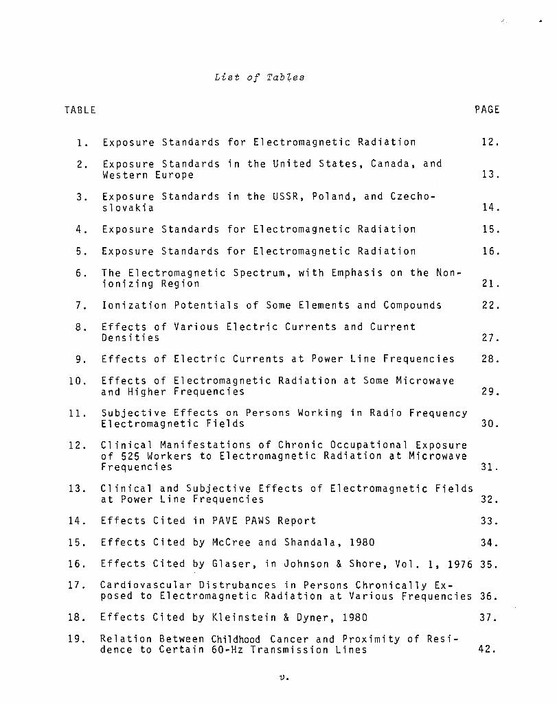
List of
TABLE PAGE
1. Exposure Standards for Electromagnetic Radiation 12.
2. Exposure Standards in the United States, Canada, and Western Europe 13.
3. Exposure Standards in the USSR, Poland, and Czecho-slovakia 14.
4. Exposure Standards for Electromagnetic Radiation 15.
5. Exposure Standards for Electromagnetic Radiation 16.
6. The Electromagnetic Spectrum, with Emphasis on the Non-ionizing Region 21.
7. Ionization Potentials of Some Elements and Compounds 22.
8. Effects of Various Electric Currents and Current Densities 27.
9. Effects of Electric Currents at Power Line Frequencies 28.
10. Effects of Electromagnetic Radiation at Some Microwave and Higher Frequencies 29.
11. Subjective Effects on Persons Working in Radio Frequency Electromagnetic Fields 30.
12. Clinical Manifestations of Chronic Occupational Exposure of 525 Workers to Electromagnetic Radiation at Microwave Frequencies 31.
13. Clinical and Subjective Effects of Electromagnetic Fields at Power Line Frequencies 32.
14. Effects Cited in PAVE PAWS Report 33.
15. Effects Cited by McCree and Shandala, 1980 34.
16. Effects Cited by Glaser, in Johnson & Shore, Vol. 1, 1976 35.
17. Cardiovascular Distrubances in Persons Chronically Ex-posed to Electromagnetic Radiation at Various Frequencies 36.
18. Effects Cited by Kleinstein & Dyner, 1980 37.
19. Relation Between Childhood Cancer and Proximity of Resi-dence to Certain 60-Hz Transmission Lines 42.
v.

TABLE
20. Occurrence of Some Symptoms in Humans Exposed Occupa-tionally to Electromagnetic Radiation in the Frequency
PAGE
Range 750 kHz-200 MHz 43.
21. Some Characteristics Associated with Microwave Hearing 44.
22. Survey of 1300 Chinese "Microwave Workers II 45.
23. Beneficial Effects Cited by Kleinstein & Dyner, 1980 53.
24. Profiles of Cases and Modalities of Hyperthermia 54.
25. Reported Hyperthermia Modalities 55.
26. Selected Cancer Statistics and Hyperthermia Considera-tions 56.
27. Ratio of SAR (Specific Absorption Ratio) to BMR (Basal Metabolic Rate) for an Average Man Exposed to a Plane Wave with a Power Density of 1 mW/cm 2 and 5 mW/cm 2 71.
28. Dielectric Constant and Conductivity in mmho/cm of Various Body Tissue at 37 Degrees Centigrade 77.
29. Electromagnetic Constants and Related Parameters for Various Tissues 78.
30. Molecular Changes Cited by Taylor, 1979 94.
31. Activation Energy of Molecular Effects in Biological Systems 99.
vi.

Introduction
This is a report of what is known today concerning nonionizing electromagnetic field interactions with the human body. Emphasis her e iss i m u 1 tan e 0 u sly 0 nth e two a d j e c t i v e s II non ion i z i n g II and II h u -manll, and in that respect the report is probably unique. In the past, while much was being written about certain electromagnetic waves (e.g. gamma, X, and cosmic rays) called ionizing radiation, it was generally assumed that others, called NIR (nonionizing radiation), had no effects besides rather obvious ones, which were either avoid-able or controllable, such as heating and electric shock. More re-cently, this assumption has been reconsidered, but the resulting studies are overwhelmingly concerned with the effects of NIR not on humans but on plants, lower animals, and isolated organic substances. A small fraction do concern humans, however. If these human-related studies were collected together, and supplemented by interviews and other sources of information, what new and greater degree of under-standing would result? This is the chief question which the report attempts to address.
As part of the answer, many interesting and significant effects and theories were identified. Some relate to the vital issue of health and well being. For example, both theories and observations link nonionizing electromagnetic fields to cancer in humans, in at least three different ways: as a cause, as a means of detection, and as an effective treatment. Other effects are simply curious at this time: audible sounds produced by microwaves (i.e. IImicrowave hear-ingll); and visible flashes produced by magnetic fields (limagneto-phosphenes ll ) at power line frequencies. These are just a small sample of the interactions which will be described in the next two sections of this report and Curious Effects and Beneficial Effects).
Perhaps as important as the interactions mentioned above are the theories and effects which are not yet known but probably could be with modest additional effort. Though the human body is complicated, much is known about it, from the macroscopic level to at least the microscopic level of individual protein molecules in cell membranes, and to some extent beyond that. This physiological knowledge, which
1.

is the subject of the fourth section (Physical and Physiological combined carefully with existing electromagnetic theory
could vastly improve the present state of knowledge. This conclu-sion will be discussed further in the last section (Borne Speculation and Areas for Further Research).
It is important to further develop the present state of knowl-edge for at least two very broad reasons. First, as technology im-proves man's capability for travel and manufacturing, his inter-actions with electromagnetic fields will increase in both intensity and frequency. In particular:
• Man is encountering new electromagnetic environments as he explores and utilizes regions beyond and beneath the earth's surface where he evolved. He may desire to take some sort of adaptive measures. Figs. 1 and 2 illustrate some features of the terrestrial electromagnetic environ-ment. Fig. 1 shows that there is a quasi-static electric field varying slowly in intensity from 100-250 Vim (volts/ meter) over a 12-month period. Fig. 2 shows ELF (extremely low frequency) electric fields. Also shown for comparison are human electroencephalograms. The intent of the com-parison is to imply that human functions and the electro-magnetic environment are related .
• Man is changing his terrestrial electromagnetic environ-ment. Much of the change is a byproduct of industrial processes (e.g. welding and heating), communication (radio, television, and other forms of broadcasting), and energy transmission (e.g. power lines and the proposed solar power satellite). If he knew the consequences of these changes, he might wish to compensate for or enhance them. Fig. 3 shows the contribution to the environment of some existing manmade sources of electromagnetic fields. Figs. 4 and 5 show the expected contribution from the proposed solar power satellite.
2.
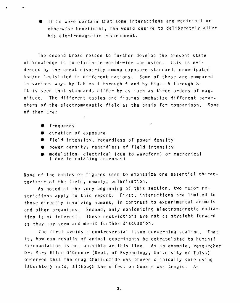
• If he were certain that some interactions are medicinal or otherwise beneficial, man would desire to deliberately alter his electromagnetic environment.
The second broad reason to further develop the present state of knowledge is to eliminate world-wide confusion. This is evi-denced by the great disparity among exposure standards promulgated and/or legislated in different nations. Some of these are compared in various ways by Tables 1 through 5 and by Figs. 6 through 8. It is seen that standards differ by as much as three orders of mag-nitude. The different tables and figures emphasize different param-eters of the electromagnetic field as the basis for comparison. Some of them are:
• frequency • duration of exposure • field intensity, regardless of power density • power density, regardless of field intensity • modulation, electrical (due to waveform) or mechanical
( due to rotating antennas)
None of the tables or figures seem to emphasize one essential charac-teristic of the field, namely, polarization.
As noted at the very beginning of this section, two major re-strictions apply to this report. First, interactions are limited to those directly involving humans, in contrast to experimental animals and 0 the r 0 r g ani s m s . Sec o.n d, 0 n 1 y non ion i z i n gel e c t rom a g net i era d i a -tion is of interest. These restrictions are not as straight forward as they may seem and merit further discussion.
The first avoids a controversial issue concerning scaling. That is, how can results of animal experiments be extrapolated to humans? Extrapolation is not possible at this time. As an example, researcher Dr. Mary Ellen O'Connor (Dept. of Psychology, University of Tulsa) observed that the drug thalidomide was proven clinically safe using laboratory rats, although the effect on humans was tragic. As
3.

another example, physician Sally Faith Dorfman (Center for Disease Control, Atlanta) noted that aspirin is lethal to laboratory rats. So, extrapolation can fail either way. Apparently beneficial ef-fects can, in fact, turn out to be harmful, and viae versa.
The second restriction requires a clear distinction between ionizing radiation and NIR, and actually there is none. Both forms of radiation are electromagnetic waves, and they seem to differ only in terms of frequency. Generally, ionizing radiation is at the high (in frequency) end of the spectrum, while NIR occupies the lower part. What frequency marks the dividing line between ionizing and nonionizing radiation? A standard definition for ionizing radiation was found; however, it is arbitrary and does not identify an abrupt physical threshold. According to that definition, all electromagnetic radiation with energy of 12.4 eV (electron volts) or greater per photon is ionizing. All with lesser energy per photon is not. The formula for the energy per photon of an electromagnetic wave is:
E: hv ( 1 )
where h = 6.626 x 10- 34 joules-sec and v is frequency. So, 12.4 eV is equivalent to about 2,998,000 GHz or a wave length of about 3,000 A (angstroms), which is in the ultraviolet band. (One ang-strom is 10- 10 meters.) Table 6 and Fig. 9 show how this wave length and frequency relate to the rest of the electromagnetic spectrum.
As already noted, the 12.4 eV definition is arbitrary. Readily available data, such as that listed in Table 7, show that many ele-ments can be ionized with lower energy photons. Further, some can be ionized twice with photon energies still less than 12. eV. The table also shows that some compounds can be ionized with photon energies below those required to ionize any of their constituent ele-ments. One familiar example is photo-ionization, which is the basis for photography, and occurs in the visible light band. So, the dis-tinction between ionizing and nonionizing radiation is not precise, at least in terms of frequency and wave length. Based on the data available, however, it seems unlikely, though not impossible, that
waves well below about 1,000,000 GHz are ionizing. So,
4.

.,
this report is concerned chiefly with those lower frequencies. Note that ionization should not be confused with other single
photon interactions. The latter are of interest here, and, in principle, they can occur at much lower photon energies, at fre-quencies below 1,000,000 GHz. These will be discussed briefly in the last section.
One more preliminary technical note will help to clarify the discussions in later sections. This concerns the relationships among electric fields, magnetic fields, power densities, and electric cur-rents. Some effects and theories will be described in terms of one or the other, as though fields, power, and currents are completely independent stimuli. In the human body, however, they are all inter-dependent. Electric currents and electric fields are related ac-cording to the formula,
( 2 )
...a where: J = total current density
j = r-I w = 21T x frequency E: = electrical permittivity cr = electrical conductivity .... E = electric field.
Magnetic fields are related to electric fields according to Faraday's Law:
( 3 )
.... where is the magnetic permeability and H is the magnetic field . .... Finally, the power density S is related to the electric and magnetic fields by the formula,
s :::; I --- E>< H 2. (4 )
So, the effects of one cannot be discussed without implicitly includ-ing all of the others. The only possible exception'is a static, uni-
5.

form (i.e. constant in both time and space) magnetic field, which can exist independently of both an electric field and a current density. This is a highly idealized situation because a human mov-ing about in such a field effectively disrupts the uniformity.
Information for this report was collected from a variety of sources. Written sources (over 1,000 in number) included journals, conference proceedings, technical reports, books, abstracts, and news items. Reportedly, over 5,000 pertinent written documents exist, and it is hoped that the ones examined are a representative sample. Additional sources included in-person meetings, telephone interviews, and lecture tapes. These provided information too recent for distribution in print, and guidance through the extensive litera-ture.
6.

FIG.!.
25[1
.s 150
"0 100 :§ t:...
In 1 JlIllN YYlYllYDlIII no Months
A nnual variation of electric field in atmosphere. 1) In Pavlovsk; 2) in Vysokaya Dubrava; 3) in Tash-kent.
ELECTRIC FIELD OCCURRING NATURALLY AT THE SURFACE OF THE EARTH (TAKEN FROM PRESMAN, 1970).
7 •

CHANNEL 4
IT
3[
Various types of signals (electric fields) of natural origin in the ELF region. I) Electromagnetic waves, Schumann-Resonance; II) Local field fluctuations of about 3 Hz; III) Local field fluctuations of about 0.7 Hz; IV) Field fluctuation as a result of thunder-
storm activity: a) Thunderstorm not yet visible
on the horizon; b) Thunderstorm on the horizon;
V) Sunrise appearance of signals. Compare Type I with EEG rhythms and Type II with 0 rhythms.
FIG. 2. SOME NATURALLY OCCURRING LOW FREQUENCY ELECTRIC FIELDS, WITH ELECTROENCEPHALOGRAMS FOR COMPARISON (FROM KONIG, IN PERSINGER, 1974).
8.

Average background levels of RF/MW radiation.
(l LOCAL RF STATIONS
0.1 1.0
UHF (\
./''/ FAR RF ./ STATIONS SUN
./
10 104 FREQUENCY MHz
FIG. 3. SOME NATURAL AND MANMADE SOURCES OF ELECTROMAGNETIC RADIATION (FROM DWYER & LEEPER, 1978).
9.
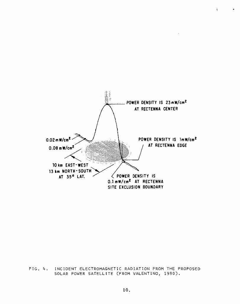
0.02 mW/em!
_
11!ll; :1 1 I ,Ii
,III : POWER DENSITY IS 23 mW/em2
AT RECTENNA CENTER
POWER DENSITY IS lmW/cm2 AT RECTENNA EDGE
10 km EAST- WEST -- - -- - -13km
AT 35 0 LAT. 17 I POWER DENSITY IS 0.1 mW/ em! AT RECTENNA SITE EXCLUSION BOUNDARY
FIG. 4. INCIDENT ELECTROMAGNETIC RADIATION FROM THE PROPOSED SOLAR POWER SATELLITE (FROM VALENTINO, 1980).
10.

Power Density Distribution (mW/cm2) for a Single Hypothetical Rectenna Site
FIG. 5. INCIDENT ELECTROMAGNETIC RADIATION FROM THE PROPOSED SOLAR POWER SATELLITE, AT DISTANCES FAR FROM THE RECEIVER (FROM VALENTINO, 1980).
11.

...... N
TABLE 1.
Exposure Occupational Frequency Duration Public
---------
Canada 1 5 rnW/crn2 (I - 300 GHz) No Iirnit I rnW/crn 2
(Proposed)
Czechoslovakia 0.01 rnW/cm2 (0.3 - 300 GHz) 8 hours 0.00 I rnW /crn 2
Poland 0.2 rnW/cm 2 (0.3 - 300 GHz) 10 hours 0.0 I rnW /crn 2
Sweden I rnW/crn2 (0.3 - 300 GHz) 8 hours None
U.S. 2 10 mW/crn2 (0.0 I - 100 GHz) No limit None
U.S.S.R. 0.01 mW/cm2 (0.3 - 300 GHz) Entire 0.00 I rnW /crn2
workshift
I. Canada is also proposing a I mW/cm2 exposure Iirnit at 10 MHz - I GHz Frequency.
2. Also with slight modification is the United Kingdom, German Federal Republic, Netherlands, and France. A new RFEM exposure guideline is being proposed by the American National Standards Institute (ANSI) that would cover the general population in the United States.
EXPOSURE STANDARDS FOR ELECTROMAGNETIC RADIATION (FROM DAVID , 1980).

...-----------;-----,-------,-------.-.---------.----.---.-.-... -.---r--·--·--·---------i--------·--·-··
Standard Type Frequency CW/ Antenna Stationary/ Exposure limit Exposure duration pulsed Rotating Remarks
-------+-----f.-- --.----.--- .. - .--- ... -.... u.s. ANSI 1974 (305)
Occupa- 10 MHz - 10 mW/cm2 no limit CW both tional 100 GHz 200 VIm
0.5 A/m 1-------+---_.----+------+--_.-----+-----.-----.--- . 1 mWhr/cm2 0.1 hour pulsed both
-.----.. ---f-.---. -- -- -.. -
u.S. Army and Air Force 1965 (292)
Occupa- 10 MHz -tional 300 GHz
10 mW/cm2 no limit both both 1--------11------------1-----+----------+-----·--- .-------- ...... - . 10-100 mW/cm2 6000/X2 both
(min.) both X-power density
in mW/cm2 1---------1----+-------+-------_ .. - -----.-.--.---... -- .. ---- .-.-
u.s. Indust. Hygienist 1971 (306)
Occupa- 100 MHz -tional 100 GHz
10 mW/cm2 8 hours both both - ._ ... _-- --._----'---------_._-_ .. _ .. _- -------- -------_._+-----------25 mW/cm2 10 min. both both
I I Canada
Can. Stan-dards Assoc. 1966 (307)
Occupa- 10 MHz -tional 100 GHz
10 mW/cm2 no limit 1--.- --.-.... - ------ ----... ---- .. ---...... --
1 mWhr/cm2 0.1 hour
both -_. .. ·'-1 both
CW
pulsed 1 '----.-- ..... _. __ .. - .. _ .... --. -- ..... -- ---- ._. _._. __ ._._-_. -
Canada Occupa- 10 MHz - 1 mW/cm2 no limit both both tional 1 GHz H&:W proposed
(301, 313) 1--_.- .. -.-. - .. - ... -. ..- .. - ... -. --- .. -.. I I .... ---...... -. ------II----------t-------------!
1-300 GHz 5 mW/cm2 i no limit both both
f- - 25 mW/cm2 both both
.-..... - ..... - -'.' ······-··---··---·----···--·-··---1--·--·--·····- .. General 10 MHz - 1 mW/cm2 I no limit both both public 300 GH?: I
.. -- ... ---.. -.-.- -- .-.-.---- --------+-------4--_.------_._._--------_ .. -Sweden Worker Prot. Authority 1976 (308)
Occupa- 0.3-300 GHz 1 mW/cm2 ! 8 hours both both
tional I -.-.. .. X-powe, den,it
I in mW/m2 ... --
!any I 10 MHz -
300 GHz 25 mW/cm2 CW, pulsed both
averaged _. J over 1 se_c_.-<-____________________ ._
TABLE 2. EXPOSURE STANDARDS IN THE UNITED STATES, CANADA, AND WESTERN EUROPE (FROM STUCHLY, 1978).
I I
·f

t
Standard
USSR Government 1977 (299)
USSR Government 1970 (42)
Antenna Type Frequency Exposure limit Exposure duration cwl pulsed Stationaryl Remarks
Rotating ------f------.--- - .
MHz I! 20 VIm i working day ____ I 30-50 MHz ,--, _lO_V_I_m ____ ... : w_or_k_i_ng_d_a_y ___ +-b_o_th ___ -+--: _bo_t_h ___ establish-
r ments of the ! ! 0.3 AIm : working day I both I Ministry of j 15 VIm ----- DexeefleUndceed I 50-300 MHz ! working day both I I
; 0.3-300 GHz 10 JJW/cm 2 working day i both I stationary !
100 JW/cm2 . working day both : rotating : 100 JW/cm2 2 I both I
-\ hours ! stationary
1 mW/cm2 2 hours both I rotating -.- ---r-- --'1 1 mW/cm2 20 min. both . stationary
--. -_._----+---- _._, General public 0.3-300 GHz 1 JJW/cm 2 24 hours fboth
I
I i both I --j--
Czechoslovakia Government 1970 (42)
Poland Government 1972 (42,300)
Poland Government 1975 proposed (42, 300)
TABLE 3.
Oecupa- . 10-30 MHz 50 VIm working day i both I both tional
General public
30- 300 MHz 10 V 1m working day I both ! both , -_._.......j
t 0.3-300 GMz 25 )JW/cm2 working day i both ------------------ -_. ----.. _-------.,.-------10 JW/cm2 working day
1.6 mW/em2 1 hour
0.64 mW/cm2 1 hour ---------
MHz 1 VIm 24 hours
, pulsed : both I
lew !
: pulsed
I both ! ! both
i max peak i 1 kW/cm 2
both i both ! : 30-300 r---------------------------tl------ i
! both i . 0.3-300 GMz 2.5 JJW/cm 2 24
1 lJW/cm2 24
! 30-300 MHz 1 VIm 24 I I ; 10-30 MHz 2.5 VIm 24 i
hours 'cw
hours , pulsed
hours . both
hours both
; i 1 both I
: both I I both
'-1 I
. Occupa- i 0.3-300 GHz 0.2 mW/cm2 10 hours both I stationary tional I
I , I I ! I I I
; I ! General 0.3-300 GHz . pulic
; occupa-II0-300 MHz tional
General 10-300 MHz public
.---- _-------i 32/p2 (hours) 0.2-10 mW/cm2
1 mW/cm2 10 hours
1-10 mW/cm2 BOO/P2 (hours)
10 lJW/cm2 24 hours
0.1 mW/cm2 24 hours
20 VIm working day
20-300 VIm 3200/E2 (hours)
7 VIm 24 hours
both
: both
: both I
I both , : both
I both
I both
I rotating
I rotating
! . ! stationary
i rotating
I both I I both
I I
iboth ! both
i ' : P - power derz- I ! sity in W/m : I 1 ! ! I . P-power derz-:
! sity in W/m :
, E-electric : field : intensity : in VIm
EXPOSURE STANDARDS IN THE USSR, POLAND, AND CZECHOSLOVAKIA (FROM STUCHLY, 1978).
14.

Country, author Frequency (MHz)
USA: Ely, T.S., 3000 Goldman,D. (1957)
USA: U.S. Army l1958) All USA: Schwan,Il.P. 1000 and Li, K. (1956) 1000-3000
3000 USA: General Electric 700 USA: Bell Tele-phone Labs. (1956) 750-30 000 USA: Mumford, W.W. (1956) -NATO (1956) -Sweden 87
87 Britain 300 West Germany (1962) -USSR (1965) 0.1-1.5
Poland (1961)
Czechoslovakia (1965)
USA (1966) Canada (1966)
1. 5-30 30-300 > 300
> 300
> 300
0.01-300 10-100 000 10-100 000
Maximum permissible intensity
100 mW/cm'" 150 mW/cm2
5 mW/cm2
10 mW/cmL
30 mW/ cmL
10 mW/cmL
20 mW/cm2
1 mW/cmL
1 mW/cm2
0.1 mW/cm2 0.5 mW/cm2 222 Vim
25 Vim 0.01 mW/cm2
10 mW/cm2 20 Vim
5 Aim 20 Vim
5 Vim 0.01 mW/cmL
0.1 mW/cm2 1 mW/cm2
0.01 mW/cm2 0.1 mW/cm2
1 mW/cm2 0.025 mW/cm2
0.01 mW/cm2 10 Vim
1 (mW/cm2)hr 1 (mW/cm2)hr
Remarks
Whole body Eyes Testes
Whole body Whole body Whole body
6 hr daily 2 hr daily 15 min daily Entire workday 2-3 hr daily 15-20 min daily Cw operation8*hr Pulsed op'n daily
Every 6 min Every 6 min
*For shorter exposure, see Figs. 39 and 40. (See also Appendix.)
TABLE 4. EXPOSURE STANDARDS FOR ELECTROMAGNETIC RADIATION (FROM MARHA & TUHA, 1971).
15.

Country. and Type of Standard
USA (ANSI) Exposure Standard
U.S.Army/ Air Force
USSR 1976 Industrial Safety Exposure Standard-
Czech. Soc. Rep. Exposure Standard
USA: Product Emission Standard
Canada: Product Emission Standard
Canada: Recommended Exposure Limit
Radiation Frequency and Waveform
10 MHz - 100 GHz (all waveforms)
50 MHz - 300 MHz (all waveforms)
30 - 50 MHz
3 - 30 MHz 60 kHz - 3 MHz 300 MHz - 300 GHz
0.01 - 300 MHz 890 - 6000 MHz
(ISM Bands in this range)
0.01 - 300 GHz
0.01 - 300 GHz
Maximum Levels
10 mW/cml
1 mW.h/cm1
10 mW/cml
100 mW/cml 0.1 - 1 mW/cml
10 - 100 /cml
5 Vim
10 Vim (or 0.3A/m) 20V/m 50V/m 25
10 Vim 1 - 5 mW/cml
1 mW/cml
5 mW/cml
1 mW/cml
Comments and Conditions
For periods of 0.1 hr or more. Whole and par-tial body. Reduction in high temperature en-vironments, or for health reasons recom-mended. Averaged over any 0.1 hr period. Continuous exposure. When power density (S) is in the 10-100 in W / cm 1 range, max allowed exposure time is 6OOO/Wl minutes, where S is expressed in mW /cml. No occupancy or protective clothing required. For a 20 min maximum exposure duration. Standard states: "Protective goggles manda-tory. Power density must not then exceed 0.1 mW /cml for balance of work day". Radiation from adjustable or scanning antennae is al-lowed at this level for 2 hrs. For a 2 hrs maximum duration (then 10
/cml for balance of work day). Required limit for a 'work day', all sources, ex-cept adjustable or scanning antennae (100
Levels in "work areas and other areas where personnel are permitted and occupationally ex-posed" ... shall not, in the course of the work day, exceed this value. Whichever is the greater.
(or 5 A/m in the range 60 kHz to 1.5 MHz) 8 hrs/day, for CW waveforms; reduced to 10 p.W /cml for pulsed waveforms. 8 hrs/day Emitted by the product at full power operation; lower level when manufactured ("prior to ac-quisition "). 5 mW /cml max in use. Measured with specified load (275 ± 15 ml H 10 at 20 ± S "C), at full power, 5 cm or more from any external surfaces by an approved instrument with effective aper-ture «2S em 1, no dimension > 10 cm. Emitted by product at maximum output, at points "at least S em from the external surface of the oven", when the oven is loaded with a load equal to the water equivalent of the minimum operating load, as specified by the manufacturer, at 20 ± 5 "C. Instrument specified in standard. Emission at no load, if total microwave output is < 3 kW. A verage power density limit in any 1 hr period (max 2S mW /cm1 averaged over 1 min).
-The reader is referred to the actual standards for more detail. Great caution is required in interpreting and translating standards.
TABLE 5. EXPOSURE STANDARDS FOR ELECTROMAGNETIC RADIATION.
16.

" 10 ................... , ...... ....... ............ ... -. __ --0:1 __ --=0 __ _ ..... '...." ..... . "'" .... Po " ······X,f,rn ,'" " · ... or,q >-, •••• ..... I.) ..... .
'. 1.0 ..... POi •••. ,,····.(,·(S7:..
0.1
0.01
11.6 SEC.
ey, " (' ·····.1/) '/'--0' ...: / ('.y ••••
,'\:./.: : , : , ":.; : , " : , ": , ,: , ! ... , .. , ,= .. ,
4.8 6 20 2 8 24 MIN. MIN. HRS. HRS. HRS.
EXPOSURE TIME
FIG. 6. EXPOSURE STANDARDS FOR ELECTROMAGNETIC RADIATION, WITH EMPHASIS ON DURATION OF EXPOSURE (FROM JOHNSON & SHORE, 1975).
17.

10
5.---------""'\1 610 Proposed machine NIOSH standard workers -----,
\, Present ANSI standard 104 194 ,
\ \ \
103 \ /
F: :';:'i 61 Soviet workers standard
U.S. mllitaryF' IGt'1' ....: .. c;, c:
iii
a> .... u::>
A
10 61
1
Chinese standard
Soviet population standard
personnel \:>; ':';i>;-·'.,J .. >., \)' workers
:3 Heavy users I::::] of mobile
radio
Population near FM transmitter
1.9l--:-____ + ____ 0.1 100
Frequency, MHz Note: Standards apply up to a frequency of 3 x 10⁵ MHz
104 194
B
103 Proposed ANSI standard E ;;; 61
c;, c:
iii 'C Q; Soviet standard u:: 102
19.4
"",,,, "'ffi '" CB radios E u
::l
2!' 10 'in 6.1 c OJ '0 Q;
0 Cl.
1 Persons near leaky microwave ovens
1.9 0.1
Exposure, hours
FIG. 7. EXPOSURE STANDARDS FOR ELECTROMAGNETIC RADIATION, WITH EMPHASIS ON FREQUENCY AND DURATION OF EXPOSURE (FROM LERNER, 1980).
18.
10

....... 1.0
________________
:-.I e u '" e c '" 10 .. '" en C QJ
og
"" Ql ::J 0 Po .. C Ql
og
'" u c 1-4
5
900 -r-fornz
f MHZ 300
0.3 Frequency in MHz
FIG. 8. AMERICAN NATIONAL STANDARDS INSTITUTE PROPOSED EXPOSURE STANDARD FOR ELECTROMAGNETIC RADIATION.

FIG. 9.
Frequency Wayelength (Hertz) (Meters)
3 x 10⁸ 1 Hz
3 x 10⁵ 1 KHz
Rodio
1 MHz w ..... 3 x 10²
t 1 GHz 3 x 10.1
'" Miero ••• "
i ·c 3 x 10.4 .g
1012 Hz t \5 Z I"fr.rod 1 ! Visibl, ! 3 x 10· 7 1015 Hz
I 1018 Hz + 3 x 10.10
'" X-Rays c
I .;; 3 x 10.13 ·c
1021 Hz "l' -Rays 2
l 1024 Hz
1 3 x 10.16
3 x 10.19 1027 Hz Cosmic Rays
THE ELECTROMAGNETIC SPECTRUM, SHOWING IONIZING AND NON-IONIZING REGIONS (FROM DAVID, 1980).
ZOo

Type of Radiation Energy Range Frequency Range Wavelength Range
Ionizing above 12.4 eV above 3,000 THz below 100 run
Ultraviolet 6.15-3.08 eV 1,500-750 THz 200-400 run
Visible 3.08-1. 76 eV 750-428 THz 400-700 nm
Infrared 1. 76 eV-1. 24 meV 428 THz-300 GHz 700 nm-l mm
Microwaves 1. 2 meV-I. 2 lleV 300 GHz-300 MHz 1 mm - 1 m EHF 1.2 meV-I00 lleV 300-30 GHz 1 mm - 10 nun SHF 100 lleV-12 lleV 30-3 GHz 10 rom - 100 mm UHF 12 lleV-l.2 lleV 3 GHz-300 MHz 100 mm - 1 m
Radar 200 lleV-900 neV 56 GHz-220 MHz 5.4 mm - 103m Radiofrequency 1.2 lleV-l. 2 neV 300 MHz-300 KHz 1 m-llan
TABLE 6. THE ELECTROMAGNETIC SPECTRUM, WITH EMPHASIS ON THE NONIONIZING REGION (FROM DWYER, 1978).
21.

IONIZATION POTENTIALS OF THE ELE:\IENTS
Different methods ha.ve been employed to measure ionization potentials. Abbrevia-tions 01 the methods usod lor data listed in the lollowing table are:
S; '"&tuum ultra\'iolet spectroscopy SI: Surface ionization. mass spectrometric EI; Electron impact with mass &nalysi!
Ionization potential in volts At.
EI. II III IV V VI
18 15.755 27.62 40.9 59.79 75 91.3 Ac 89 6.9 12.1 20 . " ....
47 7.574 2l.48 34.82 ii99i; is:i: 77' ilio 42' Al 13 5 984 18 28.44
33 9.81 18 63 28.34 50.1 62.6 127.5 .U 85 9.5 ....... .......
79 9.22 20.5 ':i792 2S9 :298 40:i27
....... B 5 8.296 25.149 Ba 56 5.21 10.001 35.5 Be 4 932 18.206 153.85 217.657 ....... Bi 83 7.287 16.68 25.56 45.3 56 88.3 Br 35 11.84 21.6 35.9 47.3 59.7 88.6 C 6 11.256 24.376 47 .871 64.476 91. 986 489.84 C. 20 6.lll 11.868 51. 21 67 84.39 109 Cb(Nb 41 6.88 14 .32 25 04 38.3 50 103 Cd 48 8 991 16.90t 37.47
':i:iX' .......
C. 58 5.6 12.3 20 CI 17 13 01 23 8 399 53 5 67.8 96.7 Co 27 7.86 17.05 33 49 83.1 ..... Cr 24 6.764 16.49 30.95 50 73 91 C. 55 3.893 25.1 35 Cu 29 7.724 0.29 36.83 ....... Dr 66 6 8 ....... F.r 68 6.08 ....... Eu 63 5.67 11.24
62 li4il 87 :i4 iS7:ii7 F 9 17 418 4 98 114.214 Fe 26 7.87 16.18 30.643 56.8 Fr 87 4 '54:2' . Go 31 6 0.57 30.7 Gd 64 6.16 12 '44:7 .. '9:i:4"' Ge 32 7.88 15.93 34.21 H 1 13.595 ...... ...... He 2 24.481 4.403
2:i:2 . HI 72 7 14.9 33.3 '67··' . HI 80 10.43 18.751 34.2 49.5·· I 53 10.454 19.13
'28:0:i' In 49 5.785 18 86 54.4 Ir 77 9 's09 . ·S2S·· K 19 4339 1.81 46 99.7 Kr 36 13.996 l2u6 36.9 43.5·· 63" 94·· La 57 5.61 11.43 19.17 ....... ....... Li 3 5.39 75.619 122.4U Lu 71 ....... 14.7
ici9:29' iSS:49· Mg 12 7.6H 15.031 80.14 141. 23 Mn 25 7.432 15.636 33.69 52 76 ....... Mo 42 7.10 16.15 27.13 46.4 61.2 68 N 7 14 .53 29.593 47 426 77.45 97.863 551. 92 S. 11 5.138 7.29 71. 98.88 138 37 172.09 )lb(Cb 41 6.88 14 .32 25.04 38.3 50 103 Sd 60 5.51
4i :07' '6:iX 'li7 :02' i26X' iS79i' S. 10 21. 559 l:i 28 7.633 18.15
77j94 ii:i873 i3i1:08' 0 8 13 614 35.108 Os 76 8.5 17
':iO:iS6 '5i :3S4 65007 220 4i P 15 10.484 19.72 Pb 82 i .415 15.028 31. 93 42.31 68.8 Pd 46 8.33 19.42 32.92 Po 84 8.43 Pr 59 5.46
iil:5iJ . Pt i8 9.0 Pu 94 5.1 Ra 88 5.277 10.144 Rb 37 4.176 7.5 40 Re 75 7.87 16.6
':ii:05 Rh 45 7.46 18.07 ....... ..... Rn 86 10.746 .....
'2846 . ...
Ru 44 7.364 16.76 .. 'ss:ozi s 16 10.357 23.4 35 47 29 72.5
Sb 51 8.639 16 5 25.3 44.1 56 108 Be 21 6.54 12.8 24.75 73.9 92 111 50 34 9.75 21.5 32 43 68 82 Si 14 8.149 16.34 33.488 45.13 166.73 205.11 Sm 62 5.6 11.2
'30:4g '40:72' Sn 50 7.342 14.628 72.3 Sr 38 5.692 11. 027 ...... 57 T. 73 7.88 16.2 Tb 65 5.98
i5:2iJ' '21i:5t T. 43 7.28 ':is''' . '60'" . T. 52 9.01 18.6 31 72 Th 90 6 95 '27:47 29.38 Ti 22 6.82 13.57 43.24 '99:8" . 120 TI 81 6.106 20.42 29.8 50.7 ....... ....... Tm 69 5.81 ...... ....... . ...... U 92 6.08
i.:65 . '21i::ii . ...
V 23 6.74 48 65 129 W 74 7.98 17. j ':ii:i'
....... X. 54 12 127· 21.2 42 53 58 Y 39 6.38 12.23 20.5 77 ....... Yb 70 6.2 12 10 '39j' Z. 30 9.391 17 96 ....... ....... Zr 40 6.84 13.13 22.98 ....... 99
VII
I2t ....... 24(38'
103 izs'" . 125 ....... 114.27 iei'" .
....... ....... ....... iss:i:i9
.......
....... iiil····
....... ....... 224:9' . 119 126 666.83 208.444 125 ....... ....... i39:ii.
26:iji' .......
....... 280:99' ijg'" . 155 246.41
'i:i:7""
i4i'" . ....... ....... isi'" .
i:is" . ....... ....... .... , .. ....... "These ,tepo by S method. -rh ... stepo by EI method.
VIII Meth.
143.46 S S
2S.j:5:i . s S S S S S S S S
193 S i.j:i:i· . s
S S ....... S
348.3 SI S
iss s S S ....... S
.. S .. .... Sl .
s S
151 S s S s S S s S
i70" . s s ....... S
i55 s S S . ...... S ....... S S
265.957 S 196 S 153 S
264 iss S S S
....... SI ....... S
sii:i2 . s S
:i09.26· s S ....... S S S SI S S S S s S S
:j2i1S" s S
iS9 s S S
30:i:07 s s S S S SI s S SI
172 S ... ... S ....... S i70'" . SI
S s EI ....... S ....... S
. ...... S ....... S
IONIZATION POTENTIALS
The first ionization potential 01 the molecule. indicated i. given in volts.
Compound Ionization potential I
volts 1 Compound
------I---!y, ............... . B" ......... _ ........ 12.8 , H, ......... . BrCI 12. 9(calc.): HBr.... . . .. . .. C, .................. 12 HC:-< ............ . CH,O. formaldehyde .. 11.3 HCI. ............ . CH,Br. methyl bromide 10.0 HF ..•..•......... CH,CI. methyl chloride 10.7 HI .............. . CH,I. methyl iodid •.•.. 9.1 , H 20 ............•.
.. :' .. :::: :,::.5 CO •.•••.............. 14.1 lBr .............. . CO................ 14.4 ICI. ............. .
..... ::::::::::: C,H,. acetylene.... 11.6 NO .............. . C.H •• ethyleM..... 12.2 . :-<0, ...........•.. C.H,. ethan.... .... 12.8 N,O ............. . C,H,. benzene .... 9.6 0, ..............•. e,H,. toluene...... 8.5 S •..............•. Cit... . . . . . . . . . . • . . 13.2 SQ •............•••
Ionization potential I
volts
17.8 (calc.) 15.5 13.2 14.8 13.8 17.7 (calc.) 12.8 12.58 10.42 9.7
11.6 (calc.) 11.9 (calc.) 15.51 11.2 9.5
11.0 12.9 12.5 10.7 13.1
TABLE 7 • IONIZATION POTENTIALS OF SOME THE CRC HANDBOOK OF CHEMISTRY
ELEMENTS AND COMPOUNDS AND PHYSICS).
(FROM
22.

Adverse, Benign, and Curious Effects
The effects listed and/or discussed in this section relate to at least three of the five senses (touch, hearing, and sight) and nearly every system of the human body (including circulatory, di-gestive, nervous, and muscular). Many of them are temporary; how-ever, some result in death and persistent disease. Some are better understood and more widely accepted than others.
Despite an exhaustive search of the literature and information from other sources, this section may appear to be sketchy and loosely organized. Some effects have been explored only qualitatively, and such seemingly essential information as frequency, polarization, in-tensity, and duration of exposure may not be precisely described, as well as the effect itself. This state of affairs seems typical of a new area of investigation for which neither the electromagnetic vari-ables nor biological endpoints are yet clear. This uncertainty has resulted in nonuniform reporting, variable rigor, and inadequately planned research. Still, it does not necessarily mean the effects reported are not real. It most surely means that a stronger theoret-ical foundation is required to positively identify, quantify, and otherwise understand them.
Nonetheless, some effort has been made to organize the informa-tion in this section. The presentation proceeds roughly from the general to the specific. Described first are overall results in-ferred from many different experimental, epidemiological, clinical, and other type studies. Next, the results from individual reports are tabulated. Then, at the most specific level, original illustra-tions and charts from individual reports are reproduced. Finally, at the end of the section is a recapitulation of results.
Overall Results
Tables 8 through 10 show some of the widely acknowledged effects and the electromagnetic fields and currents required to produce them. Only a few frequency ranges are represented in these tables; however, the most extreme adverse effect, death (by ventricular fibrillation) is included.
23.

Tables 11 and 12 list some of the more controversial effects. The frequency ranges and exposure levels are not described exactly, though some individual reports mentioned later in this section are more precise about those parameters. This lack of precision, and the subsp.quent controversy surrounding the results, are chief dis-advantages of restricting attention to humans. After an adverse effect is diagnosed, perhaps after years of growing discomfort, it may be difficult to isolate the conditions exclusively responsible from the general complexity of the subject's occupational and living routines. For example, a "microwave worker" might service a great variety of radars and radio transmitters, each with different fre-quencies, exposure levels, etc. Without a solid theoretical founda-tion to use as a guide, the most suspect source of electromagnetic radiation cannot be identified.
Of these controversial effects, the ones associated with the central nervous system are collectively termed "neurasthenia". Some of these are reportedly reversible. That is, when the electro-magnetic field vanishes, so do the effects.
Results From Individual Reports
,
Tables 13 through 18 summarize various individual reports and studies, and in some cases provide more specific information concern-ing frequency, exposure level, and other essential parameters. Table 13 concerns power line frequencies (50-60 Hz) exclusively. Tables 14 through 17 concern the frequency bands most often associated with communication and radar. The researchers listed in these tables are exclusively Eastern European. Table 18 covers many frequency bands from ELF (0-300 Hz) through SHF (3-30 GHz), and includes reports from outside Eastern Europe.
Table f8 contains some especially unusual entries. First, there is a report by Eckert of the most extreme adverse effect, death (by sudden infant death syndrome). Second, there is a report from the United States by Bise concerning neurasthenic effects previously re-ported exclusively from Eastern Europe. Also, an Italian report by Alberti concerns neurasthenic effects. Third, an extreme adverse ef-fect, promotion of cancer, is reported from Australia by Holt.
24.

Original Data Displays From Individual Reports
The remaining tables and figures are taken from individual studies. They are included not only for their informational content, but also as examples of how original data are encountered during the preparation of a report such as this one.
Figs. 10 through 13 concern effects in the ELF band. Fig. 10 attempts to correlate quasi-static magnetic fields with the most extreme adverse effect, death. Figs. 11 and 12, due to Konig, are unusual because they result from controlled experiments. They show that 3 Hz electric fields increase reaction time and galvanic skin response. Similarly, Fig. 13, due to Wever, results from controlled experiments. They show that removal of the earth's natural electric field and/or the application of a manmade field can disrupt circa-dian rhythm.
Table 19, taken from Wertheimer and Leeper, is unusual for at least two reasons. First, it is an epidemiological study. Second, it concerns an extreme adverse effect (childhood cancer).
Tables 20 through 22 and Figs. 14 through 17 concern effects in the frequency bands most often associated with communication and radar. Of these, Table 22 concerns an especially curious effect, microwave hearing. Evidently, pulsed microwaves produce click like audio sensations.
Recapitulation
From all of the tables and figures in this section, it is seen that electromagnetic fields may cause death in at least two ways, ventricular fibrillation and sudden infant death syndrome. Both of these occur at power line frequencies; however, one technical news-letter (BioeZectromagnetic Society NewsZetter, December, 1980) re-ported one fatality associated with much higher frequencies. Death followed accelerated aging, and a New York State court was convinced that protracted exposure to microwaves was the cause.
It is also seen from the tables and figures that electromagnetic
25.

fields may promote cancer. Holt (Table 18) reported the stimulation of human cancers at VHF (30-300 MHz) frequencies and exposure levels less than 10 mW/cm 2 (milliwatts per square centimeter). Wertheimer and Leeper (Table 19) performed a formal epidemiological study re-lating childhood cancer to fields at 60 Hz.
At least one adverse effect is not included in the tables and figures, and that is cataracts. Over fifty have been attributed to exposure at microwave frequencies (roughly 3-300 GHz). One opthamologist claims these cataracts can be uniquely identified by clouding of the posterior part of the lens, in contrast to clouding of the anterior part in other cases.
With respect to curious effects, at least two have been included in this section, microwave hearing and visual effects. has already been discussed in connection with Table 22.
The former Visual
effects include the distortion of thresholds for various colors (Table 15) and magnetophosphenes (Table 18). The latter effect is the perception of light flashes in response to a magnetic field.
26.

Cardiology
Electrosleep and Electrical Anaesthesia
Electro-Hazards: Sensation "Let Go" Fibrillation
Biophysics and Axonology
0.03 - 10 mA/cm2
10 - 100 rnA ( o. J - / ",A /c'lPf1.)
1 rnA 10 rnA 100 rnA
1 rnA/cm2
TABLE 8. EFFECTS OF VARIOUS ELECTRIC CURRENTS AND CURRENT DENSITIES (FROM SCHWAN, IN TAYLOR & CHEUNG, 1977).
27.
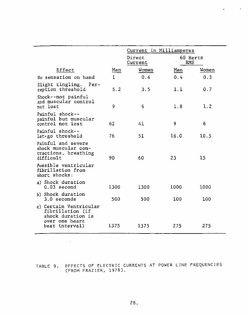
Current in MilliamEeres Direct 60 Hertz Current RMS
Effect Men Women Men Women No sensation on hand 1 0.6 0.4 0.3 Slight tingling. Per-ception threshold 5.2 3.5 1.1 0.7 Shock--not painful and muscular control not lost 9 6 1.8 1.2 Painful shock--painful but muscular control not lost 62 41 9 6 Painful shock--let-go threshold 76 51 16.0 10.5 Painful and severe shock muscular con-tractions, breathing difficult 90 60 23 15 Possible ventricular fibrillation from short shocks: a) Shock duration
0.03 second 1300 1300 1000 1000 b) Shock duration
3.0 seconds 500 500 100 100 c) Certain Ventricular
fibrillation (if shock duration is over one heart beat interval) 1375 1375 275 275
TABLE 9. EFFECTS OF ELECTRIC CURRENTS AT POWER LINE FREQUENCIES (FROM FRAZIER, 1978).
28.

Threshold for pain at 3 GHz
--.----
'Power Density (W /cm2) 3.1 I 2.5 1.8 1.0 0.83
Exposure Time (sec.) 20 30 60 120 180
Power density and exposure time to produce sensation of warmth
,
Exposure Time Power Density at Power Density at Power Density at far ( sec.) 3 GHz (mW/cm2) 10 GHz (mW/cm2) infrared CmW/cm2)
1 i 58.6 21.0 I 4.2 - 8.4 2 I 46.0 16.7 I 4.2 4 i 33.5 12.6 I 4.2
I j
TABLE 10. EFFECTS OF ELECTROMAGNETIC RADIATION AT SOME MICROWAVE AND HIGHER FREQUENCIES (FROM 1978).
29.
,

Headaches Eyestrain Fatigue Dizziness Disturbed sleep at night Sleepiness in daytime Moodiness Irritability Unso ciab ili ty Hypochondriac reactions Feelings of fear Nervous tension Mental depression Memory impairment Pulling sensation in the
scalp and brow Loss of hair Pain in muscles and heart
region Breathing difficulties Increased perspiration of
extremities Difficulty with sex life
TABLE 11. SUBJECTIVE EFFECTS ON PERSONS WORKING IN RADIO FREQUENCY ELECTROMAGNETIC FIELDS (FROM DWYER, 1978).
30.

Symptomatology
Bradycardia Disruption of the endocrine-humoral process Hypotension Intensification of the activity of thyroid gland Exhausting influences on the central nervous system Decrease in sensitivity to smell Increase in histamine content of the blood
Subjective Complaints
Increased fatigability Periodic or constant headaches Extreme irritability Sleepiness during work
TABLE 12. CLINICAL MANIFESTATIONS OF CHRONIC OCCUPATIONAL EXPOSURE OF 525 WORKERS TO ELECTROMAGNETIC RADIATION AT MICROWAVE FREQUENCIES (FROM DWYER, 1978).
31.

W N
T lIS t Subject ( Investigator)
Substation workers in U.S.S.R. (Asanova and Rakov, 1966) Humans in laboratory (Hauf, R., 1974)
Hl.man (Johansson. et al., 1973) Human (Johansson. et .• 1972)
Human Linemen (Kouwenhoven. 1966)
Human. lice, yeast. bacteria. wheat germ (Konig, 1962)
Humans--Compari son of "Opera-tions" to "Maintenance" per-sonnel U.S.S.R. (Sazanova, 1967) Human, Lineman (Singewald, et !l., 1973)
of those near and far from 200-400 kV lires (Strumza. 1970)
Electric Field (VIm)
8-14.S X 103
1-20 X 103
20 X 103 at head
Fields encountered in normal line and barehand work
1-2
open swi tchyard environment
Fields encountered in normal line and barehand work
< 7 x 10³
tlOT£5: (a) 147 x 106 modulated by ELF signal.
so
50
50
b
60
c
50
60
50
Reviewed by
(+) nervous system disorders Shepard & Eisenbud,
blood values. blood pressure pulse. [CG. EEG. reaction time
psychological tests
subjective feeling and psycho-loti cal functioning
(+ )
(- ) (- )
(+ )
(- )
physical examination CV. thyroid. (-) kidney. urine. ECG. EEG. visual. auditory. X-ray. emotional stress human reaction time, growth of (+) other test specimens
temperature, pulse, blood pressure. (+) reaction time, flicker frequency. adductor muscle reaction same as Kouwenhoven, 1966; this is a follow-up report
(- )
leukocytes, neutrophila, reticulocytes increase but within normal range other factors
a few subjects showed re-duced ability and tension for pulse test for sine wave test
NAS Shepard & Eisenbud, EIS (Bridges & NAS review equivalent 1973 paper) Bridges. Shepard & Eisenbud. EIS
Bridges. !'lAS
reaction time increased or decreased depending on signal characteri s tics. Growth changes in other specimens
NAS
average blood pressure of maintenance personnel lower
NAS. EIS
Shepard & Eisenbud,
visits to and from physicians, (-) no statistically significant Bridges, Sheppard & use of medicine, medical histories difference Eisenbud, EIS
(b) Pulse simulating lightning, 3-14 Hz swept sine wave.
(c) 2-100. Natural and man-made simulations of geomagnetic signals.
TABLE 13. CLINICAL AND SUBJECTIVE EFFECTS OF ELECTROMAGNETIC FIELDS AT POWER LINE FRE-QUENCIES (FROM FRAZIER, 1978).
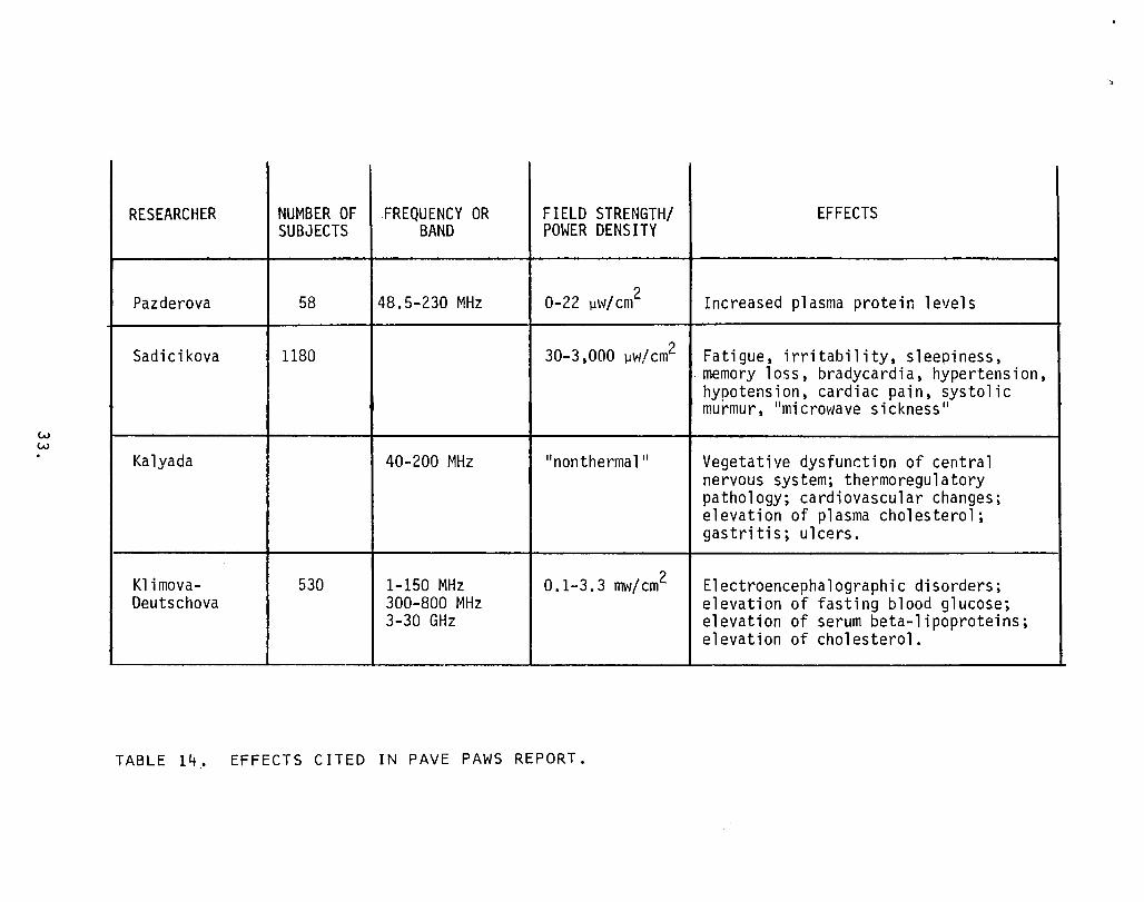
w w
RESEARCHER NUMBER OF FREQUENCY OR FIELD STRENGTH/ SUBJECTS BAND POWER DENSITY
Pazderova 58 48.5-230 MHz 0-22 )1w/cm2
Sadicikova 1180 30-3,000 )1\'1/ cm2
Kalyada 40-200 MHz "nonthermal"
Klimova- 530 1-150 MHz 0.1-3.3 mVI/cm2 Deutschova 300-800 MHz
3-30 GHz
TABLE EFFECTS CITED IN PAVE PAWS REPORT.
')
EFFECTS
Increased plasma protein levels
Fatigue, irritability, sleepiness, . memory loss, bradycardia, hypertension, hypotension, cardiac pain, systolic murmur, "microwave sickness"
Vegetative dysfunction of central nervous system; thermoregulatory pathology; cardiovascular changes; elevation of plasma cholesterol; gastritis; ulcers.
Electroencephalographic disorders; elevation of fasting blood glucose; elevation of serum beta-lipoproteins; elevation of cholesterol.

RESEARCHER NUMBER OF FREQUENCY OR FIELD STRENGTH/ EFFECTS SUBJECTS BAND POWER DENS ITY
Gabovich & 66 centimeter waves to 370 Increase in threshold of red, green, Zhukovskiy and blue light.
Zalyubovskaya 102 to 1 mW/cm2 Fatigue, drowsiness, headaches, loss & Kiselev of memory, decrease in hemoglobin, decrease in erythrocytes, hypercoagula-tion) decrease in leukocytes, increase in lymphocytes, decrease in segmento-nuclear neutrophils, increase in reticulocytes, increase in thrombocytes, drecrease in osmotic and acid re-sistance of erythrocyes, decrease in bactericidal action of skin and oral cavity, decrease in blood serum lyso-zyme, decrease in phagocytic activity of neutrophil s.
TABLE 15. EFFECTS CITED BY MCCREE AND SHANDALA, 1980.

w (J'1
'.
RESEARCHER NUMBER OF FREQUENCY OR FIELD STRENGTH/ EFFECTS SUBJECTS BAND pm.fER DENS ITY
Sadchikova "microwaves" O. 03-3 cm2 Reversible changes in nervous and (USSR) cardiovascular systems and blood;
"radio sickness".
Baranski & "microwaves" Decrease in EEG alpha rhythm; decreased Edelwejn tolerance of neurotropic drugs. (Poland)
Lancranjan 31 "mi c rowa ves II 10-100 mW/cm2 Decrease in sex function; decrease in (Romania) spermatogenesis.
Pazderova (Czechoslovakia Change in blood protein chemistry.
Slotolnik- Chromosome changes in human leukocytes. Baranska
TABLE 16. EFFECTS CITED BY GLASER, IN JOHNSON & SHORE, VOL. 1, 1976.

TABLE 17.
Ratio of percentage of cases with particular defect EmF parameters due to EmF to percentage of cases in control (not
exposed to EmF)
QRS interval in reduced blood ECG increased to
range intensity pressure (ar- slow heart beat 0.1 sec (reduced terial hypotonia) (bradycardia)
ventricular con-ductivity)
From one to SHF (centimeter several 1. 85 24 11.5
waves) mW/cm 2
< 1 mW/cm 2 2.0 16 12.5
Low, not 8 21 UHF thermal 1.2
Tens to Short-wave HF hundreds 0.21 12 -
of Vim
Medium -wave Hundreds to 5 -
HF 1000 V,'m 1.2
Percentage of cases in control 14"/0 3"/0 2"/0
CARDIOVASCULAR DISTURBANCES IN PERSONS CHRONICALLY EXPOSED TO ELECTROMAGNETIC RADIATION AT VARIOUS FREQUENCIES (FROM PRESMAN, 1970).
36.

RESEARCHER NUMBER OF FREQUENCY OR FIELD STRENGTH/ EFFECTS SUBJECTS BAND POWER DENS ITY
Eckert 494 60 Hz Crib death (Sudden Infant Death (Germany) Syndrome) .
Bogucka 72 "radio and Functional disorders of central (Poland) television" nervous system, hyperacidity, epi-
gastric pain, disorders of cardio-vascular system, leukopenia of blood, esinophobia of blood.
Bise 10 0.1-960 MHz 10-16_10- 13 Changes in electroencephalogram, loss (United States) W/cm2 of memory, inability to concentrate,
irritability, apprehension.
Dumanskii 34 50 Hz 5-12 kV/m Local changes in skin temperature, (USSR) reduced heart rate, reduced blood
pressure, change in electrocardiogram, change in blood composition.
Katorgi na, 230 2-1000 kHz 3-5 Vim Eye pain, headache, vascular changes Semenova, et al in eye.
Alberti 31 5-50 MHz Decreased male fertil ity, insomnia, (Italy) headache.
Holt VHF Below 10 mW/cm2 Cancer growth stimulated.
Lovsund, Obey, 10-50 Hz 0-40 mi 11 i- Excitation of magnetophosphenes. Nilsson (Sweden) Teslas
TABLE 18. EFFECTS CITED BY KLEINSTEIN & DYNER, 1980.

FIG. 10.
;" 15 ------
ti 15 14
'0 13 Magnetic storms (I) and mor-tality from nervous and cardiovascular
§ diseases (II) in Copenhagen and Frank-e: furt-0n- Main. e: '" <> ::;:
-mdays-)days fI
RELATION BETWEEN NATURALLY OCCURRING MAGNETIC FIELDS AND DEATH RATE (FROM BERG I 1960 1 IN PRESMAN I 1970).
38.

-40 Jr
" -':j ., .,
w" E 9
·20 'Ld 2V.20min a:: <l H
.40 H 7V,Smin H
r:I .60 H
FIG. 11. EFFECT OF A 3-HERTZ, VERTICALLY POLARIZED, ELECTRIC FIELD ON THE REACTION TIME OF A SINGLE TEST SUBJECT IN A SINGLE-BLIND EXPERIMENT (FROM IN 1974). THE DURATION AND INTENSITY OF EXPOSURE ARE INDICATED BY THE SHADED AREAS. THE VOLTAGES WERE APPLIED BETWEEN PLATES 2.5 METERS APART. THE TIME-DEPENDENCE OF THE FIELD WAS INTENDED TO RESEMBLE THE TYPE II SIGNAL SHOWN PREVIOUSLY IN FIG. 2.
39.

40 I
36
32
28
24
20
16 12
8
4
0 REST ---+0- RADI ATiON --+--- REST - ,
24 min 20 min 14 min_
-EXPOSURE TIME -
FIG. 12. EFFECT OF A 3-HERTZ ELECTRIC FIELD ON GALVANIC SKIN RESPONSE (FROM KONIG .. IN PERSINGER .. 1974). THE TIME-DEPENDENCE OF THE FIELD WAS INTENDED TO RESEMBLE THE TYPE II SIGNAL SHOWN PREVIOUSLY IN FIG. 2. FOR AN APPLIED FIELD INTENSITY OF 1 VIM, POSITIVE REACTION WAS OBSERVED IN 1 OF 12 TEST SUBJECTS. FOR 5 VIM, POSITIVE REACTION WAS OBSERVED IN 5 OUT OF 10 TEST SUBJECTS.
40.
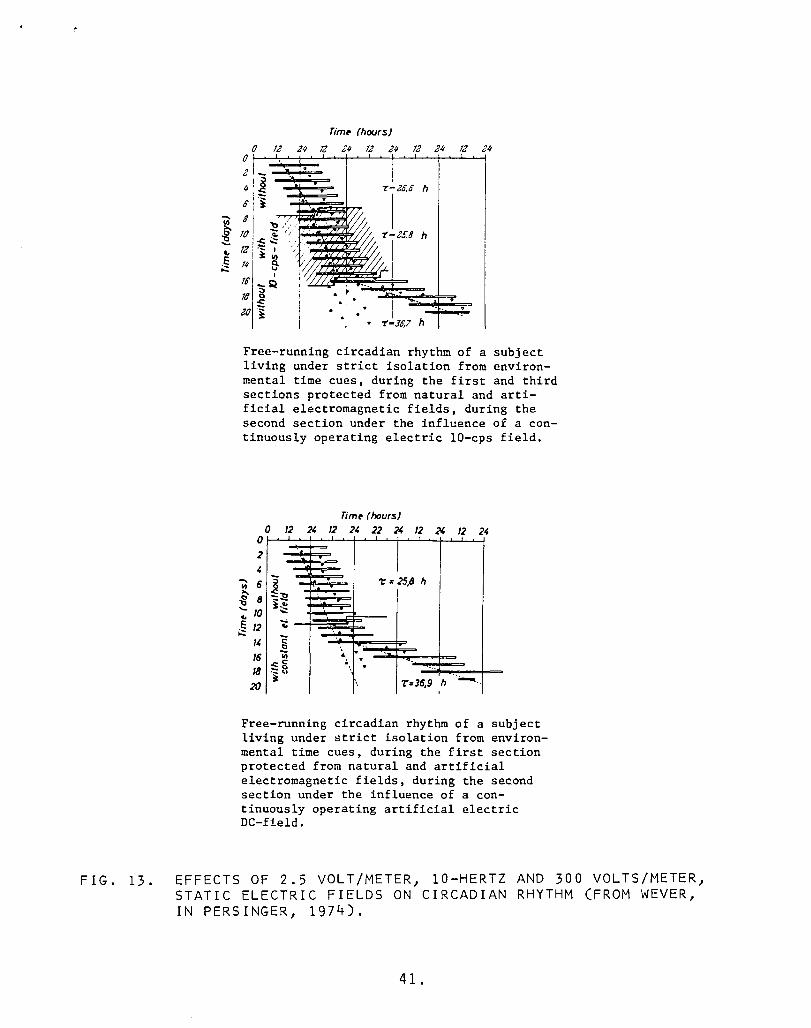
FIG. 13.
....... 1/1
.. E ;:::
(hours) o U M 2 M Q M Q M Q M o
Z II
0 8
'0 '3 'II
'0 '8 20
. I . I I
7--20.0 h ,-i . h ..c:..;: .-:s:: I ' 4/ . 1/1 ,M. e-
I
:;Q --. <) . . ::: . . .
• j . . . . 7:-J0.7 h
Free-running circadian rhythm of a subject living under strict isolation from environ-mental time cues, during the first and third sections protected from natural and arti-ficial electromagnetic fields, during the second section under the influence of a con-tinuously operating electric IO-cps field.
o 2 ,
....... 6 8
';:' 10 12
" 16 18 20
Timp (hours) o X D X
-. '== I
't' = 25,8 h . . ... ....
" i: <)
'iii .. . . :,:::8 " . '. It 7:=36,9 h -...
I
Free-running circadian rhythm of a subject living under strict isolation from environ-mental time cues, during the first section protected from natural and artificial electromagnetic fields, during the sec.ond section under the influence of a con-tinuously operating artificial electric DC-field.
EFFECTS OF 2.5 VOLT/METER, 10-HERTZ AND 300 VOLTS/METER, STATIC ELECTRIC FIELDS ON CIRCADIAN RHYTHM (FROM WEVER, IN PERSINGER, 1974).
41.

Ltukem.a L)'lnpho .... Nrrvoua syatem tumon Other I!ftldo_ Typo o(..,nne
e ... Control e_ Control e ... ContrOl c:onfirur·'10n- ea .. Control
Birth address HCC 52 29 10 22 12 17 9 LeC S4 107 21 26 35 45 31 39
1\\ HCCI (38.21 (21.31 (32.3) 116.11 (38.61 (21.11 (35.41 (18. 71
Death .ddr ... HCC 63 29 18 11 30 17 18 17 LeC 92 126 26 33 36 49 45 46
('l HCC) 140.6) 1187) 140.91 (2501 145.5) (25.81 (28.61 (27.01
- HCC - hi&h-CUrTent Lee - I09l'-<::'UJ'I"ent configw-ahon.
TABLE 19. RELATION BETWEEN CHILDHOOD CANCER AND PROXIMITY OF RESIDENCE TO CERTAIN 60-HZ TRANSMISSION LINES (FROM WERTHEIMER & LEEPER, 1979).
42.

Lensth of 1-6 years 7-16 years
(average 4.3) (average 9.6) Symptoms (73 Eersons) (73 Eersons)
percent number percent number of cases of cases of cases of cases
Headache 20.5 15 32.9 24 Disturbance of sleep 13.7 10 23.3 17 Fatigue 12.3 9 17.8 13 General weakness 7.0 5 12.3 9 Disturbance of memory 5.5 4 8.2 6 Lowering of sexual potency 5.5 4 8.2 6 Drop in body weight 2.7 2 12.3 9 Disturbance of equilibration 5.5 4 11.0 8 Neurological symptoms 0.0 0 15.1 11 Changes in ECG 17.8 13 28.8 21
TABLE 20. OCCURRENCE OF SOME SYMPTOMS IN HUMANS EXPOSED OCCUPATIONAL-LY TO ELECTROMAGNETIC RADIATION IN THE FREQUENCY RANGE 750 KHZ-200 MHZ (FROM DWYER J 1978).
43.

TABLE 21.
I ClH MWHIJ\\',\vl.-hlll:n.D 1\11.oIIIJI<' :': , " A , I"IIH'T 11'> HUMA"
Pulsl!widlh (/oIs)
1 2 4 5 5
10 10 15 15 20 32
SI'IIII(IS (45 dB B,\(,Kc;HOI'''i> l'\oISE. 24S0 Mil,)
40 16 120 20 8 120 10 4 120 8 3.2 120
2,5 1.0 25 4 \.6 120
1.3 0.5 26 2.33 0.93 lOS
5 2,0 150 2.15 0.86 129 I. 25 0.5 120
·Peak SAR (specil"ic absorption rate) is based on absorption in eql. alent sphericail1lodel of the head
SOME CHARACTERISTICS ASSOCIATED WITH MICROWAVE HEARING (FROM LIN, 1980).
44.

TABLE 22.
Exposure level High (over 200 mW/cm')
Low (10-200 mW/cm')
None (control group)
Frequency of complaint, percentage - Abnormal
cardiac Neurological Brachycardia ST waves
32
24
11
1.63
3.93
0.42
11.8
11.2
5.6
SURVEY OF 1300 CHINESE "MICROWAVE WORKERS" (FROM LERNER, 1980).
45.

0'1
70 @ 70 ®
60 60 50 50 ..
40 40 30 30 20 20 10 10
2 3 4 5 6 7 8 9 10 II
70 © 60 50 40 30 20 10
12 13 14 15 16 Ordinate - frequency of changes in percentages; abscissa - main indicators: A - neurological, B - autonomic vascular and C - cardiac. White columns - control; oblique shading - persons of the first group, exposed previously to periodic action of microwaves of substantial intensities; dotted shading - persons of the second group working under conditions of exposure to microwaves of lower intensities. All indicators are presented with confidence limits. 1 - feeling of heaviness in the head, 2 - tiredness, 3 - irritability, 4 - sleepiness, 5 - partial loss of memory, 6 - inhibited dermographism, 7 - expressed dermographism, B - hyperhidrosis, 9 - bradycardia (upon counting), 10 - arterial hypotension, 11 - arterial hypertension, 12 - cardiac pain, 13 - dullness of the heart sounds, 14 - systolic murmur, 15 - bradycardia (according to ECG), 16 - lowering of deflections T I and T II.
FIG. 14. CHANGES IN THE NERVOUS AND CARDIOVASCULAR SYSTEMS AMONG WORKERS EXPOSED TO MICROWAVES AND CONTROL SUBJECTS (FROM STUCHLY, 1978).

Years of work Percentages or headaches vs years or work. •• Exploatation; D. special
exploatation; D. repair.
FIG. 15. OCCURRENCE OF HEADACHES IN "MICROWAVE WORKERS" (FROM BARANSKI & EDELWEJN, IN TYLER, 1975).
47.

3 Years of work
Percentages of oversweating vs years of work.
FIG. 16. OCCURRENCE OF EXCESSIVE PERSPIRATION IN "MICROWAVE WORKERS" (FROM BARANSKI & EDELWEJN, IN TYLER, 1975).
48.

FIG. 17.
----
---------------------------------------------------------
15ol'V
EEG tracings in an individual occupationally exposed for 5 years to microwaves (group R) while working in a repair shop.
SUBSTANTIALLY FLAT ELECTROENCEPHALOGRAMS OF "MICROWAVE WORKERS" (FROM BARANSKI & EDELWEJN, IN TYLER, 1975).
49.

Beneficial Effects
Electromagnetic field effects have both established and develop-ing uses in medicine. In addition, some alleged benefits have not been acknowledged in medical circles.
Table 23 is a summary of various beneficial effects, at fre-quencies ranging from 0-434 MHz. Most of these are included in the broad category of electromagnetic diathermy. That is, electromag-netic fields are used to heat various parts of the body. It is generally believed that heat stimulates the natural healing and/or defense mechanisms to relieve or cure the ailment. Of all known effects, relief by diathermy is probably the oldest, beginning in 1892 when D'Arsonval observed tissue heating due to low frequency electric currents. Comprehensive text books on the technique ap-peared by the early part of this century (e.g. Danzer et al, 1938).
The table also includes two techniques, however, which have been used only recently compared to diathermy. They are: bone healing and hyperthermia. The table shows that Brighton and Frieden-berg have used microampere currents to mend stubborn bone fractures. Not shown is similar work by Bassett, who reportedly used 75 Hz pulsed currents of only a few nanoamperes to produce bone healing. Note that in either case, the currents are so small that heating probably is not part of this effect. With respect to hyperthermia, the table shows that Holt and Nelson reported positive results in the treatment of a great variety of cancers. Briefly, the objective of hyperthermia is to raise the temperature of cancerous tissue to 42-45 degrees Centigrade. In that range, the cancer cells tend to die, but healthy cells manage to survive as they would during a high fever. The reason for the preferential survival is generally at-tributed to the relatively poor circulatory system of cancerous tissue. That is, lack of blood prevents the cancer cells from dis-sipating lethal amounts of heat energy.
Electromagnetic hyperthermia is increasingly used as part of cancer treatment, as indicated by Tables 24 through 26 and Figures 18 and 19. Table 24 shows that three different frequencies (27, 915, and 2450 MHz) are used. The chief reason is that the electro-
50.

magnetic spectrum is regulated, and only certain frequencies have been reserved for such use. These constitute the ISM (Industrial, Scientific, and Medical) band and include the frequencies 13, 27, 40, 915, 2450, and 6000 MHz. Note that Holt and Nelson (Table 23) used 434 MHz in Australia, but that frequency is not available in the United States. Of the ISM band, 915 MHz is closest to a physi-
optimum, in the sense that absorbed power is maximum for much bulk tissue.
Table 25 shows how hyperthermia is used in conjunction with more traditional forms of cancer therapy, such as chemotherapy and ionizing radiation. The latter seem to be so much more effective when used simultaneously with that only very small doses (about 10% of the usual amount) are required. Therefore, the toxic side effects are largely avoided and prolonged treatment is feasible.
Table 26 shows how hyperthermia is realized at a variety of local sites on or in the body, and some of the associated· problems. Electromagnetic fields are applied using specialized antennas. In medicine, these have been given a special name, lIapplicators ll
•
Typically, they couple 1-100 watts into various parts of the body. Figs. 18 and 19 show two different applicators. The bean bag
type is suitable for the surface of the body. The coaxial type is designed for use inside the body.
The tables and figures do not include a number of both tradi-tional and experimental electromagnetic field effects. The former include: defibrillation; various types of shock treatment; and diagnosis using the body's own electromagnetic signals, such as the electrocardio- and electroencephalogram. The novel ones include at least two diagnostic techniques, microwave thermography and micro-wave scanning.
Microwave thermography is a passive technique similar to radiom-etry. The electromagnetic signature of cancerous breast tissue seems to be unique. The advantage over conventional X-rays is the apparent lack of toxic effects. Two different frequencies are presently being used, 3.3 and 1.3 GHz (Barret and Myers, 1979).
Microwave scanning is a bistatic radar system. That is, the transmitting and receiving antennas are at different (in this case, opposite) sites. Evidently, microwaves passing through an organ can be used to construct a high resolution image of it, rivaling
51.

those obtained using conventional X-rays. This is possible in part due to the high bulk dielectric constant of tissue, so that, at a fixed frequency, a wave length in the organ is about one-n i nth w hat it \10 u 1 d be ina i r . For e x amp 1 e, at 3 G H z a wa vel eng t h is only 1.1 centimeter. For purposes of radar imaging, the smaller the wave length, the greater the resolution. The advantage over X-rays is apparent lack of toxic effects.
Finally, one controversial beneficial effect concerns negative airborne ions. Reportedly, these can impart a feeling of exuberance and general well being. Positive ions reportedly have the opposite effect. This belief supports a world wide ion generator industry.
52.

RESEARCHER NUMBER OF FREQUENCY OR FIELD STRENGTH/ EFFECTS SUBJECTS BAND POWER DENS lTV
20 3-70 Hz Relief of arthritis pain. (SVJeden)
Dikova 87 II ra dar ll 30 watts Cured sterility i n vwmen. (Bulgaria)
Ben ta 11 (U K) 50 500 Hz Accelerated bruise healing.
Nikoruikina 152 IImicro\'1ave ll 40 watts Relief of chronic colitis and (USSR) pancreatic disorders.
Brighton & 57 Friedenberg
o Hz 10-20 microamps Healed bone fractures. (United States)
Deeell e, 27.12 MHz 1.5 watts Cured arterial ulcer; relief from Lorthiour, et varicose veings, eczema, arthritis; al (Belgium) healed bone fractures.
Novak 141 o Hz 40-480 garrunas Relief/cures relating to bronchial (Czechoslovakia asthma, neuralgia, epilepsy, steno-
cardia, photodermatoses.
Holt & Nelson 1300 434 MHz Improvement in patients with cancers (Australia) of the: head, neck, breast, bone,
liver, brain, lung, abdomen, rectum, bladder, sarcomata, lymphomata.
TABLE 23. BENEFICIAL EFFECTS CITED BY KLEINSTEIN & DYNER, 1980.

TABLE 24.
Tumor Location Breast Breast Skin
Lung Leg Leg Leg Foot Eye Head and neck
Prostate
Cheek Axilla
Abdomen Abdomen
Scalp Vagina
Pathology Adenocarcinoma-local recurrence Adenocarcinoma-local recurrence Metastasis from cancer of breast,
post-mastectomy Carcinoma Liposarcoma-recurrence-post surgery Disseminated metastatic melanoma Squamous carcinoma Primary melanoma Primary melanoma Primary melanoma, carcinoma of tonsils,
sarcoma of tongue, squamous carcinoma Carcinoma
Carcinoma Metastatic skin lesion from breast
carcinoma Skin metastasis from sarcoma of colon Pelvic metastasis from colorectal adeno-
carcinoma Metastasis from lung carcinoma Melanoma
Treatment Method 2450-MHz cross-fire conformal applicators 2450-MHz ceramic waveguide applicator 915- and 2450-MHz air-filled waveguide applicators
27-MHz water-filled ridged waveguide applicator 27-MHz water-filled ridged waveguide applicator 2450-MHz ceramic waveguide applicator 2450-MHz conformal applicator 2450-MHz ceramic waveguide and conformal applicators 2450-MHz ceramic waveguide applicator 915- and 2450-MHz ceramic waveguide applicators
915- and 2450-MHz coaxial applicators and 27-MHz water-filled ridged waveguide applicator
2450-MHz cross-fire conformal and coaxial applicators 2450-MHz conformal applicator
2450-MHz conformal applicator 27-MHz water-filled ridged wave applicator
2450-MHz ceramic waveguide and conformal applicators 2450-MHz ceramic waveguide applicator
PROFILES OF CASES AND MODALITIES OF HYPERTHERMIA (FROM STERZER ET AL, 1980).

Anatumic Adjuvant Cumments and Extent Method Temperature Time Therapy Considerations Rtfcrences
Whole lIot air and/or radiant 41-41.5 5-21 h X-ray Cumbersomc-limited ac- Warren Body heat cess to patient
1I0t water by immersion 40 2Y1 h • Slow temperature control Von Ardenne Hot water by space suit 39.5-42 90 min- Systemic • Larkin, Bull
5 h chemotherapy Melted paraffin by 41-41.8 90 min- Cytotoxic drugs Limi ted access to pa tien t Pettigrew
immersion 4h Perfusion -cx tracorpureal 41.5-42 5h Cytutoxic drugs Surgical procedure Parks
heat exchange Microwave and hot ilir 43-43.6 40 min- • • Morieca
cabinet (Siemens) 1 h Regional 1I0t water by immersion 46-47 67 min X·ray, cytotoxic Limited to Crill:
drugs Radiant heat -visible and 43-45 20 min • * Lehmann
infrart·d Perfusion 38-45 30 min- X-ray, chemotherapy, Surgical procedure Cavaliere, Stehlin,
c.n 8h cytotoxic drugs Shingleton, Woodhall c.n Capacitive Rr- 42-50 30 min- • Skin and rat burns have LeVeen, Storm
3h been a problem. Inductive RF 41-63 35 min • Storm
Localil.cd Irrigation 41-45 1-3 h • Bladder tumors, peritoneal lIall et a/. cavity Ludgatc
Capacitive RF 48.4 30 min- • • LeVcen, G. Hahn 3h
Inductive RF 42.8-45 30 min- X-ray • Kim and lIahn, I h V()n Ardenne
Ultrasuund 43.5 30min • Best f",using. Relle,tions, G.llahn caviatiun, and hone heat-ing arc prohlems.
Microwave 42-44 12-20 min • Deep heating (lO CfII) diffj· Sandhu, Dick",n. cult with noninvasive Mendecki, Sal1laris, (NI) applicators. c:/ a/.
TABLE 25. REPORTED HYPERTHERMIA MODALITIES (FROM SHORT & TURNER, 1980).

Site
Buccal cavity and pharynx
Esophagus
S4JJnach
Colon
Rectum
Liver and biliary
Pancreas
Larynx
Lung
Bone and con-nectivc tissue
Breast
Uterus
Ovary
Prostate
Brain
Thyroid Leukemia
Lymphoma
Bladder
Kidney
All sitcs
Estimated Ncw Cases
24400
8400
23 000
77 000
35 000
11600
23 000
10400
112 000
6400
106900
13600 53 000
17 000
64 000
11600
9000 21 500
38500
35 000
16200
765 000
Estimatcd Deaths
8650
7500
14100
42800
9100
9200
20200
3500
97500
3350
34500
4300 10700
II 100
21 000
9500
1000 IS 400
20300
10000
7500
395 000
Po."iblc Regional ur Lucal lIyperthermic Apprnaches ULT CAP IND MIC
NI NI NI NI
OR NI OR
NI NI OR OR NI NI NI
OR OP
OR NI OR IN
NI NI NI NI IN
NI NI Nl NI OR or
NI NI OR
NI NI NI OP
NI NI NI
Nl Nl Nl NI IN
NI NI OR NI OR IN IN
or NI NI
or NI OR NI
OR IN NI IN
OP
NI Nl NI
NI NI Nl NI
Nl OR
Nl Nl Nl NI IN
Problems and Considerations
Proximity to eyes, complex geometry-reflections, uneven heat depo-sition. Specialized applicators. Hig.h vascular perfusion.
Local metastases at first diagnosis. Large blood vessels adjacent. Dif-ficult thermometry (danger of mediastinitis).
Local mctastases at first diagnosis. Difticult geometry and placement of applicators. Some success with WBII and hyperthermic irrigation.
Right colon not acct!ssiblc by orificial applicator (too deep). Adjacent loops of bowel containing liquids and gases. Thermometry difficult.
Metastases in regional lymph nodes may be diffh;ult to heat.
Most sensitive tissue to heat. Largc organ. Usually good differential heating of tumors. Gall bladder contents may overheat.
Deep, inaccessiblc. Intestinal mucosa arc heat sensitive. Thermometry difficult. May require surgical implantation-active and passive radi-ators and temperaturc probes.
Good candidate for hyperthermia-accessible. early symptoms, second chancc laryngectomy if hyperthermia fails.
Often far advanced at first diagnosis. metastasis common. Geometry dirficult-air. bone. major vessels. Thermometry tricky.
Commonly mctastasize to lung. Problems relate to size and location, etc.
Deep surface against musde may be harder to heat. Metastases to ax-illary lymph nodes often small (1 mm). Synchronous phased array?
Metastasize widely. Nonsurgical candidates have Io<:al e,,"tension which is deep for external
applicators, distant from intrauterine cavity.
Many tumors cysti.:. c",ily ruptured-tht:rmolllt:try difficult. Tumors deep in pelvis. Disseminate through peritoneal cavity.
Metastasize to lower spine.
Refle.:tions, resonance, fluid-filled vcntridcs, proxlIlllty to eyes. Surgical pro.:edure for invasive applicators and temperature probes. lIypt:rthcrmic chemotherapeutic perfusion?
Adjacent tissue many large blood vessels and important nerves. Diffuse disease. Whole body hyperthermia with C"lwmothcrapy and/or
X-irradiation? i':xtracorporeal hyperthermia of blood? Orten diffuse. May be diflieult to heat pn:ferentially. Dosimetry
may be difficult. Temperature probe placement tricky in invasive tumors. Some suc-
cess with hyperthermic irrigation. May require X-ray placement of temperature probcs and IN applicators.
Abbreviations: ULT-Ultrasound. CAP-Capacitive Rf-, IND-Inductivc Rf-. MIC-Radiative UHf-, microwave, Nl-Noninvasive, IN-Invasive, OR-Natural orificc (orificial), OP-Operativc (surgical exposure), WBII-Whole body hyperthermia.
TABLE 26. SELECTED CANCER STATISTICS AND HYPERTHERMIA CONSIDERA-TIONS (FROM SHORT & TURNER, 1980).
56.

Bean·bag applicator designed for operation at 2450 MHz uses multiple printed·circuit dipole anten· nas. The base conforms to the contours of the body. The mesh around the applicator minimizes the extraneous ra· dlation. This type of applica· tor is used for treating one or more shallow tumors in a large area.
RF input Printed circuit antenna
Meta! cavity
FIG. 18. BEAN BAG TYPE APPLICATOR FOR HYPERTHERMIA (FROM STERZER ET AL, 1980).
57.

FIG. 19.
ANTENNA _ SURROUNDED BY TEFLON BULB
1 liNCH
COAXIAL _ TRANSMISSION
UNE
t--MICROWAVE INPUT
Coaxial applicator designed for oper· ation at 2450 MHz.
CENTER CONDU CTOR-
OIELECTRIC SLEEVE . ---','
OUTER CONDUCTOR -
DIELECTRI SLEEVE
C
'" I'
'" " '" '"
AVE oJ MlCROW INPUT
i DIPO LE
Cross section of the dipole antenna of a coaxial applicator.
COAXIAL TYPE APPLICATOR FOR HYPERTHERMIA (FROM STERZER ET AL, 1980).
58.

Physical and Physiological Foundations
A great variety of theories have been developed to help under-stand the effects described in the preceding sections. In this sec-tion, the theories are presented roughly in order of how homogeneous the human body is assumed to be. At one extreme, it may be regarded as a simple shape (e.g. a prolate spheroid) with a single set of electromagnetic constants (permittivity, permeability, and tivity). In that sense, the body is a simple antenna or probe capable of intercepting a certain amount of electromagnetic energy, which is converted entirely into heat. At the other extreme, the body may be regarded as a collection of countless electronic microcircuits, each one corresponding to an individual cell or part thereof. Electro-magnetic energy somehow finds its way to individual microcircuits and influences the electronic functions there. These functions in-clude various communication and control processes essential to life and its activities. Probably neither extreme is a totally correct or incorrect viewpoint, and each provides useful physical insights.
Theories Based on Power Dissipation
Figs. 20 and 21 follow from adopting the simplest point of view (i.e. modeling the human body as a homogeneous, prolate sphe-roid). Fig. 20 shows qualitatively the electric current densities that result from electric and magnetic fields. These can be calcu-lated exactly by solving Maxwell's Equations for a prolate spheroid, a task that is straight forward but tedious. Because the body is resistive, the currents result in the dissipation of power. If the longitudinal axis of the body is parallel to the electric field, then the total dissipated power is given by the formula,
59.
(5 )
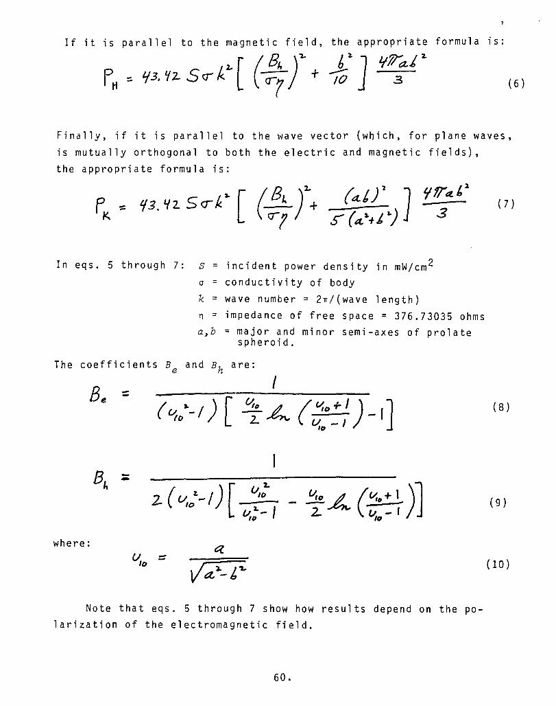
If it is parallel to the magnetic field, the appropriate formula is:
1ft/a' z. 3 (6 )
Finally, if it is parallel to the wave vector (which, for plane waves, is mutually orthogonal to both the electric and magnetic fields), the appropriate formula is:
'ItTa.l/ 3
In eqs. 5 through 7: s = incident power density in mW/cm 2 cr = conductivity of body k = wave number = 2rr/(wave length) n = impedance of free space = 376.73035 ohms a,b = major and minor semi-axes of prolate
spheroid.
The coefficients Be and Bh are:
I
-- - ('/,0+ l .... )] 2 1.1.-1 10
where:
Note that eqs. 5 through 7 show how results depend on the po-larization of the electromagnetic field.
60.
(7)
( 8 )
( 9 )
(10 )

Fig. 21 shows how the dissipated power varies as a function of frequency for each of the three possible polarizations. Actually, the dependent variable in the figure is not exactly power but a quantity called the average IIspecific absorption ratioll or average IISAR". This is simply the dissipated power normalized by the mass of the body. The mathematical definition is:
where: Fd = dissipated power density p = mass density
( 1 1 )
Note that in eq. 11, Fa is not that total dissipated power Rather, it is the dissipated power which, in general, is riot constant throughout the volume of the body. Therefore, the SAR also varies.
Which of the above three quantities (current density, dissipated power, or SAR) is most physiologically meaningful is not clear. SAR seems to have received the most attention, however. It can be com-pared directly to the body's basal metabolic rate (BMR), at least because they have the same dimensions (watts per kilogram). This is done in Table 27. The BMR is a measure of how fast the body can convert incident energy into forms other than heat. So, if the SAR exceeds the BMR, then body temperature should rise. Temperature change is associated with some of the adverse and beneficial changes described in the previous sections, including cataracts, diathermy, and hyperthermia. So, the SAR concept is useful for understanding at least these effects.
The calculations of SAR and electric currents have been extended to geometries which are more complicated than prolate spheroids and which more closely resemble the human form. Fig. 22 shows such a geometry, composed of multiple cubes. Figs. 23 through 25 show some results obtained using the multiple cube model. As already noted, the SAR is not uniform throughout the body. Instead, there are "hot spotsll, or regions of the body where the SAR is distinctly greatest. The location and intensity of the hot spots change as functions of frequency and other parameters.
61.

Fig. 26 demonstrates that current densities can also be calculated for detailed regions of the body.
The sort of computations described thus far require an a priori knowledge of electromagnetic constants (permittivity, permeability, and conductivity) for various parts of the body. These have been measured extensively, at least for bulk tissue, which is adequate for the multiple cube and simpler geometries. Tables 28 and 29 and Figs. 27 and 28 show values for many of the constants. They are used to calculate the dissipated power density, and thus the SAR, according to the formula,
(12)
In eq. 12, E" is the imaginary part of the complex permittivity E,
and r is the electric field at the point of interest inside the body. Fig. 28 emphasizes that the electromagnetic constants are
actually frequency dependent composites. That is, tissue is not homogeneous but composed of different substances (e.g. membranes, fluids, and various particles). In general, each substance has dif-ferent electromagnetic constants. The relative contributions from each substance to the composite electromagnetic constants may
with frequency. So, the theory and techniques described thus far are approximations which tend to ignore the detailed structure of tissue. This approximation may be acceptable, especially for modeling effects in which detectable tissue heating occurs. These are often termed "thermal" effects.
Theories Based on the Movement of Charge
On the other hand, many of the effects described in the previous· sections do not seem to be associated with tissue heating or tempera-ture changes. Examples include fibrillation, bone healing, and the various effects collectively known as neurasthenia, which seem to involve the central nervous system. These are often termed "athermal" effects. Efforts to understand these have focussed attention on the (microscopic) components of tissue (e.g. cells, membranes, fluids, molecules in solution) rather than the tissue taken as a whole.
62.

According to one theory, an electric field can cause the ag-glomeration of particles (e.g. molecules in solution) which are normally separated. Figs. 29 and 30 show this process, which is called IIpearl chain formation ll
• The individual particles, or pearls, are electrically neutral but the electric field polarizes them. Then, they are attracted to each other, negative end to positive end. The minimum, or theshold, field strength required to sustain the process is given approximately by the formula,
kT (13)
where: a = radius of particle £1 = permittivity of particle £2 = permittivity of ambient medium k = Boltzmann constant = 1.381 x 10- 23 joules/deg. T = temperature
Due to the viscosity of the solution, the field must be applied for at least a time given by the formula,
3 Z- -::. 12.1rr a
kT where n is viscosity and the x are roots of the equation, mn
In eq. 15, jn = spherical Bessel function of the first kind Yn = spherical Bessel function of the second kind 2R = separation between particles in the absence of an
applied field
63.
(14 )

The prime denotes a derivative. The concept of pearl chain formation implies rather drastic
changes can be induced within the body. Extensive numerical evalua-tion of the formulas and comparison with physiological data does not seem to be available in the literature, however.
In addition to particles in solution, theories have been de-veloped concerning individual cells. Fig. 31 shows the equivalent electric circuit of a cell. Given an incident current density, and using the circuit model, the total current or current density actual-ly passing across the cell membrane and through the cell can be cal-culated. If the current is sufficiently great, different responses are possible. For example, a current density of 1 mA/cm 2 is about the amount associated with the action potential of nerve and muscle cells. Perhaps a pulsed electromagnetic field could simulate these action potentials and confuse the body by generating false signals.
Another response that has received much attention relates to the IIblood brain barrierll. The exact nature of this barrier is not resolved; however, according to at least one theory, it is composed of specialized cells which line the capillaries of the brain and central nervous system. Similar cells are not found elsewhere in the body. They are very tightly packed, so that substances cannot leak between them, and they carefully select those which may pass through them. Thus. they isolate the brain and central nervous system in a manner unique in the body. Evidently. electric currents cause the specialized cells to contract, allowing normally excluded substances to leak between them. The frequency, and other character-istics of the required current are not known at this time.
Of the cell's parts. the membrane probably has attracted most attention, at least with respect to electromagnetic effects. Many membrane properties can be stated quantitatively. Some of them are:
• Typical thickness: 45 angstroms
• Typical capacitance: 1 microfarad/cm2
• Typical leakage conductance: 1-10 mhos/cm 2
• Typical resting potential: 100 millivolts
64.

From the above numbers the following additional ones can be derived:
• Dielectric constant: 5 Electric field: 2.2 x 10 7 volts/meter
• Surface charge density: 9.7 x 10- 8 coulombs/cm2 = 6.1 x 10¹¹ fundamental charges/cm2
Of these, perhaps the electric field is most remarkable because it is greater than almost any other found in nature. For example, recall
from a previous section that the electric field at the earth's sur-face is only about 100 Vim.
Fig. 32 shows a typical voltage waveform which occurs across membranes in response to the body's natural excitation.
Equations and equivalent electronic circuits have been developed based on the membrane properties and waveforms just described. The voltage across the membrane is given by the Hodgkin-Katz ion (HKI) equation:
V= 3r L F
f't<. [K7] + C Nat] Fe, [ C.e; -] IK [KeT1 -\- fN4 [Na:J + r::R [ C.e:.-]
In eq. 16, R = gas constant = joules/(degree-mole) T = temperature F = Faraday constant = 0.96487 x 10⁵ coulomb
The P are permeability constants, and the brackets denote concentra-tions of the ions within them. Subscripts i and e denote locations internal and external to the cell, respectively. An alternative to the HKI equation is the Association-Induction (AI) equation:
( 1 f )
( 17)
where the K are adsorption constants.
65.

The current across the membrane is given by the Hodgkin-Huxley equation, which has the form,
In eq. 18, C is the membrane capacitance and the g are leakage con-ductances. The latter are different for each charge species, and they are also nonlinear.
Fig. 33 shows a circuit model of the membrane based on eq. 18. As already noted, the are nonlinear, and Fig. 34 shows how they change in time for a step excitation.
There is some similarity between the cell membrane and the p-n junction found in electronic semiconducting devices, such as diodes and transistors. Figs. 35 and 36 indicate this similarity. Both the membrane and p-n junction sustain a voltage between regions with different charge concentrations. At least one theory exploits this similarity. According to that theory, the membrane is a rectifying junction. The implications could be dramatic. Direct currents might be created from high-frequency ones, and different frequencies might be mixed to create new ones to which the body is more sensitive i.e. resonant.
So far, the cell membrane has been treated as a homogeneous substance characterized by a capacitance and empirically derived, nonlinear conductances; however, the membrane is, in fact, inhomo-geneous. Fig. 37 shows the detailed structure of membranes. The basic structure is a double layer of molecules called lipids. The layer is penetrated to varying depths by proteins. Fig. 38 shows the chemical composition of a lipid molecule. The tails of lipids are hydrophobic, that is, they repel water. It is this repulsion which largely holds the membrane together. Proteins are chains of amino acids, the formula for one of which is shqwn in Fig. 39. The residues R are chemical compounds such as thymine, cytosine, adenine, and gua-nine. The chains combine to form large varieties of proteins. By
66.

one estimate, 50,000 different types are in the body, of which 100-3,000 have been documented. Fig. 40 shows some of the different proteins, separated according to gram molecular weight (i.e. the weight in grams of one mole, or 6.02 x 10 23 molecules). It is seen that the range in weight is over an order of magnitude.
The proteins are important, electromagnetically, because they can conduct charges, while lipids are good insulators. Thus, proteins are the conductances g in the various equations and circuit models of the membrane. Exactly how current passes through proteins is not well understood, but the size and shape of the protein is almost
certainly significant. If it somehow becomes bent or compressed, or if its site in the lipid layer is disrupted, then conduction is probably affected.
Electromagnetic fields can interact with proteins, possibly disturbing them as described above, over a wide range of frequencies. Between 1-10 MHz, depending on its size and mass, the entire pro-tein may be regarded as a single dipole which rotates in response to an oscillating field. According to research physician David Straub (VA Hospital, Little Rock), if proteins are dislodged from their sites in membranes, much time passes until random motion re-stores them. Meantime, the conduction function performed by those proteins is interrupted. At high frequencies, individual parts of proteins may be regarded as dipoles. Many of these rotate in response to fields in the 10-20 GHz band.
Fig. 41 and Table 30 show additional responses of individual molecules to electromagnetic fields of various frequencies.
Proteins figure predominantly in at least one theory of cancer, advanced by Nobel laureate Albert Szent-Gyorgyi, which is currently being researched. According to this theory, conduct electrons out of the cell interior. Oxygen molecules at the cell exterior accept the electrons and carry them away. These free electrons are products of some chemical process inside the cell that inhibits reproduction. If the electrons are not conducted away, then the process stops, and the cell divides at an uncontrolled rate. Eventually, there are enough cells to form a tumor, which
67.

characteristically has a poor circulatory system. So, little or no oxygen-carrying blood reaches the cells. The continued lack of oxygen exacerbates the situation, and reproduction continues un-checked. This theory is consistent with and, in part, based upon evolution. The earliest living cells were cancerous in nature, reproducing without limit and they developed and thrived in the early atmosphere of this planet, which was oxygen starved.
As noted at the beginning of this section, theories exist for every geometric scale within the body. At one extreme, the entire body can be treated at once as a homogeneous object. Other theories focus on the cell. Still others concentrate on but a part of the cell, the membrane. Even the membrane is not homogeneous, and theories were presented based on one type of molecule within the membrane, the protein. Possibly, in an effort to better understand micro-scopic charge conduction, future theories will be specialized to certain chemical compounds within the protein.
68.

E H
A B
Human body (ellipsoid) in a uniform elecnic (A) and magnetic (6) field. The broken lines indicate the direction of the induced cur-ren!.
FIG. 20. ELECTRIC CURRENT DENSITIES PRODUCED WHEN THE LONGITUDINAL AXIS OF THE HUMAN BODY IS PARALLEL TO ELECTRIC (E) AND MAGNETIC (H) FIELDS (FROM PRESMAN, 1970).
69.

____________________________________________________ -,
-
-O! a: en w t.!) a: .. Ej'8 :> ex:
'0 '. H
E '. ". ' . .... '" '" ........... K
' ...... .... ,----1 ••••• Esttmated values
Calculated average SAR in a prolate spheroidal model of an aver.ge man for an incident power density of I mW/cm2 for three polarizations; a. 0.875 m, b - 0.138 m, V • 0.07 .3.
FIG. 21. POWER ABSORBED BY A HUMAN BODY IN THE FAR FIELD OF AN ELECTROMAGNETIC RADIATOR, FOR THREE DIFFERENT POLARIZA-TIONS. NOTE THAT THE LOW-FREQUENCY RESPONSE MIGHT BE VERY DIFFERENT IF THE BODY IS IN THE NEAR FIELD (FROM DURNEY ET AL, 1980).
70.

Frequency (MHz) 10 20 50 60 80 100
1 mW/cm2 0.13 0.6 5.8 16 16 12 SAR/BMR
(%) 5 mW/cm2 0.65 3.0 29 80 80 60
Frequency (MHz) 200 500 1,000 2,000 5,000 10,000 20,000
1 mW/cm2 5.2 3.7 2.9' 2.5 2.5 2.5 2.5 SAR/BMR
(%) 5 mW/cm2 26 18.5 14.5 12.5 12.5 12.5 12.5
TABLE 27. RATIO OF SAR (SPECIFIC ABSORPTION RATIO) TO BMR (BASAL METABOLIC RATE) FOR AN AVERAGE MAN EXPOSED TO A PLANE WAVE WITH A POWER DENSITY OF 1 MW/CM2 AND 5 MW/CM2 (FROM STUCHLY, 1978).
71.

Block "",de of Iftln
I
!7=pt; .".
\ -71
r- :0-
.t-r- :--
'f-I
.-
\ 1" I
i. 1 I
- 'f--
I \-
Y"WV 7 x·
FIG. 22. MULTIPLE CUBE MODEL USED FOR DETAILED SAR COMPUTATIONS (FROM CHATTERJEE ET AL, 1980).
72.

FIG. 23.
III • 0.2U3
-;:..\. _____ 0.38
0.198 ----If-__ 1-1-__ 0.147
0.438 __ ---.j-t--- 0.351
0.558 0.287
1.435
2.35 J
4.04
7.64 ---- ....
Distrihution of power deposition for a human being with fCl'1 in electrical contact with the ground, The numbers are rc1atlvo: to whole.bodY.Bvo:raged SAR values of (1.75/L m )· 4.0 W/kg for 10 mW/cm' .
CALCULATED NONUNIFORM DISTRIBUTION OF DISSIPATED POWER ( FROM GANDH I" 1980).
73.

FIG. 24.
Free Ir.":(. htadlatiDn
Lt) • 0.417
0.491 ___ _ 0.456
S .087 ----/'--
0.689 0.348
0.558 ---+--.::-::--,-.
7.974 ---t- 0.539
1.6Z0
1.3J3
3.221 ----'+- 3.Z9Z
8.230 ----1 \;!." .:7 9.664
5.471 ----t ..... 4.996
4.768 ----t: 4.096
Distribution of power deposition for a human under free·space irradiation. The numbers arc relative to whole·body·
SAR of (1.7S/L m )· 1.88 W/kg for 10 mW/cm' incident fields.
CALCULATED NONUNIFORM DISTRIBUTION OF DISSIPATED POWER IN RESPONSE TO UNIFORM INCIDENT ELECTROMAGNETIC FIELD (FROM GANDH I I 1980).
74.

o o .......
,X-x X \
/ W E-a: , "'\ \ / p ,\
\ \ \ X ."-..L :. ". ...
z a -E-CL
0' (J)O CO ....... a: u -u... ....... u w CL (J)
W t!)
LEG NECK
WHOLE-BODY
TORSO
HEAD
" . + . x/ ¢
:' "A ... .... .
(!) " \+ I ,'\ I :4 A : \ + \
\ \ A
\ \ \ \ \
4 a: : I I
LL.. I I W + \ I > ARM \ I
I I
I
I I
I ,
....... 101 10Z (MHZ)
d d " 1 2 Part-body SAR for homogeneous mo el of man. Inci ent 1ntens1ty z mW.em.
FIG. 25. DISSIPATED POWER AS A FUNCTION OF FREQUENCY IN VARIOUS PARTS OF THE HUMAN BODY (FROM TAYLOR & CHEUNG, 1977).
75.

180 kV/m
60 nA/cm2
200) nA/cm2
FIG. 26. CALCULATED CURRENT DENSITIES IN A GROUNDED MAN IN RESPONSE TO A VOLT/METER ELECTRIC FIELD (FROM KAUNE & 1980).
7G.

.......
.......
Frc'Iuenc' (Me)
25 50 100 2UO 400 70U 10UO 3000 HSOO
(a) Dielectric cOllslanl f
Muscle I03-IIS 85-97 71-76 56 52-54 52-S3 49-S2 45-48 40-42 Bearl 59-63 52-56 50-55
muscle ... liver 136-138 88-93 76-79 50-S6 44-51 42-51 46-47 42-43 34-3B Spkell >200 135-140 100-101 Kidney >200 119-132 87-92 62 53-55 50-53 lUll!! 35 35 34 Brain >160 110-114 81-H3 Fat 11-13 4.5-7.5 4-7 5.3 -7.5 3.9-7.2 3.5-4.5 Bone 6.8-7.7 4.3-7.3 4.2-5.H 4.4-5.4
marrow
(b) Condudivity K in mmho/cllI
Muscle 6.80-8.85 9.52-10.5 11.1-11.8 12.7-13.7 12.7-13.3 21.7 -23.3 83.3 Heart 8.7-10.5 10 -11.8 10.5-12.8
muscle liver 4.76-5.41 5.13-5.78 5.59-6.49 6.67-9.09 7.69-9.52 B.7-11.8 9.43-10.2 20 -20.4 5B.B-66.7 Splcen 6.62-7.81 Kidney 6.9-11.1 11.1 11.8 1.3-1.32 lUll!! 2.22-3.85 6.25 7.14 7.69 Brain 4.55 4.76-5.26 5.13-5.56 Fal 0.4 -0.59 0.29-0.95 0.36 -11.1 0.83-1.49 1.11-2.27 2.7-4.17 Bone 0.2-0.36 0.43-\ 1.16 - 2.25 1.67 -4. 76
marrow ._-----------_._-----------".-- -- --+- - -------- ----------------------._-
(a) Values at 25, 50, and 100 Mill from Osswald (1937); at 2(JO. 400, :lIld 700 Mllz twm Sd\\vln ,,,,d Li (1953); al I lillO, 30!)O, and B5()0 Mill. froJl1 Herrick cl 01. (1950). The values frum 200 to 700 Mllz have been obtained al 27°(' and are adjusted 10 37°(' "ilh lhe help uf lernpcralure coefficients discussed above.
TABLE 28. DIELECTRIC CONSTANT AND CONDUCTIVITY IN MMHO/CM OF VARIOUS BODY TISSUE AT 37 DEGREES CENTIGRADE (FROM SCHWAN & FOSTER, 1980).
..
\

.---- - . .. •..
Fat. Ron ... and Tissues with I.ow "'at,,r Cuntt'nt --------. Reflection Cocffidcnt
Wavc!"nJ;th Dit'ketrie Conductivity \Vavelength Depth of Air-Fat Interface Interface Frequt'ney in Air Constant "I. . )II. Penetration
(MHz) (em) fl. (mmho/m) (em) (em) r f[I r '" 1 30000 10 3000 27.12 1106 20 10.943.2 241 159 0.660 +174 0.651 +169 40.68 738 14.6 12.6-52.8 187 118 0.617 +173 0.652 +170
100 300 7.45 19.1-75.9 106 60.4 +168 0.650 +172 200 150 5.95 2S.8-94.2 S9.7 39.2 0.458 +168 0.612 +172 300 100 S.7 31.6-107 41 32.1 0.438 +169 0.592 +172 433 69.3 5.6 37.9-118 28.8 26.2 0.427 +170 0.S62 +173 750 40 5.6 49.8-138 16.8 23 0.·415 +173 0.532 +174 915 32.8 5.6 S5.6-147 13.7 17.7 0.417 +173 0.519 +176
1500 20 5.6 70.8-171 8.41 13.9 0.412 +174 0.506 +176 2450 12.2 5.5 96.4-213 5.21 11.2 0.406 +176 0.500 +176 JOOO 10 5.5 110-234 4.25 9.74 0.406 +176 0.495 +177 5000 6 5.5 162-309 2.63 6.67 0.393 +176 0.502 +175 5900 5.17 5.05 186-338 2.29 5.24 0.388 +176 0.502 +176 8000 3.75 4.7 255-431 I. 73 4.61 0.371 +176 0.513 +173
10000 3 4.5 324-549 1.41 3.39 0.363 +175 0.518 +174
-----.. -- ---_ .. Muscle. Skin. and Tissues with lIiCh Water Content
Reflection Coefficient
\Vavelength Dielectric Conductivity Depth of Air-Mu:!Clt' Interface MUlJCle-Fat Interface Frcquene)' In Air Constant "N )"" Penl'lra tion
(MHz) (em) fH !mho/m) (em) (em) , f[I r '" 1 30000 2000 0.400 436 91.3 0.982 +179 10 JOOO 160 0.625 118 21.6 0.956 +178 27.12 1106 113 0.612 68.1 14.3 0.925 +177 0.651 -11.13 40.68 738 97.3 0.693 51.3 11.2 0.913 +176 0.652 -10.21
100 300 71. 7 0.88'1 27 6.66 0.881 +175 0.650 -7.96 200 150 56.5 1.28 16.6 4:79 0.844 +175 0.612 -8.06 300 100 54 1.37 11.9 3.89 0.825 +175 0.592 -8.14 433 69.3 53 1.43 8.76 3.57 0.803 +175 0.562 -7.06 750 40 52 1.54 5.34 3.18 0.779 +176 0.532 -5.69 915 32.8 51 1.60 4.46 3.04 0.772 .+177 0.519 -4.32
1500 20 49 1. 77 2.81 2.42 0.761 +177 0.506 -3.66 2450 12.2 47 2.21 1. 76 1. 70 0.754 +177 0.500 -3.U 3000 10 46 2.26 1.45 1.61 0.751 +17& 0.495 -3.20 5000 6 44 3.92 0.89 0.788 0.749 +177. 0.502 -4.95 5800 5.17 43.3 ,:.' 4.73 0.775 0.720 0.746 +177 0.502 -4.29 8000 3.75 40 7.65 0.578 0.413 0.744 +176 0.513 -6.65
10000 3 39.9 10.3 0.464 0.343 0.743 +176 0.518 -5.95
TABLE 29. ELECTROMAGNETIC CONSTANTS AND RELATED PARAMETERS FOR VARIOUS TISSUES (FROM JOHNSON & GUY, 1972).
78.

>-'" :i . t; .. z o u
HUMAN BRAIN • ..
E/ rI i·· I H r .. f ! I £ ! f- ..
..
. 3:00 3:2; ':!IO 4.00
FREQUENCY (GHll
FIG. 27. DIELECTRIC PROPERTIES OF HUMAN BRAINS IN THE MICROWAVE S-BAND, AT 37 DEGREES CENTIGRADE (FROM LIN, 1975).
79.
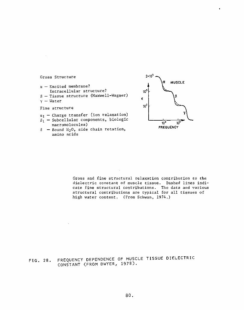
Gross Structure
a - Excited membrane? Intracellular structure?
S - Tissue structure (Maxwell-Wagner) y - Water E
Fine structure
FIG. 28.
Charge transfer (ion relaxation) Subcellular components, biologic macromolecules) Bound H20, side chain rotation, amino acids
102 106
FREQUENCY
Gross and fine structural relaxation contribution to the dielectric constant of muscle tissue. Dashed lines indi-cate fine structural contributions. The data and various structural contributions are typical for all tissues of high water content. (From Schwan, 1974.)
FREQUENCY DEPENDENCE OF MUSCLE TISSUE DIELECTRIC CONSTANT (FROM DWYER, 1978).
80.

o 00
o o 0
o 000 o o
000 without field
§ 8
with field
FIG. 29. PEARL CHAIN FORMATION (FROM STUCHLY, 1977).
81.

FIG. 30.
+ + + + +
+ 8 + (a) + +
+ + ... ... + ... +
+ + + + + + 8 (b) + +
+ + + ... +
... + ... + + + ... + + + + + + + +
+ 8+ 8+ (c) + -) + '++ ++ "-..A
+ + + + + + + ... ... + + + + + +
(a) A negatively charged particle (-), together with a double (+) layer, forms an electrically neutral whole; (b) an in-duced dipole which arises when a charged particle enters an elec-trical field; (e) linking of oriented dipoles in the direction of an electrical field (chain formation).
STEPS IN THE PROCESS OF PEARL CHAIN FORMATION (FROM MARHA & TUHA, 1971).
82.
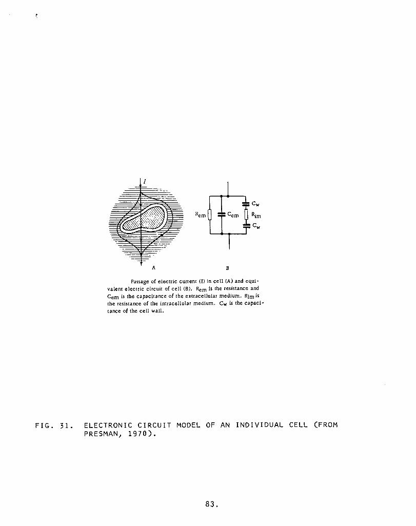
FIG. 31.
I
A 8
Passage of electric current (I) in cell (A) and equi-valent electric circuit of cell (8). Rem is the resistance and Cern is the capacitance of the extracellular medium. Rim is the resistance of the intracellular medium. Cw is the capaci-tance of the cell wall.
ELECTRONIC CIRCUIT MODEL OF AN INDIVIDUAL CELL (FROM PRE 5 MAN I 1970).
83.

mV f8D
+60
+-20
defoo/at'I z.a. t 10It
r-e(ovetj
;zs 0 mdlt"sec
o i/ft1e
RECOVERY CURVE OR TRANSMEMBRANE ACTION POTENTIAL FOR A VENTRIaJLAR HEART MJSCLE CELL.
FIG. 32. VOLTAGE ACROSS THE MEMBRANE OF A HEART MUSCLE CELL (FROM BENEDEK & 1979).
84.

FIG. 33.
r
v
Equivalent circuit of the Hodgkin-Huxley model, showing three ionic path-ways, for sodium, potassium, and leakage ions, and the membrane capacitor.
HODGKIN-HUXLEY CIRCUIT MODEL FOR A CELL MEMBRANE (FROM SCHWAN, 1969).
85.

y= :109 'n\V
time 6 8 (JTrilk5cc) K
y= 88 'TT1Y v = 88 1l1V
y= 6.3 -mV
V= 38 mY
v=Z6mV
:=;=t· '7 tme z 6 8 1.0 FIG. 3.3-20: TIME COURSE OF SODIUM AND POTASSIUM CONDUCTANCES
FOLLOWING A DEPOLARIZATION STEP v AT t=O. CONDUCTANCE DNITS 10-2/0hm cm2 • DEPOLARIZATION v IN MILLIVOLTS.
FIG. 34. TIME-DEPENDENT CONDUCTANCES IN THE CIRCUIT MODEL OF A CELL MEMBRANE. (FROM BENEDEK & VILLARS, 1979).
86.

II' Concentvatron (Ynfl1/f)
200- Na +
150
100 ce
50 -K+
D
CP(X)
Thtenor
i , 1 j I I"", I ", I I ' / / I !
'<. /'1 em bra ne I / 1
/ I // ; I c.e-V i-"-
I .P / 1 Nc! '/
/ / "; / '/ 1 I / /
1/ "" I /
/ --" / /
"/ /
Na.t- / /
I 1 I --ce- _------ 1
I", -------- t- "'-I Ki-1 F--- _ _ _ K
---
/VJem br-ane c.p (1.)
/
/
PROFILES OF IONIC CONCENTRATIONS AND ELECTRIC POTENTIAL ACROSS A BIOMEM-BRANE. FOR THE DONNAN EQUILIBRIUM
X
FIG. 35. SIMILARITY OF CELL MEMBRANE TO SPACE CHARGE LAYER IN AN ELECTRONIC SEMICONDUCTING DEVICE (FROM BENEDEK & VILLARS, 1979).
87.

FIG. 36.
N or p
Space·charge region ---
p 'x----:--------*"
Hole antll'lcdrun iu a p-n junl"lioll at thermal equilihrium.
SPACE CHARGE LAYER IN A SEMICONDUCTOR.
88.

STRUCTURAL FRAMEWORK typical of cell membranes is made UI) of a bilayer of lipids with their hydrophilic heads forming out. er and inner membrane surfaces alld their hydrophobic tails meet· ing lit the center of tiJ-e membrane; the bilayer is about 45 ang.
stroms thick. Proteins, tbe other membrane constituents, are of two kinds. Some (a) lie at or near either membrane ludace. The oth· en penetrate the membrane; they may intrll4e only a shon way (b) or may bridge tile membrane completely (c), 6in,ly or in pain •
. j 0
. Upld bilayer containing membrane proteins. The proteins can serve as channels through the membrane for selected ions, as active trans·membrane pumps for specific mole. :ules. as catalysts in chemical reactions, or as structural units in the membrane.
!TIITIT .. Q: .. '.
TIITTI ·i
FIG. 37.
.c -.. .
LIPIDS AND PROTEINS IN A CELL MEMBRANE (ABOVE FROM CAPALDI, 1976, BELOW FROM CHANCE ET AL, 1980).
89.

AMPHIPATHIC STRUCTURE of a lipid molecule, with a hydrophilic head and twin hydrophobic tails, is exemplified by tbis typical phospholipid, specifically a molecule of phosphatidylcholine. Various lipid mole. cules comprise about hall of the mass of mammalian membrane, forming the memo brane's structural framework. Their fatty. add tails mlly be saturated (le!t), with a hy. drn8p.n atom linked to every carbon bond or unsaturated (right), with carbons free:
H I
H-C I H' ,',-:'
.....
Ii H
FIG. 38. CHEMICAL COMPOSITION OF A LIPID MOLECULE (FROM CAPALDI, 1976).
90.

0)
b)
R I + -N H - C -COO
3 I H
R! R.;z ... I I
NH3 - - --COO-H H
a) SINGLE AMINO ACID. IN AQUEOUS SOLtITION THIS WILL GENERALLY EXIST AS A BIPOLAR ("ZWITTERION"). R IS THE RESIDUE (see text). b) TWO AMINO ACIDS BONDED BY THE PEPTIDE BOND.
FIG. 39. CHEMICAL COMPOSITION OF AN AMINO ACID FROM WHICH PROTEINS ARE CONSTRUCTED (FROM BENEDEK & VILLARS, 1979).
91.

FIG. 40.
t I
I z o a. a: o (/) In 04:
220,000 255,000
195,000
87,000
73,000
IDENTITY OF PROTEINS in the membrane of red blood cella e:m be determined by means of gel electrophoresis. In this instance the proteins have been stained and i1c:anned. using a densiometer. after those with the lowest molecular weights have migrated fanhelt through the gel in response to an electric potentiaL The two proteins the highest mo-lecular weight (left) form the climer collectively called spectrin. The ScaD shows at leut 10 more absorption bands 6ignifying the presence of other proteins even lowell' in weighL
WIDE RANGE OF MOLECULAR WEIGHTS FOR VARIOUS PROTEINS (FROM CAPALDI, 1976).
92.
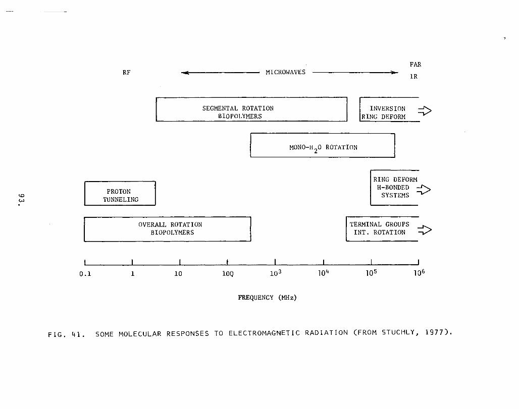
\.0 W .
0.1
RF
PROTON TUNNELING
1
OVERALL ROTATION BIOPOLYMERS
10
MICROWAVES
SEGMENTAL ROTATION BIOPOLYMERS
INVERSION RING DEFORM
FAR lR
MONO-H 20 ROTATION
10Q
FREQUENCY (MHz)
RING DEFORM H-BONDED
SYSTEMS
TERMINAL GROUPS INT. ROTATION .,..........
FIG. 41. SOME MOLECULAR RESPONSES TO ELECTROMAGNETIC RADIATION (FROM STUCHLY, 1977).

RESEARCHER SUBSTANCE FREQUENCY EFFECT
Prohofsky DNA helix 100 GHz Conformational change from A to B.
Prohofsky DNA helix 430 MHz Bases pull ed apart.
McCammon DNA helix 2 GHz Rippling motions, or traveling wave displacements.
TABLE 30. MOLECULAR CHANGES CITED BY TAYLOR, 1979.
94.

Some Speculation and Areas for Further Research
Based on the exhibits and discussions of the preceding sections, the present state of knowledge would most likely benefit from the following types of efforts:
• Improved observations of effects • More intensive, quantitative exploitation of existing
physiological knowledge and electromagnetic theory.
• Formulation of new physio-electromagnetic theories.
Elaboration on these follows.
Improved Observations of Effects
The need for more uniform and rigorous reporting is especially evident in the section concerning adverse effects. Precise records of frequency, polarization, intensity, and duration of exposure are sometimes lacking. Quantitative measures of the effect (e.g. how severe, how extensive, how persistent) are similarly lacking. A standard procedure for conducting formal epidemiological studies is needed, including precise definitions of the electromagnetic variables and biological endpoints.
In contrast, beneficial effects, especially electromagnetic diathermy and hyperthermia, have been exploited in a much more pur-poseful fashion. The reason is probably that at least the desired biological endpoint (selective, localized heating) was clear. On the other hand, identification and optimization of electromagnetic variables is still in progress.
A sound theoretical foundation would certainly clarify the variables and endpoints.
Quantitative Exploitation of Existing Physiological Knowledge
The preceding section indicated that the properties and functions of the human body have been investigated at many levels of detail, ranging from the bulk properties of organs and tissue to the structure
95.

of cell membranes and proteins. This knowledge has already been the basis for some rigorous computations, but many more are still waiting to be performed. Among those already demonstrated, the SAR concept, discussed in the preceding section, has been highly refined. Computer models can predict heat dissipation and tempera-ture changes for local regions of the body.
On the other hand, no computation has yet performed to determine the current density incident at the membrane of individual cells in situ for heart muscles or nerves due to external electro-magnetic fields. Such a computation might be practical, and it could provide much quantitative information concerning the effects of frequency, polarization, intensity, and other variables on the func-tioning of these cells. The computation might be further extended to include arrays of cells to determine whether they respond co-herently.
New Physio-Electromagnetic Theories
Some novel interactions between electromagnetic fields and the human body have been proposed, and they await further investigation, both theoretically and experimentally. The ones discussed here include: nonionizing single photon interactions, coherent phenomena, coupled oscillators, and the relative importance of different charge species.
As noted in the introduction of this report, single photon interactions other than ionization are expected, based on fundamental physics. In principle, external electromagnetic fields can cause an abnormal response if the energy per photon is greater than the back-ground, or thermal, noise due to others encountered randomly within the human body. The energy of a randomly encountered photon is, on the average:
E:; k. T ( 19 )
where k = 1.381 x 10- 23 joules/degree and T is temperature in degrees Kelvin. For a body temperature of about 300 0 , E is about 0.025 eV, or about 500 times less than the standard definition for ionizing radiation. Using eq. 1, the equivalent frequency is about 6,000 GHz,
96.

or about 500 times lower than the arbitrary minimum frequency for ionizing radiation. Table 31 lists some of the single photon interactions that are expected. Fig. 42 shows the ratio of photon energy to kT for the lower part of the electromagnetic spectrum.
After investigating single photon interactions, it would be logical to consider the possibility of multiple photon interactions. If one photon can alter a protein at 6,000 GHz, then 6,000 photons can do it at 1 GHz. 6,000 I-GHz photons?
But how efficiently can one protein intercept What would the corresponding incident field
intensity be at the surface of the body? Would it be so great that thermal injury would overwhelm the interaction?
The answer, that is, the field intensity, might be surprisingly low. The concept of cooperative, or long-range, or coherent, inter-actions suggests this result. According to the concept, individual microscopic particles, such as proteins, can have very large cross sections (i.e. receiver areas or gain) if they interact with each other. Actually, this is not such a novel idea. High gain antennas and lasers are established In electromagnetic ·physiology, however, only a few theories based on it have appeared so far.
According to one such theory, proposed by Frohlich, the surface charges on a cell membrane are all coherent. Further, they may also be coherent with surface charges on the membranes of other cells. All of these charges, considered collectively, can oscillate between at least two different states. Based on quantum mechanical formulas, Frohlich has estimated the natural frequency of oscillation. It is in the band 50-3,000 GHz.
Frohlich suggests his theory may lead to a better understanding of cancer. The energy required to sustain the oscillations comes from within the cells. The energy drain inhibits cell reproduction. If the oscillations cease, then the cell divides without bound, ultimately resulting in cancer. This implies that electromagnetic waves in the 50-3,000 GHz band might promote or be used to inhibit the disease.
According to another theory, charges and currents in the brain and central nervous system are also coherent. They are, therefore, sensi-tive to very low level external fields.
97.

In the preceding section, microscopic parts of the body were modelled as various circuit elements, including capacitors, conductances, and rectifying junctions. The theories just described suggest th3t further modeling might be useful in terms of high gain antennas. Practical antennas today achieve gains of 30 dB or more. If biological antennas have similar gains,how would that affect the body's sensitivity to electromagnetic fields?
Another, perhaps related, concept that could be further explored is coupled oscillators. This has been partially developed for at least one part of the human body, the gastro-intestinal system. It is rich in low-frequency electrical signals, as indicated by Figs. 43 and 44. From Fig. 44, it is seen that the waveforms are not sinu-soidal. They are called "limit cycles". Limit cycles are solutions to the Van der Pol equation, which is of the form,
o (20)
The observed gastro-intestinal signals have been reproduced using a model made of coupled Van der Pol oscillators. Fig. 45 shows such a model. The preceding section described the currents across individual cell membranes associated with the action potentials of nerves and muscles. What currents might be required to excite a large system of coupled oscillators?
Little distinction has so far been made between species of electric currents. In fact, however, it is probably significant. The flow of sodium currents probably has a different effect than the flow of proton currents or calcium currents. What other species of currents are found within the body, and what are their different physiological functions? The answer might be found in the detailed structure and conduction mechanisms of the many different proteins embedded in cell membranes. This level of microscopic detail is now the frontier of understanding for the human body. The frontier awaits exploration.
98.

i I
I I ACTIVA TION ENERGY CORRESPONDING FREQUENCY EFFECT (eV) (GHz)
\ Thermal Motion 0.026 6.3 x 10³
I (at 30 0 C)
I Ionization 10 2.4 x 10⁶ I
Covalent bond 5 1.2 x 10⁶ disruption I
I London-van der Waals I x 105 1 i 2.4 i interactions
i I Hydrogen bond I distruption O.OB - 0.2 1.9 x 10⁴ - 4.B x 104
I Disruption of I 105 0.56 1.4 x 1 bound water
I Reversible confor- 4 I mational changes 0.4 9.7 x 10 : in protein molecules
Charge transfer 3 - 6 7.3 5 1.45 x 10⁶ interaction x 10 -
Semiconduction 1 - 3 2.4 5 x 10 - 7.25 x 10⁵
TABLE 31. ACTIVATION ENERGY OF MOLECULAR EFFECTS IN BIOLOGICAL SYSTEMS (FROM STUCHLY, 1977).
99.
I :
I
I I
i

Region of Electromagnetic Spectrum from Infralow to Superhigh Frequencies, in Which hv < kT
Ultraradio waves Radio waves
Microwaves
Wave ranges Low-frequency waves j E , , , = I)) Milli- "'-I)) (J_ - I)) c '" (; c_
I "0 1))"0 ;; I)) I)) I)) I)) meter '" c I)) -I)) "" QE UE 0 ::;; 'sE en ::;; f-<::::
Wavelength, cm l1dl3 I 1bll I I 161 160 I I I 110s 1b 1 O.bt 1012 1010 10' 10s 10' 10'
I I I I I I I I I I I I 1 I I 3.1dn Frequency, Hz 3.10""23.10-1 3 3.101 3.102 3·1()3 3·10' 3·1Us 3·106 3.101 3·18 3·tO· 3.1010
I I 4.5.10-11 I 1 hvjkT 4.5.10-16 4.5·10-$ 0.45
Frequency j Infra- Low I Industrial I Audio I High (HF) I Ultrahigh Superhigh ranges low (UHF) (SHF)
FIG. 42. ELECTROMAGNETIC SPECTRUM, WITH EMPHASIS ON ENERGY PER PHOTON (FROM PRESMAN, 1970).
100.

CARDIAC SPHINCTER
I----O) 5 Hz
____ __ ______________
[
0.05 Hz 0'12Hz
°
Hz
FIG. 43. DIGESTIVE TRACT DIAGRAM INCLUDING TYPICAL SLOW-WAVE FREQUENCIES (FROM LINKENS, 1979).
101.

FIG. 44.
" .. """" ... "
Recording of human gastric electrical activity. Top trace is from a surfactl "tlCtrode, bottom trace is from an internsl electrode
LIMIT CYCLE TYPE WAVEFORMS OBSERVED IN HUMAN DIGESTIVE TRACT (FROM LINKENS, 1979).
102.

FIG. 45. TWO-DIMENSIONAL COUPLED OSCILLATOR STRUCTURE FOR DIGESTIVE TRACT MODELLING.
103.

Acknowledgments
The author is indebted to a number of individuals at the NASA Goddard Space Flight Center for their enthusiastic support. Mr. Tom Golden, Director of Technology Transfer, National Needs Office, participated in many helpful and inspiring discussions, and he was instrumental in realizing a series of telephone 'interviews which reached across the United States. Ms. Adelaide del Frate and Ms. Jane Riddle performed numerous literature searches and tirelessly pursued documents which were difficult to obtain. Mr. John Fuchs translated documentation which was available only in German.
Prof. Augustine Cheung, Dept. of Molecular Physics, University of Maryland, gave generously of his time and technical library, during at least two in-person interviews. Prof. Leonard Taylor, of the same department, delivered an informative lecture on biologi-cal effects and directed the author towards useful references.
Sally Faith Dorman, MD, MSHSA, Center for Disease Control, Atlanta, identified and provided a number of obscure documents which proved to be a wealth of pertinent information.
A number of exceptional professionals gave generously of their time via telephone interviews, which in some instances continued for nearly three hours without interruption. The author is very grate-ful to: Dr. Joseph C. Sharp, Director of Life Sciences, NASA, Ames Research Center; Prof. William Guy, Dept. of Rehabilitation Medicine, University of Washington, David Straub, MD, Veteran's Administration, Little Rock; Peter Gascoyne, Ph.D., National Founda-tion for Cancer Research, Woods Hole, Massachusetts; Robert MacElroy, Ph.D., NASA, Ames Research Center; and Prof. Charles Pouvaris, Dept. of Electrical Engineering, Catholic University. To enumerate their individual contributions would easily fill a large report. In fact, they largely filled this one.
Helpful article reprints were thoughtfully provided by Ross Adey, MD, Veteran's Administration, Lorna Linda, and by Stephanie Seidman, Ph.D., NIH, Bethesda, Maryland.
104.

References
1. Bar ret & r'l y e r s, II M i c row a veT her m 0 g rap h y 0 f Nor mal and Can c e r 0 u s Breast Tissue," Preprint to be published in Annals of the
York Academy of Sciences.
2. Benedek & Villars, Physics With Illustrative Examples from Medicine and Vol. 3: Electricity and Addison-Wesley, 1979.
3. Capaldi, "A Dynamic t·lodel o'f Cell t',1embranes," Scientific Am e ric a n arc h, 1 9 7 6, P p. 2 7 - 3 3 .
4. Chance, Mueler, de Vault &. Powers, "Biological t',lembranes," Physics Oct. 1980, pp. 32-38.
5. Chatterjee, Hagmann & Gandhi, "Electromagnetic Energy Deposi-tion in an Inhomogeneous Block Model of Man for Near Field Irradiation Conditions," IEEE Trans. on Theory and Te c h n i qu e s Dec. 1980, P p. 1452 -14 5 9 .
6. Danzer, Hollman, Rajewsky, Schaefer & Schliephake, Ultrakurz-in ihren Medizinsch-Biologischen Leipzeg:
Georg Thieme, 1938.
7. David, "A Study of Federal Standards," DOE Rept. August, 1980.
8. Durney, Iskander, Massoudi, Allen & Mitchell, "Radiofrequency Radiation Dosimetry Handbook, Third Edition," USAF School of Aerospace Medicine Rept. August, 1980.
9. Dwyer & Leeper, "A Current Literature Report on the Carcino-genic Properties of Ionizing and Nonionizing Radiation," DHEW Publication (NIOSH) March, 1978.
10. Frazier, "An Evaluation of Health and Environmental Effects of Extra High Voltage (EHV) Transmission, First Interim Report," Chicago: ITT Research Institute May, 1978.
11. Gandhi, "State of the Knowledge for Electromagnetic Absorbed Dose in Man and Animals,1I Proc. Jan. 1980, pp. 24-40.
12. Johnson & Guy, "Nonionizing Electromagnetic Wave Effects in Biological Materials and Systems,1I Proc. June, 1972, pp. 692-718.
13. Johnson & Shore (eds.) "Biological Effects of Electromagnetic Waves, Vol. I," HEW Publication (FDA) Dec. 1976.
14. Johnson & Shore (eds.) IIBiological Effects of Electromagnetic Waves, Vol. II" HEW Publication (FDA) December, 1976.
105.

15. Kaune & Phillips, IIComparison of the Coupling of Grounded Humans, Swine, and Rats to Vertical, 60-Hz Electric Fields,1I Bioelectromagnetics 1980, pp. 117-130.
16. Kleinstein & Dyner (eds.) Biological Effects of Nonionizing Electromagnetic Radiation Philadelphia: The Franklin Institute, 1980.
17. Lerner, "RF Radiation: Biological Effects," IEEE Dec. 1980, pp. 51-59.
18. Lin, liThe Microwave Auditory Phenomenon,1I Proc. Jan. 1980, pp. 67-73.
19. Lin, IIMicrowave Properties of Fresh Brain Tissue at Body Temperature,1I IEEE Trans. on Biomedical Jan. 1975, pp. 74-76.
20. Linkens (ed.) Biological Modelling and Stevenage, UK: Peter Peregrinus, 1979.
21. Marha, Musil & Tuha, Electromagnetic Fields and the Life San Francisco Press, 1971.
22. McCree, IISoviet and Eastern European Research on Biological Effects of Microwave Radiation,1I Proc. of Jan. 1980, p-. 84-91.
23. Persinger (ed.), ELF and VLF Electromagnetic Field New York: Plenum Press, 1974.
24. Presman, Electromagnetic Fields and New York: Plenum Press, 1970.
25. Schwan (ed.) Biological McGraw-Hill, 1969.
26. Schwan & Foster, IIRF Field Interactions With Biological Systems: Electrical Properties and Biophysical Mechanisms,1I Proc. Jan. 1980, pp. 104-114.
27. Short & Turner, IIPhysical Hyperthermia and Cancer Therapy,1I Proc. Jan. 1980, pp. 133-142.
28. Sterzer, Paglione, Friedenthal & Botstein, IIRF Therapy for Malignancy,1I IEEE Dec. 1980, pp. 32-37.
29. Stuchly, IIHealth Aspects of Radio Frequency and Microwave Radiation Exposure, Part 1," Ottawa, Canada: Dept. of National Health and Welfare Rept. Nov. 1977.
30 . Stu c h 1 y, II He a 1 t hAs p e c t s 0 f R ad i 0 F r e que n c y and i c row a v e Radiation Exposure, Part 2," Ottawa, Canada: Dept. of National Health and Welfare Rept. March, 1978.
106.
31. Taylor & Cheung (eds.), The Mechanisms of Microwave Biological Effects, University of Maryland Publication, May, 1979.
32. Taylor & Cheung (eds.) "The Physical Basis of Electromagnetic Interactions With Biological Systems," HEW Publication (FDA) 78-8055, April, 1978.
33. Tyler (ed.) "Biological Effects of Nonionizing Radiation," Annals of the New York Academy of Sciences, Vol. 247, Feb. 28, 1975.
34. Valentino, "Environmental Assessment for the Satellite Power System (SPS) Concept Development and Evaluation Program (CDEP)" DOE Rept. ER-0069, August, 1980.
35. Wertheimer & Leeper, "Electrical Wiring Configuration and Childhood Cancer," American Journal of Epidemiology, 1979, pp. 273-84.
36. --- "Death By Microwave?" Bioelectromagnetics Society Newsletter, Dec. 1980, p. 1.
37. --- "Draft Environmental Impact Statement for PAVE PAWS System at Otis Air Force Base, Massachusetts," USAF Report, Dec. 1978 ..
107.

Bibliography
38. Abramson, Moyer & Gorin, Electrophoresis of Proteins and the Chemistry of Cell Surfaces, New York: Reinhold Publishing, 1942.
39. Adey, "t'lodels of Membranes of Cerebral Cells As Substrates for Information Storage," BioSystems, 8 (1977), pp. 163-78.
40. Adey, "Neurophysiologic Effects of Radiofrequency and Microwave Radiation," Bulletin of the New York Academy of Medicine, Dec: 1979, pp. 1079-1093.
41. Arthur (ed.) "Proceedings of the 1978 Electromagnetic Inter-ference Workshop," NBS Special Publication No. 551, July, 1979.
42. Becker, liThe Significance of Bioelectric Potentials," Biochemistry and Bioenergetics, 1 (1974), pp. 187-199.
43. Beischer, "Human Tolerance to Magnetic Fields," Astronautics, March, 1962, p. 24.
44. Benedick, "Blood-Brain Barrier Workshop," Chicago: IIT Research Institute Rept. May, 1979.
45. Browne, "Benign Radiation Increasingly Cited As Dangerous,1t New York Times, Oct. 21, 1980.
46. Cain, "A Theoretical Basis for Microwave and RF Field Effects on Excitable Cellular Membranes," IEEE Transactions on Micro-wave Theory and Techniques, Feb. 1980, pp. 142-147.
47. Chou & Guy, "Holographic Assessment of Microwave Hearing," Science, Sept. 5, 1980, pp. 1143-5.
48. Dodge, Workshop on Mechanisms Underlying Effects of Long-term, Low-level, 2450 MHz Radiation on Semi-Final Draft, Science Policy Research Division, Congressional Research Service, Library of Congress, Sept. 5, 1980.
49. Duke & Schein, "Organic Solids: Is Energy-Band Theory Enough?" Physics Today, Feb. 1980, pp. 42-48.
50. Edmonds, "Making (Micro)waves over Trade Center TV," New York: Daily News, Feb. 25, 1980, p. 1.
51. Emery, Short, Guy, Kraning & Lin, liThe Numerical Thermal Simulation of the Human Body When Undergoing Exercise or Nonionzing Electromagnetic Irradiation," Trans. of the ASME, May, 1976, pp. 284-291.
52. French, "Can Ion Device Make Magic, Or Just Profits?" Wall Street Journal, Oct. 8, 1980.
108.

53. Frohlich, lIBose Condensation of Strongly Excited Longitudinal Electric Modes,1I Physics Letters 3 25,1968, pp. 402-403.
54. Frohlich, "Coherent Electric Vibrations in Biological Systems and the Cancer Problem," IEEE Trans. on Microwave Theory and Techniques 3 Aug. 1978, pp. 613-617.
55. Glaser, Brown & Brown, "Bibliography of Reported Biological Phenomena (IiEffects") and Clinical Manifestations Attributed to Microwave and Radio-Frequency Radiation: Compilation and Integration of Report and Seven Supplements," Naval Medical Research Institute Rept.3 Sept. 1976.
56. Graham, IIElectric Stimulators Heal Bones, Show Potential for Other Cures," The Wall street Journal 3 July 11, 1980, p. 11.
57. Guy, Webb & McDougall, "RF Radiation Absorption Patterns: Human and Animal Modeling Data," DHEW (NIOSH) Publication No. 77-183 3 Sept. 1977.
58. Habegger, Gasper & Brown, IIHealth and Safety: Preliminary Comparative Assessment of the Satellite Power System (SPS) and Other Energy Alternatives,1I Dept. of Energy Rept. ER-0053 3 April, 1980.
59. Hazzard (ed.) IISymposium on Biological Effects and Measurement of Radio HEW Publication (FDA) 77-8026 3
July, 1977.
60. Hopwood, IIDowsing, Ley Lines and the Electromagnetic Link,1I New Scientist 3 Dec. 27, 1979, pp. 948-9.
61. Ishimaru (ed.), IIBiological Effects of Electromagnetic Waves,1I Radio Science 3 14:6S, Nov.-Dec. 1979.
62. Lavery, Pullman, Pullman & de Oliveira, liThe Electrostatic Molecular Potential of RNA,II Nucleic Acids Research 3 8:21 (1980), pp. 5095-5111.
63. Lee, Bracken, Capon, Sarkinen, Ihle, Perry & Eyler, "Electrical and Biological Effects of Transmission Lines: A Review,1I Bonneville Power Administration Rept. BPA-BIO-78-1 3 June, 1977.
64. MacGregor, IIA Possible Mechanism for the Influence of Electro-magnetic Radiation on Neuroelectric Potentials,1I IEEE Trans. on Microwave Theory and Techniques 3 Nov. 1979, pp. 914-921.
65. Matusow, IINIOSH Probes Bio-Effects of RF Heat Sealer Leakage,1I Microwaves 3 Oct. 1980, pp. 17.
66. McAuliffe, "I Sing the Body Electric,1I Omni3 Nov. 1980, pp. 70-73.
67. Newsom, "Research Plan for Study of Biological and Ecological E f f e c t s 0 f the Sol a r Power Sat ell i t e T ran s m iss ion S y s t em, 11
NASA Contractor Rept. 30443 August, 1978.
109.

68. Peyton (ed.), Biological Effects of Microwave Radiation, New York: Plenum Press, 1961.
69. Plonsey, Bioelectric Phenomena, McGraw-Hill, 1969.
70. Pullman, Perahia & Cauchy, liThe Molecular Electrostatic Po-tential of the B-DNA Helix," Nucleic Acids Research, 6:12 (1979) pp. 3821-3829.
71. Reeves & Sutko, IIS odium-Calcium Exchange Activity Generates a Current in Cardiac Membrane Vesicles," Science, June, 1980, pp. 1461-1463.
72 . Rob i net t e, S i 1 ve rm an & Jab 1 0 n, II E f fee t sUp 0 n He a 1 tho fOe cup a -tional Exposure to Microwave Radiation (Radar)," American Journal of Epidemiology, 112 (1980) pp. 39-53.
73. Samars & Cheung, "Microwave Hyperthermia for Cancer Therapy," Preprint to be published in Critical Reviews in Bioengineering, CRC Press. I
74. Schmitt, Schneider & CrGthers, Functional Linkage in Bio-molecular Systems, New York: Raven Press, 1975.
75. Sharp, IIS ome Perspectives on Research Into the Biological Response to Non-Ionizing Electromagnetic Radiation," Radio Science, Jan.-Feb. 1979, pp. 5-10.
76. Sheppard, Bawin & Adey, "Possible Mechanisms of Weak Electro-magnetic Field Coupling in Brain Tissue," Bioelectrochemistry and Bioenergetics, 5 (1978), pp. 67-76.
77. Snyder, IIBrain Peptides as Neurotransmitters," Science, Aug. 29, 1980, pp. 976-83.
78. Soyka & Edmonds, The Ion Effect, Bantam, 1977.
79. Steneck, Cook, ·Vander & Kane, liThe Origins of U.S. Safety Standards for Microwave Radiation," Science, June 13, 1980, pp. 1230-1237.
80. Sterzer, Paglione, Nowogrodzki, Beck, Mendecki, Friedenthal & Botstein, IIMicrowave Apparatus for the Treatment of Cancer,"
Journal, January, 1980, pp. 39-44.
81. Stuchly, IIInteraction of Radiofrequency and Microwave Radiation with Living Systems,1I Radiation and Environmental Biophysics, 16 (1979), pp. 1-14.
82. Szent-Gyorgyi, liThe Study of Energy Levels in Biochemistry," Nature, Aug. 9, 1941, pp. 157-159.
110.

83. Taylor, IIf1icrowave Antenna Implanted in the Human Brain," IEEE Antennas and Propagation Society Oct. 1980, pp. 10-11.
84. Tenforde Ced.), Magnetic Field Effect on Biological New York: Plenum Press, 1978.
85. Toler & Seals, IIRF Dielectric Properties Measurement System: Human and Animal Data,1I DHEW (NIOSH) Publication No. July, 1977.
86. Wei, "A New Theory of Nerve Conduction," IEEE Sept. 1966, pp. 123-127.
87. Weiss, "Cellular Dynamics," Reviews of Modern Jan. 1959, pp. 11-20.
88. Weiss, "Interactions Between Cells," Reviews of Modern April, 1959, pp. 449-454.
89. Weymouth, liThe Electrical Connection," New Nov. 24, 1980, pp. 26-47, and Dec. 1, 1980, pp. 44-58.
90. --- "Biological Effects of Ionizing Radiation, Pertinent Fed era 1 Law san d Reg u 1 a t ion s ,liN I H Pu b lie a t ion No. ? 9 - 20 6 0 August, 1979.
91. --- "Fifth Report on Program for Control of Electromagnetic Pollution of the Environment: The Assessment of Biological Hazards of Nonionizing Electromagnetic Radiation,1I NTIA Rept.
March, 1979.
92. --- ilIon Generators: Old Fad, New Fashion,1I Oct. 1980, pp. 31-32.
93. --- "Ions Affect Health, Behavior," Feb. 26, 1960, p. 45.
94. --- "Microwaves Seek and Help Destroy Cancers,1I Jan. 1980, p. 17.
95. --- IINew Exposure Standards Set and Cancer Detected,1I Microwave System May, 1980, pp. 109-111.
96. --- IIPulsed Energy Goes Into Clinics," July 31, 1959, pp. 42-43.
97. --- IIRadiofrequency Radiation Dosimetry Handbook,1I USAF School of Aerospace Medicine Rept. Sept. 1976.
98. --- IIA Technical Review of the Biological Effects of Non-Ionizing Radiation,1I Washington, D.C.: Office of Science and Technology Policy Rept. May 15, 1978.
111.

APPENDIX:
Questionnaire Used During Telephone Interviews
112.

QUESTIONS ABOUT THE EFFECTS OF ELECTROMAGNETIC FIELDS ON THE HUMAN BODY
General
1. What are the mechanisms by which fields interact with the body on different geometric scales (whole-organism, organ, cellular, subcellular, molecular, atomic, subatomic)?
2. What symptoms or observed effects result from these interactions, particularly with respect to low level, long term exposures?
3. What is the difference between ionizing and nonionizing radiation?
4. Are present ANSI exposure standards adequate, in the senses that they reflect present understanding of how fields affect the body and that they provide protection from verified, temporary, reversible effects?
5. What epidemiological studies have demonstrated a (positive or negative) relationship between electromagnetic fields and the body?
6. By what axioms or laws do experimental results on animals apply to humans?
113 •

Physiology
7. What electric fields (in volts/meter), magnetic fields (in amps/ meter), current densities (in amps/square meter), and currents (in amps)" exist normally within the human body? Where do they exist and what is their purpose?
8. What external field (or other stimulus) is required to interfere with these fields and what are the results?
9. What is the role of membranes in the intereaction between fields and the body, including those which separate individual cells and parts of cells, and those which surround entire organs?
10. How do lipids contribute to the role of membranes?
11. What forces or energy levels are required to either ionize or simply decompose substances found within the body, such as proteins and DNA?
12. Can a nonlinear, dispersive (that is, lossy) medium such as the human body or parts thereof sustain lossless, or high-Q, modes (electromagnetic, mechanical, or other)?
13. Is there any evidence (theoretical or experimental) that electro-magnetic energy penetrates unexpectedly far into the body by means of surface waves (that is, by following the boundary between dissimilar media)?
114.

Medicine
14. What different frequencies and field intensities are used for hyperthermia and why?
15. What different frequencies and field intensities are used for bone healing and why?
16. What is the role of electromagnetic fields and/or electric currents in treating disorders of the: nervous system, cardio-vascular system, pancreas, gastro-intestinal system, other systems?
Conclusions
17. What other questions do you feel would bring out useful informa-tion?
18. What references and researchers would you recommend be consulted?
115.

BIBLIOGRAPHIC DATA SHEET
1. Report No. 12. Government Accession No. 3. Recipient's Catalog No. CR 166661
4. Title and Subtitle 5. Report Date Electromagnetic Field Interactions with April 9, 1981 the Human Body: Observed Effects and 6. Performing Organization Code Theories 7. Authors) 8. Performing Organization Report No.
Jeremy K. Raines 9. Performing Organization Name and Address 10. Work Unit No. Dr. Jeremy K. Raines, P. E. 13420 Cleveland Drive 11. Contract or Grant NoPurchase Rockville, MD 20850 Order No. S-75151B
13. Type of Report and Period Covered 12. Sponsoring Agency Name and Address NASA/Goddard Space Flight Center State-of-the-art Survey National Needs Office, Code 702 and Compendium Greenbelt, MD 20771 14. Sponsoring Agency Code
702 15. Supplementary Notes
16. Abstract Mankind has been immersed in electromagnetic TEr,n fields since his appearance on the earth. Only relatively recently has his environmentinclud ed more or less coherent EM fields (from electric power lines, radio and television broadcasting, radar, lasers, etc.). Most previous studies have in-volved effects of lIionizingli radiation on the human body. Significantly les5 has been done with "non-ionizingll EM fields. This report attempts to summa-rise the state of published knowledge about the effects of non-ionizing EM fields on humans. Information has been collected from a variety of sources. The written sources (over 1000) included a wide variety running from journals to news articles. Other types of sources included in-person meetings, tele-phone interviews and lecture tapes. Of the reported 5000 pertinent documents and items that exist on the subject, it is believed that the report repre-sents an accurate sampling of existing relevant subject matter. A major pur pose of the report is to indicate that there are good, bad and benign to be expected from non-ionizing EM fields and much more knowledge appears necessary to properly categorize and qualify EM field characteristics. Knowl edge of the boundary between categories, perhaps largely dependent on field intensity, is vital to proper future use of EM radiation for any and the protection of the individual citizen from hazard. It is hoped t at the rf>Dort will rthnllt nrinrit" riirl>rt-inn TnV' t-hl> n
by Authors 18. Distribution Statement Survey of effects of non-ionizing radiation in humans
19. Security Classif. (of this report) 20. Security Classif. (of this page) 21. No. of Pages 22. Price* Unclassified Unclassified 123
.. ·For sale by the National Technical Information Service, Springfield, Virginia 22151. GSFC 25-44 (10/77)

End of Document

!!!
Attachment!5!

International Journal of Innovative Research in Engineering & Management (IJIREM) ISSN: 2350-0557, Volume-2, Issue -5, September 2015
How to Approach the Challenge of Minimizing Non-Thermal Health Effects of
Microwave Radiation from Electrical Devices
Martin L. Pall, PhD, Professor Emeritus of Biochemistry and Basic Medical Sciences,
Washington State University 638 NE 41st Ave., Portland, OR 97232, USA
Phone: 503-232-3883 [email protected]
ABSTRACT Dozens of reviews and thousands of primary literature studies have shown the existence of many different non-thermal health effects of microwave and lower frequency electromagnetic fields (EMFs); however current safety guidelines and standards only recognize thermal effects. This leaves both individuals and companies unprotected, particularly with the very large increases in microwave frequency exposures that are occurring over time. It has recently been shown that many, perhaps even all non-thermal health effects are produced by activation of voltage-gated calcium channels (VGCCs) in the plasma membranes of cells, with EMFs activating these channels, producing large increases in intracellular calcium levels [Ca2+]i. The voltage sensor controlling the VGCCs is thought to be extremely sensitive to activation by weak EMFs. Diverse health effects are thought to be produced by downstream effects of increased [Ca2+]i produced by VGCC activation. It is difficult if not impossible to currently predict the biological effects of different EMFs because pulsation patterns, frequencies and EMF polarization each have strong influences on biological effects; there are also windows of exposure producing maximum biological effects within the exposure window. While decreasing exposures on the order of 100 to 1000-fold will no doubt be useful, we also need to have genuine biological measures of damage to allow optimization of both the type of EMF exposures as well as intensities. Biological optimization should be done by studying cells in culture that have high densities of various types of VGCCs, measuring such effects as increases in [Ca2+]i and increases in nitric oxide (NO) production following EMF exposures. Such cell culture-based assessment of biological damage should allow progressive improvement of wireless communication devices and various other electronic devices by choosing designs that lower biological responses. Keywords
Microwave frequency EMFs, calcium signaling, nitric oxide, peroxynitrite, oxidative stress 1. There Is a Widespread Literature on Non-Thermal Effects Being Produced by Low-Intensity Microwave/RF Exposures The earliest major report of widespread non-thermal effects of microwave frequency radiation exposures was the 1971 Naval Medical Research Institute (NMRI) Research Report [1] which listed 40 apparent neuropsychiatric changes produced by non-thermal microwave frequency exposures, including 5 central/peripheral nervous system (NS) changes, 9 central NS effects, 4 autonomic system effects, 17 psychological disorders, 4 behavioral changes and 2 misc. effects [1,2]. It also listed cardiac effects including ECG changes and cardiac necrosis as well as both hypotension and hypertension, and also 8 different endocrine effects. Changes affecting fertility included tubular degeneration in the testis, decreased spermatogenesis, altered sex ratio, altered menstrual activity, altered fetal development and decreased lactation. Many other non-thermal changes were also listed for a total of over 100 non-thermal effects. This NMRI report also provided a supplementary document listing over 2300 citations documenting these and other effects of microwave exposures in humans and in animals, with approximately 2000 of these documenting apparent non-thermal effects. Tolgskaya and Gordon [3] published a long and detailed review of effects of microwave and lower frequency EMFs on experimental animals, mostly rodents. They report that non-thermal exposures impact many tissues, with the nervous system being the most sensitive organ in the body, based on histological studies, followed by the heart and the testis. They also report effects of non-thermal exposures on liver, kidney, endocrine and many other organs. The nervous system effects are very extensive

and are discussed in Reference [2,3] and more modern studies reporting extensive effects of such non-thermal EMF exposures on the brain are also cited in [2]. There are also many modern studies showing effects of non-thermal exposures on fertility in animals. The Raines 1981 National Aeronautics and Space Administration (NASA) report [4] reviewed an extensive literature based on occupational exposures to non-thermal microwave EMFs. Based on multiple studies, Raines [4] reports 19 neuropsychiatric effects to be associated with occupational microwave/radiofrequency EMFs, as well as cardiac effects, endocrine including neuroendocrine effects and several other effects. The Bolen 1994 report put out by the Rome Laboratory of the U.S. Air Force [5], acknowledged the role of non-thermal effects of microwave EMFs on humans. This report states in the Conclusion section that “Experimental evidence has shown that exposure to low intensity radiation can have a profound effect on biological processes. The nonthermal effects of RF/MW radiation exposure are becoming important measures of biological interaction of EM fields.” Clearly Bolen [5] rejects the claim that only thermal effects occur. So we can see from these four reviews (1,3-5), that there was already a well accepted literature on non-thermal effects of microwave frequency EMFs back in the 1970’s through the mid-1990’s but it is still the case that U.S. and international safety guidelines and standards are based solely on thermal effects. 22 additional scientific published reviews have each reviewed various types of non-thermal microwave effects in humans and/or experimental animals in various contexts [2,6-26], as have 26 studies in a recently published book [27]. It can be seen from this that there is a widely held consensus in much of the scientific community that various non-thermal effects of microwave EMFs are well documented. 2. Safety Guidelines and Standards Are Based Only On Thermal Effects Nevertheless, U.S., ICNIRP and almost all other safety guidelines/standards for microwave/lower frequency EMFs have been based solely on thermal (heating) effects, not on non-thermal effects. These have, therefore left both the general public and also companies designing devices emitting electromagnetic fields unprotected by genuine scientifically-based standards. It is the central focus of this paper as to how such companies should respond to this situation. There have been many scientific statements that have expressed great concern about the inadequacy of these safety guidelines/standards because of their failure to include what in the views of many scientists, are well established non-thermal effects. For example, Havas in a 2013 paper [6] lists 14 statements of this type,
written between 2002 and 2012 by various groups of international scientists, each expressing concern about non-thermal effects and the inadequacy of safety guidelines and standards. In addition, recently, there was a petition from various scientists, arguing that the World Health Organization should reclassify microwave EMFs as a Class 1 human carcinogen; 53 scientists signed a petition that the 2014 Canadian Report (discussed further below) had inadequate protection standards for human health; and 206 international scientists signed a statement sent to the United Nations Secretary General and to member states, stating that international safety guidelines and standards are inadequate to protect human health. 3. Four Important Factors Which Make the Biological Activity of EMFs Unpredictable in Terms of Intensity and Unpredictable in General Many have assumed that it is possible to predict the effects of such EMFs based simply on EMF exposure intensities but such assumptions are clearly false. Empirical observations have shown that four types of factors greatly influence biological responses to microwave EMFs , with all four reviewed by Belyaev [28] and 3 of the 4 each reviewed elsewhere [24,25].
1. One of these is that pulsed fields are in most cases more biologically active than non-pulsed fields. The literature on comparing pulsed fields with non-pulsed fields goes back to the 1960’s [3] and continues right up to the present [24-26,28,29]. One example of pulsation effects is from studies of therapeutic effects of non-thermal microwave frequency EMFs [26], when they are of the right type and intensity and focused on the right tissue. Such therapy was standardized using pulsed microwave fields back in the mid-1970s because these pulse fields were more active, a standardization that continues to the present day [26]. There are some 4000 studies of pulsed microwave therapy which make up the largest literature on non-thermal biological effects. Unfortunately we don’t have enough detailed knowledge of these pulsation effects to be able to predict how biologically active EMFs with different patterns of pulsation will be. With very complex pulsed fields like those from smart meters or smart phones, prediction becomes still more difficult. Panagopoulos et al [29] have argued that complex pulsation patterns are consistently more biologically active than are simpler patterns. There is some evidence that very low frequency pulsations (10 Hz or less) may lower biological responses, which if confirmed may be useful for lowering biological effects of electronic

devices. Because all wireless communication devices communicate via pulsations, pulsation effects may be inherent factors with such devices.
2. There are non-linearities in dose response curves and specifically there are specific intensity windows of exposure which produce greater biological effects than exposures of either higher or lower intensity [24,28,29]. In one experiment, an effect seen within a window was studied and it was found that increasing intensity to even to 150 times higher intensity of exposure lead to lower biological responses than was found in the window. Clearly these intensity windows also create important uncertainties in trying to predict biological effects of EMF exposures.
3. It has also been shown that different frequencies have different biological effects [28]. While this is a simpler issue, than either pulsations or the window effects, it may well add substantial complexity in combination with each of these other two factors.
4. Perhaps most importantly, artificial EMFs are polarized and can be linearly or circularly polarized. However most naturally occurring EMFs are non-polarized or only weakly polarized. Polarized fields can produce much stronger forces on charged groups, which, as discussed below, are likely to have central roles in producing non-thermal biological effects [28,29]. One of the other effects discussed by Belyaev [28] is that circularly polarized fields can be either right handed or left handed and that the handedness of specific fields have extremely large effects on the biological responses, such that fields that are identical in intensity and frequency and differ only in their handedness of circular polarization can have almost completely different biological effects.
All of these things – the effects of pulsations, of window effects, of frequencies and of linear and circular polarization argue compellingly that we cannot predict biological effects based simply on the intensity of EMFs and certainly not on heating effects of EMFs. An attractive approach to measuring biological effects empirically is discussed below. 4. How Do Non-Thermal EMF Exposures Produce Biological Effects? The above discussed studies, clearly show that there has been a consensus in the scientific literature from the early 1970s up to the present time on the existence of widespread non-thermal EMF health effects but it has been unclear what mechanism(s) generated these health effects. There were various suggestions about
how these might be generated but no confirmation that those suggested mechanisms were correct. The author stumbled onto the mechanism in 2012 and published on it in mid-2013. This 2013 paper [30] was honored by being placed on the Global Medical Discovery web site as one of the most important medical papers of 2013. At this writing, it has been cited 42 times according to the Google Scholar database, with 18 of those citations during the first half of 2015. So clearly it is having a substantial and rapidly increasing impact on the scientific literature. I have given 26 professional talks, in part or in whole on EMF effects in 10 different countries over the last 2 1/4 years. So it is clear that there has been a tremendous amount of interest in this. What the 2013 study showed [30], was that in 24 different studies (and there are now 2 more that can now be added [2]), effects of low-intensity EMFs, both microwave frequency and lower frequency EMFs could be blocked by calcium channel blockers, drugs that block what are called voltage-gated calcium channels (VGCCs). There were a total of 5 different types of calcium channel blocker drugs used in these studies, with each type acting on a different site on the VGCCs and each thought to be highly specific for blocking VGCCs. What these studies tell us is that these EMFs act to produce non-thermal effects by activating the VGCCs. Where several effects were studied, when one of them was blocked or greatly lowered, each other effect studied was also blocked or greatly lowered. This tells us that the role of VGCC activation is quite wide – many effects go through that mechanism, possibly even all non-thermal effects in mammals. There are a number of other types of evidence confirming this mechanism of action of microwave frequency EMFs [2,24,30]. It is now apparent [24] that these EMFs act directly on the voltage sensor of the VGCCs, the part of the VGCC protein that detects electrical changes and can open the channel in response to electrical changes. The voltage sensor (and this is shown on pp. 102-104 in [24]) is predicted, because of its structure and its location in the plasma membrane of the cell, to be extraordinarily sensitive to activation by these EMFs, about 7.2 million times more sensitive than are single charged groups elsewhere in the cell. What this means is that arguments that EMFs produced by particular devices are too weak to produce biological effects [31], are immediately highly suspect because the actual target, the voltage sensor of the VGCCs is extremely sensitive to these EMFs. How, then can the stimulation of the VGCC mechanism lead to health impacts? When the VGCCs are activated, they open up a channel and leads to large increases in intracellular calcium ([Ca2+]i) and it is the excessive intracellular calcium that leads to most if not all of the biological effects. Calcium signaling is very important to the cell, with some effects of it

being produced through increases in nitric oxide (NO) as seen in Fig. 1 and Ref 2.
!VGCCs [Ca2+]i NO protein
kinase G
Microwave/ Low Freq. EMFs
cGMP Therapy
Superoxide OO(-)
ONOO(-) Peroxy- nitrite
+/-CO2 Free radicals Oxidative
nitrosative stress
Pathophysiological effects
!
Figure 1. EMFs Act via Downstream Effects of VGCC Activation to Produce Pathophysiological and Therapeutic Effects. Taken from Ref. [24] with permission. There are non-thermal therapeutic effects produced by these EMFs where they are at the appropriate level and where they are focused on the proper tissue; Such therapeutic effects are produced by the NO signaling pathway across the top of the Figure. However NO can also react with superoxide (which is also elevated by excessive Ca2+]i) to form peroxynitrite, ONOO(-), a potent oxidant. Peroxynitrite can break down to produce reactive free radicals and cause oxidative stress, with all of these acting to produce pathophysiological (that is disease causing) effects (Fig.1). Excess calcium signaling by elevated [Ca2+]i can also contribute to pathophysiological effects. A number of repeatedly reported effects of effects of microwave EMF exposures can be generated by these mechanisms, as shown in Ref. [24]. Table 1. Apparent Mechanisms of Action for Microwave Exposures Producing Diverse Biological Effects (See Fig. 1) Reported Biologic Response
Apparent Mechanism(s)
Oxidative stress Peroxynitrite & consequent free radical formation
Single strand breaks in cellular DNA
Free radical attack on DNA
Double strand breaks in cellular DNA
Same as above
Cancer Single and double strand breaks, 8-nitroguanine and other pro-mutagenic changes in cellular DNA; produced by elevated NO, peroxynitrite
Breakdown of blood-brain barrier
Peroxynitrite activation of matrix metalloproteinases (MMPs) leading to proteolysis of tight junction proteins
Male and female infertility
Induction of double strand DNA breaks; Other oxidative stress mechanisms; [Ca2+]i mitochondrial effects causing apoptosis; in males, breakdown of blood-testis barrier
Therapeutic effects
Increases in [Ca2+]i and NO/NO signaling
Depression; diverse neuropsychiatric symptoms
VGCC activation of neurotransmitter release; other effects?; possible role of excess epinephrine/norepinephrine
Melatonin depletion; sleep disruption
VGCCs, elevated [Ca2+]i leading to disruption of circadian rhythm entrainment as well as melatonin synthesis; elevated [Ca2+]i may also lead to elevated night time levels of norepinephrine
Cataract formation
VGCC activation and [Ca2+]i elevation; calcium signaling and also peroxynitrite/oxidative stress
Tachycardia, arrhythmia, sometimes leading to sudden cardiac death
Very high VGCC activities found in cardiac (sinoatrial node) pacemaker cells; excessive VGCC activity and [Ca2+]i levels produces these electrical changes in the heart
Taken from ref [24] with permission. A large number of these repeatedly reported effects of such EMF exposures can be caused by various downstream effects of VGCC activation as shown in Fig. 1. This suggests that both Fig. 1 and also Table 1 may explain many of the effects produced by non-thermal exposures to microwave frequency EMFs. These apparent mechanisms of action provide further support that most if not all effects of microwave and lower frequency EMFs are likely to be produced via downstream effects of VGCC activation. In contrast to this, when the author examined the evidence supporting a strictly thermal mode of action of these microwave frequency EMFs in the 2014 Canadian Report [32], that evidence was found to be deeply flawed [24]. 5. Biologically-Based EMF Safety Standards – Why Industry Needs to Look at These and How They May Be Useful Hardell and Sage [34], the Scientific Panel on Electromagnetic Health Risks [17] and the author [24] have called for biologically-based EMF safety standards, standards that are based on genuine biologically relevant responses to low-level microwave and other EMFs. The best approach to doing so, in the author’s view, as discussed earlier [24] involves looking at biological responses of

VGCC-containing cells in culture (using methods outlined below). The initial focus here is on how such responses should be useful in quantifying biological effects of electronic devices that produce EMFs. The goal here is both to use such cell culture studies to quantify biological effects of various EMFs, with regard to effects of frequency, intensity, pulsation pattern and polarization. A wide variety of electronic devices can be tested, so as to improve designs by lowering biological effects. These would include various types of broadcasting devices including antennae, all types of wireless communication devices and also many other electronic devices that inadvertently broadcast EMFs and/or dirty electricity. Smaller devices such as cell phones, cordless phones, cordless phone bases, smart meters, Wi-Fi fields and computers/tablets generating Wi-Fi signals but also many other devices. Panagopoulos et al [25] have recently argued that complex pulsation patterns such as produced by smart phones and smart meters produce higher biological activity. A wide variety of factors should be investigated for improved safety, including improved antenna design, use of frequencies producing lowered biological effects, use of shielding materials and changes in polarization and pulsation patterns. Improved sensitivity of receivers can allow lowered intensities to be used. In dirty electricity, transients produced by various devices, produce transients in electrical power wiring such that the wiring acts as an antenna, producing in turn, human exposure to EMFs. All digital technology has the potential to produce such dirty electricity, but digital technology involving high current flows may be the major challenge, such as broadcasting antennas, digital power supplies and inverters. It may be important to investigate the use of filters to lower such transients in electrical wiring. It is not uncommon for electronic devices to purposefully introduce signals onto electrical power wiring, such that the wiring is used as a communication conduit. Clearly such purposeful use of power wiring needs to be investigated for biological effects. Filters and other technologies should be investigated to see if these lower biological responses. Even static magnetic fields can activate VGCCs [30], possibly because rapid movement of the VGCCs due to movement of plasma membranes in which they are located. The effects, therefore of many types of EMFs can be assessed biologically through testing of such biological responses. How then should cells in culture be used to monitor biological effects of various EMFs? Studies would use cell lines with such high VGCC levels, such as neuroblastoma cell lines, glioblastoma/glioma hybrid cell lines or perhaps cell lines derived from endocrine cells with relatively high VGCC levels. Among these cell lines should be the neuroblastoma cell lines previously studied by Dutta et al (discussed in [24]) and shown to produce changes in calcium fluxes in
response to very low level EMF exposures. PC12 cells, a commonly used chromaffin cell line may also be useful. In addition, it may useful to use cardiac pacemaker cells which have very high activities of VGCCs and can be derived from stem cells [24]. Because the growth conditions of cells may influence their responsiveness, such conditions must be standardized. Standardization should include growth of cells in a Faraday cage such as to prevent, to the extent possible, previous exposures to EMFs. Two approaches should be used to measure responses of such cells to EMF exposure: Cells in culture could be monitored for nitric oxide (NO) production using an NO electrode in the gas phase over the culture, using methods similar to those used by Pilla [33]. NO synthesis is stimulated by [Ca2+]i elevation because there are two NO synthase enzymes that are each calcium-dependent and therefore increase in activity with increasing [Ca2+]i. Continuous measurements from an NO electrode can be recorded and easily quantified, allowing accumulation of very large amounts of data in very short time periods in response to various EMFs. Therefore, issues such as reproducibility should be quickly resolved. Another approach to such studies involves using calcium-sensitive fluorescent probes that concentrate into the cytoplasm of cells, allowing assessments of [Ca]i levels with a fluorescence microscope or of multiple cells using a fluorometer. Alternatively, transgenic cell lines containing green fluorescent protein (GFP) can be used, where GFP functions as the calcium-sensitive fluorescent probe. This may allow one of obtain information of different types than described in the previous paragraph. One can get information on heterogeneity of responses at the cellular level and also how raised [Ca]i levels may propagate over time from one part of the cell to another. However a limitation to this approach may occur if the fields generated by the microscope perturb the [Ca2+]i levels and cannot be well shielded using a small Faraday cage that does not cage exposures that are to be studied. So these two approaches are distinct from one another and whether they will complement each other as they develop is uncertain. It is my view that both of these should be investigated if only to explore their strong points and weak points, but that the NO electrode approach may be a very good place to start because it has already been used to assess EMF effects [33] and because it allows easy quantification. These two types of approaches should allow comparison of different wireless communications devices for their relative biological effects, possibly permitting easy improvements in design. There is some evidence that some pulsation patterns may lower biological effects and this type of effect might be studied as well. From the standpoint of industry and engineering of electronic devices, the four factors we discussed above, that each influence biological responses each

need to be considered: the roles of pulsations, window effects, frequency and polarization. Each of these can be viewed as a challenge, but also as an opportunity. The opportunities come because by manipulating these factors, it may well be possible to develop devices with much lower biological effects than are produced by current devices. A smart company that gets the information early and uses it effectively may well have a marketing advantage over its competitors. 6. Conclusions Non-thermal effects of EMF exposures have been extensively documented for over 40 years. However only recently has the mechanism of action of such non-thermal effects been demonstrated. These act via EMF activation of VGCCs, producing increases in intracellular calcium [Ca2+]i. This allows the development of techniques using cells in culture with high densities of multiple types of VGCCs, to assess different devices that emit microwave frequency EMFs by measuring either increases in [Ca2+]i or increases in nitric oxide (NO) produced as a consequence of increased [Ca2+]i. It is the author’s view that smart companies should use these cell culture techniques to greatly improve the safety of such devices. REFERENCES [1] Naval Medical Research Institute Research Report, June 1971. Bibliography of Reported Biological Phenomena (“Effects”) and Clinical Manifestations Attributed to Microwave and Radio-Frequency Radiation. Report No. 2 Revised. [2] Pall, M. L. 2015. Microwave frequency electromagnetic fields (EMFs) produce widespread neuropsychiatric effects including depression. J. Chem. Neuroanat. 2015 Aug 20. pii: S0891-0618(15)00059-9.doi: 10.1016/j.jchemneu.2015.08.001. [Epub ahead of print] Review. [3] Tolgskaya, M. S., Gordon, Z. V. 1973. Pathological Effects of Radio Waves, Translated from Russian by B Haigh. Consultants Bureau, New York/London, 146 pages. [4] Raines, J. K. 1981. Electromagnetic Field Interactions with the Human Body: Observed Effects and Theories. Greenbelt, Maryland: National Aeronautics and Space Administration 1981; 116 p. [5] Bolen, S. M. 1994 Radiofrequency/Microwave Radiation Biological Effects and safety standards: a review. AD-A282 886, Rome Laboratory, U.S. Air Force Material Command, Griffiss Air Force Base, New York. [6] Havas, M. 2013. Radiation from wireless technology affects the blood, the heart, and the autonomic nervous system. Rev. Environ. Health. 28(Nov 2013), 75-84. [8] Adams, J. A., Galloway, T. S., Mondal, D., Esteves, S. C. 2014 Effect of mobile telephones on
sperm quality: A systematic review and meta-analysis. Environment. Int. 70, 106-112. [9] Adey, WR. 1993 Biological effects of electromagnetic fields. J Cell Biochem 51:410-416. [10] Adey W. R. 1981 Tissue interactions with nonionizing electromagnetic fields. Physiol. Rev. 61, 435-514. [11] Dumanskij, J. D., and Shandala, M. G., 1974. The biologic action and hygienic significance of electromagnetic fields of super-high and ultrahigh frequencies in densely populated areas. Effects and Health Hazards of Microwave Radiation, Proceedings of an International Symposium, Warsaw, 15-18 Oct. 1973, P. Czerski et al., eds. [12] Dwyer, M. J., Leeper, D. B. 1978 A Current Literature Report on the Carcinogenic Properties of Ionizing and Nonionizing Radiation. DHEW Publication (NIOSH) 78-134, March 1978. [13] Frey, A. H. 1998. Headaches from cellular telephones: are they real and what are the implications? Environ. Health Perspect. 106, 101-103. [14] Khurana, V. G., Hardell, L., Everaert, J., Bortkiewicz, A., Carlberg, M., Ahonen, M. 2010. Epidemiological evidence for a health risk from mobile phone base stations. Int. J. Occup. Environ. Health 16, 263-267. [15] Lai, H. 1997. Neurological effects of radiofrequency electromagnetic radiation relating to wireless communication technology. Paper presented at the IBC-UK Conference: “Mobile Phones – Is There a Health Risk?” www.papcruzin.com/radiofrequency/henry_lai1.htm [16] Lerner, E. J. 1980. RF radiation: Biological effects. IEEE Spectrum 17(Dec 1980), 51-59. [17] Fragopoulou A, Grigoriev Y, Johansson O, Margaritis LH, Morgan L, Richter E, Sage C. 2010. Scientific panel on electromagnetic field health risks: consensus points, recommendations, and rationales. Rev. Environ. Health 25, 307-317. [18] Levitt, B. B., Lai, H. 2010. Biological effects from exposure to electromagnetic radiation emitted by cell towers base stations and other antenna arrays. Environ. Rev. 18, 369-395. [19] Murbach, M., Neufeld, E., Christopoulou, M., Achermann, P., Kuster, N. 2014. Modeling of EEG electrode artifacts and thermal ripples in human radiofrequency exposure studies. Bioelectromagnetics 35, 273-283. [20] Walleczek, J. 1992. Electromagnetic field effects on cells of the immune system: the role of calcium signaling. FASEB J. 6, 3177-3185. [21] Yakymenko, I., Sidorik, E., Kyrylenko, S., Chekhun, V. 2011. Long-term exposure to microwave radiation provokes cancer growth: evidences from radars and mobile communication systems. Exp. Oncol. 33(2), 62-70. [22] Herbert, M. R., Sage, C. 2013 Autism and EMF? Plausibility of a pathophysiological link – Part I. Pathophysiology 20, 191-209.

[23] Herbert, M. R., Sage, C. 2013. Autism and EMF? Plausibility of a pathophysiological link part II. Pathophysiology 20, 211-234. [24] Pall, M. L. 2015. Scientific evidence contradicts findings and assumptions of Canadian Safety Panel 6: microwaves act through voltage-gated calcium channel activation to induce biological impacts at non-thermal levels, supporting a paradigm shift for microwave/lower frequency electromagnetic field action. Rev. Environ. Health 3, 99-116. [25] Panagopoulos, D. J., Johansson, O., Carlo, G. L. 2015. Real versus simulated mobile phone exposures in experimental studies. BioMed. Res. Int. 2015, article ID 607053, 8 pages. [26] Pilla, A. A. 2013 Nonthermal electromagnetic fields: from first messenger to therapeutic applications. Electromagn. Biol. Med. 32, 123-136. [27] Markov, M. S., Ed. 2015. Electromagnetic Fields in Biology and Medicine. CRC Press, Taylor and Francis Group, Boca Raton, FL. [28] Belyaev, I. 2015. Biophysical mechanisms for nonthermal microwave effects. In: Electromagnetic Fields in Biology and Medicine, Marko S. Markov, ed, CRC Press, New York, pp 49-67. [29] Panagopoulos, D. J., Johansson, O., Carlo, G. L. 2013. Evaluation of specific absorption rate as a dosimetric quantity for electromagnetic fields bioeffects. PloS ONE 8(6): e62663. doi:10:1371 [30] Pall, M. L. 2013. Electromagnetic fields act via activation of voltage-gated calcium channels to produce beneficial or adverse effects. J. Cell. Mol. Med. 17,958-965. [31] Sheppard, A. R., Swicord, M. L., Balzano, Q. 2008. Quantitative evaluations of mechanisms of radiofrequency interactions with biological molecules and processes. Health Phys. 95, 365-396. [32] Canadian Royal Society Expert Panel Report on Radiofrequency Fields. 2014. https://rsc-src.ca/sites/default/files/pdf/SC6_Report_Formatted_1.pdf (accessed July 14, 2014). [33] Pilla, A. A. 2012. Electromagnetic fields instantaneously modulate nitric oxide signaling in challenged biological systems. Biochem. Biophys. Res. Commun. 426, 330-333. [34] Hardell, L., Sage, C. 2008. Biological effects from electromagnetic field exposure and public exposure standards. Biomed. Pharmacother. 62, 104-109.

!!!
Attachment!6!

1
To: His Excellency Ban Ki-moon, Secretary-General of the United Nations; Honorable Dr. Margaret Chan, Director-General of the World Health Organization; Honorable Achim Steiner, Executive Director of the U.N. Environmental Programme; U.N. Member Nations
International Appeal:
Scientists call for Protection from Non-ionizing Electromagnetic Field Exposure
We are scientists engaged in the study of biological and health effects of non-ionizing electromagnetic fields (EMF). Based upon peer-reviewed, published research, we have serious concerns regarding the ubiquitous and increasing exposure to EMF generated by electric and wireless devices. These include–but are not limited to–radiofrequency radiation (RFR) emitting devices, such as cellular and cordless phones and their base stations, Wi-Fi, broadcast antennas, smart meters, and baby monitors as well as electric devices and infra-structures used in the delivery of electricity that generate extremely-low frequency electromagnetic field (ELF EMF). Scientific basis for our common concerns Numerous recent scientific publications have shown that EMF affects living organisms at levels well below most international and national guidelines. Effects include increased cancer risk, cellular stress, increase in harmful free radicals, genetic damages, structural and functional changes of the reproductive system, learning and memory deficits, neurological disorders, and negative impacts on general well-being in humans. Damage goes well beyond the human race, as there is growing evidence of harmful effects to both plant and animal life. These findings justify our appeal to the United Nations (UN) and, all member States in the world, to encourage the World Health Organization (WHO) to exert strong leadership in fostering the development of more protective EMF guidelines, encouraging precautionary measures, and educating the public about health risks, particularly risk to children and fetal development. By not taking action, the WHO is failing to fulfill its role as the preeminent international public health agency. Inadequate non-ionizing EMF international guidelines The various agencies setting safety standards have failed to impose sufficient guidelines to protect the general public, particularly children who are more vulnerable to the effects of EMF.

2
The International Commission on Non-Ionizing Radiation Protection (ICNIRP) established in 1998 the “Guidelines For Limiting Exposure To Time-Varying Electric, Magnetic, and Electromagnetic Fields (up to 300 GHz)”1. These guidelines are accepted by the WHO and numerous countries around the world. The WHO is calling for all nations to adopt the ICNIRP guidelines to encourage international harmonization of standards. In 2009, the ICNIRP released a statement saying that it was reaffirming its 1998 guidelines, as in their opinion, the scientific literature published since that time “has provided no evidence of any adverse effects below the basic restrictions and does not necessitate an immediate revision of its guidance on limiting exposure to high frequency electromagnetic fields2. ICNIRP continues to the present day to make these assertions, in spite of growing scientific evidence to the contrary. It is our opinion that, because the ICNIRP guidelines do not cover long-term exposure and low-intensity effects, they are insufficient to protect public health. The WHO adopted the International Agency for Research on Cancer (IARC) classification of extremely low frequency electromagnetic field (ELF EMF) in 20023 and radiofrequency radiation (RFR) in 20114. This classification states that EMF is a possible human carcinogen (Group 2B). Despite both IARC findings, the WHO continues to maintain that there is insufficient evidence to justify lowering these quantitative exposure limits. Since there is controversy about a rationale for setting standards to avoid adverse health effects, we recommend that the United Nations Environmental Programme (UNEP) convene and fund an independent multidisciplinary committee to explore the pros and cons of alternatives to current practices that could substantially lower human exposures to RF and ELF fields. The deliberations of this group should be conducted in a transparent and impartial way. Although it is essential that industry be involved and cooperate in this process, industry should not be allowed to bias its processes or conclusions. This group should provide their analysis to the UN and the WHO to guide precautionary action. Collectively we also request that:
1. children and pregnant women be protected; 2. guidelines and regulatory standards be strengthened; 3. manufacturers be encouraged to develop safer technology; 4. utilities responsible for the generation, transmission, distribution, and monitoring of electricity
maintain adequate power quality and ensure proper electrical wiring to minimize harmful ground current;
5. the public be fully informed about the potential health risks from electromagnetic energy and taught harm reduction strategies;
6. medical professionals be educated about the biological effects of electromagnetic energy and be provided training on treatment of patients with electromagnetic sensitivity;
7. governments fund training and research on electromagnetic fields and health that is independent of industry and mandate industry cooperation with researchers;
8. media disclose experts’ financial relationships with industry when citing their opinions regarding health and safety aspects of EMF-emitting technologies; and
9. white-zones (radiation-free areas) be established. 1 http://www.icnirp.org/cms/upload/publications/ICNIRPemfgdl.pdf 2 http://www.icnirp.org/cms/upload/publications/ICNIRPStatementEMF.pdf 3 http://monographs.iarc.fr/ENG/Monographs/vol80 4 http://monographs.iarc.fr/ENG/Monographs/vol102/

3
Initial release date: May 11, 2015 This version’s date: December 1, 2015 Inquiries, including those from qualified scientists who request that their name be added to the Appeal, may be made by contacting Elizabeth Kelley, M.A., Director, EMFscientist.org, at [email protected]. Note: the signatories to this appeal have signed as individuals, giving their professional affiliations, but this does not necessarily mean that this represents the views of their employers or the professional organizations they are affiliated with.
Signatories
Armenia Prof. Sinerik Ayrapetyan, Ph.D., UNESCO Chair - Life Sciences International Postgraduate Educational Center, Armenia Australia Dr. Priyanka Bandara, Ph.D., Independent Env.Health Educator/Researcher, Advisor, Environmental Health Trust; Doctors for Safer Schools, Australia Dr. Bruce Hocking, MD, MBBS, FAFOEM (RACP), FRACGP, FARPS, specialist in occupational medicine; Victoria, Australia Dr. Gautam (Vini) Khurana, Ph.D., F.R.A.C.S., Director, C.N.S. Neurosurgery, Australia Dr. Don Maisch, Ph.D., Australia Dr. Elena Pirogova, Ph.D., Biomed Eng., B. Eng (Hon) Chem. Eng., Engineering & Health College; RMIT University, Australia Dr. Mary Redmayne, Ph.D., Department of Epidemiology & Preventive Medicine, Monash University, Australia Dr. Charles Teo, BM, BS, MBBS, Member of the Order of Australia, Director, Centre for Minimally Invasive Neurosurgery at Prince of Wales Hospital, NSW, Australia Austria Dr. Michael Kundi, MD, University of Vienna, Austria Dr. Gerd Oberfeld, MD, Public Health Department, Salzburg Government, Austria Dr. Bernhard Pollner, MD, Pollner Research, Austria Prof. Dr. Hugo W. Rüdiger, MD, Austria Bahrain Dr. Amer Kamal, MD, Physiology Department, College of Medicine, Arabian Gulf University, Bahrain Belgium Prof. Marie-Claire Cammaerts, Ph.D., Free University of Brussels, Faculty of Science, Brussels, Belgium Brazil Vânia Araújo Condessa, MSc., Electrical Engineer, Belo Horizonte, Brazil Prof. Dr. João Eduardo de Araujo, MD, University of Sao Paulo, Brazil Dr. Francisco de Assis Ferreira Tejo, D. Sc., Universidade Federal de Campina Grande, Campina Grande, State of Paraíba, Brazil Prof. Alvaro deSalles, Ph.D., Federal University of Rio Grande Del Sol, Brazil Prof. Adilza Dode, Ph.D., MSc. Engineering Sciences, Minas Methodist University, Brazil Dr. Daiana Condessa Dode, MD, Federal University of Medicine, Brazil Michael Condessa Dode, Systems Analyst, MRE Engenharia Ltda, Belo Horizonte, Brazil Prof. Orlando Furtado Vieira Filho, PhD, Cellular&Molecular Biology, Federal University of Rio Grande do Sul, Brazil Canada Dr. Magda Havas, Ph.D., Environmental and Resource Studies, Centre for Health Studies, Trent University, Canada Dr. Paul Héroux, Ph.D., Director, Occupational Health Program, McGill University; InvitroPlus Labs, Royal Victoria Hospital, McGill University, Canada Dr. Tom Hutchinson, Ph.D., Professor Emeritus, Environmental and Resource Studies, Trent University, Canada Prof. Ying Li, Ph.D., InVitroPlus Labs, Dept. of Surgery, Royal Victoria Hospital, McGill University, Canada James McKay M.Sc, Ecologist, City of London; Planning Services, Environmental and Parks Planning, London, Canada Prof. Anthony B. Miller, MD, FRCP, University of Toronto, Canada Prof. Klaus-Peter Ossenkopp, Ph.D., Department of Psychology (Neuroscience), University of Western Ontario, Canada Dr. Malcolm Paterson, PhD. Molecular Oncologist (ret.), British Columbia, Canada Prof. Michael A. Persinger, Ph.D., Behavioural Neuroscience and Biomolecular Sciences, Laurentian University, Canada China Prof. Huai Chiang, Bioelectromagnetics Key Laboratory, Zhejiang University School of Medicine, China Prof. Yuqing Duan, Ph.D., Food & Bioengineering, Jiangsu University, China

4
Dr. Kaijun Liu, Ph.D., Third Military Medical University, Chongqing, China Prof. Xiaodong Liu, Director, Key Lab of Radiation Biology, Ministry of Health of China; Associate Dean, School of Public Health, Jilin University, China Prof. Wenjun Sun, Ph.D., Bioelectromagnetics Key Lab, Zhejiang University School of Medicine, China Prof. Minglian Wang, Ph.D., College of Life Science & Bioengineering, Beijing University of Technology, China Prof. Qun Wang, Ph.D., College of Materials Science & Engineering, Beijing University of Technology, China Prof. Haihiu Zhang, Ph.D., School of Food & BioEngineering, Jiangsu University, China Prof. Jianbao Zhang, Associate Dean, Life Science and Technology School, Xi'an Jiaotong University, China Prof. Hui-yan Zhao, Director of STSCRW, College of Plant Protection, Northwest A & F University, Yangling Shaanxi, China Prof. J. Zhao, Department of Chest Surgery, Cancer Center of Guangzhou Medical University, Guangzhou, China Croatia Ivancica Trosic, Ph.D., Institute for Medical Research and Occupational Health, Croatia Egypt Prof. Dr. Abu Bakr Abdel Fatth El-Bediwi, Ph.D., Physics Dept., Faculty of Science, Mansoura University, Egypt Prof. Dr. Emad Fawzy Eskander, Ph.D., Medical Division, Hormones Department, National Research Center, Egypt Prof. Dr. Heba Salah El Din Aboul Ezz, Ph.D., Physiology, Zoology Department, Faculty of Science, Cairo University, Egypt Prof. Dr. Nasr Radwan, Ph.D., Neurophysiology, Faculty of Science, Cairo University, Egypt Estonia Dr. Hiie Hinrikus, Ph.D., D.Sc, Tallinn University of Technology, Estonia Mr. Tarmo Koppel, Tallinn University of Technology, Estonia Finland Dr. Mikko Ahonen, Ph.D, University of Tampere, Finland Dr. Marjukka Hagström, LL.M., M.Soc.Sc, Principal Researcher, Radio and EMC Laboratory, Finland Prof. Dr. Osmo Hänninen, Ph.D., Dept. of Physiology, Faculty of Medicine, University of Eastern Finland, Finland; Editor-In-Chief, Pathophysiology, Finland Dr. Dariusz Leszczynski, Ph.D., Adjunct Professor of Biochemistry, University of Helsinki, Finland; Member of the IARC Working Group that classified cell phone radiation as possible carcinogen. Dr. Georgiy Ostroumov, Ph.D. (in the field of RF EMF), independent researcher, Finland France Prof. Dr. Dominique Belpomme, MD, MPH, Professor in Oncology, Paris V Descartes University, ECERI Executive Director Dr. Pierre Le Ruz, Ph.D., Criirem, Le Mans, France
Georgia Prof. Besarion Partsvania, Ph.D., Head of Bio-cybernetics Department of Georgian Technical University, Georgia Germany Prof. Dr. Franz Adlkofer, MD, Chairman, Pandora Foundation, Germany Prof. Dr. Hynek Burda, Ph.D., University of Duisburg-Essen, Germany Dr. Horst Eger, MD, Electromagnetic Fields in Medicine, Association of Statutory Health Insurance Physicians, Bavaria, Germany Dr. rer. nat. Lebrecht von Klitzing, Ph.D., Head, Institute of Environ. Physics; Ex-Head, Clinical Research, Fribourg Medical University, Germany Dr.Sc. Florian M. König, Ph.D., Florian König Enterprises (FKE) GmbH, Munich, Germany Dr. Ulrich Warnke, Ph.D., Bionik-Institut, University of Saarlandes, Germany Greece Dr. Adamantia F. Fragopoulou, M.Sc., Ph.D., Department of Cell Biology & Biophysics, Biology Faculty, University of Athens, Greece Dr. Christos Georgiou, Ph.D., Biology Department, University of Patras, Greece Prof. Emeritus Lukas H. Margaritis, Ph.D., Depts. Cell Biology, Radiobiology & Biophysics, Biology Faculty, Univ. of Athens, Greece Dr. Aikaterini Skouroliakou, M.Sc., Ph.D., Department of Energy Technology Engineering, Technological Educational Institute of Athens, Greece Dr. Stelios A Zinelis, MD, Hellenic Cancer Society-Kefalonia, Greece Iceland Dr. Ceon Ramon, Ph.D., Affiliate Professor, University of Washington, USA; Professor, Reykjavik University, Iceland India Prof. Dr. B. D. Banerjee, Ph.D., Fmr. Head, Environmental Biochemistry & Molecular Biology Laboratory, Department of Biochemistry, University College of Medical Sciences, University of Delhi, India

5
Prof. Jitendra Behari, Ph.D., Ex-Dean, Jawaharlal Nehru University; presently, Emeritus Professor, Amity University, India Prof. Dr. Madhukar Shivajirao Dama, Institute of Wildlife Veterinary Research, India Associate Prof. Dr Amarjot Dhami, PhD., Lovely Professional University, Phagwara, Punjab, India Dr. Kavindra K. Kesari, MBA, Ph.D., Resident Environmental Scientist, University of Eastern Finland, Finland; Assistant Professor, Jaipur National University, India Prof. Girish Kumar, Ph.D., Electrical Engineering Department, Indian Institute of Technology, Bombay, India Dr. Pabrita Mandal PhD.,Department of Physics, Indian Institute of Technology, Kanpur, India Prof. Rashmi Mathur, Ph.D., Head, Department of Physiology, All India Institute of Medical Sciences, New Delhi, India Sivani Saravanamuttu, M.Sc., M.Phil., Dept. Advanced Zoology and Biotechnology, Loyola College, Chennai, India Prof. N.N. Sareesh, Ph.D., Melaka Manipal Medical College, Manipal University, India Dr. R.S. Sharma, MD, Sr. Deputy Director General, Scientist - G & Chief Coordinator - EMF Project, Indian Council of Medical Research, Dept. of Health Research, Ministry/Health and Family Welfare, Government of India, Ansari Nagar, New Delhi, India Prof. Dr. Dorairaj Sudarsanam, M.Sc., M.Ed., Ph.D., Fellow - National Academy of Biological Sciences, Prof. of Zoology, Biotechnology And Bioinformatics, Dept. Advanced Zoology & Biotechnology, Loyola College, Chennai, South India Iran (Islamic Republic of) Prof. Dr. Soheila Abdi, Ph.D., Physics, Islamic Azad University of Safadasht, Tehran, Iran Prof. G.A. Jelodar, D.V.M., Ph.D., Physiology, School of Veterinary Medicine, Shiraz University, Iran Prof. Hamid Mobasheri, Ph.D., Head BRC; Head, Membrane Biophysics&Macromolecules Lab;Instit.Biochemistry&Biophysics,University.of Tehran, Iran Prof. Seyed Mohammad Mahdavi, PhD., Dept of Biology, Science and Research, Islamic Azad University, Tehran, Iran Prof. S.M.J. Mortazavi, Ph.D., Head, Medical Physics & Engineering; Chair, NIER Protection Research Center, Shiraz University of Medical Sciences, Iran Prof. Amirnader Emami Razavi, Ph.D., Clinical Biochem., National Tumor Bank, Cancer Institute, Tehran Univ. Medical Sciences, Iran Dr. Masood Sepehrimanesh, Ph.D., Gastroenterohepatology Research Center, Shiraz University of Medical Sciences, Iran Prof. Dr. Mohammad Shabani, Ph.D., Neurophysiology, Kerman Neuroscience Research Center, Iran Israel Michael Peleg, M.Sc., radio communications engineer and researcher, Technion - Israel Institute of Technology, Israel Dr. Yael Stein, MD, Hebrew University of Jerusalem, Hadassah Medical Center, Israel Dr. Danny Wolf, MD, Pediatrician and General Practitioner, Sherutey Briut Clalit, Shron Shomron district, Israel Dr. Ronni Wolf, MD, Assoc. Clinical Professor, Head of Dermatology Unit, Kaplan Medical Center, Rehovot, Israel Italy Prof. Sergio Adamo, Ph.D., La Sapienza University, Rome, Italy Prof. Fernanda Amicarelli, Ph.D., Applied Biology, Dept. of Health, Life and Environmental Sciences, University of L'Aquila, Italy Dr. Pasquale Avino, Ph.D., INAIL Research Section, Rome, Italy Dr. Fiorella Belpoggi, Ph.D., FIATP, Director, Cesare Maltoni Cancer Research Center, Ramazzini Institute, Italy Prof. Giovanni Di Bonaventura, PhD, School of Medicine, "G. d'Annunzio" University of Chieti-Pescara, Italia Prof. Emanuele Calabro, Department of Physics and Earth Sciences, University of Messina, Italy Prof. Franco Cervellati, Ph.D., Department of Life Science and Biotechnology, Section of General Physiology, University of Ferrara, Italy Vale Crocetta, Ph.D. Candidate, Biomolecular and Pharmaceuthical Sciences, "G. d'Annunzio" University of Chieti, Italy Prof. Stefano Falone, Ph.D., Researcher in Applied Biology, Dept. of Health, Life&Environmental Sciences, University of L'Aquila, Italy Prof. Dr. Speridione Garbisa, ret. Senior Scholar, Dept. Biomedical Sciences, University of Padova, Italy Dr. Settimio Grimaldi, Ph.D., Associate Scientist, National Research Council, Italy Prof. Livio Giuliani, Ph.D., Director of Research, Italian Health National Service, Rome-Florence-Bozen; Spokesman, ICEMS-International Commission for Electromagnetic Safety, Italy Prof. Dr. Angelo Levis, MD, Dept. Medical Sciences, Padua University, Italy Prof. Salvatore Magazù, Ph.D., Department of Physics and Science, Messina University, Italy Dr. Fiorenzo Marinelli, Ph.D., Researcher, Molecular Genetic Institute of the National Research Council, Italy Dr. Arianna Pompilio, PhD, Dept. Medical, Oral & Biotechnological Sciences. "G. D'Annunzio" University of Chieti-Pescara, Italy Prof. Dr. Raoul Saggini, MD, School of Medicine, University G. D'Annunzio, Chieti, Italy Dr. Morando Soffritti, MD, Honorary President, National Institute for the Study and Control of Cancer and Environmental Diseases, B.Ramazzini, Bologna. Italy Prof. Massimo Sperini, Ph.D., Center for Inter-University Research on Sustainable Development, Rome, Italy
Japan Prof. Tsuyoshi Hondou, Ph.D., Graduate School of Science, Tohoku University, Japan Prof. Hidetake Miyata, Ph.D., Department of Physics, Tohoku University, Japan Jordan Prof. Mohammed S.H. Al Salameh, Department of Electrical Engineering, American Univeristy of Madaba, Jordan

6
Kazakhstan Prof. Dr, Timur Saliev, MD, Ph.D., Life Sciences, Nazarbayev University, Kazakhstan; Institute Medical Science/Technology, University of Dundee, UK
New Zealand Dr. Bruce Rapley, BSc, MPhil, Ph.D., Principal Consulting Scientist, Atkinson & Rapley Consulting Ltd., New Zealand Nigeria Dr. Idowu Ayisat Obe, Department of Zoology, Faculty of Science, University of Lagos, Akoka, Lagos, Nigeria Prof. Olatunde Michael Oni , Ph.D, Professor of Radiation & Health Physics, Ladoke Akintola University of Technology, Ogbomoso, Nigeria
Oman Prof. Najam Siddiqi, MBBS, Ph.D., Human Structure, Oman Medical College, Oman Poland Dr. Pawel Bodera, Pharm. D., Department of Microwave Safety, Military Institute of Hygiene and Epidemiology, Poland Prof. Dr. Stanislaw Szmigielski, MD, Ph.D., Military Institute of Hygiene and Epidemiology, Poland Russian Federation Prof. Vladimir N. Binhi, Ph.D., A.M.Prokhorov General Physics Institute of the Russian Academy of Sciences; M.V.Lomonosov Moscow State University Dr. Oleg Grigoyev, DSc., Ph.D., Deputy Chairman, Russian National Committee on Non-Ionizing Radiation Protection, Russian Federation Prof. Yury Grigoryev, MD, Chairman, Russian National Committee on Non-Ionizing Radiation Protection, Russian Federation Dr. Anton Merkulov, Ph.D., Russian National Committee on Non-Ionizing Radiation Protection, Moscow, Russian Federation Dr. Dr. Maxim Trushin, PhD., Kazan Federal University, Russia Serbia Dr. Snezana Raus Balind, Ph.D., Research Associate, Institute for Biological Research "Sinisa Stankovic", Belgrade, Serbia Prof. Danica Dimitrijevic, Ph.D., Vinca Institute of Nuclear Sciences, University of Belgrade, Serbia Dr. Sladjana Spasic, Ph.D., Institute for Multidisciplinary Research, University of Belgrade, Serbia Slovak Republic Dr. Igor Belyaev, Ph.D., Dr.Sc., Cancer Research Institute, Slovak Academy of Science, Bratislava, Slovak Republic South Korea (Republic of Korea) Prof. Young Hwan Ahn, MD, Ph.D, Ajou University Medical School, South Korea Prof. Kwon-Seok Chae, Ph.D., Molecular-ElectroMagnetic Biology Lab, Kyungpook National University, South Korea Prof. Dr. Yoon-Myoung Gimm, Ph.D., School of Electronics and Electrical Engineering, Dankook University, South Korea Prof. Dr. Myung Chan Gye, Ph.D., Hanyang University, South Korea Prof. Dr. Mina Ha, MD, Dankook University, South Korea Prof. Seung-Cheol Hong, MD, Inje University, South Korea Prof. Dong Hyun Kim, Ph.D., Dept. of Otorhinolaryngology-Head and Neck Surgery, Incheon St. Mary's Hospital, Catholic University of Korea, South Korea Prof. Hak-Rim Kim, Dept.of Pharmacology, College of Medicine, Dankook University, South Korea Prof. Myeung Ju Kim, MD, Ph.D., Department of Anatomy, Dankook University College of Medicine, South Korea Prof. Jae Seon Lee, MD, Department of Molecular Medicine, NHA University College of Medicine, Incheon 22212, South Korea Prof. Yun-Sil Lee, Ph.D., Ewha Woman’s University, South Korea Prof. Dr. Yoon-Won Kim, MD, Ph.D., Hallym University School of Medicine, South Korea Prof. Jung Keog Park, Ph.D., Life Science & Biotech; Dir., Research Instit.of Biotechnology, Dongguk University, South Korea Prof. Sungman Park, Ph.D., Institute of Medical Sciences, School of Medicine, Hallym University, South Korea Prof. Kiwon Song, Ph.D., Dept. of Chemistry, Yonsei University, South Korea Spain Prof. Dr. Miguel Alcaraz, MD, Ph.D., Radiology and Physical Medicine, Faculty of Medicine, University of Murcia, Spain Dr. Alfonso Balmori, Ph.D., Biologist, Consejería de Medio Ambiente, Junta de Castilla y León, Spain Prof. J.L. Bardasano, D.Sc, University of Alcalá, Department of Medical Specialties, Madrid, Spain Dr. Claudio Gómez-Perretta, MD, Ph.D., La Fe University Hospital, Valencia, Spain Prof. Dr. Miguel López-Lázaro, PhD., Associate Professor, Department of Pharmacology, University of Seville, Spain Prof. Dr. Elena Lopez Martin, Ph.D., Human Anatomy, Facultad de Medicina, Universidad de Santiago de Compostela, Spain
7
Prof. Enrique A. Navarro, Ph.D., Department of Applied Physics and Electromagnetics, University of Valencia, Spain Sweden Dr. Michael Carlberg, MSc, Örebro University Hospital, Sweden Dr. Lennart Hardell, MD, Ph.D., University Hospital, Örebro, Sweden Prof. Olle Johansson, Ph.D., Experimental Dermatology Unit, Dept. of Neuroscience, Karolinska Institute, Sweden Dr. Bertil R. Persson, Ph.D., MD, Lund University, Sweden Senior Prof. Dr. Leif Salford, MD. Department of Neurosurgery, Director, Rausing Laboratory, Lund University, Sweden Dr. Fredrik Söderqvist, Ph.D., Ctr. for Clinical Research, Uppsala University, Västerås, Sweden Switzerland Dr. nat. phil. Daniel Favre, Association Romande Alert, Switzerland Taiwan (Republic of China) Prof. Dr. Tsun-Jen Cheng, MD, Sc.D., National Taiwan University, Republic of China Turkey Prof. Dr. Mehmet Zülküf Akdağ, Ph.D., Department of Biophysics, Medical School of Dicle University, Diyarbakir, Turkey Associate Prof.Dr. Halil Abraham Atasoy, MD, Pediatrics, Abant Izzet Baysal University, Faculty of Medicine, Turkey Prof. Ayse G. Canseven (Kursun), Ph.D., Gazi University, Faculty of Medicine, Dept. of Biophysics, Turkey Prof. Dr. Mustafa Salih Celik, Ph.D., Fmr. Head, Turkish Biophysical Society; Head, Biophysics Dept; Medical Faculty, Dicle Univ.,Turkey Prof. Dr. Suleyman Dasdag, Ph.D., Dept. of Biophysics, Medical School of Dicle University, Turkey Prof. Omar Elmas, MD, Ph.D., Mugla Sitki Kocman University, Faculty of Medicine, Department of Physiology, Turkey Prof. Dr. Ali H. Eriş, MD, faculty, Radiation Oncology Department, BAV University Medical School, Turkey Prof. Dr. Arzu Firlarer, M.Sc. Ph.D., Occupational Health & Safety Department, Baskent University, Turkey Prof. Suleyman Kaplan, Ph.D., Deputy Chancellor; Dir. Health Services; Head, Dept. Histology & Embryology, Turkey Prof. Dr. Mustafa Nazıroğlu, Ph.D., Biophysics Dept, Medical Faculty, Süleyman Demirel University, Isparta, Turkey Prof. Dr. Ersan Odacı, MD, Ph.D., Karadeniz Technical University, Medical Faculty, Trabzon, Turkey Prof. Dr. Elcin Ozgur, Ph.D., Biophysics Department, Faculty of Medicine, Gazi University, Turkey Prof. Dr. Cemil Sert, Ph.D., Department of Biophysics of Medicine Faculty, Harran University, Turkey Prof. Dr. Nesrin Seyhan, B.Sc., Ph.D., Medical Faculty of Gazi University; Chair, Biophysics Dept; Director GNRK Ctr.; Panel Mbr, NATO STO HFM; Scientific Secretariat Member, ICEMS; Advisory Committee Member, WHO EMF, Turkey Prof. Dr. Bahriye Sirav (Aral), PhD.,Gazi University Faculty of Medicine, Dept of Biophysics, Turkey Ukraine Dr. Oleg Banyra, MD, 2nd Municipal Polyclinic, St. Paraskeva Medical Centre, Ukraine Prof. Victor Martynyuk, PhD., ECS "Institute of Biology", Head of Biophysics Dept, Taras Shevchenko National University of Kiev, Ukraine Prof. Igor Yakymenko, Ph.D., D.Sc., Instit. Experimental Pathology, Oncology & Radiobiology, National Academy of Sciences of Ukraine United Kingdom Mr. Roger Coghill, MA,C Biol, MI Biol, MA Environ Mgt; Member.Instit.of Biology; Member, UK SAGE Committee on EMF Precautions, UK Mr. David Gee, Associate Fellow, Institute of Environment, Health and Societies, Brunel University, UK Dr. Andrew Goldsworthy BSc PhD, Lecturer in Biology (retired), Imperial College, London, UK Dr. Mae-Wan Ho, Ph.D., Institute of Science in Society, UK Dr. Gerard Hyland, Ph.D., Institute of Biophysics, Neuss, Germany, UK Dr. Isaac Jamieson, Ph.D., Biosustainable Design, UK Emeritus Professor, Michael J. O’Carroll, PhD., former Pro Vice-Chancellor, University of Sunderland, UK. Mr. Alasdair Phillips, Electrical Engineer, UK Dr. Syed Ghulam Sarwar Shah, M.Sc., Ph.D., Public Health Consultant, Honorary Research Fellow, Brunel University London, UK Dr. Sarah Starkey, Ph.D., UK
USA Dr. Martin Blank, Ph.D., Columbia University, USA Prof. Jim Burch, MS, Ph.D., Dept. of Epidemiology & Biostatistics, Arnold School of Public Health, University of South Carolina, USA Prof. David O. Carpenter, MD, Director, Institute for Health and the Environment, University of New York at Albany, USA Prof. Prof. Simona Carrubba, Ph.D., Biophysics, Daemen College, Women & Children's Hospital of Buffalo Neurology Dept., USA Dr. Zoreh Davanipour, D.V.M., Ph.D., Friends Research Institute, USA Dr. Devra Davis, Ph.D., MPH, President, Environmental Health Trust; Fellow, American College of Epidemiology, USA

8
Prof. Om P. Gandhi, Ph.D., Department of Electrical and Computer Engineering, University of Utah, USA Prof. Beatrice Golomb, MD, Ph.D., University of California at San Diego School of Medicine, USA Dr. Martha R. Herbert, MD, Ph.D., Harvard Medical School, Harvard University, USA Dr. Donald Hillman, Ph.D., Professor Emeritus, Michigan State University, USA Elizabeth Kelley, MA, Fmr. Managing Secretariat, ICEMS, Italy; Director, EMFscientist.org, USA Dr. Henry Lai, Ph.D., University of Washington, USA B. Blake Levitt, medical/science journalist, former New York Times contributor, EMF researcher and author, USA Dr. Albert M. Manville, II, Ph.D. and C.W.B., Adj. Professor, Johns Hopkins University Krieger Graduate School of Arts & Sciences; Migratory Bird Management, U.S. Fish & Wildlife Service, USA Dr. Andrew Marino, J.D., Ph.D., Retired Professor, LSU Health Sciences Center, USA Dr. Marko Markov, Ph.D., President, Research International, Buffalo, New York, USA Dr. Jeffrey L. Marrongelle, DC, CCN, President/Managing Partner of BioEnergiMed LLC, USA Dr. Samuel Milham, MD, MPH, USA L. Lloyd Morgan, Environmental Health Trust, USA Dr. Joel M. Moskowitz, Ph.D., School of Public Health, University of California, Berkeley, USA Dr. Martin L. Pall, Ph.D., Professor Emeritus, Biochemistry & Basic Medical Sciences, Washington State University, USA Dr. Jerry L. Phillips, Ph.D. University of Colorado, USA Dr. William J. Rea, M.D., Environmental Health Center, Dallas, Texas, USA Camilla Rees, CEO, Electromagnetichealth.org; CEO, Wide Angle Health, LLC, USA Prof. Narenda P. Singh, MD, University of Washington, USA Prof. Eugene Sobel, Ph.D., Retired, School of Medicine, University of Southern California, USA David Stetzer, Stetzer Electric, Inc., Blair, Wisconsin, USA Dr. Lisa Tully, Ph.D., Energy Medicine Research Institute, Boulder, CO, USA _____________________________
Concerned Scientists who have published peer reviewed papers in related fields
Michele Casciani, MA, Environmental Science, President/Chief Executive Officer, Salvator Mundi International Hospital, Rome, Italy Enrico Corsetti, Engineer, Research Director, Salvator Mundi International Hospital, Rome, Italy Prof. Dr. Karl Hecht, MD, former Director, Institute of Pathophysiology, Charité, Humboldt University, Berlin, Germany Xin Li, PhD candidate MSc, Department of Mechanical Engineering, Stevens Institute of Technology, New Jersey, USA Dr. Robin Maytum, PhD, Senior Lecturer in Biological Science, University of Bedfordshire, Luton, UK Prof. Dr. Raúl A. Montenegro, Ph.D, Evolutionary Biology, National University of Cordoba; President, FUNAM; Recognitions: Scientific Investigation Award from University of Buenos Aires, UNEP 'Global 500' Award (Brussels, Belgium), the Nuclear Free Future Award (Salzburg, Austria), and Alternative Nobel Prize (Right Livelihood Award, Sweden), Argentina. Dr. Georgiy Ostroumov, Ph.D. (in the field of RF EMF), independent researcher, Finland Claudio Poggi, Electronics Engineer, Research Director, Sistemi s.r.l., (TN), Genoa, Italy Dr. Hugo Schooneveld, PhD, Biologist, Neuroscientist, Adviser to the Dutch EHS Foundation, Netherlands
.

!!!
Attachment!7!

Rev Environ Health 2015; 30(2): 99–116
*Corresponding author: Martin L. Pall, Washington State University, 638 NE 41st Ave., Portland, OR 97232-3312, USA, E-mail: [email protected]
Open Access
Martin L. Pall*
Scientific evidence contradicts findings and assumptions of Canadian Safety Panel 6: microwaves act through voltage-gated calcium channel activation to induce biological impacts at non-thermal levels, supporting a paradigm shift for microwave/lower frequency electromagnetic field actionAbstract: This review considers a paradigm shift on microwave electromagnetic field (EMF) action from only thermal effects to action via voltage-gated calcium channel (VGCC) activation. Microwave/lower frequency EMFs were shown in two dozen studies to act via VGCC activation because all effects studied were blocked by calcium channel blockers. This mode of action was fur-ther supported by hundreds of studies showing micro-wave changes in calcium fluxes and intracellular calcium [Ca2+]i signaling. The biophysical properties of VGCCs/similar channels make them particularly sensitive to low intensity, non-thermal EMF exposures. Non-thermal studies have shown that in most cases pulsed fields are more active than are non-pulsed fields and that expo-sures within certain intensity windows have much large biological effects than do either lower or higher inten-sity exposures; these are both consistent with a VGCC role but inconsistent with only a heating/thermal role. Downstream effects of VGCC activation include calcium signaling, elevated nitric oxide (NO), NO signaling, per-oxynitrite, free radical formation, and oxidative stress. Downstream effects explain repeatedly reported bio-logical responses to non-thermal exposures: oxidative stress; single and double strand breaks in cellular DNA; cancer; male and female infertility; lowered melatonin/sleep disruption; cardiac changes including tachycardia, arrhythmia, and sudden cardiac death; diverse neuropsy-chiatric effects including depression; and therapeutic effects. Non-VGCC non-thermal mechanisms may occur,
but none have been shown to have effects in mammals. Biologically relevant safety standards can be developed through studies of cell lines/cell cultures with high levels of different VGCCs, measuring their responses to different EMF exposures. The 2014 Canadian Report by a panel of experts only recognizes thermal effects regarding safety standards for non-ionizing radiation exposures. Its posi-tion is therefore contradicted by each of the observations above. The Report is assessed here in several ways includ-ing through Karl Popper’s assessment of strength of evi-dence. Popper argues that the strongest type of evidence is evidence that falsifies a theory; second strongest is a test of “risky prediction”; the weakest confirms a predic-tion that the theory could be correct but in no way rules out alternative theories. All of the evidence supporting the Report’s conclusion that only thermal effects need be considered are of the weakest type, confirming pre-diction but not ruling out alternatives. In contrast, there are thousands of studies apparently falsifying their posi-tion. The Report argues that there are no biophysically viable mechanisms for non-thermal effects (shown to be false, see above). It claims that there are many “incon-sistencies” in the literature causing them to throw out large numbers of studies; however, the one area where it apparently documents this claim, that of genotoxic-ity, shows no inconsistencies; rather it shows that vari-ous cell types, fields and end points produce different responses, as should be expected. The Report claims that cataract formation is produced by thermal effects but ignores studies falsifying this claim and also studies showing [Ca2+]i and VGCC roles. It is time for a paradigm shift away from only thermal effects toward VGCC activa-tion and consequent downstream effects.
©2015, Martin L. Pall, published by De Gruyter. This work is licensed under the Creative Commons Attribution-NonCommercial-NoDerivatives 3.0 License.
!"#$%&'"%()#%'*+,-".,#*/+#%'/0/1234251/1678/9:

100 Pall: Scientific evidence contradicts findings and assumptions of Canadian Safety Panel 6
Keywords: calcium and nitric oxide signaling; calcium channel blockers; low level microwave/radiofrequency radiation; oxidative and nitrosative stress; peroxynitrite.
DOI 10.1515/reveh-2015-0001Received January 8, 2015; accepted March 10, 2015; previously pub-lished online April 16, 2015
IntroductionThere has been a literature reporting various non-thermal effects of microwave/radiofrequency radiation exposures starting with the Soviet literature in the 1950s. Subse-quently, there have been thousands of international published studies reporting non-thermal or what are sometimes called micro-thermal effects producing thera-peutic responses, changes in calcium fluxes and signal-ing, increased oxidative stress, and a wide variety other health-related responses in humans and animal models.
Nevertheless, there has been a series of medical reports, arguing that only thermal effects need be con-sidered when setting guidelines or safety standards for microwave electromagnetic field (EMF) exposures. These have been based mainly on two types of arguments:
– That there cannot be any biophysically viable mecha-nism for any such non-thermal effects and therefore that reports of such effects should be viewed with great skepticism.
– That there are many “conflicts” or “inconsistencies” in the literature which according to these reports, jus-tify rejection of the various thousands of publications showing apparent non-thermal effects.
The focus of this review is to consider whether it is time for a “paradigm shift” away from strictly thermal effects toward non-thermal effects. Specifically, it is focused on the recent finding that most, possibly all non-thermal effects can be produced by microwave activation of voltage-gated calcium channels (VGCCs). It is also focused on the 2014 Report of the Canadian Panel of Experts on Safety Code 6 as the most recent and therefore up-to-date summary of the evidence supporting the strictly thermal point of view.
EMFs act via stimulation of voltage-gated calcium channels (VGCCs)Calcium provides an essential role in cell function, being normally maintained at very low, circa 10–7 M
intracellular levels, but also with transient intracellular calcium ([Ca2+]i) increases being used for widespread and important regulatory signaling. A recent review (1), noted that in two dozen studies, calcium channel block-ing drugs block a wide range of electromagnetic field (EMF) effects on cells and organisms by blocking voltage-gated calcium channels (VGCCs which are also known as voltage-operated, voltage-dependent or voltage-regulated calcium channels). In most but not all cases, L-type VGCCs were studied, but T-type, N-type and P/Q-type channels can also have roles, as shown by channel blockers specific for these other channels (1). In each of these studies, calcium channel blockers blocked or greatly lowered each of the responses studied, showing that VGCC activation is required for low intensity fields to produce a wide range of responses (1). Each of these channel blockers is thought to be highly specific, such that with two different types of L-type blockers being used that act at different sites on the L-type VGCCs and also one each of the T-type, N-type and P/Q type block-ers being used, with each showing activity in blocking or greatly lowering EMF responses, it is highly unlikely that a non-VGCC mechanism is involved here.
VGCC activation is thought to act mainly by increas-ing [Ca2+]i. Other considerations also support VGCCs as a major EMF target, accounting for numerous biological impacts of microwave exposures (1–3) at levels not pro-ducing substantial changes in temperature.
Pilla published a very important paper, suggesting in retrospect that these low-level fields directly activate the VGCCs (4, see also 1–3). He showed that cells in culture when exposed to a low intensity pulsed microwave field, produce an almost instantaneous Ca2+/calmodulin-dependent increase in nitric oxide (NO), occurring in < 5 s. The NO increase is produced by the [Ca2+]i activating the two Ca2+/calmodulin-dependent NO synthases, which can occur almost instantaneously. These results show that the [Ca2+]i increases must also occur almost instantaneously, providing strong evidence that the VGCCs are directly acti-vated by the low intensity field in this study. The known properties of the VGCCs are discussed below, properties that are expected to make them particularly susceptible to activation by such low intensity fields.
In addition to calcium channel blocker studies, the important role of VGCC activation for the biological effects of microwave radiation at levels that do not produce meas-ured changes in temperature is also supported by a large number of studies, some of which were reviewed earlier (5, 6), showing that low level microwave EMF exposures lead to measured changes in calcium signaling and/or calcium fluxes consistent with VGCC activation. There are
!"#$%&'"%()#%'*+,-".,#*/+#%'/0/1234251/1678/9:

Pall: Scientific evidence contradicts findings and assumptions of Canadian Safety Panel 6 101
also hundreds of studies of oxidative stress responses to low intensity field exposures, which can also be produced by downstream effects of increased [Ca2+]i (1–3). The mode of microwave action via VGCC activation also confirms earlier predictions of Panagopoulos et al. (7, 8) that EMFs may act via voltage-gated ion channel activation. The whole issue of the biophysics of VGCCs and other voltage-gated ion channels is discussed in some detail below.
Various frequencies, intensities and pulse patterns of EMFs act via VGCC activation (1), including extremely low frequency fields of 50 or 60 Hz electrical wiring, micro-wave frequency EMFs also referred to as radiofrequency (RF), very short “nanosecond” pulses, and even static electric or magnetic fields. Given recent global increases in exposures to microwave/RF EMFs, the findings for microwave EMFs create the most concerns for both human and environmental health.
We are therefore in a situation where the paradigm of EMF action focused solely on heating (9–13), should be replaced by one based on VGCC activation of microwave and other EMFs (1–3).
In addition to impacts of EMFs directly involving VGCCs, there are a number of other related mechanisms which should be explored. For instance, Pilla reviewed 2 studies in which microwave EMFs increased apparent calmodulin activation (14). Calmodulin is regulated by [Ca2+]i such that calmodulin activation may act along with VGCC activation in two related pathways of action discussed below.
Three other types of observations that contradict the assumptions of current safety standardsCurrent safety standards are based on the assumption that all important biological effects of microwave and lower frequency EMFs are due to tissue heating (thermal effects) and that specific absorption rates (SARs) of EMFs are therefore a measure of their ability to produce all impor-tant biological effects. While the VGCC studies, discussed above clearly invalidate that assumption, there are three other distinct types of observations that also contradict that assumption. As discussed below, an extensive scien-tific literature reports biological microwave EMF effects at exposure levels well within safety standards and that therefore should not occur according to current safety standards. Two other types of falsifying evidence are the findings that pulsed fields are often much more biologi-cally active than non-pulsed fields and that certain inten-sity windows of exposure are more biologically active than
are exposures of both lower and higher intensities. These two are each discussed in some detail immediately below.
It has been known for well over 30 years that pulsed microwave fields are often much more biologically active than are continuous non-pulsed fields. This was shown, for example, by Seaman and Wachtel in studies of micro-wave exposures of Aplysia pacemaker cells (15). Pacemaker cells have a very high density of VGCCs, suggesting that the pulsed microwave exposures may in this study act via VGCC activation. This was shown by Bassett et al. (16) and by Pilla (17) both in 1974 studies of augmentation of bone repair, that pulsed field microwaves were much more active than continuous field microwave exposures. Both Baran-ski (18) and Czerski (19) showed that microwave pulsed field exposures were more active than non-pulsed fields in terms of their impact on blood forming cells. Micro pulsed field exposures were also more effective than non-pulsed continuous wave (CW) fields in producing a breakdown of the blood-brain barrier (20). Adey’s review (21) stated that “There is evidence of interactions with radio and micro-wave fields pulse-modulated at higher frequencies from 500 to 1500 Hz and an absence of similar effects with CW fields of the same average power density at the same carrier frequency.” Several other studies are cited in the Adey (21) review documenting higher biological activity of pulsed fields than non-pulsed CW fields at identical power levels. A recent study showing that pulsed microwave EMFs acted via activation of L-type VGCCs (22) suggests that all these inconsistencies of the pulsed field findings with any heating mechanism may be due to their action in VGCC activation.
More than four decades ago, the biological impact of non-thermal levels of pulsed fields was sufficiently well documented that it became the basis for a number of therapeutic applications of microwave pulses. Therapies currently employed include a wide range of bone growth and orthopedic rehabilitation regimens as well as some applications to enhance the uptake of chemotherapeu-tic agents (14). These numerous therapeutic effects are well established to be non-thermal and operate through increased levels of [Ca2+]i and nitric oxide (NO) signaling (2, 14). The medical use of these pulsed fields provides therefore prima facie evidence that such fields are often more active in VGCC activation than are non-pulsed fields.
The greater biological activity of pulsed field expo-sures were sufficiently well documented 30–48 years ago, such that it influenced safety standards of the 1960s and 1970s. For example, the Canadian Standards Association 48 years ago in 1966, adopted lower standards [see Table 2 in ref. (23)] for occupational exposure to pulsed field expo-sures (1 mWhr/cm2, limited to 6 min exposure) in contrast to those for continuous, that is non-pulsed exposures
!"#$%&'"%()#%'*+,-".,#*/+#%'/0/1234251/1678/9:

102 Pall: Scientific evidence contradicts findings and assumptions of Canadian Safety Panel 6
(10 mW/cm2, for which there was no time limitation). In 1974, in the United States, the American National Stand-ards Institute (ANSI) adopted essentially identical stand-ards as had Canada for occupational pulsed field and non-pulsed field exposure (23). In 1970, the Czechoslo-vakian government adopted more stringent occupational and general public standards for pulsed field exposures vs non-pulsed field exposures (23). Pulsed fields are, of course, produced by any type of wireless communication device since it is the pattern of pulsations that conveys the information. Different devices often use different types of pulsation patterns. However, we do not know how biologi-cally active the different pulsation patterns are, because this has not been systematically studied. As a result, we cannot rationally compare the dangers of one device vs another.
Furthermore, Barrie Trower, a retired military intel-ligence expert from the United Kingdom, has stated that classified research indicates that different wavelengths vary in their biological activities as well. He reports that the specific details about the biological impacts of vari-ations in pulsed electromagnetic fields are classified by multiple countries because of “national security”. Thus much of what research appears to have been done in this field remains unavailable to decision makers charged with setting standards on such devices that emit pulsed elec-tromagnetic fields.
It has been shown that there can be intensity “windows” where biological activity is greater than at intensities both higher and lower than the window intensity (24–32). This again argues against a heating mechanism as there are no known thermal dose-response curves with similar windows. In addition, these window effects are also found at levels where there is extremely low heating. For example, Blackman et al. (28) state that “Because of the extremely small increments of tempera-ture associated with positive findings [less that 4 × 10(–4)degrees C], and the existence of more than one productive absorption rate (“window”), a solely thermal explanation appears extremely unlikely”. It is (31) stated that “Since there was no detectable temperature increase during exposures, the recorded effects are considered non-ther-mal”. The suggested mechanism (31) may involve a role of voltage-gated ion channels such that “the action of exter-nal EMF on cells is dependent on irregular gating of mem-brane electrosensitive ion channels whenever a force on the channel sensors exceeds the force exerted on them by a change in the membrane potential of about 30 mV which is necessary to gate the channel normally. If in some kind of cells there is an upper limit for this value of mem-brane potential change, then the channel would be gated
whenever the force exerted on its sensors is within this ‘window’”. Five of these studies show effects on [Ca2+]i fluxes (24–28), consistent with possible roles of VGCCs. These studies provide strong evidence that these window effects occur at levels where there is either no measured change in temperature or extremely low heating.
Perhaps the strongest evidence for non-thermal effects of EMFs comes from studies on animal female and human male reproduction. This literature indicates that sperm exposed to microwave radiation emitted by approved mobile phones die three times faster and develop sig-nificantly more damage to their mitochondrial DNA (33). Studies of pregnant mice, rats and rabbits report that prenatally exposed offspring develop significantly more damage to their eyes, skin and liver (33) with hippocam-pus and pyramidal cell formation are impaired as well.
In summary, four distinct types of evidence provide contradictory information about the basic assumption underlying current US, Canadian and International Com-mission on Non-Ionizing Radiation Protection (ICNIRP) safety standards that non-thermal effects do not exist: Microwave and other lower frequency EMFs act via VGCC activation rather than by heating; there are numer-ous papers in the scientific literature reporting biologi-cal effects with exposures well within safety standards where substantial heating cannot occur. Moreover, pulsed fields are, in most cases, more biologically active than non-pulsed fields that produce equal heating; windows of exposure intensities occur which are more active than both higher and lower exposures of the same fields. While, in general, lower intensities are safer than higher inten-sities, this “window” effect shows that there are some major, biologically and medically important exceptions to this pattern. The pulsed field effects and the window effects make it impossible to currently predict biological activity without doing actual measurements of biological activity of specific devices at specific exposure intensities. The question of how to best approach and evaluate such biological effects is discussed below.
The properties of VGCCs and other voltage-gated ion channels may make them uniquely susceptible to low intensity MF activationThere has been an argument repeatedly put forth that there cannot be a biophysically viable mechanism for low intensity, apparently non-thermal effects. This claim
!"#$%&'"%()#%'*+,-".,#*/+#%'/0/1234251/1678/9:
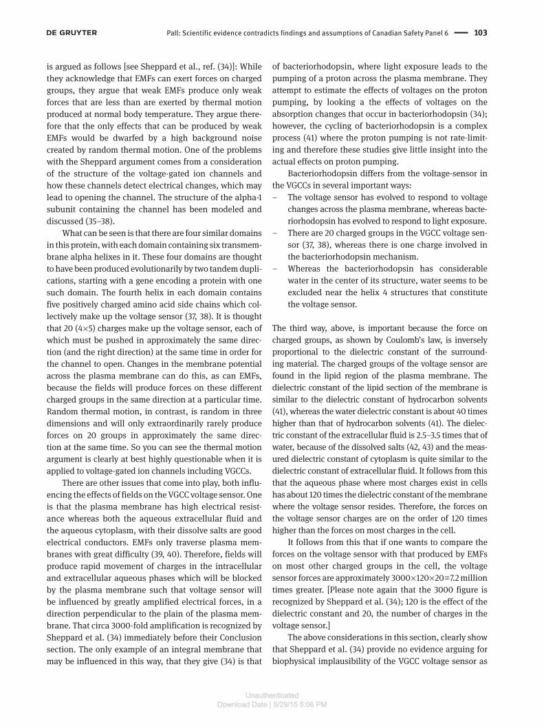
Pall: Scientific evidence contradicts findings and assumptions of Canadian Safety Panel 6 103
is argued as follows [see Sheppard et al., ref. (34)]: While they acknowledge that EMFs can exert forces on charged groups, they argue that weak EMFs produce only weak forces that are less than are exerted by thermal motion produced at normal body temperature. They argue there-fore that the only effects that can be produced by weak EMFs would be dwarfed by a high background noise created by random thermal motion. One of the problems with the Sheppard argument comes from a consideration of the structure of the voltage-gated ion channels and how these channels detect electrical changes, which may lead to opening the channel. The structure of the alpha-1 subunit containing the channel has been modeled and discussed (35–38).
What can be seen is that there are four similar domains in this protein, with each domain containing six transmem-brane alpha helixes in it. These four domains are thought to have been produced evolutionarily by two tandem dupli-cations, starting with a gene encoding a protein with one such domain. The fourth helix in each domain contains five positively charged amino acid side chains which col-lectively make up the voltage sensor (37, 38). It is thought that 20 (4 × 5) charges make up the voltage sensor, each of which must be pushed in approximately the same direc-tion (and the right direction) at the same time in order for the channel to open. Changes in the membrane potential across the plasma membrane can do this, as can EMFs, because the fields will produce forces on these different charged groups in the same direction at a particular time. Random thermal motion, in contrast, is random in three dimensions and will only extraordinarily rarely produce forces on 20 groups in approximately the same direc-tion at the same time. So you can see the thermal motion argument is clearly at best highly questionable when it is applied to voltage-gated ion channels including VGCCs.
There are other issues that come into play, both influ-encing the effects of fields on the VGCC voltage sensor. One is that the plasma membrane has high electrical resist-ance whereas both the aqueous extracellular fluid and the aqueous cytoplasm, with their dissolve salts are good electrical conductors. EMFs only traverse plasma mem-branes with great difficulty (39, 40). Therefore, fields will produce rapid movement of charges in the intracellular and extracellular aqueous phases which will be blocked by the plasma membrane such that voltage sensor will be influenced by greatly amplified electrical forces, in a direction perpendicular to the plain of the plasma mem-brane. That circa 3000-fold amplification is recognized by Sheppard et al. (34) immediately before their Conclusion section. The only example of an integral membrane that may be influenced in this way, that they give (34) is that
of bacteriorhodopsin, where light exposure leads to the pumping of a proton across the plasma membrane. They attempt to estimate the effects of voltages on the proton pumping, by looking a the effects of voltages on the absorption changes that occur in bacteriorhodopsin (34); however, the cycling of bacteriorhodopsin is a complex process (41) where the proton pumping is not rate-limit-ing and therefore these studies give little insight into the actual effects on proton pumping.
Bacteriorhodopsin differs from the voltage-sensor in the VGCCs in several important ways:
– The voltage sensor has evolved to respond to voltage changes across the plasma membrane, whereas bacte-riorhodopsin has evolved to respond to light exposure.
– There are 20 charged groups in the VGCC voltage sen-sor (37, 38), whereas there is one charge involved in the bacteriorhodopsin mechanism.
– Whereas the bacteriorhodopsin has considerable water in the center of its structure, water seems to be excluded near the helix 4 structures that constitute the voltage sensor.
The third way, above, is important because the force on charged groups, as shown by Coulomb’s law, is inversely proportional to the dielectric constant of the surround-ing material. The charged groups of the voltage sensor are found in the lipid region of the plasma membrane. The dielectric constant of the lipid section of the membrane is similar to the dielectric constant of hydrocarbon solvents (41), whereas the water dielectric constant is about 40 times higher than that of hydrocarbon solvents (41). The dielec-tric constant of the extracellular fluid is 2.5–3.5 times that of water, because of the dissolved salts (42, 43) and the meas-ured dielectric constant of cytoplasm is quite similar to the dielectric constant of extracellular fluid. It follows from this that the aqueous phase where most charges exist in cells has about 120 times the dielectric constant of the membrane where the voltage sensor resides. Therefore, the forces on the voltage sensor charges are on the order of 120 times higher than the forces on most charges in the cell.
It follows from this that if one wants to compare the forces on the voltage sensor with that produced by EMFs on most other charged groups in the cell, the voltage sensor forces are approximately 3000 × 120 × 20 = 7.2 million times greater. [Please note again that the 3000 figure is recognized by Sheppard et al. (34); 120 is the effect of the dielectric constant and 20, the number of charges in the voltage sensor.]
The above considerations in this section, clearly show that Sheppard et al. (34) provide no evidence arguing for biophysical implausibility of the VGCC voltage sensor as
!"#$%&'"%()#%'*+,-".,#*/+#%'/0/1234251/1678/9:

104 Pall: Scientific evidence contradicts findings and assumptions of Canadian Safety Panel 6
a target of low-intensity EMFs, such that when we have compelling empirical evidence that it is the main target, that evidence should be taken at face value. Furthermore, the VGCC voltage sensor is likely to be many orders of magnitude more sensitive to EMF effects than are any non-plasma membrane localized target. Because heating is pro-duced by the joggling of charged/partially charged groups almost all of which are outside the plasma membrane, the much greater forces on the VGCC voltage sensors show that fields 6–7 orders of magnitude lower than produce heating may activate the VGCC voltage sensors.
Have others been influenced by somewhat similar con-siderations? I believe it is likely that W.R. Adey was influ-enced by the plasma membrane properties when in the 1980s he proposed that a plasma membrane protein was the likely target of weak EMFs. Panagopoulos et al. (7, 8) may have been influenced by these plasma membrane and voltage sensor considerations when they decided to do biophysical modeling on voltage gated ion channels. The two reviewers of this paper each had some criticisms of the Panagopoulos et al. (7, 8) modeling, and some of the things in their papers go beyond my biophysics understanding, so I am unable to judge. What I would say is that the modeling studies came to three important predictions: That voltage-gated ion channels may be targets of low-intensity EMFs, that the VGCCs may be particularly activated because of the mechanism of the actual calcium flux through the channel and that pulsed fields may be more active than non-pulsed fields. Biophysical modeling of such complex membrane proteins as the voltage-gated ion channels is, at best a work in progress, given their complexity.
At this point, there is much evidence implicating VGCC activation but no apparent evidence implicat-ing other voltage-gated ion channels in low intensity EMF responses (1–3). Possible reasons for this should be assessed elsewhere.
What is most needed at this point is not more biophys-ical modeling, although that would be useful, but exten-sive detailed information on the effects of various fields on VGCC activation. Such information can be obtained via the types of studies advocated below for biologically-based safety standards.
Canadian Royal Society Expert Panel Report on radiofrequency fieldsThis Royal Society Expert Panel was charged with review-ing Safety Code 6 (2013) safety limits for exposure to radiofrequency (primarily microwave frequency) fields,
following the charge to “advance knowledge, encourage integrated interdisciplinary understanding and address issues that are critical to Canadians”. The Expert Panel Report (44) can be judged based on these charges and also the requirements that apply to authors of all purportedly scientific documents:
– The need to provide documentation that it has given as objective an assessment of the science as possible;
– The need for clarity of thought and clarity of expres-sion, such that it will be clear to the reader what the Report is trying to say;
– The need to provide the reader of the Report with sufficient information in the Report and in the cita-tions provided in the Report such that the reader can make an independent assessment of the quality of the science;
– And perhaps most importantly, the need to follow widely accepted principles for assessing scientific evidence.
This paper considers both the charges to the panel and these more generally applicable scientific principles to judge the scientific merit of the Report.
What is in the report?The Report is, in the author’s view, stronger on opinion than on evidence (44). Let us consider some specifics.
The Report states that “The Panel considered an ‘established adverse health effect’ as an adverse effect that is observed consistently in several studies with strong methodology. With this definition in mind, the Panel reviewed the evidence for a wide variety of negative health impacts from exposure to RF energy, including cancer, cognitive and neurologic effects, male and female repro-ductive effects, developmental effects, cardiac function and heart rate variability, electromagnetic hypersensitiv-ity, and adverse health effects in susceptible regions of the eye.” Despite this claim to have reviewed a broad array of biological impacts, in fact the Report does not provide a comprehensive review. Rather it engages, as documented below, in what can be referred to as “cherry-picking” – selecting studies consistent with its assumptions. More-over, it often ignores studies that are not consistent with its assumption that there are no biological effects except-ing those that, in their view, may be tied to heating. Thus the Report completely excludes many different studies on prenatally exposed animals and those on spermatogen-esis, on oxidative stress, changes of calcium fluxes and
!"#$%&'"%()#%'*+,-".,#*/+#%'/0/1234251/1678/9:

Pall: Scientific evidence contradicts findings and assumptions of Canadian Safety Panel 6 105
thousands of studies on therapeutic effects, all at non-thermal levels of exposure.
The Report uses the existence of what it calls “incon-sistent,” and others have called “conflicting” studies to argue that conflict per se indicates a lack of established health impact. This paper considers below whether there are any genuine “inconsistencies” in this literature. Henry Lai and Devra Davis have documented that “conflicting” scientific evidence in the field of bioelectromagnetics relating to mobile phones has been carefully cultivated (45), an inference that may also explain the data of Huss et al. (46). Huss et al. stated “We found that the studies funded exclusively by industry were indeed substantially less likely to report statistically significant effects on a range of end points that may be relevant to health. Our findings add to the existing evidence that single-source sponsorship is associated with outcomes that favor spon-sors’ products.” The panel ignores these findings and con-siders that conflicting evidence about effects of exposure to RF energy on cancer or other end points means that effects are possible but are not ‘established’ in accordance with its definition of ‘established health effects’. Simi-larly, while the Report notes that effects of exposure to RF energy on aspects of male reproductive function have been found, it concludes that “the evidence has not been established to indicate that these translate into fertility or health effects” even when such aspects are used clinically to assess male fertility.
The Panel reviewed “inconsistent” evidence about effects of exposure to RF energy on cancer, concluding that effects are possible but are not ‘established in accord-ance with its definition of ‘established health effects’. The Report states that the Panel’s conclusion on cancer is in agreement with a recent report from the International Agency for Research on Cancer (47). In fact, the Report’s characterization of the IARC (47) position does not agree with the IARC actual position. IARC states that “In the text, the Working Group provides comments on those findings that are of greatest relevance to the evaluation, e.g., risk in the overall exposed group, patterns of change in risk with increasing exposure (such as a monotonic increase in risk with increasing exposure), and changes in risk with duration of exposure or latency.” Furthermore, the Report ignores the fact that WHO considers microwave radiation to be a Class 2B carcinogen, and the Report also ignores the fact that four prominent reviews on this topic (48–51) all come to the conclusion that microwave exposures can cause cancer. It is apparent therefore that the Panel of Experts on Safety Code 6 has allowed its assumptions to greatly influence its assessment here, rather than provid-ing an objective assessment of the literature.
There are complexities here that the Expert Panel fails to consider. For example, oxidative stress produced by microwave EMF exposure is likely to have a role in causation of cancer. For decades, it has been established that low level oxidative stress can lower oxidative stress markers below initial, pre-stress levels and protect the body from subsequent higher level oxidative stress, a phenomenon known as hormesis that has been recently shown to act by raising the activity of a transcriptional regulator, Nrf2; it has been suggested that this may explain some observations that low level cell phone use may lower cancer incidence via this mechanism, whereas higher level, long-term cell phone use may produce major elevation of cancer incidence. However, the Expert Panel apparently considers these studies to be conflicting, when to the contrary, these studies may raise the issue of biological complexity and a possible U- or J-shaped dose-response curve.
Another even clearer example where inferences of “inconsistencies” or “conflicts” in the literature have been misconstrued regarding the induction of single strand breaks in cellular DNA, measured by what are known as alkaline comet assays, a well-documented method for such studies (1). This literature was reviewed by the author (1), who found 19 different studies where greatly elevated levels of such single strand breaks were found following exposure as well as eight studies where they were not found. However, in examining these studies in detail, it is clear that the differences can be easily explained. For instance, regarding in vitro studies of DNA damage, some of the studies have used different cell types and studied different microwave source EMFs. Thus adult lymphocytes appear relatively resistant to EMF, while neural stem cells are much more susceptible. Different cell types differ from one another in how many and what types of VGCCs may be present and they may differ as well in how the VGCCs are regulated and so may be expected to differ widely in terms of response. All of these studies were done using exposures that were well within current safety standards. Consequently, each of these 19 positive findings contra-dict the assumptions behind the current safety standards, assumptions that are being defended by the Expert Panel Report, but the Report ignores all of these studies. More-over, in two of the 19 positive studies, results were posi-tive in some cell types but not others (1), clearly showing that in measurements using identical methodologies, the properties of the cells being studied are critical in deter-mining the biological response found.
Thus the Panel has failed to take into account impor-tant nuances regarding scientific research in this field. It has limited considerations to what the Panel calls
!"#$%&'"%()#%'*+,-".,#*/+#%'/0/1234251/1678/9:

106 Pall: Scientific evidence contradicts findings and assumptions of Canadian Safety Panel 6
“established health effects” defined in terms of con-sistent responses of various cell and tissue types (44). Where apparent conflict exists, the Panel uses its exist-ence as proof that an effect is not established. In doing so, the Panel fails to take into account scientific details that account for many “inconsistent” results. Such details are likely to include, in addition to the factors discussed above in this section, such factors as the role of different pulsation patterns in different types of exposures, the presence of “window effects” providing very complex dose-response relationships and the role of field fre-quencies in determining biological response. In effect, the panel dismisses science that does not comport with their underlying assumptions that only thermal effects are relevant.
Genotoxicity of non-thermal microwave exposures: examples of inconsistency?This inconsistency issue is central to the Report’s consid-eration of genotoxicity of non-thermal microwave expo-sures. This is one of the two areas (pp. 80–82) where the Report cites substantial numbers of primary citations (22 in this case). It lists 13 citations where studies found genotoxicity following exposure levels, well within safety standards. It also lists nine citations where the Report states that no genotoxic effect was found. The Report only cites a small fraction of the overall literature on geno-toxicity. For example, it only cites one of the 19 studies reviewed earlier by the author (1) on induction of single strand DNA breaks in microwave frequency exposed cells [that of Kesari et al. (52)]. In overall outline, the lit-erature cited in the Report on this topic reflects fairly well this overall much larger literature. There are, however, a number of ways in which the Report is problematic in dealing with this subject. The author has looked up all 22 of these studies to determine from the original papers what the original authors stated.
Scientists often look at genotoxicity because of its importance in carcinogenesis and this section of the Report is part of a larger section on carcinogenesis. However, the Panel of Experts nowhere considers that many of the authors of these studies discuss their own work as strengthening the case that such fields are car-cinogenic. A second connection, to male infertility, is also hidden in the report. Two of the positive studies (53, 54) are falsely stated in the Report as being on blood formation but what was actually being studied
in both of these studies was testicular sperm formation. The positive study Liu et al. (55) which shows genotox-icity in a spermatocyte cell line may also have implica-tions regarding male infertility, because of the cell type being studied. There is also a connection with male infertility of one of the negative studies (56). This study of effects of mobile phones, found no genotoxic effects on human sperm, but the same group published two earlier studies showing that other EMFs had substan-tial effects that suggested lowered fertility as a conse-quence of exposure. The Report cited the Falzone et al. (56) study but not the two earlier studies. Perhaps this is an overreaction, but the Report seems to be hiding studies providing substantial support for the view that these EMFs can substantially impact male fertility and also hiding the implications of many of these studies on carcinogenesis.
There are other aspects of this section that are prob-lematic. The Report listed the Franzellitti et al. (57) study as a negative one but it is not; it reports increased single strand DNA breaks as measured by alkaline comet assays following exposure. The Report accurately lists the Bourthoumieu et al. (58) study as being negative, but that study cites other studies by the same research group using other cell types as being positive; these positive studies are not cited or discussed in the Report. Similarly, the Report correctly lists two studies by Zeni, Sannino and their colleagues as being negative for apparent geno-toxicity; however, this same research group published 6 additional studies, with three showing positive effects, depending on the cell type being studied. The Xu et al. (59) study found genotoxicity in two cell types but not in four other cell types. These studies clearly show that dif-ferent types of cells respond differently to low level micro-wave exposures, but for some reason, the Panel of Experts seems unable to draw this very important conclusion. The cell type differences are discussed above in relation to the role of VGCCs in producing single strand breaks in cellu-lar DNA (1). Another problematic aspect of this part of the Report, is that it lists seven of the 13 positive studies as studies providing evidence for “genotoxic or epigenetic” changes but none of those seven have anything to do with epigenetics.
We have here 13 (14 actually when the Franzellitti study is added) studies each of which provide clear evidence for genotoxic activity of non-thermal microwave fields and each of which therefore falsify the heating/thermal hypothesis underlying the Report and also falsify current safety standards. Therefore, based on widely accepted sci-entific standards, the heating/thermal hypothesis and the safety standards should be rejected.
!"#$%&'"%()#%'*+,-".,#*/+#%'/0/1234251/1678/9:

Pall: Scientific evidence contradicts findings and assumptions of Canadian Safety Panel 6 107
What conclusion does the Panel draw? It concludes that “Extensive in vitro studies have generated inconsist-ent evidence that RF energy has genotoxic or epigenetic potential”. There is, however, no inconsistent evidence whatsoever. When one studies different cell types, differ-ent fields with different pulsation patterns, and different end points, even an elementary understanding of biology argues that different results are likely to be obtained. This section of the Report makes very clear on what basis the Panel is inferring “inconsistency”. The authors of the Report are simply looking at superficial similarities of studies and falsely inferring that differences should be interpreted as “inconsistencies” or “conflicts”, when they are not inconsistent or conflicting at all. The only type of studies that can produce clear evidence of inconsist-ency are identical studies that produce different results. Neither the Report nor, to my knowledge, its predecessors have provided any examples of such identical studies. Because this inconsistency argument underlies so much of the Report, one can see that this argument and the Report and also the current safety standards are each deeply flawed.
Karl popper and how to assess scientific evidenceWhat is the responsibility of the Expert Panel as a group of scientists attempting to produce a scientifically defen-sible Report? Probably the most influential work on this topic comes from the famous philosopher of science Karl Popper. In his work, Conjectures and refutations, Popper argues that scientific hypotheses cannot be proven, but they can be falsified (60). Thus science is to be regarded as tentative information that can always be advanced through further research. Falsifying information, informa-tion that apparently falsifies a theory, is the most impor-tant type of scientific information and needs therefore to be considered very carefully. The next more important type of evidence is what he calls “risky predictions” where one makes a prediction based on a hypothesis, a predic-tion that is not likely to be made based on any other unre-lated hypothesis. Confirmation of such a risky prediction provides substantial support whereas lack of confirma-tion can again lead to falsifying the hypothesis. Finally, there are confirmatory evidence studies where multiple hypotheses may explain any confirmation and conse-quently such confirmation is of low scientific significance.
When considered against the Popperian frame-work, all of the evidence supporting the heating/thermal
hypothesis, favored by the Expert Panel (44) is of the third type. It is widely established therefore that a scientific assessment of this area needs to consider in detail each apparently falsifying study and unless each of them can clearly be shown to be deeply flawed, the inference that should be drawn is that the heating hypothesis should be rejected. This rejection is the one aspect of this that may need to be modified in biology, given the inherent com-plexity of biology. It is possible that rather than rejection, the hypothesis needs instead to be modified in such a way that the information no longer falsifies the new hypothe-sis. However, in this situation where perhaps thousands of such modifications may be needed because of thousands of apparent falsifying studies, the difference in practice from outright falsification by each study may be trivial. It is clear, in any case that the Expert Panel has completely avoided doing its scientific duty here, failing to assess each of the thousands of apparent falsifying studies, and opting instead, as seen above, to make specious argu-ments. That is tragic, in my view, failing to protect the health of many Canadians, and indeed others around the world.
Some other aspectsMost of the Report is focused on their heating/thermal interpretation of microwave radiofrequency effects (44). That is, perhaps, not surprising. What is however very surprising, is that having made such a fetish out of the “inconsistencies” in dealing with various topics, nowhere does the Expert Panel consider in this very large section of the Report, the thousands of findings that clearly conflict with their own favorite hypothesis. What sections of data should be thrown out that may be relevant to this section? The Panel of Experts seem to be completely oblivious that if in its view “inconsistencies” are sufficient to throw out many studies in one area, it should have at least a little consistency in dealing with “inconsistencies” in the heart of their own Report.
In the first paragraph in the conclusion section, the Panel of Experts state that (44) “No viable biophysical mechanism has been proposed for carcinogenic effects for exposure below the levels of SC6 that are supported by results in experimental systems,” citing three earlier studies but neglecting to consider the VGCC mechanism of microwave EMF action. The VGCC mechanism is clearly a viable biophysical mechanism, because of the properties of the voltage sensor located in the plasma membrane. VGCC activation produces downstream effects including [Ca2+] i elevation, NO elevation and peroxynitrite/oxidative stress/
!"#$%&'"%()#%'*+,-".,#*/+#%'/0/1234251/1678/9:
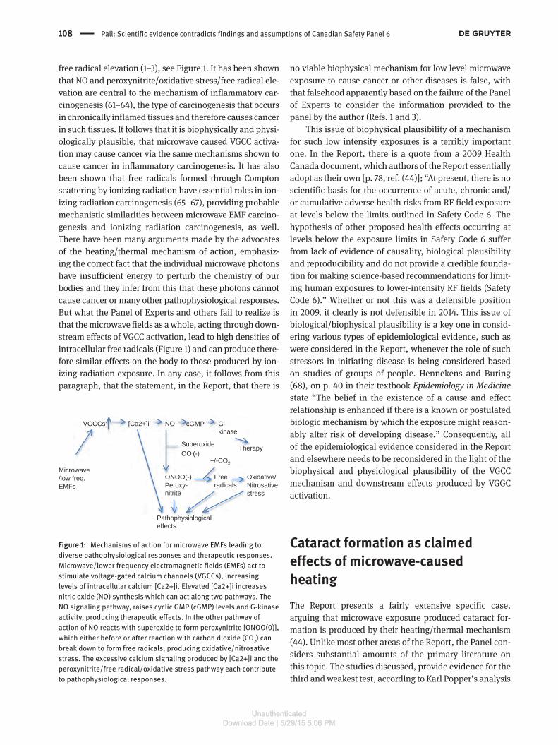
108 Pall: Scientific evidence contradicts findings and assumptions of Canadian Safety Panel 6
free radical elevation (1–3), see Figure 1. It has been shown that NO and peroxynitrite/oxidative stress/free radical ele-vation are central to the mechanism of inflammatory car-cinogenesis (61–64), the type of carcinogenesis that occurs in chronically inflamed tissues and therefore causes cancer in such tissues. It follows that it is biophysically and physi-ologically plausible, that microwave caused VGCC activa-tion may cause cancer via the same mechanisms shown to cause cancer in inflammatory carcinogenesis. It has also been shown that free radicals formed through Compton scattering by ionizing radiation have essential roles in ion-izing radiation carcinogenesis (65–67), providing probable mechanistic similarities between microwave EMF carcino-genesis and ionizing radiation carcinogenesis, as well. There have been many arguments made by the advocates of the heating/thermal mechanism of action, emphasiz-ing the correct fact that the individual microwave photons have insufficient energy to perturb the chemistry of our bodies and they infer from this that these photons cannot cause cancer or many other pathophysiological responses. But what the Panel of Experts and others fail to realize is that the microwave fields as a whole, acting through down-stream effects of VGCC activation, lead to high densities of intracellular free radicals (Figure 1) and can produce there-fore similar effects on the body to those produced by ion-izing radiation exposure. In any case, it follows from this paragraph, that the statement, in the Report, that there is
no viable biophysical mechanism for low level microwave exposure to cause cancer or other diseases is false, with that falsehood apparently based on the failure of the Panel of Experts to consider the information provided to the panel by the author (Refs. 1 and 3).
This issue of biophysical plausibility of a mechanism for such low intensity exposures is a terribly important one. In the Report, there is a quote from a 2009 Health Canada document, which authors of the Report essentially adopt as their own [p. 78, ref. (44)]; “At present, there is no scientific basis for the occurrence of acute, chronic and/or cumulative adverse health risks from RF field exposure at levels below the limits outlined in Safety Code 6. The hypothesis of other proposed health effects occurring at levels below the exposure limits in Safety Code 6 suffer from lack of evidence of causality, biological plausibility and reproducibility and do not provide a credible founda-tion for making science-based recommendations for limit-ing human exposures to lower-intensity RF fields (Safety Code 6).” Whether or not this was a defensible position in 2009, it clearly is not defensible in 2014. This issue of biological/biophysical plausibility is a key one in consid-ering various types of epidemiological evidence, such as were considered in the Report, whenever the role of such stressors in initiating disease is being considered based on studies of groups of people. Hennekens and Buring (68), on p. 40 in their textbook Epidemiology in Medicine state “The belief in the existence of a cause and effect relationship is enhanced if there is a known or postulated biologic mechanism by which the exposure might reason-ably alter risk of developing disease.” Consequently, all of the epidemiological evidence considered in the Report and elsewhere needs to be reconsidered in the light of the biophysical and physiological plausibility of the VGCC mechanism and downstream effects produced by VGGC activation.
Cataract formation as claimed effects of microwave-caused heatingThe Report presents a fairly extensive specific case, arguing that microwave exposure produced cataract for-mation is produced by their heating/thermal mechanism (44). Unlike most other areas of the Report, the Panel con-siders substantial amounts of the primary literature on this topic. The studies discussed, provide evidence for the third and weakest test, according to Karl Popper’s analysis
VGCCs [Ca2+]i NO
Microwave/low freq.EMFs
cGMP
Therapy
G-kinase
Pathophysiologicaleffects
SuperoxideOO.(-)
ONOO(-)Peroxy-nitrite
Freeradicals
+/-CO2
Oxidative/Nitrosativestress
Figure 1: Mechanisms of action for microwave EMFs leading to diverse pathophysiological responses and therapeutic responses.Microwave/lower frequency electromagnetic fields (EMFs) act to stimulate voltage-gated calcium channels (VGCCs), increasing levels of intracellular calcium [Ca2+]i. Elevated [Ca2+]i increases nitric oxide (NO) synthesis which can act along two pathways. The NO signaling pathway, raises cyclic GMP (cGMP) levels and G-kinase activity, producing therapeutic effects. In the other pathway of action of NO reacts with superoxide to form peroxynitrite [ONOO(0)], which either before or after reaction with carbon dioxide (CO2) can break down to form free radicals, producing oxidative/nitrosative stress. The excessive calcium signaling produced by [Ca2+]i and the peroxynitrite/free radical/oxidative stress pathway each contribute to pathophysiological responses.
!"#$%&'"%()#%'*+,-".,#*/+#%'/0/1234251/1678/9:

Pall: Scientific evidence contradicts findings and assumptions of Canadian Safety Panel 6 109
(60), namely that the exposures studied are mostly within the range that produce substantial tissue heating and may therefore produce both cataracts and lens opacifi-cation via heating. This type of evidence is considered to be the weakest of the three types of evidence in Popper’s schema, because alternative mechanisms are not in any way ruled out.
What is interesting is that there are three published studies which argue strongly against a heating mecha-nism for cataract formation by microwave exposures. One of these, a study by Cleary and Mills (69), showed that in comparison with other treatments raising lens tempera-tures, microwave radiation “appears to exert a unique component of thermal stress in the induction of opacifi-cation in the mammalian lens,” arguing against a strictly thermal mechanism. Two studies have been published testing in effect the “risky prediction” that microwave-induced cataracts are produced by heating. One of these showed that neither eye-localized or whole-body hyper-thermia to 42o produced any cataract-like opacity in the rabbit (70). The other showed that localized eye heating in the rabbit, producing the same temperature for the same duration as cataractogenic microwave exposures, produced no opacity in the rabbit eye (71). Both of these “risky predictions” failed to confirm the prediction and strongly suggest falsification of the hypothesis that micro-wave-induced cataracts are produced through heating. What is particularly disturbing about the Report is that it fails to cite any of these three studies (44) despite the fact that each of them has been cited by others in this context, according to the Google Scholar database. Clearly, the lit-erature the Expert Panel cites regarding cataract forma-tion, which includes the second most extensive primary literature in the Report, does not provide an objective assessment of the scientific literature in this area.
In contrast to studies discussed in the previous par-agraph, the equally “risky prediction” that VGCCs and excessive [Ca2+]i have roles in such cataract formation have produced validation of the hypothesis that micro-wave-induced VGCC activation causes cataracts. Walsh and Patterson (72) demonstrated that elevated [Ca2+]i in the lens of the frog eye has a central role in cataract forma-tion and that calcium channel blockers, which of course block VGCC activation, can block cataract formation. In a recent review, it was shown that excessive [Ca2+]i in the lens of the human and mammalian eye plays a major role in the opacification process producing cataracts and that VGCCs can have a substantial role in this process (73). While these studies do not directly relate to microwave exposures, they clearly show that excessive [Ca2+]i in the lens of the eye has essential roles in cataract formation
and that excessive VGCC activity causes cataract forma-tion in experimental animals. Much of the action of [Ca2+]i in cataract formation has been shown to occur through the action of several calcium receptors that act indepen-dently of NO. However, there is also an established role of oxidative stress in cataract formation, and it is thought that peroxynitrite also has a role because of the elevation of a marker for peroxynitrite, 3-nitrotyrosine in cataracts (74). It is likely therefore that microwaves act to produce cataracts via calcium signaling as well as via downstream effects involving peroxynitrite and oxidative stress (see Figure 1). The difference in confirmation of these “risky predictions” clearly shows that the VGCC/[Ca2+]i role in producing cataracts is far better documented than any possible heating role.
It can be seen from the above, that although the Cana-dian Panel of Experts seems to argue that cataract forma-tion is the strongest example of a strictly thermal EMF response (44), the case for such a thermal mechanism is to the contrary extremely weak. Their case is totally depend-ent on ignoring both evidence that falsifies their view and also evidence that confirms “risky predictions” of the VGCC mechanism that is ignoring the two strongest types of evidence. Thus the claimed role for heating being the cause of cataract formation following microwave expo-sure, advocated by the Expert Panel, has now been appar-ently debunked.
Summary of the reportIn summary, then each of the following failures in the Report can be seen to be important in our rejecting its conclusions:
– It fails to individually assess the thousands of studies that provide evidence apparently falsifying their heat-ing/thermal paradigm. By failing to assess studies con-taining this most important type of evidence, as shown by Popper (60), this failure provides more than suffi-cient reason to reject the conclusions of the Report.
– The Report fails to provide any “risky prediction” type evidence (the second most important type of evi-dence) in favor of the heating/thermal hypothesis, but such risky predictions are available supporting the VGCC mechanism of action.
– The Report bases its conclusion on the weakest type of evidence, evidence that some responses could be gen-erated by heating but does not rule out other types of mechanisms. A close examination of what the Expert Panel considers to be the strongest case for heating,
!"#$%&'"%()#%'*+,-".,#*/+#%'/0/1234251/1678/9:

110 Pall: Scientific evidence contradicts findings and assumptions of Canadian Safety Panel 6
that of cataract formation, shows that this is another example of a probable VGCC mechanism, not heating.
– The Report repeatedly fails to provide an objective assessment of the scientific literature. Because omit-ted citations consistently have the effect of weakening their position, it seems unlikely that these omissions are just coincidental.
– The Report claims that there is no biophysically viable alternative to the heating/thermal paradigm, a claim clearly shown here to be false.
– The Report claims extensive inconsistencies (what others have called conflicts) occur in the literature, where what it considers “similar” studies produced different results and it uses these claims of “inconsist-encies” to throw out large amounts of the literature. However, these “similar” studies are in fact, dissimi-lar, differing in cell type being studied, the properties of the fields being studied and/or the end point being studied, with each of these having demonstrated roles in determining outcome. It follows that the Report provides no evidence for any such “inconsistencies.” Any claims of such “inconsistencies” are at best undocumented.
– The Report fails to use its own inconsistency argu-ment (6 above) in the heart of the report, the part that argues for a heating/thermal mechanism, thus failing to be consistent in its own treatment of this issue.
– The Report fails to give the reader enough informa-tion in the Report itself or in the citations provided to allow the reader to assess its scientific merit.
The author is aware that similar flaws to those described immediately above occur in earlier studies arguing for the heating/thermal/SARs mechanism (9–13). But that only emphasizes the fact that this whole point of view has been on extraordinarily weak ground all along. That makes it crucially important that safety standards on which the health of most Canadians and indeed, most people around the world are dependent, be examined in scien-tifically defensible ways.
It is perhaps surprising that the case developed by the Panel of Experts is so weak. That is especially so because industry-funded research has been skewed in support of the heating/thermal interpretation (45, 46), so one would think that with a lot of industry-supported research, the Expert Panel would have come up with some stronger evidence.
Let me say that it is my opinion that the Panel of Experts may not have been corrupted by industry influ-ence, but rather it may have fallen victim to a common affliction, that of groupthink. Groups of people each
carrying misconceptions in common, act to encour-age their common misconceptions in other members of the group. What was apparently lacking in the Panel of Experts was someone who could challenge those mis-conceptions, rather than encourage them. However the “logic” presented in the Report provides industry with a strategy to indefinitely prevent any true scientific stand-ards from being used to assess safety. Industry need only fund research that ends up making “inconsistent” con-clusions, thus allowing all independently funded studies to be thrown out because of these “inconsistencies” and thus indefinitely preventing adoption of safety standards based on genuine, independent science. It is my hope and expectation that this was not the goal of the Expert Panel, but it is nevertheless an apparent consequence of their Report, if it is viewed as being scientific.
Still, it can be argued, that the Panel of Experts has perhaps unwittingly fulfilled a very valuable function. By clearly showing how weak their case is in 2014, the Panel has shown that none of the more recent evidence has substantially strengthened their case. It is still based on a false premise (biophysical implausibility of alternative mechanisms) and circular reasoning, it is still based on the failure to consider large numbers of apparent falsify-ing studies, it is still based on ignoring large amounts of the relevant literature and it is still based on the failure to provide the most well supported types of evidence needed to establish biological mechanisms in medicine, just as was true earlier (9–13). Of course, the weakness of the Panel’s case means that the current safety standards are based on quicksand.
How VGCC activation by microwave/RF exposure can produce a variety of important biological responsesTable 1 summarizes how VGCC activation may plausibly produce a wide range of reported responses to microwave and, in some cases, lower frequency EMF exposures. It can be seen that a wide range of reported responses to low level microwave exposures can apparently all be understood as being a consequence of VGCC activation and downstream effects of such activation that were outlined in Figure 1. These can all be seen as “risky predictions” of the VGCC activation mechanism produced by EMF exposures. While these mechanisms support the inference that all of these effects seem to be produced by VGCC activation, that inference must be viewed as being surprising. After all,
!"#$%&'"%()#%'*+,-".,#*/+#%'/0/1234251/1678/9:

Pall: Scientific evidence contradicts findings and assumptions of Canadian Safety Panel 6 111
Table 1: Apparent mechanisms of action for microwave exposures producing diverse biological effects (see Figure 1).
Reported biologic response
Apparent mechanism(s) Citation(s)/Comments
Oxidative stress Peroxynitrite and consequent free radical formation (1–3); detected via a large number of oxidative stress markers
Single strand breaks in cellular DNA
Free radical attack on DNA (1, 3)
Double strand breaks in cellular DNA
Same as above Same as above; detected from micronuclei and other chromosomal changes
Cancer Single and double strand breaks, 8-nitroguanine and other pro-mutagenic changes in cellular DNA; produced by elevated NO, peroxynitrite
This paper and (3)
Breakdown of blood-brain barrier
Peroxynitrite activation of matrix metalloproteinases leading to proteolysis of tight junction proteins
(3)
Male and female infertility Induction of double strand DNA breaks; other oxidative stress mechanisms; [Ca2+]i mitochondrial effects causing apoptosis; in males, breakdown of blood-testis barrier
(3)
Therapeutic effects Increases in [Ca]i and NO/NO signaling (1–3; 13)Depression; diverse neuropsychiatric symptoms
VGCC activation of neurotransmitter release; other effects? possible role of excess epinephrine/norepinephrine (75)
These were reported in occupational exposures (22); also reported in people living near cell phone towers
Melatonin depletion; sleep disruption
VGCCs, elevated [Ca]i leading to disruption of circadian rhythm entrainment as well as melatonin synthesis
(3)
Cataract formation VGCC activation and [Ca]i elevation; calcium signaling and also peroxynitrite/oxidative stress
This paper
Tachycardia, arrhythmia, sometimes leading to sudden cardiac death
Very high VGCC activities found in cardiac (sinoatrial node) pacemaker cells; excessive VGCC activity and [Ca2+]i levels produces these electrical changes in the heart
(3)
although low level EMF activation of VGCCs is now well-documented, other possible direct targets of EMFs cannot be ruled out, targets that may produce changes that cannot be easily explained as being caused by VGCC acti-vation and downstream effects of such activation. When the apparent mechanisms summarized in Table 1 are put together with the calcium channel blocker studies and other studies on widespread changes in calcium fluxes and calcium signaling following microwave EMF expo-sures, we are left without any alternative, non-VGCC target of EMF action that currently can be studied for its role in producing biological effects in humans.
Biologically-based EMF safety standardsHardell and Sage (76), the Scientific Panel on Electromag-netic Health Risks (77) and the author (3) have called for biologically-based EMF safety standards that are based on genuine biologically relevant responses to low-level microwave and other EMFs, rather than SARs. The only approaches we have available for this based on a known
biological end point, as shown in the previous section, are approaches based on VGCC activation. There are experi-mental whole animal approaches based on VGCC activa-tion (3), but my feeling is that initial studies should focus on using cells in culture, cells that have high levels of some VGCCs. Some such studies would use cell lines with such high VGCC levels, such as neuroblastoma cell lines or perhaps cell lines derived from endocrine cells with relatively high VGCC levels. Among these cell lines should be the neuroblastoma cell lines previously studied by Dutta et al. (78) and shown to produce changes in calcium fluxes in response to very low level EMF exposures. PC12 cells, a commonly used chromaffin cell line should also be considered for such studies. In addition, it may useful to use cardiac pacemaker cells which have very high activi-ties of VGCCs (35) and can be derived from stem cells (79).
Two approaches suggest themselves for measuring responses of such cells to EMF exposure: Cells in culture could be monitored for NO production using an NO elec-trode in the gas phase over the culture, both before and following EMF exposure. This approach was used by Pilla in studying effects of pulsed microwave fields (4) in trying to understand the mechanism of microwave therapy. Pilla found that the NO increase in such cultures on EMF field
!"#$%&'"%()#%'*+,-".,#*/+#%'/0/1234251/1678/9:

112 Pall: Scientific evidence contradicts findings and assumptions of Canadian Safety Panel 6
exposure was almost instantaneous, using a NO electrode in the gas phase (4). With this sort of approach, many dif-ferent fields can be quickly and easily studied for their ability to produce NO increases, including different fre-quencies, pulsation patterns and possibly intensities, with the last of these needed to analyze window effects. Different cordless communication devices can be com-pared for activity using several cell types. Continuous measurements from an NO electrode can be recorded and easily quantified, allowing accumulation of very large amounts of data in very short time periods. Therefore, issues such as reproducibility should be quickly resolved. One might even be able to determine whether previous exposures produce increased sensitivity to exposure, pos-sibly developing a cell culture model of electromagnetic hypersensitivity.
Another approach to such studies involves using calcium-sensitive fluorescent probes that concentrate into the cytoplasm of cells, allowing assessments of [Ca]i levels with a fluorescence microscope. This may allow one of obtain information of different types than described in the previous paragraph. One can get information on heterogeneity of responses at the cellular level and also how raised [Ca]i levels may propagate over time from one part of the cell to another. However, a limitation to this approach may occur if the fields generated by the micro-scope perturb the [Ca]i levels and cannot be well shielded using a small Faraday cage that does not cage exposures that are to be studied. It is also true that the NO electrode studies are easier to quantify than such fluorescent probe studies. So these two approaches are distinct from one another and whether they will complement each other as they develop is uncertain. It is my view that both of these should be investigated if only to explore their strong points and weak points but that the NO electrode approach may be a very good place to start because it has already been used to assess EMF effects (4) and because it allows easy quantification.
Brief overviewHavas’ recent review (80) discusses 14 different docu-ments prepared by international scientists (dated 2002 through 2012) expressing deep concern about various non-thermal effects of microwave radiation exposures and other studies have expressed similar views. W.R. Adey’s papers (6, 21) reviewed much of the then current evidence for many non-thermal effects of microwave radiation. But his prescience is most clearly shown by his statement that
“Collective evidence points to cell membrane receptors as the probable site of first tissue interactions with both extremely low frequency and microwave fields for many neurotransmitters, hormones, growth-regulating enzyme expression, and cancer-promoting chemicals. In none of these studies does tissue heating appear to be involved causally in the responses” [italics added, from a talk at the Royal Society of Physicians, London May 16–17, 2002, quoted in ref. (81)]. The recent Herbert and Sage review (81) discusses “the emergence of ever larger bodies of evidence supporting a large array of non-thermal but pro-found pathophysiological impacts of EMF/RFR in trans-forming our understanding of the nature of EMF/RFR impacts on the organism.” In a second paper (82), Herbert and Sage state that “Our EMF/RFR standards are also based on an outdated assumption that it is only heating (thermal injury) which can do harm. These thermal safety limits do not address low-intensity (non-thermal) effects. The evidence is now overwhelming that limiting exposure to those causing thermal injury alone does not address the much broader array of risks and harm now clearly evident with chronic exposure to low-intensity (non-ther-mal) effects.” The Khurana et al. review (83) states: “The authors reviewed more than 2000 scientific studies and reviews, and have concluded that: (1) the existing public safety limits are inadequate to protect public health; (2) from a public health policy standpoint, new public safety limits on further deployment of risky technologies are warranted based on the total weight of evidence. A pre-cautionary limit of 1 mW/m2 was suggested ….” The Sci-entific Panel on Electromagnetic Field Health Risks listed four well-documented central conclusions at the begin-ning of their publication (77):
– Low-intensity (non-thermal) bioeffects and adverse health effects are demonstrated at levels significantly below existing exposure standards.
– ICNIRP and IEEE/FCC public safety limits are inad-equate and obsolete with respect to prolonged, low-intensity exposures.
– New biologically-based public exposure standards are urgently needed to protect public health worldwide.
– It is not in the public interest to wait.
Canadian Panel of Experts do not cite these papers or others providing clear and focused views that contradict the views advocated in the Report, showing again that the Report fails to provide an objective assessment of the scientific literature. The current paper adds a number of specific considerations to the needed debate:
– VGCC activation produces most, possibly even all microwave and lower frequency EMF health-related
!"#$%&'"%()#%'*+,-".,#*/+#%'/0/1234251/1678/9:

Pall: Scientific evidence contradicts findings and assumptions of Canadian Safety Panel 6 113
responses. Each of the studies on VGCC activation or on changes in calcium fluxes and signaling following low level exposure clearly falsifies the thermal/heat-ing paradigm.
– This VGCC activation mechanism by low level micro-wave and lower frequency fields, rather than individ-ual photons, is biophysically plausible based on the special properties of the voltage sensor and its locali-zation to lipid region of the plasma membrane.
– Downstream effects of VGCC activation (Figure 1) can generate each of 13 different health effects repeat-edly found to be produced by microwave exposure (Table 1).
– Studies document roles of pulsation in influencing biological responses to microwave exposures, influ-ences that are incompatible with these being produced by heating.
– “Window” effects occur, where specific intensities of microwave EMF exposure produce higher biological effects than those produced by both lower and higher intensities, observations incompatible with heating effects.
– Thousands of studies have reported biological effects at intensities well within safety standards, each of which appear to falsify the heating/thermal para-digm, none of which have been considered in this light by the Panel of Experts, despite the scientific requirement to do so under well-accepted scientific principles.
– The claims in the Report that microwave induction of cataracts is produced by heating has been tested in three studies, each contradicting this claim; two of them produce clear falsification, but none of these three studies are cited in the Report. Because VGCC activation can cause cataracts and elevated [Ca2+]i has essential roles in producing cataracts, a VGCC mechanism for microwave-induced cataracts is much more strongly supported than is the claimed heating mechanism.
– The claim in the Report of widespread “inconsistency” in the literature is tested here through examination of the literature cited on genotoxic effects. No inconsist-encies were found in this literature despite the Report claiming such. Furthermore, no identical studies are cited anywhere in the Report showing inconsistency of results, these being the only types of studies that can clearly show inconsistency. Claims of widespread “inconsistency” or “conflict” in the literature must be viewed as, at best, undocumented.
– Each of the 8 considerations listed immediately above clearly show that the Report fails to provide anything
resembling an objective assessment of the evidence on biological effects of microwave EMF exposures and pro-vides therefore no scientifically valid support for Safety Code 6, ICNIRP or other current safety standards.
– Development of biologically-based safety standards has been called for and approaches to using cell culture-based tests that may be used to develop such safety standards are discussed.
It has been clear for a long time that the heating paradigm is indefensible and that a new paradigm is much needed. We now have that with VGCC activation, and while VGCC activation may not be the entire story behind the biologi-cal actions of such EMFs in humans and other mammals, it clearly is most of the story. It is time therefore for a para-digm shift away from strictly thermal effects and toward a central role for VGCC activation in the cellular response to microwave and lower frequency EMFs.
Acknowledgments: I wish to thank Dr. Devra Lee Davis for her many helpful suggestions. Any remaining errors in this are, of course, my own. This paper is dedicated to the memory of W. Ross Adey (1922–2004) whose studies in this area were impressively prescient – someone I regret never meeting.
References1. Pall ML. Electromagnetic fields act via activation of voltage-gated
calcium channels to produce beneficial or adverse effects. J Cell Mol Med 2013;17:958–65.
2. Pall ML. Electromagnetic field activation of voltage-gated calcium channels: role in therapeutic effects. Electromagn Biol Med 2014;33:251.
3. Pall ML. Microwave electromagnetic fields act by activating volt-age-gated calcium channels: why the current international safety standards do not predict biological hazard. Recent Res Devel Cell Biol 2014;7: 0-00 ISBN: 978-81-308-0000-0 Available at (http://wirelesseducationaction.org/wp-content/uploads/2014/11/microw-vgccnoheat.pdf).
4. Pilla AA. Electromagnetic fields instantaneously modulate nitric oxide signaling in challenged biological systems. Biochem Bio-phys Res Commun 2012;426:330–3.
5. Walleczek J. Electromagnetic field effects on cells of the immune system: the role of calcium signaling. FASEB J 1992;6:3177–85.
6. Adey WR. Biological effects of electromagnetic fields. J Cell Bio-chem 1993;51:410–6.
7. Panagopoulos DJ, Messini N, Karabarbounis A, Philippetis AL, Margaritis LH. A mechanism for action of oscillating electric fields on cells. Biochem Biophys Res Commun 2000;272:634–40.
8. Panagopoulos DJ, Karabarbounis A, Margaritis LH. Mechanism for action of electromagnetic fields on cells. Biochem Biophys Res Commun 2002;298:95–102.
!"#$%&'"%()#%'*+,-".,#*/+#%'/0/1234251/1678/9:

114 Pall: Scientific evidence contradicts findings and assumptions of Canadian Safety Panel 6
9. Osepchuk JM, Petersen RC. Historical review of RF exposure stand-ards and the International Committee on Electromagnetic Safety (ICES). Bioelectromagnetics Supplement 2003;Suppl 6:S7–16.
10. Osepchuk JM, Petersen RC. Safety standards for exposure to RF electromagnetic fields. Microwave Magazine IEEE 2001;2:57–69.
11. D’Andrea JA, Ziriax JM, Adair ER. Neurobiology of hyperthermia. Prog Brain Res 2007;162:107–35.
12. Tripathy H, Pathak PP. Thermal effect due to induced field of broadcasting radiation. Int J Environ Sci 2012;1:50–5.
13. Lin JC. A new IEEE standard for safety levels with respect to human exposure to radio-frequency radiation. Antennas and Propagation Magazine 2006;48:157–9.
14. Pilla AA. Nonthermal electromagnetic fields: from first mes-senger to therapeutic applications. Electromagn Biol Med 2013;32:123–36.
15. Seaman RL, Wachtel H. Slow and rapid responses to CW and pulsed microwave radiation by individual Aplysia pacemakers. J Microwave Power 1978;13:77–86.
16. Bassett CA, Pawluk RW, Pilla AA. Augmentation of bone repair by inductively coupled electromagnetic fields. Science 1974;184:575–7.
17. Pilla AA. Electrochemical information transfer at living cell mem-branes. Ann NY Acad Sci 1974;238:149–70.
18. Baranski S. Effect of chronic microwave radiation on the blood forming system in guinea pigs and rabbits. Aerospace Med;1971;42:1196–9.
19. Czerski P. Microwave effects on the blood-forming system with particular reference to the lymphocyte. Ann NY Acad Sci 1975;247:232–42.
20. Frey AH, Feld SR, Frey B. Neural function and behavior. Ann NY Acad Sci 1975;247:433–9.
21. Adey WR. Tissue interactions with nonionizing electromagnetic fields. Physiol Rev 1981;61:435–514.
22. Li Y, Yan X, Liu J, Li L, Hu X, et al. Pulsed electromagnetic field enhances brain-derived neurotrophic factor expression through L-type voltage-gated calcium channel and Erk-dependent signal-ing pathways in neonatal rat dorsal root ganglion neurons. Neurochem Int 2014;75:96–104.
23. Raines JK. Electromagnetic field interactions with the human body: observed effects and theories. Greenbelt, MD: National Aeronautics and Space Administration 1981:116.
24. Bawin SM, Kaczmarek LK, Adey WR. Effects of modulated VHF fields on the central nervous system. Ann N Y Acad Sci 1975;247:74–81.
25. Bawin SM, Adey WR. Sensitivity of calcium binding in cerebral tissue to weak environmental electric fields oscillating at low frequency. Proc Natl Acad Sci USA 1976;73:1999–2003.
26. Blackman CF, Benane SG, Elder JA, House DE, Lampe JA, et al. Induction of calcium-ion efflux from brain tissue by radiofre-quency radiation: effect of sample number and modulation frequency on the power-density window. Bioelectromagnetics 1980;1:35–43.
27. Blackman CF, Kinney LS, House DE, Joines WT. Multiple power-density windows and their possible origin. Bioelectromagnetics 1989;10:115–28.
28. Blackman CF, Benane SG, Joines WT, Hollis MA, House DE. Calcium-ion efflux from brain tissue: power-density versus internal field-intensity dependencies at 50-MHz RF radiation. Bioelectromagnetics 1980;1:277–83.
29. Goodman EM, Greenebaum B, Marron MT. Effects of electro-magnetic fields on molecules and cells. Int Rev Cytol 1995;158: 279–338.
30. Shcheglov VS, Belyaev IY, Alipov YD, Ushakov VL. Power-dependent rearrangement in the spectrum of resonance effect of millimeter waves on the genome conformational state of Escherichia Coli cells. Electro- Magnetobiol 1997;16:69–82.
31. Panagopoulos DJ, Margaritis LH. The identification of an inten-sity ‘window’ on the bioeffects of mobile telephony radiation. Int J Radiat Biol 2010;86:358–66.
32. Panagopoulos DJ, Chavdoula ED, Margaritis LH. Bioeffects of mobile telephony radiation in relation to its intensity or dis-tance from the antenna. Int J Radiat Biol 2010;86:345–57.
33. Adams JA, Galloway TS, Mondal D, Esteves SC. Effect of mobile telephones on sperm quality: a systematic review and meta-analysis. Environment Int 2014;70:106–12.
34. Sheppard AR, Swicord ML, Balzano Q. Quantitative evaluations of mechanisms of radiofrequency interactions with biological molecules and processes. Health Phys 2008;95:365–96.
35. Catterall WA, Perez-Reyes E, Snutch TP, Striessnig J. Inter-national Union of Pharmacology. XLVIII. Nomenclature and structure-function relationships of voltage-gated calcium chan-nels. Pharmacol Rev 2005;57:411–25.
36. Khosravani H, Zamponi GW. Voltage-gated calcium channels and idiopathic generalized epilepsies. Physiol Revs 2006;86:941–66.
37. Dolphin AC. Calcium channel auxiliary α2δ and β subunits: trafficking and one step beyond. Nature Reviews Neuroscience 2012;13:542–55.
38. Oesterhelt D. Bacteriorhodopsin. Available at https://www.biochem.mpg.de/523002/Protein_BR.
39. Artacho-Cordón F, Salinas-Asensio Mdel M, Calvente I, Ríos-Arrabal S, León J, et al. Could radiotherapy effectiveness be enhanced by electromagnetic field treatment? Int J Mol Sci 2013;14:14974–95.
40. Funk RH, Monsees T, Ozkucur N. Electromagnetic effects – From cell biology to medicine. Prog Histochem Cytochem 2009;43:177–264.
41. Huang W, Levitt DG. Theoretical calculation of the dielectric constant of a bilayer membrane. Biophys J 1977; 17:111–28.
42. Morgavi G, Mela GS. Differences in the dielectric constant of human sera from patients with different pathological condi-tions. Med Biol Engineer Comput 1982;20:108–10.
43. Irimajiri A, Asami K, Ichinowatari T, Kinoshita Y. Passive electrical properties of the membrane and cytoplasm of cultured rat baso-phil leukemia cells. I. Dielectric behavior of cell suspensions in 0.01-500 MHz and its simulation with a single-shell model. Biochim Biophys Acta 1987;896:203–13.
44. Canadian Royal Society Expert Panel Report on Radiofrequency Fields Available at https://rsc-src.ca/sites/default/files/pdf/SC6_Report_Formatted_1.pdf.
45. Davis, DL. 2010 Disconnect: The truth about cell phone radia-tion, what the industry is doing to hide it, and how to protect your family. New York: Plume Publishers; 2010:285.
46. Huss A, Egger M, Hug K, Huwiler-Müntener K, Röösli M. Source of funding and results of studies of health effects of mobile phone use: systematic review of experimental studies. Environ Health Perspect 2007;115:1–4.
!"#$%&'"%()#%'*+,-".,#*/+#%'/0/1234251/1678/9:

Pall: Scientific evidence contradicts findings and assumptions of Canadian Safety Panel 6 115
47. IARC (International Agency for Research on Cancer). Non- ionizing Radiation, Part 2: Radiofrequency Electromagnetic Fields, International Agency for Research on Cancer (IARC) Monograph, volume 102. 2013. From http://monographs.iarc.fr/ENG/Monographs/vol102/index.php.
48. Kesari KK, Siddiqui MH, Meena R, Verma HN, Kumar S. Cell phone radiation exposure on brain and associated biological systems. Indian J Exp Biol 2013;51:187–200.
49. Yakymenko I, Sidorik E, Kyrylenko S, Chekhun V. Long-term exposure to microwave radiation provokes cancer growth: evidences from radars and mobile communication systems. Exp Oncol 2011;33:62–70.
50. Khurana VG, Teo C, Kundi M, Hardell L, Carlberg M. Cell phones and brain tumors: a review including the long-term epidemio-logic data. Surg Neurol 2009;72:205–14.
51. Hardell L, Carlberg M, Hansson Mild K. Use of mobile phones and cordless phones is associated with increased risk for glioma and acoustic neuroma. Pathophysiology 2013; 20:85–110.
52. Kesari KK, Behari J, Kumar S. Mutagenic response of 2.45 GHz radiation exposure on rat brain. Int J Radiat Biol 2010;86: 334–43.
53. Kumar S, Behari J, Sisodia R. Influence of electromagnetic fields on reproductive system of male rats. Int J Radiat Biol 2013;89:147–54.
54. Atasoy HI, Gunal MY, Atasoy P, Elgun S, Bugdayci G. Immunohis-topathologic demonstration of deleterious effects on growing rat testes of radiofrequency waves emitted from conventional Wi-Fi devices. J Pediatr Urol 2013;9:223–9.
55. Liu C, Duan W, Xu S, Chen C, He M, et al. Exposure to 1800 MHz radiofrequency electromagnetic radiation induces oxidative DNA base damage in a mouse spermatocyte-derived cell line. Toxicol Lett 2013;218:2–9.
56. Falzone N, Huyser C, Franken DR, Leszczynski D. Mobile phone radiation does not induce pro-apoptosis effects in human sper-matozoa. Radiat Res 2010;174:169–76.
57. Franzellitti S, Valbonesi P, Ciancaglini N, Biondi C, Contin A, et al. Transient DNA damage induced by high-frequency electro-magnetic fields (GSM 1.8 GHz) in the human trophoblast HTR-8/SVneo cell line evaluated with the alkaline comet assay. Mutat Res 2010;683:35–42.
58. Bourthoumieu S, Terro F, Leveque P, Collin A, Joubert V, et al. Aneuploidy studies in human cells exposed in vitro to GSM-900 MHz radiofrequency radiation using FISH. Int J Radiat Biol 2011;87:400–8.
59. Xu S, Chen G, Chen C, Sun C, Zhang D, et al. Cell type-dependent induction of DNA damage by 1800 MHz radiofrequency electro-magnetic fields does not result in significant cellular dysfunc-tions. PLoS One 2013;8(1):e54906.
60. Popper KR. Conjectures and refutations: the growth of scientific knowledge. New York: Routledge Publishers (Originally Karl Raimund Publishers), 1963: 582.
61. Graham PM, Li JZ, Dou X, Zhu H, Misra HP, et al. Protection against peroxynitrite-induced DNA damage by mesalamine: implications for anti-inflammation and anti-cancer activity. Mol Cell Biochem 2013;378:291–8.
62. Ohshima H, Sawa T, Akaike T. 8-nitroguanine, a product of nitrative DNA damage caused by reactive nitrogen species: formation, occurrence, and implications in inflammation and carcinogenesis. Antioxid Redox Signal 2006;8:1033–45.
63. Kim HW, Murakami A, Williams MV, Ohigashi H. Mutagenicity of reactive oxygen and nitrogen species as detected by co-culture of activated inflammatory leukocytes and AS52 cells. Carcino-genesis 2003:24:235–41.
64. Kawanishi S, Hiraku Y, Pinlaor S, Ma N. Oxidative and nitrative DNA damage in animals and patients with inflammatory dis-eases in relation to inflammation-related carcinogenesis. Biol Chem 2006;387:365–72.
65. Spitz DR, Hauer-Jensen M. Ionizing radiation-induced responses: where free radical chemistry meets redox biology and medicine. Antioxid Redox Signal 2014;20:1407–9.
66. Sun Y. Free radicals, antioxidant enzymes, and carcinogenesis. Free Radic Biol Med 1990;8:583–99.
67. Ward JF. Some biochemical consequences of the spatial distri-bution of ionizing radiation-produced free radicals. Radiat Res 1981;86:185–95.
68. Hennekens CH, Buring JE, with Mayrent SL, editors. Epidemiol-ogy in medicine. Boston: Little Brown and Co., 1989.
69. Cleary SF, Mills WA. Biological effects of microwaves and radiofrequency radiation. In: Taylor LS, Cheung AT, editors. The physical basis of electromagnetic interactions with biological systems, College Park, MD: University of Maryland Press, 1977: 1–34.
70. Kramar PO, Harris, C, Guy AW, Lin J. Mechanism of microwave cataractogenesis in rabbits. In: Johnson CC, Shore ML, editors. Biological effects of electromagnetic waves. Rockville, MD: Bureau of Radiological Health, HEW Publication 77-8010, 1977: 49–60.
71. Carpenter RL, Hagan GJ, Donovan GL. Are microwave cataracts thermally caused? In: Hazzard DG, editor. Biological Effects and Measurement of Radio Frequency/Microwaves. Rockville, MD: Bureau of Radiological Health, HEW Publication 77-8026, 1977: 352–79.
72. Walsh SP, Patterson JW. Effects of hydrogen peroxide oxidation and calcium channel blockers on the equatorial potassium current of the frog lens. Exp Eye Res 1994;58:257–65.
73. Rhodes JD, Sanderson J. The mechanisms of calcium homeostasis and signalling in the lens. Exp Eye Res 2009;88:226–34.
74. Lupachyk S, Stavniichuk R, Komissarenko JI, Drel VR, Obrosov AA, et al. Na+/H+-exchanger-1 inhibition counteracts diabetic cataract formation and retinal oxidative-nitrative stress and apoptosis. Int J Mol Med 2012;29:989–98.
75. Buchner K, Eger H. Changes of clinically important neurotrans-mitters under the influence of modulated RF fields – a long-term study under real-life conditions. Umwelt-Medizin-Gesellschaft 2011;24:44–57.
76. Hardell L, Sage C. Biological effects from electromagnetic field exposure and public exposure standards. Biomed Pharmacother 2008;62:104–9.
77. Fragopoulou A, Grigoriev Y, Johansson O, Margaritis LH, Morgan L, et al. Scientific panel on electromagnetic field health risks: consensus points, recommendations, and rationales. Rev Envi-ron Health 2010;25:307–17.
78. Dutta SK, Ghosh B, Blackman CF. Radiofrequency radiation-induced calcium ion efflux enhancement from human and other neuroblastoma cells in culture. Bioelectromagnetics 1989;10:197–202.
79. Chauveau S, Brink PR, Cohen IS. Stem cell-based biological pacemakers from proof of principle to therapy: a review. Cyto-therapy 2014;16:873–80.
!"#$%&'"%()#%'*+,-".,#*/+#%'/0/1234251/1678/9:

116 Pall: Scientific evidence contradicts findings and assumptions of Canadian Safety Panel 6
80. Havas M. Radiation from wireless technology affects the blood, the heart, and the autonomic nervous system. Rev Environ Health 2013;28:75–84.
81. Herbert MR, Sage C. Autism and EMF? Plausibility of a patho-physiological link – Part I. Pathophysiology 2013;20:191–209.
82. Herbert MR, Sage C. 2013 Autism and EMF? Plausibility of a pathophysiological link Part II. Pathophysiology 2013;20:211–34.
83. Khurana VG, Hardell L, Everaert J, Bortkiewicz A, Carlberg M, et al. Epidemiological evidence for a health risk from mobile phone base stations. Int J Occup Environ Health 2010;16:263–7.
!"#$%&'"%()#%'*+,-".,#*/+#%'/0/1234251/1678/9:

!!!
Attachment!8!

Electromagnetic fields act via activation of voltage-gated
calcium channels to produce beneficial or adverse effects
Martin L. Pall *
Professor Emeritus of Biochemistry and Basic Medical Sciences, Washington State University, Portland, OR, USA
Received: January 8, 2013; Accepted: May 20, 2013
Introduction Possible modes of action followingvoltage-gated calcium channel stimulation
Therapeutic bone-growth stimulationvia Ca2+/nitric oxide/cGMP/protein kinase G
Ca2+/nitric oxide/peroxynitrite andpathophysiologicalresponses to EMF exposures: the example ofsingle-strand DNA breaks
Discussion and conclusions
Abstract
The direct targets of extremely low and microwave frequency range electromagnetic fields (EMFs) in producing non-thermal effects have notbeen clearly established. However, studies in the literature, reviewed here, provide substantial support for such direct targets. Twenty-threestudies have shown that voltage-gated calcium channels (VGCCs) produce these and other EMF effects, such that the L-type or other VGCCblockers block or greatly lower diverse EMF effects. Furthermore, the voltage-gated properties of these channels may provide biophysicallyplausible mechanisms for EMF biological effects. Downstream responses of such EMF exposures may be mediated through Ca2+/calmodulinstimulation of nitric oxide synthesis. Potentially, physiological/therapeutic responses may be largely as a result of nitric oxide-cGMP-proteinkinase G pathway stimulation. A well-studied example of such an apparent therapeutic response, EMF stimulation of bone growth, appears towork along this pathway. However, pathophysiological responses to EMFs may be as a result of nitric oxide-peroxynitrite-oxidative stress path-way of action. A single such well-documented example, EMF induction of DNA single-strand breaks in cells, as measured by alkaline cometassays, is reviewed here. Such single-strand breaks are known to be produced through the action of this pathway. Data on the mechanism ofEMF induction of such breaks are limited; what data are available support this proposed mechanism. Other Ca2+-mediated regulatory changes,independent of nitric oxide, may also have roles. This article reviews, then, a substantially supported set of targets, VGCCs, whose stimulationproduces non-thermal EMF responses by humans/higher animals with downstream effects involving Ca2+/calmodulin-dependent nitric oxideincreases, which may explain therapeutic and pathophysiological effects.
Keywords: intracellular Ca2+! voltage-gated calcium channels! low frequency electromagnetic field exposure! nitricoxide! oxidative stress! calcium channel blockers
Introduction
An understanding of the complex biology of the effects of electromag-netic fields (EMFs) on human/higher animal biology inevitably mustbe derived from an understanding of the target or targets of suchfields in the impacted cells and tissues. Despite this, no understand-ing has been forthcoming on what those targets are and how they
may lead to the complex biological responses to EMFs composed oflow-energy photons. The great puzzle, here, is that these EMFs arecomprised of low-energy photons, those with insufficient energy toindividually influence the chemistry of the cell, raising the question ofhow non-thermal effects of such EMFs can possibly occur. The author
*Correspondence to: Martin L. PALL, Ph.D.,Professor Emeritus of Biochemistry and Basic MedicalSciences, Washington State University, 638 NE 41st Ave.,
Portland, OR 97232 USATel: +01-503-232-3883E-mail: [email protected]
ª 2013 The Author.Journal of Cellular and Molecular Medicine Published by Foundation for Cellular and Molecular Medicine/Blackwell Publishing Ltd
This is an open access article under the terms of the Creative Commons Attribution License, which permits use,distribution and reproduction in any medium, provided the original work is properly cited.
doi: 10.1111/jcmm.12088
J. Cell. Mol. Med. Vol 17, No 8, 2013 pp. 958-965

has found that there is a substantial literature possibly pointing to thedirect targets of such EMFs and it is the goal of this study to reviewthat evidence as well as review how those targets may lead to thecomplex biology of EMF exposure.
The role of increased intracellular Ca2+ following EMF exposurewas already well documented more than 20 years ago, when Wallec-zek [1] reviewed the role of changes in calcium signalling that wereproduced in response EMF exposures. Other, more recent studieshave confirmed the role of increased intracellular Ca2+ following EMFexposure, a few of which are discussed below. His review [1]included two studies [2, 3] that showed that the L-type voltage-gatedchannel blocker, verapamil could lower or block changes in responseto EMFs. The properties of voltage-gated calcium channels (VGCCs)have been reviewed elsewhere [4]. Subsequently, extensive evidencehas been published clearly showing that the EMF exposure can act toproduce excessive activity of the VGCCs in many cell types [5–26]suggesting that these may be direct targets of EMF exposure. Manyof these studies implicate specifically the L-type VGCCs such that var-ious L-type calcium channel blockers can block responses to EMFexposure (Table 1). However, other studies have shown loweredresponses produced by other types of calcium channel blockersincluding N-type, P/Q-type, and T-type blockers (Table 1), showingthat other VGCCs may have important roles. Diverse responses toEMFs are reported to be blocked by such calcium channel blockers(Table 1), suggesting that most if not all EMF-mediated responsesmay be produced through VGCC stimulation. Voltage-gated calciumchannels are essential to the responses produced by extremely lowfrequency (including 50/60 Hz) EMFs and also to microwave fre-quency range EMFs, nanosecond EMF pulses, and static electricaland magnetic fields (Table 1).
In a recent study, Pilla [27] showed that an increase in intracellu-lar Ca2+ must have occurred almost immediately after EMF exposure,producing a Ca2+/calmodulin-dependent increase in nitric oxideoccurring in less than 5 sec. Although Pilla [27] did not test whetherVGCC stimulation was involved in his study, there are few alternativesthat can produce such a rapid Ca2+ response, none of which has beenimplicated in EMF responses. Other studies, each involving VGCCs,summarized in Table 1, also showed rapid Ca2+ increases followingEMF exposure [8, 16, 17, 19, 21]. The rapidity of these responses ruleout many types of regulatory interactions as being involved in produc-ing the increased VGCC activity following EMF exposure and sug-gests, therefore, that VGCC stimulation in the plasma membrane isdirectly produced by EMF exposure.
Possible modes of action followingVGCC stimulation
The increased intracellular Ca2+ produced by such VGCC activationmay lead to multiple regulatory responses, including the increasednitric oxide levels produced through the action of the two Ca2+/cal-modulin-dependent nitric oxide synthases, nNOS and eNOS.Increased nitric oxide levels typically act in a physiological contextthrough increased synthesis of cGMP and subsequent activation of
protein kinase G [28, 29]. In contrast, in most pathophysiologicalcontexts, nitric oxide reacts with superoxide to form peroxynitrite, apotent non-radical oxidant [30, 31], which can produce radical prod-ucts, including hydroxyl radical and NO2 radical [32].
Therapeutic bone-growth stimulationvia Ca2+/nitric oxide/cGMP/proteinkinase G
An example of a therapeutic effect for bone repair of EMF exposure invarious medical situations includes increasing osteoblast differentia-tion and maturation and has been reviewed repeatedly [33–44]. Theeffects of EMF exposure on bone cannot be challenged, althoughthere is still considerable question about the best ways to apply thisclinically [33–44]. Our focus, here, is to consider possible mecha-nisms of action. Multiple studies have implicated increased Ca2+ andnitric oxide in the EMF stimulation of bone growth [44–49]; threehave also implicated increased cGMP and protein kinase G activity[46, 48, 49]. In addition, studies on other regulatory stimuli leading toincreased bone growth have also implicated increased cGMP levelsand protein kinase G in this response [50–56]. In summary, then, itcan be seen from the above that there is a very well-documentedaction of EMFs in stimulating osteoblasts and bone growth. The avail-able data, although limited, support the action of the main pathwayinvolved in physiological responses to Ca2+ and nitric oxide, namelyCa2+/nitric oxide/cGMP/protein kinase G in producing suchstimulation.
Ca2+/nitric oxide/peroxynitrite andpathophysiological responses to EMFexposures: the example of single-strand DNA breaks
As was noted above, most of the pathophysiological effects of nitricoxide are mediated through peroxynitrite elevation and consequentoxidative stress. There are many reviews and other studies, implicat-ing oxidative stress in generating pathophysiological effects of EMFexposure [see for example 57–64]. In some of these studies, the risein oxidative stress markers parallels the rise in nitric oxide, suggest-ing a peroxynitrite-mediated mechanism [64–67].
Peroxynitrite elevation is usually measured through a marker ofperoxynitrite-mediated protein nitration, 3-nitrotyrosine (3-NT). Thereare four studies where 3-NT levels were measured before and afterEMF exposure [66, 68–70]. Each of these studies provides some evi-dence supporting the view that EMF exposure increases levels of per-oxynitrite and therefore 3-NT levels [66, 68–70]. Although thesecannot be taken as definitive, when considered along with the evi-dence on oxidative stress and elevated nitric oxide production inresponse to EMF exposure, they strongly suggest a peroxynitrite-mediated mechanism of oxidative stress in response to EMFs.
ª 2013 The Author.Journal of Cellular and Molecular Medicine Published by Foundation for Cellular and Molecular Medicine/Blackwell Publishing Ltd
959
J. Cell. Mol. Med. Vol 17, No 8, 2013

Table 1 EMF responses blocked or lowered by calcium channel blockers
Ref. no. EMF type Calcium channel Cell type or organism Response measured
2 Pulsed magneticfields
L-type Human lymphocytes Cell proliferation; cytokineproduction
3 Static magneticfield (0.1 T)
L-type Human polymorphonuclearleucocytes
Cell migration; degranulation
5 ELF L-type Rat chromaffin cells Differentiation; catecholamine release
6 Electric field L-type Rat and mouse bone cells Increased Ca2+, phospholipase A2, PGE2
7 50 Hz L-type Mytilus (mussel) immunocytes Reduced shape change, cytotoxicity
8 50 Hz L-type AtT20 D16V, mouse pituitarycorticotrope-derived
Ca2+ increase; cell morphology,premature differentiation
9 50 Hz L-type Neural stem/progenitor cells In vitro differentiation, neurogenesis
10 Static magneticfield
L-type Rat Reduction in oedema formation
11 NMR L-type Tumour cells Synergistic effect of EMF on anti-tumourdrug toxicity
12 Static magnetic field L-type Myelomonocytic U937 cells Ca2+ influx into cells and anti-apoptoticeffects
13 60 Hz L-type Mouse Hyperalgesic response to exposure
14 Single nanosecondelectric pulse
L-type Bovine chromaffin cells Very rapid increase in intracellular Ca2+
15 Biphasic electric current L-type Human mesenchymal stromal cells Osteoblast differentiation and cytokineproduction
16 DC & AC magneticfields
L-type b-cells of pancreas, patch clamped Ca2+ flux into cells
17 50 Hz L-type Rat pituitary cells Ca2+ flux into cells
18 50 Hz L-type, N-type Human neuroblastoma IMR32 andrat pituitary GH3 cells
Anti-apoptotic activity
19 Nanosecond pulse L-type, N-type,P/Q-type
Bovine chromaffin cells Ca2+ dynamics of cells
20 50 Hz Not determined Rat dorsal root ganglion cells Firing frequency of cells
21 700–1100 MHz N-type Stem cell–derived neuronal cells Ca2+ dynamics of cells
22 Very weak electricalfields
T-type Sharks Detection of very weak magnetic fieldsin the ocean
23 Short electric pulses L-type Human eye Effect on electro-oculogram
24 Weak static magneticfield
L-type Rabbit Baroreflex sensitivity
25 Weak electric fields T-type Neutrophils Electrical and ion dynamics
26 Static electric fields,‘capacitive’
L-type Bovine articular chondrocytes Agrican & type II collagen expression;calcineurin and other Ca2+/calmodulinresponses
EMF: electromagnetic field; ELF: extremely low frequency.
960 ª 2013 The Author.Journal of Cellular and Molecular Medicine Published by Foundation for Cellular and Molecular Medicine/Blackwell Publishing Ltd

Such a peroxynitrite-mediated mechanism may explain the manystudies showing the single-stranded breaks in DNA, as shown byalkaline comet assays or the similar microgel electrophoresis assay,following EMF exposures in most such studies [71–89], but not in all[90–97]. Some of the factors that are reported to influence whethersuch DNA single-strand breaks are detected after EMF exposureinclude the type of cell studied [79, 86], dosage of EMF exposure[78] and the type of EMF exposure studied [73, 77]. Oxidativestress and free radicals have roles, both because there is a con-comitant increase in oxidative stress and because antioxidantshave been shown to greatly lower the generation of DNA single-strand breaks following EMF exposure [72, 75, 81, 82] as hasalso been shown for peroxynitrite-mediated DNA breaks producedunder other conditions. It has also been shown that one can blockthe generation of DNA single-strand breaks with a nitric oxidesynthase inhibitors [82].
Peroxynitrite has been shown to produce single-strand DNAbreaks [98–100], a process that is inhibited by many but not all an-tioxidants [99, 100]. It can be seen from this that the data on genera-tion of single-strand DNA breaks, although quite limited, support amechanism involving nitric oxide/peroxynitrite/free radical (oxidativestress). Although the data on the possible role of peroxynitrite inEMF-induced DNA single-strand breaks are limited, what data areavailable supports such a peroxynitrite role.
Discussion and conclusions
How do EMFs composed of low-energy photons produce non-thermalbiological changes, both pathophysiological and, in some cases,potentially therapeutic, in humans and higher animals? It may be sur-prising that the answer to this question has been hiding in plain sightin the scientific literature. However, in this era of highly focused andhighly specialized science, few of us have the time to read the relevantliterature, let alone organize the information found within it in usefuland critical ways.
This study shows that:1 Twenty-three different studies have found that such EMFexposures act via activation of VGCCs, such that VGCC channelblockers can prevent responses to such exposures (Table 1).Most of the studies implicate L-type VGCCs in these responses,but there are also other studies implicating three other classesof VGCCs.2 Both extremely low frequency fields, including 50/60 cycleexposures, and microwave EMF range exposures act via activa-tion of VGCCs. So do static electric fields, static magnetic fieldsand nanosecond pulses.3 Voltage-gated calcium channel stimulation leads toincreased intracellular Ca2+, which can act in turn to stimulatethe two calcium/calmodulin-dependent nitric oxide synthasesand increase nitric oxide. It is suggested here that nitric oxidemay act in therapeutic/potentially therapeutic EMF responsesvia its main physiological pathway, stimulating cGMP and pro-tein kinase G. It is also suggested that nitric oxide may act inpathophysiological responses to EMF exposure, by acting as a
precursor of peroxynitrite, producing both oxidative stress andfree radical breakdown products.4 The interpretation in three above is supported by two spe-cific well-documented examples of EMF effects. Electromagneticfields stimulation of bone growth, modulated through EMFstimulation of osteoblasts, appears to involve an elevation/nitricoxide/protein kinase G pathway. In contrast to that, it seemslikely that the EMF induction of single-stranded DNA breaksinvolves a Ca2+/elevation/nitric oxide/peroxynitrite/free radical(oxidative stress) pathway.
It may be asked why we have evidence for involvement of VGCCsin response to EMF exposure, but no similar evidence for involvementof voltage-gated sodium channels? Perhaps, the reason is that thereare many important biological effects produced in increased intracel-lular Ca2+, including but not limited to nitric oxide elevation, but muchfewer are produced by elevated Na+.
The possible role of peroxynitrite as opposed to protein kinase Gin producing pathophysiological responses to EMF exposure raisesthe question of whether there are practical approaches to avoidingsuch responses? Typically peroxynitrite levels can be highly elevatedwhen both of its precursors, nitric oxide and superoxide, are high.Consequently, agents that lower nitric oxide synthase activity andagents that raise superoxide dismutases (SODs, the enzymes thatdegrade superoxide) such as phenolics and other Nrf2 activators thatinduce SOD activity [101], as well as calcium channel blockers maybe useful. Having said that, this is a complex area, where otherapproaches should be considered, as well.
Although the various EMF exposures as well as static electricalfield exposures can act to change the electrical voltage-gradientacross the plasma membrane and may, therefore, be expected tostimulate VGCCs through their voltage-gated properties, it may besurprising that static magnetic fields also act to activate VGCCsbecause static magnetic fields do not induce electrical changes onstatic objects. However, cells are far from static. Such phenomena ascell ruffling [102,103] may be relevant, where thin cytoplasmic sheetsbounded on both sides by plasma membrane move rapidly. Suchrapid movement of the electrically conducting cytoplasm, may beexpected to influence the electrical charge across the plasma mem-brane, thus potentially stimulating the VGCCs.
Earlier modelling of electrical effects across plasma membranesof EMF exposures suggested that such electrical effects were likely tobe too small to explain EMF effects at levels reported to produce bio-logical changes (see, for example [22]). However, more recent andpresumably more biologically plausible modelling have suggestedthat such electrical effects may be much more substantial [104–109]and may, therefore, act to directly stimulate VGCCs.
Direct stimulation of VGCCs by partial depolarization across theplasma membrane is suggested by the following observations dis-cussed in this review:1 The very rapid, almost instantaneous increase in intracellularCa2+ found in some studies following EMF exposure [8, 16, 17,19, 21, 27]. The rapidity here means that most, if not all indi-rect, regulatory effects can be ruled out.2 The fact that not just L-type, but three additional classes ofVGCCs are implicated in generating biological responses to EMF
ª 2013 The Author.Journal of Cellular and Molecular Medicine Published by Foundation for Cellular and Molecular Medicine/Blackwell Publishing Ltd
961
J. Cell. Mol. Med. Vol 17, No 8, 2013

exposure (Table 1), suggesting that their voltage-gated proper-ties may be a key feature in their ability to respond to EMFs.3 Most, if not all, EMF effects are blocked by VGCC channelblockers (Table 1).4 Modelling of EMF effects on living cells suggests that plasmamembrane voltage changes may have key roles in such effects[104–109]. Saunders and Jefferys stated [110] that ‘It is wellestablished that electric fields … or exposure to low frequencymagnetic fields, will, if of sufficient magnitude, excite nerve tissuethrough their interactions with … voltage gated ion channels’.They further state [110] that this is achieved by direct effects onthe electric dipole voltage sensor within the ion channel.
One question that is not answered by any of the available data iswhether what is known as ‘dirty electricity’ [111–113], generated byrapid, in many cases, square wave transients in EMF exposure, alsoacts by stimulating VGCCs. Such dirty electricity is inherent in anydigital technology because digital technology is based on the use ofsuch square wave transients and it may, therefore, be of special con-cern in this digital era, but there have been no tests of such dirty elec-tricity that determine whether VGCCs have roles in response to suchfields, to my knowledge. The nanosecond pulses, which are essen-tially very brief, but high-intensity dirty electricity do act, at least inpart, via VGCC stimulation (Table 1), suggesting that dirty electricitymay do likewise. Clearly, we need direct study of this question.
The only detailed alternative to the mechanism of non-thermalEMF effects discussed here, to my knowledge, is the hypothesis ofFriedman et al. [114] and supported by Desai et al. [115] where the
apparent initial response to EMF exposure was proposed to be NADHoxidase activation, leading to oxidative stress and downstream regu-latory effects. Although they provide some correlative evidence for apossible role of NADH oxidase [114], the only causal evidence isbased on a presumed specific inhibitor of NADH oxidase, diphenyle-neiodonium (DPI). However, DPI has been shown to be a non-specificcation channel blocker [116], clearly showing a lack of such specific-ity and suggesting that it may act, in part, as a VGCC blocker. Conse-quently, a causal role for NADH oxidase in responses to EMFexposure must be considered to be undocumented.
In summary, the non-thermal actions of EMFs composed of low-energy photons have been a great puzzle, because such photons areinsufficiently energetic to directly influence the chemistry of cells. Thecurrent review provides support for a pathway of the biological actionof ultralow frequency and microwave EMFs, nanosecond pulses andstatic electrical or magnetic fields: EMF activation of VGCCs leads torapid elevation of intracellular Ca2+, nitric oxide and in some cases atleast, peroxynitrite. Potentially therapeutic effects may be mediatedthrough the Ca2+/nitric oxide/cGMP/protein kinase G pathway. Patho-physiological effects may be mediated through the Ca2+/nitric oxide/peroxynitrite pathway. Other Ca2+-mediated effects may have roles aswell, as suggested by Xu et al. [26].
Conflicts of interest
The author confirms that there are no conflicts of interest.
References1. Walleczek J. Electromagnetic field effects
on cells of the immune system: the role ofcalcium signaling. FASEB J. 1992; 6: 3177–85.
2. Cadossi R, Emilia G, Ceccherelli G, et al.1988 Lymphocytes and pulsing magneticfields. In: Marino EE, editor. Modern bio-electricity. New York: Dekker; 1998. pp.451–96.
3. Papatheofanis FJ. Use of calcium channelantagonists as magnetoprotective agents.Radiat Res. 1990; 122: 24–8.
4. Catterall WA. Structure and regulation ofvoltage-gated Ca2+ channels. Annu Rev CellDev Biol. 2000; 16: 521–55.
5. Morgado-Valle C, Verdugo-D!ıaz L, Garc!ıaDE, et al. The role of voltage-gated Ca2+
channels in neurite growth of culturedchromaffin cells induced by extremely lowfrequency (ELF) magnetic field stimulation.Cell Tissue Res. 1998; 291: 217–30.
6. Lorich DG, Brighton CT, Gupta R, et al.Biochemical pathway mediating theresponse of bone cells to capacitivecoupling. Clin Orthop Relat Res. 1998;246–56.
7. Gobba F, Malagoli D, Ottaviani E. Effectsof 50 Hz magnetic fields on fMLP-induced shape changes in invertebrateimmunocytes: the role of calcium ionchannels. Bioelectromagnetics. 2003; 24:277–82.
8. Lisi A, Ledda M, Rosola E, et al. Extremelylow frequency electromagnetic field expo-sure promotes differentiation of pituitarycorticotrope-derived AtT20 D16V cells. Bio-electromagnetics. 2006; 27: 641–51.
9. Piacentini R, Ripoli C, Mezzogori D, et al.Extremely low-frequency electromagneticfields promote in vitro neurogenesis viaupregulation of Ca(v)1-channel activity. JCell Physiol. 2008; 215: 129–39.
10. Morris CE, Skalak TC. Acute exposure to amoderate strength static magnetic fieldreduces edema formation in rats. Am JPhysiol Heart Circ Physiol. 2008; 294:H50–7.
11. Ghibelli L, Cerella C, Cordisco S, et al.NMR exposure sensitizes tumor cells toapoptosis. Apoptosis. 2006; 11: 359–65.
12. Fanelli C, Coppola S, Barone R, et al.Magnetic fields increase cell survival by
inhibiting apoptosis via modulation of Ca2+
influx. FASEB J. 1999; 13: 95–102.13. Jeong JH, Kum C, Choi HJ, et al. Extre-
mely low frequency magnetic field induceshyperalgesia in mice modulated by nitricoxide synthesis. Life Sci. 2006; 78: 1407–12.
14. Vernier PT, Sun Y, Chen MT, et al. Nano-second electric pulse-induced calciumentry into chromaffin cells. Bioelectro-chemistry. 2008; 73: 1–4.
15. Kim IS, Song JK, Song YM, et al. Noveleffect of biphasic electric current on in vitroosteogenesis and cytokine production inhuman mesenchymal stromal cells. TissueEng Part A. 2009; 15: 2411–22.
16. H€ojevik P, Sandblom J, Galt S, et al. Ca2+
ion transport through patch-clamped cellsexposed to magnetic fields. Bioelectromag-netics. 1995; 16: 33–40.
17. Barbier E, Vetret B, Dufy B. Stimulation ofCa2+ influx in rat pituitary cells under expo-sure to a 50 Hz magnetic field. Bioelectro-magnetics. 1996; 17: 303–11.
18. Grassi C, D’Ascenzo M, Torsello A, et al.Effects of 50 Hz electromagnetic fields on
962 ª 2013 The Author.Journal of Cellular and Molecular Medicine Published by Foundation for Cellular and Molecular Medicine/Blackwell Publishing Ltd

voltage-gated Ca2+ channels and their rolein modulation of neuroendocrine cell prolif-eration and death. Cell Calcium. 2004; 35:307–15.
19. Craviso GL, Choe S, Chatterjee P, et al.Nanosecond electric pulses: a novel stimu-lus for triggering Ca2+ influx into chromaf-fin cells via voltage-gated Ca2+ channels.Cell Mol Neurobiol. 2010; 30: 1259–65.
20. Marchionni I, Paffi A, Pellegrino M, et al.Comparison between low-level 50 Hz and900 MHz electromagnetic stimulation onsingle channel ionic currents and on firingfrequency in dorsal root ganglion isolatedneurons. Biochim Biophys Acta. 2006;1758: 597–605.
21. Rao VS, Titushkin IA, Moros EG, et al.Nonthermal effects of radiofrequency-fieldexposure on calcium dynamics in stemcell-derived neuronal cells: elucidation ofcalcium pathways. Radiat Res. 2008; 169:319–29.
22. Adair RK, Astumian RD, Weaver JC.Detection of weak electric fields by sharks,rays and skates. Chaos. 1998; 8: 576–87.
23. Constable PA. Nifedipine alters the light-rise of the electro-oculogram in man. Frae-fes Arch Clin Exp Ophthalmol. 2011; 249:677–84.
24. Gmitrov J, Ohkuba C. Verapamil protectiveeffect on natural and artificial magnetic fieldcardiovascular impact. Bioelectromagnet-ics. 2002; 23: 531–41.
25. Kindzelskii AL, Petty HR. Ion channel clus-tering enhances weak electric field detec-tion by neutrophils: apparent role ofSKF96365-sensitive cation channels andmyeloperoxidase trafficking cellularresponses. Eur Biophys J. 2005; 35: 1–26.
26. Xu J, Wang W, Clark CC, et al. Signaltransduction in electrically stimulated artic-ular chondrocytes involves translocation ofextracellular calcium through voltage-gatedchannels. Osteoarthritis Cartilage. 2009;17: 397–405.
27. Pilla AA. Electromagnetic fields instanta-neously modulate nitric oxide signaling inchallenged biological systems. BiochemBiophys Res Commun. 2012; 426: 330–3.
28. McDonald LJ, Murad F. Nitric oxide andcyclic GMP signaling. Proc Soc Exp BiolMed. 1996; 211: 1–6.
29. Francis SH, Busch JL, Corbin JD, et al.cGMP-dependent protein kinases andcGMP phosphodiesterases in nitric oxideand cGMP action. Pharmacol Rev. 2010;62: 525–63.
30. Pacher P, Beckman JS, Liaudet L. Nitricoxide and peroxynitrite in health and dis-ease. Physiol Rev. 2007; 87: 315–424.
31. Pryor WA, Squadrito GL. The chemistry ofperoxynitrite: a product from the reactionof nitric oxide with superoxide. Am J Phys-iol. 1995; 268: L699–722.
32. Lymar SV, Khairutdinov RF, Hurst JK.Hydroxyl radical formation by O-O bondhomolysis in peroxynitrous acid. InorgChem. 2003; 42: 5259–66.
33. Ryabi JT. Clinical effects of electromag-netic fields on fracture healing. Clin OrthopRelat Res. 1998; 355(Suppl. l): S205–15.
34. Oishi M, Onesti ST. Electrical bone graftstimulation for spinal fusion: a review. Neu-rosurgery. 2000; 47: 1041–55.
35. Aaron RK, Ciombor DM, Simon BJ. Treat-ment of nonunions with electric and elec-tromagnetic fields. Clin Orthop Relat Res.2004; 10: 579–93.
36. Goldstein C, Sprague S, Petrisor BA. Elec-trical stimulation for fracture healing: cur-rent evidence. J Orthop Trauma. 2010; 24(Suppl. 1): S62–5.
37. Demitriou R, Babis GC. Biomaterial osseo-integration enhancement with biophysicalstimulation. J Musculoskelet NeuronalInteract. 2007; 7: 253–65.
38. Griffin XL, Warner F, Costa M. The role ofelectromagnetic stimulation in the manage-ment of established non-union lf long bonefractures: what is the evidence? Injury.2008; 39: 419–29.
39. Huang LQ, He HC, He CQ, et al. Clinicalupdate of pulsed electromagnetic fields onosteroporosis. Chin Med J. 2008; 121:2095–9.
40. Groah SL, Lichy AM, Libin AV, et al. Inten-sive electrical stimulation attenuates femo-ral bone loss in acute spinal cord injury.PM R. 2010; 2: 1080–7.
41. Schidt-Rohlfing B, Silny J, Gavenis K,et al. Electromagnetic fields, electric cur-rent and bone healing – what is the evi-dence? Z Orthop Unfall. 2011; 149: 265–70.
42. Griffin XL, Costa ML, Parsons N, et al.Electromagnetic field stimulation for treat-ing delayed union or non-union of longbone fractures in adults. Cochrane Data-base Syst Rev. 2011; CDO08471. doi:10.1002/14651858.CD008471.pub2.
43. Chalidis B, Sachinis N, Assiotis A, et al.Stimulation of bone formation and fracturehealing with pulsed electromagnetic fields:biologic responses and clinical implica-tions. Int J Immunopathol Pharmacol.2011; 24(1 Suppl. 2): 17020.
44. Zhong C, Zhao TF, Xu ZJ, et al. Effects ofelectromagnetic fields on bone regenera-tion in experimental and clinical studies: areview of the literature. Chin Med J. 2012;125: 367–72.
45. Diniz P, Soejima K, Ito G. Nitric oxidemediates the effects of pulsed electromag-netic field stimulation on the osteoblastproliferation and differentiation. NitricOxide. 2002; 7: 18–23.
46. Fitzsimmons RJ, Gordon SL, Ganey T,et al. A pulsing electric field (PEF)increases human chondrocyte proliferationthrough a transduction pathway involvingnitric oxide signaling. J Orthopaedic Res.2008; 26: 854–9.
47. Lin H-Y, Lin Y-J. In vitro effects of low fre-quency electromagnetic fields on osteo-blast proliferation and maturation in aninflammatory environment. Bioelectromag-netics. 2011; 32: 552–60.
48. Cheng G, Zhai Y, Chen K, et al. Sinusoidalelectromagnetic field stimulates rat osteo-blast differentiation and maturation via acti-vation of NO-cGMP-PKG pathway. NitricOxide. 2011; 25: 316–25.
49. Pilla A, Fitzsimmons R, Muehsam D,et al. Electromagnetic fields as first mes-senger in biological signaling: applicationto calmodulin-dependent signaling in tissuerepair. Biochim Biophys Acta. 2011; 1810:1236–45.
50. Rangaswami H, Schwappacher R, Tran T,et al. Protein kinase G and focal adhesionkinase converge on Src/Akt/b-catenin sig-naling module in osteoblast mechanotrans-duction. J Biol Chem. 2012; 287: 21509–19.
51. Marathe N, Rangaswami H, Zhuang S,et al. Pro-survival effects of 17b-estradiolon osteocytes are mediated by nitric oxide/cGMP via differential actions of cGMP-dependent protein kinases I and II. J BiolChem. 2012; 287: 978–88.
52. Rangaswami H, Schwappacher R, Mar-athe N, et al. Cyclic GMP and proteinkinase G control a Src-containing me-chanosome in osteoblasts. Sci Signal.2010; 3: ra91.
53. Rangaswami H, Marathe N, Zhuang S,et al. Type II cGMP-dependent proteinkinase mediates osteoblast mechanotrans-duction. J Biol Chem. 2009; 284: 14796–808.
54. Saura M, Tarin C, Zaragoza C. Recentinsights into the implication of nitric oxidein osteoblast differentiation and prolifera-tion during bone development. Scientific-WorldJournal. 2010; 10: 624–32.
55. Zaragoza C, L!opez-Rivera E, Garc!ıa-RamaC, et al. Cbfa-1 mediates nitric oxide regu-lation of MMP-13 in osteoblasts. J Cell Sci.2006; 119: 1896–902.
56. Wang DH, Hu YS, Du JJ, et al. Ghrelinstimulates proliferation of human osteo-
ª 2013 The Author.Journal of Cellular and Molecular Medicine Published by Foundation for Cellular and Molecular Medicine/Blackwell Publishing Ltd
963
J. Cell. Mol. Med. Vol 17, No 8, 2013

blastic TE85 cells via NO/cGMP signalingpathway. Endocrine. 2009; 35: 112–7.
57. Simk!o M. Cell type specific redox status isresponsible for diverse electromagneticfield effects. Curr Med Chem. 2007; 14:1141–52.
58. Consales C, Merla C, Marino C, et al.Electromagnetic fields, oxidative stress,and neurodegeneration. Int J Cell Biol.2012; 2012: 683897.
59. Johansson O. Disturbance of the immunesystem by electromagnetic fields-A poten-tially underlying cause for cellular damageand tissue repair reduction which couldlead to disease and impairment. Patho-physiology. 2009; 16: 157–77.
60. Kovacic P, Somanathan R. Electromag-netic fields: mechanism, cell signaling,other bioprocesses, toxicity, radicals, an-tioxidants and beneficial effects. J ReceptSignal Transduct Res. 2010; 30: 214–26.
61. Wolf FI, Torsello A, Tedesco B, et al. 50-Hz extremely low frequency electromag-netic fields enhance cell proliferation andDNA damage: possible involvement of aredox mechanism. Biochim Biophys Acta.2005; 1743: 120–9.
62. Iakimenko IL, Sidorik EP, Tsybulin AS.Metabolic changes in cells under electro-magnetic radiation of mobile communica-tion systems. Ukr Biokhim Zh. 2011; 83:20–8.
63. Jing J, Yuhua Z, Xiao-qian Y, et al. Theinfluence of microwave radiation from cel-lular phone on fetal rat brain. ElectromagnBiol Med. 2012; 31: 57–66.
64. Esmekaya MA, Ozer C, Seyhan N.900 MHz pulse-modulated radiofrequencyradiation induces oxidative stress on heart,lung, testis and liver tissues. Gen PhysiolBiophys. 2011; 30: 84–9.
65. Aydin B, Akar A. Effects of a 900-MHz elec-tromagnetic field on oxidative stressparameters in rat lymphoid organs, poly-morphonuclear leukocytes and plasma.Arch Med Res. 2011; 42: 261–7.
66. Guler G, Turkozer Z, Tomruk A, et al. Theprotective effects of N-acetyl-L-cysteineand epigallocatechin-3-gallate on electricfield-induced hepatic oxidative stress. Int JRadiat Biol. 2008; 84: 669–80.
67. Guney M, Ozguner F, Oral B, et al.900 MHz radiofrequency-induced histo-pathologic changes and oxidative stress inrat endometrium: protection by vitamins Eand C. Toxicol Ind Health. 2007; 23: 411–20.
68. Sypniewska RK, Millenbaugh NJ, Kiel JL,et al. Protein changes in macrophagesinduced by plasma from rats exposed to
35 GHz millimeter waves. Bioelectromag-netics. 2010; 31: 656–63.
69. Grigoriev YG, Mikhailov VF, Ivanov AA,et al. Autoimmune processes after long-term low-level exposure to electromagneticfields part 4. Oxidative intracellular stressresponse to the long-term rat exposure tononthermal RF EMF. Biophysics. 2010; 55:1054–8.
70. Erdal N, G€urg€ul S, Tamer L, et al. Effectsof long-term exposure of extremely low fre-quency magnetic field on oxidative/nitrosa-tive stress in rat liver. J Radiat Res. 2008;49: 181–7.
71. Ahuja YR, Vijayashree B, Saran R, et al.In vitro effects of low-level, low-frequencyelectromagnetic fields on DNA damage inhuman leucocytes by comet assay. Indian JBiochem Biophys. 1999; 36: 318–22.
72. Amara S, Douki T, Ravanat JL, et al. Influ-ence of a static magnetic field (250 mT) onthe antioxidant response and DNA integrityin THP1 cells. Phys Med Biol. 2007; 52:889–98.
73. Focke F, Schuermann D, Kuster N, et al.DNA fragmentation in human fibroblastsunder extremely low frequency electromag-netic field exposure. Mutat Res. 2010; 683:74–83.
74. Franzellitti S, Valbonesi P, Ciancaglini N,et al. Transient DNA damage induced byhigh-frequency electromagnetic fields(GSM 1.8 GHz) in the human trophoblastHTR-8/SVneo cell line evaluated with thealkaline comet assay. Mutat Res. 2010;683: 35–42.
75. Garaj-Vrhovac V, Gajski G, Pa#zanin S,et al. Assessment of cytogenetic damageand oxidative stress in personnel occupa-tionally exposed to the pulsed microwaveradiation of marine radar equipment. Int JHyg Environ Health. 2011; 214: 59–65.
76. Hong R, Zhang Y, Liu Y, et al. Effects ofextremely low frequency electromagneticfields on DNA of testicular cells and spermchromatin structure in mice. Zhonghua LaoDong Wei Sheng Zhi Ye Bing Za Zhi. 2005;23: 414–7. [Article in Chinese].
77. Ivancsits S, Diem E, Pilger A, et al. Induc-tion of DNA strand breaks by intermittentexposure to extremely-low-frequency elec-tromagnetic fields in human diploid fibro-blasts. Mutat Res. 2002; 519: 1–13.
78. Ivancsits S, Diem E, Jahn O, et al. Inter-mittent extremely low frequency electro-magnetic fields cause DNA damage in adose-dependent way. Int Arch Occup Envi-ron Health. 2003; 76: 431–6.
79. Ivancsits S, Pilger A, Diem E, et al. Celltype-specific genotoxic effects of intermit-
tent extremely low-frequency electromag-netic fields. Mutat Res. 2005; 583: 184–8.
80. Kesari KK, Behari J, Kumar S. Mutagenicresponse of 2.45 GHz radiation exposureon rat brain. Int J Radiat Biol. 2010; 86:334–43.
81. Lai H, Singh NP. Melatonin and a spin-trapcompound block radiofrequency electro-magnetic radiation-induced DNA strandbreaks in rat brain cells. Bioelectromagnet-ics. 1997; 18: 446–54.
82. Lai H, Singh NP. Magnetic-field-inducedDNA strand breaks in brain cells of the rat.Environ Health Perspect. 2004; 112: 687–94.
83. Lee JW, Kim MS, Kim YJ, et al. Genotoxiceffects of 3 T magnetic resonance imagingin cultured human lymphocytes. Bioelectro-magnetics. 2011; 32: 535–42.
84. Paulraj R, Behari J. Single strand DNAbreaks in rat brain cells exposed to micro-wave radiation. Mutat Res. 2006; 596: 76–80.
85. Romeo S, Zeni L, Sarti M, et al. DNA elec-trophoretic migration patterns change afterexposure of Jurkat cells to a single intensenanosecond electric pulse. PLoS ONE.2011; 6: e28419.
86. Schwarz C, Kratochvil E, Pilger A, et al.Radiofrequency electromagnetic fields(UMTS, 1,950 MHz) induce genotoxiceffects in vitro in human fibroblasts but notin lymphocytes. Int Arch Occup EnvironHealth. 2008; 81: 755–67.
87. Svedenst$al BM, Johanson KJ, MattssonMO, et al. DNA damage, cell kinetics andODC activities studied in CBA mice exposedto electromagnetic fields generated bytransmission lines. In Vivo. 1999; 13: 507–13.
88. Svedenst$al BM, Johanson KJ, Mild KH.DNA damage induced in brain cells of CBAmice exposed to magnetic fields. In Vivo.1999; 13: 551–2.
89. Trosi!c I, Pavici!c I, Milkovi!c-Kraus S, et al.Effect of electromagnetic radiofrequencyradiation on the rats’ brain, liver and kidneycells measured by comet assay. Coll Antro-pol. 2011; 35: 1259–64.
90. Burdak-Rothkamm S, Rothkamm K, Fol-kard M, et al. DNA and chromosomal dam-age in response to intermittent extremelylow-frequency magnetic fields. Mutat Res.2009; 672: 82–9.
91. Fairbairn DW, O’Neill KL. The effect ofelectromagnetic field exposure on the for-mation of DNA single strand breaks inhuman cells. Cell Mol Biol (Noisy-le-grand).1994; 40: 561–7.
964 ª 2013 The Author.Journal of Cellular and Molecular Medicine Published by Foundation for Cellular and Molecular Medicine/Blackwell Publishing Ltd

92. Fiorani M, Cantoni O, Sestili P, et al. Elec-tric and/or magnetic field effects on DNAstructure and function in cultured humancells. Mutat Res. 1992; 282: 25–9.
93. Malyapa RS, Ahern EW, Straube WL,et al. Measurement of DNA damage afterexposure to 2450 MHz electromagneticradiation. Radiat Res. 1997; 148: 608–17.
94. McNamee JP, Bellier PV, Chauhan V,et al. Evaluating DNA damage in rodentbrain after acute 60 Hz magnetic-fieldexposure. Radiat Res. 2005; 164: 791–7.
95. Scarf!ı MR, Sannino A, Perrotta A, et al.Evaluation of genotoxic effects in humanfibroblasts after intermittent exposure to50 Hz electromagnetic fields: a confirma-tory study. Radiat Res. 2005; 164:270–6.
96. Stronati L, Testa A, Villani P, et al.Absence of genotoxicity in human bloodcells exposed to 50 Hz magnetic fields asassessed by comet assay, chromosomeaberration, micronucleus, and sister chro-matid exchange analyses. Bioelectromag-netics. 2004; 25: 41–8.
97. Testa A, Cordelli E, Stronati L, et al. Eval-uation of genotoxic effect of low level50 Hz magnetic fields on human bloodcells using different cytogenetic assays.Bioelectromagnetics. 2004; 25: 613–9.
98. Szab!o G, B€ahrle S. Role of nitrosativestress and poly(ADP-ribose) polymeraseactivation in myocardial reperfusioninjury. Curr Vasc Pharmacol. 2005; 3:215–20.
99. Moon HK, Yang ES, Park JW. Protection ofperoxynitrite-induced DNA damage by die-tary antioxidants. Arch Pharm Res. 2006;29: 213–7.
100. Sakihama Y, Maeda M, Hashimoto M,et al. Beetroot betalain inhibits peroxyni-trite-mediated tyrosine nitration and DNAstrand damage. Free Radic Res. 2012; 46:93–9.
101. Hybertson BM, Gao B, Bose SK, et al. Oxi-dative stress in health and disease: thetherapeutic potential of Nrf2 activation. MolAspects Med. 2011; 32: 234–46.
102. Ridley AJ, Paterson HF, Johnston CL,et al. The small GTP-binding protein racregulates growth factor-induced membraneruffling. Cell. 1992; 70: 401–10.
103. Wennstr€om S, Hawkins P, Cooke F,et al. Activation of phosphoinositide3-kinase is required for PDGF-stimulatedmembrane ruffling. Curr Biol. 1994; 4:385–93.
104. Joucla S, Yvert B. Modeling of extracellularneural stimulation: from basic understand-ing to MEA-based applications. J PhysiolParis. 2012; 106: 146–58.
105. Pashut T, Wolfus S, Friedman A, et al.Mechanisms of magnetic stimulation ofcentral nervous system neurons. PLoSComput Biol. 2011; 7: e1002022.
106. Fatemi-Ardekani A. Transcranial magneticstimulation: physics, electrophysiology,and applications. Crit Rev Biomed Eng.2008; 36: 375–412.
107. Silva S, Basser PJ, Miranda PC. Elucidat-ing the mechanisms and loci of neuronalexcitation by transcranial magnetic stimula-tion using a finite element model of a corti-cal sulcus. Clin Neurophysiol. 2008; 119:2405–13.
108. Radman T, Ramos RL, Brumberg JC,et al. Role of cortical cell type and mor-phology in subthreshold and suprathresh-
old uniform electric field stimulation invitro. Brain Stimul. 2009; 2: 215–28.
109. Minelli TA, Balduzzo M, Milone FF, et al.Modeling cell dynamics under mobilephone radiation. Nonlinear cell dynamicsunder mobile phone radiation. NonlinearDynamics Psychol Life Sci. 2007; 11: 197–218.
110. Saunders RD, Jefferys JGR. A neurobio-logical basis for ELF guidelines. HealthPhys. 2007; 92: 596–603.
111. Havas M. Dirty electricity elevates bloodsugar among electrically sensitive diabet-ics and may explain brittle diabetes.Electromagn Biol Med. 2008; 27: 135–46.
112. Havas M. Electromagnetic hypersensitivity:biological effects of dirty electricity withemphasis on diabetes and multiple sclero-sis. Electromagn Biol Med. 2006; 25: 259–68.
113. de Vochta F. “Dirty electricity”: what,where, and should we care? J Expo SciEnviron Epidemiol. 2010; 20: 399–405.
114. Friedman J, Kraus S, Hauptman Y, et al.Mechanism of short-term ERK activation byelectromagnetic fields at mobile phone fre-quencies. Biochem J. 2007; 405: 559–68.
115. Desai NR, Kesari KK, Agarwal A. Patho-physiology of cell phone radiation: oxida-tive stress and carcinogenesis with focuson the male reproductive system. Reprod-uct Biol Endocrinol. 2009; 7: 114. doi:10.1186/1477-7827-7-114.
116. Wyatt CN, Weir EK, Peers C. Diphenyl-amine iodonium blocks K+ and Ca2+ cur-rents in type I cells isolated from the ratcarotid body. Neurosci Lett. 1994; 172:63–6.
ª 2013 The Author.Journal of Cellular and Molecular Medicine Published by Foundation for Cellular and Molecular Medicine/Blackwell Publishing Ltd
965
J. Cell. Mol. Med. Vol 17, No 8, 2013

!!!
Attachment!9!


Captured Agency
How the Federal Communications Commission Is Dominated by the Industries It Presumably Regulates
By Norm Alster
-- Copyright:
This ebook is available under the Creative Commons 4.0 license. https://creativecommons.org/licenses/by/4.0/
Published by: Edmond J. Safra Center for Ethics
Harvard University 124 Mount Auburn Street, Suite 520N
Cambridge, MA 02138 USA http://www.ethics.harvard.edu/

CONTENTS
1. The Corrupted Network
2. Just Don‘t Bring Up Health
3. Wireless Bullies and the Tobacco Analogy
4. You Don‘t Need Wires To Tie People Up
5. $270 Billion . . . and Looking for Handouts
6. The Cable Connection
7. What about Privacy?
8. Dependencies Power the Network of Corruption
9. A Modest Agenda for the FCC
10. Stray Thoughts
Appendix – Survey of Consumer Attitudes
Endnotes

Chapter One: The Corrupted Network
Renee Sharp seemed proud to discuss her spring 2014 meeting with the Federal Communications Commission.
As research director for the non-profit Environmental Working Group, Sharp doesn‘t get many chances to visit with the FCC. But on this occasion she was able to express her concerns that lax FCC standards on radiation from wireless technologies were especially hazardous for children.
The FCC, however, should have little trouble dismissing those concerns.
Arguing that current standards are more than sufficient and that children are at no elevated risk from microwave radiation, wireless industry lobbyists don‘t generally have to set up appointments months in advance. They are at the FCC‘s door night and day.
Indeed, a former executive with the Cellular Telecommunications Industry Association (CTIA), the industry‘s main lobbying group, has boasted that the CTIA meets with FCC officials ―500 times a year.‖1
Sharp does not seem surprised. ―There‘s no question that the government has been under the influence of industry. The FCC is a captured agency,‖ she said.2
Captured agency.
That‘s a term that comes up time and time again with the FCC. Captured agencies are essentially controlled by the industries they are supposed to regulate. A detailed look at FCC actions—and non-actions—shows that over the years the FCC has granted the wireless industry pretty much what it has wanted. Until very recently it has also granted cable what it wants. More broadly, the FCC has again and again echoed the lobbying points of major technology interests.
Money—and lots of it—has played a part. The National Cable and Telecommunications Association (NCTA) and CTIA have annually been among Washington‘s top lobbying spenders. CTIA alone lobbied on at least 35 different Congressional bills through the first half of 2014. Wireless market leaders AT&T and Verizon work through CTIA. But they also do their own lobbying, spending nearly $15 million through June of 2014, according to data from the Center for Responsive Politics (CRP). In all, CTIA, Verizon, AT&T, T-Mobile USA, and Sprint spent roughly $45 million lobbying in 2013. Overall, the Communications/Electronics sector is one of Washington‘s super heavyweight lobbyists, spending nearly $800 million in 2013-2014, according to CRP data.
But direct lobbying by industry is just one of many worms in a rotting apple. The FCC sits at the core of a network that has allowed powerful moneyed interests with limitless access a variety of ways to shape its policies, often at the expense of fundamental public interests.

As a result, consumer safety, health, and privacy, along with consumer wallets, have all been overlooked, sacrificed, or raided due to unchecked industry influence. The cable industry has consolidated into giant local monopolies that control pricing while leaving consumers little choice over content selection. Though the FCC has only partial responsibility, federal regulators have allowed the Internet to grow into a vast hunting grounds for criminals and commercial interests: the go-to destination for the surrender of personal information, privacy and identity. Most insidious of all, the wireless industry has been allowed to grow unchecked and virtually unregulated, with fundamental questions on public health impact routinely ignored.
Industry controls the FCC through a soup-to-nuts stranglehold that extends from its well-placed campaign spending in Congress through its control of the FCC‘s Congressional oversight committees to its persistent agency lobbying. ―If you‘re on a committee that regulates industry you‘ll be a major target for industry,‖ said Twaun Samuel, chief of staff for Congresswoman Maxine Waters.3 Samuel several years ago helped write a bill aimed at slowing the revolving door. But with Congress getting its marching orders from industry, the bill never gained any traction.
Industry control, in the case of wireless health issues, extends beyond Congress and regulators to basic scientific research. And in an obvious echo of the hardball tactics of the tobacco industry, the wireless industry has backed up its economic and political power by stonewalling on public relations and bullying potential threats into submission with its huge standing army of lawyers. In this way, a coddled wireless industry intimidated and silenced the City of San Francisco, while running roughshod over local opponents of its expansionary infrastructure.
On a personal level, the entire system is greased by the free flow of executive leadership between the FCC and the industries it presumably oversees. Currently presiding over the FCC is Tom Wheeler, a man who has led the two most powerful industry lobbying groups: CTIA and NCTA. It is Wheeler who once supervised a $25 million industry-funded research effort on wireless health effects. But when handpicked research leader George Carlo concluded that wireless radiation did raise the risk of brain tumors, Wheeler‘s CTIA allegedly rushed to muffle the message. ―You do the science. I‘ll take care of the politics,‖ Carlo recalls Wheeler saying.4
Wheeler over time has proved a masterful politician. President Obama overlooked Wheeler‘s lobbyist past to nominate him as FCC chairman in 2013. He had, after all, raised more than $700,000 for Obama‘s presidential campaigns. Wheeler had little trouble earning confirmation from a Senate whose Democrats toed the Presidential line and whose Republicans understood Wheeler was as industry-friendly a nominee as they could get. And while Wheeler, at the behest of his Presidential sponsor, has taken on cable giants with his plans for net neutrality and shown some openness on other issues, he has dug in his heels on wireless.

Newly ensconced as chairman of the agency he once blitzed with partisan pitches, Wheeler sees familiar faces heading the industry lobbying groups that ceaselessly petition the FCC. At CTIA, which now calls itself CTIA - The Wireless Association, former FCC commissioner Meredith Atwell Baker is in charge.
And while cell phone manufacturers like Apple and Samsung, along with wireless service behemoths like Verizon and AT&T, are prominent CTIA members, the infrastructure of 300,000 or more cellular base stations and antenna sites has its own lobbying group: PCIA, the Wireless Infrastructure Association. The President and CEO of PCIA is Jonathan Adelstein, another former FCC commissioner. Meanwhile, the cable industry‘s NCTA employs former FCC chairman Michael Powell as its president and CEO. Cozy, isn‘t it?
FCC commissioners in 2014 received invitations to the Wireless Foundation‘s May 19th Achievement Awards Dinner. Sounds harmless, but for the fact that the chief honoree at the dinner was none other than former wireless lobbyist but current FCC Chairman Tom Wheeler. Is this the man who will act to look impartially at the growing body of evidence pointing to health and safety issues?
The revolving door also reinforces the clout at another node on the industry-controlled influence network. Members of congressional oversight committees are prime targets of

industry. The cable industry, for example, knows that key legislation must move through the Communications and Technology Subcommittee of the House Energy and Commerce Committee. Little wonder then that subcommittee chairman Greg Walden was the second leading recipient (after Speaker John Boehner) of cable industry contributions in the last six years (through June 30, 2014). In all, Walden, an Oregon Republican, has taken over $108,000 from cable and satellite production and distribution companies.5 But he is not alone. Six of the top ten recipients of cable and satellite contributions sit on the industry‘s House oversight committee. The same is true of senators on the cable oversight committee. Committee members were six of the ten top recipients of campaign cash from the industry.6


The compromised FCC network goes well beyond the revolving door and congressional oversight committees. The Washington social scene is one where money sets the tone and throws the parties. A look at the recent calendar of one current FCC commissioner shows it would take very disciplined and almost saintly behavior on the part of government officials to resist the lure of lavishly catered dinners and cocktail events. To paraphrase iconic investigative journalist I.F. Stone, if you‘re going to work in Washington, bring your chastity belt.
All that free liquor, food and conviviality translates into the lobbyist‘s ultimate goal: access. ―They have disproportionate access,‖ notes former FCC commissioner Michael Copps. ―When you are in a town where most people you see socially are in industry, you don‘t have to ascribe malevolent behavior to it,‖ he added.7
Not malevolent in motive. But the results can be toxic. And blame does not lie solely at the feet of current commissioners. The FCC‘s problems predate Tom Wheeler and go back a long way.
Indeed, former Chairman Newton Minow, enduringly famous for his 1961 description of television as a ―vast wasteland,‖ recalls that industry manipulation of regulators was an issue even back then. ―When I arrived, the FCC and the communications industry were both regarded as cesspools. Part of my job was to try to clean it up.‖8
More than 50 years later, the mess continues to pile up.

Chapter Two: Just Don’t Bring Up Health
Perhaps the best example of how the FCC is tangled in a chain of corruption is the cell tower and antenna infrastructure that lies at the heart of the phenomenally successful wireless industry.
It all begins with passage of the Telecommunications Act of 1996, legislation once described by South Dakota Republican senator Larry Pressler as ―the most lobbied bill in history.‖ Late lobbying won the wireless industry enormous concessions from lawmakers, many of them major recipients of industry hard and soft dollar contributions. Congressional staffers who helped lobbyists write the new law did not go unrewarded. Thirteen of fifteen staffers later became lobbyists themselves.9
Section 332(c)(7)(B)(iv) of the Act remarkably—and that adverb seems inescapably best here—wrests zoning authority from local governments. Specifically, they cannot cite health concerns about the effects of tower radiation to deny tower licenses so long as the towers comply with FCC regulations.
In preempting local zoning authority—along with the public‘s right to guard its own safety and health— Congress unleashed an orgy of infrastructure build-out. Emboldened by the government green light and the vast consumer appetite for wireless technology, industry has had a free hand in installing more than 300,000 sites. Church steeples, schoolyards, school rooftops, even trees can house these facilities.
Is there any reason to believe that the relatively low level radiofrequency emissions of these facilities constitute a public health threat? Certainly, cell phones themselves, held close to the head, have been the focus of most concern on RF emissions. Since the impact of RF diminishes with distance, industry advocates and many scientists dismiss the possibility that such structures pose health risks.

But it‘s not really that simple. A troubling body of evidence suggests exposure to even low emission levels at typical cellular frequencies between 300 MHz and 3 GHz can have a wide range of negative effects.
In a 2010 review of research on the biological effects of exposure to radiation from cell tower base stations, B. Blake Levitt and Henry Lai found that ―some research does exist to warrant caution in infrastructure siting.‖10 They summarized the results on one 2002 study that compared the health of 530 people living at various distances within 300 meters of cell towers with a control group living more than 300 meters away. ―Results indicated increased symptoms and complaints the closer a person lived to a tower. At <10 m, symptoms included nausea, loss of appetite, visual disruptions, and difficulties in moving. Significant differences were observed up through 100 m for irritability, depressive tendencies, concentration difficulties, memory loss, dizziness, and lower libido.‖11
A 2007 study conducted in Egypt found similar results. Levitt and Lai report, ―Headaches, memory changes, dizziness, tremors, depressive symptoms, and sleep disturbance were significantly higher among exposed inhabitants than controls.‖12
Beyond epidemiological studies, research on a wide range of living things raises further red flags. A 2013 study by the Indian scientists S. Sivani and D. Sudarsanam reports: ―Based on current available literature, it is justified to conclude that RF-EMF [electro magnetic fields] radiation exposure can change neurotransmitter functions, blood-brain barrier, morphology, electrophysiology, cellular metabolism, calcium efflux, and gene and protein expression in certain types of cells even at lower intensities.‖13
The article goes on to detail the effects of mobile tower emissions on a wide range of living organisms: ―Tops of trees tend to dry up when they directly face the cell tower antennas. . . . A study by the Centre for Environment and Vocational Studies of Punjab University noted that embryos of 50 eggs of house sparrows were damaged after being exposed to mobile tower radiation for 5-30 minutes. . . . In a study on cows and calves on the effects of exposure from mobile phone base stations, it was noted that 32% of calves developed nuclear cataracts, 3.6% severely.‖14
Does any of this constitute the conclusive evidence that would mandate much tighter control of the wireless infrastructure? Not in the estimation of industry and its captured agency. Citing other studies—often industry-funded—that fail to establish health effects, the wireless industry has dismissed such concerns. The FCC has typically echoed that position.
Keep in mind that light regulation has been one factor in the extraordinary growth of wireless—CTIA says exactly that in a Web post that credits the Clinton Administrations light regulatory touch.

Obviously, cellular technology is wildly popular because it offers many benefits to consumers. But even allowing for that popularity and for the incomplete state of science, don‘t some of these findings raise enough concern to warrant some backtracking on the ham-fisted federal preemption of local zoning rights?
In reality, since the passage of the 1996 law, the very opposite has occurred. Again and again both Congress and the FCC have opted to stiffen—rather than loosen—federal preemption over local zoning authority. In 2009, for example, the wireless industry convinced the FCC to impose a ―shot clock‖ that requires action within 90 days on many zoning applications. ―My sense is that it was an industry request,‖ said Robert Weller, who headed up the FCC‘s Office of Engineering and Technology when the shot clock was considered and imposed.15
And just last November, the FCC voted to further curb the rights of local zoning officials to control the expansion of antenna sites Again and again, Congress and the FCC have extended the wireless industry carte blanche to build out infrastructure no matter the consequences to local communities.
The question that hangs over all this: would consumers‘ embrace of cell phones and Wi-Fi be quite so ardent if the wireless industry, enabled by its Washington errand boys, hadn‘t so consistently stonewalled on evidence and substituted legal intimidation for honest inquiry? (See Appendix for online study of consumer attitudes on wireless health and safety.)
Document searches under the Freedom of Information Act reveal the central role of Tom Wheeler and the FCC in the tower siting issue. As both lobbyist and FCC chairman, Wheeler has proved himself a good friend of the wireless industry.
In January of 1997, CTIA chieftain Wheeler wrote FCC Wireless Telecommunications Bureau Chief Michele C. Farquhar citing several municipal efforts to assert control over siting. Wheeler, for example, asserted that one New England state had enacted a law requiring its Public Service Commissioner to issue a report on health risks posed by wireless facilities.16 He

questions whether such a study—and regulations based on its results—would infringe on FCC preemption authority.
FCC bureau chief Farquhar hastily reassured Wheeler that no such study could be consulted in zoning decisions. ―Therefore, based on the facts as you have presented them, that portion of the statute that directs the State Commissioner to recommend regulations based upon the study‘s findings would appear to be preempted,‖17 the FCC official wrote to Wheeler. She emphasized that the state had the right to do the study. It just couldn‘t deny a siting application based on anything it might learn.
The FCC in 1997 sent the message it has implicitly endorsed and conveyed ever since: study health effects all you want. It doesn‘t matter what you find. The build-out of wireless cannot be blocked or slowed by health issues.
Now let‘s fast forward to see Wheeler on the other side of the revolving door, interacting as FCC chairman with a former FCC commissioner who is now an industry lobbyist.
A March 14, 2014 letter18 reveals the chummy relationship between Wheeler and former commissioner Jonathan Adelstein, now head of PCIA, the cellular infrastructure lobbying group. It also references FCC Chairman Wheeler seeking policy counsel from lobbyist Adelstein:
“Tom – It was great to see you the other night at the FCBA event, and wonderful to see how much fun you’re having (if that’s the right word). I know I enjoyed my time there (thanks to your help with Daschle in getting me that role in the first place!).”
“Thanks for asking how we think the FCC can help spur wireless broadband deployment,” the wireless lobbyist writes to the ex-wireless lobbyist, now running the FCC.

Adelstein‘s first recommendation for FCC action: “Amend its rules to categorically exclude DAS and small deployments [Ed. note: these are compact tower add-ons currently being widely deployed] from environmental and historic review.” Adelstein outlined other suggestions for further limiting local antenna zoning authority and the FCC soon did its part. Late last year, the agency proposed new rules that largely (though not entirely) complied with the antenna industry‘s wish list.
James R. Hobson is an attorney who has represented municipalities in zoning issues involving the FCC. He is also a former FCC official, who is now of counsel at Best, Best and Krieger, a Washington-based municipal law practice. ―The FCC has been the ally of industry,‖ says Hobson. Lobbyist pressure at the FCC was intense even back in the 70s, when he was a bureau chief there. ―When I was at the FCC, a lot of my day was taken up with appointments with industry lobbyists.‖ He says of the CTIA that Wheeler once headed: ―Their reason for being is promoting the wireless industry. And they‘ve been successful at it.‖19
The FCC‘s deferential compliance has allowed industry to regularly bypass and if necessary steamroll local authorities. Violation of the FCC-imposed ―shot clock,‖ for example, allows the wireless license applicant to sue.
The FCC‘s service to the industry it is supposed to regulate is evidently appreciated. The CTIA web site, typically overflowing with self-congratulation, spreads the praise around in acknowledging the enabling contributions of a cooperative FCC. In one brief summation of its own glorious accomplishments, CTIA twice uses the word ―thankfully‖ in describing favorable FCC actions.
In advancing the industry agenda, the FCC can claim that it is merely reflecting the will of Congress. But the agency may not be doing even that.
Remember the key clause in the 96 Telecom Act that disallowed denial of zoning permits based on health concerns? Well, federal preemption is granted to pretty much any wireless outfit on just one simple condition: its installations must comply with FCC radiation emission standards. In view of this generous carte blanche to move radiation equipment into neighborhoods, schoolyards and home rooftops, one would think the FCC would at the very least diligently enforce its own emission standards. But that does not appear to be the case.
Indeed, one RF engineer who has worked on more than 3,000 rooftop sites found vast evidence of non-compliance. Marvin Wessel estimates that ―10 to 20% exceed allowed radiation standards.‖20 With 30,000 rooftop antenna sites across the U.S. that would mean that as many as 6,000 are emitting radiation in violation of FCC standards. Often, these emissions can be 600% or more of allowed exposure levels, according to Wessel.
Antenna standards allow for higher exposure to workers. In the case of rooftop sites, such workers could be roofers, painters, testers and installers of heating and air conditioning

equipment, to cite just a few examples. But many sites, according to Wessel, emit radiation at much higher levels than those permitted in occupational standards. This is especially true of sites where service providers keep adding new antenna units to expand their coverage. ―Some of these new sites will exceed ten times the allowable occupational radiation level,‖ said Wessel.21 Essentially, he adds, this means that nobody should be stepping on the roof.
―The FCC is not enforcing its own standard,‖ noted Janet Newton, who runs the EMF Policy Institute, a Vermont-based non-profit. That group several years ago filed 101 complaints on specific rooftop sites where radiation emissions exceeded allowable levels. ―We did this as an exercise to hold the FCC‘s feet to the fire,‖ she said. But the 101 complaints resulted in few responsive actions, according to Newton.22
Former FCC official Bob Weller confirms the lax—perhaps negligible is the more appropriate word—FCC activity in enforcing antenna standards. ―To my knowledge, the enforcement bureau has never done a targeted inspection effort around RF exposure,‖ he said.23 Budget cuts at the agency have hurt, limiting the FCC‘s ability to perform field inspections, he added. But enforcement, he adds, would do wonders to insure industry compliance with its limited regulatory compliance requirements. ―If there were targeted enforcement and fines issued the industry would pay greater attention to ensuring compliance and self-regulation,‖ he allowed.
Insurance is where the rubber hits the road on risk. So it is interesting to note that the rating agency A.M. Best, which advises insurers on risk, in 2013 topped its list of ―emerging technology-based risks‖ with RF Radiation:
“The risks associated with long-term use of cell phones, although much studied over the past 10 years, remain unclear. Dangers to the estimated 250,000 workers per year who come in close contact with cell phone antennas, however, are now more clearly established. Thermal effects of the cellular antennas, which act at close range essentially as open microwave ovens can include eye damage, sterility and cognitive impairments. While workers of cellular companies are well trained on the potential dangers, other workers exposed to the antennas are often unaware of the health risks. The continued exponential growth of cellular towers will significantly increase exposure of these workers and others coming into close contact with high-energy cell phone antenna radiation,” A.M. Best wrote.24
So what has the FCC done to tighten enforcement? Apparently, not very much. Though it does follow up on many of the complaints filed against sites alleged to be in violation of standards it takes punitive actions very rarely. (The FCC did not provide answers to written questions on details of its tower enforcement policies.)
The best ally of industry and the FCC on this (and other) issues may be public ignorance.

An online poll conducted for this project asked 202 respondents to rate the likelihood of a series of statements.25 Most of the statements were subject to dispute. Cell phones raise the risk of certain health effects and brain cancer, two said. There is no proof that cell phones are harmful, another declared. But among the six statements there was one statement of indisputable fact: ―The U.S. Congress forbids local communities from considering health effects when deciding whether to issue zoning permits for wireless antennae,‖ the statement said.
Though this is a stone cold fact that the wireless industry, the FCC and the courts have all turned into hard and inescapable reality for local authorities, just 1.5% of all poll respondents replied that it was ―definitely true.‖
Public ignorance didn‘t take much cultivation by the wireless industry on the issue of local zoning. And maybe it doesn‘t matter much, considering the enormous popularity of wireless devices. But let‘s see how public ignorance has been cultivated and secured—with the FCC‘s passive support—on the potentially more disruptive issue of mobile phone health effects.

Chapter Three: Wireless Bullies and the Tobacco Analogy
Issues of cable and net neutrality have recently attracted wide public attention (more on that in Chapter Six). Still, the bet here remains that future judgment of the FCC will hinge on its handling of wireless health and safety issues.
And while the tower siting issue is an egregious example of an industry-dominated political process run amuck, the stronger health risks appear to reside in the phones themselves. This is an issue that has flared up several times in recent years. Each time, industry has managed to beat back such concerns. But it‘s worth noting that the scientific roots of concern have not disappeared. If anything, they‘ve thickened as new research substantiates older concerns.
The story of an FCC passively echoing an industry determined to play hardball with its critics is worth a further look. The CTIA‘s own website acknowledges the helpful hand of government‘s ―light regulatory touch‖ in allowing the industry to grow.26
Former congressman Dennis Kucinich ventures one explanation for the wireless industry‘s success in dodging regulation: ―The industry has grown so fast its growth has overtaken any health concerns that may have gained attention in a slow growth environment. The proliferation of technology has overwhelmed all institutions that would have attempted safety testing and standards,‖ Kucinich said.27
But the core questions remain: Is there really credible evidence that cell phones emit harmful radiation that can cause human health problems and disease? Has the FCC done an adequate job in protecting consumers from health risks? Or has it simply aped industry stonewalling on health and safety issues?
Before wading into these questions, some perspective is in order.
First, there‘s simply no denying the usefulness and immense popularity of wireless technology. People depend on it for safety, information, entertainment and communication. It doesn‘t take a keen social observer to know that wireless has thoroughly insinuated itself into daily life and culture.
The unanswered question, though, is whether consumers would embrace the technology quite so fervently if health and safety information was not spun, filtered and clouded by a variety of industry tactics.
To gain some insight into this question, we conducted an online survey of 202 respondents, nearly all of whom own cell phones, on Amazon‘s Mechanical Turk Web platform (see Appendix). One striking set of findings: many respondents claim they would change behavior—reduce wireless use, restore landline service, protect their children—if claims on health dangers of wireless are true.

It is not the purpose of this reporter to establish that heavy cell phone usage is dangerous. This remains an extremely controversial scientific issue with new findings and revised scientific conclusions repeatedly popping up. Just months ago, a German scientist who had been outspoken in denouncing the view that cell phones pose health risks reversed course. In an April 2015 publication, Alexander Lerchl reported results confirming previous research on the tumor-promoting effects of electromagnetic fields well below human exposure limits for mobile phones. ―Our findings may help to understand the repeatedly reported increased incidences of brain tumors in heavy users of mobile phones,‖ the Lerchl team concluded.28 And in May 2015, more than 200 scientists boasting over 2,000 publications on wireless effects called on global institutions to address the health risks posed by this technology.
But the National Cancer Institute still contends that no cell phone dangers have been established. A representative of NCI was the sole known dissenter among the 30 members of the World Health Organization‘s International Agency for Research on Cancer (IARC) when it voted to declare wireless RF ―possibly carcinogenic.‖29 If leading scientists still can‘t agree, I will not presume to reach a scientific conclusion on my own.

But let‘s at least look at some of the incriminating clues that health and biology research has revealed to date. And let‘s look at the responses of both industry and the FCC.
The most widely cited evidence implicating wireless phones concerns gliomas, a very serious type of brain tumor. The evidence of elevated risk for such tumors among heavy cell phone users comes from several sources.
Gliomas account for roughly half of all malignant brain tumors, which are relatively rare. The annual incidence of primary malignant brain tumors in the U.S. is only 8.2 per 100,000 people, according to the International Radio Surgery Association.
Still, when projected over the entire U.S. population, the public health impact is potentially very significant.
Assuming roughly four new glioma cases annually in the U.S. per 100,000 people, yields over 13,000 new cases per year over a total U.S. population of 330 million. Even a doubling of that rate would mean 13,000 new gliomas, often deadly, per year. A tripling, as some studies have found, could mean as many as 26,000 more new cases annually. Indeed, the respected online site Medscape in January 2015 reported results of Swedish research under the headline: Risk for Glioma Triples With Long-Term Cell Phone Use.30
And here‘s some eye-opening quantitative perspective: the wars in Iraq and Afghanistan, waged now for more than a decade each, have together resulted in roughly 7,000 U.S. deaths.
Preliminary—though still inconclusive—research has suggested other potential negative health effects. Swedish, Danish and Israeli scientists have all found elevated risk of salivary gland tumors. One Israeli studied suggested elevated thyroid cancer risk. Some research has found that men who carry their phones in their pockets may suffer sperm count damage. One small study even suggests that young women who carry wireless devices in their bras are unusually vulnerable to breast cancer.
And while industry and government have never accepted that some portion of the population is unusually sensitive to electromagnetic fields, many people continue to complain of a broad range of symptoms that include general weakness, headaches, nausea and dizziness from exposure to wireless.
Some have suggested that the health situation with wireless is analogous to that of tobacco before court decisions finally forced Big Tobacco to admit guilt and pay up. In some ways, the analogy is unfair. Wireless research is not as conclusively incriminating as tobacco research was. And the identified health risks with wireless, significant as they are, still pale compared with those of tobacco.
But let‘s not dismiss the analogy outright. There is actually a very significant sense in which the tobacco-wireless analogy is uncannily valid.

People tend to forget that the tobacco industry—like the wireless industry—also adopted a policy of tone-deaf denial. As recently as 1998, even as evidence of tobacco toxicity grew overwhelming, cigarette maker Phillip Morris was writing newspaper advertorials insisting there was no proof smoking caused cancer.
It seems significant that the responses of wireless and its captured agency—the FCC—feature the same obtuse refusal to examine the evidence. The wireless industry reaction features stonewalling public relations and hyper aggressive legal action. It can also involve undermining the credibility and cutting off the funding for researchers who do not endorse cellular safety. It is these hardball tactics that look a lot like 20th century Big Tobacco tactics. It is these hardball tactics—along with consistently supportive FCC policies—that heighten suspicion the wireless industry does indeed have something to hide.
Begin with some simple facts issuing from meta-analysis of cellular research. Dr. Henry Lai, emeritus professor of bioengineering at the University of Washington, has reviewed hundreds of published scientific papers on the subject. He wanted to see how many studies demonstrated that non-ionizing radiation produces biological effects beyond the heating of tissue. This is critical since the FCC emission standards protect only against heating. The assumption behind these standards is that there are no biological effects beyond heating.
But Dr. Lai found that just over half—actually 56%—of 326 studies identified biological effects. And the results were far more striking when Dr. Lai divided the studies between those that were industry-funded and those that were independently funded. Industry-funded research identified biological effects in just 28% of studies. But fully 67% of non-industry funded studies found biological effects (Insert Slide—Cell Phone Biological Studies).
A study conducted by Swiss and British scientists also looked at how funding sources affected scientific conclusions on the possible health effects of cell phone usage. They found that of studies privately funded, publicly funded and funded with mixed sponsorship, industry-funded studies were ―least likely to report a statistically significant result.‖31 ―The interpretation of results from studies of health effects of radiofrequency radiation should take sponsorship into account,‖ the scientists concluded.32
So how does the FCC handle a scientific split that seems to suggest bias in industry-sponsored research?
In a posting on its Web site that reads like it was written by wireless lobbyists, the FCC chooses strikingly patronizing language to slight and trivialize the many scientists and health and safety experts who‘ve found cause for concern. In a two page Web post titled ―Wireless Devices and Health Concerns,‖ the FCC four times refers to either ―some health and safety interest groups,‖ ―some parties,‖ or ―some consumers‖ before in each case rebutting their presumably groundless concerns about wireless risk.33 Additionally, the FCC site references the World Health Organization as among those organizations who‘ve found that ―the weight of scientific

evidence‖ has not linked exposure to radiofrequency from mobile devices with ―any known health problems.‖
Yes, it‘s true that the World Health organization remains bitterly divided on the subject. But it‘s also true that a 30 member unit of the WHO called the International Agency for Research on Cancer (IARC) was near unanimous in pronouncing cell phones ―possibly carcinogenic‖ in 2011. How can the FCC omit any reference to such a pronouncement? Even if it finds reason to side with pro-industry scientists, shouldn‘t this government agency also mention that cell phones are currently in the same potential carcinogen class as lead paint?
Now let‘s look a bit more closely at the troublesome but presumably clueless crowd of ―some parties‖ that the FCC so cavalierly hastens to dismiss? Let‘s begin with Lennart Hardell, professor of Oncology and Cancer Epidemiology at the University Hospital in Oreboro, Sweden.
Until recently it was impossible to gain any real sense of brain tumor risk from wireless since brain tumors often take 20 or more years to develop. But the cohort of long-term users has been growing. In a study published in the International Journal of Oncology in 2013, Dr. Hardell and Dr. Michael Carlberg found that the risk of glioma—the most deadly type of brain cancer—rose with cell phone usage. The risk was highest among heavy cell phone users and those who began to use cell phones before the age of 20.34
Indeed, those who used their phones at least 1640 hours (which would be roughly 30 minutes a day for nine years) had nearly three times the glioma incidence. Drs. Hardell and Carlberg also found that gliomas tend to be more deadly among heavy wireless callers.35
Perhaps of greatest long-term relevance, glioma risk was found to be four times higher among those who began to use mobile phones as teenagers or earlier. These findings, along with the established fact that it generally takes decades for tumors induced by environmental agents to appear, suggest that the worst consequences of omnipresent wireless devices have yet to be seen.
In a 2013 paper published in Reviews on Environmental Health, Drs. Hardell and Carlberg argued that the 2011 finding of the IARC that identified cell phones as a ―possibly carcinogenic‖ needs to be revised. The conclusion on radiofrequency electromagnetic fields from cell phones should now be ―cell phones are not just a possible carcinogen.‖ They can now be ―regarded as carcinogenic to humans‖ and the direct cause of gliomas (as well as acoustic neuromas, a less serious type of tumor).36 Of course, these views are not universally accepted.
The usual spin among industry supporters when presented with research that produces troubling results is along the lines of: ―We might pay attention if the results are duplicated.‖ In fact, the Hardell results were echoed in the French CERENAT study, reported in May of 2014. The CERENAT study also found higher risk among heavy users, defined as those using their phones at least 896 hours (just 30 minutes a day for five years). ―These additional data support

previous findings concerning a possible association between heavy mobile phone use and brain tumors,‖ the study concluded.37
Cell phones are not the only wireless suspects. Asked what he would do if he had policy-making authority, Dr. Hardell swiftly replied that he would ―ban wireless use in schools and pre-schools. You don‘t need Wi-Fi,‖ he noted.38 This is especially interesting in view of the FCC‘s sharply hiked spending to promote and extend Wi-Fi usage, as well as its consistent refusal to set more stringent standards for children (more on all this later). But for now let‘s further fill out the roster of the FCC‘s unnamed ―some parties.‖
Martin Blank is a Special Lecturer in Physiology and Cellular Biophysics at Columbia University. Unlike Dr. Hardell, who looks at broad epidemiological effects over time, Dr. Blank sees cause for concern in research showing there is biological response at the cellular level to the type of radiation emitted by wireless devices. ―The biology tells you unequivocally that the cell treats radiation as a potentially damaging influence,‖ Dr. Blank said in a late 2014 interview.39
―The biology tells you it‘s dangerous at a low level,‖ he added. Though some results have been difficult to replicate, researchers have identified a wide range of cellular responses including genetic damage and penetration of the blood brain barrier. Dr. Blank specifically cited the ―cellular stress response‖ in which cells exposed to radiation start to make proteins.
It is still not clear whether biological responses at the cellular level translate into human health effects. But the research seems to invalidate the basic premise of FCC standards that the only biological effect of the type of radiation produced by wireless devices is tissue heating at very high power levels. But the standards-setting agencies ―ignore the biology,‖ according to Dr. Blank. He describes the FCC as being ―in industry‘s pocket.‖40
Sweden‘s Lund University is annually ranked among the top 100 universities in the world. Leif Salford has been chairman of the Department of Neurosurgery at Lund since 1996. He is also a former president of the European Association for Neuro-Oncology. In the spring of 2000, Professor Salford told me that wireless usage constituted ―the world‘s largest biological experiment ever.‖41
He has conducted numerous experiments exposing rats to cellular-type radiation. Individual experiments have shown the radiation to penetrate the blood-brain barrier, essential to protecting the brain from bloodstream toxins. Professor Salford also found that rats exposed to radiation suffered loss of brain cells. ―A rat‘s brain is very much the same as a human‘s. They have the same blood-brain barrier and neurons. We have good reason to believe that what happens in rat‘s brains also happens in humans,‖ he told the BBC in 2003. Dr. Salford has also speculated that mobile radiation could trigger Alzheimer‘s disease in some cases but emphasized that much more research would be needed to establish any such causal relationship. Does this man deserve to be dismissed as one of a nameless and discredited group of ―some parties?‖

And what about the American Academy of Pediatrics (AAP), which represents 60,000 American doctors who care for children? In a December 12, 2012 letter to former Ohio Congressman Dennis Kucinich, AAP President Dr. Thomas McInerny writes: ―Children are disproportionately affected by environmental exposures, including cell phone radiation. The differences in bone density and the amount of fluid in a child‘s brain compared to an adult‘s brain could allow children to absorb greater quantities of RF energy deeper into their brains than adults.‖42
In a subsequent letter to FCC officials dated August 29, 2013, Dr. McInerny points out that ―children, however, are not little adults and are disproportionately impacted by all environmental exposures, including cell phone radiation.‖ Current FCC exposure standards, set back in 1996, ―do not account for the unique vulnerability and use patterns specific to pregnant women and children,‖ he wrote. (Insert slide: A Plea from Pediatricians). Does an organization representing 60,000 practitioners who care for children deserve to be brushed off along with ―some health and safety interest groups?‖
So what is the FCC doing in response to what at the very least is a troubling chain of clues to cellular danger? As it has done with wireless infrastructure, the FCC has to this point largely relied on industry ―self-regulation.‖ Though it set standards for device radiation emissions back in 1996, the agency doesn‘t generally test devices itself. Despite its responsibility for the safety of cell phones, the FCC relies on manufacturers‘ good-faith efforts to test them. Critics contend that this has allowed manufacturers undue latitude in testing their devices.
Critics further contend that current standards, in place since cell phones were barely in use, are far too lax and do not reflect the heavy usage patterns that have evolved. Worse still, industry is allowed to test its own devices using an imprecise system that makes no special provision for protecting children and pregnant women. One 2012 study noted that the procedure widely used by manufacturers to test their phones ―substantially underestimates‖ the amount of RF energy absorbed by 97% of the population, ―especially children.‖ A child‘s head can absorb over two times as much RF energy. Other persons with smaller heads, including women, are also more vulnerable. The authors recommend an alternative computer simulation technique that would provide greater insight into the impact of cellular radiation on children and on to the specific RF absorption rates of different tissues, which vary greatly.43
Acting on recommendations of the General Accounting Office, the FCC is now reconsidering its standards for wireless testing and allowed emissions. On the surface, this may seem to represent an effort to tighten standards to promote consumer health and safety. But many believe the FCC‘s eventual new standard will actually be weaker, intensifying any health risk from industry‘s self-reported emission levels. ―They‘re under great pressure from industry to loosen the criteria,‖ notes Joel Moskowitz, director of the Center for Family and Community Health at UC Berkeley‘s School of Public Health.44 One fear is that the FCC could measure the allowed radiation absorption level (SAR) over a wider sample of tissue, effectively loosening the

standard allowable energy absorption. One FCC official, who asked that his name not be used, contended that a decision had not yet been made to loosen the standard.
But to this point, there is little evidence the FCC is listening to anyone beyond its familiar friends in the wireless industry. Carl Blackman, a scientist at the Environmental Protection agency until retiring in 2014, notes that the FCC does rely to some degree on an inter-agency governmental group for advice on health matters. The group includes, for example, representatives from the EPA and the FDA.
Blackman served on that advisory group and he says that it has been divided. Though some government advisers to the FCC find evidence of wireless health risks convincing, others remain skeptical, said Blackman. Root of the skepticism: even though numerous researchers have found biological and health effects, the mechanism for action by non-ionizing radiation on the human body has still not been identified. ―I don‘t think there‘s enough of a consensus within the Radio Frequency Inter-agency Working Group for them to come out with stricter standards,‖ he says.45
But political pressures also figure mightily in all this. The EPA, notably, was once a hub of research on RF effects, employing as many as 35 scientists. However, the research program was cut off in the late 80s during the Regan presidency. Blackman says he was personally ―forbidden‖ to study health effects by his ―supervisory structure.‖46 He termed it ―a political decision‖ but recognized that if he wanted to continue to work at the EPA he would have to do research in another area.
Blackman is cautious in imputing motives to the high government officials who wanted his work at EPA stopped. But he does say that political pressure has been a factor at both the EPA and FCC: ―The FCC people were quite responsive to the biological point of view. But there are also pressures on the FCC from industry.‖ The FCC, he suggests, may not just be looking at the scientific evidence ―The FCC‘s position—like the EPA‘s—is influenced by political considerations as well.‖47
Still, the FCC has ultimate regulatory responsibility and cannot indefinitely pass the buck on an issue of fundamental public health. Remarkably, it has not changed course despite the IARC classification of cell phones as possibly carcinogenic, despite the recent studies showing triple the glioma risk for heavy users, despite the floodtide of research showing biological effects, and despite even the recent defection of core industry booster Alex Lerchl. It is the refusal of both industry and the FCC to even acknowledge this cascade of warning signs that seems most incriminating.
Of course, industry behavior goes well beyond pushing for the FCC‘s willful ignorance and inaction. Industry behavior also includes self-serving public relations and hyper aggressive legal action. It can also involve undermining the credibility of and cutting off the funding for researchers who do not endorse cellular safety. It is these hardball tactics that recall 20th century Big Tobacco tactics. It is these tactics that heighten suspicion that the wireless industry does

indeed have a dirty secret. And it is those tactics that intensify the spotlight on an FCC that so timidly follows the script of the fabulously wealthy, bullying, billion-dollar beneficiaries of wireless.

Chapter Four: You Don’t Need Wires To Tie People Up
So let‘s look a little more deeply at some of the actions of an industry group that boasts of 500 meetings a year with the FCC. Lobbying is one thing. Intimidation is another. CTIA has shown its skill at—and willingness to use—both.
Outright legal bullying is a favored tactic. The City of San Francisco passed an ordinance in 2010 that required cell phone manufacturers to display more prominently information on the emissions from their devices. This information was already disclosed—but often buried—in operator manuals and on manufacturer websites. The idea was to ensure that consumers saw information already mandated and provided.
Seeing this as a threat to its floodtide of business, the industry sued the City of San Francisco. The City, fearing a prolonged legal fight with an industry that generates hundreds of billions of dollars in annual revenue, backed down.
On May 12, 2015, Berkeley, California‘s City Council unanimously passed a similar ordinance. Joel Moskowitz, director of the Center for Family and Community Health at the University of California-Berkeley‘s School of Public Health, has been involved in the effort. Berkeley, he says, didn‘t want to run into the same legal threats that paralyzed San Francisco. So it tried to draft the most inoffensive and mild language possible. The proposed Cell Phone Right to Know ordinance: ―To assure safety, the Federal Government requires that cell phones meet radio frequency (RF) exposure guidelines. If you carry or use your phone in a pants or shirt pocket or tucked into a bra when the phone is ON and connected to a wireless network, you may exceed the federal guidelines for exposure to RF radiation. This potential risk is greater for children. Refer to the instructions in your phone or user manual for information about how to use your phone safely.‖48
Sounds pretty inoffensive, no? Not to the CTIA, which indicated that it was prepared to sue, according to Berkeley City Attorney Zach Cowan.49 (On June 8th, CTIA did indeed sue the City of Berkeley.)
Well, from the industry point of view, why not throw around your weight? Smash mouth legal tactics have been highly successful thus far as industry has managed to throttle several efforts to implicate manufacturers in cases where heavy users suffered brain tumors.
But one current case has advanced in district court in Washington to the point where the judge allowed plaintiffs to present expert witness testimony. The industry response: file a legal action seeking to invalidate long-held court methods for qualifying expert witnesses.
This is a very rich industry that does not hesitate to outspend and bully challengers into submission. Meanwhile, amidst the legal smoke and medical confusion, the industry has

managed to make the entire world dependent on its products. Even tobacco never had so many hooked users.
Such sustained success in the face of medical doubt has required industry to keep a lid on critics and detractors. Many scientists who‘ve found real or potential risk from the sort of microwave radiation emanating from wireless devices have learned there is a price to be paid for standing up to the industry juggernaut. A few prominent examples:
--
In 1994, University of Washington researchers Henry Lai and N.P. Singh found that rats exposed to microwave radiation suffered DNA damage to their brain cells. This was a scary finding since DNA damage can lead to mutations and possibly cancer.
The reaction from industry was swift. Motorola was at that time the U.S. market leader in cell phones. In a memorandum obtained by the journal Microwave News, Motorola PR honcho Norm Sandler outlined how the company could ―downplay the significance of the Lai study.‖ One step: ―We have developed a list of independent experts in this field and are in the process of recruiting individuals willing and able to reassure the public on these matters,‖ Sandler wrote. After outlining such measures, he concluded that Motorola had ―sufficiently war-gamed‖ the issue. The practices of lining up industry-friendly testimony and ―war-gaming‖ researchers who come up with unfavorable results have been persistent themes with this industry.
--
After Lai‘s results were published, Motorola decided to sponsor further research on microwaves and DNA damage. Oftentimes, lab results cannot be reproduced by other

researchers, particularly if experiments are tweaked and performed a bit differently. Non-confirming studies raise doubt, of course, on the original work.
Motorola lined up Jerry Phillips, a scientist at the Veteran‘s Administration Medical Center in Loma Linda, California, and Phillips tested the effect of radiation at different frequencies from those tested by Lai and Singh. Nevertheless, Phillips found that at some levels of exposure, DNA damage increased, while at other levels it decreased. Such findings were ―consistent‖ with the sorts of effects produced by chemical agents, Phillips said in an interview.50 In some cases, the radiation may have activated DNA repair mechanisms, reducing the overall microwave effect. But what was important, Phillips explained, is that there were any biological effects at all. The wireless industry has long contended—and the FCC has agreed—that there is no evidence that non-ionizing radiation at the frequencies and power levels used by cell phones is biologically active.
Understanding the potential impact of ―biological effect‖ findings, Motorola again turned to damage control, said Phillips. He recalls receiving a phone call from a Motorola R&D executive. ―I don‘t think you‘ve done enough research,‘‖ Phillips recalls being told. The study wasn‘t ready for publication, according to the Motorola executive. Phillips was offered more money to do further research without publishing the results of what he‘d done.
But Phillips felt he‘d done enough. Despite warnings for his own boss to ―give Motorola what it wants,‖ Phillips went ahead and published his findings in 1998. Since then, Phillips‘ industry funding has dried up. Meanwhile, as many other researchers report, government funding to do independent research on microwave radiation has dried up, leaving the field at least in the U.S. to industry-funded scientists. ―There is no money to do the research,‖ Said Phillips. ―It‘s not going to come from government because government is controlled by industry.‖51
--
Om P. Gandhi is Professor of Electrical and Computer Engineering at the University of Utah and a leading expert in dosimetry—measurement of non-ionizing radiation absorbed by the human body. Even before cell phones were in wide use, Professor Gandhi had concluded that children absorb more emitted microwave radiation. ―The concentration of absorbed energy is 50 to 80% greater,‖ he explained.52
These conclusions were not acceptable to Professor Gandhi‘s industrial sponsors. In 1998, he recalls, an executive from a cell phone manufacturer—which he did not want to identify—told him directly that if he did not discontinue his research on children his funding would be cut off. Professor Gandhi recalled replying: ―I will not stop. I am a tenured professor at the University of Utah and I will not reject my academic freedom.‖ Professor Gandhi also recalled some of his thought process: ―I wasn‘t going to order my students to alter their results so that I can get funding.‖ His industry sponsors cancelled his contract and asked for a return of funds.

Professor Gandhi believes that some cell phone users require extra protection because their heads are smaller and more absorptive. ―Children, as well as women and other individuals with smaller heads absorb more concentrated energy because of the proximity of the radiating antenna to the brain tissue,‖ he said. And yet the FCC has not acted to provide special protection for these groups. Asked why not, Professor Gandhi conceded that he doesn‘t know. He does note, however, that recent standards-setting has been dominated by industry representatives.53
--
While the mobile industry refuses to admit to even the possibility that there is danger in RF radiation, giant insurance companies see things differently. Several insurers have in recent years issued reports highlighting product liability risk with cell phones. This is important because it is evidence that where money is on the line professionals outside the industry see the risk of legal liability.
Legal exposure could be one reason—perhaps the central one—the industry continues to stonewall. Should legal liability be established, one key question will be how much wireless executives knew—and at what point in time. Meanwhile, the combination of public relations denials, legal intimidation and the selective application of pressure on research follows a familiar pattern. ―The industry is basically using the tobacco industry playbook,‖ UC Berkeley‘s Moskowitz said in a recent radio interview.54
That playbook has thus far been highly successful in warding off attention, regulation and legal incrimination.

Chapter Five: $270 Billion . . . and Looking for Handouts
The FCC‘s network of corruption doesn‘t just shield industry from needed scrutiny and regulation on matters of public health and safety. Sometimes it just puts its hand directly into the public pocket and redistributes that cash to industry supplicants.
Such is arguably the case with the Universal Service Fund. Originally established to extend telephone service to rural and urban areas that industry would find difficult or uneconomical to wire, the USF is now shifting from subsidizing landline phone service to subsidizing the extension of broadband Internet. USF monies also support the Lifeline program, which subsidizes cell phone service to low-income consumers, and the E-Rate program, which subsidizes Internet infrastructure and service to schools and libraries.
Since 1998, more than $110 billion has been allocated to Universal Service programs, notes Charles Davidson, director of the Advanced Communications Law & Policy Institute at New York Law School. The FCC has allocated over $40 billion to the E-Rate program alone.
Who pays the freight for these high-cost programs? You do.
Technically, landline and wireless phone companies are assessed for the Universal Service fund‘s expenditures. But the FCC also allows those companies to pass on such charges to their subscribers, which they do. Both landline and wireless subscribers pay a monthly Universal Service charge that is tacked on to their phone bills. That charge has been rising and recently amounted to a 16% surcharge on interstate calls.
Consumers who pay for these programs might be interested to learn that both the E-Rate and Lifeline programs have been riddled with fraud. Government watchdogs have repeatedly found the programs to be inefficient and prone to inflated and fraudulent claims. But the programs have been a windfall for tech and telecom industry beneficiaries. Wherever the FCC presides, it seems, these industries reap a windfall.
The General Accounting Office (GAO) has issued several reports citing fraud, waste and mismanagement, along with inadequate FCC oversight of the subsidy program. Bribery, kickbacks and false documentation can perhaps be expected in a handout program mandated by Congress and only indirectly supervised by the FCC.
But the scope of fraud has been impressive. The most striking corruption has marred the E-Rate program, which subsidizes Internet hardware, software and service for schools and libraries, and the Lifeline cell phone subsidies.
In recent years, several school districts have paid fines to settle fraud cases involving bribery, kickbacks, non-competitive bidding of contracts and false documentation in the E-Rate

program. More eye opening perhaps are the settlements of fraud claims by tech giants like IBM, Hewlett Packard and AT&T. The HP case, for example, involved some colorful bribery allegations, including gifts of yachts and Super Bowl tickets. HP settled for $16 million. An HP official and a Dallas Independent School District official both received jail sentences.
The Lifeline program has also been riddled with fraud. A Wall Street Journal investigation of the five top corporate beneficiaries of Lifeline showed that 41% of more than 6 million subsidy claimants ―couldn‘t demonstrate their eligibility or didn‘t respond to requests for certification.‖55 AT&T, Verizon, and Sprint Nextel were three of the major Lifeline beneficiaries.
The FCC has initiated several efforts to clean up USF programs and seems honestly determined to bring greater accountability and efficiency to its subsidy efforts. Nevertheless, problems with fraud persist, as reported recently by the FCC‘s own top investigator.
Congress established the FCC‘s Office of Inspector General in 1989 to ―provide objective and independent investigations, audits and reviews of the FCC‘s programs and operations.‖ Here‘s what the FCC‘s internal investigative unit said in a September 30, 2014 report to Congress about its Office of Investigation (OI): ―The bulk of the work of OI involves investigating and supporting civil and criminal investigations/prosecutions of fraud in the FCC’s federal universal service program.‖56
Fraud—as pervasive and troubling as it has been—is just one of the problems with the programs of universal service. It may not even be the fundamental problem. More fundamental issues concern the very aim, logic and efficiency of programs to extend broadband and wireless technology at public expense. Though the aims of extending service to distant impoverished areas seem worthy on the surface, there are many reasons to think the major beneficiaries of these programs are the technology companies that win the contracts.

Lobbyists have long swarmed over the FCC looking to get an ever-growing piece of the USF honeypot. An FCC report on meetings with registered lobbyists details a 2010 meeting with representatives of the International Society for Technology in Education and other education lobbyists. Topics discussed, according to the FCC report, included ―the need to raise the E-Rate‘s annual cap.‖57
The CTIA, leaving no stone unturned in its efforts to pump up member revenues, last year responded to a House hearing on the USF by grousing that ―current USF-supported programs skew heavily toward support of wireline services. . . . The concentration of USF monies to support wireline services is inconsistent with technological neutrality principles and demonstrated consumer preferences,‖ CTIA wrote..58 An industry that generates hundreds of billions of dollars in equipment and service revenues annually bellies up for a bigger slice of the $8 billion a year USF.
The grousing has paid off. The FCC recently announced that it will raise spending on E-Rate from what had been a cap of $2.4 billion a year to $3.9 billion. A significant portion of new outlays will go to Wi-Fi—yet another wireless industry victory at the FCC. But the CTIA is by no means the only industry group pressing the FCC.
Leading the roster of active lobbyists on E-Rate issues is the Software and Information Industry Association. Beginning in 2006, SIAA led all lobbyists with 54 mentions of E-Rate in its filings, according to the Center for Responsive Politics. SIAA board members include executives from tech heavyweights Google, Oracle and Adobe Systems.
Tech business leaders—many of them direct beneficiaries of FCC programs—made a direct pitch to FCC Chairman Wheeler last year to hike E-Rate funding. ―The FCC must act boldly to modernize the E-Rate program to provide the capital needed to upgrade our K-12 broadband connectivity and Wi-Fi infrastructure within the next five years,‖ the executives wrote.59
There were dozens of corporate executive signees to this letter, including the CEOs of many Fortune 500 giants. But let‘s just consider the participation of three: top executives of Microsoft, Google and HP all joined the call to expand E-Rate subsidies. Consider the simple fact that these three tech giants alone had revenues of $270 billion—more than a quarter of a trillion dollars—in a recent four-quarter period. Together, they produced nearly $40 billion in net income. And yet their top executives still thought it necessary to dun the FCC—and really, they were surreptitiously hitting up the public—for ramped-up spending on what was then a $2.4 billion a year program.
Is that greed? Arrogance? Or is it simply behavior conditioned by success in repeatedly getting what they want at the public trough? Almost never mentioned in these pleas for higher subsidies is the fact that ordinary American phone subscribers are the ones footing the bill for the E-Rate program—not the FCC or the telecom industry.

Much of the added spending, as noted, will go towards the installation of wireless networks. And yet Wi-Fi does not have a clean bill of health. When Lennart Hardell, professor of Oncology and Cancer Epidemiology at the University Hospital in Orebro, Sweden, was asked what he would do if given policy authority over wireless health issues, he replied swiftly that he would ―ban wireless use in schools and pre-school.‖ Noting that there are wired alternatives, Professor Hardell flatly stated: ―You don‘t need Wi-Fi.‖60 And yet the FCC, prodded by an industry ever on the lookout for incremental growth opportunities, is ignoring the health of youngsters to promote expanded Wi-Fi subsidies in schools across the U.S.
And what about the merit of the program itself? Overlooking the fraud and lobbying and Wi-Fi safety issues for a moment, shouldn‘t schools and libraries across the country be equipped with the best electronic gear, accessing the Internet at the fastest speeds? Doesn‘t the government owe that to its younger citizens, especially those disadvantaged by the long-referenced digital divide?
Well, maybe. But answers to these questions hinge on even more fundamental question: Do students actually learn more or better with access to the latest high-speed electronic gadgetry?
It would be foolish to argue that nobody benefits from access to high-speed Internet. But the benefits are nowhere near as broad or rich as corporate beneficiaries claim. Some researchers, for example, have concluded that computers don‘t seem to have positive educational impact—they may even have negative impact—when introduced into the home or freely distributed to kids from low income backgrounds.
Duke University researchers Jacob Vigdor and Helen Ladd studied the introduction of computers into North Carolina homes. They found that the academic performance of youngsters given computers actually declined. “The introduction of home computer technology is associated with modest but statistically significant and persistent negative impacts on student math and reading test scores,” the authors wrote in a National Bureau of Economic Research Working Paper.61 The impact was actually most negative on the poorer students.
A study in the Journal of International Affairs examined the impact of the global One Laptop Per Child Program (OLPC), which has distributed millions of computers to children around the world. Researchers Mark Warschauer and Morgan Ames conclude: “The analysis reveals that provision of individual laptops is a utopian vision for the children in the poorest countries, whose educational and social futures could be more effectively improved if the same investments were instead made on more proven and sustainable interventions. Middle- and high-income countries may have a stronger rationale for providing individual laptops to children, but will still want to eschew OLPC’s technocratic vision. In summary, OLPC represents the latest in a long line of technologically utopian schemes that have unsuccessfully attempted to solve complex social problems with overly simplistic solutions.‖62

Access to computers in the home may not work educational magic. But what about computers in the classroom? Don‘t they have educational value there?
The anecdotal evidence is mixed at best. Consider how students in Los Angeles, newly equipped with flashy iPads at a mind-boggling taxpayer cost of more than $1 billion, went about using the new tools to improve their educational performance. ―Instead of solving math problems or doing English homework, as administrators envisioned, more than 300 Los Angeles Unified School District students promptly cracked the security setting and started tweeting, posting to Facebook and playing video games.‖63
But let‘s cut through the self-serving corporate claims and the troubling anecdotes to hear from someone who actually has had extensive and unique field experience. Kentaro Toyama was co-founder of Microsoft‘s research lab in India. Over more than five years he oversaw at least a dozen projects that sought to address educational problems with the introduction of computer technology. His conclusion: ―The value of technology has been over-hyped and over-sold.‖
The most important factor in improving schools, says Toyama, now the W.K Kellogg Associate Professor of Community Information at the University of Michigan, is good teachers. Without good, well-trained teachers, adequate budgets and solid school administration, technology does little good. ―Technology by itself never has any kind of positive impact,‖ he said.64
The only schools in his experience that benefited from increased technology investment were those where ―the teachers were very good, the budgets adequate.‖ The richer schools, in essence. But as both Vigdor and Warschauer found, the introduction of technology has by itself little if any positive effect. For a public conditioned to believe in the virtues of new technology, such testimony is a bracing dose of cold reality.

But what about cost? Doesn‘t technology in the schools more efficiently replace alternative investments? Cost reductions are often the most persuasive argument for technology, Toyama agrees. But even these have been overstated. The costs of introducing new technology run far beyond initial hardware and software investments, said Toyama. In reality, the total costs of ownership—including maintenance, training, and repair—typically run to five or ten times the initial cost, according to Toyama. He said of the investment in technology for cost benefits: ―I would say that in the long run—and even in the medium run and the short-run—that‘s probably the worst and most misguided conclusion to come to.‖65
He adds: ―The inescapable conclusion is that significant investments in computers, mobile phones and other electronic gadgets in education are neither necessary nor warranted for most school systems. In particular, the attempt to use technology to fix underperforming class rooms . . . is futile. And for all but wealthy, well-run schools, one-to-one computer programs cannot be recommended in good conscience.‖66
But that doesn‘t keep industry lobbyists from recommending them. And it hasn‘t kept the FCC for spending scores of billions subsidizing technology to the very groups least likely to benefit from it.
Unmoved by the arguments of researchers and educators like Vigdor, Warschauer, and Toyama, the FCC keeps moving to increase technology subsidies. Ignoring research that disputes the value of technology in closing the so-called ―digital divide,‖ the FCC has even pioneered a new slogan: ―the Wi-Fi gap.‖
In announcing that it was lifting E-Rate‘s annual budget from $2.4 billion to $3.9 billion and stepping up investment in wireless networking, FCC chairman Wheeler exulted that ―10 million students are going to experience new and better opportunities.‖67 The impact on consumer pocketbooks (and potentially on youngsters‘ health from daily Wi-Fi exposure) were not mentioned.
The two Republican members of the FCC did at least recognize the pocketbook impact. ―It always seems easier for some people to take more money from the American people via higher taxes and fees rather than do the hard work,‖ said Commissioner Michael O‘Reilly.68
The subsidized provision of high-speed Internet service is yet another pet project of the FCC. Julius Genachowski, chairman from 2009 to 2013, championed the transition of the USF from landline phone service to broadband. Universal broadband Internet connections would begin to absorb the monies collected from consumers to extend basic phone service.
As with government subsidies for cell phone service, classroom technology, and Wi-Fi, there are basic questions about the wisdom of subsidizing broadband. Charles Davidson and Michael Santorelli of the New York Law School found that spending billions to extend broadband is a flawed approach since there are many largely ignored reasons people choose not to adopt

broadband. ―Everybody is pushing broadband non-stop,‖ noted Davidson, director of the Law School‘s Advanced Communications Law and Policy Institute. ―I think the FCC is focused on the wrong set of issues,‖ he said.69
Already, he explained, over 98% of Americans have access to wired or wireless broadband. The issue is not one of supply. It‘s one of demand. Many people—for a variety of reasons—don‘t really care about broadband, he contends. Price is one issue. Also powerful factors—but given almost no attention—are privacy and security concerns. ―In our view, they should be focused on barriers to meaningful broadband utilization: privacy and security,‖ said Davidson.70
But consumer privacy (more on this subject in Chapter Seven) has no well-funded lobby with limitless access to the FCC.

Chapter Six: The Cable Connection
The network has also been active in diluting FCC control of the cable television industry. Over the years, cable has devolved into major de facto local monopolies. Comcast and Time Warner Cable, whose merger proposal was dropped in April, are dominant forces in both cable television and broadband Internet subscriptions. Somehow, though, they have managed to steer clear of one another in specific markets, giving each pricing power where it faces little local competition.
It‘s interesting that cable companies annually rank in consumer polls among the ―most hated‖ or ―most disliked‖ American corporations. Indeed, Comcast and Time Warner Cable often top the ―most hated‖ list.71 Why would these companies—providers of the TV programming that has so expanded consumer options in recent decades—be so widely scorned? After all, the U.S. has been a leader in developing both cable technology and diverse television programming.
The problem is that it hasn‘t been anything close to a leader in bringing down subscriber prices. Industry consultants typically measure pricing by the metric of average revenue per subscriber. Industry trackers at IHS compared the price of U.S. pay television (which includes satellite services) to those in more than 60 other countries. U.S. prices were the highest, with only Australia even coming close. The average revenue per subscriber in the U.S. in 2013 was $81. But in France it was just $18.55. In Germany it was $19.68. In Japan it was just over $26.
Pay TV Monthly Revenue Per Person:

And U.S. cable prices have risen in recent years at rates three or more times the rate of inflation. This has been going on for some time. From 1995 to 2013 cable rates increased at a 6.1% annual clip. The Consumer Price Index, by contrast, rose by just 2.4% annually. Former FCC commissioner Michael Copps says the FCC shares a major part of the blame. ―The FCC is as culpable for allowing that as much as the companies for imposing it,‖ he said.72
One area where the FCC has contributed to the problem is in its traditional rubber-stamping of merger agreements. The proposed Comcast/Time Warner Cable deal has been shelved, largely because of Justice Department reservations. But a long run of earlier FCC-sanctioned deals allowed Comcast and Time Warner Cable to grow to the market dominance—and attendant pricing power—they currently command.
Lofty monthly cable bills pinch consumers. But it‘s more than that. Subscribers paying $80 a month are often paying for a lot of channels they don‘t watch and don‘t want. The FCC has never required cable operators to charge for what consumers actually want to watch. Kevin Martin, who chaired the FCC from 2005 to 2009, pushed to ―debundle‖ programming in hopes of lowering bills. But the issue was never resolved. Only recently have viable competitive alternatives to cable‘s ―bundled‖ packages become available. The satellite service Dish, for example, months ago introduced its Sling offering that enables consumers to opt for smaller and cheaper packages.
In fairness to cable operators, it should be pointed that programmers often require operators to take unwanted or fledgling channels along with their stars. New York cable operator Cablevision Systems filed suit against Viacom in 2013, charging that in order to get popular channels like MTV and Nickelodeon it was also forced to take low-rated channels like Nicktoons and VH1 Soul. But the simple truth is that no matter who is to blame, the cable consumer pays high prices, typically for some programming he doesn‘t want. As it often does when powerful interests pursue dubious practices, the FCC has for the most part idly stood by.
Still, the FCC isn‘t entirely to blame. Some factors in the growth of the cable giants cannot be laid at its doorstep. Local municipalities often granted monopoly or duopoly status in granting franchises to cable network builders. With the huge capital investments required to cable metropolitan areas, this once seemed to make sense.
And over the years, the cable giants have used a variety of tactics to weaken what little local competition they may have had. Active lobbyists on the local level, the cable giants have managed to convince a growing number of states to outlaw municipal systems that could threaten private corporate incumbents. The FCC for many years declined to tangle with the states in this matter, partly due to the opposition of Republican commissioners. But the Wheeler-led Commission did vote recently to override state laws that limit the build-out of municipal cable systems.

Still, many years of industry subservience will be difficult to swiftly undo. One linchpin merger shows how FCC decision-making has been thoroughly undermined by the revolving door, lobbying, and carefully targeted campaign contributions. All conspired in Comcast‘s pivotal 2011 buyout of NBC Universal, a deal which reinforced Comcast‘s domination of both cable and broadband access. This deal also set the stage for the recent headline-grabbing acrimony over the issue of net neutrality.
In 2011, mighty Comcast proposed to acquire NBC Universal. A series of mergers including the 1986 acquisition of Group W assets and the 2002 acquisition of AT&T‘s cable assets had already vaulted Comcast into cable market leadership. In bidding for NBC Universal, a huge step towards vertical integration, Comcast was once again raising the stakes. NBC Universal would give Comcast a treasure trove of programming, including valued sports content like NFL football and the Olympics.
Suddenly, the issue was not just cable subscriber base size—where Comcast had already bought its way to dominance. NBC Universal would also allow Comcast to consolidate its growing power as a broadband Internet provider. And with NBC Universal‘s programming assets, Comcast would gain new leverage when negotiating prices to carry the competing programming content of rivals. This would prompt a new round of debate over net neutrality. Couldn‘t a programming-rich Comcast slow down rival services—or charge them more to carry their programming?
To short-circuit any potential opposition to the merger, Comcast assembled a superstar cast of lobbyists. As Susan Crawford reports in her 2013 book, ―Comcast hired almost eighty former government employees to help lobby for approval of the merger, including several former chiefs of staff for key legislators on congressional antitrust committees, former FCC staffers and Antitrust Division lawyers, and at least four former members of Congress.73 Such ―profligate hiring,‖ Crawford observes, pretty much silenced the opposition to the deal. If Comcast had already retained one member of a lobbying firm, the firm could not under conflict of interest rules object to the deal. And Comcast had locked up key lobbying shops. Money was both weapon and silencer.
Of course, Comcast had always been a big spender on lobbying, with outlays exceeding $12 million every year since 2008. Lobbying costs peaked in 2011 at $19.6 million, according to the Center for Responsive Politics.
For its part, the FCC had a long history of approving most media mergers. So it was hardly a great surprise when the agency, after exacting some relatively minor concessions from Comcast, rubber-stamped the deal. Comcast would thus broaden its footprint as local monopoly distributor of cable. And with its new programming assets, it would enhance its leverage in negotiating deals to carry its rivals‘ programming. It would also fortify its position of growing strength as broadband Internet gatekeeper.

The most telling footnote to the deal would come just four months later. FCC Commissioner Meredith Atwell Baker, who voted to approve the merger in January 2011, left the FCC to become a top-tier Comcast lobbyist in May. It was the ultimate—and perhaps most telling—glide of the revolving door.
Baker‘s was a high-profile defection. But it was neither the first nor the last. Comcast had successfully convinced other FCC officials to take their expertise and government contacts to the cable giant. Comcast has long been a master at spinning the revolving door to its own advantage. ―Comcast has been very good at hiring everyone who is very smart,‖ said Crawford.74
Approval of the NBC Universal deal was another in the long string of FCC merger approvals that made Comcast a nationwide monopolist that could dictate both pricing and viewer programming choice.
But the deal may have had another unintended consequence. It set the stage for Comcast‘s subsequent battles on net neutrality. ―Those mergers gave additional oomph to the issue of net neutrality,‖ noted former commissioner Copps. Speaking specifically of Comcast‘s buyout of NBC Universal, IHS senior analyst Eric Brannon agreed. ―That merger laid the grounds for net neutrality.‖
In allowing Comcast to acquire major programming assets, the deal would sharpen questions about the power of gatekeepers like Comcast to control the flow of traffic from rival Web services. So in bowing to lobbyist pressure, the FCC would bring on itself a whole new set of pressures by focusing public attention on the issue of net neutrality.
With activists rounding up comments from the public and hip TV personalities like HBO‘s John Oliver also beating the drums, net neutrality quickly grew into a popular issue that won the support of President Obama, and by proxy, his hand-picked appointee Tom Wheeler. When the FCC ruled in February of 2015 that it would seek Title II authority to regulate the Internet and presumably block any favoritism by broadband gatekeepers, it seemed to finally cast its lot with the public against steamrolling corporate interests
The issue had simmered for years but reached full boil when movie purveyor Netflix, which had argued that its service was slowed down by Comcast, signed a side deal ensuring better download speeds for its wares. This triggered an outburst of public concern that Comcast was now in position to operate ―fast‖ and ―slow‖ lanes, depending on whether a rival programmer could afford to ensure that Comcast provide adequate download speed.
With nearly 4 million comments—many supplied or encouraged by public interest groups—filed to the FCC, net neutrality was a bankable political issue. And there‘s no question, net neutrality attracted public interest because it gave cable viewers—long furious at the treatment by the monopolists who send them monthly bills—issues of both viewing pleasure and economics.

But it also fed into the longstanding sentimental but increasingly unrealistic view of the Internet as the last bastion of intellectual freedom. Internet romanticists have long seen the Web as a place that somehow deserves special rules for breaking the stranglehold of traditional media and offering exciting new communications, information retrieval and shopping efficiencies.
Yes, the Internet is a modern marvel. This is beyond dispute. But some of the favors it has won from government over the years have had unfortunate unintended consequences.
In the 1990s, for example, net access providers were repeatedly exempted as an ―infant industry‖ from paying access charges to the Baby Bells even though they had to connect users through local phone networks. The long distance companies were then paying as much as $30 billion a year for the privilege. But the Internet was exempted.
As the late 90s approached, the Internet was no longer an infant industry. Still, the exemption from access charges was extended. That exemption essentially allowed AOL in the late 90s to offer unlimited unmetered online time, a key factor in boosting usage and siphoning advertisers from print media. Why buy an ad in print that might get viewed with the transitory flip of a page when you can get round-the-clock attention online?75 FCC decisions to grant the Internet access-charge exemptions arguably accelerated the decline of print media and much of the quality journalism print advertising could once support.
Meanwhile, retailers on the Internet were making inroads into brick and mortar retail business with the help of a Supreme Court-sanctioned exemption from collecting sales tax.76 This judicial coddling of the Internet was the death knell for many smaller mom and pop local businesses, already challenged to match online pricing. And that‘s not all. The special favors continue virtually every year, as Congress proposes and/or passes legislation to extend special tax exemptions to Internet services.
Well, maybe tax breaks aren‘t such a bad idea for such an innovative and transformational emerging technology. For all its faults, the Internet—gateway to all goods, repository of all things, wizardly guide to all knowledge, enabler of universal self-expression—is undeniably cool.
But let‘s not deny that the combination of tax advantages and deregulation was toxic. Allow an industry to emerge with advantages over useful existing industries that largely play by the rules—well, maybe that can be rationalized. But then fail to hold the upstart industry to the same rules, allowing it more leeway to trample fundamental rights because it has the technical capacity to do so. Well, then you have a cruel Faustian bargain.
With the see-no-evil deregulatory gospel loosing all constraints, the Web would devolve into a playground for corporate snoops and criminals. For all its wonders, the Internet comes at a cost: the loss of control over personal data, the surrender of personal privacy, sometimes even the confiscation of identity.
Perhaps the most favorable consequence of net neutrality—and one that has gotten surprisingly little attention—is that it could set the stage for privacy reform. (More on this in Chapter Seven). The FCC can now choose to exercise its Title II powers to enforce privacy standards over broadband Internet. Privacy is one area where the FCC has done a pretty good job in the past.
Worth remembering, though, is that the hard-fought public victory over Net Neutrality may be transitory. AT&T and others have threatened to go to court to upend the FCC rules. And there‘s a fair chance a Republican Congress will legislate against Title II.
Meanwhile, though, one supreme irony has begun to unfold in the marketplace.
Modern-day laissez fair ideologues love to invoke the wisdom of markets as represented by the ―mysterious hand‖ of Adam Smith. Unfortunately, in the absence of effective regulation, the putatively wise ―mysterious hand‖ generally seems to work its magic for those with huge financial resources and the political access it buys.
In the current cable situation, however, the mysterious hand may actually be working in consumer-friendly ways. Years of regulation that favored the cable companies have now backfired as the market reacts to monopolistic pricing and content control.
Whereas cable giants have commanded premium monthly subscriber prices to deliver packages of largely unwatched channels, the market is now beginning to burst with new ―debundled‖ options that are whittling away at cable‘s vast subscriber base.
Satellite service Direct TV, as noted, now offers its streaming video Sling TV package of popular networks that includes live sports and news. Amazon, Apple, CBS, HBO, Netflix, Sony, and others offer a variety of streaming video options that allow viewers to cut the cable cord. Suddenly, consumers have the cherry-picking capability that bundled—and expensive—cable packages have never allowed.
In this case, at least, the unintended consequences of the FCC‘s pro-industry policies may be producing an unexpected pro-consumer twist.

Chapter Seven: What about Privacy?
Has any issue gotten as much lip service—and as little meaningful action?
For all the various congressional bills, corporate self-regulatory schemes and presidential Privacy Bill of Rights proposals, the simple truth remains that no personal information is safe on the Internet. Data brokers have built a multi-billion dollar business exchanging information used to build profiles of Net users. Your shopping and surfing habits, your health history, your banking data, your network of social ties, perhaps even your tax filings are all potentially exposed online. Both legal and criminal enterprises amass this information. And it doesn‘t go away.
At any given moment people you don‘t know somehow know where you are. They may very well know when you made your last bank deposit, when you had your last asthma attack or menstrual period. Corporations encourage and pay for every bit of information they can use or sell. Creepy? Perhaps, but as Jeff Chester, president of the Center for Digital Democracy points out: ―The basic business model that drives online is advertising.‖77
The FCC largely escapes blame on this one. It is the Federal Trade Commission that has had primary responsibility for protecting Internet privacy. The FCC does have some limited authority, which, some critics say, could have been exercised more vigorously. But for the most part the FCC is not to blame for the rampant online abuse of personal privacy and identity.
The FCC does however have privacy authority over the phone, cable and satellite industries. Until recently, at least, the FCC has kept privacy issues at bay among the companies in these industries. ―The FCC has generally taken privacy very seriously,‖ noted Harold Feld, a senior vice president at the non-profit Public Knowledge.78
But dynamics now in place suggest that privacy may be the next great testing ground for the FCC. A new chance, perhaps, to champion public interest. Even before the opportunity for privacy enforcement under Title II regulatory powers, the FCC faces new challenges from phone companies, now itching to monetize their vast consumer data stashes the way Net companies have. The commonly used term is ―Google envy.‖
―Until now, ISPs (Internet Service Providers) have mostly not gotten into hot water on privacy—but that‘s changing,‖ observed Jonathan Mayer, a fellow at the Center for Internet and Society.79 Verizon and AT&T, major providers of mobile Internet access, have each introduced ―super cookies‖ that track consumer behavior even if they try to delete older, less powerful, forms of cookies. AT&T is actually charging its customers an extra $30 a month not to be tracked.
Showdowns loom.

In adopting Title II to enforce net neutrality, the FCC has made broadband Internet access a telecom service subject to regulation as a ―common carrier.‖ This reclassification means that the FCC could choose to invoke privacy authority under Title II‘s Section 222. That section, previously applied to phone and cable companies, mandates the protection of consumer information. Such information—called CPNI for Customer Proprietary Network Information—has kept phone companies from selling data on whom you call, from where you call and how long you spend on the phone. Consumers may have taken such protection for granted on their phone calls. But they have no such protection on their Internet activity—which, as noted, has been a multi-billion dollar safe house hideaway for corporate and criminal abusers of personal privacy.
Now, though, the FCC could put broadband Internet communications under Section 222 protection. To Scott Cleland, a telecom industry consultant who has often been ahead of the analytic pack, this would be a momentous decision.
When the smoke clears—and it hasn‘t yet—the FCC could make consumer identifiers like IP addresses the equivalent of phone numbers. Suddenly, the Internet companies that have trafficked in all that personal data would be subject to the same controls as the phone and cable companies.
Cleland argues that the risk for privacy abuses extends beyond broadband access providers like Comcast and Verizon to Internet giants like Google and Facebook that have until now flourished with all that personal data. ―They are at risk and they are going to live under the uncertainty their business model could be ruled illegal by the FCC,‖ Cleland said.80
Much has been written about the legal challenges broadband access providers intend to mount against the FCC‘s new rules. But Cleland argues that a very different type of legal action could engulf companies that have benefited from the use and sale of private data. Trial lawyers, he argues, will see opportunity in rounding up massive class action suits of Internet users whose privacy has been violated. What sorts of privacy abusers face legal action? Anyone who has ―collected CPNI via some type of cookie,‖ according to Cleland.
―Right now, edge providers like Google, Facebook and Twitter are at risk of being sued by trial lawyers,‖ he said.81
Sounds great for consumers who care about privacy on the Internet and how it has been abused. But the FCC, Cleland was reminded, has never been a consumer advocate. ―Bingo,‖ replied Cleland. That‘s what makes the FCC‘s potential move into privacy protection so important and so surprising, he suggests.
There are other signs that the FCC under Tom Wheeler might actually become more consumer-friendly on the issue of data privacy. While Wheeler has brought some former associates from lobbying groups to the FCC, he has also peppered his staff with respected

privacy advocates. Indeed, he named Gigi Sohn, longtime president of the non-profit Public Knowledge, as Counsellor to the Chairman in April.
Another appointee with a privacy background is Travis LeBlanc, head of the FCC‘s Enforcement Bureau. In previous employment in California‘s Office of the Attorney General, LeBlanc was active in enforcing online privacy. LeBlanc has stated an interest in privacy and has already taken action against two firms that exposed personal information—including social security numbers—on unprotected Internet servers.
But many aspects of LeBlanc‘s approach to regulating Internet privacy under Title II remain unclear. Unfortunately, the FCC declined repeated requests to make LeBlanc available for an interview. (It also declined to answer written questions on its enforcement intentions in both privacy and cell tower infrastructure emissions.)
It remains to be seen if LeBlanc and his superiors at the FCC are really willing to take on privacy enforcement. Such a stance would require great courage as the entire Internet infrastructure is built around privacy abuse. It is also questionable whether the FCC would have the courage to challenge Google—a rare corporate ally in the battles over Net Neutrality.

Chapter Eight: Dependencies Power the Network of Corruption
As a captured agency, the FCC is a prime example of institutional corruption. Officials in such institutions do not need to receive envelopes bulging with cash. But even their most well-intentioned efforts are often overwhelmed by a system that favors powerful private influences, typically at the expense of public interest.
Where there is institutional corruption, there are often underlying dependencies that undermine the autonomy and integrity of that institution. Such is the case with the FCC and its broader network of institutional corruption.
As noted earlier, the FCC is a single node on a corrupt network that embraces Congress, congressional oversight committees and Washington social life. The network ties the public sector to the private through a frictionless revolving door—really no door at all.
Temptation is everywhere in Washington, where moneyed lobbyists and industry representatives throw the best parties and dinners. Money also allows industry to control other important factors, like the research agenda. All of this works together to industry‘s advantage because—as with other instances of institutional corruption—there are compromising dependencies. Policy makers, political candidates and legislators, as well as scientific researchers are all compromised by their dependence on industry money.
Dependency #1 – So much of the trouble here comes back to the core issue of campaign finance. Cable, cellular and educational tech interests know where to target their funds for maximum policy impact. And the contributions work, seemingly buying the silence of key committee congressmen—even those with past records as progressives. Key recipients of industry dollars include Massachusetts Senator Ed Markey and, until he retired, California Democrat Henry Waxman. Though they have intermittently raised their voices on such issues as data privacy and cellular health and safety, neither has shown any great inclination to follow through and take up what would have to be a long and tough fight on these issues.
Dependency #2 – Democrats might be expected to challenge industry now and then. They traditionally have done so, after all. But this is the post-Citizens United era where the Supreme Court has turned government into a giant auction house.
Bid the highest price and you walk home with the prize—your personal congressman, legislative loophole, even an entire political party.
Such is the case with technology industries and the Democrats. The communications/electronics industry is the third largest industry group in both lobbying and campaign contributions, according to the Center for Responsive Politics. In just 2013 and 2014, this industry sector spent well over $750 million on lobbying.82

Only the finance/insurance/real estate and health industries outspend the tech sector on lobbying. But those industry groups lean Republican. Over 62% of the finance/insurance/real estate campaign contributions go to the GOP. Health contributions lean Republican 57% to 43%. But the technology group leans sharply to Democrats, who got 60% of contributions in the 2013-2014 election cycle.83 The two next largest industry groups—energy/natural resources and agribusiness—also lean heavily Republican. So of the top five industry groups whose money fuels and often tilts elections four are strongly Republican. The Democrats need the tech industry—and they show that dependence with consistent support, rarely raising such public interest issues as wireless health and safety and Internet privacy.
Dependency #3 – Spectrum auctions give the wireless industry a money-making aura. In recent Congressional testimony, an FCC official reminded legislators that the FCC has over the years been a budget-balancing revenue-making force.84 Indeed, the auctions of electromagnetic spectrum, used by all wireless communications companies to send their signals, have yielded nearly $100 billion in recent years. The most recent auction to wireless providers produced the unexpectedly high total of $43 billion. No matter that the sale of spectrum is contributing to a pea soup of electromagnetic ―smog‖ whose health consequences are largely unknown. The government needs money and Congress shows its appreciation with consistently pro-wireless policies.
Dependency #4 – Science is often the catalyst for meaningful regulation. But what happens when scientists are dependent on industry for research funding? Under pressure from budget cutters and deregulators, government funding for research on RF health effects has dried up. The EPA, which once had 35 investigators in the area, has long since abandoned its efforts.85 Numerous scientists have told me there‘s simply no independent research funding in the U.S. They are left with a simple choice: work on industry-sponsored research or abandon the field.
Chapter Nine: A Modest Agenda for the FCC
Nobody is proposing that cell phones be banned. Nor does anyone propose the elimination of the Universal Service program or other radical reforms. But there are some steps—and most are modest—that the FCC can take now to right some of the wrongs that result from long years of inordinate industry access and influence:
1. Acknowledge that there may be health risks in wireless communications. Take down the dismissive language. Maturely and independently discuss the research and ongoing debate on the safety of this technology.
2. In recognition of this scientific uncertainty, adopt a precautionary view on use of wireless technology. Require prominent point-of-sale notices suggesting that users who want to reduce health risks can adopt a variety of measures, including headphones, more limited usage and storage away from at-risk body parts.
3. Back off the promotion of Wi-Fi. As Professor Lennart Hardell has noted, there are wired alternatives that do not expose children to wireless risk.
4. Petition Congress for the budgetary additions needed to expand testing of emissions on antenna sites. It was Congress after all that gave industry carte blanche for tower expansion so long as they comply with FCC standards. But there is evidence of vast non-compliance and Congress needs to ensure that tower infrastructure is operating within the law.
5. Acknowledge that children and pregnant women may be more vulnerable to the effects of RF emissions and require special protection.
6. Promote cable debundling as a way to lighten consumer cable bills, especially for those customers who don‘t care about high-cost sports programming.
7. Apply more rigorous analysis to properly assess the value of technology in education. Evidence continues to pile up that technology in education is not as valuable as tech companies claim. Pay less attention to tech CEOs—pay more attention to the researchers who‘ve actually studied the impact of trendy technology fixes on learning
8. Take over enforcement of personal privacy rights on the Internet. Of all the basic suggestions here, this would require the most courage as it would involve challenging many of the entrenched powers of the Internet.

Chapter Ten: Stray Thoughts
Some concluding thoughts:
Why do so many of the most dubious FCC policies involve technology?
In large part, of course, because the FCC has authority over communications and that is a sector that has been radically transformed—along with so many others—by technology.
Let‘s be clear, though. The problem is not technology, which unarguably brings countless benefits to modern life. The problem is with the over-extension of claims for technology‘s usefulness and the worshipful adulation of technology even where it has fearful consequences. Most fundamentally, the problem is the willingness in Washington—for reasons of both venality and naïveté—to give technology a free pass.
Personally, I don‘t believe that just because something can be done it should heedlessly be allowed. Murder, rape and Ponzi schemes are all doable—but subject to prohibition and regulation. Government regulators have the responsibility to examine the consequences of new technologies and act to at least contain some of the worst. Beyond legislators and regulators, public outrage and the courts can also play a role—but these can be muffled indefinitely by misinformation and bullying.
There are precedents for industries (belatedly perhaps) acting to offset the most onerous consequences of their products. In responding to a mix of litigation, public demand and regulatory requirement, the auto industry, for example, has in the last 50 years substantially improved the safety and environmental footprint of its products.
Padded instrument panels, seat belts, air bags, and crumple zones have all addressed safety issues. Environmental concerns have been addressed with tightened emissions and fuel consumption standards. The response to new safety challenges is ongoing. Before side air bags were widely deployed, sedan drivers side-swiped by much larger SUVs were at vastly disproportionate risk of death and dismemberment.86 But the deployment of side air bags has ―substantially‖ reduced the risk of collision deaths.87 Overall, auto fatality rates per 100,000 persons have dropped by nearly 60% in the U.S. since 1966.88 Today, automakers continue to work on advanced safety features like collision avoidance.
It can be argued that most of these safety improvements came decades after autos were in wide usage and only in response to outrage at Ralph Nader‘s 1965 revelations on the auto industry.89 No matter the catalysts. The simple truth remains that the auto industry—and its regulators—have for the last half-century been addressing safety and environmental issues.

But with the overwhelming application of money and influence, information and communications technologies have almost totally escaped political scrutiny, regulatory control, and legal discipline.
Should the Internet have been allowed to develop into an ultra-efficient tool for lifting personal information that includes financial records, health histories and social security numbers? Should wireless communications be blindly promoted even as new clues keep suggesting there may be toxic effects? Should local zoning authorities and American citizens be stripped of the right to protect their own health? Should education be digitized and imposed just because technology companies want to develop a new market and lock in a younger customer base?
All these questions can perhaps be rolled up in one: do we all just play dead for the corporate lobbyists and spinners who promote the unexamined and unregulated application of their products?
Finally, a word about the structure of the FCC. With five commissioners—no more than three from the same party—the structure seems to make some kind of sense.
But in practice, it works out poorly. The identification of commissioners by party tends to bring out the worst in both Republicans and Democrats. Instead of examining issues with clear-sighted independence, the commissioners seem to retreat into the worst caricatures of their parties. The Republicans spout free market and deregulatory ideology that is most often a transparent cover for support of business interests. The Democrats seems satisfied if they can implement their pet spending programs—extension of broadband wireless to depressed urban and rural schools, cell phone subsidies for low income clients. The result is a Commission that fulminates about ideology and spends heavily to subsidize powerful interests.
Perhaps one solution would be to expand the Commission to seven by adding two public interest Commissioners. The public interest only rarely prevails at the FCC. So it would represent vast improvement if both Republican and Democrat commissioners had to vie for support of public interest representatives in order to forge a majority. The public interest, in other words, would sometimes carry the swing votes.
It‘s very hard to believe, though, that Congress would ever approve such a plan. It simply represents too much of a threat to the entrenched political power of the two parties. Why would they ever agree to a plan that dilutes that power?
It‘s also worth noting that the public interest is not always easy to define. Sometimes there are arguably conflicting definitions. Still, an FCC with public interest commissioners is an idea worth consideration. It would at least require party apologists to defend how they so consistently champion the moneyed interests that have purchased disproportionate access and power in Washington.

Appendix—Survey of Consumer Attitudes
What does the public believe about the science and politics of wireless health research? Under what conditions would people change wireless usage patterns? Is the FCC currently trusted to protect public health? How would confirmation of health risks affect trust in the FCC?
These are some of the questions Ann-Christin Posten90 and Norm Alster91 hoped to answer with an April 2015 online survey of 202 respondents. Participants were recruited through Amazon‘s Mechanical Turk online platform. All were U.S. residents and had achieved qualifying approval rates in prior Mechanical Turk surveys.
Participants were asked how likely they believed the following statements to be true:
Statement 1. Prolonged and heavy cell phone use can have a variety of damaging effects on health.
Statement 2. Prolonged and heavy cell phone use triples the risk of brain tumors.
Statement 3. There is no scientific evidence that proves that wireless phone usage can lead to cancer or a variety of other problems.
Statement 4. Children and pregnant women are especially vulnerable to radiation from wireless phones, cell towers and Wi-Fi
Statement 5. Lobbying and campaign contributions have been key factors in keeping the government from acknowledging wireless hazards and adopting more stringent regulation.
Statement 6. The U.S. Congress forbids local communities from considering health concerns when deciding whether to issue zoning permits for wireless antennae.

Two findings seem especially interesting:
1. Statement 3 received a higher credibility rating than Statements 1 and 2. The different credibility levels are statistically significant. Respondents are more likely to trust in wireless safety than to believe there are general or specific health risks.
2. The only statement that is a matter of uncontested fact is Statement 6 on the outlawing of opposition to antenna sites on health grounds. (All other statements have been both proclaimed and denied.) And yet Statement 6 was least likely to be believed. Just 1.5% of respondents recognized this as an ―absolutely true‖ statement. Over 14% thought this statement was ―not true at all.‖ Answers to this question would seem to reflect public ignorance on the political background to wireless health issues.
Participants were also asked how they would change behavior if claims of wireless health risks were established as true:


The greatest impact on behavior came when respondents were asked to assume it is true that prolonged and heavy cell phone use triples the risk of brain tumors. More than half said they would ―definitely‖ restrict the amount of time spent on the phone. Just over 43% would ―definitely‖ restrict their children‘s phone use. Perhaps most surprisingly, close to 25% would ―definitely‖ start up a new landline phone account. (This last response suggests it may be foolishly premature for the phone giants to exit the landline business just yet.)
The inclination of consumers to change behavior should negative health effects be confirmed suggests the stakes are enormous for all companies that derive revenue from wireless usage.
This survey points to—but cannot answer—some critical questions: Do wireless companies better protect themselves legally by continuing to deny the validity of all troublesome research? Or should they instead be positioning themselves to maintain consumer trust? Perhaps there is greater financial wisdom in listening to the lawyers right now and denying all chance of harm. If so, however, why would anyone seriously concerned about health listen to the industry—or to its captured agency? That‘s a question the FCC will eventually need to answer.
Trust could eventually become a central issue. Respondents were initially asked to describe their level of trust in the wireless industry and in the FCC as its regulator. Not surprisingly, establishment of any of the presumed health risks—or confirmation of inordinate industry pressure—resulted in statistically significant diminution of trust in both the industry and the FCC.
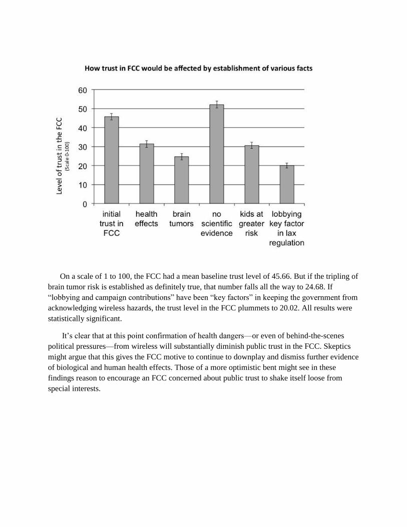
On a scale of 1 to 100, the FCC had a mean baseline trust level of 45.66. But if the tripling of brain tumor risk is established as definitely true, that number falls all the way to 24.68. If ―lobbying and campaign contributions‖ have been ―key factors‖ in keeping the government from acknowledging wireless hazards, the trust level in the FCC plummets to 20.02. All results were statistically significant.
It‘s clear that at this point confirmation of health dangers—or even of behind-the-scenes political pressures—from wireless will substantially diminish public trust in the FCC. Skeptics might argue that this gives the FCC motive to continue to downplay and dismiss further evidence of biological and human health effects. Those of a more optimistic bent might see in these findings reason to encourage an FCC concerned about public trust to shake itself loose from special interests.

Endnotes
1 Former CTIA vice president John Walls in Kevin Kunze‘s documentary film Mobilize, introduced in 2014 at the California Independent Film Festival. 2 November 2014 interview with Renee Sharp. 3 December 2014 interview with Twaun Samuel. 4 Dr. George Carlo and Martin Schram, Cell Phones, Invisible Hazards In The Wireless Age (Carroll & Graf, 2001), 18. 5 Center for Responsive Politics. 6 Id. 7 November 2014 interview with Michael Copps. 8 January 2015 interview with Newton Minow. 9 Daniel Lathrop, ―From Government Service to Private Practice: Writers of Telecom Law Move to K Street,‖ Center for Public Integrity, October 28, 2004, http://www.publicintegrity.org/2004/10/28/6597/government-service-private-practice. 10 B. Blake Levitt and Henry Lai, ―Biological Effects from Exposure to Electromagnetic Radiation Emitted By Cell Tower Base Stations and Other Antenna Arrays,‖ NRC Research Press Web site, November 5, 2010. 11 Id., 381. 12 Id. 13 S. Sivani and D. Sudarsanam, ―Impacts of Radio-Frequency Electromagnetic Field (RF_EMF) from Cell Phone Towers and Wireless Devices on Biosystem and Ecosystem – A Review,‖ Biology and Medicine 4.4 (2013): 202. 14 Id., 206-208. 15 January 2015 interview with Robert Weller. 16 Letter from Michelle C. Farquhar, Chief of the FCC‘s Wireless Telecommunications Bureau, to Thomas Wheeler, President and CEO of the Cellular Telecommunications Industry Association, January 13, 1997. 17 Id. 18 Letter from FCC Chairman Thomas Wheeler to former FCC Commissioner Jonathan Adelstein, President and CEO, PCIA-The Wireless Infrastructure Association, March 14, 2014. 19 December 2014 interview with James R. Hobson. 20 January 2015 interview with Marvin Wessel. 21 Id. 22 January 2015 interview with Janet Newton. 23 Robert Weller interview. 24 Best‘s Briefing, ―Emerging Technologies Pose Significant Risks with Possible Long-Tail Losses,‖ February 11, 2013, http://www.ambest.com/directories/bestconnect/EmergingRisks.pd. 25 Online survey conducted in April 2015 on Amazon‘s Mechanical Turk platform. 26 CTIA, ―Policy & Initiatives: Innovation,‖ http://www.ctia.org/policy-initiatives/policy-topics/innovation. 27 February 2015 interview with Dennis Kucinich. 28 Alexander Lerchl, Melanie Klose, and Karen Grote et al., ―Tumor Promotion by Exposure to Radiofrequency Electromagnetic Fields below Exposure Limits for Humans,‖ Biochemical and Biophysical Research Communications 459.4 (2015): 585-590. 29 WHO/International Agency for Research on Cancer (IARC), ―IARC Classifies Radiofrequency Electromagnetic Fields As Possibly Carcinogenic To Humans,‖ Press Release No. 208, May 31, 2011. 30 Medscape, ―Brain Cancer CME Learning Center,‖ http://www.medscape.org/resource/brain-cancer/cme. 31 Anke Huss, Matthias Egger, Kerstin Hug, Karin Huwiler-Muntener, and Martin Roosli, ―Source of Funding and Results of Studies of Health Effects of Mobile Phone Use: Systemic Review of Experimental Studies,‖ Environmental Health Perspectives 115.1 (2007): 1-4, 1. 32 Id.

33 Federal Communications Commission, ―Wireless Devices and Health Concerns,‖ http://www.fcc.gov/guides/wireless-devices-and-health-concerns. 34 Lennart Hardell, Michael Carlberg, Fredrik Soderqvist, and Kjell Hansson Mild, ―Case-Control Study of the Association between Malignant Brain Tumours Diagnosed between 2007 and 2009 and Mobile and Cordless Phone Use,‖ International Journal of Oncology 43.6 (2013): 1833-1845. 35 Lennart Hardell and Michael Carlberg, ―Use of Mobile and Cordless Phones and Survival of Patients with Glioma,‖ Neuroepidemiology 40.2 (2012): 101-108. 36 Lennart Hardell and Michael Carlberg, ‗Using the Hill Viewpoints from 1965 for Evaluating Strengths of Evidence of the Risk for Brain Tumors Associated with Use of Mobile and Cordless Phones,‖ Reviews on Environmental Health 28.2-3 (2013): 97-106. 37 Gaelle Coureau, Ghislaine Bouvier, and Pierre Lebailly, et al., ―Mobile Phone Use and Brain Tumors in the CERENAT Case-Control Study,‖ Occupational and Environmental Medicine 71.7 (2014): 514-522, doi:10.1136/oemed-2013-101754. 38 October 2014 interview with Lennart Hardell. 39 December 2014 interview with Martin Blank. 40 Id. 41 Norm Alster, ―Cell Phones: We Need More Testing,‖ BusinessWeek, August 14, 2000, 39. 42 Quoted in American Academy of Pediatrics, ―American Academy of Pediatrics Endorses Cell Phone Safety Bill,‖ Press Release, December 20, 2012, http://www.ewg.org/release/american-academy-pediatrics-endorses-cell-phone-safety-bil. 43 Om P. Gandhi, L. Lloyd Morgan, Alvaro Augusto de Salles, Yueh-Ying Han, Ronald B. Herberman, and Devra Lee Davis, ―Exposure Limits: The Underestimation of Absorbed Cell Phone Radiation, Especially in Children,‖ Electromagnetic Biology and Medicine 31.1 (2012): 34-51. 44 November 2014 interview with Joel Moskowitz. 45 February 2015 interview with Carl Blackman. 46 Id. 47 Id. 48 Lawrence Lessig, Roy L. Furman Professor of Law and Leadership at Harvard Law School, helped to draft the Right to Know ordinance and has offered pro bono legal representation to the city of Berkeley. Professor Lessig was director of the Lab at Harvard‘s Safra Center for Ethics, from which the Project on Public Narrative was spun off in November of 2014. 49 May 2015 interview with Berkeley City Attorney Zach Cowan 50 December 2014 interview with Jerry Phillips. 51 Id. 52 February 2015 interview with Om P. Gandhi. 53 Id. 54 Radio interview on WBAI-FM, ―Wireless Radiation: What Scientists Know and You Don‘t, With Dr. Joel Moskowitz,‖ March 10, 2015. 55 Spencer Ante, ―Millions Improperly Claimed U.S. Phone Subsidies,‖ Wall Street Journal, February 11, 2013, http://allthingsd.com/201330212/millions-improperly-claimed-u-s-phone-subsidies/. 56 Federal Communications Commission Office of Inspector General, ―Semiannual Report to Congress for the Period April 1, 2014 - September 30, 2014,‖ 20, http://transition.fcc.gov/oig/FCC_OIG_SAR_09302014a.pdf. 57 Federal Communications Commission, ―Reports on Meetings and Telephone Calls with Registered Lobbyists Regarding General Recovery Act Policy Issues,‖ March 2, 2010. 58 CTIA - The Wireless Association, ―Response to White House Paper on Universal Service Policy,‖ September 19, 2014, http://www.ctia.org/docs/default-source/Legislative-Activity/ctia-usf-response-to-house-white-paper-091914.pdf?sfvrsn=0. 59 Open Letter from Executives of 50 Leading Companies to Tom Wheeler, Chairman of the FCC, January 30, 2014, http://erate2.educationsuperhighway.org/#ceos-letter. See also David Nagel, ―50 Top Execs Urge E-Rate Modernization To Propel Broadband in Schools,‖ The Journal, January 30, 2014. 60 October 2014 interview with Lennart Hardell. 61 Jacob L. Vigdor and Helen F. Ladd, ―Scaling the Digital Divide: Home Computer Technology and Student Achievement,‖ Calder Urban Institute Working Paper, No. 48, June 2010.

62 Mark Warschauer and Morgan Ames, ―Can One Laptop Per Child Save the World‘s Poor?‖ Journal of International Affairs 64.1 (2010): 33-51. 63 John Rogers, ―L.A. Students Get iPads, Crack Firewall, Play Games,‖ Associated Press, October 5, 2013, http://bigstory.ap.org/article/la-students-get-ipads-start-playing-video-games. 64 April 2015 interview with Kentaro Toyama. 65 Id. 66 Id. 67 FCC Chairman Tom Wheeler, quoted in Grant Gross, ―FCC Approves Plan to Spend $1B a Year on School Wi-Fi,‖ IDG News Service, July 11, 2014. 68 Michael O‘Rielly, ―Dissenting Statement by Commissioner Michael O‘Rielly,‖ 2, http://e-ratecentral.com/files/fcc/DOC-328172A7.pdf, after FCC in July of 2014 voted to increase Wi-Fi spending. 69 February 2015 interview with Charles Davidson and Michael Santorelli. 70 Id. 71 The University of Michigan‘s American Customer Satisfaction Index, http://www.theacsi.org/the-american-customer-satisfaction-index. 72 September 2014 interview with Michael Copps. 73 Susan Crawford, Captive Audience: The Telecom Industry and Monopoly Power in the New Gilded Age (Yale University Press, 2013), 212. 74 October 2014 interview with Susan Crawford. 75 Norm Alster, ―A Little Help from the Feds,‖ BusinessWeek, January 24, 2000, 42. 76 1992 Supreme Court decision in Quill Corp. v. North Dakota, 504 U.S. 298 (1992). 77 February 2015 conversation with Jeff Chester. 78 April 2015 interview with Harold Feld. 79 March 2015 interview with Jonathan Mayer. 80 April 2015 interview with Scott Cleland. 81 Id. 82 Center for Responsive Politics. 83 Id. 84 ―Testimony of Jon Wilkins, Managing Director, Federal Communications Commission,‖ Before the Committee on Energy and Commerce, Subcommittee on Communications and Technology, U.S. House of Representatives, March 4, 2015. 85 Alster, ―Cell Phones: We Need More Testing,‖ 39. 86 Danny Hakim and Norm Alster, ―Lawsuits: This Year‘s Model,‖ New York Times, May 30, 2004, http://www.nytimes.com/2004/05/30/business/lawsuits-this-year-s-model.html. 87 A.T. McCartt and S.Y. Kyrychenko, ―Efficacy of Side Airbags in Reducing Driver Deaths in Driver-Side Car and SUV Collisions,‖ Traffic Injury Prevention 8.2 (2007): 162-170. 88 National Highway Traffic Safety Administration, ―Traffic Safety Facts 2012,‖ 18, http://www-nrd.nhtsa.dot.gov/Pubs/812032.pdf. 89 Ralph Nader, Unsafe At Any Speed: The Designed-In Dangers of the American Automobile (Grossman Publishers, 1965). 90 Lab Fellow, Edmond J. Safra Center for Ethics, Harvard University. 91 Investigative Journalism Fellow, Project on Public Narrative at Harvard Law School.

!!!
Attachment!10!

Immunohistopathologic demonstration ofdeleterious effects on growing rat testes ofradiofrequency waves emitted from conventionalWi-Fi devices
Halil I. Atasoy a,*, Mehmet Y. Gunal b, Pinar Atasoy c, Serenay Elgun d,Guler Bugdayci e
aDepartments of Pediatrics, Abant Izzet Baysal University School of Medicine, Bolu 14280, TurkeybDepartment of Physiology, Yeditepe University School of Medicine, Istanbul 34755, TurkeycDepartment of Pathology, Kirikkale University School of Medicine, Kirikkale 71100, TurkeydDepartment of Medical Biochemistry, Ankara University School of Medicine, Ankara 06100, TurkeyeDepartment of Clinical Biochemistry, Abant Izzet Baysal University School of Medicine, Bolu 14280, Turkey
Received 2 October 2011; accepted 28 February 2012Available online 30 March 2012
KEYWORDSCarcinogenesis tests;Infertility;Internet;Oxidative stress;Wireless technology;Testes
Abstract Objective: To investigate effects on rat testes of radiofrequency radiation emittedfrom indoor Wi-Fi Internet access devices using 802.11.g wireless standards.Methods: Ten Wistar albino male rats were divided into experimental and control groups, withfive rats per group. Standard wireless gateways communicating at 2.437 GHz were used asradiofrequency wave sources. The experimental group was exposed to radiofrequency energyfor 24 h a day for 20 weeks. The rats were sacrificed at the end of the study. Intracardiac bloodwas sampled for serum 8-hydroxy-20-deoxyguanosine levels. Testes were removed and exam-ined histologically and immunohistochemically. Testis tissues were analyzed for malondialde-hyde levels and prooxidanteantioxidant enzyme activities.Results: We observed significant increases in serum 8-hydroxy-20-deoxyguanosine levels and 8-hydroxyguanosine staining in the testes of the experimental group indicating DNA damage dueto exposure (p < 0.05). We also found decreased levels of catalase and glutathione peroxidaseactivity in the experimental group, which may have been due to radiofrequency effects onenzyme activity (p < 0.05).
* Corresponding author. Tel.: þ90 374 2534656/3454, þ905325998953; fax: þ90 374 253 46 15.E-mail addresses: [email protected], [email protected] (H.I. Atasoy), [email protected] (M.Y. Gunal), pinara33@
yahoo.com (P. Atasoy), [email protected] (S. Elgun), [email protected] (G. Bugdayci).
1477-5131/$36 ª 2012 Journal of Pediatric Urology Company. Published by Elsevier Ltd. All rights reserved.http://dx.doi.org/10.1016/j.jpurol.2012.02.015
Journal of Pediatric Urology (2013) 9, 223e229
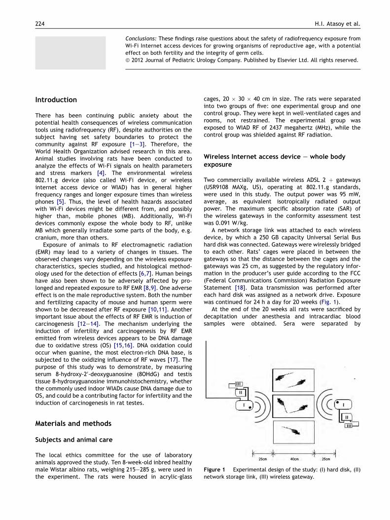
Conclusions: These findings raise questions about the safety of radiofrequency exposure fromWi-Fi Internet access devices for growing organisms of reproductive age, with a potentialeffect on both fertility and the integrity of germ cells.ª 2012 Journal of Pediatric Urology Company. Published by Elsevier Ltd. All rights reserved.
Introduction
There has been continuing public anxiety about thepotential health consequences of wireless communicationtools using radiofrequency (RF), despite authorities on thesubject having set safety boundaries to protect thecommunity against RF exposure [1e3]. Therefore, theWorld Health Organization advised research in this area.Animal studies involving rats have been conducted toanalyze the effects of Wi-Fi signals on health parametersand stress markers [4]. The environmental wireless802.11.g device (also called Wi-Fi device, or wirelessinternet access device or WIAD) has in general higherfrequency ranges and longer exposure times than wirelessphones [5]. Thus, the level of health hazards associatedwith Wi-Fi devices might be different from, and possiblyhigher than, mobile phones (MB). Additionally, Wi-Fidevices commonly expose the whole body to RF, unlikeMB which generally irradiate some parts of the body, e.g.cranium, more than others.
Exposure of animals to RF electromagnetic radiation(EMR) may lead to a variety of changes in tissues. Theobserved changes vary depending on the wireless exposurecharacteristics, species studied, and histological method-ology used for the detection of effects [6,7]. Human beingshave also been shown to be adversely affected by pro-longed and repeated exposure to RF EMR [8,9]. One adverseeffect is on the male reproductive system. Both the numberand fertilizing capacity of mouse and human sperm wereshown to be decreased after RF exposure [10,11]. Anotherimportant issue about the effects of RF EMR is induction ofcarcinogenesis [12e14]. The mechanism underlying theinduction of infertility and carcinogenesis by RF EMRemitted from wireless devices appears to be DNA damagedue to oxidative stress (OS) [15,16]. DNA oxidation couldoccur when guanine, the most electron-rich DNA base, issubjected to the oxidizing influence of RF waves [17]. Thepurpose of this study was to demonstrate, by measuringserum 8-hydroxy-20-deoxyguanosine (8OHdG) and testistissue 8-hydroxyguanosine immunohistochemistry, whetherthe commonly used indoor WIADs cause DNA damage due toOS, and could be a contributing factor for infertility and theinduction of carcinogenesis in rat testes.
Materials and methods
Subjects and animal care
The local ethics committee for the use of laboratoryanimals approved the study. Ten 8-week-old inbred healthymale Wistar albino rats, weighing 215e285 g, were used inthe experiment. The rats were housed in acrylic-glass
cages, 20 " 30 " 40 cm in size. The rats were separatedinto two groups of five: one experimental group and onecontrol group. They were kept in well-ventilated cages androoms, not restrained. The experimental group wasexposed to WIAD RF of 2437 megahertz (MHz), while thecontrol group was shielded against RF radiation.
Wireless Internet access device e whole bodyexposure
Two commercially available wireless ADSL 2 þ gateways(USR9108 MAXg, US), operating at 802.11.g standards,were used in this study. The output power was 95 mW,average, as equivalent isotropically radiated outputpower. The maximum specific absorption rate (SAR) ofthe wireless gateways in the conformity assessment testwas 0.091 W/kg.
A network storage link was attached to each wirelessdevice, by which a 250 GB capacity Universal Serial Bushard disk was connected. Gateways were wirelessly bridgedto each other. Rats’ cages were placed in between thegateways so that the distance between the cages and thegateways was 25 cm, as suggested by the regulatory infor-mation in the producer’s user guide according to the FCC(Federal Communications Commission) Radiation ExposureStatement [18]. Data transmission was performed aftereach hard disk was assigned as a network drive. Exposurewas continued for 24 h a day for 20 weeks (Fig. 1).
At the end of the 20 weeks all rats were sacrificed bydecapitation under anesthesia and intracardiac bloodsamples were obtained. Sera were separated by
Figure 1 Experimental design of the study: (I) hard disk, (II)network storage link, (III) wireless gateway.
224 H.I. Atasoy et al.

centrifugation (at 1250 g for 15 min at 25 #C) and stored at$80 #C. Testes were removed, one of the testes snap-frozen in liquid nitrogen ($196 #C) and stored at $80 #Cuntil biochemical analyses, and other testis tissues fixedin a solution of 10% formaldehyde for histopathologicexamination.
Determination of 8-hydroxy-20-deoxyguanosineserum levels
8OHdG was measured with the highly sensitive 8OHdGcompetitive sandwich enzyme immunoassay kit (CatalogNo. 839320, Cayman Chemical Co., MI, USA). The testprinciple is based on competition between 8OHdG and8OHdG acetylcholinesterase conjugate via 8OHdG mono-clonal antibodies. Plates were read by a Bio-Rad BenchmarkPlus microplate reader at 420 nm (Bio-Rad LaboratoriesInc., CA, USA). Results were expressed in nanograms permilliliter.
Histologic examination of testes
After fixation, the testes were embedded in paraffin,sectioned at 4 mm, deparaffinized, and stained withhematoxylin & eosin. The testicular tissues were evaluatedhistopathologically by using Johnsen’s testicular biopsyscore (TBS) count [19] (Table 1).
8-Hydroxyguanosine immunohistochemistry oftestes
Tissue sections from every paraffin block were immuno-stained with antibodies for 8-hydroxyguanosine, whichwere mouse-raised monoclonal antibodies (ab62623;1 mcg/mL) obtained from AbCAM. Nuclear or perinuclearcytoplasmic staining was considered as ‘positive’. Intensitywas graded as 0 (no staining), 1 (weak staining), 2 (strongstaining) or 3 (very strong staining). On each slide, theintensity of staining in 10 seminiferous tubules was
counted and averaged. The modified immunohistochemicalhistologic score (HSCORE) was used for semiquantitativeestimation of 8OHdG antibody staining of the testiculartissue. This is a score used for the evaluation of intensityand percentage of cells that stain at each intensity [20].The HSCORE is formularized mathematically as follows:
HSCOREZXðPi" I=100Þ
where Pi is the percentage of stained cells, ranging from 1%to 100%, and I is the intensity of staining, ranging from 0 to3. Staining intensities were calculated as shown in Table 2.
Biochemical analysis of prooxidanteantioxidantparameters of testis tissues
The tissues were removed from the freezer ($80 #C) anddisaggregated thoroughly with physiological saline (1 g in5 mL) in a homogeniser (Heidolph Diax 900, Germany).After centrifugation at 4000 g for 20 min (Harrier 18/80MSB080.CR2.K, UK), the supernatants were removed to beused in the analyses.
Malondialdehyde (MDA) level was measured by the thi-obarbituric acid reactive substances method. MDA reactswith thiobarbituric acid to form a colored complex that hasmaximum absorbance at 532 nm. Xanthine oxidase (XO)activity was determined by measuring uric acid formationfrom xanthine at 293 nm. Superoxide dismutase (SOD)activity was measured by a method based on the nitrobluetetrazolium reduction rate. Catalase (CAT) activity wasdetermined by measuring decrease in absorbance ofhydrogen peroxide (H2O2) at 240 nm. Glutathione peroxi-dase (GPX) activity was measured by following changes inNADPH absorbance at 340 nm. In the activity calculations,extinction coefficients of H2O2, NADPH and uric acid wereused for CAT, GPX and XO, respectively. Absorbances wereread by a Unicam Helios a (UVeVIS) spectrophotometer(Spectronic Unicam, Cambridge, UK).
Statistical analysis
Five animals per group were required for the study witha power of 87% and error type I of 5% to detect a one unitdifference between the groups in nonparametric unpairedtests. For central tendency and dispersion of data we usedmedian and quartile deviation (QD), respectively. TheManneWhitney U-test was used for group comparisons.Multivariate analysis of variance was used to search for anysignificant relationship with dependent variables of 8OHdGand parameters of biochemical OS in groups. Pillai’s tracesignificance test was used for significance of the overallrelationship between dependent variables and RF groups. A
Table 1 Evaluation of testicular biopsy score (TBS) count[19].
Score Description
1 No cells in tubular section2 No germ cells, but Sertoli cells present3 Spermatogonia are the only germ cells present4 Only a few spermatocytes (<5), but no spermatids
or spermatozoa present5 No spermatozoa or spermatids, but many
spermatocytes present6 No spermatozoa and only a few spermatids
present (<5e10)7 No spermatozoa, but many spermatids present8 Only a few spermatozoa present (<5e10)9 Much spermatogenesis, but germinal epithelium
disorganized with marked sloughing or obliterationof lumen
10 Complete spermatogenesis with many spermatozoa
Table 2 Evaluation of HSCORE count [20].
Score Description
0 Percentage of unstained cells " 01 Percentage of weakly stained cells " 12 Percentage of moderately stained cells " 23 Percentage of intensely stained cells " 3
Effects of wireless Internet access devices on testes 225

p value less than 0.05 was considered as significant. PASWversion 17 (Chicago, IL, USA) was used for the statisticalprocedures.
Results
There was no difference between the weights of thecontrol and experimental groups before and after theexperiment. Median values of TBS, as evaluated usingJohnsen’s scale, were lower in the experimental than thecontrol group (9 ' 0 vs 10 ' 0). The difference in the TBSbetween the control and experimental groups was statis-tically significant (p < 0.01). 8OHdG levels in the experi-mental group were significantly higher than in the controlgroup. Routine histological examinations of the experi-mental and control group were not different. 8-Hydroxyguanosine immune reactivity, which shows oxida-tive damage to DNA, was positive in both groups. TheHSCOREs of the experimental group were significantlyhigher than those of the control group (p < 0.01) (Table 3).GPX and CAT activities of the experimental group werestatistically lower than those of the control group (p < 0.05and p < 0.01, respectively). SOD and XO activities and MDAlevels of the experimental group were lower than in thecontrol group, but the difference was not statisticallysignificant (p > 0.05) (Table 4). The histological andimmunohistochemical changes in the experimental andcontrol groups are shown in Fig. 2. Multivariate analysis ofvariance showed a significant relationship between RFgroup and the OS parameters of 8OHdG, MDA, GPX, CAT, XOand SOD (F value 19.66; p Z 0.017).
Discussion
Various studies have investigated the effects on the humanbody of RF EMR from mobile phones, with inconclusiveresults, but there have been no studies as yet on the healtheffects of WIADs, which are associated with higherfrequency radio waves and longer exposure times than MBs.Currently, the most popular wireless technology employsthe 802.11.g protocol, which is used in many wirelesselectronic devices, including baby video monitors, Wi-FiMBs and television devices. A wireless 802.11.g RF devicehas unique properties, e.g. with regard to frequency range,output power and exposure time, which may have anuntoward influence on health. It uses the 2400-MHz band,which is higher than the frequencies used by GSM (Global
System for Mobile Communications) or 3G (third genera-tion) phones. The output power of Wi-Fi devices rangesfrom 20 mW to 4 W, and is similar to or greater than that ofMBs, which range from 1 mW to 2 W [21,22]. WIADs aregenerally ‘on’ while transceiving data from the net andthey cause longer human exposure than MBs. Furthermore,they commonly expose the whole body to RF EMR, unlikeMBs, which generally cause a more uneven and local irra-diation of some parts of the body, e.g. the head and pelvicorgans.
The reason why young growing rats were used in thisstudy is that their growing organs may be more prone to theeffects of RF EMR, like children and adolescents who spenda lot of time using wireless computers at home and school.We studied the effects on testes since there have beenseveral studies analyzing the effects of wireless phones onmale fertility [23,24], and the reproductive system is moresensitive than other organs to RF devices such as laptopcomputers. Whole body irradiation with WIADs wasemployed in this experiment, and thus the methodologyused in this study differs significantly from that of theprevious investigators, and is unique because it exploresthe effects of the commonly used conventional WIADs onthe health of living organisms.
Previous animal studies have not found any significanteffect of RF EMR on testicular histology, sperm count andmorphology. Cairnie and Harding [25] studied the effects ofRF in the mouse testis and found no significant differencesin sperm count and no increase in the percentage ofmorphologically abnormal sperm in microwave-exposedanimals. They used a 2.45-GHz continuous wave forshorter periods of up to 30 days (16 h/day). Ono et al. [26]could not find any mutagenic effect of 2.45-GHz intermit-tent whole body RF exposure in the testes of mouseembryos. The methodological differences compared tothose studies may account for the different outcomes weobserved in our study. Since we suspected that thecontinuous exposure to Wi-Fi devices may have morepronounced effects compared with the shorter and inter-mittent exposure to MBs, we used a longer and continuousexposure as the test environment in this study. One maysuspect that this does not reflect real life conditions, wherethe duration and intensity of exposure may be more vari-able. However, the growing technological involvement ofthe modern human being (with wireless modems, wireless
Table 3 Clinical and laboratory characteristics of ratsexposed to WIAD RF radiation and control group.
Controlgroup
Experimentalgroup
p
No. of rats 5 5Weight (g) 230 (30) 250 (9) 0.4568OHdG (ng/mL) 1.15 (0.67) 3.00 (0.44) 0.021a
HSCORE 30 (5) 250 (30) 0.008a
TBS 10 (0) 9 (0) 0.003a
Values are medians, with QD in parentheses.a Statistically significant difference.
Table 4 MDA levels and prooxidanteantioxidant enzymeactivities in testicular tissues of experimental and controlgroups.
Control group(n Z 5)
Experimentalgroup (n Z 5)
p
MDA(nmol/mg)
0.23 (0.025) 0.21 (0.005)a 0.166
XO (mIU/mg) 0.0083 (0.0010) 0.0064 (0.0005) 0.209SOD (U/mg) 0.84 (0.02) 0.76 (0.01) 0.074GPX (IU/mg) 0.0040 (0.0003) 0.0024 (0.0009) 0.036a
CAT (IU/mg) 5.57 (0.115) 2.65 (0.180) 0.009a
Values are medians with QD in parentheses.a Statistically significant difference.
226 H.I. Atasoy et al.

routers, wireless home monitoring systems, wireless babymonitoring systems, wireless data bridges in betweenbuildings, multiple Wi-Fi mobile phones that interact withwireless routers, use of wireless facilities for variousentertainment purposes, etc.) probably exposes manychildren and adults to RF energy levels comparable to thoseused in this study. We also used a highly sensitive method todemonstrate any histopathologic changes that might haveoccurred.
In this study, 8-hydroxyguanosine immune reactivity wasobserved in both the control and study group. We think thatthe immune reactivity of the control group may haveresulted from endogenous DNA damage caused by back-ground electromagnetism emitted by the Earth or elec-tronic devices or the metabolic rate of animals [27]. Weavoided using reverberating or shielding chambers since wedid not want to eliminate the effects caused by the back-ground electromagnetism of the Earth on both groups. If wehad eliminated this interference for the control group, thesignificant difference we found in our laboratory findingswould not have reflected the differential real effect of RFfrom our wireless gateways, and would have yieldeda falsely higher statistical difference between the twogroups.
De Iuliis et al. [9] also showed a correlation betweenincreasing SAR and decreased sperm motility and vitality,and increased OS and 8OHdG markers, stimulating DNA baseadduct formation and ultimately DNA fragmentation.Although their results are in very good accordance withours, they studied purified human spermatozoa ex vivo andused one night’s exposure time with lower RF frequencyand higher SAR values. To the best of our knowledge, our
study is the first in the literature to show oxidative DNAdamage in the reproductive tissues of living organismscaused by conventional Wi-Fi devices.
OS is a well-established cause of male infertility andcarcinogenesis [28,29]. Reactive oxygen species (ROS) fromspermatozoa and invading leukocytes cause infertility byaffecting sperm motility [30]. Physiologically, ROS are keptat low levels by intracellular antioxidant mechanisms ofspermatozoa. Glutathione (GSH), a major thiol inmammals, is an important antioxidant, and plays a majorrole in the antioxidant defense mechanism against proox-idant offenders. Tissue GSH reflects the capability oftissues to scavenge the radicals, preserve the cellularreductioneoxidation balance, and defend the cells. Thedepletion of GSH in RF EMR-exposed animals could beresponsible for the low percentages of motile sperm byaffecting the spindle microtubule formation. Though we didnot measure the GSH level of testis tissues, the decreasedactivity of GPX in the experimental group reflects a distur-bance in the antioxidant defense system, and one possiblemechanism for this may be the reduction in the availabilityof its substrate, GSH. The decreased activity of CAT, whichis also an important part of the cellular antioxidant defensemechanism, may result in increased levels of prooxidantssuch as superoxide radicals and hydrogen peroxide. Thesefindings support the theory that the decreased activities ofantioxidant enzymes leading to OS in testis tissues might beresponsible for the increased DNA oxidation as demon-strated by increased serum levels of 8OHdG and staining ofspermatocytic cells by nuclear and perinuclear 8-hydroxyguanosine antibodies. The decreased SOD and XOactivities, though insignificant, could imply a generalized
Figure 2 Hematoxylin & eosin (A, B) and immunohistochemical (C, D) staining of testicular tissues of control and Wi-Fi RF-exposed rats. (A,B) No difference was detected in routine histological examinations of the control and Wi-Fi groups (originalmagnification " 200). (C) In the control group, 8-hydroxyguanosine nuclear positivity is nearly undetectable (original magnification" 200). (D) Diffuse and intense bead-like nuclear immunopositivity (short and long arrows) is seen in the spermatocytic cells of theWi-Fi group (original magnification " 200).
Effects of wireless Internet access devices on testes 227

RF-induced decrease in enzyme activities. Similar findingswere reported for human ejaculate exposed to RF EMR fromMBs [31]. As MDA levels did not differ between our RFgroups, we suggest that RF EMR from WIADs induces OSleading to DNA oxidation but not lipid peroxidation whichdisturbs testicular function and structure in rats exposed tothese devices. The decrease in enzyme activities in generaltogether with the increased 8OHdG serum levels andimmunohistochemical staining observed are of greatinterest, since both would appear to result from WIAD RF.
The association of parameters used in our study withfertility is not certain. Even so, recent studies havedemonstrated the relationship between oxidative DNAdamage and infertility [32,33]. Others have founddecreased CAT and GPX testicular activities to be respon-sible for fertility problems in animal models and humans[34e36]. Very similar to these studies, we have immuno-histologically demonstrated DNA damage and founddecreased CAT and GPX activities in the testicular tissues ofrats, which may cause infertility. It should be noted thatthe DNA damage that occurred in the rat testes may beheritable. In order to demonstrate how this DNA damageaffects the fertility and organ histology of offspring frommated rats, further functional and structural studies aboutfertility should be performed.
Due to their height, small children are more likely to beaffected by wireless devices positioned close to the floor.Also, children using wireless computers or staying betweentwo wireless devices are exposed to higher levels of radi-ation than the dose used in our study. The model we haveused may not be the perfect one for demonstrating thedeleterious effect of Wi-Fi devices, which would be a limi-tation. However, achieving a model 100% compatible withhumans is nearly impossible in experimental animalstudies.
Studies on the effects of MBs have used RF sources indirect or very close contact to the organs studied, while inreal life this is hardly the case [37]. Unlike in previous MBstudies where animals were restrained during the exposureor shielded against background Earth or exogenous elec-tromagnetism, we allowed the animals to move freely, didnot shield the control group from background radiation, anddid not radiate the experimental group in direct contactwith the RF antennae, in order to set up an experimentaldesign more akin to real world conditions.
Our study is in agreement with previous studies thatshowed the role of MBs in male infertility and carcino-genesis [23,38]. Although the energy levels used in thisstudy were derived from the parameters suggested by themanufacturer, such as distance, power output and dura-tion, they were not safe for rat testicles. The question ofwhether tissues or organs other than the testicles areaffected by these energy levels was not within the scopeof this study and requires further research to be resolved.Obviously, extrapolating our findings to humans may notbe justified, since the human body may have a moreeffective shielding capacity against RF energy comparedto rats. Further studies are required to evaluate thelong-term effects of the commonly used current (g type)and newer (n type) versions of WIADs on male infertilityand germ cell mutagenesis in other species and humanbeings.
Conclusion
The results of the present study demonstrate that contin-uous long-term WIAD exposure oxidatively affects thetestes in growing rats. Further human studies are needed toanswer the question of whether RF waves emitted from Wi-Fi devices affect fertility.
Conflict of interest
None.
Funding
None.
Statement of financial support
Author’s themselves.
References
[1] Litvak E, Foster KR, Repacholi MH. Health and safety impli-cations of exposure to electromagnetic fields in the frequencyrange 300 Hz to 10 MHz. Bioelectromagnetics 2002;23:68e82.
[2] Gajsek P, Pakhomov AG, Klauenberg BJ. Electromagnetic fieldstandards in central and eastern European countries: currentstate and stipulations for international harmonization. HealthPhys 2002;82:473e83.
[3] Leszczynski D, Xu Z. Mobile phone radiation health riskcontroversy: the reliability and sufficiency of science behindthe safety standards. Health Res Policy Syst 2010;8:2.
[4] Sambucci M, Laudisi F, Nasta F, Pinto R, Lodato R, Altavista P,et al. Prenatal exposure to non-ionizing radiation: effects ofWiFi signals on pregnancy outcome, peripheral B-cellcompartment and antibody production. Radiat Res 2010;174:732e40.
[5] Viel JF, Cardis E, Moissonnier M, de Seze R, Hours M. Radio-frequency exposure in the French general population: band,time, location and activity variability. Environ Int 2009;35:1150e4.
[6] Dasdag S, Ketani MA, Akdag Z, Ersay AR, Sari I, Demirtas OC,et al. Whole-body microwave exposure emitted by cellularphones and testicular function of rats. Urol Res 1999;27:219e23.
[7] Westerman R, Hocking B. Diseases of modern living: neuro-logical changes associated with mobile phones and radio-frequency radiation in humans. Neurosci Lett 2004;361:13e6.
[8] Omura Y, Losco M. Electro-magnetic fields in the home envi-ronment (color TV, computer monitor, microwave oven,cellular phone, etc) as potential contributing factors for theinduction of oncogen C-fos Ab1, oncogen C-fos Ab2, integrinalpha 5 beta 1 and development of cancer, as well as effectsof microwave on amino acid composition of food and livinghuman brain. Acupunct Electrother Res 1993;18:33e73.
[9] De Iuliis GN, Newey RJ, King BV, Aitken RJ. Mobile phoneradiation induces reactive oxygen species production and DNAdamage in human spermatozoa in vitro. PLoS One 2009;4:e6446.
[10] Cleary SF, Liu LM, Graham R, East J. In vitro fertilization ofmouse ova by spermatozoa exposed isothermally to radio-frequency radiation. Bioelectromagnetics 1989;10:361e9.
228 H.I. Atasoy et al.

[11] Agarwal A, Desai NR, Makker K, Varghese A, Mouradi R,Sabanegh E, et al. Effects of radiofrequency electromagneticwaves (RF-EMW) from cellular phones on human ejaculatedsemen: an in vitro pilot study. Fertil Steril 2009;92:1318e25.
[12] Balcer-Kubiczek EK, Harrison GH. Evidence for microwavecarcinogenesis in vitro. Carcinogenesis 1985;6:859e64.
[13] Balcer-Kubiczek EK, Harrison GH. Neoplastic transformationof C3H/10T1/2 cells following exposure to 120-Hz modulated2.45-GHz microwaves and phorbol ester tumor promoter.Radiat Res 1991;126:65e72.
[14] Krewski D, Byus CV, Glickman BW, Lotz WG, Mandeville R,McBride ML, et al. Potential health risks of radiofrequencyfields from wireless telecommunication devices. J ToxicolEnviron Health B Crit Rev 2001;4:1e143.
[15] Sage C, Carpenter DO. Public health implications of wirelesstechnologies. Pathophysiology 2009;16:233e46.
[16] Aitken RJ, Baker MA, Sawyer D. Oxidative stress in the malegerm line and its role in the aetiology of male infertility andgenetic disease. Reprod Biomed Online 2003;7:65e70.
[17] Kornhauser A, Krinsky NI, Huang PK, Clagett DC. A compara-tive study of photodynamic oxidation and radiofrequency-discharge-generated 1O2 oxidation of guanosine. PhotochemPhotobiol 1973;18:63e9.
[18] Wireless MAXg ADSL Gateway User Guide. Available at: http://www.usr.com/support/9108/9108-ug/regulatory.htm[accessed 06.03.2012].
[19] Johnsen SG. Testicular biopsy score count e a method forregistration of spermatogenesis in human testes: normalvalues and results in 335 hypogonadal males. Hormones 1970;1:2e25.
[20] Atasoy P, Bozdogan O, Erekul S, Bozdogan N, Bayram M. Fas-mediated pathway and apoptosis in normal, hyperplastic, andneoplastic endometrium. Gynecol Oncol 2003;91:309e17.
[21] Lonn S, Forssen U, Vecchia P, Ahlbom A, Feychting M. Outputpower levels from mobile phones in different geographicalareas; implications for exposure assessment. Occup EnvironMed 2004;61:769e72.
[22] ftp://ftp.zyxel.com/FSG1100HN/support_note/FSG1100HN_1.0.pdf [accessed 21.03.2012].
[23] Deepinder F, Makker K, Agarwal A. Cell phones and maleinfertility: dissecting the relationship. Reprod Biomed Online2007;15:266e70.
[24] Wdowiak A, Wdowiak L, Wiktor H. Evaluation of the effect ofusing mobile phones on male fertility. Ann Agric Environ Med2007;14:169e72.
[25] Cairnie AB, Harding RK. Cytological studies in mouse testisirradiated with 2.45-GHz continuous-wave microwaves. RadiatRes 1981;87:100e8.
[26] Ono T, Saito Y, Komura J, Ikehata H, Tarusawa Y, Nojima T,et al. Absence of mutagenic effects of 2.45 GHz radio-frequency exposure in spleen, liver, brain, and testis of lacZ-transgenic mouse exposed in utero. Tohoku J Exp Med 2004;202:93e103.
[27] Ames BN. Endogenous oxidative DNA damage, aging, andcancer. Free Radic Res Commun 1989;7:121e8.
[28] Rajesh Kumar T, Doreswamy K, Shrilatha B, Muralidhara.Oxidative stress associated DNA damage in testis of mice:induction of abnormal sperms and effects on fertility. MutatRes 2002;513:103e11.
[29] Wan J, Winn LM. In utero-initiated cancer: the role of reactiveoxygen species. Birth Defects Res C Embryo Today 2006;78:326e32.
[30] Henkel R, Kierspel E, Stalf T, Mehnert C, Menkveld R,Tinneberg HR, et al. Effect of reactive oxygen speciesproduced by spermatozoa and leukocytes on sperm functions innon-leukocytospermic patients. Fertil Steril 2005;83:635e42.
[31] Kilgallon SJ, Simmons LW. Image content influences men’ssemen quality. Biol Lett 2005;1:253e5.
[32] Makker K, Agarwal A, Sharma R. Oxidative stress & maleinfertility. Indian J Med Res 2009;129:357e67.
[33] Tunc O, Tremellen K. Oxidative DNA damage impairs globalsperm DNA methylation in infertile men. J Assist Reprod Genet2009;26:537e44.
[34] Alkan I, Simsek F, Haklar G, Kervancioglu E, Ozveri H, Yalcin S,et al. Reactive oxygen species production by the spermatozoaof patients with idiopathic infertility: relationship to seminalplasma antioxidants. J Urol 1997;157:140e3.
[35] Morakinyo AO, Iranloye BO, Daramola AO, Adegoke OA. Anti-fertility effect of calcium channel blockers on male rats:association with oxidative stress. Adv Med Sci 2011;56:95e105.
[36] Shukla KK, Mahdi AA, Ahmad MK, Jaiswar SP, Shankwar SN,Tiwari SC. Mucuna pruriens reduces stress and improves thequality of semen in infertile men. Evid Based ComplementAlternat Med 2010;7:137e44.
[37] Anderson V, Rowley J. Measurements of skin surface temper-ature during mobile phone use. Bioelectromagnetics 2007;28:159e62.
[38] Heynick LN, Johnston SA, Mason PA. Radio frequency elec-tromagnetic fields: cancer, mutagenesis, and genotoxicity.Bioelectromagnetics 2003;(Suppl. 6):S74e100.
Effects of wireless Internet access devices on testes 229

!!!
Attachment!11!

CELL JOURNAL(Yakhteh), Vol 17, No 2, Summer 2015 322
Original Article
Effects of Wi-Fi (2.45 GHz) Exposure on Apoptosis, Sperm Parameters and Testicular Histomorphometry
in Rats: A Time Course Study
Saeed Shokri, Ph.D.1*, Aiob Soltani, M.Sc.2, Mahsa Kazemi, M.Sc.3, Dariush Sardari, Ph.D.2, Farshid Babapoor Mofrad, Ph.D.2
1. Department of Anatomical Sciences, Faculty of Medicine, Zanjan University of Medical Sciences (ZUMS), Zanjan, Iran2. Department of Nuclear Engineering, Science and Research Branch, Islamic Azad University, Tehran, Iran
3. Department of Physiology, Faculty of Medicine, Zanjan University of Medical Sciences, Zanjan, Iran
*Corresponding Address: P.O.Box: 45139-56111, Department of Anatomical Sciences, Faculty of Medicine, Zanjan
University of Medical Sciences (ZUMS), Zanjan, Iran
Email: [email protected]
Received: 24/Apr/2014, Accepted: 18/Sep/2014AbstractObjective: In today’s world, 2.45-GHz radio-frequency radiation (RFR) from industrial,
scientific, medical, military and domestic applications is the main part of indoor-outdoor electromagnetic field exposure. Long-term effects of 2.45-GHz Wi-Fi radiation on male reproductive system was not known completely. Therefore, this study aimed to investigate the major cause of male infertility during short- and long-term exposure of Wi-Fi radiation.
Materials and Methods: This is an animal experimental study, which was conducted in the Department of Anatomical Sciences, Faculty of Medicine, Zanjan University of Medi-cal Sciences, Zanjan, IRAN, from June to August 2014. Three-month-old male Wistar rats (n=27) were exposed to the 2.45 GHz radiation in a chamber with two Wi-Fi antennas on opposite walls. Animals were divided into the three following groups: I. control group (n=9) including healthy animals without any exposure to the antenna, II. 1-hour group (n=9) ex-
posed to the 2.45 GHz Wi-Fi radiation for 1 hour per day during two months and III.7-hour group (n=9) exposed to the 2.45 GHz Wi-Fi radiation for 7 hours per day during 2 months. Sperm parameters, caspase-3 concentrations, histomorphometric changes of testis in ad-
dition to the apoptotic indexes were evaluated in the exposed and control animals. Results: Both 1-hour and 7-hour groups showed a decrease in sperm parameters in a time dependent pattern. In parallel, the number of apoptosis-positive cells and caspase-3 activity increased in the seminiferous tubules of exposed rats. The seminal vesicle weight reduced significantly in both1-hour or 7-hour groups in comparison to the control group. Conclusion: Regarding to the progressive privilege of 2.45 GHz wireless networks in our environment, we concluded that there should be a major concern regarding the time-dependent exposure of whole-body to the higher frequencies of Wi-Fi networks existing in the vicinity of our living places.
Keywords: Apoptosis, Electromagnetic Radiation, Testis, Spermatogenesis Cell Journal(Yakhteh), Vol 17, No 2, Summer 2015, Pages: 322-331
Citation: Shokri S, Soltani A, Kazemi M, Sardari D, Babapoor Mofrad F. Effects of Wi-Fi (2.45 GHz) exposure on apoptosis, sperm parameters and testicular histomorphometry in rats: a time course study. Cell J. 2015; 17(2): 322-331.
IntroductionElectromagnetic radiation (EMR) from different
sources, such as microwave ovens, radar, satellite links, wireless communication, frequency modula-tion (FM) radio and television (TV) transmitters/antennas, is the main part of indoor- outdoor elec-tromagnetic field exposure spectrum (1, 2). Wide-
spread usage of industrial, scientific, medical, military and domestic applications with 2.45-GHz radio-frequency radiation is inevitable in today’s world. As the Wi-Fi technology is low cost and operates in the unlicensed spectrum at 2.40-2.4 GHz, the leakage of Wi-Fi radiation into the envi-ronment is unavoidable (3, 4).

CELL JOURNAL(Yakhteh), Vol 17, No 2, Summer 2015 323
Shokri et al.
It has been suggested that male infertility during the past several decades is related to the direct or indirect exposure to certain environ-mental factors such as radio-frequency electro-magnetic waves (RF-EMW) (5, 6). The effects of 2.45-GHz EMR on reproductive system have already been shown (7-10). Kumar et al. (11) showed 2.45 GHz microwave exposure causes an increase in caspase-3 and creatine kinase ac-tivities in the sperm in addition to a decrease in plasma levels of testosterone and melatonin in the exposed rat. In vitro study by Avendano et al. (12), focused on the effect of Wi-Fi radiation on the motility reduction and DNA fragmenta-tion of human spermatozoa. The negative effect of Wi-Fi emitting RF-EMW has been also re-ported on the ex vivo human sperm parameters (13), sexual behavior (14) and testis structure of exposed animals (15). It is believed that expo-sure to EMR can enhance production of reac-tive oxygen species (ROS) (9, 12, 15-18). An increase in lipid peroxidation levels in addition to a decrease in antioxidant enzymes and vita-min A and E levels (11, 19) can explain some aspects of 2.45-GHz EMR effect on reproduc-tive tissues of male rats. Kim et al. (20) showed that the effect of exposure to 2.45-GHz EMR on proliferation and differentiation of spermatogo-nia is correlated with serum sex hormone level. In parallel with defect in spermatogenesis pro-cess, the negative effects of 2.45-GHz EMR on histopathological changes and apoptosis sta-tus of rat testis are inevitable (7). Nowadays 2.45 GHz wireless networks have become much more commonplace in our environment (21). Wireless devices have been widespread used in our living and working environments for longer exposure times than wireless phones which may have an untoward influence on health (2). Ac-cording to the Bioinitiative Report (http://www.bioinitiative.org/), current safety guidelines for electromagnetic field (EMF) exposure are not sufficient and should be revised based on data from various toxicological tests (22). Due to whole body exposure to the RF-EMR, we tried to analyze potential effects of 2.45 GHz Wi-Fi radiation from a wireless antenna on the repro-ductive system of freely moving male rats for short- and long-term. Indeed, the consequences of exposure to the emitted radiofrequency waves
from Wi-Fi antenna were the major concerns of the present study.
Materials and MethodsAnimals
This is an animal experimental study, which was conducted in the Department of Anatomi-cal Sciences, Faculty of Medicine, Zanjan University of Medical Sciences, Zanjan, Iran, from June to August 2014. Animals, 3-month old Wistar strain rats (n=27), were maintained as national guidelines and protocols approved by the Institutional Animal Ethics Committee (IAEC no.03/028/07).
All experimental protocols were approved by the Ethics Committee of Zanjan University of Medical Sciences, Zanjan, Iran. Healthy adult male albino rats weighing 250 g, were random-ly selected and housed under environmentally controlled conditions. The rats were fed with a standard laboratory diet (Pars Dam Co., Tehran, Iran) and clean drinking water ad libitum.
Exposure system
The exposure system was a chamber (180 cm×80 cm×70 cm), designed for whole-body exposure of free-moving rats to a Wi-Fi sig-nal. Two Wi-Fi antennas (NanoStation Loco M2, 2.45 GHz, 8.5 dBi, Ubiquiti Networks, Inc. USA) were placed at the center of two sides of the chamber. A previous study applied a restrainer to fix space between antenna and rat (19). Since it was a stressful condition that could probably affect hormonal balance of ani-mals, we tried to assess the effect of radiation on the free moving animals (14, 23).
Animals were divided into three following groups (n=9 per each group): I. control group in-cluding healthy animals without any exposure to the antenna, II. 1-hour group including animals exposed to the 2.45 GHz Wi-Fi radiation one hour per day during two months (1 hour/day/2 months) (7, 14, 20) and III. 7-hour group including animals exposed to the 2.45 GHz Wi-Fi radiation seven hours per day during two months (7 hours/day/2 months). All exposure conditions were coded and analyzed in a blind manner.

CELL JOURNAL(Yakhteh), Vol 17, No 2, Summer 2015 324
Effects of 2.45 GHz Wi-Fi Radiation on The Testis
Laboratory studies, body and reproductive
organ weights
Animals were anesthetized intraperitoneally with a mixture of ketamine (45 mg⁄kg, Sigma- Al-drich, Germany) and xylazine (35 mg⁄kg, Sigma Aldrich, Germany). The weight gain of animal in each group was defined as the differences between initial and final body weights. The reproductive organs including testes, epididymis, seminal vesi-cles and ventral prostate were accurately weighed following being dissected out from surrounded adipose and connective tissues by an expert anat-omist. The relative weights of each dissected re-productive organ were expressed as the weight of organ to the body weight ratio. The samples of tes-ticular tissues were fixed in a 4% buffered formal-dehyde solution (Merck, Germany) and then were embedded in paraffin wax (Merck, Germany) us-ing standard techniques for preparing 5-µm thick sections. Other side testicle was randomly dissect-ed out and transferred to a cryotube for storing in liquid nitrogen in order to determine the caspase-3 activity.
Sperm characteristics
Caudal part of epididymis was dissected out and chopped in the 5 ml of Ham’s F10 medium solution (GIBCO, USA). Epididymal sperm were collected following 5 minutes incuba-tion at 37˚C to allow sperm to swim out of the epididymal tubules. One drop of sperm sus-pension was placed on a microscope slide and cover slipped. At least 10 microscopic fields were observed at ×40 magnification by a phase contrast microscope (Olympus BX51, Tokyo, Japan). The sperm motility parameters were re-corded according to the World Health Organi-zation (WHO) recommendations. The percent-ages of progressive, motile, and immotile sperm were expressed as the ratio to the total counted sperm. The sperm count parameters were also obtained by the method described in the WHO recommendations (24). Briefly, 5 µl aliquot of epididymal sperm was diluted with 95 µl of diluents (0.35% formalin containing 5% NaH-CO3 and 0.25% trypan blue, Merck, Germany), and approximately 10 µl of this diluted speci-men was transferred to the counting chambers of the haemocytometer. The cells were counted with a light microscope at ×40 magnification.
For morphological abnormalities, sperm smears were drawn on slides and allowed to air-dry overnight. Slides were stained with 1% eosin-Y⁄5% nigrosin (Merck, Germany) and exam-ined at ×40 magnification. Amorphous, hook less, bicephalic, coiled or abnormal tails were considered as the morphological abnormalities (25). The total percentages of abnormal and normal sperm were then calculated.
Histopathological evaluation of spermatogenesis
Either the number of germinal cell layers or Johnson’s score were measured for categoriz-ing spermatogenesis in the testes. According to Miller et al. (26) description, the number of ger-minal epithelial layers was counted in 10 semi-niferous tubules. Based on Johnson’s method, a score of 1-10 was applied for each cross-sec-tioned tubule (27).
Apoptosis in reproductive tissues of rats
Germ cell apoptosis was evaluated by terminal deoxynucleotidyl transferase (TdT) enzymemedi-ated dUTP nick end labeling (TUNEL) assay kit (Roche, Germany). Briefly, 5-µm thick paraffin-embedded sections were microwave-pretreated in 10 mM citrate buffer (Merck, Germany, pH=6.0) for 10 minutes. Sections were incubated with blocking solution (3% H2O2 in methanol, Merck, Germany) for 10 minutes, then were washed with phosphate-buffered saline (PBS, Merck, Germa-ny). The specimens were incubated with TUNEL reaction mixture (TdT and nucleotide mixtures in reaction buffer) at 37˚C for 60 minutes. Finally the slides were stained with converter-POD (ant-ifluorescein antibody, Fab fragment from sheep, conjugated with horse-radish peroxidase-POD) for 30 minutes.
The 3, 3΄- Diaminobenzidine (DAB) substrate (Roche, Germany) was applied for color develop-ment. TUNEL positive cells exhibited a brown nu-clear stain. In each group, the number of stained cells was counted in 100 seminiferous tubules. The number of stained germ cells was counted. Apoptotic index-1 (AI-1) was defined as the num-ber of apoptotic TUNEL-positive cells per 100 tu-bules and apoptotic index-2 (AI-2) as the number of tubules containing apoptotic cells per 100 tu-bules. All of measurements were performed by an expert technician who was blinded to the experi-

CELL JOURNAL(Yakhteh), Vol 17, No 2, Summer 2015 325
Shokri et al.
ment procedure.
Caspase-3 activity assay
Briefly, lysis buffer at pH=7.5, including 10 mM Tris-HCL, 10 mM NaH2PO4/NaHPO4, 130 mM NaCl, 1% Triton-X100 and 10 mM NaPPi, all materials were purchased from Merck prod-ucts-Germany that were added to the testes tis-sue samples and lysates were incubated at 4˚C for 20 minutes. The lysates were centrifuged at 14000 rpm and stored in liquid nitrogen for fur-ther analysis. Next 100 ml proteins from lysates were incubated with Ac-DEVD-pNA in a 96-well plate at 37˚C for 1 hour, and colorimet-ric substrate (DEVD-AFC, Biomol, Plymouth Meeting, PA, USA) was preferentially cleaved by caspase- 3. The amounts of 7-amino-4-me-thyl-coumarin (AMC) were monitored 1 hour with a plate reader (Anthos2020, USA) and ab-sorption was measured, normalized to the ab-sorbance of time zero and expressed as percent of control.
The data were expressed as mean ± standard errors of the mean (SEM). The variables were analyzed by one-way ANOVA. When a signifi-cance found, Tukey post hoc tests were per-formed. All analyses were performed using the SPSS (SPSS Inc., Chicago, IL, USA) version 16. The statistical significance level was set at P≤0.05.
ResultsTable 1 shows two months exposure of animals
to the 2.45 GHz Wi-Fi radiation in the designed exposure apparatus (Fig.1), indicating no signifi-cant changes in the body weight of both 1- and 7-hour groups.
Fig.1: Represents a schematic picture of designed apparatus as the exposure system. Box dimension was 180 cm×80 cm×70 cm. Two Wi-Fi antennas (NanoStation Loco M2, 2.45 GHz, 8.5 dBi, Ubiquiti Networks, Inc. USA) were placed at the center of two sides of the chamber. Animal cages were placed between two antennas.
Despite right and left seminal vesicles, 1 hour and 7 hours chronic exposure caused no signifi-cant changes in the relative weight of testicles or other accessory sex organs. The relative weight of both right and left seminal vesicles reduced significantly (P≤0.001) following two months chronic exposure of animals to the 2.45 GHz Wi-Fi radiation either for 1 hour per day or 7 hours per day (Table 1).
Table 1: The effect of chronic exposure to the 2.45 GHz Wi-Fi radiation on the weights of testis, epididymis, prostate and seminal vesicle in mature male rats
Weight
gain (g)
Relative
Ventral prostate
weight (%)
Relative left
Seminal vesicle
weight (%)
Relative right
Seminal vesicle
weight (%)
Relative left
epididymis
weight (%)
Relative right
epididymis
weight (%)
Relative left
testis
weight (%)
Relative right
testis
weight (%)
Groups
59.37 ± 3.830.18 ± 0.010.16 ± 0.010.18 ± 0.010.18 ± 0.00.19 ± 0.00.45 ± 0.010.43 ± 0.01Control
40.11 ± 8.900.19 ± 0.00.08 ± 0.0110.08 ± 0.010.18 ± 0.00.18 ± 0.00.43 ± 0.010.42 ± 0.011-hour group
50.54 ± 6.880.22 ± 0.010.09 ± 0.010.08 ± 0.0210.17 ± 0.00.17 ± 0.00.42 ± 0.010.41 ± 0.017-hour group
Values are expressed as mean ± standard errors of the mean (SEM). Chronic exposure to the 2.45 GHz Wi-Fi radiation caused sig-nificant differences in the relative weights of both right and left seminal vesicles. The right and left seminal vesicles column were compared with the control group. 1-hour group; exposed to the 2.45 GHz Wi-Fi radiation one hour per day during two months (1 hour/day/2 months). 7-hour group; exposed to the 2.45 GHz Wi-Fi radiation seven hour per day during two months (7 hour/day/2 months).1; P value≤0.001.

CELL JOURNAL(Yakhteh), Vol 17, No 2, Summer 2015 326
Effects of 2.45 GHz Wi-Fi Radiation on The Testis
We examined the proportion of the different sperm motility grades as shown in figure 2. Two months exposure to the 2.45 GHz Wi-Fi radiation caused significant changes on the sperm motility parameters (Fig.2). Although the percentage of pro-gressive sperm showed no significant differences in the experimental groups, the percentages of total motility parameters, considered as the percentage of progressive and motile sperm, reduced significantly in both 1- and 7-hour groups. Therefore, our findings showed a significant reduction in the percentage of motile sperm in 1-hour (27.75 ± 1.27 vs. 44.89 ± 0.81, P≤0.001) and 7-hour (31.87 ± 1.58 vs. 44.89 ± 0.81, P≤0.001) groups as compared to control group.
Fig.2: The effect of chronic exposure to the 2.45 GHz Wi-Fi radia-tion on total percentage of progressively motile sperm. Values are expressed as mean ± standard errors of the mean (SEM). *; P val-ue≤0.001.
Table 2 shows that chronic exposure to the 2.45 GHz Wi-Fi radiations showed a clear negative im-pact on the concentration parameters. Sperm sam-ples from both 1-hour (P≤0.001) and 7-hour groups (P≤0.05) exhibited a significant lower concentration as compared to the control group. In parallel with the sperm count reduction, the proportion of normal to abnormal sperm showed a similar reduction in the both 1- and 7-hour groups.
Table 2: The effect of chronic exposure to the 2.45 GHz Wi-Fi radiations on the concentration parameters
7-hour group 1-hour groupControl
2.68 ± 1.2612.16 ± 2.7723.49 ± 1.72Sperm count (×106/ml)
83.68 ± 0.90178.66 ± 1.22287.34 ± 1.05Normal sperm (%)
Values are expressed as mean ± standard errors of the mean (SEM). The 1-hour and 7-hour groups were compared to the control ones. 1-hour group: exposed to the 2.45 GHz Wi-Fi radiation one hour per day during two months (1 hour/day/2 months). 7-hour group: ex-posed to the 2.45 GHz Wi-Fi radiation seven hour per day during two months (7 hour/day/2 months). 1; P≤0.05 and 2; P≤0.001.
Table 3 shows that the 1-hour group exposed to the 2.45 GHz Wi-Fi radiations demonstrated a normal architecture of the seminiferous tubules and interstitial tissue. The germinal epithelium of testis was intact with an average thickness of about five cell layers. On the contrary, 7-hour group exposed to the 2.45 GHz Wi-Fi radiations caused a significant decrease in both the num-ber of germ cell layers (P≤0.01) and the mean testicular score (P≤0.001). Quantitative and de-scriptive analysis of TUNEL stained slides in figure 3A and B respectively, show that in par-allel with the significant reduction in both the number of germ cell layers and the Johnson’s criteria of the 7-hour group, evaluation of apop-totic indexes showed a significant increase in the either the number of apoptotic cells (P≤0.001) or positive tubules per 100 tubules (P≤0.001) in the same group. As it is shown in the figure 4, the increased level of caspase-3 can be a good explanation for testicular apoptosis occurring in the testis of 7-hour animals. Interestingly, lack of significant differences in the number of germ cell layers and the mean testicular score of 1-hour group was accompanied with lack of significant criteria in apoptotic indexes and the caspase-3 concentration. However, two experi-mental groups showed a significant differences in apoptotic indexes, caspase 3 activity and Johnson’s criteria.
Table 3: The Effects of chronic exposure to the 2.45 GHz Wi-Fi radiations on apoptosis of spermatogenesis
Number of
germ cell layers
Johnson’s
criteria
5.58 ± 0.089.48 ± 0.14Control
5.61 ± 0.059.24 ± 0.051-hour group
5.25 ± 0.051*8.75 ± 0.062*7-hour group
Values are expressed as mean ± standard errors of the mean (SEM). The 1-hour and 7-hour groups were compared to the control ones. 1; P≤0.01, 2; P≤0.001 and *; The comparison be-tween 1-hour and 7-hour groups. 1-hour group: exposed to the 2.45 GHz Wi-Fi radiation one hour per day during two months (1 hour/day/2 months). 7-hour group: exposed to the 2.45 GHz Wi-Fi radiation seven hour per day during two months (7 hour/day/2 months). P≤0.001.

CELL JOURNAL(Yakhteh), Vol 17, No 2, Summer 2015 327
Shokri et al.
Fig.3: A. The effect of chronic exposure to the 2.45 GHz Wi-Fi radiation on the apoptotic indexes as either number of apoptotic terminal deoxynucleotidyl transferase (TdT) enzyme mediated dUTP nick end labeling (TUNEL)-positive cells per 100 tubules (AI-1) or the number of tubules containing apoptotic cells per 100 tubules (AI-2). Values are expressed as mean ± standard errors of the mean (SEM). The 1-hour and 7-hour groups were compared to the control ones. ***; P≤0.001, **; P≤0.01 and *; P≤0.05 and B. The effect of chronic ex-posure to the 2.45 GHz Wi-Fi radiation on the apoptosis of spermatogenic cell line. Control group (a. magnification ×10, b. magnification ×40 and c. magnification ×100). 1-hour group (d. magnification ×10, e. magnification ×40 and f. magnification ×100). 7-hour group (g. magnification ×10, h. magnification ×40 and i. magnification ×100). (scale bar=100 µm) arrows show apoptotic cells.
Fig.4: The effect of chronic exposure to the 2.45 GHz Wi-Fi radiation on the concentration of caspase-3. Values are expressed as mean ± standard errors of the mean (SEM). *; P≤0.001.
A
B a
d
g
b
e
h
c
f
i

CELL JOURNAL(Yakhteh), Vol 17, No 2, Summer 2015 328
Effects of 2.45 GHz Wi-Fi Radiation on The Testis
DiscussionDecline in male fertility, as one of parameters in
this study, is considered as a major concern dur-ing the past several decades. It has been suggested that direct or indirect exposure to RF-EMW as the main environmental factor plays a dominant role in the observed decline (28). The 2400-2500 GHz radio frequency emitting from Wi-Fi-enabled de-vices has a long exposure time over a very wide area (2, 19, 21). Hence, this transmitted energy can be absorbed by human body (8, 29).
No deleterious effects of 2.4 GHz Wi-Fi expo-sure on the body weight and reproductive organ weights were observed in the either 1- or 7-hour groups; however, exposure effect on the seminal vesicle weights was observed. This present re-sult is in line with previous reported animal ex-periment that demonstrated no adverse effects of 2.45 GHz radio-frequency exposure on the body weight (14) as well as testis and prostate weights (15, 19). Interestingly, 1 hour and 7 hours expo-sure caused a decline in seminal vesicles weight in comparison to related value of the control group. Although there is no previous report indicating the deleterious effect of 2.45 GHz radiation on seminal vesicles, khaki et al. (30) showed that 50 Hz non-ionizing radiation during two months caused a decrease in seminal vesicles weight. It is noted that epithelial cell proliferation in the seminal vesicles is testosterone-dependent (31). It has been shown that RF-EMF exposure proba-bly reduces the serum testosterone in experimen-tal animals (32, 33).
Alternatively, deficiency in blood testosterone can alter epithelial proliferation in the seminal vesicles. Specifically, Kumar et al. (11) showed that long-term exposure of 2.45 GHz radiation from microwave source can reduce the level of serum testosterone in rats. Consequently, we speculated that the reduced seminal vesicle weight following 2.45 GHz exposure is likely to be related to the reduction of serum testosterone in rats.
Some evidences have indicated that sperm ab-normalities are frequent following exposure to RF-EMW (34, 35). We found that sperm concen-tration, motility and morphology were affected significantly by exposure to the 2.45 GHz RFR
from a Wi-Fi antenna. The observed effects were dependent on the longevity of exposure per day. Recent in vitro pilot studies on the effect of expo-sure of the 2.45 GHz RFR on human ejaculated se-men found changes in the motility and DNA frag-mentation of exposed sperm (12, 13). Kim et al. (20) found no significant reduction in the epididy-mal sperm count after exposure of rats to the 2.45 GHz EMF [a designed magnetron (Samsungelec-tronics, Korea) operating at 2.45 GHz by Institute of Biomedical Engineering, Yeungnam University, Daegu, Korea] for 1 hour or 2 hours per day during 8 weeks. Moreover, they reported no abnormal morphology in the exposed groups.
It was also shown that microwave radiation decreases the sperm count (20). A plausible ex-planation for the impaired sperm motility could be induced oxidative stress by RF-EMW from Wi-Fi devices (12). Oxidation of phospholipids, as a major component in the sperm mitochon-drial sheath (36), can disturb mitochondrial membrane potential which causes high levels of ROS to be released into the cytoplasm, lead-ing to deplete the energy supply and to affect both sperm motility and kinetics (37, 38). Per-oxidation of unesterified polyunsaturated fatty acids in the cell membrane of spermatozoa can lead to cell death as well (39). However, an in vitro pilot study by Oni et al. (13) showed that 1 hour exposure of 2.45 RFR from a laptop an-tenna (a 2.4 GHz picostation by Ubiquity Net-works, USA) had no effects on sperm concen-tration and sperm head, whereas tail and middle piece defect were evitable following exposure to the RFR. The negative effect of chronic RF exposure from cell phones on the count and the quality of sperm was also reported in the previ-ous researches (40, 41). Interestingly, the nega-tive correlation between both abnormal struc-ture and decreased motility of sperm with the longevity of exposure to the RFR from mobile phones was showed by Wdowiak et al. (42). It is believed that EMF, especially extremely low frequency, induces free radical production that is responsible for sperm deformities (43). Al-though, the mechanism of cascade is unknown, it has been recently demonstrated that depletion in the activity of both histone kinase and pro-tein kinase may serve as a measure of micro-wave EMF’s ability to affect spermatogenesis

CELL JOURNAL(Yakhteh), Vol 17, No 2, Summer 2015 329
Shokri et al.
and cell cycle in sperm (8).
In the testis tissue of the animals exposed to 7 hours of 2.45 GHz Wi-Fi radiation for 60 days, the number of germinal cell layers (5.25 ± 0.05 vs. 5.58 ± 0.08, P≤0.01) and Johnson’s score (8.75 ± 0.06 vs. 9.48 ± 0.14, P≤0.001) showed a significant reduction as compared to control group. In parallel, the profound DNA damage in 7-hour group was accompanied with an increase in the activity of caspase-3. In accordance with these findings, several authors focused mainly on the destructive effects of RFR on the germi-nal cell layers of male reproductive organ (11, 14, 15, 19-20, 32, 34, 41). It is shown that 2.45 GHz microwave radiation decreases the diam-eters of seminiferous tubule (41, 44). Saygin et al. (7) showed changes in histopathology and apoptosis status of rat testis under exposure to 2.45-GHz EMF, at 3.21 W/kg specific absorp-tion rate for 60 minutes/day for 28 days.
On the other hand, Poulletier de Gannes et al. (14) found no microscopic lesions in the tes-tes of male Wistar rats by exposing animals to the 2450 MHz Wi-Fi signal (1 hour/day, 6 days/week, 0.08 and 4 specific absorption rate). Moreover, Kim et al. (20) showed that both the measured diameter of seminiferous tubule and average Johnson’s score of testicular biopsy did not change significantly by exposure to the 2.45 GHz EMF (1 hour or 2 hours per day/8 weeks, 1.41W/Kg and 60.1 mV/m electric field intensity. Although they observed no signifi-cant difference in the number of spermatids, a significant difference was seen in the number of spermatocytes between the control and ex-posed group. Atasoy et al. (15) applied stand-ard wireless gateways (2.437 GHz, 24 hours a day for 20 weeks) and their results showed that median values of testicular biopsy score, using Johnson’s scale, were significantly lower in the exposed than the control group. They attributed the occurrence of DNA damage to the decreased levels of catalase and glutathione peroxidase activity as a consequence of 2.45 GHz RF that led to induce oxidative stress. Apoptosis is in-duced by ROS through cytochrome C and cas-pases-3 and -9 which in turn leads to a high rate of single and double DNA strand break (45).
Actually, caspase-3 is a key mediator of apop-tosis (46).
It is showed that 2.45-GHz microwave exposure (2 hours per day/ 2 months) increases caspase and creatine kinase activities and decreases melatonin level in the testes of exposed rats (11). The role of 2.45-GHz EMF in inducing oxidative stress by enhancing the lipid peroxidation, free radical formation and modifying antioxidant systems has been approved previously (19, 47, 48). Interest-ingly, the 2.45 GHz induced oxidative stress was attributed to the reduced levels of testosterone and non-enzymatic antioxidants such as vitamin A and E (19, 32).
Conclusion
High frequency, specifically 2.45 GHz Wi-Fi radiation, induces a decrease in sperm parameters along with an increase in apoptosis-positive cells and caspase-3 activity in the seminiferous tubules of Wistar rats, specially in 7-hour group. It re-duced seminal vesicle weight following 2.45 GHz exposure. Considering the progressive privilege of 2.45 GHz wireless networks in our environment, we concluded that there should be a major concern about the time-dependent exposure of our body to the higher frequencies of Wi-Fi antenna.
Acknowledgments
This project was financially supported by a grant from the Vice Chancellor of Zanjan University of Medical Sciences and Science and Research Branch of Islamic Azad University of Tehran. The authors indicate no potential conflict of interest.
References1. Bolte JF, Eikelboom T. Personal radiofrequency electro-
magnetic field measurements in The Netherlands: expo-
sure level and variability for everyday activities, times of day and types of area. Environ Int. 2012; 48: 133-142.
2. Viel JF, Cardis E, Moissonnier M, de Seze R, Hours M. Radiofrequency exposure in the French general popula-
tion: band, time, location and activity variability. Environ Int. 2009; 35(8): 1150-1154.
3. Crouzier D, Testylier G, Perrin A, Debouzy JC. Which neu-
rophysiologic effects at low level 2.45 GHz RF exposure?. Pathol Biol (Paris). 2007; 55(5): 235-241.
4. Wang J, Sakurai T, Koyama S, Komatubara Y, Suzuki Y, Taki M, et al. Effects of 2450 MHz electromagnetic fields with a wide range of SARs on methylcholanthrene-in-
duced transformation in C3H10T1/2 cells. J Radiat Res. 2005; 46(3): 351-361.
5. Boivin J, Bunting L, Collins JA, Nygren KG. International estimates of infertility prevalence and treatment-seeking:

CELL JOURNAL(Yakhteh), Vol 17, No 2, Summer 2015 330
Effects of 2.45 GHz Wi-Fi Radiation on The Testis
potential need and demand for infertility medical care. Hum Reprod. 2007; 22(6): 1506-1512.
6. Skakkebaek NE, Jorgensen N, Main KM, Rajpert-De Mey-
ts E, Leffers H, Andersson AM, et al. Is human fecundity declining?. Int J Androl. 2006; 29(1): 2-11.
7. Saygin M, Caliskan S, Karahan N, Koyu A, Gumral N, Uguz A. Testicular apoptosis and histopathological chang-
es induced by a 2.45 GHz electromagnetic field. Toxicol Ind Health. 2011; 27(5): 455-463.
8. Kumar S, Kesari KK, Behari J. Influence of microwave ex-
posure on fertility of male rats. Fertil Steril. 2011; 95(4): 1500-152.
9. Naziroglu M, Gumral N. Modulator effects of L-carnitine and selenium on wireless devices (2.45 GHz)-induced oxidative stress and electroencephalography records in brain of rat. Int J Radiat Biol. 2009; 85(8): 680-689.
10. Hossmann KA, Hermann DM. Effects of electromagnetic radiation of mobile phones on the central nervous system. Bioelectromagnetics. 2003; 24(1): 49-62.
11. Kumar S, Kesari KK, Behari J. The therapeutic effect of a pulsed electromagnetic field on the reproductive patterns of male Wistar rats exposed to a 2.45-GHz microwave field. Clinics (Sao Paulo). 2011; 66(7): 1237-1245.
12. Avendano C, Mata A, Sanchez Sarmiento CA, Doncel GF. Use of laptop computers connected to internet through Wi-Fi decreases human sperm motility and increases sperm DNA fragmentation. Fertil Steril. 2012; 97(1): 39-45. e2.
13. Oni OM, Amuda DB, Gilbert CE. Effects of radiofrequency radiation from WiFi devices on human ejaculated semen. Int J Res Rev Appl Sci. 2011; 9(2): 292-294.
14. Poulletier de Gannes F, Billaudel B, Haro E, Taxile M, Le Montagner L, Hurtier A, et al. Rat fertility and embryo fetal development: influence of exposure to the Wi-Fi signal. Reprod Toxicol. 2013; 36: 1-5.
15. Atasoy HI, Gunal MY, Atasoy P, Elgun S, Bugdayci G. Im-
munohistopathologic demonstration of deleterious effects on growing rat testes of radiofrequency waves emitted from conventional Wi-Fi devices. J Pediatr Urol. 2013; 9(2): 223-229.
16. Gumral N, Naziroglu M, Koyu A, Ongel K, Celik O, Saygin M, et al. Effects of selenium and L-carnitine on oxidative stress in blood of rat induced by 2.45-GHz radiation from wireless devices. Biol Trace Elem Res. 2009; 132(1-3): 153-163.
17. Kim MJ, Rhee SJ. Green tea catechins protect rats from microwave-induced oxidative damage to heart tissue. J Med Food. 2004; 7(3): 299-304.
18. Aweda MA, Gbenebitse S, Meidinyo RO. Effects of 2.45 GHz microwave exposures on the peroxidation status in Wistar rats. Niger Postgrad Med J. 2003; 10(4): 243-246.
19. Oksay T, Naziroglu M, Dogan S, Guzel A, Gumral N, Ko-
sar PA. Protective effects of melatonin against oxidative injury in rat testis induced by wireless (2.45 GHz) devices. Andrologia. 2012. (In Press)
20. Kim JY, Kim HT, Moon KH, Shin HJ. Long-term exposure of rats to a 2.45 GHz electromagnetic field: effects on re-
productive function. Korean J Urol. 2007; 48(12): 1308-1314.
21. Foster KR. Radiofrequency exposure from wireless LANs utilizing Wi-Fi technology. Health Phys. 2007; 92(3): 280-289.
22. Johansson O. Disturbance of the immune system by electromagnetic fields-A potentially underlying cause for cellular damage and tissue repair reduction which could lead to disease and impairment. Pathophysiology. 2009; 16(2-3): 157-177.
23. Chung MK, Lee SJ, Kim YB, Park SC, Shin DH, Kim SH, et al. Evaluation of spermatogenesis and fertility in F1
male rats after in utero and neonatal exposure to extreme-
ly low frequency electromagnetic fields. Asian J Androl. 2005; 7(2): 189-194.
24. World Health Organization. WHO laboratory manual for the examination and processing of human semen. 5th ed.
Geneva: WHO Press; 2010; 21-26. 25. Wyrobek AJ, Gordon LA, Burkhart JG, Francis MW, Kapp
RW Jr, Letz G, et al. An evaluation of the mouse sperm morphology test and other sperm tests in nonhuman mammals. A report of the U.S. Environmental Protection Agency Gene-Tox Program. Mutat Res. 1983; 115(1): 1-72.
26. Miller DC, Peron SE, Keck RW, Kropp KA. Effects of hy-
pothermia on testicular ischemia. J Urol. 1990; 143(5): 1046-1048.
27. Johnsen SG. Testicular biopsy score count--a method for registration of spermatogenesis in human testes: normal values and results in 335 hypogonadal males. Hormones. 1970; 1(1): 2-25.
28. Agarwal A, Desai NR, Ruffoli R, Carpi A. Lifestyle and tes-
ticular dysfunction: a brief update. Biomed Pharmacother. 2008; 62(8): 550-553.
29. Paulius K, Napoles P, Maguina P. Thigh burn associated with laptop computer use. J Burn Care Res. 2008; 29(5): 842-844.
30. Khaki AA, Zarrintan S, Khaki A, Zahedi A. The effects of electromagnetic field on the microstructure of seminal vesicles in rat: a light and transmission electron micro-
scope study. Pak J Biol Sci. 2008; 11(5): 692-701.31. Justulin LA Jr, Ureshino RP, Zanoni M, Felisbino SL. Dif-
ferential proliferative response of the ventral prostate and seminal vesicle to testosterone replacement. Cell Biol Int. 2006; 30(4): 354-364.
32. Kesari KK, Behari J. Evidence for mobile phone radiation exposure effects on reproductive pattern of male rats: role of ROS. Electromagn Biol Med. 2012; 31(3): 213-222.
33. Meo SA, Al-Drees AM, Husain S, Khan MM, Imran MB. Effects of mobile phone radiation on serum testosterone in Wistar albino rats. Saudi Med J. 2010; 31(8): 869-873.
34. Agarwal A, Desai NR, Makker K, Varghese A, Mouradi R, Sabanegh E, et al. Effects of radiofrequency electromag-
netic waves (RF-EMW) from cellular phones on human ejaculated semen: an in vitro pilot study. Fertil Steril. 2009; 92(4): 1318-1325.
35. De Iuliis GN, Newey RJ, King BV, Aitken RJ. Mobile phone radiation induces reactive oxygen species production and DNA damage in human spermatozoa in vitro. PLoS One. 2009; 4(7): e6446.
36. Plante M, de Lamirande E, Gagnon C. Reactive oxygen species released by activated neutrophils, but not by de-
ficient spermatozoa, are sufficient to affect normal sperm motility. Fertil Steril. 1994; 62(2): 387-393.
37. Mahfouz R, Sharma R, Thiyagarajan A, Kale V, Gupta S, Sabanegh E, et al. Semen characteristics and sperm DNA fragmentation in infertile men with low and high levels of seminal reactive oxygen species. Fertil Steril. 2010; 94(6): 2141-2146.
38. Pasqualotto FF, Sharma RK, Nelson DR, Thomas AJ, Agarwal A. Relationship between oxidative stress, semen characteristics, and clinical diagnosis in men undergoing infertility investigation. Fertil Steril. 2000; 73(3): 459-464.
39. Aitken RJ, Wingate JK, De Iuliis GN, Koppers AJ, McLaughlin EA. Cis-unsaturated fatty acids stimulate re-
active oxygen species generation and lipid peroxidation in human spermatozoa. J Clin Endocrinol Metab. 2006; 91(10): 4154-4163.
40. Agarwal A, Deepinder F, Sharma RK, Ranga G, Li J. Ef-fect of cell phone usage on semen analysis in men attend-

CELL JOURNAL(Yakhteh), Vol 17, No 2, Summer 2015 331
Shokri et al.
ing infertility clinic: an observational study. Fertil Steril. 2008; 89(1): 124-128.
41. Kesari KK, Kumar S, Behari J. Mobile phone usage and male infertility in Wistar rats. Indian J Exp Biol. 2010; 48(10): 987-992.
42. Wdowiak A, Wdowiak L, Wiktor H. Evaluation of the effect of using mobile phones on male fertility. Ann Agric Environ Med. 2007; 14(1): 169-172.
43. Hong R, Liu Y, Yu YM, Hu K, Weng EQ. [Effects of ex-
tremely low frequency electromagnetic fields on male re-
production in mice]. Zhonghua Lao Dong Wei Sheng Zhi Ye Bing Za Zhi. 2003; 21(5): 342-345.
44. Dasdag S, Ketani MA, Akdag Z, Ersay AR, Sari I, Demir-tas OC, et al. Whole-body microwave exposure emitted by cellular phones and testicular function of rats. Urol Res.
1999; 27(3): 219-223.45. Said TM, Paasch U, Glander HJ, Agarwal A. Role of cas-
pases in male infertility. Hum Reprod Update. 2004; 10(1): 39-51.
46. Porter AG, Janicke RU. Emerging roles of caspase-3 in apoptosis. Cell Death Differ. 1999; 6(2): 99-104.
47. Amara S, Abdelmelek H, Garrel C, Guiraud P, Douki T, Ravanat JL, et al. Effects of subchronic exposure to static magnetic field on testicular function in rats. Arch Med Res. 2006; 37(8): 947-952.
48. Kesari KK, Kumar S, Behari J. Effects of radiofrequency electromagnetic wave exposure from cellular phones on the reproductive pattern in male Wistar rats. Appl Biochem Biotechnol. 2011; 164(4): 546-559.

!!!
Attachment!12!


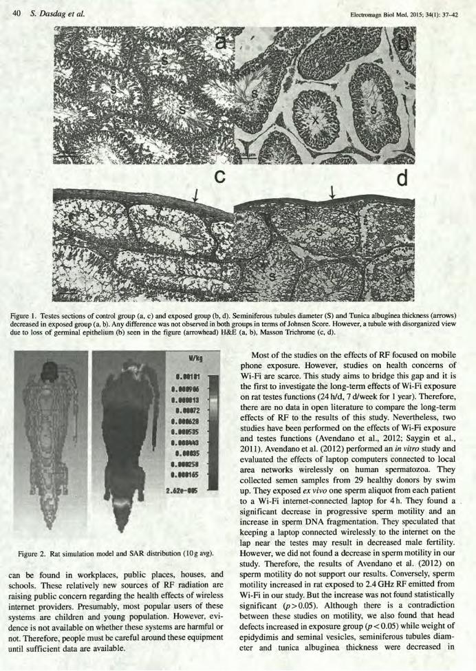


!!!
Attachment!13!

Use of laptop computers connectedto internet through Wi-Fi decreaseshuman sperm motility and increasessperm DNA fragmentationConrado Avenda~no, M.S.,a Ariela Mata, M.S.,a C!esar A. Sanchez Sarmiento, M.D., Ph.D.,a
and Gustavo F. Doncel, M.D., Ph.D.b
a NascentisMedicina Reproductiva, C!ordoba, Argentina; and b CONRAD, Department ofObstetrics andGynecology, Eastern VirginiaMedicalSchool, Norfolk, Virginia
Objective: To evaluate the effects of laptop computers connected to local area networks wirelessly (Wi-Fi) on human spermatozoa.Design: Prospective in vitro study.Setting: Center for reproductive medicine.Patient(s): Semen samples from 29 healthy donors.Intervention(s): Motile sperm were selected by swim up. Each sperm suspension was divided into two aliquots. One sperm aliquot (experimental) fromeach patient was exposed to an internet-connected laptop by Wi-Fi for 4 hours, whereas the second aliquot (unexposed) was used as control, incubatedunder identical conditions without being exposed to the laptop.Main Outcome Measure(s): Evaluation of sperm motility, viability, and DNA fragmentation.Result(s): Donor sperm samples, mostly normozoospermic, exposed ex vivo during 4 hours to a wireless internet-connected laptop showed a significantdecrease in progressive spermmotility and an increase in sperm DNA fragmentation. Levels of dead sperm showed no significant differences between thetwo groups.Conclusion(s): To our knowledge, this is the first study to evaluate the direct impact of laptop use on human spermatozoa. Ex vivo exposure of humanspermatozoa to a wireless internet-connected laptop decreased motility and induced DNA fragmentation by a nonthermal effect. We speculate thatkeeping a laptop connected wirelessly to the internet on the lap near the testes may result in decreased male fertility. Further in vitro and in vivostudies are needed to prove this contention. (Fertil Steril! 2012;97:39–45. "2012 by American Society for Reproductive Medicine.)Key Words: Laptop computer, Wi-Fi, sperm quality, fertility, sperm DNA fragmentation
I n recent years, the use of portablecomputers (laptops, connected tolocal area networks wirelessly, also
known as Wi-Fi) has increased dramat-ically. Laptops have become indispens-able devices in our daily life, offeringflexibility and mobility to users. Peopleusing Wi-Fi may be exposed to radiosignals absorbing some of the transmit-ted energy in their bodies. Portablecomputers are commonly used on thelap (1–3), therefore exposing thegenital area to radio frequency
electromagnetic waves (RF-EMW) aswell as high temperatures (3, 4).
Infertility is a common worldwidecondition that affects more than 70million couples of reproductive age(5). It has been suggested that male fer-tility has declined during the past sev-eral decades (6). Such decline has beenattributed to the direct or indirect expo-sure to certain environmental factorssuch as RF-EMW (7).
Extremely low frequency magneticfields can initiate a number of biochemi-
cal and physiological alterations inbiological systems of different species(8–12). Many of these effects have beenassociated with free-radical production(13, 14). Free radicals are causativefactors of oxidative damage of cellularstructures and molecules such as lipids,proteins, and nucleic acids. Free radicalsreact with polyunsaturated fattyacids in cell membranes promotinga process called lipid peroxidation. Inhuman spermatozoa the presence ofunesterified polyunsaturated fatty acidsis causally associated with the inductionof reactive oxygen species (ROS)generation and lipid peroxidation (15).Damage may occur at the membranelevel, leading to immotility and celldeath, or at the DNA level. DNA integrityis essential to normal conception.Sperm DNA fragmentation has beenassociated with impaired fertilization,poor embryonic development, high
Received June 14, 2011; revised September 9, 2011; accepted October 11, 2011; published onlineNovember 23, 2011.
C.A. has nothing to disclose. A.M. has nothing to disclose. C.A.S.S. has nothing to disclose. G.F.D. hasnothing to disclose.
Reprint requests: ConradoAvenda~no,M.S., NascentisMedicinaReproductiva,Montevideo359, C!ordoba,C!ordoba X5000AXA, Argentina (E-mail: [email protected] or [email protected]).
Fertility and Sterility® Vol. 97, No. 1, January 2012 0015-0282/$36.00Copyright ©2012 American Society for Reproductive Medicine, Published by Elsevier Inc.doi:10.1016/j.fertnstert.2011.10.012
VOL. 97 NO. 1 / JANUARY 2012 39
ORIGINAL ARTICLES: ANDROLOGY

rates of miscarriage, and increased incidence of morbidity in theoffspring, including childhood cancer (16, 17). It hasbeen proposed that genetic and environmental factors wouldbe involved in the etiology of sperm DNA damage (18).
The RF-EMW from mobile phones may cause DNAdamage (19), in addition to decreased motility and viability(20, 21). Increased levels of intracellular ROS (22) would bethe cause of these deleterious effects.
Portable computers using Wi-Fi emit RF-EMW and aretypically positioned close the male reproductive organs. Theirpotential negative effects on male germ cells have not beenelucidated. To assess this potential association we used anin vitro model incubating human sperm in the presence ofan active portable computer connected to the internet byWi-Fi. Sperm viability, motility, and DNA fragmentationwere the main study end points.
MATERIALS AND METHODSSubjectsUse of these samples for research purposes was approved bythe Institutional Ethics Committee of Nascentis Medicina Re-productiva, C!ordoba, Argentina, and all participants gavewritten informed consent. Twenty-nine semen samples werecollected by masturbation from healthy donors after 2–5days of sexual abstinence. After liquefaction, sperm concen-tration and motility were determined by light microscopy, us-ing a Makler chamber (Mid Atlantic Diagnostics Inc.). Spermmorphology was examined at !1,000 oil immersion micros-copy by strict criteria after staining with the Papanicolaumethod as previously described (23). Preparation andassessment were performed by a single experienced operator.Semen samples with more than 0.5 million/mL of peroxidase-positive leukocytes were discarded and not used in the study.
Motile spermatozoa were selected by swim up performedin modified human tubal fluid (HTF; Irvine Scientific) supple-mented with 10% synthetic serum substitute (SSS; Irvine).Briefly, each sperm sample was diluted 1:1 with modifiedHTF and then centrifuged at 300 ! g for 10 minutes. The su-pernatant was discarded and the pellet was gently layeredwith 1 mL of modified HTF/SSS and incubated at 37"C, ata 45" angle, for 1 hour. After the incubation period, the top0.5 mL of the supernatant, which is enriched in the motilesperm, was withdrawn carefully and sperm concentrationand motility were determined. The sperm concentration wasadjusted to 10–20 million/mL with modified HTF/SSS.
Each sperm suspension sample was aliquoted in two frac-tions (A and B) and a drop of 400 mL was placed in 35 ! 10mm Petri dishes (Falcon 3001). This was covered with 3 mL ofembryo oil (Irvine) to avoid evaporation. Fractions Bwere incu-bated under a laptop computer. Fractions A (control group)were incubated under similar conditions without the computer.
Incubation of Spermatozoa Under LaptopFor each sperm sample, one of the dishes (fraction B) was in-cubated at room temperature under a laptop computer (Tosh-iba Satellite M305D-S4829) connected to the internetwirelessly (Wi-Fi, frequency 2.4 GHz defined by IEEE
802.11b). To induce the greatest possible effect, the laptopworked actively (uploading and downloading information)throughout the period of exposure (24). To maximize the like-lihood of observing deleterious effects the distance betweenthe computer and each specimen was kept constant at 3 cm.This distance was the estimated distance between the com-puters resting on the lap and the testis/epididymis (Fig. 1Band C). The duration of exposure was 4 hours (Fig. 1A). Thetemperature under the laptop was kept at 25"C during the in-cubation time by an air conditioning system. The temperatureon each medium drop was thoroughly controlled by an IVFThermometer (Research Instruments) and recorded every 5minutes. Unexposed aliquots (fraction A) were used as controland kept under identical temperature and conditions in an-other room away from any computers or electronic devices.After the incubation period, sperm vitality, motility, andDNA fragmentation were determined on each aliquot.
Vitality and MotilitySperm vitality was evaluated by eosin stain according tospecifications of the World Health Organization (25). Spermmotility was assessed microscopically using a Makler cham-ber (Mid Atlantic Diagnostics Inc.), and sperm movementwas classified as progressive motility, nonprogressive motil-ity, and immotility.
TUNEL AssaySperm DNA fragmentation was evaluated with TUNEL assayusing the in situ cell death detection kit, fluorescein (RocheDiagnostics GmbH). The assay uses fluorescein-dUTP to labelsingle and double DNA strand breaks according to manufac-turer’s instructions and was performed as previously de-scribed (26). Briefly, spermatozoa were fixed withparaformaldehyde (final concentration 2%, permeabilizedwith 0.1% Triton X-100) and incubated in the dark at 37"Cfor 1 hour in TUNEL reaction mixture containing 0.5 IU/mLof calf thymus terminal deoxynucleotidyl transferase andfluorescein-dUTP. Negative (omitting the enzyme terminaltransferase) and positive (using deoxyribonuclease I,1 U/mL for 20 minutes at room temperature) controls wereincluded in each experiment. Mounting medium for fluores-cence (Vectashield, Vector Laboratories) was added beforethe evaluation to prevent fluorescence quenching. A total of500 cells were randomly analyzed per sample in a Zeiss Axi-oplan (Carl Zeiss MicroImaging) microscope with a!1,000 oilimmersion objective. Each sperm cell was classified as havingintact DNA (no fluorescence) or fragmented DNA (green nu-clear fluorescence). As expected, none of the cells showedfluorescent staining in the negative control, whereas 100%of the cells showed fragmentation in the positive control(treated with DNase). The results were expressed as percentageof sperm with fragmented DNA.
Power DensityA RF Field Strength Meter (Alphalab) was used to measureradiation under the experimental conditions. The RF FieldStrength Meter detects the electric field of radio and
40 VOL. 97 NO. 1 / JANUARY 2012
ORIGINAL ARTICLE: ANDROLOGY

microwaves from 0.5 MHz–3 GHz, and expresses the fieldstrength as power density (0.001–2,000mW/cm2). The RF FieldStrength Meter is directional and detects only the componentof the electric field that has the same polarization as the longaxis of the meter. To find the highest reading, the meter waslocated at the same distance as the Petri dishes and positionedvertically and horizontally, according to the manufacturer’sspecifications. The horizontal way showed higher readingand was recorded. Power density was monitored at the samedistance of the Petri dishes, during basal condition (no expo-sure to laptop), laptop without Wi-Fi connection and laptopworking in Wi-Fi mode throughout the experiment.
Statistical AnalysisData were expressed as mean # SD. The Mann-Whitney testwas used to identify differences between two groups. P< .05was considered statistically significant.
RESULTSDonors’ mean age was 34.1 # 5.6 years (range 26–45 years).Semen parameters (volume, concentration, motility, vitality,and morphology) are presented in Supplemental Table 1(available online). Many samples showed normozoospermia,whereas four samples showed low semen volume (LS6,LS13, LS27, and LS29) and three (LS15, LS16, and LS25) pre-sented isolated teratozoospermia, according to World HealthOrganization reference values (25, 27).
Room and under-the-laptop temperatures were moni-tored during the incubation time and kept at 25"C for bothsperm fractions (A and B) by an air conditioning system(Supplemental Fig. 1, available online). The RF-EMW wererecorded every 10 minutes in both groups throughout the ex-periment. The RF-EMW from a laptop working without Wi-Ficonnection were checked in a pilot experiment (Fig. 2). Theradiation from the computer operating with Wi-Fi was threeor more times higher than without Wi-Fi and 7–15 timeshigher than basal conditions (not exposed to laptop).
Sperm parameters were evaluated after 4 hours ofincubation of motile sperm selected by swim up and exposedto an active laptop computer under controlled temperature con-ditions. There were no differences in the percentage of viablesperm between the test and control groups (Fig. 3A). On thecontrary, laptop exposure induced a significant decrease insperm progressivemotilitywith a concomitant increase in non-motile sperm compared with the unexposed controls (P< .05).The percentage of nonprogressive sperm did not show statisti-cally significant differences (Fig. 3B). Important, a significantincrease in spermDNA fragmentationwas found in the fractionincubated under the computer comparedwith the control group(3.3 # 6.0 vs. 8.3 # 6.6; P< .05; Fig. 4A and B).
DISCUSSIONTo our knowledge, this is the first study to examine the effectof portable computers on human spermatozoa in vitro. In the
FIGURE 1
Study design and set-up for the exposure of human sperm to laptop. (A) Experimental design. (B) Schematic situation of the use of the laptop on thelap near the testes. (C) Diagram of the in vitro study. The Petri dishes were placed at 3 cm from a laptop computer. Each Petri dish contained a dropof 400 mL of sperm suspension in human tubal fluid/synthetic serum substitute (HTF/SSS) covered with mineral oil to prevent evaporation.Avenda~no. Laptop usage and sperm quality. Fertil Steril 2012.
VOL. 97 NO. 1 / JANUARY 2012 41
Fertility and Sterility®

present study we demonstrate that laptop computers con-nected wirelessly to the internet decrease sperm quality bya nonthermal effect. We evaluated vitality, motility, andDNA fragmentation in sperm selected by swim up after incu-bation under a laptop connected to the internet by Wi-Fi. Theresults demonstrate a significant decrease in sperm progres-sive motility and a significantly higher proportion of spermwith DNA fragmentation when samples were incubated for4 hours under the laptop. These differences were seen in com-parison with aliquots of the same semen samples incubated
under similar conditions but outside the proximity of anycomputer or electronic device.
Several lifestyle and environmental factors may ad-versely impact human health and, in particular, reproductiveperformance (18). Approximately 15% of the sexually activepopulation is affected by clinical infertility and in 50% ofthe cases a male factor is involved, either as a primary prob-lem or in combination with a problem in the female partner(28). In this regard it has been proposed that the increaseduse of certain new technologies may decrease fertility
FIGURE 2
Variation of electromagnetic radiation (in microwatts per centimeter squared) during incubation time under the following experimental conditions:CCC laptop conneted to Wi- Fi; laptop without connection to Wi-Fi; —— no laptop (basal conditions). The radio frequencyelectromagnetic waves were 7–15 times higher under the laptop than in basal conditions. They were significantly decreased when the Wi-Fiwas turned off compared with the laptop working with Wi-Fi.Avenda~no. Laptop usage and sperm quality. Fertil Steril 2012.
FIGURE 3
Laptop exposure and human sperm quality. Spermatozoa (10–20 ! 106 cells/mL) were suspended in modified human tubal fluid/synthetic serumsubstitute (HTF/SSS)medium and incubated under a laptop computer connected to internet toWi-Fi (FB). Another sperm aliquot was placed outsidethe reach of other computers or electronic devices (FA). Both groups were incubated for 4 hours at 25"C. (A) Percentage of dead sperm were notsignificantly different between the laptop exposed and unexposed groups and the unexposed cells (9.5% # 3.3% vs. 8.9% # 3.3%, P>.05). (B)Progressive sperm motility (PG) was significantly reduced in the group incubated under the laptop compared with that of control group (68.7% #8.8% to 80.9% # 7.5%, *P<.01). No difference was found in the percentage of nonprogressive (NP) spermatozoa between groups. Immotilesperm (IM) were significantly increased after laptop exposure (24.5% # 7.6% vs. 13.6% # 5.6%, *P<.01).Avenda~no. Laptop usage and sperm quality. Fertil Steril 2012.
42 VOL. 97 NO. 1 / JANUARY 2012
ORIGINAL ARTICLE: ANDROLOGY

potential by increasing long-term exposure to nonionizingradiation (18).
Laptop computer usage has increased significantly in re-cent years, especially in people of reproductive age. Fre-quently laptops are connected to the internet through Wi-Fiand commonly placed on the lap near the testes. Portablecomputers actively generate high temperatures that can in-crease the scrotal temperature and may produce deleteriouseffects on spermatogenesis (3, 29). In addition, laptopcomputers working by Wi-Fi are connected through RF-EMW (4, 24), which may damage spermatozoa in the malereproductive organs through microwave radiation.
To set up this study we first evaluated the radiation emit-ted from a laptop. The radiation varied during the test and de-pended on the flow of information between the computer andthe network to which it was connected (Fig. 2).
Overall, however, the RF-EMW were 7–15 times higherunder the laptop than under basal conditions (no laptop).Compared with the laptop working with Wi-Fi, RF-EMWwere significantly decreased when the Wi-Fi was turned off.
It is well known that increased temperature may decreasesperm quality (30) and the use of portable computers on thelap increases scrotal temperature (3). Therefore to preventconfounding thermal effects, room and incubation tempera-tures were kept constant in both the unexposed and theunder-the-laptop groups (Fig. 1) during the incubation time.
The first relevant finding of this study was a significantdecrease in sperm progressive motility after exposure to thelaptop. A plausible explanation for the impaired sperm motil-ity could be magnetic and electromagnetic fields inducingoxidation of phospholipids, which are a major componentin the sperm mitochondrial sheath (31). Several studies haveshown that higher ROS values have detrimental effects onthe motility of normal human spermatozoa (15, 32).Furthermore, it has been reported that infertile men withhigh seminal ROS levels have a lower percentage of motile
sperm (27). This can be explained by a disturbance of the mi-tochondrial membrane potential, which causes high levels ofROS to be released into the cytoplasm, depleting the energysupply and affecting both sperm motility and kinetics(33, 34). Interestingly, in our study, sperm vitality was notdifferent between the two experimental groups (Fig. 3).
Concerning spermatozoa, RF-EMW generated by mobilephones cause a decrease in their progressive movement, inboth human and rat cells (21, 22, 35). In vitro humanspermatozoa exposed to mobile phone radiation showedreduced sperm head area and decreased sperm binding tothe zona pellucida (ZP) without an increase in acrosomalreaction compared with controls (36). High levels of spermROS (22, 37), as well as an increased percentage of spermDNA fragmentation (37), have been reported after mobilephone exposure. However, other studies did not find anychanges in DNA integrity or the induction of proapoptoticmarkers (22, 38). The lack of DNA damage observed in thesestudies might be explained by the shorter time of exposureto cell phone radiation or by the antioxidant effect ofseminal plasma. The RF-EMW generated by mobile phonesare similar to those generated by laptop computer and othermobile devices connected wirelessly to the internet (4).
In this regard we found that ex vivo exposure of humanspermatozoa for 4 hours in the absence of seminal plasma in-duces DNA damage by a nonthermal effect (Fig. 4). This effectis similar to that observed by De Iuliis and co-workers (37)with sperm exposed to mobile phones in vitro. These investi-gators observed highly significant relationships betweenRF-EMW emitted by mobile phones, covering a range ofspecific absorption rates, the oxidative DNA damage marker,8-hydroxy-20-deoxyguanosine, and sperm DNA fragmenta-tion. These changes were unrelated to thermal effects.
Research has shown negative consequences of electro-magnetic fields on biological mechanisms. Genotoxic damageby 1.8 GHz in human fibroblast has been proposed as a direct
FIGURE 4
Laptop exposure and human sperm DNA fragmentation. Sperm suspensions were incubated under a laptop computer connected to the internet byWi-Fi (FB) during 4 hours at 25"C. Aliquots of the same samples were placed outside of the reach of other computers or electronic devices, ina separate room (FA). (A) Sperm DNA fragmentation was increased after 4 hours of laptop exposure. In the test group, 8.6% # 6.6% of thecells were fragmented, whereas only 3.3% # 6.0% of the controls showed DNA fragmentation (*P<.01). (B) Plot of individual responses ofsperm DNA fragmentation to laptop exposure. The number of sperm with fragmented DNA was evaluated in two aliquots of the same sample(500 cells/aliquot).Avenda~no. Laptop usage and sperm quality. Fertil Steril 2012.
VOL. 97 NO. 1 / JANUARY 2012 43
Fertility and Sterility®

consequence of intermittent exposure to RF-EMW (39). Afterexposure to 2.45 GHz, alteration of gene expression wasfound in cultured human cells mediated by a nonthermalmechanism (40). Electromagnetic radiation from mobilephones induces activation of the extracellular signal-regu-lated kinases (ERK)-cascade thereby altering transcriptionand other key cellular processes (8). Chronic exposure tolow intensity microwaves (2.45 and 16.5 GHz) causes statisti-cally significant increase in DNA strand breaks in rat braincells (41). A recent work showed that the exposure of oviductsto extremely low frequency electromagnetic field negativelyaffects early embryo development, causing a slowdown inthe embryo cleavage rate (42).
In an in vivo study, it was demonstrated that acute expo-sure to radiofrequency fields of cellular phones may modulatethe oxidative stress of free radicals by enhancing lipidperoxidation, and a decrease in the activity of the antioxi-dants, superoxide dismutase, and glutathione peroxidase, inerythrocytes from human volunteers (43). In addition, geno-toxic effect on epididymal spermatozoa has been reportedwhen mice were irradiated for 7 days at 12 hours per day (19).
As opposed to somatic cells, the spermatozoon is a highlyspecialized cell with a condensed DNA (packaged by prot-amines) and very small cytoplasmic area (44). Duringspermatogenesis, human spermatozoa may take up to 1week to move from the seminiferous tubules in the testes tothe cauda epididymis (45) and throughout this time theywould be highly vulnerable to RF-EMW exposure (19, 37),especially from a source close to the testes and epididymissuch as a Wi-Fi laptop computer.
Our data suggest that the use of a laptop computer wire-lessly connected to the internet and positioned near themale reproductive organs may decrease human sperm qual-ity. At present we do not know whether this effect isinduced by all laptop computers connected by Wi-Fi tothe internet or what use conditions heighten this effect.The mechanisms involved in mediating the decrease insperm motility and DNA integrity also need further study.We speculate that RF-EMW from laptop computers wire-lessly connected to the internet may be the cause of spermdamage. However, we cannot discard the possibility thatdamage to sperm is caused by the low radiation producedby the computer without internet connection. With thecaveat that these data were obtained with sperm samplesincubated in vitro, our findings suggest that prolongeduse of portable computers sitting on the lap of a maleuser may decrease sperm fertility potential. The potentialimplications of these findings warrant this report and fur-ther basic and clinical investigation.
Acknowledgments: The authors thank Professor NestorHugo Mata for invaluable contribution in RF-EMWmeasure-ments, Valeria Martinez for her assistance and technical sup-port, and the staff of Nascentis Medicina Reproductiva fortheir helpful input. Conrado Avenda~no is grateful to the Insti-tuto de Fisiología, Facultad de Ciencias M!edicas and Centro deInvestigaciones en Química Biol!ogica de C!ordoba, Facultad deCiencias Químicas from Universidad Nacional de C!ordoba,Argentina, for allowing the use of their fluorescence micro-
scope. Special thanks to Cecilia Sampedro for her help inthe fluorescence microscope.
REFERENCES1. Paulius K, Napoles P, Maguina P. Thigh burn associated with laptop com-
puter use. J Burn Care Res 2008;29:842–4.2. Ostenson CG. Lap burn due to laptop computer. Lancet 2002;360:1704.3. Sheynkin Y, Jung M, Yoo P, Schulsinger D, Komaroff E. Increase in scrotal
temperature in laptop computer users. Hum Reprod 2005;20:452–5.4. World Health Organization. WHO research agenda for radio frequency fields.
Available at: http://whqlibdoc.who.int/publications/2010/9789241599948_eng.pdf. Accessed May 11, 2011.
5. Boivin J, Bunting L, Collins JA, Nygren KG. International estimates ofinfertility prevalence and treatment-seeking: potential need and demandfor infertility medical care. Hum Reprod 2007;22:1506–12.
6. ESHRE Capri Workshop Group. Europe, the continent with the lowest fertil-ity. Hum Reprod Update 2010;16:590–602.
7. Agarwal A, Desai NR, Ruffoli R, Carpi A. Lifestyle and testicular dysfunction:a brief update. Biomed Pharmacother 2008;62:550–3.
8. Friedman J, Kraus S, Hauptman Y, Schiff Y, Seger R. Mechanism ofshort-term ERK activation by electromagnetic fields at mobile phonefrequencies. Biochem J 2007;405:559–68.
9. Lupke M, Frahm J, Lantow M, Maercker C, Remondini D, Bersani F, et al.Gene expression analysis of ELF-MF exposed human monocytes indicatingthe involvement of the alternative activation pathway. Biochim BiophysActa 2006;1763:402–12.
10. Yamaguchi DT, Huang J, Ma D,Wang PK. Inhibition of gap junction intercel-lular communication by extremely low-frequency electromagnetic fields inosteoblast-like models is dependent on cell differentiation. J Cell Physiol2002;190:180–8.
11. Volkow ND, Tomasi D, Wang GJ, Vaska P, Fowler JS, Telang F, et al. Effectsof cell phone radiofrequency signal exposure on brain glucose metabolism.JAMA 2011;305:808–13.
12. Cuccurazzu B, Leone L, PoddaMV, Piacentini R, Riccardi E, Ripoli C, et al. Ex-posure to extremely low-frequency (50 Hz) electromagnetic fields enhancesadult hippocampal neurogenesis in C57BL/6 mice. Exp Neurol 2010;226:173–82.
13. Blank M. Do electromagnetic fields interact with electrons in the Na,K-ATPase? Bioelectromagnetics 2005;26:677–83.
14. Palumbo R, Capasso D, Brescia F, Mita P, Sarti M, Bersani F, et al. Effects onapoptosis and reactive oxygen species formation by Jurkat cells exposed to50 Hz electromagnetic fields. Bioelectromagnetics 2006;27:159–62.
15. Aitken RJ, Wingate JK, De Iuliis GN, Koppers AJ, McLaughlin EA. Cis-unsa-turated fatty acids stimulate reactive oxygen species generation and lipidperoxidation in human spermatozoa. J Clin Endocrinol Metab 2006;91:4154–63.
16. Avendano C, Franchi A, Duran H, Oehninger S. DNA fragmentation of nor-mal spermatozoa negatively impacts embryo quality and intracytoplasmicsperm injection outcome. Fertil Steril 2010;94:549–57.
17. Aitken RJ, De Iuliis GN. Origins and consequences of DNA damage in malegerm cells. Reprod Biomed Online 2007;14:727–33.
18. Pacey AA. Environmental and lifestyle factors associated with sperm DNAdamage. Hum Fertil (Camb) 2010;13:189–93.
19. Aitken RJ, Bennetts LE, Sawyer D, Wiklendt AM, King BV. Impact of radiofrequency electromagnetic radiation on DNA integrity in the male germline.Int J Androl 2005;28:171–9.
20. Erogul O, Oztas E, Yildirim I, Kir T, Aydur E, Komesli G, et al. Effects ofelectromagnetic radiation from a cellular phone on human sperm motility:an in vitro study. Arch Med Res 2006;37:840–3.
21. Yan JG, Agresti M, Bruce T, Yan YH, Granlund A, Matloub HS. Effects ofcellular phone emissions on sperm motility in rats. Fertil Steril 2007;88:957–64.
22. Agarwal A, Desai NR, Makker K, Varghese A, Mouradi R, Sabanegh E, et al.Effects of radiofrequency electromagnetic waves (RF-EMW) from cellularphones on human ejaculated semen: an in vitro pilot study. Fertil Steril2009;92:1318–25.
44 VOL. 97 NO. 1 / JANUARY 2012
ORIGINAL ARTICLE: ANDROLOGY

23. Menkveld R, Kruger TF. Sperm morphology—predictive value? Fertil Steril1992;57:942–3.
24. Foster KR. Radiofrequency exposure from wireless LANs utilizing Wi-Fitechnology. Health Phys 2007;92:280–9.
25. World Health Organization. WHO laboratory manual for the examination ofand procesing of human semen. 5th ed. Cambridge, UK: CambridgeUniversity Press; 2010.
26. Avendano C, Franchi A, Taylor S, Morshedi M, Bocca S, Oehninger S.Fragmentation of DNA in morphologically normal human spermatozoa.Fertil Steril 2009;91:1077–84.
27. Cooper TG, Noonan E, von Eckardstein S, Auger J, Baker HW, Behre HM,et al. World Health Organization reference values for human semen charac-teristics. Hum Reprod Update 2010;16:231–45.
28. Thonneau P, Marchand S, Tallec A, Ferial ML, Ducot B, Lansac J, et al. Inci-dence and main causes of infertility in a resident population (1,850,000) ofthree French regions (1988–1989). Hum Reprod 1991;6:811–6.
29. Sheynkin Y, Welliver R, Winer A, Hajimirzaee F, Ahn H, Lee K. Protectionfrom scrotal hyperthermia in laptop computer users. Fertil Steril 2011;95:647–51.
30. Thonneau P, Bujan L, Multigner L, Mieusset R. Occupational heat exposureand male fertility: a review. Hum Reprod 1998;13:2122–5.
31. Tremellen K. Oxidative stress and male infertility—a clinical perspective.Hum Reprod Update 2008;14:243–58.
32. Plante M, de Lamirande E, Gagnon C. Reactive oxygen species released byactivated neutrophils, but not by deficient spermatozoa, are sufficient toaffect normal sperm motility. Fertil Steril 1994;62:387–93.
33. Mahfouz R, Sharma R, Thiyagarajan A, Kale V, Gupta S, Sabanegh E, et al.Semen characteristics and sperm DNA fragmentation in infertile men withlow and high levels of seminal reactive oxygen species. Fertil Steril2010;94:2141–6.
34. Pasqualotto FF, Sharma RK, Nelson DR, Thomas AJ, Agarwal A. Relation-ship between oxidative stress, semen characteristics, and clinical diagno-sis in men undergoing infertility investigation. Fertil Steril 2000;73:459–64.
35. Falzone N, Huyser C, Fourie F, Toivo T, Leszczynski D, Franken D. In vitro ef-fect of pulsed 900 MHz GSM radiation on mitochondrial membrane poten-tial and motility of human spermatozoa. Bioelectromagnetics 2008;29:268–76.
36. Falzone N, Huyser C, Becker P, Leszczynski D, Franken DR. The effect ofpulsed 900-MHz GSM mobile phone radiation on the acrosome reaction,head morphometry and zona binding of human spermatozoa. Int J Androl2010;34:20–6.
37. De Iuliis GN, Newey RJ, King BV, Aitken RJ. Mobile phone radiation inducesreactive oxygen species production and DNA damage in human spermato-zoa in vitro. PLoS One 2009;4:e6446.
38. Falzone N, Huyser C, Franken DR, Leszczynski D. Mobile phone radiationdoes not induce pro-apoptosis effects in human spermatozoa. Radiat Res2010;174:169–76.
39. Diem E, Schwarz C, Adlkofer F, Jahn O, Rudiger H. Non-thermal DNA break-age by mobile-phone radiation (1800 MHz) in human fibroblasts and intransformed GFSH-R17 rat granulosa cells in vitro. Mutat Res 2005;583:178–83.
40. Lee S, Johnson D, Dunbar K, Dong H, Ge X, Kim YC, et al. 2.45 GHz radio-frequency fields alter gene expression in cultured human cells. FEBS Lett2005;579:4829–36.
41. Paulraj R, Behari J. Single strand DNA breaks in rat brain cells exposed to mi-crowave radiation. Mutat Res 2006;596:76–80.
42. Bernabo N, Tettamanti E, Russo V, Martelli A, Turriani M, Mattoli M, et al.Extremely low frequency electromagnetic field exposure affects fertilizationoutcome in swine animal model. Theriogenology 2010;73:1293–305.
43. Moustafa YM, Moustafa RM, Belacy A, Abou-El-Ela SH, Ali FM. Effects ofacute exposure to the radiofrequency fields of cellular phones on plasmalipid peroxide and antioxidase activities in human erythrocytes. J Pharm Bi-omed Anal 2001;26:605–8.
44. Eddy EM. The spermatozoon. In: Knobil E, Neil J, editors. The physiology ofreproduction. New York: Raven Press; 1994:3–54.
45. Turner TT. On the epididymis and its role in the development of the fertileejaculate. J Androl 1995;16:292–8.
VOL. 97 NO. 1 / JANUARY 2012 45
Fertility and Sterility®

SUPPLEMENTAL FIGURE 1
Incubation temperature in test (under the laptop; ) and control (unexposed;CCCC) groups. Room temperature was kept constant duringincubation (4 hours) both in the control and the experimental groups.Avenda~no. Laptop usage and sperm quality. Fertil Steril 2012.
45.e1 VOL. 97 NO. 1 / JANUARY 2012
ORIGINAL ARTICLE: ANDROLOGY

SUPPLEMENTAL TABLE 1
Basic semen parameters of study samples.
Semen parameter Mean
Volume (mL) 2.8 # 1.6a
Concentration (! 106 / mL) 111.0 # 49.8Vitality (%) 85.3 # 4.6Progressive motility (%) 60.2 # 9.3Morphology (%) 8.2 # 4.7a Mean # SD.
Avenda~no. Laptop usage and sperm quality. Fertil Steril 2012.
VOL. 97 NO. 1 / JANUARY 2012 45.e2
Fertility and Sterility®

!!!
Attachment!14!

ORIGINAL ARTICLE
What is harmful for male fertility: Cell phone orthe wireless internet?
Mehmet Erol Yildirim a,*, Mehmet Kaynar b, Huseyin Badem c,Mucahıt Cavis a, Omer Faruk Karatas a, Ersın Cimentepe a
a Department of Urology, Turgut Ozal University School of Medicine, Ankara, Turkeyb Department of Urology, Selcuk University School of Medicine, Konya, Turkeyc Department of Urology, Yuksek _Ihtisas Education and Research Hospital, Ankara, Turkey
Received 16 February 2015; accepted 4 May 2015Available online 26 July 2015
KEYWORDSCellular phone;Radiofrequency-electromagneticradiation;Sperm;Wi-Fi
Abstract In this study, we aimed to assess the potential harmful effects of radiofrequency-electromagnetic radiation on sperm parameters. We requested semen for analyses from themale patients coming to our infertility division and also asked them to fill out an anonymousquestionnaire. We queried their mobile phone and wireless internet usage frequencies in orderto determine their radiofrequency-electromagnetic radiation exposure. A total of 1082 pa-tients filled the questionnaire but 51 of them were excluded from the study because of azoo-spermia. There was no significant difference between sperm counts and sperm morphologyexcluding sperm motility, due to mobile phone usage period, (p Z 0.074, p Z 0.909, andp Z 0.05, respectively). The total motile sperm count and the progressive motile sperm countdecreased due to the increase of internet usage (p Z 0.032 and p Z 0.033, respectively). Inline with the total motile sperm count, progressive motile sperm count also decreased withwireless internet usage compared with the wired internet connection usage (p Z 0.009 andp Z 0.018, respectively). There was a negative correlation between wireless internet usageduration and the total sperm count (r Z !0.089, p Z 0.039). We have also explored the nega-tive effect of wireless internet use on sperm motility according to our preliminary results.Copyright ª 2015, Kaohsiung Medical University. Published by Elsevier Taiwan LLC. All rightsreserved.
Conflicts of interest: All authors declare no conflicts of interest.* Corresponding author. Department of Urology, Turgut Ozal University School of Medicine, 06510, Yenimahalle, Ankara, Turkey.E-mail address: [email protected] (M.E. Yildirim).
http://dx.doi.org/10.1016/j.kjms.2015.06.0061607-551X/Copyright ª 2015, Kaohsiung Medical University. Published by Elsevier Taiwan LLC. All rights reserved.
Available online at www.sciencedirect.com
ScienceDirect
journal homepage: http: / /www.kjms-onl ine.com
Kaohsiung Journal of Medical Sciences (2015) 31, 480e484

Introduction
Cell phone and wireless internet have become an indis-pensable part of our lives. Especially, after the develop-ment of smart phones and 3G internet technologies, theexposure to the radiofrequency (RF) electromagnetic radi-ation (EMR) has increased to terrifying levels. Cell phoneand wireless technologies (Wi-Fi) operate from 850 MHz to1800 MHz and w2400 MHz; respectively [1,2]. Tissues canabsorb RF-EMR in many ways including aerial effect and/orcoupling the signal [3]. Previously, the harmful effects ofRF-EMR on DNA integrity and on various organs such as thebrain and heart have been very well described [4]. TheWorld Health Organization (WHO) officially declared thatcell phones can cause brain cancer [5]. After this declara-tion, usage of hands-free devices has increased but cellphones still remain close to the gonads of individuals thatmay result in infertility due to the harmful effects of RF-EMR.
Infertility is a common disorder that affects 15% ofcouples and nearly half of the cases are due to maleinfertility. As mentioned above, RF-EMR affects many or-gans including the testes by a direct or a thermal effect [6].In one study, detrimental effects of RF-EMR on Leydig cells,seminiferous tubules, and especially the spermatozoa wereclearly defined [1]. Although RF-EMR reduces testosteronelevels, impairs spermatogenesis, and causes sperm DNAdamage [4], the relationship between RF-EMR devices andmale infertility is still controversial.
In the literature, the harmful effects of RF-EMR on malereproductive systems are shown in rats, however; humanstudies are very rare and can only be planned with a smallerpopulation [4,7]. For instance, Agarwal et al [4] detectedthe negative effects of cell phones on sperm parameters in361 men. Similarly, Fejes at al [7] showed the negativecorrelation between the daily cell phone usage durationand semen quality in 371 men. In this decade, wirelessinternet connection has been involved in our lives as muchas cell phones with 3G technologies. As we all know, Wi-Ficonnection transmits more RF-EMR than cell phones, so weexamined the effects of both cell phone and wirelessinternet use on sperm parameters in healthy males in orderto determine the possible harmful effects of RF-EMRdevices.
Materials and methods
This study was performed under the approval of ourInstitutional Review Board in our university (Turgut OzalUniversity, Ankara, Turkey) (999500669/869), and informedconsents were obtained from all patients. In our popula-tion based observational study, we collected data from1082 healthy men who attended the Andrology subdivisionof the Urology Department (Turgut Ozal University) be-tween June 2013 and June 2014. Men with a history oforchitis, varicocele, diabetes mellitus, cardiac, neuraldisease, nephritic disease, and hypertension, or men whohad a family history of any genetic disease were excludedfrom the study. In addition, patients who suffered from aviral/bacterial infection in the previous 4 weeks, had anin vitro fertilization history, or were already recruited to
an intacytoplasmic sperm injection program were alsoexcluded from the study. Azoospermic patients wereexcluded from the study. Semen samples were collectedby masturbation in a sterile wide-mouthed calibratedcontainer (Sigma, St. Louis, MO, USA) with the abstinenceof ejaculation for a minimum of 2 days and no longer than5 days before the semen collection. Semen analyses wereperformed according to the WHO guidelines that includeeight sperm parameters: volume, liquefaction time, pH,viscosity, sperm count, motility, viability, and percentageof the normal morphology [8]. Assessments of semenanalysis were performed at the end of the 30-minuteperiod. Sperm motility was analyzed by using a phase-contrast microscope (Nikon, Alphaphot-2, YS-2, Tokyo,Japan) with > 20" magnification. Semen analyses wereperformed by two experienced and blinded operators.Motility and concentrations of semen were evaluated byusing a Makler counting chamber (Sefi-Medical Instrument,Haifa, Israel). WHO criteria (4 categories of sperm move-ment; A: rapid progressive, B: slow progressive, C:nonprogressive, and D: no motility) were used in theassessment of sperm movement. Azoospermic patients andthe patients whose sperm counts were < 5 million/mLwere excluded from the study due to possible factors suchas genetic, testicular hypofunction, or idiopathic. Ananonymous questionnaire including (1) daily the cell phoneusage duration, (2) habits of carrying mobile phone, (3)wireless internet usage duration, and (4) type of internetusage. According to an anonymous questionnaire, dailyactive cell phone usage was divided into three groups asfollowing: Group A, < 30 min/d; Group B, from 30 min/d to 2 h/d; and Group C, > 2 h/d. Habits of carrying amobile phone was recorded as (A) in the pocket of trou-sers, (B) in a handbag, or (C) in the pocket of jackets.Wireless internet usage was divided in to three groups,Group A: < 30 min/d; Group B, from 30 min/d to 2 h/d;and Group C, > 2 h/d. Internet usage types recorded aswireless or not. Body mass index and annual smokinghabits (at least 10 cigarettes a day) were also recorded.Because of the high number of participants we could notask about the cell phone models but we know that all ofthe cell phones operate between the 850e1800 Mhz in ourcountry.
Correlation between the eight sperm parameters wasevaluated by the determination of the Pearson correlationcoefficients. Data were presented as mean # standarddeviation. Statistical analyses were performed by usingStudent t test (2-tailed) and one way analysis of variance(ANOVA). SPSS for Windows (version 16.0; SPSS Inc., Chi-cago, IL, USA) was used for statistical analyses and p < 0.05was considered as statistically significant.
Results
Fifty-one azoospermic patients were excluded from thestudy, and the data of 1031 patients were collected. Theaverage age of the participants was 30.9 # 6.2 (18e63)years. The average body mass index of participants was26.8 # 3.9 (14.9e46.24). Smoking rate was 352/1031 par-ticipants. Of those men, the average smoking duration was9.94 # 5.64 (2e35) years.
Effect of cellular phone and Wi-Fi on infertility 481

Duration of cell phone use
According to our results, there was no significant differencebetween these three groups regarding sperm parameterssuch as semen volume, sperm count, total motile spermcount, progressive motile sperm count, and morphology ofthe sperm. However, there was a significant correlationbetween smoking which has a significantly shorter durationin Group B than the other groups (p Z 0.022; Table 1).
Mobile phone carrying habits
There was a significant difference among the three carriageplaces regarding only sperm morphology (p Z 0.028) but noother sperm parameters (Table 2). The duration of smokingwas significantly shorter in participants who carry their cellphone in a handbag (p Z 0.034).
Duration of wireless internet use
When we compare the wireless internet users regarding theduration of internet usage, there were a significantdecrease of total motile sperm count and progressivemotile sperm count (p Z 0.032 and p Z 0.033; respec-tively; Table 3). Similarly, the duration of smoking wassignificantly shorter in Group B than the other groups. Inaddition to that, there were no significance among the
semen parameters regarding the wired internet group(p Z 0.128).
Type of internet usage
Total motile sperm count and progressive motile spermcount were lower in the wireless internet usage groupcompared with the wired internet usage group (p Z 0.009and p Z 0.018; respectively). Type of internet connectiondoes not affect the other sperm parameters (Table 4).
There was a negative correlation between the cell phoneusage duration and the total sperm count (r Z !0.064,p Z 0.04). Similarly, there was also a negative correlationbetween the wireless internet usage duration and the totalsperm count (r Z !0.089, p Z 0.019). Otherwise therewere no significant correlations among the other four mainquestion branches (cell phone usage time, cell phone car-riage habits, wireless internet usage time. and internetconnection type) and sperm parameters.
Leucocyte count from the semen analyses were normalin all of the patients.
Discussion
Even though there are possible harmful effects from cellphones and wireless internet, we use them frequently inour lives [4]. Kumar et al. [9] have shown that 10 GHz
Table 1 Comparison of the demographic data and spermparameters among the duration of cell phone usage groups.
< 30 min(n Z 382)
30 mine2 h(n Z 286)
> 2 h(n Z 363)
p*
Age (y) 32.3 # 6.63 30.8 # 5.74 29.5 # 5.84 0.059BMI (kg/m2) 26.1 # 3.7 26.5 # 3.4 26.5 # 4.2 0.132Smoking (y) 3.96 # 6.37 2.52 # 4.91 3.47 # 5.59 0.021Volume 2.9 # 1.41 2.9 # 1.19 3.01 # 1.45 0.194TSC (million) 42.3 # 16.3 39.2 # 16.3 37.8 # 16.1 0.074TMS (million) 61.1 # 60.6 54.6 # 50.6 53.8 # 59 0.05PMS (million) 47.5 # 50.8 42.5 # 42.1 41.6 # 51.2 0.083Morphology 2.8 # 1.9 2.57 # 1.76 2.74 # 1.72 0.909
*One way analysis of variance was performed. A p value < 0.05between all groups was considered significant.BMIZ body mass index; PMS Z progressive motile sperm count;TMS Z total motile sperm count; TSC Z total sperm count.
Table 2 Comparison of the demographic data and sperm parameters among the mobile phone carrying habits groups.
Trouser pocket (n Z 767) Handbag (n Z 106) Jacket pocket (n Z 158) p*
Age (y) 30.4 # 6.25 33.3 # 5.9 31.8 # 5.8 0.671BMI (kg/m2) 26.1 # 3.6 27.5 # 4.1 26.7 # 4.6 0.150Smoking (y) 3.46 # 5.55 1.7 # 5.53 4 # 6.53 0.032Volume 2.9 # 1.37 3.08 # 1.4 3.02 # 1.38 0.973TSC (million) 39.1 # 31.1 45 # 31.6 40.3 # 27 0.256TMS (million) 56.5 # 60.1 63 # 48.6 53.6 # 49.1 0.168PMS (million) 43.8 # 51 49.6 # 41.4 41.9 # 41.1 0.538Morphology 2.72 # 1.81 3.18 # 2.47 2.43 # 1.38 0.034
*One way analysis of variance was performed. A p value < 0.05 between all groups was considered significant.
Table 3 Comparison of the demographic data and spermparameters among the duration of wireless internet usagegroups.
< 30 min(n Z 327)
30 mine2h(n Z 164)
> 2 h(n Z 540)
p*
Age (y) 30.9 # 6.77 31.1 # 5.9 30.8 # 5.9 0.023BMI (kg/m2) 25.8 # 3.5 26.8 # 3.7 26.5 # 4.1 0.157Smoking (y) 4.23 # 6.51 2.45 # 5.08 3.16 # 5.38 0.017Volume 2.99 # 1.4 2.81 # 1.32 2.99 # 1.36 0.43TSC (million) 43 # 33 41.8 # 28.2 37.4 # 29.4 0.093TMS (million) 61.7 # 60.2 56.2 # 57.5 53.8 # 57.5 0.032PMS (million) 48.2 # 53.7 43 # 42.1 41.8 # 49.6 0.033Morphology 2.73 # 1.84 2.65 # 1.75 2.73 # 1.85 0.305
*One way analysis of variance was performed. A p value < 0.05between all groups was considered significant.
482 M.E. Yildirim et al.

electromagnetic fields resulted in DNA damage of sperm viamicronuclei formation due to the oxidative stress. To thebest of our knowledge, there are limited numbers of humanand animal studies with respect to cell phone effects ontestes and semen parameters [4,7,10,11]. Additionally, notmuch study has been performed on the impacts of wirelessinternet and cell phones on male fertility. Therefore, thepresent study aimed to determine the effects of cell phoneand wireless internet usage habits on human sperm pa-rameters. For this purpose, an anonymous questionnairewas applied to a total of 1082 healthy men who attendedour urology clinic for semen analysis, and only 1031 patientswere included in the study. According to their answersregarding their habits of carrying mobile phones, cell phoneusage duration, and wireless usage, participants weredivided into four different groups.
In our previous study, we compared the effects of thestandby mode or the active mode of the cell phone on rattestes, and we detected significant apoptotic rates intesticular tissue in the active cell phone group comparedwith other groups [10]. Mailankot et al. [11] have shown adecrease in sperm motility in RF-EMR exposed rats for 28days. Wang et al. [12] have indicated that Leydig cells werethe most susceptible cells to RF-EMR exposure. However,rat studies could have some problems due to their smalltestis size, the nonpendulous structure of the scrotum, andthey are also less affected by environmental factors inlaboratory conditions [13].
There are some human studies which have shown theeffect of cell phones on semen parameters. In one study, ithas been indicated that semen parameters decreased whenthe cell phone was carried in the pocket of trousers nearthe testes [14]. Similarly, Fejes et al. [7] have investigatedthat habits of carrying a mobile phone and speaking dura-tions were negatively correlated with rapid progressivesperm count. In another study, Agarwal et al. [4] has foundthat when cell phone usage duration increased, the qualityof sperm including the sperm count, the motility, and theviability decreased, and the normal sperm morphologychanged in 361 men. Contrary to these two reports, we didnot find any effect of cell phone usage duration and habitsof carrying a mobile phone on the total sperm count, themotility, the semen volume, and the normal spermmorphology. However, in our study, there were only
negative correlations between cell phone usage durationand the total sperm count.
Turkish Telecommunication and Information TechnologyAgency declared that the daily usage of internet is nearly6 hours in Turkey [15]. Therefore, according to us, Wi-Fiusage needs to get more attention than cell phone usagedue to its higher frequency ranges and longer exposuretimes [16]. Even though there are some studies performedon the effects of RF-EMR and cell phones on male fertility,there is not so much scientific data about the associationbetween Wi-Fi internet usage and male fertility [17]. Unlikeother RF-EMR sources, devices such as laptops and tabletsusually stay near the reproductive organs. In some studies,investigators did not find any histopathological or muta-genic alterations in mouse testes due to 2.45 GHz RF-EMR[13]. However, Atasoy et al. [17] have demonstrated thatcontinuous Wi-Fi exposure with 2.45 GHz affected thetestes of growing rats and led to DNA damage. Moreover, inan in vitro study performed with the motile spermatozoafrom 29 healthy donors, motile spermatozoas were split anddivided in two aliquots and they were exposed to a Wi-Ficomputer for 4 hours. According to results of this study,the Wi-Fi group showed a significant decrease in progressivesperm motility and an increase in sperm DNA fragmentation[18]. In our study, the total motile sperm and progressivemotile sperm were decreased in a group who used a wire-less internet compared with ones who used the wiredinternet. Also, there was a statistically significant decreasein total motile sperm and progressive motile sperm with theincreased wireless internet usage duration. In addition,there was a negative correlation between the wirelessinternet usage duration and the total sperm count.
Smoking is another factor for decreased sperm quality[19]. The polycyclic aromatic hydrocarbons induce a pro-apoptotic mechanism in female embryos and male germcells [20]. However, there is still a debate about whethersmoking is harmful for sperm parameters [21,22]. Evenmost studies have showed that smoking mostly affectedsemen quality such as sperm progressive motility, we didnot detect any correlation among smoking and the spermvolume, the total sperm count, and the total motile spermcount. Also the discrimination of the smoking habits amongthe questionnaire subgroups were irregular, such as meanyears of smoking decreased while the wireless internetusage time increased so that we could not say any exactopinion about the relation between smoking and the spermparameters.
The limitations of the present study are the absence ofcontrol individuals who do not use cellular phones which ishard to find in the present technological era. Besides,environmental factors have an impact on the exposurelevels. Therefore, the difference between experimentalstudies and social studies is inevitable.
Conclusion
The possible effects of RF-EMR due to cellular phone andWi-Fi usage should be investigated by researchers in moredetail because the harmful effects should be proveninstead of just implying the possible detrimental effects.Our findings display contrast results when we compared
Table 4 Comparison of the demographic data and spermparameters between the internet usage (wired or wireless)groups.
Cable (n Z 350) Wireless (n Z 681) p*
Age (y) 30.8 # 6.38 30.9 # 6.1 0.279BMI (kg/m2) 26 # 3.76 26.5 # 3.9 0.646Smoking (y) 3.68 # 5.83 3.24 # 5.7 0.281Volume 2.92 # 1.25 2.98 # 1.43 0.064TSC (million) 42 # 32.3 38.8 # 29.6 0.054TMS (million) 62.7 # 61.3 53.6 # 55.2 0.009PMS (million) 48.9 # 50.3 41.1 # 47.7 0.018Morphology 2.82 # 1.72 2.67 # 1.88 0.182
*Student t test performed. A p value < 0.05 between groups wasconsidered significant.
Effect of cellular phone and Wi-Fi on infertility 483

them with existing information and beliefs. We have notseen any difference between sperm parameters and cellphone and wireless internet usage. Larger population basedstudies combined with the laboratory results are needed toreach a definitive conclusion.
Acknowledgments
We would like to thank our embryologists Aslıhan Pekel andGulsum Ercan for their great help with this study.
References
[1] La Vignera S, Condorelli RA, Vicari E, D’Agata R, Calogero AE.Effects of the exposure to mobile phones on male reproduc-tion: a review of the literature. J Androl 2012;33:350e6.
[2] Ozorak A, Nazıro!glu M, Celik O, Yuksel M, Ozcelik D,Ozkaya MO, et al. Wi-Fi (2.45 GHz)- and mobile phone (900and 1800 MHz)-induced risks on oxidative stress and elementsin kidney and testis of rats during pregnancy and the devel-opment of offspring. Biol Trace Elem Res 2013;156:221e9.
[3] D’Andrea JA, Emmerson RY, Bailey CM, Olsen RG, Gandhi OP.Microwave radiation absorption in the rat: frequency-dependent SAR distribution in body and tail. Bio-electromagnetics 1985;6:199e206.
[4] Agarwal A, Deepinder F, Sharma RK, Ranga G, Li J. Effect ofcell phone usage on semen analysis in men attending infer-tility clinic: an observational study. Fertil Steril 2008;89:124e8.
[5] Baan R, Gross Y, Lauby-Secretan B, El Ghissassi F, Bouvard V,Benbrahim-Tallaa L, et al. Carcinogenicity of radiofrequencyelectromagnetic fields. Lancet Oncol 2011;12:624e6.
[6] Blackwell RP. Standards for microwave radiation. Nature1979;282:360.
[7] Fejes I, Zavaczki Z, Szollosi J, Koloszar S, Daru J, Kovacs L,et al. Is there a relationship between cell phone use andsemen quality? Arch Androl 2005;51:385e93.
[8] World Health Organization. WHO laboratory manual for theexamination of human semen and sperm-cervical mucusinteraction. 4th ed. Cambridge: Cambridge University Press;1999.
[9] Kumar S, Behari J, Sisodia R. Influence of electromagneticfields on reproductive system of male rats. Int J Radiat Biol2013;89:147e54.
[10] Koc A, Unal D, Cimentepe E, Bayrak O, Karatas OF,Yildirim ME, et al. The effects of antioxidants on testicularapoptosis and oxidative stress produced by cell phones. Turk JMed Sci 2013;43:131e7.
[11] Mailankot M, Kunnath AP, Jayalekshmi H, Koduru B,Valsalan R. Radio frequency electromagnetic radiation (RF-EMR) from GSM (0.9/1.8 GHz) mobile phones induces oxidativestress and reduces sperm motility in rats. Clinics (Sao Paulo)2009;64:561e5.
[12] Wang SM, Wang DW, Peng RY, Gao YB, Yang Y, Hu WH, et al.Effect of electromagnetic pulse irradiation on structure andfunction of Leydigcells in mice. Zhonghua Nan Ke Xue 2003;9:327e30.
[13] Cairnie AB, Harding RK. Cytological studies in mouse testisirradiated with 2.45-GHz continuous-wave microwaves. RadiatRes 1981;87:100e8.
[14] Kilgallon SJ, Simmons LW. Image content influences men’ssemen quality. Biol Lett 2005;1:253e5.
[15] Guler I, Cetin T, Ozdemir AR, Ucar N. Turkey EMF report. In-formation Technology Agency; 2013. p. 47.
[16] Viel JF, Cardis E, Moissonnier M, de Seze R, Hours M. Radio-frequency exposure in the French general population: band,time, location and activity variability. Environ Int 2009;35:1150e4.
[17] Atasoy HI, Gunal MY, Atasoy P, Elgun S, Bugdayci G. Immu-nohistopathologic demonstration of deleterious effects ongrowing rat testes of radiofrequency waves emitted fromconventional Wi-Fi devices. J Pediatr Urol 2013;9:223e9.
[18] Avendano C, Mata A, Sanchez Sarmiento CA, Doncel GF. Use oflaptop computers connected to internet through Wi-Fi de-creases human sperm motility and increases sperm DNAfragmentation. Fertil Steril 2012;97:39e45.
[19] Wogatzky J, Wirleitner B, Stecher A, Vanderzwalmen P,Neyer A, Spitzer D, et al. The combination matters-distinctimpact of lifestyle factors on sperm quality: a study onsemen analysis of 1683 patients according to MSOME criteria.Reprod Biol Endocrinol 2012;10:115.
[20] Zenzes MT. Smoking and reproduction: gene damage to humangametes and embryos. Hum Reprod Update 2000;6:122e31.
[21] Tawadrous GA, Aziz AA, Mostafa T. Effect of smoking status onseminal parameters and apoptotic markers in infertile men. JUrol 2011;186:1986e90.
[22] De Jong AM, Menkveld R, Lens JW, Nienhuis SE, Rhemrev JP.Effect of alcohol intake and cigarette smoking on sperm pa-rameters and pregnancy. Andrologia 2012;20:1e6.
484 M.E. Yildirim et al.

!!!
Attachment!15!

Wi-Fi (2.45 GHz)- and Mobile Phone (900 and 1800 MHz)-Induced Risks on Oxidative Stress and Elements in Kidneyand Testis of Rats During Pregnancy and the Developmentof Offspring
Alper Özorak & Mustafa Nazıroğlu & Ömer Çelik &
Murat Yüksel & Derviş Özçelik & Mehmet Okan Özkaya &
Hasan Çetin & Mehmet Cemal Kahya & Seyit Ali Kose
Received: 1 September 2013 /Accepted: 24 September 2013 /Published online: 8 October 2013# Springer Science+Business Media New York 2013
Abstract The present study was designed to determine theeffects of both Wi-Fi (2.45 GHz)- and mobile phone (900 and1800 MHz)-induced electromagnetic radiation (EMR) on ox-idative stress and trace element levels in the kidney and testisof growing rats from pregnancy to 6 weeks of age. Thirty-tworats and their 96 newborn offspring were equally divided intofour different groups, namely, control, 2.45 GHz, 900 MHz,and 1800 MHz groups. The 2.45 GHz, 900 MHz, and 1,800MHz groups were exposed to EMR for 60min/day duringpregnancy and growth. During the fourth, fifth, and sixthweeks of the experiment, kidney and testis samples were taken
from decapitated rats. Results from the fourth week showedthat the level of lipid peroxidation in the kidney and testis andthe copper, zinc, reduced glutathione (GSH), glutathione per-oxidase (GSH-Px), and total antioxidant status (TAS) valuesin the kidney decreased in the EMR groups, while iron con-centrations in the kidney as well as vitamin A and vitamin Econcentrations in the testis increased in the EMR groups.Results for fifth-week samples showed that iron, vitamin A,and β-carotene concentrations in the kidney increased in theEMR groups, while the GSH and TAS levels decreased. Thesixth week results showed that iron concentrations in thekidney and the extent of lipid peroxidation in the kidney andtestis increased in the EMR groups, while copper, TAS, andGSH concentrations decreased. There were no statisticallysignificant differences in kidney chromium, magnesium, andmanganese concentrations among the four groups. In conclu-sion, Wi-Fi- and mobile phone-induced EMR caused oxida-tive damage by increasing the extent of lipid peroxidation andthe iron level, while decreasing total antioxidant status, cop-per, and GSH values. Wi-Fi- and mobile phone-induced EMRmay cause precocious puberty and oxidative kidney and testisinjury in growing rats.
Keywords Element . Growing rat . Kidney .Mobile phone .
Oxidative stress .Wi-Fi . Testis
Introduction
The use of Wi-Fi and radiation-emitting wireless devices suchas mobile phones in schools and places of employment has
A. ÖzorakDepartment of Urology, Faculty of Medicine, Süleyman DemirelUniversity, Isparta, Turkey
M. Nazıroğlu (*) :Ö. ÇelikDepartment of Biophysics, Faculty of Medicine, Süleyman DemirelUniversity, Isparta, Turkeye-mail: [email protected]
M. Yüksel :M. O. Özkaya : S. A. KoseDepartment of Obstetrics and Gynecology, Faculty of Medicine,Süleyman Demirel University, Isparta, Turkey
D. ÖzçelikDepartment of Biophysics, Cerrahpasa Faculty of Medicine,İstanbul University, İstanbul, Turkey
H. ÇetinDepartment of Pediatrics, Faculty of Medicine, Süleyman DemirelUniversity, Isparta, Turkey
M. C. KahyaDepartment of Biophysics, Faculty of Medicine, İzmir Katip ÇelebiUniversity, İzmir, Turkey
Biol Trace Elem Res (2013) 156:221–229DOI 10.1007/s12011-013-9836-z

increased dramatically in the recent century [1, 2]. The wide-spread use of Wi-Fi and mobile phones has drastically in-creased radiofrequency electromagnetic fields. Digital cellularphones using the Global System for Mobile Communications(GSM) transmit information in bursts of microwaves of 900and 1800 MHz, while Wi-Fi functions at 2.45 GHz [3].Different types of Wi-Fi and mobile phone stations and theirsources must be placed indoors or near living/working andresidential areas. The biological effects of electromagneticradiation (EMR) and their consequences have become thesubject of great public debate. However, the effects of EMRsremain unclear, and previous studies have reported conflictingresults.
Electromagnetic fields can alter the energy levels and spinorientation of electrons and increase the production of reactiveoxygen species (ROS). Thus, exposure to EMR is associatedwith enhanced ROS production [3–5]. These species and/orother free radicals may affect reproductive systems, but thecellular and molecular mechanisms involved in this processare unclear [6, 7]. The human body is equipped with a com-plete arsenal of defenses against ROS. ROS concentrations aremaintained under strict control through the activity of a redoxdefense system that includes enzymatic and non-enzymaticantioxidants [8]. Vitamin E is the primary ROS scavenger inthe lipid phase of cell membranes [9]. Retinol is the majorcirculating form of vitamin A; it also has an antioxidant roleand plays an essential role in spermatogenesis and combatingthe oxidative toxicity associated with EMR [10, 11]. Elementsthat act as cofactors of several enzyme antioxidant systemshave been shown to have protective effects against EMR-induced oxidative stress injury [12]. For example, zinc (Zn)plays a critical role in biological membrane stabilization,protein synthesis, and nucleic acid metabolism as well as inthe growth of normal tissue [13, 14]. Zn and copper (Cu)protect against oxidation by acting as cofactors for antioxidantenzymes such as superoxide dismutase and catalase [15].Selenium (Se) is a cofactor for glutathione peroxidase(GSH-Px), an important antioxidant enzyme that removeslipid hydroperoxides and hydrogen peroxide [16–18]. Mag-nesium (Mg) stabilizes DNA, RNA, and ribosomes and acti-vates approximately 300 enzymes, including those involvedin energy metabolism and ROS production [19]. The calcium(Ca) ion has a basic function in neurotransmitter secretion,oxidative stress, and apoptosis [20, 21]. Magnesium (Mg)blocks the entrance of Ca ions into cells and reduces oxidativestress levels. Cu, Se, and Zn act as cofactors of antioxidantenzymes and are essential for inhibiting the free radical pro-duction associated with EMR exposure [12, 22].
Devices that incorporate wireless technology such as lap-top computers and cell phones are often used near reproduc-tive organs and kidneys and may have harmful effects on thekidney and testis. Testes are extremely susceptible to oxidativedamage induced by ROS because they contain large amounts
of peroxidation-susceptible [23, 24] polyunsaturated fattyacids (PUFAs), but contain low amounts of antioxidants[23]. The kidneys generate very high levels of ROS throughtheir very highly aerobic metabolism and blood perfusion, butalso have relatively poor enzymatic antioxidant defensesystems [25]. Some recent studies reported that exposureto Wi-Fi [10, 26, 27] and mobile phone [23, 28] EMR-induced oxidative stress and decreased the levels of antiox-idants in the kidney and testis of experimental animals.However, whether EMR affects element levels, lipid peroxi-dation, or antioxidant levels in the kidney and testis duringdevelopment of offspring is currently unknown and thereforewarrants further investigation.
The present study was designed to determine the possibleeffects whole-body 2.45-GHz and 900- and 1800-MHz EMRexposure on oxidative stress and element levels in the ratkidney and testis from pregnancy up to 6 weeks of age. Youngrats were used in the current study because their growingorgans may be more prone to the effects of EMR, similar tochildren and adolescents who spend a large amount of timeusing mobile phone and Wi-Fi devices at school and home.
Materials and Methods
Animals
All experimental procedures in this double-blind study wereapproved by the Medical Faculty Experimentation EthicsCommittee of Suleyman Demirel University (protocol num-ber 2013-03/02). Wistar albino female rats (n =32) and their96male offspring were used in the current experiment. Femalerats were 12 weeks old and weighed 180±21 g at the begin-ning of the experiment. Animals were maintained and used inaccordance with the AnimalWelfare Act and the Guide for theCare and Use of Laboratory Animals prepared by theSuleyman Demirel University. The rats were housed individ-ually in stainless steel cages in a pathogen-free environment inour laboratory at +22 °C±2 with light from 0800–2000 andfree access to water and were fed a commercial diet.
Study Groups
The rats were exposed to EMR radiation during pregnancy.The 96 newborn male offspring of female rats were selectedand randomly divided into four equal groups as follows.Female newborn rats were excluded from the study.
Group A (n =24), control rats—the rats exposed to cagestress 60 min/day during gestation up to 6 weeks of age(5 days per week)
222 Özorak et al.

Group B (n =24), rats exposed to 2.45 GHz during60 min/day during gestation up to 6 weeks of age (5 daysper week) [12]Group C (n =24), rats exposed to 900 MHz during60 min/day during gestation up to 6 weeks of age (5 daysper week) [25, 29]Group D (n =24), rats exposed to 1800 MHz during60 min/day during gestation up to 6 weeks old (5 daysper week) [29]
One-hour exposure to radiation in groups B, C, and D wasexecuted between 0900 and 1200 each day. Control rats wereexposed to cage stress without exposure to the radiofrequencies.Pregnancy of the rats was detected by the presence of spermin the vaginal smear. Malformation or prenatal death of theoffspring was not observed during the experiment. After preg-nancy, female and male rats were exposed to EMR radiation upto 3 weeks of age. After determining the genders of the new-borns, male newborns were exposed to EMR radiation until6 weeks of age.
Exposure System and Design
Details of the exposure system have been described in detailelsewhere [6, 30]. A generator from Biçer Electronic, Co.(Sakarya, Turkey) with a half-wave dipole antenna systemwas used to irradiate the cells with 900, 1,800, and 2.45 GHzradio frequencies with 217-Hz pulses. The electric field densitywas set at 20 dB and 11 V/m to achieve a 0.1-W/kg whole-body average specific absorption rate (SAR). The distance ofthe antenna from the head of rats for Wi-Fi (2.45 GHz) andmobile phone (900 and 1800MHz) exposure was 25 and 1 cm,respectively (Fig. 1). The exposure system was kept in aspecific room that contained plastic furniture such as tablesand chairs for protecting the cells from possible radiationreflection. Walls of the room were completely covered bychromium–nickel sheets to protect the cells from possibleexternal telemetric exposure. The required electrical field den-sity (0.1-W/kg whole-body average SAR) for 900- and 1800-MHz exposures was continuously recorded every 5 min usinga satellite level meter (EXTECH-480836, Extech InstrumentsCorporation, Nashua, NH, USA), as described in a previousstudy [31].
Radiation reflection and exposure were measured using aportable radio frequency survey system (Extech-480836) witha standard probe. The electromagnetic radiation dose wascalculated from the measured electric field density (volts permeter). Dielectric permittivity and conductivity values of rattissues at certain frequencies were obtained from a report byPeyman et al. [32]. SAR values at the input 12μW/cm2 powerflux density were calculated using a software program [29].Whole-body SAR values were in the 0.01–1.2-W/kg range,with SAR mean values of 0.18±0.07 W/kg for whole-body
900 and 1800 MHz, and 2.45-GHz EMR exposures, with avalue of 10 V/m at the closest point in the body.
Control group rats were placed in the cylindrical restrainerwith the radio frequency source switched off for time periodssimilar to those used for irradiation. Control animals were keptin their cages without treatment or restraints of any kind.
Preparation of Kidney and Testes Samples
Inhalation anesthesia was provided before sacrifice by decap-itation followed by removal of the kidney and testis. Thedissected testes were weighed and washed twice with coldsaline solution, placed into glass bottles, labeled, and storedin a deep freezer (−33 °C) until processing (maximum3 weeks). Next, kidney and testis samples were cut into smallpieces using scissors and homogenized (2 min at 5,000 rpm) infive volumes (1:5, w /v) of ice-cold Tris–HCl buffer (50 mM,pH 7.4) using an ultrasonic homogenizer (Bandelin-2070,BANDELIN Electronic, GmbH & Co. KG, Berlin, Germany).All preparation procedures were performed on ice. Lipid per-oxidation and enzyme activities were then immediatelyassessed in the kidney and testis homogenate samples. totalantioxidant status (TAS) and element levels in the kidneysamples were analyzed within 4 weeks. TAS and elementlevels were only measured in the kidney samples due to limitedtissue samples.
Assessments of Lipid Peroxidation and Protein Levels
Lipid peroxidation levels in kidney and testis homogenatewere measured by assaying for thiobarbituric acid-reactivesubstances, as described previously [33]. The pink-colored
Fig. 1 The experimental setup for irradiation of rats
Effects of EMR on Oxidative Stress in Kidney and Testis of Newborn Rats 223
chromogen formed during the reaction of thiobarbituric acidwith lipid peroxidation breakdown products was measuredspectrophotometrically (Shimadzu UV-1800; ShimadzuCorp., Kyoto, Japan) at a wavelength of 535 nm. The levelsof lipid peroxidation in the kidney and testis homogenate wereexpressed as micromoles per gram protein.
Reduced Glutathione, Glutathione Peroxidase, and ProteinAssays
GSH content in the kidney and testis was measured at awavelength of 412 nm, according to the method of Sedlakand Lindsay [34]. GSH-Px activity in the kidney and testiswas measured spectrophotometrically (Shimadzu UV-1800;Shimadzu Corp., Kyoto, Japan) at 37 °C, at a wavelength of412 nm, according to the method of Lawrence and Burk [35].Protein content of the kidney and testis samples was measuredaccording to the method of Lowry et al. [36], using bovineserum albumin as the protein standard.
Total Antioxidant Status Level Determinations
The kidney TAS levels were measured calorimetrically usingthe TAS kit (Mega Tıp Inc, Gaziantep, Turkey) [37]. Theresults in the serum and erythrocytes were expressed in mi-cromoles of H2O2 equivalent per gram tissue.
Plasma Vitamin A, Vitamin E, and β-Carotene Analyses
Levels of vitamins A (retinol) and E (α-tocopherol) in thekidney and testis samples were measured using the methoddescribed by Desai [38] and Suzuki and Katoh [39] with somemodifications. Approximately 0.25 g of each tissue samplewas saponified by the addition of 0.3 mL of 60 % (w /v inwater) KOH and 2 mL of 1 % (w /v in ethanol) ascorbic acid,followed by heating at 70 °C for 30 min. After cooling thesamples on ice, 2 mL of water and 1 mL of n -hexane wereadded to the samples. The samples were mixed and allowed tostand for 10 min to facilitate phase separation. A 0.5-mL n -hexane extract aliquot was taken, and the vitamin A concen-tration was measured at 325 nm. Next, reactants were added,and the hexane absorbance value was measured at 535 nm in aspectrophotometer. Calibrations were performed using stan-dard solutions of all-trans retinol and α-tocopherol in hexane.
The β-carotene concentration in kidney and testis wasdetermined according to the method of Suzuki and Katoh[39]. For this, 2 mL of hexane was mixed with 0.25 g ofkidney and testis. The β-carotene concentration in thehexane mixture was measured at 453 nm in aspectrophotometer.
Measurement of Element Levels in Kidney
The kidney tissue chromium (Cr), Cu, iron (Fe), Mg, manga-nese (Mn), Se, and Zn levels were determined using aninductively coupled plasma atomic emission spectroscopy(ICP-AES) system after digestion with nitric acid (65 % and2.5 mL) and perchloric acid (65 % and 0.5 mL) and ashing(150–180 °C) as described in a previous study [40]. Approx-imately 0.5 g of tissue was obtained from each animal for thismeasurement. The tissue concentrations of the elements areexpressed as micrograms per gram wet tissue weight.
Apparatus The ICP-AES system used was an ICAP 6000ICP-OES emission spectrometer equipped with the plusautosampler and was controlled by a computer (Thermo Fish-er Scientific Inc., Istanbul, Turkey). The plasma operatingconditions in this study were containing ICP system 15 L/min plasma gas flow rate, 0.5 argon carrier flow rate, 1.51/minsample flow rate, and elution flow rate. The speed of theperistaltic pump was 100 rpm. Transport lines were madeusing a 1.25-mm-i.d. polytetrafluoroethylene tubing. The an-alytic lines of ICP-AESmeasurements in Cr, Cu, Fe, Mg, Mn,Se, and Zn analyses were 267.72, 324.75, 259.94, 285.21,257.61, 196.09, and 206.20 nm, respectively.
Reagents All reagents were of analytical reagent grade, anddeionized water was used. Stock solutions of Cr, Cu, Fe, Mg,Mn, Se, and Zn were prepared by taking appropriate amountsof standards in nitric acid solution. Working solutions wereprepared immediately before use. Adjustment of pH wasmade with buffer (acetic acid, boric acids, and their potassiumsalts). Doubly distilled deionized water was used in the currentstudy. All glassware used was washed with 10 % nitric acidfor 1 day and rinsed with deionized water before use.
Statistical Analyses
All results are expressed as means ± standard deviation (SD).p values of less than 0.05 were regarded as significant. Sig-nificant values were assessed with Mann–Whitney U test.Data was analyzed using the SPSS statistical program (version17.0 software, SPSS Inc. Chicago, IL, USA).
Results
Fourth-Week Results
Mean MDA levels (an index of in vivo lipid peroxidation),GSH, GSH-Px, and antioxidant vitamin results from thefourth week in the four groups are shown in Table 1, whereaskidney TAS and element levels from the fourth week are
224 Özorak et al.

shown in Table 2. Mean levels of lipid peroxidation (p <0.05and p <0.001) in kidney and testis and mean levels of Cu (p <0.05), Zn (p <0.05), GSH (p <0.05), TAS (p <0.05 and p <0.001), and GSH-Px (p <0.05) in the kidney were significant-ly lower in the 2.45 GHz and 900 and 1800 MHz groups thanin the control group. In addition, iron concentrations in thekidney (p <0.001) and vitaminA (p <0.05) and vitamin E (p <0.05 and p <0.001) concentrations in the testis were signifi-cantly higher in the 2.45 GHz and 900 and 1800 MHz groupsthan in the control group. This suggests that EMR-inducedlipid peroxidation levels are decreased in the kidney and testesdue to increased antioxidant activities. However, Cr, Mg, Mn,Se, and β-carotene concentrations in the four groups were notstatistically significant.
Fifth-Week Results
Mean kidney and testis levels of lipid peroxidation, GSH,GSH-Px, and antioxidant vitamins in the fifth week for the
four groups are shown in Table 3. Kidney TAS and elementlevels in the fifth week are shown in Table 4. The Fe (p <0.05), vitamin A (p <0.05 and p <0.01), and β-carotene (p <0.05 and p <0.01) concentrations in the kidney were signifi-cantly higher in the EMR groups than in the control group,although kidney GSH and TAS (p <0.05 and p <0.01) levelswere significantly (p <0.05) lower in the EMR groups than inthe control group. An elevation in the level of lipid peroxida-tion indicates that lipid peroxidation caused cell membraneand intracellular component injury. However, lipid peroxida-tion, GSH-Px, vitamin E, and Cu, Cr, Mg, Mn, Se, and Znvalues in kidney and testis of the four groups were not statis-tically significant.
Sixth-Week Results
Mean lipid peroxidation, GSH, GSH-Px, and antioxidantvitamin in the sixth week in the kidney and testis forthe four groups are shown in Table 5. Kidney TAS and
Table 1 Effects of mobile phone (900 and 1800 MHz) and Wi-Fi (2.45 GHz) frequencies on kidney and testis lipid peroxidation (LP), glutathioneperoxidase (GSH-Px), reduced glutathione (GSH), and antioxidant vitamin values in 4-week-old rats (n=8, mean ± SD)
Values Controls 2.45 GHz 900 MHz 1800 MHz
Kidney Testis Kidney Testis Kidney Testis Kidney Testis
LP (μM/g protein) 13.0±0.7 11.9±1.4 8.1±0.8*** 9.6±0.9* 6.0±0.8***,**** 9.1±0.8* 8.8±0.7*** 9.4±0.4*
GSH-Px (IU/g protein) 26.3±0.9 17.5±3.4 22.6±2.5* 23.3±3.7* 21.1±1.3* 23.6±2.1* 22.1±1.3* 24.1±2.0*
GSH (μM/g protein) 15.1±0.5 17.5±0.3 12.9±0.7* 20.4±1.4 11.8±0.7* 20.0±2.5 12.8±0.5* 17.1±1.6
Vitamin A (μM/g tissue) 1.3±0.3 1.3±0.3 1.4±0.4 1.7±0.2* 1.4±0.1 1.8±0.3* 1.6±0.2 1.8±0.2*
β-Carotene (μM/g tissue) 0. 8±0.1 0.8±0.1 0.9±0.2 0.8±0.2 1.0±0.2 0.7±0.2 1.0±0.2 0.7±0.1
Vitamin E (μM/g tissue) 12.8±1.1 21.8±1.0 12.3±1.5 27.3±2.1* 12.4±1.5 25.6±2.4* 12.7±1.0 34.6±2.6***,****
*p <0.05; **p <0.01 and ***p<0.001 as compared with the control group; ****p <0.05, as compared with the 2.45 GHz group
Table 2 Effects of mobile phone (900 and 1800 MHz) andWi-Fi (2.45 GHz) EMR exposure on kidney total antioxidant status (TAS), chromium (Cr),copper (Cu), iron (Fe), magnesium (Mg), manganese (Mn), selenium (Se), and zinc (Zn) levels in 4-week-old rats (n =8, mean ± SD)
Parameters 4th week
Control 2.45 GHz 900 MHz 1800 MHz
TASa 6.59±0.64 5.68±0.93* 5.72±0.72* 4.19±0.81***
Crb 2.60±0.48 2.67±0.51 2.70±0.31 2.65±0.41
Cub 6.95±0.64 5.33±0.95* 5.05±0.53* 4.78±0.50**
Feb 63.66±9.25 82.79±6.20*** 86.72±7.68*** 79.49±9.50***
Mgb 231.19±30.25 213.44±28.93 209.91±7.58 215.27±15.20
Mnb 0.84±0.12 0.75±0.07 0.82±0.04 0.78±0.09
Seb 2.40±0.42 2.31±0.63 2.31±0.54 2.33±0.45
Znb 24.63±1.54 19.26±1.94* 19.63±1.11* 19.00±0.61*
*p <0.05, **p<0.01 and ***p<0.001 as compared with the control groupaMicromoles H2O2 equiv. per gram tissuebMicrograms per gram tissue
Effects of EMR on Oxidative Stress in Kidney and Testis of Newborn Rats 225

element levels in the sixth week are shown in Table 6.Testis and kidney lipid peroxidation (p <0.05) and kid-ney Fe (p <0.05 and p <0.01) levels were higher in the2.45 GHz and 900 and 1800 MHz groups than in thecontrol group, although Cu (p <0.05), TAS (p <0.001),and GSH (p <0.05) concentrations were significantlydecreased in the EMR groups. The other parametersassessed in the four groups were not statisticallysignificant.
Discussion
Wireless devices are widely used in today's world. Modernwireless devices such as Wi-Fi-enabled devices or wirelessinternet access devices use higher frequency ranges (2,400–2,500 Hz) than cell phones and typically have a longer exposuretime and wider area of exposure [1, 2]. Devices using this typeof wireless technology such as laptop computers are primarily
used near reproductive organs and may have harmful effectson the kidney and testis.
Oxidative stress plays a role in EMR exposure effects onbody tissues [8, 20, 21]. Kidney and testicular tissues are verysensitive to ROS effects because the testis is rich in PUFAs butlow in antioxidants. The kidney exhibits very high metabolicactivity and blood flow (oxygen). Additionally, testis andsperm membranes consist of dense unsaturated fatty acids thatmake sperm sensitive to oxygen-induced damagemediated bylipid peroxidation and free water-induced oxygen [23–25].Elevated levels of ROS may damage DNA, lipids, protein,and enzymes in these tissues. Some studies showed thatWi-Fi- and mobile phone-induced EMR can generate oxida-tive stress in the testis and kidneys [10, 25, 27], while otherssuggested that EMR exposure does not affect oxidative stressmarkers such as MDA [26]. Certain protective enzymatic andnon-enzymatic antioxidants play important roles in antioxi-dant defense, but increased oxidative stress in cells stemmingfrom EMR may lead to cell injury when defense systems are
Table 3 Effects of mobile phone (900 and 1800 MHz) and Wi-Fi (2.45 GHz) frequencies on kidney and testis lipid peroxidation (LP), glutathioneperoxidase (GSH-Px), reduced glutathione (GSH), and antioxidant vitamin values in 5-week-old rats (n=8, mean ± SD)
Values Controls 2.45 GHz 900 MHz 1800 MHz
Kidney Testis Kidney Testis Kidney Testis Kidney Testis
LP (μM/g protein) 9.5±0.9 10. 7±0.7 8.5±0.7 11.7±1.5 8.9±0.8 12.5±2.3 8.9±0.8 12.8±1.2
GSH-Px (IU/g protein) 21.8±1.1 22.9±1.9 19.6±1.5 22.6±2.0 19.7±1.4 20.6±1.3 19.7±1.4 20.3±0.8
GSH (μM/g protein) 9.7±1.0 15.1±1.2 7.7±1.0* 16.0±1.7 7.4±0.5* 17.4±1.7 7.4±0.5* 16.1±1.1
Vitamin A (μM/g tissue) 1.4±0.3 1.9±0.2 1.9±0.3* 1.8±0.3 2.2±0.3** 1.9±0.3 2.2±0.3** 1.9±0.3
β-Carotene (μM/g tissue) 0. 8±0.1 0.8±0.2 1.3±0.1* 0.7±0.1 1.3±0.2** 0. 8±0.2 1.3±0.2** 0.7±0.1
Vitamin E (μM/g tissue) 11.7±1.3 20.6±0.7 11.3±2.0 18.6±2.0 11.4±1.3 23.6±1.4 11.4±1.3 24.8±2.2
*p <0.05 and **p <0.01 as compared with the control group
Table 4 Effects of mobile phone (900 and 1800 MHz) and Wi-Fi (2.45 GHz) frequencies on kidney total antioxidant status (TAS), chromium (Cr),copper (Cu), iron (Fe), magnesium (Mg), manganese (Mn), selenium (Se), and zinc (Zn) levels in 5-week-old rats (n =8, mean ± SD)
Parameters 5th Week
Control 2.45 GHz 900 MHz 1800 MHz
TASa 6.63±0.72 5.37±0.72* 4.34±0.28** 5.52±0.62*
Crb 2.80±0.48 2.71±0.38 2.83±0.64 2.70±0.86
Cub 6.91±0.81 6.60±0.78 6.60±0.72 5.91±0.68
Feb 51.70±8.86 63.27±7.30* 58.26±5.94* 58.52±8.15*
Mgb 210.56±15.21 233.23±8.40 228.21±11.26 232.77±19.13
Mnb 0.88±0.23 0.93±0.05 0.88±0.08 0.88±0.17
Seb 2.03±0.38 2.00±0.49 1.97±0.36 1.98±0.50
Znb 22.77±1.67 19.60±2.36 19.39±1.96 20.32±1.91
*p <0.05 and **p <0.001 as compared with the control groupaMicromoles H2O2 equiv. per gram tissuebMicrograms per gram tissue
226 Özorak et al.

overwhelmed [10]. The EMR absorption rate in tissues isdirectly related to dielectric properties and conductivity ofthe organs. Because of increased water uptake during preg-nancy, whole-body electrical conductivity increases duringpregnancy, making pregnant women and their fetuses moresensitive to EMR [41].
The current study showed that lipid peroxidation values inthe fourth week were decreased in the 2.45 GHz as well as the900 and 1,800 GHz EMR groups compared to levels in thecontrol group, although antioxidant values increased. Atasoyet al. [26] reported that levels of the lipid peroxidation-associated molecule MDAwere decreased in the sperm cellsof rats following Wi-Fi exposure. Kismali et al. [41] reportedthat whole-body 1800-MHz EMR exposure for 15 min/dayfor 1 week did not change MDA levels in the blood ofpregnant rabbits. The report supported the lipid peroxidationresults obtained during the fourth week. In the fifth and sixthweeks, lipid peroxidation levels were higher in the EMRgroups than in the control groups, while GSH values weredecreased. The current study also showed that 4-week-old rats
were more sensitive than mature (5- or 6-week-old) rats to themain oxidative stress parameters, as demonstrated by de-creased lipid peroxidation levels in the kidney and testis anddecreased TAS, GSH, and GSH-Px levels in the kidney.Furthermore, the normalization observed in rats after the fifthweek was higher than that in the fourth week after the recov-ery period. Thus, 5-week-old rats more successfully dealt withoxidative stress than 4-week-old rats. Similarly, Oksay et al.[10] reported increased MDA levels in the testis of adult ratsafter 30-day 2.45-GHz exposure. Oktem et al. [25] andDevrim et al. [42] reported that MDA levels increased in adultrats after 900-MHz exposure, although GSH-Px activitieswere decreased by EMR exposure. Our results confirm thoseof Oksay et al. [10], Oktem et al. [25], and Devrim et al. [42].
GSH is a major thiol group-containing antioxidant andplays an important role in protecting cells against ROS-induced damage [17, 43]. It also acts as substrate in manyessential enzymatic reactions involving GSH-Px and glutathi-one reductase. Tissue GSH levels reflect the capability oftissues to scavenge free oxygen radicals, preserve the cellular
Table 6 Effects of mobile phone (900 and 1800 MHz) and Wi-Fi (2.45 GHz) frequencies on kidney total antioxidant status (TAS), chromium (Cr),copper (Cu), iron (Fe), magnesium (Mg), manganese (Mn), selenium (Se), and zinc (Zn) levels in 6-week-old rats (n =8, mean ± SD)
Parameters 6th Week
Control 2.45 GHz 900 MHz 1800 MHz
TASa 6.88±0.91 4.89±0.74** 4.05±0.80** 3.19±0.62**
Cub 5.37±0.53 4.87±0.66* 4.76±0.49* 4.66±0.82*
Feb 59.96±7.22 71.82±10.84* 67.94±5.87* 75.38±9.89**
Mgb 209.64±12.73 214.77±22.75 202.31±15.78 206.68±19.63
Mnb 0.88±0.09 0.96±0.09 0.92±0.09 0.88±0.11
Seb 2.40±0.28 2.24±0.53 2.37±0.40 2.46±0.31
Znb 22.37±1.56 20.79±1.69 19.42±1.87 20.22±1.31
*p <0.05 and **p <0.001 as compared with the control groupaMicromoles H2O2 equiv. per gram tissuebMicrograms per gram tissue
Table 5 Effects of mobile phone (900 and 1800 MHz) and Wi-Fi (2.45 GHz) frequencies on kidney and testis lipid peroxidation (LP), glutathioneperoxidase (GSH-Px), reduced glutathione (GSH), and antioxidant vitamin values in 6-week-old rats (n=8, mean ± SD)
Values Controls 2.45 GHz 900 MHz 1800 MHz
Kidney Testis Kidney Testis Kidney Testis Kidney Testis
LP (μM/g protein) 9.6±1.7 15.1±0.9 6.8±0. 7* 19.6±1.7* 6.2±1.1* 20.0±2.4* 6.1±0.7* 20.4±1.0*
GSH-Px (IU/g protein) 20.4±0.9 25.1±2.9 18.9±1.9 27.1±1.2 17.5±2.0 27.1±2.8 18.0±1.7 27.2±2.8
GSH (μM/g protein) 7.9±1.0 24.3±1.6 6.1±0.3* 24.4±1.8 5.9±0.4* 26.8±2.2 6.0±0.6* 27.1±1.7
Vitamin A (μM/g tissue) 2.1±0.4 2.1±0.4 2.0±0.3 2.0±0.3 2.1±0.3 2.1±0.3 2.2±0.2 2.2±0.2
β-Carotene (μM/g tissue) 0.9±0.2 0.9±0.2 0.8±0.1 0.8±0.1 0.9±0.1 0.9±0.1 0.9±0.1 0.9±0.1
Vitamin E (μM/g tissue) 12.7±1.1 18.5±1.9 12.6±1.3 16.7±2.3 12.7±1.2 19.5±1.4 12.1±1.0 21.1±1.6
*p <0.05 as compared with the control group
Effects of EMR on Oxidative Stress in Kidney and Testis of Newborn Rats 227

reduction–oxidation balance, and defend cells against oxida-tive damage. Depletion of GSH in EMR-exposed animals maybe responsible for lipid peroxidation in the testis and kidney. Inthe current study, GSH-Px activity decreased in the testis andkidney during the fourth week, and GSH levels decreased inthe fourth, fifth, and sixth weeks in the kidney, whichmay havebeen a consequence of depleted GSH stores. Similarly, in-creases in lipid peroxidation and a concomitant depletion inGSH level were observed in the kidney after EMR exposure,suggesting that increased lipid peroxidation levels may havebeen a consequence of depleted GSH stores [43].
Free oxygen radicals are very reactive paramagnetic chem-ical species because they have several unpaired electrons andthus unpaired spins. Because the electron spin of moleculeshas an associated magnetic moment, the interconversion andchemical fates of singlet and triplet states are influenced byEMR [44]. Several reports indicated that EMR enhances ROSlevels in cells via Fenton reactions of metals such as Zn, Cu,and Fe [29, 45, 46]. The Fenton reaction is a catalytic processthat converts hydrogen peroxides into the highly toxichydroxyl radical [8, 47]. We observed that Fe levels andthe extent of lipid peroxidation were increased in the kidneyfollowing EMR exposure, due to Fe oxidation in the tissue viaFenton reaction.
Kidney Cu, Zn, and lipid peroxidation levels after thefourth week were lower in the three EMR groups than in thecontrol group, although the values returned to control levels orwere increased by EMR exposure after the fifth and sixthweeks. Cu is an essential element in biological systems. Znis a micronutrient abundantly present in meat and seafood andserves as a cofactor for more than 80 metalloenzymes in-volved in DNA transcription and protein synthesis [14]. Be-cause DNA transcription is a major factor in germ cell devel-opment, Zn is likely important for reproduction [44]. Further-more, Zn finger proteins are implicated in the genetic expres-sion of steroid hormone receptors [14], and zinc inducesantioxidant properties and interacts with Fe moleculesto inhibit oxidative stress [47]. Zn and Cu protectagainst oxidative stress by acting as cofactors for anti-oxidant enzymes such as superoxide dismutase [14, 15].The biological functions of Cu are intimately related toits redox properties as a transition metal. Redox cyclingbetween Cu2+ and Cu1+ can catalyze the production ofhighly toxic hydroxyl radicals [16].
The vitamin A active metabolite retinoic acid plays animportant role in spermatogenesis and promotes the entranceof spermatogonia into the meiotic pathway. Retinoic acidappears to be responsible for differentiation of undifferentiat-ed spermatogonia by upregulating Kit expression in germcells [11]. Vitamin E has also been shown to suppress lipidperoxidation in testicular microsomes and mitochondria, anddeficiencies in this vitamin lead to a state of oxidative stressand deterioration in both spermatogenesis and testosterone
production [2]. We observed that liver vitamin A and β-carotene concentrations were decreased by the 2.45-GHz,900- and 1800-GHz EMR exposures (unpublished data). Inthe current study, we observed increased concentrations ofvitamin A, vitamin E, and β-carotene concretions after thefourth and fifth weeks, although lipid peroxidation levels weredecreased. The increase in vitamin A, vitamin E, and β-carotene concentrations in the kidney and testis of EMRexposure groups may reflect transport from the liver to thesecells, which is very important for spermatogenesis and kidneyoxidative functions.
Conclusion
In conclusion, our results demonstrated thatWi-Fi (2.45 GHz)and mobile phone (900 and 1800 MHz) devices contribute tooxidative stress in the kidney and testes as demonstrated byincreased lipid peroxidation and oxidizable iron content anddecreased antioxidant trace elements (copper and zinc), TAS,and GSH during kidney and testis development. The presentstudy demonstrated that Wi-Fi- and mobile phone-inducedEMRmay cause precious puberty and kidney oxidative injuryin growing rats. This is the first study that investigated the roleof Wi-Fi- and mobile phone-derived EMR exposure on grow-ing rat kidney and testis.
References
1. Behari J, Kesari KK (2006) Effects of microwave radiations onreproductive system of male rats. Embryo Talk 1:81–85
2. NazıroğluM, YukselM, ÖzkayaMO, Köse SA (2013) Effects ofWi-Fi and mobile phone on reproductive systems in female and male. JMembr Biol. doi:10.1007/s00232-013-9597-9
3. Murphy JC, Kaden DA, Warren J, Sivak A (1993) Interna-tional Commission for Protection Against Environmental Mu-tagens and Carcinogens. Power frequency electric and mag-netic fields: a review of genetic toxicology. Mutat Res 296:221–240
4. Nazıroğlu M, Tokat S, Demirci S (2012) Role of melatonin onelectromagnetic radiation-induced oxidative stress and Ca2+ signal-ing molecular pathways in breast cancer. J Recept Signal TransductRes 32:290–297
5. Türker Y, Nazıroğlu M, Gümral N et al (2011) Selenium and L-Carnitine reduce oxidative stress in the heart of rat induced by 2.45-GHz radiation from wireless devices. Biol Trace Elem Res 143:1640–1650
6. Nazıroğlu M, Çelik Ö, Özgül C et al (2012) Melatonin modulateswireless devices (2.45 GHz)-induced brain and dorsal root ganglioninjury through TRPM2 and voltage gated calcium channels in rat.Physiol Behav 105:683–692
7. Kim JY, Kim HT, Moon KH, Shin HJ (2007) Longterm exposure ofrats to 2.45 GHz electromagnetic field: effects on reproductive func-tion. Korean J Urol 48:1308–1314
228 Özorak et al.

8. Nazıroğlu M (2007) Newmolecular mechanisms on the activation ofTRPM2 channels by oxidative stress and ADP-ribose. NeurochemRes 32:1990–2001
9. Nazıroğlu M, Karaoğlu A, Orhan Aksoy A (2004) Selenium andhigh dose vitamin E administration protects cisplatin- induced oxi-dative damage to renal, liver and lens tissues in rats. Toxicology 195:221–230
10. Oksay T, Nazıroğlu M, Doğan S, Güzel A, Gümral N, Koşar PA(2013) Protective effects of melatonin against oxidative injury in rattestis induced by wireless (2.45 GHz) devices. Andrologia. doi:10.1111/and.12044
11. Hogarth C, Griswold MD (2010) The key role of vitamin A in sper-matogenesis. J Clin Invest 120:956–962
12. NazıroğluM, Gumral N (2009)Modulator effects of selenium and L-carnitine onwireless devices (2.45 GHz) induced oxidative stress andelectroencephalography records in brain of rat. Int J Radiat Biol 85:680–689
13. Ebisch IM, Thomas CM, PetersWH, Braat DD, Steegers-TheunissenRP (2007) The importance of folate, zinc and antioxidants in thepathogenesis and prevention of subfertility. HumReprod 13:163–174
14. Favier AE (1992) The role of zinc in reproduction. Hormonal mech-anism. Biol Trace Elem Res 32:363–382
15. Fatmi W, Kechrid Z, Nazıroğlu M, Flores-Arce M (2013) Seleniumsupplementation modulates zinc levels and antioxidant values inblood and tissues of diabetic rats fed zinc-deficient diet. Biol TraceElem Res 152:243–250
16. Nazıroğlu M, Özgül C, Küçükayaz M, Çiğ B, Hebeisen S, Bal R(2013) Selenium attenuates calcium ion influx and oxidative stressthrough voltage gated and TRPM2 cation channels in transfectedcells. Basic Clin Pharmacol Toxicol 112(2):96–102
17. Nazıroğlu M (2009) Role of selenium on calcium signaling andoxidative stress-induced molecular pathways in epilepsy. NeurochemRes 34:2181–2191
18. Uğuz AC, Nazıroğlu M, Espino J et al (2009) Selenium modulatesoxidative stress-induced cell apoptosis in human myeloid HL-60cells via regulation of caspase-3, -9 and calcium influx. J MembrBiol 232:15–23
19. Gulczynska E, Gadzinowski J, Wilczynski J, Zylinska L (2006)PrenatalMgSO4 treatment modifies the erythrocyte band 3 in pretermneonates. Pharmacol Res 53:347–352
20. Nazıroğlu M (2011) TRPM2 cation channels, oxidative stress andneurological diseases: where are we now? Neurochem Res 36:355–366
21. Nazıroğlu M (2012) Molecular role of catalase on oxidative stress-induced Ca(2+) signaling and TRP cation channel activation innervous system. J Recept Signal Transduct Res 32:134–141
22. Çelik MS, Güven K, Akpolat V, et al (2013) Extremely low frequen-cy magnetic field induces manganese accumulation in brain, kidneyand liver of rats. Tox Ind Health (in press)
23. Wathes DC, Abayasekara DR, Aitken RJ (2007) Polyunsatu-rated fatty acids in male and female reproduction. Biol Reprod77:190–201
24. Nazıroğlu M (2003) Enhanced testicular antioxidant capacity instreptozotocin induced diabetic rats: protective role of vitamins C,E and Selenium. Biol Trace Elem Res 94:61–71
25. Oktem F, Ozguner F, Mollaoglu H et al (2005) Oxidative damage inthe kidney induced by 900-MHz-emitted mobile phone: protectionby melatonin. Arch Med Res 36:350–355
26. Atasoy HI, Gunal MY, Atasoy P, Elgun S, Bugdayci G (2013)Immunohistopathologic demonstration of deleterious effects ongrowing rat testes of radiofrequency waves emitted from convention-al Wi-Fi devices. J Pediatr Urol 9:223–229
27. Meena R, Kumari K, Kumar J, Rajamani P, Verma HN, Kesari KK(2013) Therapeutic approaches of melatonin in microwave radiations-induced oxidative stress-mediated toxicity on male fertility pattern ofWistar rats. Electromagn Biol Med (in press)
28. Jelodar G, Nazifi S, Akbari A (2013) The prophylactic effect ofvitamin C on induced oxidative stress in rat testis following exposureto 900 MHz radio frequency wave generated by a BTS antennamodel. Electromagn Biol Med 32:409–416
29. Ozlem Nisbet H, Nisbet C, Akar A, Cevik M, Karayigit MO (2012)Effects of exposure to electromagnetic field (1.8/0.9 GHz) on testicularfunction and structure in growing rats. Res Vet Sci 93(2):1001–1005
30. Nazıroğlu M, Ciğ B, Doğan S, Uğuz AC, Dilek S, Faouzi D (2012)2.45-Gz wireless devices induce oxidative stress and proliferationthrough cytosolic Ca(2+) influx in human leukemia cancer cells. Int JRadiat Biol 88(6):449–456
31. Jin Z, Zong C, Jiang B, Zhou Z, Tong J, Cao Y (2012) The effect ofcombined exposure of 900 MHz radiofrequency fields and doxoru-bicin in HL-60 cells. PLoS One 7(9):e46102
32. Peyman A, Rezazadeh A, Gabriel C (2001) Changes in the dielectricproperties of rat tissue as a function of age at microwave frequencies.Phys Med Biol 46:1617–1629
33. Placer ZA, Cushman L, Johnson BC (1966) Estimation of productsof lipid peroxidation (malonyl dialdehyde) in biological fluids. AnalBiochem 16:359–364
34. Sedlak J, Lindsay RHC (1968) Estimation of total, protein bound andnon-protein sulfhydryl groups in tissue with Ellmann's reagent. AnalBiochem 25:192–205
35. Lawrence RA, Burk RF (1976) Glutathione peroxidase activity inselenium-deficient rat liver. Biochem Biophys Res Commun 71:952–958
36. Lowry OH, Rosenbrough NJ, Farr AL, Randall RJ (1951) Proteinmeasurement with the Folin phenol reagent. J Biol Chem 193:265–275
37. Erel O (2004) A novel automated direct measurement method fortotal antioxidant capacity using a new generation, more stable ABTSradical cation. Clin Biochem 37:277–285
38. Desai ID (1984) Vitamin E analysis methods for animal tissues.Methods Enzymol 105:138–147
39. Suzuki J, Katoh N (1990) A simple and cheap method for measuringvitamin A in cattle using only a spectrophotometer. Jpn J Vet Sci 52:1282–1284
40. Clegg MS, Keen CL, Lönnerdal B, Hurley LS (1981) Influence ofashing techniques in the analyses of trace elements in animal tissue.Biol Trace Elem Res 3:107–115
41. Kismali G, Ozgur E, Guler G, Akcay A, Sel T, Seyhan N (2012) Theinfluence of 1800 MHz GSM-like signals on blood chemistry andoxidative stress in non-pregnant and pregnant rabbits. Int J Rad Biol88:414–419
42. Devrim E, Ergüder IB, Kılıçoğlu B, Yaykaşlı E, Cetin R, Durak I(2008) Effects of electromagnetic radiation use on oxidant/antioxidant status and DNA turn-over enzyme activities in erythro-cytes and heart, kidney, liver, and ovary tissues from rats: possibleprotective role of vitamin C. Toxicol Mech Methods 18:679–683
43. Nazıroğlu M, Özgül C, Çiğ B (2013) Neuroprotection induced by N-acetylcysteine against cytosolic glutathione depletion induced-Ca2+
influx through TRPV1 channels in dorsal root ganglion neuron ofmice. Neuroscience 242:151–160
44. Zago MP, Oteiza PI (2001) The antioxidant properties of zinc:interactions with iron and antioxidants. Free Radic Biol Med 31:266–274
45. Pal A, Singh A, Nag TC, Chattopadhyay P, Mathur R, Jain S (2013)Iron oxide nanoparticles and magnetic field exposure promote func-tional recovery by attenuating free radical-induced damage in ratswith spinal cord transection. Int J Nanomedicine 8:2259–2272
46. Nazıroğlu M, Yürekli VA (2013) Effects of antiepileptic drugs onantioxidant and oxidant molecular pathways: focus on trace elements.Cell Mol Neurosci 33:589–599
47. Baltaci AK, Mogulkoc R, Salbacak A, Celik I, Sivrikaya A (2012)The role of zinc supplementation in the inhibition of tissue damagecaused by exposure to electromagnetic field in rat lung and livertissues. Bratisl Lek Listy 113:400–403
Effects of EMR on Oxidative Stress in Kidney and Testis of Newborn Rats 229

!!!
Attachment!16!

IJRRAS 9 (2) November 2011 www.arpapress.com/Volumes/Vol9Issue2/IJRRAS_9_2_13.pdf
292
EFFECTS OF RADIOFREQUENCY RADIATION FROM WIFI DEVICES ON HUMAN EJACULATED SEMEN
Olatunde Michael Oni 1*, Dauda Biodun Amuda 1 & Celestine Etumonu Gilbert 2
1Department of Pure and Applied Physics Ladoke Akintola University of Technology, Ogbomoso, Nigeria
2University Health Centre Ladoke Akintola University of Technology, Ogbomoso, Nigeria
*E-mail: [email protected]; [email protected], Phone no: +2348036886236
ABSTRACT This is an in-vitro pilot study which established the effect of radiofrequency radiation (RFR) from 2.4 GHz laptop antenna on human semen. Ten samples of the semen, collected from donors between the ages of 20 and 30 years were exposed when the source of the RFR was in active mode. Sequel to the exposure, both the exposed samples and another ten unexposed samples from same donors were analysed for sperm concentration, motility and morphology grading. A test of significance between results of these semen parameters using Mann-Whitney U- test at 0.05 level of significance showed a significant effect of RFR exposure on the semen parameters considered. Keywords: Radiofrequency radiation exposure; sperm parameters; wifi devices; laptop antenna 1. INTRODUCTION Radiofrequency radiation (RFR) is field forming part of the electromagnetic spectrum. This term is used for fields within the frequency range of 10 MHz and 300 GHz. Many sources, both natural and man-made generate RF fields of different frequency. Commonly used sources of RFR include FM radio and TV transmitters and antennas, microwave ovens, radar, satellite links, wireless communication transceivers and sun [1]. The use of mobile telecommunication services in the last decade has drastically increased the amount of radiofrequency radiation exposure in our daily lives. Mobile telephones, sometimes called cellular phones or GSM (Global System of Mobile Communication) are now integral part of modern telecommunications. In addition to GSM, wireless local area network of wireless fidelity (wifi) devices , operating at 2.4 GHz are an increasingly common technology employing radiofrequency energy for communication. Communication devices using wifi technology are low cost and operate in the unlicensed spectrum at 2.40 – 2.48 GHz, popularly called the industrial, scientific and medical (ISM) band of 2.4 – 2.5 GHz in many part of the world. The low cost and easy-to-deploy nature of wifi access points (APs) and clients (users) infrastructures are identifiable reasons of popularity of wifi devices for communication purpose, mostly via the internet. A recent release by a commercial firm reported that there are presently more than 100,000 wireless local area network (WLAN) “hotspots” in operation around the world. Almost all the WLANs reported are based on the IEEE 802.11 standards or one of its amendments [2, 3, 4]. While WLANs operate at low power, Foster [5] reported that little quantitative information is available to the public or to health physicists and other professionals about the levels of exposure that they produce to the public. Very few technical reports on the potential harmful effects of RFR still remain controversial. However, more health concerns are raised recently. These concerns are not unconnected to the fact that wifi devices are placed in close contact with the body when in use. A typical example is laptop, usually placed on the laps, a distance of few centimeters to the gonads; thus raising curiosity on the effect of radiofrequency exposure from this wifi device on spermatozoans produced by the gonads. Recent epidemiologic studies [6, 7, 8, 9, 10] have highlighted the role of exposure of RF at 900 MHz on sperm motility, morphology and viability. Despite the results of the studies, indicating a decrease in fertility due to RF exposure on semen parameters, it is imperative to conduct a scientifically robust study involving use of people who are not using and have never used RF devices in the past, as control group. However, selection of such control groups is extremely difficult; thus making a study involving in-vivo human exposure not feasible. The World Health Organisation’s (WHO) recent research agenda [11] for studies on RF suggests that in- vitro studies play a supporting role in health risk assessment and are critical to the optimal design of animal and epidemiology studies. Sequel to this, an in- vitro pilot study of effects of radiation from cellular phones on human semen had been undertaken by Agarwal et al [12]. In that study, just like other related ones [13,14], radiofrequency electromagnetic waves emitted from cell phones had been reported to lead to oxidative stress in human semen. The

IJRRAS 9 (2) November 2011 Oni & al. Effects of Radiofrequency Radiation from WIFI
293
study further asserted that keeping the cell phone in trouser pocket in talking mode may negatively affect spermatozoa and impair male fertility. The possibility of RFR from cell phones having negative effects on spermatozoa motivated this current study. The goal of this study was to establish the effect of a commonly used RF source for communication in the 2.4 GHz frequency (laptop) on human semen parameters when such communication device, in an active mode is placed in close proximity to the male reproductive organs. 2. MATERIALS AND METHODS 2.1 The RF source An access point (AP), consisting of a portable radio ( a 2.4 GHz picostation by Ubiquity Networks, USA, with its integrated omnidirectional antenna was set up for internet broadcast via wireless at 2.4 GHz. A laptop usually placed at a distance of less than 60 cm from the human thigh (lap) was configured to serve as the wireless client accessing the internet broadcast signal. The AP-client arrangement was configured and left in this active mode. The traffic over this arrangement was monitored by AirView spectrum analyzer 1.0.11, a 2.4 GHz spectrum analyzing software by Ubiquity Networks, USA. 2.2 Sample Collection, Preparation and Exposure Semen samples were collected from 10 donors having ensured abstinence period of 48-72 hours, to ensure sufficient volume and quality of the semen samples. The ejaculated semen samples were allowed to liquefy completely for 15-30 min at 37 oC [12]. Subsequent to liquefaction, each sample was divided into two aliquots: control (unexposed to RFR) and the exposed. One aliquot of the samples from each of the donors was exposed to the RFR emitted from a laptop (HP G50 series) while in active mode of sending and receiving packets to and fro of the access point serving as gateway to the internet. The distance between the laptop antenna and each of the sample was kept at 60 cm. The duration of exposure was 1 hour. Unexposed (control) aliquots were kept under identical conditions but without RFR exposure (Baste et al, 2008). 2.3 Laboratory and Statistical Analyses Immediately after exposure to RFR radiation, both aliquots (control and exposed) were analysed for sperm concentration, motility and morphology grading according to WHO guidelines [15]. Comparison of all parameters between the exposed and the unexposed groups was done using non – parametric Mann-Whitney U test. The statistical analysis was done using statistical package for social sciences (SPSS) version 15. The probability, P values less than the chosen level of significanceD , were considered to be significant. 3. RESULTS AND DISCUSSION The mean of the sperm concentration and other parameters for both exposed and unexposed semen are presented in table 1. There was no significant difference noticed in sperm concentration between exposed and unexposed samples. Table 1. Result of semen parametric analysis and statistical values
Note: S.D. = standard deviation; Exp = exposed; UNE = unexposed

IJRRAS 9 (2) November 2011 Oni & al. Effects of Radiofrequency Radiation from WIFI
294
The semen parameters describing sperm motility were found to be significantly different in exposed samples relative to the unexposed samples. However, at the 1 –tail probability level, dead sperm cell was not significantly different between exposed and unexposed samples. Analysis of the morphology grading of the semen samples showed that exposure to RFR had no significant effect on the head defect of the samples. All other parameters (normal spermatozoa, tail defect and middle piece defect) considered revealed that RFR exposure had significant effect on them. The analyses were performed at the level of significant (D ) equals 0.05. 4. CONCLUSION The in-vitro pilot study of the effect of 2.4 GHz RFR exposure on human ejaculated semen had been conducted. Sperm concentration, motility and morphology grading of the semen were found to be affected significantly by exposure to RFR emanating from a laptop antenna in active mode at 2.4 GHz frequency. Being a pilot study, the results of this work can serve as a reference to further researches, most especially as wireless communication is most adopted worldwide and among the reproductive group. Also, this work creates awareness of the possible alteration by RFR at 2.4 GHz on semen analysis result if such is conducted in the vicinity of the source of the RFR. REFERENCES [1]. Fact sheet, Electromagnetic fields and public health (Health effects of radiofrequency fields). N183.
http://www.who.int. 1998. [2]. IEEE, IEEE Std 802.11 and various amendments. Piscataway, NJ: The Institute of Electrical and
Electronics Engineers, Inc. 1999. [3]. Willamson, C. Wireless internet: protocols and performance. Lecture Notes Computer Sci. 2965: 118-142,
2004. [4]. O’Hara B. and Petrick, A. The IEEE 802.11 handbook: a designer’s companion. Piscataway, NJ: IEEE Press.
2005. [5]. Foster, K.R. Radiofrequency exposure from wireless lans utilizing wi-fi technology. Health Physics; 92: 280-
289, 2007. [6]. Davoudi, M., Brossner, C. and Kuber, W. The influence of electromagnetic waves on sperm motility. Urol
Urogynecol;19:18-32, 2002. [7]. Fejes, I., Zavaczki, Z., Szollosi, J., Koloszar, S., Daru, J., Kovacs, L., et al. Is there a relationship between cell
phone use and semen quality? Arch Androl; 51: 385-393, 2005. [8]. Wdowiak, A. Wdowiak, L. and Wiktor, H. Evaluation of the effect of using mobile phones on male fertility.
Ann Agric. Environ Med; 14:169-172, 2007. [9]. Agarwal, A., Deepinder, F., Sharma, R.K., Ranga,G. and Li, J. Effect of cell phone usage on semen analysis
in men attending infertility clinic: and observational study. Fertil Steril; 89: 124-128, 2008. [10]. Baste, V., Riise, T. and Moen, B.E.Radiofrequency electromagnetic fields; male infertility and sex ratio of
offspring. Eur J. Epidemiol; 23: 369 – 377, 2008. [11]. World Health Organization , WHO research agenda for radio frequency fields. Available at:
www.who.int/peh-mf/research/rf_research_agenda_2006.pdf 2006. [12]. Agarwal, A., Desai,N.R., Makker, K., Varghese, A., Mouradi, R., Sabanegh, E. and Sharma, R. Effects of
radiofrequency electromagnetic waves (RF-EMW) from cellular phones on human ejaculated semen: an in vitro pilot study. Fertility and Sterility;92:1318-1325, 2009.
[13]. Erogul, O., Oztas, E., Yildirim,I., Kir, T., Aydur. E., Komesli, G., et al. Effects of electromagnetic radiation from a cellular phone on human sperm motility: an in vitro study. Arch Med Res;37:840-843, 2006.
[14]. Falzone, N., Huyser, C., Fourie,F., Toivo, T., Leszczynski, D. and Franken, D. In-vitro effect of pulsed 900 MHz GSM radiation on mitochondrial membrane potential and motility of human spermatozoa. Bioelectromagnetics, 2007.
[15]. World Health Organization , WHO laboratory manual for the examination of human semen and sperm-cervical mucous interaction. New York: Cambridge University Press, 1999.
ACKNOWLEDGMENT The authors wish to thank the Abdus Salam International Centre for Theoretical Physics (ICTP), Trieste, Italy, for donating the wireless training kit used in this work. The motivation received from Prof. S.M. Radicella of ICTP is highly recognized.

!!!
Attachment!17!

LARYNGOLOGY
Modulation of wireless (2.45 GHz)-induced oxidative toxicityin laryngotracheal mucosa of rat by melatonin
Giray Aynali • Mustafa Nazıroglu • Omer Celik •
Mustafa Dogan • Murat Yarıktas • Hasan Yasan
Received: 21 January 2013 / Accepted: 27 February 2013 / Published online: 12 March 2013! Springer-Verlag Berlin Heidelberg 2013
Abstract It is well known that oxidative stress induceslarynx cancer, although antioxidants induce modulator role
on etiology of the cancer. It is well known that electro-
magnetic radiation (EMR) induces oxidative stress in dif-ferent cell systems. The aim of this study was to investigate
the possible protective role of melatonin on oxidative stress
induced by Wi-Fi (2.45 GHz) EMR in laryngotrachealmucosa of rat. For this purpose, 32 male rats were equally
categorized into four groups, namely controls, sham con-
trols, EMR-exposed rats, EMR-exposed rats treated withmelatonin at a dose of 10 mg/kg/day. Except for the con-
trols and sham controls, the animals were exposed to
2.45 GHz radiation during 60 min/day for 28 days. Thelipid peroxidation levels were significantly (p \ 0.05)
higher in the radiation-exposed groups than in the control
and sham control groups. The lipid peroxidation level inthe irradiated animals treated with melatonin was signifi-
cantly (p \ 0.01) lower than in those that were only
exposed to Wi-Fi radiation. The activity of glutathione
peroxidase was lower in the irradiated-only group relativeto control and sham control groups but its activity was
significantly (p \ 0.05) increased in the groups treated
with melatonin. The reduced glutathione levels in themucosa of rat did not change in the four groups. There is an
apparent protective effect of melatonin on the Wi-Fi-
induced oxidative stress in the laryngotracheal mucosa ofrats by inhibition of free radical formation and support of
the glutathione peroxidase antioxidant system.
Keywords Melatonin ! Larynx ! Trachea ! Oxidative
stress ! Wireless devices
Introduction
Wireless devices usages in industrial, scientific, medical,
military and domestic applications, with potential leakage,of such radiation into the environment have increased by
leaps and bounds in past decade [1]. From being a luxury
and limited to the wealthy, wireless devices especially near2.45 GHz is indispensable in daily lives [2]. However,
every technological advance and its overuse possess pos-sible adverse effects [3].
Exposure to electromagnetic radiation (EMR) induces
degenerative effects via two ways, namely directly orindirectly. Direct effects of EMR induce production of
reactive oxygen species (ROS), including superoxide anion,
hydrogen peroxide, and hydroxyl radicals. The ROS con-tribute to tissue and DNA damages [1]. Exposure to
2.45 GHz EMR causes an increase in lipid peroxidation
levels and a decrease in the activity of enzymes that preventor protect against lipid peroxidation in tissues [4, 5]. The
human cells have nonenzymatic and enzymatic antioxidant
systems against degenerative effects of ROS. Glutathione
G. Aynali ! M. Yarıktas ! H. YasanEar, Nose and Throat, Head and Neck Surgery Department,School of Medicine, Suleyman Demirel University,Isparta, Turkey
G. Aynali (&)Modernevler Mah. Cevre Yolu 142. Cad. No. 7 IksirApt D :7, 32200 Isparta, Turkeye-mail: [email protected]; [email protected]
M. Nazıroglu ! O. CelikBiophysics Department, School of Medicine, SuleymanDemirel University, Isparta, Turkey
M. DoganEar, Nose and Throat, Head and Neck Surgery Department,Isparta State Hospital, Isparta, Turkey
123
Eur Arch Otorhinolaryngol (2013) 270:1695–1700
DOI 10.1007/s00405-013-2425-0

(GSH) is the most abundant thiol antioxidant in mammalian
cells [6]. The GSH is an endogenous tripeptide that actsboth as a nucleophilic scavenger of numerous compounds
and as a substrate in the selenium-dependent GSH peroxi-
dase (GSH-Px)-mediated destruction of hydroperoxides [7].Recently, we observed proliferative effects of 2.45 GHz
EMR on human leukemia cancer cells through the over
production of ROS and Ca2? influx [8]. Melatonin, the mainsecretary neurohormone of the pineal gland, has been
considered a potent antioxidant that detoxifies a variety ofROS in many pathophysiological states [9]. Melatonin also
plays a significant role in cancer production against a
variety of cancer diseases including larynx cancer whosepathogenesis involves damage of ROS [10]. Recently, we
observed also modulator role of melatonin on 2.45 GHz-
induced oxidative stress in rat dorsal root ganglion neurons[11]. To our knowledge, there is no report on 2.45 GHz-
induced oxidative stress in laryngotracheal mucosa of
human and experimental animals. Melatonin may modulateWi-Fi (2.45 GHz)-induced oxidative stress in laryngotra-
cheal mucosa of rats and it needs to be clarified.
Larynx carcinoma comprises 25 % all head and neckcancers and 2–3 % malignant tumors. Several reports
studying the oxidative related values in larynx cancer
indicated the importance of oxidative stress. Recently,Karaman et al. [12] reported that the oxidant/antioxidant
balance was impaired in favor of lipid peroxidation and
DNA damage in patients with laryngeal squamous cellcarcinoma. Manjunath et al. [13] investigated the level of
oxidative stress in laryngeal and hypopharyngeal cancer
patients in a prospective study and they observed positivecorrelations in oxidative stress and initiation of laryngeal
and hypopharyngeal cancers.
The purpose of the present study was to investigate theeffects of 2.45 GHz EMR on oxidant–antioxidant changes
in laryngotracheal mucosa of rat and the possible protective
effects of melatonin.
Materials and methods
Chemicals
All chemicals were of analytical grade, obtained from
Sigma-Aldrich Chemical Inc. (St. Louis, MO, USA) and all
organic solvents from Merck Chemical Inc. (Istanbul,Turkey). All solutions, except phosphate buffers, were
prepared daily and stored at 4.0 "C. The reagents were
allowed to equilibrate at room temperature for at least30 min before analysis. The phosphate buffers were stable
at 4.0 "C for 1 month.
Animals
This study is planned and organized as completely double-blind. All experimental procedures had been approved by
Medical Faculty Experimentation Ethics Committee of
Suleyman Demirel University (Protocol Number; 2011-01/02). Male Wistar albino (n = 32) rats were used in the
current experiment. At the start of the experiment, the rats
were 16 weeks old and weighed 220–240 g. Animals weremaintained and used in accordance with the Animal Wel-
fare Act and the Guide for the Care and Use of Laboratory
animals prepared by the Suleyman Demirel University. Therats were housed individually in stainless steel cages in a
pathogen-free environment in our laboratory at 22 "C ± 3
with light from 0800 to 2000 hours and allowed free accessto water and fed a commercial diet. Environmental average
light intensity was 4,000 lux and humidity was 40 ± 10 %.
Study groups
After 1 week adaptation process, the animals were ran-domly categorized into four equal groups, namely cage
control rats, sham control rats, rats exposed to 2.45 GHz
during 60 min/day for 30 days and same as the previousgroup, 2.45 GHz-exposed rats treated with intraperitoneal
(ip) injections of melatonin at a dose of 10 mg/kg/day.
The 1-h exposure to irradiation in last two groups tookplace between 9 AM and noon each day. The first dose of
melatonin administration was performed 24 h prior to
exposure. Sham control rats received ip injections of iso-tonic saline solution at an equal volume to that of mela-
tonin used in the last melatonin group.
Melatonin was dissolved in a small (100 ll) amount ofdimethyl sulfoxide and then diluted with physiological saline
solution. The volume of melatonin solution injected daily was
0.1 ml. The melatonin dose used in this study was chosen onthe basis of our previously published experiment [11].
Exposure system and design
Details of exposure system have been described in detail
elsewhere [4]. A ‘‘SET ELECO’’ generator from SetElectronic Co., Istanbul (Turkey), provided with a half-
wave dipole antenna system was used to irradiate the cells
with a 2.45 GHz radio frequency with 217 Hz pulses. Theelectric field density was set at 11 V/m to get a 0.1-W/kg
whole-body average specific absorption rate (SAR).
Radiation reflection and exposure were measured with aPortable radio frequency Survey System (HOLADAY,
HI-4417, Minnesota, USA) with a standard probe. Theelectromagnetic radiation dose was calculated from the
1696 Eur Arch Otorhinolaryngol (2013) 270:1695–1700
123

measured electric field density (V/m). SAR values were
calculated using electrical properties of tissue sample andmeasured electric field intensities for every distance in
certain frequency. These values are shown in Fig. 1.
We used eight rats in the exposure system at the sametime (Fig. 1). This device is organized with a special
cylindrical strainer which is appropriate for exposure
condition and physical size of one rat (length 15 cm,diameter 5 cm). The noses of the rats were positioned in
close contact to monopole antenna and the tube was ven-tilated from head to tail to decrease the stress of the rat
while in the tube. The repetition time, frequency, and
amplitude of the radio frequency energy spectrum weremonitored by a satellite level meter (PROMAX, MC-877C,
Barcelona, Spain).
Radiation reflection and exposure were measured with aPortable radio frequency Survey System (HOLADAY,
HI-4417, Minnesota, USA) with a standard probe. The
electromagnetic radiation dose was calculated from themeasured electric field density (V/m). The whole-body
SAR values are in the range of 0.008–4.2 W/kg, repre-
senting a SAR value of 0.143 W/kg for whole body, with avalue of 11.07 V/m at the closest point in the body.
The whole exposure system was kept in a faraday cage
with a shielding effectiveness of 100 dB. In each case, itwas insured that the rest of the animals were not in any
contact with the radiation-generating device by measuring
the radio frequency.The rats of sham control were placed in the cylindrical
restrainer with the radio frequency source switched offduring times similar to those used for irradiation. The cage
control animals were kept in their cage without any treat-
ment or restraint of any kind.
Preparation of laryngotracheal mucosa samples
Head of all animals were cut and the mucosal tissues were
dissected from cartilages of larynx and trachea. The
mucosal tissues were placed into glass bottles, labeledand stored in a deep freeze (-33 "C) until processing
20
1510
30
45
155
3
2
tailbodyhead
Ground plane
Monopole 2.5 GHz antenna
Plexiglass base
PVC tube
Measuring parameters ValuesPower (mW/m2) 1.0SAR 0.1434Distance between head of rat and antenna (m)
1.0
Fig. 1 The experimental setupfor irradiation of rats
Eur Arch Otorhinolaryngol (2013) 270:1695–1700 1697
123

(maximum 10 h). After weighing, half of the mucosa were
placed on ice and homogenized (2 min at 5,000 rpm) infive volumes (1:5, w/v) of ice-cold Tris–HCl buffer
(50 mM, pH 7.4), using a glass Teflon homogenizer
(Calıskan Cam Teknik, Ankara, Turkey). All preparationprocedures were performed on ice. After the addition of
butylhydroxytoluol (4 ll/ml), mucosa homogenate samples
were used for immediate lipid peroxidation, GSH levelsand GSH-Px enzyme activities.
Determination of Lipid peroxidation level
Lipid peroxidation levels in the mucosa homogenate weremeasured with the thiobarbituric acid reaction by the
method of Placer et al. [14]. The quantification of thio-
barbituric acid reactive substances was determinedby comparing the absorption to the standard curve of
malondialdehyde equivalents generated by acid catalyzed
hydrolysis of 1,1,3,3 tetramethoxypropane.
Laryngotracheal mucosa reduced glutathione (GSH),
glutathione peroxidase (GSH-Px) and protein assays
The GSH content of the mucosa homogenate was measured
at 412 nm using the method of Sedlak and Lindsay [15].GSH-Px activities of the mucosa homogenate were mea-
sured spectrophotometrically at 37 "C and 412 nm
according to the Lawrence and Burk [16]. The proteincontent in the mucosa homogenate was measured by
method of Lowry et al. [17] with bovine serum albumin as
the standard.
Statistical analyses
All results are expressed as mean ± standard deviation
(SD). To determine the effect of treatment, data were
analyzed using analysis of Mann–Whitney U test. Thep values of less than 0.05 were regarded as significant. Data
were analyzed using the SPSS statistical program (version
17.0 software, SPSS Inc., Chicago, IL, USA).
Results
The lipid peroxidation values in cage control, sham control,
2.45 GHz and 2.45 GHz ? melatonin groups are shown inFig. 2. Mean values as lmol/g protein in cage control,
sham control, 2.45 GHz and 2.45 GHz ? melatonin
groups were 18.6, 18.6, 22.0 and 17.2, respectively. Lipidperoxidation levels were significantly (p \ 0.05) higher in
2.45 GHz group than in cage and sham control groups.
However, melatonin induced lipid peroxidation loweringeffects and lipid peroxidation levels and its level was
significantly (p \ 0.01) lower in 2.45 GHz ? melatonin
than in 2.45 GHz group.GSH-Px activities in the four groups are shown in
Fig. 3. Mean activities of GSH-Px as IU/g protein in cage
control, sham control, 2.45 GHz and 2.45 GHz ? mela-tonin groups were 23.4, 24.1, 23.1 and 30.4, respectively.
GSH-Px activities were insignificantly lower in 2.45 GHz
group than in cage and sham control groups, although itsactivity was significantly (p \ 0.05) higher in 2.45 GHz ?
melatonin group than in 2.45 GHz group.GSH levels in the four groups are shown in Fig. 4. Mean
levels of GSH as lmol/g protein in cage control, sham
control, 2.45 GHz and 2.45 GHz ? melatonin groups were16.8, 17.1, 17.6 and 17.5, respectively. There was no sig-
nificant difference with respect to the levels of GSH among
the groups.
Discussion
Lipid peroxidation is an oxidative stress marker and may
trigger ROS-mediated tissue injury, such as MDA [18, 19].It has been proposed that MDA acts as a tumor promoter
and cocarcinogenic agent because of its high cytotoxicity
and inhibitory action on protective enzymes [20]. Previousstudies reported that plasma MDA levels were found to be
significantly higher in the patients with laryngeal cancer
than in the healthy controls [20, 21]. Furthermore, Inciet al. [22] reported that lipid peroxidation levels of can-
cerous tissues were found to be significantly higher than in
the cancer-free adjacent tissues in the patients with lar-yngeal cancer. Previous studies have shown that 2.45 GHz
EMR increased lipid peroxidation levels in various tissues
including dorsal root ganglion neurons [11], plasma [4],erythrocytes [4], skin [23] and human leucoma 60 cancer
cell line [8]. To our knowledge, this study is the first to
report the effects of EMR on laryngotracheal mucosal tis-sues. The present study was shown that Wi-Fi (2.45 GHz)
EMR (for 1 h/day for 30 days) increased lipid peroxidation
levels in laryngotracheal mucosal tissues of rats. Hence,
0
5
10
15
20
25
30
Control Sham control 2.45 GHz 2.45 GHz+Melatonin
(µm
ol/ g
pro
tein
)
b
a
Fig. 2 Effects of melatonin on 2.45 GHz-induced lipid peroxidationlevels in laryngotracheal mucosa of rat (mean ± SD and n = 8).ap \ 0.05 versus control and sham control groups. bp \ 0.01 versus2.45 GHz group
1698 Eur Arch Otorhinolaryngol (2013) 270:1695–1700
123

lipid peroxidation levels of the present study were con-
firmed by results of the recent studies [4, 8, 11, 23].The GSH and GSH-dependent enzyme system also
protect cells against ROS by scavenging hydro- and lipid
peroxides [6]. GSH-Px is synthesized from GSH. Hydrogenperoxide is converted to water by catalase and GSH-Px. In
the present study, we were not able to find statistical
changes in GSH and GSH-Px values between 2.45 GHzand control groups. We were also not able to measure
catalase values in the present study. We suppose that the
ROS may be inhibited by catalase instead of GSH-Px.Hence, the GSH and GSH-Px values did not change in the
present study between 2.45 GHz and control groups.
Future studies should aim to measure the catalase activityin the 2.45 GHz-exposed laryngotracheal mucosa.
Contrary, Inci et al. [22] reported that GSH level and
GSH-Px activity were higher in the laryngeal cancer tissuethan in cancer-free adjacent tissues. Similarly, Kacakci
et al. [21] observed no significant difference between the
GSH levels of cancerous and cancer-free adjacent lar-yngeal tissues, whereas in blood, it was significantly higher
in the control group, in their study. Ceyhan et al. [23]
reported that EMR (2.45 GHz/h/day for 28 days) increasedGSH-Px levels in skin tissues. Gumral et al. [4] reported
that EMR (2.45 GHz/h/day for 28 days) decreased GSH-
Px levels in erythrocytes. Similarly, Nazıroglu and Gumral[24] and Nazıroglu et al. [11] reported that EMR
(2.45 GHz/h/day for 28 days) did not have effective
influence on GSH and GSH-Px levels in brain. We
used similar setup and exposure time in the recent studies[4, 11, 23] and GSH and GSH-Px results of the present
study were confirmed by reports of Kacakci et al. [21] and
Nazıroglu and Gumral [24] and Nazıroglu et al. [11].In results of the present study, lipid peroxidation levels
were lower in 2.45 GHz ? melatonin groups than in
2.45 GHz groups, although GSH-Px activities were higherin 2.45 GHz ? melatonin groups than in 2.45 GHz groups.
Melatonin, the main secretory neurohormone of the pinealgland, has been considered a potent anti-oxidant that det-
oxifies a variety of ROS in many pathophysiological states
[9]. Pre-treatment with melatonin prevented the high lipidperoxidation level in the present study. All data reported
above aid in identifying that melatonin may play an anti-
oxidant role against EMR-induced oxidative injury.According to Kesari et al. [25], the 900 MHz EMR expo-
sure produced an excess of ROS production according to
recent data via the mitochondrial respiratory chain andimpaired the antioxidant defense system. It has been
hypothesized that melatonin, a lipophilic compound acts by
a direct or indirect mechanism on ROS production [9]. It isknow that melatonin can directly scavenge free radicals
[26]. It has also been shown that melatonin has an anti-
oxidant effect in other experimental models, such as dorsalroot ganglion neuron [11] and hippocampus [27] and that it
plays a protective role against EMR-induced neuron tox-
icity through its effect on mitochondrial chain, transientreceptor potential melastatin 2 cation channels, and oxi-
dative enzymes [11, 25].
In conclusion, in rats, exposure to 2.45 GHz EMR isaccompanied by increased oxidative stress, suggesting that
oxidative stress is a cause of EMR-induced laryngotracheal
pathophysiology. Result of this study indicated that mela-tonin plays a protective action against 2.45 GHz EMR-
induced oxidative stress in the laryngotracheal mucosa. The
moderate melatonin supplementation may play a role in2.45 GHz-induced laryngotracheal oxidative toxicity due to
the exposure to EMR. The results may be useful in the
etiology of electromagnetic radiation-induced larynx can-cer. Nevertheless, more studies regarding the exact mech-
anism by which melatonin improves the EMR-induced
oxidative stress in the larynx carcinoma are required toaccount for these data. However, results of the study are not
relevant yet for clinical relevance. Future studies should
therefore be aimed at identifying the specific intracellularpathways that transduce the Wi-Fi-induced changes in
oxidative stress of exposed larynx of animal and human.
Conflict of interest None of the authors has any conflict of interest,financial or otherwise.
Control Sham control 2.45 GHz 2.45 GHz+Melatonin
(µm
ol/ g
pro
tein
)
0
5
10
15
20
25
a
Fig. 3 Effects of melatonin on 2.45 GHz-induced reduced glutathi-one (GSH) levels in laryngotracheal mucosa of rat (mean ± SD andn = 8)
05
10152025303540
Control Sham control 2.45 GHz 2.45 GHz+Melatonin
(IU
/ g p
rote
in)
a
Fig. 4 Effects of melatonin on 2.45 GHz-induced glutathione per-oxidase (GSH-Px) activity in laryngotracheal mucosa of rat (mean ±SD and n = 8). ap \ 0.05 versus 2.45 GHz group
Eur Arch Otorhinolaryngol (2013) 270:1695–1700 1699
123
References
1. Ongel K, Gumral N, Ozguner F (2010) The potential effects ofelectromagnetic field: a review. Cell Membr Free Radic Res1:85–89
2. Crouzier D, Testylier G, Perrin A et al (2007) Which neuro-physiologic effects at low level 2.45 GHz RF exposure? PatholBiol (Paris) 55:235–241
3. Lukac N, Massanyi P, Roychoudhury S et al (2011) In vitroeffects of radiofrequency electromagnetic waves on bovinespermatozoa motility. J Environ Sci Health A Tox Hazard SubstEnviron Eng 46:1417–1423. doi:10.1080/10934529.2011.607037
4. Gumral N, Naziroglu M, Koyu A et al (2009) Effects of seleniumand L-Carnitine on oxidative stress in blood of rat induced by2.45-GHz radiation from wireless devices. Biol Trace Elem Res132:153–163. doi:10.1007/s12011-009-8372-3
5. Turker Y, Nazıroglu M, Gumral N et al (2011) Selenium andL-carnitine reduce oxidative stress in the heart of rat induced by2.45-GHz radiation from wireless devices. Biol Trace Elem Res143:1640–1650. doi:10.1007/s12011-011-8994-0
6. Anderson ME (1998) Glutathione: an overview of biosynthesisand modulation. Chem Biol Interac. 111–112:1–14
7. Nazıroglu M (2009) Role of selenium on calcium signaling andoxidative stress- induced molecular pathways in epilepsy. Neu-rochem Res 34:2181–2191. doi:10.1007/s11064-009-0015-8
8. Nazıroglu M, Cig B, Dogan S et al (2012) 2.45-Gz wirelessdevices induce oxidative stress and proliferation through cyto-solic Ca2? influx in human leukemia cancer cells. Int J RadiatBiol 88:449–456. doi:10.3109/09553002.2012.682192
9. Tan DX, Manchester LC, Terron MP et al (2007) One molecule,many derivatives: a never-ending interaction of melatonin withreactive oxygen and nitrogen species? J Pineal Res 42:28–42.doi:10.1111/j.1600-079X.2006.00407.x
10. Fic M, Podhorska-Okolow M, Dziegiel P et al (2007) Effect ofmelatonin on cytotoxicity of doxorubicin toward selected celllines (human keratinocytes, lung cancer cell line A-549, laryngealcancer cell line Hep-2). In Vivo. 21:513–518
11. Nazıroglu M, Celik O, Ozgul C et al (2012) Melatonin modulateswireless (2.45 GHz)-induced oxidative injury through TRPM2and voltage gated Ca(2?) channels in brain and dorsal rootganglion in rat. Physiol Behav 105:683–692. doi:10.1016/j.physbeh.2011.10.005
12. Karaman E, Uzun H, Papila I et al (2010) Serum paraoxonaseactivity and oxidative DNA damage in patients with laryngealsquamous cell carcinoma. J Craniofac Surg. 21:1745–1749. doi:10.1097/SCS.0b013e3181f4040a
13. Manjunath MK, Annam V, Suresh DR (2010) Significance of freeradical injury in laryngeal and hypopharyngeal cancers. J Laryn-gol Otol 124:315–317. doi:10.1017/S0022215109991721
14. Placer ZA, Cushman L, Johnson BC (1966) Estimation of prod-ucts of lipid peroxidation (malonyldialdehyde) in biologicalfluids. Anal Biochem 16:359–364
15. Sedlak J, Lindsay RHC (1968) Estimation of total, protein boundand non-protein sulfhydryl groups in tissue with Ellmann’sreagent. Anal Biochem 25:192–205
16. Lawrence RA, Burk RF (1976) Glutathione peroxidase activity inselenium-deficient rat liver. Biochem Biophys Res Commun71:952–958
17. Lowry OH, Rosebrough NJ, Farr AL et al (1951) Protein mea-surement with the Folin phenol reagent. J Biol Chem 193:265–275
18. Kovacic P, Somanathan R (2008) Unifying mechanism for eyetoxicity: electron transfer, reactive oxygen species, antioxidantbenefits, cell signaling and cell Membranes. Cell Membr FreeRadic Res 2:56–69. doi:10.3109/10799890903582578
19. Kumar S, Kesari KK, Behari J (2011) The therapeutic effect of apulsed electromagnetic field on the reproductive patterns of maleWistar rats exposed to a 2.45-GHz microwave field. Clinics (SaoPaulo) 66:1237–1245
20. Taysi S, Uslu C, Akcay F et al (2003) Malondialdehyde and nitricoxide levels in the plasma of patients with advanced laryngealcancer. Surg Today 33:651–654
21. Kacakci A, Aslan I, Toplan S et al (2009) Significance of thecounteracting oxidative and antioxidative systems in the patho-genesis of laryngeal carcinoma. J Otolaryngol Head Neck Surg.38:172–177
22. Inci E, Civelek S, Seven A et al (2003) Laryngeal cancer: inrelation to oxidative stress. Tohoku J Exp Med 200:17–23
23. Ceyhan AM, Akkaya VB, Gulecol SC et al (2012) Protectiveeffects of b-glucan against oxidative injury induced by 2.45-GHzelectromagnetic radiation in the skin tissue of rats. Arch DermatolRes 304:521–527. doi:10.1007/s00403-012-1205-9
24. Naziroglu M, Gumral N (2009) Modulator effects of L-carnitineand selenium on wireless devices (2.45 GHz)-induced oxidativestress and electroencephalography records in brain of rat. Int JRadiat Biol 85:680–689. doi:10.1080/09553000903009530
25. Kesari KK, Kumar S, Behari J (2011) 900-MHz microwaveradiation promotes oxidation in rat brain. Electromagn Biol Med30:219–234. doi:10.3109/15368378.2011.587930
26. Reiter RJ, Tan DX, Osuna C et al (2000) Actions of melatonin inthe reduction of oxidative status. J Biomed Sci 7:444–458
27. Koylu H, Mollaoglu H, Ozguner F et al (2006) Melatonin mod-ulates 900 MHz microwave-induced lipid peroxidation changesin rat brain. Toxicol Ind Health 22:211–216
1700 Eur Arch Otorhinolaryngol (2013) 270:1695–1700
123

!!!
Attachment!18!

Effects of Prenatal and Postnatal Exposure of Wi-Fion Development of Teeth and Changes in Teeth ElementConcentration in Rats
Wi-Fi (2.45 GHz) and Teeth Element Concentrations
Zülfikar Zahit Çiftçi & Zühal Kırzıoğlu &
Mustafa Nazıroğlu & Özlem Özmen
Received: 19 September 2014 /Accepted: 3 November 2014 /Published online: 14 November 2014# Springer Science+Business Media New York 2014
Abstract The present study determined the effects of prenataland postnatal exposure to Wi-Fi (2.45 GHz)-induced electro-magnetic radiation (EMR) on tooth and surrounding tissuedevelopment as well as the element levels in growing rats.Twenty-four rats and their offspring were equally divided intotwo separate groups identified as experiment and control. Theexperiment group was exposed to 2.45 GHz EMR for 2 h/dayduring the periods of pregnancy (21 days) and lactation(21 days). The offspring of these dams were also exposed toEMR up to decapitation. The control group was exposed tocage stress for 2 h per day using the same protocol establishedfor the experimental group. On the 7th, 14th, and 21st daysafter birth, 8 male offspring rats from each of the two groupswere decapitated, and the jaws were taken for histological andimmunohistochemical examination. Caspase-3 (1/50 dilution)was used in the immunohistochemical examination for apo-ptotic activity. On the last day of the experiment, the rats’incisors were also collected. In samples that were histologi-cally and immunohistochemically examined, there was an
increase in apoptosis and caspase-3 in both the controland the Wi-Fi groups during the development of theteeth. However, no significant difference was observedbetween the two groups in terms of development andapoptotic activity. Results from the elemental analysisshowed that iron and strontium concentrations wereincreased in the Wi-Fi group, whereas boron, copper,and zinc concentrations were decreased. There were nostatistically significant differences in calcium, cadmium,potassium, magnesium, sodium, or phosphorus valuesbetween the groups. Histological and immunohistochemicalexaminations between the experimental and control groupsshowed that exposure to 2.45 GHz EMR for 2 h per daydoes not interfere with the development of teeth and sur-rounding tissues. However, there were alterations in theelemental composition of the teeth, especially affectingsuch oxidative stress-related elements as copper, zinc,and iron, suggesting that short-term exposure to Wi-Fi-induced EMR may cause an imbalance in the oxidativestress condition in the teeth of growing rats.
Keywords Rat tooth development . Developmental effect .
Element .MAP,bonemorphogeneticproteins .FGF, fibroblastgrowth factor . ROS, reactive oxygen species . SAR, specificabsorption rate .Wi-Fi
Introduction
In recent times, there has been growing anxiety and specula-tion about the potential for health risks associated with expo-sure to electromagnetic radiation (EMR) [1]. A multitude ofdevices that emit EMR are used in industrial, scientific,
Z. Z. Çiftçi : Z. KırzıoğluDepartment of Pedodontics, Faculty of Dentistry, Süleyman DemirelUniversity, Isparta, Turkey
M. NazıroğluDepartment of Biophysics, Faculty of Medicine, Süleyman DemirelUniversity, Isparta, Turkey
M. Nazıroğlu (*)Neuroscience Research Center, Süleyman Demirel University,TR-32260 Isparta, Turkeye-mail: [email protected]
Ö. ÖzmenDepartment of Pathology, Faculty of Veterinary, Mehmet Akif ErsoyUniversity, Istiklal Yerleskesi, 15030 Burdur, Turkey
Biol Trace Elem Res (2015) 163:193–201DOI 10.1007/s12011-014-0175-5

medical, military, and domestic applications. The level ofEMR in our environment has increased manifold due to alarge-scale expansion of communication networks behindsuch technologies as mobile phones, base stations, WLAN,2.45-GHz irradiation-emitting Wi-Fi, etc. [2, 3]. Despite anincreasing number of studies into the potential biologicaleffects of EMR and their consequences, data pertaining to itseffects on human health are very scarce, and previous studieshave reported conflicting results.
Many studies had previously demonstrated the effects ofWi-Fi-induced EMR on brain development, DNA breakage,increased apoptosis, and alteration of homoeostasis in rats[4–6]. Several mechanisms have been proposed to explainthe biological effects of EMR on various cellular systems [7,8], but the exact molecular mechanisms remain unclear. EMRcan alter the energy levels and spin orientation of electronsand increase the production of reactive oxygen species (ROS),including superoxide anion, hydrogen peroxide, and hydroxylradicals. Thus, exposure to EMR is associated with enhancedROS production. Free radicals, presented as ROS, are gener-ated as intermediates in the metabolism and may attack lipids,proteins, and nucleic acids [2, 9]. Tissue development andrepair-related second messengers and signaling pathways,such as bone morphogenetic proteins (BMPs), fibroblastgrowth factor (FGF), and Wnt, are in interaction with ROS[10–12]. The earliest interactions regulate tooth initiation, andthe last govern the secretiom and mineralization of dentin andenamel as well as root development [13]. FGF and Wnt areresponsible for its maintenance, and BMP is responsible for itspatterning. It is important to note that these pathways, in turn,depend on each other [14]. In addition, some of the literaturementioned that EMR directly interacts with cell membranesand altered trace element concentrations in plasma and cere-brospinal fluid [15]. Moreover, there are a limited number ofmanuscripts on the effects of EMR on the teeth. These deter-mined alterations in element concentrations [16, 17]. Hence,the Wi-Fi exposure-induced ROS may affect the developmentand metal composition in the teeth of developing rats, and thesubject merits further clarification.
To o t h mo r p hog en e s i s a n d t h e s ub s e qu e n tdifferentiation of the epithelial ameloblasts and theectomesenchymal odontoblasts are followed by the forma-tion and mineralization of the enamel and dentin matrices,respectively. Epithelial-mesenchymal interactions induc-ing and governing the development of the teeth (and ofmany other organs) involve the sequential expression ofhierarchic genes and proteins, such as growth factors,homeobox-containing transcription factors, receptors, andmatrix molecules [13, 18]. In the literature, the interactionsbetween dental hard tissue formation and malnutrition,radiation, chemicals, and drugs are very well-established[19–21]. It is noteworthy that, once formed, the dentalhard tissues are not remodeled; hence, any major
disturbances to the function of the dental cells will bepermanent [22].
Trace elements play an important and complex role inhuman and animal metabolisms [23]. Laboratory animal teethhave been used as indicators of exposure to several traceelements. The trace elements in the teeth have been examinedfor a number of reasons; for example, some dental healthstudies have correlated trace element contents with the pres-ence of dental caries [23]. The importance of measuring traceelements in the teeth was also underscored by their importanceas bioindicators, connecting the deposited chemical elementsin the tooth to the environment and/or dietary habits, and withthe promotion or inhibition of tooth cavities [24–26].
To our knowledge, there is no report on Wi-Fi-inducedalterations of trace element levels in the teeth of developingrats, humans, or other animals. Moreover, we could not findany reports on the effects of EMR on tooth development. Ourhypothesis was based on its harmful effect on the cell mem-brane and alterations to the permeability of the membraneagainst some elements. It is well reported that the destructionof cell membranes leads to alterations in elemental concentra-tion. EMR-related changes in oxidative stress levels due toadverse effects in the signaling pathways could have a detri-mental effect on tooth development. Hence, we aimed todetermine the effects of Wi-Fi (2.45 GHz)-induced EMR onthe development of rat teeth and some trace element contentsof rat incisors.
Materials and Methods
Animals
This study was planned and organized as completely doubleblind. Approval for the study was obtained from MedicalFaculty Experimentation Ethics Committee of SuleymanDemirel University (approval number 26.10.2010-01). Thecare and use of the animals were conducted in accordancewith the Animal Welfare Act and the Guide for the Care andUse of Laboratory Animals prepared by the SuleymanDemirel University. Female Wistar Albino rats (n=24) of10–12 weeks weighing between 180–200 g obtained fromthe Suleyman Demirel University Animal Experiments Lab-oratory were used in the current experiment. The rats weremaintained individually in stainless steel cages with optimizedliving conditions and fed a commercial diet.
To obtain newborns, animals were mated in the proportionof one male to three females. Once the presence of sperm wasdetected, the female was considered pregnant, and this daywas considered to be day 0 of the pregnancy. Twenty-four ratswere equally divided into two separate groups as experimentand control. The experiment group was exposed to 2.45 GHzEMR for 2 h/day during the periods of pregnancy (21 days)
194 Çiftçi et al.

and lactation (21 days). Offspring (n=48) of these dams werealso exposed to EMR up to decapitation. The control groupwas exposed to cage stress 2 h in a day using the sameprotocol established for the experimental group. EMR wasapplied daily between 11:30 and 13:30.
Exposure System and Design
For experimental exposure, a SET ELECO (Set ElectronicCo./Istanbul) radio frequency (RF) generator providing a2.45-GHz RF emission, pulsed with 217 Hz, was used witha half-wave dipole antenna system. This device is capable ofproducing 0.1 V/m to 45.5 V/m electric field densities. Theoverall system performance of the exposure device was testedand verified at the Laboratory of the Department of Electron-ics and Communication Engineering (Süleyman Demirel Uni-versity, Isparta, Turkey). The exposure design and methodol-ogy were adapted from a similar study [27]. The exposuresystem was deployed in a specific room that contained plasticfurniture such as tables and chairs in order to protect theanimals from potential radiation and reflected emissions.The walls of the room were completely covered with chromi-um–nickel sheets to protect the animals from exposure toradiation from external sources. All six rats per group wereexposed at the same time in the exposure system. This deviceis organized with a specialized cylindrical PVC restrainer,which provides appropriate exposure conditions, and to ac-commodate the physical size of one rat (for pups/offspringlength 15/5 cm, diameter 5/3 cm). The noses of the rats werepositioned in close contact to the monopole antenna, and thetube was ventilated from the head to tail in order to decreasethe stress on the rats while in the tube. Repetition time,frequency, and amplitude of the RF energy spectrum wereobserved and verified by means of a satellite level meter(PROMAX, MC-877C, Barcelona/Spain). All of the reflec-tion and exposure measurements were carried out by means ofa Portable RF Survey System (HOLADAY, HI-4417,Minnesota, USA) equipped with its standard probe. The elec-tromagnetic dosimetry is calculated by using measured elec-tric field density (V/m) and digital anatomical models basedon the FDTD numerical code. Consequently, a specific ab-sorption rate (SAR) value was predicted for the same condi-tion, orientation, and antenna power by using this method as0.009±0.002 W/kg per head. The rats of the sham exposuregroup were placed in the restrainer individually with the RFsource switched off during experiments. All exposure systemswere kept in a Faraday cage. All of the exposure procedureswere carried out in this cage, which had a shielding effective-ness of 100 dB. As well, the exposure of each group was notpermitted to affect the other groups. To verify this result, datawere taken continuously by means of an RF measurementapparatus within the experiment room described above.
Teeth and Tissue Collection
The animals were anesthetized by intraperitoneal injection ofa combination of ketamine (25 mg/kg) and xylazine(10 mg/kg) and sacrificed. Eight male offsprings were ran-domly selected from each of the dams on the 7th, 14th, and21st days, and histopathological analyses were performed onthe molar region. Also, the rats’ incisors were collected fortrace elemental analysis at 21 days of life.
Histopathological and Immunohistochemical Examination
Jaw samples of the rats were fixed in 10 % buffered formalin.For the immunostaining and microscopy analyses, sampleswere decalcified in 4 % EDTA at neutral pH to allow for betterdiscrimination. As per routine procedure, tissues were blockedin paraffin and cut to 5-μm thickness in sagittal sections. Thetissue sections were stained with hematoxylin-eosin (H&E)and examined microscopically. Afterward, the jaw sampleswere immunostained with caspase-3 (rabbit polyclonal, Cat.no. 250573, Abbiotec-San Diego, USA) according to themanufacturer’s instructions. In this study, the avidin-biotincomplex peroxidase (ABCP) method was used for immuno-histochemistry. Paraffin blocks were sectioned at 5 μm forimmunohistochemical examination, and sections were at-tached to glass slides coated with poly-L-lysine. The slideswere dried overnight at 37 °C in order to optimize adhesion.Sections were deparaffinized by means of xylene, and tissueswere rehydrated in sequentially graduated ethyl alcohol.Slides were incubated in hydrogen peroxide in methanol for10 min to reduce nonspecific background staining due toendogenous peroxidase. The sections were washed twice inphosphate buffer solution (PBS). The tissues were then boiledin a 1:100 citrate buffer solution for 10 min and cooled for20 min. The cooled tissues were washed four times in PBSprior to application of blocking serum for 5 min. Primaryantibody was applied, and then, tissues were incubated for30 min at room temperature. They were rinsed four times inPBS, given an application of biotinylatedantipolyvalent anti-body, and incubated for 10 min at room temperature. Afterbeing washed three times in PBS, streptavidin peroxidase wasapplied, and the samples were incubated for 10 min at roomtemperature, and then rinsed four times in PBS. Tissues werefurther incubated for 20 min at room temperature in a solutionof 3,30-diaminobenzidine (DAB) chromogen. After beingwashed in PBS, tissues were counterstained with Mayer’shematoxylin, washed in water, and coverslips were appliedwith mounting media. For negative control, primary antibodywas not added to the sections.
To evaluate the severity of the immunohistochemical reac-tion of tumor cells with markers, semiquantitative analysiswas performed using an arbitrary visual scale with a gradingscore ranging from (−) to (+++) as follows: (−)=negative, (+
Effects of Prenatal and Postnatal Exposure of Wi-Fi on Development of Teeth and Changes in Teeth Element Concentration in Rats 195

)=weak staining, (++)=mild staining, (+++)=strong staining.In order to evaluate the percentage of immunopositive cells,100 cells calculated in 10 different microscopic high-poweredfields of each slide were examined under the 40× objective ofa trinocular microscope (Nikon E600) and microphotographyapparatus. The count of positive cells one high-power field foreach marker was noted and compared with control groups.
Inductively Coupled Plasma-Optical Emission Spectroscopy(ICP-OES) Elemental Analysis
The determination of calcium (Ca), iron (Fe), zinc (Zn), boron(B), copper (Cu), strontium (Sr), cadmium (Cd), potassium(K), magnesium (Mg), sodium (Na), and phosphorus (P) wasperformed on a model Perkin Elmer Optima 5300 DV ICP-OES under optimized measurement condition.
For the ICP analysis, each tooth was individuallydecoronated, the root discarded, and the pulp removed. Sam-ples from the teeth were then decomposed in a microwaveoven (Milestone ETHOS One, Suarlée, Belgium) with 5-mLtrace-pure HNO3 (SCP Science, NY) and 1 mL H2O2, anddiluted to 15 mL using double-DI water. These tooth solutionswere quantitatively analyzed for the elements in an ICP-OES.
The ICP-OES parameters used were the following: nebu-lizer flow, 0.80 L/min; power, 1450 W; peristaltic pump rate,1.5 mL/min; flush time, 30 s; delay time, 20 s; read time, 10 s;wash time, 60 s; and each sample was read in triplicate.
Statistical Analysis
All results are expressed as means±standard deviation. pValues of less than 0.05 were regarded as significant. Signif-icant values were assessed by means of independent sample ttest. Data was analyzed using the SPSS statistical program(version 17.0 software, SPSS Inc. Chicago, IL, USA).
Results
Body Weight Changes and Macroscopic Findings
The body weights of 7-, 14-, and 21-day-old rats are shownin Table 1. There is no statistically significant differencebetween the 2.45-GHz EMR group and the control group(p>0.05).
We did not observe any cases of tooth agenesis in either theexperimental group or the controls, and the developmentalchronology was consistent from the tooth germ to the finalphase of the crown.
Histopathological Findings
First and second molars of rats were observed far below theepithelial layer on the 7th day postpartum (Fig. 1a, b), whilethey were very close to the epithelial layer on the 14th daypostpartum (Fig. 2a, b). All three cusps of the first and secondmolars had penetrated the oral mucosa. Root developmentwas ongoing in the second and first molars with the apicesopen on the 21st day postpartum (Fig. 3a, b). There were nopathological changes to the enamel and dentin layer. Therewere no abnormalities in the layers of the teeth, and nodifferences between the groups were observed.
Immunohistochemical Findings
In the immunohistochemical staining for caspase-3, therewere mild caspase-3 reactions detected in the oral mucosaepithelium in both groups. Similar reactions were determinedin odontoblasts and ameloblasts in both groups (Fig. 4a, b).There were no differences in caspase-3 expression between7th, 14th, and 21st days.
Element Results
It was determined that Ca, Cd, K, Mg, Na, and P values inteeth are not statistically significant (p>0.05) (Table 2). Inaddition, iron and strontium concentrations are significantly(p<0.05) higher in the Wi-Fi group than those in the controlgroup (Fig. 5). However, boron, copper, and zinc concentra-tions are significantly (p<0.01) higher in the control groupthan those in the Wi-Fi group (Figs. 6 and 7).
Discussion
Nowadays, wireless devices are commonly used in all areas oflife. Modern wireless devices operate on frequency ranges(2400–2500Hz) that are higher than those used by cell phonesand typically entail longer exposure times and wider areas ofexposure. Devices that use such wireless connection technol-ogy are primarily used in proximity to the abdominal region,where the fetus is located and may cause harmful effects to itsorgan development and differentiation.
Table 1 The weights (g) of 7-, 14-, and 21-day-old rats of control andWi-Fi groups
Age (postnatal day) Control Wi-Fi
7 11.35±1.45 11.35±0.74
14 18.49±2.66 16.38±2.14
21 25.04±4.56 25.12±3.76
196 Çiftçi et al.

To the best of our knowledge, the present study is the firstto investigate the effects of Wi-Fi (2.45 GHz)-induced EMRon the development of teeth and the surrounding tissues aswell as on the levels of elements in rat teeth; therefore, theresults of this study are not directly comparable to those ofprevious studies. However, considering the differences be-tween the methodology used in the present study and thosemethodologies used in previous studies, such as EMR expo-sure time and frequency, analytical methods, and the source oftissue, some useful comparisons can be made with respect tothe control group.
Increase in body weight is associated with the generaldevelopment of the body. Our results showed no differencesin body weight. Histological analysis revealed no differencesbetween groups. These results indicate that 2.45 GHz EMRdoes not affect the development of the teeth and their sur-rounding tissues in rats.
Apoptosis is the process of programmed cell death, whichis mediated by specific proteinases, namely caspases. Thereare two major pathways of apoptosis, related to caspase-8 andcaspase-9 activation, and both of these pathways inducecaspase-3 activation [28]. It has been suggested that EMRcauses apoptosis in several tissues through an increasedcaspase-3 activity. Palumbo et al. [29] reported that 900-
MHz GSM radiation increased caspase-3 activity in humanlymphocytes. Exposure to EMR at 2.45 GHz has been report-ed to cause an increase in caspase activity, which is dependenton apoptosis, in the reproductive system [30]. However,Agustino et al. [31] also suggested that EMR at the samefrequency (2.45 GHz) alters the levels of cellular stress in ratthyroid glands without inducing apoptosis. In the presentstudy, although caspase-3 activity was increased in some areasof the teeth and surrounding tissues, we believe that thisobservation is associated with a normal process of growthand development.
Dental tissues consist of an organic matrix and inorganicconstituents. Studies have showed that the teeth with anaccurate macrostructure and microstructure and an adequatedegree of mineralization are muchmore resistant to cariogenicfactors [32]. The teeth, both in humans and in animals, areoften regarded as biological indicators of environmental haz-ards. The trace elements found in the teeth are essential tohumans, and their appropriate concentrations and ratios indental tissues are associated with normal biological functionand development. Therefore, the effects of 2.45 GHz-inducedEMR on element levels in rat teeth were investigated. Wefound no statistically significant differences in calcium, po-tassium, magnesium, sodium, phosphorus, or cadmium levels
Fig. 1 a Tooth development of 7-day-old rat in control group. The teethhave not yet erupted, and they were located below the epithelium(arrows). H&E, bar=400 μm. b Tooth development of 7-day-old rat inWi-Fi group. The teeth have not yet erupted, and they were located belowthe epithelium (arrows). H&E, bar=400 μm
Fig. 2 a Tooth development of 14-day-old rat in control group. The teethhave not yet erupted, but they were located just below the epithelium(arrows). H&E, bar=400 μm. b Tooth development of 14-day-old rat inWi-Fi group. The teeth have not yet erupted, but they were located justbelow the epithelium (arrows). H&E, bar=200 μm
Effects of Prenatal and Postnatal Exposure of Wi-Fi on Development of Teeth and Changes in Teeth Element Concentration in Rats 197

between the control and Wi-Fi groups. Results from the ele-mental analysis also showed that the iron and strontium con-centrations increased in the Wi-Fi group, whereas boron,copper, and zinc concentrations decreased.
Ca, Mg, P, K, and Na are essential for living organisms;these macroelements are present in the teeth, where they formthe basic structural components of hydroxyapatite. Decreasedconcentrations of these elements in dental hard tissue maycause deleterious effects, such as susceptibility to caries. Inour study, we observed similar levels of these elements in thecontrol and Wi-Fi groups. These findings are consistent withthe results of a previous study by Adıgüzel et al. [16] thatshowed that EMR (900 MHz) from mobile phones did notaffect the Ca and P levels in rat dental hard tissues. However,in the same study, they found reduced concentrations ofMg intheWi-Fi groups, which is in contrast to the results obtained inthe present study.
A positive correlation has been shown between the Srlevels in enamel apatite and tooth hardness and acid resistancein the teeth [33]. However, it has also been reported that thereis no association between the Sr content of the teeth and theincidence of dental caries [34]. Owing to the chemical simi-larity to Ca ions, Sr ions can easily incorporate into the
calcified matrix of hard tissues. Kaya et al. [17] were able torelate the differences observed between experimental andcontrol groups with changes in the chemical bonding of thetrace elements. It was also shown that oxidative stress
Fig. 4 a Positive caspase-3 reaction in epithelium (arrow) in 7-day-oldrat in control group, streptoavidinbiotin peroxidase method, bar=200 μm. b Positive caspase-3 reaction in odontoblasts (arrow) in 14-day-old rat in Wi-Fi group, streptoavidin biotin peroxidase method, bar=200 μm
Table 2 Effects of Wi-Fi (2.45 GHz) EMR exposure on calcium, cad-mium, potassium, magnesium, sodium, and phosphorus levels in incisorsamples of 21-day-old rats (n=8, mean±SD) (p>0,05)
Parameters Control Wi-Fi
Calcium (g/kg teeth) 203.1±10.1 204.6±9.5
Cadmium (mg/kg teeth) 0.3±0.03 0.2±0.03
Potassium (g/kg teeth) 3.1±0.3 3±0.2
Magnesium (g/kg teeth) 8.5±0.6 8.4±0.3
Sodium (g/kg teeth) 6.9±0.3 7.8±0.6
Phosphorus (g/kg teeth) 1.1±0.08 1.1±0.03
Iron (mg/kg teeth) 85.4±11.1 153.8±19.2
Strontium (mg/kg teeth) 107.6±9.6 155.4±8.9
Boron (mg/kg teeth) 10.9±1.1 2.5±0.4
Copper (mg/kg teeth) 15.5±1.5 5.3±1.2
Zinc (mg/kg teeth) 64.2±1.7 46±3.3
Fig. 3 a Tooth development of 21-day-old rat in control group. The teethhave fully erupted (arrows), and tooth structures are in normal view.H&E, bar=200 μm. b Tooth development of 21-day-old rat in Wi-Figroup. The teeth have fully erupted (arrows), and tooth structures are innormal view. H&E, bar=200 μm
198 Çiftçi et al.

associated with EMR disrupts ion transport [35]. It should benoted that this difference could be related to a genetic trait.
Zn and Cu are important trace elements in the human body.Zn has an important role in protein synthesis and is a cofactorfor many enzymes that regulate gene transcription, growthfactor metabolism, hormone levels, and cell growth. There-fore, Zn plays an important role in the formation of mineral-ized tissues and in metabolism. Animal studies suggest thatthe Zn concentrations in dental hard tissue reflects metalabsorption [26]. It has been suggested that these elementsprovide protection against oxidative stress by functioning ascofactors for antioxidant enzymes such as superoxide dismut-ase [36].
Fe is also a trace element, and it has been shown to beassociated with oxidative stress; a positive correlation be-tween Fe and ROS levels has also been reported. Severalreports have indicated that EMR affects ROS levels in cellsvia Fenton reactions of metals such as Zn, Cu, and Fe [37, 38].Zn and Cu act as antioxidants by decreasing ROS levels [37].Fe plays a key role in the important Fenton reactions of the
cell, which are involved in the generation of free radicals bychemicals that are present in vivo, such as Fe and H2O2.Increased Fe levels trigger OH− formation, which is moreharmful to cell physiology in relation to lipid peroxidation[39]. In the present study, B, which has beneficial effects onthe metabolism of many biological compounds and on tissuefunction [40], was found at lower levels in the Wi-Fi groupthan in the control group. It has recently been suggested that Bis effective in the prevention of oxidative stress and DNAdamage in rats [41]. We found significantly lower levels of Znand Cu, which also play important roles in antioxidant metab-olism, and high levels of Fe, which were shown to have aneffect on oxidative stress in the Wi-Fi group; these resultsindicate that 2.45 GHz EMR causes changes in the elementalcontent of dental hard tissue via oxidative stress. We believethat the observed increase in Fe levels is due to Fe oxidation inthe tissues via Fenton reactions [39]. We did not observe anydifferences in the histopathological examinations of the Wi-Fiand control groups, which suggests that prenatal and postnatalexposure to Wi-Fi-induced EMR for 2 h a day does not
Fig. 5 The levels of iron andstrontium in control and Wi-Figroup of Rats.*p<0.01 and versuscontrol
Fig. 6 The changes of teethboron and copper levels in controland Wi-Fi groups of rats.*p<0.01and versus control
Effects of Prenatal and Postnatal Exposure of Wi-Fi on Development of Teeth and Changes in Teeth Element Concentration in Rats 199

interfere with the development of the teeth and their surround-ing tissues. However, EMR-induced effects are not instanta-neous, unlike those induced by chemical and environmentalagents; they may arise due to the accumulation of cumulativeinteractions. Furthermore, the animals in our study were ex-posed to Wi-Fi-induced EMR for approximately 40 days,depending on the tooth development stages in the rats. Con-sidering that this period is equivalent to approximately10 years in humans, it is clear that the exposure period ofour study is of too short a duration to draw conclusions as tothe effects of Wi-Fi exposure over a lifetime.
Furthermore, this study determined the content and distribu-tion of trace elements in rat teeth that were exposed to Wi-Fi. Itwas observed that the elemental content of the crowns of theteeth of the rats differed significantly between the two groups.Differences in the levels of elements in the teeth, especially thoseof oxidative stress-related elements, suggest that short-termexposure to Wi-Fi-induced EMR causes oxidative injury tothe teeth of growing rats. However, the possibility that thesedifferences are related to genetic traits should be considered.
Considering the increased use of wireless networks and theincreased duration of its use, we suggest that studies involvinga larger number of subjects and longer periods of exposure toWi-Fi should be undertaken to further analyze the effects ofWi-Fi on the development of the teeth and their surroundingtissues.
Acknowledgments The study was supported by Scientific ResearchProject Unit of Suleyman Demirel University (BAP-3334-D2-12).The abstract of the study was submitted to the 5th InternationalCongress on Cell Membranes and Oxidative Stress: Focus on CalciumSignaling and TRP Channels, 9–12 September 2014, Isparta Turkey(http://www.cmos.org.tr/2014/).
Authors’ Roles ZZÇ, ZK and MN formulated the present hypothesisand MN was also responsible for writing the report. ÖÖ was responsiblefor pathological analyses. ZZÇ and ZKwere repsonsible for experimentalprocedure of the study. ZK made critical revisions to the manuscript.
Conflict of Interest None of the authors have any conflicts to disclose.All authors have approved the final manuscript.
References
1. Naziroglu M, Gumral N (2009) Modulator effects of L-carnitine andselenium on wireless devices (2.45 GHz)-induced oxidative stressand electroencephalography records in brain of rat. Int J Radiat Biol85(8):680–689
2. Murphy JC, Kaden DA, Warren J, Sivak A (1993) InternationalCommission for Protection Against Environmental Mutagens andCarcinogens. Power frequency electric and magnetic fields: a reviewof genetic toxicology. Mutat Res 296(3):221–240
3. Naziroglu M, Yuksel M, Kose SA, Ozkaya MO (2013) Recentreports of Wi-Fi and mobile phone-induced radiation on oxidativestress and reproductive signaling pathways in females and males. JMembr Biol 246(12):869–875
4. Fukui Y, Hoshino K, Inouye M, Kameyama Y (1992) Effects ofhyperthermia induced by microwave irradiation on brain develop-ment in mice. J Radiat Res 33(1):1–10
5. Paulraj R, Behari J (2006) Single strand DNA breaks in rat brain cellsexposed to microwave radiation. Mutat Res 596(1–2):76–80. doi:10.1016/j.mrfmmm.2005.12.006
6. SimonD,Daubos A, Pain C, Fitoussi R, VieK, Taieb A, de Benetti L,Cario-Andre M (2013) Exposure to acute electromagnetic radiationofmobile phone exposure range alters transiently skin homeostasis ofa model of pigmented reconstructed epidermis. Int J Cosmet Sci35:27–34
7. Santini MT, Rainaldi G, Indovina PL (2009) Cellular effects ofextremely low frequency (ELF) electromagnetic fields. Int J RadiatBiol 85(4):294–313
8. Kovacic P, SomanathanR (2008)Unifyingmechanism for eye toxicity:Electron transfer, reactive oxygen species, antioxidant benefits, cellsignaling and cell membranes. Cell Membr Free Radic Res 2:56–69
9. Naziroglu M, Tokat S, Demirci S (2012) Role of melatonin onelectromagnetic radiation-induced oxidative stress and Ca2+ signal-ing molecular pathways in breast cancer. J Recept Signal TransductRes 32(6):290–297
10. Kamiya N, Shafer S, Oxendine I, Mortlock DP, Chandler RL,Oxburgh L, Kim HK (2013) Acute BMP2 upregulation followinginduction of ischemic osteonecrosis in immature femoral head. Bone53(1):239–247
11. Black SM, DeVol JM, Wedgwood S (2008) Regulation of fibroblastgrowth factor-2 expression in pulmonary arterial smooth muscle cellsinvolves increased reactive oxygen species generation. Am J PhysCell Physiol 294(1):C345–354
12. Galli C, Passeri G, Macaluso GM (2011) FoxOs, Wnts and oxidativestress-induced bone loss: new players in the periodontitis arena? JPeriodontal Res 46(4):397–406
13. Thesleff I, Vaahtokari A, Partanen AM (1995) Regulation of organ-ogenesis. Common molecular mechanisms regulating the develop-ment of teeth and other organs. Int J Dev Biol 39(1):35–50
14. Heasman J (2006) Patterning the early Xenopus embryo.Development 133(7):1205–1217
15. Burchard JF, Nguyen DH, Block E (1999) Macro- and trace elementconcentrations in blood plasma and cerebrospinal fluid of dairy cowsexposed to electric and magnetic fields. Bioelectromagnetics 20(6):358–364
16. Adiguzel O, Dasdag S, Akdag MZ, Erdogan S, Kaya S, Yavuz I,Kaya FA (2008) Effect of Mobile Phones on Trace Elements Contentin Rat Biotechnol & Biotechnol EqTeeth 22(4):998–1001
17. Kaya S, Akdag MZ, Yavuz I, Celik MS, Adiguzel O, Tumen EC,Kaya FA, Erdogan S, Akpolat V (2009) ELF Electromagnetic Fieldand Strontium Ranilate Influences on the Trace Element Content ofRat Teeth. J Anim Vet Adv 8(2):322–327
18. Jernvall J, Thesleff I (2000) Reiterative signaling and pattern-ing during mammalian tooth morphogenesis. Mech Dev 92(1):19–29
Fig. 7 Teeth zinc levels in control andWi-Fi groups of rats.*p<0.01 andversus control
200 Çiftçi et al.

19. Pindborg JJ (1982) Aetiology of developmental enamel defects notrelated to fluorosis. Int Dent J 32(2):123–134
20. Alaluusua S, Lukinmaa PL, Koskimies M, Pirinen S, Holtta P, KallioM, Holttinen T, Salmenpera L (1996) Developmental dental defectsassociated with long breast feeding. Eur J Oral Sci 104(5–6):493–497
21. Alaluusua S, Lukinmaa PL, Torppa J, Tuomisto J, Vartiainen T(1999) Developing teeth as biomarker of dioxin exposure. Lancet353(9148):206
22. Lukinmaa PL, Sahlberg C, Leppaniemi A, Partanen AM, Kovero O,Pohjanvirta R, Tuomisto J, Alaluusua S (2001) Arrest of rat molartooth development by lactational exposure to 2,3,7,8-tetrachlorodibenzo-p-dioxin. Toxicol Appl Pharmacol 173(1):38–47
23. Appleton J, Lee KM, Sawicka Kapusta K, Damek M, Cooke M(2000) The heavy metal content of the teeth of the bank vole(Clethrionomys glareolus) as an exposure marker of environmentalpollution in Poland. Environ Pollut 110(3):441–449
24. Curzon ME, Crocker DC (1978) Relationships of trace elements inhuman tooth enamel to dental caries. Arch Oral Biol 23(8):647–653
25. Krewski D, Byus CV, Glickman BW, Lotz WG, Mandeville R,McBride ML, Prato FS, Weaver DF (2001) Potential health risks ofradiofrequency fields from wireless telecommunication devices. JToxicol Environ Health B Crit Rev 4(1):1–143
26. Tvinnereim HM, Eide R, Riise T, Fosse G, Wesenberg GR (1999)Zinc in primary teeth from children in Norway. Sci Total Environ226(2–3):201–212
27. Faraone A, Ballen M, Bit-Babik G, Gressner A, Kanda M, SwicordM, Chou C (2004) RF dosimetry for the ferriswheel mouse exposuresystem Motorola Labs Final Report. August
28. Shi Y (2002)Mechanisms of caspase activation and inhibition duringapoptosis. Mol Cell 9(3):459–470
29. Palumbo R, Brescia F, Capasso D, Sannino A, Sarti M, Capri M,Grassilli E, Scarfi MR (2008) Exposure to 900 MHz radiofrequencyradiation induces caspase 3 activation in proliferating humanlymphocytes. Radiat Res 170(3):327–334
30. Kumar S, Kesari KK, Behari J (2011) Influence of microwaveexposure on fertility of male rats. Fertil Steril 95(4):1500–1502
31. Misa Agustino MJ, Leiro JM, Jorge Mora MT, Rodriguez-GonzalezJA, Jorge Barreiro FJ, Ares-Pena FJ, Lopez-Martin E (2012)
Electromagnetic fields at 2.45 GHz trigger changes in heat shockproteins 90 and 70 without altering apoptotic activity in rat thyroidgland. Biol Open 1(9):831–838
32. Atar M, Atar-Zwillenberg DR, Verry P, Spornitz UM (2004)Defective enamel ultrastructure in diabetic rodents. Int J PaediatrDent Br Paedodontic Soc Int Assoc Dent Child 14(4):301–307
33. Featherstone JD, Nelson DG, McLean JD (1981) An electron micro-scope study of modifications to defect regions in dental enamel andsynthetic apatites. Caries Res 15(4):278–288
34. Anttila A (1986) Proton-induced X-ray emission analysis of Zn, Srand Pb in human deciduous tooth enamel and its relationship todental caries scores. Arch Oral Biol 31(11):723–726
35. Maridonneau I, Braquet P, Garay RP (1983) Na+ and K+ transportdamage induced by oxygen free radicals in human red cell mem-branes. J Biol Chem 258(5):3107–3113
36. Fatmi W, Kechrid Z, Naziroglu M, Flores-Arce M (2013) Seleniumsupplementation modulates zinc levels and antioxidant values inblood and tissues of diabetic rats fed zinc-deficient diet. Biol TraceElem Res 152(2):243–250
37. Pal A, Singh A, Nag TC, Chattopadhyay P, Mathur R, Jain S(2013) Iron oxide nanoparticles and magnetic field exposure pro-mote functional recovery by attenuating free radical-induced dam-age in rats with spinal cord transection. Int J Nanomedicine 8:2259–2272
38. Naziroglu M, Yurekli VA (2013) Effects of antiepileptic drugs onantioxidant and oxidant molecular pathways: focus on trace elements.Cell Mol Neurobiol 33(5):589–599
39. Thomas C, Mackey MM, Diaz AA, Cox DP (2009) Hydroxylradical is produced via the Fenton reaction in submitochondrialparticles under oxidative stress: implications for diseases associ-ated with iron accumulation. Redox Rep Commun Free RadicRes 14(3):102–108
40. Hunt CD (1994) The biochemical effects of physiologic amounts ofdietary boron in animal nutrition models. Environ Health Perspect102(Suppl 7):35–43
41. Ince S, Kucukkurt I, Demirel HH, Acaroz DA, Akbel E, Cigerci IH(2014) Protective effects of boron on cyclophosphamide induced lipidperoxidation and genotoxicity in rats. Chemosphere 108:197–204
Effects of Prenatal and Postnatal Exposure of Wi-Fi on Development of Teeth and Changes in Teeth Element Concentration in Rats 201

!!!
Attachment!19!


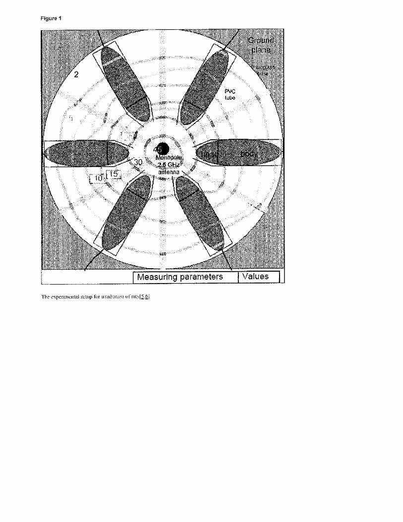


!!!
Attachment!20!

Investigation of the effects of distance from sources on apoptosis,oxidative stress and cytosolic calcium accumulation via TRPV1 channelsinduced by mobile phones and Wi-Fi in breast cancer cells
Bilal Çiğ a, Mustafa Nazıroğlu a,b,⁎a Department of Biophysics, Faculty of Medicine, Suleyman Demirel University, Isparta, Turkeyb Neuroscience Research Center, Suleyman Demirel University, Isparta, Turkey
a b s t r a c ta r t i c l e i n f o
Article history:Received 27 August 2014Received in revised form 2 February 2015Accepted 10 February 2015Available online 19 February 2015
Keywords:Mobile phoneWireless internet (Wi-Fi)Calcium signalingOxidative stressApoptosisBreast cancer
TRPV1 is a Ca2+ permeable channel and gated by noxious heat, oxidative stress and capsaicin (CAP). Somereports have indicated that non-ionized electromagnetic radiation (EMR)-induces heat and oxidative stress ef-fects.We aimed to investigate the effects of distance from sources on calcium signaling, cytosolic ROS production,cell viability, apoptosis, plus caspase-3 and -9 values induced by mobile phones and Wi-Fi in breast cancer cellsMCF-7 human breast cancer cell lines were divided into A, B, C and D groups as control, 900, 1800 and 2450MHzgroups, respectively. Cells in Group A were used as control and were kept in cell culture conditions without EMRexposure. Groups B, C andDwere exposed to the EMR frequencies at different distances (0 cm, 1 cm, 5 cm, 10 cm,20 cm and 25 cm) for 1 h before CAP stimulation. The cytosolic ROS production, Ca2+ concentrations, apoptosis,caspase-3 and caspase-9 valueswere higher in groups B, C andD than inA group at 0 cm, 1 cmand 5 cmdistancesalthough cell viability (MTT) values were increased by the distances. There was no statistically significantdifference in the values between control, 20 and 25 cm.Wi-Fi andmobile phone EMR placedwithin 10 cmof the cells induced excessive oxidative responses and apopto-sis via TRPV1-induced cytosolic Ca2+ accumulation in the cancer cells. Using cell phones andWi-Fi sourceswhichare farther away than 10 cm may provide useful protection against oxidative stress, apoptosis and overload ofintracellular Ca2+. This article is part of a Special Issue entitled: Membrane channels and transporters in cancers.
© 2015 Elsevier B.V. All rights reserved.
1. Introduction
Electromagnetic radiation (EMR) produced by mobile phones andtheir base station antennae is in the range of 900 and 1800 MHz forthe Global System for Mobile Communications (GSM). Mobile phonesemit radiofrequency EMR which may affect human health based onbiological stress responses [5]. Wireless local area network systems(WLAN, 2450 MHz), are an alternative to wired internet access in busi-ness centers, homes, and public areas providing means of communica-tion and information exchange [26]. These concerns require furtherinvestigation of possible biological effects of exposure to WLAN signals
[5]. Cancer, with 10 million new cases of per year, is one of the biggestconcerns of humanity [15,16]. A main concern has been the risk of can-cer and DNA degeneration among people living near base stations, butthe general welfare of all citizens exposed to EMR is becoming a fre-quent subject of conjecture [5,13,22,25,30]. With regard to this issue,there have been numerous reports of valuable research, but the resultsare still somewhat inconclusive [5,12,21,29]. Recently,we observed pro-liferative and tissue injury effects of exposure to 2450 MHz radiation inan HL-60 cancer cell line [22], an MDA-MB-231 breast cancer cell line[14] and in rat tissues through induction of Ca2+ influx and oxidativestress [4,13,30].
EMRcan alter the energy level and spin orientation of electrons and, asa consequence, increase the activity, concentration and lifetime of ROS[20]. There are various antioxidant mechanisms in cells that neutralizethe harmful effects of ROS [22] but exposure to EMR results in increasesof ROS due to loss of efficiency of antioxidantmechanisms and alterationsin the mitochondrial electron transfer chain [8,12]. However, whetherdistance affects the induction of oxidative stress and apoptosis in breastcancer cells exposed to 900, 1800 and 2450 MHz EMR is still unknownand deserves further study.
Ca2+ homeostasis of the cells is one of themany important functions.The proliferation of cells, to undergo apoptosis, induction of oxidative
Biochimica et Biophysica Acta 1848 (2015) 2756–2765
Abbreviations: [Ca2+]i, cytosolic free calcium ion; CAP, capsaicin; DMSO, dimethylsulfoxide; EGTA, ethylene glycol-bis[2-aminoethyl-ether-N,N,N,N-tetraacetic acid; EMR,electromagnetic radiation; GSM, Global System forMobile Communications; HBSS, Hank'sbuffered salt solution; RF, radiofrequency; ROS, reactive oxygen species; TRP, transientreceptor potential; TRPM2, transient receptor potential melastatin 2; TRPV1, transientreceptor potential vanilloid 1 This article is part of a Special Issue entitled: Membrane channels and transporters in
cancers.⁎ Corresponding author at: Department of Biophysics, Medical Faculty, Suleyman Demirel
University, TR-32260 Isparta, Turkey. Tel.: +90 246 2113310; fax: +90 246 2371165.E-mail address: [email protected] (M. Nazıroğlu).
http://dx.doi.org/10.1016/j.bbamem.2015.02.0130005-2736/© 2015 Elsevier B.V. All rights reserved.
Contents lists available at ScienceDirect
Biochimica et Biophysica Acta
j ourna l homepage: www.e lsev ie r .com/ locate /bbamem

stress and physiological functions such as signal transduction is a part ofCa2+ homeostasis [17]. Cytosolic free calcium ion concentration [Ca2+]i,which is dependent on both plasma and intracellular membrane func-tions, is controlled bymany ion channels. The transient receptor potential(TRP) family is one of these channels and they are important non-selective cation channels [23]. TRPV1 is a cation channel and is amemberof the subfamily of these channels. It can be activated by capsaicin and it isalso heat sensitive (≥ 43 °C) [24,27]. Thermal effects of electromagneticradiation caused by changes in temperature have been noted [7] andtemperature effects of exposure to EMR of 450 MHz and 2450 MHzfrequencies have been seen in various tissues and in total blood flowexaminations and increase in skin temperature [1]. This issue has beenexamined in many well performed experimental studies using rats andmice subjected to EMR exposure [19–24].
Since to date there is no report about the mechanism of 900, 1800and 2450 MHz EMR-induced actions on cellular survival and death,such an investigation may help clarify how free radical formation andapoptosis occur following EMR-induced injury. The present study wasdesigned to determine the effects of 900, 1800 and 2450 MHz EMRexposure on oxidative damage of breast cancer cells, apoptosis and ROSproduction, as well as the possible protective effects of different distanceson the values by analyzing apoptosis, caspase activities, cytosolic ROSproduction, and accumulation of [Ca2+]i concentration-induced oxidativestress.
2. Materials and methods
2.1. Cells and reagents
The Michigan Cancer Foundation-7 breast cancer cell line (MCF-7) was used in this study. The cell line was originally obtained from‘The Leibniz Institute-German Collection of Microorganisms and CellCultures (DSMZ)’ Cell Lines Bank (Braunschweig, Germany). Ethyleneglycol-bis(2-aminoethyl-ether)-N,N,N′,N′-tetraacetic acid (EGTA)and dimethyl sulfoxide and Roswell Park Memorial Institute(RPMI) 1640 medium were obtained from Sigma-Aldrich Chemical(St. Louis, MO, USA). N-acetyl-Asp-Glu-Val-Asp-7-amino-4-methylcoumarin (ACDEVD-AMC), nonidet-P-40 substitute (NP40),2-(N-morpholino)ethanesulfonic acid hydrate (MES hydrate), PEG, 4-(2-hydroxyethyl)-1-piperazineethanesulfonic acid (HEPES), 3-[(3-chomalidopropyl) dimethylammonio]-1-propanesulfonate (CHAPS),polyethylene glycol (PEG) and dithiothreitol (DTT) were obtained fromSigma Chemical. Dihydrorhodamine-123 (DHR 123/N-acetyl-Leu-Glu-His-Asp-7-amino-4-methylcoumarin (AC-LEHD-AMC) was purchasedfrom Bachem (Bubendorf, Switzerland. A mitochondrial stain 5,5′,6,6′-tetrachloro-1,1′,3,3′-tetraethylbenzimidazolylcarbocyanine iodide (JC-1)was purchased from Santa Cruz (Dallas, TX, USA). All organic solventswere also purchased from Santa Cruz (Dallas, TX, USA). The reagentswere equilibrated at room temperature for 30 min before an analysis.
2.2. Cell culture
TheMCF-7 cells were cultured in RPMI 1640medium supplementedwith fetal bovine serum in a humidified incubator at 37 °C, 5% CO2, and95% air. The cells were counted daily by removing a small volume fromthe tissue culture flask (filter cap, sterile, 250 ml, 75 cm2), diluting itwith an equal volume of trypan blue (0.4%), and tallying viablecells (trypan blue excluding) with a hemocytometer. Cultures weremaintained as a suspension without shaking or stirring at a densityof 1 × 10⁶ cells per ml by dilution with fresh media. Cultures weretransferred once a week.
2.3. Groups
Cells were seeded in 8–10 flasks at a density of 1× 10⁶ cells per flask(filter cap, sterile, 250ml, 75 cm2) and placed in a circulatingwater bath(Fig. 1). All cells were cultured in the same culture medium (37 °C) andfor an identical time (1 h). The cells were divided into fourmain groups.
A- Control group: The cells were not exposed to EMR but were keptin falcon tubes containing the same cell culturemediumand con-ditions for 1 h.
B- 900 MHz group: Cells in the group were exposed to 900 MHzEMR at different distances (0 cm, 1 cm, 5 cm, 10 cm, 20 cm and25 cm) for periods of 1 h.
C- 1800 MHz group: Cells in the group were exposed to 1800 MHzEMR at different distances (0 cm, 1 cm, 5 cm, 10 cm, 20 cm and25 cm) for 1 h.
D- 2450 MHz group: Cells in the group were exposed to 2450 MHzEMR at different distances (0 cm, 1 cm, 5 cm, 10 cm, 20 cm and25 cm) for 1 h.
All the exposures at the different distanceswere repeated 4–6 times.At the endof the 1 h incubation, the control and exposed cellswere usedfor the analyses of cytosolic free Ca2+ ([Ca2+]i) concentration, apoptosisand caspase.
2.4. Exposure system and design
The exposure system has been described in detail elsewhere [14].The cells were kept in a circulatory water bath (Fig. 1). The cells wereattached to the walls of the flask. The exposure system was performedin a special room that was fitted with plastic furniture such as tablesand chairs so as to minimize the possibility of radiation reflection. Thewalls of the roomwere covered by chromium–nickel sheets (thickness:1mm) for protecting the cells from possible outside electromagnetic in-terference. The continuous wave of radiofrequency signal (900 MHzwith 217 Hz pulses) emitted by the generator was amplified initiallyand then fed into the cancer cells in the water bath by an antenna
Fig. 1. Schematic diagram of radiofrequency exposure device.
2757B. Çiğ, M. Nazıroğlu / Biochimica et Biophysica Acta 1848 (2015) 2756–2765

(Biçer Electronic, Sakarya, Turkey). This antenna has a special Falconholder designed to accommodate the cells for appropriate exposureconditions. The repetition time, frequency, and amplitude of the radio-frequency (RF) energy spectrum were monitored by a satellite levelmeter (PROMAX, MC-877C, Barcelona, Spain). Radiation reflection andexposure weremeasured with a Portable RF Survey System (HOLADAY,HI-4417, Eden Prairie, MN, USA) with a standard probe. The EMR dosewas calculated from themeasured electric field strength (V/m) and dig-ital models based on the FDTD numerical code. Distance was arrangedat 0 cm, 1 cm, 5 cm, 10 cm, 20 cm and 25 cm between the falcontubes and probe of the exposure system. Six falcon tubes each contain-ing 1 × 10⁶ cells/ml (5 ml total medium) were placed on a non-
conductive plexi glass table at a height of 110 cm at a precise locationwhere the required power density was measured. The RF field insidethe special room was probed using a strength meter and the precisepositions which provided power densities of 1.2, 12 or 120 μW/cm2
were determined [14]. The required power density (≤12 μW/cm2)was continuously recorded every 5 min using a satellite level meter(EXTECH-480836, Extech Instruments, Nashua, NH, USA). The datawere saved on a computer. At the top of the flask, the average specificabsorption rate (SAR) estimated for 900 MHz exposure at 12 μW/cm2
power flux density was 0.36 ± 0.02 mW/kg (Table 1). The water bath(Water Bath 601, Jiangsu Zhengji Instruments, Jiangsu, China) installedin the chromium–nickel covered roomwas maintained at 37 °C (relative
Fig. 2. Effects of EMR (A-900, B-1800 and C-2450 MHz) exposure on cytosolic free Ca2+ ([Ca2+]i) concentration in MCF-7 breast cancer cells. The Ca+2 entry was estimated as describedunder Materials and methods sections. Values are presented as mean± SD of 6 separate experiments and expressed as fold increase over the pretreatment level (experimental/control).ap b 0.001 and bp b 0.01 versus control. cp b 0.05 and dp b 0.01 versus 0 cm group.
Table 1Specific absorption rate (SAR) values of falcon tubes (mean ± SD, n = 6).
Tube Numbers
1 2 3 4 5 6
SAR (W/kg)(min–max)
0.32 ± 0.05(0.24–0.39)
0.26 ± 0.08(0.18–0.40)
0.50 ± 0.06(0.43–0.58)
0.51 ± 0.06(0.43–0.58)
0.28 ± 0.08(0.20–0.40)
0.26 ± 0.04(0.20–0.31)
SAR (W/kg)mean of 6 tubes
0.36 ± 0.02
2758 B. Çiğ, M. Nazıroğlu / Biochimica et Biophysica Acta 1848 (2015) 2756–2765
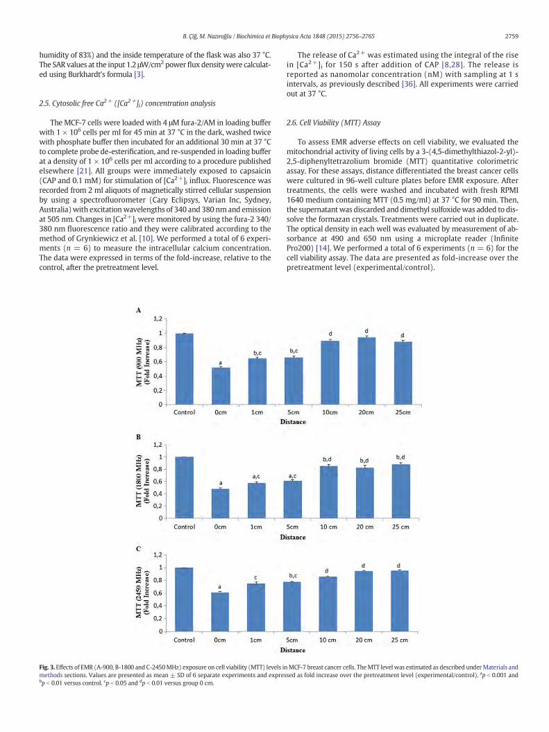
humidity of 83%) and the inside temperature of the flask was also 37 °C.The SAR values at the input 1.2 μW/cm2 powerflux densitywere calculat-ed using Burkhardt's formula [3].
2.5. Cytosolic free Ca2+ ([Ca2+]i) concentration analysis
The MCF-7 cells were loaded with 4 μM fura-2/AM in loading bufferwith 1 × 10⁶ cells per ml for 45 min at 37 °C in the dark, washed twicewith phosphate buffer then incubated for an additional 30 min at 37 °Cto complete probe de-esterification, and re-suspended in loading bufferat a density of 1 × 10⁶ cells per ml according to a procedure publishedelsewhere [21]. All groups were immediately exposed to capsaicin(CAP and 0.1 mM) for stimulation of [Ca2+]i influx. Fluorescence wasrecorded from 2 ml aliquots of magnetically stirred cellular suspensionby using a spectrofluorometer (Cary Eclipsys, Varian Inc, Sydney,Australia)with excitationwavelengths of 340 and 380 nmand emissionat 505 nm. Changes in [Ca2+]i were monitored by using the fura-2 340/380 nm fluorescence ratio and they were calibrated according to themethod of Grynkiewicz et al. [10]. We performed a total of 6 experi-ments (n = 6) to measure the intracellular calcium concentration.The data were expressed in terms of the fold-increase, relative to thecontrol, after the pretreatment level.
The release of Ca2+ was estimated using the integral of the risein [Ca2+]i for 150 s after addition of CAP [8,28]. The release isreported as nanomolar concentration (nM) with sampling at 1 sintervals, as previously described [36]. All experiments were carriedout at 37 °C.
2.6. Cell Viability (MTT) Assay
To assess EMR adverse effects on cell viability, we evaluated themitochondrial activity of living cells by a 3-(4,5-dimethylthiazol-2-yl)-2,5-diphenyltetrazolium bromide (MTT) quantitative colorimetricassay. For these assays, distance differentiated the breast cancer cellswere cultured in 96-well culture plates before EMR exposure. Aftertreatments, the cells were washed and incubated with fresh RPMI1640 medium containing MTT (0.5 mg/ml) at 37 °C for 90 min. Then,the supernatantwas discarded and dimethyl sulfoxidewas added to dis-solve the formazan crystals. Treatments were carried out in duplicate.The optical density in each well was evaluated by measurement of ab-sorbance at 490 and 650 nm using a microplate reader (InfinitePro200) [14]. We performed a total of 6 experiments (n = 6) for thecell viability assay. The data are presented as fold-increase over thepretreatment level (experimental/control).
Fig. 3. Effects of EMR (A-900, B-1800 and C-2450MHz) exposure on cell viability (MTT) levels inMCF-7 breast cancer cells. TheMTT level was estimated as described under Materials andmethods sections. Values are presented as mean ± SD of 6 separate experiments and expressed as fold increase over the pretreatment level (experimental/control). ap b 0.001 andbp b 0.01 versus control. cp b 0.05 and dp b 0.01 versus group 0 cm.
2759B. Çiğ, M. Nazıroğlu / Biochimica et Biophysica Acta 1848 (2015) 2756–2765

2.7. Apoptosis assay
The APOPercentage assay (Biocolor, Belfast, Northern Ireland) is adye-uptake method, which stains only the apoptotic cells with a reddye. When the membrane of apoptotic cell loses its asymmetry, theAPOPercentage dye is transported into cells, staining apoptotic cells red,thus allowing detection of apoptosis by spectrophotometry [2]. The apo-ptosis values were obtained from 6 separate experiments and expressedas fold increase over the pretreatment level (experimental/control).
2.8. Assay for caspase-3 and -9 activities
The determination of caspase-3 and caspase-9 activities was based ona method previously reported [8,23] with minor modifications. Stimulat-ed or resting cells were washed once with PBS. After centrifugation, cellswere re-suspended in PBS at a concentration of 103 cells/ml. Fifteenmicroliters of the cell suspension was added to a microplate of a micro-plate reader and mixed with the appropriate peptide substrate dissolved
in a standard reaction buffer that was composed of 100 mM HEPES,pH 7.25, 10% sucrose, 0.1% CHAPS, 5 mM DTT, 0.001% NP40 and 40 μMof caspase-3 substrate (AC-DEVD-AMC) or 0.1 M MES hydrate, pH 6.5,10% PEG, 0.1% CHAPS, 5 mM DTT, 0.001% NP40, and 0.1 mM of caspase-9 substrate (AC-LEHD-AMC). Substrate cleavage was measured with themicroplate reader (Infinite Pro200) with an excitation wavelength of360 nmand emission at 460 nm. The datawere calculated asfluorescenceunits/mg protein and presented as fold-increase over the pretreatmentlevel (experimental/control). The caspase-3 and caspase-9 assays wereperformed in 6 separate experiments.
2.9. Intracellular reactive oxygen species (ROS) measurement
DHR 123 is a non-fluorescent, non-charged dye that easily penetratescell membrane. Once inside the cell, DHR 123 becomes fluorescent uponoxidation to yield rhodamine 123 (Rh 123), the fluorescence being pro-portional to ROS generation. The fluorescence intensity of Rh123 wasmeasured in an automatic microplate reader (Infinite Pro200). Excitation
Fig. 4. Effects of EMR (A-900, B-1800 andC-2450MHz) exposure on intracellular ROS levels inMCF-7 breast cancer cells (mean±SDand n=6). Values are presented as fold increase overthe pretreatment level (experimental/control). ap b 0.001 and bp b 0.01 versus control. cp b 0.05 and dp b 0.01 versus group 0 cm.
2760 B. Çiğ, M. Nazıroğlu / Biochimica et Biophysica Acta 1848 (2015) 2756–2765

was set at 488 nm and emission was set at 543 nm [32]. We per-formed a total of 6 experiments (n = 6) for the intracellular ROSassay. The data were presented as fold-increase over the pretreatmentlevel (experimental/control).
2.10. Statistical analyses
Data were analyzed using the SPSS statistical program (version 17.0,software, SPSS. Chicago, IL, USA). All results are expressed as means ±standard deviation (SD). Analysis of variance (ANOVA) and an unpairedMann–Whitney U test were performed; P-values of less than 0.01 wereregarded as significant. Significant values were assessed with the leastsignificance difference test.
3. Results
3.1. Cytosolic Ca2+ Concentration analysis
MCF-7 cells are capsaicin sensitive TRPV1 ion channels and theyexhibit activity with EMR exposure and they were became more activewith respect to calcium entry to cells compared with the control theEMR exposure. Changes were observed in the cells placed at 0 cm, 1 cmand 5 cm distances from the EMR source. The mean cytosolic Ca2+ con-centration analysis values in the four groups are shown in Fig. 2 A, Band C. Ca2+ values were significantly lower in the 900, 1800 and2450 MHz EMR groups than in control at 0 cm (p b 0.001), 1 cm(p b 0.01) and 5 cm (p b 0.05). However, there was no difference be-tween any of the 10 cm, 20 cm, 25 cm and control values. Hence, we
Fig. 5. Effects of EMR (A-900, B-1800 and C-2450 MHz) exposure on apoptosis levels in MCF-7 breast cancer cells. The apoptosis level was estimated as described under Materials andmethods sections. Values are presented as mean ± SD of 6 separate experiments and expressed as fold increase over the pretreatment level (experimental/control). ap b 0.001 andbp b 0.01 versus control group. cp b 0.05 and dp b 0.01 versus group 0 cm.
2761B. Çiğ, M. Nazıroğlu / Biochimica et Biophysica Acta 1848 (2015) 2756–2765

observed that the cytosolic Ca2+ concentration of the cells was de-creased if the distance to the EMR probe was within 10 cm.
3.2. Cell viability (MTT) results
The meanMTT values in the four groups are shown in Fig. 3A, B andC. MTT values were significantly lower in the 900, 1800 and 2450 MHzEMR groups than in the control at 0 cm (p b 0.001), 1 cm (p b 0.01) and5 cm (p b 0.05). However, there was no difference between the 10 cm,20 cm, 25 cm and control values. Hence, we observed that MTT valuesof the cells were decreased if they were within 10 cm of the EMR prob.
3.3. Intracellular (ROS) production
The mean cytosolic ROS production levels in the four groups areshown in Fig. 4 A, B and C, respectively. The results showed that thelevels of cytosolic ROS production (p b 0.001) were significantly higher
in the EMR groups than in controls. Exposure to 900 MHz, 1800 MHzand 2450 MHz EMR increased the ROS production at 0 cm, 1 cm and5 cm distances from EMR prob. However, at distances of 10 cm, 20 cmand25 cm, the EMR-induced cytosolic ROSproductionwasnot statisticallydifferent from the control group.
3.4. Effects of 900, 1800 and 2450 MHz EMR on apoptosis and caspase‐3and caspase‐9 values
We investigated the effects of 900 MHz, 1800 MHz and 2450 MHzEMR exposure on the rate of programmed cell death as indicated byapoptosis and caspase values in the cancer cells. The results of apoptosis,caspase‐3 and caspase‐9 values in control, 900 MHz, 1800 MHz and2450MHzgroups are shown in Figs. 5, 6 and 7, (A, B and C) respectively.The apoptosis (p b 0.001) and (p b 0.005), caspase‐3 (p b 0.001) andcaspase‐9 (p b 0.001) values in the EMR group were significantlyhigher than in the control group. Furthermore, a significant difference
Fig. 6. Effects of EMR (A-900, B-1800 and C-2450MHz) exposure on caspase-3 activity inMCF-7 breast cancer cells (mean± SD and n=6). The values expressed as fold increase over thepretreatment level (experimental/control). ap b 0.001 and bp b 0.01 versus control group. cp b 0.05 and dp b 0.01 versus group 0 cm.
2762 B. Çiğ, M. Nazıroğlu / Biochimica et Biophysica Acta 1848 (2015) 2756–2765

(p b 0.001) was observed for 0 cm, 1 cm and 5 cm distances in eachgroup, compared to control. However, no statistical difference wasobserved at 10 cm and at distances beyond this figure.
4. Discussion
The use of mobile phones and Wi-Fi internet is currently one of thefastest growing technology developments. The likelihood of close prox-imity of the antenna of such a devices to the breast has raised concernabout a possible biological connection between EMR and breast cancerof women [13,27,14]. The exposure to such radiation depends on thelength of time and frequency of use, which varies from individual toindividual. The most investigated RF waveforms of the GSM-modulatedsignals are at 900MHz and 1800MHz and theWi-Fi (2450MHz) signals.The EMR studies have been carried out on different cancer cell types suchasHL-60 cancer cell line [22], anMDA-MB-231 breast cancer cell line [14].
Modern cell phone devices andWi-Fi internet in Turkey and many othercountries work at a frequency of 900 MHz, 1800 MHz and 2450 MHzwhich were consequently selected for the present study.
Cell membranes are amajor potential target for absorption of hazard-ous materials encountered in the environment [27,29]. In the presentstudy, the cytosolic ROS production, apoptosis, caspase-3 and caspase‐9values in the breast cancer cells increased after 900 MHz, 1800 MHzand 2450 MHz exposure. These data are in agreement with reports sug-gesting that EMR induces oxidative stress and apoptosis of cancer cellsby inducing ROS [14,27,31]. We have shown also that the changes in-duced by EMR are very dependent on the distance from the source inthe cancer cells. To our knowledge, it is the first report of the relationshipbetween different distances and 900MHz, 1800MHz and 2450MHz EMRin the MCF-7 breast cancer cells.
The most recent evidence obtained for the effects of ionizing andnon-ionizing radiation indicate that they both trigger oxidative stress
Fig. 7.Effects of EMR(A-900MHz, B-1800MHzandC-2450MHz) exposure on caspase‐9 activity inMCF-7breast cancer cells (mean±SD and n=6). The values expressed as fold increaseover the pretreatment level (experimental/control). ap b 0.001 and bp b 0.01 versus control group. cp b 0.05 and dp b 0.01 versus group 0 cm.
2763B. Çiğ, M. Nazıroğlu / Biochimica et Biophysica Acta 1848 (2015) 2756–2765

in cancer cells and neuronal cells [9,13,14,22] although antioxidantssuch as seleniumandmelatonin gave effective protection. [14,21]. In an-other study it has been reported that 900 MHz EMR causes an increasein temperature of skin and ears [35].Thus 900 MHz, 1800 MHz and2450MHz EMR exposuremay activate heat-sensitive TRPV1 cation chan-nels and thereby trigger excessive calcium entry into cells. But limited in-formation is available about whether environmental EMR exposuretriggers the free radical formation via cytosolic Ca2+ accumulation [16].
It is well known that ionized EMRhas been used to kill tumor cells insome cancer treatment protocols. Exposure of cells to non-ionized EMRleads to generation of ROS, which is known to disturb the antioxidantdefense system and induce oxidative stress [31]. In turn, the radiation-induced increases in ROS cause DNA damage, cell cycle arrest and acti-vationof some transcription and apoptotic factors e.g., the nuclear factorkappa-light-chain-enhancer of activated B cells [18,33]. In the currentstudy, cytosolic ROS production values in the EMR groups were elevat-ed. The results of our experiments confirm that exposure to 900 MHz,1800 MHz and 2450 MHz caused rises in oxidative stress, cytosolic[Ca2+]i concentration, ROS production and apoptosis of breast cancercells. According to the results, ideal distance for protection from oxida-tive and apoptotic damage of mobile phones andWi-Fi devices-inducedEMR seems 10 cm and up of the devices.
Mitochondrial function is essential for neuronal survival becauseneurons critically depend on ATP synthesis generated bymitochondrialoxidative phosphorylation [36]. Mitochondrial depolarization activitydepends on Ca2+ and is fueled by Ca2+ entry from the extracellularspace via channels such as TRPM2 and TRPV1when triggered by neuro-nal activity [2,17]. In return, some cation channels such as TRPM2 andvoltage gated calcium channels in cancer and neuronal cells are gatedby EMR-induced ROS production [21,22]. In addition the channels areactivated by capsaicin, noxious heat (42 °C), low pH and other endoge-nous ligands. TRPV1 is also activated by extracellular ROS [6,11]. Repeat-ed activation of TRPV1 has previously been shown to result in increasedcytosolic free Ca2+, oxidative stress, and apoptotic cell injury [2,11].TRPV1 activation by capsaicin in cancer cells was also found to increasesubstantially following mitochondrial oxidative stress [12]. Recentlyapoptosis and oxidative stress in colorectal cancer cells via activationof capsaicin and TRPV1 channels were reported although the TRPV1antagonist, capsazepine, potentiated the apoptotic and oxidative stresseffects through down-regulation of cell survival proteins and up-regulation of death receptors via the ROS–JNK–CHOP-mediated pathway[35]. Through thismechanism, EMR-inducedoxidative stress and temper-ature increasemaymodulate TRPV1 responses during cancer cell apopto-sis and mitochondrial injury because an overload of cytosolic Ca2+
induces depolarization of mitochondria and production of ROS [36].Until now, no studies dealing with the effects of distance on Wi-Fi andmobile phone frequencies-induced EMRonmitochondrial depolarization,oxidative stress and apoptosis in cancer cells have been published. In thecurrent study, we observed that oxidative stress values as well as cellapoptotic factors were lower in 10 cm, 20 cm and 25 cm distances thanin 0 cm, 1 cm and 5 cm distances, all groups compared to control groups.Hence we observed the protective effect of distances exceeding 10 cm oncell apoptosis, caspase-3, caspase‐9 in the cells. These results indicate thatto some extent, EMR exposure activates TRPV1 channels, consistent withour hypothesis that up-regulation of TRPV1 channels activity by EMRpre-exposure and heat effects should be responsible for the apoptosis andoxidative stress of cancer cells.
Apoptosis is programmeddeath and it ismediated by specific protein-ases namely caspases. There are two major pathways for apoptosis [34].One involves death receptors and is marked by Fas-mediated caspase-8activation, and the other is the stress or mitochondrial mediatedcaspase-9 activation. Both pathways induce caspase-3 activation [8,34].In the current breast cancer cells, apoptosis, caspase-3 and caspase-9values were increased by 900MHz, 1800MHz and 2450MHz in cells po-sitionedwithin 10 cm of the EMR source. It is likely that TRPV1-mediatedCa2+ entry in the EMR-exposed breast cancer cell involves accumulation
of ROS and opening of mitochondrial membrane pores that consequentlyleads tomitochondrial dysfunction, substantial swelling of themitochon-dria with rupture of the outer membrane and release of apoptosis-inducing factors such as caspase‐3 and caspase‐9.
In conclusion, the current results demonstrate that 900 MHz,1800 MHz and 2450 MHz radiations of mobile phones and Wi-Fiinternet in breast cancer cells induce apoptosis and ROS through calci-um accumulation of activation of TRPV1 channels. However, the in-creases of apoptosis and oxidative stress are modulated by differentdistances. We did not detect oxidative and apoptotic damage of breastcancer cells distanced 20 cm and 25 cm from the source of radiation.Using the cell phones and Wi-Fi radiation sources which are far from10 cm may provide useful distance against oxidative stress, apoptosisand overload Ca2+ entry in cancer. In addition, we suggest that use ofTRPV1 channel blockers may provide a potential therapeutic approachfor the mobile phone andWi-Fi-induced oxidative stress and apoptosisby calcium accumulation.
Declaration of interest
There is no conflict interest in the study.
Transparency document
The Transparency document associated with this article can befound, in online version.
Acknowledgments
The authorswish to thank Assoc Prof. Dr. Selçuk Çömlekçi (Electronicsand Communication Engineering, Suleyman Demirel University, Isparta,Turkey) and Dr. Peter J Butterworth (King's College, London, UK) forcalculation of the specific absorption rates and polishing English of themanuscript, respectively. MN formulated the present hypothesis andwas responsible for writing the report. BÇ were responsible for analysisof the data. Abstract of the study was published in EACR-sponsored 2ndAnticancer Agents Congress & 5th Multidisciplinary Cancer ResearchCongress, 23–27 April 2014, Bodrum, Mugla Turkey.
Financial support: The study was supported by the Unit of ScientificResearch Project (BAP), Isparta Süleyman Demirel University, Isparta,Turkey (Project Number: BAP: 3167-YL2-12).
References
[1] E.R. Adair, B.L. Cobb, K.S. Mylacraine, S.A. Kelleher, Human exposure at two radiofrequencies (450 and 2450 MHz): similarities and differences in physiologicalresponse, Bioelectromagnetics (Suppl. 4) (1999) 12–20.
[2] M. Argun, L. Tök, A.C. Uğuz, Ö. Çelik, Ö.Y. Tök, M. Naziroğlu, Melatonin and amfenacmodulate calcium entry, apoptosis, and oxidative stress in ARPE-19 cell cultureexposed to blue light irradiation (405 nm), Eye (Lond.) 28 (2014) 752–760.
[3] M. Burkhardt, K. Poković, M. Gnos, T. Schmid, N. Kuster, Numerical and experimentaldosimetry of Petri dish exposure setups, Bioelectromagnetics 17 (1996) 483–493.
[4] H. Cetin, M. Nazıroğlu, O. Celik, M. Yüksel, N. Pastacı, M.O. Ozkaya, Liver antioxidantstores protect the brain from electromagnetic radiation (900 and 1800 MHz)-inducedoxidative stress in rats during pregnancy and the development of offspring, J. Matern.Fetal Neonatal Med. (2014)http://dx.doi.org/10.3109/14767058.2014.898056 (Epubahead of print).
[5] E.D. Chavdoula, D.J. Panagopoulos, L.H. Margaritis, Comparison of biological effectsbetween continuous and intermittent exposure to GSM-900-MHz mobile phoneradiation: detection of apoptotic cell-death features, Mutat. Res. 700 (2010) 51–61.
[6] H.H. Chuang, S. Lin, Oxidative challenges sensitize the capsaicin receptor by covalentcysteine modification, Proc. Natl. Acad. Sci. U. S. A. 106 (2009) 20097–20102.
[7] J.A. D'Andrea, J.M. Ziriax, E.R. Adair, Radio frequency electromagnetic fields: mildhyperthermia and safety standards, Prog. Brain Res. 62 (2007) 107–135.
[8] J. Espino, I. Bejarano, S.D. Paredes, C. Barriga, A.B. Rodríguez, J.A. Pariente, Protectiveeffect of melatonin against human leukocyte apoptosis induced by intracellular cal-cium overload: relation with its antioxidant actions, J. Pineal Res. 51 (2011)195–206.
[9] V. Ghazizadeh, M. Nazıroğlu, Electromagnetic radiation (Wi-Fi) and epilepsy inducecalcium entry and apoptosis through activation of TRPV1 channel in hippocampusand dorsal root ganglion of rats, Metab. Brain Dis. 29 (2014) 787–799.
[10] C. Grynkiewicz, M. Poenie, R.Y. Tsien, A new generation of Ca2+ indicators withgreatly improved fluorescence properties, J. Biol. Chem. 260 (1985) 3440–3450.
2764 B. Çiğ, M. Nazıroğlu / Biochimica et Biophysica Acta 1848 (2015) 2756–2765

[11] S. Hong, L. Agresta, C. Guo, J.W. Wiley, The TRPV1 receptor is associated with prefer-ential stress in large dorsal root ganglion neurons in early diabetic sensory neuropathy,J. Neurochem. 105 (2008) 1212–1222.
[12] S.W. Ip, S.H. Lan, H.F. Lu, A.C. Huang, J.S. Yang, J.P. Lin, H.Y. Huang, J.C. Lien, C.C. Ho,C.F. Chiu, W. Wood, J.G. Chung, Capsaicin mediates apoptosis in human nasopharyn-geal carcinomaNPC-TW039 cells throughmitochondrial depolarization and endoplas-mic reticulum stress, Hum. Exp. Toxicol. 31 (2012) 539–549.
[13] Z. Jin, C. Zong, B. Jiang, Z. Zhou, J. Tong, Y. Cao, The effect of combined exposureof 900 MHz radiofrequency fields and doxorubicin in HL-60 cells, PLoS One 7(2012) e46102.
[14] M.C. Kahya, M. Nazıroğlu, B. Ciğ, Selenium reduces mobile phone (900 MHz)-inducedoxidative stress, mitochondrial function, and apoptosis in breast cancer cells, Biol.Trace Elem. Res. 160 (2014) 285–293.
[15] M. Koçer,M. Nazıroğlu, G. Koçer, T.T. Sonmez, Effects of bisphosphonate on oxidativestress levels in patients with cancer, Cancer Invest. 32 (2014) 37–42.
[16] M. Koçer, M. Nazıroğlu, Effects of 5-fluorouracil on oxidative stress and calciumlevels in the blood of patients with newly diagnosed colorectal cancer, Biol. TraceElem. Res. 155 (2013) 327–332.
[17] V.S. Kumar, A. Gopalakrishnan, M. Naziroğlu, G.K. Rajanikant, Calcium ion—the keyplayer in cerebral ischemia, Curr. Med. Chem. 21 (2014) 2065–2075.
[18] J.-S. Lee, T.-Q. Huang, T.-H. Kim, J.Y. Kim, H.J. Kim, J.-K. Pack, J.-S. Seo, Radiofrequencyradiation does not induce stress response in human T-lymphocytes and rat primaryastrocytes, Bioelectromagnetics 27 (2006) 578–588.
[19] M. Mailankot, A.P. Kunnath, H. Jayalekshmi, B. Koduru, R. Valsalan, Radio frequencyelectromagnetic radiation (RF-EMR) from GSM (0.9/1.8 GHz) mobile phonesinduces oxidative stress and reduces sperm motility in rats, Clinics (Sao Paulo) 64(6) (2009) 561–565.
[20] A.K. Manta, D.J. Stravopodis, I.S. Papassideri, L.H. Margaritis, Reactive oxygen species el-evation and recovery in Drosophila bodies and ovaries following short-term and long-term exposure to DECT base EMF, Electromagn. Biol. Med. 33 (2) (2013) 118–131.
[21] M. Nazıroğlu, Ö. Çelik, C. Özgül, S. Doğan, R. Bal, N. Gümral, A.B. Rodríguez, J.A.Pariente, Melatonin modulates wireless devices (2.45 GHz)-induced brain anddorsal root ganglion injury through TRPM2 and voltage gated calcium channels inrat, Physiol. Behav. 105 (2012) 683–692.
[22] M. Nazıroğlu, B. Ciğ, S. Doğan, A.C. Uğuz, S. Dilek, D. Faouzi, 2.45-GHz wirelessdevices induce oxidative stress and proliferation through cytosolic Ca2+ influx inhuman leukemia cancer cells, Int. J. Radiat. Biol. 88 (2012) 449–456.
[23] M. Nazıroğlu, B. Çiğ, C. Özgül, Neuroprotection induced by N-acetylcysteine againstcytosolic glutathione depletion induced-Ca2+ influx in dorsal root ganglion neuronsof mice: role of TRPV1 channels, Neuroscience 242 (2013) 151–160.
[24] M. Nazıroğlu, D.M. Dikici, S. Dursun, Role of oxidative stress and Ca2+ signaling onmolecular pathways of neuropathic pain in diabetes: focus on TRP channels,Neurochem. Res. 37 (2012) 2065–2075.
[25] M. Nazıroğlu, S. Tokat, S. Demirci, Role of melatonin on electromagnetic radiation-induced oxidative stress and Ca2+ signaling molecular pathways in breast cancer,J. Recept. Signal Transduct. Res. 32 (2012) 290–297.
[26] M. Nazıroğlu, M. Yüksel, S.A. Köse, M.O. Özkaya, Recent reports of Wi-Fi and mobilephone-induced radiation on oxidative stress and reproductive signaling pathwaysin females and males, J. Membr. Biol. 246 (12) (2013) 869–875.
[27] M. Nazıroğlu, Molecular role of catalase on oxidative stress-induced Ca(2+) signal-ing and TRP cation channel activation in nervous system, J. Recept. Signal Transduct.Res. 32 (2012) 134–141.
[28] M. Nazıroğlu, F.F. Özkan, S.R. Hapil, V. Ghazizadeh, B. Çiğ, Epilepsy but not mobilephone frequency (900 MHz) induces apoptosis and calcium entry in hippocampusof epileptic rat: involvement of TRPV1 channels, J. Membr. Biol. 248 (2015) 83–91.
[29] A. Özorak, M. Nazıroğlu, Ö. Çelik, M. Yüksel, D. Özçelik, M.O. Özkaya, H. Çetin,M.C. Kahya, S.A. Kose, Wi-Fi (2.45 GHz)- and mobile phone (900 and1800 MHz)-induced risks on oxidative stress and elements in kidney and testisof rats during pregnancy and the development of offspring, Biol. Trace Elem. Res.156 (2013) 221–229.
[30] D.J. Panagopoulos, L.H. Margaritis, The effect of exposure duration on the biologicalactivity of mobile telephony radiation, Mutat. Res. 699 (2010) 17–22.
[31] S. Reuter, S.C. Gupta, M.M. Chaturvedi, B.B. Aggarwal, Oxidative stress, inflam-mation, and cancer: how are they linked? Free Radic. Biol. Med. 49 (2010)1603–16016.
[32] G. Rothe, A. Oser, G. Valet, Dihydrorhodamine 123: a new flow cytometric indicatorfor respiratory burst activity in neutrophil granulocytes, Naturwissenschaften 75(1988) 354–355.
[33] R. Schreck, K. Albermann, P.A. Baeuerle, Nuclear factor kappa B: an oxidative stress-responsive transcription factor of eukaryotic cells (a review), Free Radic. Res. Commun.17 (1992) 221–237.
[34] Y. Shi, Apoptosome: the cellular engine for the activation of caspase-9, Structure 10(2002) 285–288.
[35] B. Sung, S. Prasad, J. Ravindran, V.R. Yadav, B.B. Aggarwal, Capsazepine, a TRPV1 antag-onist, sensitizes colorectal cancer cells to apoptosis by TRAIL through ROS-JNK-CHOP-mediated upregulation of death receptors, Free Radic. Biol. Med. 53 (2012) 1977–1987.
[36] A.C. Uğuz, M. Nazıroğlu, J. Espino, I. Bejarano, D. González, A.B. Rodríguez, J.A.Pariente, Selenium modulates oxidative stress-induced cell apoptosis in humanmyeloid HL-60 cells via regulation of caspase-3,-9 and calcium influx, J. Membr.Biol. 232 (2009) 15–23.
2765B. Çiğ, M. Nazıroğlu / Biochimica et Biophysica Acta 1848 (2015) 2756–2765

!!!
Attachment!21!

Full Terms & Conditions of access and use can be found athttp://www.tandfonline.com/action/journalInformation?journalCode=irab20
Download by: [USC University of Southern California] Date: 15 January 2016, At: 10:48
International Journal of Radiation Biology
ISSN: 0955-3002 (Print) 1362-3095 (Online) Journal homepage: http://www.tandfonline.com/loi/irab20
Effects of 2.4 GHz radiofrequency radiationemitted from Wi-Fi equipment on microRNAexpression in brain tissue
Suleyman Dasdag, Mehmet Zulkuf Akdag, Mehmet Emin Erdal, Nurten Erdal,Ozlem Izci Ay, Mustafa Ertan Ay, Senay Gorucu Yilmaz, Bahar Tasdelen &Korkut Yegin
To cite this article: Suleyman Dasdag, Mehmet Zulkuf Akdag, Mehmet Emin Erdal, NurtenErdal, Ozlem Izci Ay, Mustafa Ertan Ay, Senay Gorucu Yilmaz, Bahar Tasdelen & Korkut Yegin(2015) Effects of 2.4 GHz radiofrequency radiation emitted from Wi-Fi equipment on microRNAexpression in brain tissue, International Journal of Radiation Biology, 91:7, 555-561, DOI:10.3109/09553002.2015.1028599
To link to this article: http://dx.doi.org/10.3109/09553002.2015.1028599
Accepted author version posted online: 16Mar 2015.Published online: 20 May 2015.
Submit your article to this journal
Article views: 199
View related articles
View Crossmark data

555
International Journal of Radiation Biology, July 2015; 91(7): 555–561© 2015 Informa UK, Ltd.ISSN 0955-3002 print / ISSN 1362-3095 onlineDOI: 10.3109/09553002.2015.1028599
Correspondence: Prof. Dr Suleyman Dasdag, Department of Biophysics, Medical School of Dicle University, 21280, Diyarbakir, Turkey. E-mail: [email protected]
(Received 17 March 2014 ; revised 3 March 2015 ; accepted 6 March 2015 )
Eff ects of 2.4 GHz radiofrequency radiation emitted from Wi-Fi equipment on microRNA expression in brain tissue Suleyman Dasdag 1 , Mehmet Zulkuf Akdag 1 , Mehmet Emin Erdal 2 , Nurten Erdal 3 , Ozlem Izci Ay 2 , Mustafa Ertan Ay 2 , Senay Gorucu Yilmaz 2 , Bahar Tasdelen 4 & Korkut Yegin 5
1 Department of Biophysics, Medical School of Dicle University, Diyarbakir, Departments of 2 Medical Biology and Genetic, 3 Biophysics and 4 Biostatistics, Medical School of Mersin University, Mersin, and 5 Department of Electrical and Electronics Engineering , Ege University , Izmir , Turkey
Introduction
Th e use of wireless technologies such as Wireless Fidelity (Wi-Fi) communication devices have been growing tremen-dously over the past years. Accessing Wireless Local Area
Networks (WLAN) in houses, workplaces, public areas and schools has become a routine task in our daily lives. How-ever, rapid development of wireless technologies has steadily increased the environmental electromagnetic fi eld (EMF) levels. Public and scientifi c awareness that was previously focused on the adverse health eff ects of EMF emitted from mobile phones has shifted to the biological hazards of wireless equipment such as Wi-Fi. Because the health eff ects of such equipment are still unclear, the Council of Europe recommends restrictions on the use of mobile phones and internet access in all schools across the continent to protect young children from potentially harmful radiation (Watson 2011). Th erefore, under-standing the relationship between electromagnetic fi elds and health diseases such as reproductive disorders, cancer, etc., is very important for the public especially for young children who utilize wireless internet very frequently during adolescent years. In addition, uncontrolled wireless internet usage can turn into a habit and may continue throughout our lives without us being aware of the potential harmful eff ects of electromagnetic fi elds.
Th e relation between radiation and carcinogenesis is a well-known process. However, the underlying mechanism which identifi es the radiation-induced genetic instability is still not fully understood. Th erefore, to illuminate the under-lying mechanism between the radiation and carcinogenesis, more detailed studies, which include double-strand breaks, mutations, gene expression and disruption of mitochondrial processes, cell cycle arrest, and apoptotic cell death, are nec-essary. In addition, the underlying interaction mechanism between the radiation and microRNA (miRNA) expression, which is a new research fi eld, should be investigated. Several studies have already indicated radiation-induced epigenetic changes including DNA methylation and miRNA expression where miRNA profi les have been shown to be associated with cancer (Jones and Baylin 2007, Tunali and Tiryakioglu 2010, Aypar et al. 2011a, 2011b).
miRNA are small and non-protein-coding RNA mol-ecules. Th ey play critical roles in growth, diff erentiation,
Abstract Purpose : MicroRNAs (miRNA) play a paramount role in growth, diff erentiation, proliferation and cell death by suppressing one or more target genes. However, their interaction with radiofrequencies is still unknown. The aim of this study was to investigate the long-term eff ects of radiofrequency radiation emitted from a Wireless Fidelity (Wi-Fi) system on some of the miRNA in brain tissue. Materials and methods : The study was carried out on 16 Wistar Albino adult male rats by dividing them into two groups such as sham ( n ! 8) and exposure ( n ! 8). Rats in the exposure group were exposed to 2.4 GHz radiofrequency (RF) radiation for 24 hours a day for 12 months (one year). The same procedure was applied to the rats in the sham group except the Wi-Fi system was turned off . Immediately after the last exposure, rats were sacrifi ced and their brains were removed. miR-9-5p, miR-29a-3p, miR-106b-5p, miR-107, miR-125a-3p in brain were investigated in detail. Results : The results revealed that long-term exposure of 2.4 GHz Wi-Fi radiation can alter expression of some of the miRNAs such as miR-106b-5p (adj p * ! 0.010) and miR-107 (adj p * ! 0.005). We observed that mir 107 expression is 3.3 times and miR-106b-5p expression is 3.65 times lower in the exposure group than in the control group . However, miR-9-5p, miR-29a-3p and miR-125a-3p levels in brain were not altered. Conclusion : Long-term exposure of 2.4 GHz RF may lead to adverse eff ects such as neurodegenerative diseases originated from the alteration of some miRNA expression and more stud-ies should be devoted to the eff ects of RF radiation on miRNA expression levels.
Keywords: 2.4 GHz radiofrequency , Wi-Fi , miRNA in brain , diseases , electromagnetic fi elds
Dow
nloa
ded
by [U
SC U
nive
rsity
of S
outh
ern
Calif
orni
a] a
t 10:
48 1
5 Ja
nuar
y 20
16

556 S. Dasdag et al.
proliferation and cell death by suppressing one or more target genes. miRNA may be located in the introns and exons of protein-coding genes or in intergenic regions. More than 50% of miRNA are found in cancer-associated regions of the genome or in fragile sites; this suggests that miRNA have important roles in the pathogenesis of neoplasias (Tunali and Tiryakioglu 2010). Th erefore, miRNA represent new stars in the gene regulation galaxy, and there is a strong interest among researchers in diff erent fi elds to understand their mechanism of action and to identify their targets (Sevignani et al. 2006).
Strooper and Christen (2010) stated that the discovery of microRNAs has revealed an unexpected and spectacular additional level of fi ne tuning of the genome on how genes were used again and again in diff erent combinations to gen-erate the complexity that underlies, for instance, the brain. Th ey also reported that since the initial studies performed in Caenorhabditis elegans , they have gone a long way to begin to understand how microRNA pathways could have an impact on health and diseases in human. Although microR-NAs are abundantly expressed in the brain, relatively few are known about the multiple functions of these RNA molecules in the nervous system. Nevertheless, they already knew that microRNA pathways play major roles in the proliferation, diff erentiation, function and maintenance of neuronal cells. Several intriguing studies have linked microRNAs as major regulators of the neuronal phenotype, and implicated spe-cifi c microRNAs in the regulation of synapse formation and plasticity. Dysfunction of microRNA pathways is also slowly emerging as a potentially important contributor to the pathogenesis of major neurodegenerative disorders such as Alzheimer ’ s disease and Parkinson ’ s disease (Strooper and Christen 2010).
Alzheimer ’ s disease (AD) is a neurodegenerative disorder that currently aff ects nearly 2% of the population in indus-trialized countries. Th e risk of AD dramatically increases in individuals usually after age 70, and it is predicted that the incidence of AD will increase by 3-fold within the next 50 years. Th is progressive disease is characterized by the accu-mulation of plaques formed of short β amyloid (A β ) peptides (Boissonneault et al. 2009). Most of the studies investigate interaction between the radiation and miRNA have been usually focused on the eff ects of ultraviolet and ionizing radiation (Simone et al. 2009, Aypar et al. 2011a, b, Zhou et al. 2012). However, a study on the interaction between radiofre-quencies (RF) radiation emitted from Wi-Fi and microRNAs, especially on the interaction between Wi-Fi radiation and the brain is not available yet. Th erefore, the aim of this study was to investigate the eff ect of chronic exposure of Wi-Fi radia-tion, which is widely used in daily life, on some miRNA in the brain. In this study, eff ects of chronic exposure of 2.4 GHz radiofrequency on miR-9-5p, miR-29a-3p, miR-106b-5p, miR-107 and miR-125a-3p were observed in the brain.
Materials and methods
Subjects and animal care Sixteen Wistar Albino adult male rats with initial average weight of 313 " 25 g were acquired from the Medical Science Applica-tion and Research Center of Dicle University. Th e rats were fed
with standard pelleted food (TAVAS Inc., Adana, Turkey) in a standard Plexiglas cage. Final average weight of the animals was 348 " 28.8 g. Th ey were separated equally into two groups such as sham exposed ( n ! 8), and exposure ( n ! 8), and kept on a 14/10 h light/dark schedule. During the study, the ambi-ent temperature (22 ° C) and the relative humidity (45%) were maintained in the normal range for these animals. All animal procedures were in agreement with the Principles of Labora-tory Animal Care and the rules of Scientifi c and Ethics Com-mittee of Dicle University Health Research Center.
Exposure and fi eld measurements A signal generator, which emits Wi-Fi signals at the 2.4 GHz frequency band, was used to represent the exposure system. Rats in the sham and exposure groups were placed in a Plexi-glas cage (55 # 32 # 20 cm). Rats were free to move with no restriction in the cage during the study. Th e rats in the sham and exposure groups lived in the cage under normal cir-cumstances. Rats in the exposure group were subject to 2.4 GHz RF radiation 24 h/d for 12 months. Rats in both groups were kept 50 cm far away from the antenna of the generator (Figure 1). Th e same experimental conditions were applied to the rats in the sham group, except the generator was
Figure 1. Experimental set-up. (a) Top view, (b) side view (L ! 32 cm W ! 55 cm, H ant ! 50 cm, H caro ! 20 cm, s ! 2.5 cm.
Dow
nloa
ded
by [U
SC U
nive
rsity
of S
outh
ern
Calif
orni
a] a
t 10:
48 1
5 Ja
nuar
y 20
16

Radiofrequency radiation and microRNAs 557
turned off . Electromagnetic power density and the electrical fi eld inside the Plexiglas cage were measured by fi eld probe EMR 300 (NARDA, Pfullingen, Germany). Th e cage was surrounded with electromagnetic absorber material backed by metal to isolate outdoor electromagnetic fi elds from the test setup during the study duration of 12 months.
Specifi c absorption rate (SAR) measurement Wireless Local Area Networks (WLAN) signal generator with 100 mW peak (50 mW rms) power was connected to a tuned half-wavelength dipole antenna and the dipole antenna was positioned in front of a refl ector plate to direct electro-magnetic signals towards Plexiglas carousel. Th e separation between the antenna and the top of the carousel was approx-imately 4 λ where λ is the free space propagation wavelength at 2.45 GHz, and the refl ector plate to antenna separation was approximately 0.2 λ . To generate the far fi eld conditions, Rayleigh distance for any antenna residing in free space must satisfy the following conditions: (i) R $ 2D 2 / λ , (ii) R $ $ % , (iii) R $ $ D, where R and D represent the distance from the antenna to the rats and the maximum dimension of the antenna (i.e., diameter of the enclosing sphere), respec-tively. For the current set-up, it can be assumed that the rats in the carousel reside in the far-fi eld of the antenna. In this region of radiation, the electromagnetic signals emitted from the generator are assumed to be in the form of plane waves which represent the situation in most applications of Wi-Fi or WLAN equipment. Th e electric fi eld generated by the source was measured with an electric fi eld probe in the carousel at several locations in the absence of rats. Th ese measured values were compared to the simulated electric fi eld values to verify the reliability of the simulation set-up. Electromagnetic simulations were performed with CST Microwave Studio with rat voxel (volumetric pixel) data. Th is simulation tool utilizes a technique called fi nite integration technique (FIT) which is very similar to well-known fi nite-diff erence time domain (FDTD) technique, but FIT employs discretization on non-orthogonal grids using integral form of Maxwell ’ s equations as opposed to diff erential forms in FDTD. CST states that charge and energy conservation are more accurately preserved in FIT which, in turn, leads to stable numerical results in time-domain. Voxel rat model which was formed using computerized tomography scans of a rat was acquired from CST, and that model was used in the electric fi eld and SAR simulations of the current set-up.
RNA extraction Total RNA was extracted from rat brain tissue using Tri-Reagent (Sigma).
Reverse transcriptase PCR reactions (RT-PCR) Reverse transcriptase reactions contained 5 µ l of extracted total RNA, 50 nM stem-loop RT primer, 1 # RT buff er, 0.25 mM each of dNTPs, 50 units of modifi ed M-MuLV Reverse Transcriptase (Th ermo Scientifi c, Vilnius, Lithuania), 25 units of RiboLock RNase inhibitor (Th ermo Scientifi c, Vilnius, Lithuania) and nuclease-free water to a total reaction vol-ume of 15 µ l. Th e reaction was performed on an automated Th ermal Cycler (Techne Flexigene, Cambridge, UK). RT-PCR
conditions for 30 min at 16 ° C, 30 min at 42 ° C, 5 min at 85 ° C and then held at 4 ° C.
Quantitative-Comparative CT ( Δ Δ C T ) Real-time PCR Quantitative-Comparative C T ( Δ Δ C T ) Real-time PCR was per-formed in an ABI Prism 7500 Real-Time PCR System (Applied Biosystems) using the SDS 2.0.6 software. Th e specifi c primers and fl uorogenic ZNA ™ probes (Paris et al. 2010) for the microR-NAs were designed using Primer Express 3.0 software (Applied Biosystems) and are listed in Table I. Th e rho-miR-26b-5p was used as an endogenous control microRNA. Th e mixed RNAs created from the sham group were used as a Reference RNA sample. Primers and probes were purchased from Metabion International AG, D-82152 Martinsried/Deutschland. Th e 25 µ l PCR included 3 µ l RT-PCR product, 12.5 µ l of 2X TaqMan Universal PCR Master Mix (Applied Biosystems), 900 nmol of each primer (Primer F and Universal Primer R) and 200 nmol TaqMan ® probe. Th e reactions were incubated in a 96-well plate of preincubation at 50 ° C for 2 min and at 95 ° C for 10 min, followed by 40 cycles at 95 ° C for 15 sec and at 60 ° C for 90 sec. Amplifi cations and analysis were performed in an ABI Prism 7500 Real-Time PCR System (Applied Biosystems), using the SDS 2.0.6 software for allelic discrimination (Applied Biosys-tems). All reactions were run in triplicate.
Statistical analysis Th e data were processed and analyzed using the statistical package SPSS-11.5 for Windows. Normality assumption of 2 - Δ Δ CT values was checked by Shapiro Wilk test. Since the assumption of normality was met 2 - Δ Δ CT values were expressed as mean and standard deviation and the compari-sons between groups were performed using independent t -test and adjusted the signifi cance values for multiplicity using Benjamini-Hochberg adjustment (Benjamini and Hochberg 1995, Rai et al. 2012). Error-bar graph was used to represent data distribution of miR-9-5p, miR-29a-3p, miR-106b-5p, miR-107 and miR-125a-3p variables according to the groups. Signifi cant diff erences (two-tailed p ) less than 0.05 were regarded as signifi cant.
Results
Th e results of this study showed that the long-term exposure of 2.4 GHz Wi-Fi exposure may aff ect some of the miRNA such as miR-106b-5p and miR-107 (adj p * & 0.05). However, levels of miR-9-5p, miR-29a-3p and miR-125a-3p in the brain were not altered by long-term exposure of 2.4 GHz RF radiation. Th e results are summarized in Table II and Figure 2. On the other hand, point, 1 g and 10 g average SAR level of brain and cerebral fl uid were found as 4000 µ W/kg, 1510 µ W/kg and 1030 µ W/kg, respectively (Figure 3). Th e whole body (rms) and whole body maximum point SAR was found as 141.4 µ W/kg and 7127 µ W/kg, respectively. In SAR calculations, a representative rat with 370 g weight was used.
Discussion
Devices such as access points or Wireless Local Area Networks (WLAN) have been indispensable in houses, workplaces,
Dow
nloa
ded
by [U
SC U
nive
rsity
of S
outh
ern
Calif
orni
a] a
t 10:
48 1
5 Ja
nuar
y 20
16

558 S. Dasdag et al.
term cell phone use (918 MHz; 0.25 W/kg) provided cogni-tive benefi ts. Th ey showed that mice with AD long-term EMF exposure reduced brain amyloid-beta (A beta) deposition through decreased aggregation of A beta with an increase in soluble A beta levels (Arendash et al. 2010). However, Soder-qvist et al. (2010) proposed that transthyretin (TTR) might be involved in the fi ndings of RF exposure benefi t in mice with AD. In one of our previous studies, we also measured beta amyloid protein in rats which were exposed to long-term 900 MHz radiofrequency radiation and any alteration in amyloid beta level was not observed (Dasdag et al. 2012). On the other hand, nowadays some miRNA have been accepted as an indicator of AD. Th erefore, new strategies related to the interaction between RF and miRNA is very important in
public areas, schools, etc. However, widespread use of these devices naturally has been increasing the environ-mental electromagnetic fi eld (EMF) levels. Additionally, new wireless devices such as wireless printers, hard discs, headphones, etc., have been also the sources for increasing electromagnetic pollution. Th erefore, health eff ects of wire-less technologies need to be studied and enlightened. It is also important to note that uncontrolled wireless internet usage can turn into a habit and may continue throughout our lives without us being aware of the potential harmful eff ects of electromagnetic fi elds. Discussion on the health eff ects of radiofrequencies, especially on wireless technologies such as mobile phones, began at the beginning of 1990s. At the end of contradictive discussions, the International Agency for Research on Cancer (IARC) classifi ed mobile phones as 2B (IARC 2011). Although many of the contradictive studies on the health eff ects of mobile phone exposure exist, adequate studies are still not available on the side-eff ects of RF emitted from wireless internet equipment such as Wi-Fi.
Recently, one of the most popular topics related to the health eff ect of wireless technologies was neurodegenera-tive disease such as Alzheimer ’ s Disease (AD), which is one of the most important health problems among developed countries. However, studies on the eff ects of RF and AD are still insuffi cient. Arendash et al. (2010) claimed that long-
Table I. Primer/probe sequences of the miR analyzed by quantitative RT-PCR.
miR name Primer/probe sequence *
rno-miR-26b-5p rno-miR-26b-5p-RT, 5′GTCGTATGCAGTGCAGGGTCCGAGGTATTCGCACTGCATACGACACCTAT-3′ rno-miR-26b-5p-F, 5′GCCGCTTCAAGTAATTCAGG-3′rno-miR-26b-5p-PR, 5′FAM-TG(pdC)ATA(pdC)GA(pdC)A(pdC)CTATCC-ZNA4-BHQ-1-3′
rno-miR-9-5p rno-miR-9-5p-RT, 5′GTCGTATGCAGTGCAGGGTCCGAGGTATTCGCACTGCATACGACTCATAC-3′rno-miR-9-5p-F, 5′GCCGCTCTTTGGTTATCTAGCT-3′rno-miR-9-5p-PR, 5′FAM- TG(pdC)ATA(pdC)GA(pdC)T(pdC)ATA(pdC)AG-ZNA4-BHQ1-3′
rno-miR-29a-3p rno-miR-29a-3p-RT, 5′GTCGTATGCAGTGCAGGGTCCGAGGTATTCGCACTGCATACGACTAACCG-3′rno-miR-29a-3p-F, 5′GCCGCTAGCACCATCTGAAAT-3′rno-miR-29a-3p-PR, 5′FAM- TG(pdC)ATA(pdC)GA(pdC)TAA(pdC)CGAT-ZNA4-BHQ1-3′
rno-miR-106b-5p rno-miR-106b-5p-RT, 5′GTCGTATGCAGTGCAGGGTCCGAGGTATTCGCACTGCATACGACATCTGC-3′rno-miR-106b-5p-F, 5′GCCGCTAAAGTGCTGACAGT-3′rno-miR-106b-5p-PR, 5′FAM- TG(pdC)ATA(pdC)GA(pdC)ATCTGCAC-ZNA4-BHQ1-3′
rno-miR-107 rno-miR-107-RT, 5′GTCGTATGCAGTGCAGGGTCCGAGGTATTCGCACTGCATACGACTGATAG-3′rno-miR-107-F, 5′GCCGCAGCAGCATTGTACAGGG-3′rno-miR-107-PR, 5′FAM- TG(pdC)ATA(pdC)GA(pdC)TGATAG(pdC)C-ZNA4-BHQ-1-3′
rno-miR-125a-3p rno-miR-125a-3p-RT, 5′GTCGTATGCAGTGCAGGGTCCGAGGTATTCGCACTGCATACGACGGCTCC-3′rno-miR-125a-3p-F, 5′GCCGCACAGGTGAGGTTCTTG-3′rno-miR-125a-3p-PR, 5′FAM-TGCATACGACGGCTCCCA-ZNA4-BHQ1-3′miR-Universal-R, 5′GTGCAGGGTCCGAGGTAT-3′
* pdC: Substitution of C-5 propynyl-dC (pdC) for dC is an eff ective strategy to enhance base pairing. Using these base substitutions, duplex stability and melting temperatures are raised by C-5 propynyl-C 2.8 ° per substitution.
Table II. Statistical comparison of miRNA levels between the sham and exposure groups.
Sham-exp. group 2.4 GHz Wi-Fi exp. group
miRNA Mean " SD Mean " SD Adj p *
miR9-5p 1.852 " 2.225 0.543 " 0.123 0.157 miR29a-3p 1.960 " 1.732 1.367 " 0.924 0.868 miR106b-5p 2.743 " 2.265 0.751 " 0.218 0.010 miR107 2.118 " 2.162 0.627 " 0.156 0.005 miR125a-3p 1.758 " 1.114 1.535 " 0.576 0.955
* Benjamini-Hochberg adjusted p values.
Dow
nloa
ded
by [U
SC U
nive
rsity
of S
outh
ern
Calif
orni
a] a
t 10:
48 1
5 Ja
nuar
y 20
16

Radiofrequency radiation and microRNAs 559
terms of explaining molecular interactions of RF. As men-tioned above, since it is not possible to fi nd any study on the eff ects of RF and miRNA interactions, we investigated the eff ects of 2.4 GHz Wi-Fi radiation on some of the miRNA such as miR-9-5p, miR-29a-3p, miR-106b-5p, miR-107, miR-125a-3p in rat brain.
Determination of miR-9-5p, miR-29a-3p, miR-106b-5p, miR-107, miR-125a-3p expression in brain may be associated with some diseases such as acute myeloblastic leukemia, alcohol dependence, Alzheimer ’ s disease, autism and dia-betes, which are developed depending on the alteration in transcription of genes such as BACE1, BDNF, GAB2, PSEN1, PSEN2, SIRT1, SLC1A2 and VEGFA. Nelson and Wang (2010)
observed a correlation between decreased miR-107 expres-sion and increased neuritic plaque counts and neurofi bril-lary tangle counts in adjacent brain tissue. However, Van den Hove et al. (2014) and Van Spronsen et al. (2013) reported miR-107 as epigenetically regulated miRNA linked to Alzheimer ’ s Disease and correlate with changes in neuronal development and neuronal activity. Expression profi ling fol-lowing induction of neuronal activity demonstrated that 31 miRNA, including miR-107, were up-regulated by homeo-static plasticity protocols (Van den Hove et al. 2014). Huang et al. (2013) defi ned miR-107 and miR-103 as the strongest candidates, which are frequently deregulated in cancer. He et al. (2013) stated that low-expression of microRNA-107 inhibited cell apoptosis in glioma by up-regulation of SALL4. Th ey demonstrated that miR-107 was down-regulated in glioma tissues and up-regulation of miR-107 suppressed glioma cell growth through direct targeting of SALL4, leading to the activation of FADD/caspase-8/caspase-3/7 signaling pathway of cell apoptosis (He et al. 2013). Chen et al. (2013a) stated that miR-107 was located on chromosome 10 and was down-regulated in glioma cell lines, and they recently confi rmed that miR-107 expression was reduced in glioma tissues and cell lines. Th ey also found that miR-107 inhibited glioma cell proliferation, migration, and invasion (Chen et al. 2013a). Sharma et al. (2013) reported decreased levels of circulating and tissue miR-107 in human esophageal cancer and observed signifi cant down-regulation of miR-107 in neo-plastic and pre-neoplastic esophageal tissues. Tu et al. (2013) defi ned miR-107 as one of tumor suppressor miRNA in head
Figure 2. Comparison of sham and 2.4 GHz Wi-Fi exposure groups.
Figure 3. Rat model and SAR distribution (10 g average). (a) Rat brain and longitudinal cut; (b) cerebral fl uid and transverse cut. Th is Figure is reproduced in color in the online version of International Journal of Radiation Biology .
Dow
nloa
ded
by [U
SC U
nive
rsity
of S
outh
ern
Calif
orni
a] a
t 10:
48 1
5 Ja
nuar
y 20
16

560 S. Dasdag et al.
and neck squamous cell carcinoma and stated that miR-107 seemed to play complicated roles in regulating stemness or the epithelial-mesenchymal transition of tumor cells. Chen et al. (2013b) also demonstrated that p53-induced miR-107 suppresses brain tumor cell growth and down-regulates CDK6 and Notch-2 expression, supporting its tumor sup-pressor role and utility as a target for glioma therapy. In our study we observed that miR-107 expression in rat brain was 3.3 times decreased in rat brain exposed to long-term 2.4 GHz radiofrequencies radiation. Th erefore, it can be stated in light of the above discussion that cancer or some neuro-degenerative disease may be triggered by or associated with long-term 2.4 GHz radiofrequency radiation exposure, which reduced the miR-107 expression in this study because of miR-107 defi ned as tumor suppressor and neurodegenera-tive agent in brain or other organs. In other words, chronic exposure of 2.4 GHz RF radiation may be accepted as one of the major risk factors for brain tumors or other diseases asso-ciated with miR-107. miR-106b-5p expression is also another parameter we discussed in this study. Lu et al. (2014) inves-tigated the role of the miR-106b and miR-93 in induction of autophagy and bacterial clearance in human cell lines and the correlation between miR-106b and autophagy-related gene 16L1 (ATG16L1) expression in tissues from patients with Crohn ’ s Disease (CD). Th ey reported that miR-106b and miR-93 reduced the levels of ATG16L1 and autophagy, and prevented autophagy-dependent eradication of intracellular bacteria (Lu et al. 2014). However, they found that healthy colon tissues had low levels of miR-106b with isolated small focuses of expression in intestinal epithelia (Lu et al. 2014). In contrast, approximately 80% of the colon tissues from subjects with active CD exhibited higher levels of miR-106b. Intestinal epithelia in the actively infl amed mucosae exhib-ited the highest levels of miR-106b. Mucosae from subjects with inactive CD displayed a mild increase in expression of miR-106b with a predominantly epithelial distribution (Lu et al. 2014). Sampath et al. (2009) stated that low levels of expression of miR-106b might off er chronic lymphocytic leukemia (CLL) cells a mechanism whereby the apoptotic potential of p73 was repressed. Th eir fi ndings illustrated that the existence of regulatory mechanisms wherein critical sig-naling pathways were modulated by miRNA in cancer cells (Sampath et al. 2009). Th erefore, they reported that chemo-therapeutic drugs that activated miR-106b could potentially circumvent the resistance associated with p53 dysfunction in CLL (Sampath et al. 2009). Xu et al. (2013) reported that miR-106b could promote the proliferation and invasion of laryngeal carcinoma cells by directly targeting RUNX3, and RUXN3 knockdown could abolish this phenotype. miR-106b was reported to correlate closely with skeletal muscle insulin resistance and type 2 Diabetes (Xu et al. 2013). Zhang et al. (2013) showed that overexpression of miR-106b resulted in mitochondrial dysfunction and insulin resistance in C2C12 myotubes whereas miR-106b loss of function improved mitochondrial function and insulin sensitivity. Th ey also stated that miR-106b regulated skeletal muscle insulin sen-sitivity and inhibition of miR-106b was capable of improv-ing mitochondrial functions and insulin sensitivity (Zhang et al. 2013). As mentioned in the studies performed on miR-
106b-5p, it is obvious that alteration in miR-106b-5p expres-sion may be associated with some diseases. In our study, we observed that miR-106b-5p expression in rat brain was 3.65 times decreased when rats were exposed to long-term 2.4 GHz radiofrequencies radiation. Hence, it can be stated that some diseases may be associated with long-term 2.4 GHz radiofrequency radiation exposure, which also reduced the miR-106b-5p expression in this study because of miR-106-5p was defi ned as the tumor suppressor and neurodegenerative agent in brain or other organs. Th erefore, 2.4 GHz RF radia-tion may be accepted as one of the risk factors for prognoses of some diseases associated with miR-106-5p.
In summary, we observed that 2.4 GHz Wi-Fi radia-tion emitted from wireless internet equipment altered the expression of two of fi ve miRNAs investigated in this study. Our results showed that miR-106b-5p and miR-107 expres-sion were decreased by RF radiation while miR-9-5p, miR-29a-3p and miR-125a-3p expressions were not altered. We found that miR-106b-5p and miR-107 expression decreased 3.6 and 3.3 times in the exposure group, respectively. Th ere-fore, we state that 2.4 GHz RF radiation emitted from wireless equipment may be associated with prognoses of some brain diseases because of the relation between some diseases and alteration in miR-106b-5p and miR-107 expression. However, we note that this is the fi rst animal study to investigate the eff ects of radiofrequencies on miRNA expression. Th e results of this study may be replicated at a larger group of animals. Further investigation on the biological aspects of microRNAs dysregulation in brain may help us better understand the pathogenesis of many diseases.
Acknowledgements
Th e authors thank Assoc. Prof. Ediz Yorgancilar from Medical School of Dicle University for his valuable contributions.
Declaration of interest
The authors report no conflicts of interest. The authors alone are responsible for the content and writing of the paper.
References Aypar U , Morgan WF , Baulch JE . 2011a . Radiation-induced epige-
netic alterations after low and high LET irradiations . Mutat Res 707 : 24 – 33 .
Aypar U , Morgan WF , Baulch JE . 2011b . Radiation-induced genomic instability: Are epigenetic mechanisms the missing link? Int J Radiat Biol 87 : 179 – 191 .
Arendash GW , Sanchez-Ramos J , Mori T , Mamcarz M , Lin XY , Runfeldt M , Wang L , Zhang GX , Sava V , Tan J , Cao C . 2010 . Electromagnetic fi eld treatment protects against and reverses cognitive impairment in Alzheimer ’ s disease mice . J Alzheimer ’ s Dis 19 : 191 – 210 .
Boissonneault V , Plante I , Rivest S , Provost P . 2009 . MicroRNA-298 and MicroRNA-328 regulate expression of mouse β -amyloid precursor protein-converting enzyme 1 . J Biolog Chem 284 : 1971 – 1981 .
Benjamini Y , Hochberg Y . 1995 . Controlling the false discovery rate: A practical and powerful approach to multiple testing . J R Statist Soc B 57 : 289 – 300 .
Chen L , Chen X , Chen F , Liu Y , Li P , Zhang R , Yan K , Yi Y , Xu Z , Jiang XD . 2013a . MicroRNA-107 inhibits U87 glioma stem cells growth and invasion . Cellular Molec Neurobiol 33 : 651 – 657 .
Dow
nloa
ded
by [U
SC U
nive
rsity
of S
outh
ern
Calif
orni
a] a
t 10:
48 1
5 Ja
nuar
y 20
16

Radiofrequency radiation and microRNAs 561
Chen L , Zhang R , Li P , Liu Y , Qin K , Fa ZQ , Liu YJ , Ke YQ , Jiang XD . 2013b . P53-induced microRNA 107 inhibits proliferation of glioma cells and down-regulates the expression of CDK6 and Notch-2 . Neurosci Lett 534 : 327 – 332 .
Dasdag S , Akdag MZ , Kizil G , Kizil M , Cakir DU , Yokus B . 2012 . Eff ect of 900 MHz radio frequency radiation on beta amyloid protein, protein carbo-nyl, and malondialdehyde in the brain . Electromag Biol Med 31 : 67 – 74 .
He J , Zhang W , Zhou Q , Zhao T , Song Y , Chai L , Li Y . 2013 . Low-expression of microRNA-107 inhibits cell apoptosis in glioma by upregulation of SALL4 . Int J Biochem Cell Biol 45 : 1962 – 1973 .
Huang JW , Wang YM , Dhillon KK , Calses P , Villegas E , Mitchell PS , Tewari M , Kemp CJ , Taniguchi T . 2013 . Systematic screen identifi es miRNAs that target RAD51 and RAD51D to enhance chemosensitivity . Molec Cancer Res 11 : 1564 – 1573 .
International Agency Research on Cancer (IARC) . 2011 . IARC classifi es radiofrequency electromagnetic fi elds as possibly carcinogenic to humans, IARC press release, no. 208 . Lyon, France: IARC. pp 1 – 6 .
Jones PA , Baylin SB . 2007 . Th e epigenomics of cancer . Cell 128 : 683 – 692 .
Lu CM , Chen JF , Xu HG , Zhou XZ , He QQ , Li YL , Jang GQ , Shan YX , Xue BX , Zhao RX , Wang Y , Werle KD . 2014 . miR106B and miR93 pre-vent removal of bacteria from epithelial cells by disrupting ATG16L1-mediated autophagy . Gastroenterology 146 : 188 – 199 .
Nelson PT , Wang WX . 2010 . MiR-107 is reduced in Alzheimer ’ s disease brain neocortex: Validation study . J Alzheimer ’ s Dis 21 : 75 – 79 .
Paris C , Moreau V , Deglane G , Voirin E , Erbacher P , Lenne-Samuel N . 2010 . Zip nucleic acids are potent hydrolysis probes for quantitative PCR . Nucleic Acids Res 38 : e95 .
Rai SN , Ray HE , Yuan X , Pan J , Hamid T , Prabhu SD . 2012 . Statistical analysis of repeated microRNA high-throughput data with applica-tion to human heart failure: A review of methodology . Open Access Med Stat 2 : 21 – 31 .
Sampath D , Calin GA , Puduvalli VK , Gopisetty G , Taccioli C , Liu CG , Ewald B , Liu CM , Keating MJ , Plunkett W . 2009 . Specifi c activation of microRNA106b enables the p73 apoptotic response in chronic lymphocytic leukemia by targeting the ubiquitin ligase Itch for deg-radation . Blood 113 : 3744 – 3753 .
Sevignani C , Calin GA , Siracusa LD , Croce CM . 2006 . Mamma-lian microRNAs: A small world for fi ne-tuning gene expression . Mammalian Genome 17 : 189 – 202 .
Sharma P , Saraya A , Gupta P , Sharma R . 2013 . Decreased levels of circulating and tissue miR-107 in human esophageal cancer . Biomarkers 18 : 322 – 330 .
Simone NL , Benjamin P , Soule BP , Ly D , Saleh AD , Savage JE , DeGraff W , Cook J , Curtis C. Harris CC , Gius D , Mitchell JB . 2009 . Ionizing radiation-induced oxidative stress alters miRNA expression . PLoS ONE 4 : e6377 .
Soderqvist F , Hardell L , Carlberg M , Mild KH . 2010 . Radiofrequency fi elds, transthyretin, and Alzheimer ’ s disease . J Alzheimer ’ s Dis 20 : 599 – 606 .
Strooper B , Christen Y . 2010 . Macro roles for microRNAs in the life and death of neurons (Research and perspectives in neurosciences) . Berlin Heidelberg: Springer-Verlag .
Tu HF , Lin SC , Chang KW . 2013 . MicroRNA aberrances in head and neck cancer: Pathogenetic and clinical signifi cance . Curr Opin Oto-laryngol Head Neck Surgery 21 : 104 – 111 .
Tunali NE , Tiryakioglu NO . 2010 . Th e role of microRNA in cancer [Eng-lish abstract] . Turkiye Klinikleri T ı p Bilimleri Dergisi 30 : 1690 – 1700 .
Van den Hove DL , Kompotis K , Lardenoije R , Kenis G , Mill J , Stein-busch HW , Lesh KP , Fitzsimons CP , De Strooper B , Rutten BPF . 2014 . Epigenetically regulated microRNAs in Alzheimer ’ s disease . Neuro-biol Aging 35 : 731 – 745 .
Van Spronsen M , Van Battum EY , Kuijpers M , Vangoor VR , Rietman ML , Pothof J , Gumy LF , van Ijcken WFJ , Akhmanova A , Pasterkamp RJ , Hoogenraad CC . 2013 . Developmental and activity-dependent miRNA expression profi ling in primary hippocampal neuron cultures . PloS ONE 8 : e74907 .
Watson R . 2011 . Radiation fears prompt possible restrictions on wi-fi and mobile phone use in schools . BMJ 342 : d3428 .
Xu Y , Wang K , Gao W , Zhang CM , Huang FH , Wen SX , Wang BQ . 2013 . MicroRNA-106b regulates the tumor suppressor RUNX3 in laryngeal carcinoma cells . FEBS Lett 587 : 3166 – 3174 .
Zhang Y , Yang L , Gao YF , Fan ZM , Cai XY , Liu MY , Guo XR , Gao CL , Xia ZK . 2013 . MicroRNA-106b induces mitochondrial dysfunction and ins ü lin resistance in C2C12 myotubes by targeting mitofusin-2 . Molec Cellul Endocrinol 381 : 230 – 240 .
Zhou BR , Xu Y , Permatasari F , Liu W , Li W , Guo X , Huang Q , Ze Guo Z , Luo D . 2012 . Characterization of the miRNA profi le in UVB-irradiated normal human keratinocytes . Experim Dermatol 2012 : 299 – 319 .
Dow
nloa
ded
by [U
SC U
nive
rsity
of S
outh
ern
Calif
orni
a] a
t 10:
48 1
5 Ja
nuar
y 20
16

!!!
Attachment!22!

Environmental Toxicology and Pharmacology 40 (2015) 600–605
Contents lists available at ScienceDirect
Environmental Toxicology and Pharmacology
j o ur nal ho me page: www.elsev ier .com/ lo cate /e tap
Review
Effects of acute exposure to WIFI signals (2.45 GHz) on heartvariability and blood pressure in Albinos rabbit
Linda Saili a,∗, Amel Haninib, Chiraz Smiranib, Ines Azzouzb, Amina Azzouza,Mohsen Saklyb, Hafedh Abdelmelekb, Zihad Bouslamaa
a Laboratoire d’Ecologie des Systèmes Terrestres et Aquatiques, Faculté des Sciences, Université Badji Mokhtar, BP 12, 23005 Sidi Amar, Annaba, Algérieb Laboratoire de Physiologie Intégrée, Faculté des Sciences, Université de Carthage, Jarzouna, Bizerte 7021, Tunisie
a r t i c l e i n f o
Article history:Received 25 March 2015Received in revised form 8 August 2015Accepted 13 August 2015Available online 17 August 2015
Keywords:Heart rate (HR)Arterial pressure (PA)Electrocardiogram (ECG)Efficacy of catecholaminesDopamineEpinephrine
a b s t r a c t
Electrocardiogram and arterial pressure measurements were studied under acute exposures to WIFI(2.45 GHz) during one hour in adult male rabbits. Antennas of WIFI were placed at 25 cm at the rightside near the heart. Acute exposure of rabbits to WIFI increased heart frequency (+22%) and arterialblood pressure (+14%). Moreover, analysis of ECG revealed that WIFI induced a combined increase of PRand QT intervals. By contrast, the same exposure failed to alter maximum amplitude and P waves. Afterintravenously injection of dopamine (0.50 ml/kg) and epinephrine (0.50 ml/kg) under acute exposure toRF we found that, WIFI alter catecholamines (dopamine, epinephrine) action on heart variability andblood pressure compared to control. These results suggest for the first time, as far as we know, thatexposure to WIFI affect heart rhythm, blood pressure, and catecholamines efficacy on cardiovascularsystem; indicating that radiofrequency can act directly and/or indirectly on cardiovascular system.
© 2015 Elsevier B.V. All rights reserved.
Contents
1. Introduction . . . . . . . . . . . . . . . . . . . . . . . . . . . . . . . . . . . . . . . . . . . . . . . . . . . . . . . . . . . . . . . . . . . . . . . . . . . . . . . . . . . . . . . . . . . . . . . . . . . . . . . . . . . . . . . . . . . . . . . . . . . . . . . . . . . . . . . . . . . 6002. Materiel and methods . . . . . . . . . . . . . . . . . . . . . . . . . . . . . . . . . . . . . . . . . . . . . . . . . . . . . . . . . . . . . . . . . . . . . . . . . . . . . . . . . . . . . . . . . . . . . . . . . . . . . . . . . . . . . . . . . . . . . . . . . . . . . . . . . 601
2.1. Animals. . . . . . . . . . . . . . . . . . . . . . . . . . . . . . . . . . . . . . . . . . . . . . . . . . . . . . . . . . . . . . . . . . . . . . . . . . . . . . . . . . . . . . . . . . . . . . . . . . . . . . . . . . . . . . . . . . . . . . . . . . . . . . . . . . . . . . . . .6012.2. Exposure system . . . . . . . . . . . . . . . . . . . . . . . . . . . . . . . . . . . . . . . . . . . . . . . . . . . . . . . . . . . . . . . . . . . . . . . . . . . . . . . . . . . . . . . . . . . . . . . . . . . . . . . . . . . . . . . . . . . . . . . . . . . . . . . 6012.3. Experimental design . . . . . . . . . . . . . . . . . . . . . . . . . . . . . . . . . . . . . . . . . . . . . . . . . . . . . . . . . . . . . . . . . . . . . . . . . . . . . . . . . . . . . . . . . . . . . . . . . . . . . . . . . . . . . . . . . . . . . . . . . . . 6012.4. Analytical procedures . . . . . . . . . . . . . . . . . . . . . . . . . . . . . . . . . . . . . . . . . . . . . . . . . . . . . . . . . . . . . . . . . . . . . . . . . . . . . . . . . . . . . . . . . . . . . . . . . . . . . . . . . . . . . . . . . . . . . . . . . . 6012.5. Statistical analysis . . . . . . . . . . . . . . . . . . . . . . . . . . . . . . . . . . . . . . . . . . . . . . . . . . . . . . . . . . . . . . . . . . . . . . . . . . . . . . . . . . . . . . . . . . . . . . . . . . . . . . . . . . . . . . . . . . . . . . . . . . . . . 601
3. Results . . . . . . . . . . . . . . . . . . . . . . . . . . . . . . . . . . . . . . . . . . . . . . . . . . . . . . . . . . . . . . . . . . . . . . . . . . . . . . . . . . . . . . . . . . . . . . . . . . . . . . . . . . . . . . . . . . . . . . . . . . . . . . . . . . . . . . . . . . . . . . . . . 6014. Discussion . . . . . . . . . . . . . . . . . . . . . . . . . . . . . . . . . . . . . . . . . . . . . . . . . . . . . . . . . . . . . . . . . . . . . . . . . . . . . . . . . . . . . . . . . . . . . . . . . . . . . . . . . . . . . . . . . . . . . . . . . . . . . . . . . . . . . . . . . . . . . 6025. Conclusion . . . . . . . . . . . . . . . . . . . . . . . . . . . . . . . . . . . . . . . . . . . . . . . . . . . . . . . . . . . . . . . . . . . . . . . . . . . . . . . . . . . . . . . . . . . . . . . . . . . . . . . . . . . . . . . . . . . . . . . . . . . . . . . . . . . . . . . . . . . . . 605
Transparency document . . . . . . . . . . . . . . . . . . . . . . . . . . . . . . . . . . . . . . . . . . . . . . . . . . . . . . . . . . . . . . . . . . . . . . . . . . . . . . . . . . . . . . . . . . . . . . . . . . . . . . . . . . . . . . . . . . . . . . . . . . . . . . . . .605Acknowledgment. . . . . . . . . . . . . . . . . . . . . . . . . . . . . . . . . . . . . . . . . . . . . . . . . . . . . . . . . . . . . . . . . . . . . . . . . . . . . . . . . . . . . . . . . . . . . . . . . . . . . . . . . . . . . . . . . . . . . . . . . . . . . . . . . . . . . .605References . . . . . . . . . . . . . . . . . . . . . . . . . . . . . . . . . . . . . . . . . . . . . . . . . . . . . . . . . . . . . . . . . . . . . . . . . . . . . . . . . . . . . . . . . . . . . . . . . . . . . . . . . . . . . . . . . . . . . . . . . . . . . . . . . . . . . . . . . . . . . 605
1. Introduction
Due to the constant evolution of new technologies more andmore people are exposed at home or at work to different frequen-cies of electromagnetic fields (Feychting et al., 2005). In fact, there
∗ Corresponding author.E-mail address: [email protected] (L. Saili).
is an increase in the use of WIFI (wireless fidelity) devices 2.40 GHzby local networks (Brunel, 2004). Increasing evidence suggeststhat electromagnetic field (EMF) in the environment have manybioeffects (Lahbib et al., 2014; Ghodbane et al., 2015) that couldaffect cardiovascular system (Gmitrov, 2007) and induce oxidativestress (Salah et al., 2013). Besides that, Abdelmelek et al. (2006)showed an increase in norepinephrine in skeletal muscle after staticmagnetic field (SMF) exposure, indicating sympathetic hyperac-tivity. Interestingly, Heart rate variability was usually used for
http://dx.doi.org/10.1016/j.etap.2015.08.0151382-6689/© 2015 Elsevier B.V. All rights reserved.

L. Saili et al. / Environmental Toxicology and Pharmacology 40 (2015) 600–605 601
quantifying the autonomic nervous system activities (Acharya et al.,2002). Previous studies on animals and humans, demonstratedthat EMF induced changes in heart activities. Pawlak et al. (2013)show that the exposure of animals to EMF increased heart rate, inchick embryos especially from 17 days of incubation (Gaffey andTenforde, 1981), in rats, (Togawa et al., 1967) in rabbits, (Gaffeyet al., 1980) in baboons, (Tenforde et al., 1983) in monkeys, and(Jehensen et al., 1988) in humans. Moreover, Thomas and Tenforde(2005) demonstrated the rise of blood flow under magnetic fieldenvironment. Interestingly, magnetic exposure induced electro-cardiogram (ECG) abnormalities (Bortkiewicz et al., 1997). Creaseyand Goldberg (1993) showed increase in heart rate and arrhythmiain people working around electrical trains (26 kV/m). In addition,Braune et al. (1998) reported that exposure to GSM signals for35 min increased (+10%) blood pressure in volunteers. Vangelovaet al. (2005) found that the radiofrequency may enhance hyperten-sion and dyslipidemia. Cai et al. (2006) reported that radar exposureincreased the rate of ECG changes in soldiers. Interestingly, Israeland Tomov (2000) showed high rates of hypertension in broad-cast and TV station operators. The chronotropic effects, classicallyobserved under radiofrequencies RF could be related to heart’sexcitability characteristic and rhythm or contraction (Elmas et al.,2012).
The present study aimed to evaluate (i) firstly the effects of WIFIon heart rate variability and blood pressure, (ii) secondly the phys-iological effects of catecholamines (dopamine and epinephrine) onheart rate under WIFI in rabbit.
2. Materiel and methods
2.1. Animals
In the present investigation we used adult male rabbit weigh-ing 2.00 ± 0.50 kg (Central Pharmacy, Tunis, Tunisia). Animals werehoused in groups of six in cages at +25 C, under a 12:12 h light/darkcycle, with free access to water and commercial mash. Animalswere cared for, under the Tunisian code of practice for the careand use of animals for scientific purposes. The experimental proto-cols were approved by the Faculty Ethics Committee (Faculté desSciences de Bizerte, Tunisia).
2.2. Exposure system
The animals were exposed to an access point (AP) from WIFIdevice (D-Link DWL-3200 AP with 802.11 g mode and WPA2 net-work protection) as previously described in Salah et al. (2013). WIFIintegrated two omnidirectional antennas that were setup for inter-net broadcast via wireless at 2.45 GHz. The sham control rabbitswere placed under the same condition without applying RF (0 Hz).Antennas of WIFI were placed at 25 cm at the right side near theheart (animal in dorsal decubitus).
2.3. Experimental design
The rabbits were divided into six groups and for each group sixrabbits and treated by intravenous injection as follows:
Group 1. Normal healthy control.Group 2. Normal healthy: rabbits were exposed to WIFI one hour(between 9 h and 13 h).Group 3. Rabbits were intravenously injected once withepinephrine (0.50 ml/kg).Group 4. Rabbits were exposed to WIFI one hour (between 9 hand 13 h) following once intravenous injection of epinephrine(0.50 ml/kg).
Group 5. Rabbits were intravenously injected once with dopamine(0.50 ml/kg).Group 6. Rabbits were exposed to WIFI one hour (between 9 hand 13 h) following once intravenous injection (iv) of dopamine(0.50 ml/kg).
The variation of the frequency and the cardiac rhythm weremeasured with an electrocardiogram” ECG”. The ECG was recordedusing Biopac® (MP35/30). Changes in blood pressure were mea-sured using a pressure transducer connected to a chart recorder.
2.4. Analytical procedures
Records of changes in heart rate were done using a device con-sists of a software Biopac Student Lab 3.7.1, Biopac acquisition unit(MP35/30) with the associated cables, transformer BIOPAC, BIOPACof electrode cables (SS2L), a computer, three vinyl disposable elec-trodes subject (EL503). The electric phenomena hearts materializeon the ECG by a base line broken by a P wave, a complex QRS and a T-wave. We measured the intervals PR, QT, RR, and P wave, beats heartper minute (BPM) and maximum amplitude after each exposure toWIFI (2.45 GHz, 1 h) and before each injection of catecholamines(dopamine, epinephrine).
2.5. Statistical analysis
Statistical analysis of data was performed using analysis ofvariance (ANOVA) for comparison between groups. Values for (*)P < 0.05, (**) P < 0.01, (***) P < 0.001 were considered statistically sig-nificant. The data are shown as a mean ± standard error of the mean(SEM).
3. Results
Our investigation reported that acute exposure to WIFI deviceinduced an important reduction of the RR interval duration com-pared to controls, indicating an increase of heart frequencies.Moreover, we observe an increase PR and QT intervals (Fig. 1A–C).WIFI may influence the activity of nodal tissues especially auriculo-ventricular nodes. By contrast, the same exposure failed to alter Pwave (Fig. 1D).
The present data showed that WIFI radiation (2.45 GHz) inducedan increase of heart beats of animals. However, amplitude ofthe electrocardiogram remained unchanged during WIFI exposurecompared to controls (Fig. 2A and B). In addition, we observethat acute exposure of rabbits to WIFI (2.45 GHz) during one hourinduced an important increase in blood pressure compared to con-trols as shown in Fig. 3.
Our investigation showed that the single injection of dopamine(0.50 ml/kg, iv) induced an increase of the RR, QT intervals dura-tion and decreased the PR interval and P wave of electrocardiogramcompared to controls. Moreover, injection of dopamine under acuteexposure to RF (2.45 GHz, 1 h) induced an important decrease inthe length of the RR and QT intervals and an important decrease inthe duration of the interval PR compared with rabbits given onlydopamine, whereas P wave remained unchanged (Fig. 4A–D).
The administration of dopamine under WIFI exposure (2.45 GHz,1 h) induced an important increase of beats per minute anddecreased the maximum amplitude, compared to the rabbitsinjected only dopamine as reported in Fig. 5.
The epinephrine injection induced a decrease of RR, PR, QT inter-vals duration and P wave of electrocardiogram compared to control.Contrary, combined treatment with epinephrine (0.50 ml/kg, iv)and WIFI induced an important increase in the length of RR and QTintervals. The same treatment provoke an important decrease of theduration of interval PR and P wave compared to control (Fig. 6A–D).

602 L. Saili et al. / Environmental Toxicology and Pharmacology 40 (2015) 600–605
Fig. 1. The effects of acute exposure to WIFI (2.45 GHz, 1 h) on: (A) the RR intervals (!t R-R) and (!t R-R WIFI), (B) PR intervals (!t P-R) and (!t P-R WIFI), (C) QT intervals(!t Q-T) and (!t Q-T WIFI), (D) P wave of electrocardiogram in rabbits (!t P) and (!t P WIFI). Values are given as the mean ± SEM for groups of six animals. WIFI exposedrabbits were compared with control rabbits.
Fig. 2. The effect of acute exposure to WIFI (2.45 GHz, 1 h) on: (A) BPM control andBPM WIFI, (B) max control and max WIFI. Values are given as the mean ± SEM forgroups of six animals.
The epinephrine injection induced an important increase inbeats per minute and decreases the maximum amplitude com-pared to control. In case of rabbits treated with epinephrine(0.50 ml/kg, iv) under RF radiation (2.45 GHz), we observed animportant decrease of beats per minute and the maximum
amplitude compared to the rabbits injected only epinephrine(Fig. 7A and B).
Single injection of dopamine (0.50 ml/kg) decreased the arterialpressure compared to control. By contrast, single administration ofepinephrine (0.50 ml/kg) increased the arterial pressure. In case ofrabbits treated with dopamine and epinephrine under RF exposurethe arterial pressure return to the normal state. WIFI (2.45 GHz,1 h) exposure alters the classical responses observed following theadministration of both catecholamines as showing in Fig. 8
4. Discussion
Our investigation point that acute exposure to WIFI inducedan increase in heart rate and arterial blood pressure; showing amodulatory effects of RF on the cardiovascular system regulation.Interestingly, catecholamines (dopamine and epinephrine) bioef-fects on cardiac rhythm and vasomotricity were altered by WIFI inrabbits.
Exposure to new wireless technologies will be inevitable in ourdomestic life. The development of new technologies such as WIFI,which allows data transfer through a microwave field (2.4 GHz) and
Fig. 3. Effects of acute exposure to WIFI on blood pressure of rabbits on: PAt control and PA WIFI. Values are given as the mean ± SEM for groups of six animals.

L. Saili et al. / Environmental Toxicology and Pharmacology 40 (2015) 600–605 603
Fig. 4. Effects of single injection of dopamine on the component of electrocardiogram in rabbits under acute exposure to WIFI (2.45 GHz, 1 h). Values are given as themean ± SEM for groups of six animals.
with a transmission rate from 1 to 50 Mbps (Valberg et al., 2007);can have serious consequences on public health and multiple bioef-fects (Sage and Carpenter, 2009). Our results show clearly that WIFIincreased heart rate and arterial blood pressure probably via directand/or indirect pathways. The direct effects of RF could be relatedto their action on Ca++ and Zn++ homeostasis especially on diva-lent mineral flux modulated by EMF as shown previously by Amaraet al. (2007). Interestingly, Pilla et al. (1999) demonstrated thatstatic magnetic fields (SMFs) in the range of 0–200 mT accelerateCa++/calmodulin-dependent myosin light chain phosphorylation.In order to demonstrate the whole mechanism and the implicationof K+ channel in the heart responses induced by RF, electrophysio-logical studies of RF will be programmed for future investigations.
The indirect pathway will deal with the modulatory effects ofRF on autonomic nervous system, plasma catecholamines, and glu-cocorticoides.
In fact, Abdelmelek et al. (2006) showed an increase in nor-epinephrine in skeletal muscle following SMF (128 mT) exposure;indicating sympathetic hyperactivity. The sympathetic hyperactiv-ity classically observed following EMF exposure explain in part ourdata reporting an increase of heart rate (HR) and arterial blood pres-sure under WIFI exposure. Similar investigation by Braune et al.
(1998) pointed for the implication of sympathetic tone induced byEMF in hypertension. The present investigation reported that acuteexposure to WIFI provoked a decrease in the RR interval, indicatinga tachycardia explaining the hypertension. By contrast, Jehensenet al. (1988) showed an increase (+17%) of the length of the RR inter-val after ten minutes of SMF exposure in healthy volunteers. ThePR interval reflects the time that electrical impulse takes to travelfrom the auriculo-ventricular node and enters to the ventricle. Theincrease in the PR interval supported that the conduction system ofthe heart was altered. It is in accordance with Blanchi et al. (1973)when mice were exposed to 100 kv/m (50 Hz); the QRS duration andthe PR interval were each lengthened by 19.50%. While, the acuteexposure to WIFI increased the QT interval; showing that the timefor both ventricular depolarization and repolarisation was larger.Otherwise, the heart is a contractile organ that can generate its ownrhythm (Elmas et al., 2012). Arber and Lin (1985) showed that con-tinuous exposure of neurons to microwaves for 60 min inhibitedspontaneous activity and prolong the refractory period followingdepolarization. An increase of K+ current could be implicated as acause for prolongation of refractory period (Seaman and Wachtel,1978; Arber and Lin, 1985) as reported in our investigation by theprolonged PR and QT intervals.
Fig. 5. Effects of single injection of dopamine on the beats per minute (BPM) and maximum amplitude (max) in rabbits under WIFI (2.45 GHz, 1 h).

604 L. Saili et al. / Environmental Toxicology and Pharmacology 40 (2015) 600–605
Fig. 6. Effects of single injection of epinephrine on the component of electrocardiogram under WIFI (2.45 GHz, 1 h). Values are given as the mean ± SEM for groups of sixanimals.
Fig. 7. Effects of single injection of epinephrine on the beats per minute (BMP) and maximum amplitude (max) in rabbits under acute exposure to WIFI (2.45 GHz, 1 h). Valuesare given as the mean ± SEM for groups of six animals.
Fig. 8. Effects of epinephrine and dopamine injections on blood pressure under acute exposure to WIFI (2.45 GHz, 1 h). Values are given as the mean ± SEM for groups of sixanimals.
L. Saili et al. / Environmental Toxicology and Pharmacology 40 (2015) 600–605 605
Our data demonstrate that single injection of epinephrineincrease heart rate associated to a decrease in the RR, QT, PR inter-vals and even the P-wave. Moreover, single injection of dopaminedecreased heart rate and increased RR, QT, PR intervals and even theP-wave. Acute exposure to WIFI (2.45 GHz during one hour) altersepinephrine and dopamine effects classically observed on heartrate, arterial blood pressure and the most studied intervals. Thisdata report clearly that the action mechanism of epinephrine wasabolished through RF. We can therefore say that RF emitted by WIFIact probably on the receptors, thereby altering the ligand–receptorbinding. In fact, Chiabrera et al. (2000) showed that the proba-bility of binding could be modified by the electric component ofthe RF. Previous study reported that exposure to 50-Hz magneticfield decreased the binding affinity of the 1B receptor subtype ofserotonin (Masuda et al., 2010). Behari et al. (1998) shows that AMradio radiation alters Ca2+ binding in the membrane, Na+K+-ATPaseactivity.
Our studies point that WIFI is not completely safe at home nearthe animal or human body because it employ harmful radio waves.But it is safer compared to cellphone that it is close to our brain dur-ing communications. WIFI signals are everywhere. If you switchedoff your WiFi at night, you are still exposed to the WIFI signalscoming in from neighbors but we have a significant reduction ofthe bioeffects of WIFI with distance from the router. Future inves-tigations will focus on the long term bioeffects of WIFI placed at animportant distance from the animal or the human.
5. Conclusion
These results suggested that exposure to WIFI (2.45 GHz) affectHR variability leading to tachycardia and hypertension. The WIFIalter the physiological action of catecholamines on cardiovascu-lar system perhaps via the disruption of the interaction betweenligand-receptors in rabbit.
Transparency document
The Transparency document associated with this article can befound in the online version.
Acknowledgment
We thank Bechir Azib for his excellent technical assistance.
References
Abdelmelek, H., Molnar, S., Servais, S., Cottet-Emard, J.M., Pequignot, J.M., Favier, R.,Sakly, M., 2006. Skeletal muscle HSP72 and norepinephrine response to staticmagnetic field in rat. J. Neural Transm. 113, 821–827.
Acharya, R.U., Lim, C., Joseph, P., 2002. Heart rate variability analysis using correla-tion dimension and detrended fluctuation analysis. ITBM-RBM 23, 333–339.
Amara, S., Abdelmelek, H., Garrel, C., Guiraud, P., Douki, T., Ravanat, J.L., Favier, A.,Sakly, M., Ben Rhouma, K., 2007. Zinc supplementation ameliorates static mag-netic field-induced oxidative stress in rat tissues. Environ. Toxicol. Pharmacol.23 (2), 193–197, http://dx.doi.org/10.1016/j.etap.2006.09.001.
Arber, S.L., Lin, J.C., 1985. Microwave-induced changes in nerve cells: effects ofmodulation and temperature. Bioelectromagnetics 6, 257–270.
Behari, J., Kunjilwar, K.K., Pyne, S., 1998. Interaction of low level modulated RFradiation with Na+-K+-ATPase. Bioelectrochem. Bioenerg. 47, 247–252.
Blanchi, D., Cedrini, L., Ceria, F., Meda, E., Re, G.G., 1973. Exposure of mammals tostrong 50-Hz electric fields. Arch. Fisiol. 70, 33.
Bortkiewicz, A., Zmyslony, M., Gadzicka, E., Palczynski, S., 1997. Ambulatory ECGmonitoring in workers exposed to electromagnetic fields. J. Med. Eng. Technol.21 (2), 41–46.
Braune, S., Wrocklage, C., Raczek, J., Gailus, T., Lucking, C.H., 1998. Resting blood pres-sure increase during exposure to radio-frequency electromagnetic field. Lancet351, 1857–1858.
Brunel, J.L., 2004. Les risques liés au WiFi. Observatoire Académique de la SécuritéInformatique: études/actualités.
Cai, G., Li, Q.Y., Wang, D.W., Qu, H.Y., Wang, S.M., Li, H., 2006. The ECG effection ofexposure to radar. Chin. Heart J. 18 (3), 325.
Chiabrera, A., Bianco, B., Moggia, E., Kaufman, J.J., 2000. Zeeman–Stark modelingof the RF EMF interaction with ligand binding. Bioelectromagnetics 21 (4),312–324.
Creasey, W.A., Goldberg, R.B., 1993, August. Safety of High Speed Guided GroundTransportation Systems: Potential Health Effects of Low Frequency Electromag-netic Fields Due to Maglev and Other Electric Rail Systems. US Department ofTransportation, Federal Railroad Administration. Report DOT-FRA/ORD 93/31,Washington, DC.
Elmas, O., Comlekci, S., Koylu, H., 2012. Effects of short-term exposure to powerline-frequency electromagnetic field on the electrical activity of the heart. Arch.Environ. Occup. Health 67 (2), 65–71, http://dx.doi.org/10.1080/19338244.2011.578680.
Feychting, M., Anders, A., Leeka, K., 2005. EMF AND HEALTH – Annual Review ofPublic Health, vol. 26., pp. 165–189.
Gaffey, C.T., Tenforde, T.S., 1981. Alterations in the rat electrocardiogram inducedby stationary magnetic fields. Bioelectromagnetics 2, 357–370.
Gaffey, C.T., Tenforde, T.S., Dean, E.E., 1980. Alterations in the electrocardiograms ofbaboons exposed to DC magnetic fields. Bioelectromagnetics 1, 209.
Ghodbane, S., Lahbib, A., Ammari, M., Sakly, M., Abdelmelek, H., 2015. static magneticfield-exposure induced oxidative stress and apoptosis in rat kidney and muscle?Effect of vitamin E and selenium supplementations. Gen. Physiol. Biophys. 34 (1),23–32, http://dx.doi.org/10.4149/gpb 2014027.
Gmitrov, J., 2007. Static magnetic field effect on the arterial baroreflex-mediated control of microcirculation: implications for cardiovascular effectsdue to environmental magnetic fields. Radiat. Environ. Biophys. 46 (3),281–290.
Israel, M., Tomov, P., 2000. Epidemiological study of the effect radiofrequency radi-ation on operators in radio, TV and relay stations. In: Israel, M., Repacholi, M.(Eds.), Proceedings of Eastern European Regional EMF Meeting and Workshop.VM-OFSET, Sofia. , pp. 145–154.
Jehensen, P., Duboc, D., Lavergne, T., Guize, L., Gueırin, F., Degeorges, M., Syrota, A.,1988. Change in human cardiac rhythm induced by a 2-T static magnetic field.Radiology 166, 227–230.
Lahbib, A., Ghodbane, S., Sakly, M., Abdelmelek, H., 2014. Vitamins and glucosemetabolism: the role of static magnetic fields. Int. J. Radiat. Biol. 90 (12),1240–1245, http://dx.doi.org/10.3109/09553002.2014.930537.
Masuda, H., de Gannes, F.P., Haro, E., Billaudel, B., Ruffié, G., Lagroye, I., Veyret, B.,2010. Lack of effect of 50-Hz magnetic field exposure on the binding affinity ofserotonin for the 5-HT 1B receptor subtype. Brain Res. 1368, 44–51.
Pawlak, K., Sechman, A., Nieckarz, Z., Wojtysiak, D., 2013. Effect of weak electro-magnetic field on cardiac work, concentration of thyroid hormones and bloodaminotransferase level in the chick embryo. Electromagn. Biol. Med. 32 (2),173–181, http://dx.doi.org/10.3109/15368378.2013.776424.
Pilla, A.A., Muehsam, D.J., Markov, M.S., Sisken, B.F., 1999. EMF signals and ion/ligandbinding kinetics: prediction of bioeffective waveform parameters. Bioelec-trochem. Bioenerg. 48, 27–34.
Salah, M.B., Abdelmelek, H., Abderraba, M., 2013. Effects of olive leave extract onmetabolic disorders and oxidative stress induced by 2.45 GHz WIFI signals. Envi-ron. Toxicol. Pharmacol. 36 (3), 826–834, http://dx.doi.org/10.1016/j.etap.2013.07.013.
Sage, C., Carpenter, D.O., 2009. Public health implications of wireless technologies.Pathophysiology 16, 233–246.
Seaman, R., Wachtel, H., 1978. Slow and rapid responses to CW and pulsedmicrowave radiation by individual Aplysia pacemakers. J. Microw. Power 13,77–86.
Tenforde, T.S., Gaffey, C.T., Moyer, B.R., Budinger, T.F., 1983. Cardiovascular alter-ations in Macaca monkeys exposed to stationary magnetic fields: experimentalobservations and theoretical analysis. Bioelectromagnetics 4, 1–9.
Thomas, S., Tenforde, 2005. Magnetically induced electric fields and currents in thecirculatory system. Progress Biophys. Mol. Biol. 87, 279–288.
Togawa, T., Okai, O., Oshima, M., 1967. Observation of blood flow E.M.F. in exter-nally applied strong magnetic fields by surface electrodes. Med. Biol. Eng. 5,169–170.
Valberg, P.A., van Deventer, T.E., Repacholi, 2007. Workgroup report: base stationsand wireless networks – radiofrequency (RF) exposures and health conse-quences. Environ. Health Perspect. 115 (3), 416–424, http://dx.doi.org/10.1289/ehp.9633.
Vangelova, K., Deyanov, C., Israel, M., 2005. Cardiovascular risk in operators underradiofrequency electromagnetic radiation. Int. J. Hyg. Environ. Health 209 (2),133–138, http://dx.doi.org/10.1016/j.ijheh.2005.09.008.

!!!
Attachment!23!

June 8, 2011 10:25 WSPC/S0219-6352 179-JIN S0219635211002695
Journal of Integrative Neuroscience, Vol. 10, No. 2 (2011) 189–202c⃝ Imperial College PressDOI: 10.1142/S0219635211002695
EFFECTS OF WI-FI SIGNALS ON THE P300 COMPONENTOF EVENT-RELATED POTENTIALS DURING
AN AUDITORY HAYLING TASK
CHARALABOS C. PAPAGEORGIOU∗,‡, CHRISSANTHI D. HOUNTALA†,§,ARGIRO E. MAGANIOTI†, MILTIADES A. KYPRIANOU‡,
ANDREAS D. RABAVILAS‡, GEORGE N. PAPADIMITRIOU∗
and CHRISTOS N. CAPSALIS†
∗University of Athens, 1st Department of PsychiatryEginition Hospital, 74 Vas.Sophias Ave
Athens, 11528, Greece†National Technical University of Athens
Department of Electrical EngineeringDivision of Information Transmission Systems and Material Technology
9 Iroon Polytecneioy str., Athens, 15773, Greece‡University Mental Health Research Institute (Umhri)
2 Soranou tou Efesiou (PO Box 66 517) Athens, 15601, Greece§[email protected]
Received 25 September 2010Accepted 7 February 2011
The P300 component of event-related potentials (ERPs) is believed to index attention andworking memory (WM) operation of the brain. The present study focused on the possiblegender-related effects of Wi-Fi (Wireless Fidelity) electromagnetic fields (EMF) on theseprocesses. Fifteen male and fifteen female subjects, matched for age and education level,were investigated while performing a modified version of the Hayling Sentence Completiontest adjusted to induce WM. ERPs were recorded at 30 scalp electrodes, both without andwith the exposure to a Wi-Fi signal. P300 amplitude values at 18 electrodes were found tobe significantly lower in the response inhibition condition than in the response initiationand baseline conditions. Independent of the above effect, within the response inhibitioncondition there was also a significant gender X radiation interaction effect manifested at15 leads by decreased P300 amplitudes of males in comparison to female subjects onlyat the presence of EMF. In conclusion, the present findings suggest that Wi-Fi exposuremay exert gender-related alterations on neural activity associated with the amount ofattentional resources engaged during a linguistic test adjusted to induce WM.
Keywords: Wi-Fi; P300 ERP component; Hayling; gender; EMF.
1. Introduction
Concern of health effects due to EMF, specifically radiofrequency (RF) exposure iscurrently arising. Numerous studies have investigated the potential effects of EMF,
§Corresponding author.
189

June 8, 2011 10:25 WSPC/S0219-6352 179-JIN S0219635211002695
190 Papageorgiou et al.
mainly those emitted by GSM mobile phones (Global System for Mobile communi-cations) on cognitive functioning.
In a recent meta-analytic review [1], taking into consideration 19 studies, it wasconcluded that EMFs may have a small impact on human attention and workingmemory without clarifying the exact nature of this impact. In particular, it hasbeen reported that human attention measured by the subtraction task was mildlyaffected in regard to decreased reaction time. Additionally, working memory beingmeasured by the N-back test seems to be affected. The significant effects concerningthe N-back test for working memory showed discrepant effect sizes: under condition0-back, target response time was lower under exposure, while under condition 2-back,target response time increased. The number of errors under condition 2-back for non-targets appears to be higher under exposure. At other levels of the N-back test, nosignificant effect sizes were detected.
Event-related potentials (ERPs) are one of the most informative and dynamicmethods of monitoring the information stream in the living brain. Because of thehigh time resolution, ERPs allow the investigation of the time course of auditory pro-cessing down to the scale of milliseconds. The P300 component of ERPs is thoughtto reflect attentional operation resources when working memory (WM) updating isengaged [13, 33, 48]. The P300 amplitude is thought to index attentional processingof target stimulus events — phenomena that appear related to memory process-ing, while the P300 peak latency is proportional to the time required to detect andevaluate a target stimulus [18, 36, 48].
As far as the effects of EMFs on the P300 component are concerned, the existingliterature is rather conflicting. During an oddball task no effect has been foundon the P300 component under the exposure of pulsed, GSM or Universal MobileTelecommunications System (UMTS) signals [31, 55]. However in another studywhich examined the effects of electromagnetic fields emitted by GSM mobile phoneson the human P300 component during an auditory task, results suggested thatmobile phone exposure may affect neural activity [22].
A series of studies by our team provided evidence that it is necessary to examinethe possible impact of EMF on brain activity separately for males and females, inorder to unveil the possible confounding effects of gender and its interaction withEMF [24, 41, 46].
As far as Wi-Fi signals are concerned, due to the fact that daily public exposureto such signals increases rapidly, several investigations on its potential adverse healtheffects and dosimetry studies are ongoing [11, 17], although the exposure level is lowcompared to other sources [37].
In view of the above considerations, it can be hypothesized that the electro-physiological brain activity, as reflected by P300, in association with cognitive taskoperations, could be of value in identifying possible pathophysiological alterationsevoked by Wi-Fi signals and their connection with gender. Thus, the present studywas designed to determine whether the presence of Wi-Fi signals affects the patternsof P300 ERP component elicited during a Hayling Sentence Completion test adjusted

June 8, 2011 10:25 WSPC/S0219-6352 179-JIN S0219635211002695
Effect of Wi-Fi on P300 During an Auditory Hayling Task 191
to induce working memory (WM) operation [3, 5, 6]. Contemporary neuropsycho-logical views define WM as the capacity to keep information “online” as necessaryfor an ongoing task [2, 10]. Accordingly, WM is thought to be in the service of com-plex cognitive activities, such as reasoning, monitoring, problem solving, decisionmaking, planning, and searching/shifting the initiation or inhibition response, thuscomprising (among others) a central executive system [19, 38, 40].
2. Materials and Methods
2.1. Participants
Thirty healthy individuals (15 men and 15 women, mean age = 23.76 ± 1.67 years,mean education = 16.9 ± 1.06 years) participated in the experiment. The partic-ipants were homogeneous with regards to age and educational level and had nohistory of any hearing problem. Informed consent was obtained from all subjects.
2.2. Hayling sentence completion test
The modified version of the Hayling Sentence Completion test used in the presentstudy is made up from three different conditions: response initiation, response inhi-bition and baseline. In the response initiation condition, participants completedauditory presented sentences with a word clearly suggested by the context. In theresponse inhibition condition, participants produced a word that made no sense inthe context of an auditory-presented sentence from which the last word was missing.Finally in the baseline condition, subjects were asked to repeat the last word of thepresented sentence. The sentences were presented through earphones to the partic-ipants and the administration order of the three conditions was counterbalanced.The duration of the sentences was from 3–5 s. After the presentation of each sen-tence, there was a 500-ms EEG recording period, then a warning stimulus (100-msduration, 65 dB, 500 Hz) was given, followed by an interval of 900 ms; the warningstimulus was then repeated. Individuals were instructed to give their response afterthe conclusion of the second warning stimulus. Each condition of the task contained30 sentences. Before the ERP recording, there was a training period for each condi-tion of the Hayling test in order for the participants to comprehend the nature of acorrect response.
It should be noted that the task design involved the 1600-ms period after theparticipants had heard the sentence and before they were required to respond, inorder to avoid interference during the recording session. The onset of ERP recordingwas 500 ms after the end of the auditory presentation of the sentence (Table 1).
2.3. EMF exposure
The subjects performed the tasks twice, with and without radiation, with an intervalof two weeks between the measurements. The order in which the subject was exposed

June 8, 2011 10:25 WSPC/S0219-6352 179-JIN S0219635211002695
192 Papageorgiou et al.
Table 1. Sequence of events in each experimental trial.
Sequence of Action Duration of Action
Auditory sentence presentation 3–5 sEEG recording 500 msWarning stimulus* 100 msERP recording∗,† 1 sWarning stimulus repetition 100 msResponse onset Within 5 sPeriod between response completion and
onset of next sentence presentation4–9 s
Notes: ∗Simultaneous onset of warning stimulus and of ERPrecording.†Peak amplitudes were measured relatively to the mean ampli-tude of the 100 ms pre-stimulus baseline period; latency mea-surements were computed relatively to warning stimulus onset.
at the EMF (exposure at the first or second visit) was random. The EMF was emittedby a Wi-Fi access point that was operating at 2.45-GHz frequency. The access pointwas present at both tasks and the subjects were blinded to the presence or absenceof the radiation. The Wi-Fi signal was radiated by a dual dipole antenna, with 20-dBm power and orthogonal frequency-division multiplexing (OFDM) modulation.The access point was placed at a distance of 1.5 m from the head. The field strengthwas 0.49 V/m at the point where the subjects’ head was standing. According toKapareliotis et al. [29] there is no evidence that a Wi-Fi signal causes interferenceat the EEG recording at the distance of 1.5 m from the EEG electrodes.
The experiment was conducted in a Faraday room, which screened any electro-magnetic interference that could affect the measurements. The attenuation of themean field was more than 30 dB.
2.4. Recordings
Electroencephalographic (EEG) activity was recorded from 30 scalp Ag/AgCl elec-trodes (F7, FC5, C3, CP1, P3, Fpz, Afz, Cz, O1, O2, F8, FC6, T4, CP2, P4, CP6,T6, F3, FC1, T3, CP5, T5, FP1, FP2, Fz, Pz, Oz, F4, FC2, C4) based on the Inter-national 10–20 system of electroencephalography [26]. Linked ear lobes served asreference. Electrode resistance was kept constantly below 5 kΩ. The bandwidth ofthe amplifiers was between 0.05–35 Hz in order to avoid interference of the powersupply network’s signal, which is at 50 Hz. Eye movements were recorded with theuse of electro-oculogram (EOG) and recordings with EEG higher than 75 µV wereexcluded. The evoked biopotential signal was digitalized at a sampling rate of 1 kHzand was averaged by a computerized system.
The signals were recorded for a 1500-ms interval, which means 500 ms before thefirst warning stimulus (EEG) and 100 ms after that (ERP).

June 8, 2011 10:25 WSPC/S0219-6352 179-JIN S0219635211002695
Effect of Wi-Fi on P300 During an Auditory Hayling Task 193
2.5. Data transformations
For each question, 1500 data points, each corresponding to time segments of 1 msduration for each electrode, were saved. This procedure was done separately for eachEMF condition. The final data for analysis for each subject and condition consistedof 1500 amplitude values for each electrode, expressed in µVolts corresponding tothe 1500 ms of the time period [46], 500 ms before the onset of the first warningstimulus (EEG), and 1000 ms after the onset (ERP).
In order to optimize the signal-to-noise ratio for each subject, each channel ERPamplitudes were averaged using the voltage over the 100-ms pre-stimulus epoch asthe baseline. An algorithm was used for identifying the amplitude and latency of thepositive peak between 220 and 500 ms after the onset of the first warning stimulus.The sLORETA software was used to calculate and compare the relevant scalp maps[43, 47].
2.6. Statistical analysis
The values of the P300 amplitudes at the 30 leads were subjected to multivariateanalysis of variance (MANOVA) with the three Hayling conditions (A, B and C),the two radiation conditions (OFF and Wi-Fi exposure) and the gender (male andfemale) as the between subjects factors. The effects of the interactions between thefactors were also taken into consideration. In cases where statistically significanteffects were discovered, multiple post-hoc pairwise comparisons were applied withBonferroni corrections. Statistical significance was set at the 0.05 level.
3. Results
Figure 1 shows the ERP waveforms at the FPz lead averaged over all measurementsand over the three different Hayling conditions. The perpendicular dotted lines showthe time window (220–500 ms) within which the P300 component was sought. Thesubjects’ ERP patterns at the specific electrode are characteristic of the patterns atvirtually all the electrodes. The pattern of the ERPs at condition B is quite distinctfrom the ones at conditions A and C. There is, for all the conditions, a clearlydefined P300 component. Post-hoc comparisons showed that the P300 amplitudevalues at condition B are lower than at both conditions A and C, while conditionsA and C are practically equal. Specifically, differences between conditions A andB achieve statistical significance at 18/30 leads, which (as Fig. 2 shows) form acohesive network.
Exclusively within Hayling condition B, a significant Gender X Radiation inter-action effect is manifested. The nature of this interaction is clarified in Fig. 3 whichshows the mean P300 amplitudes at the CP6 lead for male and female subjects atthe presence and absence of the Wi-Fi signal. In the absence of the Wi-Fi signal,male subjects had greater P300 amplitudes than female subjects, but the differencewas not statistically significant. Switching the Wi-Fi signal on significantly reduces

June 8, 2011 10:25 WSPC/S0219-6352 179-JIN S0219635211002695
194 Papageorgiou et al.
-3
-2
-1
0
1
2
3
4
5
0 100 200 300 400 500 600 700 800 900 1000
ERPs of the FPz lead
Overall Condition A
Condition B Condition C
Fig. 1. Average ERP waveforms at the FPz lead for the overall measurements and for the threedifferent Hayling conditions. The perpendicular dotted lines show the time window within whichthe P300 component was sought.
Fig. 2. Comparisons of the P300 component between conditions A and B. Leads at which differ-ences are statistically significant are shown in red.

June 8, 2011 10:25 WSPC/S0219-6352 179-JIN S0219635211002695
Effect of Wi-Fi on P300 During an Auditory Hayling Task 195
0
4
8
Off On
P30
0 am
plitu
de (µ
V)
Wi-Fi signal
Mean amplitudes at the CP6 lead
Male
Female
Fig. 3. Mean P300 amplitudes at the CP6 lead for male and female subjects at the presence andabsence of the Wi-Fi signal at Hayling condition B.
the P300 amplitudes of the males, while that of the females is enhanced. As a con-sequence, at the “on” condition, the P300 amplitude of the males is significantlylower than that of the females. The behavior depicted in Fig. 3 is the same for theother leads. As a result of this pattern, while at the “off” condition, there were nosignificant differences of the P300 amplitudes between males and females (except forelectrode AFz), at the “on” condition (as post-hoc pairwise comparisons with Bon-ferroni corrections proved) significant differences were observed at 15/30 electrodes(Table 2). These leads, as well as the corresponding activation maps, where statis-tically significant differences between the two genders occur, are shown in Fig. 4.
4. Discussion
There is a significant interaction effect of the gender X radiation that is exclu-sively manifested in Hayling B condition; this is due to the relative reduction of theamplitudes for the male subjects at the “on” in comparison to the “off” radiationcondition and the relative increase in the respective values for the female subjects.As a result, the P300 amplitudes of males are significantly lower than of females at15 electrodes at the “on” condition.
The comparison between experimental conditions of a modified version of theHayling Sentence Completion test adjusted to induce WM showed a reduced acti-vation of the P300 component during the inhibition condition (B), than at boththe initiation (A) and baseline (C) conditions, while conditions A and C are prac-tically equal. The Hayling condition effect was significant at 18 of the 30 leads overwidespread areas of the scalp.
The results of the present study may be interpreted in the light of the psy-chophysiological and brain-imaging studies related to the P300 ERP waveform andthe Hayling test. It has been suggested that P300 originates from task conditionsinvolving working memory operation [13, 33]. In addition, P300 amplitude is thoughtto be sensitive to the amount of attentional resources engaged during the execution

June 8, 2011 10:25 WSPC/S0219-6352 179-JIN S0219635211002695
196 Papageorgiou et al.
Table 2. Mean ± standard deviations of the P300 component for male and female subjects atthe “off” and “on” radiation condition in Hayling condition B. p-values in bold denote statisticallysignificant differences.
OFF ON
Leads Male Female p-values Male Female p-values
F7 0.74 ± 5.14 2.58 ± 4.57 0.31 1.77 ± 4.50 2.34 ± 4.73 0.74FC5 2.56 ± 3.19 2.34 ± 4.55 0.88 1.12 ± 2.82 2.79 ± 4.25 0.23C3 5.62 ± 5.45 2.77 ± 5.34 0.16 1.79 ± 6.53 5.64 ± 4.72 0.08CP1 2.73 ± 3.34 2.37 ± 4.96 0.81 0.51 ± 2.45 3.43 ± 4.16 0.03P3 2.89 ± 3.78 2.11 ± 4.82 0.63 0.28 ± 2.41 2.84 ± 3.46 0.03Fpz 2.25 ± 3.63 2.08 ± 4.57 0.91 −0.25 ± 2.60 3.12 ± 4.17 0.02Afz 0.62 ± 4.38 4.69 ± 5.71 0.04 1.83 ± 5.37 2.94 ± 6.29 0.62Cz 1.38 ± 3.76 3.63 ± 5.17 0.18 0.74 ± 4.43 2.40 ± 5.42 0.38O1 2.88 ± 3.55 2.18 ± 4.61 0.65 0.52 ± 2.61 3.19 ± 3.51 0.03O2 2.55 ± 3.73 1.36 ± 3.75 0.39 −1.18 ± 2.83 2.65 ± 3.75 0.00F8 2.54 ± 3.78 0.90 ± 4.67 0.30 −0.21 ± 3.18 1.89 ± 3.24 0.09FC6 1.21 ± 3.56 2.94 ± 4.59 0.26 0.26 ± 4.48 3.38 ± 5.11 0.09T4 1.55 ± 3.60 2.86 ± 4.49 0.38 −0.09 ± 6.02 3.73 ± 4.03 0.05CP2 2.70 ± 3.67 2.19 ± 4.59 0.74 0.40 ± 2.92 2.91 ± 5.06 0.12P4 2.94 ± 3.89 1.94 ± 4.72 0.53 0.76 ± 2.65 2.97 ± 3.93 0.09CP6 7.04 ± 5.96 4.48 ± 4.90 0.21 1.70 ± 5.59 7.70 ± 6.38 0.01T6 2.14 ± 3.74 2.38 ± 4.54 0.88 −0.36 ± 3.13 3.47 ± 4.66 0.02F3 1.62 ± 3.60 1.82 ± 4.48 0.89 −0.78 ± 3.55 1.70 ± 3.05 0.05FC1 2.29 ± 3.62 2.88 ± 4.07 0.68 0.50 ± 3.00 3.17 ± 3.96 0.05T3 2.18 ± 3.87 2.01 ± 4.54 0.92 −0.35 ± 2.85 2.93 ± 4.59 0.03CP5 2.53 ± 3.59 1.84 ± 4.50 0.65 0.22 ± 2.34 2.67 ± 4.21 0.07T5 1.78 ± 3.47 1.38 ± 4.32 0.78 −0.50 ± 2.01 2.03 ± 3.51 0.03FP1 0.71 ± 4.31 3.92 ± 6.09 0.11 2.47 ± 5.62 3.72 ± 7.92 0.63FP2 1.24 ± 5.21 5.06 ± 5.85 0.07 3.26 ± 5.98 2.27 ± 7.31 0.69Fz 6.09 ± 7.28 4.27 ± 5.96 0.46 3.29 ± 4.84 3.50 ± 2.86 0.89Pz 2.50 ± 3.88 1.89 ± 4.50 0.70 0.60 ± 2.11 2.18 ± 4.10 0.21Oz 2.83 ± 3.63 1.05 ± 4.08 0.22 −0.34 ± 2.76 2.16 ± 3.40 0.04F4 1.50 ± 3.73 3.37 ± 5.28 0.27 0.11 ± 3.87 2.61 ± 4.87 0.14FC2 2.48 ± 2.99 3.22 ± 4.33 0.59 −0.09 ± 3.01 3.44 ± 4.37 0.02C4 2.67 ± 3.81 2.74 ± 4.02 0.96 0.19 ± 4.04 4.42 ± 4.67 0.01
of a task [25, 35]. It is postulated that difficult processing tasks that induce highcognitive demand limit attentional resources to resist inhibitory control and producesmaller P300 components [48].
Studies attempting to identify the cerebral generators of the P300 provide evi-dence that P300 is seen simultaneously, with uniform latency, over widespread areasof the scalp [54] and suggest also, either that it is produced by multiple, relativelyindependent generators, or that it is a reflection of a central integrated system withwidespread connections and impact throughout the brain [14, 44]. However, it isbelieved that frontal generators are more involved in automated orienting, whiletemporoparietal generators are more responsive to stimuli, requiring more effort[58].

June 8, 2011 10:25 WSPC/S0219-6352 179-JIN S0219635211002695
Effect of Wi-Fi on P300 During an Auditory Hayling Task 197
Electrode amplitudes Activation maps Wi-Fi signal OFF
Wi-Fi signal ON
Fig. 4. Comparisons of the electrode amplitudes (left) and activation maps (right) of the P300component between male and female subjects at the presence (top) and absence (bottom) of theWi-Fi signal at Hayling B condition. Red color denotes statistically significant differences betweenthe two genders.
In reference to the Hayling test, Collette et al. [9] applied PET methodology andfound a greater frontal activation during the inhibition than the initiation condition.They attributed the greater activation in the inhibition condition to the complexityof the procedure that involves additional cognitive processes than the processes inthe initiation condition that includes planning, semantic search, manipulation ofinformation, selection and evaluation of the response.
Conversely, Nathaniel-James et al. [42] using also PET during the applicationof another version of the Hayling test, found increased activity in prefrontal areasduring the initiation as compared to the inhibition condition. A potential explana-tion provided by the authors is that the initiation condition might rely less on highlevels of linguistic processing and more on low levels of word production, generatinga functional pattern that could lead to higher frontal activation.
The inconsistency between the findings of the two studies was thought to resultfrom differences in the modified forms of the Hayling test applied in the studies[9]. The Hayling test measures executive functioning and in this regard, although it

June 8, 2011 10:25 WSPC/S0219-6352 179-JIN S0219635211002695
198 Papageorgiou et al.
has been suggested that the prefrontal cortex possesses a pivotal role in executivecontrol [32, 57], research evidence emphasizes the importance of additional brainareas, such as broad cortical and subcortical networks, including thalamic pathways[27]. This broader view might result from the fact that the tests applied for assess-ment of executive functioning are complex and induce a wide range of skills, thuscomplicating efforts to identify a unitary interpretation framework.
In the EMF “off” condition, female subjects had significantly lower P300 ampli-tudes than male subjects. The obtained results may be interpreted in terms ofthe “neural efficiency hypothesis”, which postulates a more efficient use of brainresources in people who are more skilled (trained) than those less skilled [20]. Thisis in accordance with the notion that a linguistic-related executive functioning hasa stronger effect on women than on men [7, 28, 30], and indicate that attentionalresources processing when WM updating is engaged during a go and no-go linguistictask undergoes stronger facilitation in women than in men. It is worth noting thatprevious studies demonstrated also that P300 amplitudes were greater in males thanfemales, supporting the notion that the P300 is sensitive to gender. For example,Oliver-Rodriguez et al. [45] studying facial attractiveness and its emotional compo-nent, found that P300 amplitudes were greater in male participants.
The relationship between gender and the P300 has been controversial, as somestudies found no gender differences [8, 23, 39, 56, 59]. These contradictory findingsare difficult to explain. One hypothesis could be based on the account that thedifference between the two genders concerning the P300 patterns is attributable tothe size and geometry of the head rather than to actual biological and physiologicaldifferences [21]. Other possible explanations are seasonal variation [12] and emotion[39, 59]. Furthermore, it has been suggested that hemispheric asymmetry and/orbrain lateralization might contribute to these differences [34, 49, 56].
The effect of RF exposure (reduction of amplitudes of the P300 for males andthe reverse patterns for females) are in accordance with several studies of our team,regarding gender-related differences in the EEG under 900 MHz and 1800 MHz EMFexposure, similar to that of mobile phones, although the present cognitive taskdiffered from the previous one [24, 41, 46]. Also, Smythe and Costall [53] havereported sex-dependent effects of EMF exposure on the human memory during amemory task.
Emerging evidence provides plausible mechanisms for the explanation of thesedifferences. In particular, central nervous system effects of EMFs have been con-sidered to be secondary to damage to the blood–brain barrier (BBB) permeability[50–52]. It is reasonable to consider the existence of gender-related blood barrierdifferences, a fact which would explain the fundamental differences between malesand females in the intrinsic cognitive processes and in the way they are affectedby different types of electromagnetic radiation. Other studies indicate that EMFexposure affects melatonin release. Specifically, a reduced excretion of the urinarymetabolite of melatonin among persons using a mobile phone for more than 25mins per day has been demonstrated [4]. In a study of pubertal individuals, it has
June 8, 2011 10:25 WSPC/S0219-6352 179-JIN S0219635211002695
Effect of Wi-Fi on P300 During an Auditory Hayling Task 199
been found that nocturnal and diurnal 6-sulfatoxymelatonin excretion is higher ingirls [16].
5. Conclusions
To the best of our knowledge, this is the first attempt to investigate the imme-diate effects of Wi-Fi signals upon brain operation, specifically on the P300 ERPcomponent. Our investigation revealed that P300 amplitude values are decreasedfor males and increased for females during exposure while performing a HaylingSentence Completion task. These gender-related differences provide further supportto previous studies of our team conducted under different exposure conditions anddifferent auditory tests. As far as the different Hayling tasks are concerned, resultsshow significantly decreased amplitude values for the response inhibition conditionin a large area of the brain.
References
[1] Barth A, Winker R, Ponocny-Seliger E, Mayrhofer W, Ponocny I, Sauter C, Vana N, Ameta-analysis for neurobehavioural effects due to electromagnetic field exposure emit-ted by GSM mobile phones, Occup Environ Med 65:342–346, 2008.
[2] Baddeley A, Recent developments in working memory, Curr Opin Neurobiol 8:234–238,1998.
[3] Beratis I, Rabavilas A, Nanou E, Hountala C, Maganioti A, Capsalis C, PapadimitriouG, Papageorgiou C, Effect of initiation-inhibition and handedness on the patterns of theP50 event-related potential component: A low resolution electromagnetic tomographystudy, Behav Brain Funct 5:51, 2009.
[4] Burch JB, Reif JS, Noonan CW, Ichinose I, Bachand AM, Koleber TL, Yost MG, Mela-tonin metabolite excretion among cellular telephone users, Int J Radiat Biol 78:1029–1036, 2002.
[5] Burgess PW, Shallice T, Response suppression, initiation and strategy use followingfrontal lobe lesions, Neuropsychologia 34:263–273, 1996.
[6] Burgess PW, Shallice T, Hayling Sentence Completion Test, Thames Valley Test Co.Ltd, Suffolk-England, 1997.
[7] Canli T, Desmond JE, Zhao Z, Gabrieli JD, Sex differences in the neural basis ofemotional memories, Proc Natl Acad Sci USA 99:10789–10794, 2002.
[8] Chu NS, Pattern-reversal visual evoked potentials: Latency changes with gender andage, Clin Electroencephalogr 18:159–162, 1987.
[9] Collette F, Van der Linden M, Delfiore G, Degueldre C, Luxen A, Salmon E, The func-tional anatomy of inhibition processes investigated with the Hayling task, Neuroimage14:258–267, 2001.
[10] Collette F, Van der Linden M, Brain imaging of the central executive component ofworking memory, Neurosci Biobehav Rev 26:105–125, 2002.
[11] de Gannes PF, Taxile M, Duleu S, Hurtier A, Haro E, Geffard M, Ruffie G, BillaudelB, Leveque P, Dufour P, Lagroye I, Veyret B, A confirmation study of Russian and

June 8, 2011 10:25 WSPC/S0219-6352 179-JIN S0219635211002695
200 Papageorgiou et al.
Ukrainian data on effects of 2450MHz microwave exposure on immunological processesand teratology in rats, Rad Res 172:617–624, 2009.
[12] Deldin PJ, Duncan CC, Miller GA, Season, gender, and P300, Biol Psychol 39:15–28,1994.
[13] Donchin E, Coles MGH, Is the P300 component a manifestation of context updating?Behav Brain Sci 11:357–374, 1988.
[14] Duncan C, Barry R, Connolly J, Fischer C, Michie P, Naatanen R, Polich J, Reinvang I,Van Petten C, Event-related potentials in clinical research: Guidelines for eliciting,recording, and quantifying mismatch negativity, P300, and N400, Clin Neurophysiol120:1883–1908, 2009.
[15] Eberhardt JL, Persson BRR, Brun AE, Salford LG, Malmgren LOG, Blood–brainbarrier permeability and nerve cell damage in rat brain 14 and 28 days after exposureto microwaves from GSM mobile phones, Electromagn Biol Med 27:215–229, 2008.
[16] Fideleff HL, Boquete H, Fideleff G, Albornoz L, Lloret SP, Suarez M, Esquifino AI,Honfi M, Cardinali DP, Gender-related differences in urinary 6-sulfatoxymelatonin lev-els in obese pubertal individuals, J Pineal Res 40:214–218, 2006.
[17] Foster KR, Radiofrequency Exposure from wireless LANS utilizing Wi-Fi technology,Health Phys 92:280–289, 2007.
[18] Friston K, A theory of cortical responses, Philos Trans R Soc Lond B Biol Sci 360:815–836, 2005.
[19] Glassman RB, A “theory of relativity” for cognitive elasticity of time and modalitydimensions supporting constant working memory capacity: Involvement of harmon-ics among ultradian clocks? Prog Neuropsychopharmacol Biol Psychiatry 24:163–182,2000.
[20] Grabner RH, Stern E, Neubauer AC, When intelligence loses its impact: Neural effi-ciency during reasoning in a familiar area, Int J Psychophysiol 49:89–98, 2003.
[21] Guthkelch AN, Bursick D, Sclabassi RJ, The relationship of the latency of the visualP100 wave to gender and head size, Electroencephalogr Clin Neurophysiol 68:219–222,1987.
[22] Hamblin DL, Wood AW, Croft RJ, Stough C, Examining the effects of electromag-netic fields emitted by GSM mobile phones on human event-related potentials andperformance during an auditory task, Clin Neurophysiol 115:171–178, 2004.
[23] Hoffman LD, Polich J, P300, handedness, and corpus callosal size: Gender, modality,and task, Int J Psychophysiol 31:163–174, 1999.
[24] Hountala C, Maganioti A, Papageorgiou C, Nanou E, Kyprianou M, Tsiafakis V,Rabavilas A, Capsalis C, The spectral power coherence of the EEG under differentEMF conditions, Neurosci Lett 441:188–192, 2008.
[25] Isreal JB, Chesney GL, Wickens CD, Donchin E, P300 and tracking difficulty: Evidencefor multiple resources in dual-task performance, Psychophysiology 17:259–273, 1980.
[26] Jasper H, The ten-twenty electrode system of the international federation, Electroen-cephalogr Clin Neurophysiol 10:371–375, 1958.
[27] Jurado MD, Rosselli M, The elusive nature of executive functions: A review of ourcurrent understanding, Neuropsychol Rev 17:213–233, 2007.
[28] Kansaku K, Kitazawa S, Imaging studies on sex differences in the lateralization oflanguage, Neurosci Res 41:333–337, 2001.
June 8, 2011 10:25 WSPC/S0219-6352 179-JIN S0219635211002695
Effect of Wi-Fi on P300 During an Auditory Hayling Task 201
[29] Kapareliotis E, Nanou E, Tsiafakis V, Sotiriou A, Pragiatis L, Capsalis C, Elec-tromagnetic compatibility between Wi-Fi access point and EEG signals, Proc. 4thInternational Workshop Biological Effects of Electromagnetic Fields, Crete-Greece,pp. 545–551, 2006.
[30] Kemp AH, Silberstein RB, Armstrong SM, Nathan PJ, Gender differences in the corti-cal electrophysiological processing of visual emotional stimuli, Neuroimage 21:632–646,2004.
[31] Kleinlogel H, Dierks T, Koenig T, Lehmann H, Minder A, Berz R, Effects of weakmobile phone — Electromagnetic fields (GSM, UMTS) on event related potentials andcognitive functions, Bioelectromagnetics 29:488–497, 2008.
[32] Koechlin E, Corrado G, Pietrini P, Grafman J, Dissociating. The role of the medialand lateral anterior prefrontal cortex in human planning, Proc Natl Acad Sci USA97:7651–7656, 2000.
[33] Kok A, On the utility of P3 amplitude as a measure of processing capacity, Psychophys-iology 38:557–577, 2001.
[34] Kolb B, Wilshaw IQ, Fundamentals of Human Neurophysiology, 5th edn., Freeman,New York, 1996.
[35] Kramer AF, Wickens CD, Donchin E, Processing of stimulus properties: Evidence fordual-task integrality, J Exp Psychol Hum Percept Perform 11:393–408, 1985.
[36] Magliero A, Bashore TR, Coles MGH, Donchin E, On the dependence of P300 latencyon stimulus evaluation processes, Psychophysiology 21:171–186, 1984.
[37] Martınez-Burdalo M, Martın A, Sanchis A, Villar R, FDTD assessment of humanexposure to electromagnetic fields from Wi-Fi and bluetooth devices in some operatingsituations, Bioelectromagnetics 30:142–151, 2009.
[38] Miyake A, Shah P, Models of Working Memory, Cambridge University Press, NewYork, 1999.
[39] Morita Y, Morita K, Yamamoto M, Waseda Y, Maeda H, Effects of facial affectrecognition on the auditory P300 in healthy subjects, Neurosci Res 41:89–95,2001.
[40] Mueller NG, Knight RT, The functional neuroanatomy of working memory: Contribu-tions of human brain lesion studies, Neuroscience 139:51–58, 2006.
[41] Nanou E, Hountala C, Maganioti A, Papageorgiou C, Tsiafakis V, Rabavilas A, Cap-salis C, Influence of a 1,800 MHz electromagnetic field on the EEG energy, Environ-mentalist 29:205–209, 2009.
[42] Nathaniel-James DA, Fletcher P, Frith CD, The functional anatomy of verbal initiationand suppression using the Hayling test, Neuropsychologia 35:559–566, 1997.
[43] Nichols TE, Holmes AP, Nonparametric permutation tests for functional neuroimaging:A primer with examples, Hum Brain Mapp 15:1–25, 2001.
[44] Nieuwenhuis S, Aston-Jones G, Cohen J, Decision making, the P3, and the locuscoeruleus-norepinephrine system, Psychol Bull 131:510–532, 2005.
[45] Oliver-Rodriguez JC, Guan Z, Johnston VS, Gender differences in late positive com-ponents evoked by human faces, Psychophysiology 36:176–185, 1999.
[46] Papageorgiou C, Nanou E, Tsiafakis V, Capsalis C, Rabavilas A, Gender related differ-ences on the EEG during a simulated mobile phone signal, Neuroreport 15:2557–2560,2004.

June 8, 2011 10:25 WSPC/S0219-6352 179-JIN S0219635211002695
202 Papageorgiou et al.
[47] Pascual-Marqui RD, Standardized low resolution brain electromagnetic tomography(sLORETA): Technical details, Methods Find Exp Clin Pharmacol 24:5–12, 2002.
[48] Polich J, Updating P300: An integrative theory of P3a and P3b, Clin Neurophysiol118:2128–2148, 2007.
[49] Roalf D, Lowery N, Turetsky BI, Behavioral and physiological findings of gender dif-ferences in global–local visual processing, Brain Cogn 60:32–42, 2006.
[50] Salford LG, Brun A, Sturesson K, Eberhardt JL, Persson BR, Permeability of theblood brain barrier induced by 915-MHz electromagnetic radiation continuous waveand modulated at 8, 16, 50 and 200Hz, Microsc Res Tech 27:535–542, 1994.
[51] Salford LG, Brun AE, Eberhardt JL, Malmgren L, Persson BR, Nerve cell damagein mammalian brain after exposure to microwaves from GSM mobile phones, EnvironHealth Persp 111:881–883, 2003.
[52] Schirmacher A, Electromagnetic fields (1.8GHz) increase the permeability of sucroseof the blood–brain barrier in vitro, Bioelectromagnetics 21:338–345, 2000.
[53] Smythe JW, Costall B, Mobile phone use facilitates memory in male, but not female,subjects, Neuroreport 14:243–246, 2003.
[54] Soltani M, Knight RT, Neural origins of the P300, Crit Rev Neurobiol 14:199–224,2000.
[55] Stefanics G, Thuroczy G, Kellenyi L, Hernadi I, Effects of twenty-minute 3G mobilephone irradiation on event related potential components and early gamma synchro-nization in auditory oddball paradigm, Neuroscience 157:453–462, 2008.
[56] Steffensen SC, Ohran AJ, Shipp DN, Hales K, Stobbs SH, Fleming DE, Gender-selectiveeffects of the P300 and N400 components of the visual evoked potential, Vision Res48:917–925, 2008.
[57] Stuss DT, Alexander M, Executive functions and the frontal lobes: A conceptual view,Psychol Res 63:289–298, 2000.
[58] Winterer G, Mulert C, Mientus S, Gallinat J, Schlattmann P, Dorn H, Herrmann WM,P300 and LORETA: Comparison of normal subjects and schizophrenic patients, BrainTopogr 13:299–313, 2001.
[59] Yamamoto M, Morita K, Tomita Y, Tsuji K, Kawamura K, Maeda H, Effect of facialaffect stimuli on auditory and visual P300 in healthy subjects, The Kurume Med J47:285–290, 2000.

Copyright of Journal of Integrative Neuroscience is the property of World Scientific Publishing Company andits content may not be copied or emailed to multiple sites or posted to a listserv without the copyright holder'sexpress written permission. However, users may print, download, or email articles for individual use.

!!!
Attachment!24!

Seediscussions,stats,andauthorprofilesforthispublicationat:https://www.researchgate.net/publication/267816859
WI-FIELECTROMAGNETICFIELDSEXERT
GENDERRELATEDALTERATIONSONEEG
ARTICLE
CITATION
1
READS
43
7AUTHORS,INCLUDING:
ArgiroMaganioti
NationalTechnicalUniversityofAthens
9PUBLICATIONS32CITATIONS
SEEPROFILE
MiltiadesAKyprianou
NationalTechnicalUniversityofAthens
21PUBLICATIONS202CITATIONS
SEEPROFILE
GeorgeNPapadimitriou
NationalandKapodistrianUniversityofAth…
254PUBLICATIONS3,876CITATIONS
SEEPROFILE
Allin-textreferencesunderlinedinbluearelinkedtopublicationsonResearchGate,
lettingyouaccessandreadthemimmediately.
Availablefrom:MiltiadesAKyprianou
Retrievedon:13January2016
WI-FI ELECTROMAGNETIC FIELDS EXERT GENDER RELATED ALTERATIONS ON EEG
ARGIRO E. MAGANIOTI 1, CHARALABOS C. PAPAGEORGIOU 2,3,
CHRISSANTHI D. HOUNTALA 1, MILTIADES A. KYPRIANOU 3,
ANDREAS D. RABAVILAS 3, GEORGE N. PAPADIMITRIOU 2,
CHRISTOS N. CAPSALIS 1
1 NATIONAL TECHNICAL UNIVERSITY OF ATHENS, DEPARTMENT OF
ELECTRICAL ENGINEERING, DIVISION OF INFORMATION TRANSMISSION
SYSTEMS AND MATERIAL TECHNOLOGY, 9 Iroon Polytecneioy str., Athens, 15773,
GREECE, Tel: ++30210-7722574, Fax: ++30210-7723520
2 UNIVERSITY OF ATHENS,1ST DEPARTMENT OF PSYCHIATRY, EGINITION
HOSPITAL, 74 Vas.Sophias Ave., Athens, 11528, GREECE
3 UNIVERSITY MENTAL HEALTH RESEARCH INSTITUTE (UMHRI), 2 Soranou tou
Efesiou (PO Box 66 517) Athens, 15601, GREECE, Tel: ++30210-6536902, Fax: ++30210-
6537273
Abstract The present study investigated the influence of electromagnetic fields, similar to that emitted by Wi-Fi system, on brain activity. Fifteen female and fifteen male subjects performed a short memory task (Wechsler test), both without and with exposure to a 2.4GHz Wi-Fi signal. For each subject, radiation condition and electrode, the amplitude in the frequency domain of the EEG signal was calculated from the recordings of 30 scalp electrodes, using the Fourier transform. The presence of radiation had no effect on the energies of alpha and beta band of male subjects, while it reduced these energies of female subjects, resulting in significantly lower energies, as compared to those of males. Delta and theta band energies did not experience any noteworthy effect from gender, radiation condition and their interaction. Conversely, there was a significant interaction effect (gender x radiation) on the energies of alpha and beta rhythms. Interestingly, this pattern was observed for a number of electrodes, which formed two distinct clusters. one located at right- anterior and the second at occipital brain areas. The present data support the idea that Wi-Fi signal may influence normal physiology through changes in gender related cortical excitability, as reflected by alpha and beta EEG frequencies.

A. MAGANIOTI ET AL.
1. INTRODUCTION
A lot of research has been done in the last years on the possible effects of Radio Frequency Electromagnetic Fields (RF - EMF) on biological matter. The majority of this research regards the potential health impacts of cell phones and other mobile communication emitters.
There are several studies involving subjects who perform various tasks while exposed to RF [1-6], which conclude that some aspects of cognitive function and some measures of brain physiology may be affected by the exposure to RF of the type emitted by cellular phones. Interestingly, the RF effect was found to be gender related [7, 8].
Based on the reviewed publications examining possible biological effects of RF exposure, the evidence suggests that the exposure to RF affect the human brain and its subsequent output in the form of cognition and behavior. This assumption is in line with recent reviews by Cook et al., (2006) and Valentini et al., (2007) [9, 10]. However, there are also reports contradicting this assumption Kleinlogel et al., (2008) [11].
Very common and constantly increasing sources of RF exposure are wireless networks that allow high-speed internet access and services, such as Wi-Fi. Inevitably, there has been concern about possible health effects from such exposure; however little research has been devoted to investigate the possible effects of Wi-Fi signal on biological systems.
Most of the studies that have been conducted, investigating the biological effects of Wi-Fi on humans, are mainly dealing with the amount of energy absorbed in human tissue and therefore limited in the measurement of the Specific Absorption Rate (SAR). There are also dosimetry studies ongoing, measuring RF levels around the globe, including that coming from wireless local area networks (WLANs) [12] and others which indicate that the exposure level is low compared to other sources [13]. Another approach, involved experimental procedures for whole-body RF exposure of animals in order to investigate the possible effects of Wi-Fi signal on biological systems [14].
In view of the above considerations, it can be hypothesized that the electrophysiological brain activity, as reflected by Electroencephalography (EEG) – alpha, beta, theta, delta bands – in association with cognitive task operations, could be of value in identifying possible pathophysiological alterations evoked by Wi-Fi signals and their connection with gender. Thus, the present study was designed to determine whether the presence of Wi-Fi signal affects the patterns of EEG activity elicited during a short memory task (Wechsler test).
2. METHODS
2.1. Participants
Thirty healthy individuals (15 men and 15 women, mean age = 23.76 ± 1.67 years, mean education = 16.9 ± 1.06 years) participated in the experiment. The participants were homogeneous with regards to age and educational level and had no history of any hearing problem. Informed consent was obtained from all subjects.
2.2. Experimental setup and Measurement Procedure
The subjects were evaluated with the digit span Wechsler Auditory test. A warning stimulus of either high (3000 Hz) or low frequency (500 Hz) was presented through earphones to the subjects, who were asked to memorize the numbers that followed. The warning stimulus lasted 100msec. A one second interval followed the onset of the warning stimulus and then the numbers to be memorized were presented by a female voice. At the end of the number sequence presentation, the same signal tone was repeated. The signals were recorded for a 1500msec interval, which means 500msec before the warning stimulus (EEG) and 1000msec after that (ERP), as described in previous articles [8, 15]. The numbers were recalled by the subject in the same (low frequency tone) or in the opposite order (high frequency tone) than that presented to the participant. The total task consisted of 52 repetitions for a period of about 45min. The subjects performed the tasks twice, with and without radiation, with an interval of two weeks between the measurements. The order in which the subject was exposed at the EMF (exposure at the first or second visit) was random. The EMF was emitted by a Wi-Fi access point that was operating at 2.4 GHz frequency. The Wi-Fi signal was radiated by a dual dipole antenna, with 20dBm power and

WI-FI EXERT GENDER RELATED ALTERATIONS ON EEG
OFDM modulation. The access point was placed at a distance of 1.5m from the right part of their head. The field strength was 0.49V/m at the point where the subjects’ head was standing. According to E. Kapareliotis et al. [16] there is no evidence that a Wi-Fi signal causes interference at the EEG recordings at the distance of 1.5m from the EEG electrodes.
The experiment was conducted in a Faraday room, which screened any electromagnetic interference that could affect the measurements. The attenuation of the mean field was more than 30 dB.
Figure1. Experimental Setup
The electrophysiological signals were recorded with Ag/AgCl electrodes. Electrode resistance was kept constantly below 5 kΩ. EEG activity was recorded from 30 scalp electrodes (FC6, FC2 F8, F4, Fz, AFz, Fp2, Fz, Fp1, P3, T3, FC1, FC5, F3, F7, T6, P4, CP6, CP2, T4, C4, O2, Oz, O1, Pz, Cz, T5, CP1, CP5, C3) based on the International 10-20 system of Electroencephalography [17], referred to both earlobes. Linked ear lobes served as reference. The bandwidth of the amplifiers was set at 0.05 – 35 Hz. Eye movements were recorded with the use of electro-oculogram (EOG) and recordings with EEG higher than 75 μV were excluded. The evoked bio potential signal was submitted to an analogue-to-digital conversion, at a sampling rate of 1 KHz and was averaged by a computerized system.
2.3. Data Transformation
For each question 1500 data points, each corresponding to time segments of 1msec duration for each electrode were saved. In order to optimize the signal to noise ratio for each subject and each channel all EEG values were average referenced on the basis of the grand average across the 52 repetitions of the EEG values. This procedure was done separately for each EMF condition. The final data for analysis for each subject and condition consisted of 1500 amplitude values for each electrode, expressed in μVolts corresponding to the 1500msec of the time period [8, 15], 500ms before the onset of the first warning stimulus (EEG), and 1000 ms after the onset (ERP).
For each subject, each radiation condition and each electrode, the amplitude in the frequency domain was calculated using the Fourier transform (FFT) (EEG). This analysis confirmed the expected pattern of the spectral distribution of the signals, with the appearance of peaks within the range of the four basic rhythms d (0–4 Hz), θ (5–7 Hz), a (8–13 Hz) and b (14–30 Hz) in all EEG series.

A. MAGANIOTI ET AL.
2.4. Statistical analysis
The EEG energies were log-normalized so that their distribution for all the electrodes and both experimental conditions did not deviate from normality. For each band, the energies at the thirty electrodes were subjected to multivariate analysis of variance with gender (male-female) and radiation condition (on-off) as the independent factors, followed by post-hoc pair wise comparisons with Bonferroni corrections. The significance level was set at 0.05.
3. RESULTS
Multivariate analysis of variance did not reveal any significant effect of gender, radiation condition and their interaction on the energies of the delta and theta bands. Conversely, there was a significant interaction effect (gender x radiation) on the energies of the alpha and beta band. The nature of this interaction effect can be clarified by Figure 2, which shows the average values at the alpha band for male and female subjects, for the two radiation conditions at electrode F8. As this figure shows, at the absence of EMF the alpha band energies of the two genders are practically the same. The presence of radiation did not have any effect on the energy of male subjects, whereas it reduced the energy of female subjects, resulting in a significantly lower energy, as compared to the energy of male subjects.
Figure 2. Average values at the alpha band for male and female subjects, for the two radiation conditions at electrode F8
The same pattern was observed for a number of electrodes (FC6, F8, Fp2, Fpz, C4, O2, Oz and O1). As Figure 3 shows these electrodes form two distinct clusters, one located at right- anterior and the second at occipital brain areas. Interestingly, analysis revealed that the beta band energies have practically the same behavior.

WI-FI EXERT GENDER RELATED ALTERATIONS ON EEG
Figure3. p-values of the differences between male and female subjects in the absence and presence of the Wi-Fi signal
3. SUMMARY - DISCUSSION
The comparison between experimental conditions showed that the presence of radiation did not have any effect on the energies of the alpha and beta band of male subjects, while it reduced these energies of female subjects, resulting in significantly lower energies, as compared to the energies of male subjects.
Additionally, the energies of the delta and theta bands did not experience any significant effect from gender, radiation condition and their interaction. Conversely, there was a significant interaction effect (gender x radiation) on the energies of the alpha and beta rhythms.
Interestingly, this pattern was observed for a number of electrodes, which formed two distinct clusters, one located at right-anterior and the second at occipital brain areas.
The results of the present study may be interpreted in the light of the psycho physiological and brain-imaging studies related to EEG functional anatomy. The quantification of EEG was proven a useful and appropriate method in measuring the level and topographical distribution of cortical activation during cognitive task performance. In general, the functional significance of varying brain activity can be seen in the vicinity to the underlying neural circuits. For instance, it is assumed that alpha band activity reflects an increased excitability level of neurons in the certain cortical areas, which may be related to an enhanced information transfer in thalamocortical circuits and is strongly correlated with working memory as well as with long-term memory engramms [18-21]. Beta bursts being related to cortico-cortical interactions shift the system to an attention state that consequently allows for gamma synchronization and perception [22-24].
The effect of Wi-Fi exposure (significant interaction effect -gender x radiation-on the energies of the alpha and beta bands) are in accordance with previous studies of our team regarding gender related differences in the EEG under EMF exposure of 900MHz and 1800MHz similar to that of mobile phones [8, 15, 25, 26, 5, 7]. Also Smythe and Costall (2003) [27] have reported sex-dependent effects of EMF exposure on human memory during a memory task.
Although, the biological basis for these sex differences remains elusive, emerging evidence provides plausible mechanisms for the explanation of these differences. In particular, central nervous system effects of EMFs have been considered to be secondary to damage to the blood–brain barrier (BBB) permeability [28-31].At this point is reasonable to consider the existence of gender-related blood-barrier differences, a fact which would explain the fundamental differences between males and females in the intrinsic cognitive processes and in the way they are affected by different types of electromagnetic radiation [32-34]. Furthermore research also indicates that EMF exposure affects the melatonin release, specifically it has been demonstrated a reduced excretion of the urinary metabolite of melatonin among persons using a mobile phone for more than 25 min per day [35]. This observation would be better understood taking into account that in a study of pubertal individuals it has been

A. MAGANIOTI ET AL.
found significantly higher total, nocturnal and diurnal 6-sulfatoxymelatonin excretion in girls [36].
It is concluded that Wi-Fi may influence normal physiology through changes in gender related cortical excitability as it is reflected by the alpha and beta EEG frequencies.
4. REFERENCES [1] D. Hamblin, A. Wood, R. Croft, C. Stough, Examining the effects ofelectromagnetic fields emitted by GSM mobile phones on human event-related potentials and performance during an auditory task, Clinical Neurophysiology 115 (2004) 171-178. [2] R. Jech, K. Sonka, E. Ruzicka, A. Nebuzelsky, J. Bohm, M. Juklickova, S. Nevsimalova, Electromagnetic field of mobile phones affects visual event related potential in patients with narcolepsy, Bioelectromagnetics 22 (2001) 519-528. [3] C. Krause, L. Sillanmaki, M. Koivisto, A. Haggqvist, C. Saarela, A. Revonsuo, M. Laine, H. Hamalainen, Effects of electromagnetic field emitted by cellular phones on the EEG during a memory task, Neuroreport 11 (2000) 761-764. [4] E. Maby, R. Le Bouquin Jeannes, G. Faucon, Short-term effects of GSM mobile phones on spectral components of the human electroencephalogram, Proceedings of Engineering in Medicine and Biology Society (2006) 3751-3754. [5] A. Maganioti, C. Hountala, C. Papageorgiou, A. Rabavilas, G. Papadimitriou, C. Capsalis, Cointegration of ERP signals in experiments with different EMF conditions, Health 2 (2010) 400-406. [6] C. Papageorgiou, E. Nanou, V. Tsiafakis, E. Kapareliotis, K. Kontoangelos, C. Capsalis, A. Rabavilas, C. Soldatos, Acute mobile phone effects on pre-attentive operation, , Neuroscience Letters 397 (2006) 99-103. [7] A. Maganioti, C. Hountala, C. Papageorgiou, M. Kyprianou, A. Rabavilas, C. Capsalis, Principal component analysis of the p600 waveform: RF and gender effects, Neuroscience Letters 478 (2010) 19-23. [8] C. Papageorgiou, E. Nanou, V. Tsiafakis, C. Capsalis, A. Rabavilas, Gender related differences on the EEG during a simulated mobile phone signal, Neuroreport 15 (2004) 2557-2560. [9] C. Cook, D. Saucier, A. Thomas, F. Prato, Exposure to ELF magnetic and ELF-modulated radiofrequency fields: the time course of physiological and cognitive effects observed in recent studies, Bioelectromagnetics 27 (2006) 613-627. [10] E. Valentini, G. Cursio, F. Moroni, M. Ferrara, L. De Gennaro, M. Bertini, Neurophysiological effects of mobile phone electromagnetic fields on humans: a comprehensive review, Bioelectromagnetics 28 (2007) 415-432. [11] H. Kleinlogel, T. Dierks, T. Koenig, H. Lehmann, A. Minder, R. Berz, Effects of weak mobile phone - electromagnetic fields (GSM, UMTS) on event related potentials and cognitive functions, Bioelectromagnetics 6 (2008) 488-97. [12] K. Foster, Radiofrequency Exposure from Wireless LANS Utilizing Wi-Fi Technology, Health Physics 92 (2007) 280-289.
[13] M. Martínez-Búrdalo, A. Martín, A. Sanchis, R. Villar, FDTD assessment of human exposure to electromagnetic fields from Wi-Fi and bluetooth devices in some operating situations. Bioelectromagnetics 30 (2009) 142-151. [14] C. Marino, P. Galloni, F. Nasta, R. Pinto, C. Pioli, G. Lovisolo, Measures for the exposure of newborn animals to Wi-Fi signals, Proceedins of Open questions in the research on biological and health effects of low-intensity RF-EMF (2008), 12. [15] E. Nanou, V. Tsiafakis, E. Kapareliotis, C. Papageorgiou, A. Rabavilas, C. Capsalis, Influence of the interaction of the 900 MHz signal with gender on EEG energy: experimental study on the influence of 900 MHz

WI-FI EXERT GENDER RELATED ALTERATIONS ON EEG
radiation on EEG, Environmentalist 25, (2005) 173-179. [16] E. Kapareliotis, E. Nanou, V. Tsiafakis, A. Sotiriou, L. Pragiatis, C. Capsalis, Electromagnetic compatibility between Wi-Fi access point and EEG signals, Proceedings of 4th International Workshop Βiological Effects of Electromagnetic Fields (2006) 545-551, Crete-Greece. [17] H. Jasper, The ten-twenty electrode system of the international federation, Electroencephalography Clinical Neurophysiology 10 (1958) 371–375. [18] C. Neuper, G. Pfurtscheller, Event-related dynamics of cortical rhythms: frequency-specific features and functional correlates, International Journal of Psychophysiology 43 (2001) 41-58. [19] O. Jensen, J. Gelfand,J. Kounios,J. Lisman, Oscillations in the alpha band (9-12 Hz) increase with memory load during retention in a short-term memory task, Cerebral Cortex. 12 (2002) 877-882. [20] S. Leiberg, W. Lutzenberger, J. Kaiser, Effects of memory load on cortical oscillatory activity during auditory pattern working memory. Brain Research 1120 (2006) 131-140. [21] S. Palva, M. Palva, New vistas for alpha-frequency band oscillations. Trends in Neurosciences 30 (2007) 150-158. [22] A. Wrobel, Beta activity: a carrier for visual attention, Acta neurobiologiae experimentalis (Wars) 60 (2000) 247-260. [23] R. Gaillard, S. Dehaene, C. Adam, S. Clémenceau, D. Hasboun, M. Baulac, L. Cohen, L Naccache, Converging intracranial markers of conscious access, PLoS Biology 7 (2009) e61. [24] S. Pockett, G. Bold, W. Freeman, EEG synchrony during a perceptual-cognitive task: widespread phase synchrony at all frequencies, Clinical Neurophysiology 120 (2009) 695-708. [25] C. Hountala, A. Maganioti, C. Papageorgiou, E. Nanou, M. Kyprianou, V. Tsiafakis, A. Rabavilas, C. Capsalis, The spectral power coherence of the EEG under different EMF conditions. Neuroscience Letters 441 (2008) 188-192. [26] E. Nanou C. Hountala, A. Maganioti, C. Papageorgiou,, V. Tsiafakis, A. Rabavilas, C. Capsalis, Influence of a 1,800 MHz electromagnetic field on the EEG energy, Environmentalist 29 (2009) 205-209. [27] J. Smythe, B. Costall, Mobile phone use facilitates memory in male, but not female, subjects, Neuroreport 14 (2003) 243-246. [28] L. Salford, et al., Permeability of the blood brain barrier induced by 915MHz electromagnetic radiation continuous wave and modulated at 8, 16, 50 and 200 Hz, Microscopy Research and Technique 27 (1994) 535–542. [29] A. Schirmacher, Electromagnetic fields (1.8 GHz) increase thepermeability of sucrose of the blood–brain barrier in vitro, Bioelectromagnetics 21 (2000) 338–345. [30] L. Salford, A. Brun, et al., Nerve cell damage in mammalian brain after exposure to microwaves from GSM mobile phones, Environmental Health Perspectives 111 (2003) 881–883. [31] J. Eberhardt, B. Persson, A. Brun, L. Salford, L. Malmgren, Blood–brain barrier permeability and nerve cell damage in rat brain 14 and 28 days after exposure to microwaves from GSM mobile phones, Electromagnetic Biology and Medicine27 (2008) 215–229. [32] W. Skrandies, P. Reik, C. Kunze, Topography of evoked brain activity during mental arithmetic and language tasks: sex differences, Neuropsychologia 37 (1999), 421-430. [33] W. Dimpfel, W. Wedekind, I. Keplinger, Gender difference in electrical brain activity during presentation of various film excerpts with different emotional content, European Journal of Medical Research 8 (2003): 192-198.

A. MAGANIOTI ET AL.
[34] M. Briere, G. Forest, S. Chouinard, R. Godbout, Evening and morning EEG differences between young men and women adults, Brain and Cognition 53 (2003) 145-148. [35] J. Burch, J. Reif, C. Noonan, T. Ichinose, A. Bachand, T.. Koleber, M. Yost, Melatonin metabolite excretion among cellulartelephone users, International Journal of Radiation Biology 78 (2002) 1029–1036. [36] H. Fideleff, H. Boquete, G. Fideleff, L. Albornoz, S. Lloret, M. Suarez, A. Esquifino, M. Honfi, D. Cardinali, Gender-related differences in urinary 6-sulfatoxymelatonin levels in obese pubertal individuals, Journal of pineal research 40 (2006) 214-218.

!!!
Attachment!25!

Influence of Smartphone Wi-Fi Signals onAdipose-Derived Stem Cells
Sang-Soon Lee, MD, PhD,* Hyung-Rok Kim, MD,* Min-Sook Kim, MS,* Sanghoon Park, PhD,†Eul-Sik Yoon, MD, PhD,* Seung-Ha Park, MD, PhD,* and Deok-Woo Kim, MD, PhD*
Abstract: The use of smartphones is expanding rapidly around theworld, thus raising the concern of possible harmful effects of radio-frequency generated by smartphones. We hypothesized that Wi-Fisignals from smartphones may have harmful influence on adipose-derived stem cells (ASCs). An in vitro study was performed to as-sess the influence of Wi-Fi signals from smartphones. The ASCswere incubated under a smartphone connected to a Wi-Fi network,which was uploading files at a speed of 4.8 Mbps for 10 hours aday, for a total of 5 days. We constructed 2 kinds of control cells,one grown in 37°C and the other grown in 39°C. After 5 days ofWi-Fi exposure from the smartphone, the cells underwent cell pro-liferation assay, apoptosis assay, and flow cytometry analysis. Threegrowth factors, vascular endothelial growth factor, hepatocytegrowth factor, and transforming growth factor-β, were measuredfrom ASC-conditioned media. Cell proliferation rate was higher inWi-Fi–exposed cells and 39°C control cells compared with 37°Ccontrol cells. Apoptosis assay, flow cytometry analysis, and growthfactor concentrations showed no remarkable differences amongthe 3 groups. We could not find any harmful effects of Wi-Fi elec-tromagnetic signals from smartphones. The increased proliferationof ASCs under the smartphone, however, might be attributable tothe thermal effect.
Key Words: Adipose-derived stem cell, electromagnetic,smartphone, Wi-Fi, growth factor, proliferation, apoptosis
(J Craniofac Surg 2014;25: 1902–1907)
The use of mobile phones is expanding rapidly worldwide, andpossible harmful effects of radiofrequency (RF) energy gener-
ated by mobile phones are of great concern to public health.1Traditional mobile phones were used mainly for voice com-
munication, and the close location of the RF-emitting phone tothe head raised concerns about the relation between mobile phoneuse and brain tumor.2,3 Recently, popularized smartphones are alsoused for voice communication, but people tend to spend more timecompared with tradition mobile phones for the purpose of datacommunication. Smartphones make use of more than 2 frequency
bands of RF, with 2.4-GHz Wi-Fi being the most commonly usedsignal for data transmission. Data transmissions through Wi-Fisignals are popular because of easy availability of free access pointsand unlimited data usage. Power output of 2.4-GHz Wi-Fi signalsis restricted to 100 mW in many countries, which is less thanthose of other bands of RF signals supplied by mobile carriers(100–500 mW).4 However, there has been concern that Wi-Fi signalsmight have harmful influence on human health. In addition, a recentstudy supported this idea of possible harmful effects of Wi-Fi signals.It showed that Wi-Fi signals from laptop computers had harmfuleffects on human sperm.5
The amount of RF exposure is inversely related to the squareof the distance from the source.4 If the distance from the RF-emitting device is the main factor responsible for the biologiceffects without consideration of tissue vulnerabilities, skin and sub-cutaneous tissues will be the most affected tissues. Many studieshave been conducted regarding the biologic effects and safety issuesrelated to RF energy. The immune system, brain tissue, bacteriacells, and prenatal exposure have been extensively studied to deter-mine biologic response to RF exposure.1,6–10 However, only fewstudies have been undertaken on embryonic stem cells,11–13 andno report has been done regarding the effect of RF on adipose-derived stem cells (ASCs).
The ASCs are multipotent cells involved in tissue repair andregeneration. These cells can undergo adipogenic, chondrogenic,osteogenic, and neurogenic differentiation.14 In addition, ASCs pro-duce various cytokines such as vascular endothelial growth factor(VEGF), hepatocyte growth factor (HGF), and transforming growthfactor-β (TGF-β).15,16 Moreover, there is a growing opinion that alarge part of the beneficial effects of cell therapy are due to the se-cretion of cytokines.15–18
We used ASCs to investigate cellular responses to Wi-Fisignals for the following reasons. Superficial locations of the cells willreceive more RFenergy than other cells in the human body. The ASCsgrow rapidly in vitro culture, and these rapidly growing cells will bemore vulnerable to harmful external stimuli. Multipotent cells willdisplay latent characteristics of differentiated cells in the body andcan be studied further for the effect of RF on the differentiation poten-tial of ASCs.
To assess the influence of Wi-Fi signals from a smartphone,we performed an in vitro study. The ASCs were incubated undera smartphone connected to a Wi-Fi network sending gigabytes offile data.
MATERIALS AND METHODS
Isolation of ASCHuman abdominal fat tissues were obtained from 5 patients
who underwent abdominoplasty or breast reconstruction surgery.The study was approved by the institute of review board of KoreaUniversity Ansan Hospital. Informed consent was taken from allpatients for use of their tissues.
From the *Department of Plastic and Reconstructive Surgery, Korea Univer-sity Medical Center; and †Korea Electronics Technology Institute.
Received July 27, 2013.Accepted for publication February 17, 2014.Address correspondence and reprint requests to Dr. Deok-Woo Kim,
Department of Plastic and Reconstructive Surgery, Korea UniversityAnsan Hospital, 123 Jeokgeum-ro, Danwon-gu, Ansan-si, Gyeonggi-do425-707, Republic of Korea; E-mail: [email protected]
The authors report no conflicts of interest.Copyright © 2014 by Mutaz B. Habal, MDISSN: 1049-2275DOI: 10.1097/SCS.0000000000000939
SCIENTIFIC FOUNDATION
1902 The Journal of Craniofacial Surgery • Volume 25, Number 5, September 2014
Copyright © 2014 Mutaz B. Habal, MD. Unauthorized reproduction of this article is prohibited.

Subcutaneous fat was washed with phosphate buffered saline(PBS) and cut into small pieces of less than 1 mm using scissors.Adipose tissues were then treated with PBS containing 0.05% ofcollagenase type I (Sigma-Aldrich, St. Louis) under gentle agitationfor 1 hour at 37°C.
The digested fat was centrifuged at 300 g for 10 minutes, andthe ASC fraction was washed with PBS containing 1% of penicillin/streptomycin (Gibco, NY). The sample was centrifuged again at300 g for 5 minutes. The supernatant was discarded, and cell pelletwas resuspended in Dulbecco modified eagle’s medium (DMEM;Hyclone, Logan, UT) containing 10% of fetal bovine serum(Hyclone, Logan, UT) and 1% of penicillin/streptomycin and thenfiltered through a 70-μm nylon mesh. The filtered cell fraction wasincubated overnight, and adherent cells were collected. The cellswere further cultured for passage 3 and used for the next step.
Incubation of ASCs and Wi-Fi Signal ExposureThe ASCs were plated on culture plates at a density of 1.5 !
103 cells/cm2. The cells were maintained in DMEM containing 10%of fetal bovine serum and 1% of antibiotics at 37°C, in 5% of CO2environment. Twenty-four hours after cell plating, the cells wereincubated under a smartphone (SHW-M110S; Samsung, Seoul,Korea) connected to a Wi-Fi local area network (IEEE 802.11 g,2.4 GHz). The smartphone worked actively, uploading files at600-KB/s speed for 10 hours a day, for a total of 5 days. Distancefrom the bottom of the smartphone to the adherent cells was 2 cm(Fig. 1). The mean power density of Wi-Fi signals was 26 μW/cm2,and the calculated specific absorption rate (SAR) was 240 mW/kg.The SAR is defined as σE2/ρ (W/kg), where E is the electric fieldstrength, σ is the electric conductivity (S/m) for the frequency, andρ is the sample density (kg/m3).
After 5 days of exposure, the ASCs were harvested for prolif-eration, apoptosis, and flow cytometry analysis. The culture mediawere cryopreserved for growth factor analysis.
In the preliminary study, we found that the temperature of theculture media under the working smartphone rose approximately2°C in 2 hours. Thus, we categorized 2 groups of control cells: cellsincubated at 37°C were defined as normal temperature control, andcells incubated at 39°C were defined as high temperature control.Temperature changes of the culturemediawere serially recorded usinga temperature probe inside the culture dish.
Cell Proliferation AssayCell proliferationwas assessed usingCell CountingKit 8 (CCK-8;
Dojindo, Japan) and total DNA measurement. For CCK-8 assay,ASCs were incubated in 96 well culture plates. The culture mediumwas changed with 100 μl of serum-free DMEM before the assay.
The cells in serum-free medium were added with 10 μl of CCK-8 so-lution and incubated for 2 hours at 37°C. Absorbancewasmeasured at450 nm using a microplate spectrophotometer (uQuant; BioTek, VT).
For the total DNA measurement, ASCs were homogenized inTrizol (Gibco, NY), mixed with 120 μl of chloroform by shakingvigorously, and then incubated for 15 minutes at room temperature.The sample was centrifuged at 3000 g for 15 minutes, and any re-maining aqueous phase was removed. The remaining sample wasadded with 180 μl of 100% ethanol and then centrifuged. The pre-cipitated pellet was washed with sodium citrate/ethanol solutionand centrifuged again. The pellet was suspended with 1 mL of70% ethanol, centrifuged, air dried, and resuspended with 20 μl of8-mM sodium hydroxide. Absorbance was measured at 260 nm usinga spectrophotometer (ND-1000; Nanodrop Tech, DE).
Quantification of ApoptosisCell death was detected using Cell Death Detection ELISA
plus kit (Roche, Mannheim, Germany). Briefly, the cells were incu-bated with 200 μl of lysis buffer for 30 minutes. After centrifuga-tion at 200g for 10 minutes, 20 μl of the lysate was transferredinto a strepavidin-coated 96-well microplate. Immunoreagent of80 μl was added to each well and incubated for 2 hours. Unboundcompounds were removed by washing with buffer supplied withthe kit. 2,2'-azino-bis (3-ethylbenzothiazoline-6-sulphonic acid)solution of 100 μl was added to each well, and the samples wereincubated for 20 minutes. 2,2'-azino-bis (3-ethylbenzothiazoline-6-sulphonic acid) stop solution of 100 μl was added to each well, andabsorbance was measured at 450 nm.
Growth Factor AnalysisThe ASCs were incubated in 60-mm culture plates with 3 mL
of growth medium under the influence of Wi-Fi electromagneticwaves. The supernatant was centrifuged at 300 g for 5 minutesand then filtered. Concentration of the following cytokines weremeasured using sandwich ELISA kits (R&D Systems, Minneapolis,MN): HGF, TGF-β1, and VEGF.
An activation step was preceded for TGF-β1: 100 μl of thesupernatant was mixed with 20 μl of 1 N of hydrochloric acid andneutralized with 20 μl of 1.2 N of sodium hydroxide/0.5 M of 4-(2-hydroxyethyl)-1-piperazineethanesulfonic acid.
Assay diluents RD1W was added to a 96-well microplatecoated with specific monoclonal antibody, followed by the additionof 50 μl of the standard and supernatant samples to each well. After2 hours of incubation, each well was washed with washing buffer.Peroxidase conjugated specific antibody was added to each welland incubated for 2 hours. After washing, substrate solution wasadded to each well and incubated for 30 minutes. Stop solutionwas added, and absorbance was measured at 450 nm.
Surface Antigen AnalysisWe performed flow cytometry analysis for 2 mesenchymal
stem cell markers, CD73 and CD105, and 2 negative markers, CD45and CD31.
Cells were collected and centrifuged at 300 g for 5 minutes,and the pellets were resuspended in PBS. Aliquots containing 1 !105 cells were incubated with fluorescence-conjugated anti-CD105,anti-CD73, anti-CD45, and anti-CD31 antibodies (BD Bioscience,San Jose) for 1 hour at room temperature. The cells were precipi-tated by centrifugation at 300 g for 5 minutes and washed inPBS. The 4 surface antigens were analyzed using the flowcytometry system (FACSCalibur; BD Bioscience, San Jose) withCellQuest Pro software.
FIGURE 1. Study design for exposure of ASCs to Wi-Fi signals from asmartphone. The ASC culture plate was located under the smartphone, and thecells were incubated under the influence of a 2.4-GHz Wi-Fi electromagneticfield in a CO2 incubator. The smartphone uploaded data files at a rate of 4.8Mbps to a laptop connected to a wireless router.
The Journal of Craniofacial Surgery • Volume 25, Number 5, September 2014 Wi-Fi Signals on Adipose Stem Cells
© 2014 Mutaz B. Habal, MD 1903
Copyright © 2014 Mutaz B. Habal, MD. Unauthorized reproduction of this article is prohibited.

Statistical AnalysisStatistical analysis was performed with SPSS 12.0 software
(SPSS Inc, Chicago, IL). Two nonparametric tests were used foranalysis. The Friedman test was used to determine the differencesamong the 3 groups, and the Wilcoxon signed-rank test was usedfor post hoc comparison. A P < 0.05 was considered statisticallysignificant.
RESULTS
Temperature Change of Culture MediumThe temperature setting of the CO2 incubator was 37°C. A
temperature probe was soaked in culture medium of the 60-mm cul-ture plate. The temperature of the culture medium rose after thesmartphone started to upload data and reached a steady state in2 hours. The temperature change of the medium under the smart-phone was 2.0°C. The temperature changes of the medium on thesmartphone and beside the smartphone were 1.9°C and 1.2°C, respec-tively (Fig. 2).
Cell Proliferation AssayWi-Fi–exposed cells showed a higher proliferation rate than
37°C control cells. Both CCK-8 assay and total DNA amountresults were statistically significant (P< 0.05). However, cells incu-bated in the 39°C environment also showed increased cell prolifer-ation in the 2 tests compared with 37°C control cells (P < 0.05).There were no significant differences between Wi-Fi–exposed cellsand 39°C control cells (Figs. 3, 4).
Quantification of ApoptosisApoptosis assay showed a decreasing tendency of cell death
in both Wi-Fi–exposed cells and 39°C control cells compared with37°C control cells. However, the P value was not statistically signif-icant (P > 0.05) (Fig. 5).
Growth Factor AnalysisWi-Fi exposure had no influence on the growth factor secretion
of ASCs. The TGF-β1 concentrations in 37°C control cell medium,39°C control cell medium, and Wi-Fi–exposed cell medium were1253 (142), 1219 (130), and 1198 (64) pg/mL, respectively. TheVEGF concentrations were 247 (60), 265 (80), and 255 (92) pg/mLin each group. The HGF concentrations were 62 (7), 64 (8), and
61 (5) pg/mL in each group. There were no significant differencesin growth factor concentrations among the 3 groups (Fig. 6).
Surface Antigen AnalysisFlow cytometry analysis showed no differences in the fre-
quencies of CD105, CD73, CD45, and CD31 positive cells amongthe 3 groups. Two stem cell markers (CD105 and CD73) were con-sistently stained positive, and 2 negative markers (CD45 and CD31)were almost undetectable in all cell groups (Fig. 7; Table 1).
DISCUSSIONWi-Fi communication is based on the pulses of RF signals
with no RF between bursts.19 The fraction of time for RF signaltransmission (duty cycle) is usually low; thus, time-averaged poweroutput becomes far lower than peak output power (100 mW in manydevices). When Wi-Fi devices are not transferring data, there areonly beacon signals that correspond to a duty cycle of 0.01%. Theduty cycle increases when a user transmits data through Wi-Fi,and the RF power output of Wi-Fi client is 10 times higher duringuploading than downloading.4 In this study, we continuously
FIGURE 2. Temperature changes of culture media around the smartphoneworking in a Wi-Fi network. The temperature rose as the phone startedworking; the peak temperatures were 1.2°C to 2.0°C, which were higher thanbaseline.
FIGURE 3. Growth rates of ASCs measured by CCK-8 level in 3 differentconditions. The relative growth rate of Wi-Fi–exposed cells was significantlyhigher than control cells incubated in the 37°C environment. However, controlcells incubated at 39°C showed the same results. *Significant difference forP < 0.05.
FIGURE 4. The total DNA amount of cultured cells, which reflect that cellreplication was significantly higher in both Wi-Fi–exposed cells and control cellsincubated at 39°C. *Significant difference for P < 0.05.
Lee et al The Journal of Craniofacial Surgery • Volume 25, Number 5, September 2014
1904 © 2014 Mutaz B. Habal, MD
Copyright © 2014 Mutaz B. Habal, MD. Unauthorized reproduction of this article is prohibited.

uploaded file data from the smartphone at a rate of 600 KB/s, whichmeans that RF exposure to cells (SAR of 125 mW/kg) would bemuch more than that of ordinary use in downloading applicationsor web surfing.
The RF produces heat when absorbed in tissues or water.The RF signals between 2.4 and 2.5 GHz are also used in micro-wave ovens of which the power is 10,000 times greater than thatof Wi-Fi. In our study, the temperature of the medium rose approx-imately 2°C when the smartphone uploaded data through Wi-Fisignals. Absorption of Wi-Fi signals by the culture medium wouldcontribute to the elevated temperature of the medium. However,direct transfer of thermal energy from the heated smartphone mightbe another cause of thermal elevation of the medium.
Cellular response to RF exposure includes DNA damage,generation of heat shock proteins, apoptosis, and gene/protein ex-pression changes.1 Cytogenetic DNA damage is the most concernedproblem of RF energy. However, there is a lack of evidence for pos-sible DNA damage after exposure to RF energy below the basicrestrictions for the human body (SAR of 2 W/kg). Many experimen-tal studies have reported that exposure of mammalian cells to RFdid not result in increased cytogenetic damage.20–23 Other studieshave reported possible cytogenetic changes, but the powers of RFwere much larger than the restricted values for body exposure.24,25
Heat shock proteins are a group of proteins that are expressedin response to stress conditions such as heat, cold, exposure to toxicchemicals, and other environmental insults.1 These proteins areknown to contribute to heat tolerance and perform essential func-tions for cell survival under stress conditions.10 These proteins havebeen proposed as possible stress markers of RF exposure, and therehave been a large number of studies on the relation between RF ex-posure and heat shock protein release. However, most research didnot support the role of nonthermal RF radiation in the generationof cellular stress proteins.10,26 Strict temperature control is essentialfor studying the nonthermal effects of RF on heat shock protein re-lease. However, some studies with positive results seemed to havehad difficulties in preventing temperature changes.27,28 Our studywas planned to make an experimental setup similar to the normalconditions of Wi-Fi signal exposure during smartphone use, andthe steady control of temperature was not intended. The warmingof cutaneous tissue nearby the phone itself was regarded as an influ-ence of smartphone and Wi-Fi signals.
Apoptosis of RF-exposed cells was investigated in manystudies, and most of them revealed that the apoptosis was not in-creased in the RF-exposed cell culture model. However, recentlypublished studies on human sperm, which was incubated under alaptop computer connected with a 2.4-GHz Wi-Fi network, showedincreased apoptosis measured by the terminal deoxynucleotidyltransferase dUTP nick end labeling assay. The sperms are heat sen-sitive; thus, the experimental results could be influenced by the heatfrom the laptop. Our results showed that apoptosis was not in-creased, but the growth rate was increased in both Wi-Fi–exposedcells and 39°C control cells. This enhanced cell proliferation canbe attributable to the thermal effect from the smartphone. Hyper-thermia in the physiologic range can enhance cell proliferationand differentiation depending on the cell type.29 However, prolifer-ation of ASCs has not been shown to be enhanced by hyperthermia
FIGURE 5. Apoptotic cell death measured by histone-associated DNAfragmentation was relatively low in both Wi-Fi–exposed cells and control cellsincubated at 39°C. However, there was no statistical significance (P > 0.05).
FIGURE 6. Growth factors released by ASCs incubated under 3 differentconditions. There were no significant differences in growth factorconcentrations among the 3 groups (P > 0.05).
FIGURE 7. Flow cytometry analysis of ASCs incubated under 3 differentconditions. The surface antigen expressions of the 3 cell groups showed similarpatterns.
The Journal of Craniofacial Surgery • Volume 25, Number 5, September 2014 Wi-Fi Signals on Adipose Stem Cells
© 2014 Mutaz B. Habal, MD 1905
Copyright © 2014 Mutaz B. Habal, MD. Unauthorized reproduction of this article is prohibited.

in the physiologic range in other studies; thus, it will serve as a cluefor studying the influence of hyperthermia on the proliferation anddifferentiation of ASCs.
Many studies have attempted to identify gene expressionchanges of various cells after RF exposure. Most studies were per-formed with the microarray technique and reported some changesin the messenger ribonucleic acid level.10,26,30,31 However, micro-array methods have high false-positive rates, and changes in themessenger ribonucleic acid level may not be expressed in the proteinlevel. We tried to observe gene expression changes in the proteinlevel. Concentrations of 3 outstanding cytokines were measured,and the expression levels of 4 CD markers were analyzed. The ASCsproduce a significant level of angiogenic cytokines, which is the mostimportant ASC characteristic that differentiates from fibroblasts.16
The VEGF and HGF are angiogenic cytokines, and the levels of se-cretion were not changed by Wi-Fi exposure form the smartphone.The TGF-β1 plays an important role in fibroplasia and immune mod-ulation and shows different activities on different types of cells orcells at different developmental stages. The TGF-β1 concentrationof the ASC culture medium was approximately 1200 pg/mL andwas not changed by experimental conditions. Some reports have pos-tulated that specific therapeutic actions of ASCs such as collagensynthesis and whitening are mediated by TGF-β1.32,33 However,the concentration of TGF-β1 (1200 pg/mL) in the ASC conditionedmedia was much lower than the human serum level (40 ng/mL).34
Freshly isolated plastic adherent cells from adipose tissue un-dergo remarkable changes in CD marker expression. The CD31,CD34, and CD45 expression rates rapidly fall as culture timeprogresses, and CD73, CD90, and CD105 expression levels dramat-ically increase.35 After 3 passages of culture, flow cytometry analy-sis of ASCs exhibits a CD31-, CD34-, CD45-, CD73+, CD90+, andCD105+ pattern. In our study, Wi-Fi signals from the smartphonedid not influence the surface marker expression pattern of ASCs.
We did not induce adipogenic, osteogenic, and chodrogenicdifferentiation of ASCs under Wi-Fi exposure, which remains tobe examined in a further study.
CONCLUSIONSWe could not find any harmful effects of emitting Wi-Fi
signals from a smartphone on human ASCs. The enhanced prolifer-ation of ASCs under the smartphone, however, might be attributableto the thermal effect.
REFERENCES1. Sage C, Carpenter DO. Public health implications of wireless
technologies. Pathophysiol 2009;16:233–2462. Hardell L, Carlberg M, Soderqvist F, et al. Meta-analysis of long-term
mobile phone use and the association with brain tumours. Int J Oncol2008;32:1097–1103
3. Kan P, Simonsen SE, Lyon JL, et al. Cellular phone use and brain tumor:a meta-analysis. J Neurooncol 2008;86:71–78
4. Foster KR. Radiofrequency exposure from wireless LANs utilizingWi-Fi technology. Health Phys 2007;92:280–289
5. Avendano C, Mata A, Sanchez Sarmiento CA, et al. Use of laptopcomputers connected to internet through Wi-Fi decreases human spermmotility and increases sperm DNA fragmentation. Fertil Steril2012;97:39–45
6. Imai N, Kawabe M, Hikage T, et al. Effects on rat testis of 1.95-GHzW-CDMA for IMT-2000 cellular phones. Syst Biol Reprod Med2011;57:204–209
7. Laudisi F, Sambucci M, Nasta F, et al. Prenatal exposure toradiofrequencies: effects of WiFi signals on thymocyte development andperipheral T cell compartment in an animal model. Bioelectromagnetics2012;33:652–661
8. Sambucci M, Laudisi F, Nasta F, et al. Prenatal exposure to non-ionizingradiation: effects of WiFi signals on pregnancy outcome, peripheralB-cell compartment and antibody production. Radiat Res2010;174:732–740
9. Sambucci M, Laudisi F, Nasta F, et al. Early life exposure to 2.45GHzWiFi-like signals: effects on development and maturation of the immunesystem. Prog Biophys Mol Biol 2011;107:393–398
10. McNamee JP, Chauhan V. Radiofrequency radiation and gene/proteinexpression: a review. Radiat Res 2009;172:265–287
11. Czyz J, Guan K, Zeng Q, et al. High frequency electromagnetic fields(GSM signals) affect gene expression levels in tumor suppressorp53-deficient embryonic stem cells. Bioelectromagnetics2004;25:296–307
12. Maioli M, Rinaldi S, Santaniello S, et al. Radiofrequency energy loopprimes cardiac, neuronal, and skeletal muscle differentiation in mouseembryonic stem cells: a new tool for improving tissue regeneration.Cell Transplant 2012;21:1225–1233
13. Nikolova T, Czyz J, Rolletschek A, et al. Electromagnetic fields affecttranscript levels of apoptosis-related genes in embryonic stemcell-derived neural progenitor cells. FASEB J 2005;19:1686–1688
14. Guilak F, Lott KE, Awad HA, et al. Clonal analysis of the differentiationpotential of human adipose-derived adult stem cells. J Cell Physiol2006;206:229–237
15. Rehman J, Traktuev D, Li J, et al. Secretion of angiogenic andantiapoptotic factors by human adipose stromal cells. Circulation2004;109:1292–1298
16. Blasi A, Martino C, Balducci L, et al. Dermal fibroblasts display similarphenotypic and differentiation capacity to fat-derived mesenchymalstem cells, but differ in anti-inflammatory and angiogenic potential.Vasc Cell 2011;3:5
17. Kim DW, Lee JS, Yoon ES, et al. Influence of human adipose-derivedstromal cells on Wnt signaling in organotypic skin culture.J Craniofac Surg 2011;22:694–698
18. Sadat S, Gehmert S, Song YH, et al. The cardioprotective effect ofmesenchymal stem cells is mediated by IGF-I and VEGF. BiochemBiophys Res Commun 2007;363:674–679
19. Khalid M, Mee T, Peyman A, et al. Exposure to radio frequencyelectromagnetic fields from wireless computer networks: duty factors ofWi-Fi devices operating in schools. Prog Biophys Mol Biol2011;107:412–420
20. Kim JY, Hong SY, Lee YM, et al. In vitro assessment of clastogenicityof mobile-phone radiation (835 MHz) using the alkaline comet assayand chromosomal aberration test. Environ Toxicol2008;23:319–327
21. Lagroye I, Anane R, Wettring BA, et al. Measurement of DNA damageafter acute exposure to pulsed-wave 2450 MHz microwaves in ratbrain cells by two alkaline comet assay methods. Int J Radiat Biol2004;80:11–20
22. Hook GJ, Zhang P, Lagroye I, et al. Measurement of DNA damage andapoptosis in Molt-4 cells after in vitro exposure to radiofrequencyradiation. Radiat Res 2004;161:193–200
23. Malyapa RS, Ahern EW, Straube WL, et al. Measurement of DNAdamage after exposure to 2450 MHz electromagnetic radiation.Radiat Res 1997;148:608–617
24. d’Ambrosio G, Massa R, Scarfi MR, et al. Cytogenetic damage inhuman lymphocytes following GMSK phase modulated microwaveexposure. Bioelectromagnetics 2002;23:7–13
TABLE 1. CD Antigen Expression Rates of ASCs in Different Conditions
Surface Antigen Control 37, % Control 39, % Wi-Fi, %
CD105 98.2 (1.3) 98.4 (2.7) 96.9 (3.5)CD73 86.7 (9.9) 87.5 (5.5) 89.4 (4.8)CD45 0.2 (0.1) 0.2 (0.1) 0.3 (0.5)CD31 0.2 (0.1) 0.1 (0.1) 0.1 (0.1)
Data are presented as mean (SD). There was no statistical difference (P > 0.05).
Lee et al The Journal of Craniofacial Surgery • Volume 25, Number 5, September 2014
1906 © 2014 Mutaz B. Habal, MD
Copyright © 2014 Mutaz B. Habal, MD. Unauthorized reproduction of this article is prohibited.

25. Findlay RP, Dimbylow PJ. SAR in a child voxel phantom from exposureto wireless computer networks (Wi-Fi). Phys Med Biol2010;55:N405–N411
26. Sekijima M, Takeda H, Yasunaga K, et al. 2-GHz band CW andW-CDMA modulated radiofrequency fields have no significant effecton cell proliferation and gene expression profile in human cells.J Radiat Res 2010;51:277–284
27. de Pomerai D, Daniells C, David H, et al. Non-thermal heat-shockresponse to microwaves. Nature 2000;405:417–418
28. Dawe AS, Smith B, Thomas DW, et al. A small temperature rise maycontribute towards the apparent induction by microwaves ofheat-shock gene expression in the nematode Caenorhabditis elegans.Bioelectromagnetics 2006;27:88–97
29. Park HG, Han SI, Oh SY, et al. Cellular responses to mild heat stress.Cell Mol Life Sci 2005;62:10–23
30. Nylund R, Leszczynski D. Mobile phone radiation causes changes ingene and protein expression in human endothelial cell lines and theresponse seems to be genome- and proteome-dependent. Proteomics2006;6:4769–4780
31. Belyaev IY, Koch CB, Terenius O, et al. Exposure of rat brainto 915 MHz GSM microwaves induces changes in geneexpression but not double stranded DNA breaks or effects onchromatin conformation. Bioelectromagnetics2006;27:295–306
32. Kim WS, Park SH, Ahn SJ, et al. Whitening effect of adipose-derivedstem cells: a critical role of TGF-beta 1. Biol Pharm Bull2008;31:606–610
33. Jung H, Kim HH, Lee DH, et al. Transforming growth factor-beta1 in adipose derived stem cells conditioned medium is a dominantparacrine mediator determines hyaluronic acid and collagen expressionprofile. Cytotechnology 2011;63:57–66
34. Feizollahzadeh S, Taheripanah R, Khani M, et al. Promoterregion polymorphisms in the transforming growth factor beta-1(TGFbeta1) gene and serum TGFbeta1 concentration inpreeclamptic and control Iranian women. J Reprod Immunol2012;94:216–221
35. Gaiba S, Franca LP, Franca JP, et al. Characterization of humanadipose-derived stem cells. Acta Cir Bras 2012;27:471–476
The Journal of Craniofacial Surgery • Volume 25, Number 5, September 2014 Wi-Fi Signals on Adipose Stem Cells
© 2014 Mutaz B. Habal, MD 1907
Copyright © 2014 Mutaz B. Habal, MD. Unauthorized reproduction of this article is prohibited.

!!!
Attachment!26!

Bioelectromagnetics 18:455–461 (1997)
RF Radiation–Induced Changes in thePrenatal Development of Mice
Ioannis N. Magras1* and Thomas D. Xenos2
1Department of Anatomy, Histology, and Embryology, School of Veterinary Medicine,Aristotle University of Thessaloniki, Thessalonike, Greece
2Department of Telecommunications, School of Electrical Engineering and ComputerEngineering, Aristotle University of Thessaloniki, Thessaloniki, Greece
The possible effects of radiofrequency (RF) radiation on prenatal development has been investigatedin mice. This study consisted of RF level measurements and in vivo experiments at several placesaround an ‘‘antenna park.’’ At these locations RF power densities between 168 nW/cm2 and1053 nW/cm2 were measured. Twelve pairs of mice, divided in two groups, were placed in locationsof different power densities and were repeatedly mated five times. One hundred eighteen newbornswere collected. They were measured, weighed, and examined macro- and microscopically. A progres-sive decrease in the number of newborns per dam was observed, which ended in irreversible infertility.The prenatal development of the newborns, however, evaluated by the crown-rump length, the bodyweight, and the number of the lumbar, sacral, and coccygeal vertebrae, was improved. Bioelectromag-netics 18:455–461, 1997. ! 1997 Wiley-Liss, Inc.
Key words: RF radiation effects; prenatal development; mice development
Five years ago the ‘‘antenna-park of Thessalo- controlled laboratory conditions, might add a certainamount of uncertainty; therefore, these experimentsniki’’ progressively developed on the top of the nearby
mountain Chortiatis, 1.5 km away from a small village should be considered preliminary.of the same name. Today, almost 100 commercial TVand FM-radio broadcasting transmitters in the VHF
MATERIALS AND METHODSand the UHF bands are situated there. The antennasare installed on towers well visible from a large part We used a total of 36 mice (18 females and 18of the village. Living so close to the antennae and the males), 2 months old and sexually mature (BALB/c/fvast amount of RF power they transmit, which is of the breed colony). Breeding colony virgin males and fe-order of 300 kW, the people of the village Chortiatis, males were obtained from the ‘‘Theageneion Antican-anxious for their health, encouraged the author to un- cer Institute of Thessaloniki.’’ The use of these experi-dertake a research program. mental animals was approved by the Veterinary Service
The hypothesis that RF radiation may adversely of the Municipality of Thessaloniki, according to theaffect the health of the animal organism is still under provisions of the laws 1197/81 and 2015/92 and theconsideration in public and scientific forums. One of Presidential Decree 160/91 of the Greek Democracy.the critical issues seems to be the RF effects on the Upon arrival, all experimental animals were quaran-reproductive process [Chernoff et al., 1992]. Numerous tined for 2 weeks to discover and to allow them tostudies dealing with this subject ended up with seem- acclimatise the mountain environment, an altitudeingly contradictory results. Therefore, an ‘‘in vivo’’ ranging between 570 (position h) and 730 m (positionstudy on experimental animals sensitive to RF radia- d) above sea level. All the mice were healthy at thetion, was chosen. Based on the relevant literature, this end of this period and showed no signs of illness duringresearch investigated RF radiation effects on the repro-ductive system, particularly on prenatal development.The mouse was selected as the experimental animal, *Correspondence to: Ioannis N. Magras, Department of Anatomy, His-
tology, and Embryology, School of Veterinary Medicine, Aristotle Uni-because it is easily manipulated in the environment inversity of Thessaloniki, 540-06 Thessaloniki, Greece.which the experiments had to take place. Of course,
experimenting at the mountain sites, far from the easily Received for review 9 June 1996; revision received 30 January 1997
! 1997 Wiley-Liss, Inc.
833D/ 8507$$833d 06-27-97 09:08:37 bema W: BEM

456 Magras and Xenos
TABLE 1. Light–Dark Cycle during the Experimental Matings
Day Night
Gestation Date Min Max Min Max
1st 25.5–16.6 14.28 14.47 09.13 09.322nd 21.6–12.7 14.37 14.48 09.12 09.233rd 6.9–29.9 11.54 12.45 11.15 12.064th 7.10–28.10 10.45 11.35 12.25 13.155th 23.11–13.12 09.34 09.55 14.05 14.26
the course of the study. Tap water and certified feed(Greek Sugar Factory) were freely available.
The mice were maintained under natural lighting,both during the daytime and at the night (Table 1).Twelve Plexiglas cages transparent to RF radiation,were placed at several locations with one female ineach cage. Each female was caged with one male for12 h. Vaginal smears were taken the next morning andsuccessful mating was identified by the presence ofsperm. The day on which evidence of mating was ob-served was considered to be the first day of gestation.The litters were collected in the first 2 h after deliveryand were moved to the laboratory for examination.After a period of recovery, the same mating procedurewas repeated for each dam. Five experimental pregnan-cies were carried out in a period of almost 6 months.
The first pregnancy of the experimental animalstook place in eight selected positions (a–h, Fig. 1),some close to the ‘‘antenna-park’’ and some near thevillage of Chortiatis. Then the experimental animalswere moved to two positions, because these positionspresented almost the same RF radiation levels withthose initially selected and the experiment could bemanaged more effectively. Six dams (labelled as groupA), initially placed at positions a, b, c, and d, withtheir males, were moved to the position d (Refuge ofHypaithrios Life). The other six dams (labelled groupB), with their males, initially placed at positions e, f,g, and h were moved to position h (Public PrimarySchool of Chortiatis). These two positions were se-lected because the most important living conditions,i.e., light, temperature, ventilation, food, etc., were thesame.
Finally, all the experimental animals were movedto position i (Laboratory of Anatomy, School of Veteri-nary Medicine, University of Thessaloniki) about 10km away from the Mountain Chortiatis, in the city ofThessaloniki, for the fifth pregnancy. This relocationwas done to seek an indication of a possible reversibil-ity of the observed phenomena. In fact, we wanted torepeat the experiment in an environment almost freeof RF. An extra group of six couples of mice were Fig. 1. Wide area of Chortiatis, where the fist four matings took
place.mated once and used as controls in the laboratory (posi-
833D/ 8507$$833d 06-27-97 09:08:37 bema W: BEM

RF-Radiation-Induced Changes in Mice 457
tion i), far from the ‘‘antenna park’’ in a more or lessfree-of-RF radiation environment.
It was extremely difficult to use RF-free controlsat the mountain sites, because it was almost impossibleto make ‘‘electromagnetically screened cages.’’ Sucha cage should ideally provide high (of the order of 30dB) screening at the frequency range between 88.5 and950 MHz (Commercial Radio FM band, UHF TV band,and Mobile Communication band), and thereforewould require a very dense and well-grounded, highly Fig. 2. Comparison of the mean values standard deviation of
number of newborns per dam and mating from all experimentalconductive external metal grid. Obviously, mice couldgroups.hardly survive in such cages for about 5 months.
The litter was considered to be the experimentalunit for the analysis of data. We measured the crown-rump length, the body weight, the number of the poste- some thymol crystals as contamination prevention).
The stained newborns were inspected for skeletal de-rior (lumbar, sacral, and coccygeal) vertebrae, the con-genital malformations, and the ossification of the fects as well as for the degree of ossification of their
bones. The ossification of the skeleton and particularlyskeleton.The RF power was measured in each position, of the vertebrae is an excellent and creditable indicator
of the prenatal exposure to noxious agents and can beusing an electric field meter and a low gain (4 dB)wide-band (80–900 MHz) log-periodic antenna and a measure of development delay.spectrum analyser. To obtain comparable results the‘‘IEEE std. C95.3.1991’’ was used. On the third floor RESULTSof the public school, where the mice were situated, a360 degree integration was also performed, due to the The RF power levels measured, although below
the limits proposed by the ‘‘ENV50166-2’’ and thedirectivity of the measuring antenna together with theclose proximity of the walls and metal furniture. Wher- ‘‘IEEE C95.1.1991’’ standards, are high and well
above the power levels that are likely to be measuredever iron bars or metal screens existed in front of thewindows, two series of measurements were carried out; in other European or U.S. residential areas. In fact, on
the third floor of the public primary school (positionone on each side of the screen.The collected newborns were killed for examina- h), an average power density of 1.053 mW/cm2 was
found, equivalent to a specific absorption rate of 1.935tion. Their crown-rump length was measured, and theywere weighed and inspected under the dissecting mi- mW/kg. In the Hypaithrios Life Refuge (position d)
the average power density in which the mice werecroscope for external congenital malformations. Thenthey were fixed and subsequently cleared and stained located was of the order of 168 nW/cm2. This reduced
level was due to the screening effect of the iron barsin toto by a double staining of their skeleton [Peters,1977]. The procedure was lightly modified as follows: in front of the windows, which gave an 8–10 dB RF-
power decrease. The average power density levels inThe newborns were fixed with alcohol 86% for3 days; their skin, eyes, and viscera were removed; position i (Laboratory of Anatomy, School of Veteri-
nary Medicine, University of Thessaloniki), where thethen they were immersed for 3 days in alcohol 100%and for 4 days in a mixture of alcohol 100% and ether controls were placed and the fifth experimental matings
were performed, was 40 dB weaker.1:1. They were stained for 1–2 days with blue alcyancoloration [alcohol 86% 80 ml, acetic acid 20 ml, al- The number of the littered newborns by the exper-
imental dams of groups A and B were, compared withcyan blue 20 mg] until the nonmineralised carti-lagenous parts of the bones became blue. They were those littered by the controls, progressively reduced
from the first to the fifth pregnancy. This reduction isimmersed in alcohol 100% for 4 days. Then they werestained for 12–24 days with red alizarin coloration more evident in group B and is clearly shown in Table
2 and in Figure 2.[KOH 1 g, H2O 100 ml, alizarin solution (alcohol 86%saturated with alizarin red S) 0.1 ml] until the ossified On the other hand, the rest of the four measured
parameters, i.e., the crown rump length and the weightparts of the bones became red. They were immersedin solution Mall I (KOH 1 g, distilled water 80 ml, and the number of the lumbar, sacral, and coccygeal
vertebrae increased in the newborns from groups A andglycerine 20 ml) until the transparency of their bodywas completed. Finally, they were stored in a conserva- B compared with the controls. This was more evident in
group A than in group B (Table 2 and Fig. 3). Ation solution (distilled water and glycerine 1:1, with
833D/ 8507$$833d 06-27-97 09:08:37 bema W: BEM

458 Magras and Xenos
TABLE 2. Statistical Characteristics of All Four Measurable Parameters per Dam, per Group, and per Gestation
Litters per dammean s.d. Length Weight
Mating median (cm) (gr) Vertebrae
Group A (6 dams)1st (25.05.1995) 3.5 0.9 1.47 0.13 2.71 0.09 31.48 1.43
4.0 1.44 2.69 32.072nd (21.06.1995) 5.8 0.7 1.25 0.06 2.55 0.05 24.28 0.97
7.0 1.22 2.50 24.293rd (08.09.1995) 5.5 0.9 1.72 0.25 2.71 0.13 28.72 1.92
6.5 1.72 2.60 28.714th (07.10.1995)a 1.5 1.10 2.47 23.22
0.0 1.10 2.47 23.225th (23.11.1995)a 0.0
0.0Mean value 3.3 1.39 2.61 26.93
Group B (6 dams)1st (25.05.1995)a 1.2 1.19 2.53 28.57
0.0 1.19 2.53 28.572nd (21.06.1995) 1.7 0.9 1.25 0.04 2.60 0.06 28.55 1.14
1.5 1.26 2.58 27.263rd (08.09.1995)a 0.0
0.04th (07.10.1995)a 0.0
0.05th (23.11.1995) 0.2 1.05 2.50 30.00
0.0 1.05 2.50 30.00Mean value 0.6 1.16 2.54 29.04
Controls (6 dams)1st (23.11.1995) 8.0 0.07 0.96 0.15 2.38 0.02 19.59 0.47
7.5 0.97 2.37 19.52Mean value 8.0 0.96 2.38 19.59
aSingle or no gestation.
thorough external and internal examination under the tion of skeletal ossification worth mentioning was ob-served; only five cases out of 116 showed limited retar-dissecting microscope revealed only one case of exten-
sive and two cases of limited malformation. No retarda- dation. It has to be noted here, that the evaluation ofthe skeleton ossification was focused in the bones ofthe forelimbs and hindlimbs and in the lumbar, sacral,and coccygeal vertebrae.
DISCUSSION
To study effects of a possibly noxious agent ona mammalian embryo, three groups should be consid-ered: the embryos, the dams, and the males. In thiswork, all three have been studied: the infertility fordams and males, the lethality for embryos, the teratoge-nicity or the reduction in deformity for foetuses, or anycombinations of them. They all have been consideredby exposing male and female mice (before and duringpregnancy) to an RF-radiation environment close tothe ‘‘antenna park.’’
Infertility and lethality were assessed by countingFig. 3. Comparison of the mean values of all four measurableparameters for all gestations. Controls (C). the number of their newborns, whereas the possible
833D/ 8507$$833d 06-27-97 09:08:37 bema W: BEM

RF-Radiation-Induced Changes in Mice 459
teratogenicity and the reduction deformity by autopsy duced testicular damage. However, interpretation ofthese data with respect to damage thresholds or interac-was considered by the study of the embryonic skele-
tons. An important stage in this study was the examina- tion mechanisms is difficult. This difficulty is due to anumber of factors, including the time, intensity, ortion of the skeletons, since the ossification of the bones
is considered an excellent and creditable indicator of both, the variations in species sensitivities, and thefrequency-dependent non-uniform microwave energythe prenatal exposure to noxious agents and can be a
measure of development delay. In the beginning of absorption in tissue. Consequently, although thesefindings seem to be consistent with a hypothesis thatorganogenesis, the neural tube functions as a precursor
of the cartilages and bones of the developing skeleton the RF-induced heating is associated with testiculardamages, the borderline between the ‘‘direct’’ effects[Noden and Delahunta, 1985]. Teratogenic factors of
any kind, that affect the embryonic nervous system, of radiation and the effects that are indirectly associatedwith the tissue heating is not very clear.result in structural defects of the skeletal components.
Therefore, to detect the teratogenic action of a factor Our observations could also be attributed to anintra-uterus death of the irradiated embryos in the earlyon the embryonic nervous system, it is technically con-
venient to study the foetal skeleton rather than the stages of the prenatal development, a speculation thatcould not be investigated in our experimental designembryonic nervous system itself.
A very important result of this experimental study because it required a postmortem autopsy of the dam.On the other hand, the prerequisite to these scenarios(Table 2 and Fig. 2) is a progressive decrease of the
number of the size of the litters of the dams of group is a large RF power density, whereas the power densi-ties we measured were of the order of mW/cm2 or nW/A (position d) and group B (position h), compared with
the controls (position i) and with the breeding history of cm2, rather than mW/cm2, or in terms of specific ab-sorption rate (SAR), mW/kg rather than W/kg. There-these mice. Mice from the BALB/c/f breeding colony
obtained from the ‘‘Theageneion Anticancer Institute fore, we cannot exclude the possibility of an indirectnonthermal mechanism focused on the endocrinologi-of Thessaloniki’’ have been used for years in our labo-
ratory for reproduction. Repeated pregnancies with a cal axon hypophysis-gonads that causes infertility tothe males or the females [Thuery, 1991].recovery period of 1–4 weeks for over a year, had
never affected the fertility of the dams or any morpho- It should be noted here that the male experimentalanimals progressively developed a very bad physiolog-logical parameters of the offspring, a fact that to our
knowledge has not been questioned in the available ical condition (rough hair, emaciation, etc.), not corre-lated to any other sickness symptoms, during their stayliterature.
It is worth noting that the RF power density lev- at the experimental positions a–g. Therefore, despiteof the limited amount of data, the duration of the expo-els, although very different from place to place, were
very low and well below the CENELEC and IEEE sure to low intensity RF electromagnetic fields seemsto be a repression parameter. In fact, chronic or long-relevant standards. Yet, it should be pointed out that:
(a) the experimental animals lived in this environ- term exposure to low intensity electromagnetic fieldsis generally associated with adverse results [Lary etment for 6 months, which is a long period of time,
(b) there was a considerable difference in power al., 1983]. The most peculiar findings of this studywere the increases in the crown-rump length, the bodydensity levels of the order of 10 dB between the two
main positions d and h and almost of 40 dB between weight, and the number of the posterior vertebrae (lum-bar, sacral, and coccygeal) of the experimental off-d and i,
(c) there is a considerable difference between the springs compared with the controls (Table 2, Fig. 3).It must be noted that a study of mice [Jensh etvolumes and consequently the body mass of the adult
mouse and other experimental animals used as models al., 1977; 1978a; 1978b] under low levels of irradiationduring the whole period of a single gestation (10 and 20in the international standards applied to humans.
The interpretation of our observations could fol- mW/cm2) had no effect on maternal, foetal, or placentalmasses and no effect on the frequency of resorption,low various directions. The most popular view in nu-
merous studies of the relevant literature, that this is a foetal death rate, size of litter, sex of the newly born,and their ability to perform. Other studies [Michaelsonconsequence of the overheating of the irradiated testis
[Lary et al., 1986, 1987; O’Connor, 1980)] could be et al., 1976] reported a faster development of rat foe-tuses. This finding agrees with another report [Johnsonconsidered. On the other hand, the assumption that RF
and microwave radiation effects are limited to heating et al., 1977] that noted an increase in the weight ofnewly born rats and a premature opening of the eyeshas been questioned in a series of studies [Cleary, 1988,
1990]. The exposure conditions in these ‘‘in vivo’’ after prenatal irradiation (5 mW/cm2 at 918 MHz, for380 h), as well as an impaired ability to learn. On thestudies may suggest a thermal component of RF-in-
833D/ 8507$$833d 06-27-97 09:08:37 bema W: BEM

460 Magras and Xenos
other hand, other studies found lower average weight ated with health effects different to embryo-toxicity[Salford et al., 1992; Cleary, in press].at birth. At medium power density levels (10, 20, and
The findings of this preliminary experimental50 mW/cm2, at 2375 MHz), which are above the limitsstudy have led to several conclusions. Of course, theimposed by CENELEC and the relevant IEEE standard,final word to the problem in question has not been saidthe reproductive capacity of mice was somewhat im-as yet. Therefore, more work is called for; laboratory-paired, with smaller litter size and a rise in neonatalbased simulation might provide valuable information.mortality, which is a direct function of the power flux
density [Il’Cevic and Gordodeckaja, 1976; McRee,1980].
ACKNOWLEDGMENTSAlthough it is difficult to explain this foetal devel-opment increase, we believe that it could be due to a The authors thank I. Grivas, G. Marangos, andfavourable placental nourishment of the foetuses dur- V. Oiconomou, students of the Faculty of Veterinarying the pregnancy. In fact, this finding could be associ- Medicine of Thessaloniki, and Mr. I. Milarakis of theated with: Department of Telecommunications of the School of
(a) reproductive causes, i.e., blood-flow to a Electrical Engineering and Computer Engineering,smaller number of foetuses, because of the reduction who followed this experimental study and offered theirof the fertility of the irradiated males or females, technical assistance.
(b) thermal causes, i.e., possible increase of theblood flow of the dams, directly due to the RF irradia-
REFERENCEStion,(c) endocrinological causes, i.e., increase of the CENELEC ENV50166-2: Human exposure to electromagnetic fields.
High frequency (10 kHz to 300 GHz).somatotrophic hormone because of the RF irradiationChernoff N, Roger JM, Kavet R (1992): A review of the literature onand
potential reproductive and developmental toxicity of electric and(d) environmental causes, i.e., the vasodilatation magnetic fields. Toxicology, 74:91–126.and partial increase of the blood pressure of the experi- Cleary SF (1988): Biological effects of non-ionising radiation. In Web-mental dams because of the mountain altitude. ster E (ed): ‘‘Encyclopaedia of Medical Devices and Technol-
ogy,’’ Vol. 1, New York: John Wiley & Sons, pp 274–303.Of course combinations of these possibilities cannotCleary SF (1990): Biological effects of radio-frequency electromagneticbe excluded.
fields. In Gandhi OP (ed): ‘‘Biological Effects and Medical Ap-According to various references [Tell and Harlen, plications of Electromagnetic Energy.’’ Englewood Cliffs, NJ:1979; Lu et al., 1980; Deschaux et al., 1983] discrepan- Prentice Hall, pp 236–255.
Cleary SF (in press): Electromagnetic energy: Biological effects andcies between the results of experiments may be due topossible health significance. In Craighead J (ed): ‘‘The Pathologydifferent experimental conditions, random formation ofof Human Environmental and Occupational Disease.’’ Boston:hot spots in the glands and the hypothalamus, or a Mosby YearBook.
variety of other factors, as the cicardian rhythm and Deschaux P, Jimenez C, Santini R, Pellissier J (1983): Effet d’ undifferences between species. With the exception of the rayonnement micro-onde sur la reproduction de la souris male.
Econ, progrs electr., No 8-9. Mars-Juin, 15–17.high power effects on testicles, that do not belong toIEEE std. C95.3.1991: ‘‘IEEE recommended practice for the measure-the endocrine ensemble, the interaction seems to in-
ment of potentially hazardous electromagnetic fields-RF and mi-volve the pituitary gland or even the central nervous crowaves.’’system rather than the terminal glands. IEEE C95.1.1991: ‘‘IEEE standard for safety levels with respect to
human exposure to RF electromagnetic fields, 3 KHz to 3 GHz.’’We would close this discussion with what JacquesIl’Cevic N, and Gordodeckaja S (1976): Effects of the chronic applica-Thuery wrote (1991), that the true state of affairs is
tion of the electromagnetic microwave fields on the function andprobably far more complex, but the available data are morphology of the reproductive organs of animals. US joint Pubnot sufficient to allow us to outline it more clearly, and Res Serv Rep, JPRS, L/5615, Feb. 5–7.
Jensh R, Ludlow J, Weinberg I, Vogel W, Rudder T, Brent R (1977):that all attempts to extrapolate these results to humansTeratogenic effects on rat offspring of non-thermal chronic pre-lead to very high power densities, partly because geo-natal microwave irradiation. Teratology 15/2, 14 A.metric resonance effects are very significant in small Jensh R, Ludlow J, Weinberg I, Vogel W, Rudder T, Brent R (1978a):animals. Consequently, taking into account the con- Studies concerning the postnatal effects of protracted low dose
stant exposure of the human population living close to prenatal 915 MHz microwave radiation. Teratology 17/2, 21 A.Jensh R, Ludlow J, Weinberg I, Vogel W, Rudder T, Brent R (1978b):the ‘‘antenna park’’ to low intensity RF radiation, these
Studies concerning the effects of protracted prenatal exposure toadverse health effects in mice resulting from chronica non-thermal level of 2450 MHz microwave radiation in theor prolonged exposure may prove of importance in pregnant rat. Teratology 17/2, 48 A.
the near future. Indeed, there is evidence that chronic Johnson R, Mizumari S, Myers D, Guy A, Lovely R (1977): Effects ofpre- and post-natal exposures to 918 MHz microwave radiationexposure to low-intensity RF radiation may be associ-
833D/ 8507$$833d 06-27-97 09:08:37 bema W: BEM

RF-Radiation-Induced Changes in Mice 461
on development and behaviour in rats. In International Sympo- Noden MD, Delahunta A (1985): The embryology of domestic animals.sium of biological effects of electromagnetic waves. Airlie. Baltimore: Williams & Wilkins.
Lary JM, Conover DL, Johnson PH, Burg JAR (1983): Teratogenicity of O’Connor ME (1980): Mammalian teratogenesis and radio-frequency27.12 MHz radiation in rats is related to duration of hyperthermic fields. Proc IEEE 68:56–61.exposure. Bioelectromagnetics 4:249–255. Peters PWJ (1977): Double staining of foetal skeletons for cartilage and
Lary JM, Conover DL, Johnson PH, Hornung RW (1986): Dose-response bone. In Neubert D, Merker HJ, Kwasogroch TE (eds): ‘‘Meth-relationship between body temperature and birth defects in radio- ods in Prenatal Toxicology.’’ Stuttgart: George Thieme.frequency-irradiated rats. Bioelectromagnetics 7:141–149. Salford LG, Brun A, Eberhardt JL, Persson RR (1992): DevelopmentLary JM, Conover DL (1987): Teratogenic effects of radio-frequency of rat brain tumours during exposure to continuous and pulsedradiation. IEEE Engineering in Medicine and Biology Magazine, 915 MHz electromagnetic radiation (meeting abstract). FirstMarch.
World Congress for Electricity and Magnetism in Biology andLu S, Lotz G, Michaelson S (1980): Advances in microwave inducedMedicine, June, Lake Buena Vista, FL, 27–28.neuroendocrine effects: the concept of stress. Proc. IEEE, 68,
Tell R, Harlen F (1979): A review of selected biological effects andNo 1:73–77.dosimetric data useful for development of radio-frequency safetyMcRee D (1980): Soviet and Eastern European research on biologicalstandards for human exposure. J Microw Power 14, No 4:405–effects on microwave radiation. Proc IEEE 68/1:84–91.424.Michaelson S, Guillet R, Catallo M, Small J, Inamine G, Heggeness F
Thuery J (1991): Microwaves: Industrial, Scientific, and Medical Appli-(1976): Influence of 2450 MHz microwaves on rats exposed inutero. Proceedings of the IMPI Symposium 5A/3, Leuven. cations. Grant EH (ed): London: Artech House Inc.
833D/ 8507$$833d 06-27-97 09:08:37 bema W: BEM