
Untangling Dual-Targeting Therapeutic Mechanism of ... - MDPI
28
Pharmaceutics 2021, 13, 747. https://doi.org/10.3390/pharmaceutics13050747 www.mdpi.com/journal/pharmaceutics Article Untangling Dual‐Targeting Therapeutic Mechanism of Epidermal Growth Factor Receptor (EGFR) Based on Reversed Allosteric Communication Yuran Qiu 1,† , Xiaolan Yin 2,† , Xinyi Li 1 , Yuanhao Wang 1 , Qiang Fu 3, *, Renhua Huang 4, * and Shaoyong Lu 1, * 1 Key Laboratory of Cell Differentiation and Apoptosis of Chinese Ministry of Education, Department of Pathophysiology, School of Medicine, Shanghai Jiao Tong University, Shanghai 200025, China; [email protected] (Y.Q.); [email protected] (X.L.); [email protected] (Y.W.) 2 Department of Radiotherapy, Changhai Hospital (Hongkou District), Naval Medical University, Shanghai 200081, China; [email protected] 3 Department of Orthopedics, Shanghai General Hospital, School of Medicine, Shanghai Jiao Tong University, Shanghai 200080, China 4 Department of Radiation, Renji Hospital, School of Medicine, Shanghai Jiao Tong University, Shanghai 200120, China * Correspondence: [email protected] (Q.F.); [email protected] (R.H.); [email protected] (S.L.) † These authors contributed equally to this work. Abstract: Dual‐targeting therapeutics by coadministration of allosteric and orthosteric drugs is drawing increased attention as a revolutionary strategy for overcoming the drug‐resistance prob‐ lems. It was further observed that the occupation of orthosteric sites by therapeutics agents has the potential to enhance allosteric ligand binding, which leads to improved potency of allosteric drugs. Epidermal growth factor receptor (EGFR), as one of the most critical anti‐cancer targets belonging to the receptor tyrosine kinase family, represents a quintessential example. It was revealed that osi‐ mertinib, an ATP‐competitive covalent EGFR inhibitor, remarkably enhanced the affinity of a re‐ cently developed allosteric inhibitor JBJ‐04‐125‐02 for EGFR L858R/T790M . Here, we utilized extensive large‐scale molecular dynamics simulations and the reversed allosteric communication to untangle the detailed molecular underpinning, in which occupation of osimertinib at the orthosteric site al‐ tered the overall conformational ensemble of EGFR mutant and reshaped the allosteric site via long‐ distance signaling. A unique intermediate state resembling the active conformation was identified, which was further stabilized by osimertinib loading. Based on the allosteric communication path‐ way, we predicted a novel allosteric site positioned around K867, E868, H893, and K960 within the intermediate state. Its correlation with the orthosteric site was validated by both structural and en‐ ergetic analysis, and its low sequence conservation indicated the potential for selective targeting across the human kinome. Together, these findings not only provided a mechanistic basis for future clinical application of the dual‐targeting therapeutics, but also explored an innovative perception of allosteric inhibition of tyrosine kinase signaling. Keywords: epidermal growth factor receptor; dual‐targeting therapeutics; allosteric regulation; molecular dynamics simulations; allosteric site 1. Introduction Allostery or allosteric regulation, where protein orthosteric sites are fine‐tuned by topographically distal allosteric sites, orchestrates a plethora of biological processes such as enzyme catalysis, cellular metabolism, transcriptional regulation, and signal transduc‐ tion, and is thus considered as “the second secret of life” [1–3]. As one of the most efficient Citation: Qiu, Y.; Yin, X.; Li, X.; Wang, Y.; Fu, Q.; Huang, R.; Lu, S. Untangling Dual‐Targeting Therapeutic Mechanism of Epidermal Growth Factor Receptor (EGFR) Based on Reversed Allosteric Communication. Pharmaceutics 2021, 13, 747. https://doi.org/10.3390/ pharmaceutics13050747 Academic Editors: Tomáš Etrych, Robert Vianello and Hrvoje Rimac Received: 23 March 2021 Accepted: 21 April 2021 Published: 18 May 2021 Publisher’s Note: MDPI stays neu‐ tral with regard to jurisdictional claims in published maps and institu‐ tional affiliations. Copyright: © 2021 by the authors. Licensee MDPI, Basel, Switzerland. This article is an open access article distributed under the terms and conditions of the Creative Commons Attribution (CC BY) license (http://creativecommons.org/licenses /by/4.0/).
-
Upload
khangminh22 -
Category
Documents
-
view
5 -
download
0
Transcript of Untangling Dual-Targeting Therapeutic Mechanism of ... - MDPI
Pharmaceutics 2021, 13, 747. https://doi.org/10.3390/pharmaceutics13050747 www.mdpi.com/journal/pharmaceutics
Article
Untangling Dual‐Targeting Therapeutic Mechanism of
Epidermal Growth Factor Receptor (EGFR) Based on Reversed
Allosteric Communication
Yuran Qiu 1,†, Xiaolan Yin 2,†, Xinyi Li 1, Yuanhao Wang 1, Qiang Fu 3,*, Renhua Huang 4,* and Shaoyong Lu 1,*
1 Key Laboratory of Cell Differentiation and Apoptosis of Chinese Ministry of Education,
Department of Pathophysiology, School of Medicine, Shanghai Jiao Tong University,
Shanghai 200025, China; [email protected] (Y.Q.); [email protected] (X.L.);
[email protected] (Y.W.) 2 Department of Radiotherapy, Changhai Hospital (Hongkou District), Naval Medical University,
Shanghai 200081, China; [email protected] 3 Department of Orthopedics, Shanghai General Hospital, School of Medicine, Shanghai Jiao Tong University,
Shanghai 200080, China 4 Department of Radiation, Renji Hospital, School of Medicine, Shanghai Jiao Tong University,
Shanghai 200120, China
* Correspondence: [email protected] (Q.F.); [email protected] (R.H.); [email protected] (S.L.)
† These authors contributed equally to this work.
Abstract: Dual‐targeting therapeutics by coadministration of allosteric and orthosteric drugs is
drawing increased attention as a revolutionary strategy for overcoming the drug‐resistance prob‐
lems. It was further observed that the occupation of orthosteric sites by therapeutics agents has the
potential to enhance allosteric ligand binding, which leads to improved potency of allosteric drugs.
Epidermal growth factor receptor (EGFR), as one of the most critical anti‐cancer targets belonging
to the receptor tyrosine kinase family, represents a quintessential example. It was revealed that osi‐
mertinib, an ATP‐competitive covalent EGFR inhibitor, remarkably enhanced the affinity of a re‐
cently developed allosteric inhibitor JBJ‐04‐125‐02 for EGFRL858R/T790M. Here, we utilized extensive
large‐scale molecular dynamics simulations and the reversed allosteric communication to untangle
the detailed molecular underpinning, in which occupation of osimertinib at the orthosteric site al‐
tered the overall conformational ensemble of EGFR mutant and reshaped the allosteric site via long‐
distance signaling. A unique intermediate state resembling the active conformation was identified,
which was further stabilized by osimertinib loading. Based on the allosteric communication path‐
way, we predicted a novel allosteric site positioned around K867, E868, H893, and K960 within the
intermediate state. Its correlation with the orthosteric site was validated by both structural and en‐
ergetic analysis, and its low sequence conservation indicated the potential for selective targeting
across the human kinome. Together, these findings not only provided a mechanistic basis for future
clinical application of the dual‐targeting therapeutics, but also explored an innovative perception
of allosteric inhibition of tyrosine kinase signaling.
Keywords: epidermal growth factor receptor; dual‐targeting therapeutics; allosteric regulation;
molecular dynamics simulations; allosteric site
1. Introduction
Allostery or allosteric regulation, where protein orthosteric sites are fine‐tuned by
topographically distal allosteric sites, orchestrates a plethora of biological processes such
as enzyme catalysis, cellular metabolism, transcriptional regulation, and signal transduc‐
tion, and is thus considered as “the second secret of life” [1–3]. As one of the most efficient
Citation: Qiu, Y.; Yin, X.; Li, X.;
Wang, Y.; Fu, Q.; Huang, R.; Lu, S.
Untangling Dual‐Targeting
Therapeutic Mechanism of
Epidermal Growth Factor Receptor
(EGFR) Based on Reversed Allosteric
Communication. Pharmaceutics 2021,
13, 747. https://doi.org/10.3390/
pharmaceutics13050747
Academic Editors: Tomáš Etrych,
Robert Vianello and Hrvoje Rimac
Received: 23 March 2021
Accepted: 21 April 2021
Published: 18 May 2021
Publisher’s Note: MDPI stays neu‐
tral with regard to jurisdictional
claims in published maps and institu‐
tional affiliations.
Copyright: © 2021 by the authors.
Licensee MDPI, Basel, Switzerland.
This article is an open access article
distributed under the terms and
conditions of the Creative Commons
Attribution (CC BY) license
(http://creativecommons.org/licenses
/by/4.0/).
Pharmaceutics 2021, 13, 747 2 of 28
and precise paradigms to tweak proteins’ functional activity and an important supple‐
ment to the traditional orthosteric‐targeting strategy, allosteric modulators by targeting
allosteric sites have enhanced specificity and reduced adverse effects, thereby presenting
a promising avenue for modern drug development [4–6]. The latest decade has witnessed
the upsurge of structural biology and protein allostery research, which led to inspiring
success in allosteric drug discovery throughout an extended list of critical therapeutic tar‐
gets such as kinases [7–9], Ras [10–14], and G‐protein‐coupled receptors [15,16], proving
the enormous potential of allosteric regulation.
In addition to single targeting at either orthosteric or allosteric sites, the dual‐target‐
ing therapeutics at both sites have recently gained increasing recognition for its favorable
performances [17,18]. Several studies have validated that coadministration of allosteric
drugs can restore the efficacy of orthosteric drugs by precisely modulating the orthosteric
sites for ligand accommodation, thus conquering the notorious problem of drug re‐
sistance. For example, the combination of allosteric inhibitor ABL001 and classical tyro‐
sine kinase inhibitors (TKIs) targeting BCR‐ABL, including imatinib, nilotinib, and da‐
satinib, in the treatment of chronic myeloid leukemia (CML) yielded a durable and com‐
plete regression of the malignancy, as well as tackled the recalcitrant problem of drug‐
resistance [19]. A clinical trial (Clinical Trial Number: NCT02081378i) has been launched
to evaluate the therapeutic effects of combining ABL001 with TKIs for treating CML.
Therefore, the dual‐targeting therapeutics has been established as a revolutionary strategy
to circumvent drug resistance. Notably, in addition to the improvement of orthosteric
drug‐resistance by allosteric modulators, another fascinating phenomenon has also been
observed in multiple cases, in which the presence of orthosteric ligands dramatically con‐
tributes to the affinity of allosteric ligands in reverse [20–22]. However, the underlying
mechanism remains unclarified.
To address this challenge, we make recourse to the reversed allosteric communica‐
tion theory, which serves as a supplement to the classical allosteric communication theory.
According to this recently proposed theory, the communication signals can propagate not
only from the allosteric site to the orthosteric site, but also reversely from the orthosteric
site to the allosteric site, leading to a close bi‐direction correlation between the two sites
[23–26]. This theory has been validated in several studies as examined with a series of
classical allosteric proteins, including 15‐lipoxygenase [27] and protein kinase 1 [23], and
further promoted the discovery of several novel allosteric sites. Recently, based on the
reversed allosteric communication, we have developed a combined computational and
experimental strategy to discover cryptic allosteric sites of sirtuin 6 (SIRT6), providing a
starting point for SIRT6 allosteric drug design [28]. Herein, we hypothesized that the al‐
tered efficacy of allosteric inhibitors observed in experiments was attributed to reversed
allosteric communication, whereby the conformation of allosteric sites can be shifted by
perturbations at the functional sites, contributing to the emergence or stabilization of al‐
losteric pockets. To test our hypothesis, the epidermal growth factor receptor (EGFR), one
of the most‐studied anti‐cancer targets in the era of modern medicine, was chosen for fur‐
ther investigation in this study.
EGFR, a member of the receptor tyrosine kinases family, represents a pivotal compo‐
nent in the network of signal transduction, cell differentiation, proliferation, and survival
[29]. While accumulating evidence has shown that the malfunctions of EGFR caused by
activating mutations initiate constitutive engagement and activation of downstream ef‐
fector signaling, which account for approximately 10~30% oncogenesis of non‐small cell
lung cancer (NSCLC) [30,31]. Thus far, oncogenic EGFR mutants portray an intriguing
therapeutic target for anti‐cancer treatment.
Structurally, EGFR is composed of an extracellular receptor, a transmembrane re‐
gion, and an intracellular catalytic domain (CD). The activation of CD is induced by the
binding of EGF on the cell‐surface receptor, which triggers the dimerization of EGFR and
the following transduction of downstream signaling. CD contains a smaller N‐lobe, which
is formed by five β‐strands and the αC‐helix, and a mainly helical C‐lobe that embraces a
Pharmaceutics 2021, 13, 747 3 of 28
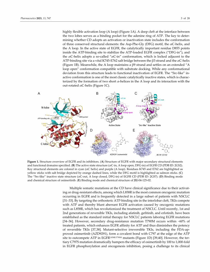
highly flexible activation loop (A loop) (Figure 1A). A deep cleft at the interface between
the two lobes serves as a binding pocket for the adenine ring of ATP. The key to deter‐
mining whether CD adopts an activation or inactivation state includes the conformation
of three conserved structural elements: the Asp‐Phe‐Gly (DFG) motif, the αC‐helix, and
the A loop. In the active state of EGFR, the catalytically important residue D855 points
inside the ATP‐binding site to stabilize the ATP‐loaded EGFR complex (“DFG‐in”), and
the αC‐helix adopts a so‐called “αC‐in” conformation, which is locked adjacent to the
ATP‐binding site via a vital K745‐E762 salt bridge between the β3 strand and the αC‐helix
(Figure 1B). Meanwhile, the A loop maintains a β9 strand and settles on an extended “A
loop open” conformation compatible with substrate docking. While any conformational
deviation from this structure leads to functional inactivation of EGFR. The “Src‐like” in‐
active conformation is one of the most classic catalytically inactive states, which is charac‐
terized by the formation of two short α‐helices in the A loop and its interaction with the
out‐rotated αC‐helix (Figure 1C).
Figure 1. Structure overview of EGFR and its inhibitors. (A) Structure of EGFR with major secondary structural elements
and functional domains specified. (B) The active state structure (αC‐in, A loop open, DFG‐in) of EGFR CD (PDB ID: 2GS2).
Key structural elements are colored in cyan (αC helix) and purple (A loop). Residues K745 and E762 are highlighted as
yellow sticks with salt bridge depicted by orange dashed lines, while the DFG motif is highlighted as salmon sticks. (C)
The “Src‐like” inactive state structure (αC‐out, A loop closed, DFG‐in) of EGFR CD (PDB ID: 2GS7). (D) Binding mode
and chemical structure of osimertinib. (E) Binding mode and chemical structure of JBJ‐04‐125‐02.
Multiple somatic mutations at the CD have clinical significance due to their activat‐
ing or drug‐resistant effects, among which L858R is the most common oncogenic mutation
occurring in EGFR and is frequently detected in a large subset of patients with NSCLC
[31–33]. By targeting the orthosteric ATP‐binding site in the interlobar cleft, TKIs compete
with ATP and thereby blunt aberrant EGFR activation caused by oncogenic mutations
such as L858R, which has revolutionized the treatment of NSCLC. Until recently, 1st and
2nd generations of reversible TKIs, including afatinib, gefitinib, and erlotinib, have been
established as the standard initial therapy for NSCLC patients laboring EGFR mutations
[34–36]. However, secondary drug‐resistance mutation T790M occurs within ~60% of
treated patients, which enhances EGFR affinity for ATP and thus diminishes the potency
of reversible TKIs [37,38]. Mutant‐selective irreversible TKIs, including the FDA‐ap‐
proved osimertinib (AZD9291), form a covalent bond with C797 at the edge of the ATP
site to outcompete ATP in EGFRL858R/T790M mutants (Figure 1D) [39,40]. However, the ter‐
tiary C797S mutation dramatically hampers the efficacy of osimertinib by 100 to 1,000‐fold
in EGFR phosphorylation and oncogenesis inhibition, posing a challenge to its clinical
Pharmaceutics 2021, 13, 747 4 of 28
performance [39,41]. Hence, with such reality check, an effective anti‐resistance strategy
may represent the ultimate answer to EGFR‐mutated NSCLC.
To circumvent acquired drug‐resistance against TKIs, a set of allosteric inhibitors tar‐
geting EGFR were previously introduced, including EAI001, EAI045, and JBJ‐04‐125‐02
(shortened as “JBJ” in the article) (Figure 1E) [20,42–44]. To our knowledge, all current
allosteric EGFR inhibitors target the MEK‐pocket, which is generated by the outward dis‐
placement of the αC‐helix and was first identified in mitogen‐activated protein (MAP)
kinase kinases (MEKs) (Figure 1E) [45]. Importantly, JBJ was reported to have synergistic
effects together with osimertinib during coadministration [20]. The combination of JBJ and
osimertinib led to not only an evident delay in orthosteric drug‐resistance, but also a
unique enhancement of JBJ affinity in EGFRL858R/T790M in the presence of osimertinib. Such
enhancement is independent of the amount of available EGFR monomers present in the
cells, which leads to enhanced efficacy of the combination. Consequently, the dual‐target‐
ing therapeutics in EGFR proposed a solution to overcome resistance and meanwhile im‐
prove therapeutic outcomes. These findings highlighted EGFR as an ideally allosteric pro‐
tein and a potential target for dual‐targeting therapeutics.
In this study, large‐scale atomistic molecular dynamics (MD) simulations revealed
that the dynamic landscape of EGFRL858R/T790M was markedly reshaped upon the binding
of osimertinib, characterized by the stabilization of a catalytically inactive intermediate
substrate that is first reported in EGFR, thus modulating the allosteric MEK‐pocket. More‐
over, we described the stepwise transition pathway of EGFR approaching the “Src‐like”
inactive states, as well as the community network of allosteric signaling, in an effort to
gain an atomistic understanding of the reversed allosteric regulatory mechanism within
the CD. Importantly, based on the reversed allosteric communication, we identified a po‐
tential druggable allosteric pocket (X‐pocket) presented in osimertinib‐loaded EG‐
FRL858R/T790M, which was well exposed and can be readily targeted in both the active and
intermediate states of EGFR. The allosteric crosstalk between the X‐pocket and the ATP‐
binding site was analyzed using structural and energetic analysis. Moreover, the noncon‐
servation characteristics of the X‐pocket across the human kinome confirmed its potential
to yield selective inhibitors targeting EGFR. Our study revealed a dual‐targeting thera‐
peutic mechanism that allosteric sites were highly correlated with orthosteric drugging
via reversed allosteric communication, which provided a mechanistic basis for future clin‐
ical application of the dual‐targeting therapeutics. Furthermore, the verification method‐
ology utilized in this study led to the discovery of a novel, potential allosteric pocket. Our
findings may aid future rational design of a new generation of selective EGFR inhibitors
and provide an innovative perception of the modulation of receptor tyrosine kinase fam‐
ily.
2. Materials and Methods
2.1. Construction of Stimulated Systems
Three systems were constructed and subjected to MD simulations, including the apo
EGFRL858R/T790M, EGFRL858R/T790M–osimertinib, and EGFRL858R/T790M–osimertinib–JBJ. The con‐
struction of the EGFRL858R/T790M–osimertinib complex was based on a co‐crystal structure of
human EGFRL858R/T790M monomeric form from the RCSB Protein Data Bank (PDB ID: 4I1Z)
[46], while the EGFRL858R/T790M–osimertinib–JBJ stemmed from a co‐crystal structure of hu‐
man EGFR in complex with JBJ and ANP (PDB ID: 6DUK) [20]. The remodel of the missing
residues, as well as the mutations of L858 and T790, was conducted with Discovery Stu‐
dio, and osimertinib was docked into the ATP‐binding site using a covalent docking pro‐
tocol. The structure of free EGFRL858R/T790M was extracted from the EGFRL858R/T790M–osimer‐
tinib complex. The water molecules in the crystal structures were removed.
Pharmaceutics 2021, 13, 747 5 of 28
2.2. Covalent Docking
The docking of osimertinib was performed by Maestro Advantage Schrödinger
(Maestro, Schrödinger, LLC, New York, NY, USA). The structures of EGFRL858R/T790M,
EGFR–JBJ–ADP, and osimertinib were separately loaded and prepared in Maestro. The
centroid of ADP was selected as the center for grid generation with a site size of 20 × 20 ×
20 Å3. Constraints of the sulfhydryl group at C797 were specified as the electron donor for
Michael Addition during grid generation. The generated grid file and the prepared small
ligand were used as input files to perform covalent docking. The 50 top‐ranked poses of
osimertinib were extracted and analyzed for rationality. Finally, the lowest energy‐dock‐
ing pose was utilized for further simulations.
2.3. MD Simulations
The C797 residue bonded with osimertinib was viewed as a single modified amino
acid, and the Amber ff14SB force field and general amber force field (GAFF) were em‐
ployed to prepare the parameter files for minimizations and simulations [47]. All three
complexes were solvated in an orthorhombic TIP3P water box and then the counterions
were added for neutralization [48]. To mimic the physical condition inside human cells,
0.15 mol L−1 NaCl was further solvated into each system.
Each system was subjected to two rounds of energy minimization. They underwent
2000 steepest descent and 3000 conjugate gradient minimization steps with a backbone
positional restraint of 500 kcal (mol−1 Å−2) for 10 ps, followed by another round of 4000
steepest descent and 6000 conjugate gradient minimization steps without any constraint
for 20 ps. After that, under the constraint of 10 kcal (mol−1 Å−2) in a canonical ensemble
(NVT), each system was heated from 0 K to 300 K in 300 ps and was further subjected to
a 700 ps equilibration run. Finally, a total of 5 μs cMD was performed for each system in
both an isothermal‐isobaric ensemble (NPT) condition and a periodic boundary condition.
During the process of MD simulation, the particle mesh Ewald method was utilized to
model the long‐range electrostatic interactions, whereas a cutoff of 10 Å was employed to
simplify the short‐range electrostatic interactions and van der Waals force calculations
[49]. In addition, the SHAKE method was conducted to constrain bond interactions in‐
volving hydrogen [50].
2.4. Principal Component Analysis (PCA)
PCA was carried out to capture the essential motions and characterize overall con‐
formational transitions within each system. In PCA, the covariance matrix of Cα atoms
was diagonalized to generate a new set of eigenvectors (also called PC), which described
the system motions. The eigenvalue of each PC was related to the mean square fluctuation
throughout the system’s trajectory projected along with that PC, therefore the first sorted
PC (PC1) corresponded to the most dominant amplitude motion within the system, and
the dynamics of the system projected along PC1 was referred as “essential dynamics” [51].
In this work, each system had its trajectory stripped down to only the Cα atoms and su‐
perposed onto a reference structure. First, to visualize the major motions within each sys‐
tem using porcupine plots, the sampled conformations within the trajectories were pro‐
jected into the collective coordinate space defined by PC1, with reference to the starting
structure of each system, respectively. Next, to identify differences in the essential struc‐
tural‐dynamic properties of EGFR among different systems, each system had its trajectory
superposed onto the same reference structure of the “Src‐like” inactive state (PDB ID:
2GS7) and projected along with the first two PCs (PC1 and PC2), so that the motions (with
ligands or not) were all projected onto the same set of eigenvectors to achieve compara‐
bility.
Pharmaceutics 2021, 13, 747 6 of 28
2.5. Community Network Analysis
The dynamic cross correlation matrix (DCCM) was applied to present the correlation
between EGFR residues, and the correlation coefficient 𝐶 was calculated through the
cMD trajectories based on Equation (1):
𝐶 ∆𝑟 Δ𝑟 / ⟨Δ𝑟 ⟩ ⟨Δ𝑟 ⟩ ⁄ (1)
In the equation, Δ𝑟 and Δ𝑟 represent the atomic displacement vectors for 𝐶 at‐oms i and j, respectively, while 𝐶 represents the fluctuation correlation between two
residues. The correlation data were further applied to weight edges and calculate the edge
distance 𝐷 by Equation (2), which indicates the possibility of information flow:
𝐷 log 𝐶 (2)
The community network was defined as a set of nodes, which derived from the C
atoms within EGFR, connected by 𝐶 weighted edges between two nodes that stay adja‐
cent within a cutoff distance of 4.5 Å for at least 75% of the trajectory. The shortest paths
were calculated utilizing the Floyd‐Warshall algorithm, and the number of pairwise short‐
est paths cross a given edge was identified as the edge betweenness [52]. A community
was defined as a set of nodes that densely interconnect with each other. Based on the be‐
tweenness information, the distribution of communities was decided and optimized using
the Girvan‐Newman algorithm [53]. Communities containing residues less than three
were discarded. The optimal paths between node pairs were calculated to reflect inter‐
community communication, during which all edges connecting the communities were
identified and those with the highest betweenness were selected.
2.6. Energy Coupling Score Calculation
The molecular mechanisms generalized Born surface area (MM‐GBSA) energy de‐
composition scheme was performed on the corresponding cavities on EGFRL858R/T790M in
both the osimertinib‐loaded (apo) and the osimertinib‐unloaded (holo) systems throughout
their trajectories. To compare the residue‐residue interactions for one cavity in the two
systems, the energy decompositions within a pocket were calculated for residue pairs sep‐
arated by at least three amino acids in the sequence based on Equation (3):
E 𝐸 𝐸 𝐸 𝐺 𝐺 (3)
In the equation, 𝐸 represents internal energy, 𝐸 represents electrostatic energy, 𝐸 represents van der Waals energy, 𝐺 is the polar solvation free energy, and 𝐺
is the solvent accessible surface energy.
The energy decomposition values were calculated and averaged over 5000 snapshots
captured from the MD simulation trajectories of apo and holo systems, respectively.
3. Results
3.1. Orthosteric Osimertinib Binding Induced a Conformational Transition of EGFRL858R/T790M by
Departing the “Src‐like” Inactive State
To reveal the detailed mechanism of orthosteric inhibitor binding that enhanced the
potency of allosteric ligands in the EGFR mutant, each 5 μs large‐scale MD simulation was
performed on three systems, including the apo EGFRL858R/T790M, EGFRL858R/T790M in complex
with osimertinib (EGFRL858R/T790M–osimertinib), and EGFRL858R/T790M complexed with both
osimertinib and JBJ (EGFRL858R/T790M–osimertinib–JBJ). Experimental results revealed that
the enhancement of allosteric binding was independent of the dimerization level of EGFR
in cells. Therefore, it was assumed that osimertinib improved JBJ affinity via conforma‐
tional regulation in EGFR monomers, instead of inhibiting dimerization to raise the pro‐
portion of αC‐out conformers [20]. Accordingly, the starting structure of EGFRL858R/T790M in
the two systems EGFRL858R/T790M and EGFRL858R/T790M–osimertinib stemmed from the crystal
structure of EGFR monomeric form (PDB ID: 4I1Z), which was characterized as αC‐out
Pharmaceutics 2021, 13, 747 7 of 28
(the K745‐E762 salt bridge was broken, while the E762‐K860 salt bridge was not formed
yet), A loop‐open (the conserved residue Y891 positioned the backbone of the phosphor‐
ylation site Y869 via salt bridges), and DFG‐out (Figure S1) [46]. The crystal structure of
EGFRL858R/T790M monomer adopted a locally disordered state (the αC helix is placed out,
whereas the A loop remains in an active‐like open state), instead of a quintessential active
state normally found in oncogenic mutants [54]. By contrast, the starting structure of the
EGFRL858R/T790M–osimertinib–JBJ system came from a JBJ‐loaded EGFRWT crystal structure,
which presented an “Src‐like” inactive state induced by the allosteric inhibitor (Figure S1)
[20,55].
For every snapshot of the trajectories, the root‐mean‐square deviations (RMSD) of all
Cα atoms were calculated, referring to the starting structure of each system (Figure S2).
The RMSD analysis revealed that all three systems reached equilibrium in 100 ns. For the
conformers in equilibrium (100–5000 ns), the RMSD values of EGFRL858R/T790M, EG‐
FRL858R/T790M–osimertinib, and EGFRL858R/T790M–osimertinib–JBJ systems were 2.88 ± 0.51 Å,
2.75 ± 0.24 Å, and 2.98 ± 0.44 Å, respectively. The rather close RMSD values of different
systems indicate minor overall conformational alterations of EGFRL858R/T790M in each system
during simulations, which implies a relatively stable overall structure of the kinase do‐
main. Therefore, instead of causing major conformational changes, the orthosteric and al‐
losteric ligands stabilized the overall structure of EGFR, leading to the reduction in the
standard deviation of RMSD (from 0.51Å to 0.24 Å and 0.44 Å, respectively).
Next, the root‐mean‐square fluctuations (RMSF) were calculated to reveal the mobil‐
ity of different regions (Figure S3A). Major fluctuating functional regions that differed
across various systems included the P loop, the β3‐αC loop, the αC helix, the αC‐β4 loop,
and the A loop (Figure S3A). The per‐residue RMSF difference between EGFR with and
without osimertinib was further calculated and projected onto the initial EGFR structure
for intuitive visualization (Figure S3B). Upon binding of osimertinib, the RMSF values of
the above‐mentioned crucial regions exhibited considerable changes that the P loop, the
β3‐αC loop, the αC‐β4 loop, and the A loop were significantly stabilized, while the αC
helix became more fluctuated instead. The RMSF differences at various functional regions
topologically distal from the orthosteric site suggested that in addition to directly compet‐
ing with ATP binding, osimertinib also triggered functional alterations within EGFR by
modulating the overall structures including allosteric regions. Further inhibition with JBJ
led to general stabilization at all these regions, denoting that coadministration with allo‐
steric and orthosteric inhibitors greatly stabilized the functional regions of EGFRL858R/T790M
in their inactive states, in agreement with the experimental results of an enhanced efficacy
(Figure S3A).
In order to characterize and compare the dominant conformations among different
complexes, principal component analysis (PCA) was performed for each system to deter‐
mine the global conformational transition patterns of EGFR, which was plotted using the
two most representative collective principal components (PC1 and PC2). Since the two
principal components are calculated without regard to biological significance, they pre‐
sent more objective information to picture the comprehensive structural shifts in the sys‐
tem. Distinct conformations were detected in EGFR with or without the presence of osi‐
mertinib. Porcupine plots were first constructed to graphically visualize the dominant
movements of different regions in EGFR. The protein dynamics were projected along with
PC1 onto the starting structure of each system for intuitionistic description of the major
subdomain movements during simulations (Figure S4). The principal dynamic motion of
the EGFRL858R/T790M system mainly concentrated on the β3‐αC loop, the N‐terminus of αC
helix, and the middle part of the A loop, but the αC helix remained an αC‐out confor‐
mation, consistent with the fluctuation analysis results. To further describe the essential
eigenvector, the β3‐αC loop moved forward in adjacence with the lifted A loop, the N‐
terminus of αC helix underwent a disordering process, and the A loop moved towards
the N‐lobe. Accordingly, we assumed a trend towards the “Src‐like” inactive state in the
Pharmaceutics 2021, 13, 747 8 of 28
apo system. In the EGFRL858R/T790M–osimertinib system, the full‐length αC helix moved in‐
ward and the C‐terminus of A loop, which was close to the orthosteric site, extended out‐
ward to accommodate the binding of osimertinib, but the A loop fixed in the open confor‐
mation, very similar to the active state of EGFR. In the EGFRL858R/T790M–osimertinib–JBJ sys‐
tem, the overall structure was notably stabilized by the allosteric inhibitor, especially the
αC helix which was locked in an out state, while the closed A loop remained its flexible
nature.
To compare the essential dynamic properties of EGFR among different systems, each
system had its trajectory superposed onto the same reference structure of “Src‐like” inac‐
tive state (PDB ID: 2GS7) [55]. The free energy surface (FES) was then projected along PC1
and PC2 onto a two‐dimensional space to visualize the conformational dynamics of EGFR
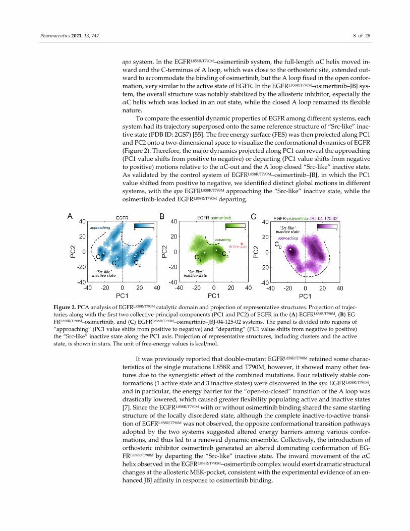
(Figure 2). Therefore, the major dynamics projected along PC1 can reveal the approaching
(PC1 value shifts from positive to negative) or departing (PC1 value shifts from negative
to positive) motions relative to the αC‐out and the A loop closed “Src‐like” inactive state.
As validated by the control system of EGFRL858R/T790M–osimertinib–JBJ, in which the PC1
value shifted from positive to negative, we identified distinct global motions in different
systems, with the apo EGFRL858R/T790M approaching the “Src‐like” inactive state, while the
osimertinib‐loaded EGFRL858R/T790M departing.
Figure 2. PCA analysis of EGFRL858R/T790M catalytic domain and projection of representative structures. Projection of trajec‐
tories along with the first two collective principal components (PC1 and PC2) of EGFR in the (A) EGFRL858R/T790M, (B) EG‐
FRL858R/T790M–osimertinib, and (C) EGFRL858R/T790M–osimertinib–JBJ‐04‐125‐02 systems. The panel is divided into regions of
“approaching” (PC1 value shifts from positive to negative) and “departing” (PC1 value shifts from negative to positive)
the “Src‐like” inactive state along the PC1 axis. Projection of representative structures, including clusters and the active
state, is shown in stars. The unit of free‐energy values is kcal/mol.
It was previously reported that double‐mutant EGFRL858R/T790M retained some charac‐
teristics of the single mutations L858R and T790M, however, it showed many other fea‐
tures due to the synergistic effect of the combined mutations. Four relatively stable con‐
formations (1 active state and 3 inactive states) were discovered in the apo EGFRL858R/T790M,
and in particular, the energy barrier for the “open‐to‐closed” transition of the A loop was
drastically lowered, which caused greater flexibility populating active and inactive states
[7]. Since the EGFRL858R/T790M with or without osimertinib binding shared the same starting
structure of the locally disordered state, although the complete inactive‐to‐active transi‐
tion of EGFRL858R/T790M was not observed, the opposite conformational transition pathways
adopted by the two systems suggested altered energy barriers among various confor‐
mations, and thus led to a renewed dynamic ensemble. Collectively, the introduction of
orthosteric inhibitor osimertinib generated an altered dominating conformation of EG‐
FRL858R/T790M by departing the “Src‐like” inactive state. The inward movement of the αC
helix observed in the EGFRL858R/T790M–osimertinib complex would exert dramatic structural
changes at the allosteric MEK‐pocket, consistent with the experimental evidence of an en‐
hanced JBJ affinity in response to osimertinib binding.
Pharmaceutics 2021, 13, 747 9 of 28
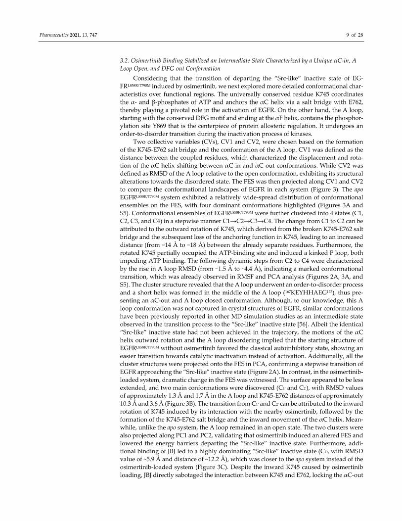
3.2. Osimertinib Binding Stabilized an Intermediate State Characterized by a Unique αC‐in, A
Loop Open, and DFG‐out Conformation
Considering that the transition of departing the “Src‐like” inactive state of EG‐
FRL858R/T790M induced by osimertinib, we next explored more detailed conformational char‐
acteristics over functional regions. The universally conserved residue K745 coordinates
the α‐ and β‐phosphates of ATP and anchors the αC helix via a salt bridge with E762,
thereby playing a pivotal role in the activation of EGFR. On the other hand, the A loop,
starting with the conserved DFG motif and ending at the αF helix, contains the phosphor‐
ylation site Y869 that is the centerpiece of protein allosteric regulation. It undergoes an
order‐to‐disorder transition during the inactivation process of kinases.
Two collective variables (CVs), CV1 and CV2, were chosen based on the formation
of the K745‐E762 salt bridge and the conformation of the A loop. CV1 was defined as the
distance between the coupled residues, which characterized the displacement and rota‐
tion of the αC helix shifting between αC‐in and αC‐out conformations. While CV2 was
defined as RMSD of the A loop relative to the open conformation, exhibiting its structural
alterations towards the disordered state. The FES was then projected along CV1 and CV2
to compare the conformational landscapes of EGFR in each system (Figure 3). The apo
EGFRL858R/T790M system exhibited a relatively wide‐spread distribution of conformational
ensembles on the FES, with four dominant conformations highlighted (Figures 3A and
S5). Conformational ensembles of EGFRL858R/T790M were further clustered into 4 states (C1,
C2, C3, and C4) in a stepwise manner C1→C2→C3→C4. The change from C1 to C2 can be
attributed to the outward rotation of K745, which derived from the broken K745‐E762 salt
bridge and the subsequent loss of the anchoring function in K745, leading to an increased
distance (from ~14 Å to ~18 Å) between the already separate residues. Furthermore, the
rotated K745 partially occupied the ATP‐binding site and induced a kinked P loop, both
impeding ATP binding. The following dynamic steps from C2 to C4 were characterized
by the rise in A loop RMSD (from ~1.5 Å to ~4.4 Å), indicating a marked conformational
transition, which was already observed in RMSF and PCA analysis (Figures 2A, 3A, and
S5). The cluster structure revealed that the A loop underwent an order‐to‐disorder process
and a short helix was formed in the middle of the A loop (167KEYHHAEG173), thus pre‐
senting an αC‐out and A loop closed conformation. Although, to our knowledge, this A
loop conformation was not captured in crystal structures of EGFR, similar conformations
have been previously reported in other MD simulation studies as an intermediate state
observed in the transition process to the “Src‐like” inactive state [56]. Albeit the identical
“Src‐like” inactive state had not been achieved in the trajectory, the motions of the αC
helix outward rotation and the A loop disordering implied that the starting structure of
EGFRL858R/T790M without osimertinib favored the classical autoinhibitory state, showing an
easier transition towards catalytic inactivation instead of activation. Additionally, all the
cluster structures were projected onto the FES in PCA, confirming a stepwise transition of
EGFR approaching the “Src‐like” inactive state (Figure 2A). In contrast, in the osimertinib‐
loaded system, dramatic change in the FES was witnessed. The surface appeared to be less
extended, and two main conformations were discovered (C1′ and C2′), with RMSD values
of approximately 1.3 Å and 1.7 Å in the A loop and K745‐E762 distances of approximately
10.3 Å and 3.6 Å (Figure 3B). The transition from C1′ and C2′ can be attributed to the inward
rotation of K745 induced by its interaction with the nearby osimertinib, followed by the
formation of the K745‐E762 salt bridge and the inward movement of the αC helix. Mean‐
while, unlike the apo system, the A loop remained in an open state. The two clusters were
also projected along PC1 and PC2, validating that osimertinib induced an altered FES and
lowered the energy barriers departing the “Src‐like” inactive state. Furthermore, addi‐
tional binding of JBJ led to a highly dominating “Src‐like” inactive state (CD, with RMSD
value of ~5.9 Å and distance of ~12.2 Å), which was closer to the apo system instead of the
osimertinib‐loaded system (Figure 3C). Despite the inward K745 caused by osimertinib
loading, JBJ directly sabotaged the interaction between K745 and E762, locking the αC‐out
Pharmaceutics 2021, 13, 747 10 of 28
state. Moreover, the allosteric inhibitor was validated to form salt bridges with F856 and
E865, leading to the stabilization of a disordered A loop [20].
Figure 3. Conformational free energy surface of EGFRL858R/T790M relevant to activation/inactivation.
The landscape is generated using the distance between K745 (zeta nitrogen) and E762 (delta car‐
bon), and the RMSD in the A loop in the (A) EGFRL858R/T790M, (B) EGFRL858R/T790M–osimertinib, and
(C) EGFRL858R/T790M–osimertinib–JBJ‐04‐125‐02 systems. The dominant conformational rearrange‐
ments in the representative structures of the free energy minima are shown with the αC helix col‐
ored in red, the K745‐E762 residue pair in blue, the A loop in yellow, and the P loop in green. The
unit of free‐energy values is kcal/mol.
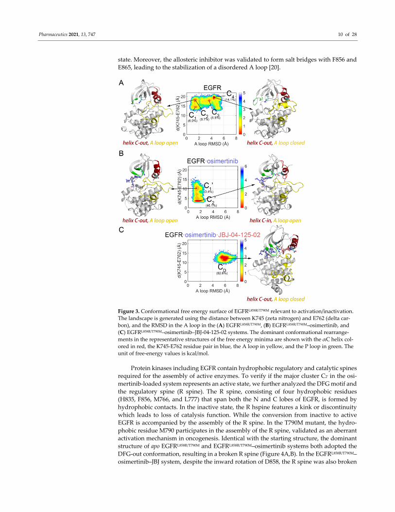
Protein kinases including EGFR contain hydrophobic regulatory and catalytic spines
required for the assembly of active enzymes. To verify if the major cluster C2′ in the osi‐
mertinib‐loaded system represents an active state, we further analyzed the DFG motif and
the regulatory spine (R spine). The R spine, consisting of four hydrophobic residues
(H835, F856, M766, and L777) that span both the N and C lobes of EGFR, is formed by
hydrophobic contacts. In the inactive state, the R hspine features a kink or discontinuity
which leads to loss of catalysis function. While the conversion from inactive to active
EGFR is accompanied by the assembly of the R spine. In the T790M mutant, the hydro‐
phobic residue M790 participates in the assembly of the R spine, validated as an aberrant
activation mechanism in oncogenesis. Identical with the starting structure, the dominant
structure of apo EGFRL858R/T790M and EGFRL858R/T790M–osimertinib systems both adopted the
DFG‐out conformation, resulting in a broken R spine (Figure 4A,B). In the EGFRL858R/T790M–
osimertinib–JBJ system, despite the inward rotation of D858, the R spine was also broken
Pharmaceutics 2021, 13, 747 11 of 28
by the allosteric ligand and the repositioned M766 (Figure 4C). Since the R spine was com‐
pleted and assembled in none of the systems, they were all identified as catalytically in‐
active. However, C2′, induced by osimertinib disturbance at the orthosteric site, was char‐
acterized as αC‐in, A loop open, and DFG‐out, highly resembling the structure of an Abl
intermediate state recently reported by Xie et al. [57]. They performed nuclear magnetic
resonance spectroscopy to speculate the transition pathway of Abl activation and discov‐
ered a novel inactive conformation (I1), characterized as αC‐in, A loop open, and DFG‐
out. Herein, we proposed that such intermediate conformation existed in the apo EGFR as
well, and osimertinib stabilized this unique state by rendering the β3 strands in the N‐
lobe to approach the αC helix, thus facilitating the formation of the K745‐E762 salt bridge.
Moreover, the inward movement of the αC helix resulted in M766 rotation, which filled
into the space between L777 and F856, contributing to the assembly of the R spine. We
calculated the sum of the M766‐M790 and M766‐H835 distance, and discovered a signifi‐
cant decrease from 16.49 Å to 14.77 Å in the sum upon osimertinib loading (Figure 4D).
The declined distance reflected a more assembled state of the R spine, which would en‐
gender stronger hydrophobic attraction to promote the proper orientation of F856, facili‐
tating complete assembly of the R spine. Additionally, we projected the active structure
(PDB ID: 2GS2) [53] along PC1 and PC2 of the EGFRL858R/T790M–osimertinib system, and C2′
was found located along the activating pathway, illustrating our hypothesis of C2′ as a
unique intermediate conformation (Figure 2B).
Figure 4. Structural analysis of the DFG motif and the R spine. Atomistic details of the DFG motif
and the R spine in the (A) EGFRL858R/T790M, (B) EGFRL858R/T790M–osimertinib, and (C) EGFRL858R/T790M–
osimertinib–JBJ‐04‐125‐02 systems. (D) Distance sum of M766‐M790 and M766‐H835 calculated
from conformations sampled during the MD simulations of EGFRL858R/T790M (yellow), EG‐
FRL858R/T790M–osimertinib (blue), and EGFRL858R/T790M–osimertinib–JBJ‐04‐125‐02 (purple).
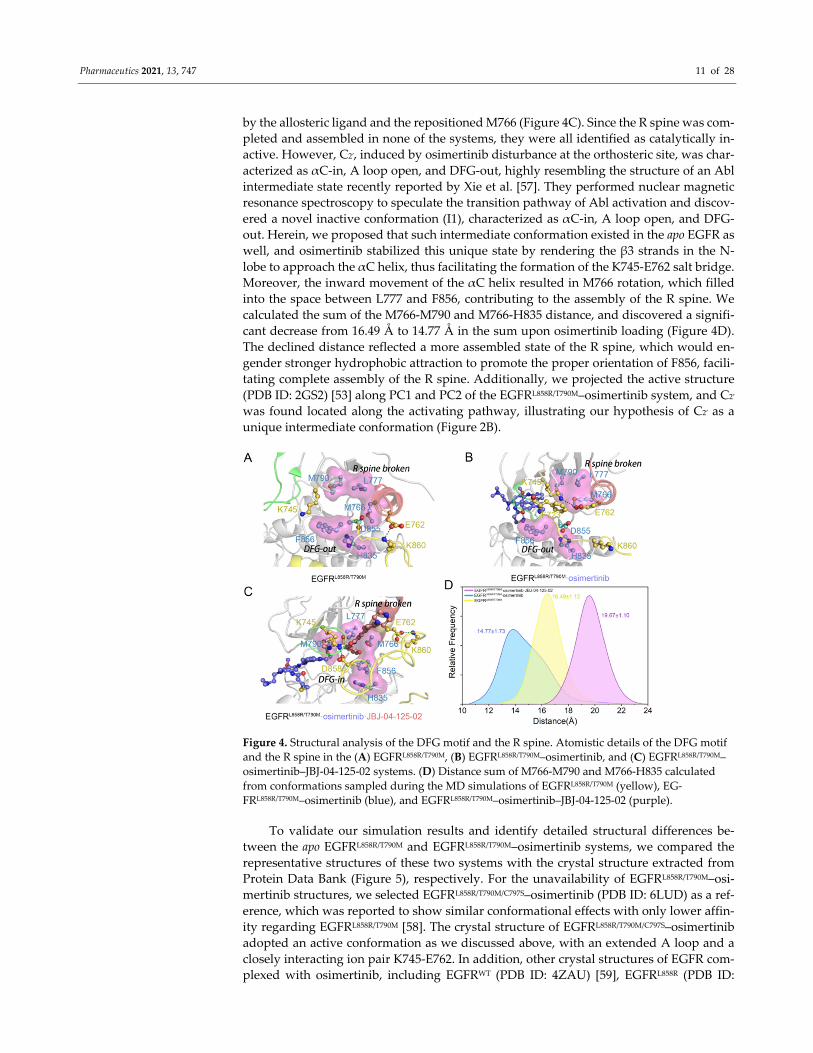
To validate our simulation results and identify detailed structural differences be‐
tween the apo EGFRL858R/T790M and EGFRL858R/T790M–osimertinib systems, we compared the
representative structures of these two systems with the crystal structure extracted from
Protein Data Bank (Figure 5), respectively. For the unavailability of EGFRL858R/T790M–osi‐
mertinib structures, we selected EGFRL858R/T790M/C797S–osimertinib (PDB ID: 6LUD) as a ref‐
erence, which was reported to show similar conformational effects with only lower affin‐
ity regarding EGFRL858R/T790M [58]. The crystal structure of EGFRL858R/T790M/C797S–osimertinib
adopted an active conformation as we discussed above, with an extended A loop and a
closely interacting ion pair K745‐E762. In addition, other crystal structures of EGFR com‐
plexed with osimertinib, including EGFRWT (PDB ID: 4ZAU) [59], EGFRL858R (PDB ID:
Pharmaceutics 2021, 13, 747 12 of 28
6JWL) [56], and EGFRT790M (PDB ID: 6JX4) [56] all exhibited an active conformation,
strengthening the reliability of our simulation results [56,59]. Upon alignment of EG‐
FRL858R/T790M–osimertinib with EGFRL858R/T790M/C797S–osimertinib, the low RMSD value (0.94
Å) of EGFR reflects no significant difference between the overall structures of these two
complexes. With detailed structural analysis zooming into several important regions,
some subtle distinctions emerged. In the C2′ conformation, F723, the bulkiest residue lo‐
cated in the middle of the P loop, swung outward by ~12 Å toward the αC helix, while it
was flipped and positioned right at the top edge of the orthosteric site in the crystal struc‐
ture (Figure 5A). The translocated F723 not only acted as a wedge to push the N‐terminus
of αC helix away from adopting its αC‐in conformation, but also posed a massive steric
hindrance against the inward rotation of F856, resulting in the kinked αC helix and DFG‐
out state. Furthermore, the outward location of F856 observed in EGFRL858R/T790M/C797S–osi‐
mertinib structure agreed with the experimental results that the affinity of JBJ was en‐
hanced by osimertinib, since type IV inhibitors including JBJ require a folded aromatic
residue to create a cavity between the P loop and the αC helix [60]. Despite the differences
found in the DFG motif, the A loop conformations were highly consistent across the two
structures. Importantly, the key phosphorylation site Y869 was exposed on the protein
surface, stabilized by the conserved salt bridges with Y891 and other electrostatic interac‐
tions with K860 and R836, thus contributing to the phosphorylation at Y869. In a previous
study on another kinase Akt, it was revealed that the binding of TKIs to the orthosteric
site was sufficient to cause hyperphosphorylation of Akt in the absence of any pathway
feedback effects [61]. Herein, our simulation results revealed a possible explanation that
the TKIs targeting Akt, similar to osimertinib with EGFR, stabilized the open state of the
A loop, thus positioning the key residue in its proper place for phosphorylation.
Figure 5. Structure of EGFR mutants in complex with osimertinib. (A) The representative structure
of the EGFRL858R/T790M–osimertinib system (lime) superimposed on the crystal structure of EG‐
FRL858R/T790M/C797S–osimertinib (slate, PDB ID: 6LUD). The magnified views highlight the similarities
Pharmaceutics 2021, 13, 747 13 of 28
(e.g., αC helix and A loop) and differences (e.g., DFG motif) in the disposition of key structural
elements between the two structures. (B) The representative structure of the EGFRL858R/T790M system
(yellow) superimposed on the crystal structure of EGFRL858R/T790M/C797S–osimertinib (slate). The mag‐
nified views highlight the differences in the disposition of key structural elements between the
two structures.
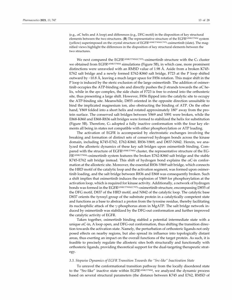
We next compared the EGFRL858R/T790M/C797S–osimertinib structure with the C4 cluster
we obtained from EGFRL858R/T790M simulations (Figure 5B), in which case, more prominent
distinctions were unraveled with an RMSD value of 1.98 Å. Aside from a broken K745‐
E762 salt bridge and a newly formed E762‐K860 salt bridge, F723 at the P loop shifted
outward by ~10.8 Å, leaving a much larger space for F856 rotation. This major shift in the
P loop is induced by the steric exclusion of the large osimertinib. The addition of osimer‐
tinib occupies the ATP‐binding site and directly pushes the β strands towards the αC he‐
lix, while in the apo complex, the side chain of F723 is free to extend into the orthosteric
site, thus presenting a large shift. However, F856 flipped into the catalytic site to occupy
the ATP‐binding site. Meanwhile, D855 oriented in the opposite direction unsuitable to
bind the implicated magnesium ion, also obstructing the binding of ATP. On the other
hand, Y869 folded into a short helix and rotated approximately 180° away from the pro‐
tein surface. The conserved salt bridges between Y869 and Y891 were broken, while the
E868‐K860 and E868‐R836 salt bridges were formed to stabilized the helix for substitution
(Figure 5B). Therefore, C4 adopted a fully inactive conformation with the four key ele‐
ments all being in states not compatible with either phosphorylation or ATP loading.
The activation of EGFR is accompanied by electrostatic exchanges involving the
breaking and formation of distinct sets of conserved hydrogen bonds across the kinase
domain, including K745‐E762, E762‐K860, R836‐Y869, and D837‐N842. Herein, we ana‐
lyzed the allosteric dynamics of these key salt bridges upon osimertinib binding. Com‐
pared with the structure of EGFRL858R/T790M cluster, the representative structure of the EG‐
FRL858R/T790M–osimertinib system features the broken E762‐K860 salt bridge and the stable
K745‐E762 salt bridge instead. This shift of hydrogen bond explains the αC‐in confor‐
mation at the allosteric site. Moreover, the essential R836‐Y869 salt bridge, which connects
the HRD motif of the catalytic loop and the activation segment, was formed upon osimer‐
tinib loading, and the salt bridge between R836 and E868 was consequently broken. Such
a shift implies that osimertinib induces the explosion of Y869 for phosphorylation at the
activation loop, which is required for kinase activity. Additionally, a network of hydrogen
bonds was formed in the EGFRL858R/T790M/C797S–osimertinib structure, encompassing D855 of
the DFG motif, D837 of the HRD motif, and N842 of the catalytic loop. The catalytic base
D837 orients the tyrosyl group of the substrate protein in a catalytically competent state
and functions as a base to abstract a proton from the tyrosine residue, thereby facilitating
its nucleophilic attack of the γ‐phosphorus atom in MgATP. The salt bridge network in‐
duced by osimertinib was stabilized by the DFG‐out conformation and further improved
the catalytic activity of EGFR.
Taken together, osimertinib binding stabled a potential intermediate state with a
unique αC‐in, A loop open, and DFG‐out conformation, thus shifting the transition direc‐
tion towards the activation state. Namely, the perturbation of orthosteric ligands not only
posed effects on nearby regions, but also spread its influence into topologically distant
areas, thus exerting an impact on the overall functions of the target protein. As such, it is
feasible to precisely regulate the allosteric sites both structurally and functionally with
orthosteric ligands, providing theoretical support for the dual‐targeting therapeutic strat‐
egy.
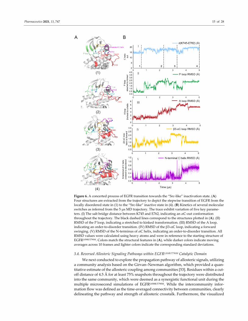
3.3. Stepwise Dynamics of EGFR Transition Towards the “Src‐like” Inactivation State
To unravel the conformational transition pathway from the locally disordered state
to the “Src‐like” inactive state within EGFRL858R/T790M, we analyzed the dynamic process
based on several structural parameters (the distance between K745 and E762, RMSD of
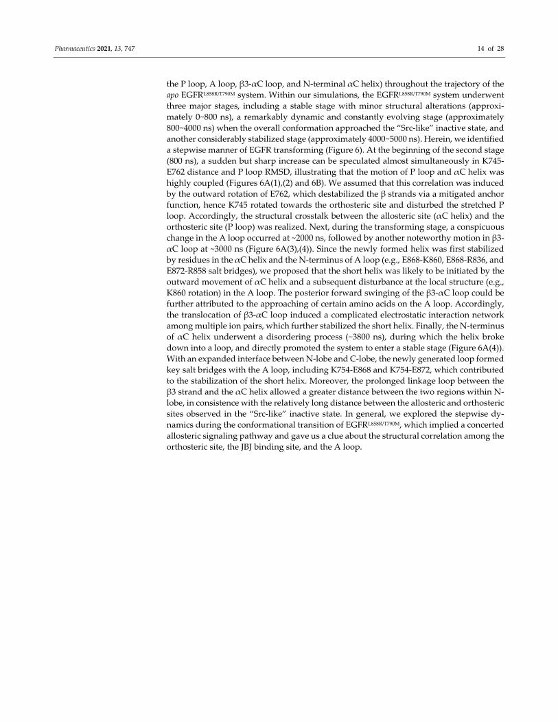
Pharmaceutics 2021, 13, 747 14 of 28
the P loop, A loop, β3‐αC loop, and N‐terminal αC helix) throughout the trajectory of the
apo EGFRL858R/T790M system. Within our simulations, the EGFRL858R/T790M system underwent
three major stages, including a stable stage with minor structural alterations (approxi‐
mately 0~800 ns), a remarkably dynamic and constantly evolving stage (approximately
800~4000 ns) when the overall conformation approached the “Src‐like” inactive state, and
another considerably stabilized stage (approximately 4000~5000 ns). Herein, we identified
a stepwise manner of EGFR transforming (Figure 6). At the beginning of the second stage
(800 ns), a sudden but sharp increase can be speculated almost simultaneously in K745‐
E762 distance and P loop RMSD, illustrating that the motion of P loop and αC helix was
highly coupled (Figures 6A(1),(2) and 6B). We assumed that this correlation was induced
by the outward rotation of E762, which destabilized the β strands via a mitigated anchor
function, hence K745 rotated towards the orthosteric site and disturbed the stretched P
loop. Accordingly, the structural crosstalk between the allosteric site (αC helix) and the
orthosteric site (P loop) was realized. Next, during the transforming stage, a conspicuous
change in the A loop occurred at ~2000 ns, followed by another noteworthy motion in β3‐
αC loop at ~3000 ns (Figure 6A(3),(4)). Since the newly formed helix was first stabilized
by residues in the αC helix and the N‐terminus of A loop (e.g., E868‐K860, E868‐R836, and
E872‐R858 salt bridges), we proposed that the short helix was likely to be initiated by the
outward movement of αC helix and a subsequent disturbance at the local structure (e.g.,
K860 rotation) in the A loop. The posterior forward swinging of the β3‐αC loop could be
further attributed to the approaching of certain amino acids on the A loop. Accordingly,
the translocation of β3‐αC loop induced a complicated electrostatic interaction network
among multiple ion pairs, which further stabilized the short helix. Finally, the N‐terminus
of αC helix underwent a disordering process (~3800 ns), during which the helix broke
down into a loop, and directly promoted the system to enter a stable stage (Figure 6A(4)).
With an expanded interface between N‐lobe and C‐lobe, the newly generated loop formed
key salt bridges with the A loop, including K754‐E868 and K754‐E872, which contributed
to the stabilization of the short helix. Moreover, the prolonged linkage loop between the
β3 strand and the αC helix allowed a greater distance between the two regions within N‐
lobe, in consistence with the relatively long distance between the allosteric and orthosteric
sites observed in the “Src‐like” inactive state. In general, we explored the stepwise dy‐
namics during the conformational transition of EGFRL858R/T790M, which implied a concerted
allosteric signaling pathway and gave us a clue about the structural correlation among the
orthosteric site, the JBJ binding site, and the A loop.
Pharmaceutics 2021, 13, 747 15 of 28
Figure 6. A concerted process of EGFR transition towards the “Src‐like” inactivation state. (A)
Four structures are extracted from the trajectory to depict the stepwise transition of EGFR from the
locally disordered state in (1) to the “Src‐like” inactive state in (4). (B) Kinetics of several molecular
switches as inferred from the 5 μs MD trajectory. The trace exhibit variation of five key parame‐
ters. (I) The salt bridge distance between K745 and E762, indicating an αC‐out conformation
throughout the trajectory. The black dashed lines correspond to the structures plotted in (A). (II)
RMSD of the P loop, indicating a stretched to kinked transformation. (III) RMSD of the A loop,
indicating an order‐to‐disorder transition. (IV) RMSD of the β3‐αC loop, indicating a forward
swinging. (V) RMSD of the N‐terminus of αC helix, indicating an order‐to‐disorder transition. All
RMSD values were calculated using heavy atoms and were in reference to the starting structure of
EGFRL858R/T790M. Colors match the structural features in (A), while darker colors indicate moving
averages across 10 frames and lighter colors indicate the corresponding standard deviations.
3.4. Reversed Allosteric Signaling Pathways within EGFRL858R/T790M Catalytic Domain
We next conducted to explore the propagation pathway of allosteric signals, utilizing
a community analysis based on the Girvan–Newman algorithm, which provided a quan‐
titative estimate of the allosteric coupling among communities [53]. Residues within a cut‐
off distance of 4.5 Å for at least 75% snapshots throughout the trajectory were distributed
into the same community, which were deemed as a synergistic functional unit during the
multiple microsecond simulations of EGFRL858R/T790M. While the intercommunity infor‐
mation flow was defined as the time‐averaged connectivity between communities, clearly
delineating the pathway and strength of allosteric crosstalk. Furthermore, the visualized
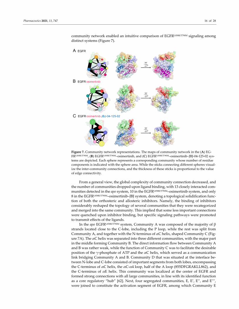
Pharmaceutics 2021, 13, 747 16 of 28
community network enabled an intuitive comparison of EGFRL858R/T790M signaling among
distinct systems (Figure 7).
Figure 7. Community network representations. The maps of community network in the (A) EG‐
FRL858R/T790M, (B) EGFRL858R/T790M–osimertinib, and (C) EGFRL858R/T790M–osimertinib–JBJ‐04‐125‐02 sys‐
tems are depicted. Each sphere represents a corresponding community whose number of residue
components is indicated with the sphere area. While the sticks connecting different spheres visual‐
ize the inter‐community connections, and the thickness of these sticks is proportional to the value
of edge connectivity.
From a general view, the global complexity of community connection decreased, and
the number of communities dropped upon ligand binding, with 13 closely interacted com‐
munities detected in the apo system, 10 in the EGFRL858R/T790M–osimertinib system, and only
8 in the EGFRL858R/T790M–osimertinib–JBJ system, denoting a topological solidification func‐
tion of both the orthosteric and allosteric inhibitors. Namely, the binding of inhibitors
considerably reshaped the topology of several communities that they were recategorized
and merged into the same community. This implied that some less important connections
were quenched upon inhibitor binding, but specific signaling pathways were promoted
to transmit effects of the ligands.
In the apo EGFRL858R/T790M system, Community A was composed of the majority of β
strands located close to the C‐lobe, including the P loop, while the rest was split from
Community A, and together with the N‐terminus of αC helix, shaped Community C (Fig‐
ure 7A). The αC helix was separated into three different communities, with the major part
in the middle forming Community B. The direct information flow between Community A
and B was rather weak, while the function of Community C was to facilitate the desirable
position of the γ‐phosphate of ATP and the αC helix, which served as a communication
link bridging Community A and B. Community D that was situated at the interface be‐
tween N‐lobe and C‐lobe consisted of important segments from both lobes, encompassing
the C‐terminus of αC helix, the αC‐α4 loop, half of the A loop (855DFGRAKLL862), and
the C‐terminus of αE helix. This community was localized at the center of EGFR and
formed strong connections with all large communities, in line with its identified function
as a core regulatory “hub” [62]. Next, four segregated communities, E, E’, E’’, and E’’’,
were joined to constitute the activation segment of EGFR, among which Community E
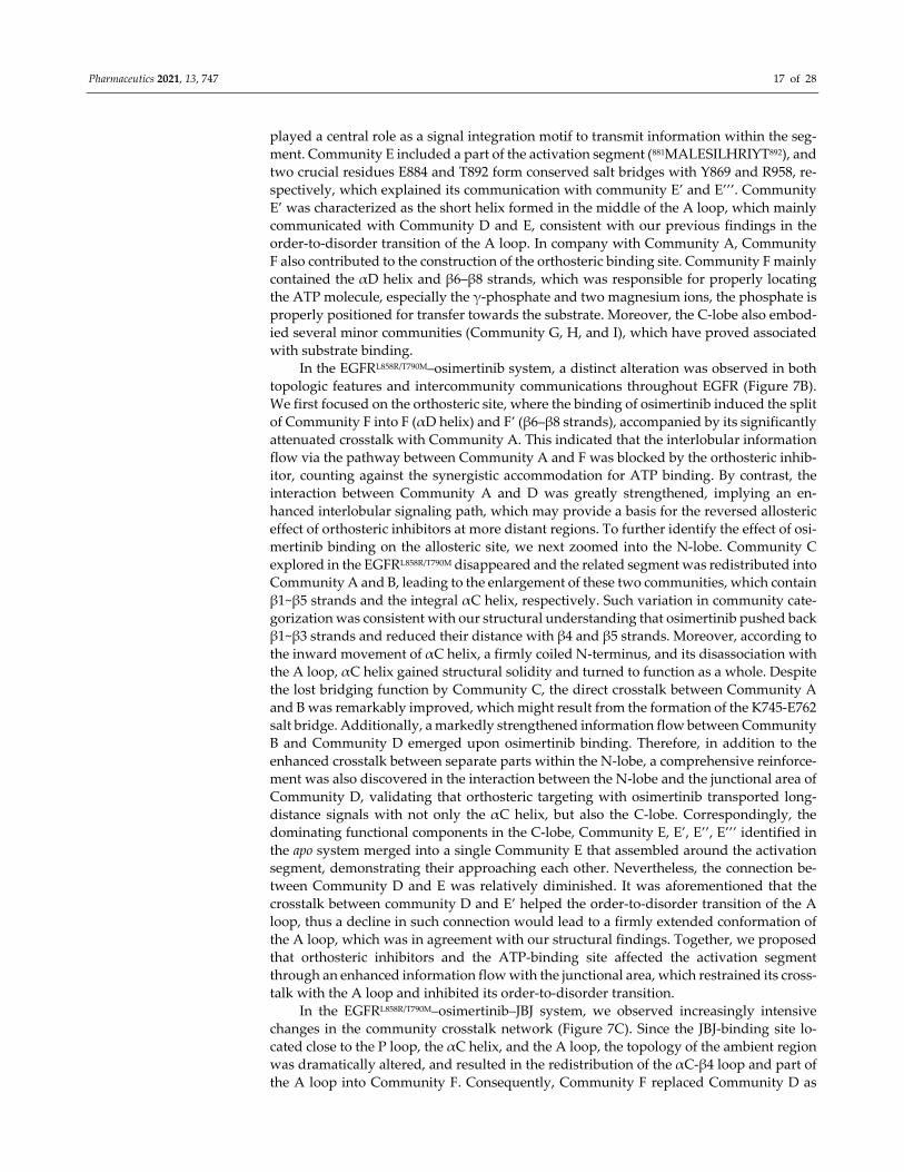
Pharmaceutics 2021, 13, 747 17 of 28
played a central role as a signal integration motif to transmit information within the seg‐
ment. Community E included a part of the activation segment (881MALESILHRIYT892), and
two crucial residues E884 and T892 form conserved salt bridges with Y869 and R958, re‐
spectively, which explained its communication with community E’ and E’’’. Community
E’ was characterized as the short helix formed in the middle of the A loop, which mainly
communicated with Community D and E, consistent with our previous findings in the
order‐to‐disorder transition of the A loop. In company with Community A, Community
F also contributed to the construction of the orthosteric binding site. Community F mainly
contained the αD helix and β6–β8 strands, which was responsible for properly locating
the ATP molecule, especially the γ‐phosphate and two magnesium ions, the phosphate is
properly positioned for transfer towards the substrate. Moreover, the C‐lobe also embod‐
ied several minor communities (Community G, H, and I), which have proved associated
with substrate binding.
In the EGFRL858R/T790M–osimertinib system, a distinct alteration was observed in both
topologic features and intercommunity communications throughout EGFR (Figure 7B).
We first focused on the orthosteric site, where the binding of osimertinib induced the split
of Community F into F (αD helix) and F’ (β6–β8 strands), accompanied by its significantly
attenuated crosstalk with Community A. This indicated that the interlobular information
flow via the pathway between Community A and F was blocked by the orthosteric inhib‐
itor, counting against the synergistic accommodation for ATP binding. By contrast, the
interaction between Community A and D was greatly strengthened, implying an en‐
hanced interlobular signaling path, which may provide a basis for the reversed allosteric
effect of orthosteric inhibitors at more distant regions. To further identify the effect of osi‐
mertinib binding on the allosteric site, we next zoomed into the N‐lobe. Community C
explored in the EGFRL858R/T790M disappeared and the related segment was redistributed into
Community A and B, leading to the enlargement of these two communities, which contain
β1~β5 strands and the integral αC helix, respectively. Such variation in community cate‐
gorization was consistent with our structural understanding that osimertinib pushed back
β1~β3 strands and reduced their distance with β4 and β5 strands. Moreover, according to
the inward movement of αC helix, a firmly coiled N‐terminus, and its disassociation with
the A loop, αC helix gained structural solidity and turned to function as a whole. Despite
the lost bridging function by Community C, the direct crosstalk between Community A
and B was remarkably improved, which might result from the formation of the K745‐E762
salt bridge. Additionally, a markedly strengthened information flow between Community
B and Community D emerged upon osimertinib binding. Therefore, in addition to the
enhanced crosstalk between separate parts within the N‐lobe, a comprehensive reinforce‐
ment was also discovered in the interaction between the N‐lobe and the junctional area of
Community D, validating that orthosteric targeting with osimertinib transported long‐
distance signals with not only the αC helix, but also the C‐lobe. Correspondingly, the
dominating functional components in the C‐lobe, Community E, E’, E’’, E’’’ identified in
the apo system merged into a single Community E that assembled around the activation
segment, demonstrating their approaching each other. Nevertheless, the connection be‐
tween Community D and E was relatively diminished. It was aforementioned that the
crosstalk between community D and E’ helped the order‐to‐disorder transition of the A
loop, thus a decline in such connection would lead to a firmly extended conformation of
the A loop, which was in agreement with our structural findings. Together, we proposed
that orthosteric inhibitors and the ATP‐binding site affected the activation segment
through an enhanced information flow with the junctional area, which restrained its cross‐
talk with the A loop and inhibited its order‐to‐disorder transition.
In the EGFRL858R/T790M–osimertinib–JBJ system, we observed increasingly intensive
changes in the community crosstalk network (Figure 7C). Since the JBJ‐binding site lo‐
cated close to the P loop, the αC helix, and the A loop, the topology of the ambient region
was dramatically altered, and resulted in the redistribution of the αC‐β4 loop and part of
the A loop into Community F. Consequently, Community F replaced Community D as
Pharmaceutics 2021, 13, 747 18 of 28
the central regulatory “hub”. Moreover, Community E again separated into E and E’, ac‐
cording to the short helices in the A loop induced by the allosteric inhibitor. The connec‐
tion among Community A, B, and F was conspicuously enhanced due to the cross‐linking
function of JBJ. While the information flow between F and E’ was further strengthened in
comparison with the apo system, supporting our notion that the information transmission
from the αC‐β4 loop and the N‐terminus of A loop to the C‐terminus of A loop induced
its disordering transformation. On the other hand, the original Community G merged
with Community D, which reflected the structural alterations within αE and αI helices.
This could lead to the impaired binding of EGFR substrates, further dampening the EGFR
activity via dual targeting.
In general, we identified the reversed and forward allosteric signaling pathways
among various functional regions, which were enhanced by osimertinib and JBJ, respec‐
tively. The characterization of these promoted interactions highlighted Community D (es‐
pecially the αC‐β4 loop and the N‐terminus of the A loop) as a core transmission hub.
Both orthosteric and allosteric inhibitors regulated the overall conformation by propagat‐
ing certain signals to distant subdomain areas, during which this interlobular junction
region played a central role to connect all major communities.
3.5. Identification of Potential Allosteric Pockets Based on Reversed Allosteric Communication
Since the MEK‐pocket is the only identified allosteric site in EGFR, revealing other
potential allosteric pockets is greatly beneficial for expanding the opportunities to develop
novel allosteric inhibitors. Based on the reversed allosteric communication, we demon‐
strated that distal regions were highly sensitive to orthosteric perturbations. Namely, the
binding of orthosteric ligands induced overall dynamics throughout the target protein,
leading to possible emergence, stabilization, and conformational alteration at potential
allosteric sites, thus providing novel targets for future drug development. Furthermore,
since the functional allosteric sites displayed significant correlations with the orthosteric
site, it presented a potential strategy to discover a new allosteric pocket depending on its
marked coupling with the orthosteric site.
Upon osimertinib binding, two major regions underwent a major conformational
transformation, which were regarded as potential allosteric sites, including the cavity be‐
tween the DFG motif and the αC helix, and the areas surrounding the A loop. The former,
also referred to as the “MEK‐pocket”, a rather conserved allosteric site across the human
kinome, represents the only currently established druggable allosteric pocket in EGFR. In
addition, a previous study demonstrated that the occurrence of oncogenic mutations over
the kinase domain was highly biased towards specific functional regions, with the highest
frequency of oncogenic mutations distributed in the A loop [63]. Such a discovery further
validated the A loop as a vital allosteric region, whose mutations can engender malfunc‐
tion of the whole protein. Therefore, we applied a pocket prediction algorithm focusing
on the surrounding areas of the A loop, aiming to detect novel potential allosteric sites for
future drug design.
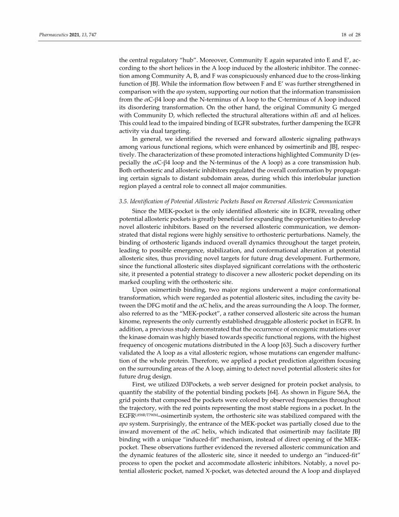
First, we utilized D3Pockets, a web server designed for protein pocket analysis, to
quantify the stability of the potential binding pockets [64]. As shown in Figure S6A, the
grid points that composed the pockets were colored by observed frequencies throughout
the trajectory, with the red points representing the most stable regions in a pocket. In the
EGFRL858R/T790M–osimertinib system, the orthosteric site was stabilized compared with the
apo system. Surprisingly, the entrance of the MEK‐pocket was partially closed due to the
inward movement of the αC helix, which indicated that osimertinib may facilitate JBJ
binding with a unique “induced‐fit” mechanism, instead of direct opening of the MEK‐
pocket. These observations further evidenced the reversed allosteric communication and
the dynamic features of the allosteric site, since it needed to undergo an “induced‐fit”
process to open the pocket and accommodate allosteric inhibitors. Notably, a novel po‐
tential allosteric pocket, named X‐pocket, was detected around the A loop and displayed
Pharmaceutics 2021, 13, 747 19 of 28
improved stability upon osimertinib binding. X‐pocket was positioned between the acti‐
vation segment and the αE, αF, αH, and αI helices, which appeared to be rather shallow
unless it was deepened by an extended A loop. Accordingly, the unique position may
enable fine‐tuning of the overall EGFR functions via non‐ATP nor substrate competitive
inhibitors. We further applied Fpocket to retrieve potential binding pockets from the rep‐
resentative structures of the EGFRL858R/T790M and the EGFRL858R/T790M–osimertinib system
(Figure 8), respectively. The druggability score of the MEK‐pocket visibly deteriorated
from 0.561 to 0.022 upon osimertinib binding, while the X‐pocket exhibited a 230‐fold in‐
crease from 0.002 to 0.469. Additionally, we noted that the X‐pocket in EGFR was roughly
analogous to a site previously identified in glycogen synthase kinase 3 (GSK3) that has
already been targeted by a number of potent allosteric inhibitors [65–67]. Therefore, we
assumed the X‐pocket as a potential allosteric site and concentrated on it to further char‐
acterize the coupling between allosteric and orthosteric sites. The results were compared
with the known MEK‐pocket for validation (Figure S6B).
Figure 8. Structure overview of the allosteric pockets. The surface representations of the N‐lobe
(pink), the C‐lobe (lightblue), the A loop (pale yellow), the MEK‐pocket (marine), and the X‐pocket
(hotpink) in the (A) EGFRL858R/T790M and (B) EGFRL858R/T790M–osimertinib systems. The magnified
views depicted the critical residues within the allosteric pockets.
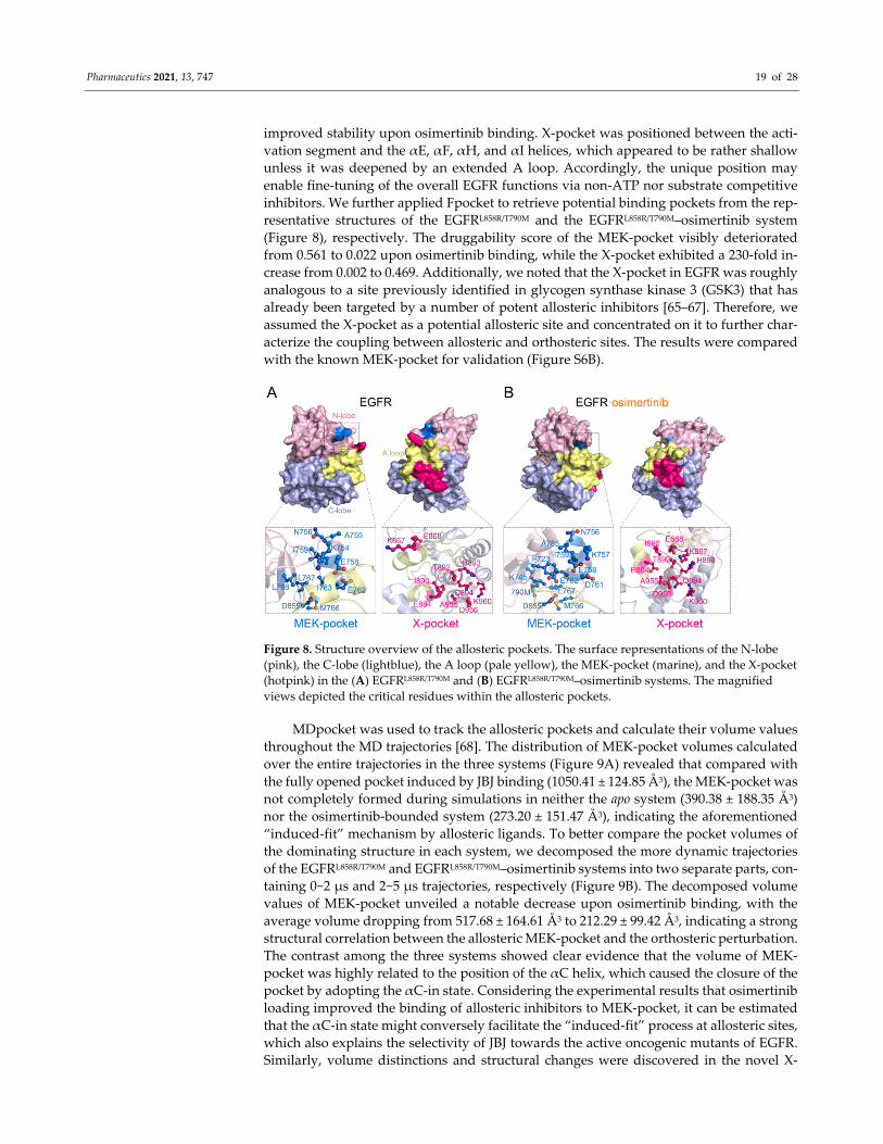
MDpocket was used to track the allosteric pockets and calculate their volume values
throughout the MD trajectories [68]. The distribution of MEK‐pocket volumes calculated
over the entire trajectories in the three systems (Figure 9A) revealed that compared with
the fully opened pocket induced by JBJ binding (1050.41 ± 124.85 Å3), the MEK‐pocket was
not completely formed during simulations in neither the apo system (390.38 ± 188.35 Å3)
nor the osimertinib‐bounded system (273.20 ± 151.47 Å3), indicating the aforementioned
“induced‐fit” mechanism by allosteric ligands. To better compare the pocket volumes of
the dominating structure in each system, we decomposed the more dynamic trajectories
of the EGFRL858R/T790M and EGFRL858R/T790M–osimertinib systems into two separate parts, con‐
taining 0~2 μs and 2~5 μs trajectories, respectively (Figure 9B). The decomposed volume
values of MEK‐pocket unveiled a notable decrease upon osimertinib binding, with the
average volume dropping from 517.68 ± 164.61 Å3 to 212.29 ± 99.42 Å3, indicating a strong
structural correlation between the allosteric MEK‐pocket and the orthosteric perturbation.
The contrast among the three systems showed clear evidence that the volume of MEK‐
pocket was highly related to the position of the αC helix, which caused the closure of the
pocket by adopting the αC‐in state. Considering the experimental results that osimertinib
loading improved the binding of allosteric inhibitors to MEK‐pocket, it can be estimated
that the αC‐in state might conversely facilitate the “induced‐fit” process at allosteric sites,
which also explains the selectivity of JBJ towards the active oncogenic mutants of EGFR.
Similarly, volume distinctions and structural changes were discovered in the novel X‐
Pharmaceutics 2021, 13, 747 20 of 28
pocket (Figure 9C,D). The dominating structures in EGFRL858R/T790M and the EGFRL858R/T790M–
osimertinib–JBJ systems shared a very similar distribution of X‐pocket volume, with an
average of 113.03 Å3 and 119.94 Å3, respectively. While in the EGFRL858R/T790M–osimertinib
system, the volume sharply increased, surging to an average of 370.94 Å3. Therefore, it
was illustrated that osimertinib binding stabilized the allosteric X‐pocket in a deepened
state with the open A loop, thus contributing to ligand targeting.
Figure 9. Volume calculations and energy coupling analysis for the allosteric pockets. (A) Volume distributions of the
MEK‐pocket. (B) Decomposed volume distributions of the MEK‐pocket, with dashed lines and solid lines representing
0~2 μs, and 2~5 μs trajectories, respectively. (C) Volume distributions of the X‐pocket. (D) Decomposed volume distribu‐
tions of the X‐pocket. (E) Energy coupling analysis for the MEK‐pocket in apo and holo EGFRL858R/T790M. (F) Energy coupling
analysis for the X‐pocket in apo and holo EGFRL858R/T790M. Each dot represents the energy decomposition of a residue
pair, while the cross lines represent the corresponding standard deviations. The dots placed in the region between green
and blue dashed lines indicate residue pairs with medium energy differences, while the dots placed outside the blue
dashed lines indicate residue pairs with large energy differences.
Since the volume calculation results only deciphered structural coupling between the
allosteric and orthosteric sites, we further exploited a recently established quantitative al‐
gorithm to delineate the crosstalk in the view of energetics [28]. The calculation model
arose from the reversed allosteric communication theory that the free energies of a pro‐
portion of residue–residue interactions within the allosteric sites undergo considerable
changes due to orthosteric perturbations. Hence, the significant differences of energetic
decomposition at each residue pair serve as predictors of potential allosteric sites. Follow‐
ing this methodology, we first divided all residue pairs into three categories according to
their interaction energy differences before and after orthosteric loading: small, medium,
and large energy differences. The energy coupling score was defined as the ratio of resi‐
due pairs with large energy differences to those with medium energy differences, and a
cutoff score was set at 0.25 to identify potential allosteric sites [69]. Namely, pockets with
an energy coupling score over 0.25 were considered allosteric sites as they exhibited sig‐
nificant correlations with the orthosteric site. Herein, we first tested it with the MEK‐
pocket and obtained an energy coupling score of 0.4197, demonstrating the reliability of
this approach (Figure 9E). It was further performed for the newly detected X‐pocket,
which gave an energy coupling score of 0.3710, far in excess of the threshold at 0.25 (Figure
9F). This confirmed that X‐pocket was closely coupled with the orthosteric site through
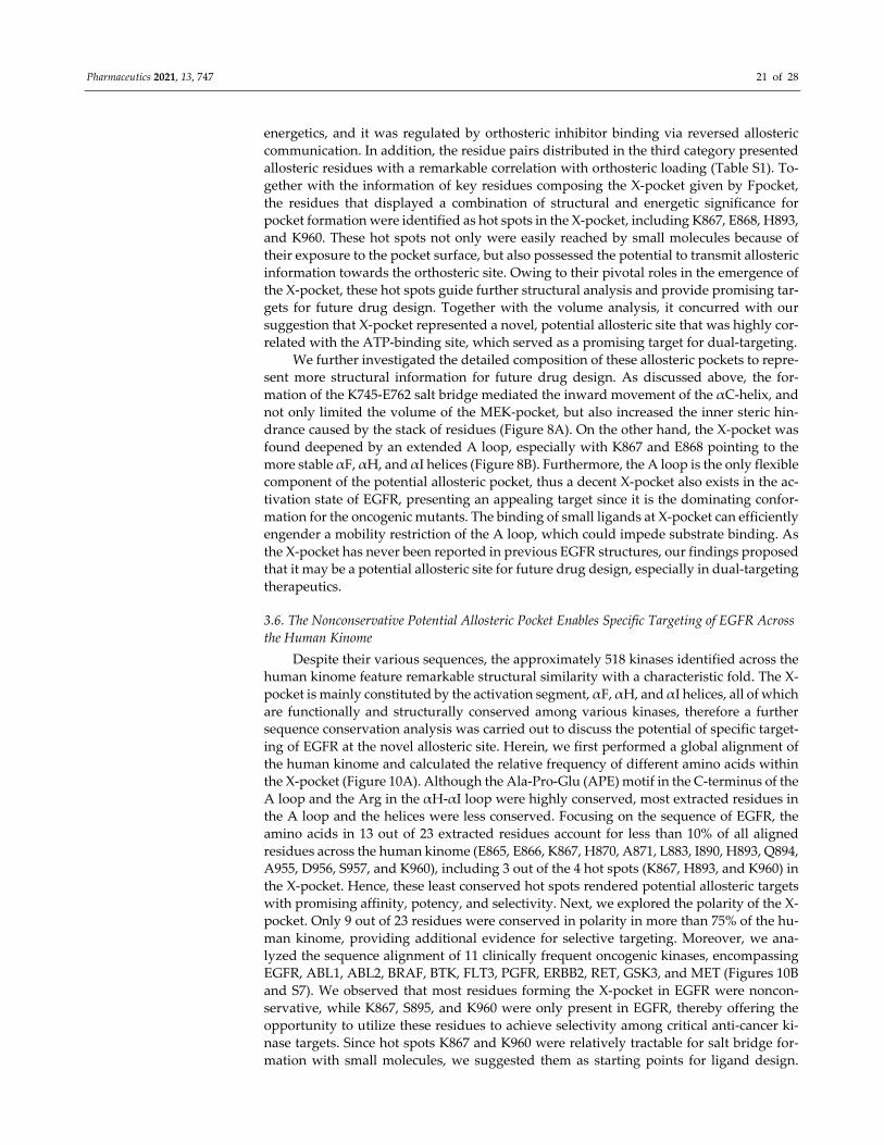
Pharmaceutics 2021, 13, 747 21 of 28
energetics, and it was regulated by orthosteric inhibitor binding via reversed allosteric
communication. In addition, the residue pairs distributed in the third category presented
allosteric residues with a remarkable correlation with orthosteric loading (Table S1). To‐
gether with the information of key residues composing the X‐pocket given by Fpocket,
the residues that displayed a combination of structural and energetic significance for
pocket formation were identified as hot spots in the X‐pocket, including K867, E868, H893,
and K960. These hot spots not only were easily reached by small molecules because of
their exposure to the pocket surface, but also possessed the potential to transmit allosteric
information towards the orthosteric site. Owing to their pivotal roles in the emergence of
the X‐pocket, these hot spots guide further structural analysis and provide promising tar‐
gets for future drug design. Together with the volume analysis, it concurred with our
suggestion that X‐pocket represented a novel, potential allosteric site that was highly cor‐
related with the ATP‐binding site, which served as a promising target for dual‐targeting.
We further investigated the detailed composition of these allosteric pockets to repre‐
sent more structural information for future drug design. As discussed above, the for‐
mation of the K745‐E762 salt bridge mediated the inward movement of the αC‐helix, and
not only limited the volume of the MEK‐pocket, but also increased the inner steric hin‐
drance caused by the stack of residues (Figure 8A). On the other hand, the X‐pocket was
found deepened by an extended A loop, especially with K867 and E868 pointing to the
more stable αF, αH, and αI helices (Figure 8B). Furthermore, the A loop is the only flexible
component of the potential allosteric pocket, thus a decent X‐pocket also exists in the ac‐
tivation state of EGFR, presenting an appealing target since it is the dominating confor‐
mation for the oncogenic mutants. The binding of small ligands at X‐pocket can efficiently
engender a mobility restriction of the A loop, which could impede substrate binding. As
the X‐pocket has never been reported in previous EGFR structures, our findings proposed
that it may be a potential allosteric site for future drug design, especially in dual‐targeting
therapeutics.
3.6. The Nonconservative Potential Allosteric Pocket Enables Specific Targeting of EGFR Across
the Human Kinome
Despite their various sequences, the approximately 518 kinases identified across the
human kinome feature remarkable structural similarity with a characteristic fold. The X‐
pocket is mainly constituted by the activation segment, αF, αH, and αI helices, all of which
are functionally and structurally conserved among various kinases, therefore a further
sequence conservation analysis was carried out to discuss the potential of specific target‐
ing of EGFR at the novel allosteric site. Herein, we first performed a global alignment of
the human kinome and calculated the relative frequency of different amino acids within
the X‐pocket (Figure 10A). Although the Ala‐Pro‐Glu (APE) motif in the C‐terminus of the
A loop and the Arg in the αH‐αI loop were highly conserved, most extracted residues in
the A loop and the helices were less conserved. Focusing on the sequence of EGFR, the
amino acids in 13 out of 23 extracted residues account for less than 10% of all aligned
residues across the human kinome (E865, E866, K867, H870, A871, L883, I890, H893, Q894,
A955, D956, S957, and K960), including 3 out of the 4 hot spots (K867, H893, and K960) in
the X‐pocket. Hence, these least conserved hot spots rendered potential allosteric targets
with promising affinity, potency, and selectivity. Next, we explored the polarity of the X‐
pocket. Only 9 out of 23 residues were conserved in polarity in more than 75% of the hu‐
man kinome, providing additional evidence for selective targeting. Moreover, we ana‐
lyzed the sequence alignment of 11 clinically frequent oncogenic kinases, encompassing
EGFR, ABL1, ABL2, BRAF, BTK, FLT3, PGFR, ERBB2, RET, GSK3, and MET (Figures 10B
and S7). We observed that most residues forming the X‐pocket in EGFR were noncon‐
servative, while K867, S895, and K960 were only present in EGFR, thereby offering the
opportunity to utilize these residues to achieve selectivity among critical anti‐cancer ki‐
nase targets. Since hot spots K867 and K960 were relatively tractable for salt bridge for‐
mation with small molecules, we suggested them as starting points for ligand design.
Pharmaceutics 2021, 13, 747 22 of 28
These results validated the selectivity potential of the X‐pocket, which indicated desirable
efficacy for allosteric drugging to cooperate with orthosteric inhibitors.
Figure 10. Sequence conservation of the allosteric pockets across the human kinome. (A) A se‐
quence logo made by WebLogo3 server (http://weblogo.threeplusone.com/, accessed on 17 March
2021) based on sequence alignment of 518 kinases across the human kinome. The overall height of
symbols within a stack indicates the relative frequency of each amino acid at the corresponding
position. While the color indicates the hydrophobicity of the amino acid, with hydrophilic amino
acids colored in blue, neutral ones in green, and hydrophobic ones in black. (B) Sequence align‐
ment of EGFR, ABL1, ABL2, BRAF, BTK, FLT3, PGFR, ERBB2, RET, GSK3, and MET, representing
the most important anti‐cancer target in the human kinome. The residues forming the X‐pocket are
pointed to by the blue triangles.
4. Discussion
Here, we illuminated a dual‐targeting therapeutic mechanism that orthosteric load‐
ing regulated allosteric sites via reversed allosteric communication, thereby exerting syn‐
ergetic effects on improving the general efficacy. Dual‐targeting with osimertinib and JBJ
in EGFRL858R/T790M was utilized to validate our proposal. We provided atomistic insight into
the reversed allosteric communication and the subsequent conformational ensemble al‐
terations induced by osimertinib binding at the orthosteric site. In particular, osimertinib
transmitted the reversed signals to allosteric sites via a communication hub in the inter‐
lobular junction region, which triggered coordinative motions in different subdomains,
thus stabilizing an intermediate state with a unique αC‐in, A loop open, and DFG‐out
conformation. Significantly, a novel potential allosteric pocket (X‐pocket) was identified,
which was well exposed and can be readily targeted in both the active and intermediate
states of EGFR. The coupling between the ATP‐binding site and the X‐pocket was vali‐
dated using both structural and energetic dynamics computations, and further sequence
conservation analysis revealed a non‐conservation characteristic of the X‐pocket across
the human kinome, manifesting its potential to yield selective allosteric inhibitors target‐
ing EGFR. Our work provided a mechanistic basis for future clinical application of com‐
bining orthosteric and allosteric drugs, and presented a novel avenue to detect allosteric
sites by orthosteric perturbations.
Pharmaceutics 2021, 13, 747 23 of 28
In the era of modern biomedicine, drug resistance remains a clinically insuperable
obstacle, which poses a leading challenge especially in the treatment of infection and can‐
cers, thus threatening global public health [70–73]. In the last decades, the long‐term im‐
petus has promoted innovative attempts to tackle the elusive enigma with multidiscipli‐
nary approaches. With accumulating evidence of the multiple advantages boosted by al‐
losteric modulation, it has attracted an ever‐growing interest in pharmaceutics, and con‐
sequently, the combination of orthosteric and allosteric agents is emerging as a revolu‐
tionary strategy to overcome resistance, as exemplified by the growing number of suc‐
cessful cases [74–79]. Accordingly, a series of orthosteric‐allosteric drug combinations
have proceeded into clinical trials, mostly involving oncology, viruses, and autoimmune
disorders [17]. In addition to BCR‐ABL and EGFR, another quintessential example is tar‐
geting nonstructural protein 5A (NS5A) for the treatment of hepatitis C virus (HCV). To
overcome the drug‐resistance against the first‐class orthosteric inhibitor daclatasvir, an
allosteric synergist Syn‐395 was developed [21]. Additional loading of Syn‐395 altered the
overall structure of NS5A daclatasvir complex, leading to a 1000‐fold enhancement in the
potency of the orthosteric inhibitor. Another recent success was achieved in selectively
inhibiting plasmodium falciparum hexose transporter 1 (PfHT1) with orthosteric‐allo‐
steric dual inhibitors for anti‐malarial treatment [18]. Huang et al. designed a set of small‐
molecules that simultaneously block the orthosteric and allosteric pockets of PfHT1, con‐
stituting a novel weapon against multidrug‐resistant malaria parasites. It was previously
reviewed that in dual‐targeting therapeutics, allosteric regulators bind to the recalcitrant
target and precisely resensitize the orthosteric site in two major pathways: either through
microcosmically reshaping the intrinsic residue network within the protein, or macro‐
scopically shifting the conformational ensemble towards a favorable state [17]. In fact,
these abovementioned resensitizing effects discovered in multiple cases imply the tradi‐
tional allosteric communication pattern where signals propagate from the allosteric site to
the orthosteric site. However, an extra improvement in the affinity of allosteric agents
during the dual‐targeting therapeutics was further reported, which manifested a reversed
signaling pathway within the target. Recently, a bidirectional model of allosteric signaling
has been proposed and validated in several previous reports [23–26]. Moreover, recent
success in the development of a single molecule spanning both the ATP binding site and
the allosteric sites also support an intrinsic connection between the topologically distant
sites [80,81].
In our study, we revealed a reversed allosteric communication mechanism underly‐
ing the unique phenomenon found in dual‐targeting, which indicates that the proper com‐
bination of orthosteric and allosteric drugs has the potential to not only circumvent drug‐
resistance, but also synergistically improve the efficacy of both therapeutic agents. Hence,
our results provide theoretical basis for the dual‐targeting therapeutics to become a uni‐
versally standard practice.
Despite the recent advances in allosteric regulation, the effective allosteric modula‐
tors remain relatively rare in comparison with traditional orthosteric drugs, indicating
tougher challenges faced by this emerging field. The detection of allosteric sites, a pre‐
requisite for allosteric drug development, represents a major obstacle. Historically, allo‐
steric site prediction was once based on serendipitous discovery, while the latest decade
has witnessed the upsurge of computational tools aiding the structure‐based rational de‐
sign of allosteric regulators [82–88]. Due to a constantly deepening understanding of pro‐
tein allostery, and the rapid development of diverse bioinformatics methodologies, the
characterization and identification of potential allosteric sites was considerably facilitated.
However, the present computation models and algorithms pose non‐negligible re‐
strictions to the application of these computational aids. First, because of the relatively
elusive understanding of protein allostery, algorithm development is faced with great
challenges to accurately approximate and reveal allosteric effects. Second, the nonphysical
perturbations, such as dummy atoms and artificial mutations, applied for allosteric signal
analysis notably hinder the performance of the prediction methods. Thirdly, current
Pharmaceutics 2021, 13, 747 24 of 28
knowledge of protein allosteric regulation is mainly concentrated on a certain list of pro‐
tein classes, represented by kinases and GPCRs. Consequently, the performance of data‐
based prediction methods is inevitably hampered by a biased dataset for model training
and optimization. Moreover, current efforts to computationally predict allosteric sites are
based on the experimentally revealed crystal structures and focus only on the pockets
present in the existing structures, while ignoring the vast potential of massive cryptic al‐
losteric sites that only emerge during conformational dynamics within the target. Last,
but not least, in terms of dual‐targeting therapeutics, some allosteric sites may overlap
with the orthosteric sites, thus impeding the utility. In our study, we developed an ap‐
proach to identify potential allosteric sites tailored for dual‐targeting therapeutics. Based
on a dynamic ensemble of the protein target, the reversed allosteric communication trig‐
gered by orthosteric drugging was exploited as the mechanistic starting point to detect
allosteric sites. Compared with the previous computational methods, we circumvented
the obstacles of biased databases and imperfect prediction algorithms. MD simulations of
a protein target in complex with its orthosteric agent enabled the discovery of cryptic al‐
losteric sites in a state resembling the physiological conditions. Importantly, non‐overlap‐
ping allosteric pockets can be intuitively selected to ensure the practicality of dual‐target‐
ing therapeutics. Such unbiased detection methods can expand the spectrum of available
allosteric pockets, facilitate the conceptualization of novel, potent, and selective kinase
inhibitors, hopefully paving an avenue towards the application of combining orthosteric
and allosteric drugs.
Major limitations include that the conformational landscape of EGFR was not fully
sampled during our simulations since the relative timescale for completed conformational
shifts reached hundreds of microseconds [8]. In addition, we only focused on the intracel‐
lular CD, leaving the extracellular receptor and the transmembrane region unexplored.
Although our study revealed a potential allosteric pocket in EGFR, further experimental
validation and lead compound identification are needed for allosteric drug design. Future
studies are expected to provide a more comprehensive understanding of the orthosteric
ligand fine‐tuning EGFR and to realize X‐pocket as a versatile allosteric pocket in human
kinases.
5. Conclusions
Here, we untangled an underlying mechanism of reversed allosteric communication
in dual‐targeting therapeutics, by which the allosteric sites were regulated by orthosteric
drugs. The reliability of this proposal was validated by the correlation between the previ‐
ously identified allosteric MEK‐pocket in EGFR and the orthosteric ATP‐binding site. A
novel, potential allosteric site was identified in EGFR based on its remarkable coupling
with the orthosteric site, both structurally and energetically. Our results laid a theoretical
foundation for the development of the dual‐targeting strategy in massive therapeutic tar‐
gets. Moreover, X‐pocket, the non‐overlapping allosteric pocket we detected in EGFR mu‐
tants, provided a potential alternative to yield selective allosteric agents, which can syn‐
ergistically function with the traditional TKIs. X‐pocket is mostly composed of noncon‐
servative residues, including hot spots K867, S895, and K960. Therefore, X‐pocket renders
potential targets with promising affinity, potency, and selectivity for future allosteric drug
design and the dual‐targeting strategy.
Supplementary Materials: The following are available online at www.mdpi.com/1999‐
4923/13/5/747/s1, Figure S1: Starting structures of the EGFRL858R/T790M, Figure S2: Root‐mean‐square
deviations (RMSD) of Cα atoms in the EGFRL858R/T790M, EGFRL858R/T790M–osimertinib, and EG‐
FRL858R/T790M–osimertinib–JBJ systems, Figure S3: RMSF and RMSD differences, Figure S4: PCA anal‐
ysis, Figure S5: The representative structures of the secondary free energy minima, Figure S6: Pocket
characterization, Figure S7: Sequence alignment of EGFR, ABL1, ABL2, BRAF, BTK, FLT3, PGFR,
ERBB2, RET, GSK3, and MET, Table S1: Residue‐residue interactions in the X pocket with medium
or large energy differences.
Pharmaceutics 2021, 13, 747 25 of 28
Author Contributions: Conceptualization, S.L.; methodology and validation, S.L., Q.F., R.H. and
Y.Q.; data curation, formal analysis, investigation, and writing‐original draft preparation, Y.Q., X.Y.,
X.L. and Y.W.; projection administration, supervision, resources, and writing‐review and editing,
S.L., Q.F. and R.H.; funding acquisition, S.L. All authors have read and agreed to the published
version of the manuscript.
Funding: This work was supported partly by grants from the National Natural Science Foundation
of China (22077082, 21778037).
Institutional Review Board Statement: Not applicable.
Informed Consent Statement: Not applicable.
Data Availability Statement: The crystal structures of human EGFRL858R/T790M monomeric form (PDB
ID: 4I1Z) and EGFRL858R/T790M–osimertinib–JBJ (PDB ID: 6DUK) were downloaded from the Protein
Data Bank.
Conflicts of Interest: The authors declare no conflict of interest.
References
1. Lu, S.; Shen, Q.; Zhang, J. Allosteric Methods and Their Applications: Facilitating the Discovery of Allosteric Drugs and the
Investigation of Allosteric Mechanisms. Acc. Chem. Res. 2019, 52, 492–500.
2. Nussinov, R.; Tsai, C.J. Allostery in disease and in drug discovery. Cell 2013, 153, 293–305.
3. Changeux, J.P.; Christopoulos, A. Allosteric modulation as a unifying mechanism for receptor function and regulation. Cell 2017,
19, 4–21.
4. Lu, S.; Li, S.; Zhang, J. Harnessing allostery: A novel approach to drug discovery. Med. Res. Rev. 2014, 34, 1242–1285.
5. Lu, S.; Huang, W.; Wang, Q.; Shen, Q.; Li, S.; Nussinov, R.; Zhang, J. The Structural Basis of ATP as an Allosteric Modulator.
PLoS Comput. Biol. 2014, 10, e1003831.
6. Lu, S.; Qiu, Y.; Ni, D.; He, X.; Pu, J.; Zhang, J. Emergence of allosteric drug‐resistance mutations: New challenges for allosteric
drug discovery. Drug Discov. Today 2020, 25, 177–184.
7. Sutto, L.; Gervasio, F.L. Effects of oncogenic mutations on the conformational free‐energy landscape of EGFR kinase. Proc. Natl.
Acad. Sci. USA 2013, 110, 10616–10621.
8. Sultan, M.M.; Kiss, G.; Pande, V.S. Towards simple kinetic models of functional dynamics for a kinase subfamily. Nat. Chem.
2018, 10, 903–909.
9. Kannan, S.; Venkatachalam, G.; Lim, H.H.; Surana, U.; Verma, C. Conformational landscape of the epidermal growth factor
receptor kinase reveals a mutant specific allosteric pocket. Chem. Sci. 2018, 9, 5212–5222.
10. Canon, J.; Rex, K.; Saiki, A.Y.; Mohr, C.; Cooke, K.; Bagal, D.; Gaida, K.; Holt, T.; Knutson, C.G.; Koppada, N.; et al. The clinical
KRAS(G12C) inhibitor AMG 510 drives anti‐tumour immunity. Nature 2019, 575, 217–223.
11. Ni, D.; Li, X.; He, X.; Zhang, H.; Zhang, J.; Lu, S. Drugging K‐RasG12C through covalent inhibitors: Mission possible? Pharmacol.
Ther. 2019, 202, 1–17.
12. Ostrem, J.M.; Peters, U.; Sos, M.L.; Wells, J.A.; Shokat, K.M. K‐Ras(G12C) inhibitors allosterically control GTP affinity and
effector interactions. Nature 2013, 503, 548–551.
13. McCormick, F. Sticking it to KRAS: Covalent Inhibitors Enter the Clinic. Cancer Cell 2020, 37, 3–4.
14. Lu, S.; Jang, H.; Muratcioglu, S.; Gursoy, A.; Keskin, O.; Nussinov, R.; Zhang, J. Ras Conformational Ensembles, Allostery, and
Signaling. Chem. Rev. 2016, 116, 6607–6665.
15. Lu, S.; Zhang, J. Small Molecule Allosteric Modulators of G‐Protein‐Coupled Receptors: Drug‐Target Interactions. J. Med. Chem.
2019, 62, 24–45.
16. Suomivuori, C.‐M.; Latorraca, N.R.; Wingler, L.M.; Eismann, S.; King, M.C.; Kleinhenz, A.L.W.; Skiba, M.A.; Staus, D.P.; Kruse,
A.C.; Lefkowitz, R.J.; et al. Molecular mechanism of biased signaling in a prototypical G protein‐coupled receptor. Science 2020,
367, 881–887.
17. Ni, D.; Li, Y.; Qiu, Y.; Pu, J.; Lu, S.; Zhang, J. Combining Allosteric and Orthosteric Drugs to Overcome Drug Resistance. Trends
Pharmacol. Sci. 2020, 41, 336–348.
18. Huang, J.; Yuan, Y.; Zhao, N.; Pu, D.; Tang, Q.; Zhang, S.; Luo, S.; Yang, X.; Wang, N.; Xiao, Y.; et al. Orthosteric‐allosteric dual
inhibitors of PfHT1 as selective anti‐malarial agents. bioRxiv 2020, doi:10.1101/2020.08.25.260232.
19. Zhang, J.; Adrián, F.J.; Jahnke, W.; Cowan‐Jacob, S.W.; Li, A.G.; Iacob, R.E.; Sim, T.; Powers, J.; Dierks, C.; Sun, F.; et al. Targeting
Bcr‐Abl by combining allosteric with ATP‐binding‐site inhibitors. Nature 2010, 463, 501–506.
20. To, C.; Jang, J.; Chen, T.; Park, E.; Mushajiang, M.; De Clercq, D.J.H.; Xu, M.; Wang, S.; Cameron, M.D.; Heppner, D.E.; et al.
Single and Dual Targeting of Mutant EGFR with an Allosteric Inhibitor. Cancer Discov. 2019, 9, 926–943.
21. Sun, J.‐H.; O’Boyle, D.R., 2nd; Fridell, R.A.; Langley, D.R.; Wang, C.; Roberts, S.B.; Nower, P.; Johnson, B.M.; Moulin, F.;
Nophsker, M.J.; et al. Resensitizing daclatasvir‐resistant hepatitis C variants by allosteric modulation of NS5A. Nature 2015, 527,
245–248.
Pharmaceutics 2021, 13, 747 26 of 28
22. Veit, G.; Xu, H.; Dreano, E.; Avramescu, R.G.; Bagdany, M.; Beitel, L.K.; Roldan, A.; Hancock, M.A.; Lay, C.; Li, W.; et al.
Structure‐guided combination therapy to potently improve the function of mutant CFTRs. Nat. Med. 2018, 24, 1732–1742.
23. Schulze, J.O.; Saladino, G.; Busschots, K.; Neimanis, S.; Süß, E.; Odadzic, D.; Zeuzem, S.; Hindie, V.; Herbrand, A.K.; Lisa, M.‐
N.; et al. Bidirectional Allosteric Communication between the ATP‐Binding Site and the Regulatory PIF Pocket in PDK1 Protein
Kinase. Cell Chem. Biol. 2016, 23, 1193–1205.
24. Leroux, A.E.; Biondi, R.M. Renaissance of Allostery to Disrupt Protein Kinase Interactions. Trends Biochem. Sci. 2020, 45, 27–41.
25. Zhang, W.; Xie, J.; Lai, L. Correlation Between Allosteric and Orthosteric Sites. Adv. Exp. Med. Biol. 2019, 1163, 89–105.
26. Ma, X.; Meng, H.; Lai, L. Motions of Allosteric and Orthosteric Ligand‐Binding Sites in Proteins are Highly Correlated. J. Chem.
Inf. Model. 2016, 56, 1725–1733.
27. Meng, H.; McClendon, C.L.; Dai, Z.; Li, K.; Zhang, X.; He, S.; Shang, E.; Liu, Y.; Lai, L. Discovery of Novel 15‐Lipoxygenase
Activators To Shift the Human Arachidonic Acid Metabolic Network toward Inflammation Resolution. J. Med. Chem. 2016, 59,
4202–4209.
28. Ni, D.; Wei, J.; He, X.; Rehman, A.U.; Li, X.; Qiu, Y.; Pu, J.; Lu, S.; Zhang, J. Discovery of cryptic allosteric sites using reversed
allosteric communication by a combined computational and experimental strategy. Chem. Sci. 2021, doi:10.1039/d0sc05131d.
29. Yarden, Y.; Sliwkowski, M.X. Untangling the ErbB signalling network. Nat. Rev. Mol. Cell Biol. 2001, 2, 127–137.
30. Stratton, M.R.; Campbell, P.J.; Futreal, P.A. The cancer genome. Nature 2009, 458, 719–724.
31. da Cunha Santos, G.; Shepherd, F.A.; Tsao, M.S. EGFR mutations and lung cancer. Annu. Rev. Pathol. 2011, 6, 49–69.
32. Paez, J.G.; Jänne, P.A.; Lee, J.C.; Tracy, S.; Greulich, H.; Gabriel, S.; Herman, P.; Kaye, F.J.; Lindeman, N.; Boggon, T.J.; et al.
EGFR mutations in lung cancer: Correlation with clinical response to gefitinib therapy. Science 2004, 304, 1497–1500.
33. Castellanos, E.; Feld, E.; Horn, L. Driven by Mutations: The Predictive Value of Mutation Subtype in EGFR‐Mutated Non‐Small
Cell Lung Cancer. J. Thorac. Oncol. 2017, 12, 612–623.
34. Mok, T.S.; Wu, Y.‐L.; Thongprasert, S.; Yang, C.‐H.; Chu, D.‐T.; Saijo, N.; Sunpaweravong, P.; Han, B.; Margono, B.; Ichinose, Y.;
et al. Gefitinib or carboplatin‐paclitaxel in pulmonary adenocarcinoma. N. Engl. J. Med. 2009, 361, 947–957.
35. Zhang, L.; Ma, S.; Song, X.; Han, B.; Cheng, Y.; Huang, C.; Yang, S.; Liu, X.; Liu, Y.; Lu, S.; et al. Gefitinib versus placebo as
maintenance therapy in patients with locally advanced or metastatic non‐small‐cell lung cancer (INFORM; C‐TONG 0804): A
multicentre, double‐blind randomised phase 3 trial. Lancet. Oncol. 2012, 13, 466–475.
36. Sequist, L.V; Yang, J.C.‐H.; Yamamoto, N.; O’Byrne, K.; Hirsh, V.; Mok, T.; Geater, S.L.; Orlov, S.; Tsai, C.‐M.; Boyer, M.; et al.
Phase III study of afatinib or cisplatin plus pemetrexed in patients with metastatic lung adenocarcinoma with EGFR mutations.
J. Clin. Oncol. 2013, 31, 3327–3334.
37. Yun, C.‐H.; Mengwasser, K.E.; Toms, A.V; Woo, M.S.; Greulich, H.; Wong, K.‐K.; Meyerson, M.; Eck, M.J. The T790M mutation
in EGFR kinase causes drug resistance by increasing the affinity for ATP. Proc. Natl. Acad. Sci. USA 2008, 105, 2070–2075.
38. Yu, H.A.; Arcila, M.E.; Rekhtman, N.; Sima, C.S.; Zakowski, M.F.; Pao, W.; Kris, M.G.; Miller, V.A.; Ladanyi, M.; Riely, G.J.
Analysis of tumor specimens at the time of acquired resistance to EGFR‐TKI therapy in 155 patients with EGFR‐mutant lung
cancers. Clin. Cancer Res. 2013, 19, 2240–2247.
39. Zhou, W.; Ercan, D.; Chen, L.; Yun, C.‐H.; Li, D.; Capelletti, M.; Cortot, A.B.; Chirieac, L.; Iacob, R.E.; Padera, R.; et al. Novel
mutant‐selective EGFR kinase inhibitors against EGFR T790M. Nature 2009, 462, 1070–1074.
40. Cross, D.A.E.; Ashton, S.E.; Ghiorghiu, S.; Eberlein, C.; Nebhan, C.A.; Spitzler, P.J.; Orme, J.P.; Finlay, M.R.V; Ward, R.A.; Mellor,
M.J.; et al. AZD9291, an irreversible EGFR TKI, overcomes T790M‐mediated resistance to EGFR inhibitors in lung cancer. Cancer
Discov. 2014, 4, 1046–1061.
41. Thress, K.S.; Paweletz, C.P.; Felip, E.; Cho, B.C.; Stetson, D.; Dougherty, B.; Lai, Z.; Markovets, A.; Vivancos, A.; Kuang, Y.; et
al. Acquired EGFR C797S mutation mediates resistance to AZD9291 in non‐small cell lung cancer harboring EGFR T790M. Nat.
Med. 2015, 21, 560–562.
42. Jia, Y.; Yun, C.H.; Park, E.; Ercan, D.; Manuia, M.; Juarez, J.; Xu, C.; Rhee, K.; Chen, T.; Zhang, H.; et al. Overcoming EGFR(T790M)
and EGFR(C797S) resistance with mutant‐selective allosteric inhibitors. Nature 2016, 534, 129–132.
43. Niggenaber, J.; Heyden, L.; Grabe, T.; Müller, M.P.; Lategahn, J.; Rauh, D. Complex Crystal Structures of EGFR with Third‐
Generation Kinase Inhibitors and Simultaneously Bound Allosteric Ligands. ACS Med. Chem. Lett. 2020, 11, 2484–2490.
44. De Clercq, D.J.H.; Heppner, D.E.; To, C.; Jang, J.; Park, E.; Yun, C.‐H.; Mushajiang, M.; Shin, B.H.; Gero, T.W.; Scott, D.A.; et al.
Discovery and Optimization of Dibenzodiazepinones as Allosteric Mutant‐Selective EGFR Inhibitors. ACS Med. Chem. Lett. 2019,
10, 1549–1553.
45. Palmieri, L.; Rastelli, G. αc Helix Displacement As a General Approach for Allosteric Modulation of Protein Kinases. Drug
Discov. Today 2013, 18, 407–414.
46. Gajiwala, K.S.; Feng, J.; Ferre, R.; Ryan, K.; Brodsky, O.; Weinrich, S.; Kath, J.C.; Stewart, A. Insights into the aberrant activity of
mutant EGFR kinase domain and drug recognition. Structure 2013, 21, 209–219.
47. Case, D.A.; Cheatham, T.E., 3rd; Darden, T.; Gohlke, H.; Luo, R.; Merz, K.M.J.; Onufriev, A.; Simmerling, C.; Wang, B.; Woods,
R.J. The Amber biomolecular simulation programs. J. Comput. Chem. 2005, 26, 1668–1688.
48. Jorgensen, W.L.; Chandrasekhar, J.; Madura, J.D.; Impey, R.W.; Klein, M.L. Comparison of simple potential functions for
simulating liquid water. J. Chem. Phys. 1983, 79, 926–935.
49. Darden, T.; York, D.; Pedersen, L. Particle mesh Ewald: An N∙log(N) method for Ewald sums in large systems. J. Chem. Phys.
1993, 98, 10089–10092.
Pharmaceutics 2021, 13, 747 27 of 28
50. Ryckaert, J.‐P.; Ciccotti, G.; Berendsen, H.J.C. Numerical integration of the cartesian equations of motion of a system with
constraints: Molecular dynamics of n‐alkanes. J. Comput. Phys. 1977, 23, 327–341.
51. Amadei, A.; Linssen, A.B.; Berendsen, H.J. Essential dynamics of proteins. Proteins 1993, 17, 412–425.
52. Floyd, R.W. Algorithm 97, Shortest Path Algorithms. Commun. ACM 1962, 5, 345.
53. Newman, M.E.J. Modularity and community structure in networks. Proc. Natl. Acad. Sci. USA 2006, 103, 8577–8582.
54. Shan, Y.; Arkhipov, A.; Kim, E.T.; Pan, A.C.; Shawa, D.E. Transitions to catalytically inactive conformations in EGFR kinase.
Proc. Natl. Acad. Sci. USA 2013, 110, 7270–7275.
55. Zhang, X.; Gureasko, J.; Shen, K.; Cole, P.A.; Kuriyan, J. An allosteric mechanism for activation of the kinase domain of
epidermal growth factor receptor. Cell 2006, 125, 1137–1149.
56. Yan, X.‐E.; Ayaz, P.; Zhu, S.‐J.; Zhao, P.; Liang, L.; Zhang, C.H.; Wu, Y.‐C.; Li, J.‐L.; Choi, H.G.; Huang, X.; et al. Structural Basis
of AZD9291 Selectivity for EGFR T790M. J. Med. Chem. 2020, 63, 8502–8511.
57. Xie, T.; Saleh, T.; Rossi, P.; Kalodimos, C.G. Conformational states dynamically populated by a kinase determine its function.
Science 2020, 370, 6513.
58. Kashima, K.; Kawauchi, H.; Tanimura, H.; Tachibana, Y.; Chiba, T.; Torizawa, T.; Sakamoto, H. CH7233163 overcomes
osimertinib resistant EGFR‐Del19/T790M/C797S mutation. Mol. Cancer Ther. 2020, 19, 2288–2297.
59. Yosaatmadja, Y.; Silva, S.; Dickson, J.M.; Patterson, A.V.; Smaill, J.B.; Flanagan, J.U.; McKeage, M.J.; Squire, C.J. Binding mode
of the breakthrough inhibitor AZD9291 to epidermal growth factor receptor revealed. J. Struct. Biol. 2015, 192, 539–544.
60. Müller, S.; Chaikuad, A.; Gray, N.S.; Knapp, S. The ins and outs of selective kinase inhibitor development. Nat. Chem. Biol. 2015,
11, 818–821.
61. Okuzumi, T.; Fiedler, D.; Zhang, C.; Gray, D.C.; Aizenstein, B.; Hoffman, R.; Shokat, K.M. Inhibitor hijacking of Akt activation.
Nat. Chem. Biol. 2009, 5, 484–493.
62. Kornev, A.P.; Taylor, S.S. Dynamics‐Driven Allostery in Protein Kinases. Trends Biochem. Sci. 2015, 40, 628–647.
63. Dixit, A.; Verkhivker, G.M. The energy landscape analysis of cancer mutations in protein kinases. PLoS ONE 2011, 6, e26071.
64. Chen, Z.; Zhang, X.; Peng, C.; Wang, J.; Xu, Z.; Chen, K.; Shi, J.; Zhu, W. D3Pockets: A Method and Web Server for Systematic
Analysis of Protein Pocket Dynamics. J. Chem. Inf. Model. 2019, 59, 3353–3358.
65. Palomo, V.; Soteras, I.; Perez, D.I.; Perez, C.; Gil, C.; Campillo, N.E.; Martinez, A. Exploring the binding sites of glycogen
synthase kinase 3. Identification and characterization of allosteric modulation cavities. J. Med. Chem. 2011, 54, 8461–8470.
66. Palomo, V.; Perez, D.I.; Roca, C.; Anderson, C.; Rodríguez‐Muela, N.; Perez, C.; Morales‐Garcia, J.A.; Reyes, J.A.; Campillo, N.E.;
Perez‐Castillo, A.M.; et al. Subtly Modulating Glycogen Synthase Kinase 3 β: Allosteric Inhibitor Development and Their
Potential for the Treatment of Chronic Diseases. J. Med. Chem. 2017, 60, 4983–5001.
67. Li, Y.; Xu, J.; Chen, Y.; Mei, Z.; Xiao, Y. Screening of inhibitors of glycogen synthase kinase‐3β from traditional Chinese
medicines using enzyme‐immobilized magnetic beads combined with high‐performance liquid chromatography. J. Chromatogr.
A 2015, 1425, 8–16.
68. Schmidtke, P.; Bidon‐Chanal, A.; Luque, F.J.; Barril, X. MDpocket: Open‐source cavity detection and characterization on
molecular dynamics trajectories. Bioinformatics 2011, 27, 3276–3285.
69. Qi, Y.; Wang, Q.; Tang, B.; Lai, L. Identifying Allosteric Binding Sites in Proteins with a Two‐State Go̅ Model for Novel Allosteric
Effector Discovery. J. Chem. Theory Comput. 2012, 8, 2962–2971.
70. Boumahdi, S.; de Sauvage, F.J. The great escape: Tumour cell plasticity in resistance to targeted therapy. Nat. Rev. Drug Discov.
2020, 19, 39–56.
71. Fisher, M.C.; Hawkins, N.J.; Sanglard, D.; Gurr, S.J. Worldwide emergence of resistance to antifungal drugs challenges human
health and food security. Science 2018, 360, 739–742.
72. Beyer, P.; Moorthy, V.; Paulin, S.; Hill, S.R.; Sprenger, M.; Garner, S.; Simão, M.; Guerra, R.; Magrini, N.; Swaminathan, S. The
drugs don’t work: WHO’s role in advancing new antibiotics. Lancet 2018, 392, 264–266.
73. Rotow, J.; Bivona, T.G. Understanding and targeting resistance mechanisms in NSCLC. Nat. Rev. Cancer 2017, 17, 637–658.
74. Byun, J.A.; VanSchouwen, B.; Akimoto, M.; Melacini, G. Allosteric inhibition explained through conformational ensembles
sampling distinct “mixed” states. Comput. Struct. Biotechnol. J. 2020, 18, 3803–3818.
75. Liang, Z.; Zhu, Y.; Long, J.; Ye, F.; Hu, G. Both intra and inter‐domain interactions define the intrinsic dynamics and allosteric
mechanism in DNMT1s. Comput. Struct. Biotechnol. J. 2020, 18, 749–764.
76. Wang, Y.; Ji, D.; Lei, C.; Chen, Y.; Qiu, Y.; Li, X.; Li, M.; Ni, D.; Pu, J.; Zhang, J.; et al. Mechanistic insights into the effect of
phosphorylation on Ras conformational dynamics and its interactions with cell signaling proteins. Comput. Struct. Biotechnol. J.
2021, 19, 1184–1199.
77. Jang, H.; Zhang, M.; Nussinov, R. The quaternary assembly of KRas4B with Raf‐1 at the membrane. Comput. Struct. Biotechnol.
J. 2020, 18, 737–748.
78. Wang, Q.; Chen, Y.Y.; Li, S.P.; Yang, W.D.; Sun, L.F.; Jang, M.Q.; Wu, X.L.; Wang, Q.C.; Chen, L.; Wu, Y.K. Ca2+‐based allosteric
switches and shape shifting in RGLG1 VWA domain. Comput. Struct. Biotechnol. J. 2020, 18, 821–833.
79. Maryam, A.; Vedithi, S.C.; Khalid, R.R.; Alsulami, A.F.; Torres, P.H.M.; Siddiqi, A.R.; Blundell, T.L. The Molecular Organization
of Human cGMP Specific Phosphodiesterase 6 (PDE6): Structural Implications of Somatic Mutations in Cancer and Retinitis
Pigmentosa. Comput. Struct. Biotechnol. J. 2019, 17, 378–389.
80. Li, Q.; Zhang, T.; Li, S.; Tong, L.; Li, J.; Su, Z.; Feng, F.; Sun, D.; Tong, Y.; Wang, X.; et al. Discovery of Potent and Noncovalent
Reversible EGFR Kinase Inhibitors of EGFR(L858R/T790M/C797S). ACS Med. Chem. Lett. 2019, 10, 869–873.
Pharmaceutics 2021, 13, 747 28 of 28
81. Wittlinger, F.; Heppner, D.E.; To, C.; Günther, M.; Shin, B.H.; Rana, J.K.; Schmoker, A.M.; Beyett, T.S.; Jänne, P.A.; Eck, M.J.; et
al. Molecular design of a “two‐in‐one” orthosteric‐allosteric chimeric mutant. ChemRxiv 2020, 1–15. DOI:
10.26434/chemrxiv.13416452.
82. Wagner, J.R.; Lee, C.T.; Durrant, J.D.; Malmstrom, R.D.; Feher, V.A.; Amaro, R.E. Emerging Computational Methods for the
Rational Discovery of Allosteric Drugs. Chem. Rev. 2016, 116, 6370–6390.
83. Qiu, Y.; Li, X.; He, X.; Pu, J.; Zhang, J.; Lu, S. Computational methods‐guided design of modulators targeting protein‐protein
interactions (PPIs). Eur. J. Med. Chem. 2020, 207, 112764.
84. Song, K.; Li, Q.; Gao, W.; Lu, S.; Shen, Q.; Liu, X.; Wu, Y.; Wang, B.; Lin, H.; Chen, G.; et al. AlloDriver: A method for the
identification and analysis of cancer driver targets. Nucleic Acids Res. 2019, 47, W315–W321.
85. Huang, M.; Song, K.; Liu, X.; Lu, S.; Shen, Q.; Wang, R.; Gao, J.; Hong, Y.; Li, Q.; Ni, D.; et al. AlloFinder: A strategy for allosteric
modulator discovery and allosterome analyses. Nucleic Acids Res. 2018, 46, W451–W458.
86. Tan, Z.W.; Tee, W.‐V.; Guarnera, E.; Booth, L.; Berezovsky, I.N. AlloMAPS: Allosteric mutation analysis and polymorphism of
signaling database. Nucleic Acids Res. 2019, 47, D265–D270.
87. Wodak, S.J.; Paci, E.; Dokholyan, N.V; Berezovsky, I.N.; Horovitz, A.; Li, J.; Hilser, V.J.; Bahar, I.; Karanicolas, J.; Stock, G.; et al.
Allostery in Its Many Disguises: From Theory to Applications. Structure 2019, 27, 566–578.
88. East, K.W.; Skeens, E.; Cui, J.Y.; Belato, H.B.; Mitchell, B.; Hsu, R.; Batista, V.S.; Palermo, G.; Lisi, G.P. NMR and computational
methods for molecular resolution of allosteric pathways in enzyme complexes. Biophys. Rev. 2020, 12, 155–174.