Therapeutic targeting of the stem cell niche in experimental hindlimb ischemia
9
SEPTEMBER 2008 VOL 5 NO 9 NATURE CLINICAL PRACTICE CARDIOVASCULAR MEDICINE 571 www.nature.com/clinicalpractice/cardio Therapeutic targeting of the stem cell niche in experimental hindlimb ischemia Claudio Napoli 1* , Sharon Williams-Ignarro 2 , Russell Byrns 3 , Maria Luisa Balestrieri 1 , Ettore Crimi 4 , Bartolomeo Farzati 1 , Francesco P Mancini 5 , Filomena de Nigris 1 , Angelo Matarazzo 1 , Maurizio D’Amora 6 , Ciro Abbondanza 1 , Carmela Fiorito 7 , Alfonso Giovane 1 , Anna Florio 1 , Ettore Varricchio 5 , Antonio Palagiano 1 , Pellegrino Biagio Minucci 1 , Mario Felice Tecce 8 , Antonio Giordano 9,10 , Antonio Pavan 11 and Louis J Ignarro 3 INTRODUCTION Many epidemiological studies have focused on the relationship between inflammation, tissue ischemia and cardiovascular disease, the last of which is the most common cause of morbidity and mortality in the world. 1,2 Several of these studies have shown that bone-marrow-cell- mediated cardiovascular repair is one of the most promising therapeutic approaches in regenerative medicine. 3 Ongoing work is focused on understanding the pivotal roles of the osteo- blastic 4–10 and vascular 11 niches (the in vivo regulatory microenvironments where hemato- poietic stem cells reside) in the regulation of stem cell function and homing. Hematopoietic stem cells reside in a stable, complex, custom micro- environment that controls their self-renewal and the framework between the bone marrow and the circulatory blood. This composite struc- ture is now recognized to be the so-called niche organization of bone marrow, which controls the fate of the stem cell. 11 Parathyroid hormone (PTH) stimulates osteoblasts of the osteoblastic bone marrow niche through the PTH/PTH- related peptide (PTHrP) receptor. The signaling lymphocytic activation molecule family of recep- tors and Tie2/angiopoietin 1-dependent signal transduction pathway are also involved, and activation of these pathways results in increased self-renewal and mobilization of cells into the bloodstream. Furthermore, PTH can stimu- late hematopoiesis 12,13 and affect endothelial cells, 14–17 a major component of the vascular niche. Although endothelial cells do not produce PTH, they do secrete PTHrP 15 and express the PTH/PTHrP receptor; 16 through this pathway they can generate an autocrine self-stimulating loop. PTH/PTHrP mediates several effects on arteries, including vasodilation (through cyclic AMP 14 and nitric oxide 17 ), and can also stimulate angiogenesis. 15 The purpose of this translational research study was to investigate whether targeting the vascular niche with PTH improves SUMMARY Background The custom microenvironment ‘vascular niche’ is a potential therapeutic target for several pathophysiological conditions. Osteoblasts regulate the hematopoietic stem cell niche, and activation of the parathyroid hormone (PTH) receptor can increase the number of cells mobilized into the bloodstream. Methods C57Bl/6 mice were randomly assigned treatment with granulocyte-colony stimulating factor (G-CSF), PTH, G-CSF plus PTH or saline. All mice underwent hindlimb ischemia. Blood flow was measured by laser Doppler imaging. Indices of capillary activity were determined by electron microscopy in muscle tissue. CD34 + and Ki67 + cells were detected and evaluated by immunofluorescence, apoptosis by TUNEL, surface antigen and endothelial progenitor cells by fluorescence-activated cell sorting analysis, and vascular endothelial growth factor-164 and angiopoietin-1 expression by reverse-transcriptase polymerase chain reaction. Frozen bone marrow sections were stained for antigen-specific B cells and fibronectin and analyzed by confocal laser scanning microscopy. Results Following mobilization induced by G-CSF treatment, mice also treated with PTH showed increases in blood flow, capillary density, nitrite/nitrate release, angiogenic factors and circulating progenitor cells, as well as reduced apoptosis, fibrosis, oxidative stress and inflammation in ischemic muscles. Furthermore, hematopoietic antigen-specific B cells in the bone marrow were also increased by G-CSF alone and in combination with PTH. Conclusions PTH might increase the efficiency of hematopoietic stem-cell-based therapy in a recognized model of peripheral ischemia. Our translational experimental therapeutic targeting of the vascular niche points to novel clinical targets for the hematopoietic stem-cell treatment of ischemic vascular diseases. KEYWORDS bone marrow, ischemic vascular diseases, neoangiogenesis, vascular niche Correspondence *Department of General Pathology and Excellence Research Center on Cardiovascular Diseases, 1st School of Medicine, II University of Naples, Complesso S. Andrea delle Dame, Via De Crecchio 7, 80138 Naples, Italy Tel: +39 (0)81 566 7567; [email protected] Received 9 November 2007 Accepted 28 February 2008 Published online 15 April 2008 www.nature.com/clinicalpractice doi:10.1038/ncpcardio1214 1 Department of General Pathology, Excellence Research Center on Cardiovascular Diseases, Department of Biochemistry and Division of Vascular Surgery, 1 st School of Medicine, II University of Naples, Naples, Italy 2 Division of Anesthesiology, and 3 Department of Molecular and Medical Pharmacology; David Geffen School of Medicine; University of California, Los Angeles, CA, USA 4 Department of Anesthesia and Critical Care, Massachusetts General Hospital, Harvard Medical School, Boston, MA, USA 5 Department of Biological and Environmental Sciences, University of Sannio, Benevento, Italy 6 Division of Clinical Pathology, Hospital of Incurabili, ASLNA1, Naples, Italy 7 IRCCS Multimedica, Milan, Italy 8 Department of Pharmacology, School of Pharmacy, University of Salerno, Italy 9 Sbarro Institute for Cancer Research and Molecular Medicine, College of Science and Technology, Temple University, Philadelphia, PA, USA 10 Department of Human Pathology and Oncology, University of Siena, Siena, Italy 11 Division of Clinical Pathology, II School of Medicine, University of Rome “La Sapienza”, Rome, Italy CLINICAL RESEARCH
Transcript of Therapeutic targeting of the stem cell niche in experimental hindlimb ischemia
september 2008 vol 5 no 9 nature clinical practice cardiovascular medicine 571
www.nature.com/clinicalpractice/cardio
Therapeutic targeting of the stem cell niche in experimental hindlimb ischemiaClaudio Napoli1*, Sharon Williams-Ignarro2, Russell Byrns3, Maria Luisa Balestrieri1, Ettore Crimi4, Bartolomeo Farzati1, Francesco P Mancini5, Filomena de Nigris1, Angelo Matarazzo1, Maurizio D’Amora6, Ciro Abbondanza1, Carmela Fiorito7, Alfonso Giovane1, Anna Florio1, Ettore Varricchio5, Antonio Palagiano1, Pellegrino Biagio Minucci1, Mario Felice Tecce8, Antonio Giordano9,10, Antonio Pavan11 and Louis J Ignarro3
INTRODUCTIONMany epidemiological studies have focused on the relationship between inflammation, tissue ischemia and cardiovascular disease, the last of which is the most common cause of morbidity and mortality in the world.1,2 Several of these studies have shown that bone-marrow-cell-mediated cardiovascular repair is one of the most promising therapeutic approaches in regenerative medicine.3 Ongoing work is focused on understanding the pivotal roles of the osteo-blastic4–10 and vascular11 niches (the in vivo regulatory microenvironments where hemato-poietic stem cells reside) in the regulation of stem cell function and homing. Hematopoietic stem cells reside in a stable, complex, custom micro-environment that controls their self-renewal and the framework between the bone marrow and the circulatory blood. This composite struc-ture is now recognized to be the so-called niche organization of bone marrow, which controls the fate of the stem cell.11 Parathyroid hormone (PTH) stimulates osteoblasts of the osteoblastic bone marrow niche through the PTH/PTH-related peptide (PTHrP) receptor. The signaling lymphocytic activation molecule family of recep-tors and Tie2/angiopoietin 1-dependent signal transduction pathway are also involved, and activation of these pathways results in increased self-renewal and mobilization of cells into the bloodstream. Furthermore, PTH can stimu-late hematopoiesis12,13 and affect endothelial cells,14–17 a major component of the vascular niche. Although endothelial cells do not produce PTH, they do secrete PTHrP15 and express the PTH/PTHrP receptor;16 through this pathway they can generate an autocrine self-stimulating loop. PTH/PTHrP mediates several effects on arteries, including vasodilation (through cyclic AMP14 and nitric oxide17), and can also stimulate angiogenesis.15
The purpose of this translational research study was to investigate whether targeting the vascular niche with PTH improves
SuMMarYBackground The custom microenvironment ‘vascular niche’ is a potential therapeutic target for several pathophysiological conditions. Osteoblasts regulate the hematopoietic stem cell niche, and activation of the parathyroid hormone (PTH) receptor can increase the number of cells mobilized into the bloodstream.
Methods C57Bl/6 mice were randomly assigned treatment with granulocyte-colony stimulating factor (G-CSF), PTH, G-CSF plus PTH or saline. All mice underwent hindlimb ischemia. Blood flow was measured by laser Doppler imaging. Indices of capillary activity were determined by electron microscopy in muscle tissue. CD34+ and Ki67+ cells were detected and evaluated by immunofluorescence, apoptosis by TUNEL, surface antigen and endothelial progenitor cells by fluorescence-activated cell sorting analysis, and vascular endothelial growth factor-164 and angiopoietin-1 expression by reverse-transcriptase polymerase chain reaction. Frozen bone marrow sections were stained for antigen-specific B cells and fibronectin and analyzed by confocal laser scanning microscopy.
Results Following mobilization induced by G-CSF treatment, mice also treated with PTH showed increases in blood flow, capillary density, nitrite/nitrate release, angiogenic factors and circulating progenitor cells, as well as reduced apoptosis, fibrosis, oxidative stress and inflammation in ischemic muscles. Furthermore, hematopoietic antigen-specific B cells in the bone marrow were also increased by G-CSF alone and in combination with PTH.
Conclusions PTH might increase the efficiency of hematopoietic stem-cell-based therapy in a recognized model of peripheral ischemia. Our translational experimental therapeutic targeting of the vascular niche points to novel clinical targets for the hematopoietic stem-cell treatment of ischemic vascular diseases.
Keywords bone marrow, ischemic vascular diseases, neoangiogenesis, vascular niche
Correspondence*Department of General Pathology and Excellence Research Center on Cardiovascular Diseases, 1st School of Medicine, II University of Naples, Complesso S. Andrea delle Dame, Via De Crecchio 7, 80138 Naples, Italy Tel: +39 (0)81 566 7567; [email protected]
Received 9 November 2007 Accepted 28 February 2008 Published online 15 April 2008
www.nature.com/clinicalpracticedoi:10.1038/ncpcardio1214
1Department of General Pathology, Excellence Research Center on Cardiovascular Diseases, Department of Biochemistry and Division of Vascular Surgery, 1st School of Medicine, II University of Naples, Naples, Italy 2Division of Anesthesiology, and 3Department of Molecular and Medical Pharmacology; David Geffen School of Medicine; University of California, Los Angeles, CA, USA 4Department of Anesthesia and Critical Care, Massachusetts General Hospital, Harvard Medical School, Boston, MA, USA 5Department of Biological and Environmental Sciences, University of Sannio, Benevento, Italy 6Division of Clinical Pathology, Hospital of Incurabili, ASLNA1, Naples, Italy 7IRCCS Multimedica, Milan, Italy 8Department of Pharmacology, School of Pharmacy, University of Salerno, Italy 9Sbarro Institute for Cancer Research and Molecular Medicine, College of Science and Technology, Temple University, Philadelphia, PA, USA 10Department of Human Pathology and Oncology, University of Siena, Siena, Italy 11Division of Clinical Pathology, II School of Medicine, University of Rome “La Sapienza”, Rome, Italy
clinical reSearch
572 nature clinical practice cardiovascular medicine napoli et al. september 2008 vol 5 no 9
www.nature.com/clinicalpractice/cardio
stem-cell-based therapy in an experimental murine model of hindlimb ischemia. The find-ings could be relevant to the clinical use of hematopoietic-stem-cell-driven angiogenesis.
METHODS Experimental protocol and hindlimb ischemia modelThis study was conducted according to the Guidelines for Animal Experiments of the AHA and the NIH. The quality standards of the laboratories at the II University of Naples, University of Sannio, ASLNA1, University of Salerno, University of Roma “La Sapienza” and University of Siena, Italy, are in accordance with rules established by the Italian Ministry of Health and the European College of Laboratory Animal Medicine; those of the laboratories of the University of California at Los Angeles, Harvard Medical School and Sbarro Institute for Cancer Research and Molecular Medicine at Temple University, USA, are in accordance with the standards of the Association for Assessment and Accreditation of Laboratory Animal Care. Mice were maintained on normal mouse chow, given free access to food and water, and kept in a room with controlled temperature and light throughout the study.
Following acclimatization for 1 week, 84 C57Bl/6 mice (12 weeks old) were randomly assigned treatment with granulocyte-colony stimulating factor (G-CSF) alone, PTH alone, G-CSF plus PTH or saline (control). All mice underwent surgery to create hindlimb ischemia, as previously described.18 Briefly, the proximal portion of the femoral artery, including the superficial and deep branches and distal portion of the saphenous artery, were ligated with 7-0 silk suture. All arterial branches between the ligations were obliterated by electrical coagula-tion. The overlying skin was closed with three surgical staples. The 34-amino acid N-terminal fragment of rat PTH (PTH 1–34; H-5460, Bachem Bioscience, Bubendorf, Switzerland) was administered by intraperitoneal injec-tion in doses of 80 µg/kg daily, starting 4 weeks before surgery. Recombinant human G-CSF (Filgrastim, AMGEN, Thousand Oaks, CA) was administered 3 days after surgery as a daily 10 µg intraperitoneal injection for 3 consecu-tive days, which corresponds to a daily dose of approximately 300 µg/kg body weight. This dose was based on preliminary data and previous studies.19,20 Control mice received
intraperitoneal injections of saline that were matched for volume and timing with G-CSF injections. The effects of G-CSF and PTH on blood flow in hind-limb ischemia were measured by laser Doppler imaging at baseline and at 7, 14, and 21 days, as previously described.18
Histology and morphometric analysis Briefly, embedded muscle blocks were selected at random from each animal, trimmed and sectioned at 90 nm and collected on slotted copper grids coated with Formvar® (Shawinigan Products Corp., New York, NY). Sections were stained with 30% uranyl acetate in methanol, followed by Reynolds lead citrate, using standard protocols for transmission electron microscopy. A counting grid was overlaid onto each print and standard points and intersect counts for cellular components were used to quantify indices of endothelial cell activity, such as volume density, surface density and surface-to-volume ratio of structural compo-nents.21 The number of CD34+ cells in muscle fiber was measured by immunofluorescence, fibrosis was analyzed by Azan–Mallory staining, apoptosis was measured by TUNEL staining,22
and the stem cell antigen 1 or kinase insert domain receptor–vascular endothelial growth factor (VEGF) receptor content of circulating cells was measured by fluorescence-activated cell sorting analysis.18,23–26
For immunofluorescence staining,18,23–26 formalin-fixed sections were blocked with 5% fibronectin-depleted fetal bovine serum in phosphate-buffered saline. Femoral bones were fixed in formalin for 3 h, decalcified in 10% EDTA (pH 7.4) for 48 h, frozen in optimal cutting temperature compound and cut into sections 8 mm thick.
Double-staining for antigen-specific B cells and fibronectin was performed using rat antibodies to CD45 receptor/B220 (BD Pharmingen, BD Biosciences, San Josè, CA) and rabbit antibodies to fibronectin (Sigma, St Louis, MO) followed by staining with Cy-5-conjugated goat anti-rat IgG (Jackson Immunoresearch Europe Ltd, Suffolk, UK) and fluorescein-isothyocyanate-conjugated anti-rabbit IgG (Cappel, Durham, NC). Images were analyzed using a confocal laser scanning microscope (LSM 510, Carl Zeiss Microimaging GmBH, Berlin, Germany) as previously described.18,23–26
To determine capillary numbers, sections of the relevant tissues were first incubated for
30 min in PBS containing 5% BSA at room
clinical reSearch clinical reSearch
september 2008 vol 5 no 9 napoli et al. nature clinical practice cardiovascular medicine 573
www.nature.com/clinicalpractice/cardio
temperature and then for 1 h with a polyclonal antibody against total fibronectin (dilution 1:50; Research Diagnostics, Flanders, NJ) or a monoclonal antibody against CD31 (20 µl/ml; JC/70A, DAKO, Glostrup, Denmark) to iden-tify capillaries. Capillary densities were calcu-lated in randomly chosen fields of a definite area and expressed as the number of capil-laries per myocyte relative to the individual nonischemic limb.27
Isoprostane and nitrite/nitrate determinationThe isoprostane 8-epi-PGF2 purified from plasma samples was measured by a commercially avail-able immunoassay (Cayman Chemical, Ann Arbor, MI) according to the manufacturer’s instructions. nitrite/nitrate levels in the plasma were measured with Griess reagent (Calbiochem, San Diego, CA) according to the manufacturer’s instructions.
Gene expressionReverse-transcriptase polymerase chain reaction (RT-PCR) was performed on total RNA prepared from ischemic and nonischemic muscle with RNAzol® (Molecular Research Center Corp., Cincinnati, OH). First-strand complementary DNA was synthesized from 1 μg total RNA, using random primer and Moloney murine leukemia virus-derived reverse transcriptase; 5% of the reaction mixture was used as template for PCR amplification. A pair of VEGF164 primers—5'-GCGGGCTGCCTCGCAGTC-3' (sense) and 5'-TCACCGCCTTGGCTTGTCAC-3' (antisense)—yielded a 644 bp product and a pair of mouse angiopoietin-1 primers— 5'-TACAACACCGGGAAGATGGAAG-3' (sense) and 5'-GTCGTTATCAGCATCCTTCTG-3' (antisense)—yielded a 339 bp product. For rela-tive quantitative RT–PCR, 18S ribosomal RNA primers and their competitors (competimers) were used as an internal control in PCR amplifica-tion with each specific primer. After amplification, PCR products were separated on 1.5% agarose gel containing ethidium bromide. The relative tran-script levels of each gene were normalized by the relative amounts of 18S ribosomal RNA (mean and SD of three experiments).
Statistical analysisStatistical analyses were performed by two independent investigators. Balanced one-way ANOVA analysis and Hochberg’s GF2 test
were done to reduce the family-wise error rate. Significance was set at P < 0.05.
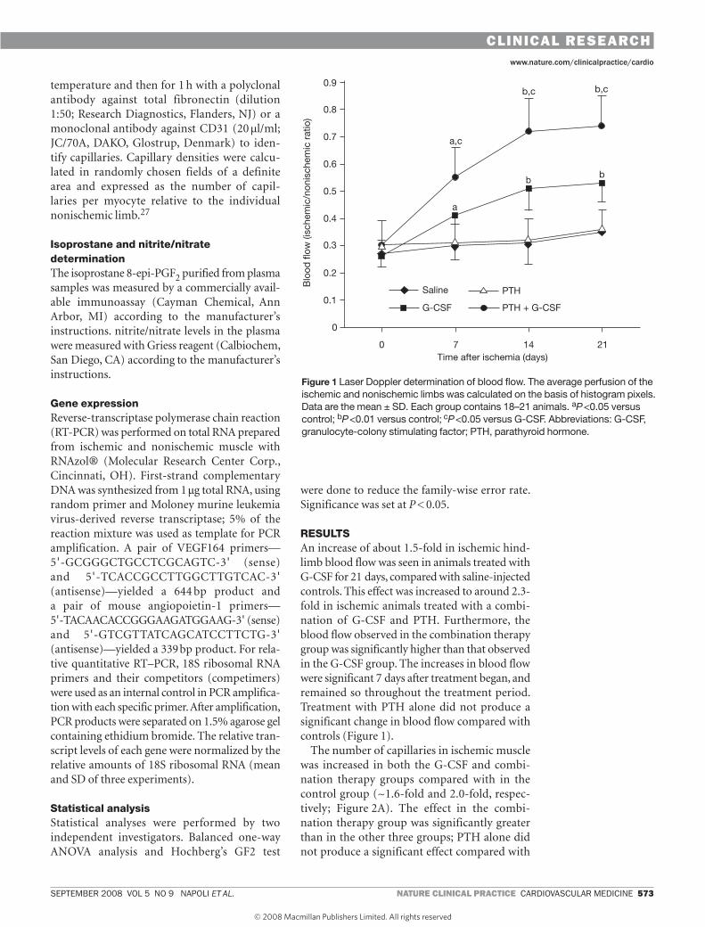
RESULTSAn increase of about 1.5-fold in ischemic hind-limb blood flow was seen in animals treated with G-CSF for 21 days, compared with saline-injected controls. This effect was increased to around 2.3-fold in ischemic animals treated with a combi-nation of G-CSF and PTH. Furthermore, the blood flow observed in the combination therapy group was significantly higher than that observed in the G-CSF group. The increases in blood flow were significant 7 days after treatment began, and remained so throughout the treatment period. Treatment with PTH alone did not produce a significant change in blood flow compared with controls (Figure 1).
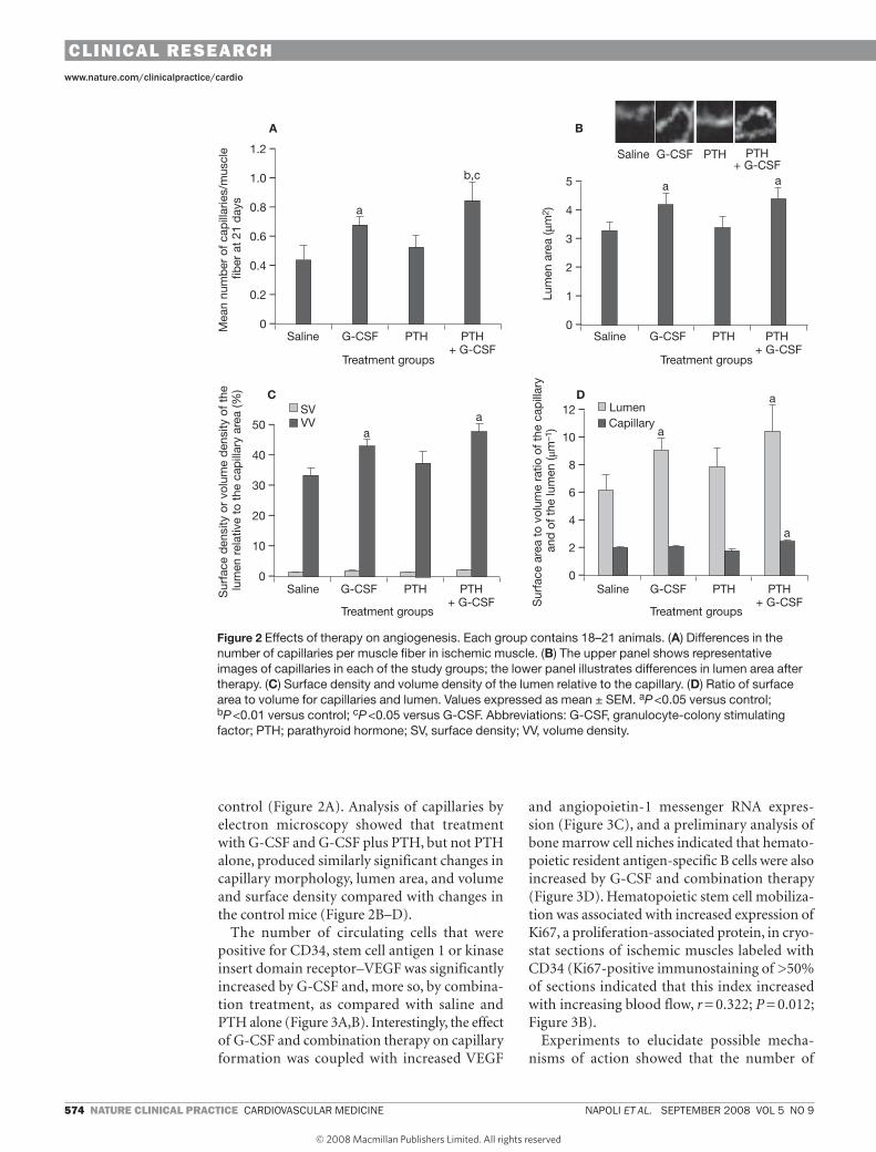
The number of capillaries in ischemic muscle was increased in both the G-CSF and combi-nation therapy groups compared with in the control group (~1.6-fold and 2.0-fold, respec-tively; Figure 2A). The effect in the combi-nation therapy group was significantly greater than in the other three groups; PTH alone did not produce a significant effect compared with
Figure 1 Laser Doppler determination of blood flow. The average perfusion of the ischemic and nonischemic limbs was calculated on the basis of histogram pixels. Data are the mean ± SD. Each group contains 18–21 animals. aP <0.05 versus control; bP <0.01 versus control; cP <0.05 versus G-CSF. Abbreviations: G-CSF, granulocyte-colony stimulating factor; PTH, parathyroid hormone.
ncpcm_2007_289f1.eps
Time after ischemia (days)
Blo
od fl
ow (i
sche
mic
/non
isch
emic
rat
io)
0 7 14 21
0
0.1
0.2
0.3
0.4
0.5
0.6
0.7
0.8
0.9
a,c
b,c b,c
a
bb
Saline
G-CSF
PTH
PTH + G-CSF
clinical reSearch clinical reSearch
574 nature clinical practice cardiovascular medicine napoli et al. september 2008 vol 5 no 9
www.nature.com/clinicalpractice/cardio
control (Figure 2A). Analysis of capillaries by electron microscopy showed that treatment with G-CSF and G-CSF plus PTH, but not PTH alone, produced similarly significant changes in capillary morphology, lumen area, and volume and surface density compared with changes in the control mice (Figure 2B–D).
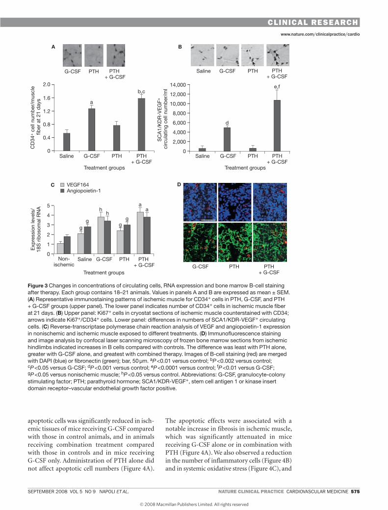
The number of circulating cells that were positive for CD34, stem cell antigen 1 or kinase insert domain receptor–VEGF was significantly increased by G-CSF and, more so, by combina-tion treatment, as compared with saline and PTH alone (Figure 3A,B). Interestingly, the effect of G-CSF and combination therapy on capillary formation was coupled with increased VEGF
and angiopoietin-1 messenger RNA expres-sion (Figure 3C), and a preliminary analysis of bone marrow cell niches indicated that hemato-poietic resident antigen-specific B cells were also increased by G-CSF and combination therapy (Figure 3D). Hematopoietic stem cell mobiliza-tion was associated with increased expression of Ki67, a proliferation-associated protein, in cryo-stat sections of ischemic muscles labeled with CD34 (Ki67-positive immunostaining of >50% of sections indicated that this index increased with increasing blood flow, r = 0.322; P = 0.012; Figure 3B).
Experiments to elucidate possible mecha-nisms of action showed that the number of
ncpcm_2007_289f2.eps
1.2 –
1.0 –
0.8 –
0.6 –
0.4 –
0.2 –
0 –
Treatment groups
Mea
n nu
mb
er o
f cap
illar
ies/
mus
cle
fiber
at
21 d
ays
Saline G-CSF PTH PTH+ G-CSF
A B
5 –
4 –
3 –
2 –
1 –
0 –
Treatment groups
Lum
en a
rea
(μm
2 )
Saline G-CSF PTH PTH + G-CSF
a
b,ca a
Saline G-CSF PTH PTH+ G-CSF
50 –
40 –
30 –
20 –
10 –
0 –
Treatment groups
Saline G-CSF PTH PTH+ G-CSF
aa
Sur
face
den
sity
or
volu
me
den
sity
of t
he
lum
en r
elat
ive
to t
he c
apill
ary
area
(%) C
SVVV
Sur
face
are
a to
vol
ume
ratio
of t
he c
apill
ary
and
of t
he lu
men
(μm
–1)
12 –
10 –
8 –
6 –
4 –
2 –
0 –
Treatment groups
Saline G-CSF PTH PTH+ G-CSF
a
aDLumenCapillary
a
Figure 2 Effects of therapy on angiogenesis. Each group contains 18–21 animals. (A) Differences in the number of capillaries per muscle fiber in ischemic muscle. (B) The upper panel shows representative images of capillaries in each of the study groups; the lower panel illustrates differences in lumen area after therapy. (C) Surface density and volume density of the lumen relative to the capillary. (d) Ratio of surface area to volume for capillaries and lumen. Values expressed as mean ± SEM. aP <0.05 versus control; bP <0.01 versus control; cP <0.05 versus G-CSF. Abbreviations: G-CSF, granulocyte-colony stimulating factor; PTH; parathyroid hormone; SV, surface density; VV, volume density.
clinical reSearch clinical reSearch
september 2008 vol 5 no 9 napoli et al. nature clinical practice cardiovascular medicine 575
www.nature.com/clinicalpractice/cardio
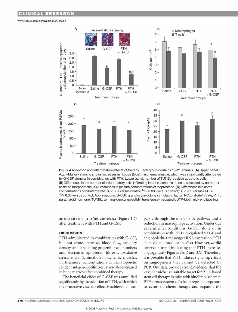
apoptotic cells was significantly reduced in isch-emic tissues of mice receiving G-CSF compared with those in control animals, and in animals receiving combination treatment compared with those in controls and in mice receiving G-CSF only. Administration of PTH alone did not affect apoptotic cell numbers (Figure 4A).
The apoptotic effects were associated with a notable increase in fibrosis in ischemic muscle, which was significantly attenuated in mice receiving G-CSF alone or in combination with PTH (Figure 4A). We also observed a reduction in the number of inflammatory cells (Figure 4B) and in systemic oxidative stress (Figure 4C), and
a
Angiopoietin-1
d
b,c
ncpcm_2007_289f3.eps
2.0 –
1.6 –
1.2 –
0.8 –
0.4 –
0 –
Treatment groups
CD
34+ c
ell n
umb
er/m
uscl
e fib
er a
t 21
day
s
Saline G-CSF PTH PTH+ G-CSF
A B
14,000 –
12,000 –
10,000 –
8,000 –
6,000 –
4,000 –
2,000 –
0 –
Treatment groups
SC
A1/
KD
R-V
EG
F+
circ
ulat
ing
cell
num
ber
/ml
Saline G-CSF PTH PTH+ G-CSF
a
e,f
Saline G-CSF PTH PTH+ G-CSF
5 –
4 –
3 –
2 –
1 –
0 –
Treatment groups
Saline G-CSF PTH PTH + G-CSF
g
a
Exp
ress
ion
leve
ls/
18S
rib
osom
al R
NA
C VEGF164 D
G-CSF PTH PTH + G-CSF
Non-ischemic
g
hh
gg
G-CSF PTH PTH+ G-CSF
Figure 3 Changes in concentrations of circulating cells, RNA expression and bone marrow B-cell staining after therapy. Each group contains 18–21 animals. Values in panels A and B are expressed as mean ± SEM. (A) Representative immunostaining patterns of ischemic muscle for CD34+ cells in PTH, G-CSF, and PTH + G-CSF groups (upper panel). The lower panel indicates number of CD34+ cells in ischemic muscle fiber at 21 days. (B) Upper panel: Ki67+ cells in cryostat sections of ischemic muscle counterstained with CD34; arrows indicate Ki67+/CD34+ cells. Lower panel: differences in numbers of SCA1/KDR-VEGF+ circulating cells. (C) Reverse-transcriptase polymerase chain reaction analysis of VEGF and angiopoietin-1 expression in nonischemic and ischemic muscle exposed to different treatments. (d) Immunofluorescence staining and image analysis by confocal laser scanning microscopy of frozen bone marrow sections from ischemic hindlimbs indicated increases in B cells compared with controls. The difference was least with PTH alone, greater with G-CSF alone, and greatest with combined therapy. Images of B-cell staining (red) are merged with DAPI (blue) or fibronectin (green); bar, 50 µm. aP <0.01 versus control; bP <0.002 versus control; cP <0.05 versus G-CSF; dP <0.001 versus control; eP <0.0001 versus control; fP <0.01 versus G-CSF; gP <0.05 versus nonischemic muscle; hP <0.05 versus control. Abbreviations: G-CSF, granulocyte-colony stimulating factor; PTH; parathyroid hormone; SCA1/KDR-VEGF+, stem cell antigen 1 or kinase insert domain receptor–vascular endothelial growth factor positive.
clinical reSearch clinical reSearch
576 nature clinical practice cardiovascular medicine napoli et al. september 2008 vol 5 no 9
www.nature.com/clinicalpractice/cardio
an increase in nitrite/nitrate release (Figure 4D) after treatment with PTH and G-CSF.
DISCUSSIONPTH administered in combination with G-CSF, but not alone, increases blood flow, capillary density, and circulating progenitor cell numbers and decreases apoptosis, fibrosis, oxidative stress, and inflammation in ischemic muscles. Furthermore, concentrations of hematopoietic resident antigen-specific B cells were also increased in bone marrow after combined therapy.
The beneficial effect of G-CSF was amplified significantly by the addition of PTH, with which the protective vascular effect is achieved at least
partly through the nitric oxide pathway and a reduction in macrophage activation. Under our experimental conditions, G-CSF alone or in combination with PTH upregulated VEGF and angiopoietin-1 messenger RNA expression; PTH alone did not produce an effect. However, we did observe a trend indicating that PTH increases angiogenesis (Figures 2A,D and 3A). Therefore, it is possible that PTH induces signaling effects on angiogenesis that cannot be detected by PCR. Our data provide strong evidence that the vascular niche is a suitable target for PTH-based stem cell therapy in mice with hindlimb ischemia. PTH protects stem cells from repeated exposure to cytotoxic chemotherapy and expands the
b,c
ncpcm_2007_289f4.eps
3.5 –
3.0 –
2.5 –
2.0 –
1.5 –
1.0 –
0.5 –
0 –
Treatment groups
Num
ber
of T
UN
EL-
pos
itive
ap
opto
tic
cells
/mus
cle
fiber
at
21 d
ays
Saline G-CSF PTH PTH + G-CSF
A B
7 –
6 –
5 –
4 –
3 –
2 –
1 –
0 –
Treatment groups
Cel
ls p
er m
m2
Saline G-CSF PTH PTH+ G-CSF
dSaline G-CSF PTH
+ G-CSF
250 –
200 –
150 –
100 –
50 –
0 –
Treatment groups
Saline G-CSF PTH PTH+ G-CSF
d d
Pla
sma
isop
rost
anes
8-e
pi-
PG
F2a
(pg/
ml)
C
Pla
sma
NO
x (μ
M)
40 –
35 –
30 –
25 –
20 –
15 –
10 –
5 –
0 –
Treatment groups
Saline G-CSF PTH PTH+ G-CSF
D
d
Non-ischemic
a
MacrophagesT cells
d
d
Azan–Mallory staining
Figure 4 Apoptotic and inflammatory effects of therapy. Each group contains 18–21 animals. (A) Upper panel: Azan–Mallory staining shows increases in fibrosis (blue) in ischemic muscle, which was significantly attenuated by G-CSF alone or in combination with PTH. Lower panel: number of TUNEL-positive apoptotic cells. (B) Differences in the number of inflammatory cells infiltrating into the ischemic muscle, assessed by computer-assisted morphometry. (C) Differences in plasma concentrations of isoprostane. (d) Differences in plasma concentrations of nitrate/nitrate. aP <0.01 versus control; bP <0.005 versus control; cP <0.05 versus G-CSF; dP <0.05 versus control. Abbreviations: G-CSF, granulocyte-colony stimulating factor; NOx, nitrate/nitrate; PTH; parathyroid hormone; TUNEL, terminal deoxynucleotidyl transferase-mediated dUTP-biotin nick end labeling.
clinical reSearch clinical reSearch
september 2008 vol 5 no 9 napoli et al. nature clinical practice cardiovascular medicine 577
www.nature.com/clinicalpractice/cardio
resident hematopoietic stem cell pool in the bone marrow in transplant recipients by increasing the number of hematopoietic stem cells that are mobilized into the peripheral blood.28 The magnitude of hematopoietic stem cell mobiliza-tion induced by the combined G-CSF and PTH therapy in that study was similar to that reported herein. We did not, however, observe a depletion of the hematopoietic stem cell pool in animals treated with G-CSF alone or an increase in the hematopoietic stem pool in animals treated with PTH alone, as reported by Adams et al.28 This dissimilarity might be owing to different experimental models.
The use of G-CSF in patients with critical limb ischemia has improved neovascularization.29,30 However, in approximately 20% of patients who receive G-CSF, mobilization of hematopoietic stem cells is insufficient to proceed successfully to autologous stem cell therapy.31 In this regard, our preliminary data indicate that modulation of the bone marrow niche is a suitable strategy for treating peripheral ischemia.32 This finding has potential therapeutic implications, as it might be possible to reduce doses of growth factors and, consequently, the risk of adverse effects such as cancer.33,34 However, further specific studies on the vascular niche are needed to investigate in depth its structure and mechanisms linked to PTH activity.
Another study indicated that PTH exerted a tissue-sparing effect on the myocardium, restored left ventricular function, and prevented the development of shock in dogs after occlusion of the left coronary artery.35 PTH might, there-fore, also have broad beneficial effects in cardio-vascular disease. In addition, PTH could promote vasodilatation directly through PTH receptors on vascular smooth muscle cells and indirectly via nitric oxide.14–17 Several studies are currently addressing the potential benefits of therapeutic angiogenesis in cardiovascular diseases.3,36,37 In a completed phase I clinical trial, PTH (up to 100 µg for 14 days) was well tolerated and no dose-limiting toxic effects were observed.38 The clinical efficacy of PTH needs to be tested in larger phase II studies. We hope that translational studies, such as the one described here, will stimulate clinical trials aimed at increasing the effectiveness of hematopoietic stem cell therapy in peripheral arterial disease by targeting the hematopoietic stem cell niche with PTH.
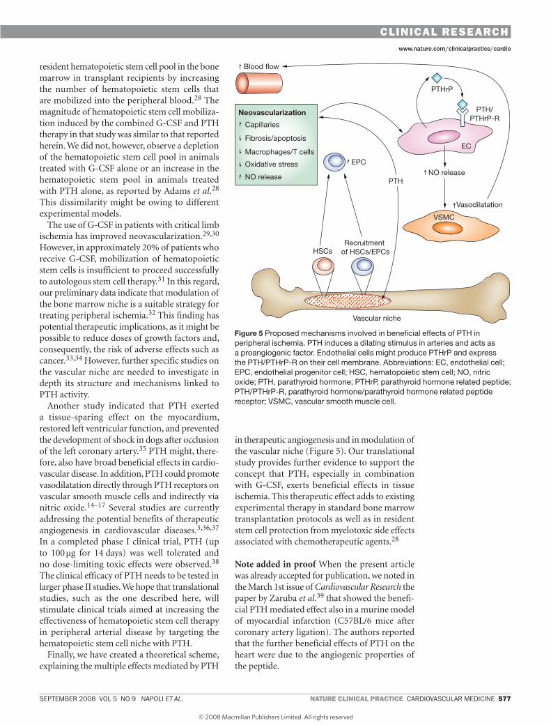
Finally, we have created a theoretical scheme, explaining the multiple effects mediated by PTH
in therapeutic angiogenesis and in modulation of the vascular niche (Figure 5). Our translational study provides further evidence to support the concept that PTH, especially in combination with G-CSF, exerts beneficial effects in tissue ischemia. This therapeutic effect adds to existing experimental therapy in standard bone marrow transplantation protocols as well as in resident stem cell protection from myelotoxic side effects associated with chemotherapeutic agents.28
Note added in proof When the present article was already accepted for publication, we noted in the March 1st issue of Cardiovascular Research the paper by Zaruba et al.39 that showed the benefi-cial PTH mediated effect also in a murine model of myocardial infarction (C57BL/6 mice after coronary artery ligation). The authors reported that the further beneficial effects of PTH on the heart were due to the angiogenic properties of the peptide.
ncpcm_2007_289f5.eps
Neovascularization
Capillaries
Fibrosis/apoptosis
Macrophages/T cells
Oxidative stress
NO releasePTH
HSCs
Vascular niche
Recruitment of HSCs/EPCs
NO release
EC
VSMC
PTH/PTHrP-R
PTHrP
Blood flow
EPC
Vasodilatation
Figure 5 Proposed mechanisms involved in beneficial effects of PTH in peripheral ischemia. PTH induces a dilating stimulus in arteries and acts as a proangiogenic factor. Endothelial cells might produce PTHrP and express the PTH/PTHrP-R on their cell membrane. Abbreviations: EC, endothelial cell; EPC, endothelial progenitor cell; HSC, hematopoietic stem cell; NO, nitric oxide; PTH, parathyroid hormone; PTHrP, parathyroid hormone related peptide; PTH/PTHrP-R, parathyroid hormone/parathyroid hormone related peptide receptor; VSMC, vascular smooth muscle cell.
clinical reSearch clinical reSearch
578 nature clinical practice cardiovascular medicine napoli et al. september 2008 vol 5 no 9
www.nature.com/clinicalpractice/cardio
References1 Krumholz HM and Masoudi FA (2006) The year in
epidemiology, health services research, and outcomes research. J Am Coll Cardiol 48: 1886–1895
2 Napoli C et al. (2006) Rethinking primary prevention of atherosclerosis-related diseases. Circulation 114: 2517–2527
3 Napoli C et al. (2007) Bone marrow cell-mediated cardiovascular repair: potential of combined therapies. Trends Mol Med 13: 278–286
4 Taichman RS and Emerson SG (1994) Human osteoblasts support hematopoiesis through the production of granulocyte colony-stimulating factor. J Exp Med 179: 1677–1682
5 Blair HC et al. (1999) Parathyroid hormone-regulated production of stem cell factor in human osteoblasts and osteoblast-like cells. Biochem Biophys Res Commun 255: 778–784
6 Taichman RS et al. (1996) Human osteoblasts support human hematopoietic progenitor cells in in vitro bone marrow cultures. Blood 87: 518–524
7 Calvi LM et al. (2003) Osteoblastic cells regulate the haematopoietic stem cell niche. Nature 425: 841–846
8 Zhang J et al. (2003) Identification of the haematopoietic stem cell niche and control of the niche size. Nature 425: 836–841
9 Kiel MJ et al. (2005) SLAM family receptors distinguish hematopoietic stem and progenitor cells and reveal endothelial niches for stem cells. Cell 121: 1109–1121
10 Arai F et al. (2004) Tie2/Angiopoietin-1 signaling regulates hematopoietic stem cell quiescence in the bone marrow niche. Cell 118: 149–161
11 Yin T and Li L (2006) The stem cell niches in bone. J Clin Invest 116: 1195–1201
12 Rixon RH et al. (1958) Increased survival of rats irradiated with x-rays and treated with parathyroid extract. Nature 182: 1374
13 Whitfield JF (2006) Parathyroid hormone: a novel tool for treating bone marrow depletion in cancer patients caused by chemotherapeutic drugs and ionizing radiation. Cancer Lett 244: 8–15
14 Schulze MR et al. (1993) Human parathyroid hormone dilates both pig coronary and human inferior epigastric arteries by a cyclic AMP-dependent pathway. Artery 20: 147–162
15 Rian E et al. (1994) Parathyroid hormone-related protein is produced by cultured endothelial cells: a possible role in angiogenesis. Biochem Biophys Res Commun 198: 740–747
16 Jiang B et al. (1998) Expression of parathyroid hormone/parathyroid hormone-related protein receptor in vascular endothelial cells. J Cardiovasc Pharmacol 31 (suppl 1): S142–S144
17 Kalinowski L et al. (2001) Nitric oxide as a second messenger in parathyroid hormone-related protein signaling. J Endocrinol 170: 433–440
18 Napoli C et al. (2005) Beneficial effects of concurrent autologous bone marrow cell therapy and metabolic intervention in ischemia-induced angiogenesis in the mouse hindlimb. Proc Natl Acad Sci USA 102: 17202–17206
19 Minamino K et al. (2005) Macrophage colony-stimulating factor (M-CSF), as well as granulocyte colony-stimulating factor (G-CSF), accelerates neovascularization. Stem Cells 23: 347–354
20 Tan Y et al. (2007) Stromal cell-derived factor-1 enhances pro-angiogenic effect of granulocyte-colony stimulating factor. Cardiovasc Res 73: 823–832
21 Egginton S et al. (1993) Fine structure of capillaries in ischaemic and non ischaemic rat striated muscle. Effect of torbafylline. Int J Microcirc Clin Exp 12: 33–44
22 Napoli C et al. (2003) Deletion of the p66Shc longevity gene reduces systemic and tisuue oxidative stress, vascular cell apoptosis, and early atherogenesis in mice fed high-fat diet. Proc Natl Acad Sci USA 100: 2112–2116
23 Baker AH et al. (2007) Brain protection using autologous bone marrow cell, metalloproteinase inhibitors, and metabolic treatment in cerebral ischemia. Proc Natl Acad Sci USA 104: 3597–3602
24 de Nigris F et al. (2007) Therapeutic effects of autologous bone marrow cells and metabolic intervention in the ischemic hindlimb of spontaneously hypertensive rats involve reduced cell senescence and CXCR4/Akt/eNOS pathways. J Cardiovasc Pharmacol 50: 424–433
25 Casamassimi A et al. (2007) Comparison between total endothelial progenitor cell isolation versus enriched CD133+ culture. J Biochem 141: 503–511
26 de Nigris F et al. (2007) Therapeutic effects of concurrent autologous bone marrow cell infusion and metabolic intervention in ischemia-induced angiogenesis in the hypercholesterolemic mouse hindlimb. Int J Cardiol 117: 238–243
27 Miranville A et al. (2004) Improvement of postnatal neovascularization by human adipose tissue-derived stem cells. Circulation 110: 349–355
28 Adams GB et al. (2007) Therapeutic targeting of a stem cell niche. Nat Biotechnol 25: 238–243
29 Zhou B et al. (2006) G-CSF-mobilized peripheral blood mononuclear cells from diabetic patients augment neovascularization in ischemic limbs but with impaired capability. J Thromb Haemost 4: 993–1002
30 Kawamura A et al. (2006) Clinical study of therapeutic angiogenesis by autologous peripheral blood stem cell (PBSC) transplantation in 92 patients with critically ischemic limbs. J Artif Organs 9: 226–233
KEY POINTS■ The vascular niche is a potential therapeutic
target for peripheral arterial disease
■ We present a translational research study investigating whether targeting the niche with parathyroid hormone (PTH) might improve stem cell-based therapy in an experimental model of hindlimb ischemia that would be relevant to the clinical use of hematopoietic stem-cell-driven angiogenesis
■ After mobilization induced by granulocyte-colony stimulating factor (G-CSF), mice also receiving PTH showed increased blood flow, capillary density, nitrite/nitrate release, angiogenic factors and circulating progenitor cells together with reduced apoptosis, fibrosis, oxidative stress and inflammation in ischemic muscles
■ Our translational study could stimulate clinical trials aimed at increasing the effectiveness of hematopoietic stem cell therapy in peripheral arterial disease by targeting the hematopoietic stem cell niche with PTH
■ In a recent phase I clinical trial,38 PTH (up to 100 μg for 14 days) was tolerated well and there was no dose-limiting toxicity; thus, the clinical efficacy of PTH will need to be tested in a larger phase II study
AcknowledgmentsThese studies were supported by grants from the following institutions: Ministero dell’Università e Ricerca Scientifica PRIN 2006 to C Napoli and PRIN 2005 to A Giovane; Fondation Jerome Lejeune to C Napoli; NIH to S Williams-Ignarro, A Giordano, LJ Ignarro and C Napoli; Regione Campania 2007 to ML Balestrieri and C Napoli; Sbarro Research Institute to A Giordano; Ricerca di Ateneo 2006 and Camera di Commercio to the University of Sannio for FP Mancini and E Varricchio; and research funds of the University of Salerno to MF Tecce.
Competing interests The authors declared no competing interests.
clinical reSearch clinical reSearch
september 2008 vol 5 no 9 napoli et al. nature clinical practice cardiovascular medicine 579
www.nature.com/clinicalpractice/cardio
31 Seggewiss R and Einsele H (2007) Hematopoietic growth factors including keratinocyte growth factor in allogeneic and autologous stem cell transplantation. Semin Hematol 44: 203–211
32 Nikolova G et al. (2007) The vascular niche and its basement membrane. Trends Cell Biol 17: 19–25
33 Lyman GH and Shayne M (2007) Granulocyte colony-stimulating factors: finding the right indication. Curr Opin Oncol 19: 299–307
34 Marsh JC et al. (2007) Hematopoietic growth factors in the treatment of acquired bone marrow failure states. Semin Hematol 44: 138–147
35 Feola M et al. (1985) Vasoactive parathyroid hormone in the treatment of acute ischemic left ventricular failure and the prevention of cardiogenic shock. Circ Shock 17: 163–177
36 Napoli C et al. (2007) Therapeutic approaches in vascular repair induced by adult bone marrow cells
and circulating progenitor endothelial cells. Curr Pharm Des 13: 3245–3251
37 Herrmann J et al. (2006) Angiogenesis in atherogenesis. Arterioscler Thromb Vasc Biol 26: 1948–1957
38 Ballen KK et al. (2007) Phase I trial of parathyroid hormone to facilitate stem cell mobilization. Biol Blood Marrow Transplant 13: 838–843
39 Zaruba et al. (2008) Parathyroid hormone treatment after myocardial infarction promotes cardiac repair by enhanced neovascularization and cell survival. Cardiovasc res 77: 722–731
Clinical contextSee the accompanying Clinical Context article by Aliev et al. [doi:10.1038/ncpcardio1295]
clinical reSearch clinical reSearch