Influence of Donor Smoking on Midterm Outcomes After Lung Transplantation
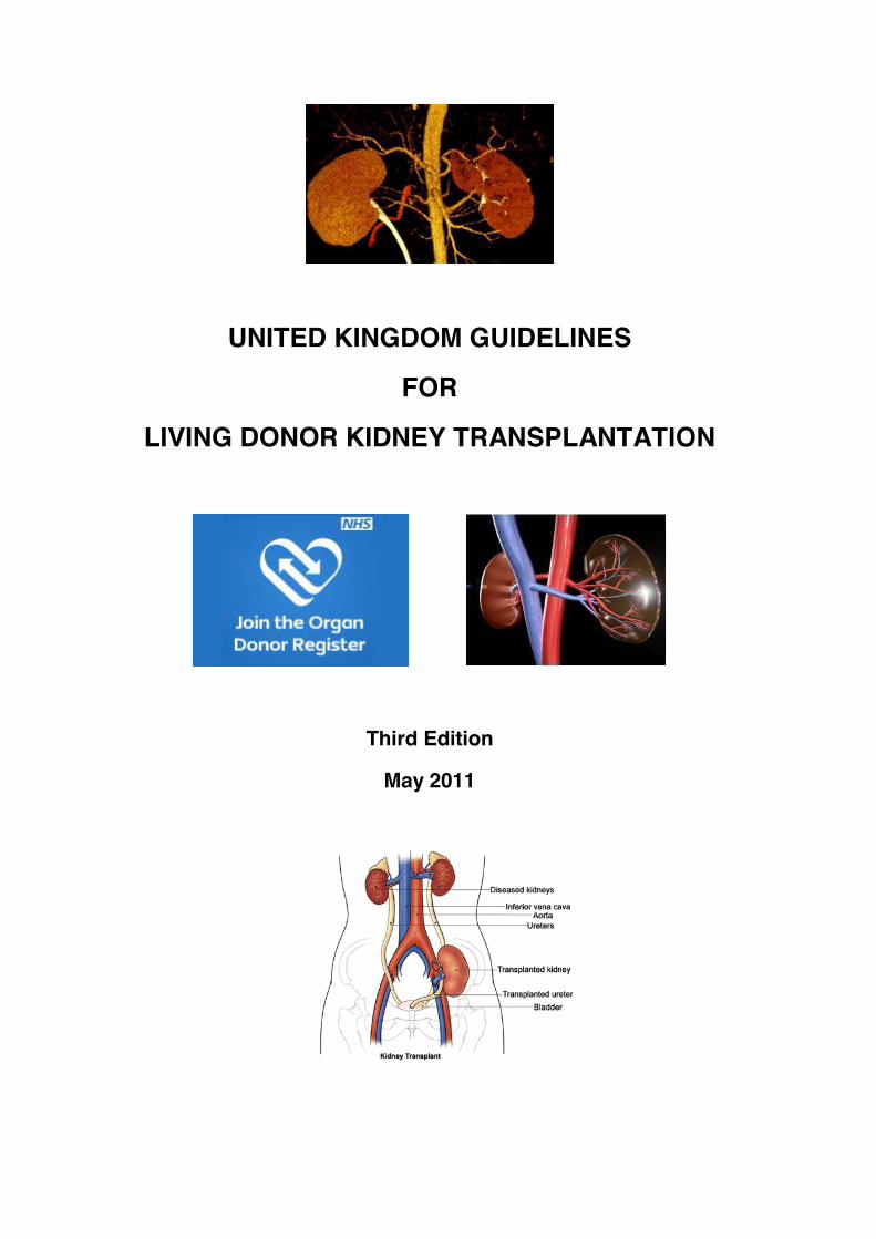
UNITED KINGDOM GUIDELINES
FOR
LIVING DONOR KIDNEY TRANSPLANTATION
Third Edition
May 2011
2
UNITED KINGDOM GUIDELINES
FOR
LIVING DONOR KIDNEY TRANSPLANTATION
Compiled by a Joint Working Party of The British Transplantation Society and
The Renal Association
Third Edition
May 2011
Posted on www.bts.org.uk & www.renal.org May 2011

3
CONTENTS
1.0 INTRODUCTION AND OBJECTIVES 7 1.1 The Need for Guidelines 7
1.2 Scope of the Guidelines 8
1.3 Process of Writing and Methodology 8
1.4 Editorial Committee 9
1.5 Contributing Authors 10
1.6 Disclaimer 12
1.7 Grading of Recommendations 13
2.0 LEGAL FRAMEWORK 15 2.1 The Human Tissue Act 2004 15
2.2 The Human Tissue Authority (HTA) 16
2.3 Consent for the Removal of Organs from Living Donors 17
2.4 Types of Living Kidney Donation Permitted by the Legislation 17
2.5 Requirements for Transplants involving a Living Donor 18
2.6 Prohibition of Commercial Dealings in Human Material 18
2.7 Reimbursement of Expenses 19
2.8 Exceptional Circumstances 19
2.9 The Human Tissue (Scotland) Act 2006 20
2.10 The EU Organ Donation Directive 21
3.0 ETHICS 24 3.1 Ethics 24
3.2 Key Ethical Principles in Living Donor Kidney Transplantation 24
3.3 T he Recipient Perspective 26
3.4 The Donor Perspective 26
3.5 The Transplant Team Perspective 28
3.6 Confidentiality 28
3.7 Expanding the Living Donor Pool 29
3.8 The Child or Young Person as a Living Donor 30
3.9 The British Transplantation Society (BTS) Ethics Committee 30

4
4.0 INFORMING THE POTENTIAL DONOR 32 4.1 Informing the Potential Donor 32
4.2 Informed Consent for Living Kidney Donation 33
4.3 Donor Identity 34
4.4 Patient Advocacy 36
4.5 Independent Translators 37
4.6 Psychological Issues 38
4.7 The Responsibility of the Donor Surgeon 40
5.0 DONOR EVALUATION 43 5.1 Introduction 43
5.2 Donor Evaluation: Summary 45
5.3 ABO Blood Grouping and Crossmatch Testing 49
5.4 Medical Assessment 50
5.5 Assessment of Renal Function 57
5.6 Donor Age 62
5.7 Donor Obesity 66
5.8 Hypertension in the Donor 71
5.9 Diabetes Mellitus 80
5.10 Cardiovascular Evaluation 85
5.11 Proteinuria 92
5.12 Non-Visible Haematuria 97
5.13 Pyuria 103
5.14 Infection in the Prospective Donor 104
5.15 Nephrolithiasis 114
5.16 Haematological Disease 120
5.17 Familial Renal Disease 124
5.18 Donor Malignancy 130
6.0 SURGERY: TECHNICAL ASPECTS, DONOR RISK AND PERI-OPERATIVE CARE 135 6.1 Introduction 136
6.2 Assessment of Renal Anatomy 136
6.3 Peri-operative Mortality 139
6.4 Peri-operative Morbidity 140
5
6.5 Long Term Mortality 142
6.6 Pre-operative Care and Preparation 143
6.7 Donor Nephrectomy 145
7.0 HISTOCOMPATIBILITY TESTING FOR LIVING DONOR KIDNEY TRANSPLANTATION 151 7.1 Assessment of Donor-Recipient HLA Mismatch Status 153
7.2 Identification and Characterisation of Alloantibodies 153
7.3 Pre-transplant Donor-Recipient Crossmatch Test 155
7.4 Selection of Suitable Donor-Recipient Pairs 157
7.5 Antibody Incompatible Living Donor Transplantation 158
8.0 EXPANDING THE DONOR POOL 161 8.1 Paired/Pooled Living Donation 161
8.2 Non-Directed Altruistic Donation 167
8.3 Antibody Incompatible Donation 172
9.0 LOGISTICAL CONSIDERATIONS 174 9.1 Reimbursement of Living Donor Expenses 174
9.2 Paired/Pooled and Non-Directed Altruistic Donors 176
9.3 Donors from Overseas 176
9.4 Annex: Template Letter for Potential Overseas Donors 181
10.0 DONOR FOLLOW-UP 184 10.1 Arrangements for Follow-up 184
10.2 The Unsuitable Donor 187
10.3 Pregnancy following Kidney Donation 187
10.4 Renal Failure following Living Kidney Donation 188
11.0 RECIPIENT OUTCOME AFTER LIVING DONOR KIDNEY TRANSPLANTATION IN ADULTS 189
6
12.0 RECURRENT RENAL DISEASE 195 12.1 Diabetic Nephropathy 196
12.2 Primary Focal Segmental Glomerulosclerosis 196
12.3 IgA Nephropathy 198
12.4 Membranous Nephropathy 198
12.5 Amyloidosis 199
12.6 Systemic Lupus Erythematosus 199
12.7 ANCA Associated Systemic Vasculitis 200
12.8 Goodpasture’s Disease 200
12.9 Alport Syndrome 201
12.10 Mesangiocapillary Glomerulonephritis 201
12.11 Haemolytic Uraemic Syndrome 202
12.12 Primary Hyperoxaluria 203
12.13 Cystinosis 203
13.0 LIVING DONOR KIDNEY TRANSPLANTATION IN CHILDREN 208

7
CHAPTER 1 INTRODUCTION AND OBJECTIVES
1.1 The Need for Guidelines
Living kidney donation has become an essential part of transplantation practice.
Historically, this has been attributed to the shortage of deceased donor kidneys and the
growing waiting list of potential recipients. However, kidney transplantation from a living
donor has become the treatment of choice for many patients and their families, offering
optimum patient and graft survival, and also the chance to avoid long periods on the
transplant waiting list. This is particularly the case in pre-emptive transplantation, when
the transplant occurs before the start of dialysis. Currently, pre-emptive transplantation
averages 31% of the patients transplanted from living donors; a figure that most believe
should increase over the next ten years (1).
Recently, living donation has offered patients who are more clinically complex, both
immunologically and/or due to other co-morbidities, the opportunity to benefit from a
transplant that they might otherwise not have received from the deceased donor waiting
list. Nonetheless, the welfare of the donor remains paramount, and vigilance in donor care
and management is essential to ensure that appropriate safeguards are in place to protect
individuals and to inspire public confidence.
At the time of writing, living donors account for 1 in 2 organ donors and 1 in 3 kidney
transplants performed in the UK are from living donors, this representing 38% of the total
kidney transplant activity per annum. The latest national statistics show that in 2008-9 and
2009-10, there was an 11% increase in living donor kidney transplants performed year on
year, to 927 and 1037 respectively (1). In part, these figures reflect a small but growing
number of transplants from paired/pooled donation and non-directed altruistic donors, of
which there were 32 and 16 transplants performed in 2009 and 2010 respectively. Over
the last 10 years, there has been a 65% increase in overall living donor activity, from 372
donors in 2000-1 to 1061 in 2009-10, with all transplant centres now actively engaged in
living donor kidney transplantation. This represents a significant change in practice and
necessitates clear, contemporary, evidence-based guidance.
8
1.2 Scope of the Guidelines
This guidance relates only to living donor kidney transplantation and reflects a growing
body of evidence, incorporating aspects of clinical practice that are relevant to both adult
and paediatric settings. These include the ethical and medico-legal aspects of donor
selection, medical and pre-operative donor evaluation, identification of high risk donors,
the management of complications, and expected outcome. Scenarios that present an
increased level of risk to the potential recipient, such as antibody incompatible
transplantation, recurrent disease and transplantation in the context of other co-
morbidities, are also included. In addition, guidance is provided on the most appropriate
investigations to be considered to assist clinical decision-making, and the best surgical
approaches when faced with different clinical scenarios.
1.3 Process of Writing and Methodology
The original ‘UK Guidelines for Living Donor Kidney Transplantation’ were commissioned
by the British Transplantation Society (BTS) and the Renal Association (RA) as part of a
wider initiative to develop ‘Best Practice’ guidance for clinicians involved in the area of
transplantation. Initially published in 2000 (2) and revised in 2005 (3), the guidelines have
achieved international repute. This third edition has continued the collaboration between
BTS and RA, under the auspices of the BTS Standards Committee, and the document
has been significantly updated in the light of new data and changing practice. It has been
produced with wide representation from UK colleagues and professional bodies involved
in both donor and recipient management.
A systematic review of the relevant literature and synthesis of the available evidence was
undertaken by selected relevant clinical experts. This was followed by peer group
appraisal and expert review. Draft proposals were amended by an editorial committee and
the appropriate levels of evidence added to recommendations. Wider consultation with the
transplant community was undertaken by ‘face to face’ consultation in the form of a BTS-
sponsored consensus meeting at the BTS Living Donor Forum, and through subsequent
e-mail commentary. The penultimate draft of the document was placed on the BTS and
RA websites in March and April 2011 for an additional period of open consultation, to
which patient and transplant groups were actively encouraged to contribute. The final
document was posted in May 2011.
9
Where available, these guidelines are based on published evidence, and the evidence
and recommendations have been graded for strength except where the published studies
are descriptive. With a handful of exceptions, conference presentations have not been
included and the publication cut off date for evidence was February 2011.
It is anticipated that these guidelines will next be revised in 2015.
1.4 Editorial Committee
Professor Derek Manas MD FCS (SA)
Professor of Transplantation
University of Newcastle upon Tyne and Newcastle NHS Trust
Co-Chair Editorial Group & BTS Living Donor Forum
Miss Lisa Burnapp RN MA
Consultant Nurse, Living Donor Kidney Transplantation, Guy’s & St Thomas’ NHS
Foundation Trust, London
Lead Nurse - Living Donation, Organ Donation and Transplantation, NHS Blood and
Transplant (NHSBT)
Co-Chair Editorial Group & BTS Living Donor Forum
Dr Peter A Andrews MD FRCP
Consultant Nephrologist & Clinical Lead for Transplantation
SW Thames Renal & Transplantation Unit, St Helier Hospital, Surrey
Chair of BTS Standards Committee
Professor J Andrew Bradley FRCS F Med Sci (Cambridge)
Professor of Surgery, University of Cambridge
Clinical Director of Transplantation, Cambridge University Hospitals NHS Trust
Co-editor of previous Living Donor Guidelines, Chair of NHSBT Kidney Advisory Group
Dr Chris Dudley MD FRCP
Consultant Nephrologist & Clinical Director of Renal and Transplant
Southmead Hospital, Bristol
Secretary of BTS, RA Representative
10
1.5 Contributing Authors
Dr Peter Andrews MD FRCP, Consultant Nephrologist & Clinical Lead for Transplantation,
SW Thames Renal & Transplantation Unit, St Helier Hospital, Surrey
Dr Kesh Baboolal MD FRCP, Director of Acute University Hospital Services, Cardiff and
Vale University Health Board, University Hospital of Wales
Dr Richard Baker PhD FRCP, Consultant Nephrologist, St. James’s University Hospital,
Leeds
Dr Simon Ball PhD FRCP, Consultant Nephrologist, Queen Elizabeth Hospital,
Birmingham
Prof J Andrew Bradley FRCS F Med Sci (Cambridge), Professor of Surgery, University of
Cambridge and Clinical Director of Transplantation, Cambridge University Hospitals NHS
Trust
Miss Lisa Burnapp RN MA, Consultant Nurse, Living Donor Kidney Transplantation, Guy’s
& St Thomas’ NHS Foundation Trust, London; & Lead Nurse, Living Donation, NHS
Blood and Transplant
Dr Jamie Cavenagh MD FRCP FRCPath, Consultant Haematologist, Barts and the
London NHS Trust
Dr Brian Clapp PhD MRCP, Consultant Cardiologist, Guy’s & St Thomas’ NHS
Foundation Trust, London
Dr Antonia Cronin MRCP MA (Medical Law and Ethics), Consultant Nephrologist, Guy’s &
St Thomas’ NHS Foundation Trust and King’s College London
Dr Susan Fuggle DPhil FRCPath, Consultant Clinical Scientist, Oxford Transplant Centre
Dr Colin Geddes FRCP (Glas), Consultant Nephrologist and Honorary Senior Lecturer,
Greater Glasgow, Clyde and Forth Valley Renal Service
Mr Paul Gibbs FRCS, Consultant Surgeon, Wessex Regional Renal and Transplant Unit,
Portsmouth
Mr David Glass, Lead Clinical Health Psychologist, Guy’s & St Thomas’ NHS Foundation
Trust, London
Ms Kay Hamilton RN, Living Donor Co-ordinator, Southmead Hospital, Bristol
Ms Sian Hedges, Central Policy Unit, Home Office, UK
Dr Robert Higgins MD FRCP, Consultant Nephrologist, University Hospitals Coventry and
Warwickshire
Dr Rachel Hilton PhD FRCP, Consultant Nephrologist, Guy’s & St Thomas’ NHS
Foundation Trust, London
11
Mrs Rachel Johnson MSc, Head of Organ Donation and Transplantation Studies, NHS
Blood and Transplant
Mr Paul Lear MS FRCS, Divisional Director of Surgery and Consultant Surgeon, Dorset
County Hospital Foundation Trust, North Bristol NHS Trust
Dr Robert Lewis MD FRCP, Consultant Nephrologist, Wessex Regional Renal and
Transplant Unit, Portsmouth
Mr Nizam Mamode MD FRCS, Consultant Surgeon, Guy’s & St Thomas’ NHS Foundation
Trust, London
Prof Derek Manas MD FCS (SA), Professor of Transplantation, University of Newcastle
upon Tyne and Newcastle NHS Trust
Ms Ann Marsden RN, Live Donor Transplant Co-ordinator, Cardiff Transplant Unit
Miss Lorna Marson MD FRCS, Senior Lecturer in Transplant Surgery, Royal Infirmary of
Edinburgh
Dr Adam Mclean DPhil FRCP, Consultant Nephrologist & Transplant Physician, West
London Renal & Transplant Centre
Ms Jen McDermott BSc Hons MA, Lead Living Donor Co-ordinator, Imperial College
Healthcare NHS Trust
Mrs Sue Moore RN, Living Donor Co-ordinator, Queen Elizabeth Hospital, Birmingham
Dr Pramod Nagaraja MRCP, SpR in Nephrology and Transplantation, University Hospital
of Wales
Dr Chas Newstead FRCP MD, Consultant Renal Physician, St James’s University
Hospital, Leeds
Mr Jonathan Olsburgh MBBS FRCS (Urol), Consultant in Urology & Transplant Surgery,
Guy’s & St Thomas’ NHS Foundation Trust, London
Dr Michael Picton PhD FRCP, Consultant Nephrologist, Manchester Royal Infirmary
Mr David Rix MD FRCS, Consultant Urologist, Freeman Hospital, Newcastle
Dr Richard Sandford PhD FRCP, Honorary Consultant in Medical Genetics, University of
Cambridge
Dr John Scoble MD FRCP, Associate Medical Director, Guy’s & St Thomas’ NHS
Foundation Trust, London
Prof Neil Sheerin PhD MRCP, Professor of Nephrology, Newcastle University
Dr John Sayer MRCP PhD, Senior Lecturer in Nephrology, Newcastle
Dr Richard Smith PhD MRCP, Consultant and Senior Lecturer, University of Bristol
Ms Linda Stowe, Central Policy Unit, Home Office, UK
Dr Craig Taylor PhD FRCPath, Director of Histocompatibility and Immunogenetics,
Cambridge University Hospital NHS Foundation Trust

12
Dr Paul Telfer FRCP FRCPath, Consultant Haematologist, Barts and the London NHS
Trust
Dr Raj Thuraisingham MD FRCP, Honorary Senior Lecturer and Consultant Nephrologist,
Barts and the London NHS Trust
Dr E Jane Tizard FRCP FRCPCH, Consultant Paediatric Nephrologist, Bristol Royal
Hospital for Children
Dr Nicholas Torpey PhD FRCP, Consultant Nephrologist, Addenbrooke’s Hospital,
Cambridge
Dr Robert Vaughan PhD FRCPath, Director of Clinical Transplantation Laboratory, King’s
College, London
Mr Peter Veitch FRCS, Consultant Transplant Surgeon, Royal Free Hospital, London
1.6 Disclaimer
This document provides a guide to best practice, which inevitably evolves over time. All
practitioners need to undertake clinical care on an individualised basis and keep up to
date with changes in the practice of clinical medicine.
These guidelines represent the collective opinions of a number of experts in the field and
do not have the force of law. They contain information/guidance for use by practitioners
as a best practice tool. It follows that the guidelines should be interpreted in the spirit
rather than to the letter of their contents. The opinions presented are subject to change
and should not be used in isolation to define the management for any individual patient.
The guidelines are not designed to be prescriptive, nor to define a standard of care.
The British Transplantation Society and Renal Association cannot attest to the accuracy,
completeness or currency of the opinions contained herein and do not accept any
responsibility or liability for any loss or damage caused to any practitioner or any third
party as a result of any reliance being placed on the guidelines or as a result of any
inaccurate or misleading opinion contained in the guidelines.

13
1.7 Grading of Recommendations In these guidelines, the GRADE system has been used to rate the strength of evidence
and the strength of recommendations (4). This approach is consistent with that adopted
by KDIGO in its recent guidance relating to renal transplantation, and also with guidelines
from the European Best Practice Committee, and from the Renal Association.
For each recommendation the quality of evidence has been graded as one of:
A (high)
B (moderate)
C (low)
D (very low)
For each recommendation, the strength of recommendation has been indicated as one of:
Level 1 (we recommend)
Level 2 (we suggest)
Not graded (where there is not enough evidence to allow formal grading)
These guidelines represent consensus opinion from experts in the field of transplantation
in the United Kingdom. They represent a snapshot of the evidence available at the time of
writing. It is recognised that recommendations are made even when the evidence is weak.
It is felt that this is helpful to clinicians in daily practice and is similar to the approach
adopted by KDIGO (5).
References
1. NHS Blood and Transplant. Transplant Activity in the UK, Activity Report 2009-2010.
http://www.organdonation.nhs.uk/ukt/statistics/transplant_activity_report/transplant_act
ivity_report.jsp
2. British Transplantation Society / Renal Association. United Kingdom Guidelines for
Living Donor Kidney Transplantation, January 2000.
14
3. British Transplantation Society / Renal Association. United Kingdom Guidelines for
Living Donor Kidney Transplantation, Second Edition, April 2005.
http://www.bts.org.uk/transplantation/standards-and-guidelines/
4. Uhlig K, Macleod A, Craig J, et al. Grading evidence and recommendations for clinical
practice guidelines in nephrology. A position statement from Kidney Disease:
Improving Global Outcomes (KDIGO). Kidney Int 2006; 70: 2058-65.
5. Kidney Disease Improving Global Outcomes (KDIGO) Transplant Work Group: KDIGO
clinical practice guideline for the care of kidney transplant recipients. Am J Transplant
2009; 9(S3): S1-S157.
15
CHAPTER 2 LEGAL FRAMEWORK Statements of Recommendation
All kidney transplants performed from living donors must comply with the requirements of the primary legislation (Human Tissue Act 2004 and Human Tissue (Scotland) Act 2006) which regulate transplantation and organ donation across the countries of the United Kingdom. (Not graded)
Consent for the removal of organs from living donors, for the purposes of
transplantation, must comply with the requirements of both the Human Tissue Act 2004, the common law for those under 16 years of age, and the Mental Capacity Act 2005 in England and Wales. Consent in Scotland must comply with the Human Tissue (Scotland) Act 2006 and the Adults with Incapacity (Scotland) Act 2000.
In September 2006, new legislation came into effect in England, Wales and Northern
Ireland. The Human Tissue Act 2004 (1) is now the primary legislation regulating
transplantation in those countries. The 2004 Act repeals and replaces earlier legislation,
including the Human Tissue Act 1961 (2), the Anatomy Act 1984 (3), and the Human
Organ Transplants Act 1989 (4).
The 2004 Act does not apply in Scotland (save for section 45 prohibiting the possession of
bodily material with the intentions of analysing DNA within it without consent). Separate
legislation, the Human Tissue (Scotland) Act 2006 (5), has been developed and now
applies in Scotland.
2.1 The Human Tissue Act 2004 The 2004 Act provides the legal framework governing the removal, storage and use of
human organs and other tissues (excluding gametes and embryos) and permits
authorised activities to be carried out for certain scheduled purposes. The Act covers
16
seven scheduled purposes requiring general consent, one of which is transplantation and
this incorporates living donor kidney transplantation (6).
Authorised activities, including transplantation, are only lawful if done with ‘appropriate
consent’ (7). Unauthorised dealings may result in offences which carry penalties (8).
Codes of practice establish guidelines for practice, particularly with regard to the meaning
and extent of ‘appropriate consent’ (9).
2.2 The Human Tissue Authority (HTA) A regulatory body, the Human Tissue Authority (HTA), was established under the 2004
Act to oversee and control the working of the Act (10). At the present time, activities
involving human tissue are regulated by the HTA (11). The HTA regulates the removal,
storage, use and disposal of human bodies, organs, and tissue from the living and
deceased (excluding gametes and embryos) (12).
In a recent review of the Department of Health’s (DH) arm’s length bodies sector, it was
proposed that the functions of HTA would be transferred by the end of the current
Parliament. In the meantime the DH will examine the practicalities (and legal implications)
of how to divide the HTA’s functions between a new ‘research regulator’, the Care Quality
Commission (13) and the Health and Social Care Information Centre (14). The
Department of Health has indicated that there is currently no intention to make any
changes to the Human Tissue Act 2004.
Living donor kidney transplants do not, at present, require a licence, but certain kinds of
transplant activities require special approval from the HTA. The HTA is responsible for
approving organ donation for kidney transplantation from living people. The HTA approves
all transplants involving living people following an independent assessment process. All
donors and recipients see a local Independent Assessor (IA) who is trained and
accredited by the HTA and acts on behalf of the Authority to ensure the best interests of
the donor. Clear guidance about the roles and responsibilities of the transplant team and
Independent Assessors in the context of living donation is published and regularly
updated by the HTA (15).
17
2.3 Consent for the Removal of Organs from Living Donors Consent for the removal of organs from living donors, for the purposes of transplantation,
is one of the matters that must be considered by the HTA in its statutory approval process
(Regulation 11). Clinicians are also required to consider consent under the common law
on consent for those under 16 years of age and, where necessary, the Mental Capacity
Act 2005 (16).
2.4 Types of Living Kidney Donation Permitted by the Legislation The Human Tissue Act 2004 (1) and the Human Tissue Act 2004 (Persons who Lack
Capacity to Consent and Transplants) Regulations 2006 (17) expressly allow the following
types of living donation for kidney transplantation:
1. Directed donation. A form of donation where a healthy person donates an organ
(kidney) to a specific recipient. These include:
(i) genetically related donation: where the potential donor is a blood relative of the
potential recipient;
(ii) emotionally related donation: where the potential donor has a relationship with the
potential recipient; for example, spouse, partner, or close friend;
(iii) paired donation: where a relative, friend or partner is fit and able to donate an organ
but is incompatible with the potential recipient and they are matched with another donor
and recipient in a similar situation, so that both people in need of a transplant receive a
compatible organ;
(iv) pooled donation: a form of paired donation whereby the pair are matched with other
donors and recipients from a pool of pairs in similar situations, and more than two
donors and two recipients are involved in the swap, so that more than two people in
need of a transplant receive a compatible organ.
2. Altruistic non-directed donation. A form of living donation whereby a kidney is donated
by a healthy person who does not have a relationship with the recipient and who is not
informed whom the recipient will be. Although not described in the initial Act, an
amendment has been made to facilitate the development of altruistic donor chains to
further optimise organ allocation.
18
2.5 Requirements for Transplants involving a Living Donor Restrictions on living donor transplants and requirements for information about transplant
operations are set out in Part 2, sections 33 and 34 of the Human Tissue Act 2004
respectively (18) and Regulations 9–14 of the Regulations (19). It is an offence to remove
or use a kidney from the body of a living person for transplantation unless the
requirements of the 2004 Act and the Regulations are met.
The Regulations require that all living donations for kidney transplantation must be
approved by the HTA before donation can take place. Before the HTA can approve such
cases, the Regulations require that the Authority must be satisfied that:
1. no reward has been, or is to be, given;
2. consent to removal for the purpose of transplantation has been given (or removal
for that purpose is otherwise lawful);
3. an Independent Assessor (IA) has conducted separate interviews with the donor
(and if different from the donor, the person giving consent) and the recipient (or the
person acting on behalf of the recipient) and submitted a report of their
assessment to the HTA.
At the present time, in cases of directed genetically or emotionally related donation, the
HTA requires evidence of relationship to be provided, so that it can be satisfied the
relationship between donor and recipient is as stated. The Regulations require that the
decision on whether a transplant proceeds must be made by an HTA panel of at least
three Authority members in all cases of paired and pooled donation; all cases of altruistic
non-directed living donation (to include altruistic donor chains); if the organ donor is a
child; and if the organ donor is an adult who lacks capacity (Regulation 12).
2.6 Prohibition of Commercial Dealings in Human Material Section 32 of the Human Tissue Act 2004 prohibits commercial dealings in human
material, including kidneys for transplantation (19). Unless designated by the HTA to carry
out such activity, a person is committing an offence if they: 1. give, offer or receive any type of reward for the supply or offer of supply of a
kidney;
2. look for a person willing to supply a kidney for reward;

19
3. offer to supply a kidney for reward;
4. initiate or negotiate any arrangement involving the giving of a reward for the supply
of, or for an offer to supply, a kidney for transplantation;
5. take part in the management or control of any type of group whose activities
consist of or include the initiation or negotiation of such arrangements;
6. cause to be published or distributed, or knowingly publish or distribute, an
advertisement inviting people to supply, or offer to supply, a kidney for reward, or
indicate that the advertiser is willing to initiate or negotiate any such arrangements.
This covers all and any types of advertising.
The following terms apply:
- ‘Transplantable material’ is defined in Part 3, Regulations 9 and 10 of the
Regulations and includes living donor kidneys for transplants (17);
- ‘Relevant Material’ is material, other than gametes, which consists of or includes
human cells;
- ‘Advertisement’ is defined in section 32(11) and includes any form of advertising,
whether to the public generally, to any section of the public, or individually to
selected persons, for reward;
- ‘Reward’ is defined in section 32(11) and means any description of financial or
other material advantage.
2.7 Reimbursement of Expenses The Human Tissue Act 2004 (20) allows donors to receive reimbursement of expenses,
such as travel costs and loss of earnings, which are reasonably attributable to and directly
result from donation (see Chapter 9).
2.8 Exceptional Circumstances
2.8.1 Children The Human Tissue Act 2004 defines a child as a person under 18 years old (21). In
England and Wales the legal position regarding consent by minors (under the age of 18
years) to medical treatment is determined in case law by ‘Gillick’ (22). It could be argued
that organ donation is not, prima facie, in the best interests of the minor as a potential
20
donor, nor is it therapeutic treatment. However, if the young person is ‘Gillick competent’
(understands fully what is proposed and is capable of making a choice in his/her best
interests), in principle, he or she may be able to consent to donation. The HTA would
always require that parental consent is obtained and that an advance ruling be sought
from the High Court before considering statutory approval for the donation (23,24).
Children should only be considered as living organ donors in exceptionally rare
circumstances. Living donation by a child under 18 years of age can only go ahead under
the 2004 Act with the approval of an HTA panel, and court approval should also be
obtained.
2.8.2 Adults without Mental Capacity The removal of an organ or part organ from an adult who lacks the capacity to consent to
such a procedure requires court approval (Mental Capacity Act Code of Practice
paragraph 8.20). Following court approval, donation may then only proceed if the case is
approved by an HTA panel.
2.9 The Human Tissue (Scotland) Act 2006 The purpose of the 2006 Act (5) is to make provision in relation to activities involving
human tissue in the context of transplantation, research and education, its removal,
retention and use following post mortem examinations, and for the purposes of the
Anatomy Act (1984), which has now been incorporated into the 2006 Act. While provisions
of the Human Tissue Scotland Act are based on ‘authorisation’ (25) rather than
‘appropriate consent’ as in the Human Tissue Act 2004 (7), these are essentially both
expressions of the same principle.
In the specific context of living organ donation, the 2006 Act replicates the approach in the
2004 Act in stipulating that the removal and use of organs, parts of organs or tissue from
the body of a living person for use in transplantation constitutes an offence unless certain
conditions are satisfied. The 2006 Act outlines specific circumstances in which the
removal of organs may take place: in which the donor gives their consent, without
coercion and there is no reward given. Restrictions on transplants involving living donors
are set out in section 17 of the 2006 Act (26). These provisions are supplemented by the

21
Human Organ and Tissue Live Transplants (Scotland) Regulations 2006 (the Scottish Live
Transplants Regulations) (27). Prohibitions of commercial dealings in parts of a human
body for transplantation are set out in section 20 of the 2006 Act (28).
Although not governed by the 2006 Act, under arrangements made between the Scottish
Executive and the HTA, potential living donors are scrutinised by the HTA to ensure that
there is no evidence of coercion or financial reward, as in other parts of the United
Kingdom. Other areas that are discussed in the 2006 Act are the introduction of paired
exchange renal transplant programmes and the legalisation of altruistic donation, in
response to the acknowledgement of the Scottish Executive of the benefits of increased
numbers of living donor transplants.
Exceptional Circumstances Under Scottish legislation children are defined as persons who have not yet reached the
age of 16 years (29). The principle of competency of children under 16 years to consent to
procedures is incorporated into Age of Legal Capacity Act (Scotland) 1991 (29), which
states that ‘A person under the age of 16 years shall have legal capacity to consent on his
own behalf to any surgical, medical or dental procedure or treatment where, in the opinion
of a qualified medical practitioner attending him, he is capable of understanding the nature
and possible consequences of the procedure or treatment’. The Children (Scotland) Act
1995 endorsed this principle. The Adults with Incapacity (Scotland) Act 2000 governs
adults without capacity to make their own decisions in Scotland (30).
The Human Tissue (Scotland) Act 2006 prohibits the donation of non-regenerative tissue
such kidneys by minors (under 16 years of age) and adults lacking capacity (31).
The Scottish Government has issued detailed guidance on the 2006 Act and its
implications for NHS Scotland (32).
2.10 The EU Organ Donation Directive
Published on 7th July 2010, the EU Organ Donor Directive (ODD) aims to bring all EU
countries up to the same standards of quality and safety with regard to human organs
intended for transplantation (27). It is the first time a formal regulatory framework has
been developed for the donation and transplant of organs in the EU. The aim is to
22
standardise the systems and processes used by member states. It will also help facilitate
the more effective exchange of organs between member states. The ultimate goal is to
ensure common high quality and safe standards for the donation, procurement,
transportation, traceability and follow-up of donated organs for transplant across the EU.
The Human Tissue Authority has been named as the Competent Authority for England
and Wales for the EU Organ Donation Directive (ODD) and will take the lead on
developing the first formal regulatory framework for the donation and transplant of organs
and its implementation into legislation by August 2012. The Scottish and Welsh
Assemblies have also asked the HTA to be their Competent Authority for the ODD.
References
1. Human Tissue Act 2004. www.opsi.gov.uk/acts/acts2004/ukpga_20040030_en_1
2. The Human Tissue Act 1961.
3. The Anatomy Act 1984.
4. Human Organ Transplant Act 1989.
www.opsi.gov.uk/acts/acts1989/ukpga_19890031_en_1
5. The Human Tissue (Scotland) Act 2006.
www.opsi.gov.uk/legislation/scotland/acts2006/asp_20060004_en_1
6. The Human Tissue Act 2004. Scheduled purposes requiring general consent are
outlined in Part 1 of Schedule 1 of the 2004 Act.
http://www.opsi.gov.uk/acts/acts2004/40030--e.htm#sch1
7. Appropriate Consent requirements are set out in section 3 of the 2004 Act.
8. Human Tissue Act 2004, section 5.
9. Human Tissue Act 2004, section 26.
10. Human Tissue Act 2004, Part 2, sections 13-15.
11. The Human Tissue Authority. www.hta.gov.uk
12. Gametes and embryos at the present time are regulated by the Human Fertilisation
and Embryology Authority (HFEA).
13. The Care Quality Commission. www.cqc.org.uk
14. The Information Centre for Health and Social Care. www.ic.nhs.uk
15. Guidance for Transplant Teams and Independent Assessors.
http://www.hta.gov.uk/_db/_documents/IA_Guidance_FINAL.pdf
16. Mental Capacity Act 2005. www.opsi.gov.uk/acts/acts2005/ukpga_20050009_en_1

23
17. Human Tissue Act 2004 (Persons who Lack Capacity to Consent and Transplants)
Regulations 2006. www.opsi.gov.uk/si/si2006/20061659.htm
18. Human Tissue Act 2004 Part 2, sections 33 and 34.
www.opsi.gov.uk/acts/acts2004/ukpga_20040030_en_4#pt2-pb6
19. Human Tissue Act 2004 Part 2, section 32.
http://www.opsi.gov.uk/acts/acts2004/ukpga_20040030_en_4#pt2-pb5-l1g32
20. Human Tissue Act 2004 Part 2.
www.opsi.gov.uk/acts/acts2004/ukpga_20040030_en_4#pt2-pb6
21. Human Tissue Act 2004 Part 3, section 54.
www.opsi.gov.uk/acts/acts2004/ukpga_20040030_en_5#pt3-pb2-l1g54
22. The Human Tissue (Scotland) Act 2006, sections 6-10.
www.opsi.gov.uk/legislation/scotland/acts2006/asp_20060004_en_2#pt1-pb2-l1g6
23. The Human Tissue (Scotland) Act 2006, section 17.
www.opsi.gov.uk/legislation/scotland/acts2006/asp_20060004_en_3#pt1-pb3
24. Human Organ and Tissue Live Transplants (Scotland) Regulations 2006 (the Scottish
Live Transplants Regulations).
www.oqps.gov.uk/legislation/ssi/ssi2006/ssi_20060390_en_1
25. The Human Tissue (Scotland) Act 2006, section 20.
www.opsi.gov.uk/legislation/scotland/acts2006/asp_20060004_en_3#pt1-pb5-l1g20
26. Adults with Incapacity (Scotland) Act 2000.
www.opsi.gov.uk/legislation/scotland/acts2000/asp_20000004_en_1
27. Directive 2010/45/EU of the European Parliament and of the Council of 7 July 2010
on standards of quality and safety of human organs intended for transplantation
http://eur-lex.europa.eu/LexUriServ/LexUriServ.do?uri=OJ:L:2010:207:0014:0029:EN:

24
CHAPTER 3 ETHICS
Statement of Recommendation
All health professionals involved in living donor kidney transplantation must acknowledge the wide range of complex moral issues which are associated with this area of transplantation and ensure that good ethical practice consistently underpins clinical practice to achieve optimum outcomes. The BTS has an Ethics Committee to provide additional support and advice if required. (Not graded)
3.1 Ethics Since its inception more than 50 years ago, living donor kidney transplantation has raised
a wide range of complex ethical issues. With continued expansion of living donor
programmes, it is essential that all health professionals involved in living donor
transplantation are fully aware of the general principles that underpin good ethical
practice. A detailed description of the theoretical and philosophical background to the
subject is beyond the scope of these guidelines, but there are several helpful reviews in
the academic literature (1-6). Here we provide a summary of the key ethical principles in
living donor kidney transplantation and guidance on how they are applied in clinical
practice.
3.2 Key Ethical Principles in Living Donor Kidney Transplantation Altruism: The basis of organ donation in the UK has, from the start, been presented as
one of altruism understood as a selfless gift to others without expectation of remuneration
(7). Altruistic giving may be to strangers, or may take place within the context of family or
other relationships. A strong emphasis on altruism reinforces the philosophy of voluntary
and unpaid donation, and solidarity between donor and recipient. Some have expressed
concern that the traditional altruistic model can often be subject to hidden coercive
pressures, as when patients on a transplant list might ‘expect’ a suitable relative to donate
an organ to help them (8).
25
Autonomy: The principle of autonomy recognises the rights of individuals to self
determination. Autonomy is widely understood as underpinning our entitlement to control
our own bodies, because they are ‘ours’. Respect for autonomy is shown primarily through
the importance placed on consent: valid consent must be given before a living donor
nephrectomy may take place. Concerns about coercion and ‘undue inducement’
undermining valid consent similarly reflect the importance attached to ensuring that
decisions about living donation are freely and autonomously made by the person (the
donor) concerned.
Beneficence: The term beneficence refers to actions that promote the wellbeing of
others. In medicine this means taking actions that serve the best interests of patients.
Dignity: Dignity is an elusive term. It is often associated with concerns that putting a price
on any part of a human body would ‘commodify’ it in such a way that is incompatible with
its unique status. The concept of the inherent dignity, or special status, of the human body
is usually traced back to the work of philosopher Immanuel Kant. According to Kant,
dignity and price are in essence mutually incompatible: the maintenance of human dignity
requires human beings to be beyond negotiable price. Putting a price on a human being,
or on part of their body, would be to give it a relative value, while human beings are of
‘incomparable ethical worth’ (9). If this view of human dignity is accepted, then any form of
financial payment, or ‘commodification’ of bodies or body parts would constitute a violation
of human dignity, even if the person concerned did not personally feel in any way
degraded. Such a view is strongly challenged by some who argue that ‘degradation very
much depends on one’s own perception of what is degrading’ (10).
Non-Maleficence: The ethical principle of ‘doing no harm’. This principle is based on the
Hippocratic Oath maxim ‘abstain from doing harm’.
Reciprocity: The principle of reciprocity refers to providing benefits or services to another
as part of a mutual exchange. Reciprocity underpins paired living donor kidney
transplantation in which one donor/recipient ‘pair’ enters into a reciprocal arrangement
with another. Pooled donations work on the same basis with three or more sets of
donors/recipients.
26
3.3 The Recipient Perspective The benefits of living donation to the recipient are detailed in the introduction and in
Chapter 11 of these guidelines. They can be summarised as:
a) A better outcome than transplantation from deceased donors – regardless of the
degree of genetic relationship or HLA mismatching between donor and recipient;
b) The avoidance of prolonged dialysis while waiting for a kidney from a deceased
donor to become available. Time on dialysis is increasingly recognised as a risk
factor for poorer outcomes after transplantation;
c) An option to facilitate pre-emptive (pre-dialysis) transplantation;
d) The opportunity to minimise disruption to school, work and social life by having a
planned procedure.
None of these benefits justify living donation unless the interests of the donor are given
primacy. The welfare of the potential living donor should always take precedence over the
needs of the potential transplant recipient.
3.4 The Donor Perspective Living kidney donation involves a detailed process of investigation, major surgery, and a
life thereafter with a single kidney. A living donor kidney transplant has a number of
benefits both for the donor and from a societal perspective. However, these good effects
notwithstanding, a living donor nephrectomy entails risk and this includes a small risk of
death (see Chapter 6). Removal of a kidney will inevitably cause physical harm, to a
lesser or greater extent, to the donor. As a result it may seem difficult to justify, particularly
when the risk of harm is considered together with the well known maxim ‘first, do no
harm’. Demonstrating that living organ donation is, or may be, harmful provides a powerful
argument against it. However, this does not take account of other morally relevant
reasons, in particular individual autonomy, which may have contributed to an individual’s
decision, and motivation, to donate. Further, although living kidney donors gain no
physical benefit from the transplant procedure, they often gain psychological benefit

27
knowing that their gift has provided an opportunity to dramatically improve the quality of
life of a relative, partner, close friend, or (in the case of paired and altruistic donation)
stranger. Some might even argue that a potential living donor may be psychologically
harmed if his/her donation, for whatever reason, does not take place.
The principle of autonomy provides the basis upon which the legitimacy of living kidney
donation can be supported. A living donor nephrectomy is morally acceptable when
carried out with ‘informed consent, freely given’ (see Chapter 4: Informing the Potential
Donor). Establishing ‘informed consent freely given’ may be more difficult in practice than
it sounds.
While all living donor programmes would expect potential donors to be given an
appropriate, detailed description of the risks of donation, it is much less clear that all such
donors will listen. There is a well-described tendency for some people to decide at an
early stage that they wish to donate and then to be impervious to or oblivious of any
suggestion that they should make a more informed decision in the light of further
counselling (11). The consent may be real, but whether it is truly informed may be
questionable.
With regard to the term ‘freely given’ – who can truly know that, other than the donor
himself? While it may be possible to identify the donor who has clearly come under
pressure or coercion, from either the recipient or from other family members, it seems
almost inevitable that more subtle pressures exist in many situations that the donor does
not reveal and that health care professionals do not detect. These may make it difficult or
impossible for a potential donor not to proceed through the process.
It is important to recognise that there will be as many variations of ‘informed consent,
freely given’ as there are donor-recipient pairs. In very many situations the motives and
autonomy of the donor will be beyond question. However, it may on occasion be more
difficult to establish that consent is both informed and freely given. For this reason,
independence between the clinicians responsible for the donor and the recipient is
recommended – allowing for, in effect, a donor advocate. A similar role may be played by
a living donor coordinator, or more formally by an independent third party, the
Independent Assessor (see Chapters 2 and 4). It is essential that this separation of
responsibility remains standard and is applied to all potential living donors.
28
3.5 The Transplant Team Perspective A major role of the transplant team is to inform the potential donor of the risks associated
with living kidney donation. There may be circumstances in which the transplant team has
concerns about the medical suitability of a potential donor and consider that proceeding
with donation and transplantation is inappropriate.
In this situation, it is important to recognise that members of the transplant team have
individual rights as well as professional responsibilities. If a fully informed potential living
donor wishes to proceed with a course of action that involves risks that goes beyond that
which the team find acceptable or appropriate, they are under no obligation to proceed. In
such circumstances, referral for a second opinion would be appropriate.
3.6 Confidentiality Both the donor and recipient have a right to a confidential relationship with their respective
clinicians. Clinical teams have a duty to respect that right. Highlighting this aspect of living
donor kidney transplantation is of particular importance because the uniqueness of the
donor-recipient scenario creates a novel proximity between all parties involved.
It is important that boundaries are made explicit from the outset and that there are realistic
expectations on both sides about what information can be shared as a matter of course
between all parties and what is confidential to each individual. It may be assumed that
both parties have an equal right to information about one another, but information should
only be shared if express consent is given by either donor or recipient. It is advisable to
have this discussion at an early stage and to ensure that the wishes of both donor and
recipient are known to each other and to their respective clinical teams to avoid any
possible misunderstanding, and breach of confidentiality. (See Chapter 4: Informing the
Potential Donor)
The same principles should be applied to keeping and maintaining clinical records for
recipients and donors. A separate clinical record should be maintained for each party.
There are no grounds for amalgamating complete recipient and donor records or for
maintaining joint clinical documentation. Nor should it be routine practice to file copies of
29
results or correspondence relating to the potential donor in the potential recipient’s notes,
or vice versa.
It may be necessary to share information that is directly relevant to the management or
performance of the kidney transplant. Examples would include HLA mismatching/
crossmatching results, CMV/EBV status (for post transplant prophylaxis or monitoring)
and recipient diagnosis (for consideration of recurrent/hereditary disease that might
impact on graft or patient survival). It is accepted that essential information will be shared
between clinical teams in the best interests of both parties when it has a direct bearing on
the outcome of the transplant or donation (e.g. renal vasculature, renal function) and is
material to the decision making process. Access to such information should be made
available via the transplant centre for the purposes of long-term follow-up.
Information regarding a donor’s identity and his or her genetic relationship with the
potential recipient may become available during the living donor transplantation work-up
process. There may be occasions when this information, quite unexpectedly, identifies
that a genetic relationship has been misattributed. The potential personal, social and
cultural implications of this for both donor and recipient may be devastating and the
effects of receiving such information should not be underestimated. Donors and recipients
may or may not wish to be informed. (See Chapter 4: Informing the Potential Donor).
Particular care is required to ensure that material is not inadvertently shared or filed in
such circumstances.
If a potential donor wishes to withdraw from the transplant process at any time, the
primary responsibility of the donor assessment team is to support him/her to do so. The
team should not feel under pressure to provide a ‘medical reason’ for withdrawal in order
to offer the recipient a plausible explanation as to why the donor is ‘unsuitable’ (see
Chapter 4). 3.7 Expanding the Living Donor Pool In the UK, as elsewhere, the landscape of living donor kidney transplantation has evolved
considerably over the last ten years. In particular, the number of genetically unrelated and
antibody incompatible donations have increased. Key developments in the UK have
included:

30
1. Paired and pooled donation (see Chapter 8);
2. Altruistic, non-directed donation (see Chapter 8);
3. The use of an altruistic donation to catalyze a cascade of transplants (see Chapter
8);
4. High risk antibody incompatible donor-recipient pairs (see Chapter 7).
There are specific considerations that are unique to these areas of living donor kidney
transplantation. They are discussed separately in the relevant chapters highlighted.
3.8 The Child or Young Person as a Living Donor The moral arguments for not subjecting young people, under the age of 18 years, to the
rigours of living kidney donation are compelling and minors should rarely, if ever, be
considered as potential living donors. There are genuine concerns about autonomy and
the validity of consent from minors in this situation. (See Chapter 2: Legal Framework).
Some regard the use of an identical twin as an acceptable child donor, on the basis that
the outcome for the recipient twin is exceptional and because the relationship between
identical twins is so close that restoring the health of the recipient confers major
psychological benefit for the donor (12). This view is highly controversial and has been
challenged (13,14). The British Medical Association has previously expressed the view
that ‘it is not appropriate for live, non-autonomous donors (minors) to donate non-
regenerative tissue or organs’ (15).
3.9 The British Transplantation Society (BTS) Ethics Committee The BTS Ethics Committee is a subcommittee of the BTS Council. Healthcare
professionals responsible for living donor kidney transplantation are encouraged to
contact the Chairman of the BTS ethics subcommittee (via [email protected]) if they
would like help or advice relating to ethical aspects of a particular living donor recipient
pair.
31
References 1. Price D. Human tissue in transplantation and research: A model legal and ethical
donation framework. Cambridge University Press, 2009.
2. Price D. Legal and ethical aspects of organ transplantation. Cambridge University
Press, 2000.
3. Plant WD, Akyol MA, Rudge CJ. The ethical dimension to organ transplantation in
transplantation surgery (2nd edn). Ed Forsythe JLR. WB Saunders London, 2002.
4. Ross LF, Glannon W, Josephson MA. Should all living donors be treated equally?
Transplantation 2002; 74: 418-21.
5. Kahn J, Matas AJ. What’s special about the ethics of living donors? Transplantation
2002; 74: 421-2.
6. Truog RD. The ethics of organ donation by living donors. N Engl J Med 2005; 353:
444-6.
7. Titmuss RM. The gift relationship: from human blood to social policy. London: Allen
and Unwin, 1970.
8. Scheper-Hughes N. The tyranny of the gift: sacrificial violence in living donor
transplants. Am J Transplant 2007; 7: 507-11.
9. Cohen CB. Selling bits and pieces of humans to make babies: the gift of the Magi
revisited. J Med Philos 1999: 24; 288-306.
10. Daar AS. Paid organ donation – the grey basket concept. J Med Ethics 1988; 24:
365-8.
11. Russell S, Jacob RG. Living related organ donation: the donor’s dilemma. Patient
Educ Couns 1993; 21: 89-99.
12. WHO guiding principles on human cell, tissue and organ transplantation.
Transplantation 2010; 90: 229-33.
13. Curran WJ. Kidney transplantation in identical twin minors – justification is done in
Connecticut. New N Engl J Med. 1972; 287: 26-7.
14. Hollenberg NK. Altruism and coercion: should children serve as kidney donors. N Engl J
Med. 1977; 296: 390-1.
15. Medical ethics today: its practice and philosophy. London: BMJ Books, 1998.
32
CHAPTER 4 INFORMING THE POTENTIAL DONOR Statements of Recommendation
The living donor must be offered the best possible environment for making a voluntary and informed choice about donation. In line with current best practice, relevant information about the recipient should be shared with the donor, provided that the recipient has given consent. The recipient must be informed that lack of permission to disclosure under these circumstances may jeopardise the transplant proceeding. (Not graded)
Independent assessment of the donor and recipient is required by primary
legislation (Human Tissue Act 2004). In order to achieve the best outcome for donor, recipient and transplant, the boundaries of confidentiality must be specified and discussed at the outset. Separate clinical teams for donor and recipient are considered best practice but healthcare professionals must work together to ensure effective communication and co-ordination of the transplant process without compromising the independence of either donor or recipient. (Not graded)
Support for the prospective donor, recipient and family is an integral part of
the donation/transplantation process. Psychological needs must be identified at an early stage in the evaluation to ensure that appropriate support and/or intervention is initiated. Access to specialist psychiatric/psychological services must be available for donors/recipients requiring referral. (B2)
4.1 Informing the Potential Donor
The General Medical Council (GMC) is explicit about the responsibility of registered
doctors when seeking informed consent (1). Central to the validity of the process is the
respect by the medical practitioner for the right of the individual to exercise autonomy and
the provision of information in the form that allows them to make an informed decision
(see Chapter 3: Ethics).

33
4.2 Informed Consent for Living Kidney Donation
The need for informed consent and its significance in terms of the validity of the consent
process should be explained to the potential donor. Ideally, both verbal and written
information about living kidney donation should be provided. The risk of death associated
with living donor nephrectomy and the risks of short and long-term complications must be
fully explained (see Chapter 6).
Although the surgical risks associated with nephrectomy are unchanged for the potential
living donor regardless of the identity of the recipient, the likelihood of transplantation
being successful may be material to the donor’s decision to donate or not. If it is
established that information regarding the likelihood of success would materially affect an
individual’s decision to donate, providing such information necessarily becomes an
integral part of the consent process. In this event the prospective living donor is entitled to,
and should be given, a realistic estimate of the likelihood of a successful transplant
outcome. Similarly, if there are factors that increase the risk of recipient mortality or
morbidity and/or graft survival, these must be discussed openly with the donor (e.g. pre-
emptive transplantation vs time on dialysis, recurrent disease, positive viral serology, age,
immunological complexity).
Providing the donor with such information will only be possible if the potential recipient
agrees to such information being shared. If the recipient is unwilling for this information to
be shared, it is imperative that he or she understands that the decision not to do so
directly impinges on the ability of a donor to give valid consent, and that as a direct
consequence it may not be possible to progress to surgery.
Where there is insufficient evidence available to give comprehensive information
regarding the likelihood of successful transplantation, this must also be shared so that
both donor and recipient have realistic expectations about possible outcomes (see
Chapter 11). These discussions with donor and recipient should be performed at an early
stage of assessment, in separate consultations so each has the opportunity to speak
openly and freely with health professionals and so that expectations can be appropriately
managed.
Consent must be freely given and the clinician responsible for obtaining consent must be
satisfied that the prospective donor has the ability to make a competent and cogent
34
decision. As above, the potential donor must be seen separately, in the absence of the
prospective recipient and their family, on at least one occasion during the donor
assessment process and be reassured that their views concerning kidney donation, as
well as their medical and social history will be treated in strict confidence (see Chapter 3:
Ethics).
A balanced view must be provided of the advantages and disadvantages of living donor
transplantation. The option for the potential donor to withdraw at any stage in the donation
process, without having to provide an explanation for his or her decision must be made
clear from the outset, and he or she must be allowed adequate time to reflect on the
decision to donate. If after discussion, the donor decides not to proceed, the decision
must be respected and this should not be regarded as a failure but as a natural result of
the informing process (2). If additional emotional support is required, this may be
adequately addressed within the transplant hub, the referring centre, or in the primary
care setting, and does not necessarily require referral to a mental health professional.
However, provision must be made to ensure access to specialist psychological/
psychiatric services are available if referral is necessary (see section 4.4).
If the prospective donor is unable to donate for a clinical reason, this can cause distress
for both donor and recipient and may be associated with negative feelings of failure, anger
at self and guilt which can trigger depression. The need for emotional support must be
anticipated and adequately provided for in this situation.
The decision regarding whether or not to proceed with living kidney donation can be
stressful for both donor and recipient, and their respective family and friends. If several
family members are contemplating donation, the decision making process as to whom
should be considered as the preferred potential donor may be complex. The healthcare
team can assist by identifying and addressing the relevant issues at an early stage so that
all parties can make a choice that is as fully informed as possible.
4.3 Donor Identity
The significance of donor identity in the context of informed consent is the subject of much
debate. Information regarding a donor’s identity and their genetic relationship with the
potential recipient of their donation may become available during the living donor
35
transplant work-up. There may be occasions when this information, quite unexpectedly,
identifies that a genetic relationship has been misattributed. For example, cases of
misattributed paternity have come to light when HLA typing has inadvertently disclosed
the lack of genetic relationship between a father and a child at an early stage in the
assessment process. To date, there has been no consistency in how such cases are
handled by healthcare professionals in terms of disclosure to both parties (3,4). While
cases of misattributed paternity are most common, others may be identified; for instance,
sibling pairs and children born to young teenage mothers who have been raised in the
belief that another relative in the family is their mother.
The Human Tissue Authority (HTA) has issued guidance that encourages transplant
teams to take responsibility for informing the donor of this possibility (i.e. that HLA typing
may identify cases of misattributed genetic identity) and to seek consent for or against
disclosure of donor identity in the event that the HLA typing does not support the claimed
genetic relationship (5).
The above should not be confused with the role of the Independent Assessor who, under
the HTA Current Codes of Practice has a responsibility, with appropriate evidence, to
confirm the claimed relationship between donor and recipient (6). This does not mean that
the Independent Assessor is responsible for establishing that claimed genetic
relationships are real; it is the responsibility of the clinical teams to establish such genetic
relationships and to provide any relevant information to the Independent Assessor, in
confidence, as part of the assessment process.
The principle of seeking donor consent prior to HLA testing is attractive as a risk
management strategy with regard to the above, particularly where there may be social
and/or cultural considerations, but it must also extend to the recipient as both parties are
inextricably linked in the context of living kidney donation. There is potential for conflict
within the relationship and within the wider family if the donor and recipient make different
decisions about disclosure with the result that one is party to information that the other is
not. However, it should be possible to uphold the underlying principle of valid consent in
this situation by appropriate discussion to ensure that the individuals concerned
understand the implications of testing and the advantages and disadvantages of agreeing
to consent for disclosure.
36
This is a difficult and controversial area because the relevance of genetic identity may be
questioned in the context of a loving relationship where the perceived identity of the donor
has never been at issue. There are also implications for the wider family and the impact
on family dynamics. There is no ‘one size fits all’ answer to this issue, and each case will
need to be judged on its merits. However, prior discussion and consent are important to
help minimise the assumptions being made about the information that donors and
recipients wish to know in the event of an issue arising.
4.4 Patient Advocacy
It has always been considered best practice for the potential donor to be given an
opportunity to meet separately with a party who is independent of the transplant team, and
this is now reflected in the legislative framework in the United Kingdom. In order to comply
with the Regulations and Codes of Practice of the HTA, every donor-recipient pair must be
assessed by an appropriately trained and accredited third party (the Independent
Assessor) (7).
It is essential that an informed health professional who is not directly involved with the
care of the recipient acts as the donor advocate in addressing any outstanding questions,
anxieties or difficult issues, and assists the donor in making a truly autonomous decision.
Separation of the donor and recipient clinical teams is also considered to represent best
practice, but it is recognised that this may not always be possible. It is important for the
potential donor to understand that he or she is not the only possible source of a
transplant. In particular, when a potential recipient is considered unsuitable for inclusion
on the deceased donor waiting list but a planned living donor transplant is considered an
acceptable risk, the donor must not feel under any ‘obligation’ to donate. When a donor
does not wish to donate but is concerned that refusal may result in family conflict, the
donor advocate should assist with discussions to limit damage to family relationships (8).
If at all possible, it is preferable to encourage open and honest discussion between the
donor and recipient from the outset. Pre-emptive discussion is helpful in ensuring that
both parties are fully informed about how information will be handled by their respective
healthcare teams and to minimise the risk of future conflict. Multi-disciplinary meetings
(MDMs) are essential to ensure appropriate information is shared and to facilitate the
parallel management of both donor and recipient pathways. This is particularly pertinent
when donor and recipient clinical teams are working independently of one another.
37
Not all recipients wish to accept living donation, but there is a tendency on the part of
healthcare professionals and/or family members to assume that they will. Provided that
their decision is an informed choice, it should be respected. In such cases, they may need
support and guidance to refuse the offer without causing the potential donor distress or
relationship conflict. Where potential recipients have formed good relationships within the
transplant team, sufficient support may be available but an independent third party offers
a different dimension and an environment in which there is potentially less pressure and
more opportunity for free expression concerning acceptance of the kidney. This is
especially important in the case of young adults (9).
While the outcome of living donor kidney transplantation is superior to that of deceased
donor kidney transplantation, particularly in the pre-emptive scenario (see Chapter 11),
some recipients may choose to remain on the national deceased donor transplant waiting
list for other reasons such as family, work and lifestyle considerations. If a potential
recipient has a living kidney donor who is healthy and keen to proceed to donation, it is
usually appropriate to recommend that the potential recipient is suspended from the
deceased donor transplant waiting list until living donation proceeds or the potential donor
is deemed unsuitable. The decision whether to remain on the waiting list should be a joint
decision between the donor and recipient so that both are aware of the risks and benefits.
Ultimately, all decisions of this nature are made on a case-by-case basis. However, at
later stages of transplant work-up, it is usually inappropriate for a patient to remain on the
deceased donor waiting list once the donor has been fully assessed and deemed suitable
to proceed, unless there are extremely strong competing arguments.
4.5 Independent Translators There is a rich cultural and ethnic diversity within the United Kingdom and a high
proportion of donors for whom English is not their first language. Novel presentations of
both verbal and written information, even when translated, often do not help individual
donors to acquire the depth and breadth of knowledge they need in order to be an
informed kidney donor. This may mean that they are vulnerable to coercion. Independent
translators are a requirement under the HTA Codes of Practice (10) to ensure that the
interests of the potential donor are protected and, as a matter of best practice, they should
always be used where there are difficulties in communicating freely with both parties. The
translator should be unknown to both the donor and recipient and should be competent to

38
discuss the implications and associated risks of donor nephrectomy and the post
operative recovery process. The translator should have sufficient knowledge and skill to
accurately translate complex discussions and to understand the nature and subtlety of the
conversation in order for the donor to make the right decision. In the absence of face-to-
face translation, ‘language line’ (telephone translation) can be helpful.
4.6 Psychological Issues Psychological problems are infrequent after donation and most donors experience
increased self-esteem, whilst donor and recipient relationships are enhanced. The
majority of donors express no regrets after donation (11). However, it is essential to
identify pre-existing or potential mental health issues that might arise for the prospective
donor, to ensure that these are appropriately addressed. An opportunity to explore any
concerns in confidence should be offered as an integral part of the assessment process,
including aspects related to the donor assessment process, family relationships and
decision-making. The purpose of such an assessment is to identify the level of support or
intervention that may be required so that appropriate arrangements can be made,
including referral to a mental health professional if necessary. A full psychological or
psychiatric assessment should be sought if there is concern about the suitability of a
donor on mental health grounds; for example, if there is evidence of previous or current
mental illness, active substance abuse, dependence on prescribed medication, self-
harming behaviour, or significantly dysfunctional family relationships, particularly between
recipient and donor. Such an assessment is valuable in establishing when it is unsuitable
to proceed to donation on these grounds (12).
Support may be provided by a variety of healthcare professionals who have the necessary
knowledge and skills to deal with a range of psychological and social needs. Most
transplant centres have designated personnel (usually a transplant co-coordinator or
nurse specialist) who play a key role in organising the assessment and surgery for donor
and/or recipient. Such individuals generally become closely acquainted with the patients
and their families and may be best placed to provide the necessary support, even in the
context of adverse events prior to or following transplantation. Other centres have
dedicated social workers, counsellors, psychologists and psychiatrists, or access to such
colleagues, to whom patients can be referred for specialist intervention and additional
support. The development of peer support/patient befriending programmes, in which
39
patients who have experienced living donor transplantation offer support and guidance to
donors and recipients who are considering this option, has also become an established
and effective part of clinical practice in some centres, providing a complementary
approach to that of healthcare professionals (13).
Current HTA policy requires all non-directed altruistic donors to undergo a mandatory
mental health assessment (14). This is because the circumstances are unique, due to the
lack of proximity with the recipient. Not all genetically and/or emotionally related donors
and recipients will require referral to a mental health professional but a clear, stratified
framework for psychological care must be in place to ensure that needs are accurately
identified and appropriately met and that there is access to a range of specialist services
for patients who may need to be referred. A ‘tiered approach’ to delivering support and
psychological services is an appropriate model in the context of living kidney donation
(15).
There is some evidence to suggest that, by merely presenting the option of living
donation, the potential donor is immediately placed under an unwarranted moral burden
and may feel in a ‘no win’ situation (16). While this may be true for some people and it
may not be possible for the donor to avoid these pressures completely, a supportive
environment which encourages discussion can relieve the strain and facilitate decision-
making.
Sibling decision-making has been reported as one of the most complex areas (15).
Motivational factors such as altruism, manipulation of familial relationships, coercion and
covert pressure are reported (see Chapter 3). Donor advocacy is essential in these
situations to ensure that donors feel supported to make the right decision for them (see
section 4.4).
Psychological problems have been reported after donation, of which both donor and
recipient should be made aware (17). These usually focus around the gift exchange
elements of donation: recipients suffer psychological distress from feelings of
indebtedness, which they can never repay; and donors exhibit proprietary interest in the
health, work, and private life of the recipient that can damage relationships. Such issues
should be raised prior to surgery to pre-empt difficulties that might arise at a later date. In
terms of psychological care, the impact of living donor transplantation for donor and

40
recipient should be considered within the context of the wider family network to ensure
effective support and intervention.
4.6.1 Death Death is a rare complication of transplant surgery, but can occur (see Chapters 6 &11).
Studies show that there is a need for immediate bereavement support to help with the
feelings of guilt, loss, anger and depression expressed by both the survivor and members
of the family. Bereavement support in these cases should be provided by qualified,
independent counsellors and should continue in the community for as long as required.
4.6.2 Transplant Failure
Early graft failure will result in feelings of profound loss for many donors and recipients.
Emotional support is essential at this time but studies show that with appropriate help the
majority of donors and recipients recover from this disappointment without psychological
morbidity (10). Support must be accessible to all patients and their families, up to and
including referral to a mental health professional.
Living donor kidney transplantation is increasingly considered the treatment of choice for
recipients with increased baseline comorbidity. An increased risk of post-operative co-
morbidity, transplant failure and death is likely and the appropriate management of
expectations is an essential part of the pre-transplant preparation for all parties
concerned.
4.7 The Responsibility of the Donor Surgeon
The surgeon performing living donor nephrectomy has a particular responsibility under
his/her duty of care to ensure that the donor fully understands the potential risks and long-
term effects of the operation (1). It is recommended that a combination of verbal and
written information is given to the potential donor and that the areas detailed in Chapter 6
of this document are specifically addressed.
41
References
1. General Medical Council. Consent: patients and doctors making decisions, 2008.
http://www.gmc-uk.org/static/documents/content/Consent_2008.pdf
2. Bratton LB, Griffin LW. A kidney donor’s dilemma: the sibling who can donate - but
doesn’t. Soc Work Health Care 1994;; 20: 75-96.
3. Ross LF. Good ethics requires good science: Why transplant programs should NOT
disclose misattributed parentage. Am J Transplant 2010; 10: 742-6.
4. Sokol DK. Truth-telling in the doctor-patient relationship: a case analysis. Clinical
Ethics 2006; 1: 1-5.
5. Human Tissue Authority. Absence of presumed genetic relationship, recommendation
by letter, 21/06/2010 (n/a on website 13/08/10).
6. Human Tissue Authority. Code of Practice 2, Donation of solid organs for
transplantation, living organ donation, revised Sept 2009; 34, para iii.
http://www.hta.gov.uk/legislationpoliciesandcodesofpractice/codesofpractice/code2do
nationoforgans.cfm
7. Human Tissue Authority. Code of Practice 2, Donation of solid organs for
transplantation, living organ donation, revised Sept 2009; 60-4.
http://www.hta.gov.uk/legislationpoliciesandcodesofpractice/codesofpractice/code2do
nationoforgans.cfm
8. Jacobs C, Johnson E, Anderson K, Gillingham K, Matas A. Kidney transplants from
living donors: how donation affects family dynamics. Adv Renal Replace Ther 1998;
5: 89-97.
9. Franklin P, Crombie A. Live related renal transplantation: psychological, social and
cultural issues. Transplantation 2003; 76: 1247-52.
10. Human Tissue Authority. Code of Practice 1, Consent, general provisions, revised
Sept 2009; 60-1.
http://www.hta.gov.uk/legislationpoliciesandcodesofpractice/codesofpractice.cfm/663-
Consent-requirements---Part-1--General-provisions.html
11. Fehrman-Ekholm I, Brink B, Ericsson C, Elinder CG, Dunér F, Lundgren G. Kidney
donors don’t regret: follow-up of 370 donors in Stockholm since 1964. Transplantation
2000; 69: 2067-71.
12. Potts SG. Triggers to Psychiatric Referral in Renal Transplant Assessment.
http://renux.dmed.ed.ac.uk/edren/Handbookbits/TPHdbkbits/appendices/TPhdbkappe
ndix3.pdf. Accessed 3rd May 2011.
42
13. Guy’s & St Thomas’ NHS Foundation Trust & King’s College Hospital Foundation
Trust Kidney Disease Modernisation Initiative. No white coat between us: developing
peer support services for kidney patients, 2008.
http://www.gsttcharity.org.uk/pdfs/whitecoat.pdf
14. Human Tissue Authority. Guidance for transplant teams and Independent Assessors,
2009, para 51.
http://www.hta.gov.uk/_db/_documents/Guidance_for_transplant_teams_and__Indep
endent_Assessors.pdf
15. British Renal Society. The Renal Team: A multi-professional renal workforce plan for
adults and children with renal disease. Recommendation of the National Renal
Workforce Planning Group, 2002. www.britishrenal.org/workfpg/wfp_renal_book.pdf
16. Russell S, Jacob R. Living-related organ donation: the donor’s dilemma. Patient Educ
Couns 1993; 21: 89-99.
17. Fox RC, Swazey JP. Spare parts. Oxford University Press, 1992.

43
CHAPTER 5 DONOR EVALUATION
5.1 INTRODUCTION The primary goals of the donor evaluation process are to ensure the suitability of the
donor and to minimise the risk of donation. This involves the identification of
contraindications to donation and the presence of unreasonable medical risks. In order to
avoid important omissions, the evaluation of potential donors should be carried out
according to an agreed, evidence-based protocol with which the donor assessment team
is fully conversant. Investigations should be undertaken in a logical sequence so that the
potential donor is protected from unnecessary, particularly invasive, procedures until the
appropriate time in the course of the assessment. Although some donors may require
additional assessment, there is good agreement regarding the routine screening tests that
should be performed (1-4).
It is important to respect the confidentiality of the donor and to maintain a clear separation
of the interest of the donor and recipient (sections 3.6 and 4.2). This is best achieved by
ensuring that the donor and recipient are assessed by separate physicians during the
process of the transplant work up.
Throughout the evaluation, it is important to maintain good communication with the GP
caring for the potential donor and to ensure the donor’s GP informs the donor assessment
team of any undisclosed medical or other issues that might influence the decision to
donate.
The stage during the donor evaluation at which to remove a recipient from the national
transplant waiting list will vary according to individual circumstances and should be
decided after discussion with individual donor and recipient pairs. However, consideration
must be given to the benefit afforded to the recipient from a living donor kidney in
comparison with a deceased donor transplant, as well as to the optimal management of
the national transplant waiting list.
The evaluation of potential living donors is an expensive and labour intensive process. A
large proportion of individuals who volunteer as donors will be found to be unsuitable for a
variety of clinical and non-clinical reasons during the evaluation process (5). Emphasis
should be placed on the earliest possible triage of unsuitable donors to maximise benefit

44
and to minimise risk for all parties concerned. Strategies should also be in place to offer
appropriate counselling and follow-up for those potential donors who are found to be
unsuitable.
References 1. Bay WH, Hebert LA. The living donor in kidney transplantation. Ann Intern Med 1987;
106: 719-27.
2. Riehle RA Jr, Steckler R, Naslund EB, Riggio R, Cheigh J, Stubenbord W. Selection
criteria for the evaluation of living related renal donors. J Urol 1990; 144: 845-8.
3. Bia MJ, Ramos EL, Danovitch GM, et al. Evaluation of living renal donors. The
current practice of US transplant centers. Transplantation 1995; 60: 322-7.
4. Davis CL. Evaluation of the Living Kidney Donor: Current Perspectives. Am J Kid Dis
2004; 43: 508-30.
5. Calder FR, Chang RW. Panning for gold: screening for potential live kidney donors.
Nephrol Dial Transplant 2004; 19: 1276–80.
45
5.2 DONOR EVALUATION: SUMMARY Best Practice
The suitability of the potential recipient for transplantation should be established prior to the evaluation of a prospective donor. If additional assessment is required, this should be performed as soon as possible to avoid unnecessary delay. (Not graded)
Donor assessment should be planned to reflect the wishes of the donor as
far as possible and to minimise inconvenience to him/her. Flexibility in terms of timescales, planning consultations, attending for investigations and date of surgery is helpful. (Not graded)
The assessment process should be achieved in a focused, coherent fashion.
Good communication between all parties is important and may be achieved most effectively by a designated co-ordinator. The results of investigations should be relayed accurately, appropriately and efficiently to the potential donor. Emphasis should be placed on identifying unsuitable donors at the earliest possible stage of assessment. (Not graded)
A policy should be established for managing prospective donors who are
found to be unsuitable and provision should be made for appropriate follow-up and support. (Not graded)
The organisational details for evaluating a prospective donor will vary
between centres, reflecting available resources and personnel. Evaluation should be undertaken according to an agreed protocol and emphasis should be placed upon the appropriateness and progression of assessment rather than the specific manner in which it is conducted. Table 5.2.1 shows a suggested model for donor evaluation1. (Not graded)
To facilitate pre-emptive transplantation, donor evaluation should start
sufficiently early to allow time for more than one donor to be assessed if necessary. Information should be provided at an early stage and
46
discussion with potential donors and recipients should be started when the recipient eGFR is approximately 20 ml/min. Thereafter, recipient and donor assessment should be tailored according to the rate of decline in recipient renal function, taking into account disease specific considerations and individual circumstances. (B2)
1 NHS 18 week commissioning pathway. Accessed at:
www.18weeks.nhs.uk/Content.aspx?path=/achieve-and-sustain/Specialty-
focussed-areas/Renal/living-donor-transplantation
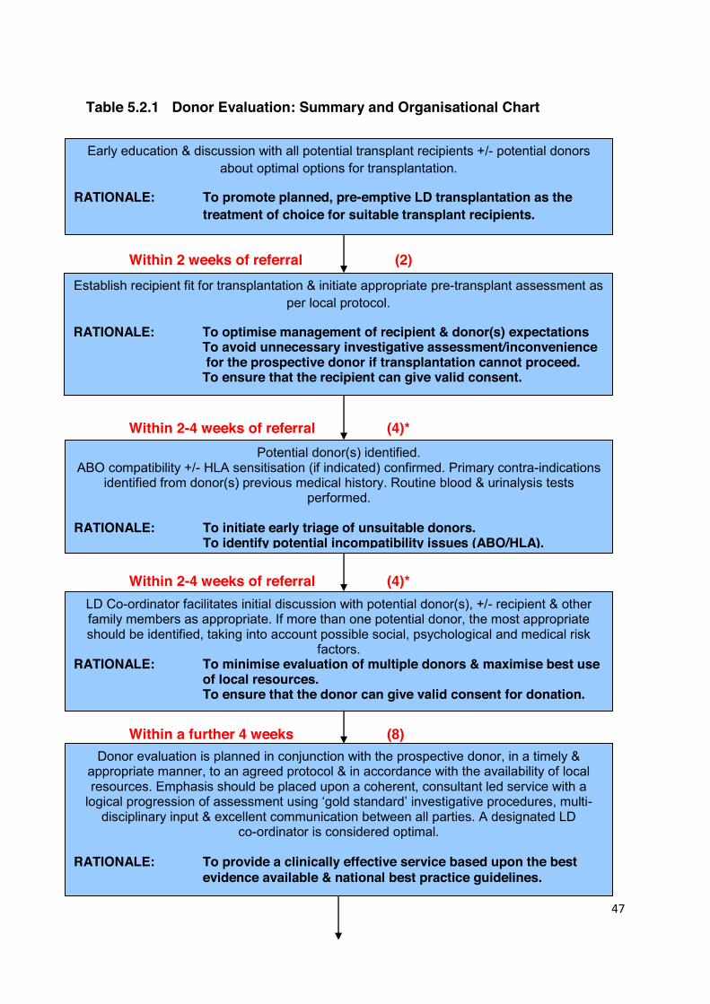
47
Table 5.2.1 Donor Evaluation: Summary and Organisational Chart
Within 2 weeks of referral (2)
Within 2-4 weeks of referral (4)*
Within 2-4 weeks of referral (4)*
Within a further 4 weeks (8)
Early education & discussion with all potential transplant recipients +/- potential donors about optimal options for transplantation.
RATIONALE: To promote planned, pre-emptive LD transplantation as the treatment of choice for suitable transplant recipients.
LD Co-ordinator facilitates initial discussion with potential donor(s), +/- recipient & other family members as appropriate. If more than one potential donor, the most appropriate should be identified, taking into account possible social, psychological and medical risk
factors. RATIONALE: To minimise evaluation of multiple donors & maximise best use of local resources. To ensure that the donor can give valid consent for donation.
Donor evaluation is planned in conjunction with the prospective donor, in a timely & appropriate manner, to an agreed protocol & in accordance with the availability of local resources. Emphasis should be placed upon a coherent, consultant led service with a logical progression of assessment using ‘gold standard’ investigative procedures, multi-
disciplinary input & excellent communication between all parties. A designated LD co-ordinator is considered optimal.
RATIONALE: To provide a clinically effective service based upon the best evidence available & national best practice guidelines.
Potential donor(s) identified. ABO compatibility +/- HLA sensitisation (if indicated) confirmed. Primary contra-indications
identified from donor(s) previous medical history. Routine blood & urinalysis tests performed.
RATIONALE: To initiate early triage of unsuitable donors. To identify potential incompatibility issues (ABO/HLA).
Establish recipient fit for transplantation & initiate appropriate pre-transplant assessment as per local protocol.
RATIONALE: To optimise management of recipient & donor(s) expectations To avoid unnecessary investigative assessment/inconvenience
for the prospective donor if transplantation cannot proceed. To ensure that the recipient can give valid consent.

48
Table 5.2.1 Donor Evaluation: Summary and Organisational Chart (Continued)
Within 2 weeks of investigations (10)
2-3 weeks from investigations (11)*
3-5 wks from completion of assessment
5-7 weeks from completion of assessment (18)*
*Maximum number of weeks
Results review by members of the MDT & feedback to the donor. RATIONALE: To ensure continuity & keep the donor informed.
Suitable donor & recipient pair referred for final pre-operative discussion with Consultant Nephrologist and Transplant Surgeon, & Independent Assessment for Human Tissue Authority. Date of transplant agreed.
RATIONALE: To ensure transplant can legally proceed & that both donor & recipient can provide valid consent for surgery.
Final cross match within the 2 weeks prior to Tx + routine pre-op investigations/pre-admission visit.
RATIONALE: To ensure transplant can proceed safely.
OPERATION
LD Co-ordinator maintains contact with donor & facilitates life-long follow-up arrangements.
RATIONALE: To provide continuing support to the donor & inform the UK Living Donor Registry.
If donor unsuitable, follow-up consultation arranged.
RATIONALE: To offer opportunity to discuss results & arrange appropriate follow-up.
49
5.3 ABO BLOOD GROUPING AND CROSSMATCH TESTING Statements of Recommendation
Where an ABO compatible donor-recipient pair is available, this is the preferred option. (A1)
Where low antibody titre ABO incompatibility is present, transplantation is
not precluded, but should be performed in a unit with the relevant experience and appropriate support. (A1)
ABO blood grouping is an important early screening test as it allows the early identification
of individuals who cannot donate because of blood group incompatibility (1). It may be
undertaken by the GP, nephrologist, specialist nurse, or at a transplant assessment clinic.
If blood group compatibility is established, initial HLA typing +/- crossmatch testing should
be performed in accordance with the recommendations in Chapter 7.
If the donor and recipient are not blood group compatible, the usual next step would be to
enquire whether there are other potential donors. One specific exemption would be in the
case of a blood group A2 donor wishing to donate to a blood group O recipient, where a
low titre of anti-A2 antibody may not preclude transplantation. Similarly, where low titres of
anti-A and/or anti-B antibody are present in other incompatible blood group combinations,
living donation may still be possible, but specialist assessment is required. ABO and HLA-
incompatible transplantation are considered in Chapter 7 and in more detail in separate
guidelines (British Transplantation Society Guidelines for Antibody Incompatible
Transplantation, http://bts.demo.eibs.co.uk/transplantation/standards-and-guidelines/).
Reference
1. Alexandre GPJ, Latinne D, Carlier M, et al. ABO-incompatibility and organ
transplantation. Transplant Rev 1991; 5: 230-41.

50
5.4 MEDICAL ASSESSMENT
It is important to manage the expectations of the donor from the outset and to emphasise
the difference between a healthy individual and one who is suitable to donate. For
example, a donor may be precluded from donation on the grounds of having a single
kidney or short renal vessels, neither of which may be detrimental to his/her own health.
The assessment may reveal previously undiagnosed disease, and potential donors must
be warned of this possibility. In addition, the existence of a previously unrecognised
condition may prejudice future attempts to obtain life insurance or specialist employment.
Conversely, screening may benefit the potential donor in that early detection of a health
problem can occur, which might otherwise have gone undiagnosed.
A full medical history must be taken and the areas listed in Tables 5.4.1 should be
specifically addressed and followed up where appropriate. The history should also aim to
identify any risk of latent or current infection in the donor that could be transmitted to the
recipient by a kidney allograft (see Table 5.4.2 and section 5.14). Importantly, all female
potential donors of childbearing age must be counselled regarding the need to take
contraceptive precautions when considering organ donation, and the possible implications
of kidney donation upon future pregnancy (see also section 10.3 Pregnancy following
Kidney Donation). Where several potential living donors are available, it may be
preferable to consider an alternative donor before assessing a woman who may still wish
to bear children or who has young dependents; although neither are an absolute
contraindication to donation.
A thorough clinical examination must be performed, taking particular account of the
cardiovascular and respiratory systems and including the assessments listed in Table
5.4.3.
In most units, donor assessment will be arranged by a specialist transplant nurse,
supported by a clinician. The clinician should undertake the medical examination of the
potential donor and, as previously noted, should not be exposed to a potential conflict of
interest by also having direct care of the transplant recipient (1). Table 5.4.4 details the
routine screening investigations that should be performed on the potential donor.
51
Reference 1. The Council of the Transplantation Society. Commercialisation in transplantation: the
problems and some guidelines for practice. Lancet 1985; 2: 715-6.
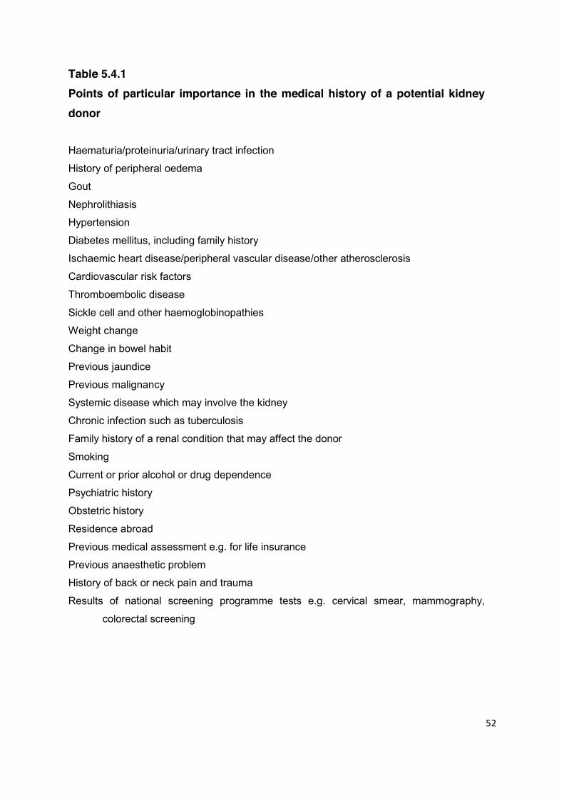
52
Table 5.4.1 Points of particular importance in the medical history of a potential kidney donor Haematuria/proteinuria/urinary tract infection
History of peripheral oedema
Gout
Nephrolithiasis
Hypertension
Diabetes mellitus, including family history
Ischaemic heart disease/peripheral vascular disease/other atherosclerosis
Cardiovascular risk factors
Thromboembolic disease
Sickle cell and other haemoglobinopathies
Weight change
Change in bowel habit
Previous jaundice
Previous malignancy
Systemic disease which may involve the kidney
Chronic infection such as tuberculosis
Family history of a renal condition that may affect the donor
Smoking
Current or prior alcohol or drug dependence
Psychiatric history
Obstetric history
Residence abroad
Previous medical assessment e.g. for life insurance
Previous anaesthetic problem
History of back or neck pain and trauma
Results of national screening programme tests e.g. cervical smear, mammography,
colorectal screening

53
Table 5.4.2 History with respect to transmissible infection
Previous illnesses
Jaundice or hepatitis
Malaria
Previous blood transfusion
Tuberculosis / atypical mycobacterium
Family history of tuberculosis
Family history of Creutzfeldt-Jakob disease (CJD), previous treatment with natural growth
hormone, or undiagnosed degenerative neurological disorder
Specific geographical risk factors: e.g. fungi and parasites, tuberculosis, hepatitis, malaria,
worms
Increased risk of HIV, HTLV1 and HTLV2 infection
History of intravenous drug use
History of infectious hepatitis or syphilis
Tattoo or skin piercing within last 6 months
Sexual partner of drug addict
Sexual partner of an HIV positive individual
Female sexual partner of man who has had sex with another man
Sexual partner of an indigenous African within the last year
Payment for, or been paid for sex within the last year
Male homosexual
Haemophiliac or sexual partner of haemophiliac
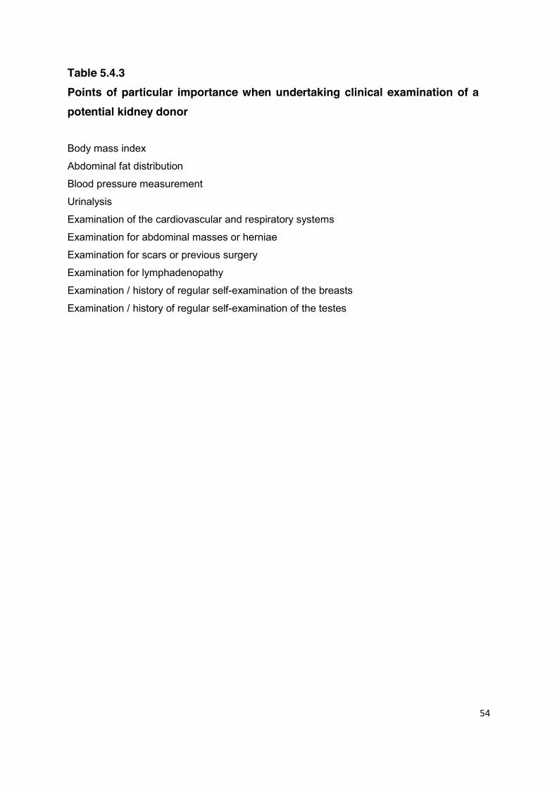
54
Table 5.4.3 Points of particular importance when undertaking clinical examination of a potential kidney donor Body mass index
Abdominal fat distribution
Blood pressure measurement
Urinalysis
Examination of the cardiovascular and respiratory systems
Examination for abdominal masses or herniae
Examination for scars or previous surgery
Examination for lymphadenopathy
Examination / history of regular self-examination of the breasts
Examination / history of regular self-examination of the testes
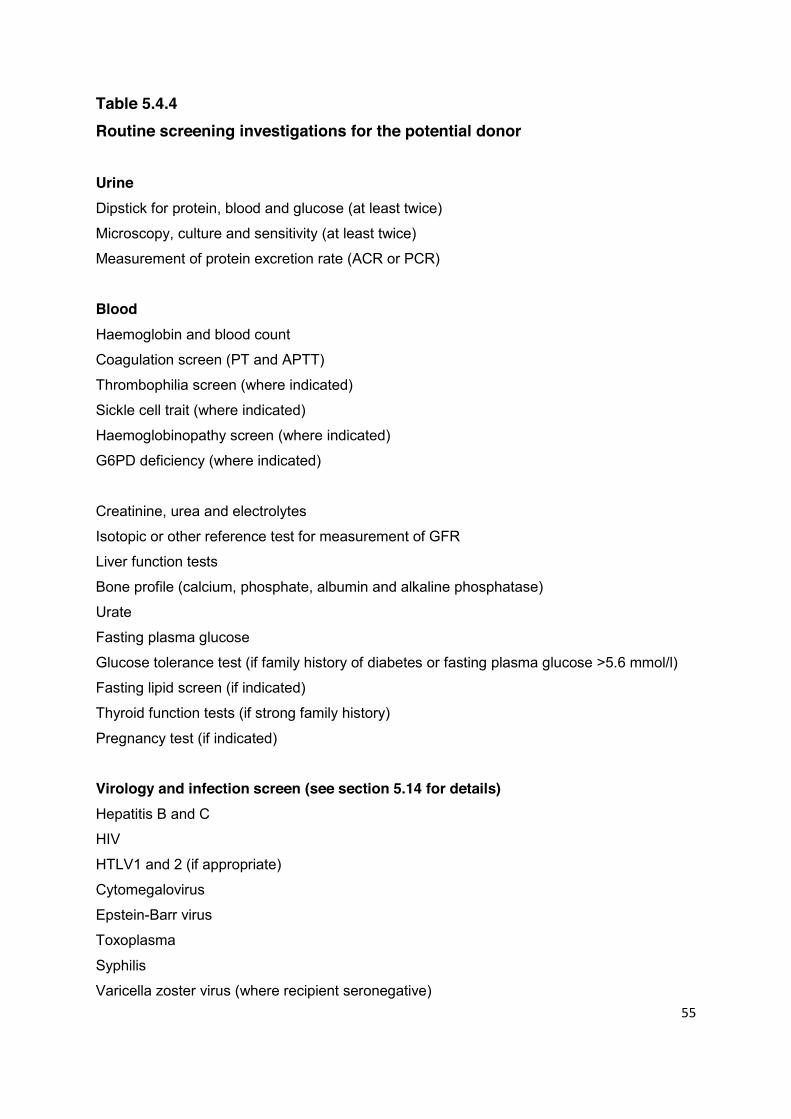
55
Table 5.4.4 Routine screening investigations for the potential donor Urine Dipstick for protein, blood and glucose (at least twice)
Microscopy, culture and sensitivity (at least twice)
Measurement of protein excretion rate (ACR or PCR)
Blood Haemoglobin and blood count
Coagulation screen (PT and APTT)
Thrombophilia screen (where indicated)
Sickle cell trait (where indicated)
Haemoglobinopathy screen (where indicated)
G6PD deficiency (where indicated)
Creatinine, urea and electrolytes
Isotopic or other reference test for measurement of GFR
Liver function tests
Bone profile (calcium, phosphate, albumin and alkaline phosphatase)
Urate
Fasting plasma glucose
Glucose tolerance test (if family history of diabetes or fasting plasma glucose >5.6 mmol/l)
Fasting lipid screen (if indicated)
Thyroid function tests (if strong family history)
Pregnancy test (if indicated)
Virology and infection screen (see section 5.14 for details) Hepatitis B and C
HIV
HTLV1 and 2 (if appropriate)
Cytomegalovirus
Epstein-Barr virus
Toxoplasma
Syphilis
Varicella zoster virus (where recipient seronegative)
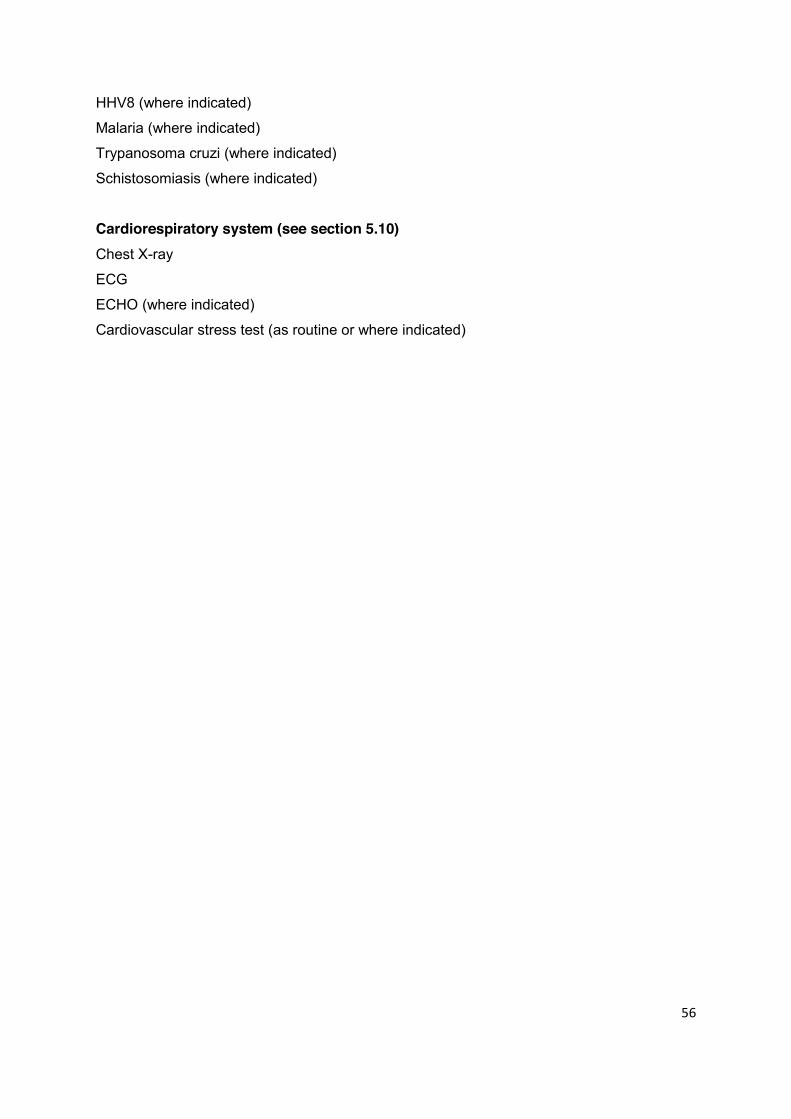
56
HHV8 (where indicated)
Malaria (where indicated)
Trypanosoma cruzi (where indicated)
Schistosomiasis (where indicated)
Cardiorespiratory system (see section 5.10) Chest X-ray
ECG
ECHO (where indicated)
Cardiovascular stress test (as routine or where indicated)
57
5.5 ASSESSMENT OF RENAL FUNCTION Statements of Recommendation
GFR should be measured using measured using a reference GFR procedure e.g. 51Cr EDTA. A prospective donor should not be considered for donation if the corrected GFR is predicted to fall below a satisfactory level of kidney function within the lifetime of the donor. A predicted GFR of at least 37.5 ml/min/1.73m2 at the age of 80 is recommended as a minimum standard. There is a lack of evidence to guide acceptable levels of kidney function for donors over 60 years of age. (B1)
A living kidney donor with normal renal function prior to donation is at no greater risk than an individual in the general population of developing end stage renal disease after unilateral nephrectomy. Measurement of eGFR in living donors has not been validated to predict the risk of long-term kidney disease and should not be used in this context. (B1)
The first principle underlying the assessment of kidney function in the potential living
donor is to ensure that the donor will have sufficient kidney function after donation such
that they will remain in good health in the future. Alongside the need to ensure adequate
residual kidney function in the donor, accurate measurement of renal function is important
to secure sufficient graft function in the recipient following transplantation.
There are now long term data to inform this process. A measurement of kidney function
was performed on a selected group from 2,949 (out of a total of 3,404) patients who had
donated over a 40 year period (1). The original requirement to qualify for donation was a
GFR of greater than 80 ml/min/1.73m2. The majority of individuals (85.5%) had a
clearance of greater than 60 ml/min/1.73m2 on follow-up, and none were below 30
ml/min/1.73m2. In a small representative sample of donors, the rate of decline of renal
function was 0.6 +/- 3.8 ml/min/1.73m2 per year, this being measured on average 12 years
after donation with two samples three years apart. There are some caveats in the
interpretation of these data, including the fact that the population was predominantly
Caucasian. However, they strongly support the view that a measured GFR over
80 ml/min/1.73m2 provides sufficient kidney function not to cause ill health in the future.
58
These data do not address the changes in kidney function that occur with age. Data from
cross-sectional studies show that there is a wide range of ‘normal’ renal function and that
this declines in a predictable manner beyond 40 years of age. When evaluated according
to the British Nuclear Medicine Society Guidelines (2), the mean GFR in young adults of
both sexes is 103 ml/min/1.73m2 with a decline of 0.9 ml/min/1.73m2 per year after the
age of 40 (3). A review of the change in kidney function with age in living donors suggests
between 0.4-0.8 ml/min/1.73m2 per year with increasing age (4). It is important to note that
the individuals in this study were being assessed as potential living donors and as such
were in good health. Overall, for the purposes of calculation, the use of an estimated rate
of change of 0.9 ml/min/1.73m2 per year is at the cautious end of the values reported.
Following donation there is a compensatory increase in function in the remaining kidney.
Across a broad age range (19-61 years), the remnant kidney increases its filtration to
provide a GFR of approximately 75% of the combined value that both kidneys had before
donation (5). Special consideration may be needed when assessing kidney function in
older donors as the degree of recovery of post-nephrectomy GFR may be less than that
for younger people, and there is not a significant body of evidence available for patients
over the age of 60.
Any guideline for donor GFR must be based upon the premise that an individual in his or
her lifetime will not develop clinically significant renal impairment as a result of unilateral
nephrectomy. On this basis, the potential kidney donor must have sufficient kidney
function prior to donation to have an effective GFR at the age of 80 years, independent of
the age at which he or she donated. Table 5.5.1 gives the values for GFR and age that
will leave a GFR of 37.5 ml/min/1.73m2 at the age of 80, given the reduction in GFR due to
donation and a cautious estimate of the rate of annual decline, as above. This threshold is
shown plotted as the red line in Figure 5.5.1. This correlates closely with previous
guidance based on smaller studies, which has supported practice to date (6). The graph
has been adjusted from the previous BTS Guideline to use a baseline age unrestricted
GFR of 80 ml/min/1.73m2. The calculation for decline from this point is based on a rate of
loss of kidney function at 0.9 ml/min/1.73 m2 per year.

59
Table 5.5.1 Acceptable GFR by donor age prior to donation Donor age (years) Acceptable corrected GFR prior to
donation (ml/min/1.73m2)
Up to 46 80
50 77
60 68
70 59
80 50
Figure 5.5.1 Acceptable GFR by donor age prior to donation
Figure 5.5.1 Diagram showing the variation with age of mean GFR. The red (upper) line
reference plot is based on an analysis of data for 428 living renal transplant donors who
had 51Cr-ETDA GFR measurements performed according to the method described in the
British Nuclear Medicine Society GFR guidelines (2). The blue (lower) line shows the
safety limit of 80 ml/min/1.73m2 (1) for adults up the age of 46 years and declining to 50
ml/min/1.73m2 at age 80. For transplant donors with pre-operative GFR values above the
blue line, the GFR of the remaining kidney will still be greater than 37.5ml/min/1.73 m2 at
age 80.
0
20
40
60
80
100
120
10 20 30 40 50 60 70 80 Age (years)
Safe to donate
Mean GFR
GFR (ml/min/1.73m2)
60
The most accurate assessment of glomerular filtration rate (GFR) is achieved using
radioisotopes such as 51Cr-EDTA or the use of iohexol clearance techniques, and these
are recommended in all potential donors. Alternative methods based upon serum
creatinine concentration are not sufficiently accurate in this context and measured
creatinine clearance, using timed urine collections, is susceptible to considerable
inaccuracy.
Divided Renal Function Divided renal function can be measured by combining a 51Cr-EDTA GFR measurement
with a 99mTc-DMSA scan of the kidneys (7). This information is advisable before
nephrectomy if there is considerable disparity in the size of the kidneys or anatomical
abnormality is noted, but is otherwise not indicated. When renal function is normal but
there is a significant (>10%) difference in function between the two kidneys, the kidney
with lower function should normally be used for transplantation.
End Stage Kidney Disease Post Nephrectomy End stage kidney disease following donation may be a consequence of issues unrelated
to the individual possessing a single kidney. A large cohort of kidney donors followed up in
Minnesota demonstrated that the cause for end stage disease in 11 individuals out of the
cohort of 3,404 was variable but that some were related to subsequent medical
conditions. The overall rate of end stage kidney disease was 180 per million persons per
year, as compared to the control adjusted rate of 268 per million per year (1).
The issue of eGFR use in living donors has been considered (8). It is important to note
that the original use of eGFR was to identify the risk of developing end stage kidney
disease in population studies, and that it has not been formally validated in living donors.
However, the data discussed above show that the risk of end stage kidney disease is not
increased in the donor population (1), and these and other observations suggest that the
eGFR overestimates the decline in kidney function when compared to formal assessment
(8).
References
1. Ibrahim HN, Foley R, Tan LP, et al. Long-term consequences of kidney donation.
N Engl J Med 2009; 360: 459-69.

61
2. Fleming JS, Zivanovic MA, Blake GM, Burniston M, Cosgriff PS. Guidelines for the
measurement of glomerular filtration rate using plasma sampling. Nucl Med Commun
2004; 25: 759-69.
3. Grewal GS, Blake GM. Reference data for 51Cr-EDTA measurements of GFR derived
from live kidney donors. Nucl Med Commun 2005; 26: 61-5.
4. Poggio E, Braun WE, Davis C. The Science of Stewardship: due diligence for kidney
donors and kidney function in living donation – evaluation, determinants and
implications for outcomes. Clin J Am Soc Nephrol 2009; 4: 1677-84.
5. Velosa JA, Griffin MD, Larson TS, et al. Can a transplanted living donor kidney
function equivalently to its native partner? Am J Transplant 2002; 2: 252-9.
6. United Kingdom Guidelines for Living Donor Kidney Transplantation. British
Transplantation Society and The Renal Association 2nd Edn April 2005.
www.bts.org.uk/transplantation/standards-and-guidelines/
7. Farmer CKT, Cook GJR, Blake GM, Reidy J, Scoble JE. Individual kidney function in
atherosclerotic nephropathy is not related to the presence of renal artery stenosis.
Nephrol Dial Transplant 1999; 14: 2880-4.
8. Tan JC, Busque S, Blouch K, Derby G, Efron B, Myers BD. Imprecision of creatinine-
based GFR estimates in uninephric kidney donors. Clin J Am Soc Nephrol 2010; 5:
497-502.

62
5.6 DONOR AGE Statements of Recommendation
Old age alone is not an absolute contraindication to donation but the medical work-up of older donors must be particularly rigorous to ensure they are suitable. (A1)
Both donor and recipient should be made aware that the older donor may be
at greater risk of peri-operative complications and that the function and possibly the long-term survival of the graft may be compromised. This is particularly evident with donors >60 years of age. (B1)
The young and the old raise different issues with respect to consideration as potential
living kidney donors (1). The ethical barriers to the use of minors and young people as
living donors are addressed in Chapter 3. For older donors the increased risk of post-
operative complications as a consequence of increased age and the potential for poorer
graft function and long term transplant outcome, as a consequence of reduced donor
GFR, must both be considered.
5.6.1 Donor Complication Rates Related to Age Early reports produced no consensus with Johnson et al reporting no increase in the
incidence of post-operative complications when older donors were used, although donor
age ≥50 years was associated with a longer post-operative stay (2). In contrast, Fauchald
reported a higher incidence of post-operative cardiac complications and pneumonia in
donors over the age of 60 years (3). Considering 80,347 living kidney donors in the US
between 1st April 1994 and 31st March 2009, Segev et al demonstrated poorer 12 year
survival for donors aged >50 years as compared to donors <40 years of age, with donors
>60 years having worse survival than those aged 50-59 years (4). However, the long-term
risk of death was no higher for older living donors than for age- and comorbidity-matched
NHANES III participants, the poorer survival therefore not being clearly attributable to
kidney donation.

63
Given the lower complication rates and faster recovery, laparoscopic nephrectomy may
have particular benefit for the older donor. Hsu et al (5) reported good outcomes following
laparoscopic nephrectomy in six donors of mean age 69.5 years (range 65-74), and
Jacobs has argued that age should not preclude laparoscopic donation on review of the
outcome of a series of 738 consecutive laparoscopic living donor nephrectomies
performed in Maryland (6). In keeping with this, some centres report higher laparoscopic
nephrectomy rates in donors >50 years (7).
When considering older donors the medical evaluation, especially that of the
cardiovascular system, needs to be particularly rigorous. Many centres consider stress
cardiac testing to be mandatory when evaluating older potential donors, particularly men
over the age of 55 years (section 5.10). Cardiopulmonary exercise testing, and in
particular definition of anaerobic threshold, has been validated as a predictor of post-
operative complications, particularly in elderly patients. If available, it may be of particular
use in the assessment of elderly donors (8).
5.6.2 Graft Outcome from Older Donors The second concern regarding the older donor is the suggestion that kidneys obtained
from older living donors have a worse outcome after transplantation (3). Renal function
declines progressively with age and kidneys from older living donors have reduced
function (9). Early studies suggested that both short-term and medium-term (5 year) graft
survival rates were similar for kidneys from older (over 55 years) and younger donors
(10,11). Kerr et al demonstrated that in the absence of rejection, graft survival at 10 years
was equivalent for donors over and under 55 years (12). However, a subsequent report of
this cohort, when 2,540 living donor kidney transplants had been performed in this centre,
documented worse outcome when the donor was >55 years of age (13). In a further study,
5 year graft survival after living donor transplantation was 76% for kidneys from donors
over 60 years (n=241) and 79% for kidneys from donors aged less than 60 years (n=518).
However, serum creatinine levels remained significantly lower in the recipients of kidneys
from younger donors and beyond 5 years their graft survival was significantly better (14).
An extensive study recently demonstrated poorer outcomes for kidneys from donors >59
years of age in 3,142 transplants performed in the UK between 2000 and 2007 (15). This
is in keeping with a Scandinavian study demonstrating no effect of donor age on
64
transplant outcome when all donors aged >50 years were considered, but poorer
outcomes in the subgroup with donor age >65 years (16). Donor GFR has been
demonstrated to be an important determinant of transplanted kidney function (17) and it
has been suggested that donor function rather than age may be the most important
determinant of outcome, although not all studies have confirmed this (16).
Older donors are more likely than younger donors to be excluded from donating on the
basis of problems discovered during the medical evaluation. However, each case should
be considered on individual merit and if the older donor is judged fit after rigorous medical
evaluation, and if the renal function of the donor is normal after correction for age and
gender, there is no compelling evidence for excluding donation on the basis of
chronological age alone (2,18,19).
References 1. Jones J, Payne WD, Matas AJ. The living donor risks, benefits, and related concerns.
Transplant Rev 1993; 7: 115-28.
2. Johnson EM, Remucal MJ, Gillingham KJ, Dahms RA, Najarian JS, Matas AJ.
Complications and risks of living donor nephrectomy. Transplantation 1997; 64:
1124-8.
3. Fauchald P, Sodal G, Albrechtsen D, Leivestad T, Berg KJ, Flatmark A. The use of
elderly living donors in renal transplantation. Transpl Int 1991; 51-3.
4. Segev DL, Muzaale AD, Caffo BS, et al. Perioperative mortality and long-term survival
following live kidney donation. JAMA 2010; 303: 959-66.
5. Hsu THS, Su L-M, Ratner LE, Kavoussi LR. Laparoscopic donor nephrectomy in the
elderly patient. Urology 2002; 60: 398-401.
6. Jacobs SC, Cho E, Foster C, Liao P, Bartlett ST. Laparoscopic donor nephrectomy:
the University of Maryland 6-year experience. J Urol 2004; 171: 47-51.
7. Johnson SR, Khwaja K, Pavlakis M, Monaco AP, Hanto DW. Older living donors
provide excellent quality kidneys: a single center experience (older living donors). Clin
Transplant 2005; 19: 600-6.
8. Hall A, Older P. Clinical review: How to identify high-risk surgical patients. Crit Care
2004, 8: 369-72.
65
9. Sumrani N, Daskalakis P, Miles AM, Hong JH, Sommer BG. The influence of donor
age on function of renal allografts from live related donors. Clin Nephrol 1993; 39:
260-4.
10. Kim YS, Kim SI, Suh JS, Park K. Use of elderly living related donors in renal
transplantation. Trans Proc 1992; 24: 1325-6.
11. Shmueli D, Nakache R, Lustig S, et al. Renal transplant from live donors over 65
years old. Trans Proc 1994; 26: 2139-40.
12. Kerr SR, Gillingham KJ, Johnson EM, Matas A. Living donors >55 years. To use or
not to use? Transplantation 1999; 67: 999-1004.
13. Matas AJ, Payne WD, Sutherland DER, et al. 2,500 living donor kidney transplants: A
single-center experience. Ann Surg 2001; 234: 149-64.
14. Kahematsu A, Tanabe K, Ishikawa N, et al. Impact of donor age on long-term graft
survival in living donor kidney transplantation. Trans Proc 1998; 30: 3118-9.
15. Fuggle SV, Allen JE, Johnson RJ, et al. Kidney Advisory Group of NHS Blood and
Transplant. Factors affecting graft and patient survival after live donor kidney
transplantation in the UK. Transplantation 2010; 89: 694-701.
16. Oien CM, Reisæter AV, Leivestad T, Dekker FW, Line PD, Os I. Living donor kidney
transplantation: The effects of donor age and gender on short- and long-term
outcomes. Transplantation 2007; 83: 600–6.
17. Hawley CM, Kearsley J, Campbell SB, et al. Estimated donor glomerular filtration rate
is the most important donor characteristic predicting graft function in recipients of
kidneys from live donors. Transpl Int 2007; 20: 64-72.
18. Kumar A, Kumar RZ, Srinadh ES, et al. Should elderly donors be accepted in live
related renal transplant programs? Clin Transplant 1994; 8: 523-6.
19. Lezaic V, Djukanov L, Blagojevic-Lazik R, et al. Living related kidney donors over 60
years old. Transpl Int 1996; 9: 109-14.
66
5.7 DONOR OBESITY Statements of Recommendation
Otherwise healthy overweight patients (BMI 25-30 kg/m2) may safely proceed to kidney donation. (B1)
Moderately obese patients (BMI 30-35 kg/m2) should undergo careful pre-
operative evaluation to exclude cardiovascular, respiratory and kidney disease. (C1)
Moderately obese patients (BMI 30-35 kg/m2) should be counselled carefully
about the increased risk of peri-operative complications, based on extrapolation of outcome data from very obese donors (BMI > 35 kg/m2
Moderately obese patients (BMI 30-35 kg/m2) should be counselled carefully about the long-term risk of kidney disease. They should be advised to lose weight prior to donation and to maintain their ideal weight following donation. (B1)
Data on the safety of kidney donation in the very obese (BMI > 35 kg/m2) are
limited and such patients should be discouraged from donating. (C1)
In 2008 almost a quarter of adults in England were classified as obese (BMI > 30 kg/m2)
(1). In the general population, obesity is associated with increased morbidity and mortality.
For a BMI of 30-35 kg/m2, the median life expectancy is reduced by 2-4 years and for a
BMI of 40-45 kg/m2, it is reduced by 8-10 years, which is comparable with the effects of
smoking (2). In comparison with individuals of normal weight, overweight and obese
individuals are at increased risk of hypertension, hypercholesterolemia, insulin resistance
and diabetes mellitus, heart disease, stroke, sleep apnoea and certain cancers (3).
Obesity is generally considered a relative contra-indication to living kidney donation
because of the increased risk of surgical complications and because of the adverse
impact of obesity on renal function in the longer term. The presence of obesity in kidney
donors is associated in some studies with an increase in peri-operative complications,

67
although these are mostly relatively minor in nature. In a single centre retrospective study
of 553 consecutive hand-assisted laparoscopic living kidney donations, those with a high
BMI (≥ 35 kg/m2) had longer operative times (mean increase 19 minutes), more minor
peri-operative complications (mostly wound complications), but the same low rate of major
surgical complications (conversion to open nephrectomy or re-operation) and a similar
length of stay (2.3 vs 2.4 days) as low BMI (< 25 kg/m2) donors (4). In a recent
retrospective cross-sectional analysis of 6,320 cases, obesity was identified in only 2% of
donors but was an independent predictor of donor risk; 28.3% of obese patients had
complications compared with 18.2% of non-obese patients (5). In another retrospective
analysis of 3,074 living kidney donors from 28 US centres during 2004 and 2005, 2.4% of
donors were obese and obesity was associated with an increase in peri-operative
complications (odds ratio 1.92), but no peri-operative mortality (6). A systematic review
and meta-analysis of ten studies, including that by Heimbach et al (4), examined 484
obese living donors with a mean BMI of 34.5 kg/m2 at donation (range 32–39 kg/m2) and
reported no deaths. It found statistically significant (but clinically insignificant) differences
in operative time, blood loss and hospital stay between obese and non-obese donors (7).
According to a recent cohort study of all (80,347) living donors during a 15-year period in
the US, 22.6% were obese (BMI ≥ 30 kg/m2) but obesity was not associated with a
statistically significant difference in surgical mortality (8). Overall, these data suggest that
laparoscopic donor nephrectomy is generally safe in otherwise healthy obese kidney
donors and does not result in a high rate of major peri-operative complications.
The principle concern for the obese living donor is the possibility that donation may have
an adverse effect on long term kidney function. Obesity associated co-morbidities, such
as hypertension, diabetes, and the metabolic syndrome, may compromise kidney function.
In addition, data suggest that obesity is independently associated with a higher risk of
developing end stage kidney disease (9). Focal glomerulosclerosis and obesity-related
glomerulopathy (glomerular enlargement and mesangial expansion) with associated
proteinuria have been described in patients with severe obesity (10), and this may be
reversible with weight loss. Obesity is also a risk factor for renal insufficiency after
unilateral nephrectomy. At 10 years post-nephrectomy, 60% of patients whose BMI was
> 30 kg/m2 at the time of nephrectomy developed proteinuria (> 3 g/day) and 30%
developed renal insufficiency (creatinine clearance < 70 ml/min) (11). These data suggest
that nephrectomy in obese patients increases the risk of developing proteinuria and/or
renal insufficiency.

68
Individual risk for developing obesity increases with time, both in the general population
and in living kidney donors. Weight gain post-donation is a common observation,
particularly in those who are overweight prior to donation (12). At mean follow-up of 12
years post-donation, a higher BMI was associated with both hypertension and a GFR that
was lower than 60 ml/min/1.73m2 (13). In a recent retrospective analysis, kidney function
in 98 obese (BMI > 30 kg/m2) and non-obese (BMI < 30 kg/m2) patients who donated a
kidney 5 to 40 years previously was similar, though both donor groups had reduced
kidney function compared with BMI-matched two-kidney control subjects (14). Obesity
was associated with a higher risk of hypertension and dyslipidaemia in both donors and
control subjects. In a study of 39 African American living kidney donors 4 to 10 years post-
donation, 8 subjects whose BMI was > 35 kg/m2 were found to have a significantly greater
fall in eGFR (MDRD) than those with BMI < 35 kg/m2 (40 and 28 ml/min/1.73m2
respectively) (15). However, in a different retrospective cohort study using OPTN data
from 5,304 donors among whom 40% were overweight (BMI > 25 kg/m2), 18% were
obese (BMI > 30 kg/m2) and 5% were very obese (BMI > 35 kg/m2), the decline in eGFR
from baseline and percentage change in creatinine at 6 months did not differ significantly
across the three groups (16). In a very recent study of 36 obese living kidney donors 7
years post-donation, 47% had an eGFR below 60 ml/min/1.73m2, 42% were hypertensive
and 19% had microalbuminuria (17). There was no control group in this study.
These findings support the current practice of using otherwise healthy overweight (BMI
25-30 kg/m2) and moderately obese (BMI 30-35 kg/m2) donors, although there are few
studies that address long-term health outcomes for the very obese (BMI > 35 kg/m2). Pre-
donation counselling should include a careful discussion of the uncertain long-term risks
of donation in obese individuals along with advice about weight maintenance following
donation.
References
1. Statistics on obesity, physical activity and diet: England, 2010. NHS Health and Social
Care Information Centre, February 2010. www.ic.nhs.uk/pubs/opad10
2. Whitlock G, Lewington S, Sherliker P, et al. Body-mass index and cause-specific
mortality in 900 000 adults: collaborative analyses of 57 prospective studies. Lancet
2009; 373: 1083-96.

69
3. Nguyen NT, Magno CP, Lane KT, Hinojosa MW, Lane JS. Association of
hypertension, diabetes, dyslipidemia, and metabolic syndrome with obesity: findings
from the national health and nutrition examination survey, 1999 to 2004. J Am Coll
Surg 2008; 207: 928-34.
4. Heimbach JK, Taler SJ, Prieto M, et al. Obesity in living kidney donors: clinical
characteristics and outcomes in the era of laparoscopic donor nephrectomy. Am J
Transplant 2005; 5: 1057-64.
5. Friedman AL, Cheung K, Roman SA, Sosa JA. Early clinical and economic outcomes
of patients undergoing living donor nephrectomy in the United States. Arch Surg
2010; 145: 356-62.
6. Patel S, Cassuto J, Orloff M, et al. Minimizing morbidity of organ donation: analysis of
factors for perioperative complications after living-donor nephrectomy in the United
States. Transplantation 2008; 85: 561-5.
7. Young A, Storsley L, Garg AX, et al. Health outcomes for living kidney donors with
isolated medical abnormalities: a systematic review. Am J Transplant 2008; 8:
1878-90.
8. Segev DL, Muzaale AD, Caffo BS, et al. Perioperative mortality and long-term
survival following live kidney donation. JAMA 2010; 303: 959-66.
9. Hsu CY, McCulloch CE, Iribarren C, Darbinian J, Go AS. Body mass index and risk
for end-stage renal disease. Ann Intern Med 2006; 144: 21-8.
10. Kambham N, Marcowitz GS, Valeri AM, Lin J, D'Agati VD. Obesity related
glomerulopathy; an emerging epidemic. Kidney Int 2001; 59: 1498-509.
11. Praga M, Hernandez E, Herrero JC, et al. Influence of obesity on the appearance of
proteinuria and renal insufficiency after unilateral nephrectomy. Kidney Int 2000; 58:
2111-8.
12. Torres VE, Offord KP, Anderson CF, et al. Blood pressure determinants in living-
related renal allograft donors and their recipients. Kidney Int 1987; 31: 1383-90. 13. Ibrahim HN, Foley R, Tan L, et al. Long-term consequences of kidney donation. N
Engl J Med 2009; 360: 459-69. 14. Tavakol MM, Vincenti FG, Assadi H, Frederick MJ, Tomlanovich SJ, Roberts JP,
Posselt AM. Long-term renal function and cardiovascular disease risk in obese kidney
donors. Clin J Am Soc Nephrol 2009; 4: 1230-8.
15. Nogueira JM, Weir MR, Jacobs S, et al. A study of renal outcomes in African
American living kidney donors. Transplantation 2009; 88: 1371-6.

70
16. Reese PP, Feldman HI, Asch DA, Thomasson A, Shults J, Bloom RD. Short-term
outcomes for obese live kidney donors and their recipients. Transplantation 2009; 88:
662-71.
17. Nogueira JM, Weir MR, Jacobs S, et al. A study of renal outcomes in obese living
kidney donors. Transplantation 2010; 90: 993-9.
71
5.8 HYPERTENSION IN THE DONOR
Statements of Recommendation
Potential donors with blood pressure <140/90 mmHg should be considered as normotensive and therefore suitable for nephrectomy on the basis of blood pressure. (B1)
Potential donors with ‘high normal’ blood pressure (>130/85 mmHg) should
be warned about the greater future risk of developing hypertension and associated cardiovascular events and the need for monitoring (which should be recommended irrespective of nephrectomy). Additional assessment (24 hour blood pressure monitoring) should be considered but is not required. (B1)
The definition and treatment of hypertension in prospective donors should
follow the British Hypertension Society guidelines. (B1)
Office blood pressure measurements are sufficient for the assessment of the majority of potential donors. Ambulatory blood pressure monitoring should be considered for potential donors who have hypertension (blood pressure greater than 140/90 mmHg or who are taking pharmacological treatment for hypertension) and if this is normal (see below) donor nephrectomy is not precluded. (B1)
Living kidney donors should be encouraged to minimise the risk of
hypertension and its consequences by lifestyle measures including smoking cessation, frequent exercise and, where appropriate, weight loss. (B1)
Prospective donors should be warned about the potential risks of
hypertension, particularly if in a high risk group. Blood pressure measurement should be part of annual donor monitoring. (B1)
The presence of mild-moderate hypertension that is controlled with 1-2
antihypertensive agents is not a contraindication to kidney donation providing significant end organ damage has been excluded. (B1)
72
Evidence of hypertensive end organ damage, poorly controlled hypertension, or hypertension that requires more than two drugs to achieve adequate control are relative contraindications to donor nephrectomy. (C2)
Donors who develop hypertension should be managed according to British
Hypertension Society guidelines and are at similar risk of developing complications as other patients with hypertension. (B1)
Hypertension is one of the commonest reasons for declaring a potential kidney donor
medically unsuitable (1). There are two concerns with hypertension in the potential donor.
The first is that the hypertension presents a risk for peri-operative morbidity and mortality.
The second is that pre-existing hypertension in the donor will be worsened by unilateral
nephrectomy and this will be associated with an unacceptable increase in long-term
cardiovascular risk. This guideline concentrates on the second of these concerns.
The paucity of high quality evidence in this field and the fact that the relationship between
pre-donation blood pressure and subsequent hypertension is likely to be continuous mean
it is not possible to give a precise pre-operative blood pressure level below which donation
is safe and above which donation should not be considered. As with other aspects of
living donation, the consideration of potential risk also must take account of the age of the
donor in that the older donor will have a higher absolute annual risk, but this will be spread
over a shorter remaining life-span. Each case needs to be considered individually, bearing
in mind that many potential donors may be willing to accept a higher risk of developing
hypertension than their transplant professionals (2).
5.8.1 Definition of Hypertension in the Donor There is a general consensus from the Joint National Committee on Prevention, Detection
and Treatment of High Blood Pressure (3), British Hypertension Society (4) and European
Society of Hypertension (5) that, in the absence of other cardiovascular risk factors or end
organ damage, adults with a blood pressure above 140/90 mmHg should be considered
hypertensive. All guidelines agree that a blood pressure above 140/90 mmHg requires
further assessment and/or treatment. British guidelines recommend treatment of all
73
patients with blood pressure above 160/100 mmHg and those with end organ damage or
high cardiovascular risk ( 20% in 10 years) if the blood pressure is above 140/90 mmHg.
Although blood pressure increases with age (6), guidelines for the diagnosis and
treatment of hypertension are applied to all age groups. As increasing numbers of older
donors are being considered, it is likely more potential donors with a blood pressure
above 140/90 mmHg will be assessed. This level of blood pressure should not preclude
further evaluation.
In addition, it is evident that the risk of cardiovascular mortality increases with blood
pressure values that are still within the normal range. The Joint National Committee
reports that cardiovascular risk doubles for every 20/10 mmHg rise in blood pressure
above 115/75 mmHg. This has in part led to the classification of blood pressure above
130/85 mmHg as ‘high normal’ (4) and recognition of a need to monitor these patients
because of the future risk of developing hypertension. There is no evidence that ‘high
normal’ blood pressure is a contra-indication to donor nephrectomy but these donors
should be informed of the high lifetime risk of developing hypertension irrespective of
nephrectomy, and therefore the need for follow-up.
5.8.2 Method of Blood Pressure Measurement Most of the large population based studies of cardiovascular risk have relied upon office
blood pressure measurements. There is no evidence to suggest that office blood pressure
measurements will be a less accurate predictor of cardiovascular risk in potential donors
undergoing nephrectomy and therefore this method should be used for the standard
measurement of blood pressure. Some individuals will exhibit a stress response (white
coat hypertension) that may lead to an incorrect diagnosis of hypertension (7). In this
situation, 24-hour ambulatory blood pressure monitoring (ABPM) may be useful. The
British and European guidelines define hypertension using ABPM as a 24-hour mean
blood pressure >125/80 mmHg (4,5) and the American guidelines as awake blood
pressure >135/85 mmHg and asleep >120/75 mmHg (3). In a study of 238 potential
donors, 36.7% were classified as hypertensive based on office measurements. However,
this proportion decreased to 11% when ABPM was used for assessment (hypertension
defined as awake blood pressure >135/85 mmHg). This discrepancy was most marked in
74
older donors (8). These data would support the use of ABPM in the assessment of
potential donors with hypertension based on office measurements.
ABPM can predict both hypertensive end organ damage and cardiovascular risk, perhaps
more accurately than office blood pressure (9,10). Ozdemir et al suggested that ABPM
was more sensitive at identifying hypertension in potential donors than office blood
pressure measurements (11). However, there is little evidence to support the routine use
of ABPM to assess potential donors who are normotensive on initial office blood pressure
measurements.
5.8.3 Risk of Developing Hypertension Post Donation There are no conclusive data that unilateral nephrectomy increases the risk of developing
hypertension and in a recent survey only 50% of transplant professionals believed that
hypertension develops after nephrectomy (12). There are no good prospective controlled
studies to assess the risk of hypertension after donation. Several small studies have failed
to show an increased risk, possibly because they were underpowered. Nevertheless,
overall cardiovascular risk should be considered in all potential donors and measures
taken to reduce risk irrespective of baseline blood pressure.
The reported incidence of hypertension after unilateral nephrectomy varies significantly
from 9-75% (13-16). Several larger studies with varying duration of follow-up suggest that
approximately one third of donors will develop hypertension (17-19). Although this rate is
high, these studies do not quote the incidence of hypertension in control populations and
therefore it is not possible to determine whether there is any excess risk attributable to
unilateral nephrectomy. Even if controlled data were available, it would not account for the
element of screening involved in living donor selection.
A large database study from the US which involved 3,698 donors concluded that the rate
of hypertension in donors was similar to the general population (18). In contrast, a similar
study from Ontario suggested that donors were more frequently diagnosed with
hypertension (16.3% vs 11.9%) (20). Several small studies have suggested an increase in
the incidence of hypertension after unilateral nephrectomy when compared to a control
population (16,21,22). However, larger studies have failed to reproduce this finding
75
(18,23,24). In addition, no difference was found when the incidence of hypertension was
compared in kidney donors and their siblings (25).
Two meta-analyses have considered the effect of unilateral nephrectomy on hypertension.
The first in 1995 reported a small increase in both systolic and diastolic pressures post
nephrectomy (2.4 and 3.1 mmHg respectively) but no increase in the incidence of
hypertension compared to controls (26). A more recent meta-analysis performed in 2006
suggested that blood pressure may rise by 5 mmHg in the first 5-10 years post donation
(27).
It is clear that the risk of developing hypertension after kidney donation is influenced by
pre-donation characteristics including pre-donation blood pressure, body mass index and
age (18,19). Higher risk groups should be warned of the higher risk of developing
hypertension and the need for monitoring, although it is unclear whether this risk is greater
than in an appropriately matched ‘two kidney’ control group (28).
There are few data on the long-term outcome of nephrectomy in ethnic groups, which may
be at greater risk of developing complicated hypertension. In a small study of African
American donors, the incidence of hypertension was 41% at a mean follow-up of 7.1
years after donation, although this rate was not compared to an age and sex matched
control population (29). A larger retrospective study of US donors with a mean 7.7 year
follow-up showed that black and Hispanic donors had an increased risk of hypertension as
compared with white donors (adjusted hazard ratio 1.52) (30).
5.8.4 Pre-existing Hypertension in the Donor There is relatively little information on the influence of nephrectomy in patients with pre-
existing hypertension. However, it is generally accepted that the presence of hypertensive
end organ damage (left ventricular hypertrophy on echocardiography), uncontrolled
hypertension, or hypertension that requires polytherapy to achieve adequate control are
contraindications to donor nephrectomy. Since it is unlikely that donor nephrectomy will be
performed in these circumstances, evidence to support this practice will not be generated
in the living donor setting.
76
Evidence is also sparse for potential donors who present with less severe hypertension
and it is difficult to draw definite conclusions from the available literature (reviewed by
Young et al) (31). This is a common scenario and will be become increasingly common as
older donors are considered. In a series by Textor et al (published only in abstract), 58
patients with hypertension controlled on 1 or 2 agents underwent nephrectomy (32). There
were no increased risks to the donor identified (renal function, proteinuria and
hypertension). In a smaller series of patients reported by the same group, 24 patients with
hypertension (>140/90 mmHg) underwent donor nephrectomy. Pre-existing hypertension
did not have an adverse effect on outcome with no evidence of higher blood pressure or
renal injury after nephrectomy (33). These reports suggest that potential donors with mild
or moderate hypertension should be considered suitable for nephrectomy, particularly if
the blood pressure is controlled with non-pharmacological methods and 1 or 2
antihypertensive agents.
Potential donors with hypertension should have this confirmed by either repeated office
measures or ABPM. If confirmed, non-pharmacological interventions should be
recommended and drug treatment initiated if required. If adequate blood pressure control
is achieved or if the long-term cardiovascular risk is deemed acceptable by both patient
and assessor, the donor can proceed to nephrectomy.
The target blood pressure in a potential donor should be the same as the general
population. A large population study suggested that the greatest reduction in
cardiovascular risk was achieved with the diastolic blood pressure below 85 mmHg (34)
and therefore the British Hypertension Society recommends an optimal treatment target of
<140/85 mmHg (4).
5.8.5 Management of Hypertension following Donor Nephrectomy Hypertension will develop in at least 30% of patients following unilateral nephrectomy.
Several studies have reported longitudinal data on patients after unilateral nephrectomy
including renal function, albuminuria and blood pressure. The data are conflicting with
some reports suggesting that hypertension after nephrectomy is associated with the
development of renal complications (23), but this has not been confirmed by others
(14,35). An association would be predicted because of the known relationship between
77
hypertension and renal disease. However, there is no evidence to suggest that this effect
is amplified by unilateral nephrectomy.
References 1. Fehrman-Ekholm I, Gabel H, Magnusson G. Reasons for not accepting living kidney
donors. Transplantation 1996; 61:1264-5.
2. Young A, Karpinski M, Treleaven D, et al. Differences in tolerance for health risk to
the living donor among potential donors, recipients, and transplant professionals.
Kidney Int 2008; 73: 1159-66.
3. Chobanian AV, Bakris GL, Black HR, et al. The seventh report of the Joint National
Committee on prevention, detection, evaluation, and treatment of high blood
pressure: the JNC 7 report. JAMA 2003; 289: 2560-72.
4. Williams B, Poulter NR, Brown MJ, et al. British Hypertension Society guidelines for
hypertension management 2004 (BHS-IV): summary. BMJ 2004; 328: 634-40.
5. 2003 European Society of Hypertension-European Society of Cardiology guidelines
for the management of arterial hypertension. J Hypertens 2003; 21: 1011-53.
6. Miall WE, Chinn S. Blood pressure and ageing; results of a 15-17 year follow-up
study in South Wales. Clin Sci Mol Med 1973; 45 Suppl 1: S23-33.
7. Pickering TG, James GD, Boddie C, Harshfield GA, Blank S, Laragh JH. How
common is white coat hypertension? JAMA 1988; 259: 225-8.
8. Textor SC, Taler SJ, Larson TS, et al. Blood pressure evaluation among older living
kidney donors. J Am Soc Nephrol 2003; 14: 2159-67.
9. Verdecchia P. Prognostic value of ambulatory blood pressure: current evidence and
clinical implications. Hypertension 2000; 35: 844-51.
10. Staessen JA, Thijs L, Fagard R, et al. Predicting cardiovascular risk using
conventional vs ambulatory blood pressure in older patients with systolic
hypertension. Systolic Hypertension in Europe Trial Investigators. JAMA 1999; 282:
539-46.
11. Ozdemir N, Guz G, Muderrisoglu H, et al. Ambulatory blood pressure monitoring in
potential renal transplant donors. Transplant Proc 1999; 31: 3369-70.
12. Housawi AA, Young A, Boudville N, et al. Transplant professionals vary in the long-
term medical risks they communicate to potential living kidney donors: an
international survey. Nephrol Dial Transplant 2007; 22: 3040-5.
78
13. Anderson CF, Velosa JA, Frohnert PP, et al. The risks of unilateral nephrectomy:
status of kidney donors 10 to 20 years postoperatively. Mayo Clin Proc 1985; 60:
367-74.
14. Eberhard OK, Kliem V, Offner G, et al. Assessment of long-term risks for living related
kidney donors by 24-h blood pressure monitoring and testing for microalbuminuria.
Clin Transplant 1997; 11: 415-9.
15. Miller IJ, Suthanthiran M, Riggio RR, et al. Impact of renal donation. Long-term
clinical and biochemical follow-up of living donors in a single center. Am J Med 1985;
79: 201-8.
16. Saran R, Marshall SM, Madsen R, Keavey P, Tapson JS. Long-term follow-up of
kidney donors: a longitudinal study. Nephrol Dial Transplant 1997; 12: 1615-21.
17. Fehrman-Ekholm I, Elinder CG, Stenbeck M, Tyden G, Groth CG. Kidney donors live
longer. Transplantation 1997; 64: 976-8.
18. Ibrahim HN, Foley R, Tan L, et al. Long-term consequences of kidney donation.
N Engl J Med 2009; 360: 459-69.
19. Torres VE, Offord KP, Anderson CF, et al. Blood pressure determinants in living-
related renal allograft donors and their recipients. Kidney Int 1987; 31: 1383-90.
20. Garg AX, Prasad GV, Thiessen-Philbrook HR, et al. Cardiovascular disease and
hypertension risk in living kidney donors: an analysis of health administrative data in
Ontario, Canada. Transplantation 2008; 86: 399-406.
21. Watnick TJ, Jenkins RR, Rackoff P, Baumgarten A, Bia MJ. Microalbuminuria and
hypertension in long-term renal donors. Transplantation 1988; 45: 59-65.
22. Hakim RM, Goldszer RC, Brenner BM. Hypertension and proteinuria: long-term
sequelae of uninephrectomy in humans. Kidney Int 1984; 25: 930-6.
23. Fehrman-Ekholm I, Duner F, Brink B, Tyden G, Elinder CG. No evidence of
accelerated loss of kidney function in living kidney donors: results from a cross-
sectional follow-up. Transplantation 2001; 72: 444-9.
24. Goldfarb DA, Matin SF, Braun WE, et al. Renal outcome 25 years after donor
nephrectomy. J Urol 2001; 166: 2043-7.
25. Williams SL, Oler J, Jorkasky DK. Long-term renal function in kidney donors: a
comparison of donors and their siblings. Ann Intern Med 1986; 105: 1-8.
26. Kasiske BL, Ma JZ, Louis TA, Swan SK. Long-term effects of reduced renal mass in
humans. Kidney Int 1995; 48: 814-9.
27. Boudville N, Prasad GV, Knoll, G, et al. Meta-analysis: risk for hypertension in living
kidney donors. Ann Intern Med 2006; 145: 185-96.

79
28. Tavakol MM, Vincenti FG, Assadi H, et al. Long-term renal function and
cardiovascular disease risk in obese kidney donors. Clin J Am Soc Nephrol 2009; 4:
1230-8.
29. Nogueira JM, Weir MR, Jacobs S, et al. A study of renal outcomes in African
American living kidney donors. Transplantation 2009; 88: 1371-6.
30. Lentine KL, Schnitzler MA, Xiao H, et al. Racial variation in medical outcomes among
living kidney donors. N Engl J Med 2010; 363: 724-32.
31. Young A, Storsley L, Garg AX, et al. Health outcomes for living kidney donors with
isolated medical abnormalities: a systematic review. Am J Transplant 2008; 8:
1878-90.
32. Textor SC, Taler SJ, Pierto M, et al. Hypertensive living renal donors have lower
blood pressures and urinary microalbumin one year after nephrectomy. Am J
Transplant 2003; 3 (Abstract).
33. Textor SC. Atherosclerotic renal artery stenosis: how big is the problem, and what
happens if nothing is done? J Hypertens Suppl 2005; 23: S5-13.
34. Hansson L, Zanchetti A, Carruthers SG, et al. Effects of intensive blood-pressure
lowering and low-dose aspirin in patients with hypertension: principal results of the
Hypertension Optimal Treatment (HOT) randomised trial. HOT Study Group. Lancet
1998; 351: 1755-62.
35. Praga M, Hernandez E, Herrero JC, et al. Influence of obesity on the appearance of
proteinuria and renal insufficiency after unilateral nephrectomy. Kidney Int 2000; 58:
2111-8.

80
5.9 DIABETES MELLITUS Statements of Recommendation
All potential living kidney donors must have a fasting plasma glucose level checked. A level between 5.6–6.9 mmol/l is indicative of an impaired fasting glucose state and an oral glucose tolerance test (OGTT) must be undertaken. (B1)
Prospective donors with an increased risk of Type 2 diabetes because of
family history, ethnicity or obesity should also undergo an OGTT. (B1) If OGTT reveals a persistent impaired fasting glucose and/or an impaired
glucose tolerance, then the risks of developing diabetes after donation must be carefully considered. (B1)
Consideration of patients with diabetes as potential kidney donors requires
very careful evaluation of the risks and benefits. In the absence of evidence of target organ damage and having ensured that other cardiovascular risk factors such as obesity, hypertension or hyperlipidaemia are optimally managed, diabetics can be considered for kidney donation after a thorough assessment of the lifetime risk of cardiovascular and progressive renal disease in the presence of a single kidney. (Not graded)
5.9.1 Diagnosis of Diabetes Mellitus All prospective donors should have a fasting plasma glucose measurement to exclude
diabetes mellitus. The WHO and American Diabetes Association recommend repeat
testing of fasting glucose on a different day before placing someone in a glucose
intolerant category (6). A fasting venous plasma glucose of >7.0 mmol/l indicates diabetes
mellitus (6). Fasting plasma glucose values of between 5.6 and 6.9 mmol/l indicate
impaired fasting glucose. A glucose value in this range together with a family history of
Type 2 diabetes (sibling or parental) is associated with a 30% 5-year risk of diabetes and
donation is usually contraindicated (7).

81
In the context of living donation, impaired fasting glucose is an indication for a standard
2-hour oral glucose tolerance test (OGTT). A 2-hour glucose value of >11.1 mmol/l
indicates diabetes (6). A 2-hour value between 7.8 and 11.0 mmol/l indicates impaired
glucose tolerance. Caucasians in this latter category have a 10% 5-year risk of diabetes
(7). The risk is higher for certain ethnic groups, notably individuals from Southern Asia and
the Caribbean (8).
Traditional guidance has suggested that individuals with diabetes should not donate
kidneys. However, in an observational study of 444 donors from a single Japanese centre
that has cautiously accepted subjects with an abnormal OGTT, including those with
diabetes, no difference was found in the rate of immediate post-operative complications or
survival at 20 years between the glucose tolerant and intolerant groups. Through self
reporting of status at follow-up, no major diabetic complications were observed in the
glucose intolerant group (21). Further studies are required in this area. Consideration of a
diabetic as a donor requires thorough evaluation of the risks and benefits of donation and
transplantation, for both donor and recipient. Specifically, a careful search should be
made for any evidence of target organ damage and cardiovascular risk factors such as
obesity, hypertension and hyperlipidaemia. Testing for glycosuria and measurement of
random blood glucose levels has low sensitivity for the diagnosis of diabetes (9). After
exclusion of pre-existing diabetes, the clinical risk factors for diabetes and diabetic
nephropathy should be evaluated and discussed with the potential donor (3,4).
5.9.2 Risk of Type 1 Diabetes Type 1 diabetes presents predominantly in childhood and early adulthood and 50% of
cases have presented by the age of 20 years (10). The incidence of Type 1 diabetes in
adults is less than 1 in 10,000 (10). First degree relatives of an individual with Type 1
diabetes have a 15-fold increased risk of developing the disease. Moreover, the relatives
of Type 1 diabetics with diabetic nephropathy appear to be at increased risk of
nephropathy should they subsequently develop diabetes (11). However, because Type 1
diabetes is relatively uncommon and most cases have presented before the age at which
living donation is under consideration, there is little need for concern even when there is a
family history of Type 1 diabetes. Sometimes it may be difficult to determine from the
history whether an affected family member had Type 1 or Type 2 diabetes. As a working
definition, Type 1 diabetes is characterised by onset below the age of 30 years and a
requirement for insulin treatment from the time of diagnosis.
82
5.9.3 Risk of Type 2 Diabetes Type 2 diabetes is predominantly a disease of later life and in 50% of cases Type 2
diabetes is clinically unrecognised (12). The crude prevalence of undiagnosed disease in
the Caucasian population is 2.3% (13). Individuals who have a family history (first degree
relative) of Type 2 diabetes are at higher risk of developing the disease (relative risk 3.0).
Because the prevalence of Type 2 diabetes is much higher than for Type 1, the absolute
risk of developing the disease is high (lifetime risk 38%) (14). The combination of family
history and obesity (BMI >30 kg/m2) places an individual at very high risk of diabetes in
later life (15). Individuals from South East Asia and the Caribbean are at increased risk of
Type 2 diabetes, independently of age and obesity. Individuals at high risk of Type 2
diabetes because of a positive family history and/or obesity should undergo an OGTT and
should only be considered further as donors if this is normal. For individuals with a normal
OGTT, the risk of developing Type 2 diabetes within 5 years is around 1% overall and is
modulated by ethnicity and obesity. In a large survey of living kidney donors in the United
States, Ibrahim et al found that the self reported prevalence of diabetes was 5.2% in the
2,929 patients who responded. The eGFR and the rate of decline of eGFR were not
significantly different between diabetic and matched non-diabetic donors (22).
If there is a history of transient gestational diabetes, the lifetime risk of Type 2 diabetes is
very high (16,17) and kidney donation is relatively contraindicated. An important
consideration for a potential kidney donor is the risk of developing nephropathy should
they subsequently develop Type 2 diabetes. There is a sharp increase in the incidence of
Type 2 diabetes after the age of 50 and the median age at diagnosis is around 60 years.
Less than 1% of Europeans with Type 2 diabetes develop ESRD but the incidence is
higher in other ethnic groups (18). However, there is a 50% cumulative incidence of
proteinuria after Type 2 diabetes has been present for 20 years (19) which may
reasonably become an issue for kidney donors who have an above average life
expectancy and may expect to live to their 80s (20). A prudent approach should be
adopted when assessing potential donors who are at increased risk of Type 2 diabetes.
44
References
1. Barnes DJ, Pinto JR, Viberti GC. The patient with diabetes mellitus. In: Davison AM,
Cameron JS, Grünfeld J-P, Kerr DNS, Ritz E, Winearls CG. Oxford Textbook of
Clinical Nephrology. Oxford University Press 1998; 723-75.
83
2. Mogensen CE. Microalbuminuria predicts clinical proteinuria and early mortality in
maturity onset diabetes. N Engl J Med 1984; 310: 356-60.
3. Simmons D, Searle M. Risk of diabetic nephropathy in potential living related kidney
donors. BMJ 1998; 316: 846-8.
4. Kasiske BL, Ravenscraft M, Ramos EL, Gaston RS, Bia MJ, Danovitch GM. The
evaluation of living renal transplant donors: Clinical practice guidelines. JASN 1996;
7: 2288-313.
5. Peters A, Kerner W. Perioperative management of the diabetic patient. Exp Clin
Endocrinol Diabetes 1995; 103: 213-8.
6. Alberti NJ, Zimmet PZ, for the WHO consultation. Definition, diagnosis and
classification of diabetes mellitus. Provisional report of a WHO consultation. Diabetic
Medicine 1998; 15: 539- 53.
7. Wareham NJ, Byrne CD, Williams R, Day NE, Hales CN. Fasting proinsulin
concentrations predict the development of type 2 diabetes. Diabetes Care 1999; 22:
262-70.
8. Yudkin JS, Alberti KG, McLarty DG, Swai AB. Impaired glucose tolerance. Is it a risk
factor for diabetes or a diagnostic ragbag? BMJ 1990; 301: 397-402.
9. Engelgau MM, Thompson TJ, Aubert RE, Herman WH. Screening for NIDDM in non
pregnant adults. Diabetes Care 1995; 18: 1606-18
10. Green A, Gale G. The aetiology and pathogenesis of IDDM – an epidemiological
perspective. In: Williams R, Papoz L, Fuller J, eds. Diabetes in Europe. London: John
Libbey & Company Ltd, 1994; 11-20.
11. Seaquist ER, Goetz FC, Rich S, Barbosa J. Familial clustering of diabetic kidney
disease: Evidence for genetic susceptibility to diabetic nephropathy. N Engl J Med
1989; 320: 1161-5.
12. Harris MI. Undiagnosed NIDDM: clinical and public health issues. Diabetes Care
1993; 16: 642-57.
13. Williams DRR, Wareham NJ, Brown DC, et al. Glucose intolerance in the community;
the Isle of Ely Diabetes Project. Diabetic Med 1995; 12: 30-5.
14. Pierce M, Keen H, Bradley C. Risk of diabetes in offspring of parents with non-insulin-
dependent diabetes. Diabetic Medicine 1995; 12: 6-13.
15. Morris RD, Rimm DL, Hartz AJ, Karlhoff RK, Rimm AA. Obesity and heredity in the
etiology of non-insulin-dependent diabetes mellitus in 32,662 adult white women. Am
J Epidemiol 1989; 130: 112-21.

84
16. O’Sullivan B. Subsequent morbidity among gestational diabetic women. In:
Sutherland HW, Stowers JM, eds. Carbohydrate metabolism in pregnancy and the
newborn. Edinburgh: Churchill Livingstone, 1984; 174-80.
17. Oats JN, Beischer NA, Grant PT. The emergence of diabetes and impaired glucose
tolerance in women who had gestational diabetes. In: Weiss PA, Coustan DR, eds.
Gestational diabetes. New York: Springer-Verlag, 1988; 199-210.
18. Fabre J, Balant LP, Dayer PG, Fox HM, Vernet AT. The kidney in maturity onset
diabetes mellitus: a clinical study of 510 patients. Kidney Int 1998; 35: 681-7.
19. Borch-Johnsen K. Renal disease in diabetes. In: Williams R, Papoz L, Fuller J, eds.
Diabetes in Europe. London: John Libbey & Company Ltd, 1994; 56-60.
20. Fehrman-Ekholm I, Elinder CG, Stenbeck M, Tyden G, Groth C-G. Kidney donors live
longer. Transplantation 1997; 64: 976-8.
21. Okamoto M, Suzuki T, Fujiki M, et al. The consequences for live kidney donors with
preexisting glucose intolerance without diabetic complication: analysis at a single
Japanese center. Transplantation 2010; 89: 1391-5.
22. Ibrahim HN, Kukla A, Cordner G, Bailey R, Gillingham K, Matas AJ. Diabetes after
kidney donation. Am J Transplant 2010; 10: 331-7.

85
5.10 CARDIOVASCULAR EVALUATION
Statements of Recommendation
A low threshold should be set for screening potential living donors for cardiovascular disease, and for their exclusion from donation. (B1)
Potential donors with an exercise capacity of <4 METS or >10% estimated risk of significant coronary atherosclerosis should undergo formal cardiological assessment. (B1)
Potential donors with exercise capacity >10 METS are at very low cardiac risk. (B1)
Screening of higher risk donors should be performed by CT calcium scoring and/or functional assessments such as dynamic stress tests. (B2)
Cardiovascular assessment prior to non-cardiac surgery is a complex subject with
conflicting advice from the available clinical evidence. When assessing individuals for
potential living kidney donation, it needs to be remembered that the retrieval operation is a
cardiovascular stress and that subclinical cardiac disease may impact upon the safety of
the procedure. Although coronary artery disease is the most commonly encountered
issue, consideration should also be given to valvular and cardiac muscle disease. In
addition to providing an assessment of the cardiovascular risk of undergoing surgery, the
pre-operative screening allows an opportunity to address the cardiovascular risk factors of
an individual, consider the long term effects of kidney donation, and act to reduce the
progression of cardiac disease.
5.10.1 Role of Screening Electrocardiogram Electrocardiography complements the clinical assessment and may indicate the presence
of pre-existing ischaemic heart disease or cardiomyopathy. The latter is important as
cardiomyopathies, particularly hypertrophic cardiomyopathy (incidence 1:500), are the
most common cause of sudden cardiac death in apparently healthy young people (1).
Particular attention needs to be given to the presence of pathological Q waves (>25% R
wave height), left bundle branch block, voltage criteria for left ventricular hypertrophy,
pathological T wave changes, and atrial arrhythmias. Any abnormality should trigger
86
formal assessment which is likely to include echocardiography and a cardiac opinion. A
normal electrocardiogram, whilst reassuring, does not exclude coronary disease.
5.10.2 Screening Patients with Established Overt Cardiac Disease Every attempt should be made to ensure that individuals presenting as potential living
kidney donors are not exposed to additional significant or unavoidable risk by taking part
in such a program. As such the threshold for refusal on health grounds will be relatively
low and the presence of overt cardiac disease is likely to exclude most individuals as
potential donors. The specific issues surrounding hypertension and diabetes are dealt with
elsewhere (sections 5.8 and 5.9). In terms of cardiac disease, a detailed history and
examination needs to be carefully focused to uncover existing problems. It is important
that further assessment is sought for those individuals excluded due to symptoms or signs
of existing disease. Usually this will be performed by a cardiologist so that current best
practice is followed in their management.
5.10.3 Screening for Occult Cardiac Disease Although there are challenges with overt disease, it is significantly more difficult to
produce clear guidance for ostensibly asymptomatic individuals. As the positive predictive
value of any test is dependent upon the risk within the population being studied there is a
significant danger that screening low risk individuals will produce an excessive number of
false positive results. This will expose potential living kidney donors to unnecessary
anxiety and result in the recommendation of further investigations which may be invasive
or use ionising radiation. In addition, further testing will lead to an additional economic
burden upon the healthcare system.
In considering this subject, evidence has been sought from existing guidance on the
investigation of patients with non-cardiac chest pain and in the management of individuals
undergoing non-cardiac surgery who are suspected of having underlying cardiac disease.
As such, this does not fully apply to the population of potential living kidney donors;
however, this is offset when it is remembered that the lack of clinical benefit from kidney
donation means that stricter than usual criteria need to be applied to define risk.

87
In defining a low risk group who do not need further investigation there are two areas
which need to be considered. Firstly, the overall risk of having underlying cardiac disease;
secondly, the exercise capacity of the individual.
In order to determine the risk of vascular disease for individuals, a number of established
cardiovascular risk factors need to be assessed: in particular, the age of the donor; the
smoking history; the presence of hypercholesterolaemia; the presence of diabetes; and
the presence of hypertension. Although a family history of cardiovascular disease has not
been always been used as a risk factor, it would seem reasonable that a confirmed
diagnosis of coronary artery disease in a first degree relative under the age of 55 years
would also elevate the donor into the ‘high risk’ group. Using these factors, the probability
of significant angiographically evident coronary disease can be estimated in a population
of similar individuals (2). Modifying the stratification used in the recent NICE guidance on
chest pain to look at asymptomatic people, these subjects may be grouped according to
risk as shown in Table 5.10.1.
Table 5.10.1 Percentage of people estimated to have coronary disease presenting with non- cardiac chest pain according to age, sex and risk.
Male Female
Age (years) Low Risk** High Risk* Low Risk** High Risk*
35 3 35 1 19
45 9 47 2 22
55 23 59 4 25
65 49 69 9 29
*High risk = presence of diabetes, smoking or cholesterol >6.47 mmol/l
**Low risk = none of above
[Modified from Diamond and Forrester (2)]
88
There is no clear guidance on what cut-off should be used to determine the need for
further investigation. However, a threshold of ten percent or greater has been used by
other guidance to indicate that investigations are required to exclude a possible cardiac
pathology and this would seem reasonable to also apply to living donation (3). As a result,
all potential donors with any of these cardiac risk factors and all men over the age of 55
years would require formal testing to exclude occult ischaemia. Although this is likely to
over-estimate the actual cardiac risk, it is appropriate in this setting to have a low
threshold for investigation as already discussed.
In all other individuals, the presence of a functional capacity in excess of 4 METS
(metabolic equivalents, where the resting oxygen consumption of a 70 kg, 40-year-old
man is 3.5 ml/kg/min is 1 MET) has been shown to predict a very low peri-operative risk
and longer term rate of cardiovascular events (4-8). Activities that require more than 4
METS include moderate cycling, climbing hills, ice skating, roller blading, skiing, singles
tennis, and jogging. Functional capacity can be assessed formally with a treadmill or using
the Duke Activity Status Index which can be determined by a short questionnaire (Figure
5.10.1) (9). Following calculation of the Activity Status, this can be used to calculate the
peak oxygen consumption (ml/min = (0.43 x index) + 9.6) and therefore METS of activity.
Provided a functional capacity of greater that 4 METS can be reliably established in
subjects without cardiovascular risk factors, as discussed above, there will be little
incremental screening benefit from formal stress testing (10).
In situations of uncertainty about functional capacity and in those with a pre-test
population probability over 10%, there is debate as to the best way to assess cardiac risk.
The conventional approach has been to perform a stress test. The choice of stress test is
likely to reflect the practice of the screening unit; however, the positive predictive value of
each option needs to be considered and becomes particularly relevant in the presence of
an abnormal resting electrocardiogram and in pre-menopausal women. Evidence from
exercise treadmill testing and other techniques indicate that completing the equivalent of
10 METS without ECG changes or symptoms indicates a low, <1% per annum, risk of
events (3,11). Recent reviews have suggested that exercise treadmill tests have poor
discriminatory value when compared with other techniques such as stress
echocardiography or myocardial perfusion scanning (3,11). These non-invasive imaging
techniques have similar sensitivity and specificity and a test which does not involve
ionising radiation (i.e. stress echo) is usually preferred. Abnormal results should trigger a
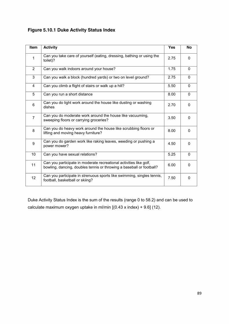
89
Figure 5.10.1 Duke Activity Status Index
Item Activity Yes No
1 Can you take care of yourself (eating, dressing, bathing or using the toilet)? 2.75 0
2 Can you walk indoors around your house? 1.75 0
3 Can you walk a block (hundred yards) or two on level ground? 2.75 0
4 Can you climb a flight of stairs or walk up a hill? 5.50 0
5 Can you run a short distance 8.00 0
6 Can you do light work around the house like dusting or washing dishes 2.70 0
7 Can you do moderate work around the house like vacuuming, sweeping floors or carrying groceries? 3.50 0
8 Can you do heavy work around the house like scrubbing floors or lifting and moving heavy furniture? 8.00 0
9 Can you do garden work like raking leaves, weeding or pushing a power mower? 4.50 0
10 Can you have sexual relations? 5.25 0
11 Can you participate in moderate recreational activities like golf, bowling, dancing, doubles tennis or throwing a baseball or football? 6.00 0
12 Can you participate in strenuous sports like swimming, singles tennis, football, basketball or skiing? 7.50 0
Duke Activity Status Index is the sum of the results (range 0 to 58.2) and can be used to
calculate maximum oxygen uptake in ml/min [(0.43 x index) + 9.6] (12).

90
formal cardiology review both in order to clarify their clinical validity and also to allow for
treatment of any uncovered cardiovascular disease.
An alternative approach is to screen patients at increased cardiac risk using CT coronary
calcium scoring. The radiation exposure of a CT calcium score is one sixth that of an
abdominal CT scan and a scan takes only 5 minutes, making this an attractive option in
many centres for the assessment of patients at low cardiac risk. Using this technique in an
asymptomatic individual, a coronary calcium score of zero effectively excludes significant
coronary atherosclerosis and obviates the need for further structural or functional
assessments (3). Higher scores will of course require careful interpretation and further
assessment in concert with local cardiological expertise. The technique has recently been
recommended by NICE as the most appropriate screening technique in patients
presenting to a rapid access chest pain clinic in whom the clinical suspicion of significant
coronary atherosclerosis is low (3). In many centres, an elevated coronary calcium score
of 1-400 can for convenience be followed by a CT coronary angiogram at the same
examination, with higher scores usually indicating a need for formal angiography.
5.10.4 Screening for Non-coronary Pathology A combination of clinical assessment and 12 lead surface ECG has a reasonable
sensitivity for the detection of non-coronary cardiac pathology. There is an extensive
literature on the pre-participation screening of athletes and in this group of young
individuals there is little incremental benefit of routine echocardiography. However, in an
older cohort this may not be true. Currently there is no consensus regarding the definition
of a relatively “high risk” cohort and it is therefore difficult to justify a policy of routine
echocardiogram screening for living kidney donors who have no abnormalities clinically or
on an electrocardiogram.
5.10.5 Conclusion Due to the nature of the procedure, a low threshold needs to be set for formal
investigation and for the exclusion of individuals for living kidney donation. As well as
determining suitability, the process should act as an opportunity to identify and correct
recognised cardiovascular risk factors. The choice of stress test will be influenced by local
service provision, although clinicians should consider the relatively increased predictive
91
value of structural techniques over electrocardiography alone. CT coronary calcium
scoring may be an alternative way of stratifying coronary risk.
References
1. Maron BJ, Shirani J, Poliac LC, Mathenge R, Roberts WC, Mueller FO. Sudden death
in young competitive athletes: clinical, demographic, and pathological profiles. JAMA.
1996; 276: 199-204.
2. Diamond GA, Forrester JS. Analysis of probability as an aid in the clinical diagnosis of
coronary-artery disease. N Engl J Med 1979; 300: 1350-8.
3. NICE Guidance 2010. Chest pain of recent onset: Assessment and diagnosis of
recent onset chest pain or discomfort of suspected cardiac origin. London: National
Clinical Guideline Centre for Acute and Chronic Conditions. Accessed 16th February
2011 at: http://guidance.nice.org.uk/CG/Wave14/25
4. Nelson CL, Herndon JE, Mark DB, et al. Relation of clinical and angiographic factors
to functional capacity as measured by the Duke Activity Status Index. Am J Cardiol.
1991; 68: 973–5.
5. Myers J, Do D, Herbert W, Ribisi P, Froelicher VF. A nomogram to predict exercise
capacity from a specific activity questionnaire and clinical data. Am J Cardiol 1994;
73: 591-6.
6. Bartels C, Bechtel JF, Hossmann V, Horsch S. Cardiac risk stratification for high-risk
vascular surgery. Circulation 1997; 95: 2473-5.
7. Reilly DF, McNeely MJ, Doerner D, et al. Self-reported exercise tolerance and the risk
of serious perioperative complications. Arch Intern Med 1999; 159: 2185–92.
8. Older P, Hall A, Hader R. Cardiopulmonary exercise testing as a screening test for
perioperative management of major surgery in the elderly. Chest 1999; 116: 355-62.
9. Hlatky MA, Boineau RE, Higginbotham MB, et al. A brief self administered
questionnaire to determine functional capacity (the Duke Activity Status Index). Am J
Cardiol 1989; 64: 651-4.
10. Lee TH, Marcantonio ER, Mangione CM, et al. Derivation and prospective validation
of a simple index for prediction of cardiac risk of major noncardiac surgery. Circulation
1999; 100: 1043-9.
11. Weiner DA, Ryan TJ, McCabe CH, et al. Prognostic importance of a clinical profile
and exercise test in medically treated patients with coronary artery disease. J Am Coll
Cardiol 1984; 3: 772-9.
92
5.11 PROTEINURIA
Statements of Recommendation
Urine protein excretion should be quantified in all potential living donors. (B1)
A urine albumin/creatinine ratio (ACR) performed on a spot urine sample voided after waking is the recommended screening test, although both urine protein/creatinine ratio (PCR) and 24-hour urine protein collection are acceptable alternatives. (A1)
Significant proteinuria is an ACR >30 mg/mmol, PCR >50 mg/mmol or 24-
hour total protein >300 mg/day, and usually contraindicates donation. (B1)
The significance of microalbuminuria (ACR 3.5-30 mg/mmol) and of 24-hour urine protein of 150-300 mg (PCR 15-30) has not been fully evaluated in living kidney donors. However, since both the risk of CKD and cardiovascular morbidity increase progressively with increasing albuminuria, such donors require careful evaluation and counselling about the risks of donation. (C2)
Proteinuria is a well established important risk factor for both chronic kidney disease (1)
and cardiovascular morbidity and mortality (2). In particular, proteinuria predicts both
progression of CKD and cardiovascular events in patients with established CKD,
established cardiovascular disease and, in patients with diabetes, co-morbidities that
clearly preclude living kidney donation (2,3). Proteinuria also predicts the development of
CKD and cardiovascular disease in those without medical co-morbidities, and for this
reason is considered an absolute contraindication to living kidney donation.
Several large cohort studies from the general population have identified proteinuria as a
risk factor for CKD and cardiovascular disease (4,5), even in those patients with an eGFR
>60 ml/min/1.73m2. In a study of nearly 1 million people from Alberta in Canada the risk of
developing end-stage renal disease (ESRD) in those with a baseline eGFR >60 ml/min
was 0.03/1,000 patient years if there was no proteinuria, 0.05 with ‘mild proteinuria’
93
(urinalysis trace or 1+, ACR 3.5-35 mg/mmol), but 1.0 in those patients with ‘heavy
proteinuria’ (urinalysis >1+, or ACR >35 mg/mmol) (5). A study of 40,854 individuals from
the Netherlands gave similar results, with a linear relationship between albuminuria and
subsequent ESRD (6). In this study, urine albumin concentrations of <20 mg/l and 20-
100 mg/l (roughly equivalent to an ACR of 3.5-20 mg/mmol, or ‘low level’
microalbuminuria) were associated with a low risk of ESRD (0.06 and 0.15% at 9 years
follow-up) compared to those with 100-200 mg/l or >200 mg/l of albuminuria (2.45 and
5.67% respectively).
There are few studies examining either the renal or cardiovascular outcome for living
kidney donors who have donated despite proteinuria. In many donors there is a modest
increase in urine protein excretion after nephrectomy, the majority of whom have no
evidence of accelerated GFR loss over time (7-10). In one study, 5 donors with low grade
proteinuria (mean 210 mg in a 24-hour urine collection) were more likely to have
significant proteinuria 20 years or more after donation (>800 mg/24 hours), although
without significant loss of kidney function (11). A review of 1,519 living kidney donors in
Japan identified 8 who developed ESRD (12). Of these, only 2 had pre-donation
proteinuria, both of whom developed cardiovascular disease, hypertension and ESRD 6
and 16 years after donation.
Methods of Testing for Proteinuria
For many years, assessment of proteinuria was based on an accurately timed 24-hour
urine collection, with <150mg protein/24 hours considered normal and >300 mg/24 hours
pathological. However the urine albumin /creatinine ratio (ACR) or protein /creatinine ratio
(PCR) in a spot urine sample are now the preferred methods as both correlate well with
24-hour urinary protein excretion and overcome inaccuracies related to incomplete urine
collection. Recent studies, including those described above, have used ACR, which is a
better screening test, and there is now consensus that ACR is an appropriate and
sufficient test for proteinuria (13-15). A normal ACR of <2.5 mg/mmol in men and <3.5
mg/mmol in women equates to <150 mg protein over 24 hours. An ACR of between 2.5
and 30 mg/mmol defines microalbuminuria (urine albumin excretion of between 30 and
300 mg/day, roughly equating to a 24-hour protein excretion of 150-500 mg), and
>30 mg/mmol defines macroalbuminura. Dipstick analysis alone is inadequate to detect
low level but clinically significant albuminuria. Table 5.11.1 provides a summary of the
comparative values detected using these screening methods.

94
Table 5.11.1 Expressions of urinary protein concentration and their approximate equivalents and clinical correlates
Dipstick
reading
Protein:creatinine
ratio (mg/mmol)
Total
protein
(mg/24h)
Albumin:creatinine
ratio (mg/mmol)
Albumin excretion
(mg/24h)
Normal Negative <15 <150 <2.5 (males)
<3.5 (females)
<30
Micro-
albuminuria
Negative <15 <150
2.5 - 35 (males)
3.5 - 35 (females)
30 – 300
‘Trace’
protein
Trace 15-50 150-500
Assessment of Proteinuria in Living Donors
There is uncertainty over the definition of significant proteinuria in a potential kidney
donor. A recent US survey reported that although many US centres use a 24-hour urine
collection for protein, some rely on a spot urine PCR, but almost one-half of centres now
use urine ACR as a screen (16). The most common exclusion criterion for kidney donors
in the US is 300 mg/day proteinuria, but almost as many centres now use 150 mg/day as
a cut-off, unless the proteinuria is postural. Recently, the Amsterdam Forum concluded by
consensus that a 24-hour urinary protein excretion of >300 mg is a contraindication to
donation (17). However, it is not clear what is to be done with patients with proteinuria
below 300 mg/day but above the upper limit of normal for the testing laboratory.
This issue was recently addressed in a single centre prospective study of 39 potential
kidney donors who had simultaneous measurements of both urinary total protein and
albumin (18). Although 13/39 had elevated 24-hour urinary total protein values, none had
elevated urinary albumin excretion, suggesting a low risk of subsequent CKD or
cardiovascular morbidity. Similarly, orthostatic proteinuria should not be considered as a
contraindication to donation. Orthostatic proteinuria appears benign (19), but confident
diagnosis requires performing an ACR on a spot urine sample voided immediately after
waking.
95
References 1. Halbesma N, Kuiken DS, Brantsma AH, et al. Macroalbuminuria is a better risk marker
than low estimated GFR to identify individuals at risk for accelerated GFR loss in
population screening. J Am Soc Nephrol 2006; 17: 2582-90.
2. Brantsma AH, Bakker SJ, de Zeeuw D, de Jong PE, Gansevoort RT. PREVEND Study
Group. Extended prognostic value of urinary albumin excretion for cardiovascular
events. J Am Soc Nephrol 2008; 19: 1785-91.
3. Perkovic V, Verdon C, Ninomiya T, et al. The relationship between proteinuria and
coronary risk: a systematic review and meta-analysis. PLoS Medicine 2008; 5:
1486-95.
4. Iseki K, Ikemiya Y, Iseki C, Takishita S. Proteinuria and the risk of developing end-
stage renal disease. Kidney Int 2003; 63: 1468-74.
5. Hemmelgarn BR, Manns BJ, Lloyd A, et al. Relation between kidney function,
proteinuria and adverse outcomes. JAMA 2010; 303: 423-9.
6. Van de Velde M, Halbesma N, de Charro FT, et al. Screening for albuminuria identifies
individuals at increased renal risk. J Am Soc Nephrol 2009; 20: 852-62.
7. Fehrman-Ekholm I, Dunér F, Brink B, Tydén G, Elinder CG. No evidence of
accelerated loss of kidney function in living kidney donors: results from a cross-
sectional follow-up. Transplantation. 2001; 72: 444-9.
8. Garg AX, Muirhead N, Knoll G, et al. Donor Nephrectomy Outcomes Research
(DONOR) Network. Proteinuria and reduced kidney function in living kidney donors: a
systematic review, meta-analysis and meta-regression. Kidney Int 2006; 80: 1801-10.
9. Gossmann J, Wilhelm A, Kachel HG, et al. Long-term consequences of live kidney
donation follow-up in 93% of living kidney donors in a single transplant center. Am J
Transplant 2005; 5: 2417-24.
10. Ibrahim HN, Foley R, Tan L, Rogers et al. Long-term consequences of kidney
donation. N Engl J Med 2009; 360: 459-69.
11. Goldfarb DA, Matin SF, Braun WE, Schreiber MJ, Mastroianni B, Papajcik D. Renal
outcome 25 years after donor nephrectomy. J Urol 2001; 166: 2043-7.
12. Kido R, Shibagaki Y, Iwadoh K, et al. How do living kidney donors develop end-stage
renal disease? Am J Transplant 2009; 9: 2514-9.
13. K/DOQI clinical practice guidelines and clinical practice recommendations for diabetes
and chronic kidney disease. Am J Kidney Dis 2007; 49(2 Suppl 2): S12.

96
14. National Collaborating Centre for Chronic Conditions. Chronic kidney disease: national
clinical guideline for early identification and management in adults in primary and
secondary care. London: Royal College of Physicians, September 2008.
15. Lamb EJ, MacKenzie F, Stevens PE. How should proteinuria be detected and
measured? Ann Clin Biochem 2009; 46: 205-17.
16. Mandelbrot DA, Pavlakis M, Danovitch GM, et al. The medical evaluation of living
kidney donors: a survey of US transplant centers. Am J Transplant 2007; 7: 2333-43.
17. Delmonico F. Council of the Transplantation Society. A report of the Amsterdam
Forum on the care of the live kidney donor: data and medical guidelines.
Transplantation 2005; 79(6 Suppl): S53-66.
18. Leischner MP, Naratadam GO, Hou SH, Singh AK, Leehey DJ. Evaluation of
proteinuria in healthy living kidney donor candidates. Transplant.Proc 2006; 38:
2796-7.
19. Springberg PD, Garrett LE Jr, Thompson AL Jr, Collins NF, Lordon RE, Robinson RR.
Fixed and reproducible orthostatic proteinuria: results of a 20-year follow-up study.
Ann Intern Med 1982; 97: 516-9.
97
5.12 NON-VISIBLE HAEMATURIA Statements of Recommendation
All potential living donors should have reagent strip (dipstick) urinalysis performed on at least 2 separate occasions. (B1)
Two or more positive tests, including trace positive, should be considered
as persistent non-visible haematuria (PNVH). (B1)
If PNVH is present, perform urine culture and renal imaging to exclude common urologic causes including infection, nephrolithiasis and urothelial carcinoma. (A1)
If no cause is found, perform cystoscopy in patients age >40 years to exclude bladder pathology. (B1)
If no cause is found and the donor still wishes to donate, then a kidney
biopsy should be considered, and is recommended if haematuria is >1+ on dipstick testing. (B2)
Glomerular pathology precludes donation, with the possible exception of
thin basement membrane disease. (B1)
Non-visible haematuria is the preferred term (replacing microscopic haematuria) for blood
identified in a urine sample either by microscopy or by reagent strip analysis. Non-visible
haematuria is a common finding in the general population, may indicate either urological
or renal parenchymal disease, and must be carefully evaluated in prospective living
kidney donors.
Non-visible haematuria is present in 1-21% of the general population, the prevalence
increasing with age (1-4). Most patients are asymptomatic with no urologic symptoms, no
proteinuria and normal renal function. Subsequent urine testing is often normal. Such
transient haematuria is generally considered insignificant, although with little supporting
evidence from longitudinal studies. In one report including 432 patients with normal

98
urological investigation and followed for 5.8 +/- 4.4 years, haematuria disappeared in 44%,
none of whom developed proteinuria or renal impairment (5). In a smaller study of 49
patients investigated for non-visible haematuria, those in whom haematuria disappeared
all had a normal kidney biopsy (6).
Persistent asymptomatic non-visible haematuria (PANVH) is present in about 25% of
those with an initial positive test (1-7) and, in two single centre reports, 2.7% and 8.3% of
potential living kidney donors in the US and Japan respectively (8,9). Malignant disease of
the urinary tract, present in 3-5% of patients overall (10,11), is rare under the age of 40
but diagnosed in up to 10% of those aged >60. In patients with normal urological
investigations, kidney biopsy is frequently abnormal. In a UK-based study 77 of 165
patients, 46% were found to have glomerular pathology, most commonly IgA nephropathy,
mesangial proliferative glomerulonephritis without IgA deposition, or thin basement
membrane nephropathy (12). Similar pathology has been demonstrated in a Dutch study
where 29 out of 49 biopsies were abnormal (6), a Korean study in which only 10 out of
156 biopsies were normal (13), and in a US study of potential living donors with PANVH in
which 8 out of 10 biopsies were abnormal (8).
Longitudinal studies have confirmed the importance of PANVH. In the Dutch study of 49
patients, those with a normal biopsy developed neither proteinuria nor worsening renal
function during 11 years of follow-up. In contrast, proteinuria (10 patients), hypertension
(14) and worsening kidney function (4) were found in the 29 patients with an abnormal
biopsy (6). In a Japanese study of 242 living donors, 8.3% had PANVH prior to donation
and 15.3% following donation. None were investigated with a kidney biopsy, but the
presence of haematuria predicted the development of proteinuria during a median follow-
up of 2.3 years (9). In a similar study including patients from the Japanese general
population, 10% of those with PANVH developed proteinuria over a median follow-up of
5.8 years (5).
The above supports current practice that persistent asymptomatic non-visible haematuria
should be investigated in potential living kidney donors, both to exclude urological disease
and to identify glomerular pathology that would preclude donation. However, there remain
uncertainties: in particular, the relevance of low levels of haematuria (‘trace’ positive), and
the importance of thin basement membrane nephropathy (TBMN) merit further discussion.
99
‘Trace’ microscopic haematuria Non-visible haematuria is routinely detected using semi-quantitative reagent strips. A
reagent strip ‘trace positive’ result corresponds to 1-5 red cells/ l (15). Urine microscopy is
not required to confirm the presence of haematuria, and indeed often produces false
negative results, although the detection of dysmorphic red cells and red cell casts may be
useful to identify glomerular haematuria.
One difficulty is that existing studies rarely, if ever, distinguish between the degrees of
non-visible haematuria recorded on dipstick testing. As the incidence of significant
disease following the investigation of trace positive haematuria is no different to that of
control populations, recent primary care and Urology guidelines in the UK have
recommended that trace non-visible haematuria be considered a normal variant (14).
However, glomerular pathology has been reliably identified in potential living donors using
thresholds of even 1 or 3 red cells/ l (8,9). No studies have directly addressed the
threshold below which investigation of the potential donor is unnecessary, and a balance
must be struck between the risk of missing significant renal disease in a potential donor,
against the inconvenience and risk of biopsy. High degrees of non-visible haematuria
mandate biopsy prior to donation, but trace haematuria is at present a relative indication.
If, after counselling, the prospective donor with non-visible haematuria remains committed
to donation and a kidney biopsy is performed, histological evaluation must include
immunofluorescence or immunohistochemistry, and electron microscopy.
Considerable evidence also suggests that cystoscopy is of limited value in the
investigation of non-visible haematuria below the age of 40 years, especially in women,
and this is reflected in current UK guidelines (14). Risk factors for uro-epithelial cancer
should be assessed including donor age, smoking history, exposure to aniline dye,
analgesics or cyclophosphamide, and pelvic irradiation. In younger asymptomatic
patients, it is reasonable to discuss the risk/benefit ratio of cystoscopy with the
prospective donor. Above the age of 40 years, however, the increased incidence of
urological disease mandates a full urological assessment, including cystoscopy.
Thin basement membrane nephropathy Thin basement membrane nephropathy (TBMN) is an autosomal dominant disorder often
associated with mutations in either the COL4A3 or COL4A4 genes (encoding the 3 and
4 chains of type 4 collagen). Individuals in whom both alleles of either gene are

100
abnormal may have autosomal recessive Alport syndrome, and TBMN can be regarded
as the carrier state for this condition. TBMN is present in 10-50% of patients biopsied for
PANVH (6,8,12,13) and although often considered a benign diagnosis may carry some
risk of progression. Both proteinuria (10-20% of patients) and renal impairment (5%) have
been described (16-18), often associated with additional pathological abnormalities
including FSGS (18) or IgA nephropathy (19,20) (both of which would preclude donation).
Many individuals with TBMN but otherwise normal investigations have undoubtedly
donated kidneys, either knowingly (8) or unknowingly (9), and although adverse outcomes
have not been reported these donors must be made aware of uncertainty over long-term
safety. Referral to a clinical geneticist for molecular testing may be warranted, especially
when donating to a family member with unexplained kidney failure or where there is a
family history of sensori-neural deafness or haematuria (see also section 5.17 Familial
Renal Disease). Referral to a geneticist is mandatory in potential donors of Cypriot origin,
where associations of TBMN and FSGS leading to significant rates of renal failure have
been noted (21).
TBMN must be distinguished from the carrier state of X-linked Alport syndrome (XLAS),
which is associated with a 5-20% risk of progressive renal impairment (22) and generally
considered to prohibit donation. A recent study describing six XLAS carriers who donated
kidneys to their affected children supports this view (23). A decline in kidney function of
between 25 and 60% was observed in four of the six donors over 2–14 years of follow-up,
although in no case was creatinine clearance <40 ml/min. Four of the six developed
microalbuminuria or proteinuria, and four developed hypertension. Some have argued
that, if no other donor can be found, women with XLAS who are over the age of 45, have
normal kidney function, no proteinuria and no hearing deficiency (both risk factors for
progression to end-stage kidney disease) might be considered as donors after appropriate
counselling (24). Involvement of a clinical geneticist would be mandatory in the screening
of such a potential donor.
References 1. Topham PS, Jethwa A, Watkins M, Rees Y, Feehally J. The value of urine screening
in a young adult population. Fam Pract 2004; 21: 18-21.
2. Mohr DN, Offord KP, Owen RA, Melton LJ. Asymptomatic microhematuria and
urologic disease: a population based study. JAMA 1986; 256: 224-9.

101
3. Messing EM, Young TB, Hunt VB. Home screening for haematuria: results of a
multiclinic study. J Urol 1992; 148: 289-92.
4. Froom P, Ribak J, Benbassat J. Significance of microhematuria in young adults. BMJ
1984; 288: 20-2.
5. Yamagata K, Kobayashi M, Koyama A. A long-term follow up study of asymptomatic
haematuria and/or proteinuria in adults. Clin Nephrol 1996; 45: 281-8.
6. Nieuwhof C, Doorenbos C, Grave W. A prospective study of the natural history of
idiopathic non-proteinuric haematuria. Kidney Int 1996; 49: 222-5.
7. Jaffe JS, Ginsberg PC, Gill R, Harkaway RC. A new diagnostic algorithm for the
evaluation of microscopic haematuria. Urology 2001; 57: 889-94.
8. Koushik R, Garvey C, Manivel C, Matas AJ, Kasiske B. Persistent, asymptomatic
microscopic hematuria in prospective kidney donors. Transplantation 2005; 80:
1425-9.
9. Kido R, Shibagaki Y, Iwadoh K, et al. Persistent glomerular haematuria in living kidney
donors confers a risk of progressive kidney disease in donors after heminephrectomy.
Am J Transplant 2010; 10: 1597-604.
10. Khadra MH, Pickard RS, Charlton M, Powell PH, Neal DE. A prospective analysis of
1,930 patients with haematuria to evaluate current diagnostic practice. J Urol 2000;
163: 524-7.
11. Edwards TJ, Dickinson AJ, Natale S, Gosling J, McGrath JS. A prospective analysis of
the diagnostic yield resulting from the attendance of 4020 patients at a protocol-driven
haematuria clinic. BJU Int 2006; 97: 301-5.
12. Topham PS, Harper SJ, Furness PN, Harris KPG, Walls J, Feehally J. Glomerular
disease as a cause of isolated microscopic haematuria. Q J Med 1994; 87: 329-35.
13. Kim BS, Kim YK, Shin YS, et al. Natural history and renal pathology in patients with
isolated microscopic haematuria. Korean J Intern Med 2009; 24: 356-61.
14. Kelly JD, Fawcett DP and Goldberg LC. Assessment and management of non-visible
haematuria in primary care. BMJ 2009; 338: 227-32.
15. Freni SC, Heederik GJ, Hol C. Centrifugation techniques and reagent strips in the
assessment of microhematuria. J Clin Pathol 1977; 30: 336-40.
16. Savige J, Rana K, Tonna S, Buzza M, Dagher H, Wang YY. Thin basement
membrane nephropathy. Kidney Int 2003; 64: 1169-78.
17. Auwardt R, Savige J, Wilson D. A comparison of the clinical and laboratory features of
thin basement membrane disease (TBMD) and IgA glomerulonephritis (IgA GN). Clin
Nephrol 1999; 52: 1-4.
102
18. van Passen P, van Breda Vriesman PJ, van Rie H, Tervaert JW. Signs and symptoms
of thin basement membrane nephropathy: a prospective regional study on primary
glomerular disease – The Limburg Renal Registry. Kidney Int 2004; 66: 909-13.
19. Cosio FG, Falkenhein ME, Sedmark DD. Association of thin glomerular basement
membrane with other glomerulopathies. Kidney Int 1996; 46: 471-4.
20. Berthoux FC, Laurent B, Alamartine E, Diab N. A new subgroup of primary IgA
nephritis with thin glomerular basement membrane (GBM): Syndrome or association.
Nephrol Dial Transplant 1996; 11: 558-61.
21. Voskarides K, Damianou L, Neocleous V, et al. COL4A3/COL4A4 mutations producing
focal segmental glomerulosclerosis and renal failure in thin basement membrane
nephropathy. J Am Soc Nephrol 2007; 18: 3004-16.
22. Kashtan CE. Alport syndrome and the X chromosome: implications of a diagnosis of
Alport syndrome in females. Nephrol Dial Transplant 2007; 22: 1499-505.
23. Gross O, Weber M, Fries JW, Muller GA. Living donor kidney transplantation from
relatives with mild urinary abnormalities in Alport syndrome: Long-term risk, benefit
and outcome. Nephrol Dial Transplant 2009; 24: 1626-30.
24. Kashtan CE. Women with Alport syndrome: risks and rewards of kidney donation.
Nephrol Dial Transplant 2009; 24: 1369-70.

103
5.13 PYURIA
Statement of Recommendation
Prospective donors found to have pyuria should only be considered for donation if it can be demonstrated that the pyuria is due to a reversible cause, such as an uncomplicated urinary tract infection. (C1)
Pyuria may be defined as the presence of at least 10 leukocytes/mm3 of uncentrifuged
urine (1), which occurs in less than 1% of asymptomatic, non-bacteriuric patients but in
greater than 96% of symptomatic men and women with significant bacteriuria.
Pyuria can occur in the absence of apparent bacterial infection, particularly in patients
who have already taken antimicrobials, or where there is infection with atypical organisms
such as Chlamydia trachomatis, Ureaplasma urealyticum, or tuberculosis. Most
symptomatic women with pyuria but without significant bacteriuria have urinary infection,
either with bacterial uropathogens present in colony counts less than 105/ml or with
Chlamydia (1). Other causes of sterile pyuria include contamination from genital
secretions, acute or chronic interstitial nephritis, nephrolithiasis and uroepithelial tumour.
The cause of the pyuria must be established before a potential donor proceeds for further
assessment.
Reference
1. Stamm WE. . Am J Med 1983; 75:
53-8.
104
5.14 INFECTION IN THE PROSPECTIVE DONOR
Statements of Recommendation
Infection screening in the prospective donor prior to donation is important to identify potential risks for the donor from previous or current infection and to assess the risks of transmission of infection to the recipient. (B1)
Active HBV and HCV infection in the donor are usually contraindications to
living donor kidney transplantation; however, donors with no evidence of active viral replication may be considered under some circumstances. (B1)
The CMV status of donor and recipient should be determined before
transplantation. When the donor is CMV positive and the recipient is CMV negative, the donor and recipient should be counselled about the risk of post-transplant CMV. (B1)
The EBV status of donor and recipient should be determined before
transplantation. When the donor is EBV positive and the recipient is EBV negative, the donor and recipient should be counselled about the risk of developing PTLD. (B1)
The presence of HIV or human T lymphotrophic virus (HTLV) infection is an absolute contraindication to living donation. (B1)
The risk of transmission of infections between donor and recipient must be kept to a
minimum. The same principles that apply to deceased donors and blood donors should be
applied to the screening of living donors in this respect (1,2). Identification of current or
previous infection in the prospective donor is an important aspect of donor evaluation. The
presence of active infection usually precludes donation. Apart from the implications for the
potential donor, a number of infections may be transmitted by organ transplantation.
Those that are of established clinical significance are listed in Table 5.14.1.
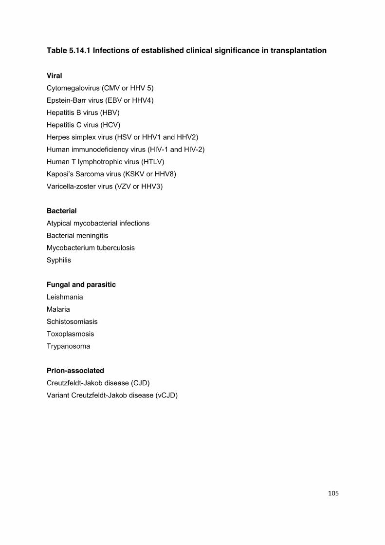
105
Table 5.14.1 Infections of established clinical significance in transplantation Viral Cytomegalovirus (CMV or HHV 5)
Epstein-Barr virus (EBV or HHV4)
Hepatitis B virus (HBV)
Hepatitis C virus (HCV)
Herpes simplex virus (HSV or HHV1 and HHV2)
Human immunodeficiency virus (HIV-1 and HIV-2)
Human T lymphotrophic virus (HTLV)
Kaposi’s Sarcoma virus (KSKV or HHV8)
Varicella-zoster virus (VZV or HHV3)
Bacterial Atypical mycobacterial infections
Bacterial meningitis
Mycobacterium tuberculosis Syphilis
Fungal and parasitic Leishmania Malaria
Schistosomiasis
Toxoplasmosis
Trypanosoma
Prion-associated Creutzfeldt-Jakob disease (CJD)
Variant Creutzfeldt-Jakob disease (vCJD)
106
5.14.1 Evaluation of the Prospective Donor A detailed clinical history is important and should include a psychosocial and sexual
history to define at-risk behaviour (see Table 5.4.2 in section 5.4). Prospective donors
who have been resident in geographical areas outside the UK where there is a high
prevalence of infection may require additional evaluation. During routine physical
examination of the donor, examination of the chest and reticuloendothelial system may
reveal evidence of infection. The routine screening investigations already outlined in Table
5.4.4 in section 5.4 include those ordinarily required to exclude infection in the prospective
donor. Particular attention should be paid to the possibility of past tuberculosis when
examining the chest X-ray. A mid-stream urine should be cultured and examined by
microscopy on at least two occasions. If sterile pyuria is detected the cause must be
identified. The presence of eosinophilia may indicate chronic parasite infection.
The serological tests that should be performed on the prospective donor and recipient are
listed in Table 5.14.2. Infections can be transmitted by both blood transfusion and organ
donation during the incubation period of the offending organism and before a serological
response has been mounted. Serology should not, therefore, be regarded as a substitute
for a detailed psychosexual and medical history. Routine testing for viral infection may, if a
positive result is obtained, raise complex ethical problems.
It is important that there is full discussion with the prospective donor before testing for viral
infection, particularly for hepatitis B virus (HBV), hepatitis C virus (HCV), and human
immunodeficiency virus (HIV). A strategy for dealing with a positive result should be
formulated before testing.
5.14.2 Viral Infections in the Prospective Donor HIV and HTLV The presence of HIV or human T lymphotrophic virus (HTLV) infection is an absolute
contraindication to living donation. HTLV serology is not routinely tested but should be
performed if the prospective donor comes from an endemic area e.g. Africa, the
Caribbean and Japan. Kidney donation should not be undertaken if significant doubt
remains about the possibility of HIV infection in the donor.
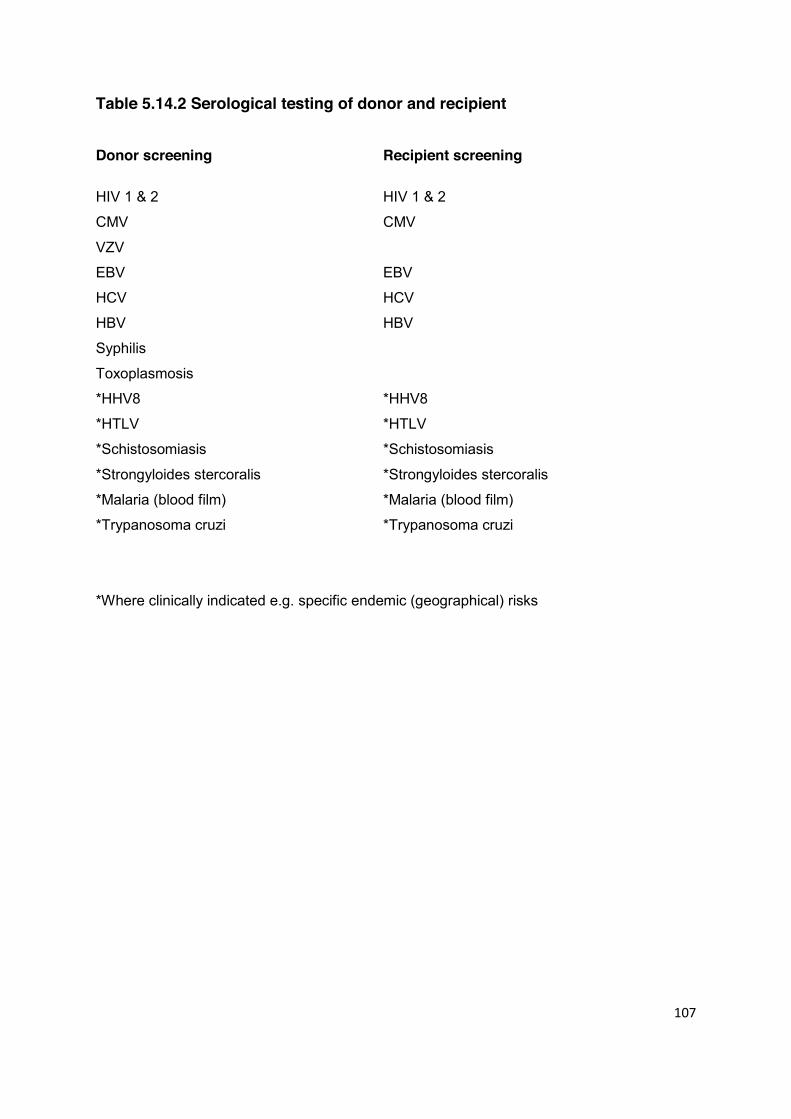
107
Table 5.14.2 Serological testing of donor and recipient Donor screening Recipient screening
HIV 1 & 2 HIV 1 & 2
CMV CMV
VZV
EBV EBV
HCV HCV
HBV HBV
Syphilis
Toxoplasmosis
*HHV8 *HHV8
*HTLV *HTLV
*Schistosomiasis *Schistosomiasis
*Strongyloides stercoralis *Strongyloides stercoralis *
*Malaria (blood film) *Malaria (blood film)
*Trypanosoma cruzi *Trypanosoma cruzi
*Where clinically indicated e.g. specific endemic (geographical) risks
108
HCV Active HCV in the donor is often a contraindication to living donation, not only because of
the risk of transmitting HCV to the recipient but also because of the risk of glomerular
disease in the donor (3,4). The risk of HCV transmission from an HCV RNA positive donor
approaches 100% if transplanted into a naïve recipient (5). All potential donors should
have HCV antibody testing performed and, if positive, HCV RNA should be checked. If the
donor is consistently RNA negative, then transplantation may be considered, even into a
naïve recipient. The risks entailed, however, must be carefully explained to both donor
and recipient. In these exceptional circumstances, the likely life expectancy of the
recipient has to be considered.
Advances in anti-viral agents and vaccination may influence such decisions in the future.
HBV Most transplant units would not consider potential donors with evidence of active HBV
viral replication. All prospective donors should have both HB surface antigen and HB core
antibody IgG checked. HBV DNA testing should be performed in prospective donors from
HBV endemic areas who are hepatitis core antibody positive, those with possible mutant
HBV, and those with abnormal liver tests or a past history of liver disease of unknown
aetiology. HB core antibody IgM is not indicated unless the donor is e antigen positive and
acute infection is being queried.
There are a substantial number of reports of kidneys transplanted from HB surface
antigen negative/DNA negative, HB core antibody-positive cadaver donors in which there
have been a low risk of HBV seroconversion and no excess risk of graft failure or short-
term morbidity (6-9). In the context of living donation, donors who are HBcAb positive with
negative HBsAg and undetectable DNA in blood may be considered as donors, providing
the recipient has been effectively immunised against HBV. In addition, the use of HBV
immunoglobulin and anti-viral drugs may be considered. Advice from a virologist and
hepatologist should be sought under these circumstances and the donor and recipient
need to be fully informed.
CMV CMV infection is the most commonly encountered clinically significant viral infection after
kidney transplantation and may cause significant morbidity and mortality, particularly if the
recipient is heavily immunosuppressed (10). It also increases the risk of chronic graft

109
dysfunction as well as post-transplant lymphoproliferative disorder (PTLD) and
opportunistic infection.
CMV disease may result from reactivation of latent infection or because of primary
infection transmitted by a kidney from a CMV positive donor. For CMV and other viral
infections, primary infection is generally more severe than reactivation and the recipients
most at risk are those who are CMV seronegative and receive a kidney graft from a CMV
seropositive donor. Matching CMV seronegative recipients with CMV seronegative donors
is an effective strategy for reducing the risk of CMV infection but is rarely practicable in the
context of living donor kidney transplantation. Either CMV prophylaxis or pre-emptive
therapy with close monitoring of viral loads should be offered (11). The donor and
recipient should be informed about the increased risk of CMV disease before the
transplant is performed.
EBV Primary EBV infection is most likely to occur in EBV negative paediatric recipients who
receive a kidney from an EBV positive donor. EBV infection increases the risk of PTLD
several-fold and this risk is increased further if the recipient is given anti-lymphocyte
antibody immunosuppressive therapy. Consideration should be given in this situation to
the prophylactic use of antiviral agents (aciclovir or valganciclovir) in order to minimise the
viral load after transplantation. This strategy may protect renal transplant recipients from
PTLD (12), but was not found to be of benefit in paediatric liver transplant recipients (13).
When the donor is EBV positive and the recipient is EBV negative, clinical vigilance is
required following transplantation to detect PTLD as early as possible. The use of
quantitative PCR to monitor the recipient viral load after transplantation is contentious.
VZV It is important to know whether the potential recipient is VZV seropositive as a primary
VZV infection may be rapidly fatal in an immunocompromised host (14,15). Vaccination is
available for recipients who are VZV antibody negative.
5 HHV8 HHV8 may be transmitted by organ transplantation and is associated with an increased
risk of Kaposi’s sarcoma (16). However, there is no evidence to support screening of
potential organ donors.

110
5.14.3 Bacterial Infections in the Prospective Donor The main risk of bacterial infection is from Mycobacterium tuberculosis (and atypical
mycobacteria). Donors should be screened for mycobacterial infection. Screening should
include a careful history, including ethnic origin and country of upbringing. Chest X-ray is
important, but the value of skin testing is questionable. If a specific bacterial
microbiological diagnosis has been made in the donor, then a course of appropriate
antibiotic is likely to be effective in preventing transmission (Table 5.14.3). A history of
urinary tract infection in a potential donor, particularly if there is a family history of reflux
nephropathy, or in a male, requires detailed imaging of the kidneys (e.g. DMSA for cortical
scarring).
Transmission of syphilis has been reported in the UK to two recipients from a deceased
donor with a past history of treated disease (18). Recipients of living donor transplants
from donors considered to carry a risk of syphilis transmission should be given
prophylactic treatment (2.5 MU benzathine penicillin im single dose, or doxycycline
100 mg po for 14 days, or 1 g azithromycin po single dose) in line with British Association
for Sexual Health and HIV guidelines (19). Discussion and liaison with specialist GU
medicine advice would be advisable.
5.14.4 Fungal and Parasitic Infections in the Prospective Donor A living donor is unlikely to transmit a fungal infection if otherwise in good health.
Nevertheless, this remains a theoretical possibility and should be considered in patients
from areas where fungal infections are endemic. Toxoplasmosis and malaria can be
transmitted by a renal transplant (14). In most of the reported cases, transmission has
been from living unrelated donor transplantation taking place in the developing world.
Other infections are either transmitted rarely (occasional case report) or are only of
theoretical risk. Table 5.14.3 summarises the use of prophylactic antimicrobial agents for
different types of donor infection.

111
Table 5.14.3 Use of prophylactic antimicrobial agents
1 HBV positive donor Vaccinate recipient
Prophylactic lamivudine
2 CMV (donor +ve, recipient -ve) Prophylactic antiviral drugs
(usually valganciclovir)
3 EBV (donor +ve, recipient -ve) Consider prophylactic aciclovir
or valganciclovir
4 Toxoplasmosis Sulphonamide, clindamycin,
clarithromycin, azithromycin
or pyrimethamine
(covered also by co-trimoxazole)
5 Mycobacterial infections Prophylactic isoniazid
6 Bacteria Low virulence 7 days of appropriate antibiotic
High virulence 14 days of appropriate antibiotic
7 Syphilis Benzathine benzylpenicillin
112
5.14.5 Prion-Associated Diseases in the Prospective Donor CJD and vCJD There is no screening test currently available for CJD or vCJD. Four individuals in the UK
have been shown to have acquired CJD prions from blood or blood products in the UK,
two of whom went on to develop clinical CJD (19). No cases of transmission by living
donor kidney transplantation have been reported. National guidance for blood and tissue
donors states that prion-associated disease in the prospective donor is an absolute
contra-indication to donation (2). Individuals who may be at increased risk of developing
such a disease are also precluded from donating and a detailed personal and family
history must be taken from the donor to identify potential risk factors. Healthy living donors
may not have been exposed to many of these, but relevant history would include
recipients of human pituitary-derived (growth) hormones, dura mater, corneal and scleral
grafts and a positive family history (two or more blood relatives) of prion-associated
disease, subject to genetic counselling. The current guidelines imply that prospective
donors who have received blood or blood products anywhere in the world after 1980
should be viewed as at increased risk (and are, in fact, excluded from blood donation).
This is not reflected in current UK practice, and is probably impractical (2).
References 1. Standards for solid organ transplantation in the United Kingdom. British
Transplantation Society 2003; ISBN 0 9542221-2-1.
2. UK Blood Transfusion & Tissue Guidelines. Donor selection guidelines.
www.transfusionguidelines.org.uk
3. Johnson RJ, Gretch DR, Yamabe H, et al. Membranoproliferative glomerulonephritis
associated with hepatitis C virus infection. N Engl J Med 1992; 328: 465-70.
4. Stehman-Breen C, Willson R, Alpers CE, Gretch D, Johnson RJ. Hepatitis C virus-
associated glomerulonephritis. Curr Opin Nephrol Hypertens 1995; 4: 287-94.
5. Pereira BJ, Milford EL, Kirkman RL, et al. Prevalence of hepatitis C virus RNA in
organ donors positive for hepatitis C antibody and in the recipients of their organs. N
Engl J Med 1992; 327: 910-5.
6. Satterthwaite R, Ozgu I, Shidban H, et al. Risks of transplanting kidneys from
hepatitis B surface antigen-negative, hepatitis B core antibody-positive donors.
Transplantation 1997; 64: 432-5.

113
7. Madayag RM, Johnson LB, Bartlett ST, et al. Use of renal allografts from donors
positive for hepatitis B core antibody confers minimal risk for subsequent
development of clinical hepatitis B virus disease. Transplantation 1997; 64: 1781-6.
8. De Feo TM, Grossi P, Poli F, et al. Kidney transplantation from anti-HBc+ donors:
results from a retrospective Italian study. Transplantation 2006; 81: 76-80.
9. Ouseph R, Eng M, Ravindra K, Brock GN, Buell JF, Marvin MR. Review of the use of
hepatitis B core antibody-positive kidney donors. Transplant Rev 2010; 24: 167-71.
10. Van Son WJ, The TH. Cytomegalovirus infection after organ transplantation: an
update with special emphasis on renal transplantation. Transpl Int 1989; 2: 147-64.5
11. Guidelines for the prevention and management of cytomegalovirus disease after solid
organ transplantation. British Transplantation Society, 2002. ISBN: 0954222105.
12. Darenkov IA, Marcarelli MA, Basadonna GP, et al. Reduced incidence of Epstein-Barr
virus associated post transplant lymphoproliferative disorder using preemptive
antiviral therapy. Transplantation 1997; 64: 848-52.
13. Green M, Kaufmann M, Wilson J, Reyes J. Comparison of intravenous ganciclovir
followed by oral acyclovir with intravenous ganciclovir alone for the prevention of
cytomegalovirus and Epstein-Barr virus disease after liver transplantation in children.
Clin Infect Dis 1997; 25: 1344-9.
14. Parnham AP, Flexman JP, Saker BM, Thatcher GN. Primary varicella in adult renal
transplant recipients: a report of three cases plus a review of the literature. Clin
Transplant 1995; 9: 115-8.
15. Rothwell WS, Gloor JM, Morgenstern BZ, Milliner DS. Disseminated varicellla
infection in pediatric renal transplant recipients treated with mycophenolate mofetil.
Transplantation 1999; 68: 158-61.
16. Regamey N, Tamm M, Wernli M, et al. Transmission of human herpesvirus 8 infection
from renal transplant donors to recipients. N Engl J Med 1998; 19: 1358-63.
17. Joint UKBTS / NIBSC Professional Advisory Committee Position Statement:
Creutzfeldt-Jakob Disease. 11th March 2010.
www.transfusionguidelines.org.uk/index.aspx?Publication=DL&Section=12&pageid=7
94
18. Cortes NJ, Afzali B, MacLean D, et al. Transmission of syphilis by solid organ
transplantation. Am J Transplant 2006; 6: 2497-9.
19. Kingston M, French P, Goh B, et al. UK National Guidelines on the Management of
Syphilis 2008. Syphilis Guidelines Revision Group 2008, Clinical Effectiveness Group
Int J STD AIDS. 2008; 19: 729-40.
114
5.15 NEPHROLITHIASIS
Statement of Recommendation
In the absence of a significant metabolic abnormality, potential donors with a limited history of previous small calcium stones, or a small renal calculus on imaging, may still be considered as potential kidney donors. Full counselling of donor and recipient is required along with access to appropriate long term donor follow-up. (C2)
Potential donors with metabolic abnormalities detected on screening should be discussed with a specialist in renal stone disease. (C2)
5.15.1 Incidence, Natural History and Management of Renal Stones In the UK, symptomatic renal stones are common with a prevalence of around 3-5%. The
use of CT to evaluate potential kidney donors has led to increased detection of
asymptomatic small kidney stones, which are present in 5% of potential kidney donors
undergoing a non-contrast CT scan.
The lifetime risk of recurrent kidney stones is an important consideration in evaluating
suitability for kidney donation. There are few data on the lifetime risk specific to the kidney
donor population. However, people who present with a symptomatic (calcium oxalate)
kidney stone have a 50% chance of developing a further stone within 5 years (1). Stone
recurrence rates also are related to the number of previous stone episodes and the time
interval to stone recurrence.
Most renal stones (75%) are composed predominantly of calcium oxalate. In symptomatic
patients who undergo metabolic evaluation (who may be a selected group), a metabolic
abnormality (e.g. hypercalciuria, hyperoxaluria, or hypocitraturia) may be detected in over
50% (2,3). The remaining 25% of stones are composed of uric acid, cystine, pure calcium
phosphate, or struvite (magnesium ammonium phosphate, also called infection stones)
(2,4). Uric acid stones are often associated with a history of gout, ileostomy diarrhoea or
with the metabolic syndrome, in all of which the urine is highly acidic. Cystine stones are
always associated with cystinuria. Calcium phosphate stones may occur with

115
hypercalciuria and are the predominant stone type formed by patients with a low urinary
citrate and distal renal tubular acidosis. Infection stones are commonly associated with an
anatomical abnormality.
Most asymptomatic stones found in potential donors are small (<5 mm). Small stones
usually pass spontaneously but can occasionally cause ureteric obstruction leading to
acute renal failure in patients with a single kidney. Small kidney stones can now be
treated using less invasive treatment modalities e.g. extra-corporeal shock wave
lithotripsy (ESWL) and flexible ureterorendoscopy. However, for the general population,
the evidence that treating small asymptomatic stones is superior to simply observing them
is mixed. Lower pole stones are more likely to progress than upper or middle pole stones,
and progression is more likely if an asymptomatic stone is >4 mm at the time of detection
(5).
In transplant recipients, the long term risks associated with a small stone in the donor
kidney appear low (6,7).
Extensive or staghorn calculi can commonly lead to chronic renal damage (2) and are
usually associated with infection or a significant metabolic abnormality
5.15.2 Assessment of Potential Donors
Imaging The use of CT for renal vascular imaging has increased the detection rate of
asymptomatic kidney stones. Where CT is not used routinely for vascular imaging and a
stone is suspected from USS or MRI, a non contrast CT KUB is advisable to determine
the number, size and location of suspected stones.
If a probable stone is identified on imaging, a urological and radiological review should be
undertaken. The number, size, position and density of the potential stones should be
considered; as should the presence of any underlying structural renal abnormality. A CT
IVU may be useful in these circumstances. A DMSA scan is useful if renal scarring is
suspected and will give an estimate of split renal function.
116
Biochemical Assessment A full metabolic screen should be carried out prior to donation on potential donors with a
history of stone disease or radiological evidence of a current stone. This screen should
include 24-hour urine collections for calcium, oxalate, citrate and urate, and early morning
pH assessment. This will require at least two separate urine collections as calcium,
oxalate and citrate analyses require an acidified collection, whereas electrolytes, urate
and pH are measured in a plain urine collection. Creatinine should be measured on each
collection as an internal marker of completeness. A pH measurement on an early morning
urine sample is useful, together with a qualitative cystine screen for cystinuria (8),
followed, if positive, by a 24-hour collection for cystine concentration. A metabolic screen
(urine and plasma biochemistry) may also be indicated in potential donors with a
significant family history of stone disease or with significant risk factors for the
development of stones e.g. inflammatory bowel disease.
In patients with previous calculus disease, where a stone has been retrieved, biochemical
stone analysis is also of value.
5.15.3 Proceeding to Donation If a significant and uncorrectable metabolic abnormality is identified then kidney donation
is contra-indicated (9). However, donation may be considered in potential donors with
minor or correctable metabolic abnormalities e.g. isolated hypocitraturia or isolated
hypercalciuria, particularly if the history of calculus disease is very limited.
A history of a previous infection-related (struvite) or cystine renal stone is generally
considered a contra-indication to donation.
A history of a previous uric acid stone would usually be considered a contra-indication to
donation. However, donation may be considered where factors that have previously put
the patient at risk of uric acid stone formation e.g. diet or medication, have been
successfully modified, urine pH has been raised to >6.5 (preferably using pH meter rather
than dipstick testing), and 24-hr urate levels have been demonstrated to have fallen within
the normal range. In such cases, careful counselling of the donor is mandatory prior to
surgery, and it is recommended that advice is obtained from a clinician with a specific
interest in this field.

117
In potential donors who have a history of previous stones but no metabolic abnormality,
proceeding with donation should be considered providing the bulk and frequency of the
previous stones has been low.
Potential donors found to have small stone(s) on imaging, or cases where there is
uncertainty as to whether there is a true calculus or parenchymal calcification, may be
suitable to donate. In all cases, the results of the metabolic screen, donor age, and history
of previous stone formation should be considered, and donation should only take place
after full counselling of the donor and recipient. Both need to be aware of the limited data
regarding long term outcomes in these circumstances (10). The smaller the stone bulk
and the older the potential donor, the lower is the threshold for proceeding with donation.
It is recognised that the natural history of small asymptomatic stones detected during a
donor work-up may be very different to stones presenting with clinical features or
described in the existing urological literature. A recent study of 1,957 potential kidney
donors evaluated at the Mayo Clinic from 2000 to 2008 reported that 3% had past
symptomatic stones, while 11% had radiographic stones detected on screening (11). In
this study, asymptomatic stone formers were not characterised by older age, male gender,
hypertension, obesity, metabolic syndrome, abnormal kidney function, hyperuricaemia,
hypercalcaemia or hypophosphataemia. One conclusion is that asymptomatic stone
formers may lack the co-morbidities found in patients with symptomatic stone disease and
that different pathophysiological mechanisms may be involved in asymptomatic stone
formation versus symptomatic stone passage.
Perhaps reflecting the above, there is a lack of evidence to guide decision making and a
lack of unanimity between the current recommendations regarding age and stone size cut-
offs (12-14). On balance, it is likely that the risks of recurrent stone formation are currently
over-emphasised in asymptomatic potential kidney donors. However, in the absence of a
reliable evidence base, a degree of caution is warranted.
If donation proceeds, it is preferable to remove the kidney containing the suspected
calculus. If the stone is very small it may be left in situ at the time of transplantation.
However, it is relatively straight forward, with urological input and modern flexible
ureterorendoscopes, to inspect the collecting system and remove any confirmed stones,
ex vivo, prior to implanting the donor kidney (15).
118
Leaving the donor with a single kidney containing a possible small stone is undesirable,
but may be considered in exceptional circumstances, e.g. strong anatomical reasons to
remove the contralateral kidney. Full counselling of the donor is required in this situation
and appropriate close long-term follow-up of the donor is necessary.
5.15.4 Follow-up All management decisions need to take into consideration the potential follow-up
requirements, with particular reference to donors from overseas.
Donors who have a past history of stones and those who have donated a stone-bearing
kidney should be counselled about symptoms of renal/ureteric colic and anuria and
information should be provided regarding the availability of local urological expertise.
Donors should also be advised to maintain a high fluid intake for life (at least 2.5 litres of
fluid per day) and also (where appropriate) to continue any medication prescribed to
reduce the risk of future stone formation. Regular follow-up imaging e.g. annual renal
ultrasound is advisable, and regular re-assessment of the metabolic profile should be
considered.
Potential donors deemed unsuitable to donate because of stone disease should be
referred to a local urologist for further management.
References
1. Coe FL, Keck J, Norton ER. The natural history of calcium urolithiasis. JAMA 1977;
238: 1519-23.
2. Johri N, Cooper B, Robertson W, Choong S, Rickards D, Unwin R. An Update and
Practical Guide to Renal Stone Management. Nephron Clin Pract 2010; 116:
c159–c171.
3. Spivacow FR, Negri AL, Del Valle EE, Calvino I, Zanchetta JR. Clinical and metabolic
risk factor evaluation in young adults with kidney stones. Int Urol Nephrol 2010; 42:
471-5.

119
4. Sayer JA. The genetics of nephrolithiasis. Nephron Exp Nephrol 2008; 110: e37-43.
5. Burgher A, Beman M, Holtzman JL, Monga M. Progression of nephrolithiasis: long-
term outcomes with observation of asymptomatic calculi.J Endourol 2004; 18: 534-9.
6. Martin G, Lipke MC, Sharfuddin A, Govani M, Sundaram P. Asymptomatic unilateral
urolithiasis in living donor transplant kidneys. Urology 2007; 70: 2-5
7. Ho KLV, Chow G. Prevalence and early outcome of donor graft lithiasis in living renal
transplants at the Mayo Clinic. J Urol 2005; 173: S439 abstract 1622.
8. Singh SK, Agarwal MM, Sharma S. Medical therapy for calculus disease. BJU Int
2011; 107: 356-68.
9. Kasiske BL, Ravenscraft M, Ramos EL, Gaston RS, Bia MJ, Danovitch GM. The
evaluation of living renal transplant donors: clinical practice guidelines: Ad Hoc
Clinical Practice Guidelines Subcommittee of the Patient Care and Education
Committee of the American Society of Transplant Physicians. J Am Soc Nephrol
1996; 7: 2288-313.
10. Strang AM, Lockhart ME, Amling CL, Kolettis PN, Burns JR. Living renal donor
allograft lithiasis: A review of stone related morbidity in donors and recipients. J Urol
2008; 179: 832-6.
11. Lorenz EC, Lieske JC, Vrtiska TJ, et al. Clinical characteristics of potential kidney
donors with asymptomatic kidney stones. Nephrol Dial Transplant 2011 Feb 1. [Epub
ahead of print].
12. Kälble T, Alcaraz A, Budde K, et al. European Urology Association Guidelines. Renal
transplantation 2009. www.uroweb.org/gls/pdf/Renal%20Transplantation%202010.pdf
13. Delmonico F. A report of the Amsterdam Forum on the care of the live kidney donor:
data and medical guidelines: Council of the Transplantation Society. Transplantation
2005; 79(S6): S53-66.
14. Rydberg J, Kopecky KK, Tann M, et al. Evaluation of prospective renal donors for
laparoscopic nephrectomy with multisection CT: the marriage of minimally invasive
imaging with minimally invasive surgery. Radiographics 2001; 21: S223-36.
15. Rashid MG, Konnak JW, Wolf JS, et al. Ex vivo ureteroscopic treatment of calculi in
donor kidneys at renal transplantation. J Urol 2004; 171: 58-60.

120
5.16 HAEMATOLOGICAL DISEASE Statements of Recommendation Donor anaemia needs to be investigated and treated prior to donation. (A1)
Haemoglobin electrophoresis should be carried out in patients with non-
Northern European heritage or if indicated by the full blood count. (A1) Careful consideration needs to be given to the use of potential donors with
haemoglobinopathies. (B1) Advice should be sought from a Consultant Haematologist for
haematological conditions not covered in this guideline. (Not graded)
Haematological abnormalities can place both the donor and recipient at risk in living donor
kidney transplantation. Any prior history of anaemia or venous thromboembolism (VTE)
should be obtained from the donor, as should any family history of haemoglobinopathy. All
donors should have a full blood count and clotting screen as part of their assessment.
Attention should be paid to the haemoglobin concentration, total and differential white
count, and the mean corpuscular volume (MCV and MCH). Abnormalities of these
parameters will require further investigation. In addition, haemoglobin electrophoresis
needs to be carried out in potential donors of non-northern European heritage or where
indicated by the MCV to screen for haemoglobinopathies. If there is a history of VTE, a
thrombophilia screen should also be undertaken in the donor.
5.16.1 Red cell disorders Anaemia Anaemia (WHO classification Hb < 13 g/dL for men and < 12 g/dL for women) should be
fully investigated and treated prior to organ donation.
121
Sickle cell disease and sickle cell trait Sickle cell disease is an absolute contraindication to living kidney donation, with as many
as 5-20% of patients developing CKD in their lifetime (1). In addition, the risks of general
anaesthetic are much greater in this population.
The situation is more complex in potential donors with sickle call trait (SCT). There is a
high incidence of urine concentrating abnormalities in such patients. In addition, visible
and non-visible haematuria are well described, often as a result of papillary necrosis.
There is some epidemiological evidence that SCT is associated with a higher risk of
progression to end stage renal disease, this being true for Hb AS as well as Hb AC (2). In
addition, the peri-operative risks may be higher in patients with SCT, including
complications such as venous thromboembolism (3). Individuals with SCT are also at
increased risk of renal medullary carcinoma. There are few data on the safety of kidney
donation in individuals with SCT. A survey of US Transplant centres found that 37% would
or might exclude patients on the basis of having SCT (4). On balance, SCT should not be
an absolute contraindication to kidney donation, but donors wishing to proceed need to be
counselled about the possible risks with input from a haematologist with an interest in
sickle cell disease. Careful screening for the presence of existing renal involvement is
required, with particular attention to a history of macroscopic haematuria.
Thalassaemia Patients with thalassaemia can be categorised into those with thalassaemia major,
thalassaemia intermedia (including Haemoglobin H disease, a form of alpha
thalassaemia) and thalassaemia trait (thalassaemia carriers). Only the latter can be
considered for living kidney donation as individuals with thalassaemia major or intermedia
require transfusions and often suffer with iron overload and associated medical sequelae.
There have been a few reports of minor tubular dysfunction in some patients with
thalassaemia trait but there is no other reported association with renal disease (5).
Haemoglobin C & Haemoglobin E These haemoglobinopathies may be encountered when screening donors of non-northern
European heritage. Neither should pose a problem with kidney donation except where
Hb C is combined with sickle haemoglobin i.e. Hb SC. Such patients behave like patients
with sickle cell disease and therefore should not be accepted as living kidney donors.
There is also some evidence that individuals with Hb AC may at increased risk of
developing CKD (2).
122
Red cell membrane disorders These include hereditary spherocytosis and hereditary eliptocytosis, inherited haemolytic
anaemias of variable severity. Some of these patients undergo splenectomy to ameliorate
anaemia. Renal function is not significantly impaired in these conditions and organ
donation is acceptable in mild forms of the conditions where treatment has not been
required. Advice from the treating haematologist should be sought.
5.16.2 White cell disorders Monoclonal gammopathy of uncertain significance (MGUS) MGUS is a plasma cell proliferative disorder that is characterised by a plasma cell content
of <10% in the bone marrow, a monoclonal band of ≤30 g/l on protein electrophoresis, and
the absence of end organ damage in the form of hypercalcaemia, renal insufficiency,
anaemia or bone lesions (6). MGUS occurs in 2% of the population over the age of 50
years. There is a small year on year risk of transformation to myeloma or AL amyloid
(1-2% per year) (7). However, MGUS per se does not cause end organ disease and as
such individuals with this condition could with caution be considered as living kidney
donors. However, such a decision has to be taken with great care and following
discussion with the donor and their haematologist. Potential donors with MGUS need to
be aware of the potential risk of progression to malignant B cell disorders which may
adversely affect their remaining kidney; and also that they will have a lower GFR following
donation, which may limit their treatment options should their MGUS transform into a
malignant condition.
Myelodysplasia Myelodysplastic syndromes (MDS) are a range of conditions resulting from abnormal
clonal proliferation of bone marrow derived stem cells. As such there is a theoretical
possibility of carry-over in a donor kidney to the recipient. In addition to the risk of
transformation into acute myeloid leukaemia, patients with MDS are also at increased risk
of premature death, especially as a result of cardiac disease (8). The presence of MDS
should be considered a strong contraindication to donation.
123
References
1. Shaw C, Sharp CC. Could sickle cell trait be a predisposing risk factor for CKD?
Nephrol Dial Transplant 2010; 25: 2403-5.
2. Derebail VK, Nachman PH, Key NS, et al. High prevalence of sickle cell trait in African
Americans with ESRD. J Am Soc Nephrol 2010; 21: 413-7.
3. Austin H, Key NS, Benson JM, et al. Sickle cell trait and the risk of venous
thromboembolism among blacks. Blood 2007; 110: 908-12.
4. Reese PP, Hoo AC, Magee CC. Screening for sickle trait among potential live kidney
donors: policies and practices in US transplant centers. Transpl Int 2008; 21: 328-31.
5. Cetin T, Oktenli C, Ozgurtas T, et al. Renal tubular dysfunction in beta-thalassemia
minor. Am J Kidney Dis 2003; 42: 1164-8.
6. Berenson JR, Anderson KC, Audell RA, et al. Monoclonal gammopathy of
undetermined significance: a consensus statement. J Haematol 2010; 150: 28-38.
7. Kyle RA, Therneau TM, Rajkumar SV, et al. A long-term study of prognosis in
monoclonal gammopathy of undetermined significance. New Engl J Med 2002; 346:
564-9.
8. Goldberg SL, Chen E, Corral M, et al. Incidence and clinical complications of
myelodysplastic syndromes among United States Medicare beneficiaries. J Clin Oncol
2010; 28: 2847-52.

124
5.17 FAMILIAL RENAL DISEASE
Statements of Recommendation
All potential transplant recipients should have a detailed family history recorded. Other family members with known kidney disease should have their diagnosis confirmed if possible. This may aid diagnosis for the recipient, clarify mode of inheritance and identify at risk relatives. (A1)
When the cause of kidney failure in the recipient is due to an inherited
condition, reasonable steps should be taken to exclude genetic disease in the potential donor. (A1)
Many inherited kidney diseases are rare so involvement of clinical genetics
services should be considered at an early stage to assess likely risks to family members. (B1)
When renal failure in the recipient is due to an inherited renal disease or where there is a
family history of renal disease, it is important to thoroughly investigate genetically related
potential donors to assess their risk of developing renal disease (1). The diagnosis of
many familial renal diseases still relies on a high index of suspicion coupled with
biochemical, radiological and histological investigations. It may also be revealed only
through a detailed pedigree which must be obtained for all individuals with renal disease.
A significant proportion of patients with ESRD (end stage renal disease) will have a family
history of renal disease and so confirmation of all diagnoses within the family is essential
to identify whether there is a clinically significant genetic predisposition to renal disease
that is relevant to potential donors (2). Information on constructing a pedigree can be
obtained via the National Genetics Education and Development Centre
(www.geneticseducation.nhs.uk). However, in most cases the family history is due to
polygenic influences such as diabetes, glomerular disease and hypertension for which no
additional genetic testing or screening is required above that recommended for routine
donor evaluation (2). A negative family history does not exclude a primary renal genetic
disease. With the exception of autosomal dominant polycystic kidney disease (ADPKD),
most other familial renal diseases are rare. Where the diagnosis is a known genetic

125
disease or the family history is suggestive of a monogenic (Mendelian) disease, the
pedigree will aid in the identification of the mode of inheritance (typically autosomal
dominant, autosomal recessive or X-linked) and the identification of at-risk relatives. This
information is important to clarify the lifetime risk to a genetically related potential donor of
developing significant renal disease.
The genetic basis of many familial renal diseases has been elucidated, providing the
opportunity to use molecular investigations for diagnostic testing in the recipient and
predictive testing in the potential living related donor (3). Genetic testing may also aid the
prediction of the likelihood of disease recurrence in the transplanted kidney e.g. in atypical
haemolytic uraemic syndrome. The UK Genetic Testing Network (www.ukgtn.nhs.uk)
provides information on all tests currently available through the NHS and links to other
sources of information such as GeneReviews (www.ncbi.nlm.nih.gov/sites/GeneTests)
and OMIM (www.ncbi.nlm.nih.gov/omim). As genetic testing may be offered to individuals
and families, involvement of clinical genetics services or specialist renal genetics services
should be considered at an early stage to support the donor assessment team. This will
be of value in identifying risks to family members and for the type and use of genetic
testing for diagnostic and exclusion purposes. Details of all UK genetics centres can be
found on the British Society of Human Genetics website (www.geneticseducation.nhs.uk).
It should also be noted that molecular testing can take in excess of 3 months, although
guidelines recommend <8 weeks for a full screen when the familial mutation is not known.
This should be considered when planning donor evaluation and screening.
In autosomal dominant (AD) diseases, first-degree relatives are at 50% risk of carrying the
familial mutation although variable penetrance and expression, common in many genetic
diseases, may suggest some at-risk family members are unaffected or that the recipient
represents a de novo mutation. At risk relatives must be carefully evaluated for specific
disease manifestations and consideration given to genetic testing to definitively clarify risk
and therefore suitability as a potential donor.
In autosomal recessive (AR) disease, unless there is a family history of consanguinity,
only siblings have a significant risk of developing disease (25%). Parents will be obligate
gene carriers and second degree relatives will be at 50% risk of also being gene carriers.
For most AR diseases, carrier status will have no important clinical sequelae and
individuals may be considered as potential donors. One exception is AR Alport syndrome
(see section 5.12 Non-Visible Haematuria). In this disease, which accounts for ~15% of
126
Alport syndrome cases, carriers may manifest non-visible haematuria as a consequence
of thin basement membrane disease due to mutation of the COL4A3 or COL4A4 genes. It
remains unclear what the risk of progression to proteinuria and renal impairment is for
carriers, although this has been described (4,5). Molecular testing can be used to confirm
the diagnosis in the affected individual and carrier status in parents and other relatives.
This will also have benefit in distinguishing AR from X-linked Alport syndrome. It is
currently unclear whether mutation carriers who do not have non-visible haematuria on
repeat testing can be donors. Despite this uncertainty, carriers with no renal abnormality
by age 45 might be considered as donors in a similar manner to X-linked Alport syndrome.
X-linked (XL) conditions should be considered in pedigrees where there are isolated or
several affected males. In X-linked conditions such as XL Alport syndrome and Dent’s
Disease, female carriers may manifest a phenotype as severe as males, or very minor
abnormalities with a low likelihood of progression. In XL Alport syndrome, female carriers
may develop ESRD (see section 5.12, Non-Visible Haematuria). The majority, >95%, will
develop non-visible haematuria by adulthood but have a life-time risk of progressive renal
disease of 5-20%. Gene testing for both conditions is available and is important for
diagnostic confirmation and the carrier testing of other female family members. Therefore
careful evaluation of renal function, possibly including renal biopsy, may be indicated in X-
linked diseases to provide accurate risks for potential female donors who have been
shown to be carriers.
In all familial renal diseases, if the familial mutation has been identified, a genetically
related potential donor can be offered predictive genetic testing. This should only be
offered by experienced individuals, usually via a regional clinical genetics service,
because of the potential impact of identifying clinical or genetic status to an otherwise
clinically asymptomatic individual. Anyone found to carry the familial mutation would
normally be excluded as a potential donor if this predicts development of disease. They
should also be referred for appropriate follow-up.
Genetic testing is currently available for diseases where a mutation has a high probability
of predicting development of disease. This is largely confined to Mendelian diseases as
discussed above. However, genetic determinants of complex diseases have also been
identified. These tend to have a much smaller predictive value of developing disease and
are relevant to populations and not families. A particular example is the association of
MYH9 variants with the development of FSGS in African Americans. Whilst mutations in

127
MYH9 can cause autosomal dominant diseases characterised by sensori-neural
deafness, platelet abnormalities and renal disease e.g. Epstein Disease, other unidentified
variants in the MYH9 gene are associated with a lifetime risk of FSGS and hypertension
associated ESRF of 0.8% and 2.25% respectively (6). Currently there are no prospective
data on which to base recommendations for screening for what are also common variants
in the normal population.
Disease status in an at-risk potential donor may also be determined by clinical
assessment without genetic testing. This requires the use of appropriate screening tests
and is straightforward for diseases such as ADPKD where robust criteria for the use of
ultrasound screening have been produced. For some diseases such as UMOD associated
nephropathy (OMIM 162000), the only abnormality may be a reduction in fractional
excretion of urate (FEur), or in Dent’s Disease the carrier status may only be revealed by
measuring low molecular weight proteinuria.
Conditions in which renal dysfunction may be inherited and transplantation indicated for
renal replacement therapy include the following:
Autosomal dominant: ADPKD; Renal cysts and diabetes; Von Hippel Lindau disease;
Familial haemolytic uraemic syndrome; Familial FSGS; Tuberose
sclerosis complex; UMOD associated nephropathy; Nail patella
syndrome.
Autosomal recessive: ARPKD; Alport syndrome; Familial nephrotic syndrome.
X-linked: Alport syndrome; Fabry disease; Dent’s disease.
Polygenic: VUR; FSGS.
In the majority of these conditions, the presence of disease in the potential donor
precludes transplantation.
The most common inherited renal disease is ADPKD, which affects over 1:1000
individuals and is responsible for ~6% of UK patients receiving renal replacement therapy.
The diagnosis of ADPKD in someone at 50% risk of being affected is based on the
following recently revised ultrasound criteria (7):
Three or more unilateral or bilateral cysts in individuals aged 15-39 years
At least two cysts in each kidney for individuals aged 40 to 59 years
At least four cysts in each kidney for individuals aged > 60 years

128
A negative renal ultrasound beyond the age of 40 years excludes disease. Between the
ages of 20-40 years, a negative ultrasound should be followed by a CT or MRI scan.
However, diagnostic criteria for CT and MRI have not been produced. Recent analysis of
the UNOS database indicates better graft survival from genetically unrelated donors in
ADPKD. As genetic testing for ADPKD has recently become available via the UKGTN, this
may permit more accurate disease exclusion for donors when combined with radiological
screening. Indeed, many units would not use a kidney from a relation under 30 years of a
patient with ADPKD who had even just one renal cyst without mutation screening. Genetic
testing may therefore be helpful where equivocal imaging studies do not allow formal
exclusion of the diagnosis. Guidelines for the use of genetic testing for living related
donors have been published and advice is also available via the UKGTN (8).
Vesico-ureteric reflux on the other hand is a condition where the genetic basis is unclear
but where family studies show a high sibling recurrence risk and significant risk of
inheritance (9). It affects around 1-2% of infants and is one of the most common reasons
for transplantation in young adults. A careful search for evidence of reflux or its
consequences should be undertaken in relatives being considered as donors. A history of
childhood enuresis or urinary infections is common in affected individuals. Nuclear
medicine scanning can detect renal scars and this can be used to look for indirect
evidence of reflux in potential donors. Genetic testing is currently unavailable.
Sources of Information The following websites may be consulted for up-to-date guidance regarding genetic
disease and testing:
UK Genetic Testing Network (www.ukgtn.nhs.uk)
OMIM (www.ncbi.nlm.nih.gov/omim)
GeneReviews (www.ncbi.nlm.nih.gov/sites/GeneTests)
United Network for Organ Sharing (www.unos.org)
British Society of Human Genetics (www.bshg.org.uk)
National Genetics Education and Development Centre (www.geneticseducation.nhs.uk)
129
References
1. Kasiske BL, Ravenscraft M, Ramos EL, Gaston RS, Bia MJ, Danovitch GM. The
evaluation of living renal transplant donors: clinical practice guidelines. Ad Hoc
Clinical Practice Guidelines Subcommittee of the Patient Care and Education
Committee of the American Society of Transplant Physicians. J Am Soc Nephrol
1996; 7: 2288-313.
2. Freedman BI, Volkova NV, Satko SG, et al. Population-based screening for family
history of end-stage renal disease among incident dialysis patients. Am J Nephrol
2005; 25: 529-35.
3. Hildebrandt F. Genetic kidney diseases. Lancet 2010; 375:1287-95.
4. Marcocci E, Uliana V, Bruttini M, et al. Autosomal dominant Alport syndrome:
molecular analysis of the COL4A4 gene and clinical outcome. Nephrol Dial
Transplant 2009; 24: 1464-71.
5. Pierides A, Voskarides K, Athanasiou Y, et al. Clinico-pathological correlations in 127
patients in 11 large pedigrees, segregating one of three heterozygous mutations in
the COL4A3/COL4A4 genes associated with familial haematuria and significant late
progression to proteinuria and chronic kidney disease from focal segmental
glomerulosclerosis. Nephrol Dial Transplant 2009; 24: 2721-9.
6. Kopp et al. 2010. Semin Nephrol 2010; 30: 409-417.
7. Pei Y, Obaji J, Dupuis A, et al. Unified criteria for ultrasonographic diagnosis of
ADPKD. J Am Soc Nephrol 2009; 20: 205-12.
8. Huang E, Samaniego-Picota M, McCune T, et al. DNA testing for live kidney donors
at risk for autosomal dominant polycystic kidney disease. Transplantation 2009; 87:
133-7.
9. Cordell HJ, Darlay R, Charoen P, et al. Whole-genome linkage and association scan
in primary, nonsyndromic vesicoureteric reflux. J Am Soc Nephrol 2010; 21: 113-23.
130
5.18 DONOR MALIGNANCY Statements of Recommendation
Careful history taking, clinical examination and investigation of potential
donors are essential to exclude occult malignancy prior to kidney donation, particularly in older (age >50 years) donors. Active malignant disease is a contraindication to living donation, but donors with certain types of successfully treated low-grade tumour may be considered after careful evaluation and discussion. (B1)
Bilateral angiomyolipomata preclude living kidney donation. Kidneys
containing lesions of 4 cm or larger should only be transplanted if ex vivo excision of the tumour is straightforward. Kidneys with lesions of 1 cm or smaller may be transplanted and followed with serial ultrasound imaging. Lesions between 1 cm and 4 cm in diameter need to be assessed on a case-by-case basis and the lack of evidence shared with the donor and recipient pair. (C1)
The accidental transmission of malignant disease from donor (deceased or living) to
recipient by kidney transplantation is well described and was relatively common before
stringent donor criteria were enforced (1-6). In a US registry review of 154 cadaveric
donors with known cancer, transmission occurred in 43% of recipients (70/154 donors to
103 recipients) (1). Two types of donor-derived malignancy are possible: inadvertent
transfer of tumour tissue (tumour transmission); and de-novo malignancy arising after
transplantation in donor-derived tissue. To minimise this risk, care must be taken during
evaluation of the potential living donor to ensure that a past medical history of malignant
disease is recorded and that symptoms consistent with undiagnosed malignancy are
identified.
During clinical examination, the possibility of occult malignancy should be borne in mind
and care taken to exclude the presence of potentially malignant skin lesions, abdominal
masses, breast lumps, testicular swelling and lymphadenopathy. Screening procedures
applicable to the general population should have been carried out e.g. cervical screening,
mammography, faecal occult blood for colorectal malignancy. A chest X-ray and imaging
131
of the renal tract should be carried out, and urine analysis to look for haematuria. Other
tests such as PSA, tumour markers or screening for aortic aneurysm are not necessary
unless indicated on the basis of history, clinical examination or routine investigation. It
should be remembered that the risk of malignancy increases with age and that this effect
is particularly marked over the age of 50; at least 75% of cancer cases are diagnosed in
those over 65 years old (7).
If the potential donor gives a history of treated malignant disease there are no reliable
data from which to accurately predict the risk of tumour transmission to the recipient. The
situation is further complicated by wide variations in the natural history of different primary
tumours. Registry data relating to tumour transmission from cadaveric donors reveals that
certain tumours seem to be particularly high risk e.g. renal cell, lung, breast, prostate and
colonic carcinomas as well as lymphoma, glioblastoma multiforme and metastatic
melanoma (6,8). It would seem prudent to exclude any potential donor with a history of
these cancers. In contrast, other registry data have documented no evidence of tumour
transmission, especially when most tumours were non-melanoma skin cancers or low-
grade malignancies (9,10). Advice from the Amsterdam Forum for Living Donation in 2005
(11) is shown in Table 5.18.1.
The biology of the tumour should be considered and there is universal agreement that
tumours with a propensity to late recurrence, e.g. breast cancer, malignant melanoma and
sarcomas are an absolute contraindication to organ donation, irrespective of the tumour
free interval. For other types of malignancy, it has been suggested that consideration for
donation may be appropriate if there is no evidence of tumour recurrence after ten years
(12). Factors such as the natural history of the disease, the grade, stage and site of the
tumour and the disease-free interval must all be taken into account when assessing the
risk of transmission.
If a donor with previously treated malignant disease is to be considered, it is important that
the consent process includes a detailed discussion of risk with both the donor and the
recipient. It should be made clear that transmission of malignant disease cannot be
completely excluded (11). It is also important to bear in mind the possibility that should a
potential donor develop recurrent malignancy, the presence of a solitary kidney may in
certain situations be a major disadvantage, either because it may be affected directly by
recurrent disease or indirectly by the additional treatment (e.g. chemotherapy) required.

132
Table 5.18.1 Previous cancer and fitness for living donation
Recommendation
Type of Cancer
Absolute contraindication
Melanoma
Testicular cancer
Renal cell carcinoma*
Choriocarcinoma
Haematological malignancy
Lung carcinoma
Breast cancer
Monoclonal gammopathy**
Possible donation
Treated cancer with high probability of cure after 5-10
years (favourable classification and staging) e.g. colon
cancer (Dukes A >5 years ago), non-melanoma skin cancer,
carcinoma-in-situ of the cervix or vulva
* In some centres, donation may be considered where there is a small (<4 cm) subcapsular renal
cell carcinoma with complete bench excision at the time of donor surgery and no distant spread. ** See also section 5.16.2 for a 2011 UK perspective
Angiomyolipomata Angiomyolipomata of the kidney in a potential donor deserve particular comment. They
are rare, benign neoplasms composed of mature adipose tissue, smooth muscle and thick
walled blood vessels. With modern imaging techniques their diagnosis as well as their
discrimination from the uncommon subtype of epitheloid angiomyolipoma, which may not
have a benign phenotype, can usually be made without recourse to biopsy (13). The
largest single series observing the natural history of isolated angiomyolipomata (not as
part of tuberous sclerosis complex) comprises 29 patients followed for approximately four
years (14). Four patients had bilateral tumours. A large proportion (40%) presented with
symptoms: pain, a mass, haemorrhage or haematuria. This group would be predicted to
have a more adverse outcome compared to incidental lesions discovered as part of living
donor work up. The initial mean tumour size was 4.5 cm and 21% of tumours had grown
at an average follow-up of 4 years (range 1 to 14 years). Overall, the proportion of
133
tumours that grew was double if the tumours were more than 4 cm in diameter at
presentation.
For living donors, bilateral disease (whether proven angiomyolipomata or small renal cell
carcinomas) would preclude donation. In unilateral disease, only the affected kidney
should be considered for donation. If the tumour is 4 cm or larger, donation should only be
contemplated if excision of the tumour is possible, because of the risk of subsequent
symptoms. This approach has been published as case reports describing either in- or
ex vivo (15-19) excision of angiomyolipoma of varying sizes from living donors with a
successful outcome.
If the tumour is small, for example 1 cm or less, and its position makes removal
particularly difficult, then donation followed by bi-annual ultrasound surveillance is
reasonable and has also been published as a case report (20). For tumours between 1 cm
and <4 cm in diameter, there is little evidence available and management will depend, in
large part, on the position of the tumour.
References
1. Penn I. Transmission of cancer from organ donors. Ann Transplant 1997; 2: 7-12.
2. Wilson RE, Hager EB, Hampers CL, Corson JM, Merrill JP, Murray JE. Immunologic
rejection of human cancer transplanted with a renal allograft. N Engl J Med 1968; 278:
479-83.
3. Matter B, Zukoski CF, Killen DA, Ginn E. Transplanted carcinoma in an
immunosuppressed patient. Transplantation 1970; 9: 71-4.
4. Martin DC, Rubini M, Rosen VJ. Cadaveric renal homotransplantation with inadvertent
transplantation of carcinoma. JAMA 1965; 192: 752-4.
5. Birkeland SA, Storm HH. Risk for tumor and other disease transmission by
transplantation: a population-based study of unrecognized malignancies and other
diseases in organ donors. Transplantation 2002; 74: 1409-13.
6. Buell JF, Beebe TM, Trofe J, et al. Donor transmitted malignancies. Ann Transplant
2004; 9: 53-6.
7. Cancer in the UK: July 2010. 2010. Accessed 11th August 2010, at
http://info.cancerresearchuk.org/cancerstats/incidence/

134
8. Ison MG, Hager J, Blumberg E, et al. Donor-derived disease transmission events in
the United States: data reviewed by the OPTN/UNOS Disease Transmission Advisory
Committee. Am J Transplant 2009; 9: 1929-35.
9. Feng S, Buell JF, Cherikh WS, et al. Organ donors with positive viral serology or
malignancy: risk of disease transmission by transplantation. Transplantation 2002; 74:
1657-63.
10. Kauffman HM, McBride MA, Delmonico FL. First report of the United Network for
Organ Sharing Transplant Tumor Registry: donors with a history of cancer.
Transplantation 2000; 70: 1747-51.
11. Delmonico F. A report of the Amsterdam Forum on the care of the live kidney gonor:
data and medical guidelines. Transplantation 2005; 79: S53-66.
12. Penn I. Donor transmitted disease: cancer. Transplant Proc 1991; 23: 2629-31.
13. Halpenny D, Snow A, McNeill G, Torreggiani WC. The radiological diagnosis and
treatment of renal angiomyolipoma - current status. Clin Radiol 2010; 65: 99-108.
14. Steiner MS, Goldman SM, Fishman EK, Marshall FF. The natural history of renal
angiomyolipomata. J Urol 1993; 150: 1782-6.
15. Chen A, Scherr D, Eid JF. Renal transplantation after in vivo excision of an
angiomyolipoma from a living unrelated kidney donor. J Urol 2000; 163: 1859.
16. Bissada NK, Bissada SA, Fitts C, Rajagopalan PR, Nelson R. Renal transplantation
from living related donor after excision of angiomylopoma of the donor kidney. J Urol
1993; 150: 174-5.
17. Johannes JR, Doria C, Lallas CD. In vivo partial nephrectomy of angiomyolipoma with
concurrent transplantation. Can J Urol 2008; 15: 4184-7.
18. Hetet JF, Rigaud J, Blancho G, Renaudin K, Bouchot O, Karam G. Renal
transplantation after excision of an angiomyolipoma on living donor kidney. Prog Urol
2004; 14: 205-6.
19. Sener A, Uberoi V, Bartlett ST, Kramer AC, Phelan MW. Living-donor renal
transplantation of grafts with incidental renal masses after ex-vivo partial nephrectomy.
BJU Int 2009; 104:1655-60.
20. Fritsche L, Budde K, Rogalla P, Turk I, Neumayer H-H, Loening SA. Successful living
related kidney transplantation despite renal angiomyolipoma in situ. J Urol 1999; 162:
480-1.
135
CHAPTER 6 SURGERY: TECHNICAL ASPECTS, DONOR RISK AND PERI-OPERATIVE CARE Statements of Recommendation
Initial evaluation of renal anatomy in potential donors should include a renal ultrasound. Kidneys which differ significantly in size should be submitted to a split function isotope scan, and the kidney with poorer function should be selected for nephrectomy irrespective of vascular anatomy. (C2)
CT evaluation is at least as good as catheter angiography (CA) and digital subtraction angiography (DSA) in depicting the detailed vascular anatomy of donor kidneys. Sixteen slice CT machines may be superior to CA and DSA. MRI may be slightly inferior to CT evaluation. Both CT and MRI provide additional information about the renal parenchyma and urinary drainage of the kidneys. Both are less expensive than CA or DSA. (B1)
Multiple renal arteries or kidneys with anatomical anomalies are not absolute
contraindications to donation. Decisions should be made on an individual basis as part of a multi-disciplinary meeting. (C2)
All living donors should receive adequate thromboprophylaxis. Intra-
operative mechanical compression and post-operative compression stockings, along with low molecular weight heparin, are recommended. (A2)
All donor surgery should be performed or directly supervised by a
consultant surgeon with appropriate training in the technique. (Not graded)
Pre-operative hydration with an overnight infusion and/or a fluid bolus during surgery may be beneficial for laparoscopic donor nephrectomy. (B2)
Laparoscopic donor surgery is the preferred technique for living donor nephrectomy, offering a quicker recovery, shorter hospital stay and less pain. Mini-incision surgery is preferable to standard open surgery. (B1)

136
6.1 Introduction Living donor nephrectomy is a major surgical operation. This section covers the pre-
operative care and preparation, including the anatomical assessment of the donor, the
nephrectomy, and the early post-operative care of the donor. Responsibility for the donor
lies ultimately with the surgeon performing the donor nephrectomy but optimal peri-
operative care depends on an effective multidisciplinary approach that includes key
contributions from medical, nursing, anaesthetic, theatre and ward staff. The importance
of effective communication between different team members cannot be over emphasised.
Transplant units should have in place a written protocol detailing the peri-operative
preparation and post-operative care of kidney donors. This should be reviewed annually
and updated where necessary. The consent of the donor to undergo nephrectomy is
made on the understanding that the operation will be performed by an experienced and
competent surgeon and that all possible steps will be undertaken to reduce the incidence
of peri-operative complications. Transplant units should regularly audit outcomes from
living donor nephrectomy.
The risks associated with donor nephrectomy can be divided into pre-operative
assessment, peri-operative risks and the long-term risks of life with a single kidney. The
majority of donor nephrectomies in the UK are now performed laparoscopically, but this
section will consider both the laparoscopic and open operation (including mini-incision),
since all are still performed.
6.2 Assessment of Renal Anatomy The use of kidneys with anatomical anomalies is now considered only a relative
contraindication to donation by most experienced transplant centres. Relevant anatomical
anomalies may include renal cysts, pelvi-ureteric junction obstruction, solitary stones
<1 cm, duplex ureteric system, and multiple arteries and veins. Despite initial caution in
the use of kidneys with multiple vessels, retrospective reports have suggested that
kidneys with multiple renal artery or vein anomalies, such as circumaortic or retroaortic
renal veins, do not carry an increased risk of complications in experienced hands (1).
137
6.2.1 Initial Evaluation The imaging of kidneys prior to donor nephrectomy can be performed using several
modalities including ultrasound (US), catheter angiography (CA), digital subtraction
angiography (DSA), computed tomography (CT), and magnetic resonance angiography
(MRA). All imaging modalities have both strengths and weaknesses. The preferred
modality is one that can best assess the renal parenchyma, the urinary drainage system
and the presence or absence of variant renal arteries, and which best anticipates
complications during the transplant procedure.
Renal anatomy should be assessed during the donor evaluation to confirm the presence
of two kidneys of normal size, and to exclude abnormalities such as hydronephrosis, pelvi-
ureteric obstruction, renal cysts and nephrolithiasis. The simplest non-invasive
investigation in this regard is an abdominal ultrasound. Although an IVU is considered to
be useful by some, this involves submitting the donor to radiation and equivalent imaging
can be performed as part of a subsequent evaluation by CT or MRI (see below). A
difference in size of 2 cm or more between the kidneys indicates that a split function
isotope scan should be considered (a difference in function of more than 10% between
the kidneys may be considered significant). Usually the kidney with significantly lower
function is selected for nephrectomy, irrespective of vascular anatomy.
Multiple renal cysts may indicate polycystic kidney disease, although 11% of individuals
over the age of 50 will have one or more simple renal cysts. Family history is important,
and in those with a family history of polycystic kidney disease under the age of 40 years,
the presence of two or more cysts (unilateral or bilateral) indicates autosomal dominant
polycystic disease (APKD) (2). It should be noted that a negative scan in this age group is
associated with a 4% false negative rate, and even the presence of a single cyst is of
sufficient concern that advice should be sought regarding genetic testing (section 5.17).
For those aged 40 to 59 years, the absence of at least two cysts in each kidney gives a
100% negative predictive value for APKD, whilst for those older up to four cysts are
acceptable in each kidney. It is, however, important to be aware that polycystic disease
can arise from spontaneous mutations, and that a family history may not always be
evident. Kidneys with large simple cysts (>2 cm) are likely to be suitable for donation but
should undergo review in a multidisciplinary meeting including a radiologist, and may
require further cross-sectional imaging.
138
6.2.2 Vascular Anatomy Approximately 25% of potential donors will have multiple arteries to one kidney and
around 7% will have multiple vessels to both kidneys (3). A donor kidney with a single
renal artery should, whenever possible, be chosen for transplantation; similarly, single
renal veins are usually preferred. If both kidneys have single vessels, the left kidney is
usually selected for donation because the longer renal vein on this side facilitates
implantation.
Multiple renal arteries are associated with an increased incidence of complications in the
recipient but do not adversely influence patient or graft survival (1). It may be acceptable
to use a kidney with multiple renal arteries and/or veins for transplantation, provided that
the surgeon responsible has the necessary experience in reconstructing the vasculature
of the kidney. Decisions should be made on an individual basis (4). Imaging is often
helpful to identify early arterial bifurcation and short renal arteries prior to the donor
nephrectomy, and to anticipate the need for additional vascular reconstruction.
6.2.3 Final Evaluation Prior to donor nephrectomy, all donors should undergo a detailed evaluation of vascular
and ureteric anatomy by CT or MR scanning. Since these investigations have a small but
defined risk for donors and are relatively costly, they are usually performed as the final
investigation during the process of donor evaluation. Definition of arterial anatomy is
important to select the most appropriate kidney for donation. CT has been shown to have
a high (98%) correlation with operative findings (5,6). MR angiography may also be used,
although the sensitivity at detecting accessory arteries may be lower (7,8). Both modalities
can be used to assess venous anatomy, although variations in venous drainage such as
duplex or retro-aortic renal veins or large lumbar veins are not normally considered as
contraindications to donation on that side. Similarly, assessment of ureteric anatomy and
exclusion of nephrolithiasis can be performed with either modality, and a duplex ureter is
not normally considered to be a contraindication to donation.
Although several case series have been published comparing the use of CT angiography
with MR angiography in the preoperative assessment of living kidney donors, there
appears to be little difference in accurately characterising the renal vasculature prior to
donation (9). It is important to recognise that local preference and facilities may affect the
preferred imaging modality, and this is perfectly acceptable in light of published evidence
and local expertise.

139
Nephrolithiasis is considered separately (see section 5.15).
6.3 Peri-operative Mortality In the USA, good data from retrospective studies show that the peri-operative mortality is
approximately 1 in 3,000 after open living donor nephrectomy (10-13). More recently, a
large study of over 80,000 donors in the US considered all donors reported using the
national mandatory reporting system and showed the 90 day mortality to be 3.1 in 10,000
donations (95% CI 2.0-4.6), despite increasing age and obesity in the donor population
(14). Mortality was higher in men than in women (5.1 vs. 1.7 per 10,000 donors), in black
vs white and Hispanic individuals (7.6 vs. 2.6 and 2.0 per 10,000 donors), and in donors
with hypertension vs. those without hypertension (36.7 vs. 1.3 per 10,000 donors).
However, the long-term risk of death was no higher for living donors than for age- and
comorbidity-matched NHANES III patients, both overall and stratified by age, sex, and
race.
In the UK, a study published in 2007 of 2,509 donors showed no peri-operative deaths
based on complete Registry data including 601 laparoscopic cases (15). Prior to 1998,
two known peri-operative donor deaths had been reported in the UK (16). One was due to
myocardial infarction and one to pulmonary embolus. Since the inception of the UK
Transplant Living Donor Registry in 2000, three further deaths have been reported, from 3
to 18 months post nephrectomy, from a cohort of 958 donors (0.3%). Two were due to
myocardial infarction/ischaemic heart disease (at 3 and 14 months) in donors who were
60 and 53 years old respectively. A third death was due to cancer of the uterus (at 14
months) in a 67 year-old donor. Although occurring relatively soon after surgery, it is not
clinically plausible that these events were directly related to the process of donation.
The most common causes of death after living donation are pulmonary emboli, hepatitis
and cardiac events (myocardial infarction and arrhythmia) (11,17,18). It has been pointed
out that these death rates are comparable with the annual risk of dying in a road traffic
accident in the USA (0.02%) (13).
140
6.4 Peri-operative Morbidity Many studies of morbidity after donor nephrectomy do not give definitive estimates of the
morbidity rate as non-standard, differing classifications are used, and large series are
often from single centres of excellence. Notwithstanding these problems, the reported
peri-operative complication rates for living donor nephrectomy have been summarised for
a large number of single centre studies (13). The mean overall complication rate was 32%
and the major peri-operative complication rate was 4.4%. The estimated ‘major
complication’ rate in a survey by Bay and Hebert (12) was 1.8%, whereas the American
Society of Transplant Physicians (ASTP) survey (10) reported that 22 out of 9,692
(0.23%) kidney donors experienced ‘potentially life-threatening or permanently debilitating’
complications.
In the UK, analysis of Registry data with mandatory reporting has shown the major
morbidity rate after laparoscopic donor nephrectomy to be 4.5%, and 5.1% for open
nephrectomy (no significant difference) (15). The rate of any morbidity was 10.3% for
laparoscopic surgery and 15.7% for open surgery (p=0.001). In a review of 10,828 living
donor nephrectomies performed in the USA between January 1999 and June 2001,
reoperation rates were 0.4% for open donors and 1% and 0.9% for hand-assisted and
non-hand-assisted laparoscopic surgery respectively (p=0.001) (19). Complications not
requiring reoperation were 0.3%, 1% and 0.8% respectively (p=0.02). However, this study
was based on a retrospective survey of transplant centres with a 73% response rate.
Randomised controlled trials (see section 6.8.3) comparing open, mini-incision (MODN)
and laparoscopic donor nephrectomy (LDN) have not allowed an adequate comparison of
the rates of complications between these techniques. The only trial of MODN versus
(totally) laparoscopic donor nephrectomy (TLDN) was a small study with 50 patients in
each group. This found the incidence of intra-operative complications to be 12% during
TLDN and 6% in MODN (p=0.49), with postoperative complications at 6% in each group
(20). Blood loss was less after TLDN (100 v 240 ml, p<0.001). Although a meta-analysis
(21) suggested that complication rates were less after MODN when compared with open
surgery and equivalent between MODN and LDN, this only included the single
randomised trial mentioned above. Table 6.4.1 gives complication rates reported in these
trials.
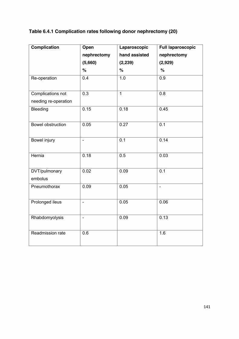
141
Table 6.4.1 Complication rates following donor nephrectomy (20) Complication Open
nephrectomy (5,660) %
Laparoscopic hand assisted (2,239) %
Full laparoscopic nephrectomy (2,929) %
Re-operation 0.4
1.0 0.9
Complications not
needing re-operation
0.3 1 0.8
Bleeding 0.15
0.18 0.45
Bowel obstruction 0.05
0.27 0.1
Bowel injury -
0.1 0.14
Hernia 0.18
0.5 0.03
DVT/pulmonary
embolus
0.02 0.09 0.1
Pneumothorax 0.09
0.05 -
Prolonged ileus - 0.05
0.06
Rhabdomyolysis - 0.09 0.13
Readmission rate 0.6 1.6
142
Of the four trials comparing open surgery and LDN, one did not report complications (22),
one found no difference in complication rates (23), one study of 122 patients found an 8%
major complication rate after TLDN compared with none after open surgery (24), and one
study of 84 patients found a complication rate of 0.3 per donor after TLDN compared with
0.6 per donor after open surgery (p=0.03) (25). A meta-analysis which included these
trials concluded that there was no difference in post-operative complication rates between
laparoscopic and open surgery (26). However, there is no trial comparing complication
rates after hand-assisted laparoscopic donor nephrectomy with open surgery.
Specific complications that require special mention include wound related problems such
as sepsis, hernia and chronic pain; the impact of conversion from laparoscopic to open
surgery (1-3%); blood loss and the requirement for blood and blood products (which
donors may find unacceptable e.g. Jehovah’s Witnesses); and finally the cosmetic
consequences, especially of open surgery.
Irrespective of the type of incision, wound pain is a major source of anxiety for the donor. The incidence of prolonged wound pain following laparoscopic surgery is difficult to
determine but a figure of 3.2% should be regarded as realistic (19). A small number of
patients may require referral to a pain clinic. A recent UK centre report of 123 donors
undergoing open nephrectomy reported that 12% of donors experienced chronic disabling
pain and 14% neuropathic pain (26).
6.5 Long Term Mortality
Counselling prospective living kidney donors about the potential of long-term risk to health
is an essential part of the pre-operative management of the prospective donor. Recent
data have shown that long term survival after donor nephrectomy is at least equal to a
matched cohort. A large study of over 80,000 donors reported through the mandatory
reporting system in the US, with only 24 donors excluded from follow-up, showed that
survival was no worse than that of a cohort of over 9,000 controls matched for age and
co-morbidity, over a period of 6 years (14). Similarly, a study of 3,698 donors showed no
difference in survival at up to 40 years after donation when compared with a group of age,
sex and ethnicity matched controls (27).
143
The best quality information on late mortality following donor nephrectomy comes from
Sweden (28). A single unit in Stockholm performed 459 living donor nephrectomies over a
20 year period from 1964 onwards. All 430 donors still living in Sweden were traced and
actual survival was compared to national mortality rates. The cause of death in the kidney
donors was similar to that seen in the general population: most deaths were due to
cardiovascular disease and cancer. Actuarial survival at 20 years was 85% compared to
an expected survival rate of 66%. This result suggests that the donor work up in
Stockholm ensured that only healthy individuals proceeded to donation and encouraged
the authors to select as a title for their publication ‘Kidney donors live longer’.
6.6 Pre-operative Care and Preparation 6.6.1 General Considerations Living donor surgery must be carried out by a team with adequate expertise, in an
environment where donors are regularly cared for. A senior anaesthetist with experience
of managing such patients should be present. It is recommended that a transplant unit
should undertake at least 20-30 living donor operations per annum, to ensure that
adequate expertise is maintained, and should regularly audit its results. Each donor
surgeon should maintain up to date surgical experience, and should also audit his or her
individual results.
6.6.2 DVT Prophylaxis Deep venous thrombosis and pulmonary embolism remain major causes of morbidity and
mortality after major surgery, and living kidney donors are no exception to this. They
should be classified as ‘medium risk’ patients, even if undergoing laparoscopic surgery
and the NICE approved thromboprophylaxis policy should be followed (29). This entails
applying the DH risk assessment tool to all donors on admission and grading the ‘relative
risk’ of venous thromboembolism (VTE), which includes the potential risk of bleeding and
which will help to inform the best form of prophylaxis. Factors such as age >60 years,
dehydration, known thrombophilia, obesity (BMI >30 kg/m2), personal history or first-
degree relative with a history of VTE, use of HRT, use of oestrogen-containing
contraceptive therapy, and varicose veins with phlebitis must all be taken into account.
Details are available at http://guidance.nice.org.uk/CG92.

144
The relative risk of VTE with laparoscopic versus open procedures has not yet been
investigated in depth. Based on the pathophysiology of VTE, factors that may heighten the
risk with laparoscopy are the duration of the procedure (>90 minutes), patient positioning,
and the effect of the pneumoperitoneum. Conversely, shorter hospital stays and more
rapid post operative mobilisation should decrease the risk (30). Typically this will mean the
use of mechanical compression during surgery and both TED stockings and LMWH
following surgery until discharge (31).
Early mobilisation (on the first postoperative day) is recommended. Donors with a
personal history of DVT or PE who undergo surgery are at high risk of developing further
venous thromboembolism (30% within 5 years) and should be screened to exclude
significant thrombophilia, as should any potential donors with a family history (first or
second degree relative) of VTE. In such cases, donation may not be precluded but advice
should be sought from a haematologist (32). Any donors deemed high risk should have
prolonged prophylaxis following discharge for at least 7 to 14 days. New oral anti-
thrombotic agents should be considered in the future, such as dabigatran etexilate.
6.6.3 Prophylactic Antibiotics
There is no evidence for the use of prophylactic antibiotics in donor surgery, although
some centres do use a single dose at induction. Local practice should be followed and
further research is required in order to make a definitive recommendation.
6.6.4 Consent and Site Marking Standard practice for major surgery is to seek written consent prior to admission, and
reconfirm this on admission for surgery. The site should be marked and confirmed with the
patient before leaving the ward for theatre. The appropriate imaging must be available in
theatre and standard safety checks, usually involving the WHO checklist (33), should be
performed prior to the start of surgery.
6.6.5 Blood Transfusion Blood is rarely needed during donor nephrectomy, but when it is the case it may be
needed urgently. All donors should be ‘group and saved’ and surgery should only take
place where adequate facilities for provision of urgent blood products are available. All
donors should be counselled about the potential risk of bleeding and the use of blood and
blood products, especially donors with specific religious affiliation such as Jehovah’s
145
Witnesses. Where blood transfusion is refused or contraindicated, the use of a cell saver
may be indicated.
6.7 Donor Nephrectomy 6.7.1 General Considerations A Consultant surgeon should perform or supervise living donor surgery. Donor and
recipient operations may be carried out sequentially or using parallel lists; the latter is
preferred where complex, high risk recipient surgery is undertaken, to ensure that the
recipient operation can continue before kidney extraction is performed. Whichever method
is employed, dedicated elective lists must be available.
In the majority of UK centres, the donor and recipient operations are undertaken
synchronously in parallel operating theatres staffed by two full teams of theatre personnel.
This minimises cold ischaemic time and ensures that the kidney is removed from the
donor only after it has been confirmed that there are no unforeseen problems with the
recipient that might prevent implantation. Sequential donor and recipient operations are
also acceptable and have been shown to give equivalent outcomes in several
uncontrolled and one controlled series (34).
6.7.2 Peri-operative Fluids There is some evidence that aggressive peri-operative fluid management is beneficial in
laparoscopic donor nephrectomy (35). In this small randomised trial of 24 patients, pre-
operative hydration prevented the intra-operative reduction in stroke volume and
creatinine clearance seen in the control group. Ideally, hydration should involve an
overnight infusion of fluid and a bolus during surgery. Some units use intravenous
mannitol or loop diuretics during surgery but there is limited human evidence for this. A
useful approach is to use trans-oesophageal Doppler guidance to titrate intravenous fluid
replacement, and this is used by some transplant centres in the UK (36,37).
6.7.3 Type of Surgery Donor nephrectomy may be performed using a standard open technique, a mini-incision
(MODN), or laparoscopically, using a hand-assisted (HLDN) or ‘totally laparoscopic’
(TLDN) approach. The minimally invasive techniques (TLDN, HLDN) have now been
widely adopted as the standard surgical approach by many transplant units in the UK and
146
worldwide. Laparoscopic operations may be performed trans- or retro-peritoneally. There
is one randomised trial of MODN versus laparoscopic donor nephrectomy (20), and one
meta-analysis (21). There are four randomised trials of laparoscopic versus open surgery
(22,23,25,38) and one meta-analysis (39). Laparoscopic surgery has been shown to result
in a quicker recovery, shorter hospital stay (by 1.6 days, p<0.001) and less pain and a
quicker return to work (by 2.4 weeks, p<0.001) than standard open surgery, with
comparable complication rates (39). Operative time may, however, be longer after
laparoscopic surgery. Mini-incision surgery results in a quicker recovery compared with
open surgery, but more pain than laparoscopic surgery (21). One small study compared
HLDN with TLDN and found no difference in outcomes (38). An ongoing trial (the HARP
study) will compare retroperitoneal HLDN with transperitoneal TLDN (40).
6.7.4 Preferred Kidney and Vasculature The left kidney is usually preferred, assuming both kidneys have equal numbers of
arteries, due to the greater length of the left renal vein. One randomised trial comparing
right and left laparoscopic donor nephrectomy showed no difference in complication rates
but a shorter operating time for right nephrectomy (41). Great care must be taken when
selecting which kidney to remove. Surgery on a kidney with a short artery risks
encroaching on the bifurcation of the renal artery and may result in two arteries to implant
which is technically more challenging; while efforts to secure a single vessel in the donor
may result in avulsion of the artery from the aorta with associated risks to the donor.
It is recommended that the side of donor nephrectomy is selected and documented at a
multi-disciplinary meeting which includes a review of the vascular imaging, and that the
potential donor is informed of any increased risk associated with this decision. Numerous
reports exist showing safe and successful laparoscopic surgery in the presence of multiple
arteries and veins, as well as in obese patients. Although there are increased anaesthetic
and peri-operative risks in patients with these conditions, they do not constitute absolute
contraindications to laparoscopic surgery.
6.7.5 Training for Laparoscopic Surgery Laparoscopic donor nephrectomy is a complex operation with potentially high risks. It
should only be undertaken by those who have been appropriately trained in the technique,
and should not be performed on an occasional basis. Difficult cases may require help from
an experienced mentor.

147
6.7.6 Pain Relief Early post-operative pain is the most frequent complaint after living donor surgery. The
laparoscopic technique does not require the use of an epidural, but PCA and a ‘pain
ladder’ approach may be useful. Early mobilisation, ideally the day after surgery, is
preferred, and in this regard newer pump devices which provide continuous analgesia for
mobile patients may be useful. References 1. Hsu TH, Su LM, Ratner LE, Trock BJ, Kavoussi LR. Impact of renal artery multiplicity
on outcomes of renal donors and recipients in laparoscopic donor nephrectomy.
Urology 2003; 61: 323-7.
2. Pei Y, Obaji J, Dupuis A, et al. Unified criteria for ultrasonographic diagnosis of
ADPKD. J Am Soc Nephrol 2009; 20: 205-12.
3. Weinstein SH, Navarre RJ, Loening SA, Corry RJ. Experiences with live donor
nephrectomy. J Urol 1980; 124: 321-3.
4. Kälble T, Lucan M, Nicita G, Sells R, Burgos Revilla FJ, Wiesel M. EAU guidelines on
renal transplantation. Eur Urol 2005; 47: 156-66.
5. Rajamahanty S, Simon R, Edye M, Butt K, Eshghi M. Accuracy of three-dimensional
CT angiography for preoperative vascular evaluation of laparoscopic living renal
donors. Endourol 2005; 19: 339-41.
6. Lewis GR, Mulcahy K, Brook NR, Veitch PS, Nicholson ML. A prospective study of
the predictive power of spiral computed tomographic angiography for defining renal
vascular anatomy before live-donor nephrectomy. BJU Int 2004; 94: 1077-81.
7. Kim JC, Kim CD, Jang MH, et al. Can magnetic resonance angiogram be a reliable
alternative for donor evaluation for laparoscopic nephrectomy? Clin Transplant 2007;
21: 126-35.
8. Israel GM, Lee VS, Edye M, et al. Comprehensive MR imaging in the preoperative
evaluation of living donor candidates for laparoscopic nephrectomy: initial experience.
Radiology 2002; 225: 427-32.
9. Gluecker TM, Mayr M, Schwarz J, et al. Comparison of CT angiography with MR
angiography in the preoperative assessment of living kidney donors. Transplantation
2008; 88; 1249-56.
10. Bia MJ, Ramos EL, Danovitch GM, et al. Evaluation of living renal donors. The
current practice of US transplant centers. Transplantation 1995; 60: 322-7.

148
11. Najarian JS, Chavers BM, McHugh LE, Matas AJ. 20 years or more of follow-up of
living kidney donors. Lancet 1992; 340: 807-10.
12. Bay WH, Hebert LA. The living donor in kidney transplantation. Ann Intern Med 1987;
106: 719-27.
13. Kasiske BL, Ravenscraft M, Ramos EL, Gaston RS, Bla MJ, Danovitch GM. The
evaluation of living renal transplant donors: clinical practice guidelines. J Am Soc
Nephrol 1996; 7: 2288-313.
14. Segev DL, Muzaale AD, Caffo BS, et al. Perioperative mortality and long-term
survival following live kidney donation. JAMA. 2010; 303: 959-66.
15. Hadjianastassiou VG, Johnson RJ, Rudge CJ, Mamode N. 2509 living donor
nephrectomies, morbidity and mortality, including the UK introduction of laparoscopic
donor surgery. Am J Transplant. 2007; 7: 2532-7.
16. Bakran A. Postal survey of living donor kidney transplant units. Presented at the
Symposium “Meeting the challenges of live donation”. Royal College of Physicians,
21 April 1998.
17. Bennett AH, Harrison JH. Experience with living familial renal donors. Surg Gynecol
Obstet 1974; 139: 894-8.
18. Uehling DT, Malek GH, Wear JB. Complications of donor nephrectomy. J Urol 1974;
111: 745-6.
19. Matas AJ, Bartlett ST, Leichtman AB, Delmonico FL. Morbidity and mortality after
living kidney donation, 1999-2001: survey of United States transplant centers. Am J
Transplant. 2003; 3: 830-4.
20. Kok NF, Lind MY, Hansson BM, et al. Comparison of laparoscopic and mini incision
open donor nephrectomy: single blind, randomised controlled clinical trial. BMJ 2006;
333: 221.
21. Antcliffe D, Nanidis TG, Darzi AW, Tekkis PP, Papalois VE. A meta-analysis of mini-
open versus standard open and laparoscopic living donor nephrectomy. Transpl Int
2009; 22: 463-74.
22. Wolf JS Jr, Merion RM, Leichtman AB, et al. Randomized controlled trial of hand-
assisted laparoscopic versus open surgical live donor nephrectomy. Transplantation
2001; 72: 284-90.
23. Simforoosh N, Basiri A, Tabibi A, Shakhssalim N, Hosseini Moghaddam SM.
Comparison of laparoscopic and open donor nephrectomy: a randomized controlled
trial. BJU Int 2005; 95: 851-5.

149
24. Øyen O, Andersen M, Mathisen L, et al. Laparoscopic versus open living-donor
nephrectomy: experiences from a prospective, randomized, single-center study
focusing on donor safety. Transplantation 2005; 79: 1236-40.
25. Nicholson ML, Kaushik M, Lewis GR. Randomized clinical trial of laparoscopic versus
open donor nephrectomy. Br J Surg 2010; 97: 21-8.
26. Owen M, Lorgelly P, Serpell M. Chronic pain following donor nephrectomy – a study
of incidence, nature and impact of chronic post nephrectomy pain. Eur J Pain 2010;
14: 732-4.
27. Ibrahim HN, Foley R, Tan L, et al. Long-term consequences of kidney donation. N
Engl J Med 2009; 360: 459-69.
28. Fehrman-Ekholm I, Elinder C-G, Stenbeck M, Tyden G, Groth CG. Kidney donors live
longer. Transplantation 1997; 64: 976-8.
29. NICE Guidance on VTE. www.nice.org.uk/guidance/CG92
30. Kakkar AK. Prevention of venous thromboembolism in general surgery. In: Colman
RW, Clowes AW, George JN, Goldhaber SZ, Marder VJ, eds. Hemostasis and
Thrombosis: Basic Principles and Clinical Practice. 5th ed. Philadelphia, PA:
Lippincott, Williams & Wilkins; 2006: 1361-7.
31. Roderick P, Ferris G, Wilson K, et al. Towards evidence-based guidelines for the
prevention of venous thromboembolism: systematic reviews of mechanical methods,
oral anticoagulation, dextran and regional anaesthesia as thromboprophylaxis. Health
Technol Assess 2005; 9: iii-iv, ix-x, 1-78.
32. British Society for Haematology (BSCH) Guidelines, Investigation and Management
of Heritable Thrombophilia, 2001. www.bcshguidelines.com/guidelinesMENU.asp
33. Editorial. WHO's patient-safety checklist for surgery. Lancet 2008: 372; 1148-9.
34. Baverstock RJ, Manson AD, Liu L, Gourlay WA. A prospective comparison of
simultaneous and sequential live-donor renal transplantation. Transplantation 2002;
74: 1194-7.
35. Mertens zur Borg IR, Di Biase M, Verbrugge S, Ijzermans JN, Gommers D.
Comparison of three perioperative fluid regimes for laparoscopic donor nephrectomy:
A prospective randomized dose-finding study. Surg Endosc 2008; 22: 146-50.
36. Abbas SM, Hill AG. Systematic review of the literature for the use of oesophageal
Doppler monitor for fluid replacement in major abdominal surgery. Anaesthesia 2008;
63: 44-51.
37. Walsh SR, Tang T, Bass S, Gaunt ME. Doppler-guided intra-operative fluid
management during major abdominal surgery: systematic review and meta-analysis.
Int J Clin Pract 2008; 62: 466-470.
150
38. Andersen MH, Mathisen L, Oyen O, et al. Postoperative pain and convalescence in
living kidney donors - laparoscopic versus open donor nephrectomy: a randomized
study. Am J Transplant 2006; 6: 1438-43.
39. Nanidis TG, Antcliffe D, Kokkinos C. Laparoscopic versus open live donor
nephrectomy in renal transplantation: a meta-analysis. Ann Surg 2008; 247: 58-70.19.
40. Dols LF, Kok NF, Terkivatan T, et al. Hand-assisted retroperitoneoscopic versus
standard laparoscopic donor nephrectomy: HARP-trial. BMC Surg 2010; 10: 11.
41. Minnee RC, Bemelman WA, Maartense S, Bemelman FJ, Gouma DJ, Idu MM. Left or
right kidney in hand-assisted donor nephrectomy? A randomized controlled trial.
Transplantation 2008; 85: 203-8.

151
CHAPTER 7 HISTOCOMPATIBILITY TESTING FOR LIVING DONOR KIDNEY TRANSPLANTATION
Statements of Recommendation
Initial assessment of donor and recipient histocompatibility status should be undertaken at an early stage in living donor kidney transplant workup to avoid unnecessary and invasive clinical investigations. (B2)
Screening of potential living donor kidney transplant recipients for clinically
relevant antibodies is important for ensuring optimal donor selection and graft survival. (A1)
Antibody screening is especially important when potential living donor
recipients reduce or withdraw immunosuppression. (B2)
Post-transplant antibody monitoring should be undertaken according to the BSHI/BTS guidelines. (B1)
Transplant units and histocompatibility laboratories should agree an
evidence-based protocol to define crossmatch results which constitute a veto to transplantation. (B2)
A pre-transplant serum sample collected within 14 days of the planned date
for transplantation must be tested in a sensitive crossmatch and if the crossmatch test is positive transplantation should not usually be performed, unless the antibody is shown to be indicative of acceptable immunological risk. (A1)
Changes in immunosuppression during the transplant work-up should be
notified to the histocompatibility laboratory and additional antibody screening and donor-recipient crossmatch tests undertaken as required. (B1)
152
HLA matching should be considered of benefit when there is an option of selecting between living donors, particularly in reducing the possibility of subsequent sensitisation. This is important for younger recipients where repeat transplantation may be required. However, it is recognised that other donor factors will be taken into account. (B1)
The histocompatibility laboratory should issue an interpretive report stating
the donor and recipient HLA mismatch, recipient sensitisation status and crossmatch results, and define the associated immunological risk for all living donor-recipient pairs. (A1)
Policies defining histocompatibility requirements for living donor kidney transplantation
should be jointly established between the clinical transplant team and the consultant
histocompatibility scientist in each centre. There are three components to the
histocompatibility assessment: determination of donor-recipient HLA mismatch status;
identification of alloantibodies in patient serum that could be potentially harmful to a
transplanted organ; and confirmation of antibody compatibility by performing a donor-
recipient crossmatch. The results of these investigations provide a risk assessment, which
together with clinical information provide guidance on the suitability of a particular living
kidney donor-recipient pair for transplantation. These guidelines are applicable to ABO
blood group compatible, HLA antibody compatible transplants and are to be read in
conjunction with the BSHI/BTS ‘Guidelines for the Detection and Characterisation of
Clinically Relevant Antibodies in Allotransplantation’ (1). The BTS has separate guidelines
for ABO Blood Group and HLA Antibody Incompatible (HLAi) Transplantation (2). Initial assessment of donor and recipient histocompatibility status should be undertaken at
an early stage in the donor workup to avoid unnecessary and invasive clinical
investigations. Histocompatibility assessments and interpretation of test results should
only be undertaken in an appropriately accredited laboratory (e.g. Clinical Pathology
Accreditation (CPA)/European Federation for Immunogenetics (EFI)) by scientists with
specialist training in Histocompatibility & Immunogenetics, as demonstrated by FRCPath
or equivalent level qualification and experience. The onus is on the referring centre to
provide appropriate information and donor and recipient samples necessary to fulfil these
guidelines.
153
7.1 Assessment of Donor-Recipient HLA Mismatch Status
In the absence of preconditioning protocols, the choice of a living donor is restricted by the
requirement for ABO blood group compatibility. HLA typing of the recipient and all
potential living donors should be performed using DNA-based methods to at least two digit
(low) resolution for HLA-A, -B, -C, -DR and -DQ and the donor-recipient mismatch
determined. The level of donor and recipient HLA compatibility is usually expressed as an
HLA-A, -B and -DR mismatch grade determined from the number of donor HLA
specificities at each locus that are absent in the recipient. A donor and recipient with no
HLA-A, -B, -DR incompatibilities is denoted ‘000’, whereas a fully mismatched
combination is denoted ‘222’. In the case of transplants between siblings there is a 1 in 4
chance of inheriting the same two HLA bearing parental haplotypes, a 1 in 2 chance of
sharing one parental haplotype and a 1 in 4 chance of sharing no parental haplotypes.
Thus in the case of genetically related donors, ABO blood group and HLA typing results
can indicate the familial relationship and therefore informed consent must be obtained by
the referring centre from both the recipient and all genetically related potential donors
before these tests are undertaken (see section 4.3).
Selection of the most suitable donor for a particular recipient is complex and the HLA
mismatch grade will be considered together with other factors such as donor and recipient
age and alternative options for transplantation both now and in the future (section 7.4 and
Chapter 11).
7.2 Identification and Characterisation of Alloantibodies Pre-transplant antibody screening The presence of pre-transplant HLA specific antibodies that are reactive against
mismatched donor HLA is potentially harmful to a transplanted kidney and therefore a
policy for the detection of such antibodies must be rigorously implemented. Immunological
sensitisation can arise through exposure of the potential recipient to allogeneic tissue
bearing foreign HLA, such as transfusion of blood products, pregnancy (including
miscarriage and terminated pregnancy), and previous transplants. HLA specific
alloantibodies can also arise naturally through crossreactivity with pathogens, when they
are termed idiopathic antibodies.
154
It is essential for the laboratory to have accurate information about the timing and nature
of all potential allosensitisation events, throughout the patient’s lifetime. Recent and past
potential allosensitisation events, including recent infections, must be documented by the
referring clinical team and reported to the histocompatibility laboratory. Recipient serum
samples must be obtained for HLA specific antibody screening at least every three
months, and additional samples collected at 14 and 28 days after transfusion of any blood
products. Potential recipients who are receiving immunosuppression while being
assessed for living kidney transplantation are at high risk of de-novo sensitisation,
particularly if immunosuppression is changed, reduced or withdrawn. It is the responsibility
of the clinical team to notify the histocompatibility laboratory of such changes and
additional serum samples should be obtained for HLA specific antibody screening at four
weeks after any change in immunosuppression.
Recipient sera must be tested for HLA specific alloantibodies according to the BSHI/BTS
guidelines (1) and HLA specificities to which the patient is sensitised should be identified
as unacceptable mismatches. In cases where HLA-DP specific antibodies are detected in
recipient serum, donor-recipient HLA-DP status and potential HLA-DP specific antibody
incompatibility should be determined. Recipients that have donor HLA specific antibodies
(unacceptable mismatches) identified in recent and/or past (historic) serum samples
should be referred for formal immunological risk assessment by the clinical team and,
where appropriate, may be considered for HLAi transplantation. These discussions should
take place at the earliest opportunity, to avoid delay and unnecessary clinical
investigation.
In many cases, the living donor kidney transplant workup may be prolonged and it is not
uncommon for a year or more to elapse between the initial histocompatibility assessment
and the planned operation. During this period, the antibody compatibility status of the
potential recipient and donor(s) must be monitored and any changes in the patient’s
antibody profile should be reported to the transplant team. The recipient must have
contemporary antibody screening results available using samples obtained within three
months of the transplant operation. Any potential alloantibody priming events that occur
within one month of the latest antibody screening sample, or after the sample collection
date could change the donor-recipient antibody compatibility status and will obviate all
previous results.
155
Post-transplant antibody screening Monitoring of HLA specific antibodies in recipient serum after the transplant operation can
provide helpful prognostic information for the diagnosis of antibody-mediated rejection and
to guide post-transplant rejection treatment, antibody reduction therapy and choice of
maintenance immunosuppressive therapy. Post-transplant antibody monitoring should be
undertaken according to the BSHI/BTS guidelines (1).
7.3 Pre-transplant Donor-Recipient Crossmatch Test A prospective pre-transplant donor-recipient crossmatch test is performed to confirm the
presence or absence of donor HLA specific alloantibodies. The results can only be
interpreted in conjunction with knowledge of pre-transplant alloantibody priming events,
donor-recipient HLA mismatches and pre-transplant antibody screening results. In the
case of donor-recipient combinations where donor HLA specific antibodies are present in
recipient serum, the crossmatch test can provide information about antibody levels and
the associated immunological risk (3). Pre-formed donor HLA specific antibodies present
in recipient serum can cause hyperacute and acute rejection and there should be close
liaison between the histocompatibility laboratory and the clinical team.
Living kidney donor crossmatch tests should be carried out according to the BSHI/BTS
Guidelines for the Detection and Characterisation of Clinically Relevant Antibodies, and
tested using lymphocytes isolated from donor peripheral blood (1). Because of the
opportunity for planned living donor transplant work-up, a virtual crossmatch is not
acceptable. Living donor crossmatch testing is usually carried out at the time of first
referral. The final crossmatch must always be undertaken using a serum sample obtained
within 14 days of the planned operation date. This time frame minimises the risk of a
change in recipient antibody status, but any potential alloantibody priming event around
the time of the final crossmatch will obviate the results.
The selection of recipient serum samples for crossmatch and choice of target cell type
(i.e. donor peripheral blood lymphocytes, isolated donor T lymphocytes and/or
B lymphocytes) and the technique used (complement dependant lymphocytotoxicity
[CDC] and/or flow cytometric [FC] crossmatch) will depend on previous alloantibody
priming events and pre-transplant antibody screening results, and should conform to the
BSHI/BTS guidelines (1). It is recommended that allosensitised recipients with pre-formed
156
HLA class I and/or class II specific alloantibodies and recipients awaiting repeat
transplantation should undergo donor T lymphocyte (for HLA class I sensitised patients) or
T and B lymphocyte (for HLA class II sensitised patients) flow cytometric crossmatching
as a minimum. Undertaking a CDC donor T and B lymphocyte crossmatch using
untreated and dithiothreitol (DTT) treated recipient serum can provide further information
for risk stratification (3). Result interpretation and acceptable immunological risk
stratification should be undertaken according to local policy and BSHI/BTS guidelines. A
positive donor lymphocyte crossmatch test performed using DTT treated sera by CDC
carries a high immunological risk of hyperacute rejection and constitutes a veto to
transplantation, unless an effective HLAi strategy is used to minimise the risk of graft
failure.
Careful consideration must be given to the sensitisation status and crossmatch results for
proposed transplants where recipient allosensitisation priming has previously occurred
through exposure to the donor HLA, either directly (e.g. offspring donor to mother) or
indirectly (shared donor HLA haplotype in spousal/partner donation to female recipient
following pregnancy, or repeat transplants using a second related donor). The occurrence
of an anamnestic immune activation of latent donor alloantigen specific lymphocytes and
uncontrolled graft rejection has been observed following crossmatch negative male to
female spousal transplantation and this risk may be pre-empted and minimised by using
sensitive antibody screening methods, appropriate crossmatch techniques and tailored
immunosuppression.
A further important consideration relates to patients undergoing living donor kidney
transplant assessment following a previous failed or failing kidney transplant that remains
in situ. Such patients often have immunosuppression reduced or withdrawn during the
period of clinical workup, because of a desire to reduce unnecessary medication. This is
frequently associated with the development of de-novo HLA specific antibodies to the
allograft which cause a previously unexpected positive crossmatch and which then
preclude future transplantation from an HLA mismatched living donor. Consideration must
be given to the relative risk of maintaining recipient immunosuppression during the donor
workup, the benefit of immunosuppressive drug reduction or withdrawal, and the risk of
de-novo allosensitisation. A reduction or cessation of immunosuppression within one-
month of the planned operation date is contraindicated and may delay or preclude
transplantation. As a minimum, this would necessitate additional antibody screening and
157
donor-recipient crossmatch tests to be undertaken using a current serum sample obtained
within 24 hours before the transplant operation.
7.4 Selection of Suitable Donor-Recipient Pairs The presence of donor specific HLA antibodies or a positive crossmatch in a sensitised
patient is a contraindication to transplantation unless desensitisation protocols are
employed. In a sensitised patient, a well matched donor is more likely to be antibody
compatible than a poorly matched donor. Transplants between siblings offer the best
opportunity for a well matched graft because of familial inheritance of HLA genes. As
described above, kidney transplants from offspring to mother or from a father to the
mother of his children should be approached with caution, but where HLA sensitisation is
excluded and a negative crossmatch achieved, transplant outcomes are equivalent to
those for other non-HLA identical living donor transplants (4,5).
A widely cited publication of the experience of living unrelated spousal donor kidney
transplantation in North America showed that graft survival rates for such transplants was
equivalent to that of HLA mismatched living related donor kidney transplants (5). This
equates with the current UK experience (see Chapter 11). The Collaborative Transplant
Study (CTS) found a significant reduction in graft survival when living donor kidney
transplants were mismatched at HLA-A, -B and -DR (4). CTS analysis of more than 5,000
living unrelated donor transplants performed between 1995 and 2002 showed a highly
significant influence of HLA matching on graft survival (6), but survival of even the worst
matched kidneys was better than seen in deceased donor transplantation. However, a
more recent analysis of the UK Transplant registry of living donor kidney transplants
performed between 2000 and 2007 did not show an influence of HLA matching on
transplant outcome (7).
A key point is that when a poorly matched kidney transplant fails because of rejection, the
recipient is at high risk of becoming highly sensitised (1), restricting options for repeat
transplantation. This is particularly relevant for paediatric recipients and young adults who
are likely to require re-transplantation within their lifetime and for whom avoiding
sensitisation, particularly to common antigens, is important. Children are often registered
on the transplant list with mismatched parental HLA specificities listed as unacceptable to
avoid sensitisation against these prospective living donors. In contrast, in the context of
158
older spouse couples where a second transplant is unlikely, the risk of sensitisation is not
a major concern.
7.5 Antibody Incompatible Living Donor Transplantation Antibody incompatible transplantation (AiT) may be an option for some patients who have
a potential living donor but where there is a specific immunological barrier to
transplantation. Such transplantation falls into two categories: ABO incompatible
transplantation, where transplantation occurs across an ABO blood group barrier (e.g.
from a blood group B donor to a blood group O recipient); and HLA-incompatible
transplantation, where the recipient has high titres of antibodies against one or more
specific HLA antigens present in the donor.
Both forms of transplantation are becoming more common in the UK and will in future
make major contributions to expansion of the living donor pool. Close liaison between
clinicians and histocompatibility laboratories is obviously critical for such transplantation,
which should be initially concentrated in units with particular expertise.
The BTS has published specific guidelines on antibody incompatible transplantation,
which should be referred to (2). The following summary points are derived from these
guidelines:
Recommendations (Not graded)
Antibody incompatible transplantation (AiT) should be considered as part of an ongoing structured programme, and should not be performed on an occasional basis.
To initiate a programme, a unit should be able to demonstrate a demand of at least
5 cases a year and appropriate support from clinical transplant, plasmapheresis and histocompatibility teams. An AiT programme requires funding for additional staff and consumables, and all programmes should receive Commissioner support.
159
There is insufficient evidence to make precise recommendations for treatment protocols, but units should have a written protocol based on best published practice. This should include recommendations on prevention, diagnosis and treatment of antibody mediated rejection.
Protocols that follow the above can be regarded as established treatment and do
not require Ethics Committee approval as research procedures. However, the standard of consent should include detailed written information which describes the risks of the procedure. The transplant donor should receive equivalent information to the recipient, so they are aware of the risks of the procedure to the recipient, whether it results in a transplant or not. Potential recipients and donors should be aware of their treatment choices, especially the option of exchange (pooled/paired) transplantation.
Laboratories should be able to define antibodies to the standard defined in the
BSHI/BTS document ‘Guidelines for the Detection and Characterisation of
Clinically Relevant Antibodies in Solid Organ Transplantation’. Sensitive and rapid
techniques for the measurement of donor-specific HLA antibody levels must be available.
If ABOi transplantation is to be performed, blood group antibody titres need to be
measured, with differentiation between A1 and A2 subgroups of recipient blood group A (when appropriate) and discrimination between IgG and IgM specific for ABO antibodies. In living donor transplantation, a 7 day per week service with same day turn-around time is required.
AiT is able to provide successful transplantation for significant numbers of patients,
potentially up to 20% of the total living donor transplant programme nationally. AiT should be supported because of the improvements in quality of life after
transplantation compared to dialysis. Additionally, many patients receiving antibody incompatible transplants may have no other chance of a transplant. Transplantation is cost effective over time with a saving of about £15,000 per annum compared to dialysis when averaged over a 10 year period

160
Every patient undergoing antibody incompatible transplantation should be audited on a local and national basis, with the national audit through the AiT Registry.
The UK AiT Registry will define the optimal dataset to be collected, and will be
able to report AiT activity against benchmark outcome data from international reports and the national dataset of renal transplantation.
References 1. Guidelines for the detection and characterisation of clinically relevant antibodies in
allotransplantation. British Society for Histocompatibility and Immunogenetics and
British Transplantation Society, 2010.
http://bts.demo.eibs.co.uk/transplantation/standards-and-guidelines/
2. Guidelines for Antibody Incompatible Transplantation. British Transplantation Society.
http://bts.demo.eibs.co.uk/transplantation/standards-and-guidelines/
3. Taylor CJ, Kosmoliaptsis V, Summers DM, Bradley JA. Back to the future: application
of contemporary technology to longstanding questions about the clinical relevance of
human leukocyte antigen-specific alloantibodies in renal transplantation. Human
Immunol 2009; 70: 563-8.
4. Opelz G. Impact of HLA compatibility on survival of kidney transplants from unrelated
live donors. Transplantation 1997; 64: 1473-5.
5. Terasaki PI, Cecka JM, Gjertson DW, Cho YW. Spousal and other living donor
transplants. In: Clinical Transplants 1997. Eds Cecka JM, Terasaki PI. UCLA Tissue
Typing Laboratory, Los Angeles, USA. pp 269-84.
6. Collaborative Transplant Study Newsletter 2004; 2, May 1. www.ctstransplant.org
7. Fuggle SV, Allen JE, Johnson RJ, et al. Factors affecting graft and patient survival
after live donor kidney transplantation in the UK. Transplantation 2010; 89: 694-701.
161
CHAPTER 8 EXPANDING THE DONOR POOL Statements of Recommendation
All donor and recipient pairs who are incompatible by blood group and/or HLA type are entitled to a full appraisal of the available treatment options, tailored to their individual circumstances, so that they can make an informed decision about their choice of treatment. (C1)
Coherent organisational processes and clinical practices between transplant
centres are essential to optimise the benefits of living donor kidney sharing schemes and to maximise the number of potential transplants that proceed. (B1)
8.1 Paired/Pooled Living Donation From September 2006, the Human Tissue Act enabled paired donation in the UK.
A national scheme was established whereby incompatible donor-recipient pairs can
exchange kidneys so that recipients can receive alternative compatible living donor
organs. Exchanges are identified between two or three incompatible pairs (Figure 8.1). It
is also possible to register compatible pairs into the scheme in order to achieve a more
favourable HLA or age match for the recipient concerned.
The paired/pooled scheme presents some logistical challenges which need careful co-
ordination and administration to ensure that the use of kidneys is optimised and maximum
patient benefit is achieved.
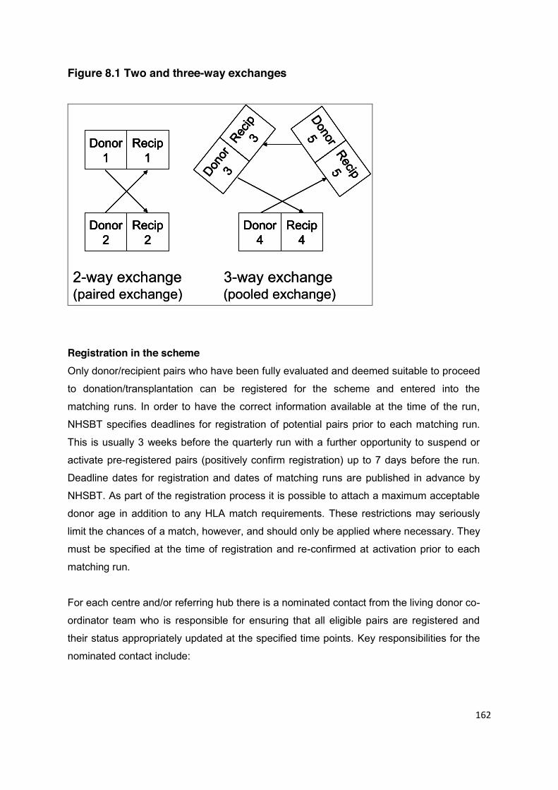
162
Figure 8.1 Two and three-way exchanges
Donor1
Recip1
Donor2
Recip2
Donor
3Rec
ip3
Donor4
Recip4
Donor5
Recip5
2-way exchange 3-way exchange(paired exchange) (pooled exchange)
Donor1
Recip1
Donor2
Recip2
Donor
3Rec
ip3
Donor4
Recip4
Donor5
Recip5
Donor1
Recip1
Donor2
Recip2
Donor1
Recip1
Donor1
Recip1
Donor2
Recip2
Donor2
Recip2
Donor
3Rec
ip3
Donor4
Recip4
Donor5
Recip5Donor
3Rec
ip3
Donor
3Rec
ip3
Donor4
Recip4
Donor4
Recip4
Donor5
Recip5Donor5
Recip5
2-way exchange 3-way exchange(paired exchange) (pooled exchange)
Registration in the scheme Only donor/recipient pairs who have been fully evaluated and deemed suitable to proceed
to donation/transplantation can be registered for the scheme and entered into the
matching runs. In order to have the correct information available at the time of the run,
NHSBT specifies deadlines for registration of potential pairs prior to each matching run.
This is usually 3 weeks before the quarterly run with a further opportunity to suspend or
activate pre-registered pairs (positively confirm registration) up to 7 days before the run.
Deadline dates for registration and dates of matching runs are published in advance by
NHSBT. As part of the registration process it is possible to attach a maximum acceptable
donor age in addition to any HLA match requirements. These restrictions may seriously
limit the chances of a match, however, and should only be applied where necessary. They
must be specified at the time of registration and re-confirmed at activation prior to each
matching run.
For each centre and/or referring hub there is a nominated contact from the living donor co-
ordinator team who is responsible for ensuring that all eligible pairs are registered and
their status appropriately updated at the specified time points. Key responsibilities for the
nominated contact include:

163
Close collaboration with the histocompatibility and immunogenetics (tissue typing)
laboratory to ensure HLA antibody screening is up to date before each matching
run.
Close collaboration with clinical colleagues to ensure that donor & recipient
assessment is up to date.
Particular donor information that is relevant to the acceptance of a kidney by a
recipient centre should be cited with the registration. These include the presence of
complex donor vasculature, borderline GFR, stone disease in the kidney to be
donated, and donor hepatitis B core antibody positivity.
Contact with both donor and recipient individually to confirm their commitment to
enter/remain in the scheme and to ensure that no issues have emerged since the
last matching run which might preclude them. It is particularly important that
donor/recipient pairs understand the implications and expectations of participation in
the scheme and the impact of late withdrawal (after pairs have been matched) on
other pairs should they decide not to proceed. This should not override their right to
withdraw consent at any time, but is sensible to highlight in advance to minimise the
risk of later ambivalence.
Collation and confirmation of information to register/positively confirm the
participation of relevant pairs in the scheme at the notified times. This includes
specifying preferences for acceptable HLA and age match criteria, if relevant (see
above).
Matching runs There are currently four matching runs per year, at approximately quarterly intervals. At
the time of a matching run, all potential matches within the pool are identified and
evaluated according to a scoring system. Scoring is necessary to decide, in some cases,
which of multiple possible transplants for a single recipient is optimal. Identification of
possible matches takes into account any donor age or HLA restrictions specified at the
time of registration. Scoring is based on a number of factors including the calculated level
of sensitisation (to promote matches for sensitised patients where such are identified); the
HLA mismatch level of the potential transplant (to promote good matching where
possible); and the age difference between the two donors. The latter acts as a tie-breaker
and ensures that, as far as possible, the exchange is fair in terms of expected outcome. It
is inevitable that the scheme will evolve, however, and up-to-date matching arrangements
can be found on the ODT website (www.organdonation.nhs.uk).
164
Experts in matching algorithms collaborate with NHSBT to ensure that the number of
transplants and the scores of different possible alternatives are optimised over all
possibilities involving both 2-way and 3-way exchanges. There are usually 100-160 pairs
in any one matching run and typically 10-30 transplants may be identified.
When a matching run has taken place, the nominated scheme leads are notified
electronically and a hard copy report is provided specifying the donor/recipient pairs that
have been successfully matched. The nominated scheme leads in transplanting centres
are then responsible for:
Liaising with local nominated scheme leads in referring centres and/or notifying the
donor/recipient pairs from their centre that they are in a potential match, but
emphasising that this cannot be confirmed until the initial crossmatch has been
performed between all pairs. Recipients should be reminded that they are
automatically suspended from the deceased donor list at this time until confirmation
of the initial crossmatch test. The initial crossmatch should be arranged as soon as
possible after the matching run and within a maximum of 14 days. In the event of a
positive crossmatch, the recipients from the matched group will be reinstated on the
national deceased donor list unless an alternative match within the same group can
proceed.
Liaising with local leads and living donor co-ordinators in the other participating
centres to arrange initial crossmatching, exchange of donor information, scheduling
of surgery and pre-admission requirements, including Independent Assessment and
HTA approval (see Chapter 3). Transport arrangements for essential samples and
organs on the day of the transplant should be co-ordinated via NHSBT transport or
an equivalent courier service to ensure door to door collection and delivery.
Updating the Scheme Co-ordinator within NHSBT of the progress of the matched
group, in particular the outcomes of crossmatch results and potential problems that
may prevent surgery proceeding or delay the scheduled date of surgery.
Liaising with the wider in-centre team to facilitate arrangements for admission, co-
ordinating the start of synchronised lists on the day of surgery and ensuring
colleagues are updated and informed throughout the process.
Special considerations It is important to manage the expectations of donor/recipient pairs entering the
paired/pooled scheme. The potential benefit from a compatible living donor transplant (or
improved HLA- or age-matched transplant for compatible pairs) must be tempered by

165
specific information giving a realistic expectation of the likelihood of being matched,
tailored to their particular circumstances (e.g. degree of sensitisation, blood group
mismatch etc).
The greatest chance of success in identifying a possible transplant is for unsensitised
incompatible pairs where the donor is blood group A and the recipient is group B, or vice-
versa (about a third of such pairs registered for the scheme have achieved a paired
donation transplant). HLA incompatible pairs where the donor is blood group O and the
recipient is blood group A with only moderate levels of sensitisation also have a good
chance of transplantation as there are many A donor, O recipient pairs in the scheme. In
general for sensitised patients, 20-30% achieve a transplant where the calculated
sensitisation is <95%, with <5% of patients achieving a paired donation transplant where
sensitisation is >95%.
Even if a potential match is identified for a particular pair, the transplant may not proceed
if the crossmatch test results are not acceptable. There have also been a number of other
reasons why identified potential transplants have not proceeded (e.g. donor or recipient
becoming unfit for transplant). Overall, about 1 in 5 registered patients successfully
receive a paired donor transplant.
Donors and recipients need to be aware of how the scheme works, the registration
requirements, and their responsibilities as participants within it. There are some key
considerations:
All donor/recipient pairs are entitled to an option appraisal of the treatment choices
that are available to them. Practices vary between centres, but in HLA or high titre
ABO blood group incompatible scenarios, it is often recommended to suggest two
attempts in the paired scheme before considering alternative interventions. There
should not be an expectation that the longer one stays in the scheme, the greater
the chance of transplantation, as this is unlikely to be true.
Multiple donors with different HLA types and blood groups can be assessed and
registered for a single recipient, to increase the potential for matching.
Registration in the paired scheme does not preclude listing for a deceased donor
kidney.
Recipients considering antibody removal treatment must be suspended from the
paired scheme if such treatments are initiated using agents (e.g. Rituximab®) that
could influence the interpretation of a crossmatch with a paired donor.
166
Donor/recipient pairs must be made aware of the implications of late withdrawal on
other matched pairs and should be encouraged to consider this carefully prior to
registration in the scheme and at the time of each subsequent matching run (see
above).
Transplants and outcomes On the day of transplantation it is usual for the kidneys, rather than the patients, to travel
between donor and recipient hospitals. This is not a requirement and other arrangements
can be made if all parties agree. Donor operations start simultaneously at the induction of
general anaesthesia for the donors, with contact either directly between the donor
surgeons or indirectly via the living donor co-ordinators to ensure that both operations
proceed and that the kidneys are dispatched to the recipient hospital at the expected time.
Cold ischaemia times have averaged about five hours in the scheme. To streamline the
transplant process and prevent delay at the time of implantation, the retrieved kidney
should be appropriately prepared in the retrieval centre so that it is ready for implantation
into the recipient on arrival.
One year transplant survival rates (not censored for patient death) are comparable for
paired donation transplantation and other forms of living donor transplantation, but the
longer term outcomes are not yet known.
Anonymity The scheme relies upon anonymity between matched donor and recipient pairs to avoid
disclosure of identity prior to donation/transplantation (1). This should be emphasised to
donors and recipients, and all members of the transplant team need to be vigilant about
the exchange of information and conscious of the confidentiality issues involved to avoid
inadvertent disclosure. This is particularly challenging when two or more pairs are
matched within the same centre and consideration needs to be given to the admission
arrangements, proximity of operating theatres, and where donor/recipient pairs are cared
for during their inpatient stay. Anonymity can be broken with the consent of all parties after
the exchange transplant has been performed and it is recommended that this is facilitated
through the respective living donor co-ordinators.
Future Developments See the end of section 8.2 for information about developments to the National Living
Donor Kidney Sharing Schemes, including altruistic donor chains.
167
8.2 Non-Directed Altruistic Donation
Non-directed altruistic donors (NDAD) have been able to donate kidneys following the
implementation of the Human Tissue Act in 2006. Non-directed donors who fulfil all the
assessment criteria and wish to proceed with donation are notified to NHSBT for the
identification of a recipient.
Registering an altruistic donor offer A donor is registered with NHSBT once he or she has been fully evaluated, including
mandatory mental health assessment and HTA Approval, and is deemed suitable to
proceed to donation. There are particular considerations about the lack of proximity
between the donor and recipient which are unique to NDADs and which must be carefully
explored during the assessment process, so that there are realistic expectations about
feedback after transplantation. Registration is usually facilitated by the living donor
co-ordinator in the referring centre or in the transplant centre where the donor assessment
and/or donor surgery will be performed. Once an offer has been accepted in principle, the
living donor co-ordinators in donor and recipient centres liaise to arrange the initial
crossmatching, exchange of donor information, scheduling of surgery and pre-admission
arrangements. Some principles of best practice have been established:
Particular donor information that is relevant to the acceptance of a kidney by a
recipient centre should be cited with the registration. These include the presence of
complex donor vasculature, borderline GFR, stone disease in the kidney to be
donated, and donor hepatitis B core antibody positivity.
If the NDAD is donating directly to a recipient on the national transplant waiting list,
the preferred timeframe for the donor surgery can be cited with donor registration,
but specific dates should not be applied in order to provide flexibility between donor
and recipient centres.
Wherever possible the donor’s wishes should be accommodated regarding the
timing of surgery and a date for surgery should be negotiated between the donor
and recipient centre within 6 weeks of the offer being made.
If a preferred timeframe is cited and the recipient centre cannot accommodate the
offer and an alternative date cannot be negotiated, the offer should be passed to
another centre before the potential recipient is informed about the kidney offer.
168
Allocation process Upon receipt of the registration form, NHSBT will enter the details into the national
database, run the matching process and notify the donor’s transplant coordinator of the
outcome within three working days.
National allocation arrangements have been agreed for these kidneys, in which the
NHSBT Duty Office offers the kidney to the most suitable recipient on the deceased donor
transplant waiting list using the allocation scheme for deceased donor kidneys. Offers are
made through the living donor co-ordinator team in the recipient transplanting centre. The
allocation scheme prioritises patients with a 000 HLA-A, B, DR mismatch with the donor,
giving first priority to paediatric patients (<18 years) and then to highly sensitised patients.
Seventy-five percent of kidneys are offered to less well matched patients, however, and
this is done according to a number of factors, of which the most important are the waiting
time on the list and the age/HLA match combined. Full details can be found on the ODT
website (www.organdonation.nhs.uk).
The allocation of kidneys from NDADs will be subject to different arrangements in future in
order to optimise the use of available organs for transplantation through altruistic donor
chains (see end of section), but the basic principles will remain consistent.
Receiving an altruistic donor offer After an offer of a kidney from a NDAD has been made through the Duty Office, the living
donor co-ordinators are responsible for liaising with appropriate colleagues to facilitate the
transplant process according to local arrangements.
Key considerations:
The timing of donor/recipient surgery is subject to negotiation between the
participating centres but consideration should be given to the preferences of the
donor and the expectations of both donor and recipient if it is envisaged that a timely
date cannot be accommodated.
Prior to accepting an offer in principle and before informing the potential recipient,
any rate-limiting steps must be identified as a priority (e.g. recipient clinical issues,
centre logistics, suitability of offer for particular recipient). Once the recipient has
been informed about the offer, expectations have been raised and it is difficult to
retract if it is not appropriate to proceed. The donor will also be subjected to delay if
the decision to accept is prolonged.
169
Initial crossmatching between donor and recipient should be facilitated within 14
days of the offer being made unless exceptional circumstances apply.
Transport arrangements for essential samples (crossmatching) and organs on the
day of transplant should be co-ordinated via NHSBT transport or an equivalent
courier service to ensure door to door collection and delivery. Costs should be met
by the recipient centre.
The Duty Office within ODT should be kept informed of the progress of the potential
transplant, and in particular the outcomes of crossmatch results, potential problems
or delays that may prevent surgery proceeding, and the scheduled date of surgery.
If the donor and recipient are within the same centre, the recipient and donor co-
ordinators should liaise with the wider in-centre team regarding arrangements for
admission, in-patient stay, and surgery and ensuring colleagues are informed about
the anonymity requirements.
To streamline the transplant process and prevent delay at the time of implantation,
the retrieved kidney should be appropriately prepared in the retrieval centre so that it
is ready for implantation into the recipient on arrival.
Anonymity Anonymity between the donor and recipient prior to surgery is required (1). The donor and
recipient and all members of the transplant team need to be vigilant about the exchange
of information and conscious of the confidentiality issues involved to avoid inadvertent
disclosure, particularly when a donor is matched to a recipient within the same centre.
Although this is not as logistically challenging as the paired/pooled situation, similar
consideration needs to be given to admission arrangements, proximity of operating
theatres and where the donor and recipient are cared for during their in-patient stay. After
the transplant has been performed, anonymity can be broken with the consent of both
parties and it is recommended that this is facilitated through the respective living donor co-
ordinators.
Special considerations Experience to date suggests that there are particular considerations that should be taken
into account in the assessment of NDADs in order to streamline the evaluation process
and manage the expectations of the donor. The attrition rate from this programme, i.e. the
number of donors who are unsuitable for donation or who exit the programme, is higher
than for most other aspects of living donation and this can lead to disappointment for the
potential donor and additional work for the clinical team. It is recommended that the
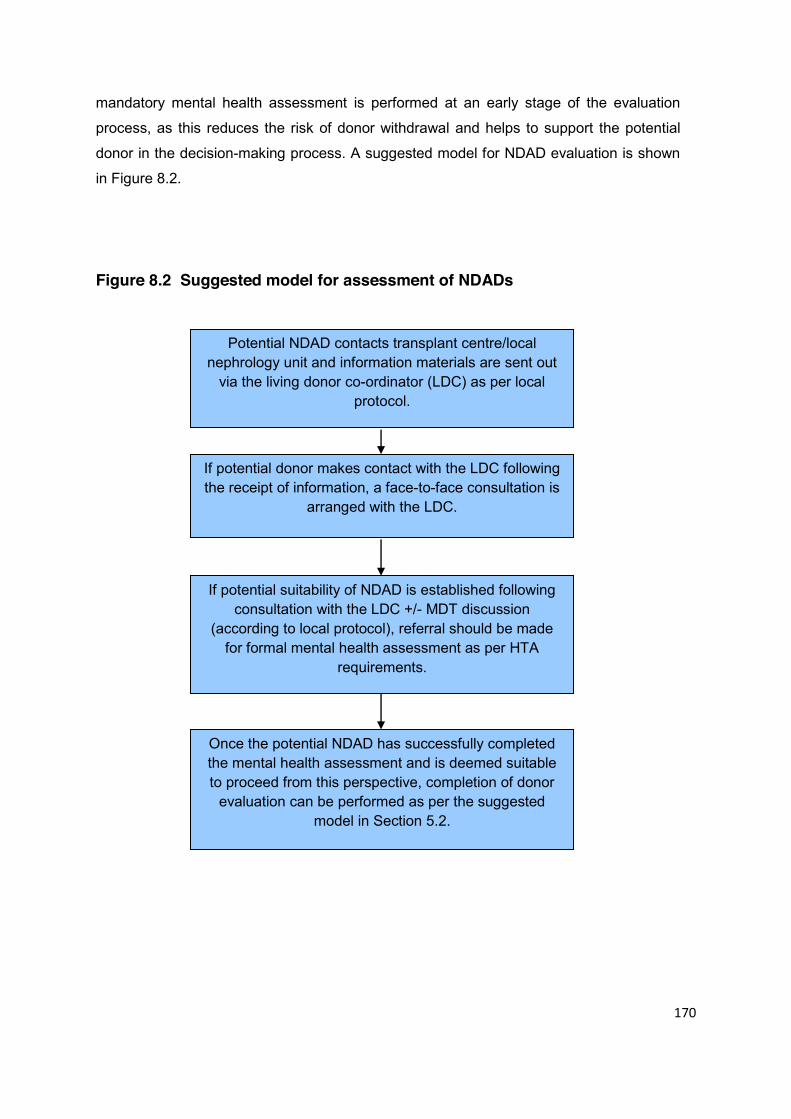
170
mandatory mental health assessment is performed at an early stage of the evaluation
process, as this reduces the risk of donor withdrawal and helps to support the potential
donor in the decision-making process. A suggested model for NDAD evaluation is shown
in Figure 8.2.
Figure 8.2 Suggested model for assessment of NDADs
Potential NDAD contacts transplant centre/local nephrology unit and information materials are sent out
via the living donor co-ordinator (LDC) as per local protocol.
If potential donor makes contact with the LDC following the receipt of information, a face-to-face consultation is
arranged with the LDC.
If potential suitability of NDAD is established following consultation with the LDC +/- MDT discussion
(according to local protocol), referral should be made for formal mental health assessment as per HTA
requirements.
Once the potential NDAD has successfully completed the mental health assessment and is deemed suitable to proceed from this perspective, completion of donor
evaluation can be performed as per the suggested model in Section 5.2.

171
The ‘stand alone’ NDAD scheme differs from the paired/pooled scheme in that there is no
specific requirement for the recipient to be automatically suspended from the deceased
donor list when an offer of a kidney has been made, or even when the initial crossmatch
has been performed.
It is clearly in the best interests of the recipient to receive a kidney from a living donor and
so consideration should be given to the relative risk of removing the potential recipient
from the national waiting list whilst finalising the arrangements for transplantation and at
which time point this should happen. There is also the potential disruption to the donor if
the recipient is offered a kidney from an alternative donor during this period, as the option
of proceeding with a different recipient may be refused. As a minimum standard,
discussion must be initiated with the recipient about suspension from the national list at
the time of the offer, and again following the outcome of initial crossmatching. The
recipient transplant centre is then responsible for activating that decision with ODT.
If a kidney is offered to a recipient and the date of surgery is subsequently postponed, a
decision has to be made about re-offering the kidney, depending upon the reason for the
delay. Without betraying confidential information, this decision should involve the donor as
he or she may be willing to reschedule for the same recipient if it is a problem that is likely
to resolve (e.g. PD peritonitis). If it is a more permanent issue, clinical or otherwise, it may
be advisable to re-offer the kidney with the donor’s agreement.
Future allocation arrangements Changes to the National Living Donor Kidney Sharing Schemes will be introduced in late
2011. Allocation arrangements for NDAD kidneys will be enhanced to allow NDADs to
benefit more than one recipient by allocation of their kidney through the paired/pooled
donation programme. The agreement is that unless a high priority patient is identified on
the deceased donor transplant list (000 mismatched child or 000 mismatched, highly
sensitised adult), the kidney will be allocated instead to a patient in the paired/pooled
donation programme who is compatible with the donor. In turn, the donor registered with
the ‘paired’ recipient would then donate to a patient on the deceased donor waiting list
through the national allocation scheme. This is called an altruistic donor chain. Longer
chains involving two or more paired donation couples are also anticipated. When this
programme is implemented, full details will be available on the ODT website
(www.organdonation.nhs.uk).
172
Donor reimbursement for paired/pooled and altruistic donors Special considerations for donors within these groups have been identified and are
addressed in Chapter 9.
8.3 Antibody Incompatible Donation
Antibody incompatible transplantation (AiT) may be an option for some patients who have
a potential living donor but where there is a specific immunological barrier to
transplantation. Such transplantation falls into two categories: ABO incompatible
transplantation, where transplantation occurs across an ABO blood group barrier
(e.g. from a blood group B donor to a blood group O recipient); and HLA-incompatible
transplantation, where the recipient has high titres of antibodies against one or more
specific HLA antigens present in the donor.
Both forms of transplantation are becoming more common in the UK and will in future
make major contributions to expansion of the living donor pool.
The BTS has published specific guidelines on antibody incompatible transplantation,
which should be referred to. These are also summarised in section 7.5.
Sources of Information
NHS Blood and Transplant. www.nhsbt.org.uk or www.organdonation.nhs.uk
Human Tissue Authority. www.hta.gov.uk
British Transplantation Society, Standards & Guidelines.
http://bts.demo.eibs.co.uk/transplantation/standards-and-guidelines/
Reference
1. Human Tissue Authority, Code of Practice 2, Donation of Solid Organs for
Transplantation, July 2009.
http://www.hta.gov.uk/legislationpoliciesandcodesofpractice/codesofpractice/code2don
ationoforgans.cfm

173
CHAPTER 9 LOGISTICAL CONSIDERATIONS Statements of Recommendation
The reimbursement of legitimate expenses incurred by a living donor as a direct result of the preparation for an act of donation is supported by the Department of Health. Prospective agreement for reimbursement from local recipient commissioners is currently recommended as the most effective mechanism for achieving reimbursement but a national scheme is being developed which will replace this guidance in the near future and this guidance will be updated accordingly. (B1)
Donors from overseas present unique logistical challenges. In order for the
process to be clinically effective and to comply with UK Border Agency and Department of Health requirements, there is an agreed entry clearance (visa) application process and duration of stay in the UK (6 months) for the donor which must be honoured in all but exceptional, unforeseen circumstances. (B1)
9.1 Reimbursement of Living Donor Expenses The reimbursement of reasonably incurred expenses to a living donor, including loss of
earnings which are directly attributable to the organ donation, is supported by the HTA
and the Department of Health (DH) (1,2) Reimbursement does not contravene the current
UK legislation under the Human Tissue Act (3) (see Chapter 2: Legal Framework) which
forbids payment for supplying a human organ, provided that the donor does not gain any
financial advantage as a result.
The NHS is not legally obliged to make such payments, but the DH recognises that the
most cost effective treatment for end stage kidney disease is transplantation and that the
costs incurred as a direct result of performing a living donor transplant are justified. The
DH expects that suitable arrangements will be made as part of local commissioning
agreements between the recipient’s Commissioning Consortium or Authority
(e.g. Scotland/Channel Islands) and the NHS Trust in which the transplant is performed.

174
Whilst some regions are well organised, there is no universal consensus about how such
arrangements are implemented and there is considerable variability at local level. Despite
the lack of standardisation, progress has been made in agreeing some key principles that
underpin the application and approval processes to prevent delay in settling claims:
Individual claims should be settled in a timely fashion to prevent unnecessary
financial hardship to the donor as a consequence of the donation
Claims will be settled by the recipient Commissioning Consortium/Authority on a
case by case basis according to agreed criteria
Early identification of potential claims during the donor assessment period is
essential to facilitate prior approval and timely settlement
Early notification to relevant Commissioning Consortium/Authority of claim must be
made and, whenever possible, prior to the date of donation to facilitate timely
settlement. However, provision should be available for considering claims
retrospectively if, for genuine reasons, it has not been possible to highlight a
prospective claim to the Commissioning Consortium/Authority
Donor expectations about the nature and size of claims that will be approved must
be appropriately managed
Donors must be provided with appropriate and specific information about criteria
for application, approval processes and timeframes at an early stage of the
assessment process
Alternative sources of reimbursement, e.g. statutory sick pay, must be declared
when a donor applies for reimbursement
The current system still lacks consistency and is often time-consuming to administer.
The development of a centrally administered, national scheme for England is under
development and it is envisaged that the implementation of such a scheme will help to
streamline processes across the UK and facilitate best practice between England and the
existing schemes within the devolved administrations. A suggested model for best
practice in effective claims management is outlined in Figure 9.1.
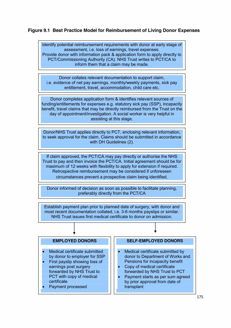
175
Figure 9.1 Best Practice Model for Reimbursement of Living Donor Expenses
Establish payment plan prior to planned date of surgery, with donor and most recent documentation collated, i.e. 3-6 months payslips or similar.
NHS Trust issues first medical certificate to donor on admission.
If claim approved, the PCT/CA may pay directly or authorise the NHS Trust to pay and then invoice the PCT/CA. Initial agreement should be for
maximum of 12 weeks with flexibility to apply for extension if required. Retrospective reimbursement may be considered if unforeseen
circumstances prevent a prospective claim being identified.
Donor informed of decision as soon as possible to facilitate planning, preferably directly from the PCT/CA
Donor collates relevant documentation to support claim, i.e. evidence of net pay earnings, monthly/weekly payments, sick pay
entitlement, travel, accommodation, child care etc.
Donor completes application form & identifies relevant sources of funding/entitlements for expenses e.g. statutory sick pay (SSP), incapacity benefit, travel claims that may be directly reimbursed from the Trust on the
day of appointment/investigation. A social worker is very helpful in assisting at this stage.
Donor/NHS Trust applies directly to PCT, enclosing relevant information, to seek approval for the claim. Claims should be submitted in accordance
with DH Guidelines (2).
Identify potential reimbursement requirements with donor at early stage of assessment, i.e. loss of earnings, travel expenses.
Provide donor with information pack & application form to apply directly to PCT/Commissioning Authority (CA). NHS Trust writes to PCT/CA to
inform them that a claim may be made.
Involve Social Worker if available to assist
EMPLOYED DONORS
Medical certificate submitted by donor to employer for SSP
First payslip showing loss of earnings post surgery forwarded by NHS Trust to PCT with copy of medical certificate
Payment processed
SELF-EMPLOYED DONORS
Medical certificate submitted by donor to Department of Works and Pensions for incapacity benefit
Copy of medical certificate forwarded by NHS Trust to PCT
Payment starts as per sum agreed by prior approval from date of transplant
176
9.2 Paired/Pooled and Non-Directed Altruistic Donors In cases of paired/pooled donation and non-directed altruistic donation, living donor
kidneys are shared and exchanged across the UK between different transplant centres,
which may be subject to different mechanisms of donor reimbursement. In both scenarios,
there is limited time to obtain prior approval for donor reimbursement from the recipient’s
Commissioning Consortium/Authority because dates for surgery are set as soon as
possible following a paired/pooled matching run and/or non-directed altruistic donor offer.
It is recommended that in cases of paired/pooled donation, an application to the local
recipient Commissioning Consortium/Authority is made by the local donor at the time of
registration into the scheme in the same way as for a direct living donation (as above).
This would facilitate prior approval of anticipated expenses and timely reimbursement
when the transplant proceeds. Reciprocity between each donor/recipient pair means that
the costs to the local Commissioning Consortium/Authority are likely to be equitable.
In cases of non-directed altruistic donation, there is no direct reciprocity between the
donor and recipient transplant centres unless the kidney is allocated by chance to a local
recipient through the national allocation scheme. However, any recipient in the UK may be
a potential beneficiary of such a kidney and, as non-directed altruistic donor activity
continues to increase, it is important to have clarity about the mechanism for donor
reimbursement. Local resolution with Commissioning Consortia/Authorities is problematic
because of the time constraints previously highlighted for prospective application. The
preferred option would be for recipient Commissioning Consortia/Authorities to undertake
to reimburse donors in this situation without prior approval, provided that the claim meets
the agreed financial criteria for settlement. With changes to the National Living Donor
Kidney Sharing Schemes due to be implemented towards the end of 2011, the same
principles will apply in the altruistic donor chain scenario if local reimbursement
arrangements are still in place.
9.3 Donors from Overseas
Donors from overseas present unique logistical challenges. Representatives from the UK
Border Agency (UKBA), the HTA and the transplant community have worked together to
overcome commonly encountered issues around overseas donors who wish to travel to
177
the United Kingdom to donate. This joint collaboration has facilitated a full discussion around the perceived definition of provision of funding to overseas donors. It has also
enabled a review of existing processes for the potential overseas donor‘s application for
entry clearance (visa) to travel to the UK to donate to UK recipients. The following issues
have been considered:
The DH Guidance emphasises the importance of avoiding donor reimbursement
from the recipient or his/her family, which could be seen as an inducement to
donate.
The DH Guidance emphasising that the recipient is ordinarily resident in the UK
and not subject to the National Health Service (Charges to Overseas Visitors)
Regulations 1989, as amended, or their equivalent regulations in the devolved
administrations.
The Immigration Rules require the UK Border Agency to ensure that any person
applying for entry to the UK in this category provides sufficient evidence to
demonstrate that:
o They are genuinely seeking entry for a specified purpose within the UK and
that the intention at the time of entry is that the stay will not exceed six
months. However, subsequent unforeseen circumstances may result in the
migrant applying for an extension.
o They will maintain and accommodate themselves adequately out of the
resources available to them (self-financing), without recourse to public
funds or taking employment, or will be maintained and adequately
accommodated by friends or family.
o They can meet the cost of the return or onward journey.
The following process has been agreed to facilitate the entry clearance application
process from a potential donor who is resident overseas and wishes to donate to a
recipient who is resident in the UK and is eligible for NHS treatment:
The Human Tissue Authority (HTA) has confirmed that if a donor is supported by
their family and/or the recipient to fund travel and living expenses, this is deemed
acceptable provided that the requirements of the Independent Assessor are
subsequently met. A claim for reimbursement may also be made by the donor to
reclaim legitimate expenses (as above) if the donation proceeds. Commissioners
have been advised of this requirement and that applications for reimbursement of
178
donor expenses will be made retrospectively (i.e. post-donation) in the majority of
cases, enabling an accurate assessment of legitimate expenses to be made once
the donor has been assessed and surgery has proceeded as planned.
The UK Border Agency has confirmed that entry clearance applications including
applications for visas should be dealt with as a specific group (on a case by case
basis) and considered against the requirements for one immigration category for
six months duration. In most cases the applications will be made direct to the
British Embassy or High Commission, and then referred back to the UK. There is
no guarantee that entry clearance will be granted by the UK Border Agency.
All applications for entry clearance will need to be accompanied by appropriate
supporting information. A template letter has been provided which has been
approved by the UK Border Agency and includes the minimum requirement for
supporting information. This is annexed at the end of this chapter. It is strongly
recommended that this template letter is used in all cases, as the format
immediately identifies the nature of the application to Entry Clearance Personnel in
local Embassies/High Commissions, and the manner in which it should be
processed. Of note, the Channel Islands and the Isle of Man have separate
immigration arrangements from the UK and additional supporting information may
be required when applying for entry clearance for a donor whose recipient is
resident in these islands. Direct liaison with these Authorities is advised in advance
of the application to avoid delay in issuing entry clearance. The letter should be
addressed to the potential donor, copied to the potential recipient and sent from
the transplanting centre detailing:
o The purpose of the application. This needs to be clearly visible at the top of
the letter to ensure that it is appropriately identified in local Embassies/High
Commissions for processing through the UK Border Agency in the UK.
o The claimed relationship between donor and recipient, specifying
demographic details for both donor and recipient.
o Confirmation of any donor evaluation (i.e. medical assessments/tests/
investigations) that have already been undertaken to support the potential
suitability of the donor.
o The reason for the choice of donor (i.e. no suitable donors in the UK).
o The treatment plan for the donor and the estimated duration of the stay in
the UK required (i.e. 6 months). It should be made explicit to the donor that
there is ‘no right to stay’ in the UK beyond the time that is clinically
179
necessary and that there is an expectation that he/she will return to the
country of origin after a suitable period of convalescence post donation.
o The prospective start date for the entry clearance and subsequent start of
donor evaluation on arrival in the UK.
o If the donor evaluation reveals that treatment for a medical condition is
required before donation can be considered, the donor must be informed
that he or she will be expected to return to the country of origin for such
treatment. Similarly, any late surgical or medical complication arising post-
nephrectomy once the donor has returned to his/her country of origin must
be treated locally and there is no recourse to the NHS to provide such
treatment.
o The DH position on entitlement to NHS treatment/donor reimbursement.
This should detail any costs which are known at the outset and are likely to
be reimbursed during the process (e.g. confirming the cost for the return
flight home is going to be reimbursed helps demonstrate that the donor has
the means to return back to country of origin).
o The relative cost-effectiveness of living donor transplantation within the UK
health economy.
o Confirmation that the recipient is ordinarily resident in the UK and not
subject to the National Health Service (Charges to Overseas Visitors)
Regulations 1989, as amended, or their equivalent regulations in the
devolved administrations.
o Contact details for further information from the transplanting centre.
Following the UK Border Agency’s full consideration of the evidence provided,
successful applicants will normally be issued entry clearance for 6 months, starting
on the day entry clearance is issued unless stipulated otherwise. For planning
purposes, a prospective date for start of the entry clearance can be specified in the
supporting information so that delays in the start of donor evaluation are minimised
once the donor arrives in the UK. This date should be requested sufficiently far
ahead to allow sufficient time (minimum of 4 weeks and maximum 12 weeks at
time of application) for the donor to receive the supporting information, present the
application to the local Embassy/High Commission and for the application to be
processed. The 6 month visa is issued to facilitate the donation process,
(i.e. required clinical evaluation, surgery, recovery of the donor post-nephrectomy)
and is the maximum stay that is permitted for a visa issued on these grounds.
180
Costs incurred can subsequently be reclaimed by the donor once the donation has
gone ahead as part of the reimbursement process (see above).
Where there are extenuating circumstances, of a medical nature, an extension of
stay can be applied for, but there is a fee for such applications and there is no
guarantee that the extension will be granted. It is important to manage the clinical
pathways for both donor and recipient effectively to minimise delay and to facilitate
transplantation in a timely manner; this includes scheduling the complete process
(including sufficient time for convalescing) within the six months granted.
Healthcare professionals must take responsibility to manage the expectations of
both donor and recipient from the outset about the requirement for the donor to
return to his/her country of origin following recovery from surgery and appropriate
convalescence.
Representatives from UK Border Agency are keen to be informed about cases where
entry applications are refused or issued for under 6 months, or where there have been
difficulties encountered in the application process, so that they can continue to monitor
and review individual cases. Please direct any specific queries about visa applications
and/or general queries about this guidance to Lisa Burnapp ([email protected]).
Lisa has agreed to provide liaison between living donor co-ordinators (LDCs) and UK
Border Agency personnel for initial enquiries, and the latter will then deal directly with the
relevant LDC as required.
References
1. Saving Lives, Valuing Donors: A Transplant Framework for England. Department of
Health, July 2003.
http://www.dh.gov.uk/en/Publicationsandstatistics/Publications/PublicationsPolicyAnd
Guidance/DH_4006700
2. Reimbursement of Living Donor Expenses by the NHS. Department of Health, July
2009.
http://www.dh.gov.uk/en/Healthcare/Longtermconditions/Vascular/Renal/RenalInform
ation/DH_4069293
3. Human Tissue Act 2004, Human Tissue Act (Scotland) 2006.
http://www.opsi.gov.uk/acts/acts2004/ukpga_20040030_en_1

181
Annex Chapter 9
Template Letter for Potential Overseas Donors Trust Headed Paper
[include contact details for living donor co-ordinator]
[Name and address (overseas) of potential donor]
Hospital No/ID (if available)
NHS No (if available)
Date of Birth
Dear [Donor’s name]
RE: PROPOSED LIVING KIDNEY DONATION FOR UK RECIPIENT:
(NAME, HOSPITAL ID, DOB, ADDRESS IN UK)
We understand that you wish to be considered as living kidney donor for your [relationship donor to recipient].
Thank you for providing us with some preliminary medical information, which indicates your suitability for proceeding to the next stage of the donation process. To ensure a thorough assessment of your medical suitability to donate a kidney and so that you have an opportunity to discuss this in detail, you will need to travel to the United Kingdom (UK) to attend [’X’] Hospital, [Name of City/Town] for further tests and consultations. As you know, we have provisionally organised for you to attend for these appointments starting [Day, Date] and so you should request a visa to start as close to this date as possible and then arrange your travel accordingly (see below). Please read this letter carefully before proceeding any further.
Next Steps:
1. Your application to travel to the UK
To travel to, enter and stay in the UK for 6 months you will need to satisfy UK immigration requirements. You will need to make an application to your local British Diplomatic Mission for a UK Visitor’s Visa (entry clearance), specifying your intention to donate an organ for your relative. Your relative, the potential recipient of your kidney, must ordinarily be resident
182
in the UK and entitled to kidney transplant treatment on the National Health Service (NHS). Your relative must check this information with the hospital in the UK before you submit an application, otherwise it will not be valid.
You will find all the information you require to apply for your visa on the UK Border Agency Visa Services website: http://www.ukvisas.gov.uk/en/ or at your local British Diplomatic Mission. To avoid unnecessary delays in processing your application, which will be referred to the UK for a decision by your local British Diplomatic Mission, you should ensure you read the information carefully before completing your application to make sure you provide all the necessary information needed to satisfy the requirements set out in the guidance and here in this letter. This should avoid any unnecessary delays or uncertainty about travel dates. As your visa application will be considered in the UK, you should allow at least 4 weeks (maximum 12 weeks) from the time of application. If successful, your visa will then be issued for 6 months, starting on the date you have agreed with us and as stated on the visa application form.
2. Your medical testing and donation in the UK
We need to plan your further medical assessment and surgery carefully and in advance, in order to ensure we complete the kidney donation process within 6 months. Once you have completed the assessments in the UK, we will be able to confirm whether or not you are a suitable donor and, if you still wish to donate, we will schedule a date for the living donor transplant operation for you and your recipient as soon as possible. You will need to recover in the UK after the surgery for up to 4 weeks before you travel back to your own country. You must arrange to stay with your family throughout your stay in the UK, or make independent accommodation arrangements before your arrival in the UK (you will have to provide evidence of this in your visa application to travel to the UK). It is particularly important to make sure that you can stay with your family when you are discharged from hospital so that you are not living alone while you recover from your surgery.
3. Your checklist to proceeding
To help us plan everything as smoothly as possible for you with the minimum of delay, you can help us by doing the following:
1. Ensure you have discussed with the hospital when you wish to travel to the UK before submitting your application. This is essential in order to allow enough time for the hospital to arrange your tests in advance, to start as soon as possible after your arrival in the UK and to provide you with enough time to recover and convalesce after your operation. Prior to this discussion you should consider the date that you think you will be able to make travel plans and UK reception arrangements (staying with your family). You will need to allow at least 4 weeks (but no more than 12 weeks) between the date you intend to submit your visa application to the British Diplomatic Mission and the date you wish to travel. Once this information has been discussed and agreed with the hospital you will have a date and all the relevant information for your visa application.
183
2. Follow the guidance on the website when applying for your visa to ensure that you have met the UK Border Agency requirements in full before you submit your application.
3. Attach this letter to your visa application, and submit it together with all your other documents for your visa application to your local British Diplomatic Mission for consideration by an Entry Clearance Officer. If your application is approved, your visa will be issued to start on the specified date, previously agreed with the hospital. Please keep a copy of all these documents for yourself for future reference.
4. It is your responsibility to advise the hospital directly if there is any delay in submitting your visa application to your local British Diplomatic Mission overseas or in approving it so that we know when to expect you and to adjust dates accordingly.
Costs
It is important that you know which costs and expenses are covered during your visit. The cost of your medical treatment specifically for the purposes of donating a kidney (donor assessment, +/- donor surgery and out-patient appointments) will be covered by the NHS whilst you are in the UK, but this is not available to you once you have returned to your own country at the end of the 6 month period. Whilst you are in the UK, any treatment outside of the donor process, including dentistry, is not covered by the NHS and, if you do not have medical insurance, you will be expected to pay for this yourself or return to [Country of residence for donor] for treatment. You are, therefore, advised to obtain medical insurance before you travel. You are entitled to apply for reimbursement for travel and living expenses specifically in connection with process of donation from the NHS once you have donated your kidney and you are advised to keep a record of expenses that you incur. If you are unable to proceed with the donation as planned following assessment in the UK, you will not be entitled to claim these expenses.
Please ensure that you and your family have read this letter and fully understand the information before proceeding with a visa application. I will be co-ordinating your donor assessment at the hospital. Please contact me directly or via your recipient if you are not clear about any aspect of this letter. My contact details are at the top of this letter.
Yours sincerely
Living Donor Co-ordinator/member of Transplant/Referring Team
Cc: [potential recipient]
184
CHAPTER 10 DONOR FOLLOW-UP Statements of Recommendation
Life-long follow-up is recommended after donor nephrectomy. For donors who are resident in the UK, this should be offered locally or at the transplant centre according to the wishes of the donor, but such arrangements must facilitate the collection of data for submission to the UK Living Donor Registry on long term morbidity and mortality. Donors from overseas who travel to the UK to donate are not entitled to follow-up in the UK but should be given advice about appropriate follow-up before returning to their country of origin (C1)
Arrangements must be put in place to ensure that the unsuitable donor, who
is unable to proceed to donation, is appropriately followed-up and referred for further investigation and management. (B1)
NHSBT has given a formal undertaking that any living kidney donor who develops renal failure in the peri-operative period as a consequence of donation will receive priority for a deceased donor kidney transplant. (Not graded)
10.1 Arrangements for Follow-up Early follow-up of the donor is recommended within the first few weeks after surgery to
ensure that he or she is making progress following the operation and is appropriately
supported. This should include monitoring of kidney function as well as the early detection
of problems such as infection and wound healing. By the end of three months it is
anticipated that the donor will have made a full recovery and have returned to normal
activities. Some centres offer a further review at this stage as an opportunity to ensure
that this has happened as well as offering ongoing support and advice for continued
optimal health.
185
Long term follow-up, on an annual basis, provides a forum for review of kidney function,
urinalysis and blood pressure as well as general health status. Existing guidelines based
upon a consensus of professional opinion recommend that the transplant centre has a
responsibility to encourage and facilitate the long term follow-up of the donor after
donation, particularly for individuals with pre-existing or acquired conditions that potentially
place them at greater risk (1-3). These include hypertension, obesity, diabetes and
proteinuria (see Figure 10.1 for a suggested model for donor follow-up). This follow-up
can be provided by the transplant centre, the referring nephrology unit or the donor’s
General Practitioner. Whilst not all donors wish to return for regular review, anecdotally
many welcome the opportunity and appreciate the continuing support and interest in their
welfare.
In the event of an unsuccessful transplant, it is particularly important to provide adequate
emotional as well as physical support for the donor, including access to counselling
facilities (see Chapter 4). Practice with respect to long term follow-up still varies between
centres and is subject to local arrangement. The principle of long-term surveillance and
monitoring of the donor is considered to be best practice and is encouraged in the UK by
the National Living Donor Registry, which was established in 2000 and is held by NHS
Blood & Transplant (5). All UK centres are expected to submit data to the Registry on all
donors at specified time points both pre-and post donation in order to optimise the value
of the Registry Data in informing living donor practice. In developing models of care for
donor follow-up, timing of annual review in accordance with the month of donation,
offering flexible clinic times and local follow-up arrangements should be considered in
order to offer choice to the donor and facilitate the timely collation of Registry Data for
willing participants. Annual review telephone clinics may be less suitable for long-term
donor follow-up due to the infrequency of the interaction with a healthcare professional.
Anecdotal feedback from previous donors when offered this option in a single centre was
not favourable on the basis that the annual face-to-face consultation is of intrinsic value to
the follow-up experience.

186
Figure 10.1 Model Referral Pathway for Follow-up Post Living Donor Nephrectomy
Referral to donor follow-up clinic for life-long annual review
Re-appointment in surgical or medical review clinic
Life-long donor follow-up
Rationale: To provide continuing support to the donor & inform the UK Living Donor Registry
Annual review subject to review according to clinical need
Performed at local transplant/referring centre or GP
Relevant research/audit data collated & data returned to UK Transplant Living Donor Registry by LD Co-ordinator
Annual review to include assessment of:
General health & lifestyle
Wound +/- complications
Medication/treatment initiated
Renal profile
Full blood count
Dipstick urinalysis +/- mid-stream urine +/- albumin/creatinine ratio
Blood pressure +/- 24hr ABPM / referral to GP for treatment if indicated
Weight & BMI Appropriate referral to be made if further nephrological, urological or surgical opinion is indicated. GP referral to be made for unrelated health problems
Upon discharge from the ward, donors must be provided with:
Appropriate advice about wound care, pain relief & general rehabilitation
Prescribed medication & wound dressings as required
Medical certificate to cover the period of in-patient stay
Discharge summary for GP
Contact numbers for the living donor co-ordinator team
Routine follow-up appointment in surgical OPD clinic with operating surgeon (when possible)
187
For donors who travel from overseas to donate, there are implications for long-term follow-
up arrangements and access to data once they return to their country of origin, particularly
in countries where living donor transplantation is not an established practice or where
individuals pay for healthcare. These donors should be provided with written advice about
appropriate annual monitoring. However, it is difficult to ensure that robust arrangements
are put in place and it is rarely possible to collect accurate data on overseas donors for
the UK Living Donor Registry.
10.2 The Unsuitable Donor An area that is easily overlooked is the care and follow-up of patients who start the donor
assessment process but who do not subsequently donate. If this is the result of concerns
about the potential donor’s health, it is essential that appropriate arrangements are made
for any necessary further investigation and management. A donor who is unsuitable for
other reasons (for example a positive crossmatch) may need emotional support as they
could conceive themselves to have “failed” the recipient – and blame themselves
inappropriately for any subsequent adverse outcome for the recipient (see Chapter 4).
10.3 Pregnancy following Kidney Donation Many centres consider women of childbearing age as potential living donors. Pregnancy
has a number of well documented effects on the kidney raising the possibility that these
may have an adverse effect in an individual with a solitary kidney. The information in this
area is relatively limited. A study of 39 pregnancies in 23 women with 32 viable births
revealed no significant problems and in particular no significant hypertension or
proteinuria (5). Another study of 23 viable births in 14 kidney donors reported no
significant problem (3). Two recent reports based upon retrospective Norwegian Registry
Data and a large single centre survey in Minnesota, USA have raised concern about the
potential for increased maternal complications after donation. Both studies are limited and
interpretation is therefore difficult (6). Nevertheless, the presence of a solitary kidney does
not appear to pose a significant risk during the course of a normal pregnancy and
outcomes for pregnant kidney donors are considered comparable to those in the general
population. Anecdotally, this opinion has been corroborated by expert obstetric opinion in
the field when seeking advice on how to advise potential kidney donors.

188
Within the UK, there is an opportunity to report births post-donation to the Living Donor
Registry as ‘a significant medical event’ at each annual review (4). This should be
encouraged in order to improve the evidence base. Close follow-up is advisable in donors
during pregnancy and periodic assessment should be undertaken of serum creatinine and
creatinine clearance in addition to urine culture and blood pressure.
10.4 Renal Failure following Living Kidney Donation Renal failure after living kidney donation is rare, but there have been occasions (at least
one in the UK) where peri-operative complications have resulted in a living kidney donor
developing chronic dialysis-dependent renal insufficiency following surgery. In this rare
situation, NHSBT has given a formal undertaking that any living kidney donor who
develops renal failure as an acute consequence of donation will receive priority for a
deceased donor kidney for transplantation. References 1. Buszta C, Steinmuller DR, Novick AC, et al. Pregnancy after donor nephrectomy.
Transplantation 1985; 40: 651-4.
2. The Ethics Committee of the Transplantation Society. The Consensus Statement of
the Amsterdam Forum on the Care of the Live Kidney Donor. Transplantation 2004;
78: 491-2.
3. A Report of the Amsterdam Forum on the care of the Live Kidney Donor; Data and
Medical Guidelines. Transplantation 2005; 79; S53-S66.
4. Living Donor Registry. www.nhsbt.org.uk
5. Jones JW, Acton RD, Elick B, Granger DK, Matas AJ. Pregnancy following kidney
donation. Transplant Proc 1993; 25: 3082.
6. Josephson, MA. Pregnancy after kidney donation: more questions than answers.
Nature Reviews, Nephrology 2009; 5: 495-7.
189
11.0 RECIPIENT OUTCOME AFTER LIVING DONOR KIDNEY TRANSPLANTATION IN ADULTS
Statements of Recommendation
Graft and patient survival after living donor kidney transplantation should be consistent with the national average. (A1)
Transplant centres should regularly audit secondary outcomes after living
donor kidney transplantation and should reappraise practice if their results are not comparable with other units. (B1)
Where a recipient is considered to be at high risk, transplantation should
only proceed if, in the view of the team of professionals involved, there is an expectation that the patient is likely to survive with a functioning transplant for more than 2 years. (C2)
Patients at higher risk (see definition in text) may be considered for
transplantation when the professional team regard the risk to the specific recipient to be acceptable. An agreement between the clinical team and the donor-recipient pair should be documented in the clinical records, including a prediction of the chance of success and the risk of failure for this specific transplant. A copy of this document should also be given to the donor and recipient. The transplant should be undertaken within 3 months of this agreement and it should be pre-emptive where possible.
Clinical audit is integral to transplant practice. Outcome after living donor transplantation
can be measured by recipient and graft survival and should be sent to National Health
Service Blood and Transplant (NHSBT). Data should also be collected by each unit for the
following secondary outcome measures, which are currently not reported centrally but
which give important audit measures for success:
incidence of delayed graft function (which is uncommon (<5%) in living donor
transplants);

190
incidence of acute rejection;
incidence of post-operative infection;
renal function one year post transplantation.
Data from the UK transplant registry (held by NHSBT) have recently been analysed for
living donor kidney transplants undertaken in the UK between 2000 and 2007 (1). Table 1
shows the patient and death-censored graft survival.
Table 11.1 Patient and death-censored graft survival at 1 and 5 years after living kidney donation (2000-7) (1)
Related donors (n=2,227) Unrelated donors (n=917)
% survival 95% CI % survival 95% CI
Graft survival
1 yr 95 94-96 96 94-97
5 yr 89 87-91 93 90-95
Patient survival
1 yr 99 98-99 97 96-98
5 yr 97 95-98 93 90-95
Higher donor age, the presence of recipient diabetes and grafts from adult offspring were
associated with poorer patient survival in the first three years after transplantation (Tables
11.2 and 11.3). The relative risk of death in recipients with diabetes compared to those
without diabetes was 8.8 after 3 years.
Poorer graft survival at 3 months was independently associated with donor age older than
59 years (relative risk of graft failure compared to 18-34 year old donors 2.95) and female
recipients (relative risk of graft failure compared to males 1.88). Poorer graft survival at 3
years was associated with recipient diabetes (relative risk of graft failure compared to non-
diabetes 2.96). The age of the recipient did not significantly affect outcome.
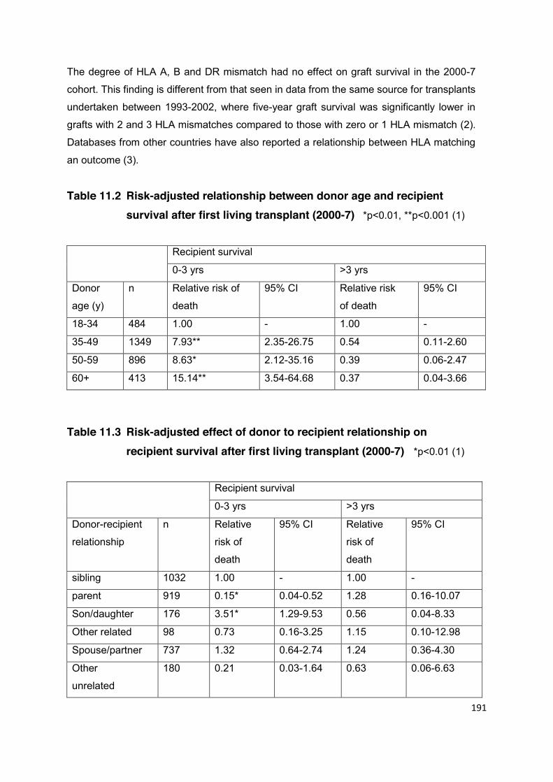
191
The degree of HLA A, B and DR mismatch had no effect on graft survival in the 2000-7
cohort. This finding is different from that seen in data from the same source for transplants
undertaken between 1993-2002, where five-year graft survival was significantly lower in
grafts with 2 and 3 HLA mismatches compared to those with zero or 1 HLA mismatch (2).
Databases from other countries have also reported a relationship between HLA matching
an outcome (3).
Table 11.2 Risk-adjusted relationship between donor age and recipient survival after first living transplant (2000-7) *p<0.01, **p<0.001 (1)
Recipient survival
0-3 yrs >3 yrs
Donor
age (y)
n Relative risk of
death
95% CI Relative risk
of death
95% CI
18-34 484 1.00 - 1.00 -
35-49 1349 7.93** 2.35-26.75 0.54 0.11-2.60
50-59 896 8.63* 2.12-35.16 0.39 0.06-2.47
60+ 413 15.14** 3.54-64.68 0.37 0.04-3.66
Table 11.3 Risk-adjusted effect of donor to recipient relationship on recipient survival after first living transplant (2000-7) *p<0.01 (1)
Recipient survival
0-3 yrs >3 yrs
Donor-recipient
relationship
n Relative
risk of
death
95% CI Relative
risk of
death
95% CI
sibling 1032 1.00 - 1.00 -
parent 919 0.15* 0.04-0.52 1.28 0.16-10.07
Son/daughter 176 3.51* 1.29-9.53 0.56 0.04-8.33
Other related 98 0.73 0.16-3.25 1.15 0.10-12.98
Spouse/partner 737 1.32 0.64-2.74 1.24 0.36-4.30
Other
unrelated
180 0.21 0.03-1.64 0.63 0.06-6.63
192
The High Risk Recipient For the purpose of this guideline, a high-risk recipient is defined as a potential recipient of
a kidney transplant who is at a significantly higher risk of death, complications or graft
failure because of pre-existing co-morbidity or immunological status. There is currently no
robust, clinically applicable scoring system upon which to base this assessment of risk.
Statistically, this equates to an expected outcome that is outside the 95% confidence
interval for graft and patient survival in the UK. It is recognised that this cannot be
predicted with any certainty and will depend entirely on the clinical judgement of the
professionals involved.
A key issue is that, whilst these patients may expect a relatively poorer outcome from
transplantation compared with individuals considered to be at lower risk, their outcome
may be better than it would be if they remained on dialysis. Pre-existing cardiovascular
disease, pulmonary disease, obesity and diabetes all affect survival of patients with a
transplant or on dialysis. Whilst the survival of patients with diabetes on the transplant
waiting list is increased if they receive a graft rather than remain on dialysis, the relevance
of this to high-risk recipients is uncertain. There are insufficient data to give clear guidance
on this issue to individual high-risk recipients. Accordingly, risk assessment in each case
must, by default, be based on multidisciplinary expert opinion. Notwithstanding this, it is
important that certain limits are set, not least to ensure appropriate use of resources.
In cases considered to be higher risk, living donation has certain advantages over
deceased donor transplant, including:
daylight operating time;
availability of greater numbers of senior staff from multidisciplinary team (e.g.
cardiology, chest physiotherapy);
known quality of the organ to be transplanted;
predicted day of surgery allowing optimisation of recipient factors (e.g. reversal of
anticoagulation);
pre-transplant immunosuppression or immunomodulation.
Since the outcome of deceased donor transplantation is affected by long cold ischemic
time and delayed graft function (both of which are avoided with a living transplant), there
193
may be circumstances where living transplant provides the best option for some high risk
patients, provided there is a clear understanding of risk by both donor and recipient.
An agreement must be reached by the clinical team and the donor-recipient pair which
includes a realistic prediction of success and recognition of the risks of failure (death of
the recipient or failure of the graft). Although such an agreement is applicable to living
donation in general, it is particularly important for the high risk recipient where
expectations will not usually accord with registry data. Although there is a paucity of
information on the balance of risks and benefits in high-risk recipients, the clinical team
should do their best to describe the best and worst case scenarios. The timing and
content of these discussions should be documented in the medical notes.
The assessment should include the following elements:
The option of living donor transplantation should be discussed with all patients,
unless it is considered unlikely that the patient would survive with a functioning
graft for more than 2 years.
The multi-professional clinical team should establish what the recipient wants and
expects from the transplant in terms of quality and extension of life.
Consideration of living donor transplantation should be started early enough for the
procedure to be performed pre-emptively (i.e. before dialysis becomes essential),
which should be considered the preferred option.
The trigger for initiation of discussion should normally be an eGFR of ~20 ml/min,
but this may vary depending on the rate of decline of renal function.
The risks and benefits of living donor transplantation should be described but so
should other management options including maximal conservative care, dialysis
and transplantation from a deceased donor.
The details of the final understanding must be compliant with the NHS consent
process. It should be recorded and signed by both the recipient and the donor in
addition to the consultant transplant surgeon taking responsibility for the recipient
operation. Copies of the agreement, which should be filed in the notes, should be
given to both the donor and the recipient alongside the consent form. This
agreement would serve as part 1 of the consent for transplantation surgery in the
two-part consent process.

194
If an agreement cannot be achieved within a particular transplant centre e.g.
because of differences in opinion on the degree of risk, the option of referral to
another transplant centre for a second opinion should be discussed with the
potential recipient and donor.
Outcomes
Recipient outcome following living donor transplantation is subject to comparative national
audit. It is important that the decision to transplant high-risk recipients is not influenced by
undue concern about outcome data. In view of demographic differences between units
and the likelihood of variations in the definition of “high-risk”, it is not possible to set
national standards for transplant outcome in this group.
The outcome data for a transplant programme may be skewed if a unit is prepared to
undertake more high risk transplants. It is recommended that each unit should maintain
detailed records of relevant clinical features for each high-risk recipient. This may be
valuable for future audit. Co-morbidity reports sent to the Renal Registry provide a
minimum data set, but more detailed assessment of risk using an in-house scoring system
is recommended.
References
1. Fuggle SV, Allen JE, Johnson RJ, et al. Factors affecting graft and patient survival
after live donor kidney transplantation in the UK. Transplantation 2010; 89: 694-701.
2. United Kingdom guidelines for living donor kidney transplantation 2nd Edition. British
Transplant Society 2005. Accessed 16th February 2011 at:
http://www.bts.org.uk/transplantation/standards-and-guidelines/
3. Opelz G. Impact of HLA compatibility on survival of kidney transplants from unrelated
live donors. Transplantation 1997; 64: 1473-5.

195
CHAPTER 12 RECURRENT RENAL DISEASE Summary Statements of Recommendation
A wide range of diseases that cause renal failure may recur in the transplanted kidney. This is important to consider when determining the optimal treatment strategy for a recipient and when counselling both donor and recipient on the relative risks and benefits of living donor transplantation. The risks of recurrence, the consequences for transplant function and the time-course of any deterioration must all be considered. A discussion of the effects of immunosuppression and transplant failure on morbidity and mortality may also be appropriate. (B1)
The risks of recurrent disease are high in atypical HUS, FSGS and MCGN.
In these diseases, the presence of specific adverse clinical features may indicate living donor transplantation should be avoided, even where a donor is available. This will require careful assessment and deliberation with all interested parties. (B2)
In patients with risks related to underlying activity such as SLE or systemic
vasculitis, adequate disease control and an appropriate period of quiescence are important to ensure optimal outcomes. (B1)
Recommendations for individual diseases follow in the subsequent text.
Many native kidney diseases can recur following transplantation and may result in
allograft failure. They include systemic disorders of metabolism and glomerulonephritis
(1,2). The reduction in acute rejection associated with modern immunosuppression means
that recurrent disease is now an important determinant of graft outcome (3). The likelihood
and consequences of recurrence are therefore important when assessing and counselling
living donor-recipient pairs.
In many diseases, the published literature on recurrent disease post-transplantation
consists largely of case series. These give only a limited quantification of risk as they are

196
confounded by ascertainment bias since there is an interaction between the indication for
biopsy and the consequences of disease recurrence (2). Large registry studies provide a
better estimation of risk; however, they too require careful interpretation because disease
rates will be influenced by diagnostic practice and convention in the contributing centres
(4-7). This is particularly important when considering heterogeneous disease processes
such as FSGS (2).
These issues are considered in the following discussion of individual diseases. However,
this is an evolving field and it may be necessary to review source data or seek specialist
advice to estimate risk and decide upon optimal treatment for individual cases. For
example, previous reports of an association between living kidney donation and the
recurrence of glomerulonephritis, particularly in zero mismatched donor-recipient pairs,
have either been unconfirmed (6) or not so clear-cut as to definitely preclude
transplantation (8). In other diseases such as atypical haemolytic uremic syndrome
(HUS), a different risk, that of unrecognised genetic susceptibility to disease in the donor
as well as in the recipient, has significantly changed practice (9).
12.1 Diabetic Nephropathy Histological recurrence of diabetic nephropathy is relatively common following renal
transplantation (10). However, the time required for this to cause kidney failure is long and
it does not contraindicate living donor transplantation. This treatment option offers
significant benefits with respect to patient and graft outcomes (11-13).
Recommendation Type 1 and type 2 diabetes are not contraindications to living donor
transplantation, irrespective of whether they are the underlying cause of renal failure. Both the donor and recipient should be counselled regarding the increased risks associated with surgery.
12.2 Primary Focal Segmental Glomerulosclerosis The recurrence of focal segmental glomerulosclerosis (FSGS) following renal
transplantation is a significant problem. It is estimated to recur in 20 to 50% of cases (1,2).
The wide range in the reported frequency of recurrence is likely to reflect heterogeneity in
the underlying diagnoses associated with FSGS. The histological description of FSGS
197
with proteinuria and renal failure occurs frequently as a non-specific finding in many forms
of kidney disease. This secondary FSGS may complicate the interpretation of
undifferentiated reports of recurrence in transplantation.
Primary FSGS, characterised by the nephrotic syndrome, is associated with a high risk of
disease recurrence in the transplant, particularly if there is:
end stage renal failure at a young age, particularly during adolescence (5,14-16)
rapid progression to end stage renal failure (17)
recurrent disease in a previous transplant (5,15,18)
In these situations the rate of graft loss secondary to recurrent disease may be
significantly above 50%.
Even primary FSGS presenting with the nephrotic syndrome seems not to be a single
disease entity, and this may explain differences in the rate of recurrence in different
groups. For example, there is evidence that recurrent disease is more common in whites
than blacks (19). Also, familial forms of FSGS, which can have a rapid course of
deterioration and present at a young age, represent a relatively low risk for recurrence in a
transplant (20-23).
Recurrent primary FSGS generally occurs in the first 6 months following transplantation
and this is an important consideration if transplant recipients are not to be incorrectly
labelled as having recurrent disease.
The report by Cibrik and colleagues that the outcome of transplantation for FSGS is best
in zero mismatched living donation is reassuring (6), although it should be remembered
that a wide range of different presentations are likely to have been included in a registry
report such as this.
Recommendation Living donor kidney transplantation is a reasonable option in patients with
primary FSGS. However, both donor and recipient need to be specifically counselled about the risk of recurrent disease, which may occur early and result in rapid graft loss. Transplantation in an individual with unequivocal evidence of graft loss secondary to recurrent disease has a high risk of subsequent failure such that some centres consider this a contraindication to repeat transplantation (24). In this context, living donor transplantation
198
should be considered only in special circumstances and after careful discussion between the multi-professional team, the donor and the recipient (15,18). The risk of recurrence is low when the previous graft did not fail due to recurrent disease.
12.3 IgA Nephropathy IgA nephropathy frequently recurs in transplanted kidneys on histological grounds but is of
less clinical significance. It may be associated with transient, but more commonly slowly
progressive transplant dysfunction. The prevalence of graft loss due to recurrent IgA
disease was 2.8% in the report of Briganti and colleagues, which gave an estimated
10-year incidence of graft loss of 9.7% (7).
Recommendation
The risk of recurrent disease does not contraindicate living donor transplantation in IgA nephropathy. Both the donor and recipient should be counselled regarding the risks of recurrent disease.
12.4 Membranous Nephropathy The recurrence rate of idiopathic membranous nephropathy has been reported as 29% in
the first 3 years post-transplantation with a corresponding graft survival of 52% at 5 years
and 38% at 10 years (25,26). In the report of Briganti and colleagues, recurrent disease
was responsible for 12.5% of the 40.1% of failed transplants at 10 years in patients with
membranous nephropathy (7).
Recurrent disease may relate to the persistence of antibody to PLA2 but this remains to
be proven (27). Living donation seems not to be a risk factor for recurrent disease.
Recommendation This risk of recurrent disease does not contraindicate living donor
transplantation in membranous nephropathy. Both the donor and recipient should be counselled regarding the risks of recurrent disease.
199
12.5 Amyloidosis In patients with amyloidosis, the underlying cause, disease activity, response to treatment
and extra-renal involvement will inform the strategy for renal transplantation. Initial
assessment will usually involve the National Amyloidosis Centre in London. Living donor
kidney transplantation is a reasonable treatment option in some circumstances, when
adequate control of the underlying disease can be achieved (28,29). The donor and
recipient will need to be counselled regarding the additional risks arising from recurrent
renal disease and the additional mortality associated with the underlying disease and its
treatment.
Recommendation Patients with amyloidosis should be discussed with the National
Amyloidosis Centre before progressing to living donor transplantation. Patients with AA amyloidosis should have effective disease control before surgery.
12.6 Systemic Lupus Erythematosus The rate of recurrence of lupus nephritis within a transplant is said to be low. The risk of
recurrence is higher in young black females and is associated with a high rate of graft loss
(30), although this is not always directly attributable to disease activity. The treatment of
active lupus should be optimised prior to transplantation, although it is recognised that
serological markers of disease, native renal histology and duration of dialysis are not
significant predictors of recurrent disease. The presence of anti-phospholipid antibodies is
a risk factor for thrombotic complications following transplantation. Where these are
present, this should be discussed with the donor and recipient prior to transplantation and
increased peri-operative anti-thrombotic prophylaxis considered.
Recommendation
The overall risks associated with recurrent disease are small in SLE and living donor transplantation is safe in quiescent disease. Both the donor and recipient should be counselled regarding the risks of recurrent disease. (B2)

200
12.7 ANCA Associated Systemic Vasculitis The risk of recurrent disease in ANCA associated systemic vasculitis (AASV) is small
when patients are transplanted in remission: reportedly 1% per year of patient follow-up.
The consequences of recurrence may, however, be significant, with increased mortality
and graft loss (31).
There is an increased risk associated with kidney transplantation less than 1 year
following the induction of remission, because of increased recipient mortality. Living donor
transplantation should therefore usually take place only after 1 year of disease
quiescence, although this should be balanced against the potential risks of staying on
dialysis (31). Although the detection of ANCA is a risk factor for disease recurrence, a
persistently positive ANCA is a common finding and is not a contraindication to
transplantation if unaccompanied by clinical disease.
Recommendation The overall risks associated with recurrent disease are small and the
outcomes of transplantation good, therefore AASV does not contraindicate living donor transplantation if the aforementioned criteria are met. Both the donor and recipient should be counselled regarding the risks of recurrent disease.
12.8 Goodpasture’s Disease Recurrent renal disease is rare following a diagnosis of Goodpasture’s disease provided
the recipient no longer produces anti-glomerular basement membrane antibodies.
Transplantation should be delayed for at least 6 months following the disappearance of
anti-GBM antibodies and for 12 months following presentation (31-33).
Recommendation The overall risks associated with recurrent disease are small and the
outcomes of transplantation good, therefore Goodpasture’s disease does
not contraindicate living donor transplantation if the aforementioned criteria are met. Both the donor and recipient should be counselled regarding the risks of recurrent disease.
201
12.9 Alport Syndrome De novo anti-GBM disease is reported in approximately 5% of patients with Alport
syndrome and despite treatment may result in transplant failure (34).
When a patient has already lost one transplant due to post-transplant anti-GBM disease,
repeat transplantation is difficult because there is a high risk of recurrence. The decision
to proceed should be considered only after careful discussion between the multi-
professional team, the donor and recipient.
Recommendation The overall risks associated with Alport syndrome are small and the
outcomes of transplantation good, therefore Alport syndrome does not contraindicate living donor transplantation. Both the donor and recipient should be counselled regarding the risks of de novo anti-GBM disease. (B2)
12.10 Mesangiocapillary Glomerulonephritis Type I mesangiocapillary glomerulonephritis (MCGN) has been reported to recur in
between 33% and 48% of renal allograft recipients after four years. The mean graft
survival following recurrence is 40 months (8) and the risk of recurrence in a subsequent
graft may be as high as 80% (35). The risk of graft loss in patients with recurrent type 1
MCGN is therefore around 15% at 5 years, and represents a significant cause of
transplant failure (7,8). The risk of recurrence may be higher in living donor transplantation
(8,35).
Type II MCGN is the most likely primary glomerulonephritis to recur after renal
transplantation and does so in virtually all cases. The outcome after transplantation is
variable, however; in 75 patients reported by the North American Pediatric Renal
Transplant Cooperative Study, the 5 year graft survival was 65.9% and 34.1% in living
and deceased donor transplantation respectively (36). Poor outcome has been associated
with heavy pre-transplant proteinuria and increased glomerular proliferation (37).
202
Recommendation Type I and II MCGN do not contraindicate living donor transplantation.
However, the risk of recurrent disease and subsequent graft loss is sufficiently high that it should be undertaken only following careful discussion between the multi-professional team, the donor and the recipient.
12.11 Haemolytic Uraemic Syndrome The subject of transplantation in HUS is discussed in detail in ‘Clinical Practice guidelines
for the management of atypical haemolytic uraemic syndrome in the United Kingdom’ (9).
HUS may be associated with infection, most commonly with diarrhoea caused by
verocytotoxin producing coliforms. It may also occur in association with disorders of
complement regulation, most commonly of genetic origin. Rarely, it may occur in other
settings including HIV infection, malignancy, pregnancy, connective tissue disease and
with certain medications.
Patients presenting with atypical HUS or wishing to be considered for transplantation
should be assessed in accordance with the aforementioned guidelines (9). The rate of
recurrence is high following transplantation in patients known to have a factor H or I
mutation and renal transplantation alone is therefore not recommended.
Patients carrying an MCP mutation but no additional mutation in factor H, factor I, factor B
and C3 or an anti-factor H autoantibody have a low risk of recurrence after transplantation
(9). Living unrelated transplantation may be considered in such cases after appropriate
counselling of donor and recipient. Living related renal transplantation should normally be
avoided in atypical HUS because there is a risk of disease occurring in the donor, even in
the absence of a currently recognised mutation. In exceptional circumstances, living
related donation may be considered after all known mutations have been excluded in the
donor and the risks of HUS in the donor have been discussed carefully.
In patients in whom the underlying cause has unequivocally been attributed to Shiga-
toxin, the recurrence rate is low and living donor transplantation may be considered (39).

203
Recommendations In patients in whom the underlying cause has unequivocally been attributed
to Shiga-toxin, the recurrence rate of HUS is low and living donor transplantation may be considered (38).
Living related renal transplantation should be avoided in atypical HUS unless all known mutations have been excluded in the donor.
Living unrelated renal transplantation may be considered in some settings after careful assessment.
12.12 Primary Hyperoxaluria Primary hyperoxaluria is a rare condition that requires careful assessment and specialist
advice to optimise management. Living donor kidney transplantation is a treatment option
in certain circumstances, whereas in others combined liver and kidney transplantation is
preferred.
Primary hyperoxaluria type 1 is generally treated with combined liver and kidney
transplantation (39,40) or early liver transplantation alone (41). However, some groups in
North America have advocated early living donor kidney transplantation, particularly if
there is evidence of pyridoxine responsiveness (42).
Primary hyperoxaluria type 2 has been treated successfully with kidney transplantation
alone. This is ideally pre-emptive, therefore living donor transplantation is a reasonable
treatment option (43)
Recommendation In appropriately selected cases, living donor kidney transplantation is a
reasonable treatment option in primary hyperoxaluria. Both the donor and recipient should be counselled regarding the risks of recurrent disease.
12.13 Cystinosis The outcome of living donor transplantation in cystinosis is primarily determined by extra-
renal complications, which can be mitigated by long-term treatment with cysteamine (44).

204
Recommendation Cystinosis is not a contra-indication to living donor transplantation.
However, both donor and recipient should be counselled regarding the long term extra-renal complications related to disease progression.
References
1. Choy BY, Chan TM, Lai KN. Recurrent glomerulonephritis after kidney transplantation.
Am J Transplant 2006; 6: 2535-42.
2. Golgert WA, Appel GB, Hariharan S. Recurrent glomerulonephritis after renal
transplantation: an unsolved problem. Clin J Am Soc Nephrol 2008; 3: 800-7.
3. El-Zoghby ZM, Stegall MD, Lager DJ, et al. Identifying specific causes of kidney
allograft loss. Am J Transplant 2009; 9: 527-35.
4. Hariharan S, Adams MB, Brennan DC, et al. Recurrent and de novo glomerular
disease after renal transplantation: a report from Renal Allograft Disease Registry
(RADR). Transplantation 1999; 68: 635-41.
5. Briggs JD, Jones E. Recurrence of glomerulonephritis following renal transplantation.
Scientific Advisory Board of the ERA-EDTA Registry. European Renal Association-
European Dialysis and Transplant Association. Nephrol Dial Transplant 1999; 14:
564-5.
6. Cibrik DM, Kaplan B, Campbell DA, Meier-Kriesche HU. Renal allograft survival in
transplant recipients with focal segmental glomerulosclerosis. Am J Transplant 2003;
3: 64-7.
7. Briganti EM, Russ GR, McNeil JJ, Atkins RC, Chadban SJ. Risk of renal allograft loss
from recurrent glomerulonephritis. N Engl J Med 2002; 347: 103-9.
8. Lorenz EC, Sethi S, Leung N, Dispenzieri A, Fervenza FC, Cosio FG. Recurrent
membranoproliferative glomerulonephritis after kidney transplantation. Kidney Int
2010; 77: 721-8.
9. Taylor CM, Machin S, Wigmore SJ, Goodship TH. Clinical practice guidelines for the
management of atypical haemolytic uraemic syndrome in the United Kingdom. Br J
Haematol 2010; 148: 37-47.
10. Hariharan S, Smith RD, Viero R, First MR. Diabetic nephropathy after renal
transplantation. Clinical and pathologic features. Transplantation 1996; 62: 632-5.
205
11. Young BY, Gill J, Huang E, et al. Living donor kidney versus simultaneous pancreas-
kidney transplant in type I diabetics: an analysis of the OPTN/UNOS database. Clin J
Am Soc Nephrol 2009; 4: 845-52.
12. Poommipanit N, Sampaio MS, Cho Y, et al. Pancreas after living donor kidney versus
simultaneous pancreas-kidney transplant: an analysis of the organ procurement
transplant network/united network of organ sharing database. Transplantation 2010;
89: 1496-503.
13. Reese PP, Israni AK. Best option for transplant candidates with type 1 diabetes and a
live kidney donor: a bird in the hand is worth two in the bush. Clin J Am Soc Nephrol
2009; 4: 700-2.
14. Ingulli E, Tejani A. Incidence, treatment, and outcome of recurrent focal segmental
glomerulosclerosis posttransplantation in 42 allografts in children - a single-center
experience. Transplantation 1991; 51: 401-5.
15. Moroni G, Gallelli B, Quaglini S, Banfi G, Montagnino G, Messa P. Long-term outcome
of renal transplantation in adults with focal segmental glomerulosclerosis. Transpl Int
2010; 23: 208-16.
16. Baum MA, Ho M, Stablein D, Alexander SR. Outcome of renal transplantation in
adolescents with focal segmental glomerulosclerosis. Pediatr Transplant 2002; 6:
488-92.
17. Schachter M, Monahan M, Radhakrishnan J, et al. Risk of recurrent focal
glomerulosclerosis (RFSGS) in the renal allograft. J Am Soc Nephrol 2007; 18: 683A.
18. Stephanian E, Matas AJ, Mauer SM, et al. Recurrence of disease in patients
retransplanted for focal segmental glomerulosclerosis. Transplantation 1992; 53:
755-7.
19. Abbott KC, Sawyers ES, Oliver JD 3rd, et al. Graft loss due to recurrent focal
segmental glomerulosclerosis in renal transplant recipients in the United States. Am J
Kidney Dis 2001; 37: 366-73.
20. Weber S, Gribouval O, Esquivel EL, et al. NPHS2 mutation analysis shows genetic
heterogeneity of steroid-resistant nephrotic syndrome and low post-transplant
recurrence. Kidney Int 2004; 66: 571-9.
21. Kuusniemi AM, Qvist E, Sun Y, et al. Plasma exchange and retransplantation in
recurrent nephrosis of patients with congenital nephrotic syndrome of the Finnish type
(NPHS1). Transplantation 2007; 83: 1316-23.
22. Ghiggeri GM, Aucella F, Caridi G, et al. Posttransplant recurrence of proteinuria in a
case of focal segmental glomerulosclerosis associated with WT1 mutation. Am J
Transplant 2006; 6: 2208-11.
206
23. Becker-Cohen R, Bruschi M, Rinat C, et al. Recurrent nephrotic syndrome in
homozygous truncating NPHS2 mutation is not due to anti-podocin antibodies. Am J
Transplant 2007; 7: 256-60.
24. Ghiggeri GM, Carraro M, Vincenti F. Recurrent focal glomerulosclerosis in the era of
genetics of podocyte proteins: theory and therapy. Nephrol Dial Transplant 2004; 19:
1036-40.
25. Cosyns JP, Couchoud C, Pouteil-Noble C, Squifflet JP, Pirson Y. Recurrence of
membranous nephropathy after renal transplantation: probability, outcome and risk
factors. Clin Nephrol 1998; 50: 144-53.
26. Moroni G, Gallelli B, Quaglini S, et al. Long-term outcome of renal transplantation in
patients with idiopathic membranous glomerulonephritis (MN). Nephrol Dial Transplant
2010; 25: 3408-15.
27. Stahl R, Hoxha E, Fechner K. PLA2R Autoantibodies and recurrent membranous
nephropathy after transplantation. N Engl J Med 2010; 363: 496-8.
28. Lachmann HJ, Gillmore JD. Renal amyloidosis. Br J Hosp Med 2010; 71: 83-6.
29. Leung N, Griffin MD, Dispenzieri A, et al. Living donor kidney and autologous stem cell
transplantation for primary systemic amyloidosis (AL) with predominant renal
involvement. Am J Transplant 2005; 5: 1660-70.
30. Contreras G, Mattiazzi A, Guerra G, et al. Recurrence of lupus nephritis after kidney
transplantation. J Am Soc Nephrol 2010; 21: 1200-7.
31. Little MA, Hassan B, Jacques S, et al. Renal transplantation in systemic vasculitis:
when is it safe? Nephrol Dial Transplant 2009; 24: 3219-25.
32. Netzer KO, Merkel F, Weber M. Goodpasture syndrome and end-stage renal failure -
to transplant or not to transplant? Nephrol Dial Transplant 1998; 13: 1346-8.
33. Kluth DC, Rees AJ. Anti-glomerular basement membrane disease. J Am Soc Nephrol
1999; 10: 2446-53.
34. Browne G, Brown PA, Tomson CR, et al. Retransplantation in Alport post-transplant
anti-GBM disease. Kidney Int 2004; 65: 675-81.
35. Andresdottir MB, Assmann KJ, Hoitsma AJ, Koene RA, Wetzels JF. Recurrence of
type I membranoproliferative glomerulonephritis after renal transplantation: analysis of
the incidence, risk factors, and impact on graft survival. Transplantation 1997; 63:
1628-33.
36. Braun MC, Stablein DM, Hamiwka LA, Bell L, Bartosh SM, Strife CF. Recurrence of
membranoproliferative glomerulonephritis type II in renal allografts: The North
American Pediatric Renal Transplant Cooperative Study experience. J Am Soc
Nephrol 2005; 16: 2225-33.
207
37. Little MA, Dupont P, Campbell E, Dorman A, Walshe JJ. Severity of primary MPGN,
rather than MPGN type, determines renal survival and post-transplantation recurrence
risk. Kidney Int 2006; 69: 504-11.
38. Bassani CE, Ferraris J, Gianantonio CA, Ruiz S, Ramirez J. Renal transplantation in
patients with classical haemolytic-uraemic syndrome. Pediatr Nephrol 1991; 5: 607-11.
39. Hoppe B, Latta K, von Schnakenburg C, Kemper MJ. Primary hyperoxaluria - the
German experience. Am J Nephrol 2005; 25: 276-81.
40. Brinkert F, Ganschow R, Helmke K, et al. Transplantation procedures in children with
primary hyperoxaluria type 1: outcome and longitudinal growth. Transplantation 2009;
87: 1415-21.
41. Kemper MJ. The role of preemptive liver transplantation in primary hyperoxaluria type
1. Urol Res 2005; 33: 376-9.
42. Scheinman JI. Liver transplantation in oxalosis prior to advanced chronic kidney
disease. Pediatr Nephrol 2010; 25: 2217-22.
43. Kemper MJ, Conrad S, Muller-Wiefel DE. Primary hyperoxaluria type 2. Eur J Pediatr
1997; 156: 509-12.
44. Gahl WA, Balog JZ, Kleta R. Nephropathic cystinosis in adults: natural history and
effects of oral cysteamine therapy. Ann Intern Med 2007; 147: 242-50.
45. Rao PS, Schaubel DE, Jia X, Li S, Port FK, Saran R. Survival on dialysis post-kidney
transplant failure: results from the Scientific Registry of Transplant Recipients. Am J
Kidney Dis 2007; 49: 294-300.

208
CHAPTER 13 LIVING DONOR KIDNEY TRANSPLANTATION IN CHILDREN
Statements of Recommendation
Every effort should be made to minimise HLA mismatches (especially with common antigens) to reduce the risk of future sensitisation. (Not graded)
All children should be seen by or discussed with a local paediatric urologist
to assess the need for complete urological investigation prior to living donor transplantation. (Not graded)
In general, children who are ≥ 10 kg in weight are suitable to receive a kidney
from an adult living donor. (Not graded)
When transplanting children from living donors, there are some specific issues that require
consideration. The purpose of this section is to highlight some of the key areas that
warrant special mention, primarily in the context of donor selection, recipient
considerations, the transplant operation and peri-operative management.
Donor Selection Parents are the usual source of a living kidney donor for children but any suitable adult
may be considered, including unrelated donors. The following issues require particular
consideration in children:
HLA mismatching As children are likely to require re-transplantation during their lifetime,
every effort should be made to minimise HLA mismatches (especially with common
antigens) to reduce the risk of future sensitisation (see Chapter 7). One parent may
fortuitously be better than a one haplotype match, or may mismatch on less common
antigens and therefore be the optimum donor.
ABO incompatible transplantation This should be considered when an ABO compatible
transplant is not available (including after consideration of a paired exchange). However, it
should only be performed in centres with appropriate support for the additional treatment
required.
209
EBV mismatch Children have a much higher chance than adults of being EBV naïve at
the time of transplantation, while most adults are EBV positive. When available, the use of
an EBV negative kidney donor could therefore reduce the risk of post transplant
lymphoproliferative disease. This should be discussed with parents where relevant.
Social aspects Choosing a donor must include assessment of the psychosocial aspects
of the family. In some transplant units, parent donors are cared for in a different hospital
from the recipient and clear plans for supporting the donor, the recipient and other
children during the post operative period should be clarified.
Recipient Considerations In comparison with adults, glomerular disease accounts for a higher proportion of children
with established renal failure. This group includes a number of conditions that may recur
after the transplant. Specific advice for these conditions is detailed in Chapter 12.
In children, the most common of these is primary focal segmental glomerulosclerosis.
Pre-transplant genetic studies may identify those at least risk of transplant recurrence and
therefore those in whom living related donation may be considered at an early opportunity.
Obstructive uropathy is another significant cause of renal failure in children, accounting for
16.2% of cases (1). All children should be seen by or discussed with a local paediatric
urologist and some will need complete urological investigation (including flow studies and
video urodynamics) prior to living donor transplantation. The most appropriate timing of
any urinary tract reconstructive surgery should be discussed between the transplant
surgeon and paediatric urologist.
In children, particularly those requiring dialysis in infancy, there is a risk of thrombosis of
major intra-abdominal vessels and this requires careful evaluation prior to surgery.
Surgery In general, children who are ≥10 kg in weight (and occasionally even less) are suitable to
receive a kidney from an adult living donor. In small children, the kidney is usually placed
in the right side of the abdomen. The intra-peritoneal approach allows access to the mid
aorta and vena cava for attachment of the renal vessels. Some surgeons prefer the extra-
peritoneal approach to the great vessels. This decision is usually dictated by the size of
the recipient but there are other factors that may influence this, including the presence of
a thrombosed inferior vena cava (IVC) or other anatomical abnormalities.
210
In small children, standard abdominal closure following transplantation onto the iliac
vessels (or onto the aorta and IVC in those closer to the minimum weight) may
compromise graft perfusion. On table Duplex scanning is valuable in assessing organ
perfusion after wound closure (2). In the presence of high intra-abdominal compartment
pressure compromising renal perfusion, delayed closure or a porcine dermal collagen
graft inserted as a patch closure of the abdominal muscle reduces the graft compression
and does not lead to herniation (3).
The implantation of an adult kidney into a paediatric recipient requires close cooperation
between the surgical and anaesthetic teams. Meticulous attention needs to be paid to the
child’s intravascular volume status. When the aortic and inferior vena cava clamps are
released, the transplanted organ and lower extremities fill with blood, potentially resulting
in severe hypovolaemia unless adequate volume loading has taken place. Washout of the
organ preservation fluid into the child’s circulation may reduce core temperature and
produce severe hyperkalaemia. Careful monitoring and replacement of on-going fluid loss
is required, remembering that the urine output from the adult kidney may be significant.
The surgical/anaesthetic team should note a target blood pressure for adequate renal
perfusion during the surgical procedure which should guide the post-operative
management. However, a peri- and post-operative blood pressure of at least 100 mmHg
systolic should be aimed for.
In the early post-operative phase, particular attention should be paid to fluid and
electrolyte balance because of the large volumes of urine that can be passed. Urine
output and insensible losses are replaced initially with 2.5% glucose/0.45% saline, volume
for volume on an hourly basis. Plasma electrolytes and blood sugar are checked at 4-6
hourly intervals for the first 12 to 24 hours and replacement fluids should be adjusted
according to these results. Central venous pressure (CVP) monitoring is mandatory and
the CVP should be maintained at 6-10 mmHg in the spontaneously breathing patient, with
intravenous normal saline or by the administration of an alternative colloid to correct
hypovolaemia.
In young children (<5 years), elective ventilation may be considered for the first 24-48
hours after transplantation to allow optimal control of fluids and blood pressure over this
critical period.
211
Where intra-peritoneal surgery has taken place, a post-operative ileus may develop and
the child may not be able to start feeds for a number of days. In such situations careful
consideration should be given to administering immunosuppressive agents via the
intravenous route where it is possible and safe to do so. The risk of vascular thrombosis is
greater in this group than in larger/adult recipients and the use of anti-platelet therapy may
be appropriate.
It may be necessary to carry out the donor and recipient procedures in separate hospitals
and, provided that the kidney is transported safely and efficiently between the two centres
to minimise cold ischaemic time, there is no impact on the incidence of primary graft
function. Consideration should be given to the geographical separation of the donor and
recipient during the post-operative period and the emotional impact that this may have on
the donor, recipient and carers. Provision should be made e.g. via webcam technology or
similar, to facilitate contact between the donor, child and their carers at this time.
References 1. Lewis M et al. Demography of renal replacement therapy in children. The UK Renal
Registry 12th Annual Report 2009; 279-88. Accessed 16th February 2011 at:
http://www.renalreg.com/Reports/2009.html
2. Wiebe S, Kellenberger CJ, Khoury A, Miller SF. Early Doppler changes in a renal
transplant patient secondary to abdominal compartment syndrome. Pediatr Radiol
2004; 34: 432-4.
3. Pentlow A, Smart NJ, Richards SK, Inward CD, Morgan JD. The use of porcine dermal
collagen implants in assisting abdominal wall closure of pediatric renal transplant
recipients with donor size discrepancy. Pediatr Transplant 2008; 12: 20-3.
Copyright © 2022 FDOKUMEN