Understanding Anatomy of the Petrous Pyramid–A New ...
45
HAL Id: hal-03486520 https://hal.archives-ouvertes.fr/hal-03486520 Submitted on 20 Dec 2021 HAL is a multi-disciplinary open access archive for the deposit and dissemination of sci- entific research documents, whether they are pub- lished or not. The documents may come from teaching and research institutions in France or abroad, or from public or private research centers. L’archive ouverte pluridisciplinaire HAL, est destinée au dépôt et à la diffusion de documents scientifiques de niveau recherche, publiés ou non, émanant des établissements d’enseignement et de recherche français ou étrangers, des laboratoires publics ou privés. Distributed under a Creative Commons Attribution - NonCommercial| 4.0 International License Understanding Anatomy of the Petrous Pyramid–A New Compartmental Approach Mamdouh Tawfik-Helika, Patrick Mertens, Guilherme Ribas, Michael D. Cusimano, Martin Catala, Ramez Kirollos, Timothée Jacquesson To cite this version: Mamdouh Tawfik-Helika, Patrick Mertens, Guilherme Ribas, Michael D. Cusimano, Martin Catala, et al.. Understanding Anatomy of the Petrous Pyramid–A New Compartmental Approach. World Neurosurgery, Elsevier, 2019, 124, pp.e65 - e80. 10.1016/j.wneu.2018.11.234. hal-03486520
-
Upload
khangminh22 -
Category
Documents
-
view
4 -
download
0
Transcript of Understanding Anatomy of the Petrous Pyramid–A New ...

HAL Id: hal-03486520https://hal.archives-ouvertes.fr/hal-03486520
Submitted on 20 Dec 2021
HAL is a multi-disciplinary open accessarchive for the deposit and dissemination of sci-entific research documents, whether they are pub-lished or not. The documents may come fromteaching and research institutions in France orabroad, or from public or private research centers.
L’archive ouverte pluridisciplinaire HAL, estdestinée au dépôt et à la diffusion de documentsscientifiques de niveau recherche, publiés ou non,émanant des établissements d’enseignement et derecherche français ou étrangers, des laboratoirespublics ou privés.
Distributed under a Creative Commons Attribution - NonCommercial| 4.0 InternationalLicense
Understanding Anatomy of the Petrous Pyramid–ANew Compartmental Approach
Mamdouh Tawfik-Helika, Patrick Mertens, Guilherme Ribas, Michael D.Cusimano, Martin Catala, Ramez Kirollos, Timothée Jacquesson
To cite this version:Mamdouh Tawfik-Helika, Patrick Mertens, Guilherme Ribas, Michael D. Cusimano, Martin Catala,et al.. Understanding Anatomy of the Petrous Pyramid–A New Compartmental Approach. WorldNeurosurgery, Elsevier, 2019, 124, pp.e65 - e80. �10.1016/j.wneu.2018.11.234�. �hal-03486520�

Understanding anatomy of the petrous pyramid - a new compartmental approach.
Mamdouh TAWFIK-HELIKA, M.D.1, Patrick MERTENS, M.D., Ph.D.2, Guilherme RIBAS,
M.D., Ph.D.3, Martin CATALA MD. PhD4, Ramez KIROLLOS, M.D.5 , Timothée
JACQUESSON, M.D., M.Sc.2,6.
1
Department of Neurosurgery, Beaujon university hospital, 100 Bd Général Leclerc, 92110
Clichy, France 2 Department of Anatomy, University of Lyon 1, 8 Avenue Rockefeller, 69003 Lyon - France
3 Department of Surgery, University of São Paulo Medical School
4 Sorbonne Université, CNRS UMR 7622, INSERM ERL 1156, IBPS, Paris, France
5 Department of Neurosurgery, Addenbrooke’s Cambridge University Hospital, Hills Road,
Cambridge CB2 0QQ, UK
6 Skull Base Multi-disciplinary Unit, Department of Neurosurgery B, Neurological Hospital
Pierre Wertheimer, Hospices Civils de Lyon, 59 Bd Pinel, 69677 Lyon Cedex - France
Corresponding Author:
Mamdouh TAWFIK-HELIKA
Department of Neurosurgery, Beaujon university hospital 100 Bd Général Leclerc, 92110 Clichy, France Tel: +33 603507357
Email: [email protected]
Keywords: anatomy, middle ear, petrous, pyramid, segmentation, space, temporal bone
Running title: Petrous compartmental approach
© 2018 published by Elsevier. This manuscript is made available under the CC BY NC user licensehttps://creativecommons.org/licenses/by-nc/4.0/
Version of Record: https://www.sciencedirect.com/science/article/pii/S1878875018327955Manuscript_853f378a9a2c285b6225e2c236fc59f3

1
1
Abstract
Background Surgical anatomy of the petrous pyramid has always been a challenge, especially
in the beginning of the training process. Providing an easier, holistic approach can be of help
to everyone with interest in learning and teaching skull base anatomy. We present the
complex organization of the petrous pyramid anatomy using a new compartmental approach
that is simple to understand and remember.
Methods The contents of the petrous pyramid of eight temporal bones were exposed through
progressive drilling of the superior surface.
Results The petrous pyramid is made of a bony container, while its contents were grouped
into four compartments (mucosal, cutaneous, neural and vascular). Two reference lines were
identified (mucosal and external-internal auditory canal lines) intersecting at the level of the
middle ear. The localization of contents relative to these reference lines was then described
and two methods of segmentation; the X-method and V-method, were then proposed. This
description was then used to describe middle ear relationships, facial nerve anatomy and air
cell distribution.
Conclusion This new simple compartmental approach allows a comprehensive understanding
of the distribution of petrous pyramid contents. Dividing it into anatomical compartments ,and
then navigating this mental map along specific reference points, lines, spaces, and segments
could bring a useful tool to teach or learn its complex tridimensional anatomy.
Introduction
The temporal bone belongs to the skull base and is known to be a complex bone, due to the
large number of named contents, numerous foramina, and specific tridimensional shape. The
unique anatomical construction of the temporal bone reflects its particular development
process and connections. The temporal bone is composed of five parts: squamous, petrous,
mastoid, styloid, and tympanic 1. It is of note that some authors described it as being
composed of three 2 or four parts, by considering the mastoid as an extension from both the
petrous and squamous parts 3-5. All five parts contribute to the middle ear walls with: the

2
2
petrous part medially, the squamous part laterally, the mastoid part posteriorly, and both
styloid and tympanic parts inferiorly 2,4-6. The middle and posterior cranial fossae are built on
a large pyramid shaped bone, called the petrous pyramid by Pellet et al. 2, which houses the
organs of hearing and equilibrium, and is crossed by cranial nerves and the internal carotid
artery (ICA).
Segmentation of the petrous pyramid has been rarely described for surgical purposes 2,7,8.
However, better understanding of its tridimensional architecture could further help teaching,
imaging, and surgical planning. Despite seeming complexity, the petrous pyramid structure
shows certain patterns that can be better appreciated form a superior view. These patterns can
simplify understanding the anatomical architecture of intra-petrous contents.
This way we propose a new anatomical compartmental approach of the petrous pyramid that
could ease understanding and navigation along simple landmarks, lines, and spaces. Thanks to
different methods of segmentation, we aim to provide an educational tool for surgeons.
Materials and methods
One dry head (two temporal bones) was used to study the walls of the petrous pyramid. In
addition, four fresh cadaver heads (eight temporal bones) were harvested in the Anatomy
laboratory of University Lyon 1 (Lyon, France), prepared with 10% formaldehyde, and then
injected with colored latex (Aérographe Colorex Technics, Magenta and Cyan: Phocéenne de
chimie, Marseille, France). The vertex and the whole brain were removed including the
brainstem. Cranial nerves II to XII were carefully left in place as were arteries of the circle of
Willis. The dura was gently removed at each side and the skull base was then exposed with
cisternal segments of all cranial nerves of the middle and posterior cranial fossa.
Drilling (Midas Rex Motor, Drill, Medtronic, USA) started at the center of the superior aspect
of the petrous pyramid, to unroof the middle ear, removing the tegmen tympani, and exposing
the malleus and incus. The hiatus of the great superficial petrosal nerve (GSPN) was anterior

3
3
medial to the tegmen tympani. Drilling continued anteriorly, lateral to GSPN, and medial to
the foramen ovale – foramen spinosum (FO-FS) line, to open the Eustachian tube (ET). The
tensor tympani muscle, lying above the ET, was sectioned. Drilling then proceeded
posteriorly, beyond the incus that marks the aditus ad antrum, to enter into the mastoid
antrum (MA). The external, as well as the internal auditory canals (EAC and IAC,
respectively), were exposed. The temporo-mandibular joint (TMJ) was just anterior to the
EAC. Drilling achieved on both sides of the IAC to find the anterior labyrinth – the cochlea –
and the posterior labyrinth – the vestibule and semicircular canals (SCC). Finally, the ICA
was freed at the petrous apex, medial to the ET, below the GSPN and trigeminal ganglion,
towards its exit through the upper part of the foramen lacerum into the cavernous sinus.
Results
The petrous pyramid can be described as being composed of two parts: the bony container,
and the contents which are described in terms of compartments. Four compartments are thus
described based on their nature and connections: mucosal, cutaneous, neural, and vascular.
The “mucosal compartment or line” consists of the ET, middle ear and MA, and is used as the
axis of reference. It is drawn obliquely antero-medially, pointing to, and opening into the
pharynx. The mucosal line is considered as the central piece of this approach since the terms
anterior, posterior, medial, and lateral are used relative to this axis, not to the midsagittal
plane. All anatomical structures were thus grouped into four compartments and relative to the
mucosal axis: the mucosal compartment lies along the axis, the cutaneous compartment
(EAC) lateral to the middle ear, the neural compartment (IAC + anterior labyrinth or cochlea
+ posterior labyrinth made up of vestibule and SCCs) medial to the middle ear, and the
vascular compartment (ICA) medial to the ET. Then, two methods of segmentation were
developed. Finally, these blueprints were used to describe anatomy of the middle ear, facial
nerve, and air cells.
The container: General form and location of the petrous pyramid

4
4
The petrous pyramid is the partition separating the middle and posterior cranial fossae, as
shown in Figure 1. It is pyramid shaped with three lateral surfaces and a base, all triangular in
shape, and is surrounded by cortical bone. The base of the pyramid is facing laterally while
the apex faces the midline. The superior / anterior surface forms the posterior part of the floor
of the middle cranial fossa and faces the temporal lobe, while the apical part faces Meckel's
cave. It faces antero-superiorly. We adopted the description of Ribas et al. 9, of its anterior
limit. It is composed of a horizontal line starting laterally at a preauricular burr hole, located
just anterior to the tragus, facing the dorsum sella medially, and passing through the foramen
spinosum. The posterior limit is the petrous ridge, while the lateral limit is the junction with
the vertical part of squamous bone.
The posterior surface forms the anterior wall of the cerebellopontine angle and is surrounded
by impressions of dural venous sinuses (sigmoid, superior and inferior petrosal sinuses), that
form a venous triangle. The inferior surface on other hand faces the neck. The lateral surface
(the base) is formed by the lateral surface of the mastoid, and having the external auditory
meatus in its antero-superior angle, just posterior to the temporomandibular joint.
The contents: compartments
The final aspect of the petrous contents after drilling of the superior surface is shown in
Figure 2. The contents were organized into four compartments as detailed below, and as
shown in Figure 3.
The mucosal compartment is defined by the nearly linear alignment of the Eustachian tube
anteriorly; the middle ear; and mastoid antrum posteriorly. It is an air-filled mucosa-lined
series of cavities extending to the pharynx 3, in an oblique anterior-medial direction.
Extending this line posteriorly into MA divides the mastoid bone into medial and lateral parts.
Extending it anteriorly leads to ET bony segment in the junction between petrous and
squamous bones 3; and the more anterior cartilaginous segment of the ET, in the inferior
groove of the petro-sphenoid suture, outside the petrous pyramid and opening into the
pharynx 3. The mucosal line was found to be interposed between two parallel lines, the GSPN

5
5
line medially and the FO-FS line laterally, as shown in figure 2. All three are nearly parallel to
the petrous ridge.
The cutaneous compartment is formed by the skin-lined and air-filled bony EAC (walled
inferiorly by the tympanic bone). It is located lateral to the mucosal line, more specifically to
the middle ear. Medially, it meets the middle ear at the tympanic membrane. The cartilaginous
EAC is the extension of the bony EAC outside the petrous pyramid.
The neural compartment is composed of the dense bone of the otic capsule, medial to the
mucosal line, or more specifically, medial to the middle ear. The otic capsule contains the
inner ear sensory organs separated by the IAC into anterior – cochlea - and posterior –
vestibule and semicircular canals - labyrinths. The cochlea and vestibule lie anterior and
posterior to the fundus of the IAC, and they receive the cochlear and vestibular nerves
respectively. The variable bone density between air cells and the otic capsule made it easily
recognizable, unlike the dense cortical bone cover of the pyramid. The cochlea has a snail-
shell shape, while SCCs are arranged as three flower leafs joined at the center of the flower:
the vestibule. The orientation of the SCCs deserves special mention: the three canals are
classically described as being perpendicular to each other; the posterior is nearly parallel to
the posterior petrous wall 3; the superior is somewhat related to the prominent arcuate
eminence at the superior aspect of the petrous pyramid, and is perpendicular to the long axis
of the petrous 3; and the lateral is angled at 30 degrees from the horizontal plane. The facial
nerve has a complex course, relations, and branches; and will thus be discussed separately.
The vascular compartment is logically composed of the ICA, surrounded by venous and
sympathetic plexi. It has anterior and posterior genus but the most prominent part, is the
horizontal segment, which was located medial and parallel to the mucosal line, or more
specifically to the ET. It runs within the carotid canal, inside the petrous apex which is part of
the petrous bone proper, and is located anterior to the otic capsule. The ICA enters the carotid
canal at the inferior aspect of the petrous pyramid, and it then angles to a horizontal segment,
just anterior to the cochlea. It crosses below the trigeminal ganglion going towards the
foramen lacerum before entering the cavernous sinus.

6
6
Segmentation methods
After having separated the petrous contents into compartments, we created two segmentation
methods that could help understanding and memorizing their distribution: the X-method and
the V-methods. Two reference lines were used; the mucosal line and the EAC-IAC line
(Figure 4). Both these lines have a nearly straight course.
The X-method (Figure 4B and E) described the pyramid organization as being divided by the
two reference lines, which intersected at the middle ear in the form of the letter “X”, thus
creating four spaces. The V-method (Figure 4C and F) described the contents as five
segments arranged around the mucosal line, in the form of the “V” letter, having two branches
and an angle pointing posteriorly. Two segments lie medial to the mucosal line as the medial
branch, two lie lateral to it as the lateral branch, and one lies behind it as the angle of the “V”.
Middle ear
The middle ear lies in the center of the petrous pyramid, and is the point of intersection of the
two reference lines; mucosal and EAC-IAC lines. We correlated the walls of the middle ear
with compartments, lines and spaces. It is shaped like a six-wall box. As part of the mucosal
line, it is connected to the ET anteriorly, and to the mastoid antrum posteriorly (by the aditus
ad antrum) 3. Just above the ET, in a separate canal, is the tensor tympani muscle, originating
from the wall of the cartilaginous ET and adjacent bone, going into the middle ear to attach to
the incus 3. It contains the ossicles (malleus, incus and stapes), muscles (tensor tympani and
stapedius), as well as mucosal folds. In view of this description, the major relations of the
middle ear are listed in Table 1 3,4,6,10.
Facial nerve
It is considered as a part of the neural compartment. Its segments and branches are closely
related to the neural compartment (meatal segment inside the IAC; and labyrinthine segment

7
7
extending between the IAC and the medial wall of the middle ear); and to the mucosal line,
especially to the walls of the middle ear (tympanic segment on its medial wall, and mastoid
segment on the posterior wall). The segments and branches are detailed in Tables 2 and 3 2,3,4,6,10, respectively, and are shown diagrammatically in Figure 5. All segments and branches
of the facial nerve did not pass lateral to the mucosal line. Branches of the intrapetrous facial
nerve also show direct relation to the mucosal line. One branch (chorda tympani) lies on the
lateral wall of the middle ear; the stapedius branch lies inside the posterior wall of the middle
ear, while the GSPN runs medial to the ET, supero-laterally to the internal carotid artery (but
it lies superiorly to the ET in 15 percent of cases, according to Rhoton 1 ).
The facial nerve lies within bony canals throughout its course within the petrous pyramid, first
lying within the internal auditory canal (meatal segment), and then the different parts of the
facial, or fallopian, canal (including its labyrinthine, tympanic and mastoid segments) 1,3,4,6,10.
The facial nerve enters the posterior surface of the pyramidal bone complex at the internal
auditory meatus. Its meatal and labyrinthine segments, take a progressively ascending course,
becoming progressively more superficial as it approaches the medial wall of the middle ear.
At this point is the geniculate ganglion, also the division point into the tympanic segment and
the GSPN, and turning point of the facial nerve as the first genu. The geniculate ganglion may
be exposed in case of dehiscence. The GSPN becomes progressively more superficial, to exit
its canal, to lie on superior surface of the petrous pyramid, while the tympanic segment
becomes progressively deeper as it runs posteriorly, below the lateral SCC, to meet the
posterior wall of the middle ear below the aditus ad antrum.
At the division point of the labyrinthine segment, on the medial wall of the middle ear, two
angles are formed. The angle between labyrinthine and tympanic segments of facial nerve
accommodated vestibule (posterior labyrinth), that can be called the “vestibular angle”, while
the angle between labyrinthine segment and the GSPN accommodated the cochlea (anterior
labyrinth), thus called the “cochlear angle”.
Air cells

8
8
Air cells are described separately in order to complete the description of temporal bone
anatomy. They arise through a pneumatization process from the mucosal line. We organized
the air cells into seven groups, five of them correlate to the segments described in the V-
method. Air cells are thus grouped into prelabyrinthine (apical), perilabyrinthine (around and
inside the otic capsule), retrolabyrinthine (mastoid), EAC roof cells (above the EAC) and
TMJ roof cells, in the roof of the glenoid fossa. The sixth group is classically described as
peritubal cells lying around the ET, and we find it useful to maintain, as a separate group.
This group is separated into lateral, medial, superior and inferior cells. The medial cells lie
between the ET and the IAC. The seventh and last group, called accessory group, is the
extension outside the petrous pyramid, such as extensions into horizontal and vertical
squamous parts, zygomatic arch, and into styloid process.
Discussion
In this article, a new description of petrous pyramid is presented, a region with a complex
anatomical architecture, using a compartmental approach. To simplify its anatomy, the
petrous pyramid can be modelled as being formed of four compartments (mucosal, cutaneous,
neural, and vascular) that are related in space, and later surrounded by the bone. All of these
schematic insights could help understanding through progressive buildup of petrous pyramid
contents (Table 4 and Figure 6). The mucosal line is found between the GSPN line and the
FO-FS line. We proposed two methods of segmentation into four spaces (the X-method) and
into five segments (the V-method), using two reference lines: the mucosal line, and the EAC-
IAC line, that intersect at the middle ear.
Anatomy of the petrous pyramid
The petrous pyramid is built from three out of the five temporal bone parts (petrous, mastoid
and squamous), with the middle ear lies obliquely in its center 2, like a royal chamber. The
petrous part lies medial to the middle ear and creates the apical and medial part of the petrous
pyramid. Otherwise, the squamous part is lateral; and mastoid posterior to the middle ear;

9
9
respectively. The other two parts of the temporal bone, namely the tympanic and styloid parts,
are attached to the petrous pyramid on its inferior surface, and are also related superiorly to
the middle ear 4. All five parts of the temporal bone can thus be imagined as gathering around
the middle ear, or around the mucosal line as a whole.
As far as anatomical nomenclature is concerned, we chose the line used by Ribas et al. 9 as the
anterior limit of the petrous pyramid, contrary to the EAC used by Pellet et al. 2. It seems a
pertinent landmark to guide supra-petrosal craniotomies and our work aims at improving
anatomical knowledge for surgeons. Also, we focused on the petrous pyramid as defined by
Pellet et al. 2. In anatomical views, the distinction between the horizontal plate of squamous
bone and the petrous pyramid is hard, and perhaps even artificial, as the squamous part
extends posteriorly to participate in the formation of the roof of the external auditory canal
and middle ear 2,4,6. Conversely, the petrous pyramid is clearly separated from the sphenoid
bone by the petro-sphenoid suture, being medial and parallel to the FO-FS line.
Segmentation: Pellet et al. 2 used a useful, surgically-based segmentation approach to the
petrous pyramid. It included four segments, or sectors: tympanic, mastoid, vestibular, and
carotid, for which they used the same names we did herein for the spaces (anterolateral,
posterolateral, posteromedial and anteromedial, respectively). These segments do not
correlate completely with the spaces proposed here, since we did not include the lines
(mucosal, EAC- IAC) in any of the spaces. Roche et al. modified this segmentation, to show
four drilling zones 8, useful in different trans-petrosal approaches. Besides, previous authors
proposed the same reference lines with various names as the air axis 4,10 or middle ear cleft 6
for the mucosal line, and the sensorial 4 or auditory line 10 for the EAC-IAC line. An angle of
8 degrees has been described between the two parts of the EAC-IAC line 2. We hence
combine theses landmarks to propose an inedited approach, including other parts not occupied
by the four compartments, such as the petrous apex, the mastoid, and the roof of the TMJ.
Both methods of segmentation help understanding the various approaches in otology and oto-
neurosurgery.

10
10
Middle ear: In the middle ear, mechanical information is transmitted by the ossicular chain
(malleus, incus and stapes), from the cutaneous compartment (via the tympanic membrane) to
the neural compartment (via the oval window). The walls of the middle ear are designated by
major relations, so the anterior wall is called the carotid wall, inferior wall the jugular, medial
wall the cochlear, and posterior wall the mastoid 4. The roof is the tegmen tympani. Detailed
description of walls of middle ear is beyond the scope of this work, and is available elsewhere
for the interested reader 4,6,10. The middle ear is classically divided into spaces relative to the
position of the tympanic membrane 4,6. The mesotympanum is the central part, lying opposite
the tympanic membrane. The epitympanum (or attic), the protympanum, the hypotympanum
and the retrotympanum, lie superior, anterior, inferior, and posterior to the tympanic
membrane, respectively. It is through the attic that the middle ear communicates with the
mastoid antrum (through the aditus ad antrum), while the ET is continuous with the
protympanum 4,6.
Facial nerve: The facial nerve, as well as its branches, is an important landmark, both on
superior petrous surface, as well as during lateral approaches to the middle ear. The
importance of preserving its function cannot be overemphasized. Understanding its course
and branches, and relating it to other structures within the petrous pyramid, offers a very
useful guide to surgical navigation, especially in middle fossa approaches. The GSPN as it
runs nearly parallel to the mucosal line and to the petrous ridge can orient surgeon to the axis
of the petrous pyramid, and to the location of the Eustachian tube, which lies between it and
the more laterally located FO-FS line. The GSPN serves as a limit between two middle fossa
triangles, the Kawase and Glascock triangles. Exposing a segment or a branch of the facial
nerve can serve as a landmark to be followed to other parts, such as following the GSPN to
the geniculate ganglion, and then to the tympanic or labyrinthine segments of the facial nerve.
Air cells: The pneumatization process starts from the mucosal line, and can replace any part
of petrous pyramid around the compartments, such as petrous apex, mastoid and roofs of the
EAC and TMJ. It can also extend to other parts of the temporal bone such as zygomatic
process, and vertical and horizontal plates of the squamous part, to a variable degree. It may
even extend into the occipital bone, and in rare cases of hyper-pneumatization, down to the

11
11
C2 vertebra 11. Air cells have been largely studied 6,12-15; its nomenclature depends mainly on
pneumatization tracts, which is very useful for understanding development and connections,
but it can be variable and difficult to memorize. In order to be concordant with our description
of the petrous pyramid, and to help precise localization and identification, we propose a
classification of temporal bone air cells, according to location, dividing them into seven
groups, as detailed in Table 5. Air cells provide pathways of disease extension, and also
surgical corridors during surgery, if studied and identified precisely. An example is using a
transmastoid approach, and then working below the otic capsule, through infralabyrinthine
cells, to access the petrous apex to drain petrous apex cholesterol granuloma 16.
Air cell presence can cause cerebrospinal fluid (CSF) leakage in the postoperative period if
not dealt with carefully during surgery 17 and thus the need for precise identification in terms
of location, both during surgical planning and execution. Pneumatization of petrous apex is
present in one third of cases, and certain pathologies depend on its presence, such as
cholesterol granuloma; while the presence of vascular bone marrow would provide more risk
for hematogenous metastasis to the petrous apex 18. The presence of fatty bone marrow inside
the petrous apex can be seen as hyperintense T1 signal on magnetic resonance images 18.After
translabyrinthine approach to the posterior cranial fossa, CSF leak through the Eustachian
tube can be problematic. CSF can reach the ET from the middle ear, but it can also take a side
way through the peritubal cells, from other connected cell groups 19. That is why plugging of
the Eustachian tube is needed 2,4, and the plug should be advanced as far as possible, to avoid
leakage using this bypass circuit 4. Bone density of the mastoid has been classified into
pneumatized, diploic (partially pneumatized) and sclerotic (non –pneumatized) 20. This can be
applied to the petrous apex as well.
The compartmental approach
To the best of our knowledge, this is the first description of petrous pyramid anatomy using
the compartmental approach and segmentation methods, based on developmental origin. The
anatomy of the temporal bone is studied in detail in excellent textbooks, some of which have
been used as references 2-6,10,14. The particularity of this work is drilling the superior surface

12
12
and grouping of contents into compartments. We also used the resulting map to re-describe
anatomy of major elements, in order to maintain concordance throughout the text. Superior
surface dissection offers a two-dimensional “x-ray” type of view of petrous contents which
gives a global view of the whole contents, and thus shows certain patterns, that cannot be
appreciated from classical drilling of the lateral surface of the petrous pyramid. Yet,
appreciation of the third dimension, i.e. depth, is not possible. This is why we used
descriptions of anatomy reported in the literature, especially of the lateral views, to offer the
reader correlations to the third dimension. Lacking the third dimension means that depth;
vertical obliquity, kinks or curves; cannot be appreciated. Examples include the relative depth
of the ICA as related to the ET; the relative depths of different segments and branches of the
facial nerve; the ascending course of the horizontal ICA, and the superiorly convex curve of
the chorda tympani.
Rayappa et al. 21 described three parallel lines in the skull base, with an angle of 45 degrees to
the midline. The medial line is formed by the ICA and the middle line formed by the
Eustachian tube. The lateral line is formed by the medial pterygoid plate, the foramen ovale,
the foramen spinosum, and spine of sphenoid, from anterior to posterior, respectively.
From a superior perspective, and in relation to the mucosal axis, petrous bone contents can be
grouped into longitudinal and horizontal structures. This simplifies understanding and
memorization of inter-relationships of different components, integrated into a single map.
Longitudinal structures are relatively parallel to the mucosal line, while horizontal structures
have a relatively perpendicular orientation, Figure 7. This is similar to the classification of
petrous pyramid fractures into longitudinal and horizontal types. It must be emphasized that
anatomy of single parts, and inter-relationships, can vary significantly, and thus anatomy must
be studied on an individual basis, on preoperative images.
The mucosal axis, nearly 45 degrees orientation to the midsagittal plan, has a large number
of skull base structures parallel to it, as seen in figure7. As such, this fact forms a solid basis
for understanding relationships, inside the petrous pyramid, part of the lateral skull base.
This is in contrast to midline skull base, which is more parallel to the mid-sagittal plane.

13
13
These differences help understand and correlate the different orientations of certain
structures, such as medial vs. lateral pterygoid plates, cavernous vs. petrous segments of the
ICA, and the different orientation of the vidian n inside the vidian canal vs the GSPN. In the
first example, both medial and lateral pterygoid plates meet at the pterygoid process, just
posterior to the pterygopalatine fossa, which is a central station in understanding the central
skull base. On the other hand, the last two examples (ICA and GSPN) change axis at the level
of the foramen lacerum. These insights provide some keys to deciphering the complex
anatomy of this region, and perhaps can help re-describe anatomy of the skull base, and even
the whole head and neck, on a compartmental basis. This would be of aid to all surgeons
working in the head and neck, especially skull base surgeons.
The view from above is helpful for surgical navigation during middle fossa approaches; for
surgical orientation and localization; and for understanding middle fossa triangles (Kawase
and Glascock triangles), as seen in figure 7. Kawase’s triangle, or rhomboid, is interposed
between the petrous ridge medially and the GSPN laterally. Its anterior limit is the mandibular
nerve, while its posterior limit is the arcuate eminence 22. It can be divided into pre and post-
meatal triangles, by the internal auditory canal 23. Drilling Kawase’s rhomboid is used in
middle fossa approaches, to access the IAC, and to access the posterior fossa (anterior
petrosectomy). Glascock’s triangle is also located posterior to the mandibular nerve, but it lies
laterally to the GSPN, and its lateral limit is a line between the foramen spinosum and the
arcuate eminence 22.
In surgical practice, different approaches dictate different angles of view that are far from
standard orthogonal projections. Spatial information depends on angle of view, and also on
position relative to chosen reference points, lines and planes. For example, access to the
medially located petrous ICA can be done through a superior oblique trajectory, using middle
fossa approach, then drilling lateral to the GSPN (Glasscock’s triangle), or medial to it
(Kawase’s rhomboid). Starting drilling in Glasscock’s triangle has higher risk of injury to the
ET 24, as can be understood from its location lateral to the GSPN. Anatomical description of
the superior petrous surface, for middle fossa approaches, was presented in the amazing
dissections by Rhoton 1.

14
14
On the other hand, approaches starting from the lateral surface of the mastoid, behind the
EAC, and passing towards the posterior petrous surface, are called posterior petrosectomies.
These allow access the posterior cranial fossa. All of them include removal of the mastoid
cells (mastoid segment) and can progressively involve more drilling of the petrous pyramid.
Approaches can be classified into retrolabyrinthine (mastoid segment only), translabyrinthine
(mastoid segment and posterior labyrinth: the whole postero-medial and postero-lateral
spaces; exposing the IAC), and transchochlear and transotic approaches (both involve
removing the mastoid and the whole otic capsule segments, but the difference is that the first
one involves re-routing of the facial nerve, with risk of facial palsy; while the second does
not, but with limited work space) 2. These last two approaches can even be pursued into the
petrous apex.
This way, understanding of surgical approaches can be easier, with the possibility to re-
describe all microsurgical and endoscopic approaches inside and around the petrous pyramid.
As such, the compartmental approach may serve to systematize all approaches used in otology
and oto-neurosurgery, according to targets, thus facilitating approach selection. This would be
of special help to all surgeons working in the head and neck region.
The compartmental approach can be extrapolated directly into radiological applications as
well. It facilitates anatomical localization on cross-sectional imaging studies for the novice
(Figure 8), and may be used to re-describe radiological anatomy, to be used as an educational
tool. This can also help radiological diagnosis through creation of differential diagnostic lists
based on anatomical location of pathology, i.e. compartmental pathology. It may thus be of
aid to radiologists as well.
The present study is limited by a lower number of dissections. Further larger studies are
required to confirm the reliability and reproducibility of this approach. Drilling from the
posterior, lateral and inferior aspect of the petrous pyramid could be of interest to enrich the
visualization of contents in three dimensions. Non-orthogonal surgical views have also to be
integrated into it, for direct surgical application. Tridimensional constructs and

15
15
sagittal/coronal schematic views could also be added to further anatomical studies. This work
is mainly qualitative, thus quantitative measures have not been addressed, as the aim was to
create the plan, before going into detailed measurements. Future work will be needed to
incorporate studies addressing measurements 25 in and around the petrous pyramid, as well as
the need to incorporate anatomical variations.
Conclusion
Surgical navigation is an exercise that requires deep anatomical knowledge. Identifying a
structure may depend on its aspect, shape or connections; but one of the most important
parameters for identification is location. Localizing a structure in a complex territory requires
a map, which may be easier to learn if its patterns or rules are well understood and
memorized. The other requirements for navigation are the presence of reference points, lines
and planes, for orientation purposes, as well as the presence of landmarks, to help jumping
from one point to the next. The compartmental approach can serve such a purpose, especially
for middle fossa approaches. It simplifies textbook description of the subject, and may thus
serve as an introduction to anatomy of the petrous pyramid. It offers the possibility to re-
describe anatomy all the way to applications; whether clinical, radiological or surgical; which
helps creating a more integrated learning process. Re-interpreting known anatomy is needed
to advance and enhance education of the subject, and is also needed in other specialties, as we
currently face massive accumulation of medical knowledge beyond our ability to handle; with
limited time and resources. Integrating anatomy and radiological and surgical applications
may serve as an aid, and example, to create more integrated curricula. As such, we hope that
this approach would provide an educational aid for surgical trainees.
Acknowledgements
We thank the technical staff of the Department of Anatomy of the University of Lyon for their
preparation of the specimens.
We warmly thank Dr P. Robinson for English editing.

16
16
This research did not receive any specific grant from funding agencies in the public,
commercial, or not-for-profit sectors.
References
1- Rhoton AL. The temporal bone and transtemporal approaches. Neurosurgery.
2000;47(3 Suppl):S211-265.
2- Pellet W, Cannoni M, Pech A, eds. Otoneurosurgery. Springer; 2012.
3- Standring S. Gray's anatomy: The anatomical basis of clinical practice. Elsevier;
2016.
4- Mansour S, Magnan J, Haidar H, Louryan S, Nicolas K, eds. Comprehensive and
Clinical Anatomy of the Middle Ear. Heidelberg: Springer-Verlag Berlin and
Heidelberg GmbH & Co. K; 2013.
5- Santini JJ. Atlas d’ostéologie Du Crâne. Masson; 1980.
6- Proctor B. Surgical Anatomy of the Ear and Temporal Bone. Thieme Medical
Publisher; 1989.
7- Fournier HD, Mercier P, Roche PH. Surgical anatomy of the petrous apex and
petroclival region. Adv Tech Stand Neurosurg. 2007;32:91-146.
8- Roche P-H, Lubrano V, Noudel R, Melot A, Régis J. Decision making for the surgical
approach of posterior petrous bone meningiomas. Neurosurg Focus. 2011;30(5):E14.
9- Ribas GC, Rodrigues AJJ. The suprapetrosal craniotomy. J Neurosurg.
2007;106(3):449-454.
10- Ladril J-P. Atlas raisonné d’anatomie: Caisse du tympan, oreille interne. Paris: Louis
Pariente; 2008.
11- Quigley A, Shannon H. Craniocervical Pneumatization. Radiology Case.
2013;7(8):27-33.
12- Allam AF. Pneumatization of the temporal bone. Ann Otol Rhinol Laryngol
1969;78:49-64

17
17
13- Jadhav AB, Fellows D, Hand AR, Tadinada A, Lurie AG . Classification and
volumetric analysis of temporal bone pneumatization using cone beam computed
tomography. Oral Surg Oral Med Oral Pathol Oral Radiol 2014;117:376-384
14- Gulya AJ. Anatomy of the temporal bone with surgical implications. CRC press; 2007.
15- Lang J. Skull base and related structures. Schattauer; 1999.
16- Wilson D, Hodgson R. Transmastoid infralabyrinthine approach to petrous temporal
bone. Skull Base Surg. 1991;1(3):188-90.
17- Yamakami I, Uchino Y, Kobayashi E, Yamaura A. Computed tomography evaluation
of air cells in the petrous bone--relationship with postoperative cerebrospinal fluid
rhinorrhea. Neurologia medico-chirurgica. 2003;43(7):334-8.
18- Harnsberger H. Temporal Bone: Top 100 Diagnoses. Amirsys; 2003.
19- Jen A, Sanelli PC, Banthia V, Selesnick SH. Relationship of petrous temporal bone
pneumatization to the eustachian tube lumen. The Laryngoscope. 2004;114(4):656-60.
20- Hindi K, Alazzawi S, Raman R, Prepageran N, Rahmat K. Pneumatization of Mastoid
Air Cells, Temporal Bone, Ethmoid and Sphenoid Sinuses. Any Correlation?. Indian J
Otolaryngol Head Neck Surg. 2014;66(4): 429–436.
21- Rayappa C, Kumaresh K: Cancer of the skull base. In Staffieri A, Sebastian P, Kapre
M, Varghese BT, Kazi R, eds. Essentials of head and neck cancer, Byword books,
2012.
22- Kiris T, Sankhla S, Lawton M, Zabramski J, Spetzler R. Microsurgical Anatomy of
the Cavernous Sinus. Barrow quarterly. 1996;12(2). Available online on
https://www.barrowneuro.org
23- Altieri R, Sameshima T, Pacca P, Crobeddu E, Garbossa D, Ducati A, Zenga F.
Detailed anatomy knowledge: first step to approach petroclival meningiomas through
the petrous apex. Anatomy lab experience and surgical series. Neurosurg Rev.
2017;40(2):231-239.
24- Banerjee A, Thakur J, Ezer H, Chittiboina P, Guthikonda B, Nanda A. Petrous carotid
exposure with eustachian tube preservation: a morphometric elucidation. Skull Base.
2011 Sep;21(5):329-34.
25- Sennaroglu L, Slattery WH. Petrous Anatomy for Middle Fossa Approach. The
Laryngoscope. 2003;113(2):332-42.

18
18

Figure 1: The right petrous pyramid of a dry skull from a superior view.
Superior view of the right side of the skull base, showing the middle cranial fossa; and parts
of the anterior and posterior cranial fossae. Inset photo shows orientation of the skull base in
this anatomical view
A: The petrous pyramid is a triangular-shaped partition between the middle and posterior
cranial fossae. It makes the posterior part of the floor of the middle cranial fossa.
B: A horizontal line is drawn (white discontinuous line) representing the anterior limit of the
petrous pyramid as proposed by Ribas et al. This line starts laterally in front of the tragus, i.e.
the pre-auricular burr hole, and extends medially passing through the foramen spinosum (FS)
to meet the petrous ridge behind the foramen lacerum (FL). The posterior limit is the petrous
ridge (PR) that comes obliquely to form a triangle (thick black line). The lateral limit is where
the superior petrous surface meets the vertical plate of squamous bone, i.e. the petro-
squamous suture. A line between the foramen ovale and spinosum is shown as black dotted
line.
ACP: anterior clinoid process, AE: arcuate eminence, DS: dorsum sellae, FL: foramen
lacerum, FM: foramen magnum, FO: foramen ovale, FR: foramen rotundum, GG: geniculate
ganglion, GSPN: groove for the greater superficial petrosal nerve, IAM: internal auditory
meatus, JF: jugular foramen, MMA: middle meningeal artery groove, OC: optic canal, PR:
petrous ridge, Slw: sphenoid lesser wing, SOF: superior orbital fissure, SS: sigmoid sinus
groove, ST: sella turcica, TS: tuberculum sellae.


Figure 2: The contents of the petrous pyramid from a superior view.
Inset photo shows orientation of the skull base in this anatomical view. Exposed contents of
the petrous pyramid after drilling the superior surface are shown in detail. The posterior limit
of the petrous pyramid is the petrous ridge (P ridge). The trigeminal nerve (V) and its
mandibular branch (V3) exiting through the foramen ovale (FO) are seen on the upper left
part. More posterior are the middle meningeal artery (MMA) entering through the foramen
spinosum (FS). More medial to the foramen ovale and spinosum are a series of mucosa lined
cavities: Eustachian tube (ET) anteriorly, middle ear (ME) and mastoid antrum (MA) more
posteriorly, ending within the mastoid bone (mastoid). The mucosal line is used as the axis of
the petrous pyramid, not the midsagittal plane. The upper parts of the ossicles (Oss); malleus
and incus; are shown. The external auditory canal (EAC) lies laterally to the middle ear and
meets it at the tympanic membrane (TM). More anterior to the EAC is the roof of the
temporomandibular joint (TMJ). Medial to the middle ear is the glistening hard bone otic
capsule (extent shown as black interrupted double arrowed line) containing the cochlea (Co)
and the vestibule (Ve), separated by the internal auditory canal containing the meatal segment
of the facial nerve (VII-me). Also seen is the common crus (CC) of the posterior and superior
semicircular canals; and the lateral semicircular canal (LSCC). The facial nerve continues
after exiting the IAC as the labyrinthine segment (VII-lab) to arrive to the middle ear as the
geniculate ganglion (GG), and then turns posteriorly (first genu) to continue as the tympanic
segment (VII-ty) running below the LSCC. It then turns down (second genu) below the aditus
ad antrum, and descends vertically as the mastoid segment (VII-ma). The greater superficial
petrosal nerve (GSPN) coming from the GG runs medial to the ET, and even more medial to
it is the internal carotid artery (ICA) running inside the petrous apex (Pet ap). A line between
the foramen ovale and spinosum (FO-FS line) is shown as black dotted line. The white
interrupted line is the location of the line set as the anterior limit of the petrous, passing
through the FS. The mixed nerves (IX-XI) are shown entering the jugular foramen (JF) below
the petrous pyramid.
CC: common curs, Co: cochlea, EAC: external auditory canal, ET: Eustachian tube, FO:
foramen ovale, FS: foramen spinosum, GG: geniculate ganglion, GSPN: groove for the
greater superficial petrosal nerve, IAC: internal auditory canal, ICA: internal carotid artery,
IX/X/XI: mixed cranial nerves, JF: jugular foramen, LSCC: lateral semicircular canal, MA:
mastoid antrum, Mastoid: drilled mastoid cells, ME: middle ear, MMA: middle meningeal

artery, Oss: ossicles, P ridge: petrous ridge, Pet ap: petrous apex, TM: tympanic membrane,
TMJ: temporomandibular joint, V: trigeminal nerve, V2: maxillary nerve, V3: mandibular
nerve, Ve: vestibule, VII/VIII: facial and vestibulocochlear nerves, VII-lab: labyrinthine
segment of facial nerve, VII-ma: mastoid segment of facial nerve, VII-me: meatal segment of
facial, VII-ty: tympanic segment of facial nerve.


Figure 3: Schematic view of petrous pyramid contents superimposed progressively on
images of skull base dissections.
Inset photo shows orientation of the skull base in this anatomical view. The superior aspect of
a right petrous pyramid after complete drilling (A), magnification of the petrous pyramid with
annotations indicating contents with colors (B and C), and a schematic representation of these
(D) are shown. Blue lines show the limits of the superior petrous pyramid. The mucosal
compartment (green) courses obliquely from the mastoid antrum (MA) posteriorly, towards
the pharynx; including the Eustachian tube (ET) and middle ear (ME). The cutaneous
compartment (pink) lies laterally to the middle ear and is composed of the external auditory
canal (EAC). The neural compartment (blue) lies medial to the middle ear, and is composed
of the anterior and posterior labyrinth, respectively; the cochlea and semicircular canals
(SCCs), on either side of the internal auditory canal. The facial nerve course is shown in
yellow running from the internal auditory canal (IAC, surrounded by interrupted blue line)
towards the middle ear to end in the geniculate ganglion (GG). It gives origin to the GSPN
and the chorda tympani (Ch), but facial nerve anatomy will be addressed separately in another
figure for clarity. The vascular compartment (red) is medial to the ET, and is composed of the
internal carotid artery (ICA) inside the petrous apex (asterisk). Moreover, some structures that
do not belong to the petrous pyramid constitute relevant landmarks: the trigeminal ganglion
and its branches lie over the petrous apex and the internal carotid artery, the cranial nerve VI
can be seen medially to the trigeminal ganglion, entering its Dorello's’ canal at the upper third
of the clivus. The lower cranial nerves IX to XI go inferiorly towards the jugular foramen.
Asterix: petrous apex, Ch: chorda tympani, EAC: external auditory canal, ET: Eustachian
tube, GG: geniculate ganglion, GSPN: greater superficial petrosal nerve, IAC: internal
auditory canal, ICA: internal carotid artery, IX-XI: cranial nerves IX to XI, MA: mastoid
antrum, ME: middle ear, SCCs: semicircular canals, TMJ: temporo-mandibular joint, V1-3:
branches of the trigeminal nerve, VII-II: the acoustico-facial nerve complex.


Figure 4: Segmentation methods of the petrous pyramid contents as seen from above.
Organization and segmentation methods are shown as diagrammatic figures (A-C) and
superimposed on dissection photos (D-F). Figures A and D show the compartments, while
figures B, C, E and F show the two methods of segmentation.
A- Four compartments could be described within the petrous pyramid: mucosal (green),
cutaneous (pink), neural (violet), and vascular (red) compartments.
B- The X-method divides the petrous pyramid into four spaces through two intersecting lines
– the mucosal line (green) and the External-Inernal auditory canal (EAC-IAC) line (blue) –
forming the letter “X”. The four spaces are located clockwise around the middle ear: (1)
antero-medial space containing the cochlea and the petrous apex carrying the ICA, (2) antero-
lateral space contained the roof of the TMJ, (3) postero-lateral space contained the smaller
lateral part of the mastoid and (4) postero-medial space containing the posterior labyrinth
(vestibule and SCC) and the larger medial part of the mastoid. The mastoid thus occupies both
the posterior lateral and medial spaces, crossing the endodermal line. On the other hand, the
labyrinths occupied both the anterior and posterior medial spaces, on either side of the IAC.
C- The V-method arranges fives segments around the mucosal line (green) in a V-shaped
form. The five segments are successively called: (A) petrous apex segment medial to the ET
and carrying the ICA, (B) otic capsule segment including the IAC, cochlea, vestibule, and
SCCs, (C) mastoid segment occupying the angle around the mastoid antrum, (D) EAC
segment lateral to the middle ear; and (E) TMJ segment which is the roof of the TMJ, lateral
to the ET.
D- The four compartments are shown as follows: mucosal (green), cutaneous (orange), neural
(blue) and vascular (red). The contour of the internal auditory canal is shown as interrupted
blue line.
E- The X-method of segmentation is shown as two lines: mucosal (black) and EAC-IAC
(dotted black), intersecting at the middle ear creating anteromedial (ant-med), anterolateral
(ant-lat), posteromedial (post-med) and posterolateral (post-lat) spaces.
F- The V-method of segmentation is based on five segments around the mucosal line (black).
These segments are named accordingly.
EAC: external auditory canal, IAC: internal auditory canal, TMJ: temporomandibular joint


Figure 5: Segments and major branches of the petrous part of the facial nerve.
A: Drilled superior surface of the petrous pyramid. The lateral semicircular canal (LSCC) is
shown.
B: Superimposed trajectory of the facial nerve segments and major branches. The facial nerve
first passes through the internal auditory canal, as the meatal segment (VII-meat). Then it
continues through the otic capsule towards the middle ear as the labyrinthine segment (VII-
lab). It meets the middle ear at the geniculate ganglion (GG, black oval shape) where it turns
posteriorly as the first genu. It then runs on the medial wall of the middle ear as the tympanic
segment (VII-t), visible as at first but then becoming hidden beneath the LSCC (interrupted
line). It then appears again as it turns down vertically at the second genu to become the
mastoid segment (VII-mast, black round shape), running on the posterior wall of the middle
ear, and this segment continues towards its exit through the sytlo-mastoid foramen into the
parotid gland.
Branches: At the level of the GG it gives rise to the greater superficial petrosal nerve (GSPN)
running anteriorly; medial and parallel to the Eustachian tube. The tympanic segment reaches
the posterior wall of the middle ear, gives rise to two branches: the chorda tympani (Ch Ty)
running on the lateral wall of the middle ear and tympanic membrane, having an upward
convex curve, and exits the middle ear through the canal of Huguier to enter the infratemporal
fossa to meet the lingual nerve. The other branch is the nerve to stapedius muscle (Stap).
Ch Ty: chorda tympani, GG: geniculate ganglion, GSPN: greater superficial petrosal nerve,
LSCC: lateral semicircular canal, Stap: nerve to stapedius, VII-me: meatal segment of facial,
VII-lab: labyrinthine segment of facial nerve, VII-ma: mastoid segment of facial nerve, VII-
ty: tympanic segment of facial nerve, VII-Ma: mastoid segment of facial nerve


Figure 6: Schematic progressive buildup of compartments on a right petrous pyramid.
The anatomy of the petrous pyramid can be modelled as four compartments (the contents)
developing in proximity, and then surrounded by the bony container. The superior surface of
the petrous pyramid is represented by a simple triangle.
A: The mucosal compartment (green arrow) is an extension of the pharynx, the cutaneous
compartment (pink arrow) is an invagination of skin on the lateral surface of the head, the
neural compartment (blue arrow) is in connection to the brainstem, while the vascular
compartment (internal carotid artery, ICA, in red) is coming from the inferior surface of the
petrous pyramid and then bends anteriorly, towards the apex of the pyramid.
B: The mucosal compartment, or line/axis, is the most important compartment; it is linear
arrangement of the Eustachian tube, middle ear, and mastoid antrum.
C: The final organization of the petrous pyramid contents.
EAC: external auditory canal, IAC: internal auditory canal, ICA: internal carotid artery.


Figure 7 : Plan of the petrous pyramid contents from superior perspective, like an X-ray
view from above
A number of linear structures exist within the petrous pyramid. Their orientation and position
can be described, relative to the mucosal axis (in green), as seen from the superior
perspective. They can be classified into longitudinal structures (continuous lines), being
relatively parallel to the mucosal axis, and horizontal ones (dashed lines), being relatively
perpendicular to it. The neural compartment is shown in blue, the cutaneous compartment in
pink and the vascular compartment in red. The crossed arrows are for orientation as to the axis
of the petrous bone, which corresponds to the mucosal line in our description.
Longitudinal structures: Level of the mucosal axis: 1- Chorda tympani (Ch Ty): on lateral
wall of middle ear, running over the tympanic membrane; and 2- Tympanic segment of facial
nerve (VII-t): runs on the medial wall of the middle ear. Three other structures lie here but are
not shown for clarity: A- The tensor tympani muscle and canal are above the ET; B-The
petro-sphenoid suture is described as being above the cartilaginous ET 3; and C- The Lesser
petrosal nerve is classically described as being parallel and lateral to the GSPN, as it arises
from the middle ear, and it thus probably lies above the ET, as it is separated from the GSPN
by an average distance of less than three millimeters 15. Lateral to the mucosal axis: 1- FO-
FS line: in the sphenoid bone, anterior to the petrous pyramid. The foramen spinosum lies on
the anterior limit of the petrous, opposite the preauricular burr hole. An extension of this line
anteriorly includes the lateral pterygoid plate (LPL) 20. Medial to the mucosal axis
(remember that the neural compartment lies medial to the middle ear and that the vascular
compartment lies medial to the ET): 1- GSPN: medial and superior to the ET; 2- ICA: medial
to the ET, making part of the lower anterior wall of the middle ear, the GSPN runs above it in
15 percent of cases according to Rhoton 15; 3- Posterior semicircular canal (PSCC): in the
posterior labyrinth, medial to the middle ear, lying near the posterior petrous surface and the
petrous ridge; and 4-Petrous ridge: the most medial, constant, and most easily accessible of
these lines. It provides attachment to the tentorium, and lies at level of the superior petrosal
sinus.
Horizontal structures: Lateral to the mucosal axis: 1- EAC: lateral to middle ear and
meeting it at tympanic membrane (not shown). Medial to the mucosal axis: 1- IAC: medial to
ME, does not reach the middle ear, contains meatal segment of facial nerve as well as

cochleo-vestibular nerve; 2- Arcuate eminence: generally overlies the superior semicircular
canal (SSCC), and is posterior to the IAC; 3- Labyrinthine segment of facial nerve (VII-t)
coming from the end of the IAC to reach the geniculate ganglion (GG) at medial wall of
middle ear; and 4- Trigeminal (CN-V) ganglion and its mandibular branch (V3) in yellow: lie
on the superior surface of the petrous apex and exits the middle fossa at the foramen oval
(FO).
Other structures (drawn as dotted gray lines), not corresponding to the above groups, are
shown for completion. These include the lateral SCC (LSCC) running above the tympanic
segment of the facial nerve, the cochlea (Co, snail shape in front of IAC), the vestibule (V,
oval shape behind the IAC), the mastoid segment of facial nerve (VIIm, white circle on
posterior wall of middle ear and connected to VII-t and Ch Ty) lying beneath the aditus ad
antrum, and descending vertically behind the middle ear. The TMJ lies anterior to the EAC.
Co: cochlea, EAC: external auditory canal, ET: Eustachian tube, FO-FS: line joining foramen
ovale and spinosum, GG: geniculate ganglion, GSPN: greater superficial petrosal nerve, IAC:
internal auditory canal, ICA: internal carotid artery, LPL: lateral pterygoid plate, LSCC:
lateral semicircular canal, SSCC: superior semicircular canal, MA: mastoid antrum, ME:
middle ear, PSCC: posterior semicircular canal, TMJ: temporomandibular joint, CN-V:
trigeminal nerve, V: vestibule, V3: mandibular nerve, VII-lab: labyrinthine segment of facial
nerve, VII-m: mastoid segment of facial nerve, VII-t: tympanic segment of facial nerve.


Figure 8: Localization of contents of the petrous pyramid on cross sectional images
Computed tomography of the skull base; showing contents of the left temporal bone (A: non-
labelled axial image, B: labelled axial image and C: corresponding drilled superior surface for
comparison). It shows the communication of the middle ear with the Eustachian tube and
mastoid antrum. Identification of petrous pyramid contents, starts with the hyperdense bone of
the otic capsule (Otic Cap, limits shown as double arrowed line), with fluid-filled labyrinths
inside it, i.e. the cochlea (Co) anteriorly and of the vestibule (Ve) posteriorly, separated by the
internal auditory canal (IAC). The common crus, of the superior and posterior semicircular
canals, is also shown. Lateral to the otic capsule is the air density of the endodermal line, with
the middle ear (ME) lying directly laterally to the otic capsule, with the Eustachian tube (ET)
anterior to it, and the mastoid antrum (MA) posterior to it, opening into mastoid air cells. The
middle ear shows parts of the malleus and incus. Medial to the ET, and anterior to the otic
capsule is the linear soft tissue density of the internal carotid artery (ICA), lying within the
petrous apex (Ap). The external auditory canal (EAC), with air density, meets the middle ear
laterally, while the temporomandibular joint (TMJ, shown is the mandibular condyle) lies
lateral to the ET.
Pet Apex: petrous apex, CC: common crus, Co: cochlea, EAC: external auditory canal, ET:
Eustachian tube, ICA: internal carotid artery, IAC: internal auditory canal, MA: mastoid
antrum, ME: middle ear, Otic Cap: otic capsule, TMJ: temporomandibular joint (mandibular
condyle), Ve: vestibule


Table 1: The middle ear as related to petrous pyramid compartments, lines and spaces.
Anterior 1- Eustachian tube (ET, anterior part of the mucosal line)
2- Tensor tympani muscle above the ET
3- Internal carotid artery (ICA) coming from below (coming from inferior
surface of temporal bone)
Posterior 1- Mastoid antrum (posterior part of mucosal line, connected by aditus ad
antrum in upper part of the posterior wall)
2- Mastoid segment of cranial nerve VII (vertically descending on this wall,
below the aditus ad antrum, to exit at stylomastoid foramen into the parotid
space and gland)
Lateral 1- Cutaneous compartment (EAC: separated from the middle ear by the
tympanic membrane, zygomatic air cells overlie the EAC)
2- Anterolateral space (roof of the temporomandibular joint)
3- Posterolateral space (mastoid bone)
Medial Neural compartment, both anterior and posterior labyrinths, as well as facial
nerve tympanic segment, are related to the medial wall. Tympanic segment of
facial nerve: lying as a somewhat horizontal line, dividing the medial wall
into upper third (attic), and lower two thirds. Yet, the neural compartment
creates some impressions on the medial wall: 1- Cochlear promontory
(anterior labyrinth): below the facial nerve, located anteriorly; 2- Oval and
round windows: with the oval window above the round windown, connecting
the middle ear to the vestibule (not cochlea), located below the facial nerve
and behind the promontory; and 3- Lateral SCC: above the facial nerve
posteriorly
Superior Tegmen tympani, separating the middle ear from the middle cranial fossa
above (harboring the temporal lobe)
Inferior 1- Jugular bulb and ICA (in the upper end of the carotid space of the neck)
2- Root of styloid process

Table 2: Facial nerve segments and their relations to petrous pyramid contents.
Petrous CN-
VII segments
Description
Meatal
segment
Inside the Internal auditory canal (IAC), that separates anterior and
posterior labyrinths, lying in the anterosuperior quadrant of the IAC. After
its exit from the IAC, the facial nerve will enter the facial canal, which
has three segments and two genus, described below.
Labyrinthine
segment
Continuation of the IAC line to the middle ear, but more often making a
curve concave anteriorly. It is the first part of the facial canal, running
towards the medial wall of middle ear, ending in the geniculate ganglion.
It lies between cochlea and vestibule.
First genu Where the labyrinthine segments turns posteriorly at the medial wall of
middle ear. This is the location of the geniculate ganglion, that is the
origin of the greater superficial petrosal nerve (GSPN) running anteriorly,
medial to the mucosal line.
Tympanic
segment
(horizontal)
Running posteriorly on the medial wall of the middle ear (cochlear wall).
It lies laterally to the vestibule anteriorly, but more posteriorly runs below
the lateral SCC.
Second genu Where the tympanic segment turns inferiorly at the posterior wall of the
middle ear, below the aditus ad antrum, and lateral semicircular canal
(SCC).
Mastoid
segment
(vertical)
Running vertically inferiorly on the posterior wall of middle ear (mastoid
wall). It exits the petrous pyramid at the stylomastoid foramen, to enter
parotid gland.
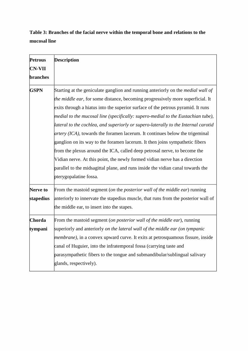
Table 3: Branches of the facial nerve within the temporal bone and relations to the
mucosal line
Petrous
CN-VII
branches
Description
GSPN Starting at the geniculate ganglion and running anteriorly on the medial wall of
the middle ear, for some distance, becoming progressively more superficial. It
exits through a hiatus into the superior surface of the petrous pyramid. It runs
medial to the mucosal line (specifically: supero-medial to the Eustachian tube),
lateral to the cochlea, and superiorly or supero-laterally to the Internal carotid
artery (ICA), towards the foramen lacerum. It continues below the trigeminal
ganglion on its way to the foramen lacerum. It then joins sympathetic fibers
from the plexus around the ICA, called deep petrosal nerve, to become the
Vidian nerve. At this point, the newly formed vidian nerve has a direction
parallel to the midsagittal plane, and runs inside the vidian canal towards the
pterygopalatine fossa.
Nerve to
stapedius
From the mastoid segment (on the posterior wall of the middle ear) running
anteriorly to innervate the stapedius muscle, that runs from the posterior wall of
the middle ear, to insert into the stapes.
Chorda
tympani
From the mastoid segment (on posterior wall of the middle ear), running
superiorly and anteriorly on the lateral wall of the middle ear (on tympanic
membrane), in a convex upward curve. It exits at petrosquamous fissure, inside
canal of Huguier, into the infratemporal fossa (carrying taste and
parasympathetic fibers to the tongue and submandibular/sublingual salivary
glands, respectively).

Table 4: Stepwise approach to understanding the petrous pyramid contents.
1. The temporal bone is composed of 5 parts: petrous, squamous, mastoid, tympanic and
styloid. The petrous pyramid separates the middle and posterior cranial fossae. It has three
surfaces (superior, posterior and inferior) and a base, also called lateral surface. Its base
faces laterally while its apex faces the midline. This pyramid is made by three temporal
bone parts: petrous, mastoid, and parts of the squamous.
2. The petrous pyramid can be described as a primitive bony container, surrounded by
cortical bone; and contents, grouped into four groups, or compartments (figure 3). The
container is cast around the compartments during development. Certain areas are not
occupied by any of the compartments, i.e. mastoid, petrous apex and TMJ roof.
3. The most important is the mucosal compartment/line. It comes obliquely from the pharynx
towards the mastoid bone, and is formed by three parts: the Eustachian tube anteriorly, the
middle ear in the middle, and the mastoid antrum posteriorly (figure 6B). This
compartment will give rise to air cells to a variable extent, through the process of
pneumatization, in areas of pyramid not occupied by any of the compartments, as listed
above. This would result in different possible composition of the mastoid and petrous apex
segments; as they can be pneumatized, diploic, or sclerotic.
4. The other three compartments coming into contact with the mucosal line are the cutaneous,
neural, and vascular compartments (figure 6C). The cutaneous compartment is lateral to
the middle ear and is composed of the external auditory canal (EAC) covered by skin and
transmits air vibrations to the middle ear. The neural compartment lies medial to the
middle ear and connects the middle ear to the brainstem, and contains anterior– cochlea -
and posterior labyrinth – vestibule and semicircular canals - on either side of the internal
auditory canal (IAC). This last leads the vestibular, cochlear, and facial nerves from the
ponto-medullary junction into the petrous bone. Lastly, the vascular compartment lies

inside the petrous apex, medial to the Eustachian tube. It includes the internal carotid
artery (ICA) coursing within the carotid canal, towards the foramen lacerum (FL).
5. Two reference lines cross at the middle ear, in the center of the pyramid: the muscosal line
and the EAC-IAC line. All anatomical structures within the petrous pyramid could be
located through two segmentation methods: the X-method creating four spaces thanks to
the previous intersecting reference lines; and the V-method delineating five segments
around the mucosal line (figure 4).

Table 5: Proposed nomenclature of air cells based on location within the temporal bone.
Air cell group Composition
Mastoid (retro-labyrinthine, inside the mastoid segment). Lying at the base of the petrous pyramid, within the triangular mastoid segment. It lies between the otic capsule and the lateral pyramidal surface, i.e., posterior to the otic capsule.
This includes the mastoid antrum and surrounding air cells. It is composed of seven groups of air cells that are easier to locate when viewed from the lateral surface according to Allam 12. From this view the mastoid looks like a triangle with three angles and three walls. The cells include: 1- Central cells, in the center of the mastoid Three groups on the angles of the triangle 2- Antral/periantral cells (in the antero-superior angle, including mastoid antrum) 3- Sinodural cells (in the postero-superior angle) 4- Tip cells (in the inferior angle, having medial and lateral cells, separated by the digastric ridge) Three groups on the walls of the triangle 5- Peri-facial cells (anteriorly, surrounding mastoid segment of the facial nerve, behind the external auditory canal) 6- Tegmental cells (superiorly, beneath the tegmen tympani) 7- Peri-sinus cells (posteriorly, around the sigmoid sinus, having medial and lateral cells)
Apical (pre-labyrinthine, inside the petrous apex segment). It lies between the otic capsule and the tip of petrous pyramid, i.e., anterior to the otic capsule.
This group can be further classified into: 1- Supracarotid cells 2- Infracarotid cells 3- Mediocarotid cells
Peri-labyrinthine (Lying across and also around the otic capsule segment). Cells interposed between the otic capsule and cortical bone of the superior, inferior and posterior surfaces, respectively. They thus provide connection between the apical and mastoid cells around the otic capsule.
It lies medial to the middle ear, and does not include cells pertaining to the mastoid groups posteriorly or to the apical group anteriorly. This includes : 1- Trans-labyrinthine cells (passing through the otic capsule, classically called subarcuate cells, passing within the arch of the superior semicircular canal (SCC), along with the petromastoid canal that carries the subarcuate artery). 2- Supra-labyrinthine cells (beneath the superior petrous surface). May be classified into anterior and posterior, as related to anterior and posterior labyrinths. 3- Infra-labyrinthine cells (beneath the inferior petrous surface). May be classified into anterior and posterior cells (below the anterior and posterior labyrinths, respectively). 4- Para-labyrinthine cells (beneath the posterior petrous surface, medial to otic capsule, as contrasted to the middle ear situated lateral to it). They surround the end of the internal auditory canal at the opening of the internal auditory meatus, being superior, inferior, anterior and posterior to it; thus called suprameatal, inframeatal, premeatal and postmeatal; respectively.
EAC roof cells Above the EAC.
TMJ roof cells In the bony roof of the glenoid fossa.
Peritubal (Cells around the Eustachian tube)
This includes: 1- Medial paratubal (between the Eustachian tube, ET, and the internal carotid artery, ICA) 2- Lateral paratubal 3- Supratubal 4- Infratubal
Accessory (extra-pyramidal) Cells extending beyond the petrous pyramid, even outside the boundaries of

the temporal bone. 1- Zygomatic: within the zygomatic arch and its base 2- Squamous (in the horizontal plate anterior to the roof of the TMJ, and also in the vertical plate) 3- Styloid 4- Occipital (classically considered in the accessory group, but it is an extratemporal extension into the occipital bone)