Ultrastructure of the ampullae of Lorenzini of Aptychotrema rostrata (Rhinobatidae
8
Zoomorphology DOI 10.1007/s00435-008-0073-5 123 ORIGINAL PAPER Ultrastructure of the ampullae of Lorenzini of Aptychotrema rostrata (Rhinobatidae) Barbara E. Wueringer · Ian R. Tibbetts · Darryl L. Whitehead Received: 10 January 2008 / Accepted: 6 October 2008 © Springer-Verlag 2008 Abstract Small epidermal pores of the electrosensory ampullae of Lorenzini located both ventrally and dorsally on the disk of Aptychotrema rostrata (Shaw and Nodder, 1794) open to jelly-Wlled canals, the distal end of which widens forming an ampulla that contains 6 § 0.7 alveolar bulbs (n = 13). The sensory epithelium is restricted to the alveolar bulbs and consists of receptor cells and supportive cells. The receptor cells are ellipsoid and their apical sur- faces are exposed to the alveolar lumen with each bearing a single central kinocilium. Presynaptic bodies occur in the basal region of the receptor cell immediately proximal to the synaptic terminals. The supportive cells that surround receptor cells vary in shape. Microvilli originate from their apical surface and extend into the alveolar lumen. Tight junctions and desmosomes connect the supportive cells with adjacent supportive and receptor cells in the apical region. The canal wall consists of two cell layers, of which the luminal cells are squamous and interconnect via desmo- somes and tight junctions, whereas the cells of the deeper layer are heavily interdigitated, presumably mechanically strengthening the canal wall. Columnar epithelial cells form folds that separate adjacent alveoli. The same cells separate the ampulla and canal wall. An aVerent sensory nerve com- posed of up to nine myelinated nerve axons is surrounded by several layers of collagen Wbers and extends from the ampulla. Each single aVerent neuron can make contacts with multiple receptor cells. The ultrastructural characteris- tics of the ampullae of Lorenzini in Aptychotrema rostrata are very similar to those of other elasmobranch species that use electroreception for foraging. Keywords Ampullae of Lorenzini · Alveolar bulbs · Receptor cell · Aptychotrema rostrata · Electroreception Introduction Ampullae of Lorenzini, the electroreceptors of elasmo- branchs, detect animate and non-animate minute electrical Welds in the environment (Kalmijn 1974; Zakon 1986). The biological roles ascribed to electroreception are prey detec- tion, geomagnetic orientation and electro-communication (Dijkgraaf and Kalmijn 1962; Kalmijn 1974; Wilkens and Hofmann 2005). Pores visible on the surface of the skin are opening into canals that lead to the sensory ampullae. The sensory epithe- lium, comprising receptor and supportive cells, is restricted to the alveolar bulbs of the ampulla (Waltman 1966; Jorgensen 2005). The canals of various lengths contain a species-speciWc gelatinous mucopolysaccharide similar in conductivity to the surrounding seawater (Waltman 1966; Murray 1974; Brown et al. 2002). In marine elasmobranch species, the length of the ampullary canals ranges from 5 to 20 cm (Brown 2002), and their ampullae are grouped into clusters by envelopes of con- nective tissue (Norris 1929; Jorgensen 2005). B. E. Wueringer Department of Zoology, University of Vienna, 1090 Vienna, Austria I. R. Tibbetts · D. L. Whitehead Centre for Marine Studies, The University of Queensland, St Lucia, QLD 4072, Australia Present Address: B. E. Wueringer (&) Sensory Neurobiology Group (formerly Vision, Touch and Hearing Research Centre), School of Biomedical Sciences, The University of Queensland, St Lucia, QLD 4072, Australia e-mail: [email protected]
Transcript of Ultrastructure of the ampullae of Lorenzini of Aptychotrema rostrata (Rhinobatidae

Zoomorphology
DOI 10.1007/s00435-008-0073-5ORIGINAL PAPER
Ultrastructure of the ampullae of Lorenzini of Aptychotrema rostrata (Rhinobatidae)
Barbara E. Wueringer · Ian R. Tibbetts · Darryl L. Whitehead
Received: 10 January 2008 / Accepted: 6 October 2008© Springer-Verlag 2008
Abstract Small epidermal pores of the electrosensoryampullae of Lorenzini located both ventrally and dorsallyon the disk of Aptychotrema rostrata (Shaw and Nodder,1794) open to jelly-Wlled canals, the distal end of whichwidens forming an ampulla that contains 6 § 0.7 alveolarbulbs (n = 13). The sensory epithelium is restricted to thealveolar bulbs and consists of receptor cells and supportivecells. The receptor cells are ellipsoid and their apical sur-faces are exposed to the alveolar lumen with each bearing asingle central kinocilium. Presynaptic bodies occur in thebasal region of the receptor cell immediately proximal tothe synaptic terminals. The supportive cells that surroundreceptor cells vary in shape. Microvilli originate from theirapical surface and extend into the alveolar lumen. Tightjunctions and desmosomes connect the supportive cellswith adjacent supportive and receptor cells in the apicalregion. The canal wall consists of two cell layers, of whichthe luminal cells are squamous and interconnect via desmo-somes and tight junctions, whereas the cells of the deeperlayer are heavily interdigitated, presumably mechanically
strengthening the canal wall. Columnar epithelial cells formfolds that separate adjacent alveoli. The same cells separatethe ampulla and canal wall. An aVerent sensory nerve com-posed of up to nine myelinated nerve axons is surroundedby several layers of collagen Wbers and extends from theampulla. Each single aVerent neuron can make contactswith multiple receptor cells. The ultrastructural characteris-tics of the ampullae of Lorenzini in Aptychotrema rostrataare very similar to those of other elasmobranch species thatuse electroreception for foraging.
Keywords Ampullae of Lorenzini · Alveolar bulbs · Receptor cell · Aptychotrema rostrata · Electroreception
Introduction
Ampullae of Lorenzini, the electroreceptors of elasmo-branchs, detect animate and non-animate minute electricalWelds in the environment (Kalmijn 1974; Zakon 1986). Thebiological roles ascribed to electroreception are prey detec-tion, geomagnetic orientation and electro-communication(Dijkgraaf and Kalmijn 1962; Kalmijn 1974; Wilkens andHofmann 2005).
Pores visible on the surface of the skin are opening intocanals that lead to the sensory ampullae. The sensory epithe-lium, comprising receptor and supportive cells, is restricted tothe alveolar bulbs of the ampulla (Waltman 1966; Jorgensen2005). The canals of various lengths contain a species-speciWcgelatinous mucopolysaccharide similar in conductivity to thesurrounding seawater (Waltman 1966; Murray 1974; Brownet al. 2002). In marine elasmobranch species, the length of theampullary canals ranges from 5 to 20 cm (Brown 2002), andtheir ampullae are grouped into clusters by envelopes of con-nective tissue (Norris 1929; Jorgensen 2005).
B. E. WueringerDepartment of Zoology, University of Vienna, 1090 Vienna, Austria
I. R. Tibbetts · D. L. WhiteheadCentre for Marine Studies, The University of Queensland, St Lucia, QLD 4072, Australia
Present Address:B. E. Wueringer (&)Sensory Neurobiology Group (formerly Vision, Touch and Hearing Research Centre), School of Biomedical Sciences, The University of Queensland, St Lucia, QLD 4072, Australiae-mail: [email protected]
123
Zoomorphology
This paper examines the ultrastructure of the electrore-ceptors of the shovelnose ray Aptychotrema rostrata (Shawand Nodder, 1794). As no species of rhinobatid has beenexamined via electron microscopy to date, comparisonswill be made to other species of elasmobranchs.
Materials and methods
Rhinobatid rays are benthic suction-crushing feeders, withdiets generally dominated by bottom-dwelling invertebratesand Wsh (Last and Stevens 1994; Kyne and Bennett 2002).Aptychotrema rostrata is endemic to eastern Australia,where it is found from Moreton Bay, Queensland to JervisBay, New South Wales. The species is generally found inshallow coastal waters to a depth of 50 m (Last and Stevens1994; Kyne and Bennett 2002).
Two specimens of Aptychotrema rostrata, ranging from650 to 755 mm total length, were captured via angling fromMoreton Bay, Queensland, Australia (27°28�S, 153°02�E).The specimens were immediately euthanized with a lethaldose of tricaine methane sulfonate (MS222; 1:2000). Totallength (§ 1 mm), disk width (§ 1 mm) and sex wererecorded. Samples of epidermal tissue containing ampullaeof Lorenzini were removed from the hyoid cluster and Wxedin Karnovsky’s (1965) formaldehyde–glutaraldehyde Wxa-tive in 0.1 M cacodylate buVer at 4°C for 3 days. The sam-ples were processed in a Lynx Tissue Processor Unit usingthree rinses of buVer, each for 15 min, followed by 80 minpostWxation in osmium tetroxide in 0.1 M cacodylate buVer(pH 7.2). After postWxation, samples were rinsed anotherthree times in the buVer, each for 15 min, and then dehy-drated in an ascending alcohol series. After dehydration,the samples were inWltrated with 50, 75% Spurrs epoxyresin (30 min each), followed by 30 min in 100% Spurrsepoxy resin before another change of Spurrs for 18 h. Thenthe samples were blocked in silicon molds and polymerizedat 60°C overnight. Survey sections (1 �m in thickness)were cut with a Nova Ultramicrotome LKB Bromma with aglass knife and stained with 3% toluidine blue in 0.1 Mphosphate buVer. Ultrathin sections (» 100 nm) were cutwith a diamond knife, mounted on 1 mm £ 2 mm singleslot carbon-stabilized colloidon grids, and stained with ura-nyl acetate and lead citrate, according to the method ofDaddow (1986). Sections were viewed and photographedusing both a JEOL 6400 and a JEOL 1010 transmissionelectron microscope at 80 kV.
Measurements of the ampullary tissues were made fromimages using AnalySIS ver.3 by soft imaging system andobtained from the male specimen (total length 755 mm),single axons of the sensory nerve Wber were measured,where Schwann cells surrounded them. The alveolar bulbsof single ampulla were counted. All measurements are
presented as mean § standard error. The results of ampul-lary morphology were compared to that described forAmblyraja radiata (Donovan, 1808), Raja clavata(Linnaeus, 1758) and Dipturus batis (Linnaeus, 1758) byWaltman (1966), Scyliorhinus canicula (Linnaeus, 1758)by Andres and von Düring (1988) and Iago omanensis(Norman, 1939) by Fishelson and Baranes (1998). All sta-tistical methods follow Köhler et al. (2002) and StatsoftInc. (2004).
Results
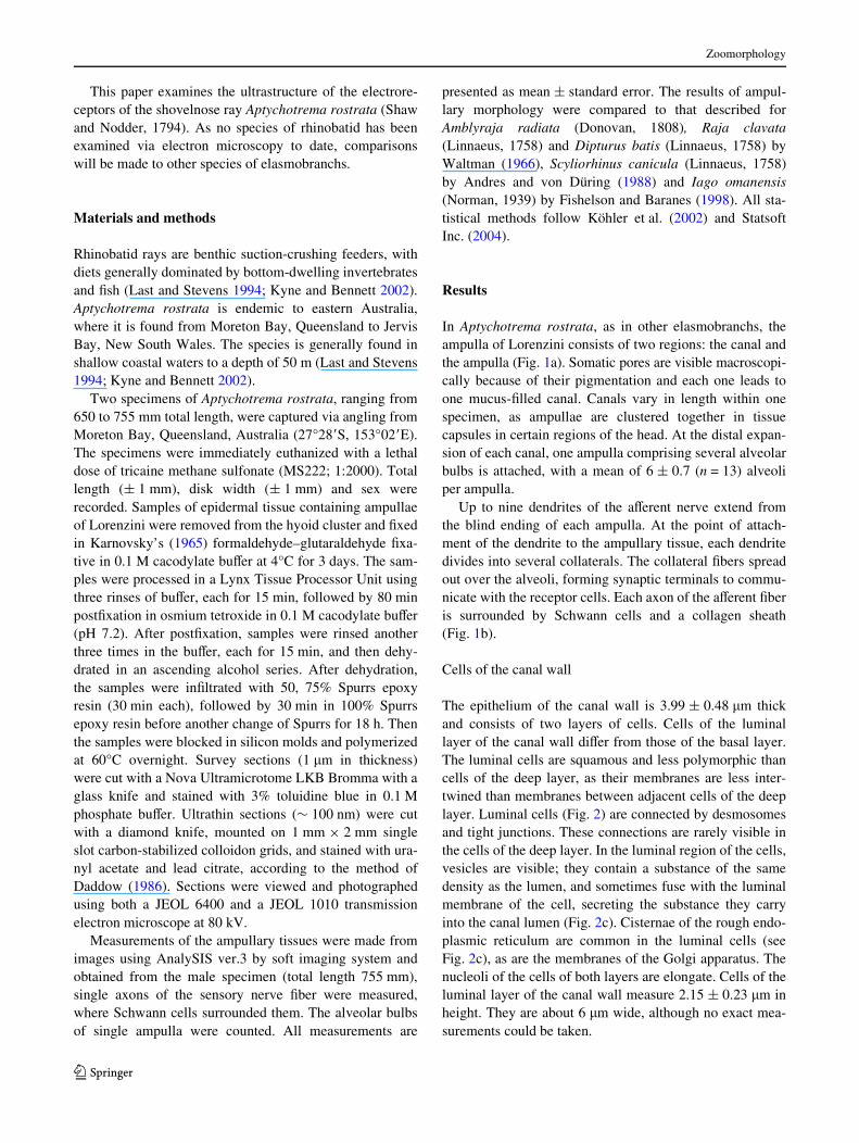
In Aptychotrema rostrata, as in other elasmobranchs, theampulla of Lorenzini consists of two regions: the canal andthe ampulla (Fig. 1a). Somatic pores are visible macroscopi-cally because of their pigmentation and each one leads toone mucus-Wlled canal. Canals vary in length within onespecimen, as ampullae are clustered together in tissuecapsules in certain regions of the head. At the distal expan-sion of each canal, one ampulla comprising several alveolarbulbs is attached, with a mean of 6 § 0.7 (n = 13) alveoliper ampulla.
Up to nine dendrites of the aVerent nerve extend fromthe blind ending of each ampulla. At the point of attach-ment of the dendrite to the ampullary tissue, each dendritedivides into several collaterals. The collateral Wbers spreadout over the alveoli, forming synaptic terminals to commu-nicate with the receptor cells. Each axon of the aVerent Wberis surrounded by Schwann cells and a collagen sheath(Fig. 1b).
Cells of the canal wall
The epithelium of the canal wall is 3.99 § 0.48 �m thickand consists of two layers of cells. Cells of the luminallayer of the canal wall diVer from those of the basal layer.The luminal cells are squamous and less polymorphic thancells of the deep layer, as their membranes are less inter-twined than membranes between adjacent cells of the deeplayer. Luminal cells (Fig. 2) are connected by desmosomesand tight junctions. These connections are rarely visible inthe cells of the deep layer. In the luminal region of the cells,vesicles are visible; they contain a substance of the samedensity as the lumen, and sometimes fuse with the luminalmembrane of the cell, secreting the substance they carryinto the canal lumen (Fig. 2c). Cisternae of the rough endo-plasmic reticulum are common in the luminal cells (seeFig. 2c), as are the membranes of the Golgi apparatus. Thenucleoli of the cells of both layers are elongate. Cells of theluminal layer of the canal wall measure 2.15 § 0.23 �m inheight. They are about 6 �m wide, although no exact mea-surements could be taken.
123

Zoomorphology
Cells of the deeper layer are heavily intertwined (Fig. 2)and often contain a Wbrous material in their cytoplasm(Fig. 2b). Compared to the cells of the luminal layer, cyto-plasmic organelles are less abundant. The endoplasmicreticulum is not as prominent as it is in the luminal cells andcontains fewer vesicles. Cells of the deeper layer of thecanal wall are 1.41 § 0.20 �m high and about 6 �m wide;however, their heteromorphy precluded any useful mea-surements of their diameter.
On the proximal side, the deeper layer rests on thebasal lamina, which separates the cells from a lamina of
Fig. 1 a Schematic presentation of an ampulla of Lorenzini of themultialveolate type. The left side of the ampulla is opened to visualizeepithelia of the canal, medial zone and alveoli. At the distal end thecanal opens up to an ampulla, which comprises several alveoli.Dendrites spread out over the alveoli and synapse with the receptorcells. A sensory nerve extends from the distal end of the ampulla.A collagen sheath surrounds the canal, ampulla and nerve. Not drawnto scale. Reprinted with permission from Wueringer and Tibbetts(2008). b A single aVerent nerve Wber of the ampullae of Lorenzini ofAptychotrema rostrata. TEM, cross section of a myelinated neuron.Several mitochondria are visible in the cytoplasm of the axon (A).A Schwann cell (S), with a nucleus (Nu), surrounds the axon, formingthe myelin sheath (My). Neurilemmal collagen sheath (C)
Fig. 2 The canal wall of the ampullae of Lorenzini of Aptychotremarostrata. TEM, to the left of the epithelium the canal lumen (Lu) isvisible. The cells of the luminal layer S1, S2 are connected by desmo-somes (De) and tight junctions (Tj), not found in the cells of the deeperlayer S3, S4. The basal lamina (Bl) separates the two cell layers from alayer of connective tissue containing regularly oriented collagen Wbers(C). a Cells of the deeper layer S3, S4 are intertwined heavily. b Vesi-cles (V) of the luminal cells contain a substance of the same density asthat in the lumen. c The cells of the luminal layer contain vesicles (V)and an extensive rough endoplasmic reticulum (ER). The adjacentmembranes of two cells S3, S4 of the deeper layer are heavily inter-twined. Nu nucleus
123

Zoomorphology
connective tissue. This lamina contains several layers ofcollagen Wbers, each oriented in a diVerent direction(Fig. 2).
Cells of the medial zone
The medial zone (Fig. 3) forms the transition between thereceptor epithelium and the cells of the canal wall. Thecanal wall is thinner than the receptor epithelium (height ofthe canal wall: 3.99 § 0.48 �m; height of the receptor epi-thelium: 11.11 § 0.37 �m), and consists of two layers ofcells compared to the monolayered receptor epithelium.The medial zone consists of one layer of columnar cells thatare about two and a half times higher than their width (i.e.height 12.61 § 0.89 �m; width 4.75 § 0.11 �m), with acentrally positioned nucleus similar to the nucleus of cellsof the luminal layer of the canal wall and receptor cells.Vesicles occur on the apical surface of the cells exposed tothe lumen. The walls of adjacent cells are interdigitated.Cells of the medial zone also create folds between the alve-olar bulbs, separating the receptor epithelia of neighboringbulbs.
The cells of the canal wall adjacent to the cells of themedial zone diVer in shape from other cells of the canalwall, as diVerences in height of the two epithelia are com-pensated. Cells of the deeper layer are consistent in height,whereas luminal cells expand in height from the side thatborders the canal wall to the side that borders the receptorepithelium (see Fig. 3).
Cells of the ampullary epithelium
The cells of the sensory epithelium measure 11.11 §0.37 �m in diameter (n = 5), surrounding a lumen with adiameter of 145.67 § 11.13 �m. An extracellular corpuscu-lar layer, varying in thickness between 2.5 and 5 �m, isfound in the lumen overlying the sensory epithelium.
The alveolar epithelium of Aptychotrema rostrata exhib-its two types of cells: elliptical receptor cells that are sur-rounded by supportive cells of various shapes (Fig. 4a). Inboth cell types a luminal, apical pole can be distinguishedfrom a basal pole, restricted by the basal lamina. Under-neath the basal lamina, a lamina of connective tissuecontains several layers of collagen Wbers.
Receptor cells
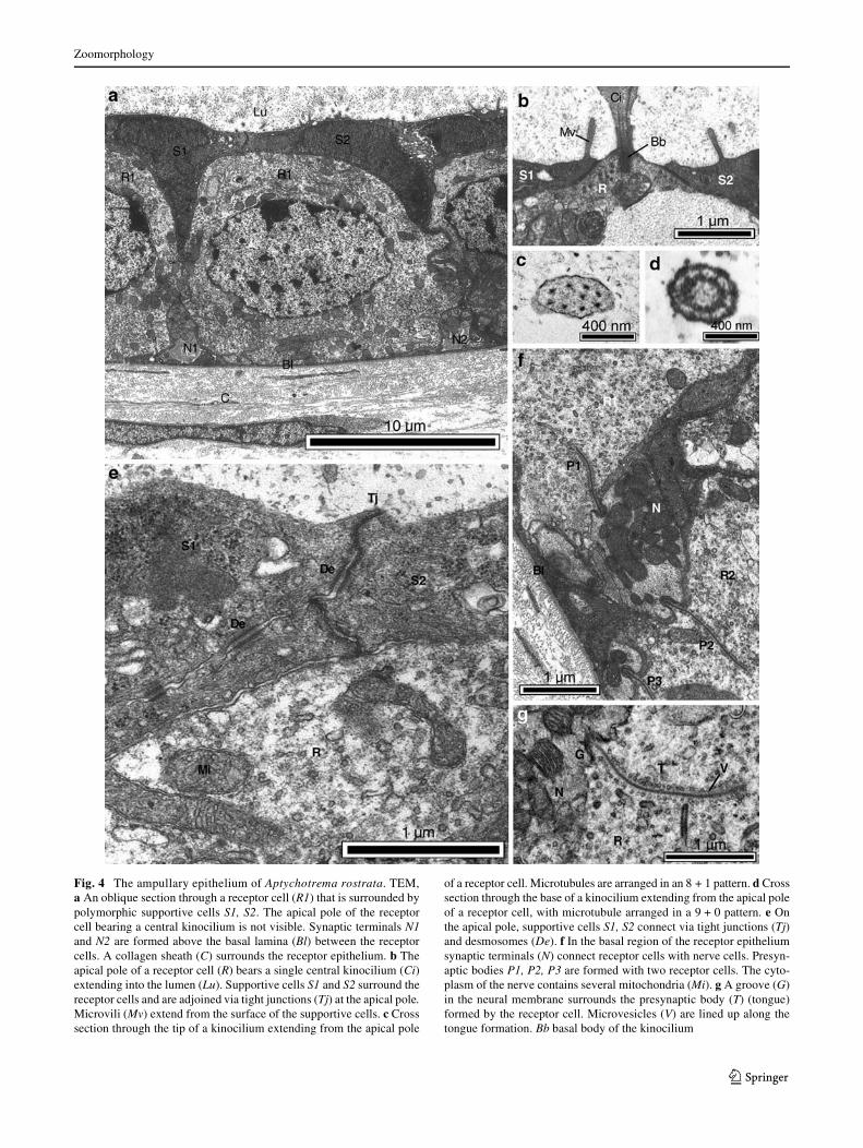
The receptor cells are elliptical, extend from the basal lam-ina to the alveolar lumen and are 10.72 § 0.48 �m high and11.22 § 0.62 �m wide. Each receptor cell contains an ellip-tical, centrally positioned nucleus measuring 5.80 §0.40 �m in height and 7.23 § 0.24 �m in width. The apicalsurface of the cell exposed to the alveolar lumen, is1.32 § 0.11 �m in diameter and bears a single central kino-cilium protruding into the lumen (Fig. 4b). The basal diam-eter of the kinocilium is 441.40 § 72.05 nm. The longestkinocilium measured 4.2 �m, but it did not appear to be anexact cross section through the center. The arrangement ofthe microtubules in the kinocilia is very unusual; along theaxoneme, microtubules are arranged in an 8 + 1 pattern(Fig. 4c), which was found up to 4.2 �m away from thereceptor cell membrane. In the basal region of the kinocil-ium the 9 + 0 structure (Fig. 4d) does not extend more than0.3 �m from the receptor cell. A basal body was found butno rootlet Wbers. No microvili extend from the apical sur-face of the receptor cells.
On the apical pole, tight junctions and desmosomes con-nect the receptor cell with adjacent supportive cells(Fig. 4a, e). In the apical cytoplasm of the receptor cells(Fig. 4e) there are numerous mitochondria, vesicles and thedictyosomes of the Golgi apparatus. The basal cytoplasmalso contains many mitochondria and vesicles but few dict-yosomes. In the basal region of the receptor cell, synapticterminals are present between the nucleus and the basallamina (Fig. 4f, g).
Supportive cells
As the supportive cells surround the elliptical receptor cells,they are quite variable in shape. In each supportive cell, theapical region extends to the alveolar lumen, whereas theproximal region abuts the basal lamina. Supportive cellsmeasure 11.71 § 0.56 �m in height and 3.91 § 0.41 �m in
Fig. 3 Semi-schematic presentation of the medial zone of the ampul-lae of Lorenzini of Aptychotrema rostrata. The cells of the medial zoneconnect with the single-layered sensory epithelium to the left and thedouble-layered canal wall on the right. Cells contain a large sphericalnucleus (Nu) and are connected via tight junctions (Tj) and desmo-somes (De) on the apical pole. Vesicles (V) are fused with the apicalmembrane. Underneath the basal lamina (Bl) are a collagen sheath (C)and Wbroblasts (Fi)
123
Zoomorphology
Fig. 4 The ampullary epithelium of Aptychotrema rostrata. TEM,a An oblique section through a receptor cell (R1) that is surrounded bypolymorphic supportive cells S1, S2. The apical pole of the receptorcell bearing a central kinocilium is not visible. Synaptic terminals N1and N2 are formed above the basal lamina (Bl) between the receptorcells. A collagen sheath (C) surrounds the receptor epithelium. b Theapical pole of a receptor cell (R) bears a single central kinocilium (Ci)extending into the lumen (Lu). Supportive cells S1 and S2 surround thereceptor cells and are adjoined via tight junctions (Tj) at the apical pole.Microvili (Mv) extend from the surface of the supportive cells. c Crosssection through the tip of a kinocilium extending from the apical pole
of a receptor cell. Microtubules are arranged in an 8 + 1 pattern. d Crosssection through the base of a kinocilium extending from the apical poleof a receptor cell, with microtubule arranged in a 9 + 0 pattern. e Onthe apical pole, supportive cells S1, S2 connect via tight junctions (Tj)and desmosomes (De). f In the basal region of the receptor epitheliumsynaptic terminals (N) connect receptor cells with nerve cells. Presyn-aptic bodies P1, P2, P3 are formed with two receptor cells. The cyto-plasm of the nerve contains several mitochondria (Mi). g A groove (G)in the neural membrane surrounds the presynaptic body (T) (tongue)formed by the receptor cell. Microvesicles (V) are lined up along thetongue formation. Bb basal body of the kinocilium
123

Zoomorphology
diameter, although these measurements should be regardedas an estimate. The nuclei are more variable in shape thanin receptor cells. On the supportive cell apical surface sev-eral microvilli extend into the lumen, encircling the centralkinocilium of the receptor cell. They measure 98.27 §12.16 nm in diameter and 519.65 § 131.78 nm in length.The apical surface of the supportive cells is coated inmucus, which is found in the alveolar lumen and secretedby exocytosis.
The synaptic terminals
In Aptychotrema rostrata each ampullary organ is inner-vated by an aVerent nerve comprising up to nine nerveWbers. Each nerve Wber is surrounded by a myelin sheath(Fig. 1b), which is surrounded by a lamina of collagenWbers, that binds all nerve Wbers together into a singlenerve. The mean diameter of the nerve Wber is13.75 § 1.095 �m; the mean height of Schwann cells is5.36 § 0.74 �m.
In the basal region of the receptor cell, between thenucleus and the basal lamina, the cell connects with aVerentdendrites (Fig. 4f, g), forming synaptic terminals. The syn-aptic terminals are unmyelinated, contain numerous mito-chondria and form presynaptic bodies. One synapticterminal can fuse with more than one receptor cell (Fig. 4f).Concomitantly, one receptor cell can possess multiple neu-ral attachments. The tongue-and-groove formation(Fig. 4f, g) is created by a groove in the neural membranein which the presynaptic body of the receptor cell extends,forming the tongue. Microvesicles occur along the length ofthe invagination of the receptor cell membrane within thetongue formation. Synaptic terminals measure 2.37 §0.60 �m in height and 3.75 § 0.10 �m in width. A maxi-mum of three tongue-and-groove formations per synapticterminal was found (n = 18).
Discussion
The hyoid cluster of Aptychotrema rostrata is the largestcluster in rajids (Raschi 1978, 1986) and also in rhinobatids(Wueringer and Tibbetts 2008). As Aptychotrema rostratais found in the marine environment and their ampullae ofLorenzini are macroscopic in the range of centimeters, theampullary organs are classiWed as macroampullae sensuAndres and von Düring (1988) (Wueringer and Tibbetts2008), compared to the microampullae of Holocephali spe-cies and the miniampullae of freshwater elasmobranchs, asdescribed in Potamotrygon laticeps (Garman, 1913) andPotamotrygon motoro (Müller and Henle, 1841) (Andresand von Düring 1988). However, both stenohaline andomnihaline rhinobatid species are known to possess ampul-
lae that can be classiWed as macroampullae (Chu and Wen1979; Wueringer and Tibbetts 2008).
Alveoli within an ampulla do not join at their distal endsas described for Iago omanensis (Fishelson and Baranes1998), but are stacked like grapes (see Fig. 1a). Therefore,following the classiWcation of ampullae by Andres and vonDüring (1988), these of Aptychotrema rostrata belong tothe multi-alveolate type ampulla of Jorgensen (2005),which is also found in another rhinobatid, Rhinobatus typus(Bennett, 1830) (Wueringer and Tibbetts 2008). The num-ber of alveoli varies between species, as Aptychotremarostrata possesses 6 § 0.7 alveoli per hyoid ampulla,whereas Raschi (1986) counted 20.5 to 8.3 alveoli perampulla for the hyoid cluster of 40 diVerent species ofrajids, and Iago omanensis possesses seven to nine alveoliper ampulla (Fishelson and Baranes 1998). In rajids a lownumber of alveoli per ampulla correlates with a shallowhabitat (Raschi 1986). Whether the same is true for rhino-batids remains to be tested.
The ampullary canal
The canal wall consists of two layers of cells, with theluminal cells being connected by tight junctions anddesmosomes. Waltman (1966) rarely found tight junctionsand desmosomes other than between the luminal ends ofthe inner cell layer. However, in Aptychotrema rostrata,desmosomes occur between adjacent cells of the deeperlayer, as described for Iago omanensis (Fishelson and Bar-anes 1998). These occlusions of intercellular space form thefunctional base for electroreception. The insulation of theampullary lumen enables the conduction of electric currentsfrom the environment along the canal to the sensory epithe-lium (Waltman 1966; Jorgensen 2005). Furthermore, deep-layered cells of the canal wall are interdigitated. Wehypothesize that these interdigitations provide mechanicalstrength to the canal, similar to that reported for cells of thesmall intestine (Ude and Koch 2002), but this hypothesisremains to be tested. Both the canal and ampulla are sur-rounded by multiple layers of collagen Wbers, which pro-vide mechanical support (Murray 1974). They are alsopresent around the myelinated nerve, between and aroundthe single nerve Wbers. Such lamina of connective tissuehave already been described for Amblyraja radiata, Rajaclavata and Dipturus batis (Waltman 1966), as well as inScyliorhinus canicula (Andres and von Düring 1988).
Canals 4.7–55.5 mm in length (Aptychotrema rostrataTL 68.0 cm, Wueringer and Tibbetts 2008) extend from thesomatic pores and form the alveolar bulbs at their proximalend. The ampullae of rhinobatid species occur in Wve dis-tinct clusters (Norris 1929), which facilitate the suppressionof interfering signals created by the animal’s own electric Weld(Kalmijn 1974) and do not change position ontogenetically.
123
Zoomorphology
Therefore, the length range of the canals depends on thesize of the animal. Moreover, canal length range may beexplained through the respective habitat of a species as itdepends on the conductivity of the surrounding water(Kalmijn 1974; Bodznick and Montgomery 2005). Inmarine elasmobranchs, the body tissues are electrically lessresistant than the surrounding water and longer canals arerequired to produce an eVective potential diVerence thatstimulates the receptor cells (Kalmijn 1974).
The medial zone
The morphology of the medial zone in Aptychotremarostrata both between adjacent alveoli and on the border ofthe sensory epithelium and the canal wall seems to conformto the description of the epithelium of the centrum cap ofScyliorhinus canicula (Andres and von Düring 1988). How-ever, Aptychotrema rostrata lacks the structure of a centrumcap and also brush-cells that border the centrum cap cells asdescribed by Andres and von Düring (1988) for S. canicula.
The ampullary epithelium
The receptor cells of Aptychotrema rostrata are encased bysupportive cells, as also observed in other species of elasmo-branchs (see Waltman 1966; Murray 1974; Szabo 1974;Andres and von Düring 1988). In Aptychotrema rostrata, theapical surface of the receptor cell exposed to the alveolarlumen measures 1.32 § 0.11 �m in diameter. The cell surfaceis approximately 378 �m2 and the exposed surface measures1.37 �m2, approximating 0.36% of the cell surface. This valueshould be regarded as an estimate. Murray (1974) found lessthan 1% exposed to lumen, whereas Andres and von Düring(1988) described 0.6% exposed to the lumen in S. canicula.
The structure of the kinocilium extending from thereceptor cells of Aptychotrema rostrata is unusual, asmicrotubules are arranged in a 9 + 0 and 8 + 1 pattern.Waltman (1966) and Szabo (1974) describe an 8 + 1arrangement of the microtubule in the axoneme, and men-tion Wnding a 9 + 0 arrangement in rajids without furtherspecifying its spatial occurrence. Contrary to Andres andvon Düring (1988) who report the 8 + 1 structure in thebase and the 9 + 0 structure in the axoneme of the kinociliaof Scyliorhinus canicula, we found microtubular arrange-ments of 9 + 0 in the base and 8 + 1 in the axoneme of thekinocilia. These interpretations were made possible as somekinocilia were found attached to a receptor cell, whileothers were cut along the shaft. The exact length of thekinocilia could not be measured as we found no exact crosssections along the whole length of the shaft, but the mini-mum lengths of ampullary kinocilia of Amblyraja radiata,Raja clavata, Dipturus batis and Scyliorhinus caniculaare 5 �m (Waltman 1966; Andres and von Düring 1988).
Waltman (1966) reports neither rootlet Wbers nor an acces-sory centriole of the kinocilium and neither were found inAptychotrema rostrata. Fishelson and Baranes (1998) onthe other hand report rootlet Wbers in the kinocilia of Iagoomanensis. They also describe a few low microvili on theapical surface of the receptor cell surrounding the kinocil-ium, which do not exist in Aptychotrema rostrata.
In the basal region of the sensory epithelium, synapticterminals connect with receptor cells. The unmyelinateddendrites form collaterals when attaching to the ampullarytissue. The tongue-and-groove formation (Waltman 1966) iscreated by the presynaptic body of the receptor cell (thetongue), which extends into the postsynaptic groove in theneural membrane (Fig. 4f, g). This structure is referred to asthe ribbon and gutter structure by Murray (1974). In thisstudy, receptor cells formed one to three presynaptic bodiesper synaptic terminal, with a mean of 1.61 § 0.18 synapticterminals per side. We observed a maximum of six presyn-aptic bodies per receptor cell. This value is regarded as anestimate, as only single sections of receptor cells wereobserved and receptor cells were not reconstructed threedimensionally. Murray (1974) found from two to seven sep-arate synapses per receptor cell, whereas Andres and vonDüring (1988) mention four to six synapses per receptor cellfor Scyliorhinus canicula. As the ultrastructural characteris-tics of the electrosensory ampullae in Aptychotrema rostrataappear identical with those of Amblyraja radiata, Raja clav-ata, Dipturus batis and Scyliorhinus canicula (Waltman1966; Andres and von Düring 1988), which use electrore-ception for foraging (see Kalmijn, 1974, 1978 for Scylio-rhinus and Raja), it is assumed that this organ has asimilar function in Aptychotrema rostrata. Although themorphology is indicative of this role, behavioral experi-ments are required to conWrm the possible uses and thesensitivity of the ampullae of Lorenzini in this species ofrhinobatid.
Acknowledgments The authors wish to thank Lina Daddow for herhelp with electron microscopy. We also wish to thank the two review-ers for their patience and valuable input. This research was carried outunder the following permits: University of Queensland Animal EthicsApproval No. CMS/420/04, issued to Ian Tibbetts, Centre for MarineStudies; Moreton Bay Marine Park Permit No. QS2004/CVL316, is-sued to Prof. Ove Hoegh-Guldberg; Fisheries Permit No. PRM02279I,issued to David Harris, Centre for Marine Studies.
References
Andres KH, von Düring M (1988) Comparative anatomy of vertebrateelectroreceptors. Prog Brain Res 74:113–131. doi:10.1016/S0079-6123(08)63006-X
Bodznick D, Montgomery JC (2005) The physiology of low frequencyelectrosensory systems. In: Bullock TH, Hopkins CD, PopperAN, Fay RR (eds) Electroreception. Springer Handbook of Audi-tory Research, vol 21. Springer, New York, pp 132–153
123

Zoomorphology
Brown BR (2002) Modeling an electrosensory landscape: behavioraland morphological optimization in elasmobranch prey capture.J Exp Biol 205:999–1007
Brown BR, Hutchinson JC, Hughes ME, Kellogg DR, Murray RW(2002) Electrical characterization of gel collected from sharkelectrosensors. Phys Rev E Stat Nonlin Soft Matter Phys65:061903. doi:10.1103/PhysRevE.65.061903
Chu WT, Wen QW (1979) A study of the lateral line canals system andthat of Lorenzini ampullae and tubules of elasmobranchiate Wshesof China. Monograph of Wshes of China, 2. Shanghai ScienceTechnology Press, Shanghai, 132p
Daddow LYM (1986) An abbreviated method of the double lead staintechnique. J Submicrosc Cytol Pathol 18:221–224
Dijkgraaf S, Kalmijn AJ (1962) Verhaltensversuche zur Funktion derLorenzinischen Ampullen. Naturwissenschaften 49:400. doi:10.1007/BF00632257
Fishelson L, Baranes A (1998) Distribution, morphology and cytologyof the ampullae of Lorenzini in the Oman shark, Iago omanensis(Triakidae) from the Gulf of Aqaba, Red Sea. Anat Rec 251:417–430. doi:10.1002/(SICI)1097-0185(199808)251:4<417::AID-AR1>3.0.CO;2-P
Jorgensen JM (2005) Morphology of electroreceptive sensory organs.In: Bullock TH, Hopkins CD, Popper AN, Fay RR (eds) Electro-reception. Springer Handbook of Auditory Research, vol 21.Springer, New York, pp 47–67
Kalmijn AJ (1974) The detection of electric Welds from inanimate andanimate sources other than electric organs. In: Fessard A (ed) Hand-book of sensory physiology, vol III/3. Springer, Berlin, pp 147–200
Kalmijn AJ (1978) Electric and magnetic sensory world of sharks,skates and rays. In: Hodgeson ES, Mathewson RF (eds) Sensorybiology of sharks, skates and rays. Department of NavalResearch, Arlington, pp 507–528
Karnovsky MJ (1965) A formaldehyde-glutaraldehyde Wxative of highosmolarity for use in electron microscopy. J Cell Biol 27:137A–138A
Köhler W, Schachtel G, Voelske P (2002) Biostatistik. Eine Einfüh-rung für Biologen und Agrarwissenschaftler. 3. AuXage Springer,Berlin, 301 pp
Kyne PM, Bennett MB (2002) Diet of the eastern shovelnose ray,Aptychotrema rostrata (Shaw and Nodder, 1794), from MoretonBay, Queensland, Australia. Mar Freshw Res 53:679–686.doi:10.1071/MF01040
Last PR, Stevens JD (1994) Sharks and rays of Australia. CSIRO Pub-lishing, Melbourne, pp 283–287
Murray RW (1974) The ampullae of Lorenzini. In: Fessard A (ed)Handbook of sensory physiology, vol III/3. Springer, Berlin,pp 125–146
Norris HW (1929) The distribution and innervation of the ampullae ofLorenzini of the dogWsh, Squalus acanthias. Some comparisonswith conditions in other plagiostomes and corrections of prevalenterrors. J Comp Neurol 47:449–465. doi:10.1002/cne.900470306
Raschi WG (1978) Notes on the gross functional morphology of theampullary system in two similar species of skates, Raja erinaceaand R. ocellata. Copeia 1:48–53. doi:10.2307/1443820
Raschi WG (1986) A morphological analysis of the ampullae ofLorenzini in selected skates (Pisces, Rajoidei). J Morphol189:225–247. doi:10.1002/jmor.1051890303
Statsoft Inc (2004) Electronic statistics textbook. Tulsa. http://www.statsoft.com/textbook/stathome.html
Szabo T (1974) Anatomy of the specialized lateral line organs of elec-troreception. In: Fessard A (ed) Handbook of sensory physiology,vol III/3. Springer, Berlin, pp 13–59
Ude J, Koch M (2002) Die Zelle—Atlas der Ultrastruktur. 3 AuXage.Spektrum Akademischer Verlag, Heidelberg. 309 pp
Waltman B (1966) Electrical ties and Wne structure of the ampullarycanals of Lorenzini. Acta Physiol Scand 66(Suppl 264):1–60
Wilkens LA, Hofmann MH (2005) Behavior of animals with passive,low-frequency electrosensory systems. In: Bullock TH, HopkinsCD, Popper AN, Fay RR (eds) Electroreception. Springer Handbookof Auditory Research, vol 21. Springer, New York, pp 229–263
Wueringer BE, Tibbetts IR (2008) Comparison of the lateral line andampullary system of two species of shovelnose ray. Rev Fish BiolFish 18:47–64. doi:10.1007/s11160-007-9063-9
Zakon HH (1986) The electroreceptive periphery. In: Bullock TH, Heili-genberg W (eds) Electroreception. Wiley, New York, pp 287–317
123