Behavioral and Psychological Symptoms in Alzheimer’s Disease
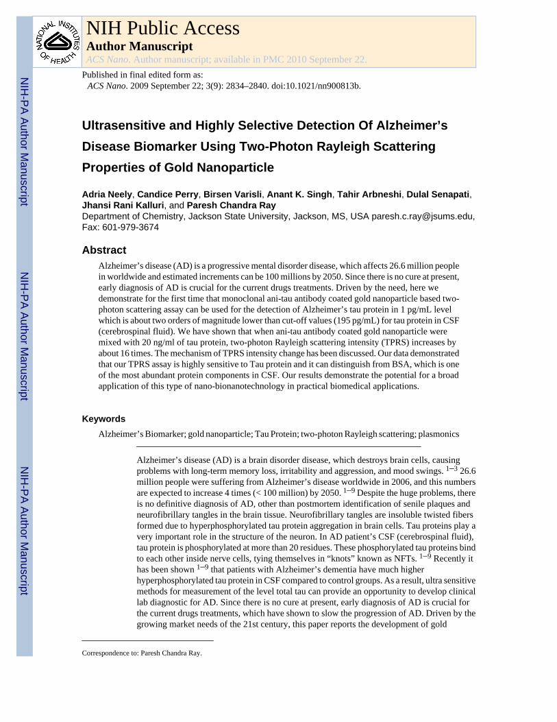
Ultrasensitive and Highly Selective Detection Of Alzheimer’sDisease Biomarker Using Two-Photon Rayleigh ScatteringProperties of Gold Nanoparticle
Adria Neely, Candice Perry, Birsen Varisli, Anant K. Singh, Tahir Arbneshi, Dulal Senapati,Jhansi Rani Kalluri, and Paresh Chandra RayDepartment of Chemistry, Jackson State University, Jackson, MS, USA [email protected],Fax: 601-979-3674
AbstractAlzheimer’s disease (AD) is a progressive mental disorder disease, which affects 26.6 million peoplein worldwide and estimated increments can be 100 millions by 2050. Since there is no cure at present,early diagnosis of AD is crucial for the current drugs treatments. Driven by the need, here wedemonstrate for the first time that monoclonal ani-tau antibody coated gold nanoparticle based two-photon scattering assay can be used for the detection of Alzheimer’s tau protein in 1 pg/mL levelwhich is about two orders of magnitude lower than cut-off values (195 pg/mL) for tau protein in CSF(cerebrospinal fluid). We have shown that when ani-tau antibody coated gold nanoparticle weremixed with 20 ng/ml of tau protein, two-photon Rayleigh scattering intensity (TPRS) increases byabout 16 times. The mechanism of TPRS intensity change has been discussed. Our data demonstratedthat our TPRS assay is highly sensitive to Tau protein and it can distinguish from BSA, which is oneof the most abundant protein components in CSF. Our results demonstrate the potential for a broadapplication of this type of nano-bionanotechnology in practical biomedical applications.
KeywordsAlzheimer’s Biomarker; gold nanoparticle; Tau Protein; two-photon Rayleigh scattering; plasmonics
Alzheimer’s disease (AD) is a brain disorder disease, which destroys brain cells, causingproblems with long-term memory loss, irritability and aggression, and mood swings. 1–3 26.6million people were suffering from Alzheimer’s disease worldwide in 2006, and this numbersare expected to increase 4 times (< 100 million) by 2050. 1–9 Despite the huge problems, thereis no definitive diagnosis of AD, other than postmortem identification of senile plaques andneurofibrillary tangles in the brain tissue. Neurofibrillary tangles are insoluble twisted fibersformed due to hyperphosphorylated tau protein aggregation in brain cells. Tau proteins play avery important role in the structure of the neuron. In AD patient’s CSF (cerebrospinal fluid),tau protein is phosphorylated at more than 20 residues. These phosphorylated tau proteins bindto each other inside nerve cells, tying themselves in “knots” known as NFTs. 1–9 Recently ithas been shown 1–9 that patients with Alzheimer’s dementia have much higherhyperphosphorylated tau protein in CSF compared to control groups. As a result, ultra sensitivemethods for measurement of the level total tau can provide an opportunity to develop clinicallab diagnostic for AD. Since there is no cure at present, early diagnosis of AD is crucial forthe current drugs treatments, which have shown to slow the progression of AD. Driven by thegrowing market needs of the 21st century, this paper reports the development of gold
Correspondence to: Paresh Chandra Ray.
NIH Public AccessAuthor ManuscriptACS Nano. Author manuscript; available in PMC 2010 September 22.
Published in final edited form as:ACS Nano. 2009 September 22; 3(9): 2834–2840. doi:10.1021/nn900813b.
NIH
-PA Author Manuscript
NIH
-PA Author Manuscript
NIH
-PA Author Manuscript
nanomaterial based two-photon Rayleigh scattering (TPRS) assay for the ultra-sensitive andselective detection of Tau Protein AD biomarker. Two-photon scattering properties have beenmonitored using hyper–Rayleigh scattering (HRS) technique. 10–18 This technique can beeasily applied to study a very wide range of materials because electrostatic fields and phasematching are not required. Other advantages are that the polarization analysis gives informationabout the tensor properties, and spectral analysis of the scattered light gives information aboutthe dynamics.
In the last 15 years, the field of biosensor using nanomaterial has witnessed an explosion ofinterest in the use of nanomaterials in assays for DNA/RNA, protein and cell markers for manydiseases. 19–34 The integration of nanotechnology with biology and medicine is expected toproduce major advances in molecular diagnostics, therapeutics and bioengineering. 19–44 Interms of biological sensing, the use of nanotechnology has led to the production of numerous,rapid, sensitive multi-analytes assays which are useful not only in the laboratory, also in thefield as portable instruments. 29–45 Due to the increasing availability of nanostructures withhighly controlled optical properties, nanosystems are attractive in their use in biotechnologicalsystem for diagnostic application. Gold nanosystem are attractive because of their uniqueproperties, including their shape and size-dependent optical properties. Due to the lack oftoxicity, 12–45 scientists have shown great interest to use gold nanosystems for biomarkerapplications and biological imaging. Mirkin et. al. 6,8 and others 28–29 have demonstrated thatnanoparticle based bio-barcode assays with localized surface plasmon resonance (LSPR), arecapable of measuring ADDL level in CSF. Here, we demonstrate for the first time thatmonoclonal ani-tau antibody (tau-mab) coated gold nanoparticle based two-photon scatteringassay 10–18,46–47 can be used for the detection of Alzheimer’s tau protein in 1 pg/mL levelwhich is about two orders of magnitude lower than cut-off values (195 pg/mL) for tau proteinin CSF. Our results reported here demonstrate the potential for a broad application ofbioconjugated nanoparticles in practical biotechnological and medical applications.
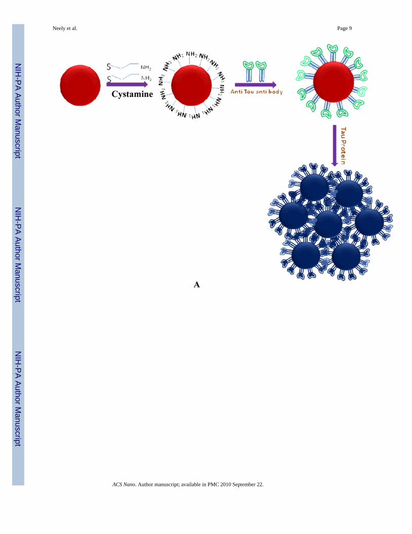
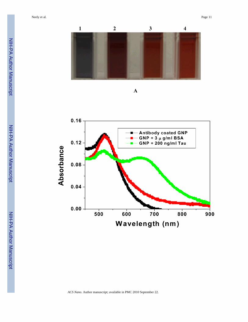
Results and DiscussionOur two-photon scattering approach for the detection of selective AD biomarker is based onthe fact that, the monoclonal ani-tau antibody -conjugated gold nanoparticles can readily andspecifically identify Tau protein, through antibody–antigen interaction and recognition (asshown in Figure 1). For a Tau protein, there are many surface antigens available for specificrecognition with monoclonal ani-tau antibody-conjugated nanoparticles. Therefore, in thepresence of Tau protein, several nanoparticles can bind to each protein, thereby producingnanoparticle aggregates (as shown in Figure 1). As a result, a colorimetric change has beenobserved from red to bluish color (as shown in Figure 2) and a new broad band appears around150 nm far from their plasmon absorption band, as shown in Figure 2B.
As shown in Figure 2C, when monoclonal ani-tau antibody-conjugated gold nanoparticles weremixed with various concentrations of Tau protein, two-photon scattering intensity increasesby about 16 times (as shown in Figure 2). Our experimental results demonstrated a very distincttwo-photon scattering intensity change (2.2 times) even upon the addition of 1 pico gram (pg)/ml of Tau protein. To evaluate whether our assay is highly selective, we have also performedhow two-photon scattering intensity changes upon addition of serum albumin (BSA) proteinand heme protein, instead of Tau protein with anti-tau-antibody conjugated gold nanoparticles.As shown in Figure 2C, two-photon scattering intenity changes only 1.2 times in presence of200 ng/ml BSA protein and 1.6 times when we added 30000 ng/ml of BSA protein tomonoclonal ani-tau antibody -conjugated gold nanoparticles. Similarly when we added 3000ng/ml heme protein to monoclonal ani-tau antibody-conjugated gold nanoparticles, two-photonscattering intensity changes only 1.2 times.
Neely et al. Page 2
ACS Nano. Author manuscript; available in PMC 2010 September 22.
NIH
-PA Author Manuscript
NIH
-PA Author Manuscript
NIH
-PA Author Manuscript
Two-photon scattering signal from monoclonal ani-tau antibody -conjugated goldnanoparticles can be expressed as, 10–19,45–47
(1)
where G is a geometric factor, Nw and Nnano the number of water molecules and monoclonalani-tau antibody -conjugated gold nanoparticle per unit volume, βω and βnano are thequadratichyperpolarizabilities of a single water molecule and a single monoclonal ani-tauantibody -conjugated gold nanoparticle, ε2ω is the molar extinction coefficient of the goldnanoparticle at 2ω, l is the path length and Iω the fundamental intensity. The exponential factoraccounts for the losses through absorption at the harmonic frequency. Considering the size ofnanoparticle, the approximation that assumes that the electromagnetic fields are spatiallyconstant over the volume of the particle may not suitable anymore. As a result, the totalnonlinear polarization consists of different contributions such as multipolar radiation of theharmonic energy of the excited dipole and possibly of higher multipoles, as we discussed inour previous publication or reported by others. 12–19 The TPRS intensity therefore alsoconsists of several contributions. The first one is the electric dipole approximation, which mayarise due to the defects in nanoparticle. This contribution is actually identical to the oneobserved for any non-centrosymmetrical point-like objects such as efficient rod-like push-pullmolecules. 10–11, 45–46 The second contribution is multipolar contribution like electricquadrupole contribution. 12–19 This contribution is very important when the size of the particleis no longer negligible when compared to the wavelength, as we reported before. Since for aTau protein, there are many surface antigens available for specific recognition with monoclonalani-tau antibody-conjugated nanoparticles, as shown in Figure 1C, antibody-conjugated goldnanoparticles undergo aggregation in presence of tau protein. Now due to the aggregation inthe presence of tau, antibody-conjugated gold nanoparticles looses the center of symmetry andas a result, one can expect significant amount of electric dipole contribution to the two-photonscattering intensity. Since electric dipole contributes several times higher than that ofmultipolar moments, we expect two-photon scattering intensity to increase upon the additionof tau.
As shown in Figure 2A, a clear colorimetric change is observed when tau protein was addedto ani-tau antibody-conjugated gold nanoparticles. This colorimetric change is mainly due tothe aggregation of antibody-conjugated gold nanoparticles, as shown in Figure 1C. We havenot observed any color change upon addition of serum albumin (BSA) protein and hemeprotein, instead of Tau protein to anti-tau-antibody conjugated gold nanoparticles (as shownin Figure 2A). Our TEM data also demosntrated (Figure 2D) that there is no significantaggregation due to the addition serum albumin (BSA) protein or heme protein to ani-tauantibody-conjugated gold nanoparticles. Figure 2B reports how the absorption maximum forsurface plasmon band of gold nanoparticle at 520 nm changes in the presence of tau, BSA andheme protein. Our experimental data show that intensity of the absorption band at 520 nmdecreases with the addition of 200 ng/ml tau and a new broad band corresponding to theabsorption of antibody -conjugated gold nanoparticle aggregates appears at 670 nm. In thesame plot, we have also shown that absorption remains about unchnaged upon the addition ofBSA to ani-tau antibody-conjugated gold nanoparticles. So our colorimetric data demonstratethat our assay is highly sensitive to Tau protein and it can distinguish from BSA, which is oneof the most abundant protein components in CSF. Our experimental results also shows that tauprotein can be detected in 2.8 ng/mL level with colorimetric study, where as TPRS can detecteven at 1 pg/ml level. Our experimental results clearly exhibit that TPRS assay can be 3 ordersof magnitude more sensitive than the colorimetric assay. Now this new absorption band
Neely et al. Page 3
ACS Nano. Author manuscript; available in PMC 2010 September 22.
NIH
-PA Author Manuscript
NIH
-PA Author Manuscript
NIH
-PA Author Manuscript
appearing at 670 nm upon the addition of Tau protein can influence the TPRS intensity veryhighly. According to the two-state model, 48
(2)
where ω is the fundamental energy of the incident light, μeg is the transition dipole momentand ωeg is the transition energy between the ground state |g> and the charge-excited state |e>,Δμeg is the difference in dipole moment between |e> and |g> states. Since ωeg ∝ 1/λmax, βshould change tremendously upon the addition of tau protein and as a result, the two-photonscattering intensity should change tremendously with the addition of tau. So, due to theaggregation after addition of Tau protein, the two-photon Rayleigh scattering (TPRS) intensitychange observed in our assay, can be due to several factors and these are, 1) one can expectsignificant amount of electric dipole contribution to the two-photon scattering intensity as wediscussed before; 2) due to the change of λmax toward red, TPRS intensity should change afteraddition of tau protein, as we discussed above. (3) Since size increases tremendously withaggregation, the two-photon scattering intensity should increase with the increase in particlesize.
To understand the response rate of the TPRS signal upon the addition of tau protein to ani-tauantibody-conjugated gold nanoparticles, we have measured the TPRS intensity at different timeintervals, as shown in Figure 3. Our TPRS experimental data indicate that after the addition of20 ng/ml tau, the TPRS intensity increases by a factor of 8 within 500 seconds, then increasesslowly with time, and then remains constant after 2000 seconds. Our time interval TPRSintensity measurement indicates that the time frame of our measurement is only half an hour.
To evaluate whether TPRS assay is capable of measuring tau protein concentrationquantitatively, we performed two-photon Rayleigh scattering intensity measurements uponaddition of different concentrations of target tau protein to ani-tau antibody -conjugated goldnanoparticles. As shown in Figure 4, the two-photon scattering intensity increment is highlysensitive to the concentration of target tau protein over the range of 5 – 350 ng/ml and theintensity increased linearly with concentration. Our experimental results also indicate that thereis no linear correlation between TPRS intensity and tau protein concentration between 1–5 pg/ml and 350 – 850 pg/ml region.
So our ani-tau antibody -conjugated gold nanoparticle based two-photon scattering assay canprovide a quantitative measurement of tau protein concentration over 5–350, ng/ml range.
ConclusionsIn conclusion, in this paper, we have demonstrated for the first time a label-free, fast and highlysensitive monoclonal ani-tau antibody (tau-mab) coated gold nanoparticle based two-photonscattering assay for the selective detection of Alzheimer’s tau protein in 1 pg/mL level. Thedetection limit of our TPRS assay is about two orders of magnitude lower than cut-off values(195 pg/mL) for tau protein in CSF. We have shown that when ani-tau antibody coated goldnanoparticle were mixed with 20 pg/ml concentrations of tau protein, two-photon scatteringintensity increases by about 16 times. Our experimental data with serum albumin (BSA) proteinas well as IgG protein with anti-tau-antibody conjugated gold nanoparticles clearlydemonstrated that our TPRS assay is highly sensitive to Tau protein and it can distinguish fromBSA, which is one of the most abundant protein components in CSF. Our experiment indicatesthis bioassay is rapid and takes less than 35 minutes from protein binding to detection and
Neely et al. Page 4
ACS Nano. Author manuscript; available in PMC 2010 September 22.
NIH
-PA Author Manuscript
NIH
-PA Author Manuscript
NIH
-PA Author Manuscript
analysis and it can be three orders of magnitude more sensitive than the usual colorimetrictechnique. Our concentartion dependent measurement point out that our antibody-conjugatedgold nanosystem based two-photon scattering assay can provide a quantitative measurementof tau protein concentration in pg/ml region. Our assay will have several advantages and theseare: i) it can be two orders of magnitude more sensitive than the usual colorimetric technique;ii) tau protein can be discriminated very easily from other protein. Our experimental resultsreported here open up a new possibility of rapid, easy and reliable diagnosis of AD biomarkersby measuring the TPRS intensity from antibody modified gold nanosystems.
Experimental MethodsHydrogen tetrachloroaurate (HAuCl4·3H2O), NaBH4, buffer solution, NaCl, sodium citrate,monoclonal ani-tau antibody (tau-mab) and tau protein were purchased from Sigma-Aldrichand used without further purification.
Gold Nanoparticle SynthesisGold nanoparticles of different sizes and shapes were synthesized by controlling the ratio ofHAuCl4, 3H2O and sodium citrate concentration as we reported recently. 16–19,37–39 Forsmaller gold nanoparticles, we have used sodium borohydride method as reported before. 0.5ml of 0.01 M HAuCl4 trihydrate in water and 0.5 mL of 0.01 M sodium citrate in water wereadded to 18 mL of deionized H2O and stirred. Next, 0.5 mL of freshly prepared 0.1 MNaBH4 was added and the solution color changed from colorless to orange. Stirring wasstopped and the solution was left undisturbed for 2h. The resulting spherical gold nanoparticleswere 4 nm in diameter. UV-visible absorption spectrum, colorimetric observation andTransmission electron microscope (TEM) (as shown in Figure 2) were used to characterize thenanoparticles. The particle concentration was measured by UV-visible spectroscopy using themolar extinction coefficients at the wavelength of the maximum absorption of each gold colloidas reported recently [ε(15) 518nm = 3.6 × 108 cm−1M−1.16–19, 37–39
Preparation of monoclonal ani-tau antibody (tau-mab) coated gold nanoparticleFor the preparation of monoclonal ani-tau antibody (tau-mab)-conjugated nanoparticles, wehave modified the gold nanoparticle surface by amine groups (as shown in Scheme 1) usingcystamine dihydrochloride using reported method. 27,41 For this purpose, we have added 30mM cystamine dihydrochloride to 50 mL of gold nanoparticle and the solution was kept at 50°C for several hours under constant sonication. After that, the excess cystamine dihydrochloridewas removed by centrifugation at 8000 rpm for several minutes. For covalent immobilizationof the monoclonal ani-tau antibody onto the amine modified gold nanoparticle (GNP) surface,we have used highly established glutaraldehyde spacer method. In brief, 10 ml of amine-functionilized GNP were incubated with monoclonal ani-tau antibody for 12 hours at 4 °C inPBS media. To remove the excess antibody, we have washed monoclonal ani-tau antibodyconjugated GNP several times with PBS. During this amine group activation andimmobilization of the antibody, we have not noted any aggregation of gold nanoparticles asexamined by TEM and UV-VIS absorption spectroscopy.
Two-photon Rayleigh scattering SpectroscopyTwo-photon Rayleigh scattering (TPRS) 10–19,45–47 from an isotropic solution was observedfor the first time in 1965, 10 shortly after the invention of high power Ruby lasers. When anintense light of frequency ω, exposed on a matter, it can generate a spectrum of the scatteredradiation that includes bands with frequency 2ω, due to TPRS and 2ω ± ω0, due to two-photonRaman scattering. In case of two-photon Raman scattering, ω0 is the frequency associated witha transition between two energy levels of the scattering molecule. The two-photon light
Neely et al. Page 5
ACS Nano. Author manuscript; available in PMC 2010 September 22.
NIH
-PA Author Manuscript
NIH
-PA Author Manuscript
NIH
-PA Author Manuscript
scattering can be observed from fluctuations in symmetry, caused by rotational fluctuations.For the TPRS or HRS experiment, we have used a mode-locked Ti:sapphire laser deliveringat fundamental wavelength of 860 nm with a pulse duration of about 150 fs at a repetition rateof 80 MHz. We performed TEM data before and after exposure of about 5–10 minutes to thelaser and we have not noted any photo-thermal damage of antibody coated gold nanoparticleswithin our HRS data collecting time. The HRS light was separated from its linear counterpartby a high-pass filter, and a monochromator and then detected with a cooled photomultipliertube. The pulses were counted with a photon counter. The fundamental input beam was linearlypolarized, and the input angle of polarization was selected with a rotating half-wave plate. Inall experiments reported, the polarization state of the harmonic light was vertical.
Since λmax for monoclonal ani-tau antibody coated gold nanoparticle (515 nm) and aggregates(650 nm) are very far from excitation source (860 nm) or second harmonic generated frequency(430 nm), we can eliminate the two-photon luminescence (TPL) contributions in our HRSexperiment. To make sure that only second harmonic signal is collected by PMT, we have used3 nm interference filter and a monochromator in front of PMT. To understand whether the two-photon scattering intensity at 430 nm light is due to second harmonic generation, we performedpower dependent as well as concentration dependent studies. Figure 5 shows the output two-photon scattering signal intensities at 430 nm from monoclonal ani-tau antibody coated goldnanoparticle at different powers of 860 nm incident light. A linear nature of the plot impliesthat the doubled light is indeed due to the two-photon Rayleigh scattering signal.
AcknowledgmentsDr. Ray thanks NIH-SCORE grant # S06GM 008047 and NSF-PREM grant # DMR-0611539 for their generousfunding. We also thank reviewers whose valuable suggestions improved the quality of the manuscript.
References1. Rookmeyer R, Johnson E, Ziegler-Graham K, Arrighi MH. Forcasting the Global Burden of
Alzheimer’s Disease. Alzheimer’s and Dementia 2007;3:186–91.2. Bu G. Apolipoprotein E and its Receptors in Alzheimer’s Disease: Pathways, Pathogenesis and
Therapy. Nat Rev Neurosci 2009;10:333–344. [PubMed: 19339974]3. Tanzi RE, Bertram L. Thirty Years of Alzheimer’s Disease Genetics: The Implications of Systematic
Meta-Analyses. Nat Rev Neurosci 2008;9:768–778. [PubMed: 18802446]4. www.accentonseniors.com/articles/Alzheimers_2008_Fact_Sheet.pdf5. Keating CD. Nanoscience Enables Ultrasensitive Detection of Alzheimer’s Biomarker. Proc Natl Acad
Sci USA 2005;102:2263–2264. [PubMed: 15703290]6. Georganopoulou DG, Chang L, Nam JM, Thaxton CS, Mufson EJ, Klein WL, Mirkin CA.
Nanoparticle-Based Detection in Cerebro Spinal Fluid of a Soluble Pathogenic Biomarker forAlzheimer’s Disease. Proc Natl Acad Sci USA 2005;102:2273–2276. [PubMed: 15695586]
7. Tapiola T, Overmyer M, Lehtovirta M, Helisalmi S, Ramberg J, Alafuzoff I. The Level of CerebrospinalFluid Tau Correlates with Neurofibrillary Tangles in Alzheimer’s Disease. Neuroreport 1997;8:3961–3963. [PubMed: 9462474]
8. Haes AJ, Chang L, Klein WL, Van Duyne RP. Detection of a Biomarker for Alzheimer’s Disease fromSynthetic and Clinical Samples Using a Nanoscale Optical Biosensor. J Am Chem Soc 2005 ;127:2264–2271. [PubMed: 15713105]
9. Buerger K, Ewers M, Pirttila T, Zinkowski R, Alafuzoff I, Teipel SJ, DeBernardis J, Kerkman D,McCulloch C, Soininen H. CSF Phosphorylated Tau Protein Correlates with NeocorticalNeurofibrillary Pathology in Alzheimer’s Disease. Brain 2006;129:3035–3041. [PubMed: 17012293]
10. Terhune RW, Maker PD, Savage CM. Measurements of Nonlinear Light Scattering. Phys Rev Lett1965;14:681–684.
11. Clays K, Persoons A. Hyper-Rayleigh Scattering in Solution. Phys Rev Lett 1991;66:2980–3.[PubMed: 10043668]
Neely et al. Page 6
ACS Nano. Author manuscript; available in PMC 2010 September 22.
NIH
-PA Author Manuscript
NIH
-PA Author Manuscript
NIH
-PA Author Manuscript
12. Antoine IR, Benichou E, Bachelier G, Jonin C, Brevet PF. Multipolar Contributions of the SecondHarmonic Generation from Silver and Gold Nanoparticle. J Phys Chem C 2007;111:9044–9048.
13. Chandra M, Indi S, Das PK. Depolarized Hyper-Rayleigh Scattering from Copper Nanoparticles. JPhys Chem C 2007;111:10652–10656.
14. Dadap JI, Shan J, Eisenthal KB, Heinz TF. Second-Harmonic Rayleigh Scattering From a Sphere ofCentrosymmetric Materials. Phys Rev Lett 1999;83:4045.
15. Novak JP, Vance FW, Johnson RC, Lemon BI, Hupp JT, Feldheium DL. Assembly ofPhenylacetylene-bridged Gold and Silver Nanoparticle Arrays. J Am Chem Soc 2000;122:12029.
16. Ray PC. Label-Free Diagnostics of Single Base-Mismatch DNA Hybridization on Gold NanoparticlesUsing Hyper-Rayleigh Scattering Technique. Angew Chem Int Ed 2006;45:1151–1154.
17. Darbha GK, Singh AK, Rai US, Yu E, Yu H, Ray PC. Highly Selective Detection of Hg2+ Ion UsingNLO Properties of Gold Nanomaterial. J Am Chem Soc 2008;130:8038. [PubMed: 18517205]
18. Darbha GK, Rai US, Singh AK, Ray PC. Gold Nanorod Based Sensing of Sequence Specific HIV-1Virus DNA using Hyper Rayleigh Scattering Spectroscopy. Chem Eur J 2008;14:3896–3903.
19. Griffin J, Singh AK, Senapati D, Lee E, Gaylor K, Jones-Boone J, Ray PC. Sequence Specific HCV-RNA Quantification Using Size Dependent Nonlinear Optical Properties of Gold Nanoparticles.Small 2009 ;5:839–845. [PubMed: 19219836]
20. Singh AK, Senapati D, Wang S, Griffin J, Neely A, Candice P, Naylor KM, Varisli B, Kalluri JR,Ray PC. Gold Nanorod Based Selective Identification of Escherichia coli Bacteria Using Two-PhotonRayleigh Scattering Spectroscopy. ACS Nano 2009;3:1906–1912.
21. Griffin J, Singh AK, Senapati D, Rhodes P, Mitchell K, Robinson B, Yu E, Ray PC. Size and DistanceDependent NSET Ruler for Selective Sensing of Hepatitis C Virus RNA. Chem Eur J 2009;15:342–351.
22. Ni W, Kou X, Yang Z, Wang J. Tailoring Longitudinal Surface Plasmon Wavelengths, Scatteringand Absorption Cross-Sections of Gold Nanorods. ACS Nano 2008;2:677–686. [PubMed: 19206598]
23. Kumar R, Roy I, Ohulchanskyy TY, Goswami LN, Bonoiu AC, Bergey EJ, Tramposch KM, MaitraA, Prasad PN. Covalently Dye-Linked, Surface-Controlled, and Bioconjugated Organically ModifiedSilica Nanoparticles as Targeted Probes for Optical Imaging. ACS Nano 2008 ;2:449–456. [PubMed:19206569]
24. Mayer KM, Lee S, Liao H, Rostro BC, Fuentes A, Scully PT, Nehl CL, Hafner JH. A Label-FreeImmunoassay Based Upon Localized Surface Plasmon Resonance of Gold Nanorods. ACS Nano2008;2:687–692. [PubMed: 19206599]
25. Sassolas A, Leca-Bouvier BD, Blum LJ. DNA Biosensors and Microarrays. Chem Rev2008;108:109–139. [PubMed: 18095717]
26. Wijaya A, Schaffer SB, Pallares IG, Kimberly HS. Selective Release of Multiple DNAOligonucleotides from Gold Nanorods. ACS Nano 2009;3:80–86. [PubMed: 19206252]
27. Pietrobon B, McEachran M, Kitaev V. Synthesis of Size-Controlled Faceted Pentagonal SilverNanorods with Tunable Plasmonic Properties and Self-Assembly of These Nanorods. ACS Nano2009;3:21–26. [PubMed: 19206244]
28. Chou IH, Benford M, Beier HT, Cot GL. Nanofluidic Biosensing for β-Amyloid Detection UsingSurface Enhanced Raman Spectroscopy. Nano Lett 2008;8:1729–1735. [PubMed: 18489171]
29. Vestergaard M, Kerman K, Kim DK, Hiep HM, Tamiya E. Detection of Alzheimer’s Tau ProteinUsing Localised Surface Plasmon Resonance-Based Immunochip. Talanta 2008;74:1038. [PubMed:18371746]
30. Zhou R, Shi M, Chen X, Wang M, Chen H. Self-Assembly of Mn-Doped ZnS Quantum Dots/Octa(3-aminopropyl)octasilsequioxane Octahydrochloride Nanohybrids for Optosensing DNA. ChemEur J 2009;15:5436–5440.
31. Marbella L, Mitasev BS, Basu P. Development of a Fluorescent Pb2+ Sensor. Angew Chem Int Ed2009;48:3996–3998.
32. Daniel WL, Han MS, Lee JS, Mirkin CA. Colorimetric Nitrite and Nitrate Detection with GoldNanoparticle Probes and Kinetic End Points. J Am Chem Soc 2009;131:6362–3. [PubMed:19368386]
33. Giljohann DA, Seferos DS, Prigodich AE, Patel PC, Mirkin CA. Gene Regulation with PolyvalentsiRNA–Nanoparticle Conjugates. J Am Chem Soc 2009;131:2072–3. [PubMed: 19170493]
Neely et al. Page 7
ACS Nano. Author manuscript; available in PMC 2010 September 22.
NIH
-PA Author Manuscript
NIH
-PA Author Manuscript
NIH
-PA Author Manuscript
34. Li Z, Li W, Camargo PHC, Xia Y. Fabrication of Two-Dimensional Polymer Arrays: TemplateSynthesis of Polypyrrole Between Redox-Active Coordination Nanoslits. Angew Chem Int Ed2008;47:9886–9889.
35. Donath E. Biosensors: Viruses for Ultrasensitive Assays. Nat Nanotech 2009;4:215–216.36. Zhao W, Karp JM. Nanoantennas Heat Up. Nat Mater 2009;8:453–454. [PubMed: 19458645]37. Darbha GK, Ray A, Ray PC. Gold-Nanoparticle-Based Miniaturized FRET Probe for Rapid and
Ultra-Sensitive Detection of Mercury in Soil, Water and Fish . ACS Nano 2007;3:208–214. [PubMed:19206651]
38. Ray PC, Fortner A, Darbha GK. Gold Nanoparticle Based FRET Asssay for the Detection of DNACleavage. J Phys Chem B 2006;110:20745. [PubMed: 17048879]
39. Singh AK, Griffin J, Senapati D, Neely A, Candice P, Naylor KM, Varisli B, Kalluri JR, Ray PC.Gold Nanorod Based Selective Identification of Escherichia coli Bacteria Using Two-PhotonRayleigh Scattering Spectroscopy. ACS Nano 2009;3:1906–1912.
40. Huang X, El-Sayed IH, Qian W, El-Sayed MA. Cancer Cell Imaging and Photothermal Therapy inthe Near-Infrared Region by Using Gold Nanorods. J Am Chem Soc 2006;128:2115–2120. [PubMed:16464114]
41. Wang C, Irudayaraj J. Gold Nanorod Probe for the Detection of Multiple Pathogens. Small2008;4:2004.
42. Tang B, Cao L, Xu K, Zhuo L, Ge J, Li Q, Yu L. A New Nanobiosensor for Glucose with HighSensitivity and Selectivity in Serum Based on Fluorescence Resonance Energy Transfer (FRET)Between CdTe Quantum Dots and Au Nanoparticles. Chem Eur J 2008;14:3637.
43. Satyabrata S, Mandal TK. Tryptophan-Based Peptides to Synthesize Gold and Silver Nanoparticles:A Mechanistic and Kinetic Study. Chem Eur J 2007;27:3160–3168.
44. Alivisatos P. The Use of Nanocrystals in Biological Detection. Nat Biotech 2004;22:47–52.45. Xiang J, Lu W, Hu Y, Wu Y, Yan H, Lieber CM. High Performance Field Effect Transistors Based
on Ge/Si Nanowire Heterostructures. Nature 2006;441:489. [PubMed: 16724062]46. Hennrich G, Murillo MT, Prados P, Al-Saraierh H, El-Dali A, Thompson DW, Collins J, Georghiou
PE, Teshome A, Asselberghs I, et al. Alkynyl Expanded Donor-Acceptor Calixarenes: Geometry andSecond-Order Nonlinear Optical Properties. Chem Eur J 2007;13:7753.
47. Duncan V, Song K, Hung S-T, Miloradovic I, Nayak A, Persoons A, Verbiest T, Therien MJ, ClaysK. Molecular Symmetry and Solution-Phase Structure Interrogated by Hyper-RayleighDepolarization Measurements: Elaborating Highly Hyperpolarizable D2-Symmetric Chromophores.Angew Chem Int Ed 2008;47:2978–2981.
48. Oudar JL. Optical Nonlinearities of Conjugated Molecules. Stilbene Derivatives and Highly PolarAromatic Compounds. J Chem Phys 1977;67:446–457.
Neely et al. Page 8
ACS Nano. Author manuscript; available in PMC 2010 September 22.
NIH
-PA Author Manuscript
NIH
-PA Author Manuscript
NIH
-PA Author Manuscript
Neely et al. Page 9
ACS Nano. Author manuscript; available in PMC 2010 September 22.
NIH
-PA Author Manuscript
NIH
-PA Author Manuscript
NIH
-PA Author Manuscript
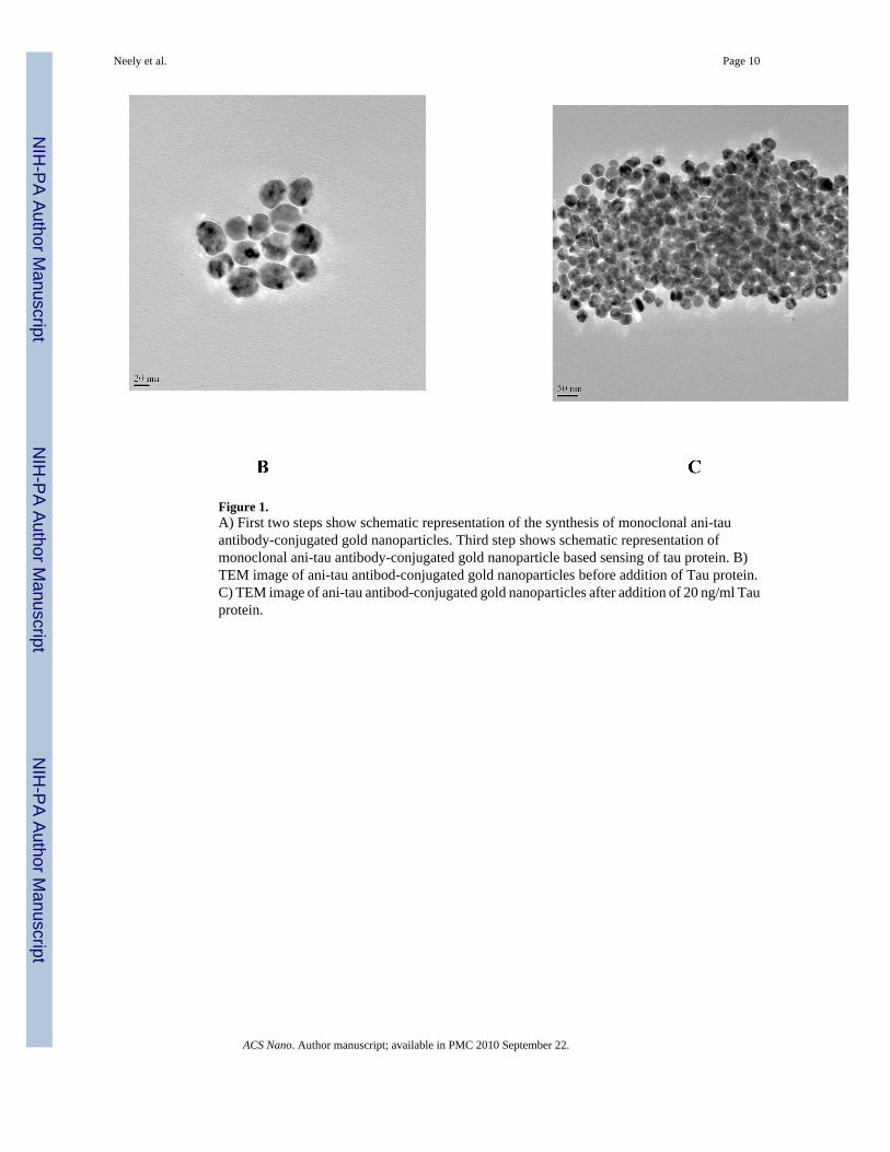
Figure 1.A) First two steps show schematic representation of the synthesis of monoclonal ani-tauantibody-conjugated gold nanoparticles. Third step shows schematic representation ofmonoclonal ani-tau antibody-conjugated gold nanoparticle based sensing of tau protein. B)TEM image of ani-tau antibod-conjugated gold nanoparticles before addition of Tau protein.C) TEM image of ani-tau antibod-conjugated gold nanoparticles after addition of 20 ng/ml Tauprotein.
Neely et al. Page 10
ACS Nano. Author manuscript; available in PMC 2010 September 22.
NIH
-PA Author Manuscript
NIH
-PA Author Manuscript
NIH
-PA Author Manuscript
Neely et al. Page 11
ACS Nano. Author manuscript; available in PMC 2010 September 22.
NIH
-PA Author Manuscript
NIH
-PA Author Manuscript
NIH
-PA Author Manuscript
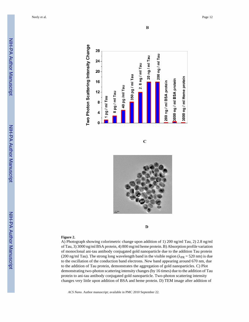
Figure 2.A) Photograph showing colorimetric change upon addition of 1) 200 ng/ml Tau, 2) 2.8 ng/mlof Tau, 3) 3000 ng/ml BSA protein, 4) 800 mg/ml heme protein. B) Absorption profile variationof monoclonal ani-tau antibody conjugated gold nanoparticle due to the addition Tau protein(200 ng/ml Tau). The strong long wavelength band in the visible region (λPR = 520 nm) is dueto the oscillation of the conduction band electrons. New band appearing around 670 nm, dueto the addition of Tau protein, demonstrates the aggregation of gold nanoparticles. C) Plotdemonstrating two-photon scattering intensity changes (by 16 times) due to the addition of Tauprotein to ani-tau antibody conjugated gold nanoparticle. Two-photon scattering intensitychanges very little upon addition of BSA and heme protein. D) TEM image after addition of
Neely et al. Page 12
ACS Nano. Author manuscript; available in PMC 2010 September 22.
NIH
-PA Author Manuscript
NIH
-PA Author Manuscript
NIH
-PA Author Manuscript

800 ng/ml BSA protein, E) TEM image demonstrating aggregation of ani-tau antibodyconjugated gold nanoparticle after the addition of 350 pg/ml Tau.
Neely et al. Page 13
ACS Nano. Author manuscript; available in PMC 2010 September 22.
NIH
-PA Author Manuscript
NIH
-PA Author Manuscript
NIH
-PA Author Manuscript
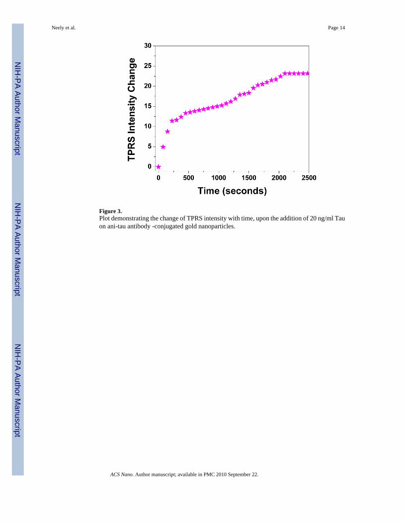
Figure 3.Plot demonstrating the change of TPRS intensity with time, upon the addition of 20 ng/ml Tauon ani-tau antibody -conjugated gold nanoparticles.
Neely et al. Page 14
ACS Nano. Author manuscript; available in PMC 2010 September 22.
NIH
-PA Author Manuscript
NIH
-PA Author Manuscript
NIH
-PA Author Manuscript
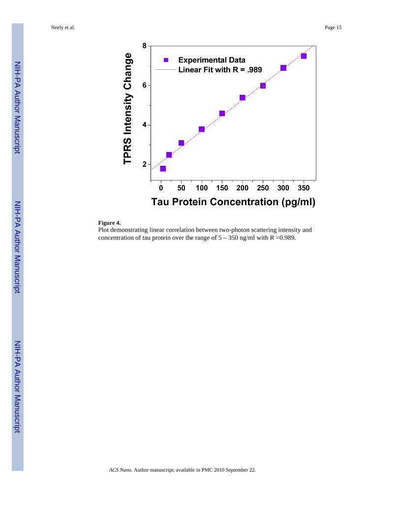
Figure 4.Plot demonstrating linear correlation between two-photon scattering intensity andconcentration of tau protein over the range of 5 – 350 ng/ml with R =0.989.
Neely et al. Page 15
ACS Nano. Author manuscript; available in PMC 2010 September 22.
NIH
-PA Author Manuscript
NIH
-PA Author Manuscript
NIH
-PA Author Manuscript
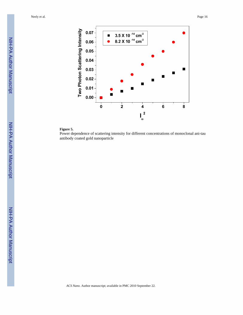
Figure 5.Power dependence of scattering intensity for different concentrations of monoclonal ani-tauantibody coated gold nanoparticle
Neely et al. Page 16
ACS Nano. Author manuscript; available in PMC 2010 September 22.
NIH
-PA Author Manuscript
NIH
-PA Author Manuscript
NIH
-PA Author Manuscript
Copyright © 2022 FDOKUMEN