TWO NOVEL MUTATIONS (2976INSA, 4311INSA) OF ATP7B IN A PATIENT WITH WILSON's DISEASE COEXISTING WITH...
10
Journal of Gastroenterology and Hepatology (2005) 20, 657–666 DOI: 10.1111/j.1400-1746.2005.03769.x Blackwell Science, LtdOxford, UKJGHJournal of Gastroenterology and Hepatology0815-93192005 Blackwell Publishing Asia Pty LtdApril 2005204657666Letter to the Editor Letters to the EditorLetters to the Editor LETTERS TO THE EDITOR RISK OF GASTROINTESTINAL NON-HODGKIN’S LYMPHOMA AT DIAGNOSIS OF CELIAC DISEASE To the Editor, Several studies have shown an increased incidence of malignancy in patients with celiac disease (CD). Such a trend is detectable both in Europe, where CD has a high prevalence, and in the USA, where CD was considered uncommon until recently. 1,2 We report some data taken from an Italian group of patients affected by CD that show that there was a higher risk, with respect to the general population, of developing intestinal non- Hodgkin’s lymphoma. In the present study, 1968 patients diagnosed with CD over a 20-year period (between January 1982 and December 2002) in 20 Ital- ian clinical centers specializing in gastrointestinal dis- eases were observed. The diagnosis of CD was made according to revised ESPGHAN criteria as follows: 1 (i) histological evidence of atrophy of duodenal or jejunum mucosa; 2 (ii) recovery at control biopsy after a gluten- free diet; 3 and (iii) serological positivity for AGA IgG or IgA and EMA IgG. 3 For each patient, demographic data and symptoms, and concomitant pathology results at the time of the diagnosis of CD were collected. Each patient gave their informed consent to take part in the study and the pro- tocol was approved by the ethics committee of each par- ticipating center. Of the 1968 patients, 20 patients had already been diagnosed with gastrointestinal non-Hodgkin’s lym- phoma at diagnosis of CD (17 cases of intestinal non-Hodgkin’s lymphoma and three cases of gastric non-Hodgkin’s lymphoma). We found CD predomi- nantly affected men (2:1). The standardized morbidity ratio (95% CI) for the malignancy results was 6.25 (3.8–9.6). 4 In addition, we noticed that the risk of developing gastrointestinal non-Hodgkin’s lymphoma correlates with the age at diagnosis. The mean age of the patients with a non-Hodgkin’s lymphoma at diagnosis of CD is 46.1 ± 13.8 years, whereas the mean age of the control group, represented by the patients with no neo- plasm at diagnosis, is equal to 36.6 ± 13.7. Therefore, the delayed diagnosis of CD is likely to be a risk factor for developing a lymphoma, probably because of the prolonged exposure to gluten. These data are consistent with those of other studies, indicating a relationship between CD and the develop- ment of gastrointestinal non-Hodgkin’s lymphoma. It is noteworthy that most of these cases occur before the diagnosis of CD. 5,6 Celiac disease is often an asympt- monatic condition, with a high rate of associated pathol- ogies and malignancies, often diagnosed before CD is diagnosed. Non-Hodgkin’s lymphomas are very frequent and can develop in sites other than the gastrointestinal tract, such as the skin, spleen, liver and central nervous sys- tem. The mechanism for the development of non- Hodgkin’s lymphoma in patients with CD is not known. However, chronic inflammation and antigenic stimula- tion, increased intestinal permeability and the release of inflammatory cytokines have been suggested. 1 In our group of patients, we found only one case of extra-intes- tinal non-Hodgkin’s lymphoma, a lymphoma of the spleen. Sometimes the concomitant pathologies suggest the diagnosis of CD. Our results stress the importance of implementing programs aimed at making an early diagnosis of CD in order to prevent its complications, even if the best method of screening has yet to be determined. Marco Silano and Massimo De Vincenzi Division of Food Science, Human Nutrition and Health, Istituto Superiore di Sanita, Rome, Italy REFERENCES 1 Green PHR, Fleischauer AT, Bhagat G et al. Risk of malig- nancy in patients with CD. Am. J. Med. 2003; 115: 191–5. 2 Talley NJ, Valdivinos M, Petterson TM et al. Epidemiology of celiac sprue: a community-based study. Am. J. Gastroen- terol. 1994; 89: 843–6. 3 Walker-Smith J, Guandalini S, Schmitz J et al. Revised cri- teria for diagnosis of coeliac disease. Report of Working Group of European Society of Paediatric Gastroenterology and Nutrition. J. Pediatr. Gastroenterol. Nutr. 1990; 65: 909– 11. 4 Ferlay J, Bray F, Pisani P et al. GLOBOCAN 2000: Cancer Incidence, Mortality and Prevalence Worldwide. Version 1.0. IARC Cancerbase No. 5. Lyon: IARC Press, 2001. 5 Holmes GKT, Prior P, Lane MR et al. Malignancy in coe- liac disease: effect of a gluten-free diet. Gut 1989; 30: 333– 8. 6 Catassi C, Fabiani E, Corrao G et al. Risk of non-Hodgkin lymphoma in celiac disease JAMA 2002; 287: 1413–19. April 2005204657660Letter to the EditorLetters to the EditorLetters to the Editor PREDICTORS OF SHORT-TERM OUTCOME OF SPONTANEOUS BACTERIAL PERITONITIS IN TURKISH CIRRHOTIC PATIENTS To the Editor, Despite the introduction of new antibiotics, earlier diagnosis and treatment, spontaneous bacterial
Transcript of TWO NOVEL MUTATIONS (2976INSA, 4311INSA) OF ATP7B IN A PATIENT WITH WILSON's DISEASE COEXISTING WITH...

Journal of Gastroenterology and Hepatology (2005) 20, 657–666 DOI: 10.1111/j.1400-1746.2005.03769.x
Blackwell Science, LtdOxford, UKJGHJournal of Gastroenterology and Hepatology0815-93192005 Blackwell Publishing Asia Pty LtdApril 2005204657666Letter to the EditorLetters to the EditorLetters to the Editor
LETTERS TO THE EDITOR
RISK OF GASTROINTESTINAL NON-HODGKIN’S LYMPHOMA AT DIAGNOSIS OF CELIAC DISEASE
To the Editor,Several studies have shown an increased incidence ofmalignancy in patients with celiac disease (CD). Such atrend is detectable both in Europe, where CD has a highprevalence, and in the USA, where CD was considereduncommon until recently.1,2 We report some data takenfrom an Italian group of patients affected by CD thatshow that there was a higher risk, with respect to thegeneral population, of developing intestinal non-Hodgkin’s lymphoma. In the present study, 1968patients diagnosed with CD over a 20-year period(between January 1982 and December 2002) in 20 Ital-ian clinical centers specializing in gastrointestinal dis-eases were observed. The diagnosis of CD was madeaccording to revised ESPGHAN criteria as follows:1 (i)histological evidence of atrophy of duodenal or jejunummucosa;2 (ii) recovery at control biopsy after a gluten-free diet;3 and (iii) serological positivity for AGA IgG orIgA and EMA IgG.3
For each patient, demographic data and symptoms,and concomitant pathology results at the time of thediagnosis of CD were collected. Each patient gave theirinformed consent to take part in the study and the pro-tocol was approved by the ethics committee of each par-ticipating center.
Of the 1968 patients, 20 patients had already beendiagnosed with gastrointestinal non-Hodgkin’s lym-phoma at diagnosis of CD (17 cases of intestinalnon-Hodgkin’s lymphoma and three cases of gastricnon-Hodgkin’s lymphoma). We found CD predomi-nantly affected men (2:1). The standardized morbidityratio (95% CI) for the malignancy results was 6.25(3.8–9.6).4 In addition, we noticed that the risk ofdeveloping gastrointestinal non-Hodgkin’s lymphomacorrelates with the age at diagnosis. The mean age of thepatients with a non-Hodgkin’s lymphoma at diagnosisof CD is 46.1 ± 13.8 years, whereas the mean age of thecontrol group, represented by the patients with no neo-plasm at diagnosis, is equal to 36.6 ± 13.7. Therefore,the delayed diagnosis of CD is likely to be a risk factorfor developing a lymphoma, probably because of theprolonged exposure to gluten.
These data are consistent with those of other studies,indicating a relationship between CD and the develop-ment of gastrointestinal non-Hodgkin’s lymphoma. It isnoteworthy that most of these cases occur before thediagnosis of CD.5,6 Celiac disease is often an asympt-monatic condition, with a high rate of associated pathol-ogies and malignancies, often diagnosed before CD isdiagnosed.
Non-Hodgkin’s lymphomas are very frequent andcan develop in sites other than the gastrointestinal tract,such as the skin, spleen, liver and central nervous sys-tem. The mechanism for the development of non-Hodgkin’s lymphoma in patients with CD is not known.However, chronic inflammation and antigenic stimula-tion, increased intestinal permeability and the release ofinflammatory cytokines have been suggested.1 In ourgroup of patients, we found only one case of extra-intes-tinal non-Hodgkin’s lymphoma, a lymphoma of thespleen.
Sometimes the concomitant pathologies suggest thediagnosis of CD. Our results stress the importance ofimplementing programs aimed at making an earlydiagnosis of CD in order to prevent its complications,even if the best method of screening has yet to bedetermined.
Marco Silano and Massimo De VincenziDivision of Food Science, Human Nutrition and Health,
Istituto Superiore di Sanita, Rome, Italy
REFERENCES
1 Green PHR, Fleischauer AT, Bhagat G et al. Risk of malig-nancy in patients with CD. Am. J. Med. 2003; 115: 191–5.
2 Talley NJ, Valdivinos M, Petterson TM et al. Epidemiologyof celiac sprue: a community-based study. Am. J. Gastroen-terol. 1994; 89: 843–6.
3 Walker-Smith J, Guandalini S, Schmitz J et al. Revised cri-teria for diagnosis of coeliac disease. Report of WorkingGroup of European Society of Paediatric Gastroenterologyand Nutrition. J. Pediatr. Gastroenterol. Nutr. 1990; 65: 909–11.
4 Ferlay J, Bray F, Pisani P et al. GLOBOCAN 2000: CancerIncidence, Mortality and Prevalence Worldwide. Version 1.0.IARC Cancerbase No. 5. Lyon: IARC Press, 2001.
5 Holmes GKT, Prior P, Lane MR et al. Malignancy in coe-liac disease: effect of a gluten-free diet. Gut 1989; 30: 333–8.
6 Catassi C, Fabiani E, Corrao G et al. Risk of non-Hodgkinlymphoma in celiac disease JAMA 2002; 287: 1413–19.
April 2005204657660Letter to the EditorLetters to the EditorLetters to the Editor
PREDICTORS OF SHORT-TERM OUTCOME OF SPONTANEOUS BACTERIAL PERITONITIS IN TURKISH CIRRHOTIC PATIENTS
To the Editor,Despite the introduction of new antibiotics, earlierdiagnosis and treatment, spontaneous bacterial

658 Letters to the Editor
peritonitis (SBP) still continues to be a serious infec-tious complication in cirrhotic patients with ascites.1–4
Short-term mortality, which denotes the proportion ofpatients who die from SBP during hospitalization, hasremained unchanged during the past decade. This iscaused by severe hepatic dysfunction in affectedpatients.5 Therefore, we aim to introduce the prognosticfactors determining short-term survival in Turkish cir-rhotics with SBP. The patients who took part in thisresearch study were from Trakya University Hospital,Edirne, Turkey and Haccettepe University Hospital,Ankara, Turkey.
The medical records of 87 consecutive cirrhoticpatients (107 episodes) with SBP that were treated atthe two university hospitals between 1999 and 2001were reviewed retrospectively. The patients were diag-nosed with cirrhosis according to their clinical signs andsymptoms, laboratory findings and radiological evalua-tion. The patients underwent a liver biopsy whenevercoagulation parameters permitted. Paracentesis forascitic fluid (AF) culture and cell count were carriedout in all cirrhotic patients with ascites on the first dayof their hospitalization and in each appropriate clinicalrequirement. The diagnostic criteria for SBP was fever,abdominal pain, encephalopathy or other signs of peri-tonitis associated with AF neutrophil (PNL) count(>250 cells/mm3) in the absence of surgically treatablecauses of secondary peritonitis. Ascitic fluid cultureswere collected and put into aerobic and anaerobicblood culture bottles at the bedside of the patients(10 cc AF drawn). The patients with culture-negativeneutrocytic ascites (CNNA) were also included in the
study. There appeared to be no statistically significantdifference among the patients treated at the two differ-ent university hospitals, with regard to continuous andcategorical variables, but alcoholic etiology was moreprevalent in the European part of Turkey (5/41 [12%]at Hacettepe University versus 17/46 [37%] at TrakyaUniversity).
The age range of the patients was 17–90 years (meanage, 53.5 ± 14.5 years), and there were 65 men and 22women. The underlying cause of chronic liver diseasewas diagnosed as hepatitis B in 52 (61%) patients,hepatitis C in five (6%) patients, hepatitis delta in two(2%) patients, alcohol abuse in 22 (25%) patients andother causes in eight (9%) patients. Seventy-eight(92%) of the patients were suffering from Child’s C andeight patients had Child’s B (9%). The signs and symp-toms of the patients are summarized in Table 1. Sev-enty-eight SBP episodes (73%) were treated with i.v.cefotaxim and the other 29 were treated with appropri-ate antibiotics or with combinations according to theclinical and microbiological requirements. The patientswere classified into two groups for statistical analysis;patients who survived (n = 64, 78%) and patients whodied (n = 23, mortality rate 22%) (Table 1). The per-centages of the patients whose deaths were directlyattributed to SBP before resolution of infection was15% (16 patients). The remaining seven patients died asa result of variceal bleeding (three) and multiorgan fail-ure (four). In 33/107 episodes (30%), at least one typeof bacteria was isolated from AF (Table 1). The mostcommon organism isolated from AF was Escherichia coliin 16 (49%) episodes followed by Staphylococcus aureus
Table 1 Clinical characteristics of survivors (computed as episodes) versus non-survivors at the time of diagnosis of sponta-neous bacterial peritonitis (SBP)
Variables No. survivors No. non-survivors P
No. patients (episodes) 64 (84) 23 (23) NDMale : Female 64:20 18:5 NS†
Age (years) 54 ± 14 52 ± 17 NS‡
Abdominal pain 32/84 (38%) 16/23 (80%) 0.002†§
Encephalopathy 44/84 (52%) 20/23 (87%) 0.002†
Ileus 1/84 (1%) 5/23 (22%) <0.0005†
WBC count (/mm3) 8550 ± 7200 19 100 ± 34 600 0.01‡
BUN (mg/dL) 34 ± 31 79 ± 79 <0.0005‡§
Creatinine (mg/dL) 1.1 ± 0.6 2.1 ± 1.4 <0.0005‡§
Triglyceride (mg/dL) 83 ± 74 99 ± 40 0.018‡
Total bilirubin (mg/dL) 5.5 ± 8.9 11.6 ± 11.5 0.002‡
AST (U/L) 77 ± 55 158 ± 153 0.003‡
SBP episodes 84 (100%) 23 (100%) NDCNNA 57 (68%) 17 (74%) NS†
Culture positivity 27 (32%) 6 (26%) NS†
Gram-negative 17 3 NS†
Gram-positive 10 3 NS†
AF cell count (/mm3) 2469 ± 5415 16 484 ± 41 520 0.039‡
PNL count (/mm3) 1355 ± 1800 13 900 ± 40 000 0.029‡
P < 0.05, statistically significant. †c2 test. ‡Mann–Whitney U-test. §Independent predictors of short-term mortality. AF, asciticfluid; AST, aspartate aminotransferase; BUN, blood urea nitrogen; CNNA, culture-negative neutrocytic ascites; ND, no data;NS, not significant; PNL, polymorphonuclear leukocytes; WBC, white blood cell.
Letters to the Editor 659
in five (15%) episodes. Fever was largely associated withGram-positive infection (12/20 [60%] in Gram-positivevs 5/29 [21%] in Gram-negative infection; P < 0.002).In the present study, we did not find clinical significanceof any type of AF culture isolate on short-term prog-nosis. It has been suggested that nosocomial andstaphylococcal infections are associated with a highermortality rate than community-acquired infections andnon-staphylococcal infections.6 The short-term hospitalmortality did not differ according to Gram-positive or-negative culture results. The prognosis was also similarat short term for the patients with both CNNA and cul-ture-positive SBP (mortality rates were 17/74 [23%]and 6/33 [18%], respectively) (P > 0.05). So, we believeconvincingly that CNNA is as serious as SBP and mustbe considered as serious as culture-positive SBP. Whenall of the clinical characteristics of the patients withCNNA and culture-positive SBP were comparativelyanalyzed, no statistically significant difference wasobserved between the groups. As a rule according to theresults of the present study, to treat all cirrhotic patientswith AF cell count >250/mm3, it must be assumed thatthey have culture-positive SBP.
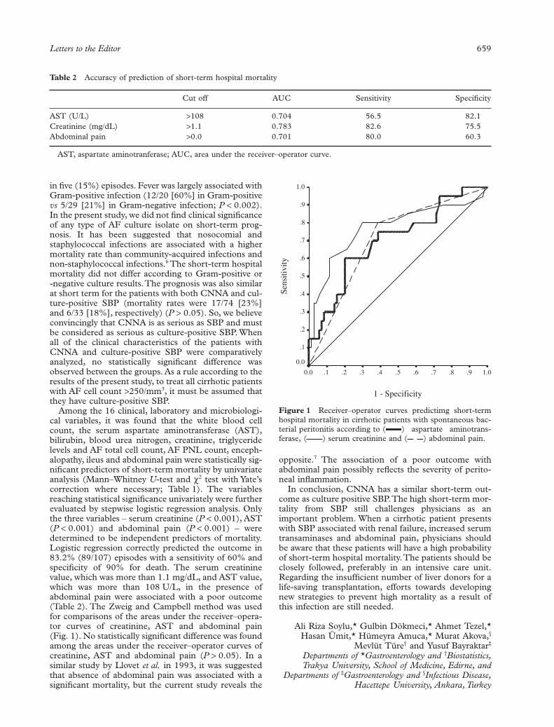
Among the 16 clinical, laboratory and microbiologi-cal variables, it was found that the white blood cellcount, the serum aspartate aminotransferase (AST),bilirubin, blood urea nitrogen, creatinine, triglyceridelevels and AF total cell count, AF PNL count, enceph-alopathy, ileus and abdominal pain were statistically sig-nificant predictors of short-term mortality by univariateanalysis (Mann–Whitney U-test and c2 test with Yate’scorrection where necessary; Table 1). The variablesreaching statistical significance univariately were furtherevaluated by stepwise logistic regression analysis. Onlythe three variables – serum creatinine (P < 0.001), AST(P < 0.001) and abdominal pain (P < 0.001) – weredetermined to be independent predictors of mortality.Logistic regression correctly predicted the outcome in83.2% (89/107) episodes with a sensitivity of 60% andspecificity of 90% for death. The serum creatininevalue, which was more than 1.1 mg/dL, and AST value,which was more than 108 U/L, in the presence ofabdominal pain were associated with a poor outcome(Table 2). The Zweig and Campbell method was usedfor comparisons of the areas under the receiver–opera-tor curves of creatinine, AST and abdominal pain(Fig. 1). No statistically significant difference was foundamong the areas under the receiver–operator curves ofcreatinine, AST and abdominal pain (P > 0.05). In asimilar study by Llovet et al. in 1993, it was suggestedthat absence of abdominal pain was associated with asignificant mortality, but the current study reveals the
opposite.7 The association of a poor outcome withabdominal pain possibly reflects the severity of perito-neal inflammation.
In conclusion, CNNA has a similar short-term out-come as culture positive SBP. The high short-term mor-tality from SBP still challenges physicians as animportant problem. When a cirrhotic patient presentswith SBP associated with renal failure, increased serumtransaminases and abdominal pain, physicians shouldbe aware that these patients will have a high probabilityof short-term hospital mortality. The patients should beclosely followed, preferably in an intensive care unit.Regarding the insufficient number of liver donors for alife-saving transplantation, efforts towards developingnew strategies to prevent high mortality as a result ofthis infection are still needed.
Ali Riza Soylu,* Gulbin Dökmeci,* Ahmet Tezel,*Hasan Ümit,* Hümeyra Amuca,* Murat Akova,§
Mevlüt Türe† and Yusuf Bayraktar‡
Departments of *Gastroenterology and †Biostatistics,Trakya University, School of Medicine, Edirne, and
Departments of ‡Gastroenterology and §Infectious Disease,Hacettepe University, Ankara, Turkey
Table 2 Accuracy of prediction of short-term hospital mortality
Cut off AUC Sensitivity Specificity
AST (U/L) >108 0.704 56.5 82.1Creatinine (mg/dL) >1.1 0.783 82.6 75.5Abdominal pain >0.0 0.701 80.0 60.3
AST, aspartate aminotranferase; AUC, area under the receiver–operator curve.
Figure 1 Receiver–operator curves predictimg short-termhospital mortality in cirrhotic patients with spontaneous bac-terial peritonitis according to ( ) aspartate aminotrans-ferase, ( ) serum creatinine and ( ) abdominal pain.
1 - Specificity
1.0.9.8.7.6.5.4.3.2.10.0
Sens
itivi
ty
1.0
.9
.8
.7
.6
.5
.4
.3
.2
.1
0.0

660 Letters to the Editor
REFERENCES
1 Franca AV, De Souza JB, Silva CM, Soares EC. Long-termprognosis of cirrhosis after spontaneous bacterial peritonitistreated with ceftriaxone. J. Clin. Gastroenterol. 2001; 33:295–8.
2 Mesquita MA, Balbino EP, Albuquerque RS et al. Ceftri-axone in the treatment of spontaneous bacterial peritonitis:ascitic fluid polymorphonuclear count response and short-term prognosis. Hepatogastroenterology 1997; 44: 1276–80.
3 Bac DJ. Spontaneous bacterial peritonitis: an indication forliver transplantation? Scand. J. Gastroenterol. Suppl. 1996;218: 38–42.
4 Puri AS, Puri J, Ghoshal UC et al. Frequency, microbialspectrum and outcome of spontaneous bacterial peritonitisin north India. Indian J. Gastroenterol. 1996; 15: 86–9.
5 Thuluvath PJ, Morss S, Thompson R. Spontaneous bacte-rial peritonitis—in-hospital mortality, predictors of survival,and health care costs from 1988–1998. Am. J. Gastroenterol.1988; 96: 1232–6.
6 Campillo B, Richardet JP, Kheo T, Dupeyron C. Nosoco-mial spontaneous bacterial peritonitis and bacteremia in cir-rhotic patients: impact of isolate type on prognosis andcharacteristics of infection. Clin. Infect Dis. 2002; 35: 1–10.
7 Llovet JM, Planas R, Morillas R et al. Short-term prognosisof cirrhotics with spontaneous bacterial peritonitis: Multi-variate study. Am. J. Gastroenterol. 1993; 88: 388–92.
April 2005204660662Letter to the EditorLetters to the EditorLetters to the Editor
TWO NOVEL MUTATIONS (2976INSA, 4311INSA) OF ATP7B IN A PATIENT WITH WILSON’S DISEASE COEXISTING WITH GLUCOSE-6-PHOSPHATE DEHYDROGENASE DEFICIENCY
To the Editor,We present a patient with Wilson’s disease who hadearly onset of the disease, accompanying hepatologi-cal and neurological manifestations and glucose-6-phosphate dehydrogenase deficiency with mutations2976insA and 4311insA in exon 13 and 21 ofATP7B.
Wilson’s disease is an autosomal recessive disorder ofcopper metabolism, resulting in hepatic cirrhosis andneuronal degeneration.1 There is extensive heterogene-ity of symptoms within Wilson’s disease, with somepatients presenting with primarily hepatic problems,some with primarily neurological impairment and somewith both types of symptoms. The age of onset variesfrom childhood to early adulthood (3–16 years) forhepatic presentation, and usually 12 years or more ofage for neurological problems.2 The Wilson’s diseasegene has been mapped to chromosome 13q14.3.Molecular cloning of WD gene, ATP7B (WD, MIM#2779000, L25591), has provided new insight into themechanism of copper transport in humans, as well asnew molecular tools for continued investigation of nor-mal and abnormal copper metabolism. The Wilson’sdisease gene is encoded by 21 exons, spanning more
than 80 kb on chromosome 13q14.3. The ATP7B genehas been shown to encode a novel member of the familyof cation-transporting P-type ATPase. Analysis of thepatient’s mutations revealed an enormous molecularheterogeneity consisting of a very small number of fre-quent mutations that are population specific, as well asa much greater number of rare individual alleles.3 Thedefect in copper ATPase results in excess accumulationof copper intracellularly, particularly in the liver andbrain, presumably because of the generation of metaldependent oxyradicals and to metal ion antagonism.The production of hydroxyl radicals associated with cel-lular accumulation of copper can damage cell mem-branes, DNA, mitochondria and proteins.4 Glutathione(GSH) has a protective role against toxicity of metals bychelating them.5,6 Glucose-6-phosphate dehydrogenaseplays an important role in maintaining the glutathionein a reduced state.
A 51/2-year-old boy was admitted to the PediatricGastroenterology ward of the Postgraduate Institute ofMedical Education and Research, Chandigarh, India,with a 1-year history of recurrent episodes of jaundice.He was passing brown cola colored urine. An examina-tion revealed normal vital signs. He was icteric and hadpalmar erythema and echymotic spots on his extremi-ties. He had ascites but his liver and spleen were not pal-pable. His handwriting had deteriorated. Investigationsshowed hemoglobin count (9.0 g/dL), total leukocytecount (31 700/mm3), normal differential count, plate-lets count (69 000/mm3) and normal renal functiontests. Liver function tests showed total protein (5.2 g/dL), albumin (2.0 g/dL), globulin (3.2 g/dL), serumbilirubin (14.7 mg/dL) with conjugated fraction(8.4 mg/dL), SGOT/SGPT 44/23 IU/I, alkaline phos-phatase 15 KAU and deranged coagulogram. Asciticfluid analysis showed evidence of spontaneous bacterialperitonitis. Glucose-6-phosphate dehydrogenase activ-ity in erythrocytes from this patient was found to be0.93 U/g Hb, which was markedly lower compared withthe age-matched controls (5.9 U/g Hb) and showed evi-dence of intravasicular hemolysis. The patient had bilat-eral Kayser–Fleischer rings on slit lamp examination.An ultrasound examination of the abdomen showed anirregular outline and a coarse echotexture of the liverand the presence of ascites. An upper gastrointestinalendoscopy showed esophageal varices. The work up forWilson’s disease showed pre-D-penicillamine urinarycopper 313 mg/24 h (normal <80 mg/24 h), post-D-penicillamine urinary copper 459 mg/24 h, serum ceru-loplasmin 3.6 mg/dL (normal >20 mg/dL) and serumcopper 50 mg/dL (normal >70 mg/dL). Serum coppernot bound to ceruloplasmin was found to be 38 mg/dL(normal <15 mg/dL), indicating free copper in theblood.2 Autoimmune markers, alpha-1 antitrypsin,hepatitis B and C were absent. An X-ray radiograph ofthe chest showed hilar lymphadenopathy and infiltratesin the parenchyma. The patient’s Montoux test washighly positive. His blood culture was positive forStaphylococcus and Pseudomonas. In view of sepsis andpulmonary tuberculosis, the patient was started onciprofloxacin and antitubercular drugs rifampicin,ethambutol and streptomycin. D-Penicillamine was alsogiven. The patient’s condition deteriorated further,
Letters to the Editor 661
leading to sepsis, encephalopathy, electrolyte imbalanceand finally death. Liver autopsy showed loss of archi-tecture, Mallory’s hyaline, neutrophilic and chroniclymphocytic infiltration, and hepatic fibrosis. Excesspigment as stained by orcein stain and rubeanic acidstain was seen in zone 1 of the lobules. Orcein stainshowed diffuse granular positivity for copper associatedprotein. In the present case, the plasma GSH level was0.64 mmol/g Hb, which were significantly lower than thenormal value of 1.2 mmol/g Hb. Superoxide dismutase(SOD) is an important enzyme for the dismutation ofsuper oxide anion free radicals (O2
- + 2H+) into harm-less products (H2O2 + O2). In this patient, the activity ofSOD in red blood cells hemolysate was 0.352 units/g
Hb, which was lower than the age matched controls0.611 units/g Hb. Membrane lipid peroxidation wasassessed by measuring malondialdehyde (MDA) in theRBC hemolysate and was found to be 0.069 mmolMDA/g Hb. It was significantly higher than the normallevel of 0.03 mmol MDA/g Hb.
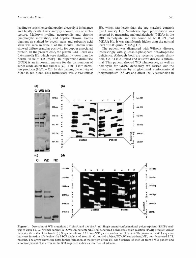
The patient was diagnosed with Wilson’s disease,interestingly with glucose-6-phosphate dehydrogenasedeficiency. Although both are recessive genetic disor-ders, G6PD is X-linked and Wilson’s disease is autoso-mal. This patient showed WD phenotypes, as well ashemolysis for G6PD deficiency. We carried out themutational analysis by single-strand conformationalpolymorphism (SSCP) and direct DNA sequencing in
Figure 1 Detection of WD mutations 2976insA and 4311insA. (a) Single-strand conformational polymorphism (SSCP) anal-ysis of exon 13. C, Normal subject; WD, Wilson patient; ND, non-denatured polymerase chain reaction (PCR) product. Arrowindicates the shifts of the bands. (b) Sequence of exon 13 from a WD patient and a control patient. The arrow in the WD sequenceindicates insertion of adenine. (c) SSCP analysis of exon 21. C, control subject; WD, Wilson patient; ND, non-denatured PCRproduct. The arrow shows the hetroduplex formation at the bottom of the gel. (d) Sequence of exon 21 from a WD patient anda control patient. The arrow in the WD sequence indicates insertion of adenine.
662 Letters to the Editor
this patient. For mutational analysis, genomic DNAwas extracted from peripheral blood leukocytes follow-ing the standard procedure of the QIAamp DNABlood Midi Kit (Qiagen GmbH, Hilden, Germany).Polymerase chain reaction (PCR) was used to amplifyall 21 exons of ATP7B and their corresponding intron-exon junction, using specific primers in the flankingregion. All amplified exons of the WD gene were sub-jected to mutational analysis by SSCP. The Wilson’sdisease PCR samples showed shifts, relative to normalsamples of corresponding exons of ATP7B on SSCPwhich were subjected to DNA sequencing. The sameprimers used for SSCP analysis were used to generateasymmetric PCR products and sequencing. Thesequence was determined using (35S)a-dATP and thesequenase kit (US Biochemicals, San Diego, CA,USA).
Direct DNA sequencing of the PCR products thatexhibited shifts showed insertion of two novel muta-tions, 2976insA and 4311insA, in exons 13 and 21,respectively (Fig. 1). These mutations are expected toalter the reading frame of the gene product and result innon-functional ATP7B protein. It is well accepted thatthe type of mutation in a specific region of the genemanifests a specific phenotype in the patient. Mutations2976insA and 4311insA cause a frame-shift by chang-ing threonine to asparagine at the 993 amino acid posi-tion in exon 13 and proline to alanine at the 1437position in exon 21 of the ATP7B. Mutation 2976insAin exon 13 affects the sixth transmembrane domain and/or cation channel of ATP7B, which would affect thecopper transport from the intracellular compartment tothe extra-cellular compartment. The significance of thecarboxy C terminus of ATP7B with respect to its bio-synthetic role in copper delivery to the ferroxidase cer-uloplasmin has recently been reported. To date, in the Cterminus of ATP7B, only two missense mutations andone deletion have been reported in a WD patient.7 Inter-estingly, the characterized 4311insA in exon 21 createsa downstream stop codon at amino acid position 1447.This change would result in a shortened functionlessprotein and is predicted to affect copper delivery. Muta-tion 2976insA has a deleted restriction site for the Sec Irestriction enzyme in a WD patient; however, 4311insAdid not create/delete any restriction site. The presenceof mutations 2976insA and 4311insA were checked in25 controls by restriction digestion and DNA sequenc-ing. Both mutations were found to be absent in all.Therefore, the patient was a compound heterozygoteand had mutations in both alleles, and both mutationscause frame-shift and would produce a shortened func-tionless WD protein. The defect in the sixth transmem-brane domain and carboxyl region of ATP7B mightresult in excessive copper accumulation in the liver,brain and other organs.
We concluded that these two mutations, 2976insAand 4311insA, and depleted levels of GSH in a patientmight lead to an increase in free copper levels, whichhave been shown to enhance DNA damage in biologi-cal systems via formation of hydroxyl radicals. Copperis a potent catalyst of the Haber–Weiss reaction, inwhich hydrogen peroxide is converted to the hydroxylradical and rapidly reacts with polyunsaturated fatty
acid residues of cell membrane, thiol-containing pro-teins and nucleic acids. In the present study, cellmembrane damage was also evident from markedenhancement of MDA. In addition, GSH has a verystrong affinity for Cu and forms stable complexes,which have been shown to act efficiently in the dona-tion of Cu (I) to copper chaperon (CSS) and subse-quently to copper-zinc SOD. Therefore, reducedactivity of SOD could be possible as a result of lowlevels of GSH. An observed reduction in G6PD couldbe a prominent factor for low levels of reduced GSHbecause G6PD deficiency is known to influence GSHstatus in humans and cause hemolysis. All of these find-ings suggest that novel mutations 2976insA and4311insA might exacerbate copper-induced toxicity byalteration of G6PD, and antioxidant status, whichmight lead to the early onset of the disease with hepa-tological and neurological manifestations, as well assusceptibility to pulmonary tuberculosis.
Rajendra Prasad,* Gurjit Kaur,‡ Sandeep Kumar* andBabu Ram Thapa†
Departments of *Biochemistry and †PediatricGastroenterology, Postgraduate Institute of Medical
Education and Research, and ‡Department of Physiology,Government Medical College, Chandigarh, India.
REFERENCES
1 Brewer GJ, Yuzbasiyan-Gurkan V. Wilson disease. Medicine1992; 71: 139–64.
2 Prasad R., Kaur G, Walia BNS. A critical evaluation of cop-per metabolism in Indian Wilson’s disease children withspecial reference to their phenotypes and relatives. Biol.Trace Element Res. 1998; 65: 153–65.
3 Shah AB, Chernov I, Zhang HT et al. Identification andanalysis of mutations in the Wilson disease gene (ATP7B):Population frequencies, genotype-phenotype correlation,and functional analysis. Am. J. Hum. Genet. 1997; 61: 317–28.
4 Halliwell B, Gutteridge JMC, Cross E. Free radicals, anti-oxidants and human disease: Where are we now? J. Lab.Clin. Med. 1992; 119: 598–620.
5 Freedman JH, Ciriolo MR, Peisach J. The role of glu-tathione in copper metabolism and toxicity. J. Boil. Chem.1989; 254: 5598–605.
6 Prasad R, Kaur G, Walia BNS. Molecular basis of patho-physiology of Indian childhood cirrhosis. Role of nuclearcopper accumulation in liver. Mol. Cell. Biochem. 1996; 156:25–30.
7 Cox DW, His G, Cullen LM, Glerum DM. Functional sig-nificance of carboxy-terminus of the Wilson disease coppertransporting ATPase. Am. J. Hum. Genet. 2003; 73: 2445.
April 2005204662664Letter to EditorLetters to the EditorLetters to the Editor

Letters to the Editor 663
CURE RATE OF HIGH DOSE OMEPRAZOLE AND AMOXICILLIN THERAPY FOR TREATMENT-RESISTANT HELICOBACTER PYLORI INFECTION
To the Editor,For patients in whom primary attempts to eradicateHelicobacter pylori (H. pylori) have failed, especially inthe setting of clarithromycin and metronidazole resis-tance, the optimum treatment regimen is unclear. Bis-muth containing quadruple therapy is potentiallyeffective but, in Australia, bismuth subcitrate as part ofa combination pack is no longer available. Dual therapywith omeprazole and amoxicillin has been reported as apossible second or third line option,1 but experiencewith such high doses of omeprazole is very limited. Wedesigned a pilot study to prospectively evaluate thesafety and efficacy of high dose omeprazole with amox-icillin in a group of patients who had treatment resistantH. pylori.
Patients were chosen if there had been at least onefailed attempt to eradicate H. pylori infection. Thisgroup was comprised of new referrals to a gastroenter-ology clinic for management of their infection and pre-existing clinic patients who had ongoing infection andmay have participated in previous trials of eradicationtherapy. An endoscopy to exclude an active peptic ulcerhad been carried out within the previous 12 months andwas only repeated if new upper gastrointestinal tractsymptoms were present or culture of H. pylori wasrequired. Current H. pylori infection was confirmed bya positive culture result obtained at endoscopy or by thestring test (Entero-test HP, HDC Corporation, CA,USA). The bacterial isolates were tested to determine
the antibiotic susceptibility levels of each strain by thedisc diffusion method. This was carried out prior to par-ticipation in the present trial. If this bacterial diagnosiswas more than 3 months prior to trial entry, active cur-rent infection was confirmed by positive 14-C ureabreath test (PYtest, Tri-Medical, Perth, Australia). Trialexclusion criteria included the following: penicillinallergy; use of other antibiotics in the trial period; preg-nancy; and lactation. Pre-existing proton pump inhibi-tor (PPI) was permitted (many subjects required theagent for dyspepsia symptom relief), with a changeoverto omeprazole for the duration of the trial drug admin-istration. The local institutional research ethics commit-tee approved the conduct of the study and writteninformed consent from each patient was obtained priorto entry into the trial.
Omeprazole 40 mg and amoxicillin 1 g were giventhree times daily for 14 days. The drugs were to betaken together, with no particular regard for meal times.Pre- and post-treatment interview, physical examina-tion and a pill count was carried out to assess compli-ance, tolerability and adverse events.
Six weeks after the completion of the treatmentcourse, subjects were interviewed to assess late adverseevents and a urea breath test (UBT) was carried outaccording to the manufacturer’s instructions. A positiveresult was considered indicative of persistent infection.In the event of an indeterminate result, a repeat UBTwas carried out 7 days later, ensuring that acid suppres-sive therapy had been ceased. Subjects who remainedpositive were offered counselling and further manage-ment at our clinic. Subjects with a negative UBT at6 weeks were offered further testing 12 weeks post-treatment to confirm persistent eradication.
The present study was carried out between Novem-ber 2000 and February 2002. Fifteen subjects (six men,
Table 1 Patient details including previous treatments, antibiotic susceptibility results and outcome in this trial
PatientPrevious
treatmentsAmoxicillin
susceptibilityMetronidazolesusceptibility
Clarithromycinsusceptibility
PPI pre-treatment
6 week UBT result
1 OAC S R S No –2 OAC ¥ 2 S R R No –3 OAC, RBcAC S R R No –4 BMT, OAC, NTZ S R S No –5 PAC, AORif, NTZ S R R Yes +6 BAMx2, OAC ¥ 2 S R R No –7 BMT, OAC ¥ 2 S R R Yes +8 BMT, OAC S R S No –9 OAC ¥ 2, NTZ S R R Yes +
10 BMT, OAC, OBMT S R S Yes –11 BMT, OAC, NTZ S R R No –12 OAC S R R No +13 BMT, OAC S R R No +14 BM, BMT, NTZ S R S Yes –15 BMT, OA (low dose), OMC S R R No –
A, amoxicillin; B, bismuth subcitrate; C, clarithromycin; M, metronidazole; NTZ, nitazoxanide; O, omeprazole; P, pantopr-ezole; PPI, proton pump inhibitor; R, resistant; RBc, ranitidine bismuth subcitrate; Rif, rifabutin; S, sensitive; T, tetracycline;UBT, urea breath test.

664 Letters to the Editor
nine women) aged 18–75 years (mean age 51 years)were enrolled. The median number of previous treat-ments was three (range 1–4). Indication for eradicationof H. pylori was gastritis with dyspepsia (seven cases),dyspepsia (four cases), long-term PPI use in gastroe-sophageal reflux disease (two cases), family history ofgastric carcinoma (one case) and previous duodenalulcer (one case). Previous regimens used and antibioticsusceptibility profiles are shown in Table 1. All patientsexhibited metronidazole resistance. No amoxicillinresistance was detected. Clarithromycin resistance was66% overall, but 100% in those we could identify withprevious clarithromycin-based treatment.
At 6 weeks post-treatment, 10/15 (67%, 95% confi-dence interval, 38–88%) subjects had a negative UBT.Eight of these 10 had a further negative UBT at12 weeks with two patients lost to follow up. In five sub-jects with a clarithromycin-sensitive isolate, the curerate was 5/5 (100%). In the setting of clarithromycinresistance, the cure rate was 5/10 (50%).
The treatment was generally well tolerated and allsubjects completed the treatment. One subject missedonly one dose of omeprazole and amoxicillin. Twopatients reported mild headache and diarrhoea dur-ing treatment and another two reported that theirreflux symptoms worsened following the trial (bothsubjects had negative UBT results at 6 weeks), whichrequired an increase in their maintenance acid-sup-pressive therapy.
The present study of H. pylori eradication in patientswith a median of three previous treatment failuresshows that high dose omeprazole and amoxicillin is welltolerated and cures about two-thirds of infections. Ourfindings are similar to those of Ellenrieder et al. whoachieved a 77% eradication rate in a group of patientswith one previous treatment failure.1
In patients with duodenal ulcers receiving dual ther-apy, a relationship between omeprazole dose and effi-cacy of eradication was attributed to increasingintragastric pH. Amoxicillin is an acid labile antibioticand its activity is enhanced in a more neutral pH envi-ronment. At an increased gastric pH induced by ome-prazole, it has also been suggested that bacterialovergrowth in the stomach can occur and might dis-place H. pylori or be inhibitory to its growth.2 Our trialused omeprazole 40 mg three times daily with the aimof maintaining gastric acid suppression throughout theday, and amoxicillin was given at the same times toimprove compliance. Recently, attention has beendrawn to the impact of CYP2C19 polymorphism on themetabolism of omeprazole and its implications forH. pylori treatment. The ‘poor metabolizer’ genotype,present in 12–70% of Asian populations, is associatedwith more profound gastric acid suppression andimproved H. pylori eradication rates compared with the‘extensive metabolizer’ genotype, which occurs in up to95% of Caucasian populations. In the latter, Kita et al.have suggested that 80 mg omeprazole has a similaracid suppressing action as 20 mg in poor metabolizersand a daily dose of 160 mg per day might be needed foroptimal anti-H. pylori activity.3 In treatment-naïve sub-jects who were extensive metabolizers, Tanigawara et al.only achieved a 50% eradication rate with omeprazole/
amoxicillin dual therapy.4 The metabolism of rabepra-zole is largely independent of CYP2C19 and Furutaet al, using rabreprazole and amoxicillin, have reporteda 100% cure rate in 17 subjects who had previouslyfailed one course of standard triple therapy, irrespectiveof the CYP2C19 genotype.5
The results of this and other studies suggest that dualtherapy with amoxicillin and potent acid suppression isan option for treating H. pylori infection, particularly asit uses easily available components. While the impor-tance of antibiotic resistance in difficult-to-treatH. pylori is well recognized, the role of CYP2C19 poly-morphism and the optimal dosing of PPI is of increasingrelevance to this subset of patients.
Charlie H Viiala,* Helen M Windsor† andBarry J Marshall*,†
*Department of Gastroenterology, Sir Charles GairdnerHospital, †NHMRC Helicobacter pylori Laboratory,
Discipline of Microbiology, University of Western Australia,QE II Medical Center, Nedlands, WA, Australia
REFERENCES
1 Ellenrieder V, Boeck W, Richter CW, Marre R, Adler G,Glasbrenner B. Prevalence of resistance to clarithromycinand its clinical impact on the efficacy of Helicobacter pylorieradication. Scand. J. Gastroenterol. 1999; 34: 750–56.
2 Sharma BK, Santana IA, Wood EC et al. Intragastric bac-terial activity and nitrosation before, during and after treat-ment with omeprazole. BMJ 1984; 289: 717–19.
3 Kita T, Sakaeda T, Aoyama N et al. Optimal dose ofomeprazole for CYP2C19 extensive metabolizers in anti-Helicobacter pylori therapy: pharmacokinetic considerations.Biol. Pharm. Bull. 2002; 25: 923–7.
4 Tanigawara Y, Aoyama N, Kita T et al. CYP2C19genotype-related efficacy of omeprazole for the treatment ofinfection caused by Helicobacter pylori. Clin. Pharmacol.Ther. 1999; 66: 528–34.
5 Furuta T, Shirai N, Xiao F et al. High-dose rabeprazole/amoxicillin therapy as the second-line regimen after failureto eradicate H. pylori by triple therapy with the usual dosesof a proton pump inhibitor, clarithromycin and amoxicillin.Hepatogastroenterology 2003; 50: 2274–8.
April 2005204664666Letter to the EditorLetters to the EditorLetters to the Editor
HETEROTOPIC MESENTERIC OSSIFICATION: A RARE REACTIVE PROCESS
To the Editor,Heterotopic ossification is an unusual process in whichabnormal formation of bone is observed outside theskeleton. The histogenesis of intra-abdominal new boneformation from connective tissue caused by trauma sup-ports a metaplastic process. Here, an extraordinaryfinding related to an osseous lesion of the mesenteryand omentum in a 74-year-old patient, who was oper-ated on for an intestinal obstruction, is described. Thepatient died after 1 month of hospitalization.

Letters to the Editor 665
Heterotopic ossification occurs in a variety of bodysites and tissues and is associated with trauma. Soft tis-sue injury can be caused by a number of mechanismssuch as trauma (60.75%), small mechanic injuries,ischemia and inflammation.1 The rapid growth of theselesions in the mesentery, which frequently arousesclinical suspicion, in conjunction with their hypercel-lularity, cytological atypia and mitotic activity resem-bling myositis ossificans, explains the use of the term‘mesenteritis ossificans’ or ‘heterotopic mesentericossification’. A more appropriate term for mesenteritisossificans would be pseudomalignant osseous tumor ofextraskeletal soft tissues. This was first described byFine and Stout in 1956 on the basis of eight cases, butnone occurred in the mesentery.2 In 1992, Yannopouloset al. described a case of multifocal heterotopic mesen-teric ossification.3
A 74-year-old man suffering from hypertensive heartdisease with a 1-year history of surgical reconstructionof an umbilical hernia, cholecystectomy and a recentprostatectomy presented with a small bowel obstruction(noticeable 4 days after the prostatectomy) and mildrenal failure. Lysis of adhesions as well as resection of asmall segment of the small bowel was carried out duringa second operation. Ten days later, persistent obstruc-tive symptoms led to a third laparotomy, which lasted18 h. During this operation, we noticed that the adhe-sions between the small bowel and mesentery had afibroosseous appearance, which made them too difficultfor us to remove without injuring the small bowel. As aconsequence, as well as the lysis of adhesions, themajority (80%) of the small bowel and the right colonup to the midtransverse colon were resected and sentfor pathological examination. During the operation, ajejunocolic anastomosis was repaired. Postoperatively,the patient was placed in an intensive care unit withmonitoring, total parenteral nutrition and i.v. antibiot-ics. The patient died from severe sepsis and liver andrenal failure on the 6th postoperative day (after the thirdoperation) in the intensive care unit.
Histologically, there were fibrous septa of variablethickening that entrapped adipose tissue, nerves andblood vessels within the mesenteric tissue. These fibroussepta were moderately cellular and mainly composed offibroblasts (Fig. 1). Cellular fasciitis-like areas, whichhave been reported in other cases,3,4 were absent in thepresent case. Trabeculae of osteoid rimmed by osteo-blasts were noticed in proximity to the fibrous septa.The osteoblasts showed occasionally prominent nucleibut lacked malignant cytological features. In the presentcase, the osteogenic proliferation did not exhibit a‘zonal’ architecture. Chronic inflammation throughoutthe process was minimal. Because the presence of for-eign bodies arguably played a role in the pathogenesis ofthe present lesion, we should underline the histologicalabsence of any foreign body granuloma in the examinedsections. The surgical procedures used were unlikely tointroduce any foreign bodies. Because the inflammatoryreaction was generally unremarkable, the reactivenature of the lesion was questionable; a metaplasticnature seems more likely.
The difficulty and importance of microscopically dis-tinguishing the present reactive lesion from a sarcoma-
tous process is noteworthy. In the present case, thedifferential diagnosis was quite easy because no mor-phological features suggesting malignancy wereobserved. However, in other reported cases of hetero-topic mesenteric ossification, where zone phenomenaare prominent, it is important not to overdiagnose thisreactive process as an extraskeletal osteosarcoma. Theterm ‘zone phenomena’ refers to the immature centralportion of the lesion (characterized by hypercellularity,atypia and mitoses) progressively maturing toward theperiphery, first forming a primitive osteoid, then a well-organized osteoid with prominent osteoblastic miming,and finally a mature lamellar bone.4 This progressivematuration pattern excludes the diagnosis of malig-nancy in such cases.4
The clinical highlight of the present case was theremarkable thickening of the bowel wall and the diffi-culty in lysis of the osteoid adhesions, which can also beobserved in ossification of abdominal scars.5 The rapidappearance (10 days after the 2nd operation) of thefibrous and osteoid structures is similar to otherreported cases,3,4 and could have been caused by trau-matic results after the lysis of bowel adhesions in thesecond operation. It is of interest that in this reportedcase, ossification expanded to the root of the mesentery.Bone formation was noticed both between the mesen-tery and the small bowel wall, and between the smallbowel loops. As in other reported cases,3,4 the peripheryof the omentum was involved but the abdominal wallwas not.
Athina Androulaki,* George Chatzoulis,†
Nicolas Kalahanis† and Andreas C Lazaris*Departments of *Pathology and †Surgery, ‘Laikon’ GeneralHospital, School of Medicine, National and Kapodistrian
University of Athens, Athens, Greece
REFERENCES
1 Peterson DC. Myositis ossificans circumscripta. Report offour cases without history of injury. J. Bone Joint Surg. Br.1970; 52: 296–301.
Figure 1 Osteoid trabeculae and fibrous septa (HE; originalmagnification, ¥40).

666 Letters to the Editor
2 Fine G, Stout AP. Osteogenic sarcoma of the extraskeletalsoft tissues. Cancer 1956; 9: 1027–43.
3 Yannopoulos K, Seymour K, Flesher L, Geller A, BerroyaR. Mesenteritis ossificans. Am. J. Gastroenterol. 1992; 87:230–33.
4 Wilson JD, Montague CJ, Salcuni P, Bordi C, Rosai J. Het-erotopic mesenteric ossification (intraabdominal myositisossificans). Am. J. Surg. Pathol. 1999; 23: 1464–70.
5 Gaffey MJ, Winston DC. Heterotopic ossification of softtissue: a review with emphasis on ossification within abdom-inal surgical scars. In: Fechner RE, Rosen PP, eds. ASCPReviews in Pathology, Anatomic Pathology. Vol. 3. Chicago:American Society of Clinical Pathologists, 1998; 195–208.