Virulence Plasmids of the Pathogenic Clostridia - Monash ...
Upload
independentCategory
view
0download
0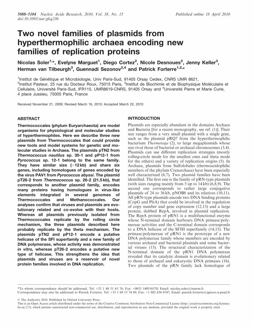
Two novel families of plasmids fromhyperthermophilic archaea encoding newfamilies of replication proteinsNicolas Soler1,*, Evelyne Marguet1, Diego Cortez2, Nicole Desnoues2, Jenny Keller3,
Herman van Tilbeurgh3, Guennadi Sezonov2,4 and Patrick Forterre1,2,*
1Institut de Genetique et Microbiologie, Univ Paris-Sud, 91405 Orsay Cedex, CNRS UMR 8621,2Institut Pasteur, 25 rue du Docteur Roux, 75015 Paris, 3Institut de Biochimie et de Biophysique Moleculaire etCellulaire, Universite Paris-Sud, IFR115, UMR8619-CNRS, 91405 Orsay and 4Universite Pierre et Marie Curie,4 place Jussieu, 75005 Paris, France
Received November 21, 2009; Revised March 16, 2010; Accepted March 22, 2010
ABSTRACT
Thermococcales (phylum Euryarchaeota) are modelorganisms for physiological and molecular studiesof hyperthermophiles. Here we describe three newplasmids from Thermococcales that could providenew tools and model systems for genetic and mo-lecular studies in Archaea. The plasmids pTN2 fromThermococcus nautilus sp. 30-1 and pP12-1 fromPyrococcus sp. 12-1 belong to the same family.They have similar size (�12 kb) and share sixgenes, including homologues of genes encoded bythe virus PAV1 from Pyrococcus abyssi. The plasmidpT26-2 from Thermococcus sp. 26-2 (21.5 kb), thatcorresponds to another plasmid family, encodesmany proteins having homologues in virus-likeelements integrated in several genomes ofThermococcales and Methanococcales. Ouranalyses confirm that viruses and plasmids are evo-lutionary related and co-evolve with their hosts.Whereas all plasmids previously isolated fromThermococcales replicate by the rolling circlemechanism, the three plasmids described hereprobably replicate by the theta mechanism. Theplasmids pTN2 and pP12-1 encode a putativehelicase of the SFI superfamily and a new family ofDNA polymerase, whose activity was demonstratedin vitro, whereas pT26-2 encodes a putative newtype of helicase. This strengthens the idea thatplasmids and viruses are a reservoir of novelprotein families involved in DNA replication.
INTRODUCTION
Plasmids are especially abundant in the domains Archaeaand Bacteria [for a recent monography, see ref. (1)]. Theirsize ranges from a very small plasmid with a single gene,such as the plasmid pRQ7 from the hyperthermophilicbacterium Thermotoga (2), to large megaplasmids whosesize rival those of bacterial or archaeal chromosomes (3,4).Plasmids can use different replication strategies (mostlyrolling-circle mode for the smallest ones and theta modefor the others) and a variety of replication origins (5). InArchaea, plasmids from Sulfolobales (thermoacidophilicmembers of the phylum Crenarchaea) have been especiallywell characterized (6,7). Two plasmid families have beenidentified. The first one is the family of pRN-type plasmids(with sizes ranging mainly from 5 up to 14 kb) (6,8,9). Thesecond one corresponds to rather large conjugativeplasmids of 24 to 36 kb, pNOB8 and its relatives (10,11).All pRN-type plasmids encode two DNA binding proteins(CopG and PlrA) that could be involved in the regulationof copy number and gene expression (12,13) and a largeprotein, dubbed RepA, involved in plasmid replication.The RepA protein of pRN1 is a multifunctional enzymewhose N-terminal domain harbours DNA primase/poly-merase activities and the C-terminal domain correspondsto a DNA helicase of the SFIII superfamily (14,15). Theprimase/polymerase of pRN1 is the prototype of a newDNA polymerase family whose members are encoded byvarious archaeal and bacterial plasmids and some bacter-ial viruses (15). The structural characterization of theN-terminal domain of the pRN1 DNA polymeraserevealed that its catalytic domain is evolutionary relatedto those of archaeal and eukaryotic DNA primases (16).Two plasmids of the pRN family lack homologue of
*To whom correspondence should be addressed. Tel: +33 1 40 51 65 76; Fax: +0033 140516570; Email: [email protected] may also be addressed to Patrick Forterre. Tel:+33 1 69 15 74 89; Fax:+1 802 656 0747; Email: [email protected]
5088–5104 Nucleic Acids Research, 2010, Vol. 38, No. 15 Published online 18 April 2010doi:10.1093/nar/gkq236
� The Author(s) 2010. Published by Oxford University Press.This is an Open Access article distributed under the terms of the Creative Commons Attribution Non-Commercial License (http://creativecommons.org/licenses/by-nc/2.5), which permits unrestricted non-commercial use, distribution, and reproduction in any medium, provided the original work is properly cited.
pRN1 RepA: one of them, pXZ1, encodes instead a largeprotein of similar size but without sequence similarity topRN1 RepA (17), whereas another one, pTAU4, encodesa MCM helicase instead of a Rep protein (8). An interest-ing member of the pRN family is pSSVx, a virus–plasmidhybrid that coexists intracellularly with the fusellovirusSSV1 and can be packaged into viral particles (18,19).Besides typical pRN proteins, pSSVx encodes two SSV1proteins probably involved in plasmid packaging. SeveralpRN-type plasmids have been found integrated intotRNA genes in the chromosomes of differentSulfolobales (18). The site-specific integrase thatpromotes their insertion is homologous to the integrasesof the SSV1 and other fuselloviruses which can also inte-grate in the chromosomes of Sulfolobus species (20). Theinsertion site recognized by SSV-type integrases is locatedin the coding part of their own gene and the integrationevent provokes its disruption. The plasmid pXZ1 alsoencodes an SSV-type integrase and coexists in Sulfolobuscell with a fusellovirus (SSV4) but, unlike pSSVx, pXZ1cannot be packaged into viral particle (17). The SSV1virus, as well as pSSVx and pRN1 has been used todevelop genetic tools for Sulfolobus species, includingshuttle vectors to express foreign proteins in Sulfolobales(21,22).
The Sulfolobus conjugative plasmids of the pNOB8-typeencodes �40–50 proteins (10,11). Ten proteins areconserved in all these plasmids and 80% of the remainingproteins are common to at least two plasmids (11). Mostof these proteins are of unknown function. Theirconserved genes are grouped in two regions: one of themincludes two genes encoding proteins with low similarityto TraG and TraE proteins involved in bacterial plasmidsconjugation, as well as several proteins with membranedomains that are supposed to be involved in DNAtransfer. The second region contains the genes encodinghomologues of the pRN-type plasmids, CopG and PrlAproteins, and an integrase gene. This integrase represents anovel family of integrases, the pNOB8-type, that arewidely distributed in archaeal genomes (10). Unlike theSSV-type, pNOB8-type integrases never bear their inser-tion site inside the sequence of their genes and consequent-ly these genes are never disrupted after integration. SeveralpNOB8-type plasmids encode transposases, and one ofthem (pKEF9) also encodes a pRN-like RepA protein.A new member of the pNOB8 conjugative plasmidfamily, pAH1, has been recently discovered in anAcidianus species that belongs to the order Sulfolobales(20). Interestingly, the replication of pAH1 is inhibitedby infection by the lipothrixvirus AFV1, revealing a newtype of plasmid/virus functional interaction (20).
All plasmids presently isolated from Sulfolobales arelarger than 5 kb. Although their mode of replication hasnot yet been determined experimentally, they probably usethe theta replication mode, since none of them encode theRep protein typical for rolling-circle replication. Incontrast, all known plasmids from Thermococcales(hyperthermophilic and anaerobic members of thephylum Euryarhaea) are small (<4 kb) and replicate viathe rolling circle (RC) mode. This has been experimentallydemonstrated for the plasmid pGT5 from Pyrococcus
abyssi (23–25) and it is most likely the replication modefor the related plasmid pTN1, isolated from Thermococcusnautilus (26). Both plasmids encode large homologousproteins related to Rep proteins that initiate RC replica-tion. These two RC initiator proteins, Rep75 and Rep74,respectively, form a new family of RC Rep proteins andare distantly related to transposases encoded by bacterialIS (IS91, IS1294 and IS801 families) that replicate them-selves via the RC mode (26). A third small plasmid, calledpRT1, from Pyrococcus sp. JT1, has been sequenced andtentatively described as a RC plasmid (27). However, itsputative Rep protein shows no clear sequence similaritywith known RC Rep proteins (26). The pGT5 and pTN1plasmids have been used to construct shuttle vectors forP. abyssi and T. kodakaraensis, respectively (28,29). ThepTN1-based shuttle vector for T. kodakaraensis has beenengineered into an expression vector for tagged proteinsthat can be used to isolate protein complexes formedin vivo (28). The recent development of efficient transform-ation methods and gene knock-out strategies forT. kodakaraensis (28,30) clearly emphasize how usefulthe vectors based on Thermococcus plasmids could be.During our characterization of the plasmid pTN1, we
noticed that the strain T. nautilus contains additionally alarger plasmid of �13 kb, that we have called pTN2.Screening for further plasmids in our collection ofThermococcales strains (31), we then isolated a plasmidof similar size, called pP12-1, from a Pyrococcus strain (sp.12-1) and a larger plasmid (�20 kb) in a Thermococcusisolate (sp. 26-2) that we called pT26-2. We present herethe analysis of the sequences of these three plasmids,focusing on the annotation of their proteins and on theirevolutionary relationships with archaeal viruses andcellular hosts. Most of the predicted proteins of thesethree new plasmids are either ORFans or have onlyclosely related homologues in viruses or integratedvirus-like elements from Thermococcales and/orMethanococcales. They do not encode typical Repproteins for RC replication, but new types of DNA repli-cation proteins with distant homologues encoded bySulfolobus pRN-type plasmids, indicating that theyprobably replicate by the theta mode. In particular, theRep protein of pTN2 and pP12-1 appears to be related tothe Rep protein of the Sulfolobus plasmids pXZ1 andpTIK4. Interestingly, the N-terminal domain of theseRep proteins corresponds to a new family of DNA poly-merase (demonstrated here experimentally for the pTN2Rep protein), whereas the pT26-2 plasmid encodesprobably a new type of helicases. These discoveries arein good agreement with the idea that plasmids andviruses are a reservoir of novel protein families involvedin DNA replication.
MATERIALS AND METHODS
Strains cultivation
Thermococcus nautilus sp. 30-1, Pyrococcus sp. 12-1 andThermococcus sp. 26-2, were isolated from single coloniesby plating on Gelrite enrichment cultures obtained fromfragments of chimneys collected at deep sea hydrothermal
Nucleic Acids Research, 2010, Vol. 38, No. 15 5089
vents (�2630m) located in the East Pacific ocean (31).Cells were grown in Zillig’s broth (ZB) made anaerobicby addition of Na2S (0.1mg/l), with sulphur flowers S�
(Fisher Scientific) as previously described (31). Thecultures were incubated in Penny’s flasks at 85�C forThermococcus sp. 26-2 and T. nautilus, or 95�C forPyrococcus sp. 12-1.
Plasmid isolation
Plasmids were obtained by alkaline extraction as describedpreviously (26) from 50ml culture of T. nautilus 30-1,Pyrococcus 12-1 or Thermococcus 26-2 cells that hadbeen grown until stationary phase. Plasmids weresequenced by Fidelity Systems, USA. The complete se-quences of pTN2, pP12-1 and pT26-2 have, respectively,been submitted to the GenBank database under accessionnumbers GU056177, GU056178 and GU056179.
Sequence analysis
ORFs were identified with a minimum of 50 amino acidslength and with one of the initiation codons (ATG, GTG,CTG, TTG). The exact position of each initation codonwas then checked individually (and manually modified ifrequired) depending on the position of putative Shine–Dalgarno sequence located upstream (Tables 1–3).BLAST and PSI-BLAST searches were performed in theNCBI non-redundant (nr) databank using the followingweb sites: http://www.ncbi.nlm.nih.gov/BLAST/ andhttp://www-archbac.u-psud.fr/genomics/GenomicsToolbox.html/. Hydrophobic regions were detected usingTMpred and TMHMM prediction programs available atthe web site http://www.expasy.org/tools/. Searches forspecific putative domains or sites were performed usingInterproscan located at the website http://www.ebi.ac.uk/Tools/InterProScan/.
Phylogenetic analyses
Homologous sequences were recovered by BLASTsearches, and multiple alignment were performed with
the selected sequences using MUSCLE program (32).Only homologous positions were used to build unrootedmaximum likelihood trees using PHYML (33). The JTTmodel of amino acid substitution was choosen, and agamma correction with four discrete classes of sites wasused. The alpha parameter and the proportion of invari-able sites were estimated. The robustness of trees wastested by non-parametric bootstrap analysis usingPHYML.
In silico identification of integrated mobile element
Mobile elements integrated in cellular genome wereidentified as Cluster of Atypical Genes (CAGs) accordingto Cortez et al. (41). Briefly, archaeal genomes wereanalysed with a species-specific Markov model in orderto obtain the list of atypical genes. We then searched foratypical genes that cluster together, since these may berecently integrated foreign elements. We used a slidingwindow of 10 ORFs that moved along the genomesequence. A CAG was defined when seven or moreORFs in that window showed an atypical composition.To define CAGs families, Blast searches were performedwith all the ORFs contained in our CAGs with an e-valueof 10e�20. We then generated several topological networksof CAGs by drawing a line between pairs of CAGs thatshare a defined number of ORFs (from two up to six). Thegraphical representation was obtained with the Cytoscapeprogram (http://www.cytoscape.org).
Cloning, expression and purification of tn2-12p
The coding sequence of tn2-12p was amplified by PCRfrom plasmidic DNA isolated from the strain T. nautilus30/1, and the amplified fragment was cloned in a pET21(Novagen) plasmid allowing fusion of a 6His tag at 30-end.Expression was done at 37�C using the Escherichia coliRosetta (DE3) pLysS strain (Novagen) and the 2xYTmedium (BIO 101 Inc.). When the cell culture reachedan OD600 nm of 0.8, induction at 15�C was performedovernight with 0.5mM IPTG (Sigma). Cells were har-vested by centrifugation and resuspended in buffer A
Table 1. Annotation of pTN2 plasmid from the strain T. nautilus 30/1
ORF Putativeprotein
Size(kDa)
ORF position(start–stop)
RBS and startcodon
Homologue inpP12-1 plasmid
Percentage identity,length of BLASTalignment (aa)
Putative motivesand function
1 tn2-1p 66.1 1–1710 aggagggggtggtcggGTG p12-1p 69%, 569 UvrD/Rep/PcrAsuperfamilyI type helicase
2 tn2-2p 104.9 1785–4622 ggggggatttgtATG – – Putative coiled-coil domains3 tn2-3p 20.3 4841–5365 tgggggatgacgATG p12-14p 46%, 1734 tn2-4p 15.2 5590–5979 gggtggtgtcgtaattATG – – Putative helix–turn–helix domain5 tn2-5p 11.2 5983–6279 aggggaggtgtaaccgATG – –6 tn2-6p 26.4 6285–6959 agaggtgtaaaacaaATG – –7 tn2-7p 19.5 6959–7477 aggaggggttatgATG p12-9p 41%, 418 tn2-8p 43 7542–8648 tggaggtgccgagcGTG p12-10p 46%, 367 ATP binding site, that is related to
ABC transporters9 tn2-9p 16 9184–9597 agggggtactcataATG – –10 tn2-10p 6.7 9551–9730 cggaggaggataATG – –11 tn2-11p 18.4 9763–10 251 tggaggtgatgtaaATG – –12 tn2-12p 106.8 10 248–4 agggggtgaaagtATG p121-17p 55%, 922 DNA polymerase/primase activities
5090 Nucleic Acids Research, 2010, Vol. 38, No. 15
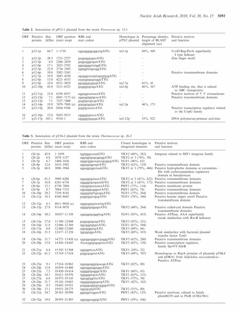
Table 3. Annotation of pT26-2 plasmid from the strain Thermococcus sp. 26-2
ORF Putativeprotein
Size(kDa)
ORF position(start–stop)
RBS andstart codon
Closest homologue inintegrated elements
Putative motivesand function
1 t26-1p 42.8 1–1059 tggtgggccaaaGTG TKV2 (68%, 282) Integrase related to SSV1 integrase family2 t26-2p 6.8 1078–1257 aggaggtgagcggaggATG TKV2 et 3 (74%, 58)3 t26-3p 6.7 1460–1636 agggcgggcacgcccgcggcgATG TGV1 (46%, 63)4 t26-4p 12.6 1633–1992 aggtggtggcagtATG TKV2 (63%, 119) Putative transmembrane domain5 t26-5p 68.8 2098–3984 aggaggtggtctaaGTG TKV2 et 3 (79%, 406) Putative hydrophobic domains at extremities –
Hit with carboxypeptidase regulatorydomain in Interproscan
6 t26-6p 81.5 3989–6208 aggaggtgataacATG TKV2 et 3 (41%, 622) Putative transmembrane domains7 t26-7p 18.0 6241–6738 agggggtgatgaattGTG TKV2 et 3 (43%, 172) Putative transmembrane domains8 t26-8p 12.1 6750–7094 ctgaggtgaaaaacaATG PHV1 (75%, 114) Putative membrane protein9 t26-9p 8.7 7084–7335 aggagggagggactATG PHV1 (65%, 78) Putative transmembrane domains10 t26-10p 29.2 7339–8145 agggggtgtttgaagaATG TGV1 (75%, 264) Putative transmembrane domain11 t26-11p 18.5 8160–8642 gaggtggactgagATG TGV1 (76%, 160) Putative leucine zipper motif–Putative
transmembrane domain12 t26-12p 8.1 8811–9020 (c) agggaggtgtactcttgATG –13 t26-13p 27.9 9114–9878 aggggtgaggagaATG TKV2 (44%, 264) Putative coiled-coil domain–Putative
transmembrane domains14 t26-14p 50.2 10 037–11 338 cggaggtgcgggttagATG TGV1 (95%, 433) Putative ATPase, AAA superfamily
(weak similarities with RuvB helicase)15 t26-15p 27.0 11 349–12 044 gaggtggtgaagATG TKV3 (92%, 231)16 t26-16p 11.8 12 046–12 369 tggagggtgtgatATG TGV1 (81%, 104)17 t26-17p 8.0 12 480–12 680 cggtggggctgATG TKV3 (89%, 66)18 t26-18p 21.5 12 677–13 228 tggtggtggaATG TKV3 (88%, 183) Weak similarities with bacterial plasmid
transfer factor TraG19 t26-19p 31.7 14 271–13 438 (c) cggaggagggcccgagggATG TKV3 (62%, 280) Putative transmembrane domains20 t26-20p 15.8 14 426–14 842 AccacggtgagagctcaATG TKV3 (65%, 138) Putative transcription regulator,
family SpoVT/AbrB21 t26-21p 6.4 15 343–15 504 aggtggttccaATG TKV1 (34%, 52)22 t26-22p 81.2 15 519–17 624 gaggcgagctctATG TKV3 (49%, 707) Homologous to RepA proteins of plasmids pTIK4
and pORA1 from Sulfolobus neozealandicus—Putative ATPase
23 t26-23p 9.1 17 814–18 062 cggaggagggtgaaagcATG TKV3 (82%, 80)24 t26-24p 13.0 18 059–18 406 aggagggaggaggaATG –25 t26-25p 7.5 18 420–18 614 cgggggtgagggcATG TKV1 (66%, 63)26 t26-26p 14.1 18 611–18 976 tggggggtcgctcATG TKV1 (63%, 121)27 t26-27p 6.6 18 973–19 143 tggaggtggtttcATG TKV1 (75%, 54)28 t26-28p 21.7 19 143–19 685 cgggggtgggagctgATG TKV1 (42%, 182)29 t26-29p 9.5 19 692–19 931 gaggggaggtgacggggcGTG –30 t26-30p 13.1 19 931–20 275 aggagcgtgATG TKV2 (51%, 80)31 t26-31p 24.7 20 363–20 998 gggtggttcggaaATG PHV1 (45%, 155) Putative resolvase, related to family
pfam00239 and to PinR (COG1961)32 t26-32p 19.8 20 995–21 495 aggaggaaggtgtATG PHV1 (39%, 166)
Table 2. Annotation of pP12-1 plasmid from the strain Pyrococcus sp. 12/1
ORF Putativeprotein
Size(kDa)
ORF position(start–stop)
RBS andstart codon
Homologue inpTN2 plasmid
Percentage identity,length of BLASTalignment (aa)
Putative motivesand function
1 p12-1p 66.7 1–1710 aggagggggaggcctgATG tn2-1p 69%, 569 UvrD/Rep/PcrA superfamilyI type helicase
2 p12-2p 20.3 1721–2257 gaggtgggagaaATG – – Zinc finger motif3 p12-3p 6.9 2260–2439 gaggagggttgattATG – –4 p12-4p 17.1 2423–2782 gggaggggatatggGTG – –5 p12-5p 22.9 2736–3305 tggtggttatgaaggATG – –6 p12-6p 10.0 3302–3565 – – – Putative transmembrane domains7 p12-7p 19.9 3685–4194 agagggcaaagttagagtgagATG – –8 p12-8p 13.0 4221–4553 aagaggtgagaaggaTTG – –9 p12-9p 18.8 4531–5019 ggaggggaggagtATG tn2-7p 41%, 4110 p12-10p 43.0 5211–6323 gaggggtgtagaATG tn2-8p 46%, 367 ATP binding site, that is related
to ABC transporters11 p12-11p 23.4 6350–6937 aggaggcaataaaATG – – Putative motives of 30–50 exonucleases12 p12-12p 7.1 6981–7175 (c) aggaggtggtgcaaATG – – Putative transmembrane domains13 p12-13p 7.1 7227–7400 gaggtgccgccgtATG – –14 p12-14p 19.9 7479–7988 (c) gtgtggtggtgatATG tn2-3p 46%, 17315 p12-15p 20.0 8564–9106 gtggtgcaccccATG – – Putative transcription regulator related
to the CopG family16 p12-16p 15.6 9103–9513 cgggggtgtccccATG – –17 p12-17p 103.1 9510–1 agggggtgagagcATG tn2-12p 55%, 922 DNA polymerase/primase activities
Nucleic Acids Research, 2010, Vol. 38, No. 15 5091
(20mM Tris–HCl pH 7.5, 200mM NaCl, 5mMb-mercaptoethanol). Cell lysis was completed by sonic-ation and the lysate was heated for 20min at 70�Cbefore centrifugation at 20 000 rpm for 20min. Thesoluble fraction was loaded on a Ni–NTA column(Qiagen Inc.) equilibrated with buffer A. The proteinwas eluted with imidazole and subsequently loaded on aheparin column (GE Healthcare) equilibrated againstbuffer A0 (20mM Tris Tris–HCl pH 7.5, 50mM NaCl,5mM b-mercaptoethanol). Elution was performed usinga gradient between buffer A0 and B (20mM Tris Tris–HClpH 7.5, 1M NaCl, 5mM b-mercaptoethanol). Thetn2-12p protein was eluted at �0.9M NaCl. Eluted frac-tions were pooled and loaded on a Superdex200 column(Amersham Pharmacia Biotech) equilibrated againstbuffer A supplemented with 10mM b-mercaptoethanol.The homogeneity of the protein sample was checked bySDS–PAGE.
DNA polymerase assay
Two different 50-labelled primer-template systems wereused (see legend of Figure 2). The standard polymeraseassay contained 10 nM primer-template substrate,2.5–15 mM of tn2-12p (see legend of Figure 2) in 10mMTris–HCl pH 9, 1mM DTT, 50mM KCl, 0.1% TritonX-100, 50mM MgCl2 and 0.2mM dNTPs. The proteinwas diluted in 50mM Tris–HCl pH 8, 1mM DTT,100mM NaCl, 0.1mM EDTA. The significant amountof protein required for the optimal polymerizationreaction could be explained by a relatively low stabilityof the isolated protein. The reactions were allowed toproceed for 20min at 70 or 80�C and were analysed bydenaturating PAGE.
RESULTS
Isolation of three new plasmids from Thermococcales
Plasmids pTN2, pP12-1 and pT26-2 were isolated fromthree strains of Thermococcales that were purified fromsamples collected at three different deep-sea hot ventslocated in the East-Pacific ridge during the AMISTADcruise (31). The strain 30-1, which harbours the plasmidpTN2, has been tentatively described as the type strain ofa new species, T. nautilus (26). The strain 26-2, thatharbours the plasmid pT26-2 is closely related toT. nautilus sp. 30-1 by 16S rRNA analysis and couldbelong to the same species, although it exhibits a verydifferent RAPD profile (31). Finally, the strain 12-1,which harbours the plasmid pP-12-1 belongs to a RAPDgroup that includes a Pyrococcus species (isolate 32-4) andwill be thereafter called Pyrococcus sp. 12-1. The threeplasmids are circular and their sizes are 13 015, 12 205and 21 566 bp, respectively. The plasmids sequences havea GC content of �47.5, 44.6 and 49.6% for pTN2, pP12-1and pT26-2, respectively, which are close to GC% ofThermococcales chromosomal DNA (40–50%). By insilico analysis, we identified 12, 17 and 32 putativeORFs in pTN2, pP12-1 and p26-2, respectively (see‘Materials and Methods’ section). Although pTN2 wasisolated from a Thermococcus species and pP12-1 from a
Pyrococcus species, they turned out to be evolutionaryrelated, since they share six homologous genes. Incontrast, although plasmids pTN2 and pT26-2 arepresent in two closely related strains (30-1 and 26-2),they turned out to be completely unrelated to each other.
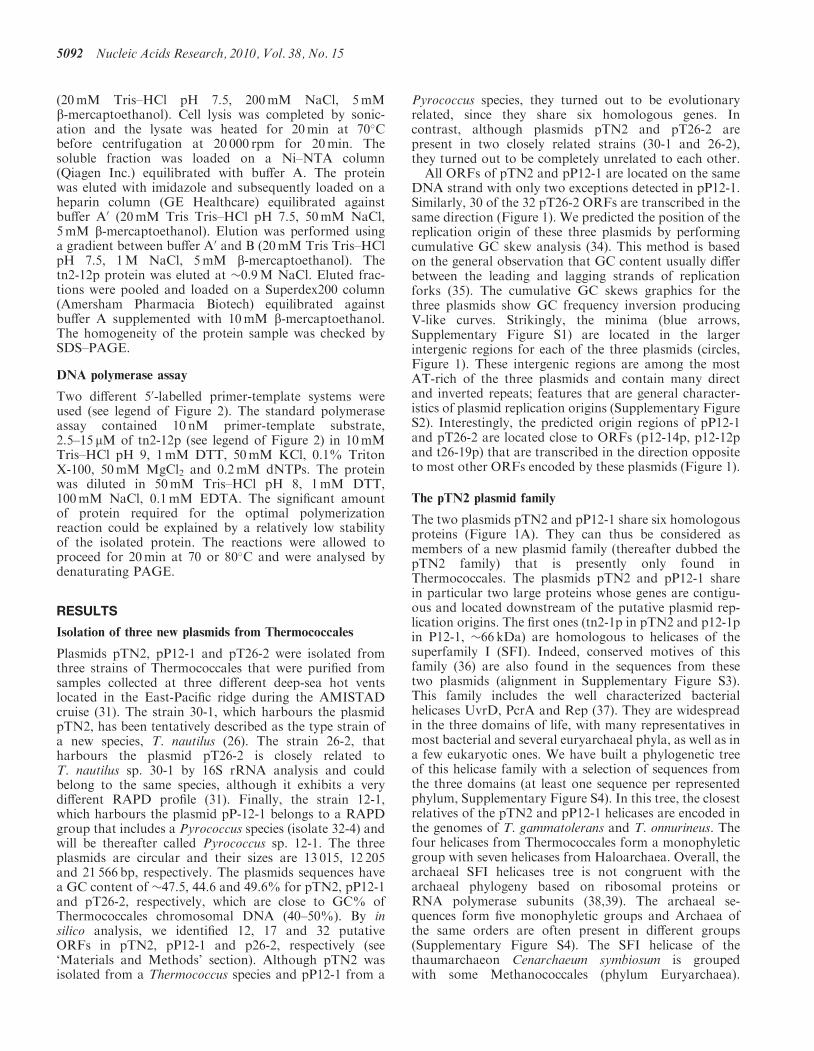
All ORFs of pTN2 and pP12-1 are located on the sameDNA strand with only two exceptions detected in pP12-1.Similarly, 30 of the 32 pT26-2 ORFs are transcribed in thesame direction (Figure 1). We predicted the position of thereplication origin of these three plasmids by performingcumulative GC skew analysis (34). This method is basedon the general observation that GC content usually differbetween the leading and lagging strands of replicationforks (35). The cumulative GC skews graphics for thethree plasmids show GC frequency inversion producingV-like curves. Strikingly, the minima (blue arrows,Supplementary Figure S1) are located in the largerintergenic regions for each of the three plasmids (circles,Figure 1). These intergenic regions are among the mostAT-rich of the three plasmids and contain many directand inverted repeats; features that are general character-istics of plasmid replication origins (Supplementary FigureS2). Interestingly, the predicted origin regions of pP12-1and pT26-2 are located close to ORFs (p12-14p, p12-12pand t26-19p) that are transcribed in the direction oppositeto most other ORFs encoded by these plasmids (Figure 1).
The pTN2 plasmid family
The two plasmids pTN2 and pP12-1 share six homologousproteins (Figure 1A). They can thus be considered asmembers of a new plasmid family (thereafter dubbed thepTN2 family) that is presently only found inThermococcales. The plasmids pTN2 and pP12-1 sharein particular two large proteins whose genes are contigu-ous and located downstream of the putative plasmid rep-lication origins. The first ones (tn2-1p in pTN2 and p12-1pin P12-1, �66 kDa) are homologous to helicases of thesuperfamily I (SFI). Indeed, conserved motives of thisfamily (36) are also found in the sequences from thesetwo plasmids (alignment in Supplementary Figure S3).This family includes the well characterized bacterialhelicases UvrD, PcrA and Rep (37). They are widespreadin the three domains of life, with many representatives inmost bacterial and several euryarchaeal phyla, as well as ina few eukaryotic ones. We have built a phylogenetic treeof this helicase family with a selection of sequences fromthe three domains (at least one sequence per representedphylum, Supplementary Figure S4). In this tree, the closestrelatives of the pTN2 and pP12-1 helicases are encoded inthe genomes of T. gammatolerans and T. onnurineus. Thefour helicases from Thermococcales form a monophyleticgroup with seven helicases from Haloarchaea. Overall, thearchaeal SFI helicases tree is not congruent with thearchaeal phylogeny based on ribosomal proteins orRNA polymerase subunits (38,39). The archaeal se-quences form five monophyletic groups and Archaea ofthe same orders are often present in different groups(Supplementary Figure S4). The SFI helicase of thethaumarchaeon Cenarchaeum symbiosum is groupedwith some Methanococcales (phylum Euryarchaea).
5092 Nucleic Acids Research, 2010, Vol. 38, No. 15
676
140
678
138
89
180b
137
375
898
136
190
153
59
52a 87
528
52b
180a
62
82 121
109
159
293
214
A 1
2
3
46 5
7
8
10
11
12
9
1
2
3
4
5
7
8
9
1011
6
13
14
15
16
17
12
12
34
5
6
7
8
9
10
1112
131415
1617
18
19
20
21
22
2324
25 26
27 28
2930
31 32
ATPaseAAA+ family
Putative transcription factorSpoVT/AbrBfamily
Integrase
Putative Repprotein(RepAfrompRN-relatedplasmids)
ResolvaseB
Figure 1. Schematic representation of the three new plasmids. (A) pTN2 and pP12-1 plasmid maps were drawn at the same scale together with PAV1genome. ORFs are numbered and represented as arrows. ORFs encoding homologous proteins have the same colour. White ORFs do not havedetectable homologues among these three genomes. (B) pT26-2 plasmid map with ORFs numbered and represented as arrows. Coloured ORFsencode proteins with expected activity or function. (A and B) Hachured ORFs harbour putative hydrophobic segments. Circles indicate largeintergenic regions including putative replication origins.
Nucleic Acids Research, 2010, Vol. 38, No. 15 5093

Furthermore, several groups of eukaryotic helicases (fromplants and protists) are interspersed between archaealgroups, whereas helicases from fungi form a monophyleticgroup with sequences from Methanomicrobiales andThermoplasmatales. This phylogeny is difficult to inter-pret because lengths are highly variable, probablyinducing phylogenetic artefacts. Interestingly, the‘cellular’ homologue of pTN2-type helicases inT. gammatolerans is located in an integrated virus-likeelement, TGV2 (40). In Methanococcus maripaludisstrains C6 (MMC6V1) and C7 (MMC7V2), the genesencoding pTN2-type helicases are located in predictedintegrated elements of plasmid/virus origin that havebeen identified in silico, based on their dinucleotidesequence composition [see ‘Materials and Methods’section and ref. (41)]. These observations suggest thatsome archaeal, and possibly eukaryotic SFI helicases,originated from viruses and plasmids and were transferredindependently into various lineages, explaining the incon-gruence between their phylogeny, and the classicalarchaeal and eukaryotic phylogenies. The frequentgrouping of Archaea from different phyla in the sameclade furthermore suggests that horizontal gene transfersof genes encoding these helicases also sometimes occurredin Archaea. The bacterial helicases of the SFI helicasefamily are quite well separated from archaeal and eukary-otic ones, with the exception of the helicase from the bac-terium Aquifex aeolicus that branches with the groupmixing some helicases from Archaea and Fungi.Bacterial UvrD-like helicases form a monophyleticgroup (with two representatives in the eukaryotesOstreococcus, a probable case of gene transfer from amitochondrial or plastid genome). However, as in thecase of Archaea, Bacteria from the same phylum areoften dispersed in different parts of the tree, suggestingthat the genes encoding these helicases have been alsosometimes transferred between bacterial phyla.The second large ORFs conserved between pTN2 and
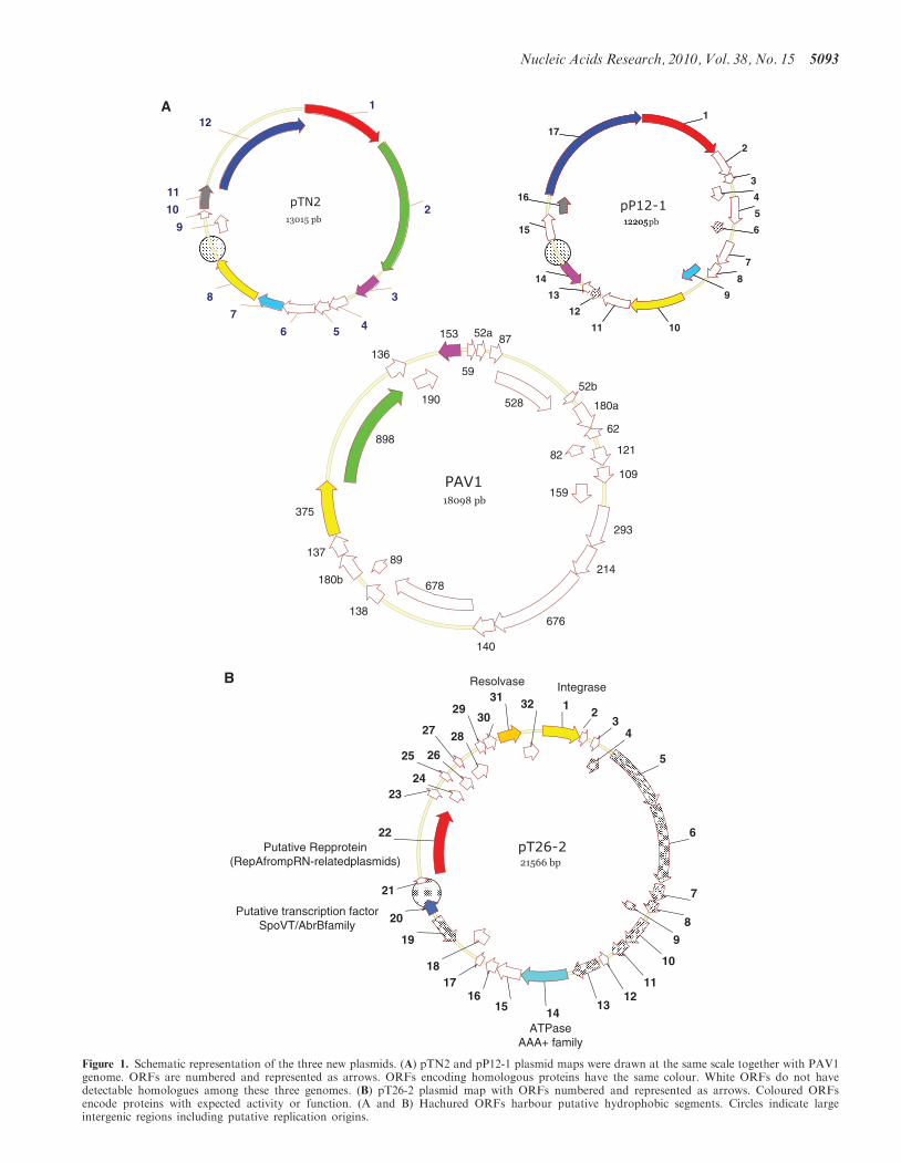
pP12-1 (tn2-12p and p12-17p, respectively) encodesproteins of 107 and 103 kDa, respectively. Psi-BLASTsearches using tn2-12p as query gave no significantresult, whereas similar searches using p12-17p gave signifi-cant matches with two very similar proteins from M.voltae A3 (MvolDRAFT_1375 and MvolDRAFT_1398).Interestingly, we noticed among hits of the firstPSI-BLAST iterations the putative Rep protein of therecently described plasmid pXZ1 from Sulfolobusislandicus (17). BLAST search with the sequence of thepXZ1 Rep protein then retrieved the Rep protein of theplasmid pTIK4 from S. neozelandicus (8). We have beenable to align manually the N-terminal regions of tn2-12pand p12-17p with those of the two proteins of M. voltaeand the Rep proteins of pXZ1 and pTIK4 (SupplementaryFigure S5). We then noticed that these proteins exhibit aconserved DhD motif, known to be present in the activesites of many DNA polymerases, primases and/ornucleotidyl transferases (42). Although this similaritywas minimal, we decided to express the tn2-12p proteinin E. coli to test if this protein exhibits a DNA polymeraseactivity in vitro. The purified recombinant tn2-12p proteinwas first incubated with dNTPs and 50-labelled 20 nt DNA
primer hybridized to a complementary 42 nt DNAtemplate. As expected if our prediction was correct, theprimer was efficiently extended up to the full-length ofthe template (Figure 2A and B). The major fraction ofthe polymerization products was, as expected, representedby a 42-bp-long fragment but we also observed a minorDNA fragment of 43 bp. In the same conditions, theThermus aquaticus (Taq) DNA polymerase synthesizedalso two subfamilies of DNA fragments: a major onecomposed of 43 bp and a second one (minor) havingonly 42 bp. It is well known that Taq DNA polymerasehas a non-template-dependent terminal transferaseactivity and is able to add an additional non-template-directed nucleotide to the 30-ends of ablunt-ended DNA fragment via this terminaltransferase-like activity (43,44). This result indicates thatthe tn2-12p protein has also a nucleotidyl transferaseactivity which will be described in details elsewhere(Desnoues et al., manuscript in preparation). The DNApolymerizing activity of tn2-12p requires the presence ofdNTPs and a DNA template (data not shown) and itsactivity is not stimulated in the presence of ATP (as wasobserved for another archaeal polymerase coded by theplasmid pRN1, 14). No extension was observed whenthe dNTPs were replaced by NTPs. The protein tn2-12pis able to extend the primer up to several kilobases at20min of the reaction times and the obtained DNAshows a pattern close to that observed in the similar ex-periment with the Taq polymerase. This result indicatesthat tn2-12p can produce long extension productswithout the help of additional proteins.
The DNA polymerase activity associated to tn2-12p isconsistent with the idea that tn2-12p and the homologousprotein p12-17p correspond to the Rep proteins of theplasmids pTN2 and pP12-1, respectively. In the case ofpRN1, the DNA polymerase domain of RepA is locatedin the N-terminal part of the protein and is fused to ahelicase domain (SFIII) in C-terminal. In the case of thepTN2 and pP12-1 Rep proteins, Psi-BLAST analysissuggests that the DNA polymerase domain is alsolocated in their N-terminal part (which contains the twoconserved aspartate residues probably present in the activesite). They also indicate that, as in the case of pRN1-typeRepA, these polymerase domains are fused to largeC-terminal domains. However, Psi-BLAST analysisusing these domains as queries retrieved no significanthit in databases.
In both pTN2 and pP12-1, the rep genes form a repli-cation cassette with the contiguous helicase gene.Interestingly, these replication cassettes are locatedupstream the large intergenic regions predicted to be thereplication origin of these plasmids by GC skew analysis(Figure 1A and Supplementary Figure S1). Several smallputative ORFs are located between the origin regions ofpTN2 and pP12-1 and the beginning of the rep genes. Oneof them (tn2-11p and p12-16p) is conserved in bothplasmids, indicating that they should encode bona fideproteins involved in the regulation of plasmid replication.In pP12-1, the protein p12-15p, which is located close tothe putative replication origin, belongs to the CopGfamily of transcriptional regulators. This protein could
5094 Nucleic Acids Research, 2010, Vol. 38, No. 15
play a role in the regulation of the Rep protein expression,similarly to the CopG-like proteins whose genes arelocated upstream of the rep gene in Sulfolobus pRN-typeplasmids (45).
Three other proteins shared by pTN2 and pP12-1(tn12-3p/p12-14p, tn2-7/pP12-9p and tn2-8p/pP12-10p)are encoded by genes whose order is not conservedbetween the two plasmids (Figure 1A). Interestingly, twoof them are homologous to proteins encoded by the virusPAV1 from P. abyssi (Figure 1A). This lemon-shapedvirus, with a double-stranded DNA genome of �16 kb,is presently the only known virus that infects anhyperthermophilic euryarchaeon (46). The tn2-8p andp12-10p proteins (�42 kDa) are homologous to theORF375 protein of PAV1, whereas the tn2-3p andp12-14p proteins (�20 kDa) are homologous to theORF153 protein of PAV1.
BLAST searches with proteins tn2-8p, p12-10p orORF375 as queries retrieved significant sequencesimilarities with proteins encoded in the genomes ofM. voltae A3 and Methanosarcina barkeri, in a DNAfragment from an environmental sample enriched for
virus sequences (47) and in several plasmids fromhaloarchaea (pNG4027, pNG1017, pNG2044 andpNG3054). See Supplementary Figure S6 for an alignmentof these homologous proteins. In M. voltae, the homo-logue of tn2-8p/p12-10p, MvolDRAFT_1394, is locatedin a large region of �60 genes located between a tRNAand a cluster of CRISPR associated Cas genes. All thesegenes encode uncharacterized proteins, except for threeintegrase genes, two putative transcriptional regulators,a resolvase gene and, strikingly, two genes encodinghomologues of the pTN2 and pP12-1 Rep-DNA polymer-ases (MvolDRAFT_1375 and 1398). This region thusmost likely corresponds to integrated virus and/orplasmids. In M. barkeri, the homologue of tn2-8p/p12-10p is located close to a gene encoding a putativeintegrase (Mbar_A2247) next to a tRNA gene, indicatingthat this protein is also encoded by an integrated mobileelement. This suggests that tn2-8p/p12-10p are the proto-type of a new protein family specific for viral/plasmid.Visual inspection of alignment of these proteins revealeda typical Walker type motif in their N-terminal extremity(Supplementary Figure S6). Indeed, Psi-BLAST iterations
A B C 6
43
42/43bp
20 nt
1 2 3 1 2 3 4 5
18nt
~7 kb
1 2 3 4 5 6 7 8
42
7 8tn2-12p tn2-12p Taq polymerase
Figure 2. tn2-12p is a DNA polymerase. (A) Lanes 1–4: 50-32P oligonucleotides of 40, 42, 43 and 45 mers. Lane 5: 50-32P labelled 20 nt oligonucleo-tide CGAACCCGTTCTCGGAGCAC, no protein added. The recombinant protein tn2-12p (2.5 and 5mM, lanes 6 and 7, respectively) or Taqpolymerase (Promega, 0.04U/ml, lane 8) were incubated with this short primer-template substrate hybridized to 42-nt templateTTCTGCACAAAGCGGTTCTGCAGTGCTCCGAGAACGGGTTCG. Primers are extended in the presence of 0.2mM dNTPs during 20min at70�C. The buffer used for the polymerization reaction is described in ‘Materials and Methods’ section. Extension products were analysed on the 16%denaturating polyacrylamide gel. Magnification: two very close bands of 42 and 43 bp are clearly visible. (B) The recombinant protein tn2-12p(15 mM, lanes 1 and 2) or Taq polymerase (Promega, 0.02U/ml, lane 3) were incubated with a short primer-template substrate (50-32P labelled 20 ntoligonucleotide CGAACCCGTTCTCGGAGCAC hybridized to 42 nt template TTCTGCACAAAGCGGTTCTGCAGTGCTCCGAGAACGGGTTCG). Primers are extended in the presence of 0.2mM dNTPs (lanes 1 and 3) or 0.2mM NTPs (lane 2) during 20min at 70�C. In thecontrol reaction with NTPs (lane 2) no primer elongation was observed. The buffer used for the polymerization reaction is described in ‘Materialsand Methods’ section. Extension products were analysed on the 16% denaturating polyacrylamide gel. (C) The primer extension activity of tn2-12pwas assayed at 70�C (lanes 1–3) and 80�C (lanes 4 and 5). A 50-32P-labelled 18-nt primer (GTAAAACGACGGCCAGTG) was hybridized withssDNA of M13. This primer-template substrate (10 nM) were incubated for 20min either without any proteins (lane 1), either with 10 mM of tn2-12p(lanes 2 and 4) or with 0.05U/ml of Taq polymerase (lanes 3 and 5) in the presence of 0.2mM dNTPs. The buffer used for the polymerizationreaction is described in ‘Materials and Methods’ section. Extension products were analysed on the 16% denaturating polyacrylamide gel.
Nucleic Acids Research, 2010, Vol. 38, No. 15 5095
retrieved weak hits with many proteins annotated asputative ATPases (including ABC transporters).The proteins tn2-3p, p12-14p and PAV1 ORF153
(alignment in Supplementary Figure S7) have only onedetectable homologue in databases, a protein ofunknown function (previously ORFans) present in thegenome of T. barophilus TERMP_2062. However, weakhits were obtained with various proteins from the threedomains of life, including a protein encoded by theplasmid pURB500 from M. maripaludis C5.Interestingly, the plasmid pTN2 (but not pP12-1)
encodes a large protein (105 kDa), with a predictedcoiled-coil domain, tn2-2p, that has also a homologousencoded by the virus PAV1, ORF898 (Figure 1A). Thegene encoding this protein is located downstream theone encoding the SFI helicase, suggesting that it couldbe involved in DNA replication. Unfortunately, thisprotein has no detectable homologue in database andBLAST searches retrieved no significant hits that couldhave suggested putative function.The plasmids pTN2 and pP12-1 encode several small
putative proteins that are specific for each plasmid(5 and 11, respectively). Only four of them (includingthe CopG-like protein previously mentioned, pP12-15p)have detectable homologue and/or motives in databases.The protein tn2-4p exhibits weak similarities with helix–turn–helix proteins annotated as putative transcriptionalregulator. The protein p12-11p has a distant homologue inthe genome of a Methanococcales, Methanocaldococcusfervens AG86 (Mefer_1580). These two proteins harbourweak similarities in N-terminal with the epsilon subunitsof bacterial DNA polymerase III (DnaQ) (data notshown). Interestingly, we could indeed detect threeputative exonuclease motives (ExoI, ExoII, ExoIII; 48)in the sequence of p12-11p (alignment in SupplementaryFigure S8) suggesting that this protein could harbour a30–50 exonuclease activity. It will be interesting to deter-mine if this protein is used for proof-reading by the DNApolymerase activity associated to the Rep protein of thepP12-1 plasmid. Finally, the protein pP12-2p harbours aC2H2-type zinc-finger motif and is homologous to a hypo-thetical protein (MJECL27) encoded by an extra-chromosomal element present in Methanocaldococcusjannaschii.
The plasmid pT26-2
The plasmid pT26-2 is unrelated to pTN2 and pP12-1 andcan be considered as the prototype of a new family ofarchaeal plasmids (thereafter dubbed the pT26-2 family).Only four of the 32 predicted proteins of pT26-2 havebiological function and/or biochemical activity that canbe predicted (Figure 1B): an integrase of the SSV type, aserine recombinase and two proteins with Walker typemotives (i.e. putative ATPases).The closest homologues of the pT26-2 integrase (t26-1p)
are encoded in the genomes of T. gammatolerans(TGAM_0651) and of T. kodakaraensis (TK0381)(71 and 68% identity, respectively). Three other homo-logues are present in T. kodakaraensis (TK0073,TK0614, TK1342), two in P. horikoshii, (PH1200
PH1863) and one in Aeropyrum pernix (APE_0716.1).More distantly related integrases are encoded by SSVviruses or by mobile elements integrated in the genomeof several Sulfolobales. The homologues of the pT26-2integrase in the genomes of Thermococcales are locatedat the border of the virus like integrated elements TKV2,TKV3 (49) and TGV1 (40). These elements containclusters of genes encoding proteins homologous to otherpT26-2 proteins, delineating a family of integratedelements related to this plasmid (see next section fortheir detailed analysis).
The plasmid pT26-2 encodes a putative nucleotidehydrolase (t26-14p) with Walker type motives inN-terminal that has no close homologues, except invirus-like elements present in Thermococcales andMethanococcales (see below) but exhibit weak similaritieswith proteins whose known biological function can berelevant for plasmid/virus physiology. Hence, thisprotein exhibits weak similarities to the RuvB protein ofDyctioglomus thermophilus. RuvB is a helicase involved inbranch migration during resolution of Holliday-junctionby the RuvABC complex, suggesting that t26-14p could beinvolved in DNA pumping, i.e. in DNA transfer duringputative conjugation.
The protein t26-22p turned out to be especially interest-ing. The closest homologue of this protein is located inthe integrated element TKV3 of T. kodakaraensis.Remarkably, this protein has been replaced by the repli-cative helicase MCM in the integrated element TKV1, inan otherwise conserved cluster of six genes conservedbetween pT26-2 and TKV1 (Figure 3). This suggeststhat t26-22p could be a helicase. This protein is also hom-ologous and closely related to the C-terminal domains oftwo RepA proteins encoded by two pRN-type plasmidsfrom S. neozealandicus pORA1 and pIT3 (8) and to twoproteins from S. islandicus (alignment in SupplementaryFigure S10). These domains are fused in N-terminal to aDNA polymerase/primase of the pRN1 type. Strikingly,in that arrangement, the homologues of t26-22p replacethe SFIII helicase of the pRN1 RepA protein (50), againsuggesting a helicase function for t26-22p. Psi-BLASTsearches with the protein t26-22p as query did notretrieved known helicases. However, after aligningt26-22p and homologous proteins of unknown functionspresent in databases, we noticed the presence of a Walkertype motif located in the central region of these proteins.Furthermore, after third iterations, we retrieve manyAAA+ATPases present in the three domains of life. Allthese data strongly suggest that t26-22p is the replicativehelicase of pT26-2 and can be considered as the prototypeof a new family of replicative DNA helicases distantlyrelated to the superfamily of AAA+ ATPases.Unfortunately, we did not succeed purifying this proteinto test its putative helicase activity. Interestingly,Psi-BLAST using t26-22p sequence as query retrieved inthe fourth iteration the C-terminal domain of the hypo-thetical protein MvolDRAFT_1375 from M. voltae A3.The N-terminal domain of this protein is precisely hom-ologous to the primase/polymerase domain of RepAproteins encoded by pTN2, pP12-1, pXZ1 and pTIK4 pre-viously described. This fusion between a pTN2 type
5096 Nucleic Acids Research, 2010, Vol. 38, No. 15
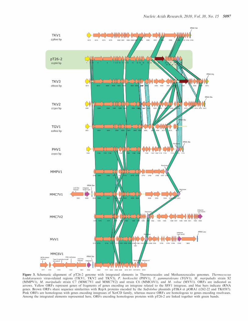
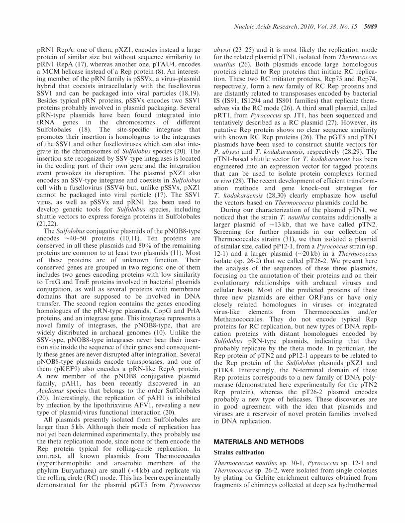
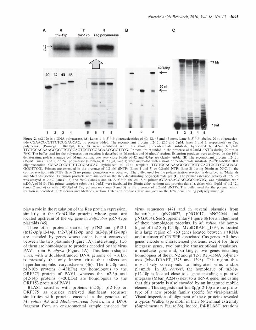
Figure 3. Schematic alignment of pT26-2 genome with integrated elements in Thermococcales and Methanococcales genomes. Thermococcuskodakaraensis virus-related regions (TKV1, TKV2 and TKV3), P. horikoschii (PHV1), T. gammatolerans (TGV1), M. maripaludis strain S2(MMPV1), M. maripaludis strain C7 (MMC7V1 and MMC7V2) and strain C6 (MMC6V1), and M. voltae (MVV1). ORFs are indicated asarrows. Yellow ORFs represent genes of fragments of genes encoding an integrase related to the SSV1 integrase, and blue bars indicate tRNAgenes. Brown ORFs share sequence similarities with RepA proteins encoded by the Sulfolobus plasmids pTIK4 et pORA1 (t262-22 and TK0587).Pink ORFs are homologous with genes encoding integrases of XerCD family, whereas mauve ORFs are homologous to genes encoding resolvases.Among the integrated elements represented here, ORFs encoding homologous proteins with pT26-2 are linked together with green bands.
Nucleic Acids Research, 2010, Vol. 38, No. 15 5097
polymerase and a pT26-2 type helicase observed in aplasmid/virus integrated in the genome of a methanogenillustrates the domain swapping that has occurred betweenpolymerase and helicase domains in the course of plasmidevolution.As in the case of the pTN2 and pP12-1 Rep proteins, the
gene encoding the t26-22p putative helicase of pT26-2 islocated just upstream of the large intergenic regionidentified as the putative replication origin of thisplasmid by GC skew analysis (Figure 1B andSupplementary Figure S1). We have expressed in E. colithe protein t26-22p but the protein turned out to be insol-uble, preventing us to test its activity.Finally, the fourth pT26-2 protein with predictable bio-
chemical function, t26-31p, belongs to a huge family ofserine recombinases widespread in Archaea and Bacteriabut missing in Eukarya (51). The most closely relatedhomologues of t26-31p are found in archaeal genomes,both in Euryarchaeota and in Crenarchaeota.Homologues in Thermococcales and Methanococcalesare present within the integrated elements (virus-like)related to pT26-2 (see below).Several putative pT26-2 proteins have no clear-cut
homologue with known function in databases but gaveinteresting hits in BLAST analyses. The protein t26-18pexhibits some weak similarities with bacterial proteinsannotated as TraG (plasmid transfer factors) suggestinga role in DNA transfer (Supplementary Figure S9). Theprotein t26-20p, located immediately adjacent to theputative replication origin, exhibits similarities with tran-scription factors of the SpoVT/AbrB family (52), suggest-ing that it could play a role in plasmid copy numberregulation. Several proteins of pT26-2, whose genes areclustered in one half of the plasmid, contain hydrophobicdomains (Figure 1B), suggesting that they could bemembrane bound proteins or part of complex architec-tural elements. These include two large proteins (t26-5pand t26-6p, 68 and 81 kDa, respectively) shared bypT26-2 and all related elements integrated in thegenomes of Thermococcales and Methanococcales (seebelow). Analysis of t26-5p using Interproscan detected acentral domain annotated as carboxypeptidase regulatorydomain and two short putative hydrophobic domains atboth extremities. This corresponds to the description ofmetalloproteases of the SSF49464 family that exhibit acentral carboxypeptidase regulatory domain surroundedby two transmembrane domains. All these observationssuggest that t26-5p could be a membrane protease thatcan hydrolyse glycoproteins. The neighbouring proteint26-6p contains five short hydrophobic domains (three inthe N-terminal part and two in the C-terminal part).We have recently solved the structure of a recombinantt26-6p protein lacking these hydrophobic domains (53).The protein has an elongated shape and is composed ofthree domains which all correspond to novel folds.Interestingly, homologues of the putative membraneprotein t26-13p are located immediately upstream thoseof the putative ATPase t26-14p (sharing weak similaritieswith RuvB helicase) in all integrated elements related topT26-2. This suggests that t26-14p and t26-13p couldwork together to pump DNA through the membrane,
and raises the possibility that pT26-2 is either aconjugative plasmid or encodes remnants of a viral appar-atus for DNA injection and/or packaging. These proteinscould also work together with the putative ATPaset26-18p which, as was previously mentioned, exhibitsweak similarities with plasmid transfer factors(Supplementary Figure S9).
A family of pT26-2 related elements in genomes ofThermococcales and Methanococcales
Clusters of genes encoding homologues of several pT26-2proteins are present in the genomes of severalThermococcales and Methanococcales species (Figure 3).Two of these clusters are located in the previouslydescribed TKV2 and TKV3 virus-like elements fromT. kodakaraensis (49) and TGV1 from T. gammatolerans(40). We identified a new closely related integratedvirus-like element (IE) in the genome of P. horikoshii(thereafter named PHV1). The plasmid pT26-2 shares20, 19, 17 and 17 homologous genes with TKV3, TKV2,TGV1 and PHV1, respectively (Figure 3). In all cases, the30-end of elements are located adjacent to a tRNA geneand the two extremities of the elements are bordered bythe two parts of a splitted gene encoding the SSV-typeintegrase homologous to the pT26-2 integrase. TheN-terminal moieties of these genes are located in 30 ofthe elements (adjacent to the tRNA gene) whereas theirC-terminal moieties are located in 50, as expected for inte-gration mediated by SSV1-type integrases. We alsoidentified clusters of genes homologous to pT26-2 inthe genomes of several mesophilic Methanococcales(M. maripaludis strains S2, C6 and C7, M. voltae,Figure 3). Importantly, the syntheny of genes homologousbetween pT26-2 and integrated elements inThermococcales is maintained in clusters present inMethanococcales. As illustrated in SupplementaryFigure S11A, both in Thermococcales andMethanococcales, these clusters (horizontal green bars)are located in larger regions (containing between �40and 100 ORFs) that contain many atypical genes(Supplementary Figure S11A, vertical blue bars), i.e.genes whose sequence composition differ from those ofgenes conserved in the genome of all Methanococcales(invisible white bars in the figure) [see ref. (41) for thedefinition of clusters of atypical genes (CAGs)]. Theseregions include integrase genes (black arrows), tRNAgenes (vertical pink bars) and serine recombinases(resolvases) related to those of pT26-2. All these observa-tions indicate that the clusters of pT26-2 homologousgenes in the genomes of Thermococcales andMethanococcales are located within bona fide integrated‘virus-like elements’. These integrated elements will becalled thereafter MMPV1, MMC6V1, MMC7V1,MMC7V2 and MMV1 to follow the nomenclatureadopted for IE from Thermococcales. As previously men-tioned, MMC7V1 and MMC6V1 IE include a geneencoding a pTN2-type helicase. In addition, MMC6V1also include a homologue of the cellular replicativehelicase MCM.
5098 Nucleic Acids Research, 2010, Vol. 38, No. 15
The plasmid pT26-2 also harbours seven genes thathave homologues in TKV1, a third integrated element ofT. kodakaraensis. Six of them are located in the regionlocated just upstream the C-terminal integrase moiety ofpT26-2 and have homologues in the corresponding regionof pT26-2 related elements present in Thermococcales (butnot in Methanococcales). The genome of pT26-2 andrelated integrated elements can thus be divided in twoparts, a highly conserved 50 region which includes genesthat are present in all integrated elements of the pT26-2family present in Thermococcales, including seven genesalso present in pT26-2 related integrated elements presentin Methanococcales (dubbed core genes thereafter, i.e.homologous of t26-5p-6p-7p-11p-13p-14p and -15p) anda variable 30 region that includes both ORFans and genesof mixed origins (TKV1 and/or TKV2/TKV3). Thissuggests that a relatively ancient recombination eventoccurred between a TKV1-like element and the ancestorof the pT26-2 and TKV2/V3 related elements.
The variable region of pT26-2 includes the genes for theputative helicase t26-22p, the putative transfer proteinst26-18p and t26-20p, and the serine recombinaset26-31p, whereas the core genes include the large
proteins t26-5p and t26-6p. Interestingly, further homo-logues of these last two proteins are encoded in thegenomes of several species of M. maripaludis that lackthe other core genes of the pT26-2 family. In order toget insight into the evolution of the pT26-2 family, wehave performed a phylogenetic analysis of a concatenationof six core proteins (the homologues of the core genet26-7p was removed from this phylogeny because theyinclude several paralogues). The topology of the resultingtree (Supplementary Figure S12) was similar to those ofthe tree that we previously constructed using only the twocore proteins t26-5p and t26-6p (53). As shown inSupplementary Figure S12, family members fromThermococcales and Methanococcales are clearlyseparated. The phylogeny obtained is roughly congruentwith the archaeal species phylogeny if the root is locatedbetween these two orders, as Thermococales andMethanococcales each form monophyletic groups.Among Thermococcales, pT26-2 and integrated elementsfrom Thermococcus species form a monophyletic group,separated from PHV1 of P. horikoshii. These datasuggest that pT26-2 and related integrated elements haveco-evolved with their hosts and diverged from an ancestor
PHV1
MMC7V2
MMPV1MMC6V1
MMC7V1
pT26-2
TKV1
TKV2
TKV3
Nanoarchaeum pharaonis
Methanococcales
Methanosarcinales
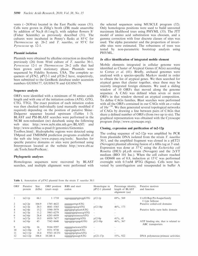
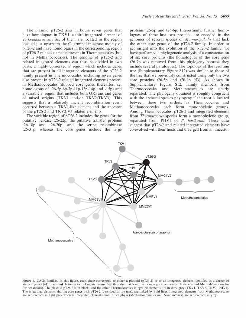
Figure 4. CAGs families. In this figure, each circle correspond to either a plasmid (pT26-2) or to an integrated element identified as a cluster ofatypical genes (41). Each link between two elements means that they share at least five homologous genes (see ‘Materials and Methods’ section forfurther details). The plasmid pT26-2 is in black, and the other Thermococcales integrated elements are in dark grey (TKV1, TKV2, TKV3, PHV1).The integrated elements sharing core genes with pT26-2 (described in the text), are linked by bold lines. Integrated elements from Methanococcalesare represented in light grey whereas integrated elements from other phyla (Methanosarcinales and Nanoarchaea) are represented in grey.
Nucleic Acids Research, 2010, Vol. 38, No. 15 5099
that already infected Archaea before the divergencebetween Methanococcale and Thermococcales.To get clue about possible more distant evolutionary
relationships between members of the pT26-2 family andother plasmids and virus-like elements integrated inarchaeal genomes (previously identified as CAGs, 41),we have draw different networks in which two elementsare linked to each other by a line if they share eitherthree, four or five genes (threshold) and a bold lineif they share the seven core genes of the pT26-2 family.Interestingly, members of the pT26-2 family becomelinked to more and more CAGs or to free plasmidswhen the threshold was reduced from five to two(Figure 4 and S11B-E). Whereas pT26-2 was only linkedto CAGs or plasmids from Thermococcales andMethanococcales with a threshold of five, it became alsolinked to Methanosarcinales with a threshold of four.Finally, when the threshold was relaxed to three, CAGsfrom Thermoplasmatales and Halobacteriales were re-cruited into the network of plasmid/CAG interactionsand several large families are clearly delineated (plasmidsfrom halobacterial were removed from this analysisbecause their high number introduced a bias). Thisanalysis thus defines a huge superfamily of mobileelements loosely related to pT26-2 that encompass thewhole phylum of Euryarchaea. Sulfolobales (phylumCrenarchaea) only entered into the network when thethreshold was reduced to two.
DISCUSSION
All plasmids previously isolated from Thermococales weresmall RC plasmids (23–25,54). Here we report the isola-tion, sequencing and characterization of three newplasmids from Thermococcales that are larger andprobably replicate via the theta mode. The plasmidspTN2 and pP12-1 encode a DNA polymerase fused to adomain of unknown function. These proteins are homolo-gous to the RepA protein of the plasmids pTIK4 fromS. neozelandicus and pXZ1 from S. solfataricus (17) andto proteins encoded by integrated elements present ingenomes of Methanococcales. They form therefore anew family of archaeal RepA proteins common tomobile elements present in both Euryarchaea andCrenarchaea. The DNA polymerase domain of theseRepA proteins have no detectable sequence similaritieswith those of previously known DNA polymerases,including the DNA polymerase discovered by GeorgLipps in pRN1 (14), indicating that these proteins couldbe the prototypes of a new DNA polymerase family. As inthe case of the pRN1 polymerase, the DNA polymerase ofpTN2 exhibits a primase activity (not shown, manuscriptin preparation) and could be used for the initiation as wellas for the elongation step of plasmid DNA replication.The DNA polymerase domain of the Rep proteins ofpTN2 and pP12-1 are linked in C-terminal to a largedomain of unknown function. In pRN-type plasmids,the DNA polymerase domain of the Rep proteins isfused in C-terminal to a helicase domain. This isprobably not the case for pTN2 and pP12-1 since their
C-terminal domain do not contain detectable ATPbinding site. Indeed, the genes encoding the Rep-DNApolymerase in pTN2 and pP12-1 are contiguous to thegenes encoding a putative SF1 helicase that is probablyinvolved, together with the Rep proteins, in plasmid rep-lication. It is tempting to speculate that the C-terminaldomains of the pTN2 and pP12-1 Rep proteins bear anorigin binding activity since the combination of suchactivity with their DNA polymerase/primase activitiesand the unwinding activity of the associated helicase SFIwould reconstitute a complete DNA replication system forthe plasmids.
The plasmid pT26-2 does not seem to encode a DNApolymerase and probably recruits a cellular DNA poly-merase. Interestingly, this plasmid encodes a putativereplicative helicase that would also correspond to a newfamily of replication protein. It is striking that thepolymerase/helicase cassette of pTN2/pP12-1 and theputative replicative helicase of pT26-2 are both locateddownstream of the largest intergenic region present oneach of the three plasmids. These regions are predictedto be replication origins by cumulative GC skew analysisand include many direct and inverted repeats, reminiscentof the ‘iterons’ typical of bacterial plasmid replicationorigins (55).
The three new plasmids of Thermococcales describedhere could become interesting models to study plasmidreplication both in vitro and in vivo in hyperthermophilicarchaea. Furthermore, the identification of their replica-tion cassettes will help to use these plasmids in futurevector construction for T. kodakaraensis, which isbecoming the model species for genetic and molecularstudies in hyperthermophilic archaea (28,30,56). Itshould be interesting for instance to express differentgenes from different compatible plasmids in the samestrain for complementation studies. We already knowthat pTN1 and pTN2 are compatible, since they bothare present in the same strain of T. nautilus (26). Itshould be also possible to use the SSV-type integrase ofpT26-2 to insert groups of foreign genes inThermococcales. Conversely, the genetic tools alreadyavailable for T. kodakaraensis should help to analyse thebiological functions of the proteins encoded by the threeplasmids described here if, as in the case of pTN1, theycan be propagated in T. kodakaraensis.
The plasmid pT26-2 encodes many homologues ofproteins encoded by ‘virus-like elements’ present inThermococcales and Methanococcales, including aSSV-type integrase, thus defining a large family of plasmidand integrated elements that probably predated the separ-ation of these two archaeal orders. The pT26-2 family ischaracterized by the presence of seven ‘core genes’ presentin all members of the family. Whereas pTN2 and pP12-1can be considered as cryptic plasmids, the evolutionaryrelationships between pT26-2 and virus-like elementspresent in Thermococcales (TKV2/3, TGV1, PHV1) andMethanococcales (MMPV1, MMC7V1/2, MVV1,MMC6V1) suggests that pT26-2 plasmid could be aconjugative plasmid or a viral genome (or derived fromsuch elements). Indeed, pT26-2 encode hydrophobicproteins that are clustered in one half of the plasmid,
5100 Nucleic Acids Research, 2010, Vol. 38, No. 15
suggesting that they could be involved in the formation ofprotein complexes involved in DNA transfer, either forconjugation or viral infection. The putative glycoproteaseactivity of the large and conserved membrane proteint26-5p also suggests that pT26-2 and related elementsindeed have the ability to destroy and/or modify the cellenvelope to allow DNA transfer either during conjugationor viral infection. Krupovic and Bamford (57) have shownthat one of the genes present in TKV4 encodes a capsidprotein of the PRD1-adenovirus type (with a double-jellyroll fold), suggesting that TKV4 is a bona fide virus. Incontrast, we could not detect gene encoding putativecapsid proteins in pT26-2 or related elements. We alsodid not detect virus particles in cultures of T. nautilus,or in cultures of the Thermococcales containing relatedelements (54). These strains produce virus-like vesiclesand some intracellular DNA is strongly associated tovesicles of T. gammatolerans (54) but we failed to specif-ically detect DNA from pT26-2 or related elements inthese vesicles, nor proteins encoded by these elements(data not shown).
The origin and mode of evolution of plasmids andviruses, as well as their position in the tree of life,remain controversial topics (58–63). It is sometimesassumed that these biological entities essentially evolveby recruiting cellular genes (62). However, in contradic-tion with this view, homologues of cellular genes areusually rare in viral genomes and plasmids, with the ex-ception of cellular genes that are present in integratedviruses and/or plasmids (58–60). Indeed, none of theproteins encoded by the three plasmids studied here hascellular homologues, except for genes mostly presentwithin plasmid and/or viruses integrated in cellulargenomes. The putative SFI helicases of pTN2 andpP12-1 are homologous to bacterial UvrD-type helicases,however, they are more closely related to putativehelicases present in archaeal genomes which are mostlikely of viral origin (Supplementary Figure S4). This in-dicates that none of the proteins encoded by the threeplasmids studied here correspond to a bona fide cellulargene that has been recently captured from a host cell.
The presence of a high proportion of ORFans amongplasmid and viral genes is another specific feature thatcannot be explained easily in the traditional view ofvirus/plasmid evolution. The question of the origin andthe nature of cellular and viral ORFans has been raisedrepeatedly (64,65). Most of them encode short proteins,and it has been argued sometimes that these ORFs do notencode bona fide proteins. The three plasmids studied herealso encode a relatively large proportion of small ORFs.We can assume that most of them are real genes becausethey are conserved either between pTN2 and pP12-1, orbetween pT26-2 and the virus-like elements present in thegenomes of Thermococcales and Methanococcales. Thecharacterization of a large family of pT26-2 relatedelements present in the genomes of Thermococcales andMethanococcales illustrates our recent observation, madeat larger scale, that viral/plasmid genes represent a largereservoir of genes that constantly invade bacterial andarchaeal genomes (41).
Plasmids and viruses often encode large proteins thatare involved in genome replication or in the formation ofstructural apparatus, such as virions or DNA transferringmachinery. These genes, that have either no cellularhomologues or only distantly related ones can be con-sidered as viral specific proteins (59), or virus hallmarkprotein (60). The three plasmids studied here encodeseveral examples of such proteins. This is the case forinstance of the two large proteins conserved in allmembers of the pT26-2 family (t26-5p and t26-6p).Structural analysis indeed confirmed the uniqueness ofone of them, the protein t26-6p, which contains threenovel folds (53). Many of the large proteins encoded byplasmids and DNA viruses are involved in DNA replica-tion, repair recombination and/or integration, and this isindeed the case in the present study. These proteins areusually conserved in all members of the same plasmid/viral family (as the integrases of the pT26-2 family orthe Rep-DNA polymerases and the putative SFI helicasesof the pTN2 family) but they can be also shared by differ-ent plasmid families (as in the case of the pTN2-typehelicase SFI, the DNA polymerase/primase or else theSSV1-like integrases).To explain the existence of many proteins involved in
DNA replication, repair or recombination among viralspecific proteins, it has been suggested that DNA andDNA-manipulating enzymes originated first in anancient virosphere, and that only a subset of theseproteins were later on transferred into modern cellularlineages (61,66,67). This hypothesis predicts that manynew types and families of viral-specific DNA-manipulating enzymes remain to be discovered in theplasmid/viral world. The present work fulfils this predic-tion since, by studying only three plasmids, we discovereda new family of DNA polymerase and probably a newfamily of helicases. It gives us the exiting expectationthat more new families of enzymes involved in DNA me-tabolism await to be discovered in systematic analyses ofyet uncharacterized proteins encoded by plasmids andviruses.Interestingly, plasmids and related viruses of
Thermococcales studied here have no close homologuesencoded by bacterial or eukaryotic viruses/plasmidsbut only in archaeal plasmids and viruses, thus confirmingthat the three domains have largely independent viralreservoirs (68). The pTN2 and pP12-1 plasmids sharethree genes with the virus PAV1 from P. abyssi, whichreminds the presence of SSV1-like genes in the genomeof the Sulfolobus pRN-type plasmids pSSVx (18). Ahomologue of a PAV1 gene has also been detectedrecently in TKV4 from T. kodakaraensis (57), showingthat gene exchange between PAV1 and plasmids has notbeen limited to the pTN2 family. Finally, the pTN2 andpP12-1 putative SFI helicase is also present in thevirus-like element TGV2 from T. gammatolerans. Allthese observations suggest that viruses and plasmids in-fecting and/or present in Thermococcales share a commonpool of genes that can be shifted from one genome to theother by recombination. The same phenomenon has beenobserved in Bacteria. For instance, Krupovic andBamford (69) have shown that some viruses of the
Nucleic Acids Research, 2010, Vol. 38, No. 15 5101

Corticoviridae family, whose prototype is thebacteriovirus PM2, use a replication machinery ofplasmid origin instead of the viral DNA replicationmachinery of PM2.It is often assumed that the evolution of plasmid and
viruses cannot be reconstructed because they are constant-ly involved in horizontal gene transfer between variouscellular lineages (62). However, a recent phylogenomicanalysis of 16 genomes of T4-related viruses identified aconserved core of 24 genes that all (but one) exhibit asimilar phylogenetic pattern, indicating that these virusesmainly evolved vertically (70). Similarly, phylogeneticanalysis of the core proteins of the pT26-2 family is con-gruent with those of Thermococcales (usingMethanococcales as outgroup, see Supplementary FigureS12). It has been recently observed that viruses and theirhosts exhibit similar biogeographic patterns in genomicstudies focusing on the archaeal species S. islandicus(71). This shows that cells and their associated virusesand plasmids co-evolved at the level of species. Theanalysis of the proteins encoded by the three plasmidsfrom Thermococcales studied here suggests thatco-evolution occurred at the order level as well. Indeed,the most closely related homologues of the pTN2, pP12-1and pT26-2 proteins are mostly found in viruses, plasmidsor integrated elements of Thermococcales. IfThermococcales proteins are removed from the analysis,the most closely related homologues are frequently foundin Methanococcales, which are closely related toThermococcales in phylogenetic trees of Archaea basedon ribosomal proteins or RNA polymerase subunits(38,39). Our large-scale analysis of the relationshipbetween the pT26-2 family and other plasmids orintegrated elements (CAGs) present in Archaea confirmsthis view, since the network of evolutionary interactionsbetween these elements overlaps with the phylogeneticpattern of the archaeal domain (Figure 4 andSupplementary Figure S11B). These results show thatgene transfers between viral, plasmids and cellularlineages have not obliterated the phylogenetic signal thattestifies for their co-evolution with their hosts. This hasimportant implications for the current debate about theinclusion of viruses in the tree of life. Indeed, althoughviruses and plasmids cannot be placed in a tree based onuniversally conserved proteins, they can be probablyincluded as companions in a universal tree of life basedon the evolution of cellular species. We think that furthersystematic analyses of free or integrated plasmids andviruses in all archaeal, bacterial and eukaryotic groupsare now mandatory in order to draw a comprehensivetree of life unifying all types of living entities present inthe biosphere.
ACCESSION NUMBERS
pTN2, pP12-1 and pT26-2 have respectively beensubmitted to the GenBank database under accessionnumbers GU056177, GU056178 and GU056179.
SUPPLEMENTARY DATA
Supplementary Data are available at NAR Online.
FUNDING
Agence Nationale de la Recherche with grant GISGenomique Marine (ANR-05-BLAN-0408-01); PhDgrant from the Ministere de l’Enseignement superieuret de la Recherche to N.S. ARC (Association pour laRecherche contre le Cancer). Funding for open accesscharge: Institut Pasteur.
Conflict of interest statement. None declared.
REFERENCES
1. Lipps,G. (2008) (ed.), Plasmids Current Research and FutureTrends. Caaister Academic Press, Norfolk, UK, p. 259.
2. Harriott,O.T., Huber,R., Stetter,K.O., Betts,P.W. and Noll,K.M.(1994) A cryptic miniplasmid from the hyperthermophilicbacterium Thermotoga sp. strain RQ7. J. Bacteriol., 176,2759–2762.
3. Soppa,J. (2006) From genomes to function: haloarchaea as modelorganisms. Microbiology, 152, 585–590.
4. Baliga,N.S., Bonneau,R., Facciotti,M.T., Pan,M., Glusman,G.,Deutsch,E.W., Shannon,P., Chiu,Y., Weng,R.S., Gan,R.R. et al.(2004) Genome sequence of Haloarcula marismortui: a halophilicarchaeon from the Dead Sea. Genome Res., 14, 2221–2234.
5. del Solar,G., Giraldo,R., Ruiz-Echevarria,M.J., Espinosa,M. andDiaz-Orejas,R. (1998) Replication and control of circular bacterialplasmids. Microbiol. Mol. Biol. Rev., 62, 434–464.
6. Lipps,G. (2008) Archaeal plasmids. In Lipps,G. (ed.), PlasmidsCurrent Research and Future Trends. Caaister Academic Press,Norfolk, UK, pp. 25–50.
7. Lipps,G. (2006) Plasmids and viruses of the thermoacidophiliccrenarchaeote Sulfolobus. Extremophiles, 10, 17–28.
8. Greve,B., Jensen,S., Phan,H., Brugger,K., Zillig,W., She,Q. andGarrett,R.A. (2005) Novel RepA-MCM proteins encoded inplasmids pTAU4, pORA1 and pTIK4 from Sulfolobusneozealandicus. Archaea, 1, 319–325.
9. Lipps,G. (2009) Molecular biology of the pRN1 plasmid fromSulfolobus islandicus. Biochem. Soc. Trans., 37, 42–45.
10. Greve,B., Jensen,S., Brugger,K., Zillig,W. and Garrett,R.A. (2004)Genomic comparison of archaeal conjugative plasmids fromSulfolobus. Archaea, 1, 231–239.
11. Erauso,G., Stedman,K.M., van de Werken,H.J., Zillig,W. and vander Oost,J. (2006) Two novel conjugative plasmids from a singlestrain of Sulfolobus. Microbiology, 152, 1951–1968.
12. Lipps,G., Ibanez,P., Stroessenreuther,T., Hekimian,K. andKrauss,G. (2001) The protein ORF80 from the acidophilic andthermophilic archaeon Sulfolobus islandicus binds highlysite-specifically to double-stranded DNA and represents a noveltype of basic leucine zipper protein. Nucleic Acids Res., 29,4973–4982.
13. Lipps,G., Stegert,M. and Krauss,G. (2001) Thermostable andsite-specific DNA binding of the gene product ORF56 from theSulfolobus islandicus plasmid pRN1, a putative archael plasmidcopy control protein. Nucleic Acids Res., 29, 904–913.
14. Lipps,G., Rother,S., Hart,C. and Krauss,G. (2003) A novel typeof replicative enzyme harbouring ATPase, primase and DNApolymerase activity. EMBO J., 22, 2516–2525.
15. Lipps,G. (2004) The replication protein of the Sulfolobusislandicus plasmid pRN1. Biochem. Soc. Trans., 32, 240–244.
16. Lipps,G., Weinzierl,A.O., von Scheven,G., Buchen,C. andCramer,P. (2004) Structure of a bifunctional DNAprimase-polymerase. Nat. Struct. Mol. Biol., 11, 157–162.
17. Peng,X. (2008) Evidence for the horizontal transfer of anintegrase gene from a fusellovirus to a pRN-like plasmid within asingle strain of Sulfolobus and the implications for plasmidsurvival. Microbiology, 154, 383–391.
5102 Nucleic Acids Research, 2010, Vol. 38, No. 15
18. Arnold,H.P., She,Q., Phan,H., Stedman,K., Prangishvili,D.,Holz,I., Kristjansson,J.K., Garrett,R. and Zillig,W. (1999) Thegenetic element pSSVx of the extremely thermophiliccrenarchaeon Sulfolobus is a hybrid between a plasmidand a virus. Mol. Microbiol., 34, 217–226.
19. Stedman,K.M., She,Q., Phan,H., Arnold,H.P., Holz,I.,Garrett,R.A. and Zillig,W. (2003) Relationships betweenfuselloviruses infecting the extremely thermophilic archaeonSulfolobus: SSV1 and SSV2. Res. Microbiol., 154, 295–302.
20. Basta,T., Smyth,J., Forterre,P., Prangishvili,D. and Peng,X.(2009) Novel archaeal plasmid pAH1 and its interactions with thelipothrixvirus AFV1. Mol. Microbiol., 71, 23–34.
21. Albers,S.V., Jonuscheit,M., Dinkelaker,S., Urich,T., Kletzin,A.,Tampe,R., Driessen,A.J. and Schleper,C. (2006) Production ofrecombinant and tagged proteins in the hyperthermophilicarchaeon Sulfolobus solfataricus. Appl. Environ. Microbiol., 72,102–111.
22. Berkner,S., Grogan,D., Albers,S.V. and Lipps,G. (2007) Smallmulticopy, non-integrative shuttle vectors based on the plasmidpRN1 for Sulfolobus acidocaldarius and Sulfolobus solfataricus,model organisms of the (cren-)archaea. Nucleic Acids Res., 35,e88.
23. Erauso,G., Marsin,S., Benbouzid-Rollet,N., Baucher,M.F.,Barbeyron,T., Zivanovic,Y., Prieur,D. and Forterre,P. (1996)Sequence of plasmid pGT5 from the archaeon Pyrococcus abyssi:evidence for rolling-circle replication in a hyperthermophile.J. Bacteriol., 178, 3232–3237.
24. Marsin,S. and Forterre,P. (1998) A rolling circle replicationinitiator protein with a nucleotidyl-transferase activity encoded bythe plasmid pGT5 from the hyperthermophilic archaeonPyrococcus abyssi. Mol. Microbiol., 27, 1183–1192.
25. Marsin,S. and Forterre,P. (1999) The active site of the rollingcircle replication protein Rep75 is involved in site-specificnuclease, ligase and nucleotidyl transferase activities. Mol.Microbiol., 33, 537–545.
26. Soler,N., Justome,A., Quevillon-Cheruel,S., Lorieux,F., LeCam,E., Marguet,E. and Forterre,P. (2007) The rolling-circleplasmid pTN1 from the hyperthermophilic archaeonThermococcus nautilus. Mol. Microbiol., 66, 357–370.
27. Ward,D.E., Revet,I.M., Nandakumar,R., Tuttle,J.H.,de Vos,W.M., van der Oost,J. and DiRuggiero,J. (2002)Characterization of plasmid pRT1 from Pyrococcus sp. strainJT1. J. Bacteriol., 184, 2561–2566.
28. Santangelo,T.J., Cubonova,L. and Reeve,J.N. (2008) Shuttlevector expression in Thermococcus kodakaraensis: contributionsof cis elements to protein synthesis in a hyperthermophilicarchaeon. Appl. Environ. Microbiol., 74, 3099–3104.
29. Lucas,S., Toffin,L., Zivanovic,Y., Charlier,D., Moussard,H.,Forterre,P., Prieur,D. and Erauso,G. (2002) Construction of ashuttle vector for, and spheroplast transformation of, thehyperthermophilic archaeon Pyrococcus abyssi. Appl. Environ.Microbiol., 68, 5528–5536.
30. Sato,T., Fukui,T., Atomi,H. and Imanaka,T. (2005) Improvedand versatile transformation system allowing multiplegenetic manipulations of the hyperthermophilic archaeonThermococcus kodakaraensis. Appl. Environ. Microbiol., 71,3889–3899.
31. Lepage,E., Marguet,E., Geslin,C., Matte-Tailliez,O., Zillig,W.,Forterre,P. and Tailliez,P. (2004) Molecular diversity of newThermococcales isolates from a single area of hydrothermaldeep-sea vents as revealed by randomly amplified polymorphicDNA fingerprinting and 16S rRNA gene sequence analysis.Appl. Environ. Microbiol., 70, 1277–1286.
32. Edgar,R.C. (2004) MUSCLE: a multiple sequence alignmentmethod with reduced time and space complexity.BMC Bioinformatics, 5, 113.
33. Guindon,S. and Gascuel,O. (2003) A simple, fast, and accuratealgorithm to estimate large phylogenies by maximum likelihood.Syst. Biol., 52, 696–704.
34. Grigoriev,A. (1998) Analyzing genomes with cumulative skewdiagrams. Nucleic Acids Res., 26, 2286–2290.
35. Lobry,J.R. (1996) Asymmetric substitution patterns in the twoDNA strands of bacteria. Mol. Biol. Evol., 13, 660–665.
36. Gorbalenya,A.E. and Koonin,E.V. (1993) Helicases: amino acidsequence comparisons and structure-funciton relationships.Curr. Opin. Struct. Biol., 3, 419–429.
37. Singleton,M.R., Dillingham,M.S. and Wigley,D.B. (2007)Structure and mechanism of helicases and nucleic acidtranslocases. Annu. Rev. Biochem., 76, 23–50.
38. Brochier-Armanet,C., Boussau,B., Gribaldo,S. and Forterre,P.(2008) Mesophilic Crenarchaeota: proposal for a third archaealphylum, the Thaumarchaeota. Nat. Rev. Microbiol., 6, 245–252.
39. Brochier,C., Forterre,P. and Gribaldo,S. (2005) An emergingphylogenetic core of Archaea: phylogenies of transcription andtranslation machineries converge following addition of newgenome sequences. BMC Evol. Biol., 5, 36.
40. Zivanovic,Y., Armengaud,J., Lagorce,A., Leplat,C., Guerin,P.,Dutertre,M., Anthouard,V., Forterre,P., Wincker,P. andConfalonieri,F. (2009) Genome analysis and genome-wideproteomics of Thermococcus gammatolerans, the mostradioresistant organism known amongst the Archaea.Genome Biol., 10, R70.
41. Cortez,D., Forterre,P. and Gribaldo,S. (2009) A hidden reservoirof integrative elements is the major source of recently acquiredforeign genes and ORFans in archaeal and bacterial genomes.Genome Biol., 10, R65.
42. Iyer,L.M., Koonin,E.V., Leipe,D.D. and Aravind,L. (2005) Originand evolution of the archaeo-eukaryotic primase superfamily andrelated palm-domain proteins: structural insights and newmembers. Nucleic Acids Res., 33, 3875–3896.
43. Clark,J.M. (1988) Novel non-templated nucleotide additionreactions catalyzed by procaryotic and eucaryotic DNApolymerases. Nucleic Acids Res., 16, 9677–9686.
44. Mole,S.E., Iggo,R.D. and Lane,D.P. (1989) Using the polymerasechain reaction to modify expression plasmids for epitopemapping. Nucleic Acids Res., 17, 3319.
45. Berkner,S. and Lipps,G. (2007) Characterization of thetranscriptional activity of the cryptic plasmid pRN1 fromSulfolobus islandicus REN1H1 and regulation of its replicationoperon. J. Bacteriol., 189, 1711–1721.
46. Geslin,C., Gaillard,M., Flament,D., Rouault,K., LeRomancer,M., Prieur,D. and Erauso,G. (2007) Analysis of thefirst genome of a hyperthermophilic marine virus-like particle,PAV1, isolated from Pyrococcus abyssi. J. Bacteriol., 189,4510–4519.
47. Andersson,A.F. and Banfield,J.F. (2008) Virus populationdynamics and acquired virus resistance in natural microbialcommunities. Science, 320, 1047–1050.
48. Bernad,A., Blanco,L., Lazaro,J.M., Martin,G. and Salas,M.(1989) A conserved 30-50 exonuclease active site in prokaryoticand eukaryotic DNA polymerases. Cell, 59, 219–228.
49. Fukui,T., Atomi,H., Kanai,T., Matsumi,R., Fujiwara,S. andImanaka,T. (2005) Complete genome sequence of thehyperthermophilic archaeon Thermococcus kodakaraensis KOD1and comparison with Pyrococcus genomes. Genome Res., 15,352–363.
50. Sanchez,M., Drechsler,M., Stark,H. and Lipps,G. (2009) DNAtranslocation activity of the multifunctional replication proteinORF904 from the archaeal plasmid pRN1. Nucleic Acids Res., 37,6831–6848.
51. Grindley,N.D., Whiteson,K.L. and Rice,P.A. (2006) Mechanismsof site-specific recombination. Annu. Rev. Biochem., 75, 567–605.
52. Asen,I., Djuranovic,S., Lupas,A.N. and Zeth,K. (2009) Crystalstructure of SpoVT, the final modulator of gene expressionduring spore development in Bacillus subtilis. J. Mol. Biol., 386,962–975.
53. Keller,J., Leulliot,N., Soler,N., Collinet,B., Vincentelli,R.,Forterre,P. and van Tilbeurgh,H. (2009) A protein encoded by anew family of mobile elements from Euryarchaea exhibits threedomains with novel folds. Protein Sci., 18, 850–855.
54. Soler,N., Marguet,E., Verbavatz,J.M. and Forterre,P. (2008)Virus-like vesicles and extracellular DNA produced byhyperthermophilic archaea of the order Thermococcales.Res. Microbiol., 159, 390–399.
55. Chattoraj,D.K. (2000) Control of plasmid DNA replication byiterons: no longer paradoxical. Mol. Microbiol., 37, 467–476.
Nucleic Acids Research, 2010, Vol. 38, No. 15 5103
56. Santangelo,T.J., Cubonova,L., Matsumi,R., Atomi,H., Imanaka,T.and Reeve,J.N. (2008) Polarity in archaeal operon transcription inThermococcus kodakaraensis. J. Bacteriol., 190, 2244–2248.
57. Krupovic,M. and Bamford,D.H. (2008) Archaeal provirusesTKV4 and MVV extend the PRD1-adenovirus lineage to thephylum Euryarchaeota. Virology, 375, 292–300.
58. Forterre,P. (2006) The origin of viruses and their possible roles inmajor evolutionary transitions. Virus Res., 117, 5–16.
59. Forterre,P. (2005) The two ages of the RNA world, and thetransition to the DNA world: a story of viruses and cells.Biochimie, 87, 793–803.
60. Koonin,E.V., Senkevich,T.G. and Dolja,V.V. (2006) The ancientVirus World and evolution of cells. Biol. Direct, 1, 29.
61. Forterre,P. and Prangishvili,D. (2009) The origin of viruses.Res. Microbiol., 160, 466–472.
62. Moreira,D. and Lopez-Garcia,P. (2009) Ten reasons to excludeviruses from the tree of life. Nat. Rev. Microbiol., 7, 306–311.
63. Raoult,D. and Forterre,P. (2008) Redefining viruses: lessons fromMimivirus. Nat. Rev. Microbiol., 6, 315–319.
64. Yin,Y. and Fischer,D. (2006) On the origin of microbial ORFans:quantifying the strength of the evidence for viral lateral transfer.BMC Evol. Biol., 6, 63.
65. Yin,Y. and Fischer,D. (2008) Identification and investigation ofORFans in the viral world. BMC Genomics, 9, 24.
66. Forterre,P. (2002) The origin of DNA genomes andDNA replication proteins. Curr. Opin. Microbiol., 5, 525–532.
67. Forterre,P. (1999) Displacement of cellular proteins by functionalanalogues from plasmids or viruses could explain puzzlingphylogenies of many DNA informational proteins.Mol. Microbiol., 33, 457–465.
68. Prangishvili,D., Forterre,P. and Garrett,R.A. (2006) Viruses ofthe Archaea: a unifying view. Nat. Rev. Microbiol., 4, 837–848.
69. Krupovic,M. and Bamford,D.H. (2007) Putative prophagesrelated to lytic tailless marine dsDNA phage PM2 arewidespread in the genomes of aquatic bacteria. BMC Genomics,8, 236.
70. Filee,J., Tetart,F., Suttle,C.A. and Krisch,H.M. (2005) MarineT4-type bacteriophages, a ubiquitous component of the darkmatter of the biosphere. Proc. Natl Acad. Sci. USA, 102,12471–12476.
71. Held,N.L. and Whitaker,R.J. (2009) Viral biogeography revealedby signatures in Sulfolobus islandicus genomes.Environ. Microbiol., 11, 457–466.
5104 Nucleic Acids Research, 2010, Vol. 38, No. 15
Copyright © 2022 FDOKUMEN