Chapter 22. Genetic diseases reveal DNA nucleotide excision ...
Upload
universiteitutrechtCategory
view
1download
0
letters
Tuning of chaperoneactivity of Hsp70 proteinsby modulation ofnucleotide exchangeDirk Brehmer1, Stefan Rüdiger1,2, Claudia S. Gässler1,Dagmar Klostermeier3,4, Lars Packschies4, Jochen Reinstein4, Matthias P. Mayer1 and Bernd Bukau1
1Institut für Biochemie und Molekularbiologie, Albert-Ludwigs-UniversitätFreiburg, Hermann-Herder-Str. 7, D-79104 Freiburg, Germany. 2CambridgeCentre for Protein Engineering, MRC Centre, Hills Road, Cambridge CB22QH, UK. 3The Scripps Research Institute, Department of Molecular Biology,MB-19, 10550 North Torrey Pines Road, La Jolla, California 92037, USA.4Max-Planck-Institut für molekulare Physiologie, Abteilung physikalischeBiochemie, Otto-Hahn-Str. 11, D-44227 Dortmund, Germany.
The Hsp70 chaperone activity in protein folding is regulatedby ATP-controlled cycles of substrate binding and release.Nucleotide exchange plays a key role in these cycles by trig-gering substrate release. Structural searches of Hsp70homologs revealed three structural elements within theATPase domain: two salt bridges and an exposed loop.Mutational analysis showed that these elements control thedissociation of nucleotides, the interaction with exchangefactors and chaperone activity. Sequence variations in thethree elements classify the Hsp70 family members into threesubfamilies, DnaK proteins, HscA proteins and Hsc70 pro-teins. These subfamilies show strong differences innucleotide dissociation and interaction with the exchangefactors GrpE and Bag-1.
Hsp70 chaperones undergo ATP-induced conformationalchanges that determine their affinity for substrates1,2. ATP boundto the N-terminal ATPase domain lowers the affinity of Hsp70for substrates, presumably through opening of the C-terminalsubstrate binding cavity. ATP hydrolysis populates the closedstate of the substrate binding cavity, thereby trapping associatedsubstrates3,4. This step is regulated by DnaJ cochaperones thatcouple ATP hydrolysis with substrate binding. ADP release israte-limiting for substrate release. ADP dissociation is furtherdecreased by physiological concentrations of Pi. ATP binds to thenucleotide-free chaperone through rapid initial association(step 1, initial encounter complex, DnaK*–ATP), followed byslow isomerization (step 2, tightly bound complex, DnaK–ATP),which is kinetically coupled to substrate release5–9.
step 1 step 2DnaK + ATP DnaK*–ATP DnaK–ATP
nature structural biology • volume 8 number 5 • may 2001 427
For the Escherichia coli homolog, DnaK, ADP dissociation isstrongly stimulated up to 5,000-fold by its cochaperone GrpEacting as a nucleotide exchange factor10,11. It has been debatedwhether members of the Bag protein family are nucleotideexchange factors for eukaryotic Hsp70/Hsc7012,13. Bag proteinsare a heterogeneous family of multidomain proteins that sharethe Bag domain and are implicated in the nucleotide exchangereaction14. Bag proteins such as Bag-1 play roles in the regulationof signal transduction proteins, transcription factors and prote-olysis. In a more general context, the extent of how thenucleotide exchange reaction and its regulation by exchange fac-tors is subjected to evolutionary variation within the Hsp70 fam-ily remains unclear. In order to dissect the nucleotide exchangereaction and its variation among Hsp70 homologs, we measuredthe nucleotide exchange kinetics of homologs representing threeHsp70 subfamilies (see below): DnaK and HscA (Hsc66) of E. coli and human Hsc70.
Variation in nucleotide dissociation ratesWe measured nucleotide dissociation rates directly, without orwith physiological concentrations of P1 (20 mM), using fluores-cent labeled N8-(4N′-methylanthraniloylaminobutyl)-8 amino-adenosine 5′-di/triphosphate (MABA-ADP and MABA-ATP).These analogs have affinities and kinetic properties similar tothose of the authentic nucleotide interacting with DnaK5, HscAand Hsc70 (not shown). Some kinetic properties were alreadyestablished earlier and are similar to those determined here5,7–9,15.Commonly, all three proteins had dissociation rates for
Fig. 1 Variability in nucleotide dissociation rates and utilization ofexchanges factors. a, Time courses of dissociation of MABA-ADP andMABA-ATP from E. coli DnaK, E. coli HscA and human Hsc70 in the pres-ence of an excess of ATP and Pi . b, Influence of GrpE on dissociation ofMABA-ADP–DnaK and MABA-ATP–DnaK (initial encounter (step 1) andtightly bound (step 2)), shown as half-lives (s). c, Influence of BAG-1M ondissociation of MABA-ADP–Hsc70 and MABA-ATP–Hsc70 (step 1, step 2)complexes shown as half lives. d, Comparison of the maximal dissocia-tion rates (s–1) of MABA-ADP–DnaK by GrpE (taken from ref. 11) andMABA-ADP–Hsc70 by BAG-1M. Maximal MABA-ADP–Hsc70 dissociationrates were obtained by measuring the koff (MABA-ADP) from Hsc70 withincreasing amounts of BAG-1M
db
a c
©20
01 N
atu
re P
ub
lish
ing
Gro
up
h
ttp
://s
tru
ctb
io.n
atu
re.c
om
© 2001 Nature Publishing Group http://structbio.nature.com

letters
MABA-ATP consistent with the biphasic association of MABA-ATP. Since the association of MABA-ATP is at least biphasic, onemay assume that ATP association and dissociation follow thesame kinetic route. Inorganic phosphate (20 mM) inhibitedMABA-ADP dissociation of all three proteins by ~10-fold. Largedifferences exist between the absolute values of dissociationrates, with DnaK having the lowest and HscA the highest rates.The half life of the complex of DnaK with MABA-ADP+Pi is 495 s, whereas for Hsc70 it is 22-fold shorter and for HscA 700-fold shorter. The same trend exists for both dissociation reac-tions of MABA-ATP (Fig. 1, Table 1 for rates with Pi). Thenucleotide dissociation reaction kinetics vary strongly within theHsp70 family.
Selective utilization of nucleotide exchange factorsWe investigated whether the homologs also differ in the interac-tion with exchange factors. As part of these experiments wedetermined whether the human Bag-1 homolog, Bag-1M, acts asnucleotide exchange factor for Hsc70, and whether GrpE andBag-1M exhibit chaperone specificity. Gel filtration revealed thatGrpE formed complexes with DnaK (GrpE2–DnaK) but not withHscA and Hsc70. Instead, Bag-1M formed complexes withHsc70 but not with DnaK and HscA (not shown).
Stopped-flow measurements revealed that GrpE stimulatednucleotide dissociation of DnaK but not of HscA and Hsc70. Atfour-fold molar excess over DnaK, GrpE stimulated dissociationof preformed DnaK–MABA-ADP complexes in the presence ofPi by 1,100-fold, consistent with earlier findings11. Interestingly,GrpE also strongly stimulated dissociation of MABA-ATP fromDnaK by acting on both kinetic steps (33- and 191-fold for steps1 and 2, respectively) (Fig. 1b). Therefore, GrpE does not distin-guish the ADP-and ATP-bound states of DnaK.
Bag-1M only stimulated ADP dissociation from Hsc70 but notfrom DnaK and HscA (Table 1). In contrast to GrpE, Bag-1M didnot stimulate dissociation of ATP (Fig. 1c). To determine itsmaximum potential as an exchange factor, we titrated increasingBag-1M to a constant amount of pre-formed Hsc70–MABA-ADP complexes. Dissociation of the complexes was stimulatedmaximally by 110-fold (26.8 s–1). Bag-1M is therefore a potentADP exchange factor for Hsc70, and Bag-1M and GrpE exhibitstrict selectivity for their Hsp70 partner.
Structural differences define three Hsp70 subfamiliesWe searched for structural elements in the ATPase domains of thethree homologs that may account for the differences in nucleotide
428 nature structural biology • volume 8 number 5 • may 2001
exchange. We modeled the ATPase domains of 12 Hsp70homologs (DnaK, HscA of E. coli and Haemophilus influencae;Ssc1, Ssa1, Ssb1 and Kar2 of Saccharomyces cerevisiae; mtHsp70,BiP of Mus musculus; Hsc70 of Bos taurus and Homo sapiens) ontwo atomic structures: the nucleotide-free ATPase domain ofDnaK with open nucleotide binding cleft in complex with GrpE16
and the ADP-bound ATPase domain of bovine Hsc70 with aclosed cleft17. Although the modeled structures were almost com-pletely superimposable, they differed in two previously unnoticedregions that allowed classification of the 12 homologs into threesubfamilies with E. coli DnaK, E. coli HscA and human Hsc70 asprototypes. Sequence alignment of ∼ 100 Hsp70 homologs con-firmed that the entire Hsp70 family can be classified accordingly(not shown).
The first region is in an exposed loop in subdomain IIB nearthe nucleotide binding cleft (Fig. 2a). DnaK subfamily membersshare a particularly long loop (Ala 276–Arg 302 in E. coli DnaK)with subfamily-specific sequence and the ability to cooperatewith GrpE homologs. Only a short segment of the loop (Pro 284–Ile 286) is close to GrpE in the GrpE2–DnaK struc-ture16. Given its positioning near the cleft, we speculated that itaffects nucleotide exchange. Members of the Hsc70 subfamilyshare a loop with subfamily-specific sequence whose tip is fourresidues shorter, whereas members of the HscA subfamily share aless conserved, 10-residue shorter loop.
The second region is at the interface of the nucleotide bindingcleft. In DnaK, we identified three conserved elements that maystrengthen the interface and close the cleft: a hydrophobic patch(Met 259–Val 59 of DnaK) at the top of the cleft and two putativesalt bridges (Glu 264–Arg 56, upper; Glu 267–Lys 55, lower) thatare mainly responsible for the opposite charge distribution and,thereby, for the polarity of the interface (Fig. 2c,d). Interestingly,residues that constitute the hydrophobic patch and the upper saltbridge (Glu 264–Arg 56) seem to interact with GrpE16. GrpEloosens the ATPase interface by interfering with the hydrophobiccontact and the salt bridges (Fig. 2c). In contrast, Hsc70 proteinslack a DnaK-like hydrophobic patch and the upper salt bridge,whereas HscA proteins lack all three elements. The classificationof Hsp70 proteins based on the loop structure tightly correlateswith the classification based on the three elements forming thesubdomain interface.
Loop and salt bridges control nucleotide dissociationTo investigate their roles in nucleotide dissociation, we engi-neered DnaK variants step-by-step into Hsc70 and HscA with
Table 1 Hsp70 homologs differ in nucleotide exchange rates and exchange factors1,2
koff(ADP) (s–1) koff(ATP) (s–1) koff(ATP) (s–1)step 1 step 2
–GrpE +GrpE –GrpE +GrpE –GrpE +GrpEDnaK 0.0014 (495) 1.55 (0.5) 0.013 (54) 0.402 (1.7) 0.00077 (900) 0.15 (5)Hsc70 0.0305 (23) 0.0307 (23) 0.214 (3.2) nd3 nd3 0.0059 (117) nd3 nd3
HscA 0.98 (0.7) 0.97 (0.7) 1.13 (0.6) nd3 nd3 0.33 (2) nd3 nd3
–BAG-1M +BAG-1M –BAG-1M +BAG-1M –BAG-1M +BAG-1MDnaK 0.0014 (495) 0.0014 (424) 0.013 (54) nd3 nd3 0.00077 (900) nd3 nd3
Hsc70 0.0305 (23) 0.32 (2.2) 0.214 (3.2) 0.174 (3.9) 0.0059 (117) 0.0052 (133)HscA 0.98 (0.7) 0.98 (0.7) 1.13 (0.6) nd3 nd3 0.33 (2) nd3 nd3
1All rates represent at least six independent measurements in the presence of 20 mM Pi. Ratios of DnaK/Hsc70/HscA to GrpE and DnaK/Hsc70/HscA toBAG-1M were 1:4 and 1:2.2All values in parenthesis are the corresponding half lives in s.3nd, not determined.
©20
01 N
atu
re P
ub
lish
ing
Gro
up
h
ttp
://s
tru
ctb
io.n
atu
re.c
om
© 2001 Nature Publishing Group http://structbio.nature.com

letters
nature structural biology • volume 8 number 5 • may 2001 429
9.3-fold, respectively, compared to wild type DnaK (495 s).Alterations in the loop also decreased its half life, by two-foldfor DnaK-Loop70 and four-fold for DnaK-LoopA. Strongerchanges occurred where the loop and salt bridge alterationswere combined, with half lives decreasing three-fold forDnaK-Loop70/R56A (Hsc70-type) and 45-fold for DnaK-LoopA/K55A/R56A (HscA-type) (Fig. 3a). Both salt bridgesand the loop therefore contribute individually and act syner-gistically to stabilize the DnaK–MABA-ADP complex. Thehydrophobic contact is probably responsible for further five-fold stabilization, since the complex of MABA-ADP withHsc70 (lacking this contact) is about five-fold less stable than
respect to the loop and salt bridges (DnaK-R56A; DnaK-K55A;DnaK-R56A/K55A; DnaK-Loop70; DnaK-Loop70/R56A;DnaK-LoopA; DnaK-LoopA/K55A/R56A). Because these alter-ations occur naturally within the Hsp70 family, they were notexpected to perturb the local structure and the coupling mecha-nism between the ATPase and substrate binding domains. Theseassumptions were supported by circular dichroism, proteolysisand tryptophan fluorescence (data not shown).
The mutations had additive effects on nucleotide exchangeof DnaK. For MABA-ADP, disruption of the upper (DnaK-R56A) or lower (DnaK-K55A) or both (DnaK-R56A/K55A)salt bridges decreased the half life of the complex by 2-, 6- and
a b c
d
Fig. 2 Structural differences in the ATPase subdomains. a, Superposition of the models of the ATPase domain of 12 Hsp70s. The exposed loop (Ala 276–Arg 302) in subdomain IIB and the interface of the nucleotide binding cleft, built up by Helix I (Gly 51–Pro 62) and Helix II (Asp 255–Lys 270),classify Hsp70 homologs into three subfamilies. Shown are four DnaK (green), six Hsc70 (red) and two HscA (blue) subfamily members. Green arrowspoint to Ala 290 of DnaK that was exchanged for cysteine and labeled with MIANS and to Trp 102 that was used for fluorescence energy transferexperiments. b, Detailed view of the interface of the nucleotide binding cleft of E. coli DnaK in the ADP-bound conformation (modeled based on thestructure of the ATPase domain of B. taurus, cocrystallized with ADP17), that visualizes the two putative salt bridges Glu 267–Lys 55 and Glu 264–Arg 56.c, Position of the hydrophobic patch of residues M 259–Val 59, and the salt bridges Glu 264–Arg 56 and Glu 267–Arg 55 within the crystal structureof the ATPase domain of DnaK in open state cocrystallized with GrpE16. d, Sequence alignment classifies Hsp70 family members according to identi-fied structural differences in the ATPase domain. Sequences of the ATPase subdomain interfaces and loop segments of Hsp70 homologs were alignedaccording to the structures of B. taurus Hsc70 and E. coli DnaK. The evolutionary occurrence of the E. coli DnaK salt bridges Glu 267–Lys 55 andGlu 264–Arg 56 and hydrophobic interaction (M259–V59; black arrowheads) are visualized. The tip of the exposed loop (T287–G292) and its flankingregions (P284–I286, P292–H295), which are highly homologous within the DnaK subfamily, were termed GrpE signature motif. Eco: E. coli,Bsu: Bacillus subtilis, Mma: Methanosarcina mazei, Psa: Pisum sativum, Sce: S. cerevisiae, Ath: Arabidopsis thaliana Hsa: H. sapiens, Hin: H. influenzae,Pae: Pseudomonas aeruginosa, Avi: Acetobacter vinelandii, Bap: Buchneria aphidicola. The ‘asterick’ shows identity and ‘colon’ indicates similaritywithin the three subgroups. Structures made by Insight II and Webviewer 3.7. Sequence alignments and structure modeling were performed usingClustal W and SWISS-Model27–29.
©20
01 N
atu
re P
ub
lish
ing
Gro
up
h
ttp
://s
tru
ctb
io.n
atu
re.c
om
© 2001 Nature Publishing Group http://structbio.nature.com
letters
that complexed with DnaK-Loop70/R56A. Furthermore,Glu 267 of DnaK forms a hydrogen bond to the ribose of thenucleotide and may thereby stabilize the complex17. Thisresidue is entirely conserved within the DnaK and Hsc70 sub-families but is missing within the HscA subfamily, which mayaccount for the even lower stability of the MABA-ADP com-plex with HscA.
For MABA-ATP dissociation, we analyzed the DnaK loopmutants with or without salt bridges (DnaK-Loop70, DnaK-Loop70/R56A, DnaK-LoopA and DnaK-LoopA/K55A/R56A). The alteration primarily affected the initial encounterDnaK*–ATP complex (Fig. 3b). Introduction of the Hsc70-typeloop in the presence (DnaK-Loop70) or absence (DnaK-Loop70/R56A) of the upper salt bridge decreased its half lifeby 1.3- and 4.9-fold, respectively, compared to wild type DnaK.Introduction of the HscA-type loop in the presence (DnaK-LoopA)or absence (DnaK-LoopA/R56A-K55A) of both salt bridgesdecreased its half life by 6- and 56-fold, respectively. Thus, the loopand salt bridges act synergistically to prevent the dissociation of ini-tially bound ATP (step 1). These elements also act during ATP asso-ciation as they shift the equilibrium to the tightly bound DnaK-ATPstate by stabilizing the transition from the nucleotide-free form tothe loosely bound state (initial encounter DnaK*–ATP complex).
Our results show that the loop and salt bridges constitute a
430 nature structural biology • volume 8 number 5 • may 2001
trap allowing rapid association of ATP and slow dissociation ofATP and ADP ± Pi. Mutation of these elements converted DnaKinto Hsc70 and HscA with respect to nucleotide exchange. Thesalt bridges and the hydrophobic contacts might function totightly close the nucleotide binding cleft. The role of the loop isless obvious. Given that the loop is exposed to the solvent, wespeculate that it may reach over to subdomain IB of the ATPasedomain of DnaK, thereby acting as a latch.
To support this hypothesis we performed fluorescence reso-nance energy transfer (FRET) experiments using Trp 102 in sub-domain IB (Fig. 2a) as donor and 2-(4′-maleimidylanilino)naphthalene-6-sulfonic acid (MIANS) linked to a cysteine intro-duced at position 290 (DnaK-A290C) at the tip of the exposedloop as acceptor. To avoid the known interference of the sub-strate binding domain with tryptophan fluorescence18, we usedthe isolated ATPase domain of DnaK for the FRET experiments.In the presence of MIANS, fluorescence of Trp 102 decreased at344 nm compared to the unlabeled protein, while a fluorescencesignal appeared at 424 nm (acceptor). The acceptor fluorescencesignal was higher in the presence of ADP and ATP than in theabsence of nucleotide. These data are consistent with ourhypothesis of a nucleotide-dependent change of distancebetween the position 290 in the exposed loop and Trp 102 insubdomain IB (Fig. 2a).
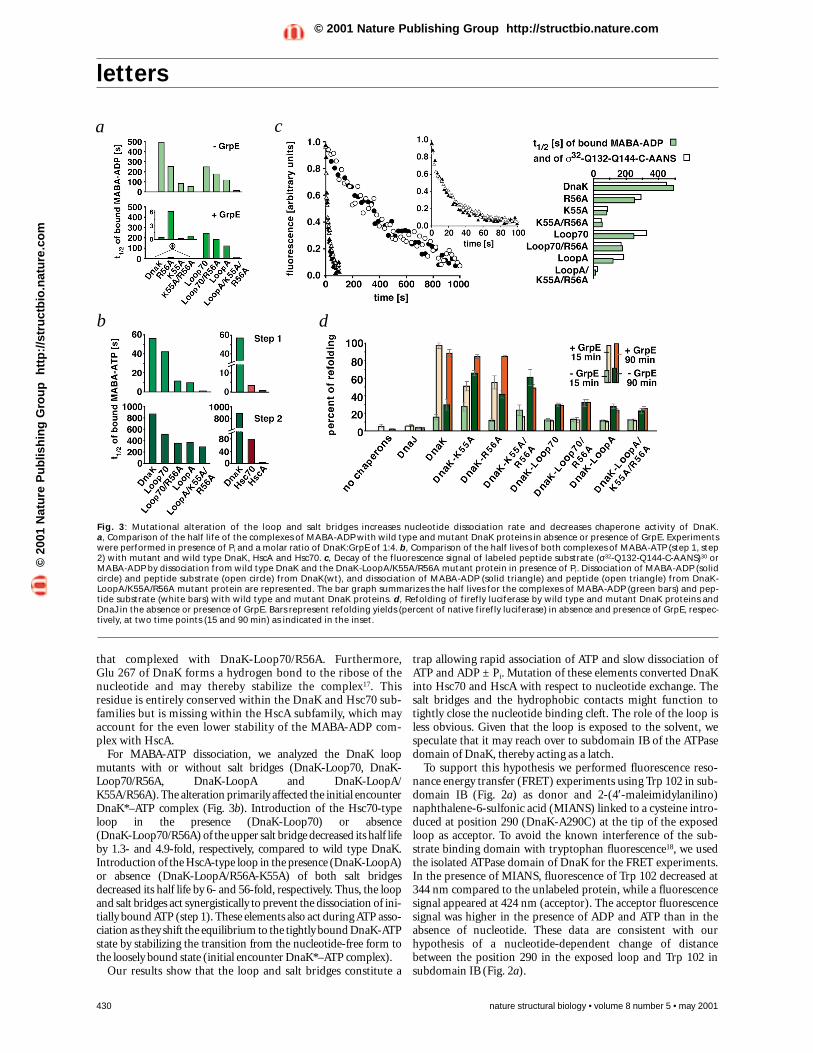
Fig. 3: Mutational alteration of the loop and salt bridges increases nucleotide dissociation rate and decreases chaperone activity of DnaK. a, Comparison of the half life of the complexes of MABA-ADP with wild type and mutant DnaK proteins in absence or presence of GrpE. Experimentswere performed in presence of Pi and a molar ratio of DnaK:GrpE of 1:4. b, Comparison of the half lives of both complexes of MABA-ATP (step 1, step2) with mutant and wild type DnaK, HscA and Hsc70. c, Decay of the fluorescence signal of labeled peptide substrate (σ32-Q132-Q144-C-AANS)30 orMABA-ADP by dissociation from wild type DnaK and the DnaK-LoopA/K55A/R56A mutant protein in presence of Pi. Dissociation of MABA-ADP (solidcircle) and peptide substrate (open circle) from DnaK(wt), and dissociation of MABA-ADP (solid triangle) and peptide (open triangle) from DnaK-LoopA/K55A/R56A mutant protein are represented. The bar graph summarizes the half lives for the complexes of MABA-ADP (green bars) and pep-tide substrate (white bars) with wild type and mutant DnaK proteins. d, Refolding of firefly luciferase by wild type and mutant DnaK proteins andDnaJ in the absence or presence of GrpE. Bars represent refolding yields (percent of native firefly luciferase) in absence and presence of GrpE, respec-tively, at two time points (15 and 90 min) as indicated in the inset.
a c
b d
©20
01 N
atu
re P
ub
lish
ing
Gro
up
h
ttp
://s
tru
ctb
io.n
atu
re.c
om
© 2001 Nature Publishing Group http://structbio.nature.com

letters
nature structural biology • volume 8 number 5 • may 2001 431
plemented growth, although at a higher level of production.Thus, alterations in the loop produced a complete loss-of-func-tion phenotype, whereas alterations in the salt bridges resulted ina weaker, partial loss-of-function phenotype.
Third, we tested the ability of the DnaK mutant proteins toassist DnaK-, DnaJ- and GrpE-dependent refolding of chemical-ly denatured firefly luciferase20,21. Spontaneous refolding ofluciferase was <5% of native control (Fig. 3c). In the presence ofDnaK, DnaJ and GrpE, refolding was rapid and efficient, reach-ing ∼ 90% of native control within five minutes in the presence of20 mM of Pi. When GrpE was omitted, refolding was slower andinefficient, reaching <30% of native control. Replacement ofwild type DnaK by DnaK-K55A and DnaK-R56A resulted inlower refolding rates and yields in the presence of GrpE, but infaster rates (DnaK-K55A) and higher yields (DnaK-K55A andDnaK-R56A) in the absence of GrpE. The DnaK-K55A/R56Adouble mutant showed an even larger decrease in refolding rateand yield in the presence of GrpE compared to the singlemutants, but the refolding rates and yields of the double mutantswere at least as the single mutant in the absence of GrpE. Thus,mutations of both salt bridges render the refolding reaction inef-ficient but independent of GrpE. The DnaK mutant poteins withalterations in the loop failed completely to assist in luciferaserefolding, with or without GrpE. These in vitro phenotypes cor-relate well to those observed in vivo.
ConclusionsThe small disturbances in the kinetics of nucleotide release andthe subsequent substrate release were found sufficient to affectthe chaperone activity of DnaK. Thus, fine-tuning the nucleotideexchange rate of the DnaK ATPase is functionally important andcould be exploited for regulation of the chaperone activity invivo. The Hsp70 family exhibits large variability in nucleotideexchange rates caused by subfamily-specific alterations in theinterface of the nucleotide binding cleft and the exposed loop.We propose that when the lower salt bridge is present in a Hsp70homolog, an exchange factor is required for catalyzingnucleotide dissociation. We show that GrpE is specific for DnaKproteins that display a subfamily-specific loop, and providemechanistic insights into the mode of GrpE action. Bag-1M is apotent exchange factor specific for Hsc70. This high selectivity ofexchange factors for Hsp70 partner contributes to the functionaldiversification of Hsp70 chaperone systems.
Note added in proof: While this manuscript was under review, twostudies were reported that identify the NMR structure of the Bag-1domain (Briknarová, K. et al. Nature Struct. Biol. 8, 349-352(2001)) and the crystal structure of the complex between the Bag-1domain and the ATPase domain of Hsc70 (Sondermann, H. et al.Science 291, 1553-1557 (2001)). These two studies provide clearstructural evidence that Bag-1 acts as a nucleotide exchange factorfor Hsc70 and are consistent with the biochemical evidence report-ed in the present manuscript.
MethodsProteins. Site-directed mutagenesis was performed as described22.DnaK mutants with changes in the loop (DnaK-Loop70; DnaK-LoopA) were produced by sequence replacement of DnaK-284-PYI-TADATGPKHMNIKVT-301 by the corresponding sequence of humanHsc70 (285-DSLYEGIDFYTSIT-298) and E. coli HscA (289-AGWQGEIS-296). DnaK wild type and mutant proteins, and HscA, DnaJ, GrpE,Hsc70 and BAG-1M were expressed in the ∆dnaK52 strain, BB1553(ref. 23) and purified as described14,24–26. Bound nucleotide wasremoved as described5. The ATPase domain of DnaK-A290C mutant
The loop is essential for interaction of GrpE with DnaKWe investigated whether these structural elements affect the abil-ity of GrpE to interact with DnaK. Gel filtration revealed that theDnaK mutants with alterations in the salt bridges (DnaK-R56A,DnaK-K55A and DnaK-K55A/R56A) formed GrpE2–DnaKcomplexes, whereas the DnaK mutant proteins with altered loop(DnaK-Loop70, DnaK-Loop70/R56A, DnaK-LoopA, DnaK-LoopA/K55A/R56A) failed to form complexes with GrpE, evenat high GrpE concentrations. We conclude that the exposed looptip, although not directly contacting GrpE in the crystallizedGrpE2–DnaK complex, is essential for physical interactionbetween DnaK and GrpE (Fig. 2c). The tip residues (Pro 284–His 295) are highly conserved within the DnaK subfamily andare therefore termed GrpE signature motif (Fig. 2a,d).
Salt bridges affect nucleotide exchange by GrpEGrpE was unable to stimulate dissociation of MABA-ADP fromthe DnaK mutant proteins with altered loop structure (Fig. 3a).In contrast, GrpE was able to stimulate MABA-ADP dissocia-tion, although with different efficiencies, from DnaK mutantproteins with altered salt bridges. At the tested concentration,GrpE stimulated MABA-ADP dissociation from DnaK-R56A byonly 73-fold, as compared to 1,100-fold in the case of wild typeDnaK (Fig. 3a). This decreased efficiency may result from Arg 56and its salt bridge partner Glu 264 contacting GrpE. The R56Amutation may dislocate Glu 264, render it inaccessible for GrpEbinding and thereby lower the affinity of GrpE for DnaK-R56A.GrpE stimulated MABA-ADP dissociation from DnaK-K55A by2,600-fold (2.4-fold above wild type). This increased efficiencymay result from reorientation of the salt bridge partner of Lys 55,Glu 267, which in wild type DnaK stabilizes the boundnucleotide17. Consistent with both proposals, GrpE stimulatedMABA-ADP dissociation from the DnaK mutant protein lackingboth salt bridges (DnaK-K55A/R56A) by an intermediate 700-fold. Thus, the effects of the single mutations (DnaK-K55A,DnaK-R56A) appear to cancel each other out in the doublemutant (DnaK-K55A/R56A).
Loop and salt bridges affect chaperone activityThe consequences of the altered nucleotide exchange for thechaperone activity of DnaK were analyzed by three approaches.First, we determined the stability of complexes between DnaKmutant proteins and a fluorescently labeled peptide (σ32-Q132-Q144-C-AANS)19. Dissociation rates of this peptide from pre-formed DnaK–ADP–peptide complexes were measured inpresence of ATP and excess unlabeled peptide (σ32-Q132-Q144).This allowed kinetic evaluation of the alterations on the crucialstep in the functional cycle of DnaK in which bound ADP mustbe released before ATP can bind and trigger substrate release. Forall proteins, the dissociation rates of bound peptide correlatedtightly with their MABA-ADP dissociation rates (Fig. 3c).
Second, we determined the ability of the DnaK mutant pro-teins to complement the thermosensitive growth phenotype ofdnaK103(am) and ∆dnaK52 mutants. Both mutants lack DnaKactivity but differ in the level of DnaJ due to deletion of the pro-motor of the dnaK, dnaJ operon in the ∆dnaK52 mutants . Thisdifference contributes to the higher thermosensitivity of∆dnaK52 mutants than dnaK103(am). In ∆dnaK52 mutants,only when wild type DnaK was produced from expression plas-mids did growth occur on LB plates at 40 °C. In dnaK103 mutantcells, production of DnaK mutant proteins with alterations inthe loop also failed to complement growth at 42 °C, whereas allDnaK mutant proteins with alterations in the salt bridges com-
©20
01 N
atu
re P
ub
lish
ing
Gro
up
h
ttp
://s
tru
ctb
io.n
atu
re.c
om
© 2001 Nature Publishing Group http://structbio.nature.com

letters
protein was prepared by papain digestion of the full-length proteinand chromatographic purification as described18.
Kinetic measurements. Kinetic measurements were performed asdescribed5,11 in 25 mM HEPES/KOH, pH 7.6, 50 mM KCl, 5 mM MgCl2,+/– 20 mM K2HPO4/KH2PO4 at 30 °C. Dissociation rates of MABA-ADPand MABA-ATP (N8-(4-N′-methylanthraniloylaminobutyl)-8 amino-adenosine 5′-di/triphosphate) were determined as described5 usinga stopped flow apparatus (SX.18MV, Applied Photophysics).Luciferase refolding was determined as described24 with 20 mMK2HPO4/KH2PO4 at 30 °C and a DnaK:DnaJ:GrpE:luciferase ratio of20:2:5:1.
Fluorescence measurements. DnaK-A290C ATPase domain waslabeled with 2-(4′-maleimidylanilino)naphthalene-6-sulfonic acid(MIANS, Molecular Probes) and purified according to the manufac-turer’s protocol. Emission spectra (300–600 nm) of unlabeled andMIANS-labeled DnaK-A290C ATPase domain in the absence andpresence of ATP-free ADP and ATP were measured in a Perkin ElmerLS-50B spectrometer at 295 nm excitation wavelength.
AcknowledgmentsWe thank J. Höhfeld and R. Morimoto for plasmids expressing Bag-1M and Hsc70;A. Valencia and A. Buchberger for design and cloning of dnaK-K55A; T.Hesterkamp, A. Hoelz and T. Laufen for helpful discussions. This work wassupported by grants of the DFG to J.R., and the DFG (Graduiertenkolleg Biochemieder Enzyme; Leibniz program) and the Fonds der Chemischen Industrie to B.B.
Correspondence should be addressed to B.B. email: [email protected]
432 nature structural biology • volume 8 number 5 • may 2001
Received 8 January, 2001; accepted 19 March, 2001.
1. Hartl, F.U. Nature 381, 571–580 (1996).2. Bukau, B. & Horwich, A.L. Cell 92, 351–366 (1998).3. Schmid, D., Baici, A., Gehring, H. & Christen, P. Science 263, 971–973 (1994).4. Mayer, M.P. et al. Nature Struct. Biol. 7, 586–593 (2000).5. Theyssen, H., Schuster, H.-P., Bukau, B. & Reinstein, J. J. Mol. Biol. 263, 657–670
(1996).6. Ha, J.-H. & McKay, D.B. Biochemistry 33, 14625–14635 (1994).7. Ha, J.-H. & McKay, D.B. Biochemistry 34, 11635–11644 (1995).8. Slepenkov, S.V. & Witt, S.N. Biochemistry 37, 1015–1024 (1998).9. Russell, R., Jordan, R. & McMacken, R. Biochemistry 37, 596–607 (1998).
10. Liberek, K., Marszalek, J., Ang, D., Georgopoulos, C. & Zylicz, M. Proc. Natl. Acad.Sci. USA 88, 2874–2878 (1991).
11. Packschies, L. et al. Biochemistry 36, 3417–3422 (1997).12. Bimston, D. et al. EMBO J. 17, 6871–6878 (1998).13. Takayama, S. et al. EMBO J. 16, 4887–4896 (1997).14. Höhfeld, J. & Jentsch, S. EMBO J. 16, 6209–6216 (1997).15. Silberg, J.J. & Vickery, L.E. J. Biol. Chem. 275, 7779–7786 (2000).16. Harrison, C.J., Hayer-Hartl, M., Di Liberto, M., Hartl, F.-U. & Kuriyan, J. Science
276, 431–435 (1997).17. Flaherty, K.M., Deluca-Flaherty, C. & McKay, D.B. Nature 346, 623–628 (1990).18. Buchberger, A. et al. J. Biol. Chem. 270, 16903–16910 (1995).19. McCarty, J.S. et al. J. Mol. Biol. 256, 829–837 (1996).20. Schröder, H., Langer, T., Hartl, F.-U. & Bukau, B. EMBO J. 12, 4137–4144 (1993).21. Szabo, A. et al. Proc. Natl. Acad. Sci. USA 91, 10345–10349 (1994).22. Kunkel, T.A., Bebenek, K. & McClary, J. Methods. Enzymol. 204, 125–139
(1991).23. Bukau, B. & Walker, G. EMBO J. 9, 4027–4036 (1990).24. Buchberger, A., Schröder, H., Büttner, M., Valencia, A. & Bukau, B. Nature Struct.
Biol. 1, 95–101 (1994).25. Hesterkamp, T. & Bukau, B. EMBO J. 17, 4818–4828 (1998).26. Schönfeld, H.-J., Schmidt, D. & Zulauf, M. Progr. Colloid. Polym. Sci. 99, 7–10
(1995).27. Peitsch, M.C. Bio/Technology 13, 658–660 (1995).28. Peitsch, M.C. Biochem. Soc. Trans. 24, 274–279 (1996).29. Guex, N. & Peitsch, M.C. Electrophoresis 18, 2714–2723 (1997).30. McCarty, J.S., Buchberger, A., Reinstein, J. & Bukau, B. J. Mol. Biol. 249, 126–137
(1995).
Gal repressosome containsan antiparallel DNA loopMark Geanacopoulos1, George Vasmatzis2, Victor B. Zhurkin3 and Sankar Adhya1
1Laboratory of Molecular Biology and 3Laboratory of Experimental andComputational Biology, National Cancer Institute, National Institutes ofHealth, Bethesda, Maryland 20892, USA. 2Mayo Clinic Cancer Center andDivision of Experimental Pathology, Mayo Clinic, Rochester, Minnesota55905, USA.
Gal repressosome assembly and repression of the gal operon inEscherichia coli occurs when two dimeric GalR proteins andthe histone-like HU protein bind to cognate sites causing DNAlooping. Structure-based genetic analysis defined the GalRsurfaces interacting to form a stacked, V-shaped, tetramericstructure. Stereochemical models of the four possible DNAloops compatible with the GalR tetramer configuration wereconstructed using the sequence-dependent structural parame-ters of the interoperator DNA and conformation changescaused by GalR and asymmetric HU binding. Evaluation oftheir DNA elastic energies gave unambiguous preference to aloop structure in which the two gal operators adopt anantiparallel orientation causing undertwisting of DNA.
Nucleoprotein complexes comprising DNA loops play a criti-cal role in the regulation of transcription1,2 and many other cell-ular processes. Despite an ongoing effort to characterize DNAloop structures, our knowledge of any one remains incompletelargely because of their size, complexity and dependence onDNA topology. One such complex, the Gal repressosome,represses transcription of genes for galactose utilization in
Escherichia coli. It forms when two dimeric GalR repressors bindto specific operator sites and when HU, an abundant componentof the bacterial nucleoid, binds to a site between them3; negative-ly supercoiled DNA is absolutely required for repressosomeassembly4,5 (Fig. 1). Here, we report an innovative approach fordetermining the structure of this complex. First, genetic datawere used to determine the overall orientation of the GalRdimers in a tetrameric structure. Next, empirically based model-ing was employed to determine the trajectory of the DNA loop.
Mapping GalR surfaces by genetic analysisTo identify the domain of GalR involved in repressosome forma-tion, we systematically altered the side chains of the surface-exposed residues without changing the protein’s overallstructure. In the absence of the X-ray diffraction structure, ahomology model of GalR was constructed from the crystal struc-tures of LacI and PurR, closely related members of the same fam-ily of DNA binding proteins6. In the model and in the X-raycrystal structures of seven GalR homologs, the highly conservedmonomer consisted of two nearly identical subdomains, eachcontaining five parallel β-sheets sandwiched between two layersof α-helices6,7. Site-directed amino acid substitutions in the GalRcore were limited to residues predicted to be surface-exposed,making only nonconservative substitutions. To prevent disrup-tion of overall structure, each residue was substituted with theamino acid occupying the corresponding position in LacI, PurRor GalS8.
An in vivo assay examined the effect of the site-directed muta-tions on GalR function by using a dual reporter strain that enablesmeasurement of the DNA binding activity of a GalR mutant sepa-rately from its ability to form the repressosome structure6. Thereporter system makes use of the two differentially regulated galpromoters, P1 and P2, fused to the lacZ and gusA genes, respective-
©20
01 N
atu
re P
ub
lish
ing
Gro
up
h
ttp
://s
tru
ctb
io.n
atu
re.c
om
© 2001 Nature Publishing Group http://structbio.nature.com
Copyright © 2022 FDOKUMEN


![The Drowsy Chaperone Program [2012] - USM Digital ...](https://static.fdokumen.com/doc/165x107/6322912f887d24588e044a66/the-drowsy-chaperone-program-2012-usm-digital-.jpg)