Tropi sm of Avian Influenza A (H5N1) Virus to Mese nchymal Stem Cells and CD34 + Hematopo ietic Stem...
12
Tropism of Avian Influenza A (H5N1) Virus to Mesenchymal Stem Cells and CD34 + Hematopoietic Stem Cells Maytawan Thanunchai 1 , Pumaree Kanrai 1 , Suwimon Wiboon-ut 1 , Pilaipan Puthavathana 2 , Suradej Hongeng 3 , Arunee Thitithanyanont 1 * 1 Department of Microbiology, Faculty of Science, Mahidol University, Bangkok, Thailand, 2 Department of Microbiology, Faculty of Medicine Siriraj Hospital, Mahidol University, Bangkok, Thailand, 3 Department of Pediatrics, Faculty of Medicine, Ramathibodi hospital, Mahidol University, Bangkok, Thailand Abstract The presence of abnormal hematologic findings such as lymphopenia, thrombocytopenia, and pancytopenia were diagnosed in severe cases of avian influenza A H5N1. Whether direct viral dissemination to bone marrow (BM) cells causes this phenomenon remains elusive. We explore the susceptibility of the two stem cell types; hematopoietic stem cells (HSCs) and mesenchymal stromal cells (MSCs) isolated from human BM cells or cord blood, to infection with avian H5N1 viruses. For the first time, we demonstrated that the H5N1 virus could productively infect and induce cell death in both human stem cell types. In contrast, these activities were not observed upon human influenza virus infection. We also determined whether infection affects the immunomodulatory function of MSCs. We noted a consequent dysregulation of MSC- mediated immune modulation as observed by high cytokine and chemokine production in H5N1 infected MSCs and monocytes cocultures. These findings provide a better understanding of H5N1 pathogenesis in terms of broad tissue tropism and systemic spread. Citation: Thanunchai M, Kanrai P, Wiboon-ut S, Puthavathana P, Hongeng S, et al. (2013) Tropism of Avian Influenza A (H5N1) Virus to Mesenchymal Stem Cells and CD34 + Hematopoietic Stem Cells. PLoS ONE 8(12): e81805. doi:10.1371/journal.pone.0081805 Editor: Kevan L Hartshorn, Boston University School of Medicine, United States of America Received June 17, 2013; Accepted October 16, 2013; Published December 10, 2013 Copyright: ß 2013 Thanunchai et al. This is an open-access article distributed under the terms of the Creative Commons Attribution License, which permits unrestricted use, distribution, and reproduction in any medium, provided the original author and source are credited. Funding: This work was supported by National Institutes of Health and National Institute of Allergy and Infectious Disease (YI-AI-5026-01), National Center for Genetic Engineering and Biotechnology (BIOTEC), Thailand (P-00-10072) and Faculty of Science, Mahidol University. The confocal microscope was partially supported by Faculty of Medicine Siriraj Hospital, Mahidol University M. Thanunchai received financial support during graduate study from The Royal Golden Jubilee Ph.D. Program (RGJPHD) scholarship. The funders had no role in study design, data collection and analysis, decision to publish, or preparation of the manuscript. Competing Interests: The authors have declared that no competing interests exist. * E-mail: [email protected] Introduction The highly pathogenic avian influenza A virus of the H5N1 subtype was originally endemic to poultry, but crossed the avian- human species barrier. It has emerged as a highly fatal infectious disease in the human population with a 60% mortality observed in more than ten countries, as reported to the World Health Organization since 2003 [1,2]. The pathologic process of H5N1 patients is initial presentation with fever and respiratory symptoms including cough and shortness of breath [3]. In addition, severe cases are characterized by fulminant viral pneumonia, acute respiratory distress syndrome, multi-organ failure and death [4]. Upon infection, the virus can spread from the lungs to other organs [5] and can also pass to the fetus [4,5], causing systemic disease which leads to an unusually high mortality rate. The fatal outcome of H5N1 viral infection has been linked to the presence of high viral load, hypercytokinemia and the associated reactive hemophagocytosis syndrome [4]. Hematologic abnormalities were commonly observed in severe cases, including lymphoid depletion, leucopenia, thrombocytopenia and pancytopenia, which are likely related to bone marrow (BM) suppression, and/or virus-associated hemophagocytosis. Despite the known systemic spread of the virus, there have been no reports of viral isolation from BM itself, even when the viral antigen was detected in other samples from the same autopsy subject [6]. If the virus-mediated BM suppression is a possible factor contributing to the observed substantial cell loss, hematologic abnormalities, and hyperinflammatory cytokine production, it is thus intriguing to determine whether the observed suppression is a result of direct invasion of virus. Bone marrow (BM) is an important source from which progenitor cells are generated. It contains two types of progenitor cells; hematopoietic stem cells and non-hematopoietic stem cells which can differentiate into blood cells and cells of mesenchymal lineages (osteoblasts, chondrocytes, adepocytes, etc.), respectively [7]. Hematopoietic stem cells (HSCs) have renewal and differen- tiation abilities [8]. HSCs are able to migrate out of the bone marrow into the blood circulation system [9]. CD34 + cell is a population that includes hematopoietic stem cells (HSCs), myeloid, erythroid and lymphoid progenitors [10]. Recently, umbilical cord and placenta were recognized as rich sources of HSCs for isolation [11] which share similar characteristics as HSCs of BM [12]. MSCs have many of the key biological characteristics that make up the defining criteria for accepted stem cells as reviewed above. In BM, MSCs act as supporting cells that regulate the normal hematopoiesis process including growth, maturation, differentia- tion and survival of HSCs [13]. Due to its ability to differentiate, MSCs can regenerate damaged tissues by differentiating into the particular phenotype of the damaged cells [14]. MSCs also exert PLOS ONE | www.plosone.org 1 December 2013 | Volume 8 | Issue 12 | e81805
Transcript of Tropi sm of Avian Influenza A (H5N1) Virus to Mese nchymal Stem Cells and CD34 + Hematopo ietic Stem...
Tropism of Avian Influenza A (H5N1) Virus toMesenchymal Stem Cells and CD34+ Hematopoietic StemCellsMaytawan Thanunchai1, Pumaree Kanrai1, Suwimon Wiboon-ut1, Pilaipan Puthavathana2,
Suradej Hongeng3, Arunee Thitithanyanont1*
1 Department of Microbiology, Faculty of Science, Mahidol University, Bangkok, Thailand, 2 Department of Microbiology, Faculty of Medicine Siriraj Hospital, Mahidol
University, Bangkok, Thailand, 3 Department of Pediatrics, Faculty of Medicine, Ramathibodi hospital, Mahidol University, Bangkok, Thailand
Abstract
The presence of abnormal hematologic findings such as lymphopenia, thrombocytopenia, and pancytopenia werediagnosed in severe cases of avian influenza A H5N1. Whether direct viral dissemination to bone marrow (BM) cells causesthis phenomenon remains elusive. We explore the susceptibility of the two stem cell types; hematopoietic stem cells (HSCs)and mesenchymal stromal cells (MSCs) isolated from human BM cells or cord blood, to infection with avian H5N1 viruses.For the first time, we demonstrated that the H5N1 virus could productively infect and induce cell death in both human stemcell types. In contrast, these activities were not observed upon human influenza virus infection. We also determinedwhether infection affects the immunomodulatory function of MSCs. We noted a consequent dysregulation of MSC-mediated immune modulation as observed by high cytokine and chemokine production in H5N1 infected MSCs andmonocytes cocultures. These findings provide a better understanding of H5N1 pathogenesis in terms of broad tissuetropism and systemic spread.
Citation: Thanunchai M, Kanrai P, Wiboon-ut S, Puthavathana P, Hongeng S, et al. (2013) Tropism of Avian Influenza A (H5N1) Virus to Mesenchymal Stem Cellsand CD34+ Hematopoietic Stem Cells. PLoS ONE 8(12): e81805. doi:10.1371/journal.pone.0081805
Editor: Kevan L Hartshorn, Boston University School of Medicine, United States of America
Received June 17, 2013; Accepted October 16, 2013; Published December 10, 2013
Copyright: � 2013 Thanunchai et al. This is an open-access article distributed under the terms of the Creative Commons Attribution License, which permitsunrestricted use, distribution, and reproduction in any medium, provided the original author and source are credited.
Funding: This work was supported by National Institutes of Health and National Institute of Allergy and Infectious Disease (YI-AI-5026-01), National Center forGenetic Engineering and Biotechnology (BIOTEC), Thailand (P-00-10072) and Faculty of Science, Mahidol University. The confocal microscope was partiallysupported by Faculty of Medicine Siriraj Hospital, Mahidol University M. Thanunchai received financial support during graduate study from The Royal GoldenJubilee Ph.D. Program (RGJPHD) scholarship. The funders had no role in study design, data collection and analysis, decision to publish, or preparation of themanuscript.
Competing Interests: The authors have declared that no competing interests exist.
* E-mail: [email protected]
Introduction
The highly pathogenic avian influenza A virus of the H5N1
subtype was originally endemic to poultry, but crossed the avian-
human species barrier. It has emerged as a highly fatal infectious
disease in the human population with a 60% mortality observed in
more than ten countries, as reported to the World Health
Organization since 2003 [1,2]. The pathologic process of H5N1
patients is initial presentation with fever and respiratory symptoms
including cough and shortness of breath [3]. In addition, severe
cases are characterized by fulminant viral pneumonia, acute
respiratory distress syndrome, multi-organ failure and death [4].
Upon infection, the virus can spread from the lungs to other
organs [5] and can also pass to the fetus [4,5], causing systemic
disease which leads to an unusually high mortality rate. The fatal
outcome of H5N1 viral infection has been linked to the presence of
high viral load, hypercytokinemia and the associated reactive
hemophagocytosis syndrome [4]. Hematologic abnormalities were
commonly observed in severe cases, including lymphoid depletion,
leucopenia, thrombocytopenia and pancytopenia, which are likely
related to bone marrow (BM) suppression, and/or virus-associated
hemophagocytosis. Despite the known systemic spread of the virus,
there have been no reports of viral isolation from BM itself, even
when the viral antigen was detected in other samples from the
same autopsy subject [6]. If the virus-mediated BM suppression is
a possible factor contributing to the observed substantial cell loss,
hematologic abnormalities, and hyperinflammatory cytokine
production, it is thus intriguing to determine whether the observed
suppression is a result of direct invasion of virus.
Bone marrow (BM) is an important source from which
progenitor cells are generated. It contains two types of progenitor
cells; hematopoietic stem cells and non-hematopoietic stem cells
which can differentiate into blood cells and cells of mesenchymal
lineages (osteoblasts, chondrocytes, adepocytes, etc.), respectively
[7]. Hematopoietic stem cells (HSCs) have renewal and differen-
tiation abilities [8]. HSCs are able to migrate out of the bone
marrow into the blood circulation system [9]. CD34+ cell is a
population that includes hematopoietic stem cells (HSCs), myeloid,
erythroid and lymphoid progenitors [10]. Recently, umbilical cord
and placenta were recognized as rich sources of HSCs for isolation
[11] which share similar characteristics as HSCs of BM [12].
MSCs have many of the key biological characteristics that make
up the defining criteria for accepted stem cells as reviewed above.
In BM, MSCs act as supporting cells that regulate the normal
hematopoiesis process including growth, maturation, differentia-
tion and survival of HSCs [13]. Due to its ability to differentiate,
MSCs can regenerate damaged tissues by differentiating into the
particular phenotype of the damaged cells [14]. MSCs also exert
PLOS ONE | www.plosone.org 1 December 2013 | Volume 8 | Issue 12 | e81805

immunomodulatory activities by suppressing NK, T, B and
monocyte-derived dendritic cells (MoDCs) proliferation and
function [15–17]. These properties appear to be more important
for therapeutics designed to regulate the immune response under
conditions such as tissue injury, transplantation, and autoimmu-
nity [18].
The fact that BM is a rich source of progenitor cells [19] and a
possible target of H5N1-induced hematologic abnormalities
[20], led us to investigate the direct infection of H5N1 virus in
BM. In this study, we demonstrated that highly pathogenic
avian influenza (HPAI) H5N1 virus could productively infect
and replicate in CD34+ HSCs and MSCs. Sialic acid (SA)
receptors on the target cells surface were believed to be the
major receptor mediating virus binding and entry. H5N1 viral
infection induced cell death in both CD34+ HSCs and MSCs.
We also observed that pro-inflammatory cytokines and chemo-
kines were enhanced in cocultures of MSCs and primary
monocytes indicating that H5N1 infection altered the immuno-
modulative effect of MSCs.
Materials and Methods
Umbilical Cord Blood (CB) and BM SamplesUmbilical cord blood (CB) was obtained from full term
newborns and collected in sterile collection bags containing anti-
coagulant citrate-phosphate dextrose. Bone marrow (BM) samples
were aspirated from the posterior superior iliac spine of healthy
donors. The CD34+ expressing hematopoietic stem cells (CD34+
HSCs) were isolated from the above two sources, whereas,
Mesenchymal Stromal Cells (MSCs) were BM derived. Stem cells
were isolated no later than 15 hours (h) after collection of the
material. All samples for the present study were obtained with
written informed consent from all the donors as per the approval
of Institutional Review Board, Faculty of Medicine Ramathibodi
Hospital, Mahidol University, Thailand.
Isolation and culture of CD34+ HSCs and CD14+
MonocytesMononuclear Cells (MCs) were isolated from both CB (CBMCs)
and BM (BMMCs) using IsoPrep (Robbins Scientific, Canada)
density centrifugation. CD34+ HSCs were isolated from both
CBMCs and BMMCs using CD34 MACS Microbeads (Miltenyi,
Gladbach, Germany) following the manufacturer’s instructions.
The efficiency of CD34+ HSCs purification was determined by
staining of isolated cell surface markers with phycoerythrin (PE)-
conjugated anti-CD34 monoclonal antibody (Miltenyi, Gladbach,
Germany) for 30 min at 4uC. Cells were washed in PBS and
analyzed by Flow Cytometry (Becton-Dickinson Pharmingen,
Heidelberg, Germany) on an instrument equipped with an argon
laser tuned at 488 nm. The percentage of obtained CD34+ HSCs
ranged from 98% to 99% in all of the purified CB and BM
samples. Cells expressing .98% CD34 antigen were cultured in
Stemline II media (Sigma, USA) supplemented with specific
hematopoietic growth factor cocktail that included 50 ng/ml stem
cell factor, 50 ng/L rh-IL-6 and 50 ng/L rh-IL-3 (R&D Systems).
CD14+ monocytes used in the cocultures study were isolated as
previously described [21]. Briefly, peripheral blood mononuclear
cells (PBMCs) were obtained by centrifugation using IsoPrep, and
CD14+ monocytes were isolated by using CD14 MACs Microbe-
ads. Monocytes were cultured in RPMI 1640 media (Gibco, Life
Technology, Rockville, Md., USA) supplemented with 10% fetal
bovine serum (FBS), 2 mM L-glutamine and 1% penicillin-
streptomycin (Pen/Strep).
MSC purification and cultureBMMCs were separated by density gradient centrifugation with
IsoPrep. Briefly, 10 ml of heparinized bone marrow samples were
mixed in with an equal volume of Dulbecco’s Modified Eagle’s
Medium (DMEM) (Gibco, Life Technology, Rockville, Md., USA)
and centrifiuged at 1000 g for 30 min at room temperature. The
interface mononuclear cells were collected and washed twice with
DMEM. Total cell count and viablility were evaluated by 0.2%
Trypan blue. A total of 26106 cells/ml of BMMCs were cultured in
DMEM complete medium supplemented with 10% FBS and 1%
Pen/Strep at 37uC, 5% CO2. After 72 hours of cultivation, non-
adherent cells were discarded and this process was repeated every
four days. Upon reaching 90% confluency, MSCs were trypsinized
by 0.05% trypsin-EDTA and passaged for the next expansion.
Lectin stainingTo identify sialyoligosaccharides reactive with SAa2,3Gal- or
SAa2,6Gal-specific lectins, 16105 each of HSCs and MSCs were
incubated with fluorescein isothiocyanate (FITC)-labelled lectins
Sambucus nigra agglutinin (SNA) and Peridinin Chlorophyll
Protein Complex (PerCP)-labelled Maackia amurensis agglutinin
(MAA) (Boehringer Mannheim Biochemicals) which primarily
detects SAa2,6galactose and SAa2,3galactose, respectively [22].
After three washes in ice cold PBS, samples were analyzed by flow
cytometry (Becton Dickinson).
Influenza virus infection of HSCs and MSCsThe different subtypes of influenza A virus: HPAI H5N1 (A/
open-billed stork/Nakhonsawan/BBD0104F/04) and human in-
fluenza A viruses; H1N1 (A/WS/33) and H3N2 (A/Hong Kong/
8/68) were propagated and quantified as previously described
[21]. These strains were used throughout this study. For CD34+
HSCs, cultured cells were washed with serum free media and
allowed to adsorb virus for 1 hour for each type of viruses at a
multiplicity of infection (MOI) of 1 and 10. All experiments with
H5N1 virus were performed inside a bio-safety level 3 facility by
trained researchers. Following adsorption, cells were again washed
with serum free medium and cultured in Stemline II media
supplemented with specific hematopoietic growth factor cocktails.
For H1N1 and H3N2 virus infection, the same media was used
but contained 0.02 ug/ml of TPCK trypsin (Sigma-Aldrich) was
added into the wells [23]. Simultaneously, MSCs were plated one
day prior to infection. After the cultures attained 100% confluent
growth, cells were washed with serum free media and adsorbed for
1 hour with virus MOI 1 and 10. Following adsorption, cells were
washed three times with serum free growth media and cultured in
serum-free media. As a control, H5N1 virus was heat-killed at
56uC for 30 min [24].
Generation of reverse genetics virusA fragment of the HA gene covering the multibasic cleavage site
had been modified to a low-pathogenic sequence was cloned into
pHw2000 plasmid, sequenced, and used for construction of
reverse genetic (rg) viruses as described previously [25]. Briefly,
HEK-293 cells cocultured with MDCK cells on a 6-well plate
were transfected with wild-type HA or mutant HA together with
the other seven genomic segments of A/Puerto Rico/8/34 (H1N1)
to generate the rgPR8-H5 and rgPR8-H5monobasic viruses,
respectively.
Cocultures of MSCs and CD14+ monocytesMSCs were seeded the day before the experiment in 24-well
flat-bottom plates in RPMI 1640 supplemented with 10% FBS
Stem Cells Tropism of H5N1
PLOS ONE | www.plosone.org 2 December 2013 | Volume 8 | Issue 12 | e81805
and 1% Pen/Strep at 37uC, 5% CO2 overnight. Freshly isolated
36105 CD14+ monocytes were added to each well (MSC:mono-
cyte ratio 1:5). Controls included monocultured MSCs and
monocytes.
Confluent monolayers of cocultures, MSCs and CD14+
monocytes were washed with serum free media and then, infected
with H5N1 virus at an MOI of 0.04, and incubated at 37uC for
1 hour. Untreated cells were used as a control. Following
infection, the inoculum was removed, cells were washed with
serum free media and then 1 ml of RPMI 1640 media
supplemented with 10% FBS was placed into the wells. Cytokines
produced from supernatants of infected cocultures, MSCs
monoculture and monocytes monoculture were determined at
24 hours post infection by a Bio-plex Pro Human Cytokine Assay.
Immunofluorescence assay for detection of influenza Aviral antigen
An immunofluorescence assay was performed on 2.56104 cells
grown on a slide culture. At 24 hours post infection (p.i.), cells
were fixed with 4% paraformaldehyde in PBS and permeablised
by cytofix/cytoperm solution (BD Biosciences). Viral nucleopro-
tein (NP) was stained with FITC-conjugated antibody (green)
(DAKO). CD34+ cells were counterstained with phycoerythrin
(PE)-conjugated anti-CD34 monoclonal antibody (red) (Miltenyi
Biotec). The stained cells were analyzed using a lasers canning
confocal microscope (LSM510 META; Carl Zeiss) at the Division
of Medical Molecular Biology, Office for Research and Develop-
ment, Faculty of Medicine Siriraj Hospital, Mahidol University.
Flow cytometry for determination of viral infection andcell surface markers
The surface markers of cells were analyzed to detect CD34+ and
CD14+ cells using PE and PerCP-conjugated specific monoclonal
antibodies, respectively, according to the manufacturer’s instruc-
tions (MACS, Germany and BD, USA). For detection of viral
antigen, all infected cells were incubated with FITC-conjugated
NP antibody (green) (Chemicon, Temecula, CA.). These cells were
washed, resuspended in 3.7% formaldehyde prior to analysis by
flow cytometry.
Real-time PCRInfected MSCs were harvested at various time points, total viral
RNA was extracted by use of an RNeasy minikit (Qiagen)
following the manufacturer’s instructions. Cells were washed for
three times with PBS to remove free virions. cDNA was
synthesized from viral genomic RNA using oligodT primers and
AMV reverse transcriptase (Promega). These cDNA samples were
used as template. The M gene of the H5N1 virus was amplified as
previously described [21] using HotStarTaq DNA Polymerase
(Qiagen). Gene copies were quantified on the basis of a SYBR
green fluorescence signal by Rotor-Gene 3000 (Corbett Robotics).
The standard curve was generated using a serial dilution of
plasmid (from 1-107 copies) containing the respective cloned gene
target. The results were standardized due to the variable quantities
of RNA and cDNA and finally expressed as numbers of target
genes.
Plaque assayCulture supernatants were collected from infected CD34+ HSCs
and MSCs at various time points. The viral titers in supernatants
were quantified by plaque assay. In brief, confluent monolayers of
MDCK cells were inoculated with 10-fold dilutions of H5N1 virus
and incubated at 37uC for 1 h. The inoculum was removed, and
cells were washed and overlaid with plaque medium containing
2% agarose. After two days, cells were stained with 1.25% crystal
violet, and the plaque numbers were evaluated.
Apoptosis detectionFollowing viral infection of 16105 cells/well on a 24-well plate
at an MOI of 1 or 10, CD34+ HSCs and MSCs were cultured in a
specific media as previously described for 16 h. Parallel samples
were treated with DNaseI as a positive control. Apoptotic cells
were characterized by positive terminal deoxynucleotidyltransfer-
ase-mediated dUTP-biotin nick end labeling (TUNEL) staining
according to the protocol provided by the manufacturer. (In Situ
Cell Death Detection Kit, TMR red, Roche). Briefly, 16 h after
infection with H5N1, H1N1, or H3N2, cells were fixed with 4%
paraformaldehyde and permeabilised by cytofix/cytoperm solu-
tion (BD Biosciences). After that, cells were then incubated in the
labeling reaction mixture containing terminal deoxynucleotidyl
transferase enzyme, TMR red-conjugated nucleotide at 37uC for
1 hour and cells were washed with PBS several times. To detect
viral antigen after TUNEL staining, cells were incubated at 4uCfor 30 min with FITC-conjugated anti-NP antibody as mentioned
earlier. Both CD34+ HSCs and MSCs were counterstained with
DAPI, and then imaged with a laser scanning confocal microscope
(LSM510 META; Carl Zeiss).
Measurement of cytokinesCytokine protein levels were measured (at 24 hours p.i.) by a
Bio-Plex Pro Human Cytokine Assay (Bio-Rad, Hercules, CA)
following the manufacturers’ instructions. Cytokine levels were
measured in triplicate and compared against a standard curve.
Statistical analysisThe Statistical analysis was performed on SPSS Statistics 17.0
using one way analysis of variance (ANOVA) to compare
susceptibilities of stem cells to H5N1 infection and to H1N1 or
H3N2 infections with statistical significance set at p,0.05
Results
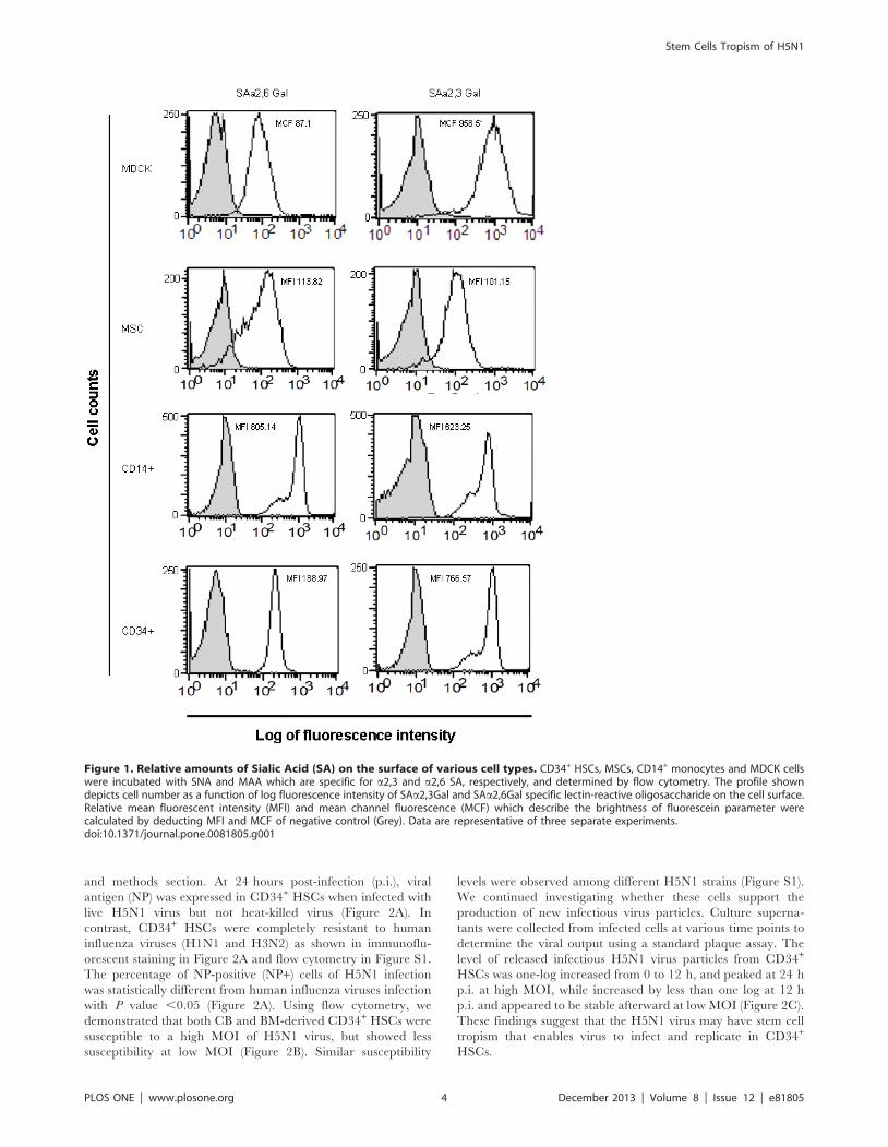
BM cells express receptors for influenza virusSialic acid (SA) on the cell surface is widely recognized as an
influenza virus receptor, which can mediate influenza virus
binding and fusion. SA linked to galactose (SAa2,3-Gal) was
distributed in the avian gastrointestinal tract, and human lower
respiratory tract, whereas, SAa2,6-Gal was expressed in the
human upper respiratory tract [22,26,27]. However, there have
been no reports on SA receptor expression on stem cells.
Therefore, we investigated whether BM stem cells expressed SA
receptors. We stained isolated CD34+ HSCs and MSCs with MAA
and SNA specific for SAa2,3-Gal and SAa2,6-Gal, respectively.
We demonstrated that CD34+ HSCs and MSCs expressed
receptors of both avian (SAa2,3-Gal) and human (SAa2,6-Gal)
influenza viruses (Figure 1). MDCK and CD14+ monocytes were
used as a control as they are known targets of influenza virus
infection and have been reported to express both SA receptors
[21]. This result indicated that CD34+ HSCs and MSCs could be
targets of avian and human influenza viruses.
HSCs are susceptible to avian influenza (H5N1) virus butnot to human influenza virus
To investigate whether H5N1 virus could infect hematopoietic
stem cells, CD34+ HSCs from BM and CB were isolated, purified,
and then infected with H5N1 virus as described in the materials
Stem Cells Tropism of H5N1
PLOS ONE | www.plosone.org 3 December 2013 | Volume 8 | Issue 12 | e81805
and methods section. At 24 hours post-infection (p.i.), viral
antigen (NP) was expressed in CD34+ HSCs when infected with
live H5N1 virus but not heat-killed virus (Figure 2A). In
contrast, CD34+ HSCs were completely resistant to human
influenza viruses (H1N1 and H3N2) as shown in immunoflu-
orescent staining in Figure 2A and flow cytometry in Figure S1.
The percentage of NP-positive (NP+) cells of H5N1 infection
was statistically different from human influenza viruses infection
with P value ,0.05 (Figure 2A). Using flow cytometry, we
demonstrated that both CB and BM-derived CD34+ HSCs were
susceptible to a high MOI of H5N1 virus, but showed less
susceptibility at low MOI (Figure 2B). Similar susceptibility
levels were observed among different H5N1 strains (Figure S1).
We continued investigating whether these cells support the
production of new infectious virus particles. Culture superna-
tants were collected from infected cells at various time points to
determine the viral output using a standard plaque assay. The
level of released infectious H5N1 virus particles from CD34+
HSCs was one-log increased from 0 to 12 h, and peaked at 24 h
p.i. at high MOI, while increased by less than one log at 12 h
p.i. and appeared to be stable afterward at low MOI (Figure 2C).
These findings suggest that the H5N1 virus may have stem cell
tropism that enables virus to infect and replicate in CD34+
HSCs.
Figure 1. Relative amounts of Sialic Acid (SA) on the surface of various cell types. CD34+ HSCs, MSCs, CD14+ monocytes and MDCK cellswere incubated with SNA and MAA which are specific for a2,3 and a2,6 SA, respectively, and determined by flow cytometry. The profile showndepicts cell number as a function of log fluorescence intensity of SAa2,3Gal and SAa2,6Gal specific lectin-reactive oligosaccharide on the cell surface.Relative mean fluorescent intensity (MFI) and mean channel fluorescence (MCF) which describe the brightness of fluorescein parameter werecalculated by deducting MFI and MCF of negative control (Grey). Data are representative of three separate experiments.doi:10.1371/journal.pone.0081805.g001
Stem Cells Tropism of H5N1
PLOS ONE | www.plosone.org 4 December 2013 | Volume 8 | Issue 12 | e81805
MSCs are highly susceptible to avian influenza H5N1 virusWe next investigated the susceptibility of non-hematopoietic
stem cells, MSCs to H5N1 virus. MSCs were isolated from BM.
Once a homogeneous cell culture was obtained upon culturing the
cells in an appropriate media, cells were tested for surface markers
characteristics. Isolated MSCs expressed CD73, CD90, and
CD105, but not CD34 and CD45 (data not shown). MSCs were
infected with H5N1, H1N1 and H3N2 viruses. Surprisingly,
MSCs were highly susceptible to H5N1 virus which live virus was
required (Figure 3A). The susceptibility of MSCs to infection was
Figure 2. Infection and Replication of CD34+ HSCs by H5N1 virus. (A) CD34+ HSCs from CB were infected with both live and heat-killed avian(H5N1) and human influenza viruses (H1N1 and H3N2) at an MOI of 10 for 24 h. MDCK infected with H5N1 at MOI 1 for 12 hours was used as apositive control. Cells were fixed and permeabilized for intracellular staining of Influenza nucleoprotein (NP; green). CD34+ cells were counterstainedwith anti-human CD34-PE (red), and then examined by confocal microscopy and photographed at a 6200 magnification. The percentage of NP-positive (NP+) cells was obtained by counting the number of NP+ cells per total cells in five random fields. (B) CD34+ HSCs from both BM and CBsources were exposed to either a high (MOI 10) or low (MOI 1) dose of H5N1 virus. The expression of viral antigens was analyzed by flow cytometry.(C) Supernatants of infected CD34+ HSCs at MOI 1 and 10 were collected at different time points after infection and then titrated on MDCK cells tomeasure virus production by plaque assay. The results were obtained from three different experiments and are presented as means plus standarderrors. P value,0.05 indicate the significance of H5N1 infection compared with human influenza virusesdoi:10.1371/journal.pone.0081805.g002
Stem Cells Tropism of H5N1
PLOS ONE | www.plosone.org 5 December 2013 | Volume 8 | Issue 12 | e81805
Stem Cells Tropism of H5N1
PLOS ONE | www.plosone.org 6 December 2013 | Volume 8 | Issue 12 | e81805
shown in different levels correlated with the exposure dose
(Figure 3B and 3E). MSCs showed a limited infection to human
influenza virus strains as seen by both fluorescence microscopy
and flow cytometry which percent infection was significantly
different from H5N1 infection (Figure 3A, 3C, and 3D).
Moreover, H5N1 virus had a high replicative efficiency in MSCs
with viral production increasing by more than one-log from 0 to
12 h p.i at an MOI of 10 (Figure 3F). An MOI of 0.008 (one virus
particle per every 120 target cells) was sufficient to infect MSCs to
generate at least a two-log increase in new H5N1 virus particles at
36 h after infection (Figure S2). This phenomenon is restricted
only to HPAI H5N1 since the percentages of infection of rgPR8-
H5 which represents HPAI H5N1 was significantly higher than
low-pathogenic rgPR8-H5monobasic in MSCs with P value ,0.05
(Figure S3). These findings indicated that H5N1 virus could infect
and replicate in MSCs efficiently in order to expand its host range
in the BM environment.
Avian influenza virus infection kills CD34+ HSCs and MSCsOnce human stem cells were infected, the number of viable cells
decreased (data not shown). We suspected that this was related to
apoptosis of these cells. With this thought, we investigated the fate
of MSCs and CD34+ HSCs after infection. Apoptotic cells were
characterized by positive terminal deoxynucleotidyltransferase-
mediated dUTP-biotin nick end labeling (TUNEL) staining (In
Situ Cell Death Detection Kit, TMR red, Roche). TUNEL assay
confirmed that live H5N1 virus could induce apoptosis in both
CD34+ HSCs and MSCs, whereas, human influenza viruses and
viral proteins of heat-killed H5N1 could not (Figure 4A and 4B).
Virus-induced cell death was observed in most of H5N1-infected
MSCs (Figure 4B). The percentages of merge signals (NP+TU-
NEL+) were comparable with single positive signal (NP+ or
TUNEL+) in H5N1-infected MSCs as shown in Figure S4, which
was correlated with co-localization of multiple colors in merge
picture in Figure 4B. However, apoptosis was also observed in
some of the uninfected CD34+ HSCs population as the number of
TUNEL+; apoptosis without infection was significantly higher
than NP+TUNEL+; infected and apoptosis (Figure S4). This data
was correlated with non-localizing red signal in merge picture of
H5N1-infected CD34+ HSCs in Figure 4A. This finding indicated
that H5N1 virus caused apoptosis in stem cells by a direct effect of
infection in MSCs, but by an indirect one on CD34+ HSCs.
Avian influenza virus subverts MSCs-mediated immunemodulation
Considering MSCs are non-immune cells that possess an
immunomodulative activity, further exploring the role of MSCs
in H5N1 pathogenesis is crucial. Previous reports demonstrated
that MSCs are capable of suppressing the differentiation of
monocyte-derived dendritic cells (MoDCs) [15,17]. It is interesting
to investigate the immune dysregulation following H5N1 infection.
Therefore, we hypothesized that H5N1 infection could alter
MSCs-mediated immune modulation. Monocytes/macrophages
secreted mainly inflammatory mediators including IL-1b, IL-6, IL-
8, MCP-1, MIP-1b and GM-CSF which are known to be key
players in the H5N1-mediated immunopathogenesis [28,29].
Here, we designed an experiment in which MSCs/monocyte
cocultures was infected with H5N1 virus at an MOI of 0.04, and
alterations in the cytokine profiles of infected cocultures were
compared to those of mock, MSCs monoculture, and monocyte
monoculture. At 24 hours post-infection, culture supernatants
were collected to determine cytokine production. Cytokines and
chemokines were measured by using a Bio-plex Pro Human
Cytokine Assay. We found that IL-6 level of H5N1-infected MSCs
was comparable with mock, whereas it was barely detectable in
infected CD14+ monocytes (Figure 5A). The chemokines; MCP-1
and MIP-1b were not up-regulated in H5N1-infected MSCs
monoculture, but slightly increased in infected CD14+ monocytes
(Figure 5B and 5C). IL-6, MCP-1 and MIP-1b were significantly
up-regulated in the H5N1-infected MSCs/Monocytes cocultures
compared with mock, and both monocultures (Figure 5A–5C). In
contrast, some cytokine and chemokines such as IL-1b, IL-8, and
GM-CSF from H5N1-infected cocultures were not significantly
different from their respective controls (Figure S5). These findings
demonstrated that H5N1-infected MSCs/Monocytes cocultures
could produce high level of IL-6, MCP-1 and MIP-1b indicating
that H5N1 infection has a potential to induce immune dysregu-
lation of MSCs when they were cocultured with monocytes.
Discussion
Details regarding the pathogenesis of H5N1 are limited. H5N1
infection is not restricted to respiratory organs, and this systemic
spread of the virus makes H5N1 unique from other human
influenza viruses. Detection of viral RNA in BM upon autopsy and
the presence of hematologic abnormalities in severe H5N1 cases
increased our interests in H5N1-mediated BM suppression. It is
therefore important to investigate viral tropism in BM cells.
Previous studies of direct infection of CD34+ HSCs and MSCs by
other persistent viruses, such as Measles virus (MV) [10] and
Cytomegalovirus (CMV) [30,31] have demonstrated virus-medi-
ated immunosuppression. However, acute viral infection of BM
cells remains unclear.
In the present study, we first demonstrated that H5N1 virus
could directly infect primary human BM progenitor cells: HSCs
and MSCs. Viral infection induced cell death and affected the
immune modulation activity of MSCs. Our obtained data could
better explain viral pathogenesis in extra-pulmonary organs. We
started with isolation of CD34+ HSCs and MSCs from the BM
and CB of healthy donors. Using different techniques including
flow cytometry, quantitative RT-PCR, plaque assay, immunoflu-
orescence and confocal microscopy, we demonstrated that CD34+
HSCs and MSCs supported a productive infection of H5N1 virus,
although to a lesser degree in CD34+ HSCs. Both stem cells were
resistant to human influenza virus (H1N1 and H3N2) infection,
even though these two cells expressed both a-2,3 SA and a-2,6 SA,
receptors for avian and human influenza viruses, respectively. Our
findings suggested that H5N1 virus had BM tropism and CD34+
HSCs and MSCs could serve as key amplifiers of virus within the
Figure 3. Avian influenza could infect and replicate in MSCs. (A) The ability of avian influenza H5N1 to infect MSCs was compared to humaninfluenza viruses. Cells were infected with H5N1; live or heat-inactivated, H1N1 and H3N2 at an MOI of 10. After 24 h, cells were stained and examinedby confocal microscopy for the expression of viral antigens (green) with Evan’s blue (red) as a counterstain. The percentages of infected MSCs at highand low MOIs of (B) H5N1 virus and (C) human influenza viruses were determined by flow cytometry. (D) Percent infection of MSCs infected mock,H1N1, H3N2, H5N1, and heat-killed H5N1 at MOI 10 for 24 hours. MDCK infected with H5N1 at MOI 1 for 12 hours was used as a positive control. (E)Real-time PCR of H5N1 M gene in MSCs after infection with H5N1 at various doses for 12 h. The amount of the virus RNA was standardized by betaactin. (F) Production of H5N1 virus from infected MSCs at various time points was measured by plaque assay. The results represent the means and SDof three independent experiments. P,0.05 indicates statistically significant differences between H5N1 and human influenza viruses infections.doi:10.1371/journal.pone.0081805.g003
Stem Cells Tropism of H5N1
PLOS ONE | www.plosone.org 7 December 2013 | Volume 8 | Issue 12 | e81805

Figure 4. H5N1 infection induced marked CD34+ HSC and MSC apoptosis. (A) CD34+ HSCs were infected with H5N1 virus at an MOI of 10,whereas (B) MSCs were infected with either avian or human influenza virus at the same MOI as CD34+ HSCs. Parallel samples were treated with DnaseIwhich breaks double stranded DNA as a positive control. After 18 h, cells were fixed, permeabilized and multiple stained using Terminal
Stem Cells Tropism of H5N1
PLOS ONE | www.plosone.org 8 December 2013 | Volume 8 | Issue 12 | e81805
BM compartment. The mobilizing property of these two cells
could support a spread of virus throughout the body [9,32]. Our
findings are consistent with Gu J., et al. [33] and Uiprasertkul M.,
et al. [34] studies that demonstrated H5N1 was able to disseminate
and infect extra-pulmonary organs showing a broad tissue tropism
of virus. However, there is a limited autopsy data to prove a
dissemination of virus in systemic organs.
The susceptibility of each cell type to H5N1 infection is partly
dependent on SA receptors since some a-2,3 SA and 2,6-SA
expressing cells are unable to support efficient infection and
replication of virus [35]. In addition, there might be some
unknown intracellular factors or mechanisms that involved with
permissibility of CD34+ HSCs to H5N1 virus. Previous studies
have demonstrated that H5N1 could infect various targets in
particular cells of the immune system [21,36–38], it is possible
that H5N1 is able to evade the host innate immune response
with multiple mechanisms such as NS1-mediated IFN inhibi-
tion, the proapoptotic function of PB1-F2 in limiting efficient
immune cell-mediated virus clearance in vivo, and the receptor
switching of avian to human receptors enabling H5N1 to evade
the virus-neutralizing effects of mucins containing a-2,3 SA
[4,39]. Further investigations should be performed to under-
stand the mechanism underlying high susceptibility of progen-
itor cells to H5N1 over H1N1 and H3N2 viruses, although these
two viruses are more likely to be laboratory strains than human
influenza strains.
Studies have shown that apoptosis plays a major role in H5N1
pathogenesis. Apoptosis was induced upon infection of epithelial
cells and lymphocytes in vitro [40,41]. Increased apoptosis in
human lymphocytes leading to severe lymphopenia which was
caused by either direct infection or over-activation of the
cytokine response [41]. In this study, H5N1 virus induced high
rates of apoptosis in both MSCs and CD34+ HSCs. Apoptosis
observed in a majority of infected MSCs was likely due to direct
infection, whereas, apoptosis in CD34+ HSCs might be induced
by paracrine factors as apoptotic signals were observed in
uninfected CD34+ HSCs population (Figure 4A). Additional
experiments to confirm unusual apoptosis in CD34+ HSCs will
be further investigated. Since CD34+ HSCs and MSCs are
primitive progenitor cells that contain self-renewal ability and
generate descendant cells during hematopoiesis and mesengenic
processes, the reduction of precursor cells may affect these two
processes which eventually decrease the number of differenti-
ated cells in all lineages. The disruption of hematopoiesis was
observed in depleted stromal and CD34+ populations by MV
infection, which significantly impaired repopulation of lymphoid
precursors following MV-induced lymphopenia [10]. Our
findings supported abnormal hematologic findings including
low peripheral blood counts and cytopenia which are prominent
clinical features in patients with severe H5N1 infection. In
addition, substantial loss of MSCs would affect the immuno-
suppressive activity, leading to hyperactivation of the immune
response.
The pathogenesis of the H5N1 virus was characterized by broad
tissue tropism, systemic replication and hypercytokinemia [42,43].
H5N1-induced pro-inflammatory cytokines were generated by
various immune cells as they coordinate an attack on invading
pathogens. Under normal conditions, anti-inflammatory cytokines
are immunoregulatory molecules that regulate pro-inflammatory
responses. On the other hand, under pathological conditions,
disruption of the balance of the anti-inflammatory response to
the pro-inflammatory response may occur [44]. MSCs have
potent immunoregulatory actions that make them attractive
targets for reducing the inflammation and injury [18,45]. MSCs
have been shown to interact with CD14+ monocytes and block
monocytes differentiation to dendritic cells (DCs) which IL-6 is
partially involved in this action [15,17,46,47]. In addition,
MSCs can modulate cytokine production by dendritic and T cell
subsets [15]. The fact that interaction of MSCs and monocytes
leads to immunopathology and dysregulated MSCs-mediated
immunomodulation during infection is controversial. Interest-
ingly, our result demonstrated that IL-6, MCP-1, and MIP-1bfrom MSCs/CD14+ monocytes cocultures were up-regulated,
whereas this effect was not seen in MSCs or monocytes
monoculture. Elevated IL-6 may enhance inflammation as
shown in previous studies [43,48,49]. It is possible that H5N1
may subvert MSCs-mediated immune modulation by skewing
immune modulator towards enhancer as evidenced by the
significantly increased levels of IL-6 in infected cocultures.
Higher levels of IL-6 are likely to promote inhibition of DC
maturation contributing to a reduction in the number of antigen
presenting cells which thus leads to slow virus clearance.
Previously published data reported that H5N1 was a potent
inducer of MCP-1 and MIP-1b which were responsible for the
recruitment of immune cells into the infected site [49,50].
Therefore, high levels of MCP-1 and MIP-1b secreted by
infected cocultures may amplify the inflammatory response by
attracting many of immune cells into the BM milieu [51]
indicating that MSCs might involve indirectly with H5N1-
induced inflammation since they are more active responding to
H5N1 infection when they are cocultured with monocytes.
Hyperactivation of cytokines, including TNF-a, soluble IL-2
receptor, IL-1 and IL-6 may play a role in the pathogenesis of
reactive hemophagocytosis syndrome (RHS) [52]. High levels of
IL-6, MCP-1, and MIP-1b production in H5N1-infected cocul-
tures are likely to induce RHS in BM. RHS is characterized by
activated macrophages which consequently engulf neighboring
cells including hematopoietic cells [53]. This can contribute to
markedly hypocellular BM. To et al., previously documented that
both hypocellular conditions and RHS in BM were the most
prominent pathologic features observed in fatal H5N1 cases [54].
Besides BM tissue, MSCs have been identified in many other
organs with similar functions including the lung [32,55]. They are
important for the maintenance of lung homeostasis and repair
following lung injury [32]. As lung is the primary site of influenza
infection, it is possible that lung-resident MSCs may get infected
by H5N1 virus resulting in disruption of renewal and differenti-
ation processes. Also, MSCs-mediated immunomodulatory func-
tion may be dysregulated as hypothesized contributing to severe
injury in the lung. Although there has been no data in humans,
influenza virus-infected lung MSCs were investigated in the lungs
of both chicken and swine [23,56].
In summary, we demonstrated that H5N1 virus more efficiently
infected and replicated in CD34+ HSCs and MSCs compared to
human influenza viruses in vitro. Apoptosis was a direct result of
infection in MSCs, whereas, it might be induced by paracrine
factors in CD34+ HSCs. In addition, H5N1 infection could
interfere with MSC-mediated immune modulation by shifting
deoxynucleotidyl transferase–mediated dUTP-biotin nick end-labeling (TUNEL) staining (red), FITC-labeled anti-NP (green) and DAPI (blue). Arrows inB. indicate cells that simultaneously expressed intracellular viral antigen and apoptotic signal. The samples were examined on a confocal microscopyand photographed at a 6200 magnification.doi:10.1371/journal.pone.0081805.g004
Stem Cells Tropism of H5N1
PLOS ONE | www.plosone.org 9 December 2013 | Volume 8 | Issue 12 | e81805
immune modulator production towards enhancers, which, in turn,
amplifies the cytokine response by the influx of circulating
monocytes and macrophages. Our explorations provide an insight
into H5N1 pathogenesis in terms of aggressive systemic infection
and hematologic abnormalities in patients with severe H5N1
infection.
Supporting Information
Figure S1 Susceptibility of CB-derived CD34+ cells to different
H5N1 strains. Infection is determined by using a specific marker
(Nucleoprotein), and detected by flow cytometry. Cells were
infected by different strains of avian and human influenza viruses
at an MOI of 10 for 24 h. The results were obtained individually
from three different experiments and are presented as means plus
standard errors. *P,0.05 indicates statistically significant differ-
ences between H5N1 infection and human influenza viruses
infection (H1N1 and H3N2).
(TIF)
Figure S2 Viral production of H5N1-infected MSCs at various
doses and incubation times. Avian influenza H5N1 could infect
and replicate in MSC. Data are plaque-forming units in the
supernatant of MSCs infected with H5N1 at various MOI for 1 h,
washed, and incubated for indicated time points.
(TIF)
Figure S3 High susceptibility of MSCs was restricted only to
HPAI H5N1. MSCs were infected with rgPR8-H5, rgPR8-
H5monobasic, and rg-PR8 which represent HPAI H5N1, LPAI
H5N1, and H1N1 viruses, respectively at MOI 1 for 24 hours.
LPAI H5N1 is the reverse genetics (rg) virus bearing monobasic
amino acids at HA cleavage site as described in materials and
methods section. Percentages of infection were determined by flow
cytometry. The results represent the means and SD of two
independent donors. *P,0.05 indicate statistically significant
differences of rgPR8-H5 compared with rgPR8-H5monobasic,
and rg-PR8 viruses.
(TIF)
Figure S4 Percentage of apoptotic cells induced by H5N1
infection. (A) CD34+ HSCs were infected with H5N1 virus and (B)
MSCs were infected with both avian and human influenza viruses
at an MOI of 10. After 18 h, cells were fixed, permeabilized and
multiple stained with TUNEL, NP and DAPI and determined by
confocal microscopy as described in Figure 4. The percentages of
positive signal were obtained by counting positive cells from five
random views. Data are given as mean 6 SD of two independent
experiments. *P,0.05 indicates statistically significant differences
between the percentages of TUNEL+ and Merge (NP+TUNEL+)
of H5N1-infected CD34+. **P,0.05 indicates the significance of
the number of H5N1-induced apoptosis compared with human
influenza viruses in MSCs.
(TIF)
Figure 5. Production of IL-6, MCP-1, and MIP-1b from H5N1-infected cocultures. (A) IL-6, (B) MCP-1, and (C) MIP-1b levels fromsupernatants of infected CD14+ monocytes and MSCs monoculture and1:5 ratio of MSCs/CD14+ monocytes cocultures at MOI 0.04 weremeasured by using Bio-plex Cytokine assay. Data was analyzed via Bio-plex 5.0 Software. The two different cell types used in the experimentwere derived from different donors. The results represent the meansand SD of two independent experiments. Single asterisk indicatesstatistically significant differences between mock and infected cells withP values of ,0.05 and double asterisks indicate statistically significantdifferences between cocultures and monoculture groups with P valuesof ,0.05.doi:10.1371/journal.pone.0081805.g005
Stem Cells Tropism of H5N1
PLOS ONE | www.plosone.org 10 December 2013 | Volume 8 | Issue 12 | e81805
Figure S5 H5N1 did not induce IL-1b, IL-8, and GM-CSF in
cocultures. (A) IL-1b, (B) IL-8, and (C) GM-CSF levels were
measured using a Bio-plex Cytokine assay. The two different cell
types in this experiment were derived from different donors. Data
shown are mean6SD of two independent experiments. Single
asterisk indicates statistically significant differences between mock and
infected cells with P values of ,0.05 and double asterisks indicate
statistically significant differences between cocultures and monocul-
ture groups with P values of ,0.05. n.s. means no significance.
(TIF)
Acknowledgments
We gratefully acknowledge Dr. Robert G. Webster for providing
pHW2000 plasmid and A/PR/8/34 backbone plasmids for reverse
genetics. We would like to thank Mr. Chokdee Wongborisut for his
assistance in obtaining human MSCs.
Author Contributions
Conceived and designed the experiments: AT MT PK SH. Performed the
experiments: MT PK SW. Analyzed the data: MT PK AT. Contributed
reagents/materials/analysis tools: PP SH AT. Wrote the paper: MT AT.
References
1. Beigel H, Farrar H, Han AM, Hayden FG, Hyer R, et al. (2005) Current
concepts - Avian influenza A (H5N1) infection in humans. New England Journal
of Medicine 353: 1374–1385.
2. WHO (2013) Cumulative number of confirmed human cases of avian influenza
A (H5N1) reported to WHO.
3. Tran TT, van Doorn HR, de Jong MD (2008) Human H5N1 influenza: Current
insight into pathogenesis. International Journal of Biochemistry & Cell Biology
40: 2671–2674.
4. Korteweg C, Gu J (2008) Pathology, molecular biology, and pathogenesis of
avian influenza A (H5N1) infection in humans. Am J Pathol 172: 1155–1170.
5. Gu J, Xie ZG, Gao ZC, Liu JH, Korteweg C, et al. (2007) H5N1 infection of therespiratory tract and beyond: a molecular pathology study. Lancet 370: 1137–
1145.
6. Zhang ZF, Zhang JX, Huang K, Li KS, Yuen KY, et al. (2009) Systemic
infection of avian influenza A virus H5N1 subtype in humans. Human
Pathology 40: 735–739.
7. Gurkan UA, Akkus O (2008) The Mechanical Environment of Bone Marrow: A
Review. Annals of Biomedical Engineering 36: 1978–1991.
8. Watt FM, Hogan BLM (2000) Out of Eden: Stem cells and their niches. Science287: 1427–1430.
9. Magnon C, Frenette PS (2008) Hematopoietic stem cell trafficking. StemBook.
Cambridge (MA).
10. Manchester M, Smith KA, Eto DS, Perkin HB, Torbett BE (2002) Targeting
and hematopoietic suppression of human CD34+ cells by measles virus. J Virol
76: 6636–6642.
11. Musina RA, Bekchanova ES, Belyavskii AV, Grinenko TS, Sukhikh GT (2007)Umbilical cord blood mesenchymal stem cells. Bull Exp Biol Med 143: 127–131.
12. NIH (2013) Stem Cell Information. The Adult Stem Cell.
13. Uccelli A, Moretta L, Pistoia V (2008) Mesenchymal stem cells in health and
disease. Nature Reviews Immunology 8: 726–736.
14. Chen L, Tredget EE, Wu PY, Wu Y (2008) Paracrine factors of mesenchymal
stem cells recruit macrophages and endothelial lineage cells and enhance wound
healing. PLoS One 3: e1886.
15. Aggarwal S, Pittenger MF (2005) Human mesenchymal stem cells modulate
allogeneic immune cell responses. Blood 105: 1815–1822.
16. Corcione A, Benvenuto F, Ferretti E, Giunti D, Cappiello V, et al. (2006)
Human mesenchymal stem cells modulate B-cell functions. Blood 107: 367–372.
17. Spaggiari GM, Abdelrazik H, Becchetti F, Moretta L (2009) MSCs inhibit
monocyte-derived DC maturation and function by selectively interfering with
the generation of immature DCs: central role of MSC-derived prostaglandin E2.
Blood 113: 6576–6583.
18. Singer NG, Caplan AI Mesenchymal stem cells: mechanisms of inflammation.
Annu Rev Pathol 6: 457–478.
19. Chamberlain G, Fox J, Ashton B, Middleton J (2007) Concise review:
Mesenchymal stem cells: Their phenotype, differentiation capacity, immuno-logical features, and potential for homing. Stem Cells 25: 2739–2749.
20. Chotpitayasunondh T, Ungchusak K, Hanshaoworakul W, Chunsuthiwat S,
Sawanpanyalert P, et al. (2005) Human disease from influenza A (H5N1),
Thailand, 2004. Emerging Infectious Diseases 11: 201–209.
21. Thitithanyanont A, Engering A, Ekchariyawat P, Wiboon-Ut S, Limsalakpetch
A, et al. (2007) High susceptibility of human dendritic cells to avian influenza
H5N1 virus infection and protection by IFN-alpha and TLR Ligands. Journal of
Immunology 179: 5220–5227.
22. Nicholls JM, Bourne AJ, Chen H, Guan Y, Peiris JS (2007) Sialic acid receptor
detection in the human respiratory tract: evidence for widespread distribution of
potential binding sites for human and avian influenza viruses. Respir Res 8: 73.
23. Khatri M, O’Brien TD, Goyal SM, Sharma JM (2010) Isolation andcharacterization of chicken lung mesenchymal stromal cells and their
susceptibility to avian influenza virus. Dev Comp Immunol 34: 474–479.
24. Shahid MA, Abubakar M, Hameed S, Hassan S (2009) Avian influenza virus
(H5N1); effects of physico-chemical factors on its survival. Virol J 6: 38.
25. Hoffmann E, Krauss S, Perez D, Webby R, Webster RG (2002) Eight-plasmid
system for rapid generation of influenza virus vaccines. Vaccine 20: 3165–3170.
26. Suzuki Y, Ito T, Suzuki T, Holland RE Jr, Chambers TM, et al. (2000) Sialic
acid species as a determinant of the host range of influenza A viruses. J Virol 74:11825–11831.
27. Kumlin U, Olofsson S, Dimock K, Arnberg N (2008) Sialic acid tissuedistribution and influenza virus tropism. Influenza Other Respi Viruses 2: 147–
154.
28. Hui KP, Lee SM, Cheung CY, Ng IH, Poon LL, et al. (2009) Induction ofproinflammatory cytokines in primary human macrophages by influenza A virus
(H5N1) is selectively regulated by IFN regulatory factor 3 and p38 MAPK.
J Immunol 182: 1088–1098.
29. Cheung CY, Poon LL, Lau AS, Luk W, Lau YL, et al. (2002) Induction of
proinflammatory cytokines in human macrophages by influenza A (H5N1)
viruses: a mechanism for the unusual severity of human disease? Lancet 360:1831–1837.
30. Smirnov SV, Harbacheuski R, Lewis-Antes A, Zhu H, Rameshwar P, et al.
(2007) Bone-marrow-derived mesenchymal stem cells as a target for cytomeg-alovirus infection: implications for hematopoiesis, self-renewal and differentia-
tion potential. Virology 360: 6–16.
31. Bego MG, St Jeor S (2006) Human cytomegalovirus infection of cells ofhematopoietic origin: HCMV-induced immunosuppression, immune evasion,
and latency. Exp Hematol 34: 555–570.
32. Sinclair K, Yerkovich ST, Chambers DC (2013) Mesenchymal stem cells andthe lung. Respirology 18: 397–411.
33. Gu J, Xie Z, Gao Z, Liu J, Korteweg C, et al. (2007) H5N1 infection of the
respiratory tract and beyond: a molecular pathology study. Lancet 370: 1137–1145.
34. Uiprasertkul M, Puthavathana P, Sangsiriwut K, Pooruk P, Srisook K, et al.
(2005) Influenza A H5N1 replication sites in humans. Emerg Infect Dis 11:1036–1041.
35. Yao L, Korteweg C, Hsueh W, Gu J (2008) Avian influenza receptor expression
in H5N1-infected and noninfected human tissues. FASEB J 22: 733–740.
36. Friesenhagen J, Boergeling Y, Hrincius E, Ludwig S, Roth J, et al. (2012) Highly
pathogenic avian influenza viruses inhibit effective immune responses of human
blood-derived macrophages. J Leukoc Biol 92: 11–20.
37. Zhao Y, Lu M, Lau LT, Lu J, Gao Z, et al. (2008) Neutrophils may be a vehicle
for viral replication and dissemination in human H5N1 avian influenza. Clin
Infect Dis 47: 1575–1578.
38. Tumpey TM, Lu X, Morken T, Zaki SR, Katz JM (2000) Depletion of
lymphocytes and diminished cytokine production in mice infected with a
highly virulent influenza A (H5N1) virus isolated from humans. J Virol 74:6105–6116.
39. Hale BG, Albrecht RA, Garcia-Sastre A (2010) Innate immune evasion
strategies of influenza viruses. Future Microbiol 5: 23–41.
40. Daidoji T, Koma T, Du A, Yang CS, Ueda M, et al. (2008) H5N1 avian
influenza virus induces apoptotic cell death in mammalian airway epithelial cells.
J Virol 82: 11294–11307.
41. Nichols JE, Niles JA, Roberts NJ, Jr. (2001) Human lymphocyte apoptosis after
exposure to influenza A virus. J Virol 75: 5921–5929.
42. Cinatl J, Jr., Michaelis M, Doerr HW (2007) The threat of avian influenza a(H5N1): part II: Clues to pathogenicity and pathology. Med Microbiol Immunol
196: 191–201.
43. de Jong MD, Simmons CP, Thanh TT, Hien VM, Smith GJD, et al. (2006)Fatal outcome of human influenza A (H5N1) is associated with high viral load
and hypercytokinemia. Nature Medicine 12: 1203–1207.
44. Darwish I, Mubareka S, Liles WC Immunomodulatory therapy for severeinfluenza. Expert Rev Anti Infect Ther 9: 807–822.
45. Cardenes N, Caceres E, Romagnoli M, Rojas M (2013) Mesenchymal stem cells:
a promising therapy for the acute respiratory distress syndrome. Respiration 85:267–278.
46. Nauta AJ, Kruisselbrink AB, Lurvink E, Willemze R, Fibbe WE (2006)
Mesenchymal stem cells inhibit generation and function of both CD34(+)-derived and monocyte-derived dendritic cells. Journal of Immunology 177:
2080–2087.
47. Nemeth K, Leelahavanichkul A, Yuen PS, Mayer B, Parmelee A, et al. (2009)Bone marrow stromal cells attenuate sepsis via prostaglandin E(2)-dependent
reprogramming of host macrophages to increase their interleukin-10 production.Nat Med 15: 42–49.
48. Chan MCW, Cheung CY, Chui WH, Tsao SW, Nicholls JM, et al. (2005)
Proinflammatory cytokine responses induced by influenza A (H5N1) viruses inprimary human alveolar and bronchial epithelial cells. Respiratory Research 6: –.
Stem Cells Tropism of H5N1
PLOS ONE | www.plosone.org 11 December 2013 | Volume 8 | Issue 12 | e81805
49. Cheung CY, Poon LLM, Lau AS, Luk W, Lau YL, et al. (2002) Induction of
proinflammatory cytokines in human macrophages by influenza A (H5N1)viruses: a mechanism for the unusual severity of human disease? Lancet 360:
1831–1837.
50. Yu WC, Chan RW, Wang J, Travanty EA, Nicholls JM, et al. (2011) Viralreplication and innate host responses in primary human alveolar epithelial cells
and alveolar macrophages infected with influenza H5N1 and H1N1 viruses.J Virol 85: 6844–6855.
51. Brandau S, Jakob M, Hemeda H, Bruderek K, Janeschik S, et al. (2010) Tissue-
resident mesenchymal stem cells attract peripheral blood neutrophils andenhance their inflammatory activity in response to microbial challenge. J Leukoc
Biol 88: 1005–1015.
52. Fisman DN (2000) Hemophagocytic syndromes and infection. Emerg Infect Dis
6: 601–608.53. Rajam L, Prasad V, Yatheesha BL (2008) Reactive hemophagocytic syndrome.
Indian J Pediatr 75: 1261–1263.
54. To KF, Chan PKS, Chan KF, Lee WK, Lam WY, et al. (2001) Pathology offatal human infection associated with avian influenza A H5N1 virus. Journal of
Medical Virology 63: 242–246.55. Sabatini F, Petecchia L, Tavian M, Jodon de Villeroche V, Rossi GA, et al.
(2005) Human bronchial fibroblasts exhibit a mesenchymal stem cell phenotype
and multilineage differentiating potentialities. Lab Invest 85: 962–971.56. Khatri M, Saif YM (2012) Influenza virus infects bone marrow mesenchymal stromal
cells in vitro: implications for bone marrow transplantation. Cell Transplant.
Stem Cells Tropism of H5N1
PLOS ONE | www.plosone.org 12 December 2013 | Volume 8 | Issue 12 | e81805