Trophic apparatus in Cyprinodontiform fishes: functional specializations for picking and scraping...
17
Trophic Apparatus in Cyprinodontiform Fishes: Functional Specializations for Picking and Scraping Behaviors L. Patricia Hernandez, 1 * Alice C. Gibb, 2 and Lara Ferry-Graham 3 1 Department of Biological Sciences, The George Washington University, Washington, District of Columbia 20052 2 Department of Biological Sciences, Northern Arizona University, Flagstaff, Arizona 86011-5640 3 California State Universities, Moss Landing Marine Labs, Moss Landing, California 95039 ABSTRACT Cyprinodontiforms are a diverse and spe- ciose order that includes topminnows, pupfishes, sword- tails, mosquitofishes, guppies, and mollies. Sister group to the Beloniformes and Atheriniformes, Cyprinodontiformes contains approximately twice the number of species of these other two orders combined. Recent studies suggest that this group is well suited to capturing prey by ‘‘picking’’ small items from the water surface, water column, and the sub- strate. Because picking places unusual performance demands on the feeding apparatus, this mode of prey cap- ture may rely upon novel morphological modifications not found in more widespread ram- or suction-based feeding mechanisms. To assess this evolutionary hypothesis, we describe the trophic anatomy of 16 cyprinodontiform spe- cies, selected to broadly represent the order as well as cap- ture intrageneric variation. The group appears to have undergone gradual morphological changes to become increasingly specialized for picking and scraping behaviors. We also identify a suite of functional characters related to the acquisition of a novel and previously undescribed mech- anism of premaxillary protrusion and retraction, including: modification of the ‘‘premaxillomandibular’’ ligament (which connects each side of the premaxilla to the ipsilat- eral mandible, or lower jaw), a novel architecture of the lig- aments and bony elements that unite the premaxillae, max- illae and palatine bones, and novel insertions of the adduc- tor muscles onto the jaws. These morphological changes to both the upper and lower jaws suggest an evolutionary trend within this group toward increased reliance on pick- ing individual prey from the water column/substrate or for scraping encrusting material from the substrate. We pro- pose that the suite of morphological characters described here enable a functional innovation, ‘‘picking,’’ which leads to novel trophic habits. J. Morphol. 270:645–661, 2009. Ó 2008 Wiley-Liss, Inc. KEY WORDS: adductor mandibulae; premaxillary pro- trusion; Cyprinodontiformes; feeding INTRODUCTION The speciose and diverse Cyprinodontiformes (Teleostei) are generally considered to be a group of generalized ‘‘midwater omnivores’’ feeding on small, non-elusive, benthic and planktonic organ- isms (Weisberg, 1986, Motta et al., 1995; Mansfield and Macardle, 1998). However, this description does not do justice to the unusual feeding behaviors and mechanisms found within the group. Though some cyprinodontiform fishes are indeed mid-water omnivores (e.g., Hunt, 1953), many species appear to capture prey by means of an unusual ‘‘picking’’ mode of prey capture (Ferry-Graham et al., 2008). Picking is a form of biting prey capture employed to procure prey items in the water column, at the air–water interface, or loosely attached to the sub- strate (Horn and Ferry- Graham, 2006). Biting and suction behaviors typical of teleosts are often con- sidered feeding modes where trade-offs occur because jaws typically can be optimized for produc- ing force or operating at high velocities, but not for both (as reviewed in Westneat, 2004). Increased feeding velocity is often associated with enhanced suction production, whereas in biting behaviors suc- tion performance is compromised to enable the jaws to remove attached prey by force. Although forceful movements of the jaw characterize strong biting, less forceful but more controlled movements of the jaw characterize picking (Ferry-Graham et al., 2008). Picking prey from the water column requires fine control of the jaws, but does not require large forces to be produced. Thus picking likely requires a suite of morphological features different from those typically observed in teleostean lineages that are considered either strong biters or suction feeders. In addition, more derived members of Cyprini- dontiformes, particularly species within the Poeci- Contract grant sponsor: NSF; Contract grant numbers: IOS- 0615827, IOS-0726087, DBI-0721662, MRI-0320972, IOS-0726001; Contract grant sponsor: Science Foundation Arizona; Contract grant number: CAA 0057-07. *Correspondence to: L. Patricia Hernandez, Department of Bio- logical Sciences, Lisner Hall Rm 340, George Washington Univer- sity, 2023 G. St. NW, Washington, DC 20052. E-mail: [email protected] Received 22 November 2007; Revised 29 September 2008; Accepted 8 October 2008 Published online 23 December 2008 in Wiley InterScience (www.interscience.wiley.com) DOI: 10.1002/jmor.10711 JOURNAL OF MORPHOLOGY 270:645–661 (2009) Ó 2008 WILEY-LISS, INC.
Transcript of Trophic apparatus in Cyprinodontiform fishes: functional specializations for picking and scraping...

Trophic Apparatus in Cyprinodontiform Fishes:Functional Specializations for Picking andScraping Behaviors
L. Patricia Hernandez,1* Alice C. Gibb,2 and Lara Ferry-Graham3
1Department of Biological Sciences, The George Washington University, Washington, District of Columbia 200522Department of Biological Sciences, Northern Arizona University, Flagstaff, Arizona 86011-56403California State Universities, Moss Landing Marine Labs, Moss Landing, California 95039
ABSTRACT Cyprinodontiforms are a diverse and spe-ciose order that includes topminnows, pupfishes, sword-tails, mosquitofishes, guppies, and mollies. Sister group tothe Beloniformes and Atheriniformes, Cyprinodontiformescontains approximately twice the number of species of theseother two orders combined. Recent studies suggest that thisgroup is well suited to capturing prey by ‘‘picking’’ smallitems from the water surface, water column, and the sub-strate. Because picking places unusual performancedemands on the feeding apparatus, this mode of prey cap-ture may rely upon novel morphological modifications notfound in more widespread ram- or suction-based feedingmechanisms. To assess this evolutionary hypothesis, wedescribe the trophic anatomy of 16 cyprinodontiform spe-cies, selected to broadly represent the order as well as cap-ture intrageneric variation. The group appears to haveundergone gradual morphological changes to becomeincreasingly specialized for picking and scraping behaviors.We also identify a suite of functional characters related tothe acquisition of a novel and previously undescribed mech-anism of premaxillary protrusion and retraction, including:modification of the ‘‘premaxillomandibular’’ ligament(which connects each side of the premaxilla to the ipsilat-eral mandible, or lower jaw), a novel architecture of the lig-aments and bony elements that unite the premaxillae, max-illae and palatine bones, and novel insertions of the adduc-tor muscles onto the jaws. These morphological changes toboth the upper and lower jaws suggest an evolutionarytrend within this group toward increased reliance on pick-ing individual prey from the water column/substrate or forscraping encrusting material from the substrate. We pro-pose that the suite of morphological characters describedhere enable a functional innovation, ‘‘picking,’’ which leadsto novel trophic habits. J. Morphol. 270:645–661,2009. � 2008Wiley-Liss, Inc.
KEY WORDS: adductor mandibulae; premaxillary pro-trusion; Cyprinodontiformes; feeding
INTRODUCTION
The speciose and diverse Cyprinodontiformes(Teleostei) are generally considered to be a groupof generalized ‘‘midwater omnivores’’ feeding onsmall, non-elusive, benthic and planktonic organ-isms (Weisberg, 1986, Motta et al., 1995; Mansfieldand Macardle, 1998). However, this descriptiondoes not do justice to the unusual feeding behaviors
and mechanisms found within the group. Thoughsome cyprinodontiform fishes are indeed mid-wateromnivores (e.g., Hunt, 1953), many species appearto capture prey by means of an unusual ‘‘picking’’mode of prey capture (Ferry-Graham et al., 2008).
Picking is a form of biting prey capture employedto procure prey items in the water column, at theair–water interface, or loosely attached to the sub-strate (Horn and Ferry- Graham, 2006). Biting andsuction behaviors typical of teleosts are often con-sidered feeding modes where trade-offs occurbecause jaws typically can be optimized for produc-ing force or operating at high velocities, but not forboth (as reviewed in Westneat, 2004). Increasedfeeding velocity is often associated with enhancedsuction production, whereas in biting behaviors suc-tion performance is compromised to enable the jawsto remove attached prey by force. Although forcefulmovements of the jaw characterize strong biting,less forceful but more controlled movements of thejaw characterize picking (Ferry-Graham et al.,2008). Picking prey from the water column requiresfine control of the jaws, but does not require largeforces to be produced. Thus picking likely requires asuite of morphological features different from thosetypically observed in teleostean lineages that areconsidered either strong biters or suction feeders.
In addition, more derived members of Cyprini-dontiformes, particularly species within the Poeci-
Contract grant sponsor: NSF; Contract grant numbers: IOS-0615827, IOS-0726087, DBI-0721662, MRI-0320972, IOS-0726001;Contract grant sponsor: Science Foundation Arizona; Contractgrant number: CAA 0057-07.
*Correspondence to: L. Patricia Hernandez, Department of Bio-logical Sciences, Lisner Hall Rm 340, George Washington Univer-sity, 2023 G. St. NW, Washington, DC 20052.E-mail: [email protected]
Received 22 November 2007; Revised 29 September 2008;Accepted 8 October 2008
Published online 23 December 2008 inWiley InterScience (www.interscience.wiley.com)DOI: 10.1002/jmor.10711
JOURNAL OF MORPHOLOGY 270:645–661 (2009)
� 2008 WILEY-LISS, INC.
liidae, often obtain prey via a scraping behavior,where the jaws are placed in direct contact withthe substrate and epiphytic material is removedusing a combing action of the teeth (Gibb et al., inpress). Scraping by cichlid fishes has been welldescribed, for example, where anatomical changesto the oral jaws increase gape and facilitateincreased contact between the teeth and the sub-strate (Liem, 1980; Bouton et al., 1998). Wehypothesize that poeciliids and some cyprinodon-tids have independently evolved a different set ofanatomical ‘‘tools’’ used for this feeding mode.
Studies examining feeding morphology amongcyprinodontiform species have revealed significanttrophic polymorphisms (Turner and Grosse, 1980;Holtmeier, 2001). Such findings suggest that thetrophic anatomy of this order may lend itself to a va-riety of feeding modes (Hernandez et al., 2008). Toappropriately evaluate potential trophic polymor-phisms, however, we must first understand phyloge-netic changes in cyprinodontiform oral jaws. Thisstudy, which is well supported by several morpho-logical analyses of cyprinodontiform relationships(Rosen and Bailey, 1963; Parenti, 1981; Costa, 1998;Ghedotti, 2000), specifically examines how thetrophic apparatus is modified across Cyprinodonti-formes in light of these different functional roles.
We describe the evolution of morphological fea-tures associated with picking and scraping behav-iors by examining several cyprinodontiform specieswith special emphasis on Poeciliidae and Cyprino-dontidae (see Fig. 1). As Poeciliidae demonstratesexceptional trophic diversity we sampled exten-sively within this clade to understand variation atthis level. Specifically, we describe the osteology ofthe anterior jaws, the morphology of the adductormandibulae muscle complex (the primary jaw clos-ing muscle in teleosts), and the associated liga-mentous attachments required to produce feedingmovements. The specific aims of this study are to:1) describe the musculoskeletal structure of theanterior jaws in the Cyprinidontiformes; 2) assessthe morphological changes that have occurredwithin these trophic structures; and 3) proposeboth proximate performance consequences as wellas ecological and evolutionary consequences associ-ated with these morphological changes.
MATERIALS AND METHODS
The following live specimens were obtained from commercialsuppliers, sacrificed according to Northern Arizona UniversityIACUC approved protocols (NAU IACUC #04-007) and used asrepresentative taxa: Kryptolebias marmoratus, Fundulus rubri-frons, Gambusia affinis, Heterandria formosa, Jordanella flori-dae, Xiphophorus helleri, and Poecilia sphenops. Additionalspecimens were obtained from museums and from the collec-tions of colleagues and were used to assess intra-clade andintra-generic variation in feeding morphology: Atherinops cali-forniensis, Fundulus grandis, F. similis, F. chrysotus, G. hol-brooki, Cyprinodon variegatus, Floridichthys carpio, Jordanellafloridae, and Poecilia latipinna. In all cases except Atherinops
californiensis, which were frozen, specimens were preserved in10% formalin and subsequently stored in 70% ethanol prior toour receipt of the specimens.
For the morphological description, fresh and preserved speci-mens were dissected and briefly immersed in iodine to facilitateidentification of muscle fiber orientation. Approximately 10specimens from each representative species were dissected todetermine musculoskeletal architecture and to assess inter-indi-vidual variation in feeding morphology. Additional specimenswere cleared and stained using a protocol presented in Dinger-kus and Uhler (1977) with modifications by Potthoff (1984).Cleared and stained specimens were used to assess the shape ofthe bones and cartilages of the anterior jaws.
Line-drawn illustrations of specimens were made from photo-graphs taken using an Olympus DP12 digital camera attachedto an Olympus SZX12 dissecting microscope, or with an Olym-pus SZX-DA camera lucida attached to the same dissectingmicroscope. Digital illustrations of skeletal and muscle morphol-ogy were constructed using Canvas (ver. X for OS X), AdobeIllustrator (ver. 10.0.3 for OS X), and a Wacom drawing pad.
The morphological analysis included characterization of thefollowing feeding structures: premaxilla, maxilla, anguloarticu-lar, dentary, and palatine bones and the adductor mandibulaemuscle complex. As previous research has shown that theadductor mandibulae complex is the primary muscle complexinvolved in jaw closing movements, a brief review of its mor-phology is warranted here. Most teleosts are characterized byfour major divisions of the adductor mandibulae complex: A1,A2, A3, and Ax. Although there is some variation in the originand insertion of the divisions of the adductor mandibulae acrossspecies, teleosts tend to follow a consistent pattern. A1 gener-ally originates from the preopercle, quadrate, symplectic, hyo-mandibular, or pterygoid bones and inserts on the maxilla, pur-portedly to assist in premaxillary protrusion (Alexander 1967b;Ballintijn et al., 1972; Motta, 1984). A2 also commonly origi-nates from the preopercle, quadrate, symplectic, hyomandibu-lar, or pterygoid bones, usually deep (i.e., medial to) to the ori-gin of A1, and inserts on the lower jaw—on either the anguloar-ticular or dentary, or on both, as is seen in strong biters (e.g.,Hernandez and Motta, 1997). A3 is typically the most medial di-vision and appears to vary the least, characteristically arisingfrom the ventral aspect of the suspensorium and inserting onthe medial side of the dentary, or on the anguloarticular (Win-terbottom, 1974). Ax originates from the medial surface of theventral-most region of the quadrate and inserts on the dentary,filling most of the Meckelian fossa.
We also characterized changes to the ligaments of the anteriorjaws across Cyprinodontiformes. To describe the ligaments asso-ciated with jaw movements, we assessed morphological changesto the ethmomaxillary, palatomaxillary, ‘‘premaxillomandibular’’,premaxillomaxillary, maxillolacrimal, as well as the ligamentsthat tie the medial hooks of the maxillae to the ascending proc-esses of the premaxilla. The ethmomaxillary connects the ante-rior end of the ethmoid to the dorsal head of the maxilla. Thepalatomaxillary connects the dorsal end of the palatine to thedorsal end of the maxilla. The ‘‘premaxillomandibular’’ ligamentconnects the lateral face of the descending arm of the premaxillato the medial side of the lower jaw. It should be noted that pre-maxillomandibular ligaments described in other species (Nan-didae; Liem, 1970) show a completely different architecture,thus rendering the homology uncertain. Although its homologyis uncertain, we use the term premaxillomandibular to refer tothis ligament when describing its condition in the species exam-ined here because it obviously connects the premaxilla to themandible. The premaxillo-maxillary ligament connects the ven-tral ends of the maxilla to the premaxilla. The maxillolacrimalconnects the lacrimal to the maxilla.
RESULTS
We first describe the osteology of the anteriorjaws, the ligamentous anatomy, and the jaw mus-
646 L.P. HERNANDEZ ET AL.
Journal of Morphology

culature of Kryptolebias marmoratus, the most ba-sal cyprinodontiform examined here. We describeeach of the more derived clades only to the extentthat they differ from this basal anatomy or theanatomy of previously described species. Addition-ally, for each genus in which more than one spe-cies was examined, we provide a brief synopsis ofintra-generic variation within these trophic struc-tures. This synopsis is especially relevant for Cyp-
rinodontidae, where we find the greatest variationand for which several genera were examined.
Musculoskeletal Aspects of the Anterior Jawsin Cyprinodontiforms
Kryptolebias marmoratus (see Fig. 2), Man-grove rivulus. In the most basal species exam-ined, the aplocheloid Kryptolebias marmoratus,
Fig. 1. Cladogram of species examined in this study. Character states are as noted on the figure. Larger atheriniform and cypri-nodontiform relationships are after Costa (1998), Ghedotti (2000), and Parenti (2005). Relationships within the Cyprinodontidaeare after Parker and Kornfeld (1995). Diet data are from published literature (Hunt, 1953; Zaret and Rand, 1971; Naiman, 1975;Davis, 1981; Arthington, 1989; D’Avanzo and Valiela, 1990; Hurlbert and Mulla, 1981; Taylor, 1992; Belk and Lydeard, 1994;Schaefer et al., 1994; Motta et al., 1995). C, benthic and midwater micro-carnivore; G, grazing herbivore; O, omnivore.
TROPHIC MORPHOLOGY OF CYPRINODONTIFORMES 647
Journal of Morphology

the overall architecture of the bony elements com-prising the anterior jaws shows a typical teleos-tean morphology (Fig. 2A). As in all species exam-ined here, the maxilla is adjacent to and slightly
lateral to the descending arm of the premaxilla.Relative to many other Cyprinidontiformes, theascending arm of the premaxilla is long, and bearsa distinct gap at the distal tip (a character used to
Fig. 2. Oral jaw morphology of Kryptolebias marmoratus. (A) Lateral view of cranial bones showing the ligaments that securethe maxilla to adjacent bones, ethmomaxillary (ethm), maxillolacrimal (mxl), and premaxillo-maxillary (pmxm). (B) Close-up viewof premaxillo-maxillary ligament that may assist in premaxillary protrusion (to clarify connections, the ligament is illustrated in arelaxed state, and longer than that seen in fixed specimens). (C) Dorsal aspect of oral jaws showing rostral cartilage and all liga-ments tying the ascending processes of the premaxillae to the medial hooks of the maxillae. (D) Lateral and medial views of thepremaxillomandibular ligament. (E) Lateral view of the cranial anatomy showing the adductor mandibulae (AM) complex. (F) Lat-eral view of the cranial anatomy showing AM A2/A3 after removal of A1. (G) Caniniform dentition on dentary. pmx, premaxilla;max, maxilla; pal, palatine; dent, dentary; ang, anguloarticular; qu, quadrate; rc, rostral cartilage; lac, lacrimal.
648 L.P. HERNANDEZ ET AL.
Journal of Morphology
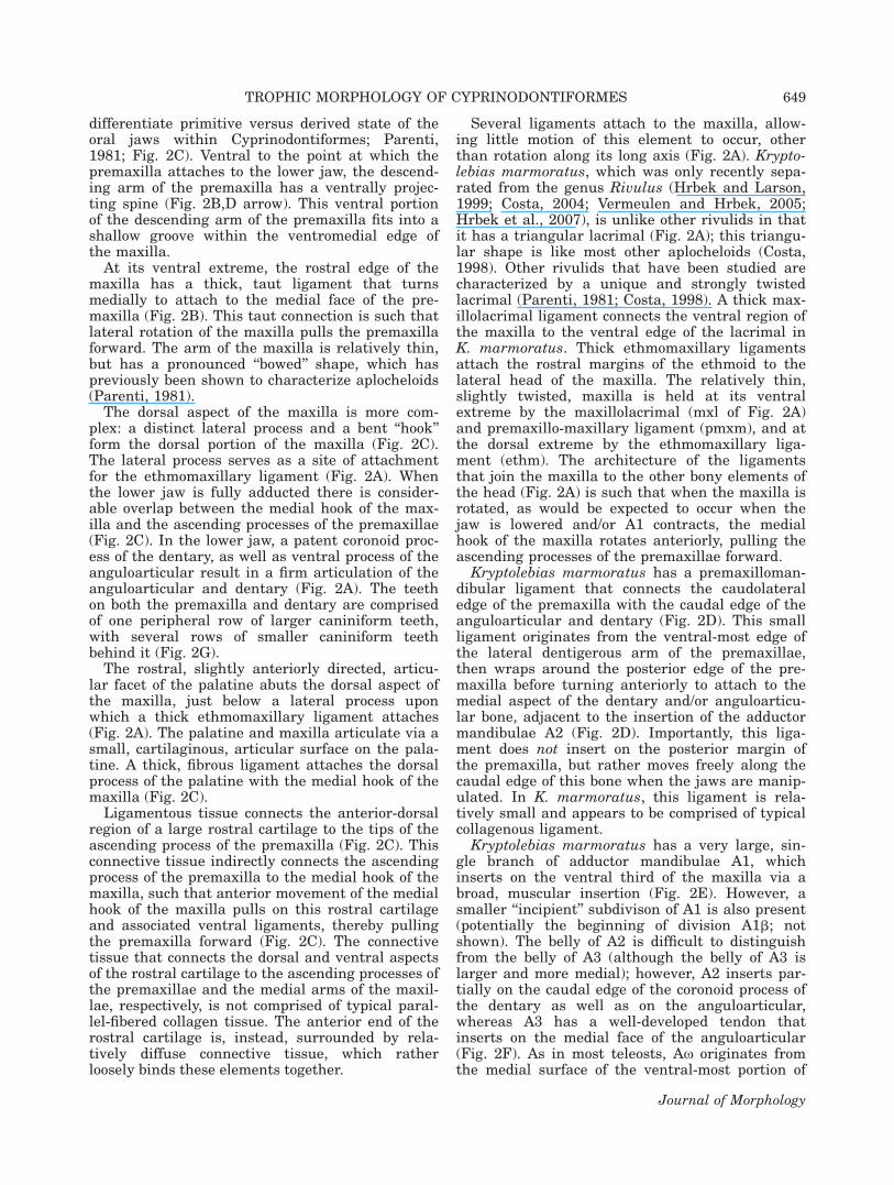
differentiate primitive versus derived state of theoral jaws within Cyprinodontiformes; Parenti,1981; Fig. 2C). Ventral to the point at which thepremaxilla attaches to the lower jaw, the descend-ing arm of the premaxilla has a ventrally projec-ting spine (Fig. 2B,D arrow). This ventral portionof the descending arm of the premaxilla fits into ashallow groove within the ventromedial edge ofthe maxilla.
At its ventral extreme, the rostral edge of themaxilla has a thick, taut ligament that turnsmedially to attach to the medial face of the pre-maxilla (Fig. 2B). This taut connection is such thatlateral rotation of the maxilla pulls the premaxillaforward. The arm of the maxilla is relatively thin,but has a pronounced ‘‘bowed’’ shape, which haspreviously been shown to characterize aplocheloids(Parenti, 1981).
The dorsal aspect of the maxilla is more com-plex: a distinct lateral process and a bent ‘‘hook’’form the dorsal portion of the maxilla (Fig. 2C).The lateral process serves as a site of attachmentfor the ethmomaxillary ligament (Fig. 2A). Whenthe lower jaw is fully adducted there is consider-able overlap between the medial hook of the max-illa and the ascending processes of the premaxillae(Fig. 2C). In the lower jaw, a patent coronoid proc-ess of the dentary, as well as ventral process of theanguloarticular result in a firm articulation of theanguloarticular and dentary (Fig. 2A). The teethon both the premaxilla and dentary are comprisedof one peripheral row of larger caniniform teeth,with several rows of smaller caniniform teethbehind it (Fig. 2G).
The rostral, slightly anteriorly directed, articu-lar facet of the palatine abuts the dorsal aspect ofthe maxilla, just below a lateral process uponwhich a thick ethmomaxillary ligament attaches(Fig. 2A). The palatine and maxilla articulate via asmall, cartilaginous, articular surface on the pala-tine. A thick, fibrous ligament attaches the dorsalprocess of the palatine with the medial hook of themaxilla (Fig. 2C).
Ligamentous tissue connects the anterior-dorsalregion of a large rostral cartilage to the tips of theascending process of the premaxilla (Fig. 2C). Thisconnective tissue indirectly connects the ascendingprocess of the premaxilla to the medial hook of themaxilla, such that anterior movement of the medialhook of the maxilla pulls on this rostral cartilageand associated ventral ligaments, thereby pullingthe premaxilla forward (Fig. 2C). The connectivetissue that connects the dorsal and ventral aspectsof the rostral cartilage to the ascending processes ofthe premaxillae and the medial arms of the maxil-lae, respectively, is not comprised of typical paral-lel-fibered collagen tissue. The anterior end of therostral cartilage is, instead, surrounded by rela-tively diffuse connective tissue, which ratherloosely binds these elements together.
Several ligaments attach to the maxilla, allow-ing little motion of this element to occur, otherthan rotation along its long axis (Fig. 2A). Krypto-lebias marmoratus, which was only recently sepa-rated from the genus Rivulus (Hrbek and Larson,1999; Costa, 2004; Vermeulen and Hrbek, 2005;Hrbek et al., 2007), is unlike other rivulids in thatit has a triangular lacrimal (Fig. 2A); this triangu-lar shape is like most other aplocheloids (Costa,1998). Other rivulids that have been studied arecharacterized by a unique and strongly twistedlacrimal (Parenti, 1981; Costa, 1998). A thick max-illolacrimal ligament connects the ventral region ofthe maxilla to the ventral edge of the lacrimal inK. marmoratus. Thick ethmomaxillary ligamentsattach the rostral margins of the ethmoid to thelateral head of the maxilla. The relatively thin,slightly twisted, maxilla is held at its ventralextreme by the maxillolacrimal (mxl of Fig. 2A)and premaxillo-maxillary ligament (pmxm), and atthe dorsal extreme by the ethmomaxillary liga-ment (ethm). The architecture of the ligamentsthat join the maxilla to the other bony elements ofthe head (Fig. 2A) is such that when the maxilla isrotated, as would be expected to occur when thejaw is lowered and/or A1 contracts, the medialhook of the maxilla rotates anteriorly, pulling theascending processes of the premaxillae forward.
Kryptolebias marmoratus has a premaxilloman-dibular ligament that connects the caudolateraledge of the premaxilla with the caudal edge of theanguloarticular and dentary (Fig. 2D). This smallligament originates from the ventral-most edge ofthe lateral dentigerous arm of the premaxillae,then wraps around the posterior edge of the pre-maxilla before turning anteriorly to attach to themedial aspect of the dentary and/or anguloarticu-lar bone, adjacent to the insertion of the adductormandibulae A2 (Fig. 2D). Importantly, this liga-ment does not insert on the posterior margin ofthe premaxilla, but rather moves freely along thecaudal edge of this bone when the jaws are manip-ulated. In K. marmoratus, this ligament is rela-tively small and appears to be comprised of typicalcollagenous ligament.
Kryptolebias marmoratus has a very large, sin-gle branch of adductor mandibulae A1, whichinserts on the ventral third of the maxilla via abroad, muscular insertion (Fig. 2E). However, asmaller ‘‘incipient’’ subdivison of A1 is also present(potentially the beginning of division A1b; notshown). The belly of A2 is difficult to distinguishfrom the belly of A3 (although the belly of A3 islarger and more medial); however, A2 inserts par-tially on the caudal edge of the coronoid process ofthe dentary as well as on the anguloarticular,whereas A3 has a well-developed tendon thatinserts on the medial face of the anguloarticular(Fig. 2F). As in most teleosts, Ax originates fromthe medial surface of the ventral-most portion of
TROPHIC MORPHOLOGY OF CYPRINODONTIFORMES 649
Journal of Morphology
the quadrate and inserts on the dentary, fillingmost of the Meckelian fossa. Because the morphol-ogy of Ax is not different among taxa used in thisstudy, we will not describe the morphology of thisbranch for other cyprinodontiform species.
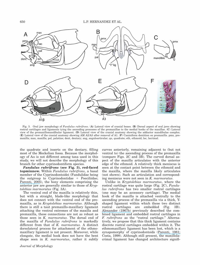
Fundulus rubrifrons (see Fig. 3), red-facedtopminnow. Within Fundulus rubrifrons, a basalmember of the Cyprinodontoidei (Fundulidae beingthe outgroup to Cyprinodontidae 1 Poeciliidae;Parenti, 2005), the bony elements comprising theanterior jaw are generally similar to those of Kryp-tolebias marmoratus (Fig. 3A).
The ventral end of the maxilla is relatively thin,but with a straight, blade-like morphology thatdoes not connect with the ventral end of the pre-maxilla, as in Kryptolebias marmoratus. Althoughthere is still a taut premaxillo-maxillary ligamentattaching the ventral extremes of the maxilla andpremaxilla, these connections are not as robust asthose seen in K. marmoratus. The dorsal end ofthe maxilla of Fundulus rubrifrons is markedlydifferent from that of K. marmoratus. A distinctdorsolateral process for attachment of the ethmo-maxillary ligament is not present. Moreover, whileelongate, the medial hook does not have the bentshape seen in K. marmoratus, rather it subtly
curves anteriorly, remaining adjacent to (but notventral to) the ascending process of the premaxilla(compare Figs. 2C and 3B). The curved dorsal as-pect of the maxilla articulates with the anterioredge of the ethmoid. A relatively thick meniscus isseen at the contact point between the ethmoid andthe maxilla, where the maxilla likely articulates(not shown). Such an articulation and correspond-ing meniscus were not seen in K. marmoratus.
Unlike in Kryptolebias marmoratus, where therostral cartilage was quite large (Fig. 2C), Fundu-lus rubrifrons has two smaller rostral cartilages(one may be an accessory cartilage). The medialhook of the maxilla is attached ventrally to theascending process of the premaxilla via a thick, Y-shaped ligament within which these two distinctrostral cartilages are embedded (Fig. 3B).Alexander (1967b) previously described the com-bined ligament and embedded rostral cartilages inF. rubrifrons as the ‘‘rostral cartilage.’’ Alterna-tively, we propose that this thick ligament containsdiscrete rostral cartilages embedded within it. Theethmomaxillary ligament has been lost, which is asynapomorphy of cyprinodontoids (Parenti, 1981;Costa, 1998). Although still present, the maxillola-crimal ligament has changed architecture signifi-
Fig. 3. Oral jaw morphology of Fundulus rubrifrons. (A) Lateral view of cranial bones. (B) Dorsal aspect of oral jaws showingrostral cartilages and ligaments tying the ascending processes of the premaxillae to the medial hooks of the maxillae. (C) Lateralview of the premaxillomandibular ligament. (D) Lateral view of the cranial anatomy showing the adductor mandibulae complex.(E) Lateral view of the cranial anatomy showing AM A2/A3 after removal of A1. (F) Caniniform dentition on premaxilla. pmx, pre-maxilla; max, maxilla; pal, palatine; dent, dentary; ang, anguloarticular; qu, quadrate; eth, ethmoid; lac, lacrimal.
650 L.P. HERNANDEZ ET AL.
Journal of Morphology

cantly in F. rubrifrons and the lacrimal bone itselfis more rectangular in shape. Importantly, this lig-ament attaches to the maxilla approximately 2/3 ofthe way up its shaft.
Several rows of similarly sized caniniform teethline the dentary and premaxilla (Fig. 3F).
The palatine in Fundulus rubrifrons is shapeddifferently from that seen in Kryptolebias marmor-atus. Within F. rubrifrons, the palatine is charac-terized by a blunt facet, which serves as both asite of articulation and as a site of ligamentousconnection with the maxilla. The point of articula-tion is at the maxilla’s most dorsal aspect wherethe medial hook begins curving anteriorly. A thickligament also attaches the palatine to the medialprocess of the maxilla (Fig. 3B). Within F. rubri-frons, the premaxillomandibular ligament is widerand covers a slightly larger portion of the ventro-lateral face of the premaxilla than that in K. mar-moratus (Fig. 3C).
In contrast to the adductor musculature of theaplocheloids, Fundulus rubrifrons is characterizedby two distinct divisions of adductor mandibulaeA1 (A1a and A1b; Ballantijn et al., 1972), bothinserting on the ventral third of the maxilla. A1binserts muscularly on the ventrolateral edge of themaxilla, whereas A1a inserts more medially, via ashort, discrete tendon.
To assess the degree of intrageneric muscularvariation, we also examined the adductor mandi-bulae complex within specimens of Fundulus simi-lis, F. chrysotus, and F. grandis. Fundulus spp.showed little variation except in the size of differ-ent divisions of the AM complex and slight differ-ences in insertions of the different divisions.
Gambusia affinis (see Fig. 4), mosquitofish.In Gambusia affinis, a relatively basal member ofthe Poeciilidae, the premaxilla is relatively shorterthan that seen in either Kryptolebias marmoratusor Fundulus rubrifrons (Fig. 4A). Moreover, from adorsal aspect, the ascending process of the pre-maxilla in G. affinis is more blunt and triangularin shape, with a broadened connection with thedescending arm of the premaxilla (Fig. 4B).
The maxilla of Gambusia affinis is more robustthan that of Fundulus rubrifrons, with a widerventral end (a condition previously described forpoeciliids; Parenti, 1981; Costa, 1998). In G. affinisthe maxilla has both an expanded dorsolateralprocess, and a slightly curved, but flattened medialprojection. Importantly, unlike both Kryptolebiasmarmoratus and F. rubrifrons, there is little over-lap in dorsal aspect between the medial hook ofthe maxilla and the ascending process of the pre-maxilla (Fig. 4B).
Neither the coronoid nor the posteroventralprocess of the dentary is as long as those seen inFundulus rubrifrons, thus the anguloarticular anddentary are not tightly linked (Fig. 4A). An elon-gate ventral process characterizes the anguloartic-
ular (Costa, 1998). Several rows of caniniformteeth line both the dentary and premaxilla (Fig.4E). The darker tips of the teeth suggest that theenamel may incorporate iron (Fig. 4E).
The head of the palatine is shaped like a ham-mer (Fig. 4A), with a broad anterior head articu-lating with a process on the dorsal edge of theshaft of the maxilla (Fig. 4B). The broad, cartilagi-nous facet at the anterior end of the palatine (asynapomorphy of poeciliids; Costa, 1998) suggeststhat there is significant movement or contact withthe maxilla against this process.
Rostral cartilages have been lost. Unlike in Kryp-tolebias marmoratus and Fundulus rubrifrons, tautligaments do not tie the ascending process of thepremaxilla to the medial arm of the maxilla.Although there are still loose ligaments connectingthese two elements, ligaments are thinner and nottaut. Importantly, without taut connections betweenthe ascending process of the premaxilla and themedial hook of the maxilla, it appears unlikely thattwisting of the maxilla can transfer force to thepremaxilla and thereby effect protrusion.
The slightly wider maxilla (relative to that ofFundulus rubrifrons) allows for more distinctinsertions of A1a and A1b. A2 and A3 are stillsomewhat interconnected, however the bulk of A2originates from the suspensorium (deep to A1) andinserts muscularly on the dorsocaudal edge of thedentary. A small dorsal section of A2 appears toinsert on ligamentous tissue that invests the pre-maxilla. The bulk of A3 is found deep to A2 andinserts on the medial face of the anguloarticularvia a discrete tendon (Fig. 4D). Examination ofGambusia holbrooki revealed only subtle size dif-ferences in the different divisions of the AM com-plex and the premaxillomandibular ligament.
Heterandria formosa (see Fig. 5), least killi-fish. One obvious difference between Heterandriaformosa and other cyprinodontiforms is that theposteroventral process of the dentary has beenlost, thus this bone is significantly shorter (Fig.5A). The ascending arm of the premaxilla is alsoquite short, relative to other taxa, with a widebase where the ascending arm abuts the descend-ing arm (Fig. 5B). The ventral edge of the descend-ing arm of the premaxilla has a strongly recurvedshape, with a distinct ventral spine (see arrow-head, Fig. 5A).
When compared with the other species describedthus far, Heterandria formosa has the simplestdorsal process and shortest medial hook of themaxilla. Importantly, the medial hook of the max-illa is separated widely from the ascending processof the premaxilla (Fig. 5B), a trait with clear func-tional implications. Because of this separation,within fresh specimens of H. formosa, the anteriormovement of the premaxilla does not result inrotation of the maxilla. The maxilla appears toform a stable base from which the premaxilla
TROPHIC MORPHOLOGY OF CYPRINODONTIFORMES 651
Journal of Morphology

protrudes. Moreover, we note that manual ventralrotation of the lower jaw in fresh specimens gener-ates synchronized movement of the dentary andpremaxilla, with little, if any, concomitant move-ments of the maxilla.
The anguloarticular and dentary of the lowerjaw do not interdigitate strongly (Fig. 5A). Severalrows of pointed, slightly spatulate teeth line boththe dentary and premaxilla.
The palatine has a reduced articular facet whencompared with Gambusia affinis. However, giventhat Heterandria formosa is a secondarily minia-turized species (Rosen and Bailey, 1963), we sug-gest that major functional implications should notbe attached to this finding. Though the palatine issmall, a patent articular facet suggests that thereis significant movement or pressure from themaxilla against this process (Fig. 5B).
Fig. 4. Oral jaw morphology of Gambusia affinis. (A) Lateral view of cranial bones. (B) Dorsal aspect of oral jaws showing thepalatomaxillary ligament. (C) Lateral view of the cranial anatomy showing the adductor mandibulae complex. (D) Lateral view ofthe cranial anatomy showing AM A2/A3 after removal of A1. (E) Caniniform dentition on dentary. pmx, premaxilla; max, maxilla;pal, palatine; dent, dentary; ang, anguloarticular; qu, quadrate; eth, ethmoid.
652 L.P. HERNANDEZ ET AL.
Journal of Morphology

The connections between the palatine and max-illa are generally similar to those seen in Gambu-sia affinis. The point of articulation between thepalatine and maxilla is ventral to the dorsal proc-ess of the maxilla. In Kryptolebias marmoratusand Fundulus rubrifrons, the point of articulationwas near the dorsal end of the maxilla, whereas inHeterandria formosa, the point of articulation isclearly upon the arm of the maxilla (Fig. 5B). Apalatomaxillary ligament connects the dorsal headof the palatine to the small dorsolateral process ofthe maxilla.
Poecilia sphenops (see Fig. 6), molly. WithinPoecilia sphenops we see significant morphologi-cal changes in the architecture of the jawelements relative to more basal members. Thestrongly recurved descending arm of the premax-
illa is more robust (from the lateral aspect) thanin most other cyprinodontiforms. The ascendingprocess of the premaxilla is shorter than in Gam-busia affinis, but not as short as in Heterandriaformosa, with wide bases merging with a mark-edly wider descending arm of the premaxilla(Fig. 6A,B).
Although the ventral arm of the maxilla is simi-lar in shape to that of Gambusia affinis, the dorsalportion, which articulates with the neurocranium,is more complex. The broad lateral process has apronounced ridge running dorsoventrally along itsface, and the medial hook is longer and even morecomplex in shape than in G. affinis (Fig. 6B).Importantly, there is no overlap between theascending process of the premaxilla and themedial hook of the maxilla.
Fig. 5. Oral jaw morphology of Heterandria formosa. (A) Lateral view of cranial bones, note pronounced spine on descendingarm of the premaxilla (arrowhead). (B) Dorsal aspect of oral jaws showing the palatomaxillary ligament on the left side. The rightside is rendered transparent to show the size and shape of the medial aspect of the maxilla. (C) Pointed, slightly spatulate denti-tion on dentary. pmx, premaxilla; max, maxilla; pal, palatine; dent, dentary; ang, anguloarticular; qu, quadrate; eth, ethmoid.
TROPHIC MORPHOLOGY OF CYPRINODONTIFORMES 653
Journal of Morphology

The posteroventral process of the dentary isshort, resulting in a loose connection between theanguloarticular and dentary. The coronoid processof the dentary is widely separate from the coronoidprocess of the anguloarticular. A patent Meckelscartilage unites the disarticulated bony elementsof the lower jaw (Fig. 6B). One row of thin, elon-gated, hooked teeth are peripheral to several rowsof smaller, less recurved teeth (Fig. 6E).
The palatine has a prominent facet where itarticulates with the maxilla (Fig. 6A,B). As inmost other poeciliids examined, the palatine arti-culates with the shaft of the maxilla, ventral tothe dorsal process of the maxilla. This is markedlydifferent than the architecture of Fundulus rubri-frons and Kryptolebias marmoratus, where articu-lation was at the dorsal-most portion of themaxilla. Such differences in sites of articulationlikely affect jaw mechanics. The palatine curvesanteriorly and has only one articular facet. A thickpalatomaxillary ligament attaches the dorsal as-pect of the palatine to a thin, elevated process onthe dorsolateral process of the maxilla (Fig. 6B).
The premaxillomandibular ligament connectingthe premaxilla and lower jaw is complex, splittinginto two elements.
In most teleosts, A3 originates deep to A2, ashas been described here for more basal cyprino-dontiforms. In Poecilia sphenops and Xiphophorushelleri (see Fig. 7), both A2 and A3 comprise onesuperficial mass that forms the bulk of the adduc-tor mandibulae. In P. sphenops A2 bifurcates suchthat a large proportion of this division goes to thepremaxilla (Fig. 6D). The insertion of A2 in P.sphenops is via a discrete tendon to the caudaledge of the dentary, whereas the insertion of A2 onthe upper jaw is via a less discrete tendinousconnection to the anterior premaxilla.
Examination of Poecilia latipinna revealed nointrageneric variation in morphology. Although A2/A3 appeared smaller than in P. sphenops, therewere no notable differences in muscular architec-ture between these species.
Xiphophorus helleri (see Fig. 7), Swordtail.Within Xiphophorus helleri, also a poeciliid, theoverall architecture of the bony elements compris-
Fig. 6. Oral jaw morphology of Poecilia sphenops. (A) Lateral view of cranial bones. (B) Dorsal aspect of oral jaws showing thepalatomaxillary ligament on the left side. The right side is rendered transparent to show the size and shape of the medial side ofthe maxilla. (C) Lateral view of the A1. (D) Medial view of the cranial anatomy showing AM A2/A3. (E) Hooked, elongated teethwith shorter teeth at their base. pmx, premaxilla; max, maxilla; mc, Meckels cartilage; pal, palatine; dent, dentary; ang, anguloar-ticular; qu, quadrate; eth, ethmoid.
654 L.P. HERNANDEZ ET AL.
Journal of Morphology

ing the anterior jaws has undergone significantchanges relative to the other species studied here.Although the basic shape of elements isunchanged, the manner in which they articulatewith one another has changed. In a resting state,the premaxilla is largely laterally occluded by themaxilla. This does not reflect an increase in thesize of the maxilla, but rather reflects a posteriorshift in the resting position of the premaxilla (com-pare Figs. 7A and 6A). The greatly reducedascending process of the premaxilla is shifted lat-erally (Fig. 7B).
The dorsal process of the maxilla has a verycomplex shape, with a prominent dorsolateral shelfof bone, and a large twisted medial hook (Fig. 7B).There is an overlap between the medial hook ofthe maxilla and the modified ascending process ofthe premaxilla (Fig. 7B). However, the long medialhook of the maxilla may serve as a strut againstwhich the premaxilla can be protruded, because,as seen in Heterandria formosa, manually rotating
the lower jaw causes little movement of the max-illa.
The extremely foreshortened lower jaw containsa visible gap between the dentary and anguloartic-ular that is partially filled by a patent Meckel’scartilage (Fig. 7C). Although a discrete coronoidprocess of the dentary is visible, the posteroventralprocess is significantly shortened (Fig. 7C). Onerow of elongated, straight-edged, recurved, spatu-late teeth lines both the dentary and premaxilla.These larger teeth are followed by several rows ofshorter, slightly spatulate teeth (Fig. 7D).
Unlike in other poeciliids examined, a thickmaxillary ligament (m.l.) connects the two dorsalprocesses of the maxillae (Fig. 7B).
As in Poecilia sphenops the greatly modified pre-maxillomandibular ligament connecting the pre-maxilla and lower jaw is split into two elements(Fig. 7C). Although one portion of this ligamentcurves around the back of the premaxilla to attachto the dentary, as described for other species, an
Fig. 7. Oral jaw morphology of Xiphophorus helleri. (A) Lateral view of cranial bones, showing unique architecture of premaxil-lae. (B) Dorsal aspect of oral jaws showing the palatomaxillary ligament, as well as a ligament (m.l.) connecting the dorsal headsof the maxillae. (C) Lateral and medial view of the premaxillomandibular ligament, showing the increased complexity of this liga-ment. Dark portions of this ligament represent hyaline cartilage whereas lighter areas connote more typical collagenous tissue. (D)Spatulate, straight edged dentition on outer row with rows of smaller teeth behind. pmx, premaxilla; max, maxilla; pal, palatine;dent, dentary; ang, anguloarticular; qu, quadrate; eth, ethmoid; m.l., maxillary ligament; pmm, premaxillomandibular ligament;mc, Meckels cartilage.
TROPHIC MORPHOLOGY OF CYPRINODONTIFORMES 655
Journal of Morphology

additional component of this ligamentous structurealso inserts on the anguloarticular. Thus, the sin-gle, relatively simple ligament observed in Krypto-lebias marmoratus, Fundulus rubrifrons, andGambusia affinis is now divided distally to attachto both the coronoid process of the dentary (adja-cent to the insertion of A2) and the anguloarticu-lar. Moreover, the material comprising this com-plex ligament has changed, with a large portion ofwhat appears to be hyaline-cell cartilage (Benja-min, 1989) located between the collagenous attach-ment points of the ligament (LPH pers. obs.).
A3 originates on the preopercular and the bulkof its muscle fibers are largely located within thecaudal half of the suspensorium. A2 originates notsolely from the suspensorium, but also from therostral, ligamentous face of A3. Thus, A2 and A3form a tightly coupled mass where the more
rostral portion constitutes A2, and the more caudalportion constitutes A3. A2 has a small, but dis-crete, tendon that inserts on the premaxilla, whilethe remainder of A2 inserts on the coronoid proc-ess of the dentary via a robust tendon.
Jordanella floridae (see Fig. 8) Americanflagfish. Within Jordanella floridae, a member ofCyprinodontidae (sister family to Poeciliidae;Parenti, 2005), the general cyprinodontiform oste-ology of the anterior jaws is retained; however, wealso see significant morphological changes withpossible functional implications (Fig. 8A).Although many basal features characterized theosteology, the musculature is characterized byseveral derived traits seen in poeciliid species.
As in Poecilia sphenops, the ascending process ofthe premaxilla is short, however, as in Fundulusrubrifrons, it is relatively narrow where it merges
Fig. 8. Oral jaw morphology of Jordanella floridae. (A) Lateral view of cranial bones. (B) Dorsal aspect of oral jaws showing athick palatomaxillary ligament, and a patent ligament between the medial hooks of the maxilla that holds a small (perhaps vestig-ial) rostral cartilage. (C) Lateral view of adductor mandibulae A1. (D) Medial view showing AM A2/A3 after removal of A1. (E) Tri-cuspid dentition on dentary. pmx, premaxilla; max, maxilla; pal, palatine; dent, dentary; ang, anguloarticular; qu, quadrate; eth,ethmoid.
656 L.P. HERNANDEZ ET AL.
Journal of Morphology

with the descending arm of the premaxilla(Fig. 8B).
As is characteristic for cyprinodontids (Parenti,1981), Jordanella floridae shows an expanded dor-solateral process of the maxilla. However, themedial hook of the maxilla is smaller, and morepointed, than in the other species examined.
In contrast to Poecilia sphenops, the posteroven-tral process of the dentary of Jordanella floridae isnot foreshortened. Patent coronoid and posteroven-tral processes of the dentary create a more tightlyarticulated lower jaw than is seen in the morederived poeciliids. A single row of tricuspid teethis found on both the premaxilla and dentary(Fig. 8E).
The palatine is shaped like a hammer with ananterior articulating facet (as described for otherspecies) but also with a pointed posterior process(Fig. 8B; Character 29 in Costa, 1998). A thick lig-ament arises from this posteriorly oriented processand curves to attach to the dorsolateral process ofthe maxilla.
As in Poecilia sphenops, loose tissue connectsthe ascending process of the premaxilla to themedial hook of the maxilla. Interestingly, althoughthe rostral cartilage is lost within most poeciliids,in this cyprinodontid a small, non-staining rostralcartilage can be seen within the ligament tying to-gether the medial hooks of the maxillae (Fig. 8B*).
The premaxillomandibular ligament connectingthe premaxilla and lower jaw is similar to thatdescribed for Fundulus rubrifrons.
The adductor mandibulae shows a combinationof character states when compared with the spe-cies studied thus far. Although A2/A3 fibers areintermingled, in Jordanella floridae A3 is deep toA2 and therefore unlike the muscular complexdescribed for Poecilia sphenops and Xiphophorushelleri (Figs. 6D vs. 8D). However, A2 bifurcates atits distal end, into relatively equal halves, to inserton both the premaxilla and dentary. Althoughthere is a muscular insertion to the caudolateraledge of the premaxilla (and associated connectivetissues), a long tendinous insertion goes to thedentary. A3 inserts on the medial side of the angulo-articular.
The adductor mandibulae complex of other cyp-rinodontids suggests that there may be significantvariation in trophic musculature within this fam-ily. Floridichthys carpio does not show the clearinsertion of A2 on the premaxilla that character-izes Jordanella floridae; whereas A2 still insertson the coronoid process of the dentary, there is noevidence of muscle fibers going to the premaxilla.Cyprinodon variegatus has jaw musculature morelike J. floridae, whereby A3 is deep to A2. How-ever, as seen in Poecilia sphenops, the bulk of A3is caudal to the bulk of A2. Additionally, C. varie-gatus has a premaxilla more like that seen in Fun-dulus rubrifrons or Kryptolebias marmoratus, with
a pronounced ascending process. In C. variegatus,A2 attaches by a distinct tendon to the coronoidprocess of the dentary, but also extends a fewfibers to the premaxilla. As seen in Heterandriaformosa and Gambusia affinis, muscle fibers fromA2 actually attach to the ligaments surroundingthe premaxilla, instead of attaching directly to thebone. It is possible that this architecture is repre-sentative of a transitional state between no inser-tion (as is seen in more basal cyprinodontiforms)and a discrete muscle attaching to the premaxilla(as is seen in J. floridae).
DISCUSSION
We posit that in the course of cyprinodontiformevolution, several mechanisms have arisen thatprotrude and retract the jaws. Specifically, changesto three key anatomical features of the feedingmechanism appear to be involved (see Fig. 1):1) changes in the architecture of the major jawclosing muscle, the adductor mandibulae complex;2) specialization of a ligament that connectsthe upper and lower jaws (premaxillomandibularligament); and, 3) pronounced changes in otherligamentous attachments that connect anteriorelements of the feeding apparatus. This researchdiffers from previous work in emphasizing the im-portance of ligamentous attachments in the evolu-tion of trophic systems.
Architecture and Novel Insertions ofAdductor Mandibulae A2
In most teleosts, A2 inserts on the anguloarticu-lar (Winterbottom, 1974). Even when A2 is second-arily duplicated, both divisions typically insert onthe anguloarticular (Friel and Wainwright, 1997).Within even the most basal cyprinodontiformexamined here, however, at least a portion of A2inserts on the dentary. This may suggest that,even during their earliest evolutionary history,cyprinodontiforms had an unusual architecture ofthe adductor mandibulae complex.
Although we see insertion of A2 on the dentaryas early as Kryptolebias marmoratus, a patentcoronoid process of the dentary creates a relativelycaudal insertion within the lower jaw in morebasal cyprinodontiforms. The evolutionary trendtoward foreshortened jaws, brought about by thedecreased size of the coronoid process, moves theinsertion of A2 rostrally in more derived poeciliids.Such a rostral migration may allow force to bemore directly transmitted to the anterior lowerjaw, which likely facilitates direct application ofmuscle force to the teeth. It should be noted thatthe loss of a prominent coronoid process mightsimultaneously lead to decreased mechanicaladvantage during feeding because of a shortenedlever arm. Moreover, given the intramandibular
TROPHIC MORPHOLOGY OF CYPRINODONTIFORMES 657
Journal of Morphology

bending seen in derived poeciliids (Gibb et al.,2008), this rostral shift may decouple the anteriorand posterior lower jaw. Such decoupling mayallow fish to procure prey using quasi-independentmovements of the anterior jaw tips.
Controlled movements of the upper jaws are alsopossible given the acquisition of a novel branch ofA2 inserting on the premaxilla within morederived poeciliids and cyprinodontids. We see mus-cle fibers impinging on ligaments surrounding thepremaxilla within Gambusia affinis, and the evolu-tionary trend toward direct muscular control ofthe upper jaw continues in more derived membersof Poeciliidae. Such a novel architecture of theadductor mandibulae complex likely allows for con-trolled adduction of the jaws; both upper and lowerjaw elements would serve as third-order leversthat are simultaneously moved toward oneanother—mechanically, the jaws would functionsimilarly to a pair of forceps.
The rostral shift in insertion of A2 on the den-tary and the insertion of A2 on the premaxillaappear to constitute a gradual transformation ofthe feeding apparatus. Although A2 inserts on thedentary in all species examined, the foreshorteningof the dentary has generated a gradual anteriorly-directed shift in the insertion of this key jawadductor. Moreover, within the poeciliids examinedhere, muscle fibers initially impinge upon the con-nective tissues surrounding the premaxilla andultimately come to insert directly on the premax-illa. Early in teleostean evolutionary history, mus-cle fibers impinged on the primordial ligament,eventually generating a new division of the adduc-tor mandibulae, A1 (Schaeffer and Rosen, 1961).We see a similar gradual transformation withinderived poeciliids, where migration of musclefibers eventually generates a new insertion for A2.Thus, evolution of the adductor musculaturewithin this clade likely constitutes an example ofgradual evolution of a derived trait.
The transformation of the adductor mandibulaecomplex within Cyprinodontiformes is strikinglysimilar to the changes that have been observedwithin the Loricaroidea. Schaefer and Lauder(1986, 1996) described the functional transforma-tion of the anterior jaws within Loricaroidea (thearmored catfishes), where relatively little protru-sion is demonstrated by basal taxa but pronouncedprotrusion coupled with independent movementsof the upper jaw characterize derived members ofthis clade. Within the species examined here, weobserve a similar gradual transformation of theadductor mandibulae complex—from species thatshow a ‘‘typical’’ teleostean morphology of theadductor mandibulae to species that possess thederived character of a branch of A2 that inserts onthe premaxilla. Schaefer and Lauder (1986, 1996)proposed that such a historical transformation wasassociated with a functional specialization for algal
scraping. The similarity between these feedingmechanisms may represent functional convergencebetween Loricaroidea and Cyprinodontiformes.
Interestingly, the likely independent origin of apremaxillary insertion of A2 in both derived poeci-liids and cyprinodontids (see Fig. 1) is correlatedwith the acquisition of dentition generally associ-ated with scraping. Both the rake-like teeth of Poe-cilia sphenops and Xiphophorus helleri, as well asthe tricuspid teeth of Jordanella floridae havebeen correlated with a scraping feeding mode(Yamaoka, 1983; Yagishita and Nakabo, 2003).
Acquisition of a Novel Ligament Connectingthe Upper and Lower Jaws
One of the most significant changes seen duringthe course of cyprinodontiform trophic evolution isthe reorganization of the ligaments that connectthe various osteological elements of the anteriorjaws. Within all the species examined here, a pre-maxillomandibular ligament connects the descend-ing arm of the premaxilla to the dentary (Her-nandez et al., 2008). Although a premaxilloman-dibular ligament providing a taut connectionbetween these two elements has been previouslydescribed for other taxa (Schaeffer and Rosen,1961), the architecture of the cyprinodontiform lig-ament is unique. When the dentary is depressed,this ligament pulls the premaxilla forward. More-over, this ligament significantly varies in morphol-ogy among cyprinodontiform species.
We suggest that the unique architecture of thisligament will enhance premaxillary protrusion. Ithas previously been suggested that a ligamentousattachment between the premaxilla and lower jawcould improve protrusion ability (Schaeffer andRosen, 1961; Motta, 1982, 1984). It is important tonote that because this ligament is not constrainedwhere it wraps around the posterior edge of the pre-maxillae, the premaxilla will follow the movementof the lower jaw directly during mandibular (lowerjaw) depression. Therefore, the range of premaxil-lary protrusion produced during mouth opening islikely determined directly by the degree of lowerjaw depression. Given that Poecilia sphenops hasbeen shown to produce extreme gape angles (>1208;Ferry-Graham et al., 2008; Gibb et al., 2008), thetype of ligamentous attachment we describe herelikely facilitates maximal upper jaw protrusion.
Although the attachment of this ligament to thelateral face of the descending arm of the premax-illa is relatively conserved among all species, theattachment to the lower jaw varies among the dif-ferent species examined. This ligament attaches toeither the dentary or anguloarticular bone (or toboth), and the location of this attachment usuallycorresponds with the location of the insertion ofA2 onto the lower jaw. Moreover, there is a pro-nounced change in the type of connective tissue
658 L.P. HERNANDEZ ET AL.
Journal of Morphology
that comprises the ligament. In Kryptolebias mar-moratus, Fundulus rubrifrons, Gambusia affinis,and Jordanella floridae it is largely comprised offibrous, collagenous tissue whereas in morederived poeciliids it appears increasingly cellular(LPH; pers. obs.). Within more derived poeciliids(Poecilia sphenops) it appears to be composedlargely of hyaline cell cartilage (Benjamin, 1989).
Within some poeciliids (e.g., Xiphophorus helleriand Poecilia sphenops), the premaxillomandibularligament has become bifurcated distally to attachto both the anguloarticular and dentary (see Fig.1). Moreover, examination of fresh specimens sug-gests that the tissue composition of this ligamenthas changed, with the basal condition showingmore collagenous fibers whereas the derived condi-tion shows increasingly cellular tissue, such as thehyaline cell cartilage seen in Poecilia sphenops(Benjamin, 1989). Poecilia sphenops, more thanthe other species examined here, relies onrepeated scraping motions of the jaws to graze onepiphytic algae (Zaret and Rand, 1971, Gibb et al.,in press). This modified premaxillomandibular lig-ament may confer a looser connection between thejaw elements. Scraping parrotfish demonstratelooser connections among the different bones ofthe oral jaw apparatus (Bellwood and Choat, 1990)as do girellids that use scraping (Yagishita andNakabo, 2003). A loose connection is thought toallow for the production of larger gapes forincreasing contact with the substrate for scraping.
Architectural Changes Associated WithPremaxillary Protrusion
Although considerable research has attemptedto elucidate mechanisms of premaxillary protru-sion across teleost taxa (e.g., Alexander, 1967b;Motta, 1984), we suggest that the ligamentousattachments that produce and coordinate thesemovements have been neglected. Here we showhow changes in the premaxillomandibular as wellas other key ligaments likely affect the protrusionmechanism used in this clade. The varied shapesof osteological elements (maxillae, palatines, den-taries, premaxillae, and neurocranium), coupledwith varying configurations of the ligaments thatconnect these elements to one another, likelydetermine both the underlying mechanism andfunctional implications of this type of premaxillaryprotrusion.
The aplocheloid Kryptolebias marmoratus servedas our representative outgroup taxa. Within K.marmoratus the taut ethmomaxillary ligamentand the architecture of the palatomaxillary liga-ment combine to restrict the movement of themaxilla to lateral rotation. Thus, lowering the jawand/or contracting A1 will result in outward rota-tion of the medial hooks of the maxillae (Fig. 2C),which will in turn pull the ligaments that connect
the maxillae to the ascending processes of the pre-maxillae (via the rostral ligament) anteriorly andhelp project the upper jaw. Although the size ofthe rostral cartilage(s) is reduced, a similar situa-tion occurs in Fundulus (Alexander, 1967a). Thus,in these two basal groups (aploceloids and fundul-ids) premaxillary protrusion is accomplished (or atleast assisted) by ‘‘twisting’’ of the maxilla (Eaton,1935; Alexander, 1967a).
The first major modification to this system is aloss or significant reduction in the rostral carti-lages and associated ligaments that attach themedial hooks of the maxillae to the ascendingprocesses of the premaxillae. Alexander (1967a)noted the loss of these structures in Xiphophorushelleri, concluding that twisting of the internalhooks of the maxillae did not contribute topremaxillary protrusion. Clearly, without a tautligament uniting these elements, premaxillary pro-trusion cannot be effected by lateral rotation ortwisting of the maxilla. Coupled with this changeis a foreshortening of the ascending process of thepremaxilla. Notably, within most poeciliids andcyprinodontids, there is little if any overlapbetween the ascending processes of the premaxil-lae and the medial hooks of the maxillae––a neces-sary configuration for premaxillary protrusion tooccur via maxillary rotation.
Pronounced premaxillary protrusion is typicallyassociated with a greatly lengthened ascendingprocess (Eaton, 1935; Lauder and Liem, 1981;Westneat and Wainwright, 1989). Within cyprino-dontiforms, significant protrusion can still beaccomplished with an evolutionary decrease in thelength of the ascending process. It is worth notinghowever that increased premaxillary protrusion inother species is correlated with increased stealthduring a suction feeding event (Motta, 1984). Incontrast, the role of increased premaxillary protru-sion in improving biting, picking, and scrapingperformance (the role it likely serves within cypri-nodontiforms) is still largely unexamined (but seeFerry-Graham et al., 2008). A novel means of pro-trusion whereby increased protrusion is decoupledfrom increased length of the ascending processmay facilitate the evolution of feeding modesbeyond the ubiquitous ram- or suction-based mech-anisms. For example, poeciliids trying to scrapealgae by applying their mouths fully to the sub-strate might be constrained in the maneuverabilityof the jaws by very long ascending processes.
In these higher cyprinodontiform taxa, there isalso a significant change in both the shape ofbones and the architecture of ligaments connectingthe maxilla to the rest of the feeding apparatus.The basal condition is rotation between the max-illa and the palatine at the dorsal extreme of themaxilla (Figs. 2 and 3A), whereas the morederived condition is to have the point of articula-tion more ventrally located, on the arm of the
TROPHIC MORPHOLOGY OF CYPRINODONTIFORMES 659
Journal of Morphology
maxilla. This derived condition should serve tobrace the maxilla, rather than to effect its rota-tion. Indeed, Schaeffer and Rosen (1961) as well asMotta (1984) have suggested that the maxilla actsas a brace against which the premaxilla is pro-truded. Indeed, depression of the lower jaw effectslittle maxillary movement but generates signifi-cant protrusion of the premaxillae in more derivedmembers of Poeciliidae.
Although there are substantial changes to thebony elements of the feeding apparatus withincyprinodontiforms, changes in ligamentous andmuscular attachments may be just as important inenhancing novel feeding modes. We have describedchanges in the ligamentous architecture of theoral jaws that likely result in a functional transfor-mation from premaxillary protrusion via twistingof the maxilla to a new, novel means of premaxil-lary protrusion that employs a novel ligamentouscoupling of the upper and lower jaws by a greatlymodified premaxillomandibular ligament. Duringthe course of this transition, there is a concomitantloss of the rostral cartilages, loss of the ligamentsthat attach the rostral cartilages to the jaws,reduction of the ascending processes of the pre-maxillae, and a significant increase in the size andcomplexity of the premaxillomandibular ligament.We suggest that within this group such changesfacilitate a general transition towards precision-based picking and scraping feeding modes.
ACKNOWLEDGMENTS
We thank the following people for the loaning ofspecimens used in this study (and for specimensultimately not included): Lynne Parenti (Smithso-nian National Museum of Natural History), PhilipMotta (University of South Florida), Dave Catania(California Academy of Sciences), Daphne Geh-ringer (Moss Landing Marine Labs). We also thankNathan Bird and Katie Staab for assistance withspecimen preparations at GWU.
LITERATURE CITED
Alexander RM. 1967a. Mechanisms of the jaws of some atheri-niform fish. J Zool (Lond) 151:233–255.
Alexander RM. 1967b. The functions and mechanisms of theprotrusible upper jaws of some acanthopterygian fish. J Zool151:43–64.
Arthington AH. 1989. Diet of Gambusia affinis holbrooki,Xiphophorus helleri, X. maculatus and Poecilia reticulata(Pisces: Poeciliidae) in streams of southeastern Queensland,Australia. Asian Fish Sci 2:193–212.
Ballintijn CM, Van Den Burg A, Egberink BP. 1972. An electro-myographic study of the adductor mandibulae complex of afree-swimming carp (Cyprinus Carpio L.) during feeding.J Exp Biol 57:261–283.
Belk MC, Lydeard C. 1994. Effect of Gambusia holbrooki on asimilar-sized, syntopic poeciliid, Heterandria formosa: Com-petitor or predator? Copeia 2:296–302.
Bellwood DR, Choat JH. 1990. A functional analysis of grazingin parrotfishes (family Scaridae): The ecological implications.Environ Biol Fish 28:189–214.
Benjamin M. 1989. The development of hyaline-cell cartilage inthe head of the black molly, Poecilia sphenops. Evidence forsecondary cartilage in a teleost. J Anat 164:145–154.
Bouton N, Van Os N, Witte F. 1998. Feeding performance ofLake Victoria rock cichlids: Testing predictions from morphol-ogy. J Fish Biol 53:118–127.
Costa W. 1998. Phylogeny and classification of the Cyprinodon-tiformes (Euteleostei: Atherinomorpha): A reappraisal. In:Malabarba LR, Reis RE, Vari RP, Lucena ZM, Lucena CAS,editors. Phylogeny and Classification of Neotropical Fishes.Edipucrs, Porto Alegre. pp 537–560.
Costa W. 2004. Relationships and redescription of Fundulusbrasiliensis (Cyprinodontiformes: Rivulidae), with descriptionof a new genus and notes on the classification of the Aplochei-loidei. Ichthyol Explor Freshw 15:105–120.
Davis JR. 1981. Diet of the pecos river pupfish, Cyprinodonpecosensis (Cyprinodontidae). Southwest Nat 25:535–540.
D’Avanzo C, Valiela I. 1990. Use of detrital foods and assimila-tion of nitrogen by coastal detritivores. Estuaries 13:20–24.
Dingerkus G, Uhler LD. 1977. Enzyme clearing of alcian bluestained whole small vertebrates for demonstration of carti-lage. Stain Techn 52:229–232.
Eaton TH. 1935. Evolution of the upper jaw mechanism in tele-ost fishes. J Morphol 58:157–172.
Ferry-Graham LA, Gibb AC Hernandez LP. 2008. Premaxillarymovements in cyprinodontiform fishes: An unusual protrusionmechanism facilitates ‘picking’ prey capture. Zoology 111:455–466.
Friel JP, Wainwright PC. 1997. A model system of structuralduplication: Homologies of adductor mandibulae muscles intetraodontiform fishes. Syst Biol 46:441–463.
Ghedotti MJ. 2000. Phylogenetic analysis and taxonomy of thepoecilioid fishes (Teleostei: Cyprinodontiformes). Zool J LinnSoc 130:1–53.
Gibb AC, Ferry-Graham L, Hernandez LP, Romasco R, BlantonJ. 2008. Functional significance of intramandibular bendingin Poeciliid fishes. Environ Biol Fish. DOI 10.1007/s10641-008-9369-z.
Hernandez LP, Motta PJ. 1997. Trophic consequences ofdifferential performance: Ontogeny of oral jaw crushing per-formance in the sheepshead, Archosargus probatocephalus(Teleostei, Sparidae). J Zool 243:737–756.
Hernandez LP, Ferry-Graham LA, Gibb AC. 2008. Morphologyof a picky eater: A novel mechanism underlies premaxillaryprotrusion and retraction within Cyprinodontiforms. Zoology111:442–454.
Holtmeier CL. 2001. Heterochrony, maternal effects, and phenotypicvariation among sympatric pupfishes. Evolution 55:330–338.
Horn MH, Ferry-Graham LA. 2006. Feeding mechanisms andtrophic interaction. The ecology of marine fishes: Californiaand adjacent waters. Berkeley, CA: University of CaliforniaPress. pp 387–410.
Hrbek T, Larson A. 1999. The evolution of diapause in the killi-fish family Rivulidae (Atherinomorpha, Cyprinodontiformes):A molecular phylogenetic and biogeographic perspective. Evo-lution 53:1200–1216.
Hrbek T, Seckinger J, Meyer A. 2007. A phylogenetic and bio-geographic perspective on the evolution of poeciliid fishes.Mol Phylog Evol 43:986–998.
Hunt BP. 1953. Food relationships between florida spotted garand other organisms in the Tamiami Canal, Dade County,Florida. Trans Am Fish Soc 82:13–33.
Hurlbert SH, Mulla MS. 1981. Impacts of mosquitofish (Gam-busia affinis) predation on plankton communities. Hydrobiolo-gia 83:125–151.
Lauder GV, Liem KF. 1981. Prey capture by Luciocephaluspulcher: Implications for models of jaw protrusion in teleostfishes. Environ Biol Fish 6:257–268.
Liem KF. 1970. Comparative functional anatomy of the Nandi-dae (Pisces: Teleostei). Fieldiana Zool 56:1–166.
660 L.P. HERNANDEZ ET AL.
Journal of Morphology

Liem KF. 1980. Adaptive significance of intra-and interspecificdifferences in the feeding repertoires of cichlid fishes. IntegrComp Biol 20:295–314.
Mansfield S, Macardle BH. 1998. Dietary composition of Gam-busia affinis (Family Poeciliidae) populations in the northernWaikato region of New Zealand. N Z J Mar Freshwater Res32:375–383.
Motta PJ. 1982. Functional morphology of the head of the iner-tial suction feeding butterflyfish. J Morphol 174:283–312.
Motta PJ. 1984. Mechanics and functions of jaw protrusion inteleost fishes: A review. Copeia 1984:1–18.
Motta PJ, Clifton KB, Hernandez P, Eggold BT, Giordano SD, Wil-cox R. 1995. Feeding relationships among nine species of sea-grass fishes of Tampa Bay, Florida. Bull Mar Sci 56:185–200.
Naiman RJ. 1975. Food habits of the Amargosa pupfish in athermal stream. Trans Am Fish Soc 104:536–538.
Parenti LR. 1981. A phylogenetic and biogeographic analysis ofcyprinodontiform fishes (Teleostei, Atherinomorpha). BullMus Nat Hist 168:335–557.
Parenti LR. 2005. The phylogeny of atherinomorphs: Evolutionof a novel fish reproductive system. In: Grier HJ, Uribe MC,editors. Viviparous Fishes. Proceedings of the I and II Inter-national Symposia. Homestead, Florida: New Life Publica-tions. pp 13–30.
Parker A, Kornfield I. 1995. A molecular perspective on evolu-tion and zoogeography of cyprinodontid killifishes. Copeia1995:8–21.
Potthoff T. 1984. Clearing and staining techniques. Ontogenyand Systematics of Fishes Special Publication 1:35–37.
Rosen DE, Bailey RM. 1963. The poeciliid fishes (Cyprinodonti-formes): Their structure, zoogeography, and systematics.Amer Mus Nat Hist 1–176.
Schaefer SA, Lauder GV. 1986. Historical transformation offunctional design: Evolutionary morphology of feeding mecha-nisms in loricarioid catfishes. Syst Zool 35:489–508.
Schaefer SA, Lauder GV. 1996. Testing historical hypotheses ofmorphological change: Biomechanical decoupling in loricarioidcatfishes. Evolution 50:1661–1675.
Schaeffer B, Rosen DE. 1961. Major adaptive levels in the evo-lution of the actinopterygian feeding mechanism. Amer Zool1:187–204.
Schaefer JF, Heulett ST, Farrell TM. 1994. Interactionsbetween two poeciliid fishes (Gambusia holbrooki and Heter-andria formosa) and their prey in a Florida marsh. Copeia1994:516–520.
Taylor DS. 1992. Diet of the killifish Rivulus marmoratus col-lected from land crab burrows, with further ecological notes.Environ Biol Fish 33:389–393.
Turner BJ, Grosse DJ. 1980. Trophic differentiation in Ilyodon,a genus of stream-dwelling goodeid fishes: Speciation versusecological polymorphism. Evolution 34:259–270.
Vermeulen FBM, Hrbek T. 2005. Kryptolebias sepia n. sp. (Acti-nopterygii: Cyprinodontiformes: Rivulidae), a new killifishfrom the Tapanahony river drainage in southeast Surinam.Zootaxa 928:1–20.
Weisberg SB. 1986. Competition and coexistence among four es-tuarine species of Fundulus. Amer Zool 26:249–257.
Westneat MW, Wainwright PC. 1989. Feeding mechanism ofEpibulus insidiator (Labridae; Teleostei): Evolution of a novelfunctional system. J Morphol 202:129–150.
Westneat MW. 2004. Evolution of levers and linkages in thefeeding mechanisms of fishes. Integr Comp Biol 44:378–389.
Winterbottom R. 1974. A descriptive synonymy of the striatedmuscles of the Teleostei. Proc Acad Nat Sci Philadelphia125:225–317.
Yagishita N, Nakabo T. 2003. Evolutionary trend in feedinghabits of Girella (Perciformes: Girellidae). Ichthyol Res50:358–366.
Yamaoka K. 1983. Feeding behaviour and dental morphology ofalgae scraping cichlids (Pisces: Teleostei) in Lake Tanganyika.Afr Study Monogr 4:77–89.
Zaret TM, Rand AS. 1971. Competition in tropical streamfishes: Support for the competitive exclusion principle. Ecol-ogy 52:336–342.
TROPHIC MORPHOLOGY OF CYPRINODONTIFORMES 661
Journal of Morphology