Trends in CE-MS 2005–2006
14
Andras Gaspar Matthias Englmann Agnes Fekete Mourad Harir Philippe Schmitt-Kopplin GSF 2 National Research Center for Environment and Health, Institute of Ecological Chemistry, Neuherberg, Germany Received September 26, 2007 Revised October 3, 2007 Accepted October 3, 2007 Review Trends in CE-MS 2005–2006 CE-MS has gained further attention as a multidimensional analytical approach. The num- ber of publications dealing with this technique is still increasing on the level of application as well as method development-oriented approaches. Additionally, 15 reviews were pub- lished the last two years focusing on CE-MS. An overview of all papers found to have been published within 2005 and 2006 is given in tabulated form. Additionally, four promising technical trends are chosen to be presented in detail: (i) chip-based CE-MS, (ii) capillary coatings in CE-MS, (iii) new trends in CEC-MS and (iv) the application of 2-D CE-MS. Keywords: CEC-MS / CE-MS / Microchip / 2-D techniques / Trends DOI 10.1002/elps.200700721 66 Electrophoresis 2008, 29, 66–79 1 Introduction CE-MS has found increasing success as a complementary analytical tool for the analysis of low-molecular-weight com- pounds as well as of higher-molecular-weight biomolecules since its introduction in the late 1980s. Figure 1 shows the development of this method over the years and in 2005 and 2006 a further significant increase to around 75 papers per year in published ones on this topic was observed. The number of application papers increased besides a still high number of very interesting development papers. This indi- cates that there is still a driving force for improvements and innovations in CE-MS, as well as more and more applica- tions. The journal Electrophoresis published a special issue on CE-MS four times and new special issues are planned for the forthcoming years 2008 and 2009. The first issues were already very successful (please see Electrophoresis 2004, vol. 25 (13), Electrophoresis 2005, vol. 26 (7–8), Electrophoresis 2006, vol. 27 (11), and Electrophoresis 2007, vol. 28 (9). The basis of CE-MS, as well as an evaluation of some data on the applied instruments and the most favored CE-MS papers were published in the previous reviews in 2003 [1], respectively in 2005 [2], covering the publication periods be- tween 1987 to 2002 and 2003 to 2004. The present review tries to cover all papers published in 2005 and 2006 with an ISI web of science database query on the same basis than the two previous ones with the keywords “capillary electropho- resis” in combination with “mass spectrometry”. Also in this paper, only online-coupled systems were taken into account with exception of CE-inductively coupled plasma (ICP)-MS, which is a special case. All hits were classified and described in a tabulated form to highlight the possible applications and new developments and to enable the reader a rapid identifi- cation of the needed article. During the years of 2005 and 2006, more applications and fewer developments in the instrumentation have been published. However, the improvements in the coupling of CE and MS are still an interesting issue by taking into con- sideration a robust and sensitive analysis. Recent trends in the development of interfaces including sheath flow, sheath- less and liquid junction with combination of different capil- lary electrophoretic modes have been recently reviewed else- where in particular. These reviews are referred to for further details [1, 3, 4]. In the recent years, miniaturization of differ- ent technologies due to its several advantages like celerity, simplicity and high possibility to apply in-field analysis is a growing research field, which is also manifested in CE-MS. Thus, chip-CE-MS is selected as a trend within the hyphe- nated CE techniques. Another interesting topic is the coating of the separation capillary since for most of the bioanalytical applications dealing with metabolome, oligopeptide and protein analysis this method is essential. In order to increase the number of the analytes, hybridization of CE and LC such as MEKC and CEC have been known for more than 10 years and are used more frequently since this technology becomes more robust. Thus, published papers in the field of CEC coupled to MS are also summarized as a recent trend in the field of CE-MS. Last but not least, a very interesting topic is the application of 2-D technologies with mass detection to increase the separation efficiency and to improve reliable differentiation of the components in the complex matrix. Therefore, these four topics, chip-based CE-MS, coating possibilities, CEC-MS and 2-D separations in the huge field of CE-MS were selected to be summarized in this review. Correspondence: Dr. Philippe Schmitt-Kopplin, GSF – Institute of Ecological Chemistry, Ingolstädter Landstraße 1, D-85764 Neu- herberg, Germany E-mail: [email protected] Fax: 149-89-3187-3358 © 2008 WILEY-VCH Verlag GmbH & Co. KGaA, Weinheim www.electrophoresis-journal.com
-
Upload
independent -
Category
Documents
-
view
0 -
download
0
Transcript of Trends in CE-MS 2005–2006

Andras GasparMatthias EnglmannAgnes FeketeMourad HarirPhilippe Schmitt-Kopplin
GSF 2 National Research Centerfor Environment and Health,Institute of Ecological Chemistry,Neuherberg, Germany
Received September 26, 2007Revised October 3, 2007Accepted October 3, 2007
Review
Trends in CE-MS 2005–2006
CE-MS has gained further attention as a multidimensional analytical approach. The num-ber of publications dealing with this technique is still increasing on the level of applicationas well as method development-oriented approaches. Additionally, 15 reviews were pub-lished the last two years focusing on CE-MS. An overview of all papers found to have beenpublished within 2005 and 2006 is given in tabulated form. Additionally, four promisingtechnical trends are chosen to be presented in detail: (i) chip-based CE-MS, (ii) capillarycoatings in CE-MS, (iii) new trends in CEC-MS and (iv) the application of 2-D CE-MS.
Keywords:
CEC-MS / CE-MS / Microchip / 2-D techniques / TrendsDOI 10.1002/elps.200700721
66 Electrophoresis 2008, 29, 66–79
1 Introduction
CE-MS has found increasing success as a complementaryanalytical tool for the analysis of low-molecular-weight com-pounds as well as of higher-molecular-weight biomoleculessince its introduction in the late 1980s. Figure 1 shows thedevelopment of this method over the years and in 2005 and2006 a further significant increase to around 75 papers peryear in published ones on this topic was observed. Thenumber of application papers increased besides a still highnumber of very interesting development papers. This indi-cates that there is still a driving force for improvements andinnovations in CE-MS, as well as more and more applica-tions. The journal Electrophoresis published a special issue onCE-MS four times and new special issues are planned for theforthcoming years 2008 and 2009. The first issues werealready very successful (please see Electrophoresis 2004, vol.25 (13), Electrophoresis 2005, vol. 26 (7–8), Electrophoresis2006, vol. 27 (11), and Electrophoresis 2007, vol. 28 (9).
The basis of CE-MS, as well as an evaluation of some dataon the applied instruments and the most favored CE-MSpapers were published in the previous reviews in 2003 [1],respectively in 2005 [2], covering the publication periods be-tween 1987 to 2002 and 2003 to 2004. The present reviewtries to cover all papers published in 2005 and 2006 with anISI web of science database query on the same basis than thetwo previous ones with the keywords “capillary electropho-resis” in combination with “mass spectrometry”. Also in thispaper, only online-coupled systems were taken into account
with exception of CE-inductively coupled plasma (ICP)-MS,which is a special case. All hits were classified and describedin a tabulated form to highlight the possible applications andnew developments and to enable the reader a rapid identifi-cation of the needed article.
During the years of 2005 and 2006, more applicationsand fewer developments in the instrumentation have beenpublished. However, the improvements in the coupling ofCE and MS are still an interesting issue by taking into con-sideration a robust and sensitive analysis. Recent trends inthe development of interfaces including sheath flow, sheath-less and liquid junction with combination of different capil-lary electrophoretic modes have been recently reviewed else-where in particular. These reviews are referred to for furtherdetails [1, 3, 4]. In the recent years, miniaturization of differ-ent technologies due to its several advantages like celerity,simplicity and high possibility to apply in-field analysis is agrowing research field, which is also manifested in CE-MS.Thus, chip-CE-MS is selected as a trend within the hyphe-nated CE techniques. Another interesting topic is the coatingof the separation capillary since for most of the bioanalyticalapplications dealing with metabolome, oligopeptide andprotein analysis this method is essential. In order to increasethe number of the analytes, hybridization of CE and LC suchas MEKC and CEC have been known for more than 10 yearsand are used more frequently since this technology becomesmore robust. Thus, published papers in the field of CECcoupled to MS are also summarized as a recent trend in thefield of CE-MS. Last but not least, a very interesting topic isthe application of 2-D technologies with mass detection toincrease the separation efficiency and to improve reliabledifferentiation of the components in the complex matrix.Therefore, these four topics, chip-based CE-MS, coatingpossibilities, CEC-MS and 2-D separations in the huge fieldof CE-MS were selected to be summarized in this review.
Correspondence: Dr. Philippe Schmitt-Kopplin, GSF – Institute ofEcological Chemistry, Ingolstädter Landstraße 1, D-85764 Neu-herberg, GermanyE-mail: [email protected]: 149-89-3187-3358
© 2008 WILEY-VCH Verlag GmbH & Co. KGaA, Weinheim www.electrophoresis-journal.com

Electrophoresis 2008, 29, 66–79 CE and CEC 67
Figure 1. Number of paperspublished on CE-MS over theyears sorted as divided in“reviews”, “methodology” and“applications” (based on an ISI-Web of science query with thekey words “ capillary electro-phoresis” and “mass spectrom-etry”).
2 Chip-based CE-MS
The application of microchips using a few square cen-timeters in size for the separation of analytes by electro-migration followed by detection with MS has many advan-tages compared to conventional systems. (i) First, thespeed of the separation is much higher. One run can beperformed within seconds or minutes due to small injec-tion plugs, high electric fields and relatively short separa-tion length. (ii) In addition, the shortage of the separationpath reduces band broadening effects. (iii) In combinationwith the small size, they are ideal for high-throughputapplications because several chips can be applied in paral-lel. (iv) The ability for mass fabrication is another factorenhancing the development of these systems, which offersthe possibility to make them disposable, which has manyadvantages like preventing contaminations and avoidingaging effects. (v) Finally, another advantage of these sys-tems lies in the low sample consumption of down to a fewpicoliters [5, 6].
For the production of the chips, generally two classes ofmaterials can be used: glass or polymer materials. Although,it is generally more expensive compared to alternative poly-mer materials, glass is still widely used due to its good opticalproperties, it is chemical inert and the fabrication proceduresare well established. Polymer materials are gaining impor-tance; polycarbonate, Zeonor, polyimide, poly(methylmethacrylate) (PMMA), SU-8 epoxy and poly(dimethyl-siloxane) (PDMS) are applied for chip-based CE-MS [5, 7].Especially PDMS seems to have many advantageous proper-ties and is applied in increasing numbers [8, 9]. The prob-lems of hydrophobic surface of the PDMS were overcome forexample by in-channel oxidation [8]. The coating of the wallsby a positively charged polymer, PolyE-323, shows good dur-ability in long-term tests [9, 10]. Lee et al. [11] reported aboutorganic-based spin-on glass and water-soluble acrylic resin-based methods.
The application of the fabrication procedure depends onthe material. For glass chips, standard photolithographictechnologies are applied. Here, after application of a sacrifi-cial layer, and a photoresist layer, a photomask is applied andUV light leads to a melting or hardening of the photoresistlayer after development. In the produced pattern, the glass isetched by a solution containing hydrofluoric acid. After theremoval of the layers, the channels are closed by attachinganother chip [5].
Chips based on polymer materials can be fabricated forexample by laser ablation, injection molding, embossing andcasting. Generally, laser ablation is the most precise methodbut it is not applicable for mass production [5].
The channels are typically 10 to 40 mm deep and 60 to200 mm wide. The length depends on the dimensions of thesample plug and the separation efficiency to be achieved.Generally, longer channels increase the theoretical platenumbers and resolution (see Fig. 2) [12]. Applying a serpen-tine geometry increases the channel length on a small chipbut it also leads to dispersions due to different flow speeds inthe turns.
The injection is normally performed by electrokineticinjection due to the small sample volumes needed. Theelectric field applied lies typically at a few hundred up toaround 2000 V/cm [5]. The linear velocities typically lie in arange between 0.1 and 0.2 cm/s [12] but this is dependentfrom several parameters. Separation efficiencies of around2000 to 20 000 theoretical plates are typical, depending on theseparation channel length [12]. They can be as high asaround 47 000 plates [6].
The online coupling of the chip-based separation to massspectrometry is performed by electrospray. The applicationof a separate emitter onto the chip is disadvantageous, be-cause a high dead volume between the chip channel and theemitter is unavoidable and the application of a separate partis elaborate, which reduces the possibilities for mass pro-duction [8, 9, 13, 14]. This leads to spray emitters, which are
© 2008 WILEY-VCH Verlag GmbH & Co. KGaA, Weinheim www.electrophoresis-journal.com

68 A. Gaspar et al. Electrophoresis 2008, 29, 66–79
Figure 2. Separations of Rhodamine B and sulfoRhodamine Awith LIF and MS detection after separation on a chip with 22.9 mm(A) and a 58.9 mm (B) channel length. Resolution improvementswere 1.6 times for MS detection applying the longer channelchip. From [12], with permission.
integrated in the chip and therefore made of the same bulkmaterial than the chip. The voltage is in most cases appliedby a graphite coating of the emitter tip [8, 9]. Dahlin et al. [9]showed that a coating of the PDMS tip after application of thegraphite by an aliphatic polyamine coating increases thedurability without passivation. Zhang et al. [6] showed theapplication of a small on-chip integrated pneumatic nebuli-zer. In addition, Wen et al. [15] showed that the application ofsheath liquid and sheath gas increases stability and sensitiv-ity of the electrospray.
Zhang et al. [16] presented an automated setup for high-throughput CE-MS combined with sampling from a micro-well plate. The samples were taken up from the sample plate,
which was mounted on a translation stage by application of avacuum after an overpressure driven washing step. After thevacuum-driven injection, the separation was performed byapplication of the separation voltage. The buffer was pro-vided by an external reservoir. This system is also applicablefor other separation modes like nano-LC.
Dahlin et al. [10] presented an approach for 2-D SPE-CEon a chip with MS detection applying a two-level cross designon the chip. One channel provided the sample over SPEextraction, while the second capillary was applied for theseparation. Small volumes of samples of a salt-containingpeptide solution could be successfully desalted, separatedand sprayed.
An interesting way of on-line coupling chip-based elec-trophoresis to MALDI-MS is presented by Musyimi et al. [17],where the sample transfer is provided by a rotating ball.
Developments in the direction of miniaturizationdeserved some special issues in the journal Electrophoresis(see Issues Vol. 26, No. 24, 2005 and Vol. 27, No. 24, 2006)and we are eager to see what this fast evolving field will showus in the near future.
3 Capillary coating in CE/MS
Bringing capillary coating in CE in routine by keeping theMS compatibility is a very challenging topic, as this enablesan increase in the molecular diversity analyzable in CE-MS.The developments are mainly performed within the frame ofPeptidomics or Proteomics. As peptides are involved innumerous physiological processes, the separation, detectionand identification of them are the key fields of bioanalyticalresearch. RPLC-MS is certainly the most used technique forpeptides and proteins analysis due to its robustness andeasiness of usage. CE has also been used for the analysis ofpeptides and proteins due to its high separation efficiency,short analysis time and very low sample consumption andcould be seen as an orthogonal complementary technology toLC-MS. However, in spite of the advantages of the CE, theanionic and hydrophobic nature of the bare fused silica basedcapillaries might yield analyte-wall interactions, especiallywhen working at neutral to higher pH separation buffers.Proteins are particularly notorious for adsorbing, sometimesirreversibly, to many different surfaces, including the insideof fused-silica capillaries. The effects of adsorption occur asband broadening, irreproducible migration times and EOF,sensitivity reduction and in some cases total failure of a pro-tein to elute. Another major reason for modifying the innersurface of the capillary is to control the EOF. The EOF isundesirable in some applications, like DNA separation orsequencing, where the electrophoretic mobility of the analytegoes toward the positive electrode, in opposition to the EOF.
Over the years, several strategies have been employed toavoid these adverse effects. One of these strategies involves ause of extreme pH values, high salt concentration and addi-tives (such as amines, zwitterions) added to the BGE, result-
© 2008 WILEY-VCH Verlag GmbH & Co. KGaA, Weinheim www.electrophoresis-journal.com

Electrophoresis 2008, 29, 66–79 CE and CEC 69
ing in conditions that are not allowing the hyphenation ofthe CE with the MS. If neutral polymers or detergents areapplied as an addition to BGE, the system behaves as a dy-namic coating. Even though dynamic coatings are easy toapply, they do not eliminate EOF and adsorption. So far, dy-namic coating or physically adsorbed coatings are widely andsuccessfully applied to avoid proteins and peptides adsorp-tion on fused-silica capillaries in CE-UV applications, butwith MS detection due to its negative effect, like ion sup-pression, high chemical background, moreover poor repro-ducibility and repeatability, dynamic coating is less common.However, recent developments on polycations, like ionine orpoly-LA 313, show, what coatings might not hinder the ESI ifproper charge density and structure were chosen. The 6,6-ionine-coated capillary was used to differentiate betweenrecombinant and urinary human erythropoietin by on-lineCZE-MS coupling [18] (see Figs. 3C and D). In case of poly-LA 313 [19], the coating showed good intra-day, but less con-vincing inter-day stability; however, it showed resistanceagainst organic modifiers, like methanol or ACN. This non-covalent polyamine coating was tested with a standard mix-ture of six neuropeptides (Fig. 3B). Physically attached Poly-brene (PB) [20] has been frequently applied in CZE-ESI-MSas a main cationic polymeric coating, although this coatinghas several drawbacks such as a lack of stability against oneof the main organic modifier (methanol) applied at BGE
when sheathless ESI interface is used, or the need forregeneration processes between each run [21]. Some of thesedisadvantages can be minimized by using so-called non-covalent bilayer coatings. These coatings are formed by rins-ing the capillary with a solution of a positively and a nega-tively charged polymer. The feasibility of one this bilayerexample, namely PB and polyvinyl sulfonate (PVS) com-bined coating for CE-MS applications was optimized withencephalin peptides, resulting in a less than 6 min separa-tion with a plate number of up to 700 000 (Fig. 3A). Theauthors did not observe any background signals, resultingfrom the coatings and showed stable and reproducibleseparations of a tryptic digest of cytochrome c [22].
Static coatings are typically based on hydrophilic neutralpolymers or more frequently the same polymer, which wasused by dynamic coating as additive of the BGE. Neutralhydrophilic polymers like dextran [23], either covalentlybound or physically adsorbed, create a thick region of veryhigh viscosity, which extends beyond the double layer, effec-tively eliminating EOF. Capillary coatings, like polyvinylalcohol (PVA) [24], ammonium persulfate (APS) [25, 26],tri(ethylene glycol)-terminated alkyltrichlorosilane [27], 3-(methacryloylamino)-propyl trimethylammonium chloride(MAPTAC) [28], [(acryloylamino)propyl]-trimethylammo-nium chloride (BCQ) [29] or copolymers like EpyM-DMA[30] have been applied in CE-MS analysis. In terms of theo-
Figure 3. (A) Base peak electropherogram of a text mixture of enkephalins using PB-PVS-coated capillary. The numbered peaks are differ-ent molecular amino acid sequences of the analyzed encephalin. From [22], with permission. (B) CE separation of neuropeptides on a poly-LA 313-coated capillary. From [19], with permission. (C) Chemical structure of the capillary coating polymer 6,6-ionine with the averagemolecular mass of 19 000 Da. (D) Electropherogram of uEPO glycoforms using a 6,6-ionine-coated capillary. From [18], with permission.
© 2008 WILEY-VCH Verlag GmbH & Co. KGaA, Weinheim www.electrophoresis-journal.com

70 A. Gaspar et al. Electrophoresis 2008, 29, 66–79
retical number plates and relatively low contribution to thebackground ionic strength, MAPTAC and BCQ coatingsshowed better performance than the ammonium persulfatecoatings, although the persistency under acidic conditions,of MAPTAC and APS was between a few days and 3 weeks.Copolymer EpyM-DMA was applied successfully in separat-ing proteins and peptides in a presence of organic modifierwithout any hints of the coating degradation [30].
In conclusion, many trials showed successful applica-tions, however, it would be very interesting to compare allapproaches systematically to find a real all-round receipt.
4 Trends in CEC-MS
CEC as a hybrid technique: it is based on CE since thedriving force of the mobile phase is the EOF and theseparation occurs by a combination of adsorption on thesolid phase and own electrophoretic mobility of the analyte.This technique theoretically provides higher separationefficiency because of the flat flow profile and of the lowerpressure drop due to the EOF. Higher selectivity in separa-tion and thus applicability for a wide range of analytes canbe provided by the diversity of the solid phases. The solidphase can be packed with the separation material, poly-merized in situ with monolithic material or can be immo-bilized on the capillary wall (open-tubular CEC). However,methods applying continuous filling of nanoparticles havealso been published recently, applying these as pseudosta-tionary phases in CEC [31, 32].
The application of MS as a detector for CEC is a fasci-nating subject for analysts since more efforts have to be donefor a better understanding of the separation system and thistechnique is among the youngest ones providing additionalchallenges for improvements [33]. MS provides additionalselectivity to CEC separation, which could be used in diag-nostic applications [34]. Nevertheless, the setup still has to beimproved for routine analysis, which is reflected by the factthat fewer applications have been reported, compared to theoptimized approaches [35]. Moreover, most of the couplingmethods applied are built-up or developed in-house andexperience in the fabrication and handling is needed [36]. Ashas been also reviewed by Klampfl [4], developments of theinterface were reported [36, 37], although more papersaddress better solutions for the capillary fabrication such asthe use of novel separation materials [38–41], improvementsof the packing method [42] or alternations of the separationcapillary [43].
The major problem that initiates these attempts is thebubble formation during the analysis causing poor repeat-ability in sense of retention times or break down of theanalysis. If CEC is used without MS, this can be solved byapplying backpressure in the inlet and the outlet vials. Thisapproach is not possible with MS (the capillary has to beopen at the interface side) but applying pressure on theinlet side was shown to decrease the risk of bubble forma-
tion. An alternative is the use of an open tubular capillaryafter separation, creating backpressure but it is increasingthe dead volume of the system and thus limiting its sensi-tivity [44]. This modification also has to be applied if thelength of the separation capillary is not sufficient for theinterface. A frequently applied technique to avoid bubbleformation in the packed capillary is the preparation oftapered capillaries, which make the outlet frit unnecessary,and functions also as spray tip [43]. Another possibility isthe application of monolithic material as stationary phase,which avoids the use of frits. Only few applications havebeen reported recently based on monolithic CEC-MS rela-tive to the amount with packed column; poor column effi-ciency and lower column-to-column repeatability could bethe reason for that even though the monolithical materialprovides huge possibilities in changing the surface chem-istry.
Chiral separation by CEC-MS is one of the mostchallenging approaches. In case of this application theamount of the chiral selector could be reduced compared toEKC-MS while it is immobile on the surface and not addedinto the BGE. One example is the separation of b-blockerssuch as propanolol or talinolol on vancmycin stationaryphases packed onto a self-developed internally tapered capil-lary [43]. An elaborate optimization had to be performed toget improved performance characteristics as shown in Fig. 4[45]. Another application was published recently based onthe chemical immobilization of avidin onto the fused-silicasurface, which provided a possibility to separate acidic drugcomponents in affinity mode (ACEC) [39].
CEC-MS was often presented as an alternative method tothe not-routinely used MEKC-MS or partial filling MEKCwhere the surfactants cause ion suppression in the electro-spray, or to CZE-MS, since neutral compounds are hardly toseparate or even to LC/(IC)-MS as it improves the selectivityand separation efficiency [38, 46, 47]. Polyaromatic hydro-carbons have been successfully separated, in hyphenationwith photoionization. For this purpose CEC-atmosphericpressure photoionization (APPI)-MS on a packed capillary[47] and a photopolymerized hybrid organic silane monolith[38] and CEC-APLI-MS on a commercially available packedcolumn [46] were set up. These two new ionization tech-niques based on atmospheric pressure photoionization notonly increase the sensitivity of the electro-driven approachbut also contribute to an increase in selectivity, which isessential when working with real samples.
Separations on a mixed solid phase packed in a taperedcapillary by a slurry pressure procedure of cationic [41] andzwitterionic [33] surfactants have been optimized recently fora better understanding of the operation variables in CEC-MS.The parallel separation of cationic, neutral and anionic com-pounds on a monolithic material applying pressure-assistedelectrophoresis has also been developed [40]. Amino acids (aspositively charged), urea derivatives (as neutrals), ATP andcitric acid (as anionic compounds) were selected as modelsolutes for that purpose.
© 2008 WILEY-VCH Verlag GmbH & Co. KGaA, Weinheim www.electrophoresis-journal.com

Electrophoresis 2008, 29, 66–79 CE and CEC 71
Figure 4. Electropherogram of simultaneous enantiomer separation of eight b-blockers. The separation was done in a 75 mm id and 60 cmlong tapered capillary packed with 3-mm vancomycin chiral stationary phase. The figure was adapted from [45].
Overall, CEC-MS provides a selective technique for thedetermination of solutes like neutrals or enantiomers whichare difficult to analyze by other electrophoretic methods, forexample neutrals or enantiomers. The most recently publishedmethods were based on packed capillaries and sheath-flow ESIinterfaces. The application of CEC-MS for routine analysis isstill delicate, because it depends on the characteristics of thecolumns, the interface and the separation parameters.
5 2-D CE-MS
One of the interesting trends in CE-MS is the use of 2-D CE,which increases the dimension of the quest to describe thechemical space of a complex mixture. Thus, the 2-D CEbecame finally a semi-preparative technique as well as itallows the characterization of separate biological com-pounds. Only a few publications have been found between2005 and 2006, may be because the interface is in the mostcases house made and needs expertise in the handling.Recently, Bergström et al. [48] developed and evaluated an on-line LC-CE interface made of PDMS followed by ESI-FTICR-MS detection. This interface had the ability to transfer suffi-cient amount of peptides from LC to CE [48–50]. However,the use of on-line coupling of LC-CE could be of interest, as itis a flexible device. Dahlin et al. [51] described a 2-D separa-tion using a microchip made of PDMS for in-line SPE-CE-ESI-TOF-MS separation (see Section 2).
6 Tabulated CE-MS developments andapplications
In Tables 1–4, the found method development and applica-tion based papers during the years 2005–2006 are summa-
rized. We categorized these to enable the reader to get animpression of all found articles in the field of interest and torapidly find the original article for further in-deep reading.We differentiated CE-MS applications in bioanalytics withthe group proteins and peptides, carbohydrates, and naturalproducts and food chemistry. Further applications in thefield of the analysis of non-enantioselective and enantiose-lective pharmaceuticals and metabolites, the analysis ofhazardous and environmentally relevant compounds, and inforensic analysis were selected. Papers were categorized asmethodology only if the applied analytes were used to testand verify a new set up and the focus was on a hardwaredevelopment.
7 Conclusions
CE coupled to MS is a technology that now takes itscomplementary and orthogonal place among the mostclassical chromatographic separation techniques coupledto MS (LC-MS, GC-MS) with a constant increase in thenumber of published papers since its introduction withinthe last 20 years. There are still tricky aspects and originaldevelopments showing up every year related to miniatur-ization, coating of the capillary, electrochromatography.Upcoming developments certainly will involve multi-dimensional approaches to reach a more detaileddescription of the chemical complexity of real samples,and new alternatives to enable better probe sensitivity andincrease substances selectivity (i.e. with on-column or on-line concentration technologies, or with new approachesin ionization technologies). In any case, the CE-platformapproach is – and will – stay an exciting and challengingseparation technology collection and a forefront develop-ment playground.
© 2008 WILEY-VCH Verlag GmbH & Co. KGaA, Weinheim www.electrophoresis-journal.com

72 A. Gaspar et al. Electrophoresis 2008, 29, 66–79
Table 1. “Methodology” papers in 2005 and 2006
Year Subject Analytes Ref.
2005 Setup of a sheathless CE-ESI-MS system for reverse polarity separation Glycopeptides [52]Development and evaluation of PDMS microchip for an on-line SPE-CE-ESI-TOF-MS
setupPeptides [10]
Fabrication of a hybrid capillary-PDMS microchip with integrated ESI tips bycasting PDMS in a mould
Neuropeptides [9]
Setup of a CE-LIF-MS system and evaluation for CZE and NACE separations b-Carbolines, alkaloids [53]Evaluation of a fully integrated large-bore stainless-steel emitter electrode tapered as a
microionspray interface for sheath-flow CE-ESI-MSPeptide standards [54]
Development of a virtual 2-D software for data process of ACE-MS measurements Antibiotics [55]Evaluation of an on-line coupled sheath-flow interface combined with APPI source Naphthalene, fluvoxamine, mebeverine [56]Optimization and evaluation of positional and geometrical anionic isomer separation by
CZE-MSIsomer separation [57]
Evaluation of an internal standard method for the control of the ionization efficiency Paracetamol, nicotinic acid, neostigmebromide
[58]
Development of an on-line SPE-CZE-MS setup and comparison with a nanoLC-MSmethod
Digest of bovine serum albumin [59]
Comparison of sheathless and sheath-flow CE-ESI-MS in proteome analysis Peptide hormone mixtures [60]Optimization of the performance characteristics of NACE separation parameters for
on-line MS detectionQuinine, propranolol, procaine [61]
Development of an on-column metal coating procedure for sheathless CE-ESI-MS Peptide mixture [62]Evaluation of a pulled bore-capillary interface for sheathless CE-ESI-MS Cytochrome c digest [63]Development of an at-line fractionation of MEKC-MALDI-MS Flourosurfactant [64]Development of an low-flow CE-ESI-MS interface Ganglioside [65]Application of ionene polymers for capillary coating Human erythropoietin standards [18]Sheathless CE-MS interface constructed for peptide analysis Standard peptides [66]Sheath flow interface for low-flow electrospray Antiretroviral dideoxynucleosides [67]
2006 Nanoparticle-based full filling CEC of neutral compounds Dialkyl phthalates [32]Combination of polarity reversion and EOF Metabolomics anions [68]New sheathless copper-coated microsprayer O-Glycosylated silylated amino acids in urine [69]New photopolymerized sol-gel monolithic column PAHs and alkyl phenyl ketones [38]Novel adsorptive polyamine coating Basic proteins and peptides [19]Alternatives for coupling of sequential injection systems to CE-MS Several amines [70]Modeling of migration behavior of peptide hormones Peptide hormones [71]Online sample cleanup on a monolithic sol-gel column Peptides in urine [72]Mass distribution and focusing properties of carrier ampholytes for IEF [73]Use of transient capillary ITP/CZE-MS Trace peptides [74]Use of Polybrene-poly(vinyl sulfonate)-coated capillaries Enkephalins [22]Use of a pulsed electrospray Mefenamic acid, phenylbutazone,
sulindac, indomethacine[75]
Analysis by carrier ampholyte-based CE-MS Protein tryptic digests [76]New constant pressure-assisted electrokinetic injection method Nucleotides [77]Development of a multiplexed interface for sequential sampling of four columns Several synthetic drugs [78]Simultaneous analysis of cationic, anionic and neutral compounds on monolithic
CEC columnsSeveral amino acids, thiourea, urea,
citric acid and ATP[40]
On-line LC-CE-MS for the analysis of complex samples BSA [48]Sample pretreatment on a microchip with an integrated electrospray emitter Standard peptides [79]Pressurized liquid junction nanoflow ESI interface b-Blockers. Peptides, spiked urine samples [80]
© 2008 WILEY-VCH Verlag GmbH & Co. KGaA, Weinheim www.electrophoresis-journal.com

Electrophoresis 2008, 29, 66–79 CE and CEC 73
Table 2. “Application” papers found during the years 2005 and 20062-1a. CE-MS applications in bioanalytics: proteins and peptides
Analytes and mechanism CE mode MS type ESI type Ref.
Peptide marker ions for bacterial identification CZE Ion trap Sheath liquid [81]Vegetable amino acid composition CZE Single quadrupole Sheath liquid [82]Glycopeptides CZE Quadrupole/Ion trap Sheath liquid [83]Rabbit liver metallothionein CZE TOF Sheath liquid [84]E. coli ribosomal protein sample CZE TOF Sheath liquid [85]Gelatin, casein, egg yolk for binding media CZE Single quadrupole Sheath liquid [86]Pathogen bacteria proteins CZE Ion trap Sheath liquid [87]Amino acid sequence of alamethicins F30 NACE Ion trap Sheath liquid [88]Siderophores CZE Ion trap Sheath liquid [89]rhEPO CZE TOF Sheath liquid [90]Estriol conjugates in amniotic fluid CZE Ion trap Sheath liquid [91]Human salivary proteome CIEF Ion trap Sheath liquid [92]Synthetic peptides CZE Ion trap Sheath liquid [93]Opioid peptides, neuropeptides CZE TOF Sheath liquid [94]Enzymatically released N-glycans from rHUEPO CZE TOF Sheath liquid [95]Eydromorphone, semisynthetic opioid CZE Ion trap Sheath liquid [96]Protein extraction from microalgae CZE TOF Sheath liquid [97]g-Glutamyl-S-ethenyl-cysteine (GEC) CZE Ion trap Sheath liquid [98]Peptides in human urine CZE QTOF Sheath liquid [99]Proteins in human breast cancer cells prepIEF TOF Sheath liquid [100]Cross-linker with acrylic functionality CZE Ion trap Sheath liquid [101]Protein profiling CIEF QTOF Sheath liquid [102]Standard proteins pCEC Ion trap Sheath liquid [103]
2-1b. CE-MS applications in bioanalytics: carbohydrates
Analytes and mechanism CE mode MS type ESI type Ref.
Lipopolysaccharides from pathogenic strains CZE QTOF Sheath liquid [104]Lipopolysaccharides gylcoforms (bacteria) CZE Triple Quadrupole Sheath liquid [105]Fucoidan sulfated l-fucose isomer constituents CZE Ion trap Sheath liquid [106]
2-1c. CE-MS applications in bioanalytics: natural products and food chemistry
Analytes and mechanism CE mode MS type ESI type Ref.
Alfa and beta acids, oxidized hop acids CZE Ion trap Sheath liquid [107]Cytokinins in coconut CZE Ion trap Sheath liquid [108]Hyaluronic acid CZE Ion trap Sheath liquid [109]Gangliosides/phenols CZE Ion trap Sheath liquid [65]N-Acylhomoserine lactones CZE Ion trap Sheath liquid [110]Ng-hydroxide-L-arginine CZE Tandem quadrupole Sheath liquid [111]Atropine, choline CZE Ion trap Sheath liquid [112]Cytokinin nucleotides in coconut water CZE Ion trap Sheath liquid [113]Antocyanins in wine and wine must CZE Ion trap Sheath liquid [114]Amino acids in food CZE Ion trap Sheath liquid [115]Methanol extract of hop CZE Ion trap Sheath liquid [116]Phenolic extract of olive oil CZE Ion trap Sheath liquid [117]Quinolone residues in chicken and fish CZE Ion trap Sheath liquid [118]Bacterial lipopolysaccharides CZE Triple quadrupole Sheath liquid [119]Antioxidants from rosemary CZE Ion trap Sheath liquid [120]
© 2008 WILEY-VCH Verlag GmbH & Co. KGaA, Weinheim www.electrophoresis-journal.com

74 A. Gaspar et al. Electrophoresis 2008, 29, 66–79
2-2a. CE-MS applications in the analysis of pharmaceuticals and metabolites: non-enantioselective
Analytes and mechanism CE mode MS type ESI type Ref.
Glucosinolates CZE Ion trap, TOF Sheath liquid [121]Renal disease markers CZE TOF Sheath liquid [122]Lectins CZE Ion trap Sheath liquid [123]Adenosine in snakes venoms CZE Ion trap Sheath liquid [124]Human immunodeficiency virus CZE Quadrupole Sheath liquid [125]Diabetic urine proteins CZE TOF Sheath liquid [126]Integral glycoforms of human erythropoietin CZE TOF Sheath liquid [127]Caffeine and its metabolites CZE Single quadrupole Sheath liquid [128]Alamethicin F30 NACE IT, TOF Sheath liquid [129]Biomarkers in human serum CZE TOF Sheathless [130]Recombinant human enzyme CZE Ion trap Sheath liquid [131]Prostate cancer biomarkers CZE TOF Sheath liquid [132]Proteomic analysis of urine CZE TOF Sheath liquid [133]Biomarkers (human urine, cerebrospinal fluids) CZE TOF Sheath liquid [134]Isoquinoline alkaloids NACE Ion trap Sheath liquid [135]Galantamine MEKC Ion trap Sheath liquid [136]Alkyltrimethylammonium halides CEC Ion trap Sheath liquid [41]b-Agonists salbutamol NACE TOF Sheath liquid [137]Antidepressants CZE QTOF Sheath liquid [138]Gadobenate dimeglumine contrast media CZE ESI Sheath liquid [139]Drug candidates CZE Ion trap Sheath liquid [140]Metabolites CZE Single quadrupole Sheath liquid [141]Secondary metabolites from Genista tenera plant CZE Ion trap Sheath liquid [142]Metabolites of prokaryotes CZE Quadrupole/ion trap Sheathless [143]Human urine CZE Triple quadrupole Sheath liquid [144]
2-2b. CE-MS applications in the analysis of pharmaceuticals and metabolites: enantioselective
Analytes and mechanism CE mode MS type ESI type Ref.
b-Blockers MEKC/CZE Single quadrupole Sheath liquid [145]Galantamine CZE QTOF Sheath liquid [146]Trans-Ketokonazole CZE Ion trap Sheath liquid [147]Arylpropionic acid, abscisic acid, amino acids CZE Single quadrupole Sheath liquid [39]Amphetamine derivatives CZE Single quadrupole Sheath liquid [148]Salbutamol enantiomers NACE Ion trap Sheath liquid [149]b-Blockers CEC Single quadrupole Sheath liquid [45]
© 2008 WILEY-VCH Verlag GmbH & Co. KGaA, Weinheim www.electrophoresis-journal.com

Electrophoresis 2008, 29, 66–79 CE and CEC 75
2-3. CE-MS applications in the analysis of hazardous and environmentally relevant compounds
Analytes and mechanism CE mode MS type ESI type Ref.
Nitramine energetic materials, TNT CZE Ion trap Sheath liquid [150]Triazolopyrimidine sulfoanilide herbicides CZE Ion trap Sheath liquid [151]Pesticide residues CZE Ion trap, single quad. Sheath liquid [152]Methyltriethanol ammonium CZE Ion trap Sheath liquid [153]Glyphosate CZE Single quadrupole Sheath liquid [154]Biodegradation products from sulfonated dyes CZE Ion trap Sheath liquid [155]Heterocyclic amines CZE Ion trap Sheath liquid [156]Triazine herbicides CEC Ion trap Sheath liquid [36]LOW amines CZE Ion trap Sheath liquid [157]Amino alcohols, formaldehyde releasers CZE Ion trap Sheath liquid [158]Antidepressants in surface waste water CZE TOF Sheath liquid [159]Arsenic compounds CZE Ion trap Sheath liquid [160]Quinolone antibiotics in bovine raw milk CZE Ion trap Sheath liquid [161]Tobacco specific N-titrosamines CZE Ion trap Sheath liquid [162]Benzalkonium chloride biocide CZE Ion trap Sheath liquid [163]Sulfonamides CZE Single quadrupole Sheath liquid [164]
2-4. CE-MS applications in forensic analysis
Analytes and mechanism CE mode MS type ESI type Ref.
Amphetamine derivatives CZE Single quadrupole Sheath liquid [165]Amphetamine derived drugs in urine CZE Single quadrupole Sheath liquid [166]
2-5. Miscellaneous and uncategorized CE-MS applications
Analytes and mechanism CE mode MS type ESI type Ref.
Polydiperse flourosurfactant NACE MALDI-TOF MALDI [64]Intracellular nucleotides CZE Ion trap Sheath liquid [167]Melanine resins CZE TOF Sheath liquid [168]Oligonucleotides CZE QTOF Sheath liquid [169]
Table 3. CEC-MS papers found 2005 and 20063-1. Method development papers in CEC-MS
Year Subject Analytes Ref.
2005 Development and evaluation of CEC-APLI-MS PAHs [46]Improvement in slurry packing procedure Carbamates [42]Comparison of ESI and APCI ion source Zwitterionic surfactants [33]
2006 Nanoparticle-based full filling CEC of neutral compounds Dialkyl phthalates [32]New photopolymerized sol-gel monolithic column PAHs and alkyl phenyl ketones [38]Simultaneous analysis of cationic, anionic and neutral compounds
on monolithic CEC columnsSeveral amino acids, thiourea, urea,
citric acid and ATP[40]
Fabrication of internally tapered capillary b-Blockers [43]
© 2008 WILEY-VCH Verlag GmbH & Co. KGaA, Weinheim www.electrophoresis-journal.com
76 A. Gaspar et al. Electrophoresis 2008, 29, 66–79
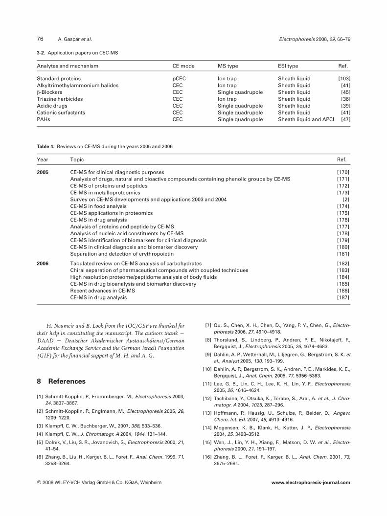
3-2. Application papers on CEC-MS
Analytes and mechanism CE mode MS type ESI type Ref.
Standard proteins pCEC Ion trap Sheath liquid [103]Alkyltrimethylammonium halides CEC Ion trap Sheath liquid [41]b-Blockers CEC Single quadrupole Sheath liquid [45]Triazine herbicides CEC Ion trap Sheath liquid [36]Acidic drugs CEC Single quadrupole Sheath liquid [39]Cationic surfactants CEC Single quadrupole Sheath liquid [41]PAHs CEC Single quadrupole Sheath liquid and APCI [47]
Table 4. Reviews on CE-MS during the years 2005 and 2006
Year Topic Ref.
2005 CE-MS for clinical diagnostic purposes [170]Analysis of drugs, natural and bioactive compounds containing phenolic groups by CE-MS [171]CE-MS of proteins and peptides [172]CE-MS in metalloproteomics [173]Survey on CE-MS developments and applications 2003 and 2004 [2]CE-MS in food analysis [174]CE-MS applications in proteomics [175]CE-MS in drug analysis [176]Analysis of proteins and peptide by CE-MS [177]Analysis of nucleic acid constituents by CE-MS [178]CE-MS identification of biomarkers for clinical diagnosis [179]CE-MS in clinical diagnosis and biomarker discovery [180]Separation and detection of erythropoietin [181]
2006 Tabulated review on CE-MS analysis of carbohydrates [182]Chiral separation of pharmaceutical compounds with coupled techniques [183]High resolution proteome/peptidome analysis of body fluids [184]CE-MS in drug bioanalysis and biomarker discovery [185]Recent advances in CE-MS [186]CE-MS in drug analysis [187]
H. Neumeir and B. Look from the IÖC/GSF are thanked fortheir help in constituting the manuscript. The authors thank 2
DAAD 2 Deutscher Akademischer Austauschdienst/GermanAcademic Exchange Service and the German Israeli Foundation(GIF) for the financial support of M. H. and A. G.
8 References
[1] Schmitt-Kopplin, P., Frommberger, M., Electrophoresis 2003,24, 3837–3867.
[2] Schmitt-Kopplin, P., Englmann, M., Electrophoresis 2005, 26,1209–1220.
[3] Klampfl, C. W., Buchberger, W., 2007, 388, 533–536.
[4] Klampfl, C. W., J. Chromatogr. A 2004, 1044, 131–144.
[5] Dolník, V., Liu, S. R., Jovanovich, S., Electrophoresis 2000, 21,41–54.
[6] Zhang, B., Liu, H., Karger, B. L., Foret, F., Anal. Chem. 1999, 71,3258–3264.
[7] Qu, S., Chen, X. H., Chen, D., Yang, P. Y., Chen, G., Electro-phoresis 2006, 27, 4910–4918.
[8] Thorslund, S., Lindberg, P., Andren, P. E., Nikolajeff, F.,Bergquist, J., Electrophoresis 2005, 26, 4674–4683.
[9] Dahlin, A. P., Wetterhall, M., Liljegren, G., Bergstrom, S. K. etal., Analyst 2005, 130, 193–199.
[10] Dahlin, A. P., Bergstrom, S. K., Andren, P. E., Markides, K. E.,Bergquist, J., Anal. Chem. 2005, 77, 5356–5363.
[11] Lee, G. B., Lin, C. H., Lee, K. H., Lin, Y. F., Electrophoresis2005, 26, 4616–4624.
[12] Tachibana, Y., Otsuka, K., Terabe, S., Arai, A. et al., J. Chro-matogr. A 2004, 1025, 287–296.
[13] Hoffmann, P., Hausig, U., Schulze, P., Belder, D., Angew.Chem. Int. Ed. 2007, 46, 4913–4916.
[14] Mogensen, K. B., Klank, H., Kutter, J. P., Electrophoresis2004, 25, 3498–3512.
[15] Wen, J., Lin, Y. H., Xiang, F., Matson, D. W. et al., Electro-phoresis 2000, 21, 191–197.
[16] Zhang, B. L., Foret, F., Karger, B. L., Anal. Chem. 2001, 73,2675–2681.
© 2008 WILEY-VCH Verlag GmbH & Co. KGaA, Weinheim www.electrophoresis-journal.com

Electrophoresis 2008, 29, 66–79 CE and CEC 77
[17] Musyimi, H. K., Guy, J., Narcisse, D. A., Soper, S. A., Murray,K. K., Electrophoresis 2005, 26, 4703–4710.
[18] Yu, B., Cong, H. L., Liu, H. W., Li, Y. Z., Liu, F., J. Sep. Sci.2005, 28, 2390–2400.
[19] Puerta, A., Axen, J., Soderberg, L., Bergquist, J., J. Chroma-togr. B 2006, 838, 113–121.
[20] Kelly, J. F., Locke, S. J., Ramaley, L., Thibault, P., J. Chroma-togr. A 1996, 720, 409–427.
[21] Dong, M., Oda, R. P., Strausbauch, M. A., Wettstein, P. J. etal., Electrophoresis 1997, 18, 1767–1774.
[22] Catai, J. R., Torano, J. S., de Jong, G. J., Somsen, G. W.,Electrophoresis 2006, 27, 2091–2099.
[23] Zhang, J., Horváth, C., Electrophoresis 2003, 24, 115–120.
[24] Lynen, F., Zhao, Y., Becu, C., Borremans, F., Sandra, P., Elec-trophoresis 1999, 20, 2462–2474.
[25] Katayama, H., Ishihama, Y., Asakawa, N., Anal. Sci. 1998, 14,407–408.
[26] Moini, M., Demars, S. M., Huang, H., Anal. Chem. 2002, 74,3772–3776.
[27] Meagher, R. J., Seong, J., Laibinis, P. E., Barron, A. E., Elec-trophoresis 2004, 25, 405–414.
[28] Samskog, J., Wetterhall, M., Jacobsson, S., Markides, K., J.Mass Spectrom. 2000, 35, 919–924.
[29] Bateman, K. P., White, R. L., Thibault, P., Rapid Commun.Mass Spectrom. 1997, 11, 307–315.
[30] Simó, C., Elvira, C., Gonzalez, N., Roman, J. S. et al., Elec-trophoresis 2004, 25, 2056–2064.
[31] Viberg, P., Spegel, P., Nilsson, J., Petersson, P. et al., Chro-matographia 2007, 65, 291–297.
[32] Nilsson, C., Viberg, P., Spegel, P., Jornten–Karlsson, M. et al.,Anal. Chem. 2006, 78, 6088–6095.
[33] Norton, D., Zheng, J., Danielson, N. D., Shamsi, S. A., Anal.Chem. 2005, 77, 6874–6886.
[34] Johannesson, N., Olsson, L., Backstrom, D., Wetterhall, M.et al., Electrophoresis 2007, 28, 1435–1443.
[35] Galceran, M. T., Puignou, L., Trends Anal. Chem. 2005, 24,743–758.
[36] Chang, C. H., Chen, C. J., Chuang, Y. C., Her, G. R., Electro-phoresis 2006, 27, 4303–4311.
[37] Chen, Z. L., Boggess, B., Chang, H. C., J. Mass Spectrom.2007, 42, 244–253.
[38] Zheng, J., Rizvi, S. A. A., Shamsi, S. A., Hou, J. G., J. Liq.Chromatogr. Relat. Technol. 2007, 30, 43–57.
[39] Kitagawa, F., Inoue, K., Hasegawa, T., Kamiya, M. et al., J.Chromatogr. A 2006, 1130, 219–226.
[40] Kato, M., Onda, Y., Sakai-Kato, K., Toyo’oka, T., Anal. Bioa-nal. Chem. 2006, 386, 572–577.
[41] Norton, D., Rizvi, S. A. A., Shamsi, S. A., Electrophoresis2006, 27, 4273–4287.
[42] Lynen, F., Buica, A., de Villiers, A., Crouch, A., Sandra, P., J.Sep. Sci. 2005, 28, 1539–1549.
[43] Zheng, J., Norton, D., Shamsi, S. A., Anal. Chem. 2006, 78,1323–1330.
[44] Barcelo-Barrachina, E., Moyano, E., Puignou, L., Galceran,M. T., Electrophoresis 2007, 28, 1704–1713.
[45] Zheng, J., Shamsi, S. A., Electrophoresis 2006, 27, 2139–2151.
[46] Droste, S., Schellentrager, M., Constapel, M., Gab, S. et al.,Electrophoresis 2005, 26, 4098–4103.
[47] Zheng, J., Shamsi, S. A., Anal. Chem. 2006, 78, 6921–6927.
[48] Bergstrom, S. K., Dahlin, A. P., Ramstrom, M., Andersson, M.et al., Analyst 2006, 131, 791–798.
[49] Palmblad, M., Wetterhall, M., Markides, K., Hakansson, P.,Bergquist, J., Rapid Commun. Mass Spectrom. 2000, 14,1029–1034.
[50] Ramström, M., Palmblad, M., Amirkhani, A., Tsybin, Y. O. etal., Acta. Biochim. Pol. 2001, 48, 1101–1104.
[51] Dahlin, A. P., Bergström, S. K., Andrén, P. E., Markides, K. E.,Bergquist, J., Anal. Chem. 2005, 77, 5356 –5363.
[52] Bindila, L., Peter-Katalinic, J., Zamfir, A., Electrophoresis2005, 26, 1488–1499.
[53] Huhn, C., Neususs, C., Pelzing, M., Pyell, U. et al., Electro-phoresis 2005, 26, 1389–1397.
[54] Liu, C. C., Alary, J. F., Vollmerhaus, P., Kadkhodayan, M.,Electrophoresis 2005, 26, 1366–1375.
[55] Machour, N., Place, J., Tron, F., Charlionet, R. et al., Electro-phoresis 2005, 26, 1466–1475.
[56] Mol, R., de Jong, G. J., Somsen, G. W., Electrophoresis 2005,26, 146–154.
[57] Nogami, C., Sawada, H., Electrophoresis 2005, 26, 1406–1411.
[58] Ohnesorge, J., Van de Griend, C. S., Wätzig, H., Electropho-resis 2005, 26, 2360–2375.
[59] Pelzing, M., Neusüss, C., Electrophoresis 2005, 26, 2717–2728.
[60] Sanz-Nebot, V., Balaguer, E., Benavente, F., Barbosa, J.,Electrophoresis 2005, 26, 1457–1465.
[61] Steiner, F., Hassel, M., J. Chromatogr. A 2005, 1068, 131–142.
[62] Trapp, O., Pearce, E. W., Kimmel, J. R., Yoon, O. K. et al.,Electrophoresis 2005, 26, 1358–1365.
[63] Wu, Y. T., Chen, Y. C., Anal. Chem. 2005, 77, 2071–2077.
[64] Al-Jarah, S. Y., Sjodahl, J., Woldegiorgis, A., Emmer, A., J.Sep. Sci. 2005, 28, 239–244.
[65] Chen, Y. R., Tseng, M. C., Her, G. R., Electrophoresis 2005, 26,1376–1382.
[66] Kele, Z., Ferenc, G., Klement, T., Toth, G. K., Janaky, T., RapidComm. Mass Spectrom. 2005, 19, 881–885.
[67] Liu, C. C., Zhang, J. Z., Dovichi, N. J., Rapid Commun. MassSpectrom. 2005, 19, 187–192.
[68] Harada, K., Fukusaki, E., Kobayashi, A., J. Biosci. Bioeng.2006, 101, 403–409.
[69] Zamfir, A. D., Dinca, N., Sisu, E., Peter-Katalinic, J., J. Sep.Sci. 2006, 29, 414–422.
[70] Santos, B., Simonet, B. M., Lendl, B., Rios, Á., Valcarcel, M.,J. Chromatogr. A 2006, 1127, 278–285.
[71] Benavente, F., Balaguer, E., Barbosa, J., Sanz-Nebot, V., J.Chromatogr. A 2006, 1117, 94–102.
[72] Johannesson, N., Pearce, E., Dulay, M., Zare, R. N. et al., J.Chromatogr. B 2006, 842, 70–74.
[73] Sebastiano, R., Simó, C., Mendieta, M. E., Antonioli, P. et al.,Electrophoresis 2006, 27, 3919–3934.
[74] An, Y. M., Cooper, J. W., Balgley, B. M., Lee, C. S., Electro-phoresis 2006, 27, 3599–3608.
[75] Chao, B. F., Chen, C. J., Li, F. A., Her, G. R., Electrophoresis2006, 27, 2083–2090.
© 2008 WILEY-VCH Verlag GmbH & Co. KGaA, Weinheim www.electrophoresis-journal.com
78 A. Gaspar et al. Electrophoresis 2008, 29, 66–79
[76] Busnel, J. M., Descroix, S., Le Saux, T., Terabe, S. et al.,Electrophoresis 2006, 27, 1481–1488.
[77] Feng, Y. L., Zhu, J. P., Anal. Chem. 2006, 78, 6608–6613.
[78] Li, F. A., Wu, M. C., Her, G. R., Anal. Chem. 2006, 78, 5316–5321.
[79] Lindberg, P., Dahlin, A. P., Bergstrom, S. K., Thorslund, S. etal., Electrophoresis 2006, 2075–2082.
[80] Fanali, S., DÓrazio, G., Foret, F., Kleparnik, K., Aturki, Z.,Electrophoresis 2006, 27, 4666–4673.
[81] Hu, A., Tsai, P. J., Ho, Y. P., Anal. Chem. 2005, 77, 1488–1495.
[82] Tessier, B., Schweizer, M., Fournier, F., Framboisier, X. et al.,Food. Res. Int. 2005, 38, 577–584.
[83] Amon, S., Plematl, A., Rizzi, A., Electrophoresis 2006, 27,1209–1219.
[84] Andon, B., Barbosa, J., Sanz-Nebot, V., Electrophoresis2006, 27, 3661–3670.
[85] Garza, S., Moini, M., Anal. Chem. 2006, 78, 7309–7316.
[86] Gluch, I., Urbanska, A., Zadrozna, I., Pawlak, K., Jarosz, M.,Chem. Anal. (Warsaw) 2006, 51, 195–210.
[87] Hu, A. R., Chen, C. T., Tsai, P. J., Ho, Y. P., Anal. Chem. 2006,78, 5124–5133.
[88] Psurek, A., Neususs, C., Degenkolb, T., Bruckner, H. et al., J.Peptide Sci. 2006, 12, 279–290.
[89] Simionato, A. V. C., Simo, C., Cifuentes, A., Lacava, P. T. etal., Electrophoresis 2006, 27, 2567–2574.
[90] Balaquer, E., Neusüss, C., Chromatographia 2006, 64, 351–357.
[91] Cho, S. H., Jung, B. H., Lee, W. Y., Chung, B. C., RapidCommun. Mass Spectrom. 2006, 20, 2995–2998.
[92] Guo, T., Rudnick, P. A., Wang, W. J., Lee, C. S. et al., J. Pro-teome Res. 2006, 5, 1469–1478.
[93] Gennaro, L. A., Salas-Solano, O., Ma, S., Anal. Biochem.2006, 355, 249–258.
[94] Sanz-Nebot, V., Benavente, F., Hernández, E., Barbosa, J.,Anal. Chim. Acta 2006, 577, 68–76.
[95] Balaguer, E., Demelbauer, U., Pelzing, M., Sanz-Nebot, V. etal., Electrophoresis 2006, 27, 2638–2650.
[96] Baldacci, A., Thormann, W., Electrophoresis 2006, 27,2444–2457.
[97] Simó, C., Herrero, M., Neusüss, C., Pelzing, M. et al., Elec-trophoresis 2005, 26, 2674–2683.
[98] Arias, M., Simó, C., Ortiz, L. T., de los Mozos-Pascual, M. etal., Electrophoresis 2005, 26, 2351–2359.
[99] Zurbig, P., Renfrow, M. B., Schiffer, E., Novak, J. et al.,Electrophoresis 2006, 27, 2111–2125.
[100] Yoo, C., Pal, M., Miller, F. R., Barder, T. J. et al., Electropho-resis 2006, 27, 2126–2138.
[101] Simó, C., Perez, P., Neusüss, C., Pelzing, M. et al., Electro-phoresis 2006, 27, 2250–2258.
[102] Zhou, F., Johnston, M. V., Electrophoresis 2005, 26, 1383–1388.
[103] Liang, Z., Zhang, L. H., Duan, J. C., Yan, C. et al., Electro-phoresis 2005, 26, 1398–1405.
[104] Li, J., Martin, A., Cox, A. D., Moxon, E. R. et al., Mass Spec-trometry: Modified Proteins and Glycoconjugates 2005,405, 369–397.
[105] Li, J. J., Cox, A. D., Hood, D. W., Schweda, E. K. H. et al.,Mol. Biosys. 2005, 1, 46–52.
[106] Tissota, B., Salpin, J. Y., Martinez, M., Gaigeota, M. P.,Daniela, R., Carbohydr. Res. 2006, 341, 598–609.
[107] Garcia-Villalba, R., Cortacero-Ramirez, S., Segura-Carre-tero, A., Contreras, J. A. M. L., Fernández-Gutierrez, A., J.Agric. Food. Chem. 2006, 54, 5400–5409.
[108] Ge, L., Yong, J. W. H., Goh, N. K., Chia, L. S. et al., J. Chro-matogr. B 2005, 829, 26–34.
[109] Kuhn, A. V., Ozegowski, J. H., Peschel, G., Neubert, R. H. H.,Carbohyd. Res. 2004, 339, 2541–2547.
[110] Frommberger, M., Hertkorn, N., Englmann, M., Jakoby, S.et al., Electrophoresis 2005, 26, 1523–1532.
[111] Meulemans, A., J. Chromatogr. B 2005, 824, 308–311.
[112] Wahby, I., Arraez-Roman, D., Segura-Carretero, A., Ligero,F. et al., Electrophoresis 2006, 27, 2208–2215.
[113] Ge, L. Y., Yong, J. W. H., Tan, S. N., Yang, X. H., Ong, E. S., J.Chromatogr. A 2006, 1133, 322–331.
[114] Bednar, P., Papouskova, B., Müller, L., Bartak, P. et al., J.Sep. Sci. 2005, 28, 1291–1299.
[115] Simó, C., Rizzi, A., Barbas, C., Cifuentes, A., Electrophoresis2005, 26, 1432–1441.
[116] Arraez-Roman, D., Cortacero-Ramirez, S., Segura-Carre-tero, A., Contreras, J. A. M. L., Fernández-Gutierrez, A.,Electrophoresis 2006, 27, 2197–2207.
[117] Carrasco-Pancorbo, A., Arraez-Roman, D., Segura-Carre-tero, A., Fernández-Gutierrez, A., Electrophoresis 2006, 27,2182–2196.
[118] Juan-Garcia, A., Font, G., Pico, Y., Electrophoresis 2006, 27,2240–2249.
[119] Li, J. J., Wang, Z., Altman, E., Rapid Commun. Mass Spec-trom. 2005, 19, 1305–1314.
[120] Herrero, M., Arraez-Roman, D., Segura, A., Kenndler, E. etal., J Chromatogr. A 2005, 1084, 54–62.
[121] Bringmann, G., Kajahn, I., Neusüss, C., Pelzing, M. et al.,Electrophoresis 2005, 26, 1513–1522.
[122] Chalmers, M. J., Mackay, C. L., Hendrickson, C. L., Wittke, S.et al., Anal. Chem. 2005, 77, 7163–7171.
[123] Ganzera, M., Piereder, D., Sturm, S., Erdelmeier, C., Stupp-ner, H., Electrophoresis 2005, 26, 1724–1731.
[124] Graham, R. L. J., McClean, S., O’Kane, E. J., Theakston, D.,Shaw, C., Biochem. Bioph. Res. Co. 2005, 333, 88–94.
[125] Liu, C. C., Huang, J. S., Tyrrell, D. L. J., Dovichi, N. J., Elec-trophoresis 2005, 26, 1424–1431.
[126] Meier, M., Kaiser, T., Herrmann, A., Knueppel, S. et al., J.Diabetes Complicat. 2005, 19, 223–232.
[127] Neusüss, C., Demelbauer, U., Pelzing, M., Electrophoresis2005, 26, 1442–1450.
[128] Peri-Okonny, U. L., Wang, S. X., Stubbs, R. J., Guzman, N.A., Electrophoresis 2005, 26, 2652–2663.
[129] Psurek, A., Neusüss, C., Pelzing, M., Scriba, G. K. E., Elec-trophoresis 2005, 26, 4368–4378.
[130] Sassi, A. P., Andel, F., Bitter, H. M. L., Brown, M. P. S. et al.,Electrophoresis 2005, 26, 1500–1512.
[131] Simó, C., Gonzalez, R., Barbas, C., Cifuentes, A., Anal.Chem. 2005, 77, 7709–7716.
[132] Theodorescu, D., Fliser, D., Wittke, S., Mischak, H. et al.,Electrophoresis 2005, 26, 2797–2808.
[133] Wittke, S., Haubitz, M., Walden, M., Rohde, F. et al., Am. J.Transplant. 2005, 5, 2479–2488.
© 2008 WILEY-VCH Verlag GmbH & Co. KGaA, Weinheim www.electrophoresis-journal.com

Electrophoresis 2008, 29, 66–79 CE and CEC 79
[134] Wittke, S., Mischak, H., Walden, M., Kolch, W. et al., Elec-trophoresis 2005, 26, 1476–1487.
[135] Sturm, S., Strasser, E. M., Stuppner, H., J. Chromatogr. A2006, 1112, 331–338.
[136] Mol, R., Kragt, E., Jimidar, L., de Jong, G. J., Somsen, G. W.,J. Chromatogr. B 2006, 843, 283–288.
[137] Anurukvorakun, O., Suntornsuk, W., Suntornsuk, L., J.Chromatogr. A 2006, 1134, 326–332.
[138] Himmelsbach, M., Klampfl, C. W., Buchberger, W., J. Sep.Sci. 2005, 28, 1735–1741.
[139] Campa, C., Rossi, M., Flamigni, A., Baiutti, E. et al., Elec-trophoresis 2005, 26, 1533–1540.
[140] Vassort, A., Barrett, D. A., Shaw, P. N., Ferguson, P. D.,Szucs, R., Electrophoresis 2005, 26, 1712–1723.
[141] Sugimoto, M., Kikuchi, S., Arita, M., Soga, T. et al., Anal.Chem. 2005, 77, 78–84.
[142] Edwards, E. L., Rodrigues, J. A., Ferreira, J., Goodall, D. M.et al., Electrophoresis 2006, 27, 2164–2170.
[143] Edwards, J. L., Chisolm, C. N., Shackman, J. G., Kennedy,R. T., J. Chromatogr. A 2006, 1106, 80–88.
[144] Ullsten, S., Danielsson, R., Backstrom, D., Sjoberg, P.,Bergquist, J., J. Chromatogr. A 2006, 1117, 87–93.
[145] Akbay, C., Rizvi, S. A. A., Shamsi, S. A., Anal. Chem. 2005,77, 1672–1683.
[146] Visky, D., Jimidar, I., Van Ael, W., Vennekens, T. et al., Elec-trophoresis 2005, 26, 1541–1549.
[147] Castro-Puyana, M., Garcia-Ruiz, C., Cifuentes, A., Crego, A.L., Marina, M. L., J. Chromatogr. A 2006, 1114, 170–177.
[148] Schappler, J., Guillarme, D., Prat, J., Veuthey, J. L., Rudaz,S., Electrophoresis 2006, 27, 1537–1546.
[149] Servais, A. C., Fillet, M., Mol, R., Somsen, G. W. et al., J.Pharm. Biomed. Anal. 2006, 40, 752–757.
[150] Groom, C. A., Halasz, A., Paquet, L., Thiboutot, S. et al., J.Chromatogr. A 2005, 1072, 73–82.
[151] Hernández-Borges, J., Rodriguez-Delgado, M. A., Garcia-Montelongo, F. J., Cifuentes, A., J. Sep. Sci. 2005, 28, 948–956.
[152] Juan-García, A., Font, G., Pico, Y., Electrophoresis 2005, 26,1550–1561.
[153] Kaech, A., Hofer, M., Rentsch, D., Schnider, C., Egli, T., Bio-degradation 2005, 16, 461–473.
[154] Safarpour, H., Asiaie, R., Electrophoresis 2005, 26, 1562–1566.
[155] Zhao, X. H., Lu, Y. P., Hardin, I., Biotechnol. Lett. 2005, 27,69–72.
[156] Viberg, P., Wahlund, K. G., Skog, K., J. Chromatogr. A 2006,1133, 347–352.
[157] Fakhari, A. R., Breadmore, M. C., Macka, M., Haddad, P. R.,Anal. Chim. Acta 2006, 580, 188–193.
[158] Fekete, A., Frommberger, M., Ping, G. C., Lahaniatis, M. R.et al., Electrophoresis 2006, 27, 1237–1247.
[159] Himmelsbach, M., Buchberger, W., Klampfl, C. W., Electro-phoresis 2006, 27, 1220–1226.
[160] Kitagawa, F., Shiomi, K., Otsuka, K., Electrophoresis 2006,27, 2233–2239.
[161] Lara, F. J., García-Campaña, A. M., Ales-Barrero, F., Bos-que-Sendra, J. M., García-Ayuso, L. E., Anal. Chem. 2006,78, 7665–7673.
[162] Li, C. C., Chen, Z., Wen, D. W., Zhang, J. X. et al., Electro-phoresis 2006, 27, 2152–2163.
[163] Para, B. V., Nunez, O., Moyano, E., Galceran, M. T., Electro-phoresis 2006, 27, 2225–2232.
[164] Santos, B., Lista, A., Simonet, B. M., Rios, Á., Valcárcel, M.,Electrophoresis 2005, 26, 1567–1575.
[165] Iio, R., Chinaka, S., Takayama, N., Hayakawa, K., Anal. Sci.2005, 21, 15–19.
[166] Boatto, G., Nieddu, M., Carta, A., Pau, A. et al., J. Chroma-togr. B 2005, 814, 93–98.
[167] Friedecky, D., Bednar, P., Prochazka, M., Adam, T., Nucleo-sides Nucleotides & Nucleic Acids 2006, 25, 1233–1236.
[168] Cook, H. A., Klampfl, C. W., Buchberger, W., Electrophore-sis 2005, 26, 1576–1583.
[169] Willems, A. V., Deforce, D. L., Van Peteghem, C. H., VanBocxlaer, J. F., Electrophoresis 2005, 26, 1412–1423.
[170] Fliser, D., Wittke, S., Mischak, H., Electrophoresis 2005, 26,2708–2716.
[171] Huck, C. W., Stecher, G., Scherz, H., Bonn, G., Electropho-resis 2005, 26, 1319–1333.
[172] Monton, M. R. N., Terabe, S., Anal. Sci. 2005, 21, 5–13.
[173] Prange, A., Profrock, D., Anal. Bioanal. Chem. 2005, 383,372–389.
[174] Simó, C., Barbas, C., Cifuentes, A., Electrophoresis 2005,26, 1306–1318.
[175] Simpson, D. C., Smith, R. D., Electrophoresis 2005, 26,1291–1305.
[176] Smyth, W. F., Electrophoresis 2005, 26, 1334–1357.
[177] Stutz, H., Electrophoresis 2005, 26, 1254–1290.
[178] Willems, A. V., Deforce, D. L., Van Peteghem, C. H., VanBocxlaer, J. F., Electrophoresis 2005, 26, 1221–1253.
[179] Weissinger, E. M., Hertenstein, B., Mischak, H., Ganser, A.,Expert Review of Proteomics 2005, 2, 639–647.
[180] Kolch, W., Neusüss, C., Pelzing, M., Mischak, H., MassSpectrom. Rev. 2005, 24, 959–977.
[181] Yu, B., Cong, H. L., Liu, H. W., Li, Y. Z., Liu, F., Trends Anal.Chem. 2005, 24, 350–357.
[182] Campa, C., Coslovi, A., Flamingi, A., Rossi, M., Electropho-resis 2006, 27, 2027–2050.
[183] Erny, G. L., Cifuentes, A., J. Pharm. Biomed. Anal. 2006, 40,509–515.
[184] Schiffer, E., Mischak, H., Novak, J., Proteomics 2006, 6,5615–5627.
[185] Servais, A. C., Crommen, J., Fillet, M., Electrophoresis2006, 27, 2616–2629.
[186] Klampfl, C. W., Electrophoresis 2006, 27, 3–34.
[187] Smyth, W. F., Electrophoresis 2006, 27, 2051–2062.
© 2008 WILEY-VCH Verlag GmbH & Co. KGaA, Weinheim www.electrophoresis-journal.com