Treatment of Residual Psychotic Symptons in Ultra-Refractory ...
14
Journal of Psychiatry and Psychiatric Disorders doi: 10.26502/jppd.2572-519X0102 J Psychiatry Psychiatric Disord 2020; 4 (4): 144-157 144 Research Article Volume 4, Issue 4 Treatment of Residual Psychotic Symptons in Ultra-Refractory Schizophrenia after 10 Sessions of Transcranial Direct Current Stimulation (tDCS) Nathália Janovik 1,2 , Victor Hugo Cordova 2,3,⃰ , Cintya Ogliari 2 , Michel Mroginski 4 , Lenise Franceschoni 2,3 , Paulo Belmonte-de-Abreu ,4 1 Post graduate program in Psychiatry and Behavior Sciences- Federal University of Rio Grande do Sul (UFRGS)-Porto Alegre-RS, Brazil 2 Schizophrenia and Dementia Program (PRODESQ)- Hospital de Clínicas de Porto Alegre(HCPA)- Porto Alegre- RS, Brazil 3 School of Pharmacy-UFRGS 4 School of Medicine-UFRGS * Corresponding Author: Victor Hugo Cordova, Vicente da Fontoura nº 1679, ap 203, Porto Alegre-RS, Brazil, Tel: +55(51)997507974; E-mail: [email protected] Received: 12 May 2020; Accepted: 19 May 2020; Published: 26 July 2020 1. Introduction A recent meta-analysis based on functional neuroimaging studies showed that, during the occurrence of Auditory Hallucinations (AH), patients with schizophrenia exhibit a significant overactivation of the left parieto-temporal cortex (middle and upper temporal and Wernicke's area), left lower frontal cortex (Drill front area, operculum, anterior insula, precentral gyrus), as well as their rights counterparts [1]. In recent years, it have been developed different noninvasive techniques of brain stimulation (NIBS), like Transcranial Direct Current Stimulation (tDCS), that promotes the partial antagonism of N-metyl-D-aspartate (NMDAr) in symptomatic ultra-refractory schizophrenia (URS) patients. The other point of this approache is the possibility of inducing lasting changes in cerebral excitability, as weel as the possibility of modulation of cortical connectivity and brain plasticity [2, 3].This findings indicate that the different neuromodulation techniques can be potential diagnostic tools and therapeutic research of cortical plasticity in the mental illness [4].
-
Upload
khangminh22 -
Category
Documents
-
view
4 -
download
0
Transcript of Treatment of Residual Psychotic Symptons in Ultra-Refractory ...
Journal of Psychiatry and Psychiatric Disorders doi: 10.26502/jppd.2572-519X0102
J Psychiatry Psychiatric Disord 2020; 4 (4): 144-157 144
Research Article Volume 4, Issue 4
Treatment of Residual Psychotic Symptons in Ultra-Refractory
Schizophrenia after 10 Sessions of Transcranial Direct Current
Stimulation (tDCS)
Nathália Janovik1,2
, Victor Hugo Cordova2,3,⃰
, Cintya Ogliari
2, Michel Mroginski
4,
Lenise Franceschoni2,3
, Paulo Belmonte-de-Abreu,4
1Post graduate program in Psychiatry and Behavior Sciences- Federal University of Rio Grande do Sul
(UFRGS)-Porto Alegre-RS, Brazil
2Schizophrenia and Dementia Program (PRODESQ)- Hospital de Clínicas de Porto Alegre(HCPA)- Porto Alegre-
RS, Brazil
3School of Pharmacy-UFRGS
4School of Medicine-UFRGS
*Corresponding Author: Victor Hugo Cordova, Vicente da Fontoura nº 1679, ap 203, Porto Alegre-RS, Brazil,
Tel: +55(51)997507974; E-mail: [email protected]
Received: 12 May 2020; Accepted: 19 May 2020; Published: 26 July 2020
1. Introduction
A recent meta-analysis based on functional neuroimaging studies showed that, during the occurrence of Auditory
Hallucinations (AH), patients with schizophrenia exhibit a significant overactivation of the left parieto-temporal
cortex (middle and upper temporal and Wernicke's area), left lower frontal cortex (Drill front area, operculum,
anterior insula, precentral gyrus), as well as their rights counterparts [1].
In recent years, it have been developed different noninvasive techniques of brain stimulation (NIBS), like
Transcranial Direct Current Stimulation (tDCS), that promotes the partial antagonism of N-metyl-D-aspartate
(NMDAr) in symptomatic ultra-refractory schizophrenia (URS) patients. The other point of this approache is the
possibility of inducing lasting changes in cerebral excitability, as weel as the possibility of modulation of cortical
connectivity and brain plasticity [2, 3].This findings indicate that the different neuromodulation techniques can be
potential diagnostic tools and therapeutic research of cortical plasticity in the mental illness [4].

Journal of Psychiatry and Psychiatric Disorders doi: 10.26502/jppd.2572-519X0102
J Psychiatry Psychiatric Disord 2020; 4 (4): 144-157 145
Thus, based on the observations of frontotemporal dysconnectivity system in AH, we hypothesized that by
modulating the abnormal activity of brain areas involved in the pathophysiology of these symptoms with NIBS, such
as tDCS, it would be possible to minimizing their impacts on this mental health problem [5].
More recently, tDCS was appraised as a new approach neurostimulation to reduce AH resistant to conventional
treatment. The tDCS protocol predicts the arrangement of the cathode placed on left temporo-parietal junction (TPJ)
and the anode on the left dorsolateral prefrontal cortical (DLPFC), which was effective in reducing AH in patients
with schizophrenia in numerous studies [6-14], in two clinical experiments [15, 16] and in a randomized clinical trial
involving 30 patients [17]. In most of these studies, tDCS sessions were conducted twice daily for five consecutive
days (10 sessions).
This article briefly holds the theoretical rational, the practice development, the analysis and the results of a
randomized, double-blind, placebo-controlled clinical trial that evaluated the tDCS efficacy in patients with URS,
with detailed emphasis over auditory hallucinations and its specific characteristics.
2. Study Design
This is a double-blind randomized placebo-controlled trial.
2.1 Sample
Considering the need for a placebo group and estimating a possible loss of 10% of patients during the follow-up
period, the initial sample size was calculated for a study power of 80% and indicated the selection of 48
schizophrenic outpatient from Porto Alegre Clinical Hospital (PRODESQ/HCPA), where the study was also
submitted for evaluation by its Ethics Committee, with subsequent approval to be a source of allocation of patients.
Patients in the sample were randomized to be included in the active group or sham group (placebo).
2.1.1 Inclusion criteria:
Age between 18 and 59 years;
schizophrenia diagnosis confirmed by OPCRIT (Operational Criteria Checklist for Psychotic Illness and
Affective Illness);
Auditory hallucinations and residual negative symptoms;
Active use of clozapine, maintaining the same dose for, at least, six weeks before starting of the stimulation
protocol.
2.1.2 Exclusion criteria:
Active neurological disease and no regular treatment;
Alcoholic or toxic psychosis;

Journal of Psychiatry and Psychiatric Disorders doi: 10.26502/jppd.2572-519X0102
J Psychiatry Psychiatric Disord 2020; 4 (4): 144-157 146
Contraindications to tDCS: brain metallic clip, cardiac pacemaker, skin lesion at the site of electrode
placement;
History of drug abuse (minimum withdrawal time required was two years);
Changing dose(s) of medication(s) during the stimulation protocol;
Refusal to follow the study.
2.2 Diagnostic and clinical evaluation
The identification, diagnosis of patients with schizophrenia and the application of OPCRIT, Brief Psychiatric Rating
Scale-Anchored version (BPRS-A) and Auditory Hallucination Rating Scale (AHRS) were performed by the same
psychiatrist with training and clinical experience in schizophrenia. Before the stimulation protocol, a clinical and
diagnostic confirmation interview by applying the OPCRIT, and also a clinical evaluation, through the BPRS-A and
AHRS focused on the measurement of auditory hallucinations and negative symptoms, were performed. The BPRS-
A and AHRS scales have been repeated in the last day of neurostimulation protocol, 4 weeks and 12 weeks after the
last procedure. Randomization was accomplished by the site http://www.random.org, diagnostic and clinical
assessments were evaluated by a single interviewer. Primary outcomes included: (1) general psychopathology score
measured by BPRS-A; (2) negative scores (sum of subitems 3, 9, 13 and 16 in BPRS-A); (3) positive scores (sum of
subitems 4, 8, 11, 12 and 15 of the BPRS-A); (4) total scores of auditory hallucinations measured by the AHRS.
2.3 tDCS technique
The application of neurostimulation protocol was performed by four academics of Medicine School of Federal
University of Rio Grande do Sul, previously trained for three months by the main investigator of this study. The
placebo stimulation was made by placing the electrodes in the same locations cited for active stimulation, but after
30 seconds, the stimulator was turned off by the protocol applicator. Neither the patients nor the scales applicator
have been known which group the patients belonged to (active or sham).
The stimulation protocol parameters are summarized below:
Anode placed on the left DLPFC and cathode located on the left TPJ marked in a cap used in
neuromodulation using 10-20 EEG method;
Intensity of electric current: 2 mA (milli-amps);
Duration of each session: 20 minutes;
Number of sessions: two sessions per day, separated by one hour, for five consecutive days (Monday to
Friday).
2.4 Statistical analysis
The quantitative variables were described as mean and standard deviation, and categorical variables were identified
by absolute and relative frequencies. In mean comparison between groups, Student's t-test was used. In proportions
Journal of Psychiatry and Psychiatric Disorders doi: 10.26502/jppd.2572-519X0102
J Psychiatry Psychiatric Disord 2020; 4 (4): 144-157 147
comparison, the Chi-square test was applied. Regarding the comparison of BDNF levels and scores of BPRS-A and
AHRS scales, over time, it was applied the model of generalized estimating equations (GEE), and the Least
Significance Difference test (LSD). For variables with normal distribution, the linear model was applied. As for the
variables with asymmetric distribution, the gamma model was used. Associations between BDNF levels and BPRS-
A scores were assessed using Pearson's correlation coefficient. To control confounding factors, Poisson regression
models (reduction of, at least, 30% in the BPRS-A scale scores) or Linear (variation in the AHRS scale scores) were
applied. The criterion for entering the variable in the multivariate model was that it should have a p <0.20 in the
bivariate analysis, except for the variable “group” (active or placebo), which was maintained in both models, as it is
the main variable in this study. To avoid a multicollinearity effect, the disease duration was maintained in the
models, instead of the patient's age. In addition, when using the BMI in the multivariate model, the current dose of
clozapine was inserted, regardless of the p-value of the multivariate, since it is known that one of the main adverse
effects of this psychotropic is weight gain. The level of significance adopted was 5%, and the analyzes were
performed using the SPSS program - version 21.0.
3. Results
The primary outcomes was the change in psychopathology in total score of BPRS-A and AHRS scales throughout
the study period comprising the time interval measured at four different times: 1) before starting the stimulation
protocol; 2) the last day of the stimulation protocol; 3) four weeks after the end of the stimulation protocol; and 4)
twelve weeks after it.
Table 1 shows the characteristics of the sample according to the study group. Of all the variables used, the results
were adjusted for education level, disease duration and number of hospitalizations during clinical history. Ages have
been excluded to avoid multicollinearity effect with disease duration. We observed that patients in the active group
had significantly fewer hospitalizations in life when compared to the placebo group. And yet, the active group
tended to be younger, to have lower education levels and less sick time, when compared with the placebo group,
nevertheless without statistical significance. The other characteristics have been similar between groups.

Journal of Psychiatry and Psychiatric Disorders doi: 10.26502/jppd.2572-519X0102
J Psychiatry Psychiatric Disord 2020; 4 (4): 144-157 148
Variable Total sample
(n=33)
Group 1 – Placebo
(n=16)
Group 2 – Active
(n=17)
P
Age (years) 37,5 ± 10,8 40,8 ± 11,3 34,5 ± 9,6 0,094
Gender (Male) 24 (72,7) 12 (75,0) 12 (70,6) 1,000
Education level - - - 0,067
ES incomplete 13 (39,4) 4 (25,0) 9 (52,9) -
ES complete 12 (36,4) 9 (56,3) 3 (17,6) -
EM complete 8 (24,2) 3 (18,8) 5 (29,4) -
Marital status - - - 0,538
With mate 11 (33,3) 4 (25,0) 7 (41,2) -
Without mate 22 (66,7) 12 (75,0) 10 (58,8) -
BMI (kg/m2) 27,4 ± 3,3 27,0 ± 2,6 27,9 ± 3,9 0,467
Age of onset symptoms (age) 18,2 ± 2,1 17,9 ± 2,1 18,5 ± 2,2 0,383
Time of disease (age) 19,3 ± 10,6 22,9 ± 10,9 15,9 ± 9,4 0,058
Current dose of clozapine (mg) 596,9 ± 132,8 634,4 ± 139,9 561,8 ± 119,3 0,118
Use of other antipsychotic 23 (69,7) 10 (62,5) 13 (76,5) 0,465
Number of hospitalizatin in life 3,4 ± 1,6 4,1 ± 1,7 2,8 ± 1,3 0,026
Previous drug abuse 8 (24,2) 3 (18,8) 5 (29,4) 0,688
ES: elementary school; HS: high school; BMI: body mass index
Table 1: Sample description of stabilized patients with URS in ambulatory treatment, using clozapine and submitted
to tDCS for 5 consecutive days.
Table 2 shows the bivariate analysis of the clinical improvement (minimum 30% reduction in BPRS-A scale). The
group receiving active stimulation had a 13.2 greater clinical improvement when compared to the placebo group.
The number of hospitalizations was inversely associated with clinical improvement of symptoms, and
hospitalization in the last year reduces the improvement in 23%.

Journal of Psychiatry and Psychiatric Disorders doi: 10.26502/jppd.2572-519X0102
J Psychiatry Psychiatric Disord 2020; 4 (4): 144-157 149
Variables 30% decrease in BPRS-A
% OR (CI 95%) P
Group – mean ± SD
Placebo 6,3 1,00
Test 82,4 13,2 (1,95-89) 0,008
BDNF – Pearson’ s correlation coeficient (r) - 0,99 (0,99-1,00) 0,551
Basal variation – 12th week
AHRS (Basal variation – 12th
week) – Pearson’ s correlation coeficient
(r)
- 0,77 (0,68-0,88) <0,001
Frequency - 0,74 (0,63-0,87) <0,001
Influence - 0,65 (0,51-0,82) <0,001
Anguish - 0,95 (0,92-0,98) <0,001
Total - 1,06 (0,95-1,19) 0,290
BMI basal Pearson’s correlation coefficient (r) - 0,98 (0,94-1,02) 0,256
Age – Pearson’s correlation coefficient (r) - 1,12 (0,94-1,34) 0,205
Onset age – Pearson’s correlation coefficient (r) - 0,97 (0,93-1,01) 0,156
Time of disease (age) – Pearson’s correlation coefficient (r)
Previous drug abuse – mean ± SD 50,0 1,14 (0,50-2,59) 0,761
Yes 44,0 1,00
No - 0,77 (0,64-0,94) 0,008
Number of hospitalization – Pearson’s correlation coefficient (r)
Education – mean ± SD 53,8 0,86 (0,41-1,80) 0,691
Until incomplete high school 25,0 0,40 (0,13-1,22) 0,108
Complete high school or more 62,5 1,00
Gender – mean ± SD
Male 45,8 1,03 (0,44-2,41) 0,943
Female 44,4 1,00
Marital status
With mate 63,6 1,75 (0,86-3,56) 0,123
Without mate 36,4 1,00
Clozapine dose – Pearson’s correlation coefficient (r) - 0,99 (0,99-1,00) 0,111
Other antipsychotic – mean ± SD
Yes 47,8 1,19 (0,50-2,86) 0,688
No 40,0 1,00
BMI: body mass index
Table 2: Association with clinical response mesuread by BPRS-A from 12th week to baseline of 33 URS stabilized
outpatient, using clozapine and submitted to tDCS for 5 consecutive days.
Journal of Psychiatry and Psychiatric Disorders doi: 10.26502/jppd.2572-519X0102
J Psychiatry Psychiatric Disord 2020; 4 (4): 144-157 150
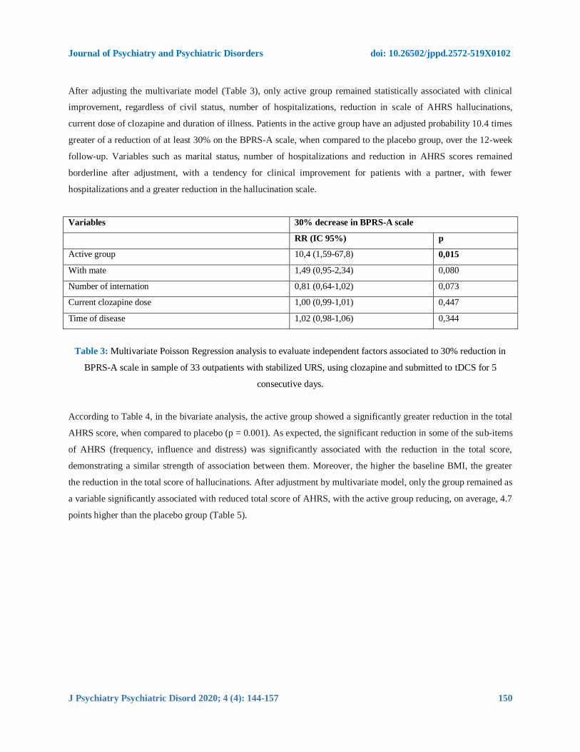
After adjusting the multivariate model (Table 3), only active group remained statistically associated with clinical
improvement, regardless of civil status, number of hospitalizations, reduction in scale of AHRS hallucinations,
current dose of clozapine and duration of illness. Patients in the active group have an adjusted probability 10.4 times
greater of a reduction of at least 30% on the BPRS-A scale, when compared to the placebo group, over the 12-week
follow-up. Variables such as marital status, number of hospitalizations and reduction in AHRS scores remained
borderline after adjustment, with a tendency for clinical improvement for patients with a partner, with fewer
hospitalizations and a greater reduction in the hallucination scale.
Variables 30% decrease in BPRS-A scale
RR (IC 95%) p
Active group 10,4 (1,59-67,8) 0,015
With mate 1,49 (0,95-2,34) 0,080
Number of internation 0,81 (0,64-1,02) 0,073
Current clozapine dose 1,00 (0,99-1,01) 0,447
Time of disease 1,02 (0,98-1,06) 0,344
Table 3: Multivariate Poisson Regression analysis to evaluate independent factors associated to 30% reduction in
BPRS-A scale in sample of 33 outpatients with stabilized URS, using clozapine and submitted to tDCS for 5
consecutive days.
According to Table 4, in the bivariate analysis, the active group showed a significantly greater reduction in the total
AHRS score, when compared to placebo (p = 0.001). As expected, the significant reduction in some of the sub-items
of AHRS (frequency, influence and distress) was significantly associated with the reduction in the total score,
demonstrating a similar strength of association between them. Moreover, the higher the baseline BMI, the greater
the reduction in the total score of hallucinations. After adjustment by multivariate model, only the group remained as
a variable significantly associated with reduced total score of AHRS, with the active group reducing, on average, 4.7
points higher than the placebo group (Table 5).

Journal of Psychiatry and Psychiatric Disorders doi: 10.26502/jppd.2572-519X0102
J Psychiatry Psychiatric Disord 2020; 4 (4): 144-157 151
Variables Variations in AHRS scale
12th
week – baseline P
Group – mean ± SD 0,001
Placebo 0,4 ± 0,8
Active -6 from 12th week to baseline, in sample of 33
outpatients with stabilized URS, in use of
clozapine and submitted to tDCS for 5
consecutive days.,6 ± 7,5
BDNF – Pearson’s correlation coefficient (r)
Baseline variation – 12th week -0,024 0,895
AHRS (baseline variation – 12th week) – Pearson’s correlation
coefficient (r)
Frequency 0,905 <0,001
Influence 0,951 <0,001
Anguish 0,906 <0,001
Baseline CMI – Pearson’s correlation coefficient (r) -0,387 0,026
Age – Pearson’s correlation coefficient (r) 0,193 0,281
Age of onset – Pearson’s correlation coefficient (r) 0,083 0,644
Time of disease (age) – Pearson’s correlation coefficient (r) 0,180 0,315
Previous use of illegal drugs – mean ± SD 0,831
Yes -2,8 ± 2,4
No -3,3 ± 7,3
Number of internation – Pearson’s correlation coefficient (r) 0,098 0,588
Education – mean ± SP 0,477
Until incomplete high school -3,6 ± 7,1
Complete high school or more -1,8 ± 3,5
Gender – mean ± SP 0,197
Male -2,3 ± 4,6
Female -5,6 ± 9,7
Marital status 0,396
With mate -4,5 ± 8,9
Without mate -2,5 ± 4,8
Clozapine dose– Pearson’s correlation coefficient (r) 0,137 0,447
Other antipsychotic – mean ± SP 0,694
Yes -2,5 ± 6,4
No -3,5 ± 6,5
Table 4: Association with clinical response mesuread by AHRS from 12th week to baseline of 33 URS stabilized
outpatient, using clozapine and submitted to tDCS for 5 consecutive days.

Journal of Psychiatry and Psychiatric Disorders doi: 10.26502/jppd.2572-519X0102
J Psychiatry Psychiatric Disord 2020; 4 (4): 144-157 152
Variable AHRS (12th
week – baseline)
b (IC 95%) P
Ative group -4,7 (-6,3 a -3,0) <0,001
CMI -0,03 (-0,3 a 0,3) 0,849
Female -0,9 (-2,9 a 1,1) 0,346
Clozapine dose -0,0 (-0,01 a 0,01) 0,861
Table 5: Multivariate Linear Regression analysis to evaluate factors associate to variation of AHRS total scores
from 12th week to baseline, in sample of 33 outpatients with stabilized URS, using clozapine and submitted to tDCS
for 5 consecutive days.
Finally, we observed no significant adverse effects on the population studied. The patients in the active and placebo
reported, respectively, headache immediately after stimulation - 9 (27%) vs. 3 (9%) -, tingling - 23 (67%) vs. 10
(30%) -, itching - 14 ( 42%) vs. 2 (6%) -, burning sensation below the electrodes - 12 (36%) vs. 2 (6%), - daytime
sedation in a patient who belonged to the active group (3%), which was not reported for the placebo group.
4. Discussion
To our knowledge, this is the first randomized placebo-controlled trial conducted in our country, which is intended
to examine the therapeutic effects of cathodic temporoparietal tDCS on AH in a population diagnosed with URS.
The pilot study proposed by Brunelin et al [18] demonstrated therapeutic effects in AH and in negative symptoms
using anodic tDCS on the left DLPFC to increase cortical excitability, based in the idea that negative and depressive
symptoms [19, 20] would be associated to the frontal hypoactivation. Moreover, it has been suggested that, both
hyperactivity left TPJ, and the impaired prefrontal inhibition, can result in a dysfunctional frontotemporal
connectivity, and this mechanism is related to the pathophysiology of auditory hallucinations in ultra-refractory
schizophrenia [18]. This justifies the stimulation protocol chosen for the present study, with the anode (stimulator)
placed on the left DLPFC and the cathode (inhibitory) on the left TPJ.
Regarding the overall clinical improvement measured by the BPRS-A, we observed a robust and statistically
significant clinical response rate, in the active group compared to placebo, during the 12th week follow-up, even
when this factor was controlled for other potential confounding factors in the multivariate analysis. This result can
be explained in part by the tDCS long-term effects associated with the neuroplasticity, phenomena that promotes
modulation of neuronal synapses, increasing the sensitivity of postsynaptic receptors and inducing late cortical
reorganization. Besides that, according to some studies, the clinical response after long-term tDCS is associated with
greater efficacy in the activation of NMDA receptors, GABAergic activity and modulation of interneurons [20, 21].
Journal of Psychiatry and Psychiatric Disorders doi: 10.26502/jppd.2572-519X0102
J Psychiatry Psychiatric Disord 2020; 4 (4): 144-157 153
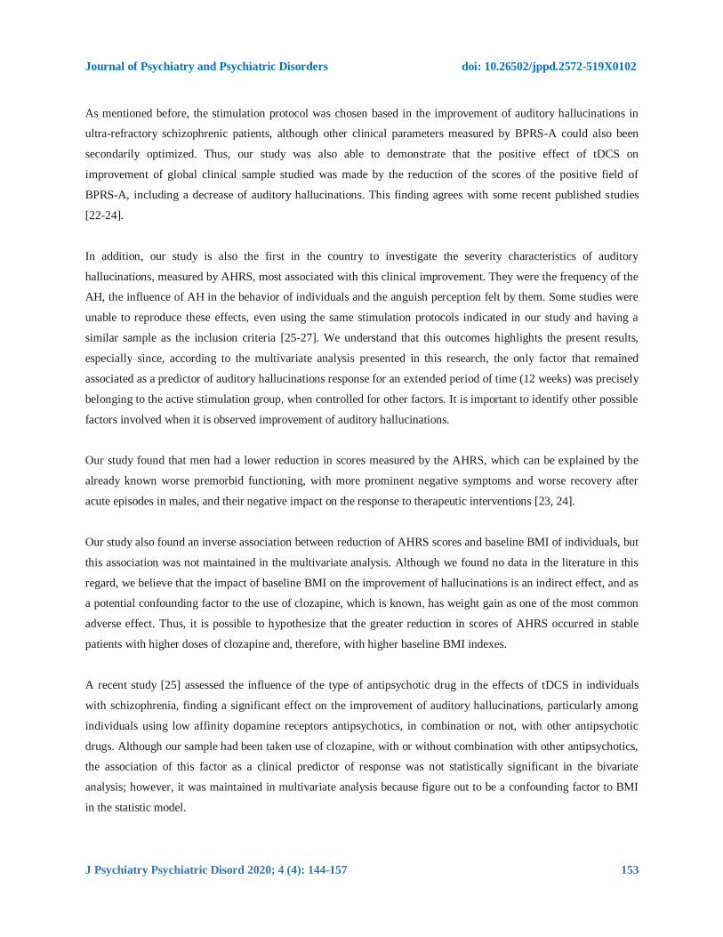
As mentioned before, the stimulation protocol was chosen based in the improvement of auditory hallucinations in
ultra-refractory schizophrenic patients, although other clinical parameters measured by BPRS-A could also been
secondarily optimized. Thus, our study was also able to demonstrate that the positive effect of tDCS on
improvement of global clinical sample studied was made by the reduction of the scores of the positive field of
BPRS-A, including a decrease of auditory hallucinations. This finding agrees with some recent published studies
[22-24].
In addition, our study is also the first in the country to investigate the severity characteristics of auditory
hallucinations, measured by AHRS, most associated with this clinical improvement. They were the frequency of the
AH, the influence of AH in the behavior of individuals and the anguish perception felt by them. Some studies were
unable to reproduce these effects, even using the same stimulation protocols indicated in our study and having a
similar sample as the inclusion criteria [25-27]. We understand that this outcomes highlights the present results,
especially since, according to the multivariate analysis presented in this research, the only factor that remained
associated as a predictor of auditory hallucinations response for an extended period of time (12 weeks) was precisely
belonging to the active stimulation group, when controlled for other factors. It is important to identify other possible
factors involved when it is observed improvement of auditory hallucinations.
Our study found that men had a lower reduction in scores measured by the AHRS, which can be explained by the
already known worse premorbid functioning, with more prominent negative symptoms and worse recovery after
acute episodes in males, and their negative impact on the response to therapeutic interventions [23, 24].
Our study also found an inverse association between reduction of AHRS scores and baseline BMI of individuals, but
this association was not maintained in the multivariate analysis. Although we found no data in the literature in this
regard, we believe that the impact of baseline BMI on the improvement of hallucinations is an indirect effect, and as
a potential confounding factor to the use of clozapine, which is known, has weight gain as one of the most common
adverse effect. Thus, it is possible to hypothesize that the greater reduction in scores of AHRS occurred in stable
patients with higher doses of clozapine and, therefore, with higher baseline BMI indexes.
A recent study [25] assessed the influence of the type of antipsychotic drug in the effects of tDCS in individuals
with schizophrenia, finding a significant effect on the improvement of auditory hallucinations, particularly among
individuals using low affinity dopamine receptors antipsychotics, in combination or not, with other antipsychotic
drugs. Although our sample had been taken use of clozapine, with or without combination with other antipsychotics,
the association of this factor as a clinical predictor of response was not statistically significant in the bivariate
analysis; however, it was maintained in multivariate analysis because figure out to be a confounding factor to BMI
in the statistic model.

Journal of Psychiatry and Psychiatric Disorders doi: 10.26502/jppd.2572-519X0102
J Psychiatry Psychiatric Disord 2020; 4 (4): 144-157 154
According to our study, the reduction in severity of AH in ultra-refractory schizophrenic patients occurs by reducing
the perception of distress and minimizing the influence of AH in their behavior. Again, our results agree with what
has been published in the most recent scientific literature. In the theory of interhemispheric imbalance in
schizophrenia, insight deficits are believed to result from abnormal activity of left dominant cerebral hemisphere
[26]. The theory has gained the support of recent functional imaging researches that attributes impaired insight to
aberrant functional connectivity in neural networks [27] and suggests various regions of the left hemisphere
representing putative targets for noninvasive treatment of neuromodulation to enhance insight into schizophrenia
[28]. In addition, evidence suggests that poor self-perception of symptoms as part of the disease is associated with
anatomical damage to the prefrontal cortex [29], as well as to greater activations of this region of the brain, whitch
may be part of a compensatory mechanism of prefrontal brain impairment [30]. Findings corroborated by other
authors [31-33]. We could intuitively imagine that the positive effects on the perception of anguish and behavioral
influences result from anodic tDCS acting on the left prefrontal cortex, increasing endogenous effort to compensate
for prefrontal activation impairment. More recent research has provided additional evidence for tDCS's ability to
modify the connectivity of the white substance [34] and frontotemporal tDCS to increase functional connectivity in
the resting state between the left TPJ and left DLPFC in patients with schizophrenia [35].
5. Conclusion
To date, this study has novelty to specifically evaluate the efficacy of tDCS anode on the left DLPFC and the
cathode on the left TPJ in patients with compensated outpatient URS for 12 weeks. However, we understand that, in
the case of such a serious and complex disease, influenced by multiple factors, some important limitations must be
considered.
Our study applied conventional tDCS which stimulates cortical areas by means of large silicon electrodes covered
by a cloth envelope humidified with saline solution. The method of identifying brain sites to be stimulated, based on
the International 10-20 EEG System, has relatively low spatial location accuracy. A growing number of research
suggests the use of high definition tDCS whose identification of cortical areas to be stimulated is made by specific
software systems and neuronavigation devices, individualizing and optimizing tDSC protocols [35]. The lack of
results and/or no significant association between some of our outcomes may also be influenced by the low accuracy
location method used in this study.
Although the demographics characteristic of the sample of patients in each group are similar, we must consider that
most of them, regardless of the group to which they belonged to, had a wide range of symptoms profiles, ie, a
complex mixture of delusions, speech disorganized behavior and negative symptoms, depression and AH,
demonstrating serious and complex neuroprogression disease. At this context, since the protocol established in this
study was directed specifically to evaluate auditory hallucinations in URS, their potential therapeutic effect may be
obscured by the presence of multiple severe and chronic symptoms.

Journal of Psychiatry and Psychiatric Disorders doi: 10.26502/jppd.2572-519X0102
J Psychiatry Psychiatric Disord 2020; 4 (4): 144-157 155
The long follow-up time for the studied population, associated with the absence of new stimulation sessions, and
with the need for repeated blood collections, also constituted a limiting scenario, making it particularly difficult to
increase the sample size beyond the 33 participants and compromising part of the statistical power of the study.
We believe that these considerations could safely confirm the notion that tDCS protocols directed to refractory
samples should be optimized, possibly using higher electrical current doses for a longer period and relying on a
long-term maintenance treatment after acute phase treatment. As future perspectives, URS should be considered as a
heterogeneous mental disorder, composed of a challenging phenotypic diversity. Therefore, in addition to refining
the technical development of tDCS, we consider that one of the main focuses of subsequent studies should be the
measurement of different groups of symptoms using the Hi-Top of RDC methodology, to identify markers of
response to treatment, respecting different clinical and neurobiological dimensions of schizophrenia.
References
1. Jardri R, Pouchet A, Pins D, et al. Cortical activations during auditory verbal hallucinations in
schizophrenia: a coordinate-based meta-analysis. Am J Psychiatry 168 (2011):73-81.
2. Boggio PS, Rigonatti SP, Ribeiro RB, et al. A randomized, double-blind clinical trial on the efficacy of
cortical direct current stimulation for the treatment of major depression. Int J Neuropsychopharmacol 11
(2008): 249e54.
3. Loo CK, Alonzo A, Martin D, et al. Transcranial direct current stimulation for depression: 3-week,
randomised, sham controlled trial. Br J Psychiatry 200 (2012): 52e9.
4. McClintock SM, Freitas C, Oberman L, et al. Transcranial magnetic stimulation: a neuroscientific probe of
cortical function in schizophrenia. Biol Psychiatry 70 (2011): 19-27.
5. Brunelin J, Mondino M, Haesebaert F, et al. Efficacy and safety of bifocal tDCS as an interventional
treatment for refractory schizophrenia. Brain Stimul 431 (2012): 431-432.
6. Andrade C. Once- to Twice-Daily, 3-Year Domiciliary Maintenance Transcranial Direct Current
Stimulation for Severe, Disabling, Clozapine-Refractory Continuous Auditory Hallucinations in
Schizophrenia: J ECT 29 (2013): 239-242.
7. Homan P, Kindler J, Federspiel A, et al. Muting the voice: a case of arterial spin labeling-monitored
transcranial direct current stimulation treatment of auditory verbal hallucinations. Am J Psychiatry 168
(2011): 853-854.
8. Narayanaswamy JC, Shivakumar V, Bose A, et al. Sustained improvement of negative symptoms in
schizophrenia with add-on tDCS. Clin Schizophr Relat Psychoses 8 (2014): 135-136.
9. Nawani H, Kalmady SV, Bose A, et al. Neural basis of tDCS effects on auditory verbal hallucinations in
schizophrenia: a case report evidence for cortical neuroplasticity modulation. J ECT 30 (2014): e2-e4.
10. Nawani H, Bose A, Agarwal SM, et al. Modulation of corollary discharge dysfunction in schizophrenia by
tDCS: preliminary evidence. Brain Stimul 7 (2014): 486-488.
Journal of Psychiatry and Psychiatric Disorders doi: 10.26502/jppd.2572-519X0102
J Psychiatry Psychiatric Disord 2020; 4 (4): 144-157 156
11. Rakesh G, Shivakumar V, Subramaniam A, et al. Monotherapy with tDCS for Schizophrenia: a case report.
Brain Stimul 6 (2013): 708-709.
12. Shiozawa P, da Silva ME, Cordeiro Q, et al. Transcranial Direct Current Stimulation (tDCS) for the
treatment of persistent visual and auditory hallucinations in schizophrenia: a case study. Brain Stimul 6
(2013): 831-833.
13. Shivakumar V, Bose A, Rakesh G, et al. Rapid improvement of auditory verbal hallucinations in
schizophrenia after add-on treatment with transcranial direct-current stimulation. J ECT 29 (2013): e43-44.
14. Shivakumar V, Narayanaswamy JC, Agarwal SM, et al. Targeted, intermittent booster tDCS: a novel add-
on application for maintenance treatment in a schizophrenia patient with refractory auditory verbal
hallucinations. Asian J Psychiatry 11 (2014): 79-80.
15. Bose A, Shivakumar V, Narayanaswamy JC, et al. Insight facilitation with add-on tDCS in schizophrenia.
Schizophr Res 156 (2014): 63-65.
16. Ferrucci R, Bortolomasi M, Tessari E, et al. EPA-1392—Transcranial direct-current stimulation (tDCS) in
patients with schizophrenia. Eur Psychiatry 29 (2014): 1.
17. Morgan HM, Davis NJ, Bracewell RM. Does transcranial direct current stimulation to prefrontal cortex
affect mood and emotional memory retrieval in healthy individuals. PLoS ONE 9 (2014): e92162
10.1371/journal.pone.0092162.
18. Brunelin J, Mondino M, Gassab L, et al. Examining transcranial direct-current stimulation (tDCS) as a
treatment for hallucinations in schizophrenia. Am J Psychiatry 169 (2012): 719-724.
19. Sanfilipo M, Lafargue T, Rusinek H, et al. Volumetric measure of the frontal and temporal lobe regions in
schizophrenia: relationship to negative symptoms. Arch Gen Psychiatry 57 (2000): 471-480.
20. Grimm S, Beck J, Schuepbach D, et al. Imbalance between left and right dorsolateral prefrontal cortex in
major depression is linked to negative emotional judgment: an fmri study in severe major depressive
disorder. Biol Psychiatry 63 (2008): 369-376.
21. Mondino M, Haesebaert F, Poulet E, et al. Fronto-temporal transcranial direct current stimulation (tDCS)
reduces source-monitoring deficits and auditory hallucinations in patients with schizophrenia. Schizophr
Res 161 (2015): 515-516.
22. Mondino M, Jardri R, Suaud-Chagny MF, et al. Effects of fronto-temporal transcranial direct current
stimulation on auditory verbal hallucinations and resting-state functional connectivity ´~of the left temporo-
parietal junction in patients with schizophrenia. Schizophr Bull 42 (2018): 318-326.
23. van Os J, Kapur S. Schizophrenia. Lancet 374 (2009): 635-645.
24. McGrath J, Saha S, Chant D, et al. Schizophrenia: a concise overview of incidence, prevalence, and
mortality. Epidemiologic reviews 30 (2008): 67-76.
25. Agarwal SM, Bose A, Shivakumar V, et al. Impact of antipsychotic medication on transcranial direct
current stimulation (tDCS) effects in schizophrenia patients. Psychiatry Res 235 (2016): 97-103.
Journal of Psychiatry and Psychiatric Disorders doi: 10.26502/jppd.2572-519X0102
J Psychiatry Psychiatric Disord 2020; 4 (4): 144-157 157
26. Shad MU, Keshavan MS, Tamminga CA, et al. Neurobiological underpinnings of insight deficits in
schizophrenia. Int Rev Psychiatry 19 (2007): 437-446.
27. Gerretsen P, Menon M, Mamo DC, et al. Impaired insight into illness and cognitive insight in
schizophrenia spectrum disorders: resting state functional connectivity. Schizophr Res 160 (2014): 43-50.
28. Gerretsen P, Menon M, Chakravarty MM, et al. Illness denial in schizophrenia spectrum disorders: a
function of left hemisphere dominance. Hum Brain Mapp 36 (2015): 213-225.
29. Parellada M, Boada L, Fraguas D, et al. Trait and state attributes of insight in first episodes of early-onset
schizophrenia and other psychoses: a 2-year longitudinal study. Schizophr Bull 37 (2011): 38-51.
30. Shad MU, Keshavan MS, Tamminga CA, et al. Neurobiological underpinnings of insight deficits in
schizophrenia. Int Rev Psychiatry 19 (2007): 437-446.
31. Bergé D, Carmona S, Rovira M, et al. Gray matter volume deficits and correlation with insight and
negative symptoms in first-psychotic-episode subjects. Acta Psychiatr Scand 123 (2011): 431-439.
32. Sapara A, Ffytche DH, Birchwood M, et al. Preservation and compensation: the functional neuroanatomy
of insight and working memory in schizophrenia. Schizophr Res 152 (2014): 201-209.
33. Buchy L, Ad-Dab’bagh Y, Malla A, et al. Cortical thickness is associated with poor insight in first-episode
psychosis. J Psychiatr Res 45 (2011): 781-787.
34. Lindenberg R, Nachtigall L, Meinzer M, et al. Differential effects of dual and unihemispheric motor cortex
stimulation in older adults. J Neurosci 33 (2013): 9176-9183.
35. Mondino M, Jardri R, Suaud-Chagny MF, et al. Effects of fronto-temporal transcranial direct current
stimulation on auditory verbal hallucinations and resting-state functional connectivity of the left temporo-
parietal junction in patients with schizophrenia. Schizophr Bull 42 (2016): 318-326.
Citation: Nathália Janovik, Victor Hugo Cordova, Cintya Ogliari, Michel Mroginski, Lenise Franceschoni, Paulo
Belmonte-de-Abreu. Treatment of Residual Psychotic Symptons in Ultra-Refractory Schizophrenia after 10 Sessions
of Transcranial Direct Current Stimulation (tDCS). Journal of Psychiatry and Psychiatric Disorders 4 (2020): 144-
157.
This article is an open access article distributed under the terms and conditions of the
Creative Commons Attribution (CC-BY) license 4. 0