Transport and Localization Elements in Myelin Basic Protein mRNA
11
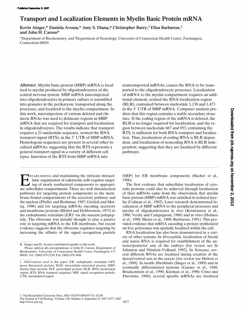
The Rockefeller University Press, 0021-9525/97/09/1077/11 $2.00 The Journal of Cell Biology, Volume 138, Number 5, September 8, 1997 1077–1087 http://www.jcb.org 1077 Transport and Localization Elements in Myelin Basic Protein mRNA Kevin Ainger,* Daniela Avossa,* Amy S. Diana,* Christopher Barry, ‡ Elisa Barbarese, ‡ and John H. Carson* *Department of Biochemistry, and ‡ Department of Neurology, University of Connecticut Health Center, Farmington, Connecticut 06030 Abstract. Myelin basic protein (MBP) mRNA is local- ized to myelin produced by oligodendrocytes of the central nervous system. MBP mRNA microinjected into oligodendrocytes in primary culture is assembled into granules in the perikaryon, transported along the processes, and localized to the myelin compartment. In this work, microinjection of various deleted and chi- meric RNAs was used to delineate regions in MBP mRNA that are required for transport and localization in oligodendrocytes. The results indicate that transport requires a 21-nucleotide sequence, termed the RNA transport signal (RTS), in the 39 UTR of MBP mRNA. Homologous sequences are present in several other lo- calized mRNAs, suggesting that the RTS represents a general transport signal in a variety of different cell types. Insertion of the RTS from MBP mRNA into nontransported mRNAs, causes the RNA to be trans- ported to the oligodendrocyte processes. Localization of mRNA to the myelin compartment requires an addi- tional element, termed the RNA localization region (RLR), contained between nucleotide 1,130 and 1,473 in the 39 UTR of MBP mRNA. Computer analysis pre- dicts that this region contains a stable secondary struc- ture. If the coding region of the mRNA is deleted, the RLR is no longer required for localization, and the re- gion between nucleotide 667 and 953, containing the RTS, is sufficient for both RNA transport and localiza- tion. Thus, localization of coding RNA is RLR depen- dent, and localization of noncoding RNA is RLR inde- pendent, suggesting that they are localized by different pathways. K. Ainger and D. Avossa contributed equally to this work. Please address all correspondence to John H. Carson, Department of Biochemistry, University of Connecticut Health Center, Farmington, CT 06030. Tel.: (860) 679-2130; Fax: (860) 679-3408. 1. Abbreviations used in this paper: ER, endoplasmic reticulum; GFP, green fluorescent protein; MAP, microtubule-associated protein; MBP, myelin basic protein; PLP, proteolipid protein; RLR, RNA localization region; RTS, RNA transport sequence; SRP, signal recognition particle; UTR, untranslated region. E stablishing and maintaining the intricate intracel- lular organization of eukaryotic cells requires target- ing of newly synthesized components to appropri- ate subcellular compartments. There are well characterized pathways for targeting protein components to the mem- brane-bound compartments of the secretory pathway and the nucleus (Pfeffer and Rothman, 1987; Görlich and Mat- taj, 1996) and for targeting mRNAs encoding secretory and membrane proteins (Blobel and Dobberstein, 1975) to the endoplasmic reticulum (ER) 1 via the nascent polypep- tide. The ribosome was initially thought to play a passive role in targeting mRNA to the ER membrane, but recent evidence suggests that the ribosome regulates targeting by increasing the affinity of the signal recognition particle (SRP) for ER membrane components (Bacher et al., 1996). The first evidence that subcellular localization of cyto- solic proteins could also be achieved through localization of their mRNAs came from the observation that myelin basic protein (MBP) mRNA was enriched in isolated mye- lin (Colman et al., 1982). Later research demonstrated lo- calization of MBP mRNA to the peripheral processes and myelin of oligodendrocytes in vivo (Kristensson et al., 1986; Verity and Campagnoni, 1988) and in vitro (Holmes et al., 1988; Shiota et al., 1989; Barbarese, 1991). This pro- vided evidence that mRNA encoding a protein synthesized on free polysomes was spatially localized within the cell. RNA localization has also been demonstrated in a vari- ety of other systems. In Drosophila, localization of bicoid and nanos RNA is required for establishment of the an- terior/posterior axis of the embryo (for review see St Johnston and Nüsslein-Volhard, 1992). In Xenopus, sev- eral different RNAs are localized during creation of the dorsal/ventral axis in the oocyte (for review see Melton et al., 1989). In motile fibroblasts (Singer et al., 1989) and in terminally differentiated neurons (Garner et al., 1988; Bruckenstein et al., 1990; Kleiman et al., 1990; Crino and Eberwine, 1996), several specific mRNAs are localized on November 2, 2013 jcb.rupress.org Downloaded from Published September 8, 1997 on November 2, 2013 jcb.rupress.org Downloaded from Published September 8, 1997 on November 2, 2013 jcb.rupress.org Downloaded from Published September 8, 1997 on November 2, 2013 jcb.rupress.org Downloaded from Published September 8, 1997 on November 2, 2013 jcb.rupress.org Downloaded from Published September 8, 1997 on November 2, 2013 jcb.rupress.org Downloaded from Published September 8, 1997 on November 2, 2013 jcb.rupress.org Downloaded from Published September 8, 1997 on November 2, 2013 jcb.rupress.org Downloaded from Published September 8, 1997 on November 2, 2013 jcb.rupress.org Downloaded from Published September 8, 1997 on November 2, 2013 jcb.rupress.org Downloaded from Published September 8, 1997 on November 2, 2013 jcb.rupress.org Downloaded from Published September 8, 1997
-
Upload
independent -
Category
Documents
-
view
0 -
download
0
Transcript of Transport and Localization Elements in Myelin Basic Protein mRNA
The Rockefeller University Press, 0021-9525/97/09/1077/11 $2.00The Journal of Cell Biology, Volume 138, Number 5, September 8, 1997 1077–1087http://www.jcb.org 1077
Transport and Localization Elements in Myelin Basic Protein mRNA
Kevin Ainger,* Daniela Avossa,* Amy S. Diana,* Christopher Barry,
‡
Elisa Barbarese,
‡
and John H. Carson*
*Department of Biochemistry, and
‡
Department of Neurology, University of Connecticut Health Center, Farmington, Connecticut 06030
Abstract.
Myelin basic protein (MBP) mRNA is local-ized to myelin produced by oligodendrocytes of the central nervous system. MBP mRNA microinjected into oligodendrocytes in primary culture is assembled into granules in the perikaryon, transported along the processes, and localized to the myelin compartment. In this work, microinjection of various deleted and chi-meric RNAs was used to delineate regions in MBP mRNA that are required for transport and localization in oligodendrocytes. The results indicate that transport requires a 21-nucleotide sequence, termed the RNA transport signal (RTS), in the 3
9
UTR of MBP mRNA. Homologous sequences are present in several other lo-calized mRNAs, suggesting that the RTS represents a general transport signal in a variety of different cell types. Insertion of the RTS from MBP mRNA into
nontransported mRNAs, causes the RNA to be trans-ported to the oligodendrocyte processes. Localization of mRNA to the myelin compartment requires an addi-tional element, termed the RNA localization region (RLR), contained between nucleotide 1,130 and 1,473 in the 3
9
UTR of MBP mRNA. Computer analysis pre-dicts that this region contains a stable secondary struc-ture. If the coding region of the mRNA is deleted, the RLR is no longer required for localization, and the re-gion between nucleotide 667 and 953, containing the RTS, is sufficient for both RNA transport and localiza-tion. Thus, localization of coding RNA is RLR depen-dent, and localization of noncoding RNA is RLR inde-pendent, suggesting that they are localized by different pathways.
K. Ainger and D. Avossa contributed equally to this work.Please address all correspondence to John H. Carson, Department of
Biochemistry, University of Connecticut Health Center, Farmington, CT06030. Tel.: (860) 679-2130; Fax: (860) 679-3408.
1.
Abbreviations used in this paper
: ER, endoplasmic reticulum; GFP,green fluorescent protein; MAP, microtubule-associated protein; MBP,myelin basic protein; PLP, proteolipid protein; RLR, RNA localizationregion; RTS, RNA transport sequence; SRP, signal recognition particle;UTR, untranslated region.
E
stablishing
and maintaining the intricate intracel-lular organization of eukaryotic cells requires target-ing of newly synthesized components to appropri-
ate subcellular compartments. There are well characterizedpathways for targeting protein components to the mem-brane-bound compartments of the secretory pathway andthe nucleus (Pfeffer and Rothman, 1987; Görlich and Mat-taj, 1996) and for targeting mRNAs encoding secretoryand membrane proteins (Blobel and Dobberstein, 1975) tothe endoplasmic reticulum (ER)
1
via the nascent polypep-tide. The ribosome was initially thought to play a passiverole in targeting mRNA to the ER membrane, but recentevidence suggests that the ribosome regulates targeting byincreasing the affinity of the signal recognition particle
(SRP) for ER membrane components (Bacher et al.,1996).
The first evidence that subcellular localization of cyto-solic proteins could also be achieved through localizationof their mRNAs came from the observation that myelinbasic protein (MBP) mRNA was enriched in isolated mye-lin (Colman et al., 1982). Later research demonstrated lo-calization of MBP mRNA to the peripheral processes andmyelin of oligodendrocytes in vivo (Kristensson et al.,1986; Verity and Campagnoni, 1988) and in vitro (Holmeset al., 1988; Shiota et al., 1989; Barbarese, 1991). This pro-vided evidence that mRNA encoding a protein synthesizedon free polysomes was spatially localized within the cell.
RNA localization has also been demonstrated in a vari-ety of other systems. In
Drosophila
, localization of bicoidand nanos RNA is required for establishment of the an-terior/posterior axis of the embryo (for review see StJohnston and Nüsslein-Volhard, 1992). In
Xenopus
, sev-eral different RNAs are localized during creation of thedorsal/ventral axis in the oocyte (for review see Melton etal., 1989). In motile fibroblasts (Singer et al., 1989) and interminally differentiated neurons (Garner et al., 1988;Bruckenstein et al., 1990; Kleiman et al., 1990; Crino andEberwine, 1996), several specific mRNAs are localized
on Novem
ber 2, 2013jcb.rupress.org
Dow
nloaded from
Published September 8, 1997 on N
ovember 2, 2013
jcb.rupress.orgD
ownloaded from
Published September 8, 1997
on Novem
ber 2, 2013jcb.rupress.org
Dow
nloaded from
Published September 8, 1997 on N
ovember 2, 2013
jcb.rupress.orgD
ownloaded from
Published September 8, 1997
on Novem
ber 2, 2013jcb.rupress.org
Dow
nloaded from
Published September 8, 1997 on N
ovember 2, 2013
jcb.rupress.orgD
ownloaded from
Published September 8, 1997
on Novem
ber 2, 2013jcb.rupress.org
Dow
nloaded from
Published September 8, 1997 on N
ovember 2, 2013
jcb.rupress.orgD
ownloaded from
Published September 8, 1997
on Novem
ber 2, 2013jcb.rupress.org
Dow
nloaded from
Published September 8, 1997 on N
ovember 2, 2013
jcb.rupress.orgD
ownloaded from
Published September 8, 1997
on Novem
ber 2, 2013jcb.rupress.org
Dow
nloaded from
Published September 8, 1997

The Journal of Cell Biology, Volume 138, 1997 1078
within the cell. Localization of mRNAs has been exten-sively reviewed (Steward and Banker, 1992; Wilhelm andVale, 1993; St Johnston, 1995).
With the exception of mRNAs encoding signal sequences,which are cotranslationally targeted to the ER by the se-quence of the nascent polypeptide in conjunction withSRP, most of the characterized
cis
-acting signals for RNAlocalization are found in the 3
9
untranslated region (UTR)of the mRNA.
Cis
-acting signals have been defined for lo-calization of bicoid (Macdonald and Struhl, 1988; Mac-donald et al., 1993), nanos (Wharton and Struhl, 1991;Gavis and Lehmann, 1992), oskar (Ephrussi and Leh-mann, 1992; Kim-Ha et al., 1993), Vg1 (Mowry and Mel-ton, 1992),
b
-actin (Kislauskis et al., 1994), cyclin B (Dalbyand Glover, 1993), K10 (Cheung et al., 1992), and even-skipped (Davis and Ish-Horowicz, 1991). In
Drosophila
,localization of oskar and bicoid mRNAs are controlled bymultiple RNA elements that control different steps in thepathway (Kim-Ha et al., 1993; Macdonald et al., 1993).
One function of mRNA localization is to ensure local-ized synthesis of the encoded protein. This implies thattranslation is repressed until the mRNA becomes local-ized. Translation is known to be tightly regulated in manyoocytes and embryos, where specific mRNAs are nottranslated until fertilization or egg activation (for reviewsee Curtis et al., 1995). Some of these translationally re-pressed maternal mRNAs, like nanos mRNA in
Dro-sophila
, are also localized. Nanos protein is not expressedfrom unlocalized nanos mRNA (Gavis and Lehmann,1994). The oskar protein functions in localization of nanosmRNA as well as enhancing translation of nanos mRNA.In addition, during localization of oskar mRNA, no oskarprotein can be detected (Kim-Ha et al., 1995). Transla-tional repression and localization of oskar mRNA are con-trolled by separated elements in the 3
9
UTR of oskarmRNA.
These observations have led to the hypothesis of local-ization-dependent translation (Gavis and Lehmann, 1994;Kim-Ha et al., 1995), in which translation of specific mRNAsis dependent on their subcellular localization. In this con-text, the term RNA localization, which was originally usedto describe the nonuniform, steady-state distribution of anmRNA in a cell, can be more narrowly defined as recogni-tion of the correct subcellular destination of the trans-ported mRNA. Thus, RNA localization can be considereda sequential process involving translational repression, trans-port, localization, and localization-dependent translation.
MBP mRNA localization in oligodendrocytes occursthrough a multi-step pathway that has been defined usingmicroinjection experiments (Ainger et al., 1993). The path-way includes assembly of the RNA into granules in theperikaryon, anterograde transport along cellular processes,and localization within the myelin compartment. The workdescribed here takes advantage of the fact that granule as-sembly, transport, and localization occur in spatially dis-tinct subcellular compartments (the perikaryon, processes,and myelin compartment, respectively) of the oligoden-drocyte. By deleting various regions of MBP mRNA andanalyzing the subcellular distribution of the injected RNA,it is possible to delineate discrete elements in MBP mRNAthat are specifically required for different steps in the lo-calization pathway.
Materials and Methods
Cell Culture
Mouse oligodendrocytes were isolated from mixed primary brain cell cul-tures and grown as described previously (Ainger et al., 1993).
Reagents
Restriction enzymes and RNA polymerases were obtained from New En-gland BioLabs (Beverly, MA), Promega (Madison, WI), and Stratagene(La Jolla, CA). RNasin and transcription buffers were from Promega.Digoxigenin-UTP was purchased from Boehringer Mannheim (Indianap-olis, IN). RNA molecular weight markers were from GIBCO BRL(Gaithersburg, MD). pSP64 and pGEM3 vectors were from Promega.pBluescript IISK was from Stratagene. Diethylpyrocarbonate (DEPC),mineral oil, monoclonal anti-digoxigenin, and ammonium sulfate werefrom Sigma Chemical Co. (St. Louis, MO). Fluorochrome-conjugated sec-ondary antibodies were from Chemicon International, Inc. (Temecula,CA) and Jackson ImmunoResearch Laboratories Inc. (West Grove, PA).Oligonucleotides were from National Biosciences (Plymouth, MN). Anti-BIP (endoplasmic reticulum chaperonin) antibody was from StressGenBiotechnologies Corp. (Victoria, BC, Canada).
Recombinant DNA and In Vitro Transcription
Transcription templates were prepared from a plasmid containing rat 14 kDMBP (Roach et al., 1983) originally subcloned into pSP64 poly A vector.Digestion with EcoRI and transcription with SP6 RNA polymerase gavean RNA with the first 1473 bases of MBP mRNA. For the other trunca-tions, full length MBP cDNA was subcloned into the EcoRI site of pBlue-script IISK to create pKS3. Transcription with T3 RNA polymerase re-sulted in incorporation of a short length of vector-derived sequences fromthe polylinker on the 5
9
end of all RNAs. Digestion with SalI, PvuII, andBstEII gave RNAs with the first 666, 953, and 1,131 bases, respectively, ofrat MBP. HindIII-cut plasmid was used to transcribe full length MBPmRNA containing plasmid-derived polyadenosine and a short 3
9
se-quence from the polylinker. To make RNA with a translational frame-shift, the XmaI to HindIII fragment of pKS3 was subcloned into pGem3.The plasmid was linearized with BamHI, filled in with the Klenow frag-ment of DNA polymerase I, and religated. The plasmids were grown indam
2
Escherichia coli
and assayed for a newly generated ClaI site. Thereading frame shifts to
2
1 after 13 amino acids of MBP. The polypeptideterminates after an additional 27 out-of-frame amino acids. The 3
9
UTR ofMBP was subcloned as a SalI/HindIII fragment into SalI/HindIII-cutpGem3. The plasmid was linearized with HindIII and transcribed with T7RNA polymerase. Similarly, the SalI/PvuII fragment of MBP 3
9
UTR wassubcloned into SalI/EcoRV-digested pBluescript IISK, digested withPvuII, and transcribed with T7 RNA polymerase. This produced an RNAwith a 280-nucleotide 3
9
extension of vector-derived sequences. To con-struct chimeric RNAs, the 3
9
UTR and the SalI/PvuII fragments of MBPcDNA were subcloned into SalI-linearized and SalI/SmaI-digested, re-spectively,
Xenopus laevis
globin cDNA (the gift of D. Melton of HarvardUniversity, Cambridge, MA; pSP64-X
b
M). To make RNA, the chimericplasmids were digested with EcoRV and SmaI, respectively, and tran-scribed with SP6 RNA polymerase. To make RNA containing the RNAtransport sequence (RTS), an oligonucleotide containing the RTS se-quence with SpeI and KpnI linkers (CTAGTGCCAAGGAGCCA-GAGAGCATGGGTAC) was cloned into SpeI/KpnI-digested pBlue-scriptII (SK) to generate pRTS, which was digested with SspI andtranscribed with T3 RNA polymerase. To construct proteolipid protein(PLP)-RTS, chimeric RNA, PLP cDNA (the gift of F. Smith, Shriver Cen-ter, Waltham, MA) was digested with SpeI and KpnI, removing 560 nucle-otides from the PLP 3
9
UTR, and ligated to SpeI/KpnI-digested pRTS.The resulting plasmid was linearized with KpnI; blunt ends were createdwith the Klenow fragment and transcribed with T3 RNA polymerase.
b
-Actin cDNA was the gift of Dr. J. Pachter of the University of Connect-icut Health Center. Protamine 2 cDNA was obtained from Dr. N. Hecht,Tufts University (Medford, MA). Green fluorescent protein (GFP)cDNA was purchased from GIBCO BRL and subcloned into a plasmidcontaining a T7 promoter for transcription and a sequence of 80 A’sdownstream of the cDNA insertion site. To construct GFP-RTS an oligo-nucleotide containing the RTS sequence was inserted between the cDNAopen reading frame and the poly A trail. All RNAs were subjected toelectrophoresis in gels containing formaldehyde (Sambrook et al., 1989)

Ainger et al.
RNA Transport and Localization
1079
to ensure that the injected RNA was of the appropriate size. RNA was la-beled by addition of digoxigenin-UTP or fluorescein-UTP during tran-scription reactions according to manufacturer’s instructions.
Microinjection and Immunocytochemistry
RNA was injected into cells maintained at room temperature for a periodof not more than 45 min, as described previously (Ainger et al., 1993). Toallow time for transport and localization of the injected RNA, cells wereincubated for 15 min at 37
8
C, in medium containing 2% newborn calf se-rum to digest extracellular RNA that leaked into the medium during theinjections and to reduce nonspecific background. To visualize digoxige-nin-labeled RNA, cells were fixed in 4% paraformaldehyde in PBS andincubated with anti-digoxigenin antibodies as previously described(Ainger et al., 1993), or using a mouse monoclonal anti-digoxigenin anti-body (1:75 dilution), followed by detection with a fluorescein-conjugatedgoat anti–mouse IgG (Chemicon International). MBP was visualized us-ing rabbit polyclonal anti-MBP followed by Texas red-conjugated donkeyanti–rabbit IgG (Jackson ImmunoResearch Laboratories, Inc.).
Microscopy and Image Processing
Laser scanning confocal microscopy was performed with an MRC-600scanning system (Bio Rad, Cambridge, MA) mounted on a microscope(Axioskope; Zeiss, Oberkochen, Germany) equipped with a variety of in-finity-corrected high numerical aperture objectives.
Computer Analysis of Sequences
Computer analysis of sequence information was performed with the Ge-netics Computer Group program (1991 Version 7; Madison, WI). Dr. M.Zuker (Ottawa University, Ottawa, Canada) kindly provided an updatedversion of the MFOLD program. Rat 14 kD MBP (Roach et al., 1983) andmouse 14 kD MBP (Takahashi et al., 1985) were used for sequence com-parisons. The other sequences listed in Table I are available from Gen-Bank/EMBL/DDJB index accession numbers as follows: human MBP,M13577; rat MOBP81-A, X87900; rat glial fibrillary acidic protein(GFAP), K01347; human N-type calcium channel
a
-1 (Ca-N), M94172;mouse microtubule-associated protein (MAP) 2A, M21041; bovineGABA(A) receptor (GABARA), X05717; bovine nitric oxide synthase,(NOS) M95674; rat activity-regulated cytoskeleton-associated protein(ARC), U19866; rat neurogranin (RC3), L09119; mouse protamine 2,X14004; pufferfish NCAML1, Z71926; rat c-jun, X17163; rat atrophin-1related protein (rARP), U44091; human clathrin light chain b, M20469and J04174; rat furosemide sensitive K-Cl cotransporter (KCCl), U55815;mouse protein kinase C
a
, M25811; human chromogranin A, J03483; hu-man glycogen phosphorylase, J03544; mouse cyritestin, X64227; mouseheparin binding protein 44 (HBP44), D00622; human LIM kinase, D26309;human insulin like growth factor I receptor (IGFRI), Xo4434; mouseRNA-binding protein (FLI-2), M99167; HIV-2 tat,vpr, J04542; hepatitis Cvirus NS-5 region, Z35506.
Results
Microinjection Assay of Modified RNAs
Previous work has shown that MBP mRNA microinjectedinto oligodendrocytes is assembled into RNA granulesthat are transported along the processes and localized tothe myelin compartment, while control mRNAs (globin andactin) are assembled into RNA granules that remain in theperikaryon (Ainger et al., 1993). The differential distributionof these mRNAs is presumably controlled by some aspectof their structure. In this study, deletion analysis of MBPRNA was used to identify sequences or structures that arerequired for transport and/or localization. Digoxigenin-labeled RNAs were synthesized by in vitro transcriptionand microinjected into cultured oligodendrocytes. Cells werefixed and labeled by immunofluorescence with both anti-digoxigenin antibody to visualize the injected RNA and anti-MBP antibody to visualize the overall cell morphology.
The unique morphology of oligodendrocytes in primaryculture makes it possible to spatially resolve three stagesin the RNA sorting pathway: (
a
) granule assembly in theperikaryon; (
b
) transport in the processes; and (
c
) localiza-tion in the myelin compartment. Therefore, by analyzingthe subcellular distribution of microinjected RNA it ispossible to determine if a particular RNA is assembledinto granules in the perikaryon, transported in the pro-cesses and/or localized to the myelin compartment. In thisregard it is important to appreciate that the very features(thin, flattened morphology, unfurled myelin compart-ment) of oligodendrocytes in culture that make them suit-able for studies on intracellular RNA trafficking are dif-ferent from oligodendrocytes in the intact nerve where themyelin compartment is wrapped spirally around the myeli-nated axon and where intracellular RNA trafficking oc-curs in three dimensions and may be influenced by signalsfrom the axons that are being myelinated or from othersurrounding cells. The oligodendrocyte in culture recapit-ulates those aspects of RNA trafficking that are intrinsicto the cell. However, RNA trafficking may be regulated inmore complex ways in vivo. These caveats notwithstand-ing, the geographic separation of perikaryon, processes,and myelin compartment in oligodendrocytes in cultureprovides means of resolving the various steps in intracellu-lar trafficking of RNA.
Representative cells illustrating the observed distribu-tion patterns are shown in Fig. 2, where the distributionpatterns of the injected RNA are shown in the left columnand the staining patterns with antibody to MBP to revealthe overall cell morphology are shown in the right column.Three distinct RNA distribution patterns were observed(illustrated in Fig. 2,
A
,
C
, and
E
). In Fig. 2
A
, the injectedRNA is present in granules in the perikaryon, processes,and myelin compartment, indicating that the RNA is as-sembled into granules, transported, and localized. In Fig. 2
C
, RNA is present in granules in the perikaryon and pro-cesses but not in the myelin compartment, indicating thatthe RNA is assembled into granules and transported butnot localized. In Fig. 2
E
, RNA is present in granules inthe perikaryon but not in the processes or myelin compart-ment, indicating that the RNA is assembled into granulesbut not transported or localized.
The microinjection assay employed here is based on theassumption that the behavior of exogenous RNA microin-jected into oligodendrocytes accurately reflects the intra-cellular trafficking pathway of endogenous RNA synthe-sized in oligodendrocytes. Microinjection directly into theperikaryon makes it possible to analyze RNA transportand localization without the confounding variables of tran-scription, post-transcriptional processing, and nuclear ex-port. However, the amount of exogenous RNA injectedinto the perikaryon may be substantially greater than theamount of endogenous MBP mRNA in the cell. This raisesthe possibility that certain steps in the intracellular traf-ficking pathway may become saturated and the quantita-tive distribution of exogenous RNA in the different sub-cellular compartments shortly after injection may notaccurately reflect the steady state distribution of endoge-nous MBP mRNA in the cell. Nevertheless, on a qualita-tive level, appearance of injected RNA in granules in theperikaryon provides evidence for granule assembly, in the

The Journal of Cell Biology, Volume 138, 1997 1080
distal processes provides evidence for transport, and in themyelin compartment provides evidence for localization.
The microinjection assay is also subject to a certainamount of inherent variability. This may reflect the deli-cacy of the microinjection process, the fragility of the cells,variations in the precise injection location, or morphologi-cal and developmental variation among oligodendrocytesin culture. Because of this variability, it was necessary tomicroinject several hundred cells with each RNA and toevaluate the distribution pattern observed in the majorityof the cells. The results obtained for each RNA were com-pared with size-matched positive and negative controlsthat were microinjected at the same time into cells fromthe same preparation. Each RNA was assayed in at leastthree different preparations of oligodendrocytes. In mostexperiments, a small proportion (
,
20%) of the injectedcells exhibited no transport or localization. This may meanthat some of the microinjected cells died after injection orthat some of the cells are not transport competent. In gen-
eral,
.
70% of the cells injected with a particular RNA dis-played a consistent distribution pattern comparable to oneof the three patterns illustrated in Fig. 2.
The structures of the various RNAs tested in the micro-injection assay are diagrammed in Fig. 1, with the distribu-tion pattern observed in the majority of injected cells tabu-lated for each RNA. When full length, polyadenylatedMBP mRNA (Fig. 1
A
) was injected into oligodendro-cytes, granules were observed in the perikaryon, pro-cesses, and myelin compartment (this pattern is illustratedin Fig. 2,
A
and
B
), indicating that this RNA contains sig-nals for granule assembly, transport, and localization, re-spectively. The distribution pattern was not affected by in-sertion of a frameshift mutation into the coding region ofMBP mRNA (Fig. 1
B
), deletion of 425 nucleotides (in-cluding the poly A tail) from the 3
9
end (Fig. 1
C
), or dele-tion of the coding region (Fig. 1
G
). Previous experimentsdemonstrated that the presence or absence of a cap at the5
9
end did not affect the distribution pattern of microin-
Figure 1. Intracellular localization of microinjected RNAs. (A) Full length MBP. (B) Frameshifted full length MBP. (C) EcoRI-trun-cated MBP. (D) BstEII-truncated MBP. (E) PvuII-truncated MBP. (F) SalI-truncated MBP. (G) SalI-fragment of MBP 39 UTR. (H)Chimeric construct of X. laevis b-globin with SalI fragment of MBP 39 UTR. (I) Chimeric construct of X. laevis b-globin with SalI–PvuIIfragment of MBP 39 UTR. (J) X. laevis b-globin. (K) SalI–PvuII fragment of MBP 39 UTR with vector-derived sequences. (L) Mouseb-actin. (M) Mouse PLP. (N) Mouse PLP-RTS. (O) GFP. (P) GFP-RTS. Plus (1) indicates that this distribution was observed in themajority of several hundred injected cells.

Ainger et al.
RNA Transport and Localization
1081
jected mRNA (Ainger et al., 1993). Thus, granule assem-bly, transport, and localization do not require the 5
9
cap,the nascent MBP polypeptide, the coding region, the polyA tail, or the 3
9
425 bases of the MBP mRNA.When the 3
9
UTR region from position 1131–1473 wasdeleted from MBP mRNA (Fig. 1
D
), granules were ob-served in the perikaryon and processes but were absentfrom the myelin compartment (this pattern is illustrated inFig. 2,
C
and
D
), indicating that this region contains signalsrequired for localization but not for granule assembly ortransport. Deletion of the region from position 953–1131,also in the 3
9
UTR (Fig. 1
E
), showed the same distribu-tion as Fig. 1
D
, indicating that this region also is not re-quired for granule assembly or transport. It is not known ifthis region is required for localization, since, in the ab-sence of the region from position 1131–1473, localizationdid not occur.
When the 3
9
UTR region from position 667–953 of MBPmRNA was deleted (Fig. 1
F
), granules were observed inthe perikaryon but not in the processes or myelin compart-ment (this pattern is illustrated in Fig. 2,
E
and
F
), indicat-ing that this region contains signals required for granuleassembly, but not for transport. The same distribution wasobserved when control RNAs encoding globin, a cytosolicprotein (Fig. 1
J
), GFP, a soluble protein (Fig. 1
O
), actin,a cytoskeletal protein (Fig. 1
L
), or PLP, a myelin mem-brane (Fig. 1
M
), were injected.When the entire 3
9
UTR region of MBP mRNA (con-taining the putative transport and localization signals de-lineated above) was attached to globin mRNA (Fig. 1
H
),granules were observed in the perikaryon, processes, andmyelin compartment, indicating that this region is suffi-cient to confer transport and localization on a heterolo-gous mRNA. When the region from position 667–953(containing the putative transport signal but not the local-ization signal) was attached to globin mRNA (Fig. 1
I
),granules were observed in the perikaryon and processesbut not in the myelin compartment, indicating that this re-gion is sufficient to confer transport but not localization ona heterologous mRNA. These results delineate two dis-tinct regions in the 3
9
UTR of MBP mRNA: one region(667–953) that contains signals required for transport, anda second region (1131–1473) that contains signals requiredfor localization.
When RNAs smaller than
z
500 bases were injected intooligodendrocytes, they dispersed nonspecifically through-out the perikaryon, processes, and myelin compartment,presumably due to diffusion (data not shown). Therefore,to assay specific transport and/or localization signals, itwas necessary to inject RNAs
.
500 bases and to use ap-propriate size, matched controls. Accordingly, to assay thetransport and localization properties of the 3
9
UTR regionfrom 667–953 in the absence of other MBP mRNA se-quences, the RNA was extended with nonspecific, vector-derived RNA sequences (Fig. 1 K). With this RNA, gran-ules were observed in the perikaryon, processes, and myelincompartment (Fig. 2 A) indicating that, in the absence of acoding region, the region from 667–953 is sufficient forboth transport and localization. This is in contrast to theresults with RNA containing the region from 667–953 at-tached to the coding region from either MBP mRNA (Fig.1 E) or globin mRNA (Fig. 1 I), which was transported but
Figure 2. Representative mouse oligodendrocytes microinjectedand immunofluorescently labeled with both anti-digoxigenin (A,C, and E) and anti-MBP antibodies (B, D, and F). A and B showan oligodendrocyte injected with RNA of Fig. 1 A. The cell ex-hibits granule formation, transport along the processes, and local-ization to the myelin membranes. This pattern is representativeof cells injected with RNAs of A, B, C, G, H, and K in Fig. 1. Cand D show an oligodendrocyte injected with RNA of Fig. 1 D.The cell exhibits granule formation and transport along the pro-cesses but without localization to the myelin membrane. This pat-tern is representative of cells injected with RNAs of D, E, I, Nand P in Fig. 1. E and F show an oligodendrocyte injected withRNA of Fig. 1 F. The cell exhibits granule formation but withouttransport along the processes or localization to the myelin mem-brane. This pattern is representative of cells injected with RNAsof F, J, L, M and O in Fig. 1. The extracellular staining in C and Eis due to RNA leaking from the microinjection needle and adher-ing nonspecifically to the glass coverslip in regions not occludedby the cell. Bar, 25 mm.

The Journal of Cell Biology, Volume 138, 1997 1082
not localized. There are two possible explanations: eitherthe coding region contains elements that inhibit localiza-tion in the absence of the specific localization region fromMBP mRNA, or the vector sequences in Fig. 1 K containelements that mediate localization.
Sequence Analysis of Transport and Localization Elements in MBP mRNA
Sequence comparison methods were used to identify con-served elements within the transport and localization re-gions delineated by the deletion analysis described above.In the case of the transport element, we reasoned thatRNA transport may use similar mechanisms in differentsystems. We have shown previously that microinjectedMBP mRNA is transported in neuroblastoma cells (whichdo not express endogenous MBP mRNA), as well as in oli-godendrocytes (Ainger et al., 1993), indicating that thetransport machinery is conserved in different cell types. Itfollows that the transport signal may also be conserved indifferent transported RNAs. In most other systems it isnot possible to differentiate RNA transport from RNA lo-calization, because they are not spatially separated as inoligodendrocytes. However, it is reasonable to assumethat RNAs that are localized are also transported and thusmay contain transport signals. Therefore, we comparedthe sequence of MBP mRNA with sequences of mRNAsthat are localized in other systems, including myelin-asso-ciated/oligodendrocytic basic protein (MOBP 81A), whichis localized to oligodendrocyte processes (Holz et al.,1996); GFAP, which is localized in astrocyte processes(Sarthy et al., 1989); MAP2A and activity regulated cy-toskeleton-associated protein (ARC), which are localizedto dendrites of hippocampal neurons (Garner et al., 1988;Link et al., 1995; Lyford, et al., 1995); neurogranin (RC3),which is localized in neurons (Landry et al., 1994); N-typecalcium channel (Ca-N), GABA receptor a subunit(GABAR(A)), and nitric oxide synthase (NOS), whichare localized to dendritic growth cones (Crino and Eber-wine, 1996); and protamine 2, which is localized in sperma-tocytes (Braun et al., 1989).
As depicted in Table I, a 21-nucleotide sequence (from794 to 814) within the transport region in the 39 UTR ofrat 14 kD MBP, which is conserved in mouse and humanMBP mRNA, is homologous to sequences from the 39UTR of MOBP 81A, GABAR(A), and protamine 2, thecoding regions of GFAP, Ca-N, MAP 2A, and ARC, andthe 59 UTR of NOS and RC3. The regions of homologyhave 13 or more identical bases within the 21-base seg-ment. In the case of MOBP 81A and MAP 2A, the homol-ogous sequences are present in localized mRNA isoformsbut absent from nonlocalized isoforms (Holz et al., 1996;Kindler et al., 1996), suggesting that the region containingthis sequence is necessary for transport of these mRNAs,as it is for MBP mRNA. In the case of protamine 2, the ho-mologous sequence is present in a region of the mRNAthat is required for localization in spermatocytes (Braun etal., 1989). Furthermore, protamine 2 mRNA (containingthe homology region) is transported when microinjectedinto oligodendrocytes (data not shown), indicating that the39 UTR of protamine 2 mRNA contains sequences thatcan mediate transport in oligodendrocytes. In the case of
GFAP, Ca-N, GABAR(A), NOS, ARC, and RC3, the re-gions required for localization have not been identified ex-perimentally. The 21-nucleotide sequence present in thesedifferent transported mRNAs represents a putative trans-port element and thus is designated the RTS.
Close inspection of the 21-nucleotide RTS consensus se-quence shown in Table I reveals that it contains two par-tially overlapping, homologous decanucleotide sequences:GCCAAGGAGC and GCCAGAGAGC, differing onlyby inversion of an AG dinucleotide in the center of the se-quence. Most of the transported mRNAs in Table I con-tain perfect or almost perfect matches to one or the otherof these decanucleotide sequences. It is possible that trans-port requires both sequences or that one or the other ofthese sequences by itself may be sufficient to mediateRNA transport. This has not been tested experimentally.
Table I. RTS Homology in mRNAs
Species mRNA Region RTS sequence
Transported RNAs
Rat MBP 39UTR GCCAAGGAGCCAGAGAGCAUGMouse MBP 39UTR GCCAAGGAGCCAGAGAGCAUGHuman MBP RTS1 39UTR GCCAUGGAGGCACACAGC UGHuman MBP RTS2 39UTR GCUGCAGAGACAGAGAGGACGRat MOBP81A 39UTR ACCCCCGAGACACAGAGCAUGRat GFAP ORF GCCAAGGAGCCCACCAAACUGHuman Ca-N ORF GCCAAGGAGCGAGAGAGGGUGMouse MAP2A ORF GCCAAGGAGUCAGAAGAGAUGBovine GABAR(A) 39UTR GAGAGGGAGCCAGAGAGCAAABovine NOS 59UTR CACGAGGAGCCACAGAGCAGARat ARC ORF GCUGAGGAGGAGGAGAUCAUURat RC3 59UTR GCCAAGGACCCUCAACACCGGMouse protamine 2 39UTR GCCAAGGAGCCACGAGAUCUG
Other RNAs
Fugu NCAML1 ORF GCCCAGGAGCCAGAGAACAUARat c-jun (AP-1) 39UTR GGCGAGGAGCCAGAGAGCAGCRat rARP ORF GCCGAGGAGCCAGAGAGCCCUHuman clathrin ORF ACCCAGGAGCCUGAGAGCAUCRat KCC1 ORF CCCAAGGAGCCAAGGAGCACGMouse PKC a 59UTR GCCAGCGAGCCAGAGAGCCGGHuman chromogranin ORF GACCAGGAGCUGGAGAGCCUGHuman phosphorylase ORF CCCAAGGAGCCAGACUGCUUCMouse cyritestin ORF GCCAAGGAGGCAGAGACACAAMouse HBP44 39UTR UCCCUAGAGCCAGAGCGCAUGHuman LIMK ORF CCCAUGGAGCCAGAGAGUGAGHuman IGFR1 ORF GACCUGGAGCCAGAGAACAUGMouse FLI-2 ORF CCCAAGGAGCCAGAACAGCUGHIV-2 tat, vpr ORF UUGAAGGAGCCAGAGAGCUCAHepatitis C NS-5 ORF GCAAGGGGGCCAGAGAGCAUC
Consensus GCCAAGGAGCCAGAGAGCAUGConsensus ORF A K E P E S M
RTS homology in mRNAs known to be transported and in other mRNAs. All se-quences are from mature RNAs. The database accession numbers are given in Mate-rial and Methods. The species and location of the RTS homology are indicated. Basesidentical to the consensus are given in standard type (non-bold face). Single letteramino acid code of the consensus reading frame is shown below the consensus RTSsequence. Note that the number of matches in protamine 2 and MAP 2A mRNAs canbe increased by single base insertions or deletions.ARC, activity-related cytoskeleton-associated protein; Ca-N, N-type calcium channela 1; GABAR(A), g amino butyric acid receptor a subunit; GFAP, glial fibrillary acidicprotein; HBP, heparin-binding protein; HIV, human immune deficiency virus; IGFR,insulin-like growth factor receptor 1; KCCl, furosemide-sensitive K-Cl cotransporter;LIMK, LIM kinase; MOBP, myelin-associated/oligodendrocytic basic protein;NCAM, neural cell adhesion molecule; NOS, nitric oxide sythase; PKC, protein kinasec; rARP, rat atrophin-1–related protein; RC3, neurogranin.

Ainger et al. RNA Transport and Localization 1083
If the RTS is a general transport signal, the presence ofan RTS homology region in a particular RNA may be in-dicative of transport. A search of the Genbank/EMBL/DDBJ database revealed several additional RNAs withRTS homology regions, as shown in Table I. It is notknown whether these RNAs are transported. However,the presence of the RTS homology region suggests thatthey may be and that the RTS may constitute a generalRNA transport signal that is used in a variety of differentsystems.
Although most previously described elements requiredfor RNA localization are found in 39 UTRs, some of theRTS homologies were found within coding regions, as in-dicated in Table I. Surprisingly, in all cases the transla-tional reading frame was conserved with respect to theRTS, although there were differences in the encoded pep-tide sequences due to noncanonical bases within the RTS.The significance of conservation of reading frame in rela-tion to RTS function is not known.
The RTS in MBP mRNA is flanked by U-rich repeats(not shown in Table I) that have a consensus of CUUUS-UUU (where S 5 C or G). There are three repeats in rat,mouse, and human MBP mRNA. Some of the RTS-con-taining mRNAs in Table I also have U-rich sequencesflanking the RTS. In addition, there is a GGACT motif 8–12bases downstream of the RTS in rat, mouse, and humanMBP mRNAs. This motif has been shown to be involvedin localization of b-actin mRNA to the lamellipodia of fi-broblasts (Kislauskis et al., 1994). The relevance of se-quences flanking the RTS to MBP mRNA transport hasyet to be examined experimentally.
Microinjection Assay of RTS Function
To test the function of the RTS in the context of a heterol-ogous mRNA, the 21-nucleotide RTS sequence was at-tached to the 39 end of PLP RNA (Fig. 1 M) or GFP RNA(Fig. 1 O), neither of which is transported in oligodendro-cytes. The chimeric PLP-RTS RNA (Fig. 1 N) and GFP-RTS RNA (Fig. 1 P) were both assembled into granulesthat were transported to the processes but not localized tothe myelin compartment. This indicates that the RTS isnecessary and sufficient for transport along the processesbut not for localization to the myelin compartment.
Insertion of the RTS into either PLP or GFP mRNA re-sults in redistribution of the RNA from the perikaryon tothe processes. This is interpreted as evidence that the RTSmediates directed RNA transport in the processes. How-ever, it could be argued that PLP and GFP RNA containperikaryon retention signals that are disrupted by inser-tion of the RTS, allowing the RNA to move to the pro-
Figure 3. Time-lapse confocal imaging of RNA movement in oli-godendrocyte microinjected with fluorescent PLP-RTS RNA.PLP-RTS RNA was fluorescently labeled and microinjected intooligodendrocytes. Time-lapse confocal images were collected at10-s intervals. The region shown contains several oligodendro-cyte processes. The cell body is out of the frame to the left. Themajority of the labeled granules were immobile during the timeinterval shown. Two granules (indicated by open and closed ar-rows) exhibited sustained vectorial motion in an anterograde di-rection during the time course shown. Bar, 1 mm.

The Journal of Cell Biology, Volume 138, 1997 1084
cesses by passive diffusion. To determine if RTS RNA inthe processes exhibits directed transport or passive diffu-sion, fluorescently labeled PLP-RTS RNA was microin-jected and visualized in living cells by time lapse confocalmicroscopy. A series of frames showing portions of severalprocesses from an injected oligodendrocyte is shown inFig. 3. The majority of the labeled granules in the pro-cesses are immobile. However at least two labeled gran-ules (indicated by arrows) exhibit sustained anterogradevectorial motion (mean displacement, 0.1–0.2 mm/sec)during the time course shown. Although this does not ruleout diffusion as a possible mechanism for RNA move-ment, it does provide evidence for directed RNA transportof at least some RTS-RNA–containing granules.
Predicted Secondary Structure of the MBP 39 UTR
While RNA transport may be a general phenomenon in-volving conserved sequences in different mRNAs in dif-ferent systems, localization of MBP mRNA to the myelincompartment of the oligodendrocyte is likely to be a cell
type-specific phenomenon because of the unique mor-phology and biochemical composition of the myelin mem-brane. If localization of MBP mRNA to the myelin com-partment occurs by the same mechanism in oligodendrocytesfrom different species, it might involve RNA sequences orstructures that are conserved among MBP mRNAs fromdifferent species. Sequence comparisons of the RNA lo-calization regions (RLR) from human, mouse, and ratMBP mRNA did not reveal any particularly highly con-served regions (data not shown). Overall, the RLR wasz70% homologous among the three species, which is simi-lar to the overall homology in the 39 UTR.
RNA secondary structure can be conserved despite con-siderable sequence divergence. To identify potential con-served RNA secondary structures, the sequences of rat,mouse, and human MBP mRNA were analyzed using theMFOLD program of Jacobson and Zuker (1993). The re-sults of the secondary structure predictions are shown inFig. 4. Suboptimal energy dot plots for rat 14-kD MBPmRNA, mouse 14 kD MBP mRNA, and human 18.5-kDMBP mRNA are shown in Fig. 5 A. In these plots, poten-
Figure 4. Secondary struc-ture predictions for MBPmRNA. (A) Energy dot plotsof predicted secondary struc-tures in mRNAs encoding rat14-kD, mouse 14-kD, andhuman 18.5-kD MBP. Sub-optimal structures within 10kcal/mole of the optimalstructures are plotted abovethe diagonal. Sparsely popu-lated vertical and horizontalregions indicate possible sta-ble structures. Optimal struc-tures are plotted below thediagonal. (B) Predicted sec-ondary structures for rat 14 kDMBP mRNA from base 1,056to 1,188, mouse 14-kDmRNA from 1,061 to 1,190,and human MBP 18.5-kDmRNA from base 1,250 to1,390. Asterisks denote basesconserved in rat, mouse, andhuman MBP mRNAs. TheBstEII site used to create theRNA in Fig. 1 D is indicatedon the rat structure.

Ainger et al. RNA Transport and Localization 1085
tially stable structures are indicated by sparsely populatedhorizontal and vertical areas. A single, well determinedstructure can be discerned in the region corresponding tothe RLR (1,056 to 1,188) in rat MBP mRNA and in thecorresponding region of mouse MBP mRNA (1,061 to1,190). Two well determined structures can be discernedwithin the human MBP mRNA: the first region (1,004 to1,248) is just downstream of the RTS region (927 to 967);the second region (1,250 to 1,390) corresponds to the re-gions of predicted, stable secondary structure in rat andmouse MBP mRNA. Predicted structures for the RLR re-gions of rat, mouse, and human MBP mRNAs are shownin Fig. 4 B. The overall structure and the distance from theRTS are conserved despite significant sequence diver-gence among the three species. Truncation of the RNA atthe BstEII site shown in rat MBP mRNA structure inter-feres with localization (as shown by the RNA in Fig. 1 D),suggesting that disruption of the potential structure inMBP mRNA by deletion interferes with localization. Wepropose that some or all of the predicted secondary struc-tures shown in Fig. 5 may comprise an element necessaryfor localization of MBP mRNA to the myelin membranes.
To estimate the significance of the predicted secondarystructures in the MBP mRNAs, several other mRNAs(29,39-cyclic nucleotide 39-phosphodiesterase [CNPase;Bernier et al., 1987], Vg1 [Weeks and Melton, 1987], andGFAP) were analyzed with the MFOLD program. ThesemRNAs were selected because one (CNPase) is expressedby oligodendrocytes, while the other two are transportedmRNAs expressed in other cell types. The number, shape,and positions of predicted secondary structures in thesemRNAs were all clearly different than the MBP mRNAs(data not shown), suggesting that the predicted secondarystructure in the RLR of MBP mRNA is significant.
DiscussionThe subcellular trafficking pathway for MBP mRNA inthe oligodendrocyte involves three distinct sorting steps:(a) assembly into granules, (b) transport along the pro-cesses, and (c) localization to the myelin compartment.The results presented here identify specific regions in the39 UTR of MBP mRNA that are required for the last twoof these steps. The first is a 21-nucleotide sequence, theRTS, that is necessary and sufficient for transport ofmRNA along oligodendrocyte processes. Homologous se-quences are found in other transported mRNAs, suggest-ing that the RTS represents a general signal to directRNAs to the transport apparatus of the cell. The second isa region, the RLR, with predicted stable secondary struc-ture, that is required for localization of MBP mRNA tomyelin. The RLR may be involved in anchoring or stabiliz-ing MBP mRNA in the myelin compartment of the oligo-dendrocyte.
It is generally assumed that the function of transportand localization of MBP mRNA in vivo, is to target MBPexpression to the developing myelin sheath in the CNS.Thus, the RTS and RLR in the 39 UTR of MBP mRNAmay be required for normal myelination in vivo. In this re-gard, a transgene consisting of a truncated cDNA frag-ment containing the coding region, RTS and RLR ofmouse 14-kD MBP mRNA (equivalent to the RNA in Fig.
1 C) was able to completely rescue the shiverer mutation,a deletion of the endogenous MBP gene (Kimura et al.,1989), suggesting that these regions are sufficient for nor-mal targetting of MBP expression in vivo. The presence ofthe RTS in localized isoforms of MOBP 81A RNA (Holzet al., 1996) suggests that the RTS functions to target ex-pression of other proteins, in addition to MBP, in oligo-dendrocytes.
RTS homologous sequences are found in a variety ofdifferent RNAs, some of which are known to be localizedin other cell types. If the RTS represents a cis-acting trans-port signal that is conserved in different RNAs, it presum-ably interacts with a cognate trans-acting factor that isexpressed in different cell types. In vitro binding experi-ments indicate that the RTS sequence interacts specificallywith hnRNP A2 (Hoek et al., personal communication), amember of a family of hnRNP proteins that shuttle be-tween the nucleus and cytoplasm. Furthermore, antisensesuppression of hnRNP A2 in oligodendrocytes inhibitstransport of microinjected MBP mRNA (Hoek et al., per-sonal communication). These results suggest that hnRNPA2 binding to the RTS is required for transport of RTS-containing RNA in oligodendrocytes. Since hnRNP A2 isexpressed in a variety of different cell types, and sinceRTS-homologous sequences are found in a variety of dif-ferent mRNAs, hnRNP A2 binding to RTS homologoussequences may be a general feature of RNA transport.
In several RNAs, an RTS-homologous sequence is lo-cated within the coding region. In each case the transla-tional reading frame is conserved. This may be a conse-quence of the constraints of coding for both proteinsequence and RNA transport, or it may represent a pro-tein structural element common to these proteins. Thestrongest evidence that an RTS-homologous sequencewithin a coding region mediates RNA transport comesfrom work on localization of alternatively spliced forms ofMAP2 mRNA (Kindler et al., 1996). The RTS-homolo-gous sequence in MAP2 mRNA is contained within a cod-ing exon that is included in localized MAP2 mRNA iso-forms but excluded from nonlocalized isoforms.
The RLR is required for localization of RNAs that con-tain a protein-coding region. In the absence of a coding re-gion, the region containing the RTS is sufficient for bothtransport and localization. It appears that the presence ofa coding region per se renders localization RLR depen-dent rather than specific sequences within the coding re-gion, since localization was RLR dependent for RNAscontaining either the MBP or globin-coding regions. It isformally possible that any 59 flanking RNA adjacent to theRTS will render localization RLR dependent. However, amore likely explanation is that the function of the codingregion in translation regulates the distribution of the RNAsuch that localization of coding mRNA occurs by a differ-ent pathway than localization of noncoding RNA. As de-fined in this work, localization involves translocation fromthe processes to the myelin. This presumably requires dis-sociation from the transport apparatus in the processesand, in the case of coding mRNAs, association with thetranslational machinery in the myelin compartment. Trans-location across the boundary between these two compart-ments is RLR dependent for coding RNA and RLR inde-pendent for noncoding RNA. One possible explanation is

The Journal of Cell Biology, Volume 138, 1997 1086
that coding RNA associates with ribosomes, which mayrender translocation RLR dependent, while translocationof ribosome-free noncoding RNA is RLR independent.This would be analogous to the role of ribosomes in regu-lating recognition of the correct compartment for localiza-tion in the secretory pathway (Bacher et al., 1996).
The RLR contains a predicted stable secondary struc-ture at a conserved position in rat, mouse, and humanMBP mRNA. The limited phylogenetic data availablemakes it difficult to assess the accuracy of the predictedstructures. Also, the MFOLD program, which was used topredict the secondary structures, uses only conventionalWatson/Crick base pairing and G–U pairs. Nuclear mag-netic resonance studies have shown that non-Watson/Crick base pairs and triplets can also contribute substan-tial energy to RNA structures (Wimberly et al., 1993). De-termining the significance of the predicted secondarystructures will require additional experimental evidence.
All the RNAs used in this study assembled into granulesin the perikaryon soon after injection. The granules repre-sent the unit of RNA transport and localization. Granulesare novel organelles that have been visualized with severaldifferent methods in both live and fixed oligodendrocytes(Ainger et al., 1993) as well as in a variety of other celltypes (Ferrandon et al., 1994; Wang and Hazelrigg, 1994;Forristall et al., 1995). Endogenous MBP RNA-containinggranules in oligodendrocytes contain many components ofthe translational machinery, suggesting that they also rep-resent the unit of RNA translation (Barbarese et al.,1995). Thus, the granule represents a common unit fortransport, localization, and translation of RNA. The mech-anism(s) responsible for coordinate regulation of thesefunctions within the granule are not known.
This work was supported by National Institutes of Health grants NS15190to J.H. Carson and NS19943 to E. Barbarese.
Received for publication 1 November 1996 and in revised form 19 June1997.
References
Agarwal, H.C., R.M. Burton, M.A. Fishman, R.F. Mitchell, and A.L. Prensky.1972. Partial characterization of a new myelin component. J. Neurochem. 19:2083–2089.
Ainger, K., D. Avossa, F. Morgan, S.J. Hill, C. Barry, E. Barbarese, and J.H.Carson. 1993. Transport and localization of exogenous myelin basic proteinmRNA microinjected into oligodendrocytes. J. Cell Biol. 123:431–441.
Bacher, G., H. Lütcke, B. Jungnickel, T.A. Rapoport, and B. Dobberstein.1996. Regulation by the ribosome of the GTPase of the signal-recognitionparticle. Nature (Lond.). 381:248–251.
Barbarese, E. 1991. Spatial distribution of myelin basic protein mRNA andpolypeptide in quaking oligodendrocytes in culture. J. Neurosci. Res. 29:271–281.
Barbarese, E., D.E. Koppel, M.P. Deutscher, C.L. Smith, K. Ainger, F. Morgan,and J.H. Carson. 1995. Protein translation components are colocalized ingranules in oligodendrocytes. J. Cell Sci. 108:2781–2790.
Bernier, L., F. Alvarez, E.M. Norgard, D.W. Raible, A. Menteberry, J.G.Schembri, D.D. Sabatini, and D.R. Colman. 1987. Molecular cloning of a29,39-cyclic nucleotide 39-phosphodiesterase: mRNAs with different 59 endsencode the same set of proteins in nervous and lymphoid tissues. J. Neurosci.7:2703–2710.
Blobel, G., and B. Dobberstein. 1975. Transfer of proteins across membranes. I.Presence of proteolytically processed and unprocessed nascent immunoglob-ulin light chains on membrane-bound ribosomes of murine myeloma. J. CellBiol. 67:835–851.
Braun, R.E., J.J. Peschon, R.R. Behringer, R.L. Brinster, and R.D. Palmiter.1989. Protamine 39-untranslated sequences regulate temporal translationalcontrol and subcellular localization of growth hormone in spermatids oftransgenic mice. Genes Dev. 3:793–802.
Bruckenstein, D.A., P.J. Lein, D. Higgins, and R.T. Fremeau, Jr. 1990. Distinct
spatial localization of specific mRNAs in cultured sympathetic neurons.Neuron. 5: 809–819.
Cheung, H.K., T.L. Serano, and R.S. Cohen. 1992. Evidence for a highly selec-tive RNA transport system and its role in establishing the dorsoventral axisof the Drosophila egg. Development (Camb.). 114:653–661.
Colman, D.R., G. Kreibich, A.B. Frey, and D.D. Sabatini. 1982. Synthesis andincorporation of myelin polypeptides into CNS myelin. J. Cell Biol. 95:598–608.
Crino, P.B., and J. Eberwine. 1996. Molecular characterization of the dendriticgrowth cone: regulated mRNA transport and local protein synthesis. Neu-ron. 17:1173–1187.
Curtis, D., R. Lehmann, and P.D. Zamore. 1995. Translational regulation in de-velopment. Cell. 81:171–178.
Dalby, B., and D.M. Glover. 1993. Discrete sequence elements control poste-rior pole accumulation and translational repression of maternal cyclin BRNA in Drosophila. EMBO (Eur. Mol. Biol. Organ.) J. 12:1219–1227.
Davis, I., and D. Ish-Horowicz. 1991. Apical localization of pair-rule transcriptsrequires 39 sequences and limits protein diffusion in the Drosophila blasto-derm embryo. Cell. 67:927–940.
Ephrussi, A.L., and R. Lehmann. 1992. Induction of germ cell formation by os-kar. Nature (Lond.). 358:387–392.
Ferrandon, D., L. Elphick, C. Nüsslein-Volhard, and D. St Johnston. 1994.Staufen protein associates with the 39 UTR of bicoid mRNA to form parti-cles that move in a microtubule-dependent manner. Cell. 79:1221–1232.
Forristall, C., M. Pondell, L. Chen, and M.L. King. 1995. Patterns of localizationand cytoskeletal association of two vegetally localized RNAs, Vg1 and Xcat-2.Development. 121:201–208.
Gavis, E.R., and R. Lehmann. 1992. Localization of nanos RNA controls em-bryonic polarity. Cell. 71:301–313.
Gavis, E.R., and R. Lehmann. 1994. Translational regulation of nanos by RNAlocalization. Nature (Lond.). 369:315–318.
Garner, C.C., R.P. Tucker, and A. Matus. 1988. Selective localization of mes-senger RNA for cytoskeletal protein MAP2 in dendrites. Nature (Lond.).336:674–677.
Görlich, D., and I.W. Mattaj. 1996. Nucleocytoplasmic transport. Science(Wash. DC). 271:1513–1518.
Holmes, E., G. Hermanson, R. Cole, and J. de Vellis. 1988. Developmental ex-pression of glial-specific mRNAs in primary cultures of rat brain visualizedby in situ hybridization. J. Neurosci. Res. 19:389–396.
Holz, A., N. Schaeren-Wiemers, C. Schaefer, U. Pott, R.J. Colello, and M.E.Schwab. 1996. Molecular and developmental characterization of novel cDNAsof the myelin-associated/oligodendrocytic basic protein. J. Neurosci. 16:467–477.
Jacobson, A.B., and M. Zuker. 1993. Structural analysis by energy dot plot of alarge RNA. J. Mol. Biol. 233:261–269.
Kindler, S., R. Müller, W.J. Chung, and C.C. Garner. 1996. Molecular charac-terization of dendritically localized transcripts of MAP2. Brain Res. Mol.Brain Res. 36:63–69.
Kim-Ha, J., P.J. Webster, J.L. Smith, and P.M. Macdonald. 1993. Multiple RNAregulatory elements mediate distinct steps in localization of oskar mRNA.Development (Camb.). 119:169–178.
Kim-Ha, J., K. Kerr, and P.M. Macdonald. 1995. Translational regulation of os-kar mRNA by bruno, an ovarian RNA-binding protein, is essential. Cell. 81:403–412.
Kimura, M., M. Sato, A. Akatsuka, S. Nozawa-Kimura, R. Takahashi, M.Yokoyama, T. Nomura, and M. Katsuki. 1989. Restoration of myelin forma-tion by a single type of myelin basic protein in transgenic shiverer mice.Proc. Natl. Acad. Sci. USA. 86:5661–5665.
Kislauskis, E.H., X. Zhu, and R.H. Singer. 1994. Sequences responsible for in-tracellular localization of b-actin messenger RNA also affect cell phenotype.J. Cell Biol. 127:441–451.
Kleiman, R., G. Banker, and O. Steward. 1990. Differential subcellular localiza-tion of particular mRNAs in hippocampal neurons in culture. Neuron. 5:821–830.
Kristensson, K., K.V. Holmes, C.S. Duchala, N.K. Zeller, R.A. Lazarini, and M.Dubois-Dalcq. 1986. Increased levels of myelin basic protein gene tran-scripts in virus-induced demyelination. Nature (Lond.). 322:544–547.
Landry, C.F., J.B. Watson, T. Kashima, and A.T. Campagnoni. 1994. Cellularinfluences on RNA sorting in neurons and glia: an in situ hybridization his-tochemical study. Brain Res. Mol. Brain Res. 27:1–11.
Link, W., U. Konietzko, G. Kauselmann, M. Krug, B. Schwanke, U. Frey, andD. Kuhl. 1995. Somatodendritic expression of an immediate early gene isregulated by synaptic activity. Proc. Natl. Acad. Sci. USA. 92:5734–5738.
Lyford, G.L., K. Yamagata, W.E. Kaufmann, C.A. Barnes, L.K. Sanders, N.G.Copeland, D.J. Gilbert, N.A. Jenkins, A.A. Lanahan, and P.F. Worley. 1995.Arc, a growth factor and activity-regulated gene, encodes a novel cytoskele-ton-associated protein that is enriched in neuronal dendrites. Neuron. 14:433–445.
Macdonald, P.M., and G. Struhl. 1988. Cis-acting sequences responsible for an-terior localization of bicoid mRNA in Drosophila embryos. Nature (Lond.).336:595–598.
Macdonald, P.M., K. Kerr, J.L. Smith, and A. Leask. 1993. RNA regulatory ele-ment BLE1 directs the early steps of bicoid mRNA localization. Develop-ment (Camb.). 118:1233–1243.
Melton, D.A., A. Ruiz i Altaba, J. Yisraeli, and S. Sokol. 1989. Localization of

Ainger et al. RNA Transport and Localization 1087
mRNA and axis formation during Xenopus embryogenesis. Ciba Found.Symp. 144:16–30.
Mowry, K.L., and D.A. Melton. 1992. Vegetal messenger RNA localization di-rected by a 340-nt RNA sequence element in Xenopus oocytes. Science(Wash. DC). 255:991–994.
Pfeffer, S.R., and J.E. Rothman. 1987. Biosynthetic protein transportation andsorting by the endoplasmic reticulum and Golgi. Annu. Rev. Biochem. 56:829–852.
Roach, A., K. Boylan, S. Horvath, S.B. Prusiner, and L.E. Hood. 1983. Charac-terization of cloned cDNA representing rat myelin basic protein: absence ofexpression in brain of shiverer mutant mice. Cell. 34:799–806.
Sambrook, J., E.F. Fritsch, and T. Maniatis. 1989. Molecular Cloning: A Labo-ratory Manual. Cold Spring Harbor Laboratory, Cold Spring Harbor, NY.
Sarthy, P.V., M. Fu, and J. Huang. 1989. Subcellular localization of an interme-diate filament protein and its mRNA in glial cells. Mol. Cell. Biol. 9:4556–4559.
Shiota, C., M. Miura, and K. Mikoshiba. 1989. Developmental profile and dif-ferential localization of mRNAs of myelin proteins (MBP and PLP) in oligo-dendrocytes in the brain and culture. Dev. Brain Res. 45:83–94.
Singer, R.H., G.L. Langevin, and J.B. Lawrence. 1989. Ultrastructural visual-ization of cytoskeletal mRNAs and their associated proteins using double-label in situ hybridization. J. Cell Biol. 108:2343–2353.
Steward, O., and G.A. Banker. 1992. Getting the message from the gene to thesynapse: sorting and intracellular transport of RNA in neurons. Trends Neu-rosci. 15:180–186.
St Johnston, D. 1995. The intracellular localization of messenger RNAs. Cell.81:161–170.
St Johnston, D., and C. Nüsslein-Volhard. 1992. The origin of pattern and po-larity in the Drosophila embryo. Cell. 68:201–219.
Takahashi, N., A. Roach, D.B. Teplow, S.B. Prusiner, and L.E. Hood. 1985.Cloning and characterization of the myelin basic protein gene from mouse:one gene can encode both 14kd and 18.5kd MBPs by alternative use of ex-ons. Cell. 43:139–148.
Verity, N.A., and A.T. Campagnoni. 1988. Regional expression of myelin pro-tein genes in the developing mouse brain: in situ hybridization. J. Neurosci.Res. 21:238–248.
Wang, S., and T. Hazelrigg. 1994. Implications for bcd mRNA localization fromspatial distribution of exu protein in Drosophila oogenesis. Nature (Lond.).369:400–403.
Weeks, D.L., and D.A. Melton. 1987. A maternal mRNA localized to the vege-tal hemisphere in Xenopus eggs codes for a growth factor related to TGF-b.Cell. 51:861–867.
Wharton, R.P., and G. Struhl. 1991. RNA regulatory elements mediate controlof Drosophila body pattern by the posterior morphogen nanos. Cell. 67:955–967.
Wilhelm, J.E., and R.D. Vale. 1993. RNA on the move: the mRNA localizationpathway. J. Cell Biol. 123:269–274.
Wimberly, B., G. Varani, and I. Tinoco, Jr. 1993. The conformation of loop E ofthe eukaryotic 5S ribosomal RNA. Biochemistry. 32:1078–1087.