Transcriptional insights on the regenerative mechanics of axotomized neurons in vitro
23
Introduction One of the striking features of the injured central nervous system (CNS) is the failure of severed axons to adequately regenerate to restore loss of function. This was initially believed to be caused by an intrinsic inability of injured axons to sprout regenerative processes. However, the seminal studies of Albert Aguayo and others using peripheral or cellular tissue grafts transplanted into the lesioned spinal cord have clearly demonstrated that the envi- ronment of the injured CNS is a critical determinant of whether injured axons can regenerate [1]. The molecular determinates of the inhibitory CNS environment are now well-understood, with major players being myelin-associated molecules (such as nogo, myelin-associated glycoprotein) and chondroitin sulphate proteo- glycans [2]. Equally important has been the discovery that injured neurons have an intrinsic capacity to regenerate following complete axonal transection. Although bearing the limitation of being one-dimen- Transcriptional insights on the regenerative mechanics of axotomized neurons in vitro Jian Ming Jeremy Ng a, # , Minghui Jessica Chen a, # , Jacqueline Y.K. Leung a , Zhao Feng Peng b, c , Jayapal Manikandan d , Robert Z. Qi e , Meng Inn Chuah a , Adrian K. West a , James C. Vickers a , Jia Lu f , Nam Sang Cheung a, g, * , Roger S. Chung a, * a Menzies Research Institute, University of Tasmania, Hobart, Tasmania, Australia b Key Laboratory of Biogeology and Environmental Geology of the Ministry of Education, China University of Geosciences, Wuhan, China c Department of Biochemistry, Yong Loo Lin School of Medicine, National University of Singapore, Singapore d Department of Physiology, Yong Loo Lin School of Medicine, National University of Singapore, Singapore e Department of Biochemistry, State Key Laboratory of Molecular Neuroscience, The Hong Kong University of Science and Technology, Clear Water Bay, Kowloon, Hong Kong, China f Defense Medical and Environmental Research Institute, DSO National Laboratories, National University of Singapore, Singapore g School of Life and Environmental Sciences, Deakin University, Burwood, Melbourne, Australia Received: April 06, 2011; Accepted: June 12, 2011 Abstract Axotomized neurons have the innate ability to undergo regenerative sprouting but this is often impeded by the inhibitory central nerv- ous system environment. To gain mechanistic insights into the key molecular determinates that specifically underlie neuronal regener- ation at a transcriptomic level, we have undertaken a DNA microarray study on mature cortical neuronal clusters maintained in vitro at 8, 15, 24 and 48 hrs following complete axonal severance. A total of 305 genes, each with a minimum fold change of 1.5 for at least one out of the four time points and which achieved statistical significance (one-way ANOVA, P 0.05), were identified by DAVID and clas- sified into 14 different functional clusters according to Gene Ontology. From our data, we conclude that post-injury regenerative sprout- ing is an intricate process that requires two distinct pathways. Firstly, it involves restructuring of the neurite cytoskeleton, determined by compound actin and microtubule dynamics, protein trafficking and concomitant modulation of both guidance cues and neurotrophic factors. Secondly, it elicits a cell survival response whereby genes are regulated to protect against oxidative stress, inflammation and cellular ion imbalance. Our data reveal that neurons have the capability to fight insults by elevating biological antioxidants, regulating secondary messengers, suppressing apoptotic genes, controlling ion-associated processes and by expressing cell cycle proteins that, in the context of neuronal injury, could potentially have functions outside their normal role in cell division. Overall, vigilant control of cell survival responses against pernicious secondary processes is vital to avoid cell death and ensure successful neurite regeneration. Keywords: neurons • axotomy • microarray • regeneration • neurite cytoskeleton • secondary processes J. Cell. Mol. Med. Vol 16, No 4, 2012 pp. 789-811 © 2011 The Authors Journal of Cellular and Molecular Medicine © 2011 Foundation for Cellular and Molecular Medicine/Blackwell Publishing Ltd doi: 10.1111/j.1582-4934.2011.01361.x # Both the authors contributed equally to this study. *Correspondence to: Roger S. CHUNG, Steve Nam Sang CHEUNG, Menzies Research Institute, School of Medicine Private Bag 23, University of Tasmania, Hobart, Tasmania 7001, Australia. Tel.: 613-6226 2657/2710 Fax: 613 6226 7704 E-mail: [email protected], [email protected]
Transcript of Transcriptional insights on the regenerative mechanics of axotomized neurons in vitro

Introduction
One of the striking features of the injured central nervous system(CNS) is the failure of severed axons to adequately regenerate to
restore loss of function. This was initially believed to be caused byan intrinsic inability of injured axons to sprout regenerativeprocesses. However, the seminal studies of Albert Aguayo andothers using peripheral or cellular tissue grafts transplanted intothe lesioned spinal cord have clearly demonstrated that the envi-ronment of the injured CNS is a critical determinant of whetherinjured axons can regenerate [1]. The molecular determinates ofthe inhibitory CNS environment are now well-understood, withmajor players being myelin-associated molecules (such as nogo,myelin-associated glycoprotein) and chondroitin sulphate proteo-glycans [2].
Equally important has been the discovery that injured neuronshave an intrinsic capacity to regenerate following complete axonaltransection. Although bearing the limitation of being one-dimen-
Transcriptional insights on the regenerative mechanics of axotomized neurons in vitro
Jian Ming Jeremy Ng a, #, Minghui Jessica Chen a, #, Jacqueline Y.K. Leung a, Zhao Feng Peng b, c,Jayapal Manikandan d, Robert Z. Qi e, Meng Inn Chuah a, Adrian K. West a, James C. Vickers a,
Jia Lu f, Nam Sang Cheung a, g, *, Roger S. Chung a, *
a Menzies Research Institute, University of Tasmania, Hobart, Tasmania, Australiab Key Laboratory of Biogeology and Environmental Geology of the Ministry of Education,
China University of Geosciences, Wuhan, Chinac Department of Biochemistry, Yong Loo Lin School of Medicine, National University of Singapore, Singapore
d Department of Physiology, Yong Loo Lin School of Medicine, National University of Singapore, Singaporee Department of Biochemistry, State Key Laboratory of Molecular Neuroscience,
The Hong Kong University of Science and Technology, Clear Water Bay, Kowloon, Hong Kong, Chinaf Defense Medical and Environmental Research Institute, DSO National Laboratories,
National University of Singapore, Singaporeg School of Life and Environmental Sciences, Deakin University, Burwood, Melbourne, Australia
Received: April 06, 2011; Accepted: June 12, 2011
Abstract
Axotomized neurons have the innate ability to undergo regenerative sprouting but this is often impeded by the inhibitory central nerv-ous system environment. To gain mechanistic insights into the key molecular determinates that specifically underlie neuronal regener-ation at a transcriptomic level, we have undertaken a DNA microarray study on mature cortical neuronal clusters maintained in vitro at8, 15, 24 and 48 hrs following complete axonal severance. A total of 305 genes, each with a minimum fold change of �1.5 for at leastone out of the four time points and which achieved statistical significance (one-way ANOVA, P � 0.05), were identified by DAVID and clas-sified into 14 different functional clusters according to Gene Ontology. From our data, we conclude that post-injury regenerative sprout-ing is an intricate process that requires two distinct pathways. Firstly, it involves restructuring of the neurite cytoskeleton, determinedby compound actin and microtubule dynamics, protein trafficking and concomitant modulation of both guidance cues and neurotrophicfactors. Secondly, it elicits a cell survival response whereby genes are regulated to protect against oxidative stress, inflammation andcellular ion imbalance. Our data reveal that neurons have the capability to fight insults by elevating biological antioxidants, regulatingsecondary messengers, suppressing apoptotic genes, controlling ion-associated processes and by expressing cell cycle proteins that,in the context of neuronal injury, could potentially have functions outside their normal role in cell division. Overall, vigilant control of cellsurvival responses against pernicious secondary processes is vital to avoid cell death and ensure successful neurite regeneration.
Keywords: neurons • axotomy • microarray • regeneration • neurite cytoskeleton • secondary processes
J. Cell. Mol. Med. Vol 16, No 4, 2012 pp. 789-811
© 2011 The AuthorsJournal of Cellular and Molecular Medicine © 2011 Foundation for Cellular and Molecular Medicine/Blackwell Publishing Ltd
doi:10.1111/j.1582-4934.2011.01361.x
#Both the authors contributed equally to this study.*Correspondence to: Roger S. CHUNG, Steve Nam Sang CHEUNG,Menzies Research Institute, School of Medicine Private Bag 23, University of Tasmania, Hobart, Tasmania 7001, Australia.Tel.: �613-6226 2657/2710Fax: �613 6226 7704E-mail: [email protected], [email protected]

790
sional, in vitro experimental models have proven particularlyinsightful in this field of research. A particular advantage of theseapproaches is the ability to specifically evaluate intrinsic regener-ation of injured axons in the absence of glial cells or inhibitorysubstrates and molecules. Elegant studies have demonstrated thatseverance of individual axons of cultured CNS neurons results ina rapid regenerative response [3, 4]. Furthermore, axotomy ofthick fasciculated axonal bundles of mature (21 days in vitro) clus-ters of cortical neurons results in regenerative sprouting within 8hrs after injury [5, 6].
Several studies have directly investigated the precise mecha-nisms that underlie the intrinsic regenerative sprouting responseof injured CNS neurons. Microtubule stabilizing drugs signifi-cantly alter the regenerative sprouting response following axonaltransection of cortical neurons in vitro, indicating that cytoskele-tal re-organization is a key process underlying axonal regenera-tion [6]. This has recently been confirmed by in vivo studiesreporting that the microtubule stabilizing drug taxol facilitatesaxonal regeneration of the injured optic nerve [7]. Highlightingthat the regenerative sprouting response is an active process,Verma et al. [8] have reported that protein synthesis is essentialfor efficient generation of regenerative growth cones followingaxotomy in vitro. They demonstrated that axotomy leads to afour- to six-fold increase in 3H-leucine incorporation, and thatthe protein synthesis inhibitors cycloheximide and anisomycinboth impair the ability of severed axons to form regenerativesprouts [8].
To identify key molecular determinants of successful axonalregeneration, a number of studies have utilized DNA microarraytechniques to reveal groups of genes that are up- or down-regulated in response to axonal injury and during axonal regener-ation. These studies have implicated a wide variety of genes in theregenerative response, including cytoskeletal, cell cycle and ionhomeostasis genes. A notable feature of these studies is that theyhave generally been undertaken within in vivo injury situations,such as the injured optic nerve [9, 10], sciatic nerve [11, 12] orspinal cord [13, 14]. Hence, because of the presence of glia andother cells and the inability of microarray approaches to discrim-inate cell-specific gene expression, these studies do not give aclear indication of the key genes directly responsible for intrinsicregenerative sprouting of injured neurons. To address this, wehave undertaken a microarray study following complete axonalseverance of mature (21 days in vitro) cultured cortical neurons.
Materials and methods
Cell culture preparation for rat primary corticalneuronal clusters
All animal experimentation was performed under the guidelines stipulatedby the University of Tasmania Animal Ethics Committee, which is in accor-
dance with the Australian code of practice for the care and use of animalsfor scientific purposes. Cortical neuron cultures were prepared accordingto previously published protocols [6, 15, 16]. Briefly, cortical tissue wasisolated from E17 hooded Wistar rat embryos and incubated in 0.1%trypsin (in HEPES buffer) for 15 min. Following trituration and filtrationthrough a 20 �m filter, neurons were plated into 12-well tissue cultureplates pre-coated overnight with 1mg/ml of L-lysine in borate buffer, pH7.4, at a cell density of 4.5 � 105 cells/well. Neuronal cultures were main-tained at 37�C in humidified air containing 5% CO2 for 21 days beforeexperimental axotomy. Neurons were initially plated into a culture mediumconsisting of NeurobasalTM medium (Gibco; Life Technologies Corp.,California, USA), supplemented with 10% foetal bovine serum, 0.1% B-27supplement (Gibco), 0.1 mM L-glutamine (Gibco) and 200 U/ml gentam-icin (Gibco). After 24 hrs, the media was replaced with medium lackingfoetal bovine serum, and replaced every 3 or 4 days.
Axotomy of rat primary cortical neuronal clusters
Neurons were maintained in culture for 21 days, at which time they hadformed large spherical clusters of neurons and an interconnected networkof thick fasciculated bundles of axons. Using a fine blade microscalpel,axonal bundles were completely severed at approximately half way alongthe axonal bundles. All axonal bundles in each culture well were severed.At the appropriate time points, RNA from neuronal cells were collected,representing neurons both proximal and distal to the axotomy.
Total RNA extraction and isolation
RNA from neuronal clusters was extracted at indicated time points (8, 15,24 and 48 hrs) after axotomy and control (no axotomy) using RNeasy MiniKit (Qiagen Cat. No. 74104) as per the manufacturer’s instructions. Thewhole procedure was performed with RNase-free filtered pipette tips. 1.5 �lof the RNA sample was taken for spectrophotometric quantification usingNanodrop ND-1000 Version 3.2.1. Another 1 �l was used for RNA qualityanalysis using E-gene HDA-GT12 genetic analyser.
Real-time (RT)-PCR
Reverse transcription was carried out according to steps specified by man-ufacturer (Applied Biosystems Taqman reverse transcription reagents; LifeTechnologies Corp.). Each cDNA sample was duplicated with two NoTemplate Control (NTC) for each probe used. All genes tested were normal-ized against either of the internal loading controls, 18S rRNA or GAPDH.Twenty microlitres of the Taqman master mix was pipetted to the bottomof each well of the optical 96-well fast reaction plate. Five microlitres ofcDNA or water (NTC) was added to the designated reaction well. The platewas then read by the 7000 Fast Real-Time PCR System with conditionsaccording to the manufacturer’s protocol.
DNA microarray
Transcriptomic profiling was performed on Illumina Rat Ref12 Ver.1arrays for rat neuronal clusters 8, 15, 24 and 48 hrs after axonal sever-
© 2011 The AuthorsJournal of Cellular and Molecular Medicine © 2011 Foundation for Cellular and Molecular Medicine/Blackwell Publishing Ltd

J. Cell. Mol. Med. Vol 16, No 4, 2012
791
ance. Six biological replicates were obtained for the control and three foreach of the four time-points after injury. Five hundred nanograms of totalRNA for each sample was brought up to an initial volume of 11 �l. RNAreverse transcription was performed with Illumina® TotalPrep RNAAmplification Kit and the concentration of cRNA was quantitated using theNanoDrop ND-1000. Seven hundred and fifty nanograms of cRNA toppedup to 5 �l RNase-free water was mixed with 10 �l hybridization buffer.Hybridization using streptavidin-Cy3 labelling was carried out accordingto the manufacturer’s instruction (Illumina Inc., San Diego, USA).Subsequently, the beadchip was read on the Illumina scanner using BeadStudio software at Scan Factor � 0.65.
Microarray data collection and analysis
Analysis of the scanned images was performed with BeadScan(Illumina Inc.). Signal data generated from the Illumina®Bead Studiosoftware was analysed using GeneSpring© v7.3 software. All differen-tially expressed genes in this study were selected based on the fol-lowing parameters: (1) a minimum of �1.5 fold change in at least oneof post-injury time-points and (2) passed the statistical screening testof one-way ANOVA (P � 0.05) and Benjamini-Hochberg FalseDiscovery Rate Correction. Genes which were differentially expressedwere annotated according to Gene Ontology Biological process withthe use of an online bioinformatics resource namely Database forAnnotation, Visualization and Integrated Discovery (DAVID) 6.7(http://david.abcc.ncifcrf.gov/) [17, 18]. All microarray data reportedhere are described in accordance with MIAME guidelines, and havebeen deposited in NCBIs Gene Expression Omnibus (GEO;http://www.ncbi.nlm.nih.gov/geo/) and are accessible through GEOSuper-Series accession number GSE 23653.
Statistical analysis
All experiments were repeated at least three times. Data were analysedusing post-hoc Tukey test with one-way ANOVA to assess significant differences in multiple comparisons. Values of P � 0.05 were consideredas statistically significant and presented as mean � S.E.
Results
DNA microarray analysis
Gene expression profiles were obtained for 8, 15, 24 and 48hrs post severance of axon bundles connecting rat neuronalclusters in culture. Genes with a minimum fold change of�1.5 for at least one of the four time points were classified asdifferentially regulated and subjected to one-way ANOVA analy-sis. Those that reached significance with a P � 0.05 were sub-jected to grouping using the online bioinformatics resourceDAVID. A total of 305 genes were identified and each of themwas then classified into one out of the 14 functional clustersidentified to avoid overestimation (Table 1). Figure 1 summa-rizes the number of identified genes in each biological process
and the percentage these genes within the group occupied outof the total. Differences between up and down regulated genesacross the four time points after axotomy were presented inFigure 2A and B, respectively. Generally, even though thegenomic profiles across time were quite similar, it was evidentfrom Figure 2A that there was a slight transient elevation ofgenes involved in certain biological processes such as axoge-nesis, morphogenesis, actin and cytoskeleton organization,cell cycle, response to oxidative stress, inflammation andchemotaxis at 8 hrs in comparison to the later time points andthis was conversely true for the down-regulated genes as seenin Figure 2B.
Association of genes with biological processes
Neurite network associated genesThere was a down-regulation of genes responsible for repul-sive axon guidance cues and their receptors that are known tolimit axonal regeneration and these include Efn, Sema, Slit,Bmp, Nrp, Plxd and Eph. Conversely, there was an increasedexpression of neurite outgrowth promoting genes such asApoe, Cntf, Vgf and Lama5. In addition, there was a transientelevation in gene expression at 8 hrs that faltered with time forSnap, Tnn and neurotrophic-associated factors such as Nrtkand Bdnf. A varied expression of genes involved in actin fila-ment formation and microtubule organization was observedand they include Tuba, Tubb, Acta, Actb, Spnb, Pfn, Stmn,Arp2/3, N-WASP and Map1b. Our data also reflected on theimportance of protein trafficking during axonal repair as genesknown to facilitate this process, mainly motor (Dnc, Dna, Kif,Myh, Tpm) and vesicle proteins (Vamp, Syt, Snap) were alsodifferentially expressed.
Oxidative insult-related genesTranscriptomic data revealed an elevated expression of genesrelated to GSH biosynthesis namely Gstm1, Mgst1, Gss and Gpx1.This was accompanied by a brief expression of Txnrd1 at 8 hrsand a prolonged expression of antioxidant enzyme genes whichinclude superoxide dismutase (Sod2 and Sod3) and peroxiredoxin(Prdx6). Moreover, heat shock proteins (Hspa2, Hspb1, Hspb8,Hspb6, Hspca), that serve as molecular chaperones were also sig-nificantly up-regulated (except Hspa5).
Cell cycle and death genesThere was a diminished expression of various pro-apoptosisgenes such as Apaf1 and Casp 3, 11 and 12 with the exception ofCsap6. In addition, Bnips, a group of pro-apoptosis Bcl-2 familyproteins, were also significantly down-regulated. Accordingly,increased gene expression of Rap1a and Braf might represent acti-vation of MEK/ERK pro-survival signalling pathway. Intriguingly, aseries of cell cycle proteins were differentially regulated and someexamples of which include Aurkb, a range of cyclins and few tran-
© 2011 The AuthorsJournal of Cellular and Molecular Medicine © 2011 Foundation for Cellular and Molecular Medicine/Blackwell Publishing Ltd

792 © 2011 The AuthorsJournal of Cellular and Molecular Medicine © 2011 Foundation for Cellular and Molecular Medicine/Blackwell Publishing Ltd
Time
GenBank Symbols Gene name 8 hrs S.E. (�) 15 hrs S.E. (�) 24 hrs S.E. (�) 48 hrs S.E. (�)
Regulation of axonogenesis
NM_013191 S100b S100 protein, ‚ polypeptide 6.57 2.62 7.94 2.10 5.81 2.44 8.52 2.37
NM_013166 Cntf Ciliary neurotrophic factor 4.06 1.73 5.76 1.75 4.33 2.04 5.30 3.02
NM_012809 Cnp1Cyclic nucleotide phosphodiesterase 1
2.52 1.83 3.69 1.28 2.87 1.39 3.44 1.01
NM_138828 Apoe Apolipoprotein E 2.00 0.54 2.30 0.69 2.27 0.68 2.28 0.81
XM_236640 Plxnb1 Plexin B1 (predicted) 1.96 0.63 2.53 0.71 2.23 0.68 2.15 0.57
NM_017026 Mbp Myelin basic protein 1.85 1.42 2.40 1.34 1.48 0.44 2.11 0.68
NM_013001 Pax6 Paired box gene 6 1.81 0.52 1.80 0.50 1.70 0.50 1.88 0.62
NM_199407 Unc5c Unc-5 homologue C 1.79 0.56 1.75 0.49 1.46 0.43 1.35 0.41
NM_012829 Cck Cholecystokinin 1.78 1.59 2.05 0.98 1.37 1.99 1.32 0.91
NM_031066 zygin I,Fez1Fasciculation and elongationprotein ˙
1.51 0.41 1.58 0.47 1.73 0.46 1.59 0.63
NM_181086 Tnfrsf12aTumour necrosis factor receptorsuperfamily, member 12a
1.46 0.43 1.56 0.21 1.18 0.50 1.67 0.29
XM_217250 Ephb1 Eph receptor B1 1.40 0.46 2.31 0.67 1.85 0.63 1.40 0.46
XM_236203 Dscaml1Down syndrome cell adhesionmolecule-like 1 (predicted)
1.29 0.40 1.60 0.54 1.37 0.44 1.21 0.35
XM_222794 Tnn Tenascin N (predicted) 1.19 1.18 1.30 0.22 1.22 0.34 1.54 0.19
NM_012934 Dpysl3 Dihydropyrimidinase-like 3 1.38 0.22 1.67 0.20 2.10 0.17 2.14 0.12
NM_012610 NgfrNerve growth factor receptor(member 16)
1.38 0.18 1.57 0.18 1.62 0.16 1.31 0.24
XM_341201.2 Epha5 Eph receptor A5 (predicted) 1.39 0.40 3.12 0.19 2.06 0.29 2.74 0.10
NM_023023 Dpysl5 Dihydropyrimidinase-like 5 1.44 0.22 1.72 0.18 1.55 0.27 1.92 0.17
NM_019272 Sema4f Semaphorin 4f 1.46 0.32 2.56 0.34 1.62 0.46 2.74 0.32
NM_017310 Sema3aShort basic domain, secreted,semaphorin 3A
1.68 0.20 1.84 0.17 1.65 0.17 1.73 0.17
NM_031073 Ntf3 Neurotrophin 3 1.69 0.16 1.73 0.18 1.66 0.17 1.80 0.16
XM_236623 Sema3fShort basic domain, secreted,semaphorin 3 F (predicted)
1.81 0.16 1.99 0.18 2.22 0.12 1.89 0.16
XM_234514 Bcl11bB-cell leukaemia/lymphoma 11B (predicted)
1.85 0.41 3.78 0.24 2.53 0.37 4.01 0.26
NM_019288 AppAmyloid ‚ (A4) precursor protein
2.38 0.11 2.59 0.12 2.85 0.11 2.89 0.14
XM_341567.2 Plxdc2Plexin domain containing 2(predicted)
2.40 0.15 1.62 0.19 2.54 0.24 2.04 0.23
NM_145098 Nrp1 Neuropilin 1 2.55 0.13 4.53 0.06 3.78 0.08 3.82 0.09
NM_053485 S100a6S100 calcium binding proteinA6 (calcyclin)
5.71 0.05 5.76 0.05 6.93 0.04 6.74 0.04
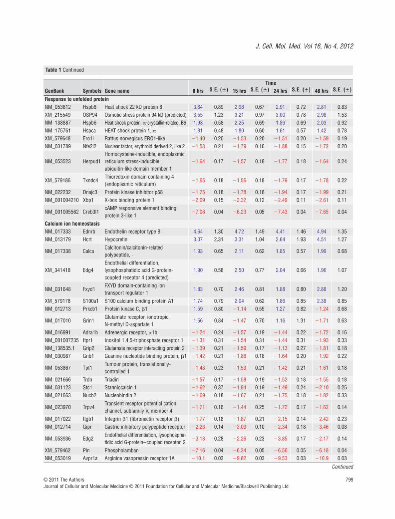
Table 1 Gene profiles of neuronal clusters in vitro, 8, 15, 24 and 48 hrs after axon severance (P < 0.05)
Continued

J. Cell. Mol. Med. Vol 16, No 4, 2012
793© 2011 The AuthorsJournal of Cellular and Molecular Medicine © 2011 Foundation for Cellular and Molecular Medicine/Blackwell Publishing Ltd
Time
GenBank Symbols Gene name 8 hrs S.E. (�) 15 hrs S.E. (�) 24 hrs S.E. (�) 48 hrs S.E. (�)
Neurite development and morphogenesis
XM_215963 Lama5 Laminin, ·5 2.39 0.73 2.21 0.75 2.05 0.56 1.88 0.54
XM_342392 Notch1 Notch gene homologue 1, 2.35 0.65 2.43 0.64 2.00 0.55 1.81 0.48
NM_030997.1 VgfVGF nerve growth factorinducible
2.25 0.58 1.42 0.40 1.71 0.64 1.15 0.35
NM_053021 Clu Clusterin 1.96 0.61 2.17 0.68 1.89 0.68 2.43 0.73
NM_053911 Pscd2Pleckstrin homology, Sec7 andcoiled/coil domains 2
1.77 0.53 1.64 0.49 1.70 0.53 1.45 0.67
XM_221672 Tiam1T cell lymphoma invasion andmetastasis 1 (predicted).
1.66 0.60 1.07 0.46 1.20 0.71 1.14 0.52
NM_031131 Tgfb2 Transforming growth factor, ‚2 1.58 0.60 1.13 0.67 1.08 0.39 1.04 0.72
NM_012924 Cd44 CD44 antigen 1.58 0.47 1.08 0.28 1.02 0.53 1.11 0.40
NM_030991 Snap25Synaptosomal-associated protein 25
1.18 0.93 2.69 0.65 1.00 1.52 2.46 0.80
NM_019248 Ntrk3Neurotrophic tyrosine kinase,receptor, type 3
1.06 0.46 1.33 0.23 1.18 0.27 1.56 0.21
NM_012731 Ntrk2Neurotrophic tyrosine kinase,receptor, type 2
1.03 0.32 2.14 0.17 1.27 0.44 2.15 0.37
NM_012513 BdnfBrain-derived neurotrophic factor
1.01 0.68 1.84 0.17 1.24 0.51 1.79 0.15
NM_012700.1 Stx1b Syntaxin 1B2 (2) 1.00 0.27 1.41 0.31 1.03 0.42 1.51 0.35
XM_232283 Plxnb1 Plexin D1 (predicted) 1.05 0.25 1.30 0.22 1.19 0.24 1.65 0.18
NM_153470 Lzts1Leucine zipper, putative tumour suppressor 1
1.12 0.73 2.09 0.51 1.17 0.81 2.02 0.47
NM_013038 Stxbp1 Syntaxin binding protein 1 1.20 0.54 2.54 0.37 1.47 0.66 2.62 0.35
NM_133652 Cspg5Chondroitin sulphate proteoglycan 5
1.28 0.22 1.59 0.19 1.55 0.21 1.30 0.37
XM_579472 Slit1 Slit homologue 1 1.53 0.30 2.35 0.30 1.56 0.45 2.91 0.17
NM_019334 Pitx2Paired-like homeodomain transcription factor 2
1.53 0.18 1.54 0.20 1.54 0.19 1.44 0.19
NM_017237 Uchl1Ubiquitin carboxy-terminalhydrolase L1
1.60 0.35 2.32 0.27 1.75 0.32 2.56 0.31
NM_012548 Edn1 Endothelin 1 1.61 0.19 1.78 0.21 1.80 0.16 1.71 0.17
NM_134331 Epha7 Eph receptor A7 1.65 0.18 1.53 0.22 1.52 0.23 1.52 0.20
XM_346464 Slit2 Slit homologue 2 1.69 0.17 1.56 0.18 1.83 0.17 1.88 0.22
XM_342172 Thbs4 Thrombospondin 4 1.70 0.16 1.74 0.19 1.95 0.15 2.19 0.12
XM_223820 Zfp312Zinc finger protein 312 (predicted)
1.79 0.47 3.76 0.13 1.55 0.64 3.10 0.10
XM_215451 Cspg2Chondroitin sulfate proteoglycan 2
1.80 0.22 1.38 0.22 1.94 0.17 2.11 0.16
XM_342591 Bmp7 Bone morphogenetic protein 7 2.12 0.25 1.27 0.23 1.76 0.32 1.22 0.58
NM_017089 Efnb1 Ephrin B1 2.56 0.10 2.94 0.14 2.86 0.10 3.15 0.08
NM_031321 Slit3 Slit homologue 3 2.84 0.11 3.26 0.10 3.19 0.09 3.52 0.08
NM_053595 Pgf Placental growth factor 5.49 0.05 4.06 0.08 5.27 0.06 4.70 0.06
NM_012774 Gpc3 Glypican 3 9.15 0.10 4.92 0.06 9.44 0.04 5.87 0.05
Table 1 Continued
Continued

794 © 2011 The AuthorsJournal of Cellular and Molecular Medicine © 2011 Foundation for Cellular and Molecular Medicine/Blackwell Publishing Ltd
Time
GenBank Symbols Gene name 8 hrs S.E. (�) 15 hrs S.E. (�) 24 hrs S.E. (�) 48 hrs S.E. (�)
Actin filament formation
XM_341553 PRKCQ Protein kinase C, theta 3.84 1.86 5.17 1.56 4.04 2.02 4.55 1.39
XM_342648.2 N-WASPNeural Wiskott--Aldrich syndrome protein
3.75 1.28 4.83 1.64 3.53 2.07 4.24 2.38
XM_218617.3 Myh14Myosin, heavy polypeptide 14 (predicted)
2.96 0.70 3.44 0.99 2.87 0.87 2.51 1.06
NM_019357 Vil2 Villin 2 2.16 0.67 1.57 0.46 1.66 0.41 1.73 0.58
NM_053484 Gas7 Growth arrest specific 7 2.15 0.96 1.14 0.75 1.65 1.52 1.10 0.54
NM_019131 Tpm1 Tropomyosin 1, 1.94 0.58 2.03 0.64 1.76 0.47 1.84 0.94
NM_017117.1 Capn3 Calpain 3 1.7 0.5 1.89 0.62 1.49 0.49 1.53 0.42
NM_022401.1 Plec1 Plectin 1 1.68 0.42 1.72 0.51 1.43 0.37 1.18 0.40
NM_001005889 Rdx Radixi 1.61 0.46 1.61 0.54 1.40 0.46 1.29 0.66
NM_019167 Spnb3 �-Spectrin 3 1.45 1.17 1.73 0.69 1.22 1.53 1.91 0.56
XM_221196 Fscn2Fascin homologue 2, actin-bundling protein (predicted)
1.20 0.39 1.32 0.45 1.30 0.43 1.58 0.52
XM_236444.3 LOC315840 Similar to Myosin VI 1.17 0.33 1.59 0.43 1.36 0.43 1.29 0.40
NM_030873 Pfn2 Profilin 2 1.06 0.24 1.31 0.22 1.14 0.24 1.58 0.34
XM_237115 Nck2Non-catalytic region of tyrosinekinase adaptor protein 2
1.08 0.39 1.72 0.30 1.36 0.35 1.74 0.32
NM_031144 Actb Actin, � 1.30 0.24 1.36 0.24 1.59 0.21 1.44 0.36
XM_579522 Actn4 Actinin ·4 1.31 0.22 1.65 0.17 1.81 0.17 1.75 0.18
NM_013194 Myh9 Myosin, heavy polypeptide 9 1.35 0.25 1.59 0.17 1.66 0.18 1.42 0.20
XM_215862 Dstn Destrin (predicted) 1.38 0.22 1.34 0.28 1.64 0.21 1.55 0.37
NM_001009689 Cdc42ep2CDC42 effector protein (Rho GTPase binding) 2
1.54 0.18 1.59 0.20 1.83 0.16 1.74 0.16
NM_019289.2 Arpc1bActin-related protein 2/3 complex, subunit 1B
1.61 0.16 1.70 0.17 1.81 0.15 1.92 0.16
NM_053814 MripMyosin phosphatase-Rho interacting protein
1.64 0.17 2.16 0.13 2.43 0.13 2.78 0.14
XM_579484 EvlEna-vasodilator stimulatedphosphoprotein
1.79 0.14 2.45 0.14 1.96 0.19 2.88 0.17
NM_019212 Acta1 Actin, 1, skeletal muscle 1.91 0.16 2.27 0.40 2.22 0.21 2.49 0.42
NM_023992 Gpr54 G protein-coupled receptor 54 2.05 0.16 2.10 0.13 2.01 0.15 2.07 0.14
XM_579179 ArheRas homologue gene family,member E
2.21 0.13 2.45 0.13 2.45 0.13 2.62 0.14
NM_001007554 Cal CSX-associated LIM 4.19 0.08 4.57 0.06 4.66 0.07 3.90 0.08
NM_012722 Eln Elastin 4.62 0.12 3.69 0.12 4.63 0.09 3.98 0.08
NM_012893.1 Actg2 Actin, �2 33.7 0.01 28.1 0.01 22.3 0.03 26.5 0.01
Table 1 Continued
Continued

J. Cell. Mol. Med. Vol 16, No 4, 2012
795© 2011 The AuthorsJournal of Cellular and Molecular Medicine © 2011 Foundation for Cellular and Molecular Medicine/Blackwell Publishing Ltd
Time
GenBank Symbols Gene name 8 hrs S.E. (�) 15 hrs S.E. (�) 24 hrs S.E. (�) 48 hrs S.E. (�)
Microtubule cytoskeleton organization and biogenesis
XM_341694.2 Dncl2bDynein, cytoplasmic, light chain2B (predicted)
20.8 6.64 23.8 7.22 16.4 13.4 22.5 10.6
NM_053508 Tekt1 Tektin 1 6.81 2.11 8.84 2.55 6.01 4.81 8.01 4.91
NM_001007726 Dnai2Dynein, axonemal, intermediatepolypeptide 2
6.65 2.05 8.07 2.65 5.66 3.42 6.58 3.78
XM_224615.3 Dnah1Dynein, axonemal, heavypolypeptide 1
4.83 1.30 7.20 2.05 5.82 3.20 5.22 2.69
XM_576475.1 vOC501059 Similar to kinesin-like protein 9 2.52 0.78 2.70 0.77 2.35 0.96 2.72 0.86
NM_012935 Cryab Crystallin, ·B 2.44 1.35 2.33 0.88 2.12 0.65 2.03 1.25
XM_234720.3 Dnah11Dynein, axonemal, heavypolypeptide 11
2.41 0.67 3.06 0.86 2.37 0.80 2.56 0.80
XM_213354.3 Dnah9Dynein, axonemal, heavypolypeptide 9
2.02 0.67 2.45 0.82 2.03 0.82 2.30 0.79
NM_001007004 Tuba4 Tubulin, ·4 1.97 0.59 1.03 0.31 1.53 0.64 1.08 0.52
NM_199094.1 Tubb2 Tubulin, ‚2 1.96 0.57 1.81 0.54 1.77 0.58 1.74 0.56
NM_206950 Mig12MID1 interacting G12-like pro-tein
1.96 0.52 2.20 0.66 2.00 0.53 1.94 0.69
XM_575908 Tekt2 Tektin 2 (predicted) 1.92 0.56 2.07 0.62 1.66 0.87 2.32 0.68
NM_031763 Pafah1b1Platelet-activating factor acetyl-hydrolase, isoform Ib
1.74 0.46 1.76 0.55 1.58 0.43 1.56 0.39
XM_218820 Prc1Protein regulator of cytokinesis1 (predicted)
1.65 0.48 1.46 0.43 1.43 0.37 1.63 0.79
NM_001009666 Dnalc4 Dynein, axonemal, light chain 4 1.56 0.46 1.43 0.36 1.28 0.42 1.24 0.51
NM_001009645 Kif22 Kinesin family member 22 1.53 0.39 1.06 0.31 1.16 0.31 1.17 0.50
XM_240978.3 Kifc3Kinesin family member C3 (pre-dicted)
1.50 0.41 1.50 0.38 1.65 0.47 1.06 0.32
XM_341686 Ap1g1Adaptor-related protein complex1, gamma 1 subunit
1.49 0.42 1.41 0.51 1.51 0.43 1.21 0.44
NM_053618 Bbs2Bardet--Biedl syndrome 2homologue
1.45 0.37 1.58 0.48 1.44 0.43 1.44 0.45
XM_224232 Cenpj Centromere protein J 1.33 0.38 1.51 0.43 1.33 0.45 1.32 0.41
NM_022507 Prkcz Protein kinase C, zeta 1.00 0.28 1.36 0.26 1.11 0.40 1.60 0.35
NM_198752.1 KIFC2 Kinesin family member C2 1.01 0.36 2.00 0.38 1.26 0.74 2.40 0.27
NM_053947 Mark1MAP/microtubule affinity-regu-lating kinase 1
1.11 0.25 1.55 0.22 1.34 0.25 1.80 0.17
XM_343018.2 Spg4Spastic paraplegia 4 (spastin)(predicted)
1.25 0.23 1.38 0.21 1.43 0.19 1.68 0.25
NM_024346 Stmn3 Stathmin-like 3 1.43 0.37 2.39 0.30 1.56 0.48 2.66 0.28
NM_133320 Ndel1nudE nuclear distribution geneE homologue like 1
1.60 0.18 2.32 0.12 1.79 0.20 2.55 0.20
XM_215469 Map1bMicrotubule-associated protein1b
1.63 0.35 2.22 0.18 2.12 0.23 2.74 0.17
NM_017166 Stmn1 Stathmin 1 2.11 0.25 2.84 0.21 2.29 0.28 3.10 0.17
Table 1 Continued
Continued

796 © 2011 The AuthorsJournal of Cellular and Molecular Medicine © 2011 Foundation for Cellular and Molecular Medicine/Blackwell Publishing Ltd
Time
GenBank Symbols Gene name 8 hrs S.E. (�) 15 hrs S.E. (�) 24 hrs S.E. (�) 48 hrs S.E. (�)
Apical protein localization
XM_223229 ShrmPDZ domain actin binding protein Shroom
1.67 0.52 1.74 0.57 1.45 0.53 1.55 0.48
XM_222896 LtapLoop tail associated protein (predicted)
1.41 0.52 1.48 0.35 1.23 0.54 1.33 0.55
Cell cycle
NM_053749 Aurkb Aurora kinase B 4.99 1.95 5.90 1.71 4.01 1.58 5.80 1.65
XM_574943 Ccna1 Cyclin A1 (predicted) 3.94 1.18 4.67 1.39 3.01 1.56 3.69 1.88
XM_573172 Pnutl2 Peanut-like 2 (predicted) 3.33 1.34 3.48 1.10 2.47 1.18 2.79 2.11
XM_579390 Hrasls3 HRAS-like suppressor 3.29 1.20 4.37 1.23 3.32 1.57 4.59 1.43
XM_220423 Sept8 Septin 8 (predicted) 2.40 1.16 1.88 1.06 2.26 1.05 1.71 1.41
NM_053677 Chek2 Protein kinase Chk2 2.33 0.84 2.58 0.75 1.91 0.70 2.45 1.03
NM_021863 Hspa2 Heat shock protein 2 2.17 0.64 2.09 0.59 1.62 0.48 2.12 0.74
XM_215222 Cetn2 Centrin 2 2.15 0.60 2.43 0.71 1.97 0.81 2.15 1.16
XM_574892 E2f5 E2F transcription factor 5 1.86 0.54 1.54 0.45 1.69 0.51 1.44 0.45
NM_001005765 Rap1a RAS-related protein 1a 1.76 0.54 1.65 0.45 1.49 0.49 1.49 0.41
XM_579383 vOC501059Adenylate cyclase activatingpolypeptide 1
1.74 1.96 1.90 0.30 1.14 2.35 2.01 0.25
NM_001004107 Tacc1aTransforming, acidic coiled-coilcontaining protein 1a
1.58 0.47 1.51 0.42 1.48 0.40 1.20 0.47
NM_133396 Tesk2 Testis-specific kinase 2 1.50 0.44 1.34 0.40 1.35 0.40 1.33 0.36
NM_138873 Nbn Nibrin 1.50 0.49 1.26 0.32 1.37 0.42 1.16 0.37
NM_013058 Id3 inhibitor of DNA binding 3 1.21 0.31 1.53 0.40 1.44 0.41 1.30 0.35
XM_579239 RT1-CE7 RT1 class I, CE7 1.20 0.37 1.62 0.70 1.09 0.29 1.39 0.42
NM_021739 Camk2bCalcium/calmodulin-dependentprotein kinase II ‚ subunit
1.16 0.51 2.35 0.33 1.22 0.73 2.37 0.43
XM_341907 Eif4g2Eukaryotic translation initiationfactor 4, gamma 2
1.16 0.28 1.35 0.26 1.56 0.21 1.32 0.38
XM_342804 Ccne2 Cyclin E2 (predicted) 1.40 0.20 1.50 0.19 1.51 0.21 1.39 0.21
XM_222157 Kntc1Kinetochore associated 1 (predicted)
1.41 0.24 1.50 0.26 1.52 0.19 1.68 0.18
XM_214152 Cdkn3Cyclin-dependent kinaseinhibitor 3 (predicted)
1.47 0.21 1.70 0.20 1.95 0.15 1.76 0.25
NM_053653 VegfcVascular endothelial growthfactor C
1.48 0.29 1.73 0.20 1.61 0.20 1.77 0.31
XM_223270 Ccng2 Cyclin G2 (predicted) 1.53 0.19 1.47 0.21 1.57 0.20 1.39 0.22
NM_053703 Map2k6Mitogen-activated proteinkinase kinase 6
1.66 0.18 1.51 0.17 1.46 0.20 1.82 0.19
NM_021693 Snf1lk SNF1-like kinase 1.67 0.27 2.16 0.15 1.84 0.33 3.06 0.08
NM_139087 Cgref1Cell growth regulator with EFhand domain 1
1.69 0.23 2.70 0.12 1.86 0.27 2.54 0.11
NM_052807 Igf1rInsulin-like growth factor 1receptor
1.81 0.17 2.06 0.16 2.08 0.14 2.52 0.11
NM_033099 PtprvProtein tyrosine phosphatase,receptor type, V
2.37 0.11 1.77 0.21 2.19 0.14 2.29 0.12
NM_053713.1 Klf4 Kruppel-like factor 4 3.30 0.15 3.81 0.10 3.45 0.09 4.09 0.10
Table 1 Continued
Continued

J. Cell. Mol. Med. Vol 16, No 4, 2012
797© 2011 The AuthorsJournal of Cellular and Molecular Medicine © 2011 Foundation for Cellular and Molecular Medicine/Blackwell Publishing Ltd
TimeGenBank Symbols Gene name 8 hrs S.E. (�) 15 hrs S.E. (�) 24 hrs S.E. (�) 48 hrs S.E. (�)
Negative regulation of programmed cell deathNM_053819 Timp1 Inhibitor of metalloproteinase 1 1.79 0.51 1.83 0.53 1.46 0.43 1.13 0.30
XM_235497 Mkl1Megakaryoblastic leukaemia(translocation) 1 (predicted)
1.05 0.27 1.25 0.26 1.09 0.31 1.68 0.27
NM_033230 Akt1 Thymoma viral proto-oncogene 1 1.20 0.26 1.50 0.24 1.43 0.23 1.49 0.30NM_207592 Gpi Glucose phosphate isomerase 1.34 0.19 1.44 0.19 1.37 0.21 1.59 0.22
NM_031345 DsipiDelta sleep inducing peptide,immunoreactor
1.37 0.21 1.40 0.21 1.69 0.18 1.68 0.33
XM_216377 Bag1Bcl2-associated athanogene 1(predicted)
1.48 0.20 1.44 0.19 1.53 0.20 1.84 0.21
NM_057130.1 Bid3BH3 interacting (with BCL2family) domain
1.58 0.18 2.35 0.31 1.74 0.27 2.44 0.22
NM_013083 Hspa5 Heat shock 70 kD protein 5 1.64 0.16 1.51 0.17 1.50 0.19 1.74 0.24
XM_225257 Txndc5Thioredoxin domain containing5 (predicted)
1.69 0.15 1.52 0.21 1.67 0.16 1.52 0.21
NM_012992 Npm1 Nucleophosmin 1 1.72 0.16 2.25 0.16 2.19 0.13 2.35 0.18NM_021691 Twist2 twist homologue 2 2.47 0.12 2.30 0.16 2.63 0.10 2.39 0.12
XM_225940 Tnfaip8Tumour necrosis factor, -induced protein 8 (predicted)
3.13 0.09 2.37 0.14 3.10 0.09 2.70 0.11
Regulation of apoptosisNM_031970 Hspb1 Heat shock 27 kD protein 1 3.35 1.04 4.01 1.24 3.34 1.33 4.61 1.37
NM_001007617 SNF1/AMPK
SNF1/AMP-activated proteinkinase
2.97 1.47 3.44 1.10 2.96 1.35 3.28 2.28
NM_031775 Casp6 Caspase 6 2.13 0.80 2.61 0.78 2.00 0.73 2.64 1.00
NM_017180 Phlda1Pleckstrin homology-likedomain, family A, member 1
1.94 0.78 1.22 0.34 1.48 0.52 1.23 0.34
XM_231692 Brafv-raf murine sarcoma viraloncogene homologue B1
1.69 0.52 1.98 0.70 1.63 0.52 1.57 0.70
NM_139261.1 Hspbp1 hsp70-interacting protein 1.68 0.51 1.39 0.45 1.54 0.45 1.26 0.72
NM_001004199 Tax1bp1Tax1 (human T cell leukaemiavirus type I) binding protein 1
1.41 0.38 1.67 0.54 1.42 0.43 1.31 0.41
XM_579385 G6pdxGlucose-6-phosphate dehydrogenase
1.37 0.35 1.57 0.48 1.51 0.44 1.32 0.46
NM_012660 Eef1a2Eukaryotic translation elongation factor 1 2
1.16 0.63 2.50 0.53 1.10 0.94 2.26 0.69
XM_217191 Bnip2BCL2/adenovirus E1B 19kDa-interacting protein 1, NIP2
1.24 0.25 1.30 0.26 1.48 0.23 1.53 0.29
NM_012922 Casp3Caspase 3, apoptosis relatedcysteine protease
1.35 0.19 1.47 0.19 1.39 0.19 1.51 0.21
NM_023979 Apaf1Apoptotic peptidase activatingfactor 1
1.40 0.21 1.68 0.18 1.52 0.20 1.66 0.17
NM_080897 Bnip1BCL2/adenovirus E1B 19 kD-interacting protein 1
1.54 0.18 1.81 0.15 1.70 0.16 2.00 0.15
NM_053420 Bnip3BCL2/adenovirus E1B 19 kD-interacting protein 3
1.62 0.17 2.19 0.15 2.10 0.14 2.27 0.18
NM_053736 Casp11 Caspase 11 2.31 0.14 2.00 0.16 2.46 0.13 2.50 0.12NM_130422 Casp12 Caspase 12 2.54 0.12 1.89 0.19 2.52 0.12 2.22 0.12NM_172336 Atf5 Activating transcription factor 5 2.62 0.15 2.17 0.15 2.14 0.20 3.14 0.08
Table 1 Continued
Continued

798 © 2011 The AuthorsJournal of Cellular and Molecular Medicine © 2011 Foundation for Cellular and Molecular Medicine/Blackwell Publishing Ltd
Time
GenBank Symbols Gene name 8 hrs S.E. (�) 15 hrs S.E. (�) 24 hrs S.E. (�) 48 hrs S.E. (�)
Response to oxidative stress
NM_017014 Gstm1 Glutathione S-transferase, Ì1 4.46 1.35 5.22 1.76 4.07 2.04 5.46 1.62
NM_012580 Hmox1 Heme oxygenase (decycling) 1 4.10 2.56 1.71 0.88 2.12 1.09 1.16 0.52
NM_134349 Mgst1Microsomal glutathione S-transferase 1
2.90 0.87 3.39 1.00 3.08 1.07 2.72 0.95
NM_012880 Sod3Superoxide dismutase 3, extracellular
2.38 0.73 2.51 0.58 2.36 1.05 2.64 1.00
NM_053576 Prdx6 Peroxiredoxin 6 2.21 0.66 2.64 0.83 2.65 0.99 2.93 0.83
NM_012837 Cst3 Cystatin C 2.06 0.52 2.18 0.63 2.07 0.62 2.08 0.63
XM_214236 Nudt15Nudix (nucleoside diphosphatelinked moiety X)-type 15
1.90 0.59 1.57 0.53 1.51 0.49 1.55 0.48
NM_017051 Sod2Superoxide dismutase 2, mitochondrial
1.70 0.47 1.87 0.59 1.32 0.43 1.62 0.51
NM_017305 GclmGlutamate cysteine ligase, modifier subunit
1.69 0.44 1.31 0.43 1.32 0.42 1.08 0.42
NM_012815 GclcGlutamate-cysteine ligase, catalytic subunit
1.67 0.49 1.14 0.32 1.23 0.33 1.04 0.47
NM_057143 Park7 Fertility protein SP22 1.56 0.43 1.68 0.45 1.52 0.45 1.53 0.56
NM_012962 Gss Glutathione synthetase 1.48 0.42 1.66 0.52 1.61 0.46 1.22 0.38
NM_030826 Gpx1 Glutathione peroxidase 1 1.48 0.36 1.67 0.42 1.42 0.37 1.53 0.57
NM_019354 Ucp2 Uncoupling protein 2 1.47 0.43 1.60 0.47 1.17 0.56 2.34 0.70
NM_031614 Txnrd1 Thioredoxin reductase 1 1.46 0.47 1.15 0.23 1.13 0.28 1.60 0.18
XM_216452 Dhcr2424-Dehydrocholesterol reductase (predicted)
1.20 0.36 1.41 0.27 1.05 0.65 1.66 0.31
XM_579339 Serpine1Serine (or cysteine) Proteinaseinhibitor, clade E, member 1
1.10 0.31 1.69 0.20 1.67 0.31 2.11 0.13
NM_017365 elfin,Pdlim1 PDZ and LIM domain 1 1.18 0.26 1.31 0.21 1.62 0.21 1.46 0.20
NM_017258 Btg1B-cell translocation gene 1,anti-proliferative
1.33 0.25 1.32 0.22 1.27 0.27 1.54 0.28
NM_031056 Mmp14Matrix metalloproteinase 14(membrane-inserted)
1.42 0.20 1.71 0.16 2.04 0.14 2.01 0.13
NM_053769 Dusp1 Dual specificity phosphatase 1 1.54 0.32 2.61 0.18 1.73 0.36 2.61 0.24
XM_216403 XpaXeroderma pigmentosum, complementation group A
1.55 0.18 1.44 0.22 1.57 0.20 1.55 0.18
NM_017025 Ldha Lactate dehydrogenase A 1.55 0.18 2.35 0.12 2.20 0.12 2.45 0.12
NM_024134 Ddit3DNA-damage inducible transcript 3
2.00 0.15 2.41 0.12 2.38 0.12 2.74 0.11
NM_001008767 TxnipUp-regulated by 1,25-dihydroxyvitamin D-3
3.59 0.22 2.32 0.11 4.98 0.22 1.97 0.15
NM_019292 Ca3 Carbonic anhydrase 3 9.36 0.06 5.14 0.05 12.0 0.13 6.89 0.08
XM_213440 Col1a1 Collagen, type 1, 1 16.8 0.02 8.92 0.03 19.1 0.04 19.8 0.02
Table 1 Continued
Continued
J. Cell. Mol. Med. Vol 16, No 4, 2012
799© 2011 The AuthorsJournal of Cellular and Molecular Medicine © 2011 Foundation for Cellular and Molecular Medicine/Blackwell Publishing Ltd
TimeGenBank Symbols Gene name 8 hrs S.E. (�) 15 hrs S.E. (�) 24 hrs S.E. (�) 48 hrs S.E. (�)
Response to unfolded proteinNM_053612 Hspb8 Heat shock 22 kD protein 8 3.64 0.89 2.98 0.67 2.91 0.72 2.81 0.83XM_215549 OSP94 Osmotic stress protein 94 kD (predicted) 3.55 1.23 3.21 0.97 3.00 0.78 2.98 1.53NM_138887 Hspb6 Heat shock protein, -crystallin-related, B6 1.98 0.58 2.25 0.69 1.89 0.69 2.03 0.92NM_175761 Hspca HEAT shock protein 1, 1.81 0.48 1.80 0.60 1.61 0.57 1.42 0.78XM_579648 Ero1l Rattus norvegicus ERO1-like 1.40 0.20 1.53 0.20 1.51 0.20 1.59 0.19NM_031789 Nfe2l2 Nuclear factor, erythroid derived 2, like 2 1.53 0.21 1.79 0.16 1.88 0.15 1.72 0.20
NM_053523 Herpud1Homocysteine-inducible, endoplasmicreticulum stress-inducible, ubiquitin-like domain member 1
1.64 0.17 1.57 0.18 1.77 0.18 1.64 0.24
XM_579186 Txndc4Thioredoxin domain containing 4 (endoplasmic reticulum)
1.65 0.18 1.56 0.18 1.79 0.17 1.78 0.22
NM_022232 Dnajc3 Protein kinase inhibitor p58 1.75 0.18 1.78 0.18 1.94 0.17 1.99 0.21NM_001004210 Xbp1 X-box binding protein 1 2.09 0.15 2.32 0.12 2.49 0.11 2.61 0.11
NM_001005562 Creb3l1cAMP responsive element binding protein 3-like 1
7.08 0.04 6.23 0.05 7.43 0.04 7.65 0.04
Calcium ion homeostasisNM_017333 Ednrb Endothelin receptor type B 4.64 1.30 4.72 1.49 4.41 1.46 4.94 1.35NM_013179 Hcrt Hypocretin 3.07 2.31 3.31 1.04 2.64 1.93 4.51 1.27
NM_017338 CalcaCalcitonin/calcitonin-related polypeptide, ·
1.93 0.65 2.11 0.62 1.85 0.57 1.99 0.68
XM_341418 Edg4Endothelial differentiation, lysophosphatidic acid G-protein-coupled receptor 4 (predicted)
1.90 0.58 2.50 0.77 2.04 0.66 1.96 1.07
NM_031648 Fxyd1FXYD domain-containing ion transport regulator 1
1.83 0.70 2.46 0.81 1.88 0.80 2.88 1.20
XM_579178 S100a1 S100 calcium binding protein A1 1.74 0.79 2.04 0.62 1.86 0.85 2.38 0.85NM_012713 Prkcb1 Protein kinase C, �1 1.59 0.80 1.14 0.55 1.27 0.82 1.24 0.68
NM_017010 Grin1Glutamate receptor, ionotropic, N-methyl D-aspartate 1
1.56 0.84 1.47 0.70 1.16 1.31 1.71 0.63
NM_016991 Adra1b Adrenergic receptor, 1b 1.24 0.24 1.57 0.19 1.44 0.22 1.72 0.16NM_001007235 Itpr1 Inositol 1,4,5-triphosphate receptor 1 1.31 0.31 1.54 0.31 1.44 0.31 1.93 0.33NM_138535.1 Grip2 Glutamate receptor interacting protein 2 1.39 0.21 1.59 0.17 1.13 0.27 1.81 0.18NM_030987 Gnb1 Guanine nucleotide binding protein, �1 1.42 0.21 1.88 0.18 1.64 0.20 1.92 0.22
NM_053867 Tpt1Tumour protein, translationally-controlled 1
1.43 0.23 1.53 0.21 1.42 0.21 1.61 0.18
NM_021666 Trdn Triadin 1.57 0.17 1.58 0.19 1.52 0.18 1.55 0.18NM_031123 Stc1 Stanniocalcin 1 1.62 0.37 1.84 0.19 1.49 0.24 2.10 0.25NM_021663 Nucb2 Nucleobindin 2 1.69 0.18 1.67 0.21 1.75 0.18 1.82 0.33
NM_023970 Trpv4Transient receptor potential cation channel, subfamily V, member 4
1.71 0.16 1.44 0.25 1.72 0.17 1.62 0.14
NM_017022 Itgb1 Integrin �1 (fibronectin receptor �) 1.77 0.18 1.87 0.21 2.15 0.14 2.42 0.23NM_012714 Gipr Gastric inhibitory polypeptide receptor 2.23 0.14 3.09 0.10 2.34 0.18 3.46 0.08
NM_053936 Edg2Endothelial differentiation, lysophospha-tidic acid G-protein--coupled receptor, 2
3.13 0.28 2.26 0.23 3.85 0.17 2.17 0.14
XM_579462 Pln Phospholamban 7.16 0.04 6.34 0.05 6.56 0.05 6.18 0.04NM_053019 Avpr1a Arginine vasopressin receptor 1A 10.1 0.03 9.82 0.03 9.53 0.03 10.9 0.03
Table 1 Continued
Continued

800 © 2011 The AuthorsJournal of Cellular and Molecular Medicine © 2011 Foundation for Cellular and Molecular Medicine/Blackwell Publishing Ltd
Time
GenBank Symbols Gene name 8 hrs S.E. (�) 15 hrs S.E. (�) 24 hrs S.E. (�) 48 hrs S.E. (�)
Vesicle transport
NM_053555.1 Vamp5Vesicle-associated membraneprotein 5
1.65 0.52 2.02 0.71 1.35 0.50 1.98 0.59
NM_057097.1 Vamp3Vesicle-associated membraneprotein 3
1.55 0.38 1.69 0.40 1.47 0.45 1.48 0.60
NM_138835.1 Syt12 Synaptotagmin 12 1.56 0.46 1.15 0.35 1.27 0.35 1.17 0.33
XM_343205.2 Syt1 Synaptotagmin 1 1.33 1.08 2.13 0.90 1.09 1.59 1.96 0.85
Inflammatory response
NM_133624.1 Gbp2Guanylate nucleotide bindingprotein 2
17.5 7.13 20.0 6.93 14.0 3.77 12.9 4.69
NM_212466 Bf B-factor, properdin 6.47 3.81 9.28 9.00 7.12 1.98 10.9 4.90
NM_172222 C2 Complement component 2 3.55 0.97 4.03 1.22 3.17 0.90 4.50 1.51
XM_215095.3 Gprc5bG protein-coupled receptor,family C, group 5, member B
3.07 1.03 2.75 0.78 2.29 0.69 3.32 1.43
NM_199093 Serping1Serine (or cysteine) peptidaseinhibitor, clade G, member 1
2.80 2.06 2.48 1.23 2.40 1.15 3.67 2.05
NM_012488 A2m ·2-Macroglobulin 2.61 0.64 3.49 0.82 2.60 1.26 3.76 1.10
NM_019363 Axo1 Aldehyde oxidase 1 2.53 0.70 5.69 3.33 4.03 2.37 2.83 0.84
XM_573457.1 Gpr37l1G protein-coupled receptor 37-like 1 (predicted)
2.52 0.85 3.05 0.98 2.63 0.88 3.62 1.94
NM_138502 Mgll Monoglyceride lipase 2.51 1.08 2.16 0.81 2.21 0.79 2.31 0.72
NM_152242.1 Gpr56 G protein-coupled receptor 56 2.42 0.73 2.16 0.53 2.23 0.64 2.10 0.68
NM_212541 Ng22 Ng22 protein 1.94 0.64 2.16 0.68 1.66 0.51 2.12 0.65
NM_138900 C1sComplement component 1, ssubcomponent
1.91 0.54 2.18 0.82 1.75 0.51 1.92 0.70
NM_031351 Atrn Attractin 1.89 0.52 1.68 0.52 1.84 0.53 1.42 0.43
NM_031007.1 Adcy2 Adenylate cyclase 2 1.88 0.51 1.78 0.55 1.80 0.52 1.78 0.56
XM_579383.1 Adcyap1Adenylate cyclase activatingpolypeptide 1 (predicted)
1.74 1.96 1.90 0.30 1.14 2.35 2.01 0.25
NM_013157 Ass Argininosuccinate synthetase 1.71 2.33 1.05 0.36 1.52 1.49 1.05 0.30
NM_031634 Mefv MEDITERRANEAN fever 1.61 0.47 1.77 0.48 1.73 0.62 1.97 0.56
NM_021690.1 Rapgef3cAMP-regulated guaninenucleotide exchange factor I
1.58 0.40 1.78 0.48 1.71 0.52 1.77 0.48
XM_579359 Cd59 CD59 antigen 1.40 0.45 1.47 0.39 1.23 0.79 2.00 0.56
NM_053734 Ncf1 Neutrophil cytosolic factor 1 1.32 0.42 1.18 0.44 1.36 0.74 2.06 1.32
NM_212490 Atp6v1g2ATPase, H+ transporting, V1subunit G isoform 2
1.31 0.75 1.50 0.62 1.18 0.88 1.50 0.78
NM_022216.1 Gpr20 G protein-coupled receptor 20 1.25 0.37 1.70 0.56 1.34 0.44 1.64 0.46
NM_153318 Cyp4f6 Cytochrome P450 4F6 1.21 0.30 1.51 0.39 1.25 0.37 1.42 0.41
NM_001009353 Pla2g7Phospholipase A2, group VII(platelet-activating factoracetylhydrolase, plasma)
1.20 0.43 1.24 0.41 1.27 0.78 1.74 1.25
NM_024157 Cfi Complement factor I 1.19 0.33 1.20 0.33 1.20 0.34 1.59 0.50
NM_022236.1 Pde10a Phosphodiesterase 10A 1.08 0.30 1.52 0.38 1.30 0.29 1.70 0.36
XM_579389 Tf Transferrin 1.05 1.91 1.50 0.82 1.04 0.76 2.02 6.19
Table 1 Continued
Continued

J. Cell. Mol. Med. Vol 16, No 4, 2012
801© 2011 The AuthorsJournal of Cellular and Molecular Medicine © 2011 Foundation for Cellular and Molecular Medicine/Blackwell Publishing Ltd
Time
GenBank Symbols Gene name 8 hrs S.E. (�) 15 hrs S.E. (�) 24 hrs S.E. (�) 48 hrs S.E. (�)
NM_017196 Aif1 Allograft inflammatory factor 1 1.15 0.56 1.29 0.92 1.11 1.11 1.79 1.40
NM_024125 CebpbCCAAT/enhancer binding protein (C/EBP), �
1.25 0.21 1.55 0.21 1.27 0.22 1.53 0.21
XM_342092 Vars2 Valyl-tRNA synthetase 2 1.31 0.21 1.56 0.19 1.38 0.19 2.08 0.14
NM_052809 Cdo1Cysteine dioxygenase 1,cytosolic
1.34 0.23 1.99 0.15 1.81 0.16 1.62 0.17
NM_024486 Acvr1 Activin A receptor, type 1 1.38 0.23 1.46 0.21 1.65 0.19 1.61 0.17
NM_012666 Tac1 Tachykinin 1 1.50 0.23 2.32 0.14 1.60 0.34 2.47 0.13
XM_239239 Map2k3Mitogen-activated proteinkinase kinase 3 (predicted)
1.67 0.18 2.12 0.14 2.08 0.15 2.46 0.15
NM_130409 CfhComplement component factor H
1.68 0.18 1.25 0.25 1.57 0.19 1.02 0.32
XM_579400 LbpLipopolysaccharide bindingprotein
1.81 0.19 1.39 0.23 1.63 0.21 1.09 0.44
XM_343169 And Adipsin 2.07 0.14 2.14 0.15 1.83 0.15 1.88 0.14
NM_019143 Fn1 Fibronectin 1 2.22 0.17 2.15 0.20 2.43 0.11 3.73 0.22
NM_019262 C1qbComplement component 1, q subcomponent, � polypeptide
2.42 0.22 2.81 0.29 2.08 0.71 1.38 0.54
NM_001008524 C1qgComplement component 1, q subcomponent, gammapolypeptide
2.49 0.15 2.97 0.18 2.17 0.39 1.44 0.47
NM_017232 Ptgs2Prostaglandin-endoperoxidesynthase 2
3.59 0.16 6.51 0.06 3.84 0.13 6.18 0.08
XM_240184 Nfatc4Nuclear factor of activated Tcells, cytoplasmic, calcineurin-dependent 4 (predicted)
15.4 0.06 8.20 0.06 14.8 0.04 10.4 0.03
Chemotaxis
NM_031530 Ccl2 Chemokine (C-C motif) ligand 2 6.01 8.84 2.87 2.48 3.68 9.35 2.66 2.08
NM_182952.2 Cxcl11Chemokine (C-X-C motif) ligand 11
5.24 1.72 6.02 8.94 2.80 0.81 2.34 1.31
NM_022205 Cxcr4Chemokine (C-X-C motif)receptor 4
3.14 1.20 4.19 1.11 2.81 0.82 3.56 2.11
NM_012953 Fosl1 Fos-like antigen 1 2.97 1.17 1.04 0.27 1.47 1.19 1.02 0.25
NM_001007612 Ccl7 Chemokine (C-C motif) ligand 7 2.80 3.76 2.09 1.76 2.01 3.77 1.56 0.75
NM_139089 Cxcl10Chemokine (C-X-C motif) ligand 10
2.67 1.43 10.3 23.6 3.87 2.87 3.86 3.14
NM_053953.1 Il1r2 Interleukin 1 receptor, type II 2.42 1.05 2.23 0.71 2.28 0.68 1.87 0.85
NM_145672 Cxcl9Chemokine (C-X-C motif) ligand 9
2.13 1.58 3.09 2.97 2.42 0.75 1.94 1.24
NM_012747.2 Stat3Signal transducer and activatorof transcription 3
2.0 0.5 1.57 0.38 1.64 0.46 1.45 0.38
NM_030845 Cxcl1Chemokine (C-X-C motif) ligand 1
1.96 4.51 1.30 0.32 1.64 4.95 1.18 1.07
XM_234422.3 c-fos c-Fos oncogene 1.8 0.5 1.61 0.45 1.52 0.52 1.54 0.51
NM_134455 Cx3cl1Chemokine (C-X3-C motif) ligand 1
1.58 0.62 1.01 0.46 1.32 0.88 1.22 0.74
Table 1 Continued
Continued
802 © 2011 The AuthorsJournal of Cellular and Molecular Medicine © 2011 Foundation for Cellular and Molecular Medicine/Blackwell Publishing Ltd
Time
GenBank Symbols Gene name 8 hrs S.E. (�) 15 hrs S.E. (�) 24 hrs S.E. (�) 48 hrs S.E. (�)
NM_031512.1 Il1b Interleukin 1� 1.57 0.77 1.30 0.48 1.30 0.42 1.46 0.62
NM_017020.1 Il6r Interleukin 6 receptor 1.54 0.44 1.21 0.36 1.28 0.37 1.28 0.33
NM_031116 Ccl5 Chemokine (C-C motif) ligand 5 1.43 0.40 2.96 2.76 1.80 0.48 3.03 2.40
NM_139111 Cklf1 Chemokine-like factor 1 1.24 0.38 1.39 0.44 1.13 0.31 1.89 0.56
NM_031643 Map2k1Mitogen-activated proteinkinase kinase 1
1.08 0.30 1.27 0.29 1.14 0.22 1.55 0.49
XM_342823 Ccl27Chemokine (C-C motif) ligand27 (predicted)
1.12 0.28 1.55 0.25 1.16 0.38 1.62 0.19
XM_579590 Enpp2Ectonucleotide pyrophos-phatase/phosphodiesterase 2
1.61 0.29 1.22 0.26 1.80 0.21 1.59 0.18
NM_031327 Cyr61 Cysteine rich protein 61 1.65 0.24 2.00 0.19 1.90 0.26 2.22 0.25
NM_031836 VegfaVascular endothelial growthfactor A
1.69 0.16 1.79 0.14 1.78 0.16 1.87 0.15
NM_022177 Cxcl12Chemokine (C-X-C motif) ligand 12
1.81 0.18 1.71 0.17 1.70 0.17 1.16 0.29
NM_001007729 Pf4 Platelet factor 4 1.92 0.33 2.98 0.10 1.95 0.23 2.36 0.12
NM_012881 Spp1 Secreted phosphoprotein 1 3.05 0.19 1.86 0.29 3.08 0.10 1.40 0.64
NM_019233 Ccl20Chemokine (C-C motif) ligand 20
3.09 0.44 2.17 0.41 1.94 0.94 2.62 0.30
Table 1 Continued
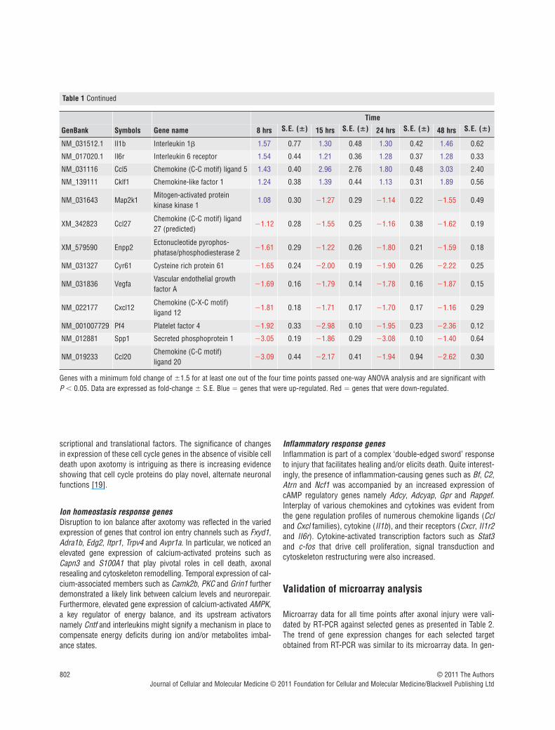
Genes with a minimum fold change of �1.5 for at least one out of the four time points passed one-way ANOVA analysis and are significant with P � 0.05. Data are expressed as fold-change � S.E. Blue � genes that were up-regulated. Red � genes that were down-regulated.
scriptional and translational factors. The significance of changesin expression of these cell cycle genes in the absence of visible celldeath upon axotomy is intriguing as there is increasing evidenceshowing that cell cycle proteins do play novel, alternate neuronalfunctions [19].
Ion homeostasis response genesDisruption to ion balance after axotomy was reflected in the variedexpression of genes that control ion entry channels such as Fxyd1,Adra1b, Edg2, Itpr1, Trpv4 and Avpr1a. In particular, we noticed anelevated gene expression of calcium-activated proteins such asCapn3 and S100A1 that play pivotal roles in cell death, axonalresealing and cytoskeleton remodelling. Temporal expression of cal-cium-associated members such as Camk2b, PKC and Grin1 furtherdemonstrated a likely link between calcium levels and neurorepair.Furthermore, elevated gene expression of calcium-activated AMPK,a key regulator of energy balance, and its upstream activatorsnamely Cntf and interleukins might signify a mechanism in place tocompensate energy deficits during ion and/or metabolites imbal-ance states.
Inflammatory response genesInflammation is part of a complex ‘double-edged sword’ responseto injury that facilitates healing and/or elicits death. Quite interest-ingly, the presence of inflammation-causing genes such as Bf, C2,Atrn and Ncf1 was accompanied by an increased expression ofcAMP regulatory genes namely Adcy, Adcyap, Gpr and Rapgef.Interplay of various chemokines and cytokines was evident fromthe gene regulation profiles of numerous chemokine ligands (Ccland Cxcl families), cytokine (II1b), and their receptors (Cxcr, II1r2and II6r). Cytokine-activated transcription factors such as Stat3and c-fos that drive cell proliferation, signal transduction andcytoskeleton restructuring were also increased.
Validation of microarray analysis
Microarray data for all time points after axonal injury were vali-dated by RT-PCR against selected genes as presented in Table 2.The trend of gene expression changes for each selected targetobtained from RT-PCR was similar to its microarray data. In gen-

J. Cell. Mol. Med. Vol 16, No 4, 2012
803© 2011 The AuthorsJournal of Cellular and Molecular Medicine © 2011 Foundation for Cellular and Molecular Medicine/Blackwell Publishing Ltd
eral, this confirmed the reliability of the gene expression profilesattained.
Discussion
Severance of fasciculated axonal bundles in mature rat neuronalclusters in culture has been used previously to study and char-acterize the cytoskeletal dynamics of regenerative sproutingafter axonal injury [6]. Using the same in vitro model, we haveapplied a DNA microarray approach to gain mechanistic insightinto the key molecular determinates that underlie neurite regen-eration after axotomy at a transcriptomic level. Briefly, we foundthat regenerative sprouting is a complicated process thatinvolves complex cytoskeleton dynamics and vigilant control ofsecondary processes such as oxidative stress, ion homeostasisand inflammation. These secondary processes have to be tightly
regulated to ensure neurite regeneration without causing evi-dent cell death. Physiologically, diffuse axonal injury is one ofthe many key pathological features associated with headtrauma. Upon trauma, stretched axons become brittle and tear.This situation is immediately salvaged by the damaged neurons’intrinsic capacity to regenerate. However, mechanisms are acti-vated for cell demise when the injury is beyond redemption.Through transcriptomic studies, we may identify crucial cellularpathways that facilitate regeneration and manipulate themwhenever necessary to lessen damage and avoid irreversibleapoptosis.
Neurite cytoskeleton reorganization
In accordance with the study by Chuckowree and Vickers [6], weconfirmed that adaptive sprouting of neurons after injury involvessubstantial cytoskeleton reorganization. For an axon to regenerate,its neurite arm, which is predominately composed of microtubules,
Fig. 1 Pie chart annotating number and percentage of genes identified in each of the 14 biological processes. A total of 305 statistically significant regu-lated genes were identified by DAVID. Each gene was classified into one of its most relevant biological processes categorized according to Gene Ontologyto avoid overestimation.

804 © 2011 The AuthorsJournal of Cellular and Molecular Medicine © 2011 Foundation for Cellular and Molecular Medicine/Blackwell Publishing Ltd
Fig. 2 Comparison of biological processes associated with axotomized neurons across 8, 15, 24 and 48 hrs post-injury. Bar chart shows the distributionof up-regulated (A) and down-regulated (B) genes. Differentially transcribed genes involved in the number count were statistically significant. Overall, thegenomic profiles for particular biological processes at 8 hrs were slightly distinct as compared to the other time points.

needs to be extended accordingly. This involves microtubule formation and stabilization. Tubulins are fundamental for micro-tubule formation and this could explain their augmented geneexpression. Stabilizing these newly formed microtubules is pivotaland this entails a concerted regulation of various microtubuledestabilizing and stabilizing proteins. Elevated gene expression ofNotch1, alongside decreased expression of Stmn, Cspg2 andSpg4, is consistent with this process. It has been found thatmicrotubule stabilization could promote axon regeneration by pre-venting chondroitin sulphate proteoglycan (Cspg) accumulation atlesion site [20]. This stabilization could be mediated by Notch acti-vation whereby it down-regulated spastin (Spg) expression, amicrotubule severing protein, and deterred axonal degeneration inprimary cortical neurons [21]. Stamins (Stmn) in general destabi-lize microtubules by preventing their assembly and promotingtheir disassembly.
The regenerating growth cone acts such as a molecular con-veyor belt whereby actin bundles at the proximal end are con-stantly fragmented by actin-destabilizing or severing proteins.Released actin fragments are then retrogradely transported andreassembled at the leading edge with help from nucleation-pro-moting factors [22]. These changes were reflected by the down-regulated gene expression of actin regulators namely Pfn, Cdc42,Arp2/3 [23, 24]. In contrast, NWASP and Gas7 gene expressionwere up-regulated by an average of 3- and 1.5-fold, respectively,throughout [25, 26]. As such, it is imperative to account for thecontrasting regulation of NWASP, Arp2/3 and Cdc42. NWASP andArp2/3 are undeniably the master components of actin polymer-ization. During neurite regrowth, actin dynamics unlike micro-tubules steer towards instability, a phenomenon observed due tothe complex processes governing polymerization and depolymer-ization of actin filament to support growth cone steering.Interestingly, Erin D.G and Matthew D.W critically reviewed that
the way Arp2/3 affects actin polymerization is more dependent onits activity regulated by ATP rather than its expression [27]. It wasmentioned that binding of WASP to ATP bound Arp2/3 and G actin,primes the complex and creates a nucleation point for daughter fil-aments formation. Subsequent ATP hydrolysis on Arp2 afternucleation has been temporally and functionally linked to actinbranch disassembly. The authors also brought up the importanceof recycling Arp2/3 itself for the formation of new actin processes,an event closely associated with actin recycling. With that, thepossibility of regulating already expressed Arp2/3 proteins by ATPin neurons and the fact that it can be recycled could potentiallyexplain the redundant need for its expression after axotomy.Unlike Arp2/3, NWASP is not directly regulated by ATP binding butits relevance is to elicit an active conformation of Arp2/3 for nucle-ation by physically binding to it. As such, our data illuminate aninteresting questionnaire on how NWASP is being regulated inregards to neurite regeneration. Moreover, apart from NWASPphysical interaction with Arp2/3 to direct actin polymerization, italso serves as focal points where transduced signals converge toorchestrate actin polymerization dynamics [28]. Hence, NWASPimportance in connecting multiple signalling pathways to initiateactin assembly as well as how it is regulated could possiblyaccount for its elevated expression in comparison to Arp2/3. Asfor Cdc42, unlike NWASP and Arp2/3, there has been evidenceshowing that NWASP recruitment of Arp2/3 that results in the formation of membrane protrusions and processes could occurvia a Cdc42-independent manner, at least for neurite outgrowth ofhippocampal neurons [26]. Because of the way actin proteins arerecycled in the growth cone, this further explains the redundantneed for newly synthesized actins and they were generally down-regulated transcriptionally following axonal injury. Recycling isessential to prevent excessive actin build-up that could thwartneuronal advances. This assumption is further supported by the
J. Cell. Mol. Med. Vol 16, No 4, 2012
805© 2011 The AuthorsJournal of Cellular and Molecular Medicine © 2011 Foundation for Cellular and Molecular Medicine/Blackwell Publishing Ltd
Table 2 Validation of microarray data using real-time PCR technique on rat neuronal samples, 8, 15, 24 and 48 hrs after axotomy
8 hrs 15 hrs 24 hrs 48 hrs
Gene title Symbol MicroarrayReal-time
PCRMicroarray
Real-timePCR
MicroarrayReal-time
PCRMicroarray
Real-timePCR
Neural Wiskott–Aldrich syndrome protein
N-WASP 3.7 � 1.3 7.4 � 2.4 4.8 � 1.6 2.7 � 0.5 3.5 � 2.1 1.7 � 0.4 4.2 � 2.4 2.9 � 0.3
Chemokine (C-X-C motif) ligand 10
Cxcl10 2.7 � 1.4 2.5 � 0.810.3 �
23.611.1 � 3.3 3.9 � 2.9 3.9 � 2.5 3.9 � 3.1 2.6 � 0.8
Dynein, cytoplasmic, light chain 2B (predicted)
Dncl2b 20.8 � 6.6 9.3 � 2.3 23.9 � 7.2 10.4 � 3.516.4 �
13.510.4 � 3.9
22.5 �10.6
10.0 � 1.7
Guanylate nucleotide binding protein 2
Gbp2 17.5 � 7.1 7.8 � 3.6 20.0 � 6.9 9.2 � 2.8 14.0 � 3.8 11.4 � 5.0 12.9 � 4.7 9.9 � 2.9
Chemokine (C-X-C motif) ligand 11
Cxcl11 5.2 � 1.7 3.2 � 1.1 6.0 � 8.9 4.8 � 1.7 2.8 � 0.8 4.8 � 1.3 2.3 � 1.3 4.0 � 1.1
Aurora kinase B Aurkb 5.0 � 2.0 3.7 � 1.0 5.9 � 1.7 5.4 � 1.3 4.0 � 1.6 5.0 � 1.2 5.8 � 1.7 4.8 � 1.6
Data are expressed as fold-change � S.E.

heightened expression of myosin genes, which encode motormolecules that transport actins in a retrograde fashion. Using flu-orescent speckle microscopy, it was found that inhibition ofmyosin II not only perturbed actin retrograde flow and affectedneuronal growth but its contraction forces were also required torecycle actin fragments for growth cone formation [29]. Together,our gene profiling data showed that neurite regeneration is anevent that involves stabilization of microtubules and dynamicinstability of actin filaments [7].
Further evidence of the dynamic response of the cytoskeletonto axotomy was the elevated expression of Tekt 1 and 2. Theserepresent a group of cilia microtubule structural proteins, andsupports an unexpected discovery that neuronal cilia might bevital for modulating signalling pathways to coordinate neuronalprocesses such as axonal guidance [30]. This highlights the needfor further investigation on the roles of cilia-associated proteins inneurobiological events such as axotomy.
The importance of protein trafficking in axonal regenerationwas indicated by the varied gene expression of motor proteinsinvolved in cytoskeletal transport such as Dnc, Dna, Kif, Myh, Tpmand machinery factors linked to membrane vesicles such as Syt,Vamp, Stx. This clearly illustrated retrograde transport of injurysignals from cut site to cell body which then evoked membraneexpansion processes that involve cargo trafficking of cytoskeletalproteins and even neurotropic factors to promote axonal re-growth. Dynein-derived forces in particular, are able to opposeaxon retraction and permit microtubules to advance [31].
Moreover, neurite regeneration is a process that involves recip-rocal regulation of permissive and repulsive axon guidance cues.Neurotrophic factors, for instance, are capable of directing axonalregrowth in damaged neurons. Bdnf can sustain axon regenera-tion upon various nerve and brain injuries [32, 33] and overex-pression of its receptor NtrkB was also found to elicit corticospinalaxonal regeneration [34]. Here, transient gene expression of Bdnfand Ntrks at 8 hrs could highlight the probable importance ofthese molecules in the recovery process during the initial phasebut not as much after specific projections had been formed. Mostrepulsive guidance molecules on the other hand, inhibit and deteraxonal regrowth after injury. Sema3a is one such example.Chemically inhibiting Sema3a prevented its binding to neuropilinand enhanced neural regeneration in damage axons [35].Eph/Ephrin signalling which controls axon guidance by contactrepulsion also inhibits regeneration of axons following injury inneurons [36]. Similarly, our transcriptomic data concurred thatthese molecules and their receptors mainly Efn, Sema, Slit, Bmp,Nrp, Plxd and Eph were not favoured during neurite regrowth.
Notably, some neurite promoting factors including Apoe, Cntfand Lama5 were also significantly up-regulated. Apoe is requiredfor lipid delivery for axon regrowth upon nerve crush injury [37]whereas Lama serves as a crucial extracellular matrix adhesionmolecule along which axons would grow and thus permit regen-eration in central neurons [38, 39]. Although Cntf is found mainlyin astrocytes, it is also expressed in cortical neurons [40]. Being acytokine-induced neurotrophic factor, it can potentiate axonregeneration in optic and spinal nerve injuries [41–43]. Overall,
our results demonstrate that to facilitate axonal regrowth, theinjured neurons themselves adapt to ensure that the cellular envi-ronment is permissive to regeneration via temporal generation ofpositive cues, concomitant restriction of negative cues andexpression of neurotrophic factors.
Apart from the genes that regulate neurite cytoskeleton, ourdata have further shown that various genes commonly linked tosecondary processes such as oxidative stress, apoptosis, cellcycle, calcium homeostasis and inflammation are also importantin dictating the regenerative process of axonally cut neurons andtheir possible roles are briefly discussed as follow.
Oxidative stress
Reactive oxygen species (ROS) are inevitable harmful by-productsof cellular activities. Oxidative stress, which could result in celldeath, ensues when the build-up of ROS in cells overwhelm exist-ing biological antioxidants under normal conditions. This occursin neurons after axonal injury. To counteract such insult, our studyshowed that genes responsible for GSH synthesis and variousantioxidant enzymes (Sods, Prdxs, Gpx) were elevated to alleviatethe stress. This demonstrated that neurons have the transcrip-tional capability to activate these vital mechanisms for their fightagainst oxidative insult to prevent death and assist in regenerativesprouting.
Several heat shock proteins (Hsp) were also transcriptionallyup-regulated. Mainly, Hsp act as molecular chaperones to refoldmisfolded proteins and prevent deleterious protein build-up.Intriguingly, small Hsp (12–43 kD) are viewed as vital neuropro-tectants in several neurological disorders [44]. Hsp27 whose geneexpression was up-regulated by 4-folds post-injury could sup-press cytochrome c–mediated cell death [45]. Besides, it couldinteract, modulate and remodel neuronal cytoskeleton [46, 47]. Assuch, in addition to its protein refolding function, local geneexpression of Hsp after axotomy might serve as anti-apoptotic andneurite regrowth signals.
Programmed cell death (PCD)
PCD is a process regulated by proto-oncogenes. It is logical that foran injured neuron to successfully regenerate, it must avoid axo-tomy-induced cell death. It is possible that apoptotic genes such asApaf1, Bnips, Casp3, 11 and 12 were down-regulated in our studyas an intrinsic protective mechanism to facilitate regeneration. Thisis supported by the fact that axotomized neuronal clusters in vitrodid not result in evident cell death [6]. Similarly, studies haveshown that overexpression of anti-apoptotic proteins could saveneurons from axotomy-induced cell death [48, 49]. However, it isimportant to note that stringent control of apoptotic genes expres-sion in denial of cell death in this case does not necessarily assistor speed up neurite regeneration, and probably indicates that neu-ron survival and neurite regeneration represent two distinct,although closely linked, processes within axotomized neurons.
806 © 2011 The AuthorsJournal of Cellular and Molecular Medicine © 2011 Foundation for Cellular and Molecular Medicine/Blackwell Publishing Ltd

Cell cycle
Cell cycle activation in response to DNA damage during extremecellular stress could lead to the demise of neurons [50].Interestingly, several cell cycle proteins were recently found to pos-sess distinct, alternate neuronal functions that are independentfrom their cell cycle roles [19]. Plausibly, this mirrors our case ofneurite regeneration in which certain cell cycle genes were elevateddespite the down-regulation of several apoptotic genes consistentwith cell death not being prominent. Aurkb, for example is a cellcycle kinase that remodels microtubule arrays for precise cell divi-sion [51, 52]. Its elevated gene expression could possibly reflect afunction in neurite regrowth, a process akin to its involvement incell division, because both processes involve complex cytoskeletonreorganization. However, functional biological studies are requiredto ascertain this speculation. Intriguingly, Aurora A kinase (Aurka),a sister kinase of Aurkb that is also known for its mitotic roles, hadlately been reported by two separate groups to play a crucial role inthe establishment of neuronal polarity [53, 54].
A more specific example involves Klf4, which was significantlydown-regulated in our studies. Early studies show that Klf4 is akey cell cycle checkpoint transcription factor and it causes G1/Sarrest by activating genes that are potent inhibitors of proliferation[55]. Recently, an alternate neuronal function of Klf4 was discov-ered whereby its overexpression in cortical neurons impeded neu-rite outgrowth while its knockout in retinal ganglion cells (RGCs)increased axon regeneration after crush injury [56]. Hence, besidecell cycle re-entry causing neuronal death, diverse gene expres-sion of cell cycle proteins after axotomy could also signify alter-nate and unexpected functions for these proteins to regulate thecytoskeleton and promote axonal regeneration.
Calcium homeostasis
Calcium homeostasis after axotomy is vital as it determines sur-vival or cell death onset. When the damage is too severe, excito-toxicity due to excessive influx of calcium ions causes neuronalcell death. However, in less damaging situations where calciumlevels are appropriately controlled, this triggers proteolysis andmembrane fusion/fission processes associated with axonal repairand cytoskeleton reorganization. First, local resealing is a criticalearly response for regrowth and this requires calcium activation ofcalpains to cleave the spectrin network and release membrane ten-sion. This might explain why Capn3 was up-regulated, whereasSpnb gene expression was varied. Secondly, calcium is a funda-mental co-factor for cytoskeletal remodelling as it is required formembrane vesicle fusion processes used in the delivery of con-structive molecules and neurotrophic factors to axotomized endsfor resealing purposes and growth cone formation [57]. Variedgene expression of calcium signalling associated genes, and theelevated gene expression of motor and machinery proteinsinvolved in membrane vesicle transport and fusion mentioned ear-lier, further supported this supposition.
In addition, up-regulation of SNF1/AMPK, a calcium-activatedkinase involve in metabolic stress, showed that energy level regu-lation after axotomy is important. This is accompanied by the con-comitant elevated expression of its upstream activators such as Il-6 and Cntf [58, 59]. Research on the role of AMPK in neurons isnew but recent studies have shown that AMPK activation in neu-rons is neuroprotective [60, 61]. More recently, plausible roles forAMPK in neuronal polarity and axon specification are indicated byits necessity for the maintenance of cell polarity in epithelial cells[62]. In the case of our study, it is possible that energy regulationin response to calcium ion levels could be crucial for the battleagainst metabolic insults and the re-establishment and/or mainte-nance of polarity in severed neurons.
Inflammation
Inflammation in response to axotomy as observed from the presence of Bf, C2, Nrf1 and Atrn could result in cell death if notproperly regulated [63–65]. cAMP levels which are increased andlimited by the activity of adenylate cyclase (Adcy) and phosphodi-esterase (Pde), respectively, could mitigate inflammation [66, 67].Elevated gene expression of Adcy, Adcyap and Gpr, but decreasedexpression of Pde, as per our study, clearly suggested that cAMPelevation was adopted to ease inflammation caused by axotomy.More important, increased levels of cAMP were found to promoteaxonal regeneration and recovery after CNS injury [68]. The likelymechanism for this is increasing the translocation of neurotrophicreceptors, for example NtrkB, to the plasma membrane of neurons[69]. cAMP could also activate a series of kinases that augmentgrowth promoting signalling pathways in brain and spinal cordinjuries [70]. Our data confirm the importance of the role of cellu-lar cAMP level to alleviate inflammation and to push regrowthmechanisms in response to injury. Henceforth, the roles of ATP inArp2/3 regulation mentioned earlier, together with the significanceof regulating secondary messengers to direct neurite regenera-tion, highlight the importance of secondary messenger precursorsin the study of axonal injury.
Apart from the primary association of cytokines andchemokines with inflammation, they could also directly or indi-rectly modulate regeneration after injury. This was evident fromtheir robust gene expression profiles. Through a knockout study,Il6, a cytokine, was found to promote re-growth of dorsal columnaxons after static nerve injury [71]. Changes in cytokine levelsoften alter expression of transcription factors involved in inflam-mation and regeneration. Stat3 is one such example and its acti-vation in axons upon injury could act as a retrograde signallingfactor to promote regeneration of sensory and motor neurons[72]. As for chemokines, Cxcr4 for instance, was implicated withneuronal regeneration because it could regulate axonal path find-ing in the CNS [73]. Notably, Il6r, Stat3 and Cxcr4 were all tran-scriptionally up-regulated in our study. These examples highlightthe direct impact cytokines and chemokines can have upon regen-erating neurons.
J. Cell. Mol. Med. Vol 16, No 4, 2012
807© 2011 The AuthorsJournal of Cellular and Molecular Medicine © 2011 Foundation for Cellular and Molecular Medicine/Blackwell Publishing Ltd
Physiologically however, cytokines and chemokines releasedupon neuroinflammation often have an indirect effect on neuronswhereby they help to recruit immune-response cells to injured site.Most of these cells when recruited excessively to injured site couldresult in neuronal apoptosis but during regeneration, they couldalso secrete bioactive forms of neurotrophic factors that wouldpotentially aid in neurons’ axon regrowth [74–76]. Supplementationof neurite promoting factors by immune cells could possibly explainthe transient need for neurons to express their own, such as Ntrkand Bdnf found in our in vitro injury model. Intriguingly, a studyshowed that oncomodulin secreted by inflammation-activatedmacrophages could promote axon regeneration in RGCs only uponelevated cAMP levels [77]. It was speculated that increased cAMPcaused translocation of specific receptors to the cell surface mem-brane. Even though this was observed in RGCs, the general ideamay still apply for neurons in which they are probably programmedto modulate cellular second messenger levels, such as cAMP, dur-ing injury to elicit actions. For example, relocation of specific recep-tors to the cell membrane in anticipation of binding to neurite-pro-moting factors released by surrounding immune-response cells.
Conclusion
It is significant that a number of genes listed in our DNA microar-ray study were in accordance with findings from previous in vitroand in vivo regenerative studies reported by other groups in rela-tion to spinal cord and brain injuries. This provides affirmationthat our protocol of severing axonal bundles in vitro to studyinjury-induced neurite regeneration is a reasonable and validexperimental model. From a transcriptomic view, we haverevealed that neurite regeneration is a complex process thatmainly involves restructuring of neurite cytoskeleton, determinedby intricate actin and microtubule dynamics, protein traffickingand appropriate control of both guidance cues and neurotrophicfactors. We conclude that the molecular response of neurons toaxotomy is not a simple process and evokes two distinct path-ways; a cell survival response accompanied by activation ofgenes to protect against oxidative stress, inflammation and cellu-lar ion imbalance and a regenerative response driven by modula-tion of the neuronal cytoskeleton. Gene expression profiles from
808 © 2011 The AuthorsJournal of Cellular and Molecular Medicine © 2011 Foundation for Cellular and Molecular Medicine/Blackwell Publishing Ltd
Fig. 3 Summarized view on the regulation of selected genes (taken from gene profiles) that were involved in different major sub-processes associatedwith neurite regeneration after axonal injury in vitro as discussed.

our study demonstrated that injured neurons have an innatecapability to survive axotomy by elevating biological antioxidantsand molecular chaperones, regulating secondary messengers,suppressing apoptotic genes, controlling calcium ion-associatedprocesses and possibly by expressing certain cell cycle proteinswhich might serve unique alternate neuronal functions. Genesinvolved in these highlighted protective and regenerativemechanics were summarized in Figure 3. Transcriptional analysisis a powerful and effective screening method to globally searchfor comprehensive clues to determine which biological pathwaysare involved in the intrinsic response of neurons to axotomy.Appropriate manipulation of selected biological targets involvedin these pathways could then potentially facilitate the design anddevelopment of novel, effective therapeutics for axonal damage inCNS injury.
Acknowledgements
This work was financially supported by Singapore Biomedical ResearchCouncil research grant R-184-000-093-305, Singapore National MedicalResearch Council research grant R-183-000-075-213, Strategic InitiativeFunding (Menzies Research Institute), National Health and MedicalResearch Council (AUSTRALIA) research grant 544913, AustralianResearch Council research grants DP0984673 and DP0556630 and aresearch grant from the Motor Accident & Insurance Board of Tasmania.
Conflict of interest
There are no conflicts of interest.
J. Cell. Mol. Med. Vol 16, No 4, 2012
809© 2011 The AuthorsJournal of Cellular and Molecular Medicine © 2011 Foundation for Cellular and Molecular Medicine/Blackwell Publishing Ltd
References
1. David S, Aguayo AJ. Axonal regenerationafter crush injury of rat central nervoussystem fibres innervating peripheral nervegrafts. J Neurocytol. 1985; 14: 1–12.
2. Sandvig A, Berry M, Barrett LB, et al.Myelin-, reactive glia-, and scar-derivedCNS axon growth inhibitors: expression,receptor signaling, and correlation withaxon regeneration. Glia. 2004; 46:225–51.
3. Taylor AM, Blurton-Jones M, Rhee SW, etal. A microfluidic culture platform for CNSaxonal injury, regeneration and transport.Nat Methods. 2005; 2: 599–605.
4. Taylor AM, Berchtold NC, Perreau VM, et al. Axonal mRNA in uninjured andregenerating cortical mammalian axons. J Neurosci. 2009; 29: 4697–707.
5. Dickson TC, Adlard PA, Vickers JC.Sequence of cellular changes followinglocalized axotomy to cortical neurons inglia-free culture. J Neurotrauma. 2000; 17:1095–103.
6. Chuckowree JA, Vickers JC. Cytoskeletaland morphological alterations underlyingaxonal sprouting after localized transectionof cortical neuron axons in vitro. J Neurosci. 2003; 23: 3715–25.
7. Sengottuvel V, Leibinger M, Pfreimer M,et al. Taxol facilitates axon regeneration inthe mature CNS. J Neurosci. 2011; 31:2688–99.
8. Verma P, Chierzi S, Codd AM, et al.Axonal protein synthesis and degradationare necessary for efficient growth coneregeneration. J Neurosci. 2005; 25:331–42.
9. McCurley AT, Callard GV. Time courseanalysis of gene expression patterns in
zebrafish eye during optic nerve regenera-tion. J Exp Neurosci. 2010; 2010: 17–33.
10. Veldman MB, Bemben MA, ThompsonRC, et al. Gene expression analysis ofzebrafish retinal ganglion cells during opticnerve regeneration identifies KLF6a andKLF7a as important regulators of axonregeneration. Dev Biol. 2007; 312:596–612.
11. Kuo LT, Tsai SY, Groves MJ, et al. Geneexpression profile in rat dorsal root gan-glion following sciatic nerve injury andsystemic neurotrophin-3 administration. JMol Neurosci. 2010; 43: 503–15.
12. Vega-Avelaira D, Geranton SM,Fitzgerald M. Differential regulation ofimmune responses and macrophage/neu-ron interactions in the dorsal root ganglionin young and adult rats following nerveinjury. Mol Pain. 2009; 5: 70.
13. Di Giovanni S, Faden AI, Yakovlev A, et al. Neuronal plasticity after spinal cordinjury: identification of a gene cluster driv-ing neurite outgrowth. FASEB J. 2005; 19:153–4.
14. Zai L, Ferrari C, Subbaiah S, et al.Inosine alters gene expression and axonalprojections in neurons contralateral to acortical infarct and improves skilled use ofthe impaired limb. J Neurosci. 2009; 29:8187–97.
15. Chung RS, Vickers JC, Chuah MI, et al.Metallothionein-III inhibits initial neuriteformation in developing neurons as well aspostinjury, regenerative neurite sprouting.Exp Neurol. 2002; 178: 1–12.
16. Chung RS, Staal JA, McCormack GH, et al. Mild axonal stretch injury in vitroinduces a progressive series of neurofila-
ment alterations ultimately leading todelayed axotomy. J Neurotrauma. 2005;22: 1081–91.
17. Dennis G Jr, Sherman BT, Hosack DA, et al. DAVID: database for annotation,visualization, and integrated discovery.Genome Biol. 2003; 4: R60.
18. Huang da W, Sherman BT, Lempicki RA.Systematic and integrative analysis oflarge gene lists using DAVID bioinformat-ics resources. Nat Protoc. 2009; 4: 44–57.
19. Frank CL, Tsai LH. Alternative functions ofcore cell cycle regulators in neuronalmigration, neuronal maturation, andsynaptic plasticity. Neuron. 2009; 62:312–26.
20. Hellal F, Hurtado A, Ruschel J, et al.Microtubule stabilization reduces scarringand causes axon regeneration after spinalcord injury. Science. 2011; 331: 928–31.
21. Ferrari-Toninelli G, Bonini SA, BettinsoliP, et al. Microtubule stabilizing effect ofnotch activation in primary cortical neu-rons. Neuroscience. 2008; 154: 946–52.
22. Lowery LA, Van Vactor D. The trip of thetip: understanding the growth conemachinery. Nat Rev Mol Cell Biol. 2009;10: 332–43.
23. Korobova F, Svitkina T. Arp2/3 complex isimportant for filopodia formation, growthcone motility, and neuritogenesis in neu-ronal cells. Mol Biol Cell. 2008; 19:1561–74.
24. Kim YS, Furman S, Sink H, et al.Calmodulin and profilin coregulate axonoutgrowth in Drosophila. J Neurobiol.2001; 47: 26–38.
25. Pinyol R, Haeckel A, Ritter A, et al.Regulation of N-WASP and the Arp2/3

complex by Abp1 controls neuronal mor-phology. PLoS One. 2007; 2: e400.
26. You JJ, Lin-Chao S. Gas7 functions withN-WASP to regulate the neurite outgrowthof hippocampal neurons. J Biol Chem.2010; 285: 11652–66.
27. Goley ED, Welch MD. The ARP2/3 com-plex: an actin nucleator comes of age. NatRev Mol Cell Biol. 2006; 7: 713–26.
28. Fawcett J, Pawson T. Signal transduction.N-WASP regulation-the sting in the tail.Science. 2000; 290: 725–6.
29. Medeiros NA, Burnette DT, Forscher P.Myosin II functions in actin-bundleturnover in neuronal growth cones. NatCell Biol. 2006; 8: 215–26.
30. Lee JH, Gleeson JG. The role of primarycilia in neuronal function. Neurobiol Dis.2010; 38: 167–72.
31. Myers KA, Tint I, Nadar CV, et al.Antagonistic forces generated by cytoplas-mic dynein and myosin-II during growthcone turning and axonal retraction. Traffic.2006; 7: 1333–51.
32. Song XY, Li F, Zhang FH, et al.Peripherally-derived BDNF promotesregeneration of ascending sensory neu-rons after spinal cord injury. PLoS One.2008; 3: e1707.
33. Harris NG, Mironova YA, Hovda DA, et al.Pericontusion axon sprouting is spatiallyand temporally consistent with a growth-permissive environment after traumaticbrain injury. J Neuropathol Exp Neurol.2010; 69: 139–54.
34. Hollis ER II, Jamshidi P, Low K, et al.Induction of corticospinal regeneration bylentiviral trkB-induced Erk activation. ProcNatl Acad Sci U S A. 2009; 106: 7215–20.
35. Montolio M, Messeguer J, Masip I, et al.A semaphorin 3A inhibitor blocks axonalchemorepulsion and enhances axonregeneration. Chem Biol. 2009; 16:691–701.
36. Goldshmit Y, McLenachan S, Turnley A.Roles of Eph receptors and ephrins in thenormal and damaged adult CNS. Brain ResRev. 2006; 52: 327–45.
37. Boyles JK, Zoellner CD, Anderson LJ, et al. A role for apolipoprotein E,apolipoprotein A-I, and low densitylipoprotein receptors in cholesterol trans-port during regeneration and remyelinationof the rat sciatic nerve. J Clin Invest. 1989;83: 1015–31.
38. Jang KJ, Kim MS, Feltrin D, et al. Twodistinct filopodia populations at the growthcone allow to sense nanotopographicalextracellular matrix cues to guide neuriteoutgrowth. PLoS One. 2010; 5: e15966.
39. Manthorpe M, Engvall E, Ruoslahti E, et al. Laminin promotes neuritic regen-eration from cultured peripheral and cen-tral neurons. J Cell Biol. 1983; 97:1882–90.
40. Dutta R, McDonough J, Chang A, et al.Activation of the ciliary neurotrophic factor(CNTF) signalling pathway in cortical neu-rons of multiple sclerosis patients. Brain.2007; 130: 2566–76.
41. Ye J, Cao L, Cui R, et al. The effects of cil-iary neurotrophic factor on neurologicalfunction and glial activity following contu-sive spinal cord injury in the rats. BrainRes. 2004; 997: 30–9.
42. Cui Q, Lu Q, So KF, et al. CNTF, not othertrophic factors, promotes axonal regenera-tion of axotomized retinal ganglion cells inadult hamsters. Invest Ophthalmol Vis Sci.1999; 40: 760–6.
43. Shuto T, Horie H, Hikawa N, et al. IL-6up-regulates CNTF mRNA expression andenhances neurite regeneration.Neuroreport. 2001; 12: 1081–5.
44. Muchowski PJ, Wacker JL. Modulation ofneurodegeneration by molecular chaper-ones. Nat Rev Neurosci. 2005; 6: 11–22.
45. Bruey JM, Ducasse C, Bonniaud P, et al.Hsp27 negatively regulates cell death byinteracting with cytochrome c. Nat CellBiol. 2000; 2: 645–52.
46. Williams KL, Rahimtula M, Mearow KM.Hsp27 and axonal growth in adult sensoryneurons in vitro. BMC Neurosci. 2005; 6:24.
47. Read DE, Gorman AM. Heat shock protein27 in neuronal survival and neurite out-growth. Biochem Biophys Res Commun.2009; 382: 6–8.
48. Dubois-Dauphin M, Frankowski H,Tsujimoto Y, et al. Neonatal motoneuronsoverexpressing the bcl-2 protooncogene intransgenic mice are protected from axo-tomy-induced cell death. Proc Natl AcadSci U S A. 1994; 91: 3309–13.
49. Shibata M, Murray M, Tessler A, et al.Single injections of a DNA plasmid thatcontains the human Bcl-2 gene preventloss and atrophy of distinct neuronal pop-ulations after spinal cord injury in adultrats. Neurorehabil Neural Repair. 2000; 14:319–30.
50. Kruman, II, Wersto RP, Cardozo-Pelaez F,et al. Cell cycle activation linked to neu-ronal cell death initiated by DNA damage.Neuron. 2004; 41: 549–61.
51. Fuller BG, Lampson MA, Foley EA, et al.Midzone activation of aurora B in anaphaseproduces an intracellular phosphorylationgradient. Nature. 2008; 453: 1132–6.
52. Murata-Hori M, Wang YL. The kinaseactivity of aurora B is required for kineto-chore-microtubule interactions duringmitosis. Curr Biol. 2002; 12: 894–9.
53. Khazaei MR, Puschel AW. Phosphorylationof the par polarity complex protein Par3 atserine 962 is mediated by aurora a and reg-ulates its function in neuronal polarity. J BiolChem. 2009; 284: 33571–9.
54. Yamada M, Hirotsune S, Wynshaw-BorisA. The essential role of LIS1, NDEL1 andAurora-A in polarity formation and micro-tubule organization during neurogensis.Cell Adh Migr. 2010; 4: 180–4.
55. Rowland BD, Peeper DS. KLF4, p21 andcontext-dependent opposing forces in can-cer. Nat Rev Cancer. 2006; 6: 11–23.
56. Moore DL, Blackmore MG, Hu Y, et al. KLFfamily members regulate intrinsic axon regen-eration ability. Science. 2009; 326: 298–301.
57. Hay JC. Calcium: a fundamental regulatorof intracellular membrane fusion? EMBORep. 2007; 8: 236–40.
58. Kelly M, Gauthier MS, Saha AK, et al.Activation of AMP-activated protein kinaseby interleukin-6 in rat skeletal muscle:association with changes in cAMP, energystate, and endogenous fuel mobilization.Diabetes. 2009; 58: 1953–60.
59. Escartin C, Pierre K, Colin A, et al.Activation of astrocytes by CNTF inducesmetabolic plasticity and increases resist-ance to metabolic insults. J Neurosci.2007; 27: 7094–104.
60. Dasgupta B, Milbrandt J. Resveratrolstimulates AMP kinase activity in neurons.Proc Natl Acad Sci U S A. 2007; 104:7217–22.
61. Spasic MR, Callaerts P, Norga KK. AMP-activated protein kinase (AMPK) molecularcrossroad for metabolic control and sur-vival of neurons. Neuroscientist. 2009; 15:309–16.
62. Williams T, Brenman JE. LKB1 and AMPKin cell polarity and division. Trends CellBiol. 2008; 18: 193–8.
63. Zipfel PF, Lauer N, Skerka C. The role ofcomplement in AMD. Adv Exp Med Biol.2010; 703: 9–24.
64. Gupta-Bansal R, Parent JB, Brunden KR.Inhibition of complement alternative pathway function with anti-properdin monoclonal antibodies. Mol Immunol.2000; 37: 191–201.
65. Hultqvist M, Sareila O, Vilhardt F, et al.Positioning of a polymorphic quantitativetrait nucleotide in the Ncf1 gene control-ling oxidative burst response and arthritisseverity in rats. Antioxid Redox Signal.2011; 14: 2373–83.
810 © 2011 The AuthorsJournal of Cellular and Molecular Medicine © 2011 Foundation for Cellular and Molecular Medicine/Blackwell Publishing Ltd

J. Cell. Mol. Med. Vol 16, No 4, 2012
811© 2011 The AuthorsJournal of Cellular and Molecular Medicine © 2011 Foundation for Cellular and Molecular Medicine/Blackwell Publishing Ltd
66. Silveira MS, Linden R. Neuroprotectionby cAMP: another brick in the wall. AdvExp Med Biol. 2006; 557: 164–76.
67. Ohta A, Sitkovsky M. Role of G-protein-coupled adenosine receptors in downregu-lation of inflammation and protection fromtissue damage. Nature. 2001; 414: 916–20.
68. Spencer T, Filbin MT. A role for cAMP inregeneration of the adult mammalian CNS.J Anat. 2004; 204: 49–55.
69. Meyer-Franke A, Wilkinson GA, KruttgenA, et al. Depolarization and cAMP eleva-tion rapidly recruit TrkB to the plasmamembrane of CNS neurons. Neuron. 1998;21: 681–93.
70. Cui Q, So KF. Involvement of cAMP in neu-ronal survival and axonal regeneration.Anat Sci Int. 2004; 79: 209–12.
71. Cafferty WB, Gardiner NJ, Das P, et al.Conditioning injury-induced spinal axonregeneration fails in interleukin-6 knock-out mice. J Neurosci. 2004; 24: 4432–43.
72. Lee N, Neitzel KL, Devlin BK, et al.STAT3 phosphorylation in injured axonsbefore sensory and motor neuron nuclei:potential role for STAT3 as a retrogradesignaling transcription factor. J CompNeurol. 2004; 474: 535–45.
73. Stumm R, Hollt V. CXC chemokine recep-tor 4 regulates neuronal migration andaxonal pathfinding in the developing nerv-ous system: implications for neuronalregeneration in the adult brain. J MolEndocrinol. 2007; 38: 377–82.
74. Elkabes S, DiCicco-Bloom EM, Black IB.Brain microglia/macrophages express
neurotrophins that selectively regulatemicroglial proliferation and function. JNeurosci. 1996; 16: 2508–21.
75. Kerschensteiner M, Gallmeier E,Behrens L, et al. Activated human T cells,B cells, and monocytes produce brain-derived neurotrophic factor in vitro and ininflammatory brain lesions: a neuroprotec-tive role of inflammation? J Exp Med.1999; 189: 865–70.
76. Nakajima K, Honda S, Tohyama Y, et al.Neurotrophin secretion from culturedmicroglia. J Neurosci Res. 2001; 65: 322–31.
77. Yin Y, Henzl MT, Lorber B, et al.Oncomodulin is a macrophage-derivedsignal for axon regeneration in retinal ganglion cells. Nat Neurosci. 2006; 9:843–52.