influence of using plant feed additives as growth promoters on ...
Upload
nottinghamCategory
view
0download
0
This Provisional PDF corresponds to the article as it appeared upon acceptance. Fully formattedPDF and full text (HTML) versions will be made available soon.
Cysteine coordination of Pb(II) is involved in the PbrR-dependent activation ofthe lead-resistance promoter, PpbrA, from Cupriavidus metallidurans CH34
BMC Microbiology 2012, 12:109 doi:10.1186/1471-2180-12-109
Jon L Hobman ([email protected])Daniel J Julian ([email protected])
Nigel L Brown ([email protected])
ISSN 1471-2180
Article type Research article
Submission date 7 January 2012
Acceptance date 7 June 2012
Publication date 18 June 2012
Article URL http://www.biomedcentral.com/1471-2180/12/109
Like all articles in BMC journals, this peer-reviewed article was published immediately uponacceptance. It can be downloaded, printed and distributed freely for any purposes (see copyright
notice below).
Articles in BMC journals are listed in PubMed and archived at PubMed Central.
For information about publishing your research in BMC journals or any BioMed Central journal, go to
http://www.biomedcentral.com/info/authors/
BMC Microbiology
© 2012 Hobman et al. ; licensee BioMed Central Ltd.This is an open access article distributed under the terms of the Creative Commons Attribution License (http://creativecommons.org/licenses/by/2.0),
which permits unrestricted use, distribution, and reproduction in any medium, provided the original work is properly cited.

Cysteine coordination of Pb(II) is involved in the
PbrR-dependent activation of the lead-resistance
promoter, PpbrA, from Cupriavidus metallidurans
CH34
Jon L Hobman1,2,3,*
Email: [email protected]
Daniel J Julian1
Email: [email protected]
Nigel L Brown1,3
Email: [email protected]
1 School of Biosciences, University of Birmingham, Edgbaston, Birmingham B15
2TT, UK
2 Present address: School of Biosciences, The University of Nottingham, Sutton
Bonington Campus, Loughborough LE12 5RD, UK
3 Present address: Senior Vice-Principal's Office, The University of Edinburgh,
Charles Stewart House, 9-16 Chambers Street, Edinburgh EH1 1HT, UK
* Corresponding author. Present address: Senior Vice-Principal's Office, The
University of Edinburgh, Charles Stewart House, 9-16 Chambers Street,
Edinburgh EH1 1HT, UK
Abstract
Background
The pbr resistance operon from Cupriavidus metallidurans CH34 plasmid pMOL30 confers
resistance to Pb(II) salts, and is regulated by the Pb(II) responsive regulator PbrR, which is a
MerR family activator. In other metal sensing MerR family regulators, such as MerR, CueR,
and ZntR the cognate regulator binds to a promoter with an unusually long spacer between
the −35 and −10 sequences, and activates transcription of resistance genes as a consequence
of binding the appropriate metal. Cysteine residues in these regulators are essential for metal
ion coordination and activation of expression from their cognate promoter. In this study we
investigated the interaction of PbrR with the promoter for the structural pbr resistance genes,
PpbrA, effects on transcriptional activation of altering the DNA sequence of PpbrA, and
effects on Pb(II)-induced activation of PpbrA when cysteine residues in PbrR were mutated
to serine.

Results
Gel retardation and footprinting assays using purified PbrR show that it binds to, and protects
from DNase I digestion, the PpbrA promoter, which has a 19 bp spacer between its −35 and
−10 sites. Using β-galactosidase assays in C. metallidurans, we show that when PpbrA is
changed to an 18 bp spacer, there is an increase in transcriptional activation both in the
presence and absence of Pb(II) salts up to a maximum induction equivalent to that seen in the
fully-induced wild-type promoter. Changes to the −10 sequence of PpbrA from TTAAAT to
the consensus E. coli −10 sequence (TATAAT) increased transcriptional activation from
PpbrA, whilst changing the −10 sequence to that of the Tn501 mer promoter (TAAGGT) also
increased the transcriptional response, but only in the presence of Pb(II). Individual PbrR
mutants C14S, C55S, C79S, C114S, C123S, C132S and C134S, and a double mutant
C132S/C134S, were tested for Pb(II) response from PpbrA, using β-galactosidase assays in
C. metallidurans. The PbrR C14S, C79S, C134S, and C132S/C134S mutants were defective
in Pb(II)-induced activation of PpbrA.
Conclusions
These data show that the metal-dependent activation of PbrR occurs by a similar mechanism
to that of MerR, but that metal ion coordination is through cysteines which differ from those
seen in other MerR family regulators, and that the DNA sequence of the −10 promoter affects
expression levels of the lead resistance genes.
Keywords
Metal-resistance, Metal-protein interactions, Metalloregulation, Bacterial gene expression
Background
Lead (Pb) is a widely distributed, environmentally persistent, toxic metal. Most bacteria that
are tolerant or resistant to lead either precipitate Pb in an insoluble form, or actively export it
[1]. Although some metal efflux ATPases, such as ZntA from Escherichia coli, and CadA
from Staphylococcus aureus plasmid pI258, can export Pb(II) as well as Zn(II) and Cd(II)
[2,3], the only characterized bacterial Pb(II) specific resistance system is from Cupriavidus
(formerly Wautersia and Ralstonia) metallidurans CH34 [4,5] - a Gram-negative, multiply
metal-resistant, β-proteobacterium originally isolated from a decantation basin at a Belgian
zinc production plant (and originally identified as Alcaligenes eutrophus CH34; [6]). Over
150 genes in CH34 are involved in metal resistance, of which at least 70 are carried on the
plasmids pMOL28 (171 kb) or pMOL30 (234 kb), and the remainder are carried on the 3.92
Mb chromosome or on a 2.58 Mb second chromosome [7]. Plasmid pMOL30 carries the czc
(Cd(II), Zn(II), Co(II)), mer (Hg(II)), sil (Ag(I)), cop (Cu(II)) and pbr Pb(II) resistance
operons [4,8].
The pbr lead resistance operon from pMOL30 was originally predicted to contain structural
genes which encode PbrT, a putative Pb(II) uptake protein belonging to the ILT (Iron Lead
Transporter) family [9], a P-type efflux ATPase (PbrA), a predicted inner–membrane protein
(PbrB), a predicted prelipoprotein signal peptidase PbrC and a Pb(II) binding protein, PbrD.
The regulator of the pbr operon was shown to be PbrR, which is a MerR family regulator

[4,10] Subsequent work has shown that the pbr operon also contains an interrupted orf; pbrU
upstream of pbrT [11,12] which is predicted to encode a putative inner membrane (Major
Facilitator Family MFS1) permease gene, which is probably inactive, but still part of the pbr
operon; and that PbrB/PbrC is a fusion protein [11,12], and encodes an inner membrane
bound undecaprenyl pyrophosphate (C55-PP) phosphatase [5]. The pbr operon contains a
predicted MerR-like promoter from which pbrRTU are transcribed on one DNA strand, and
the pbrABCD genes are transcribed as a polycistronic message on the other [4,12]. The most
recent work on the mechanism of lead resistance encoded by the pMOL30 pbr operon has
proposed a model where Pb2+
induces expression of the pMOL30-encoded PbrABCD by
PbrR, as well as expression of zinc and cadmium efflux ATPase homologs ZntA and CadA
which are carried on the chromosome or second chromosome. Each of these three ATPases is
involved in exporting Pb2+
into the periplasm where inorganic phosphates produced by PbrB
are involved in precipitating Pb2+
as insoluble lead phosphate. This model finds no role for
PbrT, C, and D, yet some reports suggest PbrC may be required for the maturation or activity
of phosphatase in the periplasm[5]. PbrR from pMOL30 (Rmet_5946) is related to several
other PbrR-like regulators that have been identified in the C. metallidurans CH34
chromosome, including pbrR2 (Rmet_2303 also known as pbr691 [13,14] which is believed
to regulate a cadA and a pbrC homolog on the chromosome, and pbrR3 (Rmet_3456 also
known as pbr710) believed to regulate a zntA homolog on the second chromosome, both of
which are believed to be involved in Pb2+
export [12]. There is evidence for only very low
levels of cross-regulation of the pMOL30 PpbrA promoter by PbrR2 or PbrR3 [15].
Other metal-sensing MerR family members include those responding to cadmium (CadR;
[16,17]), copper (CueR; [18-20], ActP; [21], SctR; [22]), zinc (ZntR, [23,24]; ZccR (Zn, Co,
Cd), [25]) and gold (GolS, [26]). Metal-sensing MerR family regulators share many common
features: they bind to and activate gene expression from promoters with unusually long
spacer sequences of 19-20 bp between the −35 and −10 sequences, and contain cysteine and
other amino acids that are essential in coordinating metals and activating gene expression
[10,16,20,27-29].
The objectives of this study were to 1) Characterize the interaction between PbrR and the
pbrA promoter, and study the effects on transcription of shortening the 19 bp spacer between
the −35 and −10 sequences, and altering the −10 sequence of PpbrA; and 2) to investigate the
importance of cysteine residues in PbrR activation of PpbrA in response to Pb(II) ions. To
this end each of the cysteine residues in PbrR (C14, C55, C79, C114, C123, C132 and C134)
were individually changed to serine residues and a double mutant (C132S, C134S) was
created. The effects of these mutations on in vivo transcriptional activation in response to
Pb(II) were determined in C. metallidurans using β-galactosidase assays.
Methods
Bacterial strains, plasmids and growth media
Bacterial strains and plasmids used in this study are shown in Table 1. Escherichia coli
strains were grown in LB broth [30] at 37°C. C. metallidurans strains were grown at 30°C in
869 medium, 284 Tris or 284 MOPS medium [4,6]. For β-galactosidase assays of PbrR-
regulated PpbrA promoter activity, C. metallidurans strains were grown in 284 MOPS
medium [4] minimising any Pb(II) precipitation during growth. C. metallidurans strains were
grown in SOB medium without MgSO4 [30] prior to electroporation of plasmids, and SOB

medium containing MgSO4 after electroporation. Pb(II) induction was achieved by growth in
PbNO3, and antibiotics were used at the following concentrations:- for E. coli: carbenicillin
(Melford laboratories, UK), 200 μg/ml; chloramphenicol 25 μg/ml; kanamycin, 50 μg/ml and
trimethoprim lactate 30 μg/ml (all from Sigma Chemical UK); for C. metallidurans:
trimethoprim lactate 500 μg/ml.

Table 1 Bacterial strains and plasmids
Bacterial strain Properties or Genotype Reference or source
E. coli
TG2 supE hsdΔ5 thiΔ (lac-proAB) F' Δ(srl-recA)306::Tn10(Tcr) lacZΔM15 [31]
BL21(DE3)pLysS F- ompT hsdSB (rB- mB-) gal dcm (DE3) pLysS (Cm
r) Novagen
C. metallidurans
CH34 Zn, Cd, Co, Pb, Cu, Hg, Ni and Cr resistance [6]
AE104 Plasmid-cured C. metallidurans strain- sensitive to toxic metals [6]
Plasmid Description Reference or source
pET32LIC Apr Overexpression plasmid for ligation-independent cloning Novagen
pET32LIC pbrR Apr pbrR cloned into pET32LIC This study
pMa5/8 Apr Cm
s Mutagenesis vector [32]
pMc5/8 Aps Cm
r Mutagenesis vector [32]
pMaPbrR/PpbrA Apr Cm
s Mutagenesis vector with pbrR/PpbrA cloned in to it This study
pMOL1139 Kmr, The pbr operon cloned into plasmid pRK415 B. Borremans
pMU2385 Tpr 13.3 kb low copy number lacZ reporter plasmid [33]
pMUPpbrA Tpr pMU2385 containing the PpbrA promoter directing lacZ transcription This study
pMUPpbrA-1 Tpr pMU2385 containing the PpbrA promoter with a 1 bp deletion This study
pMUPpbrAcon Tpr As pMUPpbrA, but −10 sequence changed to E. coli consensus This study
pMUPpbrAmer Tpr As pMUPpbrA, but −10 sequence changed to mer promoter This study
pMUPbrR/PpbrA Tpr, pMU2385 containing pbrR, PpbrA ΔpbrA directing lacZ transcription This study
pMUPbrRC14S/PpbrA As pMUPbrRPpbrA, but PbrR C14S This study
pMUPbrRC55S/PpbrA As pMUPbrRPpbrA, but PbrR C55S This study
pMUPbrRC79S/PpbrA As pMUPbrRPpbrA, but PbrR C79S This study
pMUPbrRC114S/PpbrA As pMUPbrRPpbrA, but PbrR C114S This study
pMUPbrRC132S/PpbrA As pMUPbrRPpbrA, but PbrR C132S This study
pMUPbrRC134S/PpbrA As pMUPbrRPpbrA, but PbrR C134S This study

pMUPbrRC132,134 S/PpbrA As pMUPbrRPpbrA, but PbrR C132S/C134S This study
pUC21 Apr, high copy number cloning vector; ColE1 replicon [34]
pUK21 Kmr, intermediate copy number cloning vector; p15A replicon [34]
pUK21pbr1 Kmr, HindIII/SalI pbrR/PpbrA/ΔpbrA from pMOL1139 cloned into pUK21 This study

DNA manipulations
DNA manipulations were as described by [30]. Oligonucleotides were synthesized by Alta
Bioscience, the University of Birmingham; or MWG Biotech, Germany. The DNA sequence
of all mutants and cloned PCR products were confirmed by sequencing using a PE Applied
Biosystems Big Dye version 2.0 sequencing kit according to the manufacturer’s protocol,
followed by analysis on an ABI 3700 sequencer in the Functional Genomics Laboratory,
School of Biosciences, the University of Birmingham. The primers used for sequencing were:
pMUforward and pMUreverse, complementary to the sequences flanking the multiple
cloning site of pMU2385, and PbrApe for pMapbrR/PpbrA clones (Table 2).
Table 2 Oligonucleotides used for site directed mutagenesis, and overexpression
Oligonucleotide Sequence Description or reference
pbrR C14S 5’ CCA CCG GGG ATG CGG TGC 3’ PbrR mutagenesis primer
pbrR C55S 5’ CCA GAG ACC GGG AGT GAC G 3’ PbrR mutagenesis primer
pbrR C79S 5’ GAC TTC ACC GGA ATC CTG G 3’ PbrR mutagenesis primer
pbrR C114S 5’ GGC ACC AGA AGA GGC TTC G 3’ PbrR mutagenesis primer
pbrR C123S 5’ GCA GAA TCC CGG ACG ATT G 3’ PbrR mutagenesis primer
pbrR C132S 5’ CGT ATC ACA CAC GGA GTC CGA C
3’
PbrR mutagenesis primer
pbrR C134S 5’ CGT ATC AGA CAC GCA GTC CGA C
3’
PbrR mutagenesis primer
pbrR
C132S/C134S
5’ CGT ATC AGA CAC GGA GTC CGA C
3’
PbrR mutagenesis primer
pbrApe 5’ GCG CCA ACC GTG CTC GGT TCT
GGG 3’
Primer
extension/sequencing
primer [4]
pbrBstEII 5’ GCG AAT GGT CAC CAC CGG 3’ Primer to amplify PpbrA
pbrNruI 5’ GCT TGT CGC GAA TCA GCG 3’ Primer to amplify PpbrA
pMU forward 5’ GAT TCT CCC CAC ATC ACC AG 3’ Sequencing primer for
pMU2385
pMU reverse 5’ TGC CAG CAT TTC ATA ACC AA 3’ Sequencing primer for
pMU2385
M13-F 5’ CGC CAG GGT TTT CCC AGT CAC
GAC 3’
Sequencing primer for
pUK plasmids
M13-R 5’ GAG CGG ATA ACA ATT TCA CAC
AGG 3’
Sequencing primer for
pUK plasmids
conpbr: 5’ CTAGAGGGTTAATCGGCAAC 3’ PpbrA mutagenesis
primer
merpbr: 5’ CTAGAGGGTGTAAGGTCGGCAAC 3’ PpbrA mutagenesis
primer
-1EcoPbr 5’ GGG GAA TTC GAA GCT TGC T 3’ (3’
primer)
PpbrA mutagenesis
primer
-1CentreBam 5’ GCC GAT TTA AAC CCT CTA GT 3’
(primer B)
PpbrA mutagenesis
primer

-1CentreEco 5’ CGG CTA AAT TTG GGA GAT CA 3’
(primer A)
PpbrA mutagenesis
primer
-1BamPbr 5’ CAG TAT ACC TAG GCA GCT GG 3’ (5’
primer)
PpbrA mutagenesis
primer
pbrR ATG
(LIC)
5’ GAC GAC GAC AAG ATG AAT ATC
CAG ATC GGC GAG C 3’
PbrR cloning and
overexpression primer
pbrR TAG
(LIC)
5’ GAG GAG AAG CCC GGT CTA GTC
GCT TGG ATG GGC 3’
PbrR cloning and
overexpression primer
T7 terminator 5’ CGA TCA ATA ACG AGT CGC C 3’ Sequencing primer
Underlined bases highlight alteration from the wild-type sequence.
PbrR overexpression and purification
The pbrR gene was amplified from pMOL1139 using VentR® DNA polymerase (New
England Biolabs) and the primers: pbrRATG (LIC) and pbrRTAG (LIC) (Table 2). The pbrR
PCR product was annealed with plasmid pET32LIC (Novagen), according to manufacturers’
recommendations. DNA sequencing using the primer T7 reverse (Table 2) was used to
confirm the nucleotide sequence of the cloned fragment. The thioredoxin-PbrR fusion protein
was overexpressed in E. coli BL21 (DE3) pLysS, purified and stored under reducing
conditions as described in [23]. The thioredoxin- S tag was cleaved from the fusion protein
using enterokinase, according to the manufacturer’s protocol (Novagen) and removed using
S-tag affinity agarose. PbrR purity was estimated by PAGE analysis. The concentration of the
purified protein was determined by Bradford assay [35].
Gel retardation and DNAse I protection assays of PpbrA with PbrR
Gel retardation experiments were as described in [36], with initial experiments to determine
PbrR DNA binding using a 1144 bp HindIII/SalI fragment from pMOL1139 containing pbrR,
PpbrA and a truncated pbrA (positions 409 and 1553 on the pbr operon) [4] cloned into
pUK21 [34] to make plasmid pUKpbr1. pUKpbr1 was digested with NruI/BstEII and end
labelled with [γ32
P]-dATP for gel retardations. Further gel retardation and footprinting
experiments used a 296 bp PpbrA PCR product, amplified from pMOL1139 using the primers
pbrBstEII and pbrNruI (Table 2) and labelled using [γ32
P]-dATP. DNAse I protection assays
of PpbrA with PbrR were as described by [37], using the 296 bp PpbrA promoter PCR
product detailed above. The DNA sequence of the region was obtained from the 296 bp
PpbrA PCR product using the pbrApe primer (Table 2) [4] and run alongside the DNAase I
footprint (Figure 1B).
Figure 1 (a) Gel retardation of PpbrA with PbrR. Each reaction contained the same
amount of 32
P-end-labelled 296 bp PpbrA PCR product (60 fmol). Lanes 1, 9 and 10
contained no PbrR. PbrR concentrations in lanes 2–8 and 11–17 increase 2-fold from 0.3 to
19.2 pmol. Lanes 10–17 contained 10 μM Pb(II).(b) DNase I protection assay of PbrR bound
to the 296 bp PCR product containing the PbrA promoter. Lanes AGCT, DNA sequence of
the 296 bp PCR product pbrA promoter, using the pbrApe primer. Lanes 1 and 4, no added
pbrR, lane 2 and 3 increasing amounts of added PbrR. (c) Diagram of the PpbrA promoter.
The transcript start site is marked in bold and indicated with an arrow [4]. The region of the
promoter protected by PbrR from DNAase I digestion is marked with a box. The predicted
−35 and −10 sequences are marked in bold, and the dyad symmetrical sequence is marked
with arrows

Cloning of pbrR-PpbrA-ΔpbrA and mutagenesis of the PbrR cysteines
All cloning and mutagenesis work was done in E. coli K-12 TG2. The 1144 bp pbrR-PpbrA-
ΔpbrA DNA fragment described above was cloned into pMa5/8 [32] from pUK21pbr1 using
the flanking EcoRI and BamHI sites to make pMaPbrR/PpbrA. Gapped duplex mutagenesis
of each of the cysteine residues in pbrR was as previously described [32] using the primers
pbrRC14S, pbrRC55S, pbrRC79S, pbrRC114S, pbrRC123S, pbrRC132S, pbrRC134S, or
pbrRC132S, C134S (Table 2), and mutants verified by DNA sequencing as described [15].
The wild type and mutant pbrR genes on the 1144 bp pbrR-PpbrA-ΔpbrA DNA fragment
were individually sub-cloned as EcoRI - BamHI fragments into pMU2385 [33] as described
previously [15]. The resulting constructs contained a self-regulating transcriptional unit, with
PbrR controlling the transcription of pbrR through PpbrR and regulating transcription of lacZ
in pMU2385 on the other DNA strand through PpbrA. These constructs were the basis of the
studies of the regulation of PpbrA by PbrR in C. metallidurans AE104.
Cloning and mutagenesis of PpbrA
A 266 bp SphI - NruI fragment containing the PpbrA promoter (positions 1062 and 1328 of
the pbr operon) was cloned from pMOL1139, into the HindIII site of pUK21, by rendering
the vector and insert blunt-ended using T4 DNA polymerase. The cloned PpbrA DNA
fragment was sub-cloned as an EcoRI - BamHI fragment into pMa5/8 for site directed
mutagenesis. The −10 sequence of PpbrA was mutated as described above using the primers
conpbr and merpbr (Table 2) to change the PpbrA −10 sequence from TTAAAT (wild type)
to TATAAT (consensus) or TAAGGT (mer-like). The mutant PpbrA promoters were cloned
into pMU2385 using EcoRI and BamHI, creating plasmids pMUPpbrA, pMUPpbrA(con) and
pMUPpbrA(mer) in which the pbrA promoter regulates expression of the lacZ gene. After
DNA sequencing, the activity of these mutant promoters was assayed in C. metallidurans
CH34.
Construction of the PpbrA −1 mutant
Mutagenic PCR [38] of the 1144 bp pbrR-PpbrA-ΔpbrA DNA fragment from
pMapbrR/PpbrA was used to construct the −1 promoter mutant of PpbrA, using the primers -
1CentreEco and -1CenterBam to introduce the −1 deletion, and primers -1EcoPbr and -
1BamPbr as flanking primers (Table 2). The PCR product containing the -1PpbrA promoter
was digested with EcoRI and BamHI and subcloned into the multiple cloning site of
pMU2385. The DNA sequence of the pbrR-PpbrA-ΔpbrA DNA fragment containing the −1
deletion in PpbrA was confirmed, and this plasmid provided the mutant promoter for the
assay in C. metallidurans AE104.
β-galactosidase assays in C. Metallidurans
pMU2385 plasmid constructs were electroporated into C. metallidurans, and cultures
containing pMU2385 derivatives were assayed for ß-galactosidase activity as described in
[39] with modifications described by [15].

Results
PbrR binds to the pbrA promoter and pb(II) decreases the binding affinity of
PbrR to PpbrA in vitro
PbrR was overexpressed as a thioredoxin-his Tag-S tag-fusion protein using the pET32-LIC
expression system, purified and released after enterokinase digestion as untagged, full length
PbrR, as described in Materials and Methods. The PbrR preparation was estimated as being
>95% pure PbrR by Coomassie Blue staining of standard SDS-PAGE gels (data not shown).
We had originally identified a candidate PpbrA promoter based on sequence similarity to
other MerR family promters, and on run-off transcription studies of the pbr operon [4] and
studied PbrR interactions with this region of the pbr operon. Initial PbrR gel retardation
assays on 32
P-end-labelled DNA from pUK21pbr1, which contained pbrR/PpbrA/ΔpbrA, had
been digested with BstEII and NruI showed retardation only of the 282 bp BstEII/NruI DNA
fragment containing the previously identified PpbrA promoter region, and no other fragments
from the plasmid (data not shown). Addition of PbrR to the end-labelled 296 bp PpbrA PCR
product retarded this fragment, and addition of Pb(II) to PbrR and PpbrA increased the
amount of PbrR required to retard the PpbrA DNA fragment (Figure 1A) indicating that
PbrR-Pb(II) had a lower affinity in vitro with PpbrA than did apo-PbrR did, as is the case
with MerR and Hg(II) (reviewed in [10]).
PbrR protects the pbrA promoter from DNAse I digestion in vitro
The 296 bp PpbrA PCR product described above was also used to determine the PbrR
binding site on the promoter by DNase I protection assay. Figure 1B shows the
autoradiograph of the PbrR DNase I footprint on PpbrA. The region protected by PbrR on
PpbrA includes the −35 and −10 sequences as well as the 19 bp spacer containing an
imperfect dyad symmetrical sequence between them, and is consistent with DNAse I
protection results for MerR, CueR and ZntR [18,20,23,24,40].
The transcription start site [4], the predicted −35 and −10 sites, and the region of the PpbrA
promoter protected by PbrR are shown in Figure 1C. The PpbrA promoter has a −35 sequence
(TTGACT) that is identical to those for PmerT from Tn501 and PzntA from E. coli K-12
(Figure 2) and shares 5/6 identity with the consensus E. coli −35 sequence. The predicted
PpbrA −10 sequence (TTAAAT) has a 4/6 identity to the consensus E. coli −10 sequence
(TATAAT) and the spacing between the −35 and −10 sequences is 19 bp, as is the case with
other MerR family regulatory regions except ZntR (20 bp; [23]).
Figure 2 Alignment of selected promoters for structural genes regulated by MerR
family metal responsive regulators: PbrR [4]; MerR [10], ZntR [23], CueR [20]. The −35
and −10 sequences are marked in BOLD. Arrows show dyad symmetrical DNA sequences
within the promoters
Promoter DNA mutations alter PpbrA activity in C. Metallidurans
The importance to promoter functionality of the number of nucleotides between the −35 and
−10 sequences of the PpbrA promoter, and the effects of altering the DNA sequence of the
PbrR binding site or −10 sequence of PpbrA were investigated using pMUPbrR/PpbrA −1 in
C. metallidurans AE104. The PpbrA −1 mutant (Figure 3A), in which the spacer between the

−35 and −10 sequences was shortened in such a way that the −35 and −10 sequences were not
altered, and the dyad symmetrical sequences in the spacer between the −35 and −10 were
retained, showed increased promoter activity in the absence of Pb(II) (Figure 3A) compared
to the wild type promoter, but no induction beyond the maximum level seen for the wt
promoter with 100 μM Pb(II). These results are similar to those seen for the MerR activated
promoter PmerT −1 from Tn501 [41], which is constitutively transcriptionally active in both
the presence and absence of Hg(II). Changes to the pbrA promoter −10 sequence, so that it
more closely resembled the consensus sequence for an E. coli promoter [42], caused up-
regulation of PpbrA activity both in the absence and presence of Pb(II). Changes made in
PpbrA so that it resembled the Tn501 merT promoter −10 sequence resulted in promoter
activity remaining repressed in the absence of Pb(II), but strongly induced in its presence to
expression levels 5-fold higher than the wild-type pbrA promoter (Figure 3B). These
differences in promoter sequence are likely to alter RNA polymerase binding to the promoter,
which could in turn affect the structure of the PbrR-RNA polymerase-DNA ternary complex.
Figure 3 (A) β-galactosidase assay measurement of the activation of PpbrA, containing a
1 nt deletion in the 19 bp promoter spacer, to increasing levels of Pb(II) in C.
metallidurans AE104 carrying pMUPbrRpbrA-1. Micromolar Pb(II) concentrations are
indicated by the suffix to Pb on the abscissa. Pb0 contains no added Pb(II), Pb200 contains
200 μM Pb(II) . The sequence of wild-type PpbrA and the −1 mutant PpbrA are shown below
the graph. The −35 and −10 sequences are marked in BOLD. Arrows show dyad symmetrical
DNA sequences within the promoters. (B) β-galactosidase assay measurement of the
activation of −10 sequence mutant PpbrA clones in pMU2385 in response to no added Pb(II)
or 100 μM Pb(II). WT denotes wild-type −10 sequence (TTAAAT), CON denotes the E. coli
consensus promoter −10 sequence (TATAAT) and MER the Tn501 PmerT promoter −10
sequence (TAAGGT). The sequences of the wild-type (PpbrA wt), consensus (PpbrA con),
and PmerT-like promoters (PpbrA mer) are shown below the graph. The −35 and −10
sequences are marked in BOLD. Arrows show dyad symmetrical DNA sequences within the
promoters, and altered bases are marked in Gray
Cysteines 14, 79 and 134 in PbrR are essential for pb(II) responsive
transcription from PpbrA in C. Metallidurans AE104
pMUPbrR/PpbrA derivatives carrying PbrR cysteine mutants (C14S, C55S, C79S, C114S,
C123S, C132S, C134S, and C132S/C134S) (Table 1) were assayed for Pb(II) –dependent
induction of the pbrA promoter in C. metallidurans AE104, which did not carry pMOL28 or
pMOL30. These were grown in a sublethal concentration of Pb(II) (20 μM) which was
sufficient to activate expression from PpbrA, without affecting growth of the Pb(II) sensitive
AE104 strain. β-galactosidase assays of wild type and cysteine mutant PbrR responses to
Pb(II) in C. metallidurans AE104 (Figure 4) showed cysteines C14, C79, and C134 were
essential for Pb(II) induced transcriptional activation of PpbrA by PbrR. The double mutant
C132S, C134S also lost Pb(II) induced activation of transcription from PpbrA, consistent
with the result for the single C134S mutant.
Figure 4 β-galactosidase assays in C. metallidurans AE104 of PpbrA activation in
response to 20 μM Pb(II) on wild-type PbrR and its cysteine mutants in
pMUPbrR/PpbrA

Discussion
PbrR is a member of the MerR family of regulators which sense metals and other
environmental stimuli, and activate gene expression in response to these signals. The
archetype of the family, MerR, regulates both its own expression and expression of the
mercuric ion resistance genes in the polycistronic mer operon from a divergent promoter:
Pmer. MerR activates expression of the structural genes at the PmerT operator/promoter (o/p)
site, which has an unusually long spacer of 19 bp between the −35 and −10 sequences of the
promoter (compared to the consensus E. coli σ70
promoter spacing of 16-18 bp [10]). The
MerR dimer binds to a dyad-symmetrical DNA sequence within the spacer, and when three
essential cysteine residues (C89, C117 and C126) in the MerR dimer coordinate to a mercuric
ion in a trigonal coordination [28,29] bridging between each MerR homodimer, this change
in MerR homodimer interaction is transmitted to the promoter, causing an allosteric
underwinding of ~33O of the DNA at the o/p site, which realigns the −35 and −10 sequences
of the promoter so that σ70
RNA polymerase can contact the promoter sequences forming the
transcription open complex [43,44].
PbrR from C. metallidurans CH34 plasmid pMOL30 binds to and protects from DNAase I
digestion the predicted PpbrA operator/promoter (Figure 1) (4). PpbrA has striking
similarities to other metal ion-responsive MerR family promoters (Figure 2). Assays of
PpbrA mutants where the spacing between the −10 and −35 sites are shortened to 18 bp,
whilst the internal dyad symmetry is maintained, showed that PbrR-induced expression from
PpbrA is upregulated even in the absence of Pb(II) (Figure 3). These data are all consistent
with the model of activation for the MerR promoter [41,43,44]. Change of the DNA sequence
of the −10 element of PpbrA to either the consensus E. coli promoter −10 sequence or the
Tn501 PmerT promoter −10 sequence also caused up-regulation of promoter activity,
although the PpbrA/Tn501 PmerT-like promoter still retained Pb(II) repression and induction,
rather than a constitutive up-regulation seen in the −10 consensus promoter mutant. These
data emphasize the importance of individual nucleotides within the promoter in affecting
promoter strength, and indicate that PpbrA is suboptimal for maximum induction of the
structural pbr genes. It is possible that this may represent a mechanism for fine-tuning of
expression of the pbr structural genes.
In other metal ion-sensing MerR family regulators, cysteine residues are essential for metal
coordination and functionality. In vivo assays of the activity of cysteine to serine mutant PbrR
proteins in C. metallidurans AE104 (which lacks pMOL30) have shown that C14, C79 and
C134 are essential for PbrR Pb(II) sensing and activation of PpbrA (Figure 4). PbrR C14 lies
in the turn of the predicted helix-turn-helix DNA binding domain of PbrR (Figure 5) and a
change of amino acid at this point could disrupt the binding of PbrR to PpbrA. Mutants in the
second helix of this region of MerR have lost both activation and repression activity [45,46].
The loss of Pb(II) response in the PbrR C79S mutant is consistent with the prediction from a
structure-based sequence alignment that this residue is essential for discriminating between
+1 and +2 charge ions, with a cysteine being found at this position in regulators that respond
to +2 ions [27]. Mutagenesis studies have all identified a cysteine residue at this position as
being essential for in vivo metal-dependant activation of expression in MerR, ZntR, and
ZccR.

Figure 5 ClustalW [47,48] alignment of metal sensing MerR regulators. PbrR
(Rmet_5946), PbrR691 (Rmet_2302) and PbrR710 (Rmet_3456) are from the genome of C. metallidurans CH34. CadR is from Pseudomonas stutzeri A1501. ZntR, and CueR are
from the E. coli K-12 genome, and MerR is from Tn501. The helices of the Helix-Turn-Helix
DNA binding domain are boxed. Essential cysteine residues (Cys14, Cys79, and Cys134 –
PbrR numbering) required for activation of PpbrA by PbrR are marked. Key to symbols:
* = residues in that column are identical in all sequences in the alignment. The symbol : =
conserved substitutions have been observed, and the symbol . = semi-conserved substitutions
are observed
C134 in PbrR (Rmet_5496) is also essential for Pb(II) response and is part of a CVC (CXC)
motif which is often found in PbrR regulators associated with orthologs of PbrABC, but not
in the PbrR homologues PbrR2 (PbrR691 Rmet_2302) and PbrR3 (PbrR710 Rmet_3456), or
CadR (Figure 5). A CVC motif is also found in the CadC repressor: alterations of either
cysteine in this motif in CadC reduced or abolished sensing of Pb(II), Cd(II) and Zn(II) [49]
and both cysteines are required for metal coordination [50,51]. Although C79 and C134 of
the PbrR homodimer are essential for Pb(II) induction of PpbrA, the C132S mutant shows
only a slightly reduced, not abolished, response to Pb(II). Pb(II) has been shown to have a
preference for binding to cysteine residues in a tri-coordinate Pb(II)-thiol conformation [52],
and Chen and coworkers have reported that the PbrR-related PbrR691 (PbrR2, Rmet_2302)
regulator from the C. metallidurans genomic island 1 coordinates Pb(II) via 3 (possibly 4)
cysteine coordination [14]. Pb(II) has been shown to coordinate in biological systems via a
distorted trigonal planar geometry involving S and N coordination in a biomimetic N2S
(alkylthiolate) compound [53], and the Pb(II), Cd(II) and Zn(II) response of the S. aureus
pI258 cadmium resistance repressor CadC is dependent on three cysteine residues [49,54].
DNA footprinting suggests that like MerR, PbrR functions as a homodimer. It is possible that
Pb(II) may coordinate to cysteine and histidine (or other N- side chain amino acid) residues
or O-containing side chain amino-acid residues in the PbrR homodimer and C79 could
provide the ligand for metal bridging between the homodimers, and in current models is
thought to be necessary to trigger DNA underwinding at the regulated promoter [27]. There
are histidine, glutamine, lysine and arginine residues in PbrR close to the metal-binding
domain (Figure 5). In ZntR, each homodimer coordinates two zinc atoms per metal binding
domain (MBD), one via C114 and C124 of the MBD, and C79 from the other monomer,
whilst the other zinc atom is coordinated to C115 and H119 of the MBD, and C79 from the
other monomer and both zinc atoms also coordinate to oxygen from a bridging phosphate
[27,54]. Structural studies are required to understand further how Pb(II) coordinates to PbrR.
We cannot exclude the possibility that the PbrR C79S and C134S mutants we have made may
have altered DNA-binding features, which may account for loss of Pb(II) response. However,
mutants in the MBD of other MerR family regulators do not, but mutants in the helix-turn
helix domain of these regulators do [45,46].
Conclusion
The metal-responsive MerR family transcription activators can be classified into groups
which sense Hg, or Cu/Ag/Au, or Zn/Cd/Pb, and several other phylogenetically-related but
uncharacterized regulator clusters [55]. PbrR (Rmet_5946) and the related PbrR691 (R_met
2302) are unusual amongst the phylogenetic cluster of related Zn(II)/Cd(II)/Pb(II)-sensing
MerR family regulators that have been tested for metal specificity, because they exclusively

respond to Pb(II) in plasmid based assays in C. metallidurans (PbrR: [15,56]) or using FRET
(PbrR691, [13]) without any transcriptional response to Zn or Cd, whereas related MerR
family regulators that have been tested respond to a greater or lesser extent to Zn(II), Cd(II)
and Pb(II) [10,23,57], as do SmtB/ArsR family repressors [47,54]. However, transcriptomics
experiments indicate that the pbr structural genes are also induced in the presence of other
metals, arguing that expression of the pbr operon and other metal resistance operons in C.
metallidurans is influenced by other factors [7,12].
Our experiments show that the mechanism of transcriptional activation by PbrR appears to be
essentially identical to that of MerR family regulators that have been characterized. PbrR
contains three cysteine residues that are necessary for Pb(II)-induced transcription from the
pbrA promoter. C14 is in the helix-turn-helix DNA binding domain, and may be essential for
the regulator/DNA interaction. C79 is essential in all divalent metal ion responsive MerR
regulators tested so far, whilst C134 is not found in other characterized MerR regulators. Our
data show that PbrR transcription is activated by Pb(II) using different amino acids to other
divalent metal ion-activated MerR regulators, but further work is required to determine
whether Pb(II) coordinates other residues in PbrR.
Abbreviations
Tp, Trimethoprim; Ap, Ampicillin; Km, Kanamycin.
Competing interests
The authors declare that they have no competing interests.
Authors’ contributions
JLH and DJJ carried out the experimental studies. JLH drafted the manuscript. NLB
conceived and coordinated the study. All authors read and approved the manuscript.
Acknowledgements
We gratefully acknowledge the contribution of Niels van der Lelie and Brigitte Borremans to
the start of this project and to Max Mergeay for advice and training to DJJ. We thank Chris
Kershaw for critical reading of the manuscript. This work was supported by the
Biotechnology and Biological Sciences Research Council (research grant B10333 and a
studentship to DJJ). The Birmingham Functional Genomics laboratory was supported by a
Joint Infrastructure Fund grant JIF13209 and bioinformatics facilities were provided through
MRC Infrastructure Award G.4600017.
References
1. Mire CE, Tourjee JA, O'Brien WF, Ramanujachary KV, Hecht GB: Lead precipitation by
Vibrio harveyi: evidence for novel quorum-sensing interactions. Appl Environ Microbiol
2004, 70:855–864.

2. Rensing C, Sun Y, Mitra B, Rosen BP: Pb(II)-translocating P-type ATPases. J Biol
Chem 1998, 49:32614–32617.
3. Sharma R, Rensing C, Rosen BP, Mitra B: The ATP hydrolytic activity of purified
ZntA, a Pb(II)/Cd(II)/Zn(II)-translocating ATPase from Escherichia coli. J Biol Chem
2000, 275:3873–3878.
4. Borremans B, Hobman JL, Provoost A, Corbisier P, Brown NL, van der Lelie D: Cloning
and functional analysis of the pbr lead resistance determinant of Ralstonia metallidurans CH34. J Bacteriol 2001, 183:5651–5658.
5. Hynninen A, Touzé T, Pitkänen L, Mengin-Lecreulx D, Virta M: An efflux transporter
PbrA and a phosphatase PbrB cooperate in a lead-resistance mechanism in bacteria. Mol Microbiol 2009, 74:384–394.
6. Mergeay M, Nies D, Schlegel HG, Gerits J, Charles P, van Gijsegem F: Alcaligenes
eutrophus CH34 is a facultative chemolithotroph with plasmid-bound resistance to
heavy metals. J Bacteriol 1985, 162:328–334.
7. Monchy S, Benotmane MA, Wattiez R, van Aelst S, Auquier V, Borremans B, Mergeay
M, Taghavi S, van der Lelie D, Vallaeys T: Transcriptomic and proteomic analyses of the
pMOL30-encoded copper resistance in Cupriavidus metallidurans strain CH34. Microbiology 2006, 152:1765–1776.
8. Mergeay M, Monchy S, Vallaeys T, Auquier V, Benotmane A, Bertin P, Taghavi S, Dunn
J, van der Lelie D, Wattiez R: Ralstonia metallidurans, a bacterium specifically adapted to
toxic metals: towards a catalogue of metal-responsive genes. FEMS Microbiol Rev 2003,
27:385–410.
9. Debut AJ, Dumay QC, Barabote RD, Saier MH Jr: The iron/lead supertransporter
family of Fe3+
/Pb2+
uptake systems. J Mol Microbiol Biotechnol 2006, 11:1–9.
10. Brown NL, Stoyanov JV, Kidd SP, Hobman JL: The MerR family of transcriptional
regulators. FEMS Microbiol Rev 2003, 27:145–163.
11. Monchy S, Benotmane MA, Janssen P, Vallaeys T, Taghavi S, van der Lelie D, Mergeay
M: Plasmids pMOL28 and pMOL30 of Cupriavidus metallidurans are specialized in the
maximum viable response to heavy metals. J Bacteriol 2007, 189:7417–7425.
12. Taghavi S, Lesaulnier C, Monchy S, Wattier R, Mergeay M, van der Lelie D: Lead (II)
resistance in Cupriavidus metallidurans CH34: interplay between plasmid and
chromosomally-located functions. Anthonie van Leeuwenhoek 2009, 96:171–182.
13. Chen P, Greenberg B, Taghavi S, Romano C, van der Lelie D, He C: An exceptionally
selective lead(II)-regulatory protein from Ralstonia metallidurans: Development of a
fluorescent lead(II) probe. Angew Chem Int Ed 2005, 44:2715–2719.
14. Chen PR, Wasinger EC, Zhao J, van der Lelie D, Chen LX, He C: Spectroscopic insights
into lead (II) coordination by the selective lead(II)-binding protein PbrR691. J Am Chem
Soc 2007, 129:12350–12351.

15. Julian DJ, Kershaw CJ, Brown NL, Hobman JL: Transcriptional activation of MerR
family promoters in Cupriavidus metallidurans CH34. Anthonie van Leeuwenhoek 2009,
96:149–159.
16. Brocklehurst KR, Megit SJ, Morby AP: Characterisation of CadR from Pseudomonas
aeruginosa: a Cd(II)-responsive MerR homologue. Biochem Biophys Res Commun 2003,
308:234–239.
17. Lee S-W, Glickman E, Cooksey D: Chromosomal locus for cadmium resistance in
Pseudomonas putida consisting of a cadmium transporting ATPase and a MerR family
response regulator. Appl Environ Microbiol 2001, 67:697–702.
18. Outten FW, Outten CE, Hale J, O'Halloran TV: Transcriptional activation of an
Escherichia coli copper efflux regulon by the chromosomal MerR homologue, CueR. J
Biol Chem 2000, 275:31024–31029.
19. Petersen C, Moller LB: Control of copper homeostasis in Escherichia coli by a P-type
ATPase, CopA, and a MerR-like transcriptional activator, CopR. Gene 2000, 261:289–
298.
20. Stoyanov JV, Hobman JL, Brown NL: CueR (YbbI) of Escherichia coli is a MerR
family regulator controlling expression of the copper exporter CopA. Mol Microbiol
2001, 39:502–511.
21. Reeve WG, Tiwari RP, Kale NB, Dilworth MJ, Glenn AR: ActP controls copper
homeostasis in Rhizobium leguminosarum bv. viciae and Sinorhizobium melliloti preventing low-pH induced copper toxicity. Mol Microbiol 2002, 43:981–991.
22. Kim JS, Kim MH, Joe MH, Song SS, Lee IS, Choi SY: The SctR of Salmonella enterica
serovar Typhimurium encoding a homologue of the MerR protein is involved in the
copper-responsive regulation of cuiD. FEMS Microbiol Lett 2002, 210:99–103.
23. Brocklehurst KR, Hobman JL, Lawley B, Blank L, Marshall SJ, Brown NL, Morby AP:
ZntR is a Zn(II)-responsive MerR-like transcriptional regulator of zntA in Escherichia
coli. Mol Microbiol 1999, 31:893–902.
24. Outten CE, Outten FW, O'Halloran TV: DNA distortion mechanism for transcriptional
activation by ZntR, a Zn(II)-responsive MerR homologue in Escherichia coli. J Biol
Chem 1999, 274:37517–37524.
25. Kidd SP, Brown NL: ZccR- a MerR-like regulator from Bordetella pertussis, which
responds to zinc, cadmium and cobalt. Biochem Biophys Res Comm 2003, 302:697–702.
26. Checa SK, Espariz M, Perez Audero ME, Botta PE, Spinelli SV, Soncini FC: Bacterial
sensing of and resistance to gold salts. Mol Microbiol 2007, 63:1307–1318.
27. Changela A, Chen K, Xue Y, Holschen J, Outten CE, O'Halloran TV, Mondragon A:
Molecular basis of metal-ion selectivity and zeptomolar sensitivity by CueR. Science
2003, 301:1383–1387.

28. Helmann JD, Ballard BT, Walsh CT: The MerR Metalloregulatory Protein Binds
Mercuric Ion as a Tricoordinate, Metal-Bridged Dimer. Science 1990, 248:946–948.
29. Shewchuk LM, Verdine GL, Nash H, Walsh CT: Mutagenesis of the cysteines in the
metalloregulatory protein MerR indicates that a metal-bridged dimer activates
transcription. Biochemistry 1989, 28:6140–6145.
30. Sambrook J, Fritsch EF, Maniatis T: Molecular cloning: A laboratory manual. Cold
Spring Harbor, New York: Cold Spring Harbor Laboratory Press; 1989.
31. Gibson T: Studies on the Eppstein-Barr virus genome. Cambridge, U.K: University of
Cambridge; 1984.
32. Stanssens P, Opsomer C, McKeown YM, Kramer W, Zabeau M, Fritz HJ: Efficient
oligonucleotide-directed construction of mutations in expression vectors by the gapped
duplex DNA method using alternating selectable markers. Nucleic Acid Res 1989,
17:4441–4454.
33. Praszkier J, Wilson IW, Pittard AJ: Mutations affecting translational coupling between
the rep genes of an IncB miniplasmid. J Bacteriol 1992, 174:2376–2383.
34. Vieira J, Messing J: New pUC-derived cloning vectors with different selectable
markers and DNA replication origins. Gene 1991, 100:189–194.
35. Bradford MM: Rapid and sensitive method for the quantitation of microgram
quantities of protein utilizing the principle of protein-dye binding. Anal Biochem 1976,
72:248–254.
36. Parkhill J, Ansari AZ, Wright JG, Brown NL, O'Halloran TV: Construction and
characterization of a mercury-independent MerR activator (MerRAC): transcriptional
activation in the absence of Hg(II) is accompanied by DNA distortion. EMBO J 1993,
12:413–421.
37. Savery N, Belyaeva T, Busby S: Protein-DNA interactions. In Essential Techniques:
Gene Transcription. Edited by Docherty K. Chichester: John Wiley and sons; 1996:1–33.
38. Ho SN, Hunt HD, Horton RM, Pullen JK, Pease LR: Site-directed mutagenesis by
overlap extension using the polymerase chain reaction. Gene 1989, 77:51–59.
39. Miller J: Experiments in Molecular Genetics. Cold Spring Harbor, New York: Cold
spring Harbor Laboratory Press; 1972.
40. O’Halloran TV, Frantz B, Shin MK, Ralston DM, Wright JG: The MerR heavy metal
receptor mediates positive activation in a topologically novel transcription complex. Cell
1989, 56:119–129.
41. Parkhill J, Brown NL: Site-specific insertion and deletion mutants in the mer
promoter-operator region of Tn501; the nineteen base-pair spacer is essential for
normal induction of the promoter by MerR. Nucleic Acid Res 1990, 18:5157–5162.

42. Harley CB, Reynolds RP: Analysis of Escherichia coli promoter sequences. Nucleic
Acid Res 1987, 15:2343–2361.
43. Ansari AZ, Bradner JE, O'Halloran TV: DNA-bend modulation in a repressor-to-
activator switching mechanism. Nature 1995, 374:371–375.
44. Ansari AZ, Chael ML, O'Halloran TV: Allosteric underwinding of DNA is a critical
step in positive control of transcription by Hg-MerR. Nature 1995, 355:87–89.
45. Ross W, Park S-J, Summers AO: Genetic analysis of transcriptional activation and
repression in the Tn21 mer operon. J Bacteriol 1989, 171:4009–4018.
46. Shewchuk LM, Helmann JD, Ross W, Park S-J, Summers AO, Walsh CT:
Transcriptional switching by the MerR protein: activation and repression mutants
implicate distinct DNA and mercury (II) binding domains. Biochemistry 1989, 28:2340–
2344.
47. Larkin MA, Blackshields G, Brown NP, Chenna R, McGettigan PA, McWilliam H,
Valentin F, Wallace IM, Wilm A, Lopez R, Thompson JD, Gibson TJ, Higgins DG:
ClustalW and ClustalX version 2. Bioinformatics 2007, 23:2947–2948.
48. Hobman JL, Wilkie J, Brown NL: A design for life: prokaryotic metal-binding MerR
family regulators. Biometals 2005, 18:429–436.
49. Sun Y, Wong MD, Rosen BP: Role of cysteinyl residues in sensing Pb(II), Cd(II), and
Zn(II) by the plasmid pI258 CadC repressor. J Biol Chem 2001, 276:14955–14960.
50. Apuy JL, Busenlehner LS, Russell DH, Giedroc DP: Ratiometric pulsed alkylation
mass spectrometry as a probe of thiolate reactivity in different metalloderivatives of Staphylococcus aureus pI258 CadC. Biochemistry 2004, 43:3824–3834.
51. Busenlehner LS, Weng T-C, Penner-Hahn JE, Giedroc DP: Elucidation of primary
(α3N) and vestigial (α5) heavy metal-binding sites in Staphylococcus aureus pI258 CadC:
evolutionary implications for metal ion selectivity of ArsR/SmtB metal sensor proteins. J Mol Biol 2002, 319:685–701.
52. Magyar JS, Weng T-C, Stern CM, Dye DF, Rous BW, Payne JC, Bridgewater BM,
Mijovilovich A, Parkin G, Zaleski JM, Penner-Hahn JE, Godwin HA: Reexamination of
lead(II) coordination preferences in sulphur-rich sites: implications for a critical
mechanism of lead poisoning. J Am Chem Soc 2005, 127:9495–9505.
53. Anderson RJ, diTargiani RC, Hancock RD, Stern CL, Goldberg DP, Godwin HA:
Characterization of the first N2S(alkylthiolate) lead compound: A model for three-
coordinate lead in biological systems. Inorg Chem 2006, 45:6574–6576.
54. Busenlehner LS, Pennella MA, Giedroc DP: The SmtB/ArsR family of
metalloregulatory transcriptional repressors: structural insights into prokaryotic metal
resistance. FEMS Microbiol Rev 2003, 27:131–143.

55. Permina EA, Kazakov AE, Kalinina OV, Gelfand MS: Comparative genomics of
regulation of heavy metal resistance in eubacteria. BMC Microbiol 2006, 6:49.
56. Corbisier P, van der Lelie D, Borremans B, Provoost A, de Lorenzo V, Brown NL, Lloyd
JR, Hobman JL, Csoregi E, Johansson G, Mattiasson B: Whole cell and protein-based
biosensors for the detection of bioavailable heavy metals in environmental samples. Anal
Chim Acta 1999, 387:235–244.
57. Khan S, Brocklehurst KR, Jones GW, Morby AP: The functional analysis of directed
amino-acid alterations in ZntR from Escherichia coli. Biochem Biophys Res Commun
2002, 299:438–445.

Figure 1
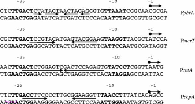
Figure 2

Figure 3

Figure 4

Figure 5
Copyright © 2022 FDOKUMEN