Toxicological Features of Catha edulis (Khat) on Livers and Kidneys of Male and Female...
11
Hindawi Publishing Corporation Evidence-Based Complementary and Alternative Medicine Volume 2012, Article ID 829401, 11 pages doi:10.1155/2012/829401 Research Article Toxicological Features of Catha edulis (Khat) on Livers and Kidneys of Male and Female Sprague-Dawley Rats: A Subchronic Study Abdulsamad Alsalahi, 1 Mahmood Ameen Abdulla, 1 Mohammed Al-Mamary, 2 Mohamed Ibrahim Noordin, 3 Siddig Ibrahim Abdelwahab, 3 Aied M. Alabsi, 4 Abdrabuh Shwter, 1 and Mohammed A. Alshawsh 1 1 Department of Molecular Medicine, Faculty of Medicine, University of Malaya, 50603 Kuala Lumpur, Malaysia 2 Department of Organic Chemistry, Faculty of Pharmacy, Sana’a University, Sana’a, Yemen 3 Department of Pharmacy, Faculty of Medicine, University of Malaya, 50603 Kuala Lumpur, Malaysia 4 Faculty of Dentistry, University of Malaya, 50603 Kuala Lumpur, Malaysia Correspondence should be addressed to Mahmood Ameen Abdulla, [email protected] Received 9 August 2012; Revised 15 October 2012; Accepted 22 October 2012 Academic Editor: Raffaele Capasso Copyright © 2012 Abdulsamad Alsalahi et al. This is an open access article distributed under the Creative Commons Attribution License, which permits unrestricted use, distribution, and reproduction in any medium, provided the original work is properly cited. Hepato- and nephrotoxicity of Khat consumption (Catha edulis Forskal) have been evoked. Therefore, this study was conducted to evaluate such possible hepatorenal toxicity in female and male Sprague-Dawley rats (SD rats) focusing primarily on liver and kidney. In addition, female and male rats were investigated separately. Accordingly, forty-eight SD-rats (100–120g) were distributed randomly into four groups of males and female (n = 12). Normal controls (NCs) received distilled water, whereas test groups received 500 mg/kg (low dose (LD)), 1000 mg/kg (medium dose (MD)), or 2000 mg/kg (high dose (HD)) of crude extract of Catha edulis orally for 4 weeks. Then, physical, biochemical, hematological, and histological parameters were analyzed. Results in Khat-fed rats showed hepatic enlargement, abnormal findings in serum aspartate aminotransferase (AST), and alkaline phosphatase (ALP) of male and female SD-rats and serum albumin (A) and serum creatinine (Cr) of female as compared to controls. In addition, histopathological abnormalities confirmed hepatic and renal toxicities of Khat that were related to heavy Khat consumption. In summary, Khat could be associated with hepatic hypertrophy and hepatotoxicity in male and female SD- rats and nephrotoxicity only in female SD-rats. 1. Introduction Khat is the most common name for Catha edulis plant [1] which is consumed for its psychostimulatory effect [2]. Unfortunately, Khat become a serious public health problem in Yemen [2]. The chewing of Khat leaves has involved at least 80% of adult males [3] and extended to women, too [4]. The WHO (2003, 2006) reported that Khat consumption has become a common problem that affects the health aspects of life [4]. In fact, many adverse effects have been associated with Khat consumption [2]. Accordingly, prolonged exposure to Khat could result in psychoneurological disturbances such as neurosis [5]. In addition, increased diastolic blood pressure [6] and vaso- constriction of coronary vasculature were also reported [7]. More commonly, gastritis [8], hemorrhoids, and duodenal ulcer had a higher prevalence among Khat chewers [9]. Furthermore, Luqman and Danowski reported that liver cirrhosis that was observed among Yemeni Khat chewers might be due to Khat consumption, but at that time it was not further investigated [10], and hepatotoxicity of Khat chewing is still debated in humans [11–13]. In animals, the administration of crude extract of Khat to New Zealander white rabbits for three months suggested toxic hepatocellular jaundice as well as histopathological abnormalities in livers of such animals [14]. Likewise, a companion study on
Transcript of Toxicological Features of Catha edulis (Khat) on Livers and Kidneys of Male and Female...
Hindawi Publishing CorporationEvidence-Based Complementary and Alternative MedicineVolume 2012, Article ID 829401, 11 pagesdoi:10.1155/2012/829401
Research Article
Toxicological Features of Catha edulis (Khat) onLivers and Kidneys of Male and Female Sprague-Dawley Rats:A Subchronic Study
Abdulsamad Alsalahi,1 Mahmood Ameen Abdulla,1 Mohammed Al-Mamary,2
Mohamed Ibrahim Noordin,3 Siddig Ibrahim Abdelwahab,3 Aied M. Alabsi,4
Abdrabuh Shwter,1 and Mohammed A. Alshawsh1
1 Department of Molecular Medicine, Faculty of Medicine, University of Malaya, 50603 Kuala Lumpur, Malaysia2 Department of Organic Chemistry, Faculty of Pharmacy, Sana’a University, Sana’a, Yemen3 Department of Pharmacy, Faculty of Medicine, University of Malaya, 50603 Kuala Lumpur, Malaysia4 Faculty of Dentistry, University of Malaya, 50603 Kuala Lumpur, Malaysia
Correspondence should be addressed to Mahmood Ameen Abdulla, [email protected]
Received 9 August 2012; Revised 15 October 2012; Accepted 22 October 2012
Academic Editor: Raffaele Capasso
Copyright © 2012 Abdulsamad Alsalahi et al. This is an open access article distributed under the Creative Commons AttributionLicense, which permits unrestricted use, distribution, and reproduction in any medium, provided the original work is properlycited.
Hepato- and nephrotoxicity of Khat consumption (Catha edulis Forskal) have been evoked. Therefore, this study was conductedto evaluate such possible hepatorenal toxicity in female and male Sprague-Dawley rats (SD rats) focusing primarily on liverand kidney. In addition, female and male rats were investigated separately. Accordingly, forty-eight SD-rats (100–120 g) weredistributed randomly into four groups of males and female (n = 12). Normal controls (NCs) received distilled water, whereastest groups received 500 mg/kg (low dose (LD)), 1000 mg/kg (medium dose (MD)), or 2000 mg/kg (high dose (HD)) of crudeextract of Catha edulis orally for 4 weeks. Then, physical, biochemical, hematological, and histological parameters were analyzed.Results in Khat-fed rats showed hepatic enlargement, abnormal findings in serum aspartate aminotransferase (AST), and alkalinephosphatase (ALP) of male and female SD-rats and serum albumin (A) and serum creatinine (Cr) of female as compared tocontrols. In addition, histopathological abnormalities confirmed hepatic and renal toxicities of Khat that were related to heavyKhat consumption. In summary, Khat could be associated with hepatic hypertrophy and hepatotoxicity in male and female SD-rats and nephrotoxicity only in female SD-rats.
1. Introduction
Khat is the most common name for Catha edulis plant[1] which is consumed for its psychostimulatory effect[2]. Unfortunately, Khat become a serious public healthproblem in Yemen [2]. The chewing of Khat leaves hasinvolved at least 80% of adult males [3] and extended towomen, too [4]. The WHO (2003, 2006) reported thatKhat consumption has become a common problem thataffects the health aspects of life [4]. In fact, many adverseeffects have been associated with Khat consumption [2].Accordingly, prolonged exposure to Khat could result inpsychoneurological disturbances such as neurosis [5]. In
addition, increased diastolic blood pressure [6] and vaso-constriction of coronary vasculature were also reported [7].More commonly, gastritis [8], hemorrhoids, and duodenalulcer had a higher prevalence among Khat chewers [9].Furthermore, Luqman and Danowski reported that livercirrhosis that was observed among Yemeni Khat chewersmight be due to Khat consumption, but at that time it wasnot further investigated [10], and hepatotoxicity of Khatchewing is still debated in humans [11–13]. In animals, theadministration of crude extract of Khat to New Zealanderwhite rabbits for three months suggested toxic hepatocellularjaundice as well as histopathological abnormalities in liversof such animals [14]. Likewise, a companion study on
2 Evidence-Based Complementary and Alternative Medicine
the same species of animals with the same dose levels ofcrude extract for six months supported the former three-month findings; however, after six months histopathologicalevidence from liver sections suggested periportal fibrosisas an initial indicator of liver cirrhosis without apparentdamage to kidneys [15]. As a limitation, those studies did notinclude females for the hepatotoxicity studies and evaluationof Khat nephrotoxic effects in either males or females wasonly minimally investigated. In addition, the doses of crudeextract that were given to animals were selected to be in theaverage of 150–200 g of fresh Khat leaves according to Kalix[16], which was less than that suggested by Al-Habori et al.[15]. However, there are no comparative studies about theeffects of the different dose levels of Khat on human in termsof body weight. Recently, several reports were published onsevere liver diseases in Khat users [11, 17, 18].
Our study was carried out to predict whether oraladministration of crude extract of Khat for 4 weeks couldbe associated with hepatorenal toxicity in either male orfemale Sprague-Dawley rats. In addition, female SD-rats andactual dose levels were included in the scope of the currentstudy. Moreover, several parameters were selected to obtain aclearer image of Khat toxicity signature on liver and kidneyincluding physical, serum biochemical, hematological, andespecially histopathological parameters.
2. Methodology
2.1. Materials
2.1.1. Plant Material. Four kilograms of fresh materialof Hamadani-type of Khat (the same type that chewerspurchase and consume) were purchased from a local Yemenimarket, Sana’a, Yemen. The fresh materials of Khat werewrapped in a plastic bundle just as chewers do. Next, the freshmaterials of Khat were transferred to the PharmacognosyLaboratory, Faculty of pharmacy, Sana’a University to beverified. Voucher specimens were kept in PharmacognosyDepartment as a reference. The fresh materials were washedto remove dust and debris with distilled water. The edibleparts were cut off to get three kilograms of fresh materialand subjected to air-drying for two weeks in a dark place.After 2 weeks, the dried materials were ground in a heavyduty blender (Moulinex-Trade blender). Finally, a net weightequal to 500 g of the dried ground material was obtained.
2.1.2. Sample Extraction. The dried material was extractedwith sufficient quantities of 80% methanol (Sigma-Aldrichand Fisher) using a Buchi-type soxhlet apparatus (5 mL ofsolvent per 1 g of dry material). Then, the resulted extractwas evaporated using a Buchi-type rotary evaporator undervacuum at 40◦C until dryness. Finally, the resulted yield(100 g dried extract) was kept in a desiccator until the timeof use.
2.1.3. Experimental Animals. Forty-eight healthy male andfemale Sprague-Dawley rats (100–120 g) were obtained from
the Animal House Unit, Faculty of Medicine, University ofMalaya, Malaysia.
2.2. Methods
2.2.1. Study Design. The study was conducted under ethicnumber of (PM/07/05/2008/1111/MAA (a) (R)) that hasbeen approved by the Ethic Committee, Animal HouseUnit, University of Malaya. The procedure followed theinstructions of Good Laboratory Practice (GLP) [19] andguidelines of Organization of Economic Co-operation andDevelopment (OECD); testing of chemicals 407 [20] Ratswere allowed to acclimatize the working conditions one weekprior to treatment, kept in a separated cage (1 rat per cage)under standard conditions of artificial light (12 hr light 12 hrsdark), temperature (25±3◦C), and relative humidity (60%±10%), and fed on a standard diet and water ad libitum. Bythe end, SD-rats were transferred into wire mesh-bottomedcages (to avoid coprophagy) with free access to water, butthe food was withheld overnight to collect fasting bloodsamples via cardiac puncture for serum biochemical assayand blood film. All rats were sacrificed 24 hours after the lasttreatment. On the day of sacrificing, rats were anaesthetizedintramuscularly with ketamine. Rats were killed throughincreasing ketamine dose. Then, rats were dissected to collectlivers and kidneys.
2.2.2. Grouping and Dosing of Rats. Rats were randomlyselected and distributed into two main groups of males andfemales (n = 24), which were allocated into four groups(n = 6) designated as high dose (HD), medium dose (MD),low dose (LD), and normal control (NC). Rats were givena single daily dose of crude extract of Khat suspended inpure distilled water according to the body weight of each ratby oral gavage. The dose levels were selected and calculatedtypically as the daily consumed amounts of fresh leaves byKhat chewers [8]. The dosages of Khat designated to beadministered to rats were 2000 mg/kg as high dose (HD),1000 mg/kg as medium dose (MD), 500 mg/kg as low dose(LD), and 10 mL/kg of distilled water for normal control(NC).
2.2.3. Clinical Observation of Rats. Prior to the start of thestudy, all rats were clinically observed for any abnormalbehavior, physical abnormalities, mortality, and morbidity atleast once daily.
2.2.4. Physical Parameters. The body weight of rats was takenonce weekly, at the first day of treatment and the day ofsacrificing. Livers were washed in normal saline and blottedwith filter paper. The weight of livers was taken to obtainabsolute liver weight for each rat. By dividing liver weight onbody weight, relative liver weight was obtained for each rat.
2.2.5. Serum Biochemical Parameters. Fasting blood sampleswere collected into plain tubes. Then, tubes were allowedto stand to give full clotting. Then, tubes were centrifugedimmediately at 2500×g for 15 minutes to obtain a clear
Evidence-Based Complementary and Alternative Medicine 3
supernatant. Then, 48 serum-containing tubes were admit-ted to the Laboratory Department, Faculty of Medicine,University of Malaya to carry out the biochemical parameteranalysis. Serum biochemical parameters included alkalinephosphatase (ALP), alanine aminotransferase (ALT), aspar-tate aminotransferase (AST), creatine kinase (CK), amylase(AM), gamma-Glutamyl transpeptidase (GGT), total biliru-bin (TB), albumin (A), creatinine (Cr), urea (BUN), sodium(Na), potassium (K), chloride (Cl), and carbon dioxide(CO2).
2.2.6. Hematological Parameters. The other part of bloodwas collected into 48 EDTA-containing tubes. Then, thoseblood samples were admitted to the Laboratory Department,Faculty of Medicine, University of Malaya. Blood sampleswere examined for various parameters including hemoglobin(HB), hematocrit (HCT), red blood cells (RBC), meancorpuscular volume (MCV), mean corpuscular hemoglobin(MCH), mean corpuscular hemoglobin concentration(MCHC), White blood cells (WBC), and platelets (PLT).
2.2.7. Histopathology Study. After autopsy, livers and kidneyswere washed in normal saline, blotted with filter paper, andvisualized for any surface visible lesions immediately afterharvesting and weighed. Then, organs were put in neutralphosphate buffer solution. After that, biopsies were obtained,fixed in 10% neutral formalin, processed, dehydrated, andembedded in paraffin wax. The 5 µm sections in thicknesswere stained according to the usual hematoxylin-eosin pro-cedure to be microscopically examined (20x magnification).
2.2.8. Statistical Analysis. The results were computed statis-tically with SPSS software package version 17 using one-wayanalysis of variance (ANOVA) for mean difference betweengroups, and males and females were analyzed separately (n =6) and P value was set on P < 0.05. The values were expressedas mean ± SEM. Then post hoc Dunnett test (two-sided)was followed for comparing the tested groups to the singlenormal control at P < 0.05 and P ≤ 0.01. Otherwise,nonparametric tests were employed when the data were notnormally distributed so that nonparametric Kruskal Wallistest was used to compare median difference between groupswhich was followed by Mann-Whitney test to compare eachKhat treated group to the normal control group.
3. Results
All rats were in good health and neither mortality normorbidity was observed.
3.1. Physical Parameters. Body weight (BW) of male SD-rats was significantly (ANOVA P < 0.05) different betweengroups, and post hoc Dunnet test indicated that mean bodyweight of the male HD-group was significantly (P < 0.05)lower than that of male NC-group (by 19%). In addition,absolute liver weight (ALW) of males was nonsignificantly(ANOVA, P ≥ 0.05) different between groups. However,relative liver weight (RLW) of male SD-rats was significantly
(ANOVA, P < 0.05) different between groups and post hocDunnet test indicated that mean value of the male HD-groupwas significantly (P < 0.05) greater than that of male NC-group (by 21%) see Table 1.
BW of females were nonsignificantly (ANOVA P ≥ 0.05)different between NC, LD, MD, and HD. However, absoluteliver weight (ALW) of female SD-rats was significantly(ANOVA, P < 0.05) different between groups and posthoc Dunnet test indicated that mean value of HD wasgreater than that of female NC-group (P ≤ 0.01) (by 34%).Likewise, relative liver weight (RLW) of female SD-rats wassignificantly (ANOVA, P < 0.05) different between groupsand post hoc Dunnet test indicated that mean values of HD,MD, and LD were significantly (P ≤ 0.01) greater than thatof female NC (by 56%, 28%, and 25%, resp.) see Table 1.
3.2. Serum Biochemistry
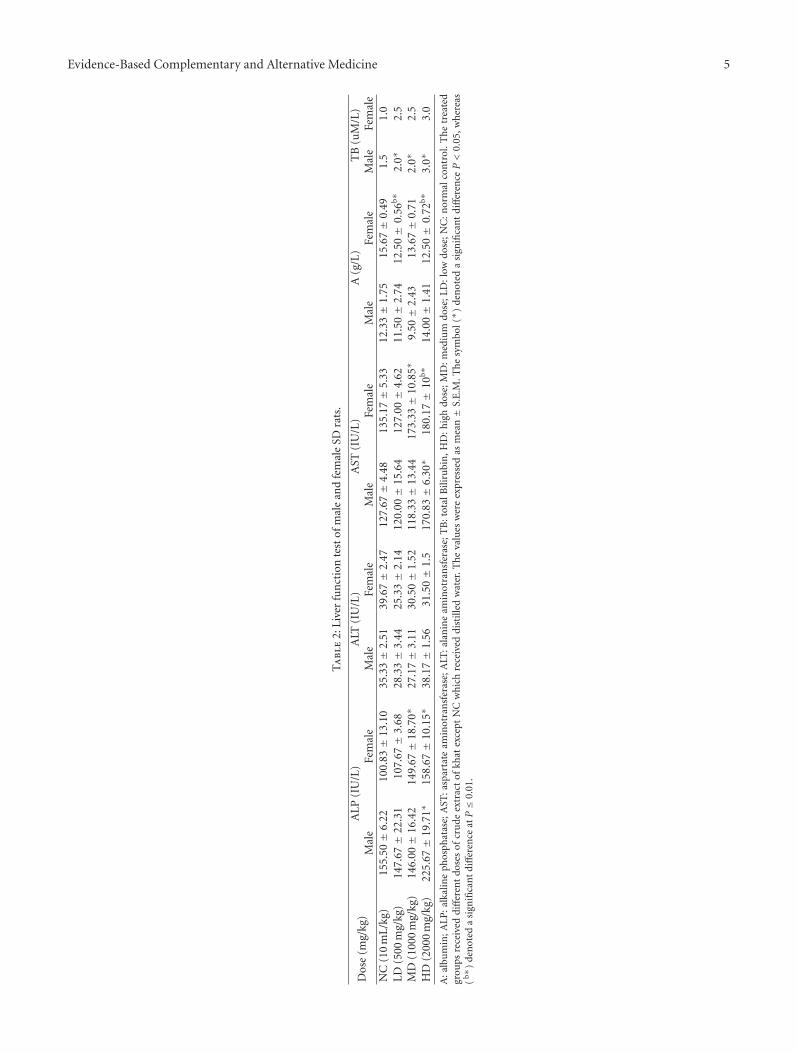
3.2.1. Liver Function Test. Serum activity of alkaline phos-phatase (ALP) in male SD-rats was significantly (ANOVA,P < 0.05) different between the groups. Accordingly, posthoc Dunnet test indicated that mean ALP of HD-groupwas significantly (P < 0.05) higher than that of NC (by45%). Serum activity of alanine aminotransferase (ALT) ofmale SD-rats seemed significantly different between groups(ANOVA, P < 0.05); however, post hoc Dunnet testindicated a non-significant difference (P ≥ 0.05) of Khatgroups compared to that of NC. Unlike ALT, serum activityof aspartate aminotransferase (AST) in male SD-rats wassignificantly (ANOVA, P < 0.05) different between groups.Post hoc Dunnet test indicated that mean AST value of HDwas significantly (P < 0.05) greater than that of NC (by34%). In addition, serum total bilirubin (TB) of male SD-rats was significantly (P < 0.05) different as indicated byKruskal-Wallis test; Mann-Whitney test indicated that HDand MD were significantly greater than that of NC (seeTable 2). Conversely, serum albumin (A) of male SD-ratswas nonsignificantly (ANOVA, P ≥ 0.05) different fromthat of NC (see Table 2). Moreover, serum activities ofgamma-glutamyl transpeptidase (GGT) and serum levels ofunconjugated bilirubin of male SD-rats were undetectable inboth the tested groups and NC.
Serum activity of ALP of female SD-rats was significantly(ANOVA, P < 0.05) different between groups. Post hocDunnet test indicated that the mean values of HD and MDwere significantly (P < 0.05) greater than that of NC (by 57%and 48%, resp.). Serum activity of alanine aminotransferase(ALT) of female SD-rats seemed significantly (ANOVA, P <0.05) different between groups; however, mean values of HD,MD, and LD were not different from NC (P ≥ 0.05). Serumactivity of aspartate aminotransferase (AST) of female SD-rats was significantly (ANOVA, P < 0.05) different betweengroups, and post hoc Dunnet test indicated that mean valuesof HD and MD were significantly (P ≤ 0.01) greater thanthat of NC (by 34% and 28%, resp.). In addition, the serumtotal bilirubin (TB) of female SD-rats was nonsignificantly(P ≥ 0.05) different from NC as indicated by Kruskal-Wallistest. Conversely, the serum albumin (A) of female SD-ratswas significantly (ANOVA, P < 0.05) different between
4 Evidence-Based Complementary and Alternative Medicine
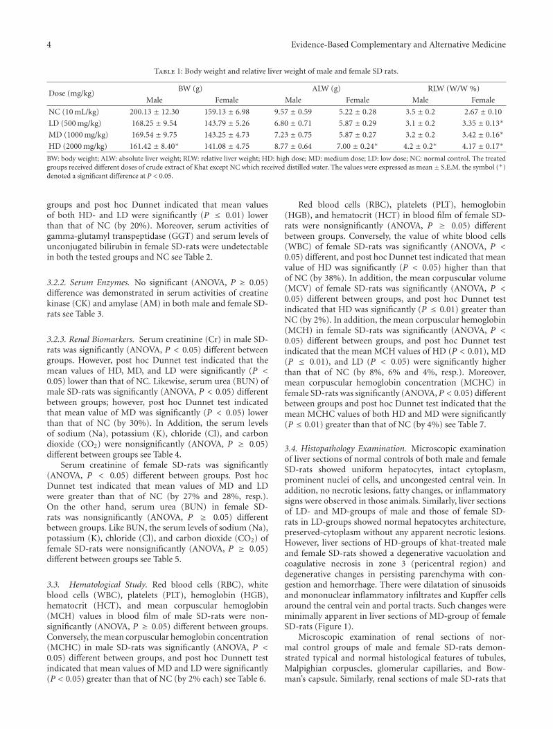
Table 1: Body weight and relative liver weight of male and female SD rats.
Dose (mg/kg)BW (g) ALW (g) RLW (W/W %)
Male Female Male Female Male Female
NC (10 mL/kg) 200.13 ± 12.30 159.13 ± 6.98 9.57 ± 0.59 5.22 ± 0.28 3.5 ± 0.2 2.67 ± 0.10
LD (500 mg/kg) 168.25 ± 9.54 143.79 ± 5.26 6.80 ± 0.71 5.87 ± 0.29 3.1 ± 0.2 3.35 ± 0.13∗
MD (1000 mg/kg) 169.54 ± 9.75 143.25 ± 4.73 7.23 ± 0.75 5.87 ± 0.27 3.2 ± 0.2 3.42 ± 0.16∗
HD (2000 mg/kg) 161.42 ± 8.40∗ 141.08 ± 4.75 8.77 ± 0.64 7.00 ± 0.24∗ 4.2 ± 0.2∗ 4.17 ± 0.17∗
BW: body weight; ALW: absolute liver weight; RLW: relative liver weight; HD: high dose; MD: medium dose; LD: low dose; NC: normal control. The treatedgroups received different doses of crude extract of Khat except NC which received distilled water. The values were expressed as mean ± S.E.M. the symbol (∗)denoted a significant difference at P < 0.05.
groups and post hoc Dunnet indicated that mean valuesof both HD- and LD were significantly (P ≤ 0.01) lowerthan that of NC (by 20%). Moreover, serum activities ofgamma-glutamyl transpeptidase (GGT) and serum levels ofunconjugated bilirubin in female SD-rats were undetectablein both the tested groups and NC see Table 2.
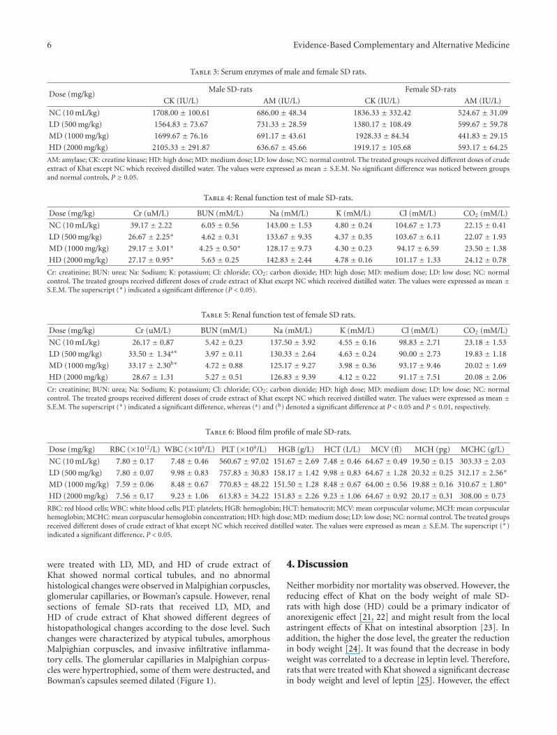
3.2.2. Serum Enzymes. No significant (ANOVA, P ≥ 0.05)difference was demonstrated in serum activities of creatinekinase (CK) and amylase (AM) in both male and female SD-rats see Table 3.
3.2.3. Renal Biomarkers. Serum creatinine (Cr) in male SD-rats was significantly (ANOVA, P < 0.05) different betweengroups. However, post hoc Dunnet test indicated that themean values of HD, MD, and LD were significantly (P <0.05) lower than that of NC. Likewise, serum urea (BUN) ofmale SD-rats was significantly (ANOVA, P < 0.05) differentbetween groups; however, post hoc Dunnet test indicatedthat mean value of MD was significantly (P < 0.05) lowerthan that of NC (by 30%). In Addition, the serum levelsof sodium (Na), potassium (K), chloride (Cl), and carbondioxide (CO2) were nonsignificantly (ANOVA, P ≥ 0.05)different between groups see Table 4.
Serum creatinine of female SD-rats was significantly(ANOVA, P < 0.05) different between groups. Post hocDunnet test indicated that mean values of MD and LDwere greater than that of NC (by 27% and 28%, resp.).On the other hand, serum urea (BUN) in female SD-rats was nonsignificantly (ANOVA, P ≥ 0.05) differentbetween groups. Like BUN, the serum levels of sodium (Na),potassium (K), chloride (Cl), and carbon dioxide (CO2) offemale SD-rats were nonsignificantly (ANOVA, P ≥ 0.05)different between groups see Table 5.
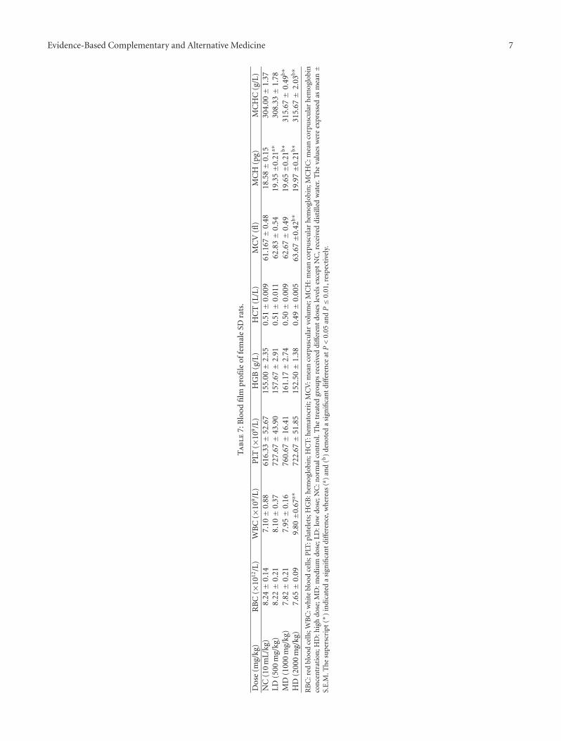
3.3. Hematological Study. Red blood cells (RBC), whiteblood cells (WBC), platelets (PLT), hemoglobin (HGB),hematocrit (HCT), and mean corpuscular hemoglobin(MCH) values in blood film of male SD-rats were non-significantly (ANOVA, P ≥ 0.05) different between groups.Conversely, the mean corpuscular hemoglobin concentration(MCHC) in male SD-rats was significantly (ANOVA, P <0.05) different between groups, and post hoc Dunnett testindicated that mean values of MD and LD were significantly(P < 0.05) greater than that of NC (by 2% each) see Table 6.
Red blood cells (RBC), platelets (PLT), hemoglobin(HGB), and hematocrit (HCT) in blood film of female SD-rats were nonsignificantly (ANOVA, P ≥ 0.05) differentbetween groups. Conversely, the value of white blood cells(WBC) of female SD-rats was significantly (ANOVA, P <0.05) different, and post hoc Dunnet test indicated that meanvalue of HD was significantly (P < 0.05) higher than thatof NC (by 38%). In addition, the mean corpuscular volume(MCV) of female SD-rats was significantly (ANOVA, P <0.05) different between groups, and post hoc Dunnet testindicated that HD was significantly (P ≤ 0.01) greater thanNC (by 2%). In addition, the mean corpuscular hemoglobin(MCH) in female SD-rats was significantly (ANOVA, P <0.05) different between groups, and post hoc Dunnet testindicated that the mean MCH values of HD (P < 0.01), MD(P ≤ 0.01), and LD (P < 0.05) were significantly higherthan that of NC (by 8%, 6% and 4%, resp.). Moreover,mean corpuscular hemoglobin concentration (MCHC) infemale SD-rats was significantly (ANOVA, P < 0.05) differentbetween groups and post hoc Dunnet test indicated that themean MCHC values of both HD and MD were significantly(P ≤ 0.01) greater than that of NC (by 4%) see Table 7.
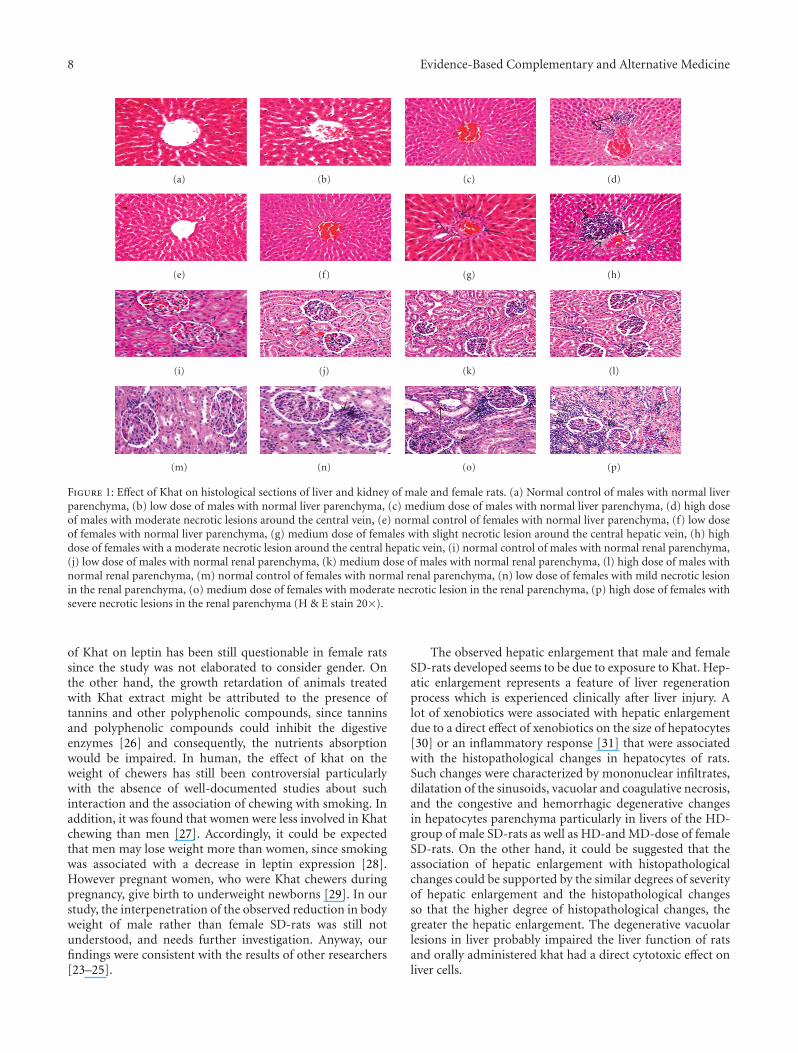
3.4. Histopathology Examination. Microscopic examinationof liver sections of normal controls of both male and femaleSD-rats showed uniform hepatocytes, intact cytoplasm,prominent nuclei of cells, and uncongested central vein. Inaddition, no necrotic lesions, fatty changes, or inflammatorysigns were observed in those animals. Similarly, liver sectionsof LD- and MD-groups of male and those of female SD-rats in LD-groups showed normal hepatocytes architecture,preserved-cytoplasm without any apparent necrotic lesions.However, liver sections of HD-groups of khat-treated maleand female SD-rats showed a degenerative vacuolation andcoagulative necrosis in zone 3 (pericentral region) anddegenerative changes in persisting parenchyma with con-gestion and hemorrhage. There were dilatation of sinusoidsand mononuclear inflammatory infiltrates and Kupffer cellsaround the central vein and portal tracts. Such changes wereminimally apparent in liver sections of MD-group of femaleSD-rats (Figure 1).
Microscopic examination of renal sections of nor-mal control groups of male and female SD-rats demon-strated typical and normal histological features of tubules,Malpighian corpuscles, glomerular capillaries, and Bow-man’s capsule. Similarly, renal sections of male SD-rats that
Evidence-Based Complementary and Alternative Medicine 5
Ta
ble
2:Li
ver
fun
ctio
nte
stof
mal
ean
dfe
mal
eSD
rats
.
Dos
e(m
g/kg
)A
LP
(IU
/L)
ALT
(IU
/L)
AST
(IU
/L)
A(g
/L)
TB
(uM
/L)
Mal
eFe
mal
eM
ale
Fem
ale
Mal
eFe
mal
eM
ale
Fem
ale
Mal
eFe
mal
eN
C(1
0m
L/kg
)15
5.50±
6.22
100.
83±
13.1
035
.33±
2.51
39.6
7±
2.47
127.
67±
4.48
135.
17±
5.33
12.3
3±
1.75
15.6
7±
0.49
1.5
1.0
LD(5
00m
g/kg
)14
7.67±
22.3
110
7.67±
3.68
28.3
3±
3.44
25.3
3±
2.14
120.
00±
15.6
412
7.00±
4.62
11.5
0±
2.74
12.5
0±
0.56
b∗2.
0∗2.
5M
D(1
000
mg/
kg)
146.
00±
16.4
214
9.67±
18.7
0∗27
.17±
3.11
30.5
0±
1.52
118.
33±
13.4
417
3.33±
10.8
5∗9.
50±
2.43
13.6
7±
0.71
2.0∗
2.5
HD
(200
0m
g/kg
)22
5.67±
19.7
1∗15
8.67±
10.1
5∗38
.17±
1.56
31.5
0±
1.5
170.
83±
6.30
∗18
0.17±
10b∗
14.0
0±
1.41
12.5
0±
0.72
b∗3.
0∗3.
0
A:a
lbu
min
;ALP
:alk
alin
eph
osph
atas
e;A
ST:a
spar
tate
amin
otra
nsf
eras
e;A
LT:a
lan
ine
amin
otra
nsf
eras
e;T
B:t
otal
Bili
rubi
n,H
D:h
igh
dose
;MD
:med
ium
dose
;LD
:low
dose
;NC
:nor
mal
con
trol
.Th
etr
eate
dgr
oups
rece
ived
diff
eren
tdo
ses
ofcr
ude
extr
act
ofkh
atex
cept
NC
wh
ich
rece
ived
dist
illed
wat
er.T
he
valu
esw
ere
expr
esse
das
mea
n±
S.E
.M.T
he
sym
bol(∗ )
den
oted
asi
gnifi
can
tdi
ffer
ence
P<
0.05
,wh
erea
s(b∗
)de
not
eda
sign
ifica
nt
diff
eren
ceat
P≤
0.01
.
6 Evidence-Based Complementary and Alternative Medicine
Table 3: Serum enzymes of male and female SD rats.
Dose (mg/kg)Male SD-rats Female SD-rats
CK (IU/L) AM (IU/L) CK (IU/L) AM (IU/L)
NC (10 mL/kg) 1708.00 ± 100.61 686.00 ± 48.34 1836.33 ± 332.42 524.67 ± 31.09
LD (500 mg/kg) 1564.83 ± 73.67 731.33 ± 28.59 1380.17 ± 108.49 599.67 ± 59.78
MD (1000 mg/kg) 1699.67 ± 76.16 691.17 ± 43.61 1928.33 ± 84.34 441.83 ± 29.15
HD (2000 mg/kg) 2105.33 ± 291.87 636.67 ± 45.66 1919.17 ± 105.68 593.17 ± 64.25
AM: amylase; CK: creatine kinase; HD: high dose; MD: medium dose; LD: low dose; NC: normal control. The treated groups received different doses of crudeextract of Khat except NC which received distilled water. The values were expressed as mean ± S.E.M. No significant difference was noticed between groupsand normal controls, P ≥ 0.05.
Table 4: Renal function test of male SD-rats.
Dose (mg/kg) Cr (uM/L) BUN (mM/L) Na (mM/L) K (mM/L) Cl (mM/L) CO2 (mM/L)
NC (10 mL/kg) 39.17 ± 2.22 6.05 ± 0.56 143.00 ± 1.53 4.80 ± 0.24 104.67 ± 1.73 22.15 ± 0.41
LD (500 mg/kg) 26.67 ± 2.25∗ 4.62 ± 0.31 133.67 ± 9.35 4.37 ± 0.35 103.67 ± 6.11 22.07 ± 1.93
MD (1000 mg/kg) 29.17 ± 3.01∗ 4.25 ± 0.50∗ 128.17 ± 9.73 4.30 ± 0.23 94.17 ± 6.59 23.50 ± 1.38
HD (2000 mg/kg) 27.17 ± 0.95∗ 5.63 ± 0.25 142.83 ± 2.44 4.78 ± 0.16 101.17 ± 1.33 24.12 ± 0.78
Cr: creatinine; BUN: urea; Na: Sodium; K: potassium; Cl: chloride; CO2: carbon dioxide; HD: high dose; MD: medium dose; LD: low dose; NC: normalcontrol. The treated groups received different doses of crude extract of Khat except NC which received distilled water. The values were expressed as mean ±S.E.M. The superscript (∗) indicated a significant difference (P < 0.05).
Table 5: Renal function test of female SD rats.
Dose (mg/kg) Cr (uM/L) BUN (mM/L) Na (mM/L) K (mM/L) Cl (mM/L) CO2 (mM/L)
NC (10 mL/kg) 26.17 ± 0.87 5.42 ± 0.23 137.50 ± 3.92 4.55 ± 0.16 98.83 ± 2.71 23.18 ± 1.53
LD (500 mg/kg) 33.50 ± 1.34a∗ 3.97 ± 0.11 130.33 ± 2.64 4.63 ± 0.24 90.00 ± 2.73 19.83 ± 1.18
MD (1000 mg/kg) 33.17 ± 2.30b∗ 4.72 ± 0.88 125.17 ± 9.27 3.98 ± 0.36 93.17 ± 9.46 20.02 ± 1.69
HD (2000 mg/kg) 28.67 ± 1.31 5.27 ± 0.51 126.83 ± 9.39 4.12 ± 0.22 91.17 ± 7.51 20.08 ± 2.06
Cr: creatinine; BUN: urea; Na: Sodium; K: potassium; Cl: chloride; CO2: carbon dioxide; HD: high dose; MD: medium dose; LD: low dose; NC: normalcontrol. The treated groups received different doses of crude extract of Khat except NC which received distilled water. The values were expressed as mean ±S.E.M. The superscript (∗) indicated a significant difference, whereas (a) and (b) denoted a significant difference at P < 0.05 and P ≤ 0.01, respectively.
Table 6: Blood film profile of male SD-rats.
Dose (mg/kg) RBC (×1012/L) WBC (×109/L) PLT (×109/L) HGB (g/L) HCT (L/L) MCV (fl) MCH (pg) MCHC (g/L)
NC (10 mL/kg) 7.80 ± 0.17 7.48 ± 0.46 560.67 ± 97.02 151.67 ± 2.69 7.48 ± 0.46 64.67 ± 0.49 19.50 ± 0.15 303.33 ± 2.03
LD (500 mg/kg) 7.80 ± 0.07 9.98 ± 0.83 757.83 ± 30.83 158.17 ± 1.42 9.98 ± 0.83 64.67 ± 1.28 20.32 ± 0.25 312.17 ± 2.56∗
MD (1000 mg/kg) 7.59 ± 0.06 8.48 ± 0.67 770.83 ± 48.22 151.50 ± 1.28 8.48 ± 0.67 64.00 ± 0.56 19.88 ± 0.16 310.67 ± 1.80∗
HD (2000 mg/kg) 7.56 ± 0.17 9.23 ± 1.06 613.83 ± 34.22 151.83 ± 2.26 9.23 ± 1.06 64.67 ± 0.92 20.17 ± 0.31 308.00 ± 0.73
RBC: red blood cells; WBC: white blood cells; PLT: platelets; HGB: hemoglobin; HCT: hematocrit; MCV: mean corpuscular volume; MCH: mean corpuscularhemoglobin; MCHC: mean corpuscular hemoglobin concentration; HD: high dose; MD: medium dose; LD: low dose; NC: normal control. The treated groupsreceived different doses of crude extract of khat except NC which received distilled water. The values were expressed as mean ± S.E.M. The superscript (∗)indicated a significant difference, P < 0.05.
were treated with LD, MD, and HD of crude extract ofKhat showed normal cortical tubules, and no abnormalhistological changes were observed in Malpighian corpuscles,glomerular capillaries, or Bowman’s capsule. However, renalsections of female SD-rats that received LD, MD, andHD of crude extract of Khat showed different degrees ofhistopathological changes according to the dose level. Suchchanges were characterized by atypical tubules, amorphousMalpighian corpuscles, and invasive infiltrative inflamma-tory cells. The glomerular capillaries in Malpighian corpus-cles were hypertrophied, some of them were destructed, andBowman’s capsules seemed dilated (Figure 1).
4. Discussion
Neither morbidity nor mortality was observed. However, thereducing effect of Khat on the body weight of male SD-rats with high dose (HD) could be a primary indicator ofanorexigenic effect [21, 22] and might result from the localastringent effects of Khat on intestinal absorption [23]. Inaddition, the higher the dose level, the greater the reductionin body weight [24]. It was found that the decrease in bodyweight was correlated to a decrease in leptin level. Therefore,rats that were treated with Khat showed a significant decreasein body weight and level of leptin [25]. However, the effect
Evidence-Based Complementary and Alternative Medicine 7
Ta
ble
7:B
lood
film
profi
leof
fem
ale
SDra
ts.
Dos
e(m
g/kg
)R
BC
(×10
12/L
)W
BC
(×10
9/L
)P
LT(×
109/L
)H
GB
(g/L
)H
CT
(L/L
)M
CV
(fl)
MC
H(p
g)M
CH
C(g
/L)
NC
(10
mL/
kg)
8.24±
0.14
7.10±
0.88
616.
33±
52.6
715
5.00±
2.35
0.51±
0.00
961
.167±
0.48
18.5
8±
0.15
304.
00±
1.37
LD(5
00m
g/kg
)8.
22±
0.21
8.10±
0.37
727.
67±
43.9
015
7.67±
2.91
0.51±
0.01
162
.83±
0.54
19.3
5±0
.21a∗
308.
33±
1.78
MD
(100
0m
g/kg
)7.
82±
0.21
7.95±
0.16
760.
67±
16.4
116
1.17±
2.74
0.50±
0.00
962
.67±
0.49
19.6
5±0
.21b∗
315.
67±
0.49
b∗
HD
(200
0m
g/kg
)7.
65±
0.09
9.80±0
.67a∗
722.
67±
51.8
515
2.50±
1.38
0.49±
0.00
563
.67±0
.42b∗
19.9
7±0
.21b∗
315.
67±
2.03
b∗
RB
C:r
edbl
ood
cells
;WB
C:w
hit
ebl
ood
cells
;PLT
:pla
tele
ts;H
GB
:hem
oglo
bin
;HC
T:h
emat
ocri
t;M
CV
:mea
nco
rpu
scu
lar
volu
me;
MC
H:m
ean
corp
usc
ula
rh
emog
lobi
n;M
CH
C:m
ean
corp
usc
ula
rh
emog
lobi
nco
nce
ntr
atio
n;H
D:h
igh
dose
;MD
:med
ium
dose
;LD
:low
dose
;NC
:nor
mal
con
trol
.Th
etr
eate
dgr
oups
rece
ived
diff
eren
tdo
ses
leve
lsex
cept
NC
,rec
eive
ddi
still
edw
ater
.Th
eva
lues
wer
eex
pres
sed
asm
ean±
S.E
.M.T
he
supe
rscr
ipt
(∗)
indi
cate
da
sign
ifica
nt
diff
eren
ce,w
her
eas
(a )an
d(b
)de
not
eda
sign
ifica
nt
diff
eren
ceat
P<
0.05
and
P≤
0.01
,res
pec
tive
ly.
8 Evidence-Based Complementary and Alternative Medicine
(a) (b) (c) (d)
(e) (f) (g) (h)
(i) (j) (k) (l)
(m) (n) (o) (p)
Figure 1: Effect of Khat on histological sections of liver and kidney of male and female rats. (a) Normal control of males with normal liverparenchyma, (b) low dose of males with normal liver parenchyma, (c) medium dose of males with normal liver parenchyma, (d) high doseof males with moderate necrotic lesions around the central vein, (e) normal control of females with normal liver parenchyma, (f) low doseof females with normal liver parenchyma, (g) medium dose of females with slight necrotic lesion around the central hepatic vein, (h) highdose of females with a moderate necrotic lesion around the central hepatic vein, (i) normal control of males with normal renal parenchyma,(j) low dose of males with normal renal parenchyma, (k) medium dose of males with normal renal parenchyma, (l) high dose of males withnormal renal parenchyma, (m) normal control of females with normal renal parenchyma, (n) low dose of females with mild necrotic lesionin the renal parenchyma, (o) medium dose of females with moderate necrotic lesion in the renal parenchyma, (p) high dose of females withsevere necrotic lesions in the renal parenchyma (H & E stain 20×).
of Khat on leptin has been still questionable in female ratssince the study was not elaborated to consider gender. Onthe other hand, the growth retardation of animals treatedwith Khat extract might be attributed to the presence oftannins and other polyphenolic compounds, since tanninsand polyphenolic compounds could inhibit the digestiveenzymes [26] and consequently, the nutrients absorptionwould be impaired. In human, the effect of khat on theweight of chewers has still been controversial particularlywith the absence of well-documented studies about suchinteraction and the association of chewing with smoking. Inaddition, it was found that women were less involved in Khatchewing than men [27]. Accordingly, it could be expectedthat men may lose weight more than women, since smokingwas associated with a decrease in leptin expression [28].However pregnant women, who were Khat chewers duringpregnancy, give birth to underweight newborns [29]. In ourstudy, the interpenetration of the observed reduction in bodyweight of male rather than female SD-rats was still notunderstood, and needs further investigation. Anyway, ourfindings were consistent with the results of other researchers[23–25].
The observed hepatic enlargement that male and femaleSD-rats developed seems to be due to exposure to Khat. Hep-atic enlargement represents a feature of liver regenerationprocess which is experienced clinically after liver injury. Alot of xenobiotics were associated with hepatic enlargementdue to a direct effect of xenobiotics on the size of hepatocytes[30] or an inflammatory response [31] that were associatedwith the histopathological changes in hepatocytes of rats.Such changes were characterized by mononuclear infiltrates,dilatation of the sinusoids, vacuolar and coagulative necrosis,and the congestive and hemorrhagic degenerative changesin hepatocytes parenchyma particularly in livers of the HD-group of male SD-rats as well as HD-and MD-dose of femaleSD-rats. On the other hand, it could be suggested that theassociation of hepatic enlargement with histopathologicalchanges could be supported by the similar degrees of severityof hepatic enlargement and the histopathological changesso that the higher degree of histopathological changes, thegreater the hepatic enlargement. The degenerative vacuolarlesions in liver probably impaired the liver function of ratsand orally administered khat had a direct cytotoxic effect onliver cells.
Evidence-Based Complementary and Alternative Medicine 9
The observed elevation in serum activity of alkalinephosphatase (ALP) in the HD-group of male SD-rats andHD- and MD-groups of female SD-rats indicates a possiblehepatotoxicity [32]. The elevation of several markers wouldbe of a greater diagnostic value than a single one [33]. Thelack of specificity of AST, ALP, and GGT cannot authorizeany conclusion because such enzymes are ubiquitous invarious tissues and serum elevation could represent not onlyliver, but also heart, bones, and muscles damages [34]. More-over, the inhibited activity of serum alanine aminotransferase(ALT) could indicate normal hepatic function because ofits specificity as a marker for hepatocytes integrity [34–38].Another possibility could be the establishment of a covalentbinding between certain components of Khat and ALT [33].Although serum ALT was reported to be a poor markerin predicting hepatotoxicity in rats [39], the higher serumactivity of aspartate aminotransferase in HD-group of malesas well as HD- and MD-groups of females than that ofnormal control rats (NC) suggests leakage into circulationfrom ruptured cell membranes of hepatocytes upon exposingto injury [34]. Although AST is widely distributed in variousorgans, its concentration in hepatocytes is still the highest[35]. Pancreatic damage was unlikely, since serum amylase(AM) was nonsignificantly changed despite its lack of speci-ficity [40]. Again, elevations of AST due to straited musclesinjury should be associated with parrallel elevations in serumcreatine kinase (CK) as a marker of skeletal [34, 35, 41]and/or heart muscles injury [24]. However, CK in the testedgroups of male and female SD-rats was nonsignificantlydifferent from that of NC. Histopathological examination ofbiopsies from heart muscles indicated no abnormal findingsin both male and female SD-rats. Elevations of GGT inrodents were rarely detected, even if liver was exposed toa known hepatotoxic compound, so this does not excludehepatotoxicity of khat [33]. Consequently, elevations ofserum AST were of hepatic origin rather than cardiac,muscular, or pancreatic origin. In addition, the low levelsof serum albumin (A) of female SD-rats with HD and LDsuggested a defect in the hepatic synthesis capacity [38, 42].It is known that albumin synthesis is restricted to liver[42]. Accordingly, our findings were consistent with thosereported by Al-Mamary et al. (2002) with Khat exposurefor 3 months and with Al-Habori et al. (2002) with Khatexposure for 6 months [15, 43]. However, the elevation ofserum activity of ALT in the rabbits in the aforementionedarticle was contrasting to the findings of our study becauseALT was higher in rabbits which maybe because the dosesgiven to rabbits were higher and exposure was longer thanthat in the current study. So, there is too much uncertaintyon biological data to draw back any conclusion and onlyhistological findings assess Khat hepatotoxicity in rats.
Similarly, serum creatinine (Cr) and urea (BUN) couldindicate the effect of xenobiotics on kidney structure andfunction [31, 33]. But the findings of normal activity ofCr and BUN and serum electrolytes in males indicatednormal renal function. Nonetheless, elevated serum val-ues of Cr in females particularly in the MD- and LD-Khat groups indicated impaired kidney function or deshy-dratation (dehydration). Accordingly, our findings were
consistent with those results of Al-Mamary et al. [14]with Khat exposure but contrasting to Al-Habori et al.[15] who suggested normal kidney functions in animalsafter six-month exposure to Khat. The hematology profilewas found to be normal except for a mild elevation inwhite blood cells (WBC) in the HD-group of females. So,despite physical, biochemical, and hematological findingsonly histological examination of liver and kidney biopsiesfit with Khat hepato- and nephrotoxicity. Actually, Khathad direct cytotoxic effects on liver and kidney whichwere confirmed by the microscopic examination of hepaticand renal sections. Such changes in liver parenchyma weredemonstrated in the form of coagulative necrosis (in zone 3)with hemorrhage, mononuclear infiltrates of inflammatorycells, and vacuolar degeneration in HD-group of malesas well as MD- and HD-groups of female SD-rats, whichindicated a regeneration process of liver [44] because ofexposure to khat. Examination of histopathological changesof renal sections indicated that only kidneys of female SD-rats were affected with Khat in different degrees of severityaccording to dose so that the higher the dose, the greater thedestructive changes in kidney structures. Such changes werecharacterized by invasive infiltrates of inflammatory cells,atypical tubules, and amorphous Malpighian corpuscles;hypertrophied and destructed capillaries of Malpighian cor-puscles were hypertrophied and dilated Bowman’s capsules.Histopathological study on liver and kidney was consistentwith those changes reported by Al-Mamary et al. [43] aswell as it was consistent with the hepatic histopathologicalstudy conducted by Al-Habori et al. [15], but no periportalfibrotic changes were observed, which may be due to thelonger duration of Khat administration to rabbits for sixmonths.
The findings of study suggest the elaboration of thestudy to clarify the possible effects of Khat, leptin, andsmoking on weight of male and female animals and khatchewers. In addition, the observed hepatic hypertrophyneeds further investigation, since it sometimes may bedue to physical congestion or hyperplasia. Reconsideringthe assessment of the specificity of hepatic biomarkersin disclosing hepatotoxicity of khat is required, and theemployment of tissue homogenate, molecular techniques,and immunohistochemistry may be more informative.
Up to now, it has been not reported that the toxic effectsexerted by Khat on livers and kidneys of rats can be relatedeither to the content of Khat leaves [10] or to the pesticides[13, 45] that are sprayed on Khat trees to improve harvesting[46]. Accordingly, further study should be performed to clar-ify such ambiguity through comparing toxicity of pesticidesprayed-Khat to that of pesticides free-Khat. In addition,our study demonstrated that oral administration of Khat torats was associated with an inhibited activity of the specifichepatic biomarker, alanine aminotransferase (ALT), whichcould be due to depletion of pyridoxine (vitamin B6) stores[47]. Therefore, the study of the relationship between Khatchewing, serum ALT, and vitamin B6 should be considered.
Our study might provide preliminary evidence of thepossible hazards of khat consumption on liver and kidney,so physicians should consider khat as a relevant factor,
10 Evidence-Based Complementary and Alternative Medicine
especially since many researchers reported that khat chewingis associated with hepatotoxicity in human [11, 13, 17, 18].
5. Conclusion
Administration of Khat to male and female SD-rats wasnot safe and resulted in hepatorenal toxicity on short-termexposure assessed by histological lesions. Hepatotoxicity wascommon in both male and female SD-rats with hepatichypertrophy, elevated liver enzymes, and zone 3 necrosison histology. In addition, Khat-induced nephrotoxicity waslimited to female and not to male SD-rats. Since hepatorenaltoxicity of Khat has now been established both clinically andexperimentally, its use should be strongly discouraged bypublic health authorities.
Acknowledgments
The authors wish to thank the University of Malaya forproviding research Grants (PV046/2012A) and HIR Grant(F000009-21001). Thanks go to Professor Mahmood AmeenAbdulla (main supervisor) from Molecular Medicine andAssociated Professor Mohammed Ibrahim Bin Nooradin(cosupervisor), head of Pharmacy Department, faculty ofMedicine, University of Malaya. Thanks also go to Sana’aUniversity for the sponsorship of scholarship.
References
[1] G. Cox and H. Rampes, “Adverse effects of khat: a review,”Advances in Psychiatric Treatment, vol. 9, no. 6, pp. 456–463,2003.
[2] A. Al-Motarreb, M. Al-Habori, and K. J. Broadley, “Khatchewing, cardiovascular diseases and other internal medicalproblems: the current situation and directions for futureresearch,” Journal of Ethnopharmacology, vol. 132, no. 3, pp.540–548, 2010.
[3] M. Al-Habori, “The potential adverse effects of habitual use ofCatha edulis (khat),” Expert Opinion on Drug Safety, vol. 4, no.6, pp. 1145–1154, 2005.
[4] S. Kassim, S. Islam, and R. Croucher, “Validity and reliabilityof a Severity of Dependence Scale for khat (SDS-khat),”Journal of Ethnopharmacology, vol. 132, no. 3, pp. 570–577,2010.
[5] R. Hoffman and M. Al’Absi, “Khat use and neurobehav-ioral functions: suggestions for future studies,” Journal ofEthnopharmacology, vol. 132, no. 3, pp. 554–563, 2010.
[6] W. Getahun, T. Gedif, and F. Tesfaye, “Regular Khat (Cathaedulis) chewing is associated with elevated diastolic bloodpressure among adults in Butajira, Ethiopia: a comparativestudy,” BMC Public Health, vol. 10, article 390, 2010.
[7] W. M. Ali, M. Zubaid, A. Al-Motarreb et al., “Associationof khat chewing with increased risk of stroke and death inpatients presenting with acute coronary syndrome,” MayoClinic Proceedings, vol. 85, no. 11, pp. 974–980, 2010.
[8] P. Nencini and A. M. Ahmed, “Khat consumption: a pharma-cological review,” Drug and Alcohol Dependence, vol. 23, no. 1,pp. 19–29, 1989.
[9] R. A. Manghi, B. Broers, R. Khan, D. Benguettat, Y. Khazaal,and D. F. Zullino, “Khat use: lifestyle or addiction?” Journal ofPsychoactive Drugs, vol. 41, no. 1, pp. 1–10, 2009.
[10] W. Luqman and T. S. Danowski, “The use of khat (Cathaedulis) in Yemen. Social and medical observations,” Annals ofInternal Medicine, vol. 85, no. 2, pp. 246–249, 1976.
[11] M. H. Chapman, M. Kajihara, G. Borges et al., “Severe, acuteliver injury and khat leaves,” New England Journal of Medicine,vol. 362, no. 17, pp. 1642–1644, 2010.
[12] C. Ardouin, B. Carteron, and D. Morvan, “Value of liverneedle-biopsy in Djibouti (1,110 specimens),” Medecine Trop-icale, vol. 39, no. 3, pp. 263–267, 1979.
[13] T. Coton, F. Simon, M. Oliver, and P. Kraemer, “Hepatotoxicityof khat chewing,” Liver International, vol. 31, no. 3, pp. 434–434, 2011.
[14] M. Al-Mamary, M. Al-Habori, A. M. Al-Aghbari, and M. M.Baker, “Investigation into the toxicological effects of Cathaedulis leaves: a short term study in animals,” PhytotherapyResearch, vol. 16, no. 2, pp. 127–132, 2002.
[15] M. Al-Habori, A. Al-Aghbari, M. Al-Mamary, and M. Baker,“Toxicological evaluation of Catha edulis leaves: a long termfeeding experiment in animals,” Journal of Ethnopharmacology,vol. 83, no. 3, pp. 209–217, 2002.
[16] P. Kalix, “The pharmacology of khat,” General Pharmacology,vol. 15, no. 3, pp. 179–187, 1984.
[17] C. G. Peevers, M. Moorghen, P. L. Collins, F. H. Gordon, andC. A. M, “Liver disease and cirrhosis because of Khat chewingin UK Somali men: a case series,” Liver International, vol. 30,no. 8, pp. 1242–1243, 2010.
[18] R. J. L. Stuyt, S. M. Willems, M. J. Wagtmans, and B. Van Hoek,“Chewing khat and chronic liver disease,” Liver International,vol. 31, no. 3, pp. 434–436, 2011.
[19] OECD, OECD Principles on Good Laboratory Practice,OECD Publishing, 2003, http://www.oecd-ilibrary.org/environment/oecd-principles-on-good-laboratory-practice9789264078536-en.
[20] OECD, Test No. 407: Repeated Dose 28-day Oral ToxicityStudy in Rodents, OECD Publishing, 2008, http://www.oecd-ilibrary.org/environment/test-no-407-repeated-dose-28-day-oral-toxicity-study-in-rodents 9789264070684-en.
[21] H. Halbach, “Medical aspects of the chewing of khat leaves.,”Bulletin of the World Health Organization, vol. 47, no. 1, pp.21–29, 1972.
[22] S. Sireeratawong, N. Lertprasertsuke, U. Srisawat et al., “Acuteand subchronic toxicity study of the water extract from rootof Sida rhombifolia Linn. in rats,” Songklanakarin Journal ofScience and Technology, vol. 30, no. 6, pp. 729–737, 2008.
[23] H. A. Aziz, K. K. Peh, and Y. T. F. Tan, “Herbal delivery systemfor treatment of obesity administration of encapsulated khat-extracts on body weight of rats,” Obesity Research and ClinicalPractice, vol. 5, no. 4, pp. e305–e312, 2011.
[24] E. Admassie and E. Engidawork, “Subchronic administrationof Catha edulis F. (khat) extract is marked by elevation ofcardiac biomarkers and subendocardial necrosis besides bloodpressure alteration in rats,” Journal of Ethnopharmacology, vol.136, no. 1, pp. 246–253, 2011.
[25] S. A. Mahmood and U. Lindequist, “A pilot study on the effectof Catha edulis frosk.,(celastraceae) on metabolic syndrome inWOKW rats,” African Journal of Traditional, Complementaryand Alternative Medicines, vol. 5, pp. 271–277, 2008.
[26] M. Al-Mamary, A. H. Molham, A. A. Abdulwali, and A.Al-Obeidi, “In vivo effects of dietary sorghum tannins onrabbit digestive enzymes and mineral absorption,” NutritionResearch, vol. 21, no. 10, pp. 1393–1401, 2001.
[27] S. Bongard, M. Al’Absi, N. S. Khalil, and M. Al Habori, “Khatuse and trait anger: effects on affect regulation during an acute
Evidence-Based Complementary and Alternative Medicine 11
stressful challenge,” European Addiction Research, vol. 17, pp.285–291, 2011.
[28] S. Nagayasu, S. Suzuki, A. Yamashita et al., “Smoking andadipose tissue inflammation suppress leptin expression inJapanese obese males: potential mechanism of resistance toweight loss among Japanese obese smokers,” Tobacco InducedDiseases, vol. 10, no. 3, 2012.
[29] N. A. Ghani, M. Eriksson, B. Kristiansson, and A. Qirbi,“The influence of khat-chewing on birth-weight in full-terminfants,” Social Science and Medicine, vol. 24, no. 7, pp. 625–627, 1987.
[30] G. K. Michalopoulos, “Liver regeneration,” Journal of CellularPhysiology, vol. 213, no. 2, pp. 286–300, 2007.
[31] A. O. T. Ashafa, M. T. Yakubu, D. S. Grierson, and A. J.Afolayan, “Toxicological evaluation of the aqueous extract ofFelicia muricata Thunb. leaves in Wistar rats,” African Journalof Biotechnology, vol. 8, no. 6, pp. 949–954, 2009.
[32] M. D. Burke, “Liver function: test selection and interpretationof results,” Clinics in Laboratory Medicine, vol. 22, no. 2, pp.377–390, 2002.
[33] D. D. Woodman, “Assessment of hepatotoxicity,” in AnimalClinical Chemistry, G. O. Evans, Ed., pp. 71–86, Taylor andFrancis, London, UK, 1996.
[34] D. S. Pratt and M. M. Kaplan, “Evaluation of abnormalliver-enzyme results in asymptomatic patients,” New EnglandJournal of Medicine, vol. 342, no. 17, pp. 1266–1271, 2000.
[35] J. Ozer, M. Ratner, M. Shaw, W. Bailey, and S. Schomaker,“The current state of serum biomarkers of hepatotoxicity,”Toxicology, vol. 245, no. 3, pp. 194–205, 2008.
[36] V. J. Navarro and J. R. Senior, “Drug-related hepatotoxicity,”New England Journal of Medicine, vol. 354, no. 7, pp. 731–739,2006.
[37] D. E. Johnston, “Special considerations in interpreting liverfunction tests,” American Family Physician, vol. 59, no. 8, pp.2223–2230, 1999.
[38] J. K. Limdi and G. M. Hyde, “Evaluation of abnormal liverfunction tests,” Postgraduate Medical Journal, vol. 79, no. 932,pp. 307–312, 2003.
[39] P. J. O’Brien, M. R. Slaughter, S. R. Polley, and K. Kramer,“Advantages of glutamate dehydrogenase as a blood biomarkerof acute hepatic injury in rats,” Laboratory Animals, vol. 36, no.3, pp. 313–321, 2002.
[40] B. Yegneswaran and C. S. Pitchumoni, “Q: when should serumamylase and lipase levels be repeated in a patient with acutepancreatitis?” Cleveland Clinic Journal of Medicine, vol. 77, no.4, pp. 230–231, 2010.
[41] D. S. Pratt and M. M. Kaplan, “Evaluation of abnormalliver-enzyme results in asymptomatic patients,” New EnglandJournal of Medicine, vol. 342, no. 17, pp. 1266–1271, 2000.
[42] E. G. Giannini, R. Testa, and V. Savarino, “Liver enzyme alter-ation: a guide for clinicians,” Canadian Medical AssociationJournal, vol. 172, no. 3, pp. 367–379, 2005.
[43] M. Al-Mamary, M. Al-Habori, A. M. Al-Aghbari, and M. M.Baker, “Investigation into the toxicological effects of Cathaedulis leaves: a short term study in animals,” PhytotherapyResearch, vol. 16, no. 2, pp. 127–132, 2002.
[44] D. Palmes and H. U. Spiegel, “Animal models of liverregeneration,” Biomaterials, vol. 25, no. 9, pp. 1601–1611,2004.
[45] J. M. Corkery, F. Schifano, A. Oyefeso et al., “Overview ofliterature and information on “khat-related” mortality: a call
for recognition of the issue and further research,” Annalidell’Istituto Superiore di Sanita, vol. 47, pp. 445–464, 2011.
[46] A. A. Al-Akwa, M. Shaher, S. Al-Akwa, and S. L. Aleryani,“Free radicals are present in human serum of Catha edulisForsk (Khat) abusers,” Journal of Ethnopharmacology, vol. 125,no. 3, pp. 471–473, 2009.
[47] T. Waner and A. Nyska, “The toxicological significance ofdecreased activities of blood alanine and aspartate amino-transferase,” Veterinary Research Communications, vol. 15, no.1, pp. 73–78, 1991.