Topical tranexamic acid to reduce bleeding from ... - NTNU Open
141
ISBN 978-82-326-4568-8 (trykt utg.) ISBN 978-82-326-4569-5 (elektr. utg.) ISSN 1503-8181 Doktoravhandlinger ved NTNU, 2020:109 Kjersti Ausen Topical tranexamic acid to reduce bleeding from surgical wounds Doktoravhandling Doktoravhandlinger ved NTNU, 2020:109 Kjersti Ausen NTNU Norges teknisk-naturvitenskapelige universitet Avhandling for graden philosophiae doctor Fakultet for medisin og helsevitenskap Institutt for sirkulasjon og bildediagnostikk
-
Upload
khangminh22 -
Category
Documents
-
view
1 -
download
0
Transcript of Topical tranexamic acid to reduce bleeding from ... - NTNU Open

ISBN 978-82-326-4568-8 (trykt utg.)ISBN 978-82-326-4569-5 (elektr. utg.)
ISSN 1503-8181
Doktoravhandlinger ved NTNU, 2020:109
Kjersti Ausen
Topical tranexamic acid toreduce bleeding from surgicalwounds
Dok
tora
vhan
dlin
g
Doktoravhandlinger ved N
TNU
, 2020:109Kjersti Ausen
NTN
UN
orge
s te
knis
k-na
turv
itens
kape
lige
univ
ersi
tet
Avha
ndlin
g fo
r gra
den
philo
soph
iae
doct
orFa
kulte
t for
med
isin
og
hels
evite
nska
pIn
stitu
tt fo
r sirk
ulas
jon
og b
ilded
iagn
ostik
k
Kjersti Ausen
Topical tranexamic acid toreduce bleeding from surgicalwounds
Avhandling for graden philosophiae doctor
Trondheim, januar 2020
Norges teknisk-naturvitenskapelige universitetFakultet for medisin og helsevitenskapInstitutt for sirkulasjon og bildediagnostikk

NTNUNorges teknisk-naturvitenskapelige universitet
Avhandling for graden philosophiae doctor
Fakultet for medisin og helsevitenskap Institutt for sirkulasjon og bildediagnostikk
© Kjersti Ausen
ISBN 978-82-326-4568-8 (trykt utg.)ISBN 978-82-326-4569-5 (elektr. utg.)ISSN 1503-8181
Doktoravhandlinger ved NTNU, 2020:109
Trykket av NTNU Grafisk senter

Topikal traneksamsyre for å redusere blødning fra kirurgiske sår
Fukting av sårflater med traneksamsyre reduserer blødning
Traneksamsyre er et velkjent og rimelig legemiddel som reduserer blødning. Legemiddelet
gis intravenøst eller i tablettform ved kirurgi med stor blødning. Usikkerhet om hvorvidt
traneksamsyre kan øke risiko for blodpropp forhindrer rutinebruk ved all kirurgi.
Avhandlingen utforsker en alternativ bruksmåte for legemiddelet: Ved å fukte sårflatene
med traneksamsyre, påføres middelet der blødningen foregår, mens det blir mindre i
kroppen forøvrig.
Avhandlingen viser at fukting av en kirurgisk sårflate med fortynnet traneksamsyre gir en
signifikant reduksjon av blødning og dermed er en alternativ bruksmåte for legemiddelet.
Avhandlingen viser også at slik lokal påføring av legemiddel gir svært lave konsentrasjoner i
resten av kroppen, og dermed en antatt mindre risiko for bivirkninger.
Effekt av fukting av sårflater med fortynnet traneksamsyre har blitt undersøkt i to studier.
Den ene studien ble gjennomført på 28 kvinner som gjennomgikk reduksjon av begge bryst;
den andre på 202 kvinner som fikk fjernet et bryst pga brystkreft. Halvparten av sårene ble
fuktet med traneksamsyre og den andre halvparten med placebo (saltvann). I begge studier
fant man en reduksjon i blødning med ca. 35% ved bruk av traneksamsyre, og dette tilsvarer
effekten man får ved intravenøs bruk av legemiddelet. Fukting av sårflaten med
traneksamsyre syntes også å redusere risikoen for å måtte opereres på nytt pga blødning.
I en tredje studie vurderte vi hvorvidt fukting av sårflater med traneksamsyre fører til opptak
av legemiddelet i blodbanen. Pasienter som får fjernet overskuddshud etter stor
vektnedgang har et stort sårareal og vil dermed ha mulighet for stor absorpsjon av
legemiddel via sårflaten. Vi sammenliknet pasienter som fikk fjernet større mengder hud fra
buken (bukplastikk) med hofteprotesepasienter, som får intravenøs traneksamsyre ved
operasjon. Vi tok hyppige blodprøver i begge grupper og sammenliknet
legemiddelkonsentrasjonen i blodet. Fukting av sårflaten med fortynnet traneksamsyre gav
meget lav konsentrasjon i blodet og bør ikke kunne gi noen øket risiko for blodpropp eller
andre bivirkninger i kroppen forøvrig grunnet opptak av legemiddel fra såret.
Navn kandidat: Kjersti Ausen
Institutt: Institutt for sirkulasjon og bildediagnostikk
Veiledere: Hilde Pleym, Olav Spigset og Birger H. Endreseth
Finansieringskilder: Samarbeidsorganet Helse Midt-Norge NTNU og Aleris Forskningsfond
Ovennevnte avhandling er funnet verdig til å forsvares offentlig for graden PhD.
Disputas finner sted i Auditoriet, Medisinsk Teknisk Forskningssenter, St Olavs Hospital.
Fredag 20. mars 2020. Prøveforelesning kl. 10.15, disputas kl 12.15.


Topical tranexamic acid to reduce bleeding from surgical wounds

2
CONTENTS
ACKNOWLEDGEMENTS 5
ABBREVIATIONS 7
LIST OF PAPERS 8
INTRODUCTION 9
BACKGROUND 11
An overview of the physiology of blood coagulation and fibrinolysis 11
Plasminogen and its many functions 12
Pharmacological inhibitors of fibrinolysis: Serine protease inhibitors and lysine analogues 13
The emergence of modern surgery and blood preservation 15
Blood transfusions 15
Pharmacological means of blood preservation 16
Blood preservation in patients without bleeding disorders 16
Enzymes, their activators and their inhibitors: The emergence of fibrinolytic and
antifibrinolytic drugs
17
Serine protease inhibitors the naturally occurring biological drugs 18
Aprotinin 19
The withdrawal of aprotinin 20
Lysine analogues the synthetic drugs 20
Clinical use of lysine analogues 21
Lysine analogues in hemophilia 22
Systemic use of TXA 22
Pharmacokinetics 22
Dose-effect relationship 23
Thromboembolic adverse events 24
CNS adverse events 24
Intravenous TXA in all surgery? 26
Topical use of TXA 27
Effect 27
Systemic concentrations and adverse effects 28
Local adverse effects from topical use 29
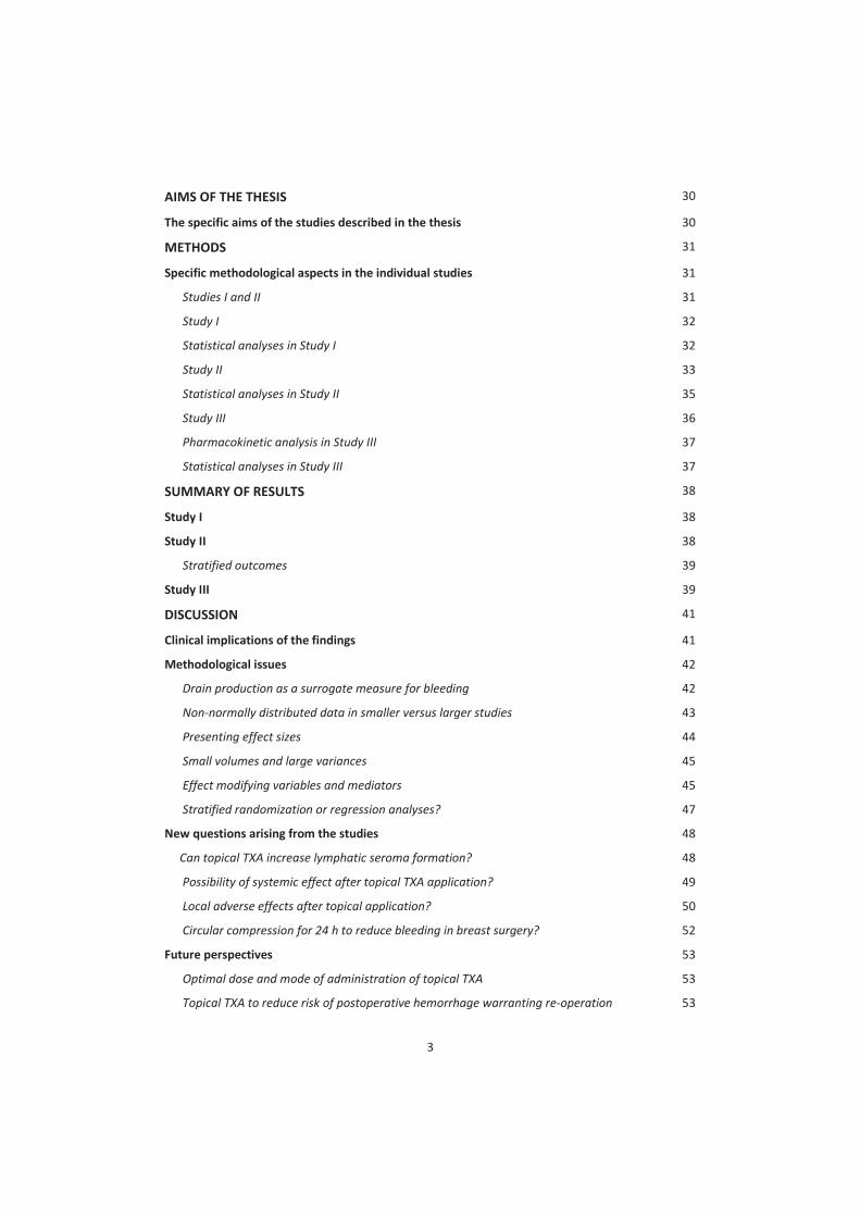
3
AIMS OF THE THESIS 30
The specific aims of the studies described in the thesis 30
METHODS 31
Specific methodological aspects in the individual studies 31
Studies I and II 31
Study I 32
Statistical analyses in Study I 32
Study II 33
Statistical analyses in Study II 35
Study III 36
Pharmacokinetic analysis in Study III 37
Statistical analyses in Study III 37
SUMMARY OF RESULTS 38
Study I 38
Study II 38
Stratified outcomes 39
Study III 39
DISCUSSION 41
Clinical implications of the findings 41
Methodological issues 42
Drain production as a surrogate measure for bleeding 42
Non-normally distributed data in smaller versus larger studies 43
Presenting effect sizes 44
Small volumes and large variances 45
Effect modifying variables and mediators 45
Stratified randomization or regression analyses? 47
New questions arising from the studies 48
Can topical TXA increase lymphatic seroma formation? 48
Possibility of systemic effect after topical TXA application? 49
Local adverse effects after topical application? 50
Circular compression for 24 h to reduce bleeding in breast surgery? 52
Future perspectives 53
Optimal dose and mode of administration of topical TXA 53
Topical TXA to reduce risk of postoperative hemorrhage warranting re-operation 53

4
Synergistic effect of topical tranexamic acid and topical adrenaline 54
Use of topical TXA on superficial wounds 54
Mechanisms behind potential adverse effects of topical tranexamic acid 55
CONCLUSIONS 57
REFERENCES 58
CORRECTIONS 71
PAPERS I-III 72

5
ACKNOWLEDGEMENTS
So many people have contributed in so many ways. Thank you all! Particular thanks to:
-Shosuke and Utako Okamoto; the inspiring Japanese husband- and wife-team who almost 70 years
ago set out to identify a chemical compound which could reduce bleeding. In memoriam.
-Jean Wong et al., for their published first randomized clinical trial on instilling tranexamic acid into
knee joint arthroplasties in 2010, which inspired this research.
-My colleague plastic surgeons Katrin Sneve, Carlos Gonzales and co-author Håvard Nordgaard who
enthusiastically agreed to introduce a new prophylactic routine of moistening our surgical surfaces
with tranexamic acid in 2012; the many plastic surgeons of Norway who have become active
believers, and particularly Helge Roald and Erling Bjordal who have spread the gospel internationally.
- Sven Erik Gisvold, anesthesiologist, Mentor and Grand Old Man, and former leader of the Regional
Ethics Committee, who during the design phase of our first trial tipped me off that someone else at
Hilde Pleym - had done research on tranexamic acid. Gisvold also volunteered to monitor
all our clinical studies at a time when other monitors were not available.
-Hilde Pleym, head of the Department of Anesthesiology and the aforementioned researcher on
tranexamic a
research, as she assisted me in every possible way with all the hurdles of a first project. She then
agreed to be my main supervisor when the first project expanded into a PhD project.
-Olav Spigset, professor of clinical pharmacology. Golden birds nest in pairs, and Hilde and Olav
constitute another inspiring husband- and wife-
production is unmatched and his eye for detail almost unnerving. Hilde and Olav allow me great
personal freedom yet always respond to me within hours from anywhere in the world. Best advice in
research: Choose your supervisors wisely. I certainly have. Things could not go wrong with this
support and the road continues!
-Birger Endreseth, head of the Department of Surgery and back-up supervisor should golden bird
family issues have affected supervision. This did not happen. Birger has allowed me a framework for
research within the surgical department which I have greatly appreciated, and he is always available
for comforting conversation when needed.
-The Liaison Committee of the Central Norway Regional Health Authority and the Norwegian
University of Science and Technology for granting me a full PhD Scholarship. Thank you for this
6
opportunity! Thank you also to the Department of Circulation and Medical Imaging for all technical
assistance along the PhD course.
-The Unit of Applied Clinical R e help! This no-
cost assistance was essential for both the initiation and later stages of my research and everything
might have stalled without them. Ingrid Ingeborg Riphagen came running to my door to help me with
registrations of clinical trials in international databases and keeps things organized to this day; Berit
Marianne Bjelkåsen randomized our clinical studies, delivered ready-made sealed envelopes on
multiple occasions and organized electronic case report forms. Øyvind Salvesen and Grethe
Albrektsen took turns unveiling statistical mysteries and leading me by the hand through increasingly
complex analyses.
-The breast-
participating in our second clinical study and pulling off the best recruitment and follow-up of any
clinical study I know of. A particularly big thank you to co-authors Anne Irene Hagen, Heidi Sæther
Østbyhaug, Sverrir Olafsson and Berit Kvalsund.
-Aleris Medical Centre for embracing the use of tranexamic acid and supporting the study on
abdominoplasties through the Aleris Research Grant. Particular thanks to co-author and mentor Ivan
Pavlovic, and to Christer Mohn and Stig Tyvold for their accommodating attitude and help.
-Jiayin Liu and Solfrid Hegstad at the laboratory facilities at the Department of Clinical Pharmacology
for developing a test to measure low serum concentrations of tranexamic acid - especially for us!
-Brita Pukstad, Tuva Eikebrokk, Brita Vassmyr, Caroline Gravastrand, Unn Granli, Patricia Mjønes og
Alexandra Kepka for all their brilliant work and assistance in the fourth study on topical toxicology of
tranexamic acid which is not included here but is ever so important for further research.
-Haukeland University Hospital and the Burn Unit - my ideas regarding tranexamic acid evolved while
I worked with you in 2011. Halvard Vindenes sent me to a Friday lecture regarding the CRASH study,
as I had mentioned tranexamic acid. A study has been planned for topical use in burn patients and I
look forward to further cooperation with Hans Christian Sylvester-Jensen, Stian Almeland and
Ragnvald Brekke.
- Reidar, Åsne and Eira: Reidar has been a major collaborator since the day we met and my 24-7
supervisor throughout the project and remains my pick among men for so many reasons. And we
smugly look at our fun, brilliant daughters and conclude heredity . My favorite
people happen to be within my nearest family! How lucky is that!

7
ABBREVIATIONS
AC axillary lymph node clearance
AKF The Unit for Applied Clinical Research
AMCHA 4-aminomethyl-cyclohexane-1-carboxylic acid
ATACAS Aspirin and Tranexamic Acid for Coronary Artery Surgery
BART Blood Conservation Using Antifibrinolytics in a Randomized Trial
CI confidence interval
Cl clearance
CNS central nervous system
CSF cerebrospinal fluid
EACA epsilon-aminocaproic acid
EudraCT European Clinical Trial Database
FDA Food and Drug Administration
HK High molecular weight kininogen
iv intravenous
ln logarithmic (ex)
log-concentration
NorCRIN Norwegian Clinical Research Infrastructure Network
OR odds ratio
PAI plasminogen activator inhibitor
SD standard deviation
SNB mastectomy with sentinel node biopsy
SM simple mastectomy
t-PA tissue-type plasminogen activator
TXA tranexamic acid
u-PA urokinase-type plasminogen activator
VAS Visual analogue scale

8
LIST OF PAPERS
Paper I
Ausen K, Fossmark R, Spigset O, Pleym H. Randomized clinical trial of topical tranexamic acid after
reduction mammoplasty. British Journal of Surgery 2015; 102: 1348 1353.
Paper II
Ausen K, Hagen AI, Østbyhaug HS, Olafsson S, Kvalsund BJ, Spigset O, Pleym H. Randomized clinical
trial of topical moistening of mastectomy wounds with diluted tranexamic acid to reduce bleeding.
British Journal of Surgery Open; in press.
Paper III
Ausen K, Pleym H, Liu J, Hegstad S, Nordgaard HB, Pavlovic I, Spigset O. Serum concentrations and
pharmacokinetics of tranexamic acid after two means of topical administration in massive weight
loss skin-reducing surgery. Plastic and Reconstructive Surgery 2019;143(6): 1169e-1178e.

9
INTRODUCTION
Bleeding is inevitable in surgery but should be kept to a minimum. All surgical bleeding implies the
use and change of bandages, swelling and bruising. Larger bleeding may provoke wound ruptures or
require cumbersome surgical drains with the associated possible risk of infection1,2. Excessive
bleeding leads to anemia and increased postoperative morbidity, and can ultimately be fatal. The
safety of blood transfusions has greatly increased in the last decades but allogeneic blood is not
harmless3. Moreover, banked blood is costly and in limited supply4. Strategies to minimize
postoperative anemia and the use of allogeneic blood include preoperative detection and treatment
of anemia or coagulopathies, optimizing per- and postoperative homeostasis, and applying a
meticulous surgical technique aided by mechanical and pharmacological means of hemostasis5,6.
Local injection of adrenaline along wound edges induces vasoconstriction and hence hemostasis.
Topical hemostatic agents such as cellulose, collagen, fibrin, thrombin, chitin and synthetic polymers
all promote clotting7. These agents are costly and mostly applicable for small surface areas only, as
excessive triggering of coagulation may lead to unwanted thrombosis.
Despite adequately performed hemostasis during surgery, secondary bleeding may occur if
fibrinolysis exceeds coagulation and when the vasoconstrictive effect of adrenaline wears off.
Antifibrinolytic agents prevent clotted blood from dissolving and may reduce the risk of secondary
bleeding. Antifibrinolytics presently represent the only pharmacological means of a general reduction
of bleeding with an acceptable safety profile8. The synthetic low-cost drug tranexamic acid (TXA) is
the most potent of the antifibrinolytic drugs currently available. Intravenous (iv) use reduces both
measurable blood loss and transfusion needs with approximately one third9. It is routinely utilized
prophylactically in surgery with high volume bleeding, particularly in cardiac and orthopedic joint
replacement surgery10.
A drug which prevents bleeding could theoretically increase the risk of thrombosis. Several large
multicenter studies have not seen an increase in thromboembolic events after TXA administration11-
14, while other publications suggest that a risk may be present15,16. TXA is also associated with a dose-
dependent increase in postoperative seizures in cardiac surgery17. Concern regarding potential
systemic adverse events may dissuade widespread and general use.
A minimization of bleeding is desirable also in moderate- to low-bleed surgery to reduce patient
discomfort and costs associated with follow-up, drains and bandages. A prophylactic measure to
reduce bleeding should ideally be low-cost, safe, quick and simple. In contrast to the previously

10
mentioned topical hemostatic agents, TXA can easily be diluted to a volume large enough to topically
treat even very large wound surfaces. Topical application of TXA can provide a sufficiently high drug
concentration at the site of the wound and an assumed low systemic concentration, hence with a
low risk of systemic adverse effects. Topical use is off-label but has been published from mainly
orthopedic and cardiac surgery with reduction in postoperative bleeding and transfusion needs
comparable to iv administration18,19. Most published studies on topical use of TXA describes the
application of a bolus into a confined space such as a joint or the mediastinum, prolonged irrigation,
or the superficial application of a soaked medium19.
The surgical surfaces of plastic surgery are readily available for manual manipulation. In 2012, the
Section for Plastic and Reconstructive Surger
prophylactic procedure where surgical wounds were moistened with 20 ml of TXA 25 mg/ml right
before wound closure. A video demonstrating our method can be found here: https://youtu.be/-
8MAE3NAHfQ. We postulated that this simple manual smearing, leaving a thin film of fluid only,
could reduce postoperative bleeding. Proper investigations were however needed to assess effect
and potential adverse events. As plastic surgery may involve large wound surfaces, e.g. skin reducing
surgery after major weight loss, we also wanted to explore to what extent topical use of TXA could
result in significant systemic absorption. These questions constitute the rationale for this thesis.
11
BACKGROUND
An overview of the physiology of blood coagulation and fibrinolysis
After tissue injury, a balance between coagulation the formation of blood clots and fibrinolysis
the dissolving of blood clots is needed to prevent blood loss yet maintain circulation. Injury will
damage small blood vessels with subsequent exposure of the subendothelial collagen. This collagen
exposure triggers the intrinsic (contact activation pathway) coagulation cascade, as contact between
exposed collagen and circulating platelets and coagulation factors initiates autoactivation of factor
XII and platelet activation. The damaged endothelium allows for release of tissue factor from
subendothelial tissue, which triggers the extrinsic (tissue factor pathway) coagulation cascade. Both
the intrinsic and extrinsic coagulation cascades terminate at the conversion of the inert prothrombin
to active thrombin, which proteolytically cuts fibrinogen into small fibrin monomers. Thrombin also
activates factor XIII, which catalyzes the crosslinking of the fibrin monomers into an insoluble clot
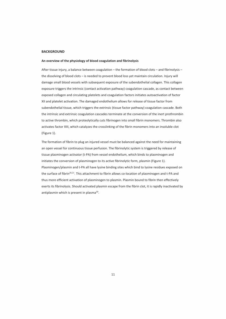
(Figure 1).
The formation of fibrin to plug an injured vessel must be balanced against the need for maintaining
an open vessel for continuous tissue perfusion. The fibrinolytic system is triggered by release of
tissue plasminogen activator (t-PA) from vessel endothelium, which binds to plasminogen and
initiates the conversion of plasminogen to its active fibrinolytic form, plasmin (Figure 1).
Plasminogen/plasmin and t-PA all have lysine binding sites which bind to lysine residues exposed on
the surface of fibrin20,21. This attachment to fibrin allows co-location of plasminogen and t-PA and
thus more efficient activation of plasminogen to plasmin. Plasmin bound to fibrin then effectively
exerts its fibrinolysis. Should activated plasmin escape from the fibrin clot, it is rapidly inactivated by
antiplasmin which is present in plasma20.
12
Plasminogen and its many functions
The zymogen (enzyme precursor) plasminogen is a ubiquitous substance in the human body. Its
activated version, plasmin, has a broad proteolytic capability beyond mere fibrinolysis and is involved
in multiple physiological and pathophysiological processes, as reviewed by Miles et al.22. In addition
to fibrin, extracellular matrix and a variety of cells express surface receptors for both plasminogen
and plasminogen activators, and free plasminogen in plasma can also be activated. Plasminogen has
Fig.1. Schematic representation of the coagulation cascade and the fibrinolytic system. The coagulation
cascade (blue arrows) can be activated during hemostasis via the intrinsic pathway (contact system; red
arrows) or the extrinsic pathway (tissue factor system; gray arrows) that ultimately converge on the
common pathway of coagulation. Both pathways lead to the activation of factor X and subsequently of
thrombin, which is required for the conversion of fibrinogen into fibrin and for activation of factor XIII. The
fibrin clot is cross-linked and stabilized by factor XIII. Fibrinolysis (green arrows) is activated at the same
time as the coagulation system but operates more slowly and is important for the regulation of
hemostasis. During fibrinolysis, plasminogen is converted into plasmin that degrades the fibrin network.
Coagulation fac
activated form; HK, high molecular weight kininogen; uPA, urokinase plasminogen activator; tPA, tissue
plasminogen activator. Reprinted with permission from Loof TG et al. Front. Cell. Infect. Microbiol. 2014.

13
multiple lysine binding sites which facilitate its interactions with other substances; one strong site
and several weaker sites23. The strong site provides high affinity binding to lysine residues and
facilitates the docking to fibrin, extracellular matrix and cell surfaces. The plasminogen activator
urokinase (u-PA) is present both in blood and extracellular matrix. It may bind to cell receptors
adjacent to docked plasminogen and promote its activation to plasmin. Thus cell surfaces are
provided with activated plasmin and proteolytic properties which facilitate multiple functions such as
inflammation, cell migration and wound healing24. Should activated plasmin escape from its docking
site, rapid inactivation by plasma antiplasmin is also mediated through the strong lysine binding site.
Mangel et al. (1990) observed that free plasminogen in plasma could undergo an exceptional
conformational change from closed to open structure if one of its weaker lysine binding sites were
occupied by a ligand23. This free-floating open form of plasminogen can bind to and be activated by
circulating u-PA. The resulting circulating plasmin may allow for intravascular fibrinolysis21.
Physiological inhibition of fibrinolysis is either by means of plasminogen activator inhibitors (mainly
PAI-1) or by the inhibition of plasmin itself by antiplasmin25.
Pharmacological inhibitors of fibrinolysis: Serine protease inhibitors and lysine analogues
Plasmin belongs to a large group of proteolytic enzymes classified as serine proteases due to the
common presence of the amino acid serine at their protease active sites. Serine protease inhibitors
block these active sites and thus inhibit the function of all serine proteases, but the different
inhibitors may have a varying and dose-dependent affinity for a specific protease. Serine protease
inhibitors are endogenous to eukaryote organisms. Exogenous serine protease inhibitors may have
various effects across species. The bovine serine protease inhibitor aprotinin has been widely used to
inhibit plasmin and thus fibrinolysis in humans, see later.
Plasminogen bound to fibrin or cells is easily activated by fibrin-bound t-PA or cell-bound u-PA , while
plasminogen in solution is not so easily activated as it needs to undergo a conformational change to
be accessible for activation in solution24. Lysine analogues competitively inhibit the lysine binding
sites on both plasminogen, plasmin and the plasminogen activators, thus preventing their binding to
fibrin/cells and their respective activations20,26-28. This blocking of lysine binding sites both reduces
the activation of plasminogen to plasmin, and it prevents activated plasmin from docking to fibrin
and exerting its proteolytic effect20 (Figure 2). However, when hyperfibrinolysis is triggered and
ongoing, and antiplasmin is depleted, systemically administered lysine analogues may bind to the
lysine binding sites of circulating plasminogen which induces its conformational change and allows
for activation of plasminogen in plasma by u-PA. Antifibrinolytics may thus enable activation of

14
circulating plasminogen to plasmin in a setting where antiplasmin both is scarce and may fail to
neutralize circulating plasmin as the target lysine binding site is blocked29. This has been proposed as
a possible mechanism behind the lack of effect and even possible negative impact of late
administration of lysine analogue in trauma29. Two synthetic lysine analogues have so far been
commercially available: -aminocaproic acid (EACA) and the 6-10 times more potent tranexamic acid
(TXA). TXA, but not EACA, has also been found to inhibit the active site of u-PA30. As u-PA and its
receptor may be involved in complex systems such as wound healing, cell migration and adhesion,
this inhibition may have effects beyond those of the fibrinolytic system31,32.
Fig. 2. Simplified antifibrinolytic acion of tranexamic acid. Normally, plasminogen binds to fibrin at a lysine binding site and is converted in the presence of tissue plasminogen activator (t-PA) to plasmin. Tranexamic
acid blocks the lysine binding site and prevents access of plasminogen to the fibrin molecule. Tranexamic acid may however also block the binding between t-PA and plasmin as this is also a lysine binding site. Reprinted with permission from Dunn CJ & Goa KL; Drugs. 1999.

15
The emergence of modern surgery and blood preservation
Surgery means inflicting tissue injury with its associated bleeding. Advances in surgery have been
closely linked to the ability to control or replace blood loss. From the earliest mechanical means of
hemostasis such as compression, tourniquets, simple cautery and ligation33, an expanding
armamentarium of refined mechanical, thermal and pharmacological means of blood preservation
has evolved.
Elective surgery was spurred in the latter half of the 1800s both by the introduction of chloroform
and ether for anesthesia34 and the establishment of the antiseptic principles35. The clotting of
blood was also extensively investigated36. Various materials such as gelatin and cellulose had been
found to enhance clotting33, while other substances such as snake venoms and leech extract could
dissolve clots37. Both fibrin and thrombin had been identified by the late 1800s and purified by the
early 1900s38, enabling the use of fibrin and thrombin as topical hemostatic agents. Although many
factors involved in the formation of fibrin had been identified, a thorough understanding of the
interaction between them had not been established until the coagulation cascade was postulated by
MacFarlane in 196439,40. The initiation of the cascade resulting from blood coming into contact with
structures designed to initiate coagulation, as well as thrombin and fibrin sealants41.
Blood transfusions
Methods for safe blood transfusion emerged in the first half of the 1900s as a response to the
desperate needs of ongoing wars linked with advances in science and technology42. Although
successful blood transfusions had been performed before the turn of the century, Landsteiner
on of blood
groups and the testing of their compatibility through cross-matching43. Blood grouping prior to
transfusion did however not become widespread until the 1920s42. The identification of coagulation
factors and their association with hemostatic disorders led to targeted substitutional therapies using
plasma-derived concentrates44. The established transfusion regimes allowed for immense surgical
developments through the 60s and 70s. As frontier procedures such as open heart surgery and joint
replacement surgery became mainstream, the limited access to banked donor blood struggled to
meet the increasing demand20. Transfusion reactions still occurred as meticulous subgroup matching
beyond the main blood groups was not feasible in emergency settings. Allogeneic blood transfusions
were also found to be associated with an increase in patient morbidity45-47, possibly due to
immunomodulatory effects48. Unrecognized blood-borne viral infections such as hepatitis C in the

16
1970s and the emerging AIDS epidemic of the 1980s also grimly illustrated that blood transfusions
and plasma-derived substances could have deleterious consequences49. This spurred renewed
interest in pharmacological blood-preserving measures.
Pharmacological means of blood preservation
Adrenaline had been isolated and purified by 190050 and was shortly thereafter shown to have
vasoconstrictive and hemostatic properties in surgery, thus representing the first pharmacological
means of hemostasis51. Its use was however debated among surgeons well into the 1970s52,53.
Clinical use of the anticoagulants heparin54 and warfarin55 was established by the 1960s56.
Transfusion of plasma could quickly reverse the effect of anticoagulants, and protamine sulfate and
vitamin K had also been recognized as pharmacological antidotes57-59. The synthetic vasopressin
analogue desmopressin was in the late 1970s found to both increase the level of circulating
coagulation factors and improve platelet dysfunctions and was thus efficient in mild hemostatic
disorders of various origins60,61. Recombinant coagulation factors for hemophilia were available by
the late 1980s62.
Blood preservation in patients without bleeding disorders
The pharmacological means of reducing bleeding in patients with various coagulation dysfunctions
do not necessarily benefit patients without such dysfunctions. Off-label administration of
recombinant coagulation factors to non-hemophilic patients has had little documented effect and
may even promote thromboembolic events63-65. Whether desmopressin has any hemostatic effect in
patients with no coagulation dysfunctions also remains uncertain66. Use of procoagulants in surgery
has mainly been limited to topical application onto bleeding sites, as systemic administration is
considered likely to involve an unacceptable risk of thromboembolic events. Fibrinogen concentrate
has received renewed attention as a possible universal hemostatic agent, but adequate
documentation on both effect and possible adverse events are still lacking67. Thus, the only
systemically administered pharmacological intervention to universally reduce bleeding with an
acceptable safety profile in patients without bleeding disorders is currently the antifibrinolytic drugs.

17
Enzymes, their activators and their inhibitors: The emergence of fibrinolytic and antifibrinolytic
drugs
W.S. Tillett demonstrated in 1933 that broth cultures of hemolytic streptococci could both dissolve
established fibrin clots in human plasma and prevent clotting when added to unclotted plasma68. The
active agent was initially named fibrinolysin. Tillett also observed that the fibrinolysis initiated by the
added fibrinolysin occurred faster in experimental fibrinogen preparations than in human plasma,
thus postulating that plasma might also contain antifibrinolytic substances69. J.H. Milstone (1941)
observed that fibrinolysin would only exert its fibrinolysis on purified fibrin clots in the presence of
human plasma, but not in the presence of human serum70. He postulated a component in human
plasma necessary for the initiation of streptococcal fibrinolysis, which was initially termed the lytic
factor. In 1944, both M.H Kaplan and L.R Christensen demonstrated that this lytic factor was a serum
enzyme, similar to trypsinogen, present in plasma in an inactive precursor state71,72. The lytic factor
could be transformed to its active fibrinolytic form catalyzed by fibrinolysin, similar to how
trypsinogen was transformed to its proteolytic form trypsin catalyzed by enterokinase. Fibrinolysin
was therefore not a fibrinolytic compound in itself but an activator, a kinase. Christensen and
MacLeod proposed to name the precursor variant of the lytic factor plasminogen and its proteolytic
form plasmin, while the catalytic streptococci-derived plasminogen activator was named
streptokinase73. MacFarlane (1946) subsequently proposed the term antiplasmin for the
antifibrinolytic substance in plasma74.
The discovery of streptokinase led to widespread utilization to lyse clots and inflammatory fibrinous
exudates of multiple conditions75. Associated publications of possible allergic reactions increased the
awareness of the potential antigenic potential of foreign proteins used as biological drugs76. The
search continued for a human-derived equivalent product for fibrinolysis.
MacFarlane and Pillig (1947) published the discovery of the presence of plasminogen activator in
urine77, which was named urokinase by Sobel et al. in 195278, later renamed urokinase-type
plasminogen activator (u-PA). Fisher (1946) proposed that an activator of plasminogen might be
present in tissue79 and Astrup and Permin (1947) confirmed that various animal tissue could indeed
activate plasminogen. Astrup and Albrechtsen (1957) managed to quantify the contents of tissue-
type plasminogen activator (t-PA) in different human tissue80 and could compare concentration to
that of previously measured still-standing postmortem-blood81. As this stagnant blood contained a
much higher concentration of t-PA than the tissues themselves, a plasminogen activator derived
from the endothelium of the vascular bed was proposed by Todd (1959)82. Only in 1980 did Rijken et
al.83 prove that this vascular plasminogen activator84 and plasminogen activators in blood and tissue

18
were identical, but different from u-PA. Urine was easily obtained and purification of u-PA from
human urine had been achieved by the late 1950s85. Obtaining t-PA was more challenging; purified t-
PA had been obtained by 1979 from perfusion of the lower leg vessels of human cadavers84, and
extracted from large volumes of human uterine tissue86.
All versions of plasminogen activators can dissolve clots and fibrous exudates. Streptokinase, u-PA, t-
PA and recombinant versions thereof have all been used as intravenously administered thrombolytic
drugs87. All were effective in restoring circulation and reducing ischemic tissue damage, but systemic
treatment with large doses involved a including potentially lethal
intracranial hemorrhage. Modern endovascular techniques have however allowed for intravascular
targeted treatment of the thrombus (catheter-directed thrombolysis) which has significantly reduced
the risk of bleeding88.
The discovery and isolation of enzymes led to parallel research on enzyme inhibitors. Isolation of
streptokinase and later urokinase provided a possibility for scientific evaluation of agents capable of
preventing fibrinolysis. A standardized experimental kinase-induced fibrinolysis could be strongly or
weakly inhibited according to agent and/or concentration. The search for antifibrinolytic agents was
thus initiated89.
Serine protease inhibitors the naturally occurring biological drugs
The presence of naturally occurring inhibitors of proteases in plasma had been demonstrated in the
early 1900s90. A kallikrein inactivator was isolated from both bovine parotid and lung tissue by Kraut
et al. in 192891, and a potent trypsin inhibitor isolated from bovine pancreas tissue by Kunitz in
193692 was found to be almost identical in structure to the kallikrein inactivator91. Northrop, Kunitz
and Herriot had succeeded in isolating animal-derived proteolytic enzymes as pure, crystalline
proteins by the 1930s93. The presence of plant-derived trypsin inhibitors was demonstrated in beans
by Ham and Bowman independently in 1944/4594,95. Mirsky demonstrated in 1944 that trypsin
inhibitors derived from both bovine pancreas and soy beans could also prevent fibrinolysis96.
Bergmann (1937) proposed that enzymes should be classified according to their structural
characteristics. The proteolytic enzymes kallikrein, trypsin, plasmin and several others were classified
as serine proteases 97. The
serine proteases were all inhibited by a variety of naturally occurring polypeptides the serine
protease inhibitors. A crystalized soybean trypsin inhibitor published by Kunitz in 194598 was used to
19
reverse fibrinolysis in vivo by Heuson in 195899. Plant-derived protease inhibitors were however
weak antifibrinolytics100 and were abandoned in favor of the bovine derived protease inhibitors.
Aprotinin
The bovine-derived serine protease inhibitor aprotinin, originally marketed as Trasylol, was initially
isolated from bovine pancreatic tissue98, but commercial preparation has since been done from
bovine lung tissue. Aprotinin was originally marketed to alleviate the auto-digestion resulting from
the leaking proteolytic enzymes of pancreatitis101. As trypsin inhibitors had been described to inhibit
fibrinolysis, reports on the use of aprotinin to treat acute fibrinolysis rapidly emerged particularly
from cardiac surgery102-104 but received little attention. The serine protease kallikrein is a potent
mediator of inflammation, and in a protocol designed to demonstrate anti-inflammatory and lung-
preserving qualities of aprotinin in open-heart surgery, van Oeveren and Royston in 1987
demonstrated an unexpected profound effect of high dose aprotinin on blood loss and transfusion
needs105. The use of aprotinin as a routine prophylactic measure to reduce bleeding in cardiac
surgery spread rapidly thereafter.
Aprotinin like the naturally occurring plasmin inhibitors in serum quickly but reversibly binds to
the active site of free plasmin in serum. Aprotinin has little interaction with plasmin when plasmin is
bound to fibrin, but aprotinin can bind to tissue plasminogen activator (t-PA), thus preventing the t-
PA from binding to plasminogen and exerting its catalytic conversion of plasminogen to plasmin.
Aprotinin inhibits all serine proteases, but with varying affinity. It has high affinity to trypsin and
plasmin, intermediate to kallikrein, and only binds to plasma thrombin at high concentrations.
Aprotinin can therefore potentially influence platelet and complement activation, inflammation,
coagulation and fibrinolysis, and while low dose may prevent fibrinolysis, a higher dose is needed for
anti-inflammatory effect106. The exact mechanisms of the net blood-preserving qualities of aprotinin
are not understood in full detail107. As aprotinin is a bovine peptide, hypersensitivity can develop in
about 5% of patients within a few days to weeks of initial exposure and may still persist after 12
months in approximately 0.5-1% of patients108.

20
The withdrawal of aprotinin
In 2006, the U.S. Food and Drug Administration (FDA) was alerted to a commissioned retrospective
study of 67 000 patients by one of the participating researchers; 30 000 patients had received
aprotinin while the others had received lysine analogues. The study had been withheld by Bayer, the
manufacturer of aprotinin who had commissioned the trial, as it suggested that aprotinin led to
increased risk of multiple adverse events109,110. 5-year follow-up also revealed an increased risk of
death111. A prospective randomized trial - Blood Conservation Using Antifibrinolytics in a
Randomized Trial (BART) 112 was commissioned by the FDA but terminated late 2007 due to a trend
towards increased mortality in the aprotinin group compared to lysine analogues, and Bayer
subsequently withdrew aprotinin from the market. Retrospective analyses were conducted in the
aftermath and seemed to confirm the increased mortality associated with aprotinin113,114, but the
withdrawal has been met with much controversy and questioning regarding selection bias in the
mentioned studies115-117. The ban has been partially lifted, as aprotinin may be the most powerful
inhibitor of fibrinolysis in procedures with particularly high volume bleeding and
hyperfibrinolysis118,119. However, in the wake of the withdrawal of aprotinin, the lysine analogues
have dominated as prophylactic antifibrinolytics in surgery.
Lysine analogues the synthetic drugs
The purification and crystallization of naturally occurring serine protease inhibitors allowed for
further analysis of their physiochemical structure, with the associated possibility of creating synthetic
selective inhibitors. Herriot (1940) demonstrated that a naturally occurring pepsin inhibitor
contained significant amounts of the amino acid arginine120, and Neurath and Schwert (1950)
demonstrated that amino acid esters were sensitive substrates and potential inhibitors for
proteolytic enzymes121. In Europe, Troll et al (1953) published that for plasmin in particular, lysine
and arginine esters seemed to be specific substrates122.
Meanwhile, in post-war Japan, the husband-and-wife-team Shosuke and Utako Okamoto had
targeted the hyperfibrinolysis of pathological bleeding. Assuming hyperfibrinolysis was caused by
excessive action of plasmin, they aimed at creating a synthetic antiplasmin and performed a
systematic screening of more than 300 chemical compounds for possible antifibrinolytic activity
through plasmin inhibition123. While lysine exerted a weak inhibition, the Okamotos found that a
chemically modified derivative of lysine, -aminocaproic acid (EACA), had a tenfold inhibitory effect,
mainly through the inhibition of plasminogen activation124. A patent claim for EACA was filed in 1953
and granted in 1957125. The Okamotos continued their search for more potent inhibitors of

21
fibrinolysis, and in 1961 patented 4-aminomethyl-cyclohexane-1-carboxylic acid, abbreviated
AMCHA125, of which the trans-stereoisomer was later discovered to carry the antifibrinolytic
properties126-128. This trans-AMCHA was named tranexamic acid (TXA) and had an approximately 6-
10-fold higher antifibrinolytic potency than EACA129,130. EACA and TXA were purely synthetic
substances and hence much cheaper to produce than aprotinin, and with less antigenic potential.
While aprotinin could only be administered intravenously, EACA and TXA could be administered both
intravenously, intramuscularly and orally.
Clinical use of lysine analogues
It had been long recognized that elective surgery carried a significant risk of postoperative
thromboembolic events, and that prophylaxis with heparin could reduce the risk131. Thus,
simultaneous use of lysine analogues and postoperative heparin could reduce the risk of
postoperative embolism with less increase in postoperative bleeding26. Initially, EACA and later TXA
was particularly used in patients with bleeding related to prostate and urological surgery, as urine
was known to be the source of the fibrinolytic plasminogen activator u-PA which could sustain the
local bleeding26. However, insoluble clots resulting from use of lysine analogues were reported to
lead to urinary retention, kidney infarction and even death from bilateral ureter obstruction132,133.
Enthusiasm was dampened, and use of TXA in larger surgery was henceforth little explored due to
fear of the thrombotic potential of lysine analogues134. Through the 1970s, use continued in prostate
surgery but was debated135. Lysine analogues were explored in the treatment of intracerebral
hemorrhage and prophylaxis of aneurysmal rebleedings136, but effect was questionable and potential
negative side effects in the central nervous system(CNS) were suggested137. Effect was however well
documented to reduce menorrhagia, and use in epistaxis, minor oral surgery, dental extractions,
tonsillectomy and cervical conisation was also recommended138.
The HIV epidemic of the mid 1980s spurred renewed interest in antifibrinolytics to avoid blood
transfusions. Aprotinin was documented by Oeveren and Royston (1987) to significantly reduce
blood loss in cardiac surgery105, and Vander et al. and Horrow et al. shortly thereafter demonstrated
similar effects from lysine analogues139,140. Although benefits were still debated141, prophylactic use
increased in cardiac surgery. In the mid 1990s, the benefits of antifibrinolytics were also recognized
in joint replacement surgery142-144. Henceforth, prophylactic use of antifibrinolytics became
widespread in surgery with high-volume bleeding, and after the withdrawal of aprotinin in 2007, TXA
has dominated as prophylactic antifibrinolytic drug.

22
Lysine analogues in hemophilia
Early research on lysine analogues suggested potential significant benefits in patients with congenital
or acquired coagulation deficits129,145. As lysine analogues were low-cost and could be administered
orally, there was much optimism regarding the potential prophylactic use in the hemophilic
population. Studies on continuous prophylactic oral use of TXA to prevent hemorrhagic episodes in
patients with hemophilia were few, inadequately powered and could not conclude regarding
effect146. Hematuria is one of the major challenges in severe hemophilia, but the use of lysine
analogues was deemed contraindicated in hematuria after the previously mentioned reports of
ureter obstruction146. Prophylactic use in larger surgery in hemophiliacs has henceforth been
inadequately explored.
Patients with coagulation deficits seem to benefit from on-demand use of TXA in conjunction with
smaller oral or nasal bleeds and prophylaxis in connection with dental surgery, but randomized
controlled trials are lacking147. Lysine analogues used as adjunct therapy along with factors
substitution can increase clot stability148 and thus potentially reduce the amount of coagulation
factors needed, and although this is clinically practiced, few controlled studies have been conducted
on the possible benefits of combining lysine analogues and coagulation factors146,148.
Systemic use of TXA
Pharmacokinetics
TXA is excreted largely unchanged via glomerular filtration; its plasma elimination half-life is
approximately 80-120 minutes and prolonged in renal failure. After iv administration, 95% can be
recovered from urine within the first 24 hours. TXA is retained in tissues with an estimated tissue
half-life of 17 h129, and the tissue retention may account for the non-recovered drug in urine at 24
hours. Oral bioavailability is approximately 35-40%130,149. Time from oral ingestion to maximal plasma
concentration is approximately 3 hours129. TXA given iv diffuses quickly to joint synovium; elimination
half-life in synovial fluid is approximately 3 hours, and synovial fluids therefore maintain therapeutic
levels longer than plasma150. TXA crosses the placenta but no teratogenic properties have been
identified130. Concentrations in breast milk is approximately 1/100 of that in plasma151. TXA crosses
the blood-brain barrier and concentration in the cerebrospinal fluid (CSF) is approximately 10% of
that in plasma, but this percentage may vary and peak CSF concentration may occur several hours
after peak plasma concentration152,153.

23
Dose-effect and concentration-effect relationships
In vitro studies suggest the minimum plasma concentration to significantly inhibit fibrinolysis to be
about 5 μg/ml in children and 10 μg/ml in adults26,154,155. Plasminogen inhibition is achieved at plasma
concentrations of 10-15 g/ml20,130, while a higher plasma concentrations can also inhibit the
plasminogen activators and plasmin itself26,30. In theory, a maximal suppression of both plasminogen
and its activators would increase hemostatic effect. Fibrinolysis in vitro is inhibited by more than 90%
at TXA plasma concentrations around 20 μg/ml, and a concentration of 100 μg/ml provides a 98%
inhibition as also the plasminogen activators are inhibited26.
The optimal dosing of TXA in vivo to reduce bleeding has been debated. Intravenous regimes have
been particularly varied in cardiac surgery, ranging from single doses, continuous infusions, or both,
from 1g to 20 g156 given over time intervals from 20 minutes to 12 hours119. While some have
advocated high-dose regimes in cardiac surgery157,158, maintaining serum concentrations above 100-
150 g/ml159, other studies do not find clinically significant differences in blood saving effect between
high- and low-dose regimes160-163. There is however no international consensus on dosing protocols
and specific definitions of high- and low-dose regimes. A classical high-dose regime yielding serum
concentrations between 100-150 g/ml is the dose recommended by Dowd et al.158 which was used
in the commissioned BART trial comparing adverse effects of aprotinin to that of TXA112. The BART
regime consists of a loading dose of TXA 30 mg/kg and then an ongoing infusion of 16 mg/kg/h.
Conversely, the lowest dose among the low-dose regimes in cardiac surgery is a loading dose of TXA
10 mg/kg with or without a continuous infusion of 1 mg/kg/h. TXA does not yield a linear dose-
response relationship, and there seems to be little added clinical benefit from iv doses exceeding 10-
20 mg/kg, which is recommended by the manufacturers151. While TXA administration in cardiac
surgery often consists of an initial bolus plus continued infusion throughout the procedure both due
to the use of cardiopulmonary bypass and the relatively long operating time, orthopedic
administration is typically a single bolus of TXA 10-20 mg/kg. Repeated administration may however
yield added benefits compared to single dose administration only, as the hyperfibrinolysis induced by
surgery may persist up to 24 hours164,165.
Different types of surgery are associated with varying magnitudes of bleeding. Although the
reduction in bleeding resulting from iv TXA as measured in ml varies between procedures, the
proportional reduction is rather constant with an approximate reduction of one third in both
measured bleeding volume and transfusion needs9,166. The incidence of explorative re-operations due

24
to hemorrhage in cardiac surgery was halved after iv TXA in two different meta-analyses119,167.
Orthopedic studies rarely report need for re-operation due to hemorrhage as a separate outcome.
Thromboembolic adverse events
A drug which prevents bleeding could in theory promote thromboembolic events, and this was a
major fear when use of TXA was initiated. After the introduction of antifibrinolytic drugs, anecdotal
reports of occluded grafts after coronary bypass168, increased incidence of myocardial infarction141
and pulmonary embolism/ deep venous thrombosis15 have maintained an uncertainty regarding this
potential adverse effect. Large multicenter studies investigating the effect of iv TXA in thousands of
arthroplasties13, trauma14, postpartum hemorrhage11, or cardiac surgery12 have not found an
association between TXA and thromboembolic events. However, case reports of thromboembolic
events maintain uncertainty15, and a recent large retrospective study by Myers et al. diverges from
the above mentioned studies and suggests an increase in venous thromboembolic events among
trauma patients receiving TXA16. Adequately powered studies to specifically address potential
adverse effects have been lacking. The Aspirin and Tranexamic Acid for Coronary Artery Surgery
(ATACAS) study was a multicenter prospective randomized study designed to investigate potential
adverse effects of TXA in high-
defined as advanced age, pulmonary/cardiac/kidney failure or obesity. The primary outcome was a
composite endpoint of death and thrombotic complications (nonfatal myocardial infarction, stroke,
pulmonary embolism, renal failure, or bowel infarction) within 30 days after surgery169. There was a
slightly lower incidence of an unfavorable outcome in the TXA group, and in particular no increase in
thromboembolic events. This trend persisted at a one-year follow-up of the population170. However,
former thromboembolic events or ongoing anticoagulation for whatever reason at the time of
surgery were exclusion criteria in this study population, and large randomized controlled trials on
the use of iv TXA in populations with known increased risk of thromboembolic events are still lacking.
Unease regarding this possibility persists, limiting routine prophylactic use of TXA to surgical
procedures associated with high-volume bleeding and transfusion needs.
CNS adverse events
As TXA so clearly reduced bleeding, administration to reduce re-bleeding after ruptured intracranial
aneurysms became widespread in the 1970s. Although re-bleeding was reduced, there was concern
regarding observed possible ischemic and thromboembolic events137. In 1981, a retrospective

25
analysis of ruptured intracerebral aneurysms which had received either a combination of low-dose
TXA in combination with aprotinin versus high-dose TXA alone found a significant increased rate of
cerebral ischemic complications in the high-dose TXA group137. The net benefit of prolonged use of
TXA after cerebral bleeding has been questioned171, while short-term treatment may be
advantageous172,173.
Potentially dramatic effects of lysine analogues coming into
direct contact with the CNS were reported in the early 1980s;
animal models investigating the effect of TXA in cerebral
hemorrhage found that TXA induced ischemia, and subdural
application of fibrin sealants containing TXA in animals evoked
epileptic convulsions174-176. Accidental intrathecal
administration of TXA has occurred on many occasions in
settings where vials of TXA were mistaken for bupivacaine for
spinal anesthesia because they were visually similar. This has
resulted in lower limb myoclonus, generalized seizures,
ventricular refractory arrhythmias and several deaths177-181.
When the blood-brain barrier is intact, iv TXA administration
renders the concentration in the CSF approximately 1/10 of that
in serum152. Ongoing intracranial bleeding would expose the CNS
to the serum concentration of TXA, and intrathecal
administration would expose the CNS to exceedingly high levels.
Yet reports from cardiac surgery indicate that even with an intact blood-brain barrier, TXA can
increase the risk of seizures182.
In the wake of the withdrawal of aprotinin from the marked in 2007, several studies were published
comparing the safety profiles of TXA vs. aprotinin. Martin et al. (2008) were the first to report the
significant association between administration of TXA and seizures in cardiac surgery182. Several
studies confirmed this observation183-186, and although overall incidence of seizures is normally in the
range of 0.5 3%, meta-analyses suggested that TXA was associated with at least a four-fold increase
in risk17,187. The increase in seizures seemed to be dose-dependent and was particularly seen in
settings with an increased risk of accumulation of TXA - such as open cardiac surgery where a
prolonged procedure also implies an extended continuous infusion, and in patients with renal failure
with subsequent delayed elimination. Benchmark studies such as the BART trial112 and the ATACAS169
study also found increased incidence of seizures in the TXA population. Both studies administered
Fig 3. Ampules of bupivacaine and
tranexamic acid. The similar size of ampules
led to a medication administration error
during spinal anesthesia.
Reprinted with permission from Hatch DM, Int Journ of Obst Anesth, 2016.

26
high-dose TXA regimes; the ATACAS study reduced their TXA administration from single dose 100
mg/kg to 50 mg/kg after awareness regarding the possibility of seizures was raised, yet this did not
significantly reduce the incidence of seizures in the TXA group. Seizures are generally associated with
an increased risk of death in the postoperative period170,186, albeit it has not been determined
whether this holds true for TXA-induced seizures.
Possible underlying mechanisms for the TXA-induced hyperexcitability was first proposed by
Furtmüller (2002)188, who demonstrated that TXA could bind antagonistically to GABAA receptors in
the CNS and thus prevent the inhibitory action of the neurotransmitter GABA. The studies by
Furtmüller were based on the observations of topical/intrathecal/hemorrhagic TXA exposure of the
CNS, thus representing high concentration exposures. The concentrations needed for GABAA receptor
inhibition were observed at drug concentrations in the mg/ml range, while CSF concentrations
obtained from therapeutic iv administrations in a setting with an intact blood-brain barrier should
normally not exceed low μg/ml levels.
Lysine analogues are chemically related to the amino acid glycine, and Lecker et al. (2012)153 showed
that TXA also acts as a competitive antagonist to the inhibitory effect of glycine via glycine receptors
in the CNS. Based on in vitro studies, CSF concentrations of approximately 15 μg/ml may be a
threshold value for a potentially excitatory effect, which is much lower than for the GABAA
mechanism. There may also be a synergy between the two means of blocking cerebral
inhibition153,189.
The CSF achieves approximately 10% of the TXA concentration of plasma, but this proportion may
vary152. TXA dosing regimens advocating the maintenance of plasma concentrations above 100
μg/ml for an extended period may therefore be at risk of reaching threshold concentrations in the
CSF. Sharma et al. have documented that high-dose administration as defined by a 30 mg/kg bolus
followed by 16 mg/kg/h infusion (The BART study dose for high-risk cardiac surgery) maintains a
plasma level above 100 μg/ml and therefore above the theoretical threshold for evoking seizures186.
A single bolus injection of 10-20 mg/kg would in all likelihood not be sufficient to reach the threshold
concentration in the CSF, but factors which may increase patient susceptibility are unexplored189.
Intravenous TXA in all surgery?
Based on the evidence reviewed in the previous sections, a single bolus of 10-20 mg/kg TXA as a
prophylactic measure at the initiation of surgery would seem like a safe prophylactic measure to
reduce surgical bleeding. However, an unease regarding potential unrecognized adverse events may

27
dissuade clinicians from using iv TXA in low- to medium-bleed surgical procedures. Intravenous TXA
as a routine pharmacological means of reducing bleeding has therefore mainly been adopted in
surgery with high-volume bleeding and potential high transfusion rates.
Topical use of TXA
Effect
Theoretically, direct application of TXA onto surgical wound surfaces would be expected to give high
local concentrations with limited systemic exposure, thereby reducing bleeding with a lowered risk of
systemic adverse events.
Topical application of TXA to reduce bleeding has been sporadically published since the 1970s190,191.
Sindet-Pedersen et al. demonstrated in the 1980s the hemostatic properties of a 50 mg/ml TXA
mouthwash in patients on anticoagulation and hemophiliacs undergoing dental extractions192,193.
During the 1990s, sporadic reports of alternative topical use emerged, such as moistened gauze on a
surgical wound194, and enema administration for intestinal bleeding195,196.
In 2000, De Bonis et al. published the first study in cardiac surgery where 100 ml of either 10 mg/ml
TXA or 0.9% saline was poured into the pericardium and mediastinal tissue prior to sternal closure,
with a significant 22% reduction in postoperative drain production197. Further studies from cardiac
surgery have been few and not unambigous198-202, although meta-analyses suggests a beneficial
effect203,204. The use of cardiopulmonary bypass in cardiac surgery leads to an increased peroperative
triggering of the fibrinolytic system, and pre/peroperative iv administration has been the route of
choice to ensure perioperative antifibrinolytic effect. The increasing awareness of associated seizures
has however spurred renewed interest in topical alternative use in cardiac surgery205.
Orthopedic surgery has to a much larger extent explored the field of topical TXA. Krohn et al.
(2003)206 irrigated the wound of low-back pain patients undergoing lumbar fusion with 50 ml of 10
mg/ml TXA for 2-5 minutes prior to closure and found a significant 40% reduction of postoperative
blood loss compared to placebo. Wong et al. (2010)207 published the first arthroplasty study in knee
joint replacement surgery, exposing the open joint to 100 ml of either 15 or 30 mg/ml TXA for five
minutes, suctioning away the excess fluids prior to closure. Wong et al. found a significant decrease
of postoperative bleeding and a maintenance of higher Hb levels in the TXA groups, with no
significant difference between the different concentrations. Instilling various concentrations in
conjunction with hip and knee arthroplasties has since been extensively published along with an

28
increasing number of meta-analyses demonstrating that the reduction in bleeding after topical
administration of TXA is non-inferior to iv use19,208-211.
Publications regarding topical use in spine surgery have been scarce since the original publication by
Krohn206, but there is an increasing awareness of the possibility of topical use reflected in a notable
increase in publications, systematic reviews and meta-analyses based on the rather few studies
available212-217. It is however worth noting that none of the authors mention the dangers of
intrathecal or topical contact of TXA with the CNS, as dural lesions and possibilities of leakage could
be an issue in spine surgery.
Topical use of TXA outside orthopedic, cardiac and spine surgery has until now only been anecdotally
published, as demonstrated by a recent review by Montroy et al.218. Topical use is off-label, and there
is no consensus as to the optimum dose and mode of application. Most published methods of
application involve instilling a bolus into a confined space, prolonged irrigation, or application of a
moistened carrier19, and concentration in the applied fluids varies from 1 to 100 mg/ml, without any
apparent dose-response relationship218. Arthroplasty studies have generally used topical solutions
with concentrations in the range of 15-100 mg/ml219, while the studies in cardiac surgery have
generally used somewhat weaker solutions with concentrations between 10 and 20 mg/ml203.
Systemic concentrations and adverse effects
Although it is assumed that topical application of TXA will result in low systemic concentrations, few
studies have actually investigated the systemic absorption and then only at a single postoperative
time point193,197,207,220, rendering peak levels uncertain. Pharmacokinetic studies after administration
of a 2 g TXA enema221 or a 2-minute mouth rinse with TXA 50 mg/ml192 resulted in systemic
concentrations below 2 g/ml. Wong et al. irrigated the knee joint with TXA after arthroplasty for 2-5
minutes207. Serum samples taken one hour after tourniquet release showed that irrigation with TXA
15 mg/ml (at a volume of 100 ml) yielded a concentration of 4.5 g/ml , while a double concentration
of 30 mg/ml (at a volume of 100 ml) led to a concentration of 8.5 g/ml. It seems reasonable that
systemic absorption after topical application of a given volume should be proportional to the drug
concentration. Well-vascularized tissue may also absorb more of a topically applied drug than less
vascularized tissue. Whether topical application can lead to systemic concentrations above the
therapeutic threshold value of 5 g/ml in children or 10 g/ml in adults needs clarifying.

29
Local adverse effects from topical use
The potential local toxicity of topical TXA has been insufficiently explored. The dramatic effect of
lysine agonists on inhibitory neuroreceptors in the CNS illustrates the possibility of unexpected
adverse effects of an apparently safe drug17,177. As topical TXA has become most widespread in
orthopedic arthroplasty, studies regarding potential local toxicity have mainly been conducted on
cartilage, tendon and synovial tissue. Published studies on the effect of topical TXA on chondrocytes
and cartilage tissue suggest a threshold for toxicity of around 25 mg/ml given at least 3 hours of
exposure, and cells embedded in a natural (cartilage) or artificial (hydrogel) matrix are more
resilient172,222-224. Marmotti et al. found that chondrocytes, tenocytes and synoviocytes were not
affected by a two-week exposure to 7 mg/ml TXA225. In other tissues, Furst et al.226 reported 50%
viability in human fibroblasts exposed to 100 mg/ml TXA and 65% viability after exposure to 50
mg/ml for 100 minutes, while Bergenholtz et al.227 found that TXA concentrations of 12 mg/ml or
lower did not prevent wound healing in vitro of incisional wounds through palatal mucosa from cats
although re-epithelialization took place with abnormal keratinocyte migration.

30
AIMS OF THE THESIS
assess the effect of a simple topical moistening of surgical wounds with TXA 25 mg/ml, to register
possible adverse events and assess the systemic concentration resulting from topical application. We
wanted to achieve this through clinical studies measuring the effect of the intervention on
postoperative bleeding and registering possible adverse events. We also wanted to compare the
systemic pharmacokinetics of TXA after topical administration to iv administration.
The specific aims of the studies described in the thesis were:
To investigate whether a single moistening of a surgical wound surface prior to wound
closure with TXA 25 mg/ml would reduce postoperative bleeding as measured by wound drain
production at 24 h using patients undergoing reduction mammoplasty and mastectomy as study
models (Studies I and II)
To investigate whether a single moistening of a surgical wound surface prior to wound
closure with TXA 25 mg/ml would influence late seroma formation in patients undergoing
mastectomy (Study II)
To investigate whether a single moistening of a surgical wound surface prior to wound
closure with TXA 25 mg/ml would be associated with an increase in postoperative complications or
adverse effects (Studies I and II).
To investigate the degree of systemic absorption of TXA after two different means of topical
application in patients having large wound surface areas after abdominal major weight loss skin
reducing surgery, and compare the systemic TXA concentrations achieved by these two methods
with standard intravenous prophylactic administration of 1 g of tranexamic acid in hip replacement
surgery (Study III).

31
METHODS
All studies were prospective clinical studies on patients receiving topical TXA. Studies I and II were
randomized double-blinded clinical intervention trials to assess the effect of moistening a surgical
wound surface with TXA, while Study III was a pharmacokinetic study investigating systemic
concentrations of TXA after topical versus systemic administration. All studies were approved by the
Norwegian Medicines Agency and The Regional Committee for Medical Research Ethics, Mid Norway.
All participants gave written informed consent prior to inclusion. In all studies, patients who were to
receive topical TXA were not considered eligible if they 1) had a known allergy to TXA, 2) were
pregnant or nursing, 3) had a known history of a thromboembolic event, or 4) were less than 18
years old. Study I did not have a monitor protocol, while studies II and III underwent formal
monitoring in cooperation with the Norwegian Clinical Research Infrastructure Network (NorCRIN).
All studies were registered in ClinicalTrials.gov and in the European Clinical Trial Database (EudraCT).
Assistance with study registrations, conduction of computer-generated randomization, development
of electronic study forms and assistance on statistical analyses were provided by the Unit for Applied
Clinical Research (AKF), Norwegian University of Science and Technology NTNU. Statistical analyses
were performed using SPSS version 25 (IBM SPSS Statistics for Windows, IBM Corp., Armonk, NY,
USA). Values of p < 0.05 were considered statistically significant.
Specific methodological aspects in the individual studies
Studies I and II
Studies I and II were randomized clinical trials to evaluate the effect of moistening a surgical wound
surface with 20 ml of TXA 25 mg/ml (intervention) versus 20 ml of NaCl 0.9% (placebo) prior to
wound closure. Study I was conducted on patients undergoing bilateral reduction mammoplasty at
during the years
2013 2014. Study II was conducted on patients undergoing mastectomy with or without lymph
and Ålesund Hospital during the years 2016 2018. Computer generated randomization and
production of corresponding sealed randomization envelopes were performed by AKF, Norwegian
University of Science and Technology NTNU, who also held the randomization code. Preparation of
study solutions was performed by a nurse not otherwise connected to the operation. Patients and all
personnel involved in surgery and postoperative follow-up, data collection and statistical analysis
were blinded to the randomization. Primary outcome in both studies was drain production at 24 h
after surgery. Possible adverse events such as hemorrhage warranting re-operation, wound infection

32
or wound rupture were defined as secondary endpoints in both studies. All patients were contacted
approximately three months postoperatively to ensure correct registration of data and possible
adverse events.
Study I
Bilateral reduction mammoplasty was chosen for this study as it is one of the few surgical procedures
where the patient has two fairly identical wounds, allowing for a paired sample randomized
controlled study using a within-patient design. A total of 28 patients were included, 56 breasts in
total. In every patient, one breast was moistened with 20 ml of TXA 25 mg/ml and the other breast
with 20 ml of NaCl 0.9%. Breast specimen weight was recorded. As the patient served as her own
control, patient characteristics were not registered.
To ensure as homogenous wounds as possible when two surgeons operate one side each, the
surgeons switched sides and controlled the hemostasis of the opposite side prior to the application
of study fluid. Care was taken to ensure that no study fluid from one side came into contact with the
opposite side. The surgeon who applied the fluid onto one breast also performed the surgical closure
of that breast. Finally, based on the assumption that bleeding is correlated to the size of a wound and
using breast specimen weight as a surrogate measure of wound size, we could calculate both crude
and adjusted differences in drain production between breasts. The adjustment was done by dividing
right breast weight with left breast weight, and multiplying left breast drain production with this
factor.
To identify possible irritative properties from TXA in this pilot study, postoperative pain was
registered for each breast both 3 and 24 h after surgery, using a 10 cm visual analogue scale (VAS)
from 0 (no pain) to 10 (unbearable).
Statistical analyses in Study I
Making a paired breasts in
bilateral reduction mammoplasties based on our in-house experience, we set the standard deviation
to 0.4 when calculating power. We also determined that for an effect to be clinically relevant, there
should be at least a 25% difference between treatment and placebo groups. Using a paired-samples
t-test to detect a difference of 0.25, and a standard deviation of 0.4, of 0.05 and power 0.80, a
sample size of 23 was needed228. As we expected non-normally distributed data in this small pilot

33
study, we included 30 patients as it is recommended to add approximately 15% to the study
population when using non-parametric tests229. Two patients were excluded and 28 remained for
final analyses. We analyzed postoperative drain production and postoperative pain VAS scores using
a paired samples Wilcoxon signed rank test. For categorical variables such as presence or absence of
defined possible adverse events
Study II
Study II was designed to further explore the suggested positive effect in Study I on surgical bleeding
by moistening the wound surface prior to closure with 25 mg/ml TXA. Mastectomy was chosen as a
suitable study model for bleeding as it is a common and standardized surgical procedure with
homogenous wounds and little occult blood loss, but with higher postoperative drain production
than reduction mammoplasties. Postoperative seroma is a major adverse event after mastectomy,
and particularly after lymph node clearance in connection with mastectomy. Our choice of study
model therefore led us to additionally investigate whether topical TXA might also influence seroma
formation.
The two largest breast cancer centers in Mid Norway, St Olav s University Hospital and Ålesund
Hospital, participated in the study. Patients who were to undergo simple mastectomy (SM),
mastectomy with sentinel node biopsy (SNB) or axillary lymph node clearance (AC) were
consecutively identified from the operation planning registries. Of 239 patients assessed for eligibility
. A total of 202 patients
were thus included. Randomization was stratified according to study center. It was discussed
whether to sub-stratify the randomization according to type of surgical procedure, as the presence or
absence of lymph node clearance is known to have a significant impact on drain output. As
randomization for practical purposes was done preoperatively and a planned procedure might be
converted perioperatively, we chose to rather adjust for type of surgery in the statistical analyses. As
patients were no longer their own control, we wanted to be able to correct for individual effect
modifying variables. A range of potentially relevant patient characteristics were obtained from the
medical journal, and both breast specimen weight and wound surface area were registered (Table 1).
Of the 202 included patients, 101 had their wound moistened with 20 ml of TXA 25 mg/ml and 101
with 20 ml of NaCl 0.9%. Stratifying for type of surgery, 137 patients underwent SM/SNB (TXA = 67,
Placebo = 70), and 65 patients underwent AC (TXA = 34, placebo = 31).
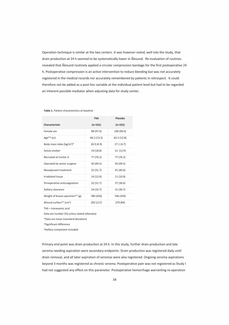
34
Operation technique is similar at the two centers. It was however noted, well into the study, that
drain production at 24 h seemed to be systematically lower in Ålesund. Re-evaluation of routines
revealed that Ålesund routinely applied a circular compression bandage for the first postoperative 24
h. Postoperative compression is an active intervention to reduce bleeding but was not accurately
registered in the medical records nor accurately remembered by patients in retrospect. It could
therefore not be added as a post-hoc variable at the individual patient level but had to be regarded
an inherent possible mediator when adjusting data for study center.
Table 1. Patient characteristics at baseline
TXA Placebo
Characteristic (n=101) (n=101)
Female sex 98 (97.0) 100 (99.0)
Age* (yr) 66.2 (13.3) 62.3 (12.8)
Body mass index (kg/m2)* 26.9 (4.9) 27.1 (4.7)
Active smoker 19 (18.8) 13 (12.9)
Recruited at Center A 77 (76.2) 77 (76.2)
Operated by senior surgeon 50 (49.5) 50 (49.5)
Neoadjuvant treatment 32 (31.7) 41 (40.6)
Irradiated tissue 14 (13.9) 11 (10.9)
Perioperative anticoagulation 32 (31.7) 37 (36.6)
Axillary clearance 34 (33.7) 31 (30.7)
Weight of breast specimen* (g) 780 (450) 746 (359)
Wound surface* (cm2) 292 (117) 279 (84)
TXA = tranexamic acid
Data are number (%) unless stated otherwise
*Data are mean (standard deviation)
Axillary component included
Primary end point was drain production at 24 h. In this study, further drain production and late
seroma needing aspiration were secondary endpoints. Drain production was registered daily until
drain removal, and all later aspiration of seromas were also registered. Ongoing seroma aspirations
beyond 3 months was registered as chronic seroma. Postoperative pain was not registered as Study I
had not suggested any effect on this parameter. Postoperative hemorrhage warranting re-operation
35
before drain removal was registered, and potential adverse effects were otherwise registered as for
Study I.
Statistical analyses in Study II
A between-group difference in mean drain production of 25% was considered clinically significant.
We proposed a standard deviation of 0.6 based on published drain volumes228 and an in-house
clinical estimate. Using an unpaired samples t-test analysis to detect a difference of 0.25, and a
standard deviation of 0.6, alpha of 0.05 and power 0.80, a sample size of 92 patients in each group
was needed. We originally planned to include a total of 210 patients to ensure additional power; 202
patients were included for final analyses.
Continuous patient characteristics data were calculated as mean ± standard deviation (SD) and
compared using independent samples t-tests. Categorical patient characteristics data were
calculated as frequency counts and percentages and compared using chi-square tests, but variables
Continuous outcome data were presented as mean (95% Confidence Interval (CI)). Categorical
outcome data were presented as frequency counts and percentages. For presentation of unadjusted
effect size of continuous data, mean difference (95% CI) between groups was used. Effect size of
categorical data was presented as odds ratio (OR) (95% CI). For statistical analyses of unadjusted
effect size, continuous outcome data were analysed using independent samples t-tests while
categorical outcome data were compared using chi-square tests or Fisher exact tests where
appropriate.
For adjusted statistical analyses, non-normally distributed continuous data underwent logarithmic
(ln) transformation. Data were then analysed using a general linear model . We included all variables
registered in Table 1 and performed a stepwise removal of non-
obtain a final model that included only individually significant variables. Results were calculated as
differences between means as percentage (95% CI). Non-normally distributed continuous data that
could not undergo logarithmic transformation due to the appearance of zero values were
dichotomized and analysed using a logistic regression model adjusting for significant variables.
Results were given as odds ratio (OR) with 95% CI. Analyses were performed in accordance to the

36
Study III
Patients undergoing hip replacement surgery at the Department of Orthopedic Surgery
University hospital routinely receive TXA 1 g iv as bleeding prophylaxis (iv group). They therefore
represent a population receiving iv TXA in accordance with established international
recommendations. Patients undergoing skin reduction abdominoplasty after massive weight loss
have very large wound surface areas and thus represent a study model with a possibility for
significant absorption of topically applied drug. The Section for Plastic and Reconstructive Surgery at
St Ola of TXA 25 mg/ml
(moistening group), while Aleris Medical Center instill a 200 ml bolus of 5 mg/ml TXA retrograde
through drains after wound closure and keep drains clamped for 1 h postoperatively (bolus group).
We wanted to investigate systemic pharmacokinetics after both moistening and bolus mode of
topical administration and compare this to iv administration. Patients with preoperative renal
insufficiency defined as glomerular filtration rate less than 60 ml/min were excluded from the study
as elimination of TXA is almost purely renal. Twelve patients were included from each patient group
during the years 2017 2018.
Age, sex, height, body weight, body mass index, serum creatinine concentration, and estimated
glomerular filtration rate were registered for all participants. The weight of the resected tissue was
registered for the abdominoplasty groups and the maximum width and length of the wound were
measured to allow calculation of an elliptical wound surface area
(length/2) × (width/2). A total of eight blood samples for the analysis of TXA were obtained: Before
drug administration, and at regular intervals thereafter for up to 6 hours for the iv group and until
the next morning for the two topical groups. Samples were centrifuged within 30 minutes and serum
secured for storage at -80°C until analysis. Assistance with blood sampling was provided by the
Clin
TXA concentrations in serum were determined by an ultra-high performance liquid chromatography
tandem mass spectrometry method specifically designed for precise analysis of low TXA
concentrations. The limit of quantification was 0.10 μg/ml. This analysis was developed by the
presented in the Appendix linked to Paper III.
37
Pharmacokinetic analysis in Study III
Maximum measured peak serum concentration and the times to achieve these concentrations were
obtained directly from the measured values. Other pharmacokinetic variables were calculated by
using the pharmacokinetic program package Kinetica, version 5.0 (ThermoFisher Scientific, Waltham,
MA, USA). Area under the time serum concentration curve was calculated using a mixed log-linear
model with extrapolation to infinity. Clearance (Cl) was calculated as dose per area under the time
serum concentration curve. By applying a non-compartment model, the parameter estimate
describing the decrease of the log-concentration d using the best-fit log-linear
regression line of the samples representing the elimination phase. The elimination half-life was
calculated as ln2 n residence time was
calculated as area under the serum concentration-time product versus time curve from zero to
infinity/ area under the time serum concentration curve.
Statistical analyses in Study III
Descriptive data are presented as mean ± SD or median (interquartile range) as appropriate.
Categorical variables were compared using s were
compared using an independent samples t test. Associations between continuous variables were
analyzed using the Pearson correlation coefficient.

38
SUMMARY OF RESULTS
Study I
Postoperative drain production was not normally distributed and we therefore chose to present
median values for effect size. Median drain production in breasts treated with TXA was 12.5 ml
(range 0-44 ml) while median drain production in breasts treated with placebo was 20.5 ml (range 0-
100 ml) (p=0.038). When adjusting drain production for breast specimen weight, median drain
production for TXA versus placebo was 13 vs. 22 ml (p=0.017). The practiced cutoff-value for drain
removal in reduction mammoplasties at the time was 40 ml/24 h. Only one of the TXA breasts
produced 40 ml of drain fluid at 24 h (44 ml), while 9 of the placebo breasts did (range 40 100 ml)
(p=0.016).
There were no significant differences in postoperative pain scores or possible adverse effects
between groups. There were two re-bleedings needing re-operation in the placebo breasts. Three
patients had signs of infection or suture reactions on the TXA side only, while two patients had such
reactions on the placebo side only. Another four patients had signs of infection or suture reactions
which were bilateral. All these complications were conservatively managed.
Study II
Crude unadjusted mean drain production at 24 h was 110 ± 67 ml in the TXA group versus 144 ± 113
ml in the placebo group, yielding a difference between means of 34 ± 13 ml. As data were non-
normally distributed, ln transformation was performed, yielding an unadjusted difference between
means of 27 ± 10 ml (P = 0.011). Hematomas warranting re-operation within the postoperative drain
period occurred in seven placebo patients and one TXA patient (P= 0.057). There were no differences
between groups regarding other complications. No thromboembolic events were registered.
Of the patient variables listed in Table 1, the following independently and significantly affected
outcome: 1) Administration of TXA, 2) type of surgery, 3) study center, 4) wound surface area and 5)
surgeon seniority. Perioperative anticoagulation did not affect the outcome variables. Outcome
adjusted for the significant variables showed that moistening the wound surface with TXA
significantly reduced 24 h drain production with 32.4% (95% CI -51.4 to -15.8, P < 0.001). Total drain
volume was reduced with 33.0% (95% CI -60.0 to -10.4, P = 0.003). Patients in the TXA group were
significantly more likely to have drains removed on the first day (OR 3.00, 95% CI 1.44 to 6.22, P =
0.003) and had a significantly shorter drain period (-15.6%, 95% CI -30.2 to -2.6, P = 0.017).
39
Stratified outcomes
In patients who had undergone AC, TXA had significant effect on drain production at 24 h compared
to placebo (-26.7%, 95% CI -55.9 to -2.9, P = 0.026), but total drain production was not significantly
reduced (-27.3%, 95% CI -91.9 to 18.4, P = 0.244). They later had significantly increased odds
compared to placebo of both needing seroma aspiration (OR 5.71, 95% CI 1.16 to 28.2, P = 0.032) and
2, 95% CI 1.50 to 21.9, P = 0.011), but no
increased risk of chronic seroma (OR 1.50, 95% CI 0.23 to 9.73, P = 0.671). In SM/SNB patients, there
was a greater effect of TXA compared to placebo on both drain production at 24 h (-33.4%, 95% CI -
58.1 to -12.6, P = 0.001) and on total drain production (--32.3%, 95% CI -62.4 to -9.4, P = 0.005).
There was no difference regarding neither need for seroma aspirations nor number of aspirations or
cumulative aspirated seroma volume, but there was a significantly reduced OR of chronic seromas
among SM/SNB patients who had received TXA (OR 0.18, 95% CI 0.04 to 0.89, P = 0.035).
Stratifying for study center, patients operated at St had 60.3% higher drain
production at 24 h than patients from Ålesund Hospital (95% CI 36.1 to 88.9, P < 0.001).
Hospital accepts larger ongoing drain production at removal than Ålesund Hospital yet despite earlier
drain removal at Hospital, they had a 33.5% larger total cumulative drain production than
Ålesund Hospital (95% CI 6.5 to 67.4, P = 0.013).
As AC and study center had such major influence on drain production at 24 h, a subgroup analysis
was done in the population who neither was operated at Ålesund Hospital nor underwent AC. These
were the SM/SNB patients at (N=108, TXA=52, Placebo=56). Adjusted for wound
surface area, those receiving TXA in this subpopulation had a 39% reduction in 24-hour drain
production (95% CI -69.2 to -14.3, P = 0.001), a 46% reduction in total drain output (95% CI -80.9 to -
17.9, P = 0.001), no significant difference on late seroma aspirations, but a suggested lower risk of
chronic seroma (OR 0.18, 95% CI 0.04-0.89, P = 0.035).
Study III
Patient characteristics were similar in the abdominoplasty populations. Due to the nature of the
procedure, patients undergoing hip replacement surgery were on average 20 years older (P < 0.001)
and had a significantly lower estimated glomerular filtration rate (P = 0.002) than the
abdominoplasty patients.
40
After iv administration of 1 g, TXA peak serum concentration (mean 66 ± 13 g/ml) occurred within
the first few minutes and stayed above the assumed threshold concentration of 10 g/ml for at least
2.5 hours. One patient in the moistening group had a peak serum concentration above the threshold
value (10.9 g/ml); otherwise the patients in both abdominoplasty groups had sub-threshold peak
values. In the moistening group, the time-concentration curves were homogenous with peak
concentrations occurring within 1-2 hours and with all patients having serum TXA concentrations of 2
g/ml or below at 6 hours. In the bolus group, the time-concentration curves were more
heterogeneous with peak concentrations occurring 4 to 8 hours after administration. In the topical
groups, peak serum concentration was not significantly correlated to body weight or wound surface
area, while there was a significant inverse correlation between serum concentration after iv
administration and body weight.
41
DISCUSSION
Clinical implications of the findings
Studies I-II suggest that moistening a surgical wound surface with TXA 25 mg/ml significantly reduces
surgical bleeding as defined by drain production at 24 h. Effect measured as percentage reduction of
blood loss seems comparable to the effect of iv prophylactic use, as iv administration in various types
of surgery seems to yield a reduction of both measured bleeding and transfusion needs with
approximately one third10. We found a 39% difference between medians in Study I and a 32%
difference between adjusted means in Study II. In Study I, the fact that only one TXA breast had a 24
h drain production above the threshold value for drain removal of 40 ml and then only barely above
at 44 ml led to the discontinuation of routine use of drains in reduction mammoplasties receiving
In Study II, TXA significantly reduced the cumulative drain production until drain removal by 33%.
Subtracting the drain production for the first 24 h, there was no longer a significant difference
between TXA and placebo regarding further drain production until drain removal. This may suggest
that the single moistening does not induce an ongoing effect beyond 24 h, but at the same time
there does not seem to be any rebound effect when the TXA effect subsides.
Moistening the wound with 25 mg/ml TXA may reduce the risk of postoperative hematoma
warranting re-operation. Although neither study was powered to investigate this variable, a possible
effect is strongly suggested as the two studies combined had ten re-bleedings, of which nine
occurred in placebo breasts ( of all breasts in Studies I and II: P=0.010).
No difference in late hematomas, wound ruptures or wound infections were seen in Studies I-II. No
patients had thromboembolic events or seizures. However, patients in Study II who had undergone
AC and had received TXA had significantly increased odds compared to the placebo group of both
(OR 5.7, P = 0.011), but no increased risk of chronic seroma (OR 1.5, P = 0.671). This is to our
knowledge the first publication of a possible adverse effect of topical TXA.
Study III renders the risk of systemic adverse events, such as thromboembolism or seizures, highly
unlikely after topical moistening of a large wound surface with TXA 25 mg/ml. We therefore
postulate that in patients with an increased risk of such adverse events, topical administration should
constitute a safe alternative to iv administration.

42
Methodological issues
Drain production as a surrogate measure for bleeding
The primary endpoint in both our intervention studies was postoperative bleeding at 24 h as
measured by drain production. There is no gold standard for the estimation of blood loss230. Methods
applied include comparing pre- versus postoperative hemoglobin (Hb), transfusion needs, and visual
and physical calculations from suctioned fluids and surgical sponges. Several formulae have been
proposed for the calculation of occult blood loss based on clinical parameters, but their accuracies
are debatable230.
As our prophylactic intervention to reduce bleeding is performed at the end of surgery and after the
completion of hemostasis, any perioperative bleeding will naturally not be affected by the
intervention. Hence any comparison of pre- and postoperative Hb or registration of transfusion
needs would falsely take into account the perioperative bleeding. To be able to assess the effect of
our intervention as accurately as possible, we wanted study models with little possibility for occult
bleeding and where measured bleeding must have occurred after the intervention.
Breast surgery is performed in tissue adherent to the thoracic wall. There is little deep space to
accommodate occult bleeding. Visual assurance of a dry surgical field prior to wound closure can be
obtained, and a drain activated after closure would deliver postoperative fluids only. We therefore
chose drain production in breast surgery as the most suitable primary endpoint to evaluate the effect
of our intervention on postoperative bleeding.
Drain fluids generally become more serous over time; we therefore deemed drain production at 24 h
as the best measurement of blood loss in our study settings, although this is a surrogate measure. To
accurately analyze the contents of collected drain fluids is a major technical challenge which we did
not undertake. However, the composition of drain fluids may have varied significantly in our study
populations, and in an ideal situation, the composition of the drain fluids should be analyzed to differ
between blood and serous fluids.
In Study I, 37 of the 56 breasts had drain production 20 ml. This is such a low production that
leftover infiltrated local anesthetics and general wound exudate may constitute a major part of the
volume at 24 h. Hence, the true effect of TXA on bleeding may have been more correctly observed in
the patients with larger volume bleeding in Study I (Fig. 4). This also demonstrated that if we were to
use drain production as primary endpoint, we should use a study population with higher volume
bleeding in a confirmatory study. We therefore performed Study II on mastectomy patients, who

43
generally bleed more. However, in Study II, patients undergoing AC were likely to have lymph leakage
which could constitute a significant proportion of the measured drain volume. Hence the most
accurate effect of TXA on pure surgical bleeding would probably be observed in patients who did not
undergo lymph node clearance. We therefore performed stratified analyses for this subgroup.
Non-normally distributed data in smaller versus larger studies
In both our studies, drain production at 24 h was not normally distributed. Non-normally distributed
data can either be transformed to a more normal distribution by means of e.g. logarithmic
transformation and then analyzed using tests for normally distributed data, or data can be analyzed
with a non-parametric test.
In Study I, two re-bleedings constituted outlier values with significantly higher drain volumes than
the rest of the breasts. On the illustration figure in the article (Fig. 4), we chose to define the two re-
to allow for a reasonable Y-axis although the real volume was much larger. A
non-parametric Wilcoxon Signed Ranks test allows for an estimation of the significance of the trend
in the treatment effect, but does not take into consideration the absolute volumes of outlier data.
In Study II, the larger data set from 202 patients allowed for a logarithmic transformation of non-
normally distributed data and we used the transformed values for further statistical analyses.
However, when registering aspirations of late seroma, patients not in need of seroma aspirations
on-normally distributed continuous data containing zero
values cannot undergo logarithmic transformation, these data had to be dichotomized and analysed
using a logistic regression model. The need for such dichotomization had not been foreseen and
which cut-off values to use had not been discussed in the protocol phase; these were therefore
established in the aftermath based on clinical reasoning and the general distribution observed in the
study. Different cut-offs for the dichotomization could have yielded different results. The significantly
increased risk of needing at least one seroma aspiration after TXA administration does not depend on
any cut-off value. Had we however set the cut-
instead of 500 ml, the OR would still have been higher after TXA administration (OR 3.28 at 1000 ml
cut-off versus 5.72 at 500 ml cut-off) but the increased OR would no longer be statistically significant
(p=0.076).

44
Presenting effect sizes
In Study I, we debated how to present the effect size. The calculation of mean paired difference in
drain volumes (13 ± 27 ml) was not normally distributed. As 13 of the participating women had drain
volumes below 20 ml in both breasts, the difference was skewed towards lower values, while the two
re-bleedings also represented significant outliers even when substituting the actual high drain
volume in re-bleedings with a volume of 100 ml. Four breasts had zero drain production and would
be excluded if we attempted to logarithmically transform the data. Excluding the two re-bleedings
would not have been in accordance with protocol. We therefore chose to present the difference in
drain production as the unpaired median drain production in TXA breasts (12.5 ml) versus the
median of placebo breasts (20.5 ml), which would convey a conservative estimate of effect size. A
published comment to Study I (Appendix, Paper I) correctly pointed out that in a paired study, we
should present paired values, and that the median paired difference was only 2.5 ml. This comment
led us to revise the figure originally published in the paper the original figure had an unfortunate
chronological sorting of included patients (Fig. 1, Paper I), while the revised figure (Fig.4) sorted
patients according to the difference in drain volume between TXA and placebo breast.
To report the paired median difference in this material (the average between patients 14 and 15) as
suggested in the comment would clearly underreport the fact that differences in drain volume to the
right of the median far exceeds the differences to the left of the median. Figure 4 illustrates an
Fig. 4 Study I. Revised figure. Drain production in TXA breast and placebo breast for each patient; patients
sorted in ascending order according to calculated difference in volume (placebo breast minus TXA breast).

45
increased paired difference in breasts with larger bleeding volumes. In 13 of the 15 patients in whom
one or both breasts had more than 20 ml drain production, the difference was in favor of TXA. We
felt the revised figure was the best way to convey the results of the study.
Small volumes and large variances
The published comment to Study I (Appendix Paper I) also mentioned that the actual variance in
Study I was much higher than the SD estimated in our power analysis, and that the study therefore
was significantly underpowered. This comment illustrates methodological challenges in Study I when
using a study model with little postoperative bleeding, and simultaneously using a simplified
surrogate measurement of that bleeding which may be increasingly inaccurate with lower values.
This is a situation where reliable SD values for the specific situation to be studied do not exist and
thus represents a challenge in the power analyses.
Reduction mammoplasties have large wound surfaces yet often bleed little, as fatty mammary tissue
is less vascularized than e.g. muscle, skin and fascia. Although reduction mammoplasty offers a study
model with an alluring possibility of within-patient randomization, low drain volumes allow for large
variances and misleading proportions between treated and untreated breasts, as mentioned
previously. Cases with a 3 ml production on one side versus 18 ml on the other contribute to large
variances but have little clinical significance. We reached non-parametric significance at the p<0.05
level but not at the p<0.01 level in our pilot study. Had more patients had bilateral low volume
bleeding, we might not at all have reached statistical significance.
Effect modifying variables and mediators
In Study I, we did statistical analyses both on crude drain volumes and on drain volumes adjusted for
breast specimen size. The rationale behind this simple adjustment was the assumption that larger
wounds will bleed more. The wound surface area of reduction mammoplasties is much more
complex than the oval wound bed of a standardized mastectomy; hence actual wound surface area
could not be calculated with sufficient precision in Study I. The weight of the resected tissue was
therefore registered as a surrogate measure of wound area. As mentioned in the Methods chapter,
adjustment was done by multiplying left breast drain volume with the factor obtained from dividing
right breast weight with left breast weight. P values of the difference in drain production using the
Wilcoxon signed rank test then decreased from 0.038 to 0.017.

46
In Study II, case and control wounds were no longer in the same patient. In an optimal study setting,
randomization should sufficiently distribute patient characteristics. We knew in advance that
variables such as presence or absence of axillary lymph node dissection were definite effect
modifying variables which would significantly affect primary outcome. We also suspected that
several other factors such as wound surface area, use of anticoagulants and surgeon seniority might
be effect modifying variables. As mentioned in the Methods chapter, we discovered well into the
study that being operated at Ålesund Hospital seemed to obviously reduce drain production at 24 h.
Although there may be many small unrecognized differences between hospitals that will justify a
separate stratified randomization for each study center when conducting a randomized clinical trial,
there was at study initiation few well-founded suspicions that differences between study centers
should be significantly effect modifying. We ultimately found an adjusted 60% difference between
drain production at 24 h between study centers, which suggested a significant effect.
Compression reduces bleeding. Park et al. have demonstrated in knee arthroplasties that the
intraarticular compression induced by clamping the drain for three hours reduces bleeding equal to
low-dose intraarticular TXA in combination with drain clamping for 30 minutes231. We had not noted
during study planning that Ålesund Hospital practiced a 24 h circular compression bandaging. This
clinical intervention to reduce bleeding was very likely to compete with the effect of the application
of TXA. It may also explain the relative lack of effect of TXA on 24 h bleeding at Ålesund hospital, as
mean bleeding at 24 h in placebo patients in Ålesund was lower than mean bleeding among TXA
intervention patients in Trondheim (Study II, supplementary tables 4 and 5). Yet we could not in
effect modifying variable at the
individual level but had to include this as an assumed mediator in
variable. However, a study designed to evaluate the isolated effect of 24 h postoperative
compression as practiced by Ålesund Hospital is needed to confirm this assumption.
There was no significant difference between the proportion of patients who at some point needed
Hospital (76%) versus Ålesund Hospital (66%) (P = 0.212)
Hospital had a significantly increased total number of aspirations (OR = 9 for 5 aspirations, 95% CI 2
to 33, P < 0.001) and cumulative aspirated volume (OR = 4 for 500 ml cumulative seroma, 95% CI 2
11, P= 0.004). As published studies232,233 have not found effect of prolonged postoperative
compression on seroma formation, it would seem strange if a 24 h compression regime should have
a major impact. An alternative explanation lies in the fact that Ålesund Hospital serves a population
Hospital has a much larger proportion of its patient
Hospital, it is
performed by the nurses at the outpatient clinic and patients can merely call or drop by. At Ålesund
47
Hospital, seroma aspirations are mainly performed by the surgeons and requires an outpatient
appointment.
Stratified randomization or regression analyses?
When planning Study II, we were well aware that there are significant differences between
mastectomy surgeries. A mastectomy with an associated AC will both have a larger and more
complex wound surface and added damage to large lymph vessels. All surgeons are aware that these
extended mastectomies have much larger drain volumes and increased risk of need for late seroma
aspirations. We could have designed Study II with two arms and stratified the randomization not only
for study center, but also for type of surgery. A two-armed study would have needed separate power
calculations for each arm. Variance in drain production is much larger after AC than after
mastectomy only, and there is approximately one mastectomy with AC to every two SM/SNB
mastectomies. A two-armed study would require a significantly larger study population of AC
patients, and the study would have required a significantly longer inclusion period. As the decision to
perform AC is sometimes not made until well into the operation, such a stratification would not have
been easily compatible with a pre-surgery randomization.
Regression analyses take into account the effects of possible effect modifying variables on a defined
outcome. Stratifying for each of these variables would have produced small study populations
possibly without the individual power to significantly demonstrate an effect.
We extensively used regression analyses in Study II. For the primary outcome, drain production at 24
h, we used a General Linear Model (GLM). This model allows for a continuous dependent variable to
be adjusted for both continuous (e.g. wound surface area) and categorical (e.g. type of surgery)
variables. For dichotomized dependent variables, such as need for late seroma aspiration (Y/N), we
used binary logistic regression which also allows for both continuous and categorical adjusting
variables.
Our regression analyses adjusted for those variables in Table 1 which independently significantly
affected outcome. Our final model explained 50% of the observed variation in the drain production
at 24 h (R2 = 0.496), and the order of importance according to partial Eta squared values were AC
(0.233), study center (0.141), wound surface area (0.117), administration of TXA (0.080) and surgeon
seniority (0.027). Clinical studies on bleeding in surgery most often present absolute and unadjusted
volumes, but surgery is a field with extensive possibilities for effect modifying factors and surgical
study populations are often relatively small. Study II illustrates the importance of adjusting for

48
possible significant variables in surgical research as many variables had greater influence on the
variance in postoperative bleeding than the application of TXA yet the adjusted data confirms the
effect of TXA, as the significance of the effect increases from P = 0.011 in an unadjusted Stud -
test to P < 0.001 for adjusted data.
Regression analyses can identify potential variables which may justify stratified analyses or inspire
tailored investigation in future studies, particularly in pilot studies such as ours. Two major findings
from Study II thus need further exploration: The aforementioned influence of study center (with the
the effect of TXA on seroma in the
presence/absence of lymph node clearance.
New questions arising from the studies
Can topical TXA increase lymphatic seroma formation?
Seroma can be defined as the collection of fluid within the dead space created by extensive
dissection. Seroma as a postoperative complication is mainly addressed in plastic and breast surgery,
and reported incidence in breast cancer surgery varies greatly from 5 80%234. The exact
composition of post-mastectomy seroma remains somewhat unclear. Analyses by Walt-Boolsen et al.
(1989)235 suggested that post-mastectomy seroma is mainly an exudate secondary to the
inflammatory reaction that constitutes the first phase of surgical wound repair, and analyses of
seroma fluids in abdominoplasty supports an exudate composition236. However, Bonnema et al.
(1999)237 demonstrated that seroma after axillary dissection contains virtually no fibrinogen and that
its composition must therefore be mainly lymph-derived, as lym is void of
fibrinogen. Bonnema therefore questions the proposed possible reduction in seroma formation in a
published study on the effect of intravenous TXA in breast cancer patients by Oertli et al.238, as a
mechanism to explain such an effect would remain obscure given the absence of fibrinogen in
seroma . These studies also
origin condition. We have not
been able to identify studies comparing the constituents of seromas after SM versus AC.
The suggested increase in postoperative seroma after lymph node clearance in Study II may
represent a not previously described adverse effect of topical TXA, and may be applicable only to
lymph-derived seroma, as no such trend was seen in the mastectomies not undergoing AC. On the
contrary, SM/SNB patients actually had a significantly decreased OR of developing chronic seroma
after TXA administration (OR = 0.18, P = 0.035). As patients with postoperative complications were

49
excluded from seroma analyses as the complications themselves may cause seroma, only 52 of 65
patients undergoing AC and 128 of 137 patients undergoing SM/SNB remained for seroma analyses.
Our study centers remove drains after axillary clearance after a mean 2.7 days, which is early by
international standards. Our centers also have a very high incidence of seroma aspirations (77% at St
): As stated by one reviewer in Study II, the participating
ade drain time for seroma
Whether moistening the wound surface with TXA 25 mg/ml in patients undergoing lymph node
clearance increases the need for late seroma aspirations needs confirmation. Our material does
suggests a different effect of TXA on what we assume is exudate-dominated seroma (SM/SNB-
patients) versus lymph-dominated seroma (AC-patients). Our findings need confirmation as
significance is at the P<0.05 level but not at the P<0.01 level. We propose a randomized controlled
study in patients undergoing lymph node clearance, regardless of location, but with a standardized
regime for drain removal. End points could be number of and cumulative volume of late seroma
aspirations. Should an increase in seroma be confirmed, explanatory mechanisms must be
investigated, and a repeat study comparing lower doses, e.g. 5 mg/ml TXA versus 25 mg/ml TXA
versus placebo should be performed to investigate whether the phenomenon is dose-dependent
within the concentration range used clinically. It would also be interesting in a later study to
evaluate early blood/lymph ratio in the drain fluids after exposure to TXA versus placebo in lymph
node clearance patients.
Possibility of systemic effect after topical TXA application?
Study III demonstrated that in large skin reduction abdominoplasties, moistening the wound surfaces
with 20 ml of 25 mg/ml TXA or instilling a 200 ml bolus of 5 mg/ml TXA into the wound cavity after
wound closure resulted in very low serum levels of TXA compared to iv administration.
To our knowledge, Study III is the first extensive pharmacokinetic study on TXA serum concentrations
after topical administration. A sensitive analytical method specifically designed to detect low serum
levels of TXA was developed and is a major strength of the study (Appendix 1, Paper III).
Using the specific moistening technique, 20 ml was sufficient to moisten even the largest
participating wounds. Depending on wound surface area, varying fractions of the administered 20 ml
would spill and the total amount of drug which remained adherent to the wound was uncertain. We
did not collect or analyze drug from absorbent materials or drains. In the study setting, we estimated
actual absorbed drug by comparing the area under the time serum concentration curve values in the
50
moistening group with the area under the time serum concentration curve values in the intravenous
group, assuming that the true clearance in the two groups was the same. Mean net absorbed dose in
the moistening group could then be estimated to be 316 ± 98 mg of the 500 mg administered, but
the dose was somewhat surprisingly not correlated to wound surface area. This led us to speculate
whether tissue micro-topography or individual tissue vascularization may affect absorption.
When administering TXA via the instillation of a voluminous bolus of diluted drug, Study III
demonstrated that this administration might give an unpredictable systemic absorption from large
wounds such as abdominoplasties. Such large wounds have nooks and crevices where study fluid
may reside, and patient mobilization may influence whether study fluid actually reaches all areas of
the cavity. Smaller cavities, such as knee and hip joints, may however be good candidates for bolus
administrations. Although serum concentrations remained below threshold values when using a 200
ml bolus of a low concentration (5 mg/ml) of TXA, elimination was considerably slower and drug can
be expected to linger much longer in the tissues, increasing exposure time compared to the
moistening method.
For both topical modes of administration, the TXA serum concentrations can be expected to
correlate to the TXA concentration in the solution administered. Wong et al.207, who introduced the
intraarticular topical bolus administration of TXA in arthroplasties, demonstrated that the serum
concentration after a 100 ml intraarticular bolus of either 15 or 30 mg/ml tranexamic acid in knee
arthroplasty resulted in serum concentrations at the same single time
point postoperatively. Similarly, we assume that a moistening technique with more concentrated
solution will give a correspondingly higher serum concentration. Systemic concentrations after
topical application may therefore exceed the threshold for therapeutic effect if sufficiently high TXA
concentrations are used in the fluid applied.
Local adverse effects after topical application?
When introducing a moistening of the wound surface as a prophylactic measure to reduce bleeding,
the concentration of 25 mg/ml was somewhat arbitrarily chosen based on the available literature.
Wong et al. had demonstrated similar effect from both 15 and 30 mg/ml solutions207, and when
leaving a thin film instead of a bolus, it seemed reasonable to go for the higher concentration as
dilution from tissue fluids would be rapid. Few studies on potential local toxicity were available;
chondrocyte studies suggested that 25 mg/ml was safe222,223. Potential local adverse effects of topical
TXA has however not been sufficiently investigated. In our clinical experience from plastic surgery,
we have not seen any obvious increase in wound ruptures, wound infections or seroma aspirations

51
after the introduction of topical TXA as routine bleeding prophylaxis. In our two RCTs, there has been
no difference in wound infections or wound ruptures between the TXA and placebo groups, but our
studies were effect studies and not designed or powered to identify potential adverse effects.
However, the aforementioned increase in seroma in patients having undergone AC triggered a search
for possible explanations.
Our group has recently exposed in vitro keratinocytes and fibroblasts to various clinically relevant
concentrations of TXA, and we have also investigated the effect of topical TXA on wound re-
epithelialization in an ex vivo human skin model239. While short exposure (10 minutes of exposure to
drug-containing medium, pour off without rinsing, replace with drug-free medium) even to high
concentrations had little cytotoxic effect and did not significantly affect wound re-epithelialization,
prolonged exposure (continuous exposure to drug-containing medium for up to 72 h in cells/8 days
in skin wounds) was increasingly cytotoxic with increasing exposure time and drug concentration.
Prolonged exposure to 25 mg/ml TXA completely prevented wound re-epithelialization which
otherwise would occur within 3-4 days. Prolonged exposure to 50 and 100 mg/ml induced
epidermiolysis in the human skin model, but without signs of cell death and thus the mechanism
seemed non-toxic. Prolonged exposure to the low concentration of 6.25 mg/ml did not significantly
affect wound re-epithelialization.
The absence of re-epithelialization in the study by our group was seen after prolonged exposure, yet
the possible increase in seroma formation in mastectomy patients undergoing AC was after an in vivo
short exposure. Both phenomena may be explained by an antiadhesive/antimigratory effect of TXA,
which may have delayed the healing of lymph vessels and prevented the migration of keratinocytes.
Findings from other studies may also suggest a potential effect of TXA on cell migration and cell
adhesion. Bergenholtz et al. have shown in an ex vivo model on incisional wounds in palatal mucosa
from cats that prolonged exposure to anti-plasminogen activators caused proliferating epithelium to
change direction in a non-physiological manner. They even observed the proliferation of epithelial
cells onto sheets of other epithelial cells, which does not occur during normal wound healing227. The
The cause of the changed direction of epithelial migration is obscure , but they
postulate that the lack of fibrin degradation/remodeling resulting from the presence of
antifibrinolytics may leave strands of fibrin guiding keratinocytes in non-physiological directions. The
changed pattern of keratinocyte migration was seen after prolonged exposure to TXA 0.12, 1.2 and
12 mg/ml. Epithelial fusion did occur at all concentrations, also after prolonged exposure to TXA 12
mg/ml, which is in accordance with our findings that threshold levels for inhibition of wound re-
epithelialization given chronic exposure to TXA must be somewhere in the range of 5-25 mg/ml.

52
Studies on fibrin sealants may suggest an effect of TXA on cell adhesion. Commercial fibrin sealants
supplemented with TXA to prevent the breakdown of fibrin have demonstrated inferior adhesive
strength compared with sealants supplemented with aprotinin226. Simultaneously, such TXA-
supplemented fibrin sealants have caused fewer unwanted intra-abdominal adhesions compared
with sealants without TXA in animal studies240,241, which may be a clinically beneficial effect of the
proposed reduced adhesive strength.
If prolonged exposure to above-threshold concentrations of TXA may prevent re-epithelialization,
reduce cell adhesions or cause cell detachment, several questions are raised. Can such exposure
reduce tensile strength in various healing tissues, delay re-epithelialization in large wound surfaces
such as burns and split-thickness skin grafts, or prevent unwanted postsurgical adhesions or capsular
contractures? More research is clearly needed to clarify these issues.
Circular compression for 24 h to reduce bleeding in breast surgery?
It is a well-known surgical principle that compression reduces bleeding. In breast surgery, focus has
been primarily on postoperative seroma formation and less on the surgical bleeding. Hence, the few
published studies on the effect of postoperative application of a compression garment have mainly
focused on and failed to find an effect on seroma232,233. In these studies compression is generally
left for several days, which may both induce tissue ischemia and restrict patient respiration. Circular
compression has therefore been somewhat forfeited in breast surgery, but proper randomized
studies on the effect on postoperative bleeding and re-operation due to hemorrhage are lacking. It is
important to distinguish postoperative bleeding and late seromas as two distinctly separate
pathophysiological phenomena, and the lack of effect on seroma should not prevent proper
intervention to reduce postoperative bleeding. In Study II, being operated at Ålesund Hospital was
more effective than TXA in reducing postoperative bleeding at 24 h (60% reduction, 95% CI 36
89%), and we postulate that this was due to circular postoperative compression. A separate study
randomizing patients to circular compression bandaging as practiced by Ålesund Hospital versus
standard bandaging as practiced by St Olav s Hospital is warranted. The patients should not receive
TXA.
53
Future perspectives
Optimal dose and mode of administration of topical TXA
Minimizing perioperative bleeding is fundamental, and topical use of TXA as a prophylactic measure
to reduce surgical bleeding has relevance for many surgical specialties and procedures. Topical use is
however still off-label, and minimum effective dose, mode of administration and duration of contact
still needs clarification. The possibility of local effects of TXA beyond its antifibrinolytic properties is
also inadequately explored.
We have shown that moistening a wound surface with 25 mg/ml TXA reduces bleeding. The lowest
effective dose in topical administration is undetermined, but TXA concentrations as low as 4 mg/ml
have been found effective201. The dose-response relationship regarding the effect of topical TXA
remains unclear; most studies have not demonstrated significant differences in bleeding when using
equal volumes with different drug concentrations202,207,242. A recent study from knee arthroplasty
suggests that 3 g is more effective than 500 mg when given as an intraarticular injection243, but this is
administered as a concentration of 50 mg/ml as an either 10 ml or 60 ml bolus. Hence the volume
injected is six fold higher in the 3 g group and whether the difference in effect is attributable to the
higher total TXA dose or the counter pressure of the larger volume in the enclosed space of a knee
joint is unknown. However, other studies have suggested increased effect from higher
concentrations when using equal volumes, although those differences have not been statistically
significant207,244. Determining the lowest effective dose in our moistening technique could be
explored in patients undergoing mastectomy without lymph node clearance, and with a strict
protocol for postoperative bandaging and drain removal. Randomization could then be into three
groups: TXA 5 mg/ml, TXA 25 mg/ml and saline. We still favor mastectomies as study model due to
the homogeneity of the surgical wounds and the limited possibility of occult bleeding. To restrict
inclusion to patients undergoing mastectomy without lymph node clearance, final inclusion and
randomization should preferably be determined perioperatively when the choice of surgery is
definite. Instant access to pre-made study drug vials would allow for such a last-minute
randomization.
Topical TXA to reduce risk of postoperative hemorrhage warranting re-operation
A reviewer of Study II suggested that postoperative hemorrhage warranting re-operation would be
the best primary end point to measure the effect of topical TXA as a prophylactic measure to reduce
bleeding. To our knowledge, no study on topical TXA has had this variable as primary endpoint but an

54
adequately powered study or meta-analysis to investigate the prophylactic effect on re-bleeding is
needed. As postoperative hemorrhage is relatively rare (4% in our material, which reviewers thought
was above average), much larger study populations are needed using to address postoperative re-
bleedings.
Synergistic effect of topical tranexamic acid and topical adrenaline
When suggesting a prophylactic moistening of the wound surface with TXA to reduce bleeding, we
reasoned that there was in all likelihood a synergistic effect between the vasoconstriction induced by
adrenaline to allow for more efficient clotting and preservation of clotting by TXA. To evaluate the
isolated effect of topical TXA in our pilot effect studies, we have diluted TXA with saline only.
However, we postulate that diluting one vial of 5 ml TXA 100 mg/ml with 15 ml of standard local
anesthesia containing adrenaline may give both the added effects of analgesia and increased
hemostasis through a synergistic effect between adrenaline and TXA.
Chinese publications have recently found an increased effect of topical TXA in combination with low-
dose intravenous adrenaline245. To evaluate whether the synergistic effect of topical TXA and topical
adrenaline may be even more efficient than topical TXA alone in reducing surgical bleeding, and to
evaluate whether added topical local anesthesia may improve analgesia, repeat randomized studies
with topical application of TXA with/without adrenaline and with/without local anesthesia are
needed.
Use of topical TXA on superficial wounds
Burn patients, trauma patients and patients with necrotizing soft tissue infections all have large
wound surface areas in need of ongoing revisions and extensive split skin grafting. Such patients are
particularly prone to massive blood loss and wound oozing with associated loss of proteins and
electrolytes246, both from their revised wounds, and from the split skin graft donor sites. These
patients are mostly in a hyperfibrinolytic state and the risk of potential systemic adverse effects of
TXA in hyperfibrinolysis has not been clarified247,248. Topical administration to these patients might be
an alternative to avoid systemic effect yet reduce bleeding.
Donor wounds after split skin graft harvesting enter the superficial dermis and leave adnexal
structures of the skin intact. Hair follicles and sweat glands contain epithelium which migrate to
cover the wound, and most donor wounds have healed by secondary intent within 10-14 days.
55
Strategies to reduce bleeding must not delay healing, as only wound healing will permanently
terminate ongoing oozing.
We had prepared a protocol for a study on topical TXA on donor site wounds in burn patients
(ClinicalTrials.gov NCT02918201). The proposed study awaited the results from our effect studies and
also the results from our cytotoxicity studies. We do believe that a reduction in bleeding from topical
application will be of great value in this study population, but as our studies on wound re-
epithelialization have postulated a potential negative effect of TXA, we must revise the protocol and
we have not yet initiated this study. The initial protocol proposed covering the superficial wounds
with moistened bandages containing TXA. As the effect of prolonged exposure is undetermined in
vivo, we will now initially use only a moistening method directly onto the wound as described in
Studies I and II, and we will primarily evaluate wound re-epithelialization in smaller donor site
wounds. If no delay in wound epithelialization is seen compared to placebo, we can progress to an
evaluation of bleeding volume using the moistening method only/alternatively re-epithelialization
studies on smaller wounds using moistened bandages.
Mechanisms behind potential adverse effects of topical tranexamic acid
Caution is warranted when promoting off-label use of a registered drug. Whether the effects of
topical TXA corresponds to dose or exposure time, or may be dependent of threshold levels, remain
undecided. While topical use of TXA seems safe regarding potential systemic adverse events, topical
TXA coming into contact with the CNS can induce potentially life-threatening seizures178. To our
knowledge, clinical adverse effects from prophylactic use of topical TXA to reduce bleeding has until
now not been published. However, our ex vivo studies in human skin suggest that prolonged
exposure of superficial wounds to higher concentrations of topical TXA may inhibit re-
epithelialization239. Results from Study II raises the questions whether damaged lymph vessels may
seal more slowly after exposure to topical TXA. Possible adverse effects of topical TXA on various
tissues therefore warrant tissue-specific studies, and potential mechanisms behind a possible
antimigratory or antiadhesive effect need to be explored.
Cox and colleagues249 have demonstrated that the presence of TXA 50 mg/ml both prevents
fibroblast adherence and causes fibroblast detachment in vitro, proposing a non-cytotoxic
mechanism with changes in integrin interaction.
The hemidesmosomes which attaches the keratinocytes and thus the epidermis to the basal
membrane utilizes integrins, and integrins are also involved in migration of both keratinocytes and

56
various other cells250. On a molecular level, plasminogen, plasmin and in particularly u-PA serve as
ligands and interact with both integrins and other cell surface receptors involved in cellular
migration, adhesion and signaling, and these events may be quite separate from the functions
related to fibrinolysis 22,30,251-253. Ploskota et al. have even described a convergence of the adhesive
and fibrinolytic systems through a mechanism where a single u-PA ligand crosslinks integrin
receptors to form a complex which may simultaneously play an important role in the control of cell
migration and vascular homeostasis253.
Human receptors with affinity for lysine analogues and the consequences of such bindings are
inadequately explored. Lysine analogues may cause convulsive seizures through their competitive
binding to GABA and glycine receptors in the CNS189. Our observations regarding possible effect on
cell adhesions and cell migration may be in accordance with described mechanisms at cellular
receptor level involving integrins, plasminogen and u-PA. Off-label use of both topical and systemic
use of TXA to treat skin hyperpigmentation and melasma has been increasingly published in the last
decade254, and a possible mechanism could be interference with melanosome transfer from
melanocyte to keratinocytes. Lysine binding sites may be widespread in the human organism and
possible effects of TXA clearly needs further investigation.

57
CONCLUSIONS
Topical moistening of a surgical wound surface with TXA 25 mg/ml reduces postoperative
bleeding as measured by drain production with approximately one third, which is comparable to the
effect of routine intravenous use of TXA.
Topical moistening of a surgical wound surface with TXA 25 mg/ml may reduce the risk of re-
bleeding warranting re-operation.
Seroma formation after lymph node clearance may be increased after topical moistening of
the surgical wound with TXA 25 mg/ml. This phenomenon needs confirmation, and the potential
underlying pathophysiological mechanism is unknown.
We did not observe an increase in thromboembolic events, wound infection, wound rupture,
suture reactions or late hematomas in wounds treated with topical TXA 25 mg/ml.
Systemic concentrations of TXA does not exceed therapeutic threshold value for
antifibrinolytic effect in large abdominoplasties after topical moistening of the wound surface with
20 ml of TXA 25 mg/ml or instillation of 200 ml of TXA 5 mg/ml into the wound cavity.

58
REFERENCES
1. Hadad E, Wiser I, Rosenthal A, Landau G, Ziv E, Heller L. Suction drains in esthetic breast implant exchange are associated with surgical site infections: A retrospective cohort study. J Plast Reconstr Aesthet Surg. 2017;70(11):1635-1640.
2. Pennington Z, Lubelski D, Molina C, Westbroek EM, Ahmed AK, Sciubba DM. Prolonged post-surgical drain retention increases risk for deep wound infection after spine surgery. World
neurosurgery. 2019. 3. Franchini M, Marano G, Mengoli C, et al. Red blood cell transfusion policy: a critical literature
review. Blood transfusion = Trasfusione del sangue. 2017;15(4):307-317. 4. Stokes EA, Wordsworth S, Staves J, et al. Accurate costs of blood transfusion: a microcosting
of administering blood products in the United Kingdom National Health Service. Transfusion.
2018;58(4):846-853. 5. Goodnough LT, Shander A. Patient blood management. Anesthesiology. 2012;116(6):1367-
1376. 6. Manjuladevi M, Vasudeva Upadhyaya K. Perioperative blood management. Indian journal of
anaesthesia. 2014;58(5):573-580. 7. Barnard J, Millner R. A review of topical hemostatic agents for use in cardiac surgery. The
Annals of thoracic surgery. 2009;88(4):1377-1383. 8. Ferraris VA, Brown JR, Despotis GJ, et al. 2011 update to the Society of Thoracic Surgeons
and the Society of Cardiovascular Anesthesiologists blood conservation clinical practice guidelines. The Annals of thoracic surgery. 2011;91(3):944-982.
9. Ker K, Edwards P, Perel P, Shakur H, Roberts I. Effect of tranexamic acid on surgical bleeding: systematic review and cumulative meta-analysis. BMJ (Clinical research ed). 2012;344:e3054.
10. Ker K, Prieto-Merino D, Roberts I. Systematic review, meta-analysis and meta-regression of the effect of tranexamic acid on surgical blood loss. The British journal of surgery.
2013;100(10):1271-1279. 11. WOMAN Trial Collaborators. Effect of early tranexamic acid administration on mortality,
hysterectomy, and other morbidities in women with post-partum haemorrhage (WOMAN): an international, randomised, double-blind, placebo-controlled trial. Lancet.
2017;389(10084):2105-2116. 12. Henry DA, Carless PA, Moxey AJ, et al. Anti-fibrinolytic use for minimising perioperative
allogeneic blood transfusion. The Cochrane database of systematic reviews.
2011(3):Cd001886. 13. Poeran J, Rasul R, Suzuki S, et al. Tranexamic acid use and postoperative outcomes in
patients undergoing total hip or knee arthroplasty in the United States: retrospective analysis of effectiveness and safety. BMJ (Clinical research ed). 2014;349:g4829.
14. Shakur H, Roberts I, Bautista R, et al. Effects of tranexamic acid on death, vascular occlusive events, and blood transfusion in trauma patients with significant haemorrhage (CRASH-2): a randomised, placebo-controlled trial. Lancet. 2010;376(9734):23-32.
15. Gerstein NS, Kelly SP, Brierley JK. Yet Another Tranexamic Acid-Related Thrombotic Complication. Journal of cardiothoracic and vascular anesthesia. 2016;30(3):e21-22.
16. Myers SP, Kutcher ME, Rosengart MR, et al. Tranexamic acid administration is associated with an increased risk of posttraumatic venous thromboembolism. The journal of trauma and
acute care surgery. 2019;86(1):20-27. 17. Takagi H, Ando T, Umemoto T. Seizures associated with tranexamic acid for cardiac surgery: a
meta-analysis of randomized and non-randomized studies. The Journal of cardiovascular
surgery. 2017;58(4):633-641. 18. Alshryda S, Sukeik M, Sarda P, Blenkinsopp J, Haddad FS, Mason JM. A systematic review and
meta-analysis of the topical administration of tranexamic acid in total hip and knee replacement. The bone & joint journal. 2014;96-b(8):1005-1015.
59
19. Ker K, Beecher D, Roberts I. Topical application of tranexamic acid for the reduction of bleeding. The Cochrane database of systematic reviews. 2013;7:Cd010562.
20. Dunn CJ, Goa KL. Tranexamic acid: a review of its use in surgery and other indications. Drugs.
1999;57(6):1005-1032. 21. Longstaff C, Kolev K. Basic mechanisms and regulation of fibrinolysis. Journal of thrombosis
and haemostasis : JTH. 2015;13 Suppl 1:S98-105. 22. Miles LA, Parmer RJ. Plasminogen receptors: the first quarter century. Seminars in
thrombosis and hemostasis. 2013;39(4):329-337. 23. Mangel WF, Lin BH, Ramakrishnan V. Characterization of an extremely large, ligand-induced
conformational change in plasminogen. Science (New York, NY). 1990;248(4951):69-73. 24. Law RH, Abu-Ssaydeh D, Whisstock JC. New insights into the structure and function of the
plasminogen/plasmin system. Current opinion in structural biology. 2013;23(6):836-841. 25. Urano T, Suzuki Y, Iwaki T, Sano H, Honkura N, Castellino FJ. Recognition of plasminogen
activator inhibitor type 1 as the primary regulator of fibrinolysis. Current drug targets. 2019. 26. Andersson L, Nilsoon IM, Colleen S, Granstrand B, Melander B. Role of urokinase and tissue
activator in sustaining bleeding and the management thereof with EACA and AMCA. Annals
of the New York Academy of Sciences. 1968;146(2):642-658. 27. Hoylaerts M, Lijnen HR, Collen D. Studies on the mechanism of the antifibrinolytic action of
tranexamic acid. Biochimica et biophysica acta. 1981;673(1):75-85. 28. Takada A, Takada Y. Inhibition by tranexamic acid of the conversion of single-chain tissue
plasminogen activator to its two chain form by plasmin: the presence on tissue plasminogen activator of a site to bind with lysine binding sites of plasmin. Thrombosis research.
1989;55(6):717-725. 29. Longstaff C, Locke M. Increased urokinase and consumption of alpha2 -antiplasmin as an
explanation for the loss of benefit of tranexamic acid after treatment delay. Journal of
thrombosis and haemostasis : JTH. 2019;17(1):195-205. 30. Wu G, Mazzitelli BA, Quek AJ, et al. Tranexamic acid is an active site inhibitor of urokinase
plasminogen activator. Blood advances. 2019;3(5):729-733. 31. Mahmood N, Mihalcioiu C, Rabbani SA. Multifaceted Role of the Urokinase-Type
Plasminogen Activator (uPA) and Its Receptor (uPAR): Diagnostic, Prognostic, and Therapeutic Applications. Frontiers in oncology. 2018;8:24.
32. Degryse B. The urokinase receptor system as strategic therapeutic target: challenges for the 21st century. Current pharmaceutical design. 2011;17(19):1872-1873.
33. Laufman H. Surgical hemostasis through the ages. Quarterly bulletin Northwestern University
(Evanston, Ill) Medical School. 1947;21(3):252-262. 34. Anaesthesia, or the Employment of Chloroform and Ether in Surgery, Midwifery, &c. The
British and foreign medico-chirurgical review. 1852;9(17):158-183. 35. Lister BJ. The classic: On the antiseptic principle in the practice of surgery. 1867. Clinical
orthopaedics and related research. 2010;468(8):2012-2016. 36. Brucke E. An Essay on the Cause of the Coagulation of the Blood. The British and foreign
medico-chirurgical review. 1857;19(37):183-212. 37. Mellanby J. The Coagulation of blood: Part II. The actions of snake venoms, peptone and
leech extract. The Journal of physiology. 1909;38(6):441-503. 38. Mellanby J. The coagulation of blood. The Journal of physiology. 1908;38(1):28-112. 39. Macfarlane RG. AN ENZYME CASCADE IN THE BLOOD CLOTTING MECHANISM, AND ITS
FUNCTION AS A BIOCHEMICAL AMPLIFIER. Nature. 1964;202:498-499. 40. Owen CA, Jr. Recent advances in blood coagulation. Historical remarks. Seminars in
thrombosis and hemostasis. 1985;11(4):335-336. 41. Spangler HP. [Tissue adhesion and local haemostasis using fibrinogen, thrombin and clotting
factor xiii. (experimental investigations--clinical experience) (author's transl)]. Wiener
klinische Wochenschrift Supplementum. 1976;49:3-18.

60
42. Giangrande PL. The history of blood transfusion. British journal of haematology.
2000;110(4):758-767. 43. Landsteiner K. On agglutination of normal human blood. Transfusion. 1961;1:5-8. 44. Franchini M. The modern treatment of haemophilia: a narrative review. Blood transfusion =
Trasfusione del sangue. 2013;11(2):178-182. 45. Bierbaum BE, Callaghan JJ, Galante JO, Rubash HE, Tooms RE, Welch RB. An analysis of blood
management in patients having a total hip or knee arthroplasty. The Journal of bone and joint
surgery American volume. 1999;81(1):2-10. 46. Frazier SK, Higgins J, Bugajski A, Jones AR, Brown MR. Adverse Reactions to Transfusion of
Blood Products and Best Practices for Prevention. Critical care nursing clinics of North
America. 2017;29(3):271-290. 47. Horvath KA, Acker MA, Chang H, et al. Blood transfusion and infection after cardiac surgery.
The Annals of thoracic surgery. 2013;95(6):2194-2201. 48. Klein HG. Immunomodulatory aspects of transfusion: a once and future risk? Anesthesiology.
1999;91(3):861-865. 49. Ainley LI, Hewitt PE. Haematology patients and the risk of transfusion transmitted infection.
British journal of haematology. 2018;180(4):473-483. 50. Yamashima T. Jokichi Takamine (1854-1922), the samurai chemist, and his work on adrenalin.
Journal of medical biography. 2003;11(2):95-102. 51. Mayer E. Clinical Experience with Adrenalin. Philadephia Med J. 1901;7:819-820. 52. Mair IM. Infiltration of adrenaline before thyroidectomy. Lancet. 1971;2(7737):1324. 53. Brooke BN. Surgical techniques. Lancet. 1971;2(7735):1195-1196. 54. Howell WH, Holt E. Two new factors in blood coagulation - heparin and pro-antithombin.
American Journal of Physiology. 1918(47):328-341. 55. Campbell HA, Liink KP. Studies on the hemorrhagic sweet clover disease. IV. The isolation and
crystallization of the hemorrhagic agent. Journal of Biological Chemistry. 1940(138):21-33. 56. Wardrop D, Keeling D. The story of the discovery of heparin and warfarin. British journal of
haematology. 2008;141(6):757-763. 57. Chargaff E, Olson KB. Studies on the chemistry of blood coagulation. IV. Studies on the
actions of heparin and other anticoagulants. The influence of protamine on the anticoagulant effect in vivo. Journal of Biological Chemistry. 1937(122):153-167.
58. Dam H. Dicumarol poisoning and vitamin K deficiency in relation to Quick's concept of the composition of prothrombin. Nature. 1948;161(4104):1010.
59. Quick AJ. Effect of synthetic vitamin K and of quinine sulfate on the prothrombin level. The
Journal of laboratory and clinical medicine. 1946;31:79-84. 60. Mannucci PM, Ruggeri ZM, Pareti FI, Capitanio A. 1-Deamino-8-d-arginine vasopressin: a new
pharmacological approach to the management of haemophilia and von Willebrands' diseases. Lancet. 1977;1(8017):869-872.
61. Svensson PJ, Bergqvist PB, Juul KV, Berntorp E. Desmopressin in treatment of haematological disorders and in prevention of surgical bleeding. Blood reviews. 2014;28(3):95-102.
62. Franchini M, Mannucci PM. The history of hemophilia. Seminars in thrombosis and
hemostasis. 2014;40(5):571-576. 63. Levi M, Levy JH, Andersen HF, Truloff D. Safety of recombinant activated factor VII in
randomized clinical trials. The New England journal of medicine. 2010;363(19):1791-1800. 64. Simpson E, Lin Y, Stanworth S, Birchall J, Doree C, Hyde C. Recombinant factor VIIa for the
prevention and treatment of bleeding in patients without haemophilia. The Cochrane
database of systematic reviews. 2012(3):Cd005011. 65. Fabes J, Brunskill SJ, Curry N, Doree C, Stanworth SJ. Pro-coagulant haemostatic factors for
the prevention and treatment of bleeding in people without haemophilia. The Cochrane
database of systematic reviews. 2018;12:Cd010649. 66. Desborough MJ, Oakland K, Brierley C, et al. Desmopressin use for minimising perioperative
blood transfusion. The Cochrane database of systematic reviews. 2017;7:Cd001884.

61
67. Costa-Filho R, Hochleitner G, Wendt M, Teruya A, Spahn DR. Over 50 Years of Fibrinogen Concentrate. Clinical and applied thrombosis/hemostasis : official journal of the International
Academy of Clinical and Applied Thrombosis/Hemostasis. 2016;22(2):109-114. 68. Tillett WS, Garner RL. THE FIBRINOLYTIC ACTIVITY OF HEMOLYTIC STREPTOCOCCI. The
Journal of experimental medicine. 1933;58(4):485-502. 69. Tillett WS. THE OCCURRENCE OF ANTIFIBRINOLYTIC PROPERTIES IN THE BLOOD OF PATIENTS
WITH ACUTE HEMOLYTIC STREPTOCOCCUS INFECTIONS. The Journal of clinical investigation.
1935;14(2):276-284. 70. Milstone H. A Factor in Normal Human Blood which Participates in Streptococcal Fibrinolysis.
J Immunol. 1941;42(2):109-116. 71. Christensen LR. STREPTOCOCCAL FIBRINOLYSIS: A PROTEOLYTIC REACTION DUE TO A SERUM
ENZYME ACTIVATED BY STREPTOCOCCAL FIBRINOLYSIN. The Journal of general physiology.
1945;28(4):363-383. 72. Kaplan MH. Nature and role of the lytic factor in hemolytic streptococcal fibrinolysis. Paper
presented at: Proc. Soc. Exper. Biol. & Med.1944. 73. Christensen LR, Macleod CM. A PROTEOLYTIC ENZYME OF SERUM: CHARACTERIZATION,
ACTIVATION, AND REACTION WITH INHIBITORS. The Journal of general physiology.
1945;28(6):559-583. 74. Macfarlane RG, Pilling J. Observations on fibrinolysis; plasminogen, plasmin, and antiplasmin
content of human blood. Lancet. 1946;2(6425):562-565. 75. Tillett WS, Johnson AJ, Mc CW. The intravenous infusion of the streptococcal fibrinolytic
principle (streptokinase) into patients. The Journal of clinical investigation. 1955;34(2):169-185.
76. Rykowski H. Streptokinase and streptodornase in inflammatory oedema. Lancet.
1956;267(6921):513-514. 77. Macfarlane RG, Pilling J. Fibrinolytic activity of normal urine. Nature. 1947;159(4049):779. 78. Sobel GW, Mohler SR, Jones NW, Dowsy ABC, Guest MM. Urokinase, an activator of plasma
profibrinolysin extracted from urine. American Journal of Physiology. 1952(171):768-769. 79. Fischer A. Mechanism of the proteolytic activity of malignant tissue cells. Nature.
1946;157:442. 80. Astrup T, Albrechtsen OK. Estimation of the plasminogen activator and the trypsin inhibitor in
animal and human tissues. Scandinavian journal of clinical and laboratory investigation.
1957;9(3):233-243. 81. Mullertz S. A plasminogen activator in spontaneously active human blood. Proceedings of the
Society for Experimental Biology and Medicine Society for Experimental Biology and Medicine
(New York, NY). 1953;82(2):291-295. 82. Todd AS. The histological localisation of fibrinolysin activator. The Journal of pathology and
bacteriology. 1959;78:281-283. 83. Rijken DC, Wijngaards G, Welbergen J. Relationship between tissue plasminogen activator
and the activators in blood and vascular wall. Thrombosis research. 1980;18(6):815-830. 84. Binder BR, Spragg J, Austen KF. Purification and characterization of human vascular
plasminogen activator derived from blood vessel perfusates. The Journal of biological
chemistry. 1979;254(6):1998-2003. 85. Celander DR, Guest MM. The biochemistry and physiology of urokinase. The American
journal of cardiology. 1960;6:409-419. 86. Rijken DC, Wijngaards G, Zaal-de Jong M, Welbergen J. Purification and partial
characterization of plasminogen activator from human uterine tissue. Biochimica et
biophysica acta. 1979;580(1):140-153. 87. Collen D, Lijnen HR. Basic and clinical aspects of fibrinolysis and thrombolysis. Blood.
1991;78(12):3114-3124. 88. Merli GJ. Pulmonary Embolism in 2017: How We Got Here and Where Are We Going?
Techniques in vascular and interventional radiology. 2017;20(3):128-134.

62
89. Kaplan MH. Studies of streptococcal fibrinolysis; the inhibition of streptococcal fibrinolysis by antifibrinolysin and antiprotease. The Journal of clinical investigation. 1946;25:337-346.
90. Opie EL. ENZYMES AND ANTI-ENZYMES OF INFLAMMATORY EXUDATES. The Journal of
experimental medicine. 1905;7(3):316-334. 91. Anderer FA, Hornle S. Chemical studies on kallikrein inactivator from bovine lung and parotid
gland. Annals of the New York Academy of Sciences. 1968;146(2):381-385. 92. Kunitz M, Northrop JH. ISOLATION FROM BEEF PANCREAS OF CRYSTALLINE TRYPSINOGEN,
TRYPSIN, A TRYPSIN INHIBITOR, AND AN INHIBITOR-TRYPSIN COMPOUND. The Journal of
general physiology. 1936;19(6):991-1007. 93. Northrop JH, Kunitz M, Herriot RM. Crystalline Enzymes. Columbia University Press, New
York; 1938. 94. Bowman DE. AMYLASE INHIBITOR OF NAVY BEANS. Science (New York, NY).
1945;102(2649):358-359. 95. Ham WE, Sandstedt RM, Mussehl FE. The proteolytic inhibiting substance in the extract from
unheated soy bean meal and its effect upon growth in chicks. The Journal of biological
chemistry. 1945;161:635-642. 96. Mirsky IA. INHIBITION OF beta HEMOLYTIC STREPTOCOCCI FIBRINOLYSIN BY TRYPSIN
INHIBITOR (ANTIPROTEASE). Science (New York, NY). 1944;100(2592):198-200. 97. Bergmann M, Fruton JS, Pollok H. THE DIFFERENTIATION OF PANCREATIC TRYPSINS ON THE
BASIS OF THEIR SPECIFICITIES. Science (New York, NY). 1937;85(2208):410-411. 98. Kunitz M. CRYSTALLIZATION OF A TRYPSIN INHIBITOR FROM SOYBEAN. Science (New York,
NY). 1945;101(2635):668-669. 99. Heuson JC, Peers W, Tagnon HJ. A new diagnostic and therapeutic approach fibrinolysis and
hemorrhage. Blood. 1958;13(9):874-882. 100. Egeblad K. Effects of soy bean trypsin inhibitor on fibrin clot lysis. Thrombosis et diathesis
haemorrhagica. 1966;15(3):542-553. 101. Nugent FW, Warren KW, Jonasson H, Garciadeparedes G. EARLY EXPERIENCE WITH TRASYLOL
IN THE TREATMENT OF ACUTE PANCREATITIS. Southern medical journal. 1964;57:1317-1321. 102. Berghoff A, Glatzel H. [On the inhibition of the fibrinolytic potential by trasylol]. Medizinische
Klinik. 1963;58:476-478. 103. Derom F, Derom E, Meirsma N-Roobroeck G, Rolly G, Rolly-Penninck J. [Extracorporeal
circulation followed by acute fibrinolysis successfully treated with Trasylol]. Acta chirurgica
Belgica. 1961;60:695-701. 104. Tice DA, Worth MH, Jr., Clauss RH, Reed GH. THE INHIBITION OF TRASYLOL OF FIBRINOLYTIC
ACTIVITY ASSOCIATED WITH CARDIOVASCULAR OPERATIONS. Surgery, gynecology &
obstetrics. 1964;119:71-74. 105. van Oeveren W, Jansen NJ, Bidstrup BP, et al. Effects of aprotinin on hemostatic mechanisms
during cardiopulmonary bypass. The Annals of thoracic surgery. 1987;44(6):640-645. 106. Levy JH, Sypniewski E. Aprotinin: a pharmacologic overview. Orthopedics. 2004;27(6
Suppl):s653-658. 107. McEvoy MD, Reeves ST, Reves JG, Spinale FG. Aprotinin in cardiac surgery: a review of
conventional and novel mechanisms of action. Anesthesia and analgesia. 2007;105(4):949-962.
108. Dietrich W, Ebell A, Busley R, Boulesteix AL. Aprotinin and anaphylaxis: analysis of 12,403 exposures to aprotinin in cardiac surgery. The Annals of thoracic surgery. 2007;84(4):1144-1150.
109. Times TNY. The aprotinin scandal. https://www.nytimes.com/2006/10/01/business/worldbusiness/01iht-bayer.2992807.html. Accessed June 16th, 2019.
110. Mangano DT, Tudor IC, Dietzel C. The risk associated with aprotinin in cardiac surgery. The
New England journal of medicine. 2006;354(4):353-365.

63
111. Mangano DT, Miao Y, Vuylsteke A, et al. Mortality associated with aprotinin during 5 years following coronary artery bypass graft surgery. Jama. 2007;297(5):471-479.
112. Fergusson DA, Hebert PC, Mazer CD, et al. A comparison of aprotinin and lysine analogues in high-risk cardiac surgery. The New England journal of medicine. 2008;358(22):2319-2331.
113. Schneeweiss S, Seeger JD, Landon J, Walker AM. Aprotinin during coronary-artery bypass grafting and risk of death. The New England journal of medicine. 2008;358(8):771-783.
114. Shaw AD, Stafford-Smith M, White WD, et al. The effect of aprotinin on outcome after coronary-artery bypass grafting. The New England journal of medicine. 2008;358(8):784-793.
115. McMullan V, Alston RP. III. Aprotinin and cardiac surgery: a sorry tale of evidence misused. British journal of anaesthesia. 2013;110(5):675-678.
116. Royston D, van Haaften N, De Vooght P. Aprotinin; friend or foe? A review of recent medical literature. European journal of anaesthesiology. 2007;24(1):6-14.
117. Westaby S. Aprotinin: twenty-five years of claim and counterclaim. The Journal of thoracic
and cardiovascular surgery. 2008;135(3):487-491. 118. Meybohm P, Herrmann E, Nierhoff J, Zacharowski K. Aprotinin may increase mortality in low
and intermediate risk but not in high risk cardiac surgical patients compared to tranexamic acid and epsilon-aminocaproic acid -- a meta-analysis of randomised and observational trials of over 30.000 patients. PloS one. 2013;8(3):e58009.
119. Ngaage DL, Bland JM. Lessons from aprotinin: is the routine use and inconsistent dosing of tranexamic acid prudent? Meta-analysis of randomised and large matched observational studies. European journal of cardio-thoracic surgery : official journal of the European
Association for Cardio-thoracic Surgery. 2010;37(6):1375-1383. 120. Herriott RM. ISOLATION, CRYSTALLIZATION, AND PROPERTIES OF PEPSIN INHIBITOR. The
Journal of general physiology. 1941;24(3):325-338. 121. Neurath H, Schwert GW. The mode of action of the crystalline pancreatic proteolytic
enzymes. Chemical reviews. 1950;46(1):69-153. 122. Troll W, Sherry S, Wachman J. The action of plasmin on synthetic substrates. The Journal of
biological chemistry. 1954;208(1):85-93. 123. Okamoto S, Nakajima T, Oka -amino -n-caproic acid on
the bleeding of dogs, produced with the activation of plasmin in the circulatory blood. The
Keio journal of medicine. 1959;8(4):247 - 266. 124. Alkjaersig N, Fletcher AP, Sherry S. xi-Aminocaproic acid: an inhibitor of plasminogen
activation. The Journal of biological chemistry. 1959;234(4):832-837. 125. Okamoto S, Okamoto U. Amino-Methyl-Cyclohexane-Carboxylic Acid: AMCHA. A new potent
inhibitor of the fibrinolysis. The Keio journal of medicine. 1962;11(3):105-115. 126. Okamoto S, Sato S, Takada Y, Okamoto U. AN ACTIVE STEREO-ISOMER (TRANS-FORM) OF
AMCHA AND ITS ANTIFIBRINOLYTIC (ANTIPLASMINIC) ACTION IN VITRO AND IN VIVO. The
Keio journal of medicine. 1964;13:177-185. 127. Melander B, Gliniecki G, Granstrand B, Hanshoff G. Biochemistry and toxicology of
amikapron; the antifibrinolytically active isomer of AMCHA. (A comparative study with epsilon-aminocaproic acid). Acta pharmacologica et toxicologica. 1965;22(4):340-352.
128. Dubber AH, McNicol GP, Douglas AS, Melander B. SOME PROPERTIES OF THE ANTIFIBRINOLYTICALLY ACTIVE ISOMER OF AMINO-METHYLCYCLOHEXANE CARBOXYLIC ACID. Lancet. 1964;2(7373):1317-1319.
129. Andersson L, Nilsson IM, Nilehn JE, Hedner U, Granstrand B, Melander B. Experimental and clinical studies on AMCA, the antifibrinolytically active isomer of p-aminomethyl cyclohexane carboxylic acid. Scandinavian journal of haematology. 1965;2(3):230-247.
130. Nilsson IM. Clinical pharmacology of aminocaproic and tranexamic acids. Journal of clinical
pathology Supplement (Royal College of Pathologists). 1980;14:41-47. 131. Bancroft FW, Stanley-Brown M, Chargaff E. POSTOPERATIVE THROMBOSIS AND EMBOLISM.
Ann Surg. 1937;106(5):868-879. 132. Gobbi F. Use and misuse of aminocaproic acid. Lancet. 1967;290(7513):472-473.

64
133. Charytan C, Purtilo D. Glomerular capillary thrombosis and acute renal failure after epsilon-amino caproic acid therapy. The New England journal of medicine. 1969;280(20):1102-1104.
134. Ratnoff OD. Epsilon aminocaproic acid--a dangerous weapon. The New England journal of
medicine. 1969;280(20):1124-1125. 135. Miller RA, May MW, Hendry WF, Whitfield HN, Wickham JE. The prevention of secondary
haemorrhage after prostatectomy: the value of antifibrinolytic therapy. Br J Urol.
1980;52(1):26-28. 136. Fodstad H. Tranexamic acid (AMCA) in aneurysmal subarachnoid haemorrhage. Journal of
clinical pathology Supplement (Royal College of Pathologists). 1980;14:68-73. 137. Guidetti B, Spallone A. The role of antifibrinolytic therapy in the preoperative management
of recently ruptured intracranial aneurysms. Surgical neurology. 1981;15(4):239-248. 138. Verstraete M. Clinical application of inhibitors of fibrinolysis. Drugs. 1985;29(3):236-261. 139. Horrow JC, Hlavacek J, Strong MD, et al. Prophylactic tranexamic acid decreases bleeding
after cardiac operations. The Journal of thoracic and cardiovascular surgery. 1990;99(1):70-74.
140. Vander Salm TJ, Ansell JE, Okike ON, et al. The role of epsilon-aminocaproic acid in reducing bleeding after cardiac operation: a double-blind randomized study. The Journal of thoracic
and cardiovascular surgery. 1988;95(3):538-540. 141. Ovrum E, Am Holen E, Abdelnoor M, Oystese R, Ringdal ML. Tranexamic acid (Cyklokapron) is
not necessary to reduce blood loss after coronary artery bypass operations. The Journal of
thoracic and cardiovascular surgery. 1993;105(1):78-83. 142. Benoni G, Fredin H. Fibrinolytic inhibition with tranexamic acid reduces blood loss and blood
transfusion after knee arthroplasty: a prospective, randomised, double-blind study of 86 patients. The Journal of bone and joint surgery British volume. 1996;78(3):434-440.
143. Hiippala S, Strid L, Wennerstrand M, et al. Tranexamic acid (Cyklokapron) reduces perioperative blood loss associated with total knee arthroplasty. British journal of
anaesthesia. 1995;74(5):534-537. 144. Janssens M, Joris J, David JL, Lemaire R, Lamy M. High-dose aprotinin reduces blood loss in
patients undergoing total hip replacement surgery. Anesthesiology. 1994;80(1):23-29. 145. Gordon AM, McNicol GP, Dubber AH, McDonald GA, Douglas AS. CLINICAL TRIAL OF EPSILON-
AMINOCAPROIC ACID IN SEVERE HAEMOPHILIA. Br Med J. 1965;1(5451):1632-1635. 146. Rizza CR. Inhibitors of fibrinolysis in the treatment of haemophilia. Journal of clinical
pathology Supplement (Royal College of Pathologists). 1980;14:50-54. 147. van Galen KP, Engelen ET, Mauser-Bunschoten EP, van Es RJ, Schutgens RE. Antifibrinolytic
therapy for preventing oral bleeding in patients with haemophilia or Von Willebrand disease undergoing minor oral surgery or dental extractions. The Cochrane database of systematic
reviews. 2019;4:Cd011385. 148. Hvas AM, Sorensen HT, Norengaard L, Christiansen K, Ingerslev J, Sorensen B. Tranexamic
acid combined with recombinant factor VIII increases clot resistance to accelerated fibrinolysis in severe hemophilia A. Journal of thrombosis and haemostasis : JTH.
2007;5(12):2408-2414. 149. Pilbrant A, Schannong M, Vessman J. Pharmacokinetics and bioavailability of tranexamic acid.
European journal of clinical pharmacology. 1981;20(1):65-72. 150. Ahlberg A, Eriksson O, Kjellman H. Diffusion of tranexamic acid to the joint. Acta
orthopaedica Scandinavica. 1976;47(5):486-488. 151. Pfizer. Drug Sheet. 152. Abou-Diwan C, Sniecinski RM, Szlam F, et al. Plasma and cerebral spinal fluid tranexamic acid
quantitation in cardiopulmonary bypass patients. Journal of chromatography B, Analytical
technologies in the biomedical and life sciences. 2011;879(7-8):553-556. 153. Lecker I, Wang DS, Romaschin AD, Peterson M, Mazer CD, Orser BA. Tranexamic acid
concentrations associated with human seizures inhibit glycine receptors. The Journal of
clinical investigation. 2012;122(12):4654-4666.

65
154. Rozen L, Faraoni D, Sanchez Torres C, et al. Effective tranexamic acid concentration for 95% inhibition of tissue-type plasminogen activator induced hyperfibrinolysis in children with congenital heart disease: A prospective, controlled, in-vitro study. European journal of
anaesthesiology. 2015;32(12):844-850. 155. Yee BE, Wissler RN, Zanghi CN, Feng C, Eaton MP. The effective concentration of tranexamic
acid for inhibition of fibrinolysis in neonatal plasma in vitro. Anesthesia and analgesia.
2013;117(4):767-772. 156. Karski JM, Teasdale SJ, Norman P, et al. Prevention of bleeding after cardiopulmonary bypass
with high-dose tranexamic acid. Double-blind, randomized clinical trial. The Journal of
thoracic and cardiovascular surgery. 1995;110(3):835-842. 157. Hodgson S, Larvin JT, Dearman C. What dose of tranexamic acid is most effective and safe for
adult patients undergoing cardiac surgery? Interactive cardiovascular and thoracic surgery.
2015;21(3):384-388. 158. Dowd NP, Karski JM, Cheng DC, et al. Pharmacokinetics of tranexamic acid during
cardiopulmonary bypass. Anesthesiology. 2002;97(2):390-399. 159. Sharma V, Fan J, Jerath A, et al. Pharmacokinetics of tranexamic acid in patients undergoing
cardiac surgery with use of cardiopulmonary bypass. Anaesthesia. 2012;67(11):1242-1250. 160. Du Y, Xu J, Wang G, et al. Comparison of two tranexamic acid dose regimens in patients
undergoing cardiac valve surgery. Journal of cardiothoracic and vascular anesthesia.
2014;28(5):1233-1237. 161. Horrow JC, Van Riper DF, Strong MD, Grunewald KE, Parmet JL. The dose-response
relationship of tranexamic acid. Anesthesiology. 1995;82(2):383-392. 162. McHugh SM, Kolarczyk L, Lang RS, Wei LM, Jose M, Subramaniam K. A comparison of high-
dose and low-dose tranexamic acid antifibrinolytic protocols for primary coronary artery bypass surgery. Indian journal of anaesthesia. 2016;60(2):94-101.
163. Waldow T, Szlapka M, Haferkorn M, Burger L, Plotze K, Matschke K. Prospective clinical trial on dosage optimizing of tranexamic acid in non-emergency cardiac surgery procedures. Clinical hemorheology and microcirculation. 2013;55(4):457-468.
164. Wang D, Wang HY, Luo ZY, et al. Blood-conserving efficacy of multiple doses of oral tranexamic acid associated with an enhanced-recovery programme in primary total knee arthroplasty: a randomized controlled trial. The bone & joint journal. 2018;100-b(8):1025-1032.
165. Xie J, Hu Q, Ma J, Huang Q, Pei F. Multiple boluses of intravenous tranexamic acid to reduce hidden blood loss and the inflammatory response following enhanced-recovery primary total hip arthroplasty: a randomised clinical trial. The bone & joint journal. 2017;99-b(11):1442-1449.
166. Ker K, Roberts I. Tranexamic acid for surgical bleeding. BMJ (Clinical research ed).
2014;349:g4934. 167. Zhang Y, Bai Y, Chen M, et al. The safety and efficiency of intravenous administration of
tranexamic acid in coronary artery bypass grafting (CABG): a meta-analysis of 28 randomized controlled trials. BMC anesthesiology. 2019;19(1):104.
168. Cosgrove DM, 3rd, Heric B, Lytle BW, et al. Aprotinin therapy for reoperative myocardial revascularization: a placebo-controlled study. The Annals of thoracic surgery.
1992;54(6):1031-1036; discussion 1036-1038. 169. Myles PS, Smith JA, Forbes A, et al. Tranexamic Acid in Patients Undergoing Coronary-Artery
Surgery. The New England journal of medicine. 2017;376(2):136-148. 170. Myles PS, Smith JA, Kasza J, et al. Tranexamic acid in coronary artery surgery: One-year
results of the Aspirin and Tranexamic Acid for Coronary Artery Surgery (ATACAS) trial. The
Journal of thoracic and cardiovascular surgery. 2019;157(2):644-652.e649. 171. Baharoglu MI, Germans MR, Rinkel GJ, et al. Antifibrinolytic therapy for aneurysmal
subarachnoid haemorrhage. The Cochrane database of systematic reviews.
2013(8):Cd001245.

66
172. Parker JD, Lim KS, Kieser DC, Woodfield TBF, Hooper GJ. Is tranexamic acid toxic to articular cartilage when administered topically? The bone & joint journal. 2018;100-b(3):404-412.
173. Post R, Germans MR, Boogaarts HD, et al. Short-term tranexamic acid treatment reduces in-hospital mortality in aneurysmal sub-arachnoid hemorrhage: A multicenter comparison study. PloS one. 2019;14(2):e0211868.
174. Yamaura A, Nakamura T, Makino H, Hagihara Y. Cerebral complication of antifibrinolytic therapy in the treatment of ruptured intracranial aneurysm. Animal experiment and a review of literature. European neurology. 1980;19(2):77-84.
175. Pellegrini A, Giaretta D, Chemello R, Zanotto L, Testa G. Feline generalized epilepsy induced by tranexamic acid (AMCA). Epilepsia. 1982;23(1):35-45.
176. Schlag MG, Hopf R, Redl H. Convulsive seizures following subdural application of fibrin sealant containing tranexamic acid in a rat model. Neurosurgery. 2000;47(6):1463-1467.
177. Al-Kadhimi S, Patel AD, Plaat F. Intrathecal tranexamic acid - an accident waiting to happen? International journal of obstetric anesthesia. 2018;34:116-117.
178. Hatch DM, Atito-Narh E, Herschmiller EJ, Olufolabi AJ, Owen MD. Refractory status epilepticus after inadvertent intrathecal injection of tranexamic acid treated by magnesium sulfate. International journal of obstetric anesthesia. 2016;26:71-75.
179. Mahmoud K, Ammar A. Accidental intrathecal injection of tranexamic Acid. Case reports in
anesthesiology. 2012;2012:646028. 180. Mohseni K, Jafari A, Nobahar MR, Arami A. Polymyoclonus seizure resulting from accidental
injection of tranexamic acid in spinal anesthesia. Anesthesia and analgesia.
2009;108(6):1984-1986. 181. Yeh HM, Lau HP, Lin PL, Sun WZ, Mok MS. Convulsions and refractory ventricular fibrillation
after intrathecal injection of a massive dose of tranexamic acid. Anesthesiology.
2003;98(1):270-272. 182. Martin K, Wiesner G, Breuer T, Lange R, Tassani P. The risks of aprotinin and tranexamic acid
in cardiac surgery: a one-year follow-up of 1188 consecutive patients. Anesthesia and
analgesia. 2008;107(6):1783-1790. 183. Couture P, Lebon JS, Laliberte E, et al. Low-Dose Versus High-Dose Tranexamic Acid Reduces
the Risk of Nonischemic Seizures After Cardiac Surgery With Cardiopulmonary Bypass. Journal of cardiothoracic and vascular anesthesia. 2017;31(5):1611-1617.
184. Manji RA, Grocott HP, Leake J, et al. Seizures following cardiac surgery: the impact of tranexamic acid and other risk factors. Canadian journal of anaesthesia = Journal canadien
d'anesthesie. 2012;59(1):6-13. 185. Murkin JM, Falter F, Granton J, Young B, Burt C, Chu M. High-dose tranexamic Acid is
associated with nonischemic clinical seizures in cardiac surgical patients. Anesthesia and
analgesia. 2010;110(2):350-353. 186. Sharma V, Katznelson R, Jerath A, et al. The association between tranexamic acid and
convulsive seizures after cardiac surgery: a multivariate analysis in 11 529 patients. Anaesthesia. 2014;69(2):124-130.
187. Lin Z, Xiaoyi Z. Tranexamic acid-associated seizures: A meta-analysis. Seizure. 2016;36:70-73. 188. Furtmuller R, Schlag MG, Berger M, et al. Tranexamic acid, a widely used antifibrinolytic
agent, causes convulsions by a gamma-aminobutyric acid(A) receptor antagonistic effect. The
Journal of pharmacology and experimental therapeutics. 2002;301(1):168-173. 189. Lecker I, Wang DS, Whissell PD, Avramescu S, Mazer CD, Orser BA. Tranexamic acid-
associated seizures: Causes and treatment. Annals of neurology. 2016;79(1):18-26. 190. Falbe-Hansen J, Jr., Jacobsen B, Lorenzen E. Local application of an antifibrinolytic in
tonsillectomy. A double-blind study. The Journal of laryngology and otology. 1974;88(6):565-568.
191. Tauber PF, Wolf AS, Herting W, Zaneveld LJ. Hemorrhage induced by intrauterine devices: control by local proteinase inhibition. Fertility and sterility. 1977;28(12):1375-1377.

67
192. Sindet-Pedersen S. Distribution of tranexamic acid to plasma and saliva after oral administration and mouth rinsing: a pharmacokinetic study. Journal of clinical pharmacology.
1987;27(12):1005-1008. 193. Sindet-Pedersen S, Ramstrom G, Bernvil S, Blomback M. Hemostatic effect of tranexamic acid
mouthwash in anticoagulant-treated patients undergoing oral surgery. The New England
journal of medicine. 1989;320(13):840-843. 194. Coker N, Higgins DJ. Tranexamic acid applied topically to achieve haemostasis. Anaesthesia.
2000;55(6):600-601. 195. Kondo M, Hotta T, Takemura S, Yoshikawa T, Fukumoto K. Treatment of ulcerative colitis by
the direct administration of an antifibrinolytic agent as an enema. Hepato-gastroenterology.
1981;28(5):270-273. 196. McElligott E, Quigley C, Hanks GW. Tranexamic acid and rectal bleeding. Lancet.
1991;337(8738):431. 197. De Bonis M, Cavaliere F, Alessandrini F, et al. Topical use of tranexamic acid in coronary
artery bypass operations: a double-blind, prospective, randomized, placebo-controlled study. The Journal of thoracic and cardiovascular surgery. 2000;119(3):575-580.
198. Dell'Amore A, Caroli G, Nizar A, et al. Can topical application of tranexamic acid reduce blood loss in thoracic surgery? A prospective randomised double blind investigation. Heart, lung &
circulation. 2012;21(11):706-710. 199. Fawzy H, Elmistekawy E, Bonneau D, Latter D, Errett L. Can local application of Tranexamic
acid reduce post-coronary bypass surgery blood loss? A randomized controlled trial. Journal
of cardiothoracic surgery. 2009;4:25. 200. Mirmohammadsadeghi A, Mirmohammadsadeghi M, Kheiri M. Does topical tranexamic acid
reduce postcoronary artery bypass graft bleeding? Journal of research in medical sciences :
the official journal of Isfahan University of Medical Sciences. 2018;23:6. 201. Nouraei M, Baradari Ag, Ghafari R, Habibi MR, Zeydi AE, Sharifi N. Decreasing blood loss and
the need for transfusion after CABG surgery: a double-blind randomized clinical trial of topical tranexamic acid. Turk J Med Sci. 2013(43):273-278.
202. Nouraei SM. What Are the Optimal Dose of Administration and Time of Drainage for Topical Tranexamic Acid in Patients Undergoing Cardiac Surgery? The Korean journal of thoracic and
cardiovascular surgery. 2017;50(6):477-478. 203. Abrishami A, Chung F, Wong J. Topical application of antifibrinolytic drugs for on-pump
cardiac surgery: a systematic review and meta-analysis. Canadian journal of anaesthesia =
Journal canadien d'anesthesie. 2009;56(3):202-212. 204. Habbab LM, Semelhago L, Lamy A. Topical Use of Tranexamic Acid in Cardiac Surgery: A
Meta-Analysis. The Thoracic and cardiovascular surgeon. 2019. 205. Habbab LM, Hussain S, Power P, et al. Decreasing Postoperative Blood Loss by Topical vs.
Intravenous Tranexamic Acid in Open Cardiac Surgery (DEPOSITION) study: Results of a pilot study. Journal of cardiac surgery. 2019;34(5):305-311.
206. Krohn CD, Sorensen R, Lange JE, Riise R, Bjornsen S, Brosstad F. Tranexamic acid given into the wound reduces postoperative blood loss by half in major orthopaedic surgery. The
European journal of surgery Supplement : = Acta chirurgica Supplement. 2003(588):57-61. 207. Wong J, Abrishami A, El Beheiry H, et al. Topical application of tranexamic acid reduces
postoperative blood loss in total knee arthroplasty: a randomized, controlled trial. The
Journal of bone and joint surgery American volume. 2010;92(15):2503-2513. 208. Li J, Zhang Z, Chen J. Comparison of efficacy and safety of topical versus intravenous
tranexamic acid in total hip arthroplasty: A meta-analysis. Medicine. 2016;95(36):e4689. 209. Lin C, Qi Y, Jie L, et al. Is combined topical with intravenous tranexamic acid superior than
topical, intravenous tranexamic acid alone and control groups for blood loss controlling after total knee arthroplasty: A meta-analysis. Medicine. 2016;95(51):e5344.
210. Shemshaki H, Nourian SM, Nourian N, Dehghani M, Mokhtari M, Mazoochian F. One step closer to sparing total blood loss and transfusion rate in total knee arthroplasty: a meta-
68
analysis of different methods of tranexamic acid administration. Archives of orthopaedic and
trauma surgery. 2015;135(4):573-588. 211. Sridharan K, Sivaramakrishnan G. Tranexamic Acid in Total Knee Arthroplasty: Mixed
Treatment Comparisons and Recursive Cumulative Meta-Analysis of Randomized, Controlled Trials and Cohort Studies. Basic & clinical pharmacology & toxicology. 2018;122(1):111-119.
212. Ren Z, Li S, Sheng L, et al. Topical use of tranexamic acid can effectively decrease hidden blood loss during posterior lumbar spinal fusion surgery: A retrospective study. Medicine.
2017;96(42):e8233. 213. Sudprasert W, Tanaviriyachai T, Choovongkomol K, Jongkittanakul S, Piyapromdee U. A
Randomized Controlled Trial of Topical Application of Tranexamic Acid in Patients with Thoracolumbar Spine Trauma Undergoing Long-Segment Instrumented Posterior Spinal Fusion. Asian spine journal. 2019;13(1):146-154.
214. Xu D, Zhuang Q, Li Z, Ren Z, Chen X, Li S. A randomized controlled trial on the effects of collagen sponge and topical tranexamic acid in posterior spinal fusion surgeries. Journal of
orthopaedic surgery and research. 2017;12(1):166. 215. Luo W, Sun RX, Jiang H, Ma XL. The efficacy and safety of topical administration of
tranexamic acid in spine surgery: a meta-analysis. Journal of orthopaedic surgery and
research. 2018;13(1):96. 216. Winter SF, Santaguida C, Wong J, Fehlings MG. Systemic and Topical Use of Tranexamic Acid
in Spinal Surgery: A Systematic Review. Global spine journal. 2016;6(3):284-295. 217. Yerneni K, Burke JF, Tuchman A, et al. Topical tranexamic acid in spinal surgery: A systematic
review and meta-analysis. Journal of clinical neuroscience : official journal of the
Neurosurgical Society of Australasia. 2018. 218. Montroy J, Hutton B, Moodley P, et al. The efficacy and safety of topical tranexamic acid: A
systematic review and meta-analysis. Transfusion medicine reviews. 2018. 219. Wang H, Shen B, Zeng Y. Comparison of topical versus intravenous tranexamic acid in primary
total knee arthroplasty: A meta-analysis of randomized controlled and prospective cohort trials. The Knee. 2014;21(6):987-993.
220. Nadeau R, Howard J, Ralley F, Somerville L, Naudie D. Systemic absorption of intravenous and topical traneamic acid in primary total hip arthroplasty Orthopaedic Proceedings. 2018.
221. Almer S, Andersson T, Strom M. Pharmacokinetics of tranexamic acid in patients with ulcerative colitis and in healthy volunteers after the single instillation of 2 g rectally. Journal
of clinical pharmacology. 1992;32(1):49-54. 222. Sitek P, Wysocka-Wycisk A, Kepski F, Krol D, Bursig H, Dylag S. PRP-fibrinogen gel-like
chondrocyte carrier stabilized by TXA-preliminary study. Cell and tissue banking.
2013;14(1):133-140. 223. Tuttle JR, Feltman PR, Ritterman SA, Ehrlich MG. Effects of Tranexamic Acid Cytotoxicity on In
Vitro Chondrocytes. American journal of orthopedics (Belle Mead, NJ). 2015;44(12):E497-502. 224. McLean M, McCall K, Smith IDM, et al. Tranexamic acid toxicity in human periarticular
tissues. Bone & joint research. 2019;8(1):11-18. 225. Marmotti A, Mattia S, Mangiavini L, et al. Tranexamic acid effects on cartilage and synovial
tissue: an in vitro study for a possible safe intra-articular use. Journal of biological regulators
and homeostatic agents. 2016;30(4 Suppl 1):33-40. 226. Furst W, Banerjee A, Redl H. Comparison of structure, strength and cytocompatibility of a
fibrin matrix supplemented either with tranexamic acid or aprotinin. Journal of biomedical
materials research Part B, Applied biomaterials. 2007;82(1):109-114. 227. Bergenholtz A, Gustafson GT, Hanstrom L. The effect of antifibrinolytic agents on would
healing in vitro. Experientia. 1977;33(2):254-256. 228. Calculation P. http://www.biomath.info/power/prt.htm. 2019. Accessed 15.01, 2012. 229. Lehmann EL. Comparing two treatments of attributes in a population model. In: Lehmann EL,
ed. Nonparametrics : Statistical Methods Based on Ranks. New York, USA: Springer; 1998:76-81.
69
230. Jaramillo S, Montane-Muntane M, Capitan D, et al. Agreement of surgical blood loss estimation methods. Transfusion. 2019;59(2):508-515.
231. Park JH, Choi SW, Shin EH, Park MH, Kim MK. The optimal protocol to reduce blood loss and blood transfusion after unilateral total knee replacement: Low-dose IA-TXA plus 30-min drain clamping versus drainage clamping for the first 3 h without IA-TXA. Journal of orthopaedic
surgery (Hong Kong). 2017;25(3):2309499017731626. 232. Chen CY, Hoe AL, Wong CY. The effect of a pressure garment on post-surgical drainage and
seroma formation in breast cancer patients. Singapore medical journal. 1998;39(9):412-415. 233. O'Hea BJ, Ho MN, Petrek JA. External compression dressing versus standard dressing after
axillary lymphadenectomy. Am J Surg. 1999;177(6):450-453. 234. Oliveira LT, Aguiar SS, Bender PF, Bergmann A, Thuler LC. Men Have a Higher Incidence of
Seroma after Breast Cancer Surgery. Asian Pacific journal of cancer prevention : APJCP.
2017;18(5):1423-1427. 235. Watt-Boolsen S, Nielsen VB, Jensen J, Bak S. Postmastectomy seroma. A study of the nature
and origin of seroma after mastectomy. Danish medical bulletin. 1989;36(5):487-489. 236. Andrades P, Prado A. Composition of postabdominoplasty seroma. Aesthetic plastic surgery.
2007;31(5):514-518. 237. Bonnema J, Ligtenstein DA, Wiggers T, van Geel AN. The composition of serous fluid after
axillary dissection. The European journal of surgery = Acta chirurgica. 1999;165(1):9-13. 238. Oertli D, Laffer U, Haberthuer F, Kreuter U, Harder F. Perioperative and postoperative
tranexamic acid reduces the local wound complication rate after surgery for breast cancer. The British journal of surgery. 1994;81(6):856-859.
239. Eikebrokk TA, Vassmyr BS, Ausen K, Gravastrand C, Spigset O, Pukstad B. Cytotoxicity and effect on wound re-epithelialization after topical administration of tranexamic acid. BJSOpen
In Press. 240. Wiseman D, Lyachovetsky Y, Keidan I, Trout JR, Nur I. The effect of tranexamic acid in fibrin
sealant on adhesion formation in the rat. Journal of biomedical materials research Part B,
Applied biomaterials. 2004;68(2):222-230. 241. Wiseman DM, Meidler R, Lyahovetsky Y, Kurman E, Horn S, Nur I. Evaluation of a fibrin
preparation containing tranexamic acid (Adhexil) in a rabbit uterine horn model of adhesions with and without bleeding and in a model with two surgical loci. Fertility and sterility.
2010;93(4):1045-1051. 242. Yamaguchi A, Goto K, Kawai T, Kuroda Y, Sano K, Matsuda S. Dose optimization of topical
tranexamic acid for primary total hip arthroplasty: A prospective cohort study. Journal of
orthopaedic science : official journal of the Japanese Orthopaedic Association. 2018. 243. Tammachote N, Raphiphan R, Kanitnate S. High-dose (3 g) topical tranexamic acid has higher
potency in reducing blood loss after total knee arthroplasty compared with low dose (500 mg): a double-blind randomized controlled trial. European journal of orthopaedic surgery &
traumatology : orthopedie traumatologie. 2019. 244. Sa-Ngasoongsong P, Wongsak S, Chanplakorn P, et al. Efficacy of low-dose intra-articular
tranexamic acid in total knee replacement; a prospective triple-blinded randomized controlled trial. BMC musculoskeletal disorders. 2013;14:340.
245. Yu Z, Yao L, Yang Q. Tranexamic acid plus diluted-epinephrine versus tranexamic acid alone for blood loss in total joint arthroplasty: A meta-analysis. Medicine. 2017;96(24):e7095.
246. Sterling JP, Heimbach DM. Hemostasis in burn surgery--a review. Burns : journal of the
International Society for Burn Injuries. 2011;37(4):559-565. 247. Khan M, Jehan F, Bulger EM, et al. Severely injured trauma patients with admission
hyperfibrinolysis: Is there a role of tranexamic acid? Findings from the PROPPR trial. The
journal of trauma and acute care surgery. 2018;85(5):851-857. 248. Harvin JA, Peirce CA, Mims MM, et al. The impact of tranexamic acid on mortality in injured
patients with hyperfibrinolysis. The journal of trauma and acute care surgery.
2015;78(5):905-909; discussion 909-911.

70
249. Cox S, Cole M, Mankarious S, Tawil N. Effect of tranexamic acid incorporated in fibrin sealant clots on the cell behavior of neuronal and nonneuronal cells. Journal of neuroscience
research. 2003;72(6):734-746. 250. Wilhelmsen K, Litjens SH, Sonnenberg A. Multiple functions of the integrin alpha6beta4 in
epidermal homeostasis and tumorigenesis. Molecular and cellular biology. 2006;26(8):2877-2886.
251. Lishko VK, Novokhatny VV, Yakubenko VP, Skomorovska-Prokvolit HV, Ugarova TP. Characterization of plasminogen as an adhesive ligand for integrins alphaMbeta2 (Mac-1) and alpha5beta1 (VLA-5). Blood. 2004;104(3):719-726.
252. Czekay RP, Wilkins-Port CE, Higgins SP, et al. PAI-1: An Integrator of Cell Signaling and Migration. International journal of cell biology. 2011;2011:562481.
253. Pluskota E, Soloviev DA, Plow EF. Convergence of the adhesive and fibrinolytic systems: recognition of urokinase by integrin alpha Mbeta 2 as well as by the urokinase receptor regulates cell adhesion and migration. Blood. 2003;101(4):1582-1590.
254. Zhang L, Tan WQ, Fang QQ, et al. Tranexamic Acid for Adults with Melasma: A Systematic Review and Meta-Analysis. BioMed research international. 2018;2018:1683414.

71
CORRECTIONS
Paper I, page 1351, right central column:
Corrected version: A mouthwash containing 48 mg/ml tranexamic acid was effective after dental
extraction

72
PAPERS
Paper I including comment and reply to study
Paper II
Paper III including appendix regarding the development of the analysis for tranexamic acid

Paper I


Randomized clinical trial
Randomized clinical trial of topical tranexamic acid afterreduction mammoplastyK. Ausen1, R. Fossmark2,5, O. Spigset3,6 and H. Pleym4,7
Departments of 1Plastic and Reconstructive Surgery, 2Internal Medicine and 3Clinical Pharmacology, and 4Clinic of Anaesthesia and Intensive Care,St Olav’s University Hospital, and Departments of 5Cancer Research and Molecular Medicine, 6Laboratory Medicine, Children’s and Women’sHealth, and 7Circulation and Medical Imaging, Faculty of Medicine, Norwegian University of Science and Technology, Trondheim, NorwayCorrespondence to: Dr K. Ausen, Department of Plastic and Reconstructive Surgery, St Olav’s University Hospital, PO Box 3250 Sluppen, N-7006Trondheim, Norway (e-mail: [email protected])
Background: The antifibrinolytic drug tranexamic acid is currently being rediscovered for both traumaand major surgery. Intravenous administration reduces the need for blood transfusion and blood loss byabout one-third, but routine administration in surgery is not yet advocated owing to concerns regardingthromboembolic events. The aim of this study was to investigate whether topical application of tranexamicacid to a wound surface reduces postoperative bleeding.Methods: This was a randomized double-blind placebo-controlled trial on 30 consecutive womenundergoing bilateral reduction mammoplasty. On one side the wound surfaces were moistened with25mg/ml tranexamic acid before closure, and placebo (saline) was used on the other side. Drain fluidproduction was measured for 24h after surgery, and pain was measured after 3 and 24h. Postoperativecomplications including infection, seroma, rebleeding and suture reactions were recorded.Results: Topical application of tranexamic acid to the wound surface after reduction mammoplastyreduced drain fluid production by 39 per cent (median 12⋅5 (range 0–44) versus 20⋅5 (0–100) ml;P= 0⋅038). Adverse effects were not observed. There were no significant differences in postoperativepain scores or complications.Conclusion: Topical application of dilute tranexamic acid reduced bleeding in this model. The studyadds to the evidence that this simple procedure may reduce wound bleeding after surgery. Registrationnumber: NCT01964781 (http://www.clinicaltrials.gov).
Paper accepted 18 May 2015Published online in Wiley Online Library (www.bjs.co.uk). DOI: 10.1002/bjs.9878
Introduction
When fibrinolysis exceeds coagulation, unwanted surgicalbleeding may occur despite adequate haemostasis. Tranex-amic acid is the most commonly used medication to pre-vent fibrinolysis. It acts by blocking the lysine-binding siteson plasminogen, thereby preventing the activation of plas-minogen to plasmin1. Tranexamic acid can be administeredorally or intravenously, but topical use is being reportedincreasingly.Intravenous administration of tranexamic acid during
major surgery has been shown to reduce the need for bloodtransfusion by 32–37 per cent, as well as measurable post-operative bleeding by 34 per cent2,3. The dose has variedconsiderably4, from 1 to 20 g administered intravenouslyover 20min to 12 h. Suggested single intravenous doses are1–2 g2,5,6. The minimal effective plasma concentration isunknown5,7–9. Safety concerns have included thrombosis,
renal impairment, and increased risk of seizures associ-ated with high doses (above 2 g)4,10. Definitive causal rela-tionships have not been established4,11,12 but, as adverseeffects may be dose-related, and doses above 1–2 g seemto provide no added benefit, high-dose intravenous admin-istration is discouraged. Because of uncertainty about theeffect of tranexamic acid, particularly on vascular occlusiveevents, it is still not recommended for routine use duringmost surgical procedures13.Topical application of tranexamic acid provides a high
drug concentration at the site of the wound and a lowsystemic concentration14,15. Studies from cardiac andorthopaedic surgery have shown an equal or superioreffect of topical compared with intravenous tranexamicacid on both bleeding and transfusion requirement16–20.Topical treatment is cost-effective21, and adverse effects ordrug interactions have not been reported.
© 2015 The Authors. BJS published by John Wiley & Sons Ltd on behalf of BJS Society Ltd. BJS 2015; 102: 1348–1353This is an open access article under the terms of the Creative Commons Attribution-NonCommercial-NoDerivs License, which permits use and distribution in anymedium, provided the original work is properly cited, the use is non-commercial and no modifications or adaptations are made.
Topical tranexamic acid after reduction mammoplasty 1349
In previous studies, topical tranexamic acid was instilledmainly as a bolus into confined spaces such as a joint, themediastinum or the pericardium20,22, or applied to acces-sible wounds using soaked gauze23–25. A few studies26–28
have described simple moistening of a wound surface.Whereas most topical haemostatic agents can cover onlya small surface area29, tranexamic acid diluted in salinecan moisten large areas, such as after massive weight losssurgery, or in patients with burns30,31.The aim of this study was to investigate whethermoisten-
ing a wound surface with tranexamic acid reduces bleeding.This hypothesis was tested in a randomized double-blindplacebo-controlled study of women undergoing bilateralreduction mammoplasty, where effects of intervention onone breast can be evaluated by comparison with the other.
Methods
Consecutive women above 18 years of age undergoingbilateral reduction mammoplasty at the Department ofPlastic Surgery, St Olav’s University Hospital, Trondheim,Norway, were eligible for inclusion in the study. Exclu-sion criteria were: a history of any thromboembolic dis-ease, pregnancy or severe co-morbidity (American Societyof Anesthesiologists (ASA) fitness grade III or IV). Womenusing platelet-inhibiting drugs or anticoagulants were notexcluded automatically. Written informed consent wasobtained from all participants before inclusion.
Randomization and masking
The women were randomized to receive topical tranex-amic acid to one breast and placebo to the other afterreduction mammoplasty. Randomization was performedelectronically by the Unit of Applied Clinical Researchat the Norwegian University of Science and Technol-ogy (NTNU-Trondheim) (http://www.ntnu.edu/dmf/akf/randomisering). Sealed envelopes were prepared statingwhether the right or left side was to receive tranexamic acidaccording to the randomization. All personnel involved insurgery and postoperative follow-up were blinded to therandomization. The randomization code was kept at theUnit of Applied Clinical Research and was not broken until4 weeks after the last patient had been enrolled.
Intervention
As the specific mode of topical application of tranexamicacid has not been published previously, the concentrationof tranexamic acid needed to achieve an antifibrinolyticeffect was unknown. To ensure a sufficiently high concen-tration, the tranexamic acid was diluted only to a volume
sufficient to moisten a fairly large wound surface32: 20mlmoistens at least 1500 cm2. On the morning of surgery, anurse not involved in the procedure prepared two iden-tical plastic bottles of 20ml saline (0⋅9 per cent sodiumchloride), marking them right and left. She then openedthe sealed envelope with the patient’s project identificationnumber. On the side stated in the envelope, 5ml salinewas extracted from the bottle and replaced with 5ml of100mg/ml tranexamic acid. The prepared solution thuscontained 20ml of 25mg/ml tranexamic acid. The otherbottle (placebo) received only an identical needle puncture.The solutions were colourless, so the two bottles lookedidentical apart from their right and left markings. Thebottles were then brought to the operating theatre.Two surgeons performed the operation, operating on
one breast each. Equal amounts of local anaestheticwere injected into the breasts at the end of surgery inaccordance with the hospital’s prevailing routine forreduction mammoplasty (20ml of 1mg/ml lidocaine with5 μg/ml adrenaline and 20ml of 2⋅5mg/ml bupivacaine(AstraZeneca, London, UK) diluted in 120ml saline foreach breast). After resecting the breast tissue, the surgeonsswitched sides to evaluate/improve haemostasis, thussecuring homogeneous wounds and equivalent operatingtimes. The weight of resected tissue was recorded. Thecontents of the appropriate bottles containing tranexamicacid or placebo were smeared on to the wound surfacesbefore closure, taking care not to use moist swabs or gloveson the opposite side. Vacuum drains (Exudrain™ FG 14;AstraZeneca) were placed symmetrically, the wounds wereclosed with standard subcutaneous and intracutaneoussuturing, and standard compression garments were fitted.The patients received oral analgesics after surgery, but noroutine thromboprophylaxis; this is standard practice afterreduction mammoplasty at this hospital.
Follow-up
All women were interviewed by a nurse 3 and 24 h aftersurgery.Drain fluid volumewas recorded 24 h after surgery,and drains were removedwhen productionwas below 40mlper 24 h, according to hospital routine. Haematoma need-ing reoperation within 24 h was registered. All women alsohad an outpatient appointment with one of the surgeons3months later. The womenwere encouraged to contact thehospital if any complications occurred.
Outcomes
The primary outcome was drain fluid production in thefirst 24 h after surgery. The drain fluid was not analysed
© 2015 The Authors. BJS published by John Wiley & Sons Ltd www.bjs.co.uk BJS 2015; 102: 1348–1353on behalf of BJS Society Ltd.
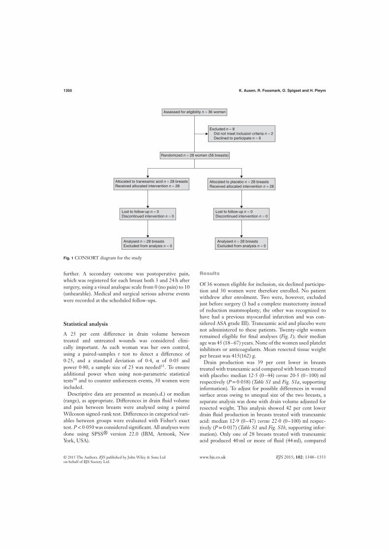
1350 K. Ausen, R. Fossmark, O. Spigset and H. Pleym
Assessed for eligibility n = 36 women
Excluded n = 8 Did not meet inclusion criteria n = 2 Declined to participate n = 6
Analysed n = 28 breastsExcluded from analysis n = 0
Lost to follow-up n = 0Discontinued intervention n = 0
Allocated to tranexamic acid n = 28 breastsReceived allocated intervention n = 28
Lost to follow-up n = 0Discontinued intervention n = 0
Allocated to placebo n = 28 breastsReceived allocated intervention n = 28
Analysed n = 28 breastsExcluded from analysis n = 0
Randomized n = 28 women (56 breasts)
Fig. 1 CONSORT diagram for the study
further. A secondary outcome was postoperative pain,which was registered for each breast both 3 and 24 h aftersurgery, using a visual analogue scale from 0 (no pain) to 10(unbearable). Medical and surgical serious adverse eventswere recorded at the scheduled follow-ups.
Statistical analysis
A 25 per cent difference in drain volume betweentreated and untreated wounds was considered clini-cally important. As each woman was her own control,using a paired-samples t test to detect a difference of0⋅25, and a standard deviation of 0⋅4, α of 0⋅05 andpower 0⋅80, a sample size of 23 was needed33. To ensureadditional power when using non-parametric statisticaltests34 and to counter unforeseen events, 30 women wereincluded.Descriptive data are presented as mean(s.d.) or median
(range), as appropriate. Differences in drain fluid volumeand pain between breasts were analysed using a pairedWilcoxon signed-rank test. Differences in categorical vari-ables between groups were evaluated with Fisher’s exacttest. P< 0⋅050 was considered significant. All analyses weredone using SPSS® version 22.0 (IBM, Armonk, NewYork, USA).
Results
Of 36 women eligible for inclusion, six declined participa-tion and 30 women were therefore enrolled. No patientwithdrew after enrolment. Two were, however, excludedjust before surgery (1 had a complete mastectomy insteadof reduction mammoplasty; the other was recognized tohave had a previous myocardial infarction and was con-sidered ASA grade III). Tranexamic acid and placebo werenot administered to these patients. Twenty-eight womenremained eligible for final analyses (Fig. 1); their medianage was 45 (18–67) years. None of the women used plateletinhibitors or anticoagulants. Mean resected tissue weightper breast was 415(162) g.Drain production was 39 per cent lower in breasts
treated with tranexamic acid compared with breasts treatedwith placebo: median 12⋅5 (0–44) versus 20⋅5 (0–100) mlrespectively (P= 0⋅038) (Table S1 and Fig. S1a, supportinginformation). To adjust for possible differences in woundsurface areas owing to unequal size of the two breasts, aseparate analysis was done with drain volume adjusted forresected weight. This analysis showed 42 per cent lowerdrain fluid production in breasts treated with tranexamicacid: median 12⋅9 (0–47) versus 22⋅0 (0–100) ml respec-tively (P= 0⋅017) (Table S1 and Fig. S1b, supporting infor-mation). Only one of 28 breasts treated with tranexamicacid produced 40ml or more of fluid (44ml), compared
© 2015 The Authors. BJS published by John Wiley & Sons Ltd www.bjs.co.uk BJS 2015; 102: 1348–1353on behalf of BJS Society Ltd.

Topical tranexamic acid after reduction mammoplasty 1351
with nine of 28 placebo-treated breasts (P= 0⋅016). Two ofthese in the placebo group required reoperation and evac-uation of haematoma.Pain scores were similar in breasts treated with tranex-
amic acid or placebo on the day of surgery (median 2⋅5(0–6) versus 2⋅0 (0–6); P= 0⋅179) and after 24 h (2⋅0 (0–6)versus 2⋅0 (0–5⋅5) respectively; P= 0⋅428).No adverse effects were recorded after topical tranexamic
acid. Five patients had a wound infection treated withantibiotics or outpatient wound drainage within 6 weeks ofsurgery. Another six women had a superficial inflammatoryreaction related to the sutures. These had no relation totreatment with tranexamic acid or placebo. One womanhad bilateral wound seromas which were evacuated.
Discussion
The present study showed that topical application oftranexamic acid significantly reduced wound drainage afterreduction mammoplasty.The main strength of the study is the model. Surface
wounds can be diverse (burns, massive weight loss surgery),and it is challenging to design a study with equivalentwounds. Women undergoing bilateral reduction mammo-plasty provide two almost identical wounds in an ordinaryclinical setting. A small study can thus provide adequatestatistical power.The study, however, has several weaknesses. It was con-
ducted using a surgical procedure that is not usually asso-ciated with major bleeding. Drain volumes, not blood loss,were measured; drain fluids consist of both blood and tran-sudates, with a transition over time to more serous drainfluid. The breasts had also been injected with local anaes-thetic containing adrenaline (epinephrine), but with equalvolumes in each breast. The constituents of the drain fluidswere not analysed, but registration took place during thefirst 24 h when the drainage is mostly blood. Whether top-ical tranexamic acid can influence transudate production oreven seroma formation through its inhibition of fibrinolysisremains unknown.The wounds after reduction mammoplasty are not com-
pletely identical, particularly when two surgeons operate,one side each. To correct for this, the surgeons undertookhaemostasis on the contralateral side, and a separate analy-sis of drain volumes was done with adjustment for resectedtissue weight.Serum concentrations of tranexamic acid were not mea-
sured. Systemic absorption of topical tranexamic acid couldin theory affect bleeding from the contralateral placebowound. In vitro, a minimum concentration of 5–10 μg/mlis needed to inhibit fibrinolysis1,7. Studies in orthopaedic
and cardiac surgery have found a negligible systemic con-centration from application of boluses of tranexamic acidin confined spaces14,15. The tranexamic acid dose in thepresent study was 20ml of 25mg/ml, a total of 500mg; thismethod should give a very low systemic concentration14,15.The overall reduction in drain fluid production of about
40 per cent after topical administration of tranexamicacid here accords with previously published studies3,16–20,which consistently reported a reduction in transfusionneed and measurable bleeding of between 30 and 40 percent after both intravenous and topical tranexamic acidadministration. There is little information on the concen-tration of tranexamic acid needed for topical effect. Instil-lation of a bolus of 1–3 g tranexamic acid diluted in 100mlsaline (concentration 10–30mg/ml) has been studied inpatients undergoing cardiac15,16 and orthopaedic17 surgery,whereas epistaxis has been treated with sponges moist-ened with undiluted tranexamic acid for intravenous use(100mg/ml)23. This present study opted for a high con-centration of 25mg/ml, but still dilute enough to provide avolume sufficient to moisten large surface areas. There arefew published studies where the mode of application wascomparable to the moistening used in this study. A mouth-wash containing 4⋅8mg/ml tranexamic acid was effectiveafter dental extraction28, whereas Hinder and Tschopp26
found no significant effect of gargling 1⋅7mg/ml tranex-amic acid solution after tonsillectomy. Athanasiadis andcolleagues27 reported a significant effect after spraying thewound surfaces with tranexamic acid in endoscopic sinussurgery. The optimal concentration remains unknown.The use of drains after reduction mammoplasty has little
scientific evidence35 but is nevertheless common. At thetime of study, the departmental routine was to use drainsuntil fluid production was below 40ml per 24 h. Thepresent study suggests that topical tranexamic acid reducesdrain fluid production after reduction mammoplasty tobelow this cut-off value in almost all patients, andmay obvi-ate the need for a drain.There were two postoperative bleeds in the study, both
in untreated breasts. The study was not powered to eval-uate whether topical tranexamic acid could prevent post-operative haematoma. Ngaage and Bland4 reported thatintravenous tranexamic acid reduced the risk of reopera-tion by 48 per cent after coronary surgery, and a significantreduction in rebleeding after tonsillectomy was describedfor the lysine analogue aminocaproic acid36. Few studieson tranexamic acid have reported rebleeding as an outcomemeasure3,37.Tranexamic acid is inexpensive. Its cost-effectiveness has
been thoroughly documented in orthopaedic surgery21.Even after operations where bleeding is less common,
© 2015 The Authors. BJS published by John Wiley & Sons Ltd www.bjs.co.uk BJS 2015; 102: 1348–1353on behalf of BJS Society Ltd.
1352 K. Ausen, R. Fossmark, O. Spigset and H. Pleym
topical application of tranexamic acid may reduce the needfor drains and outpatient visits. This simple method haspotential for widespread application after surgery.
Acknowledgements
The authors thank K. Fonstad for including all the patientsin this study, I. Spets for randomization and prepara-tion of drugs, and E. Saetnan, K. Sneve, C. Gonzales,H. Nordgaard and K. Gullestad for their contribution tothe study. This research was conducted using funding fromthe Department of Plastic and Reconstructive Surgery atSt Olav’s University Hospital.Disclosure: The authors declare no conflict of interest.
References
1 Dunn CJ, Goa KL. Tranexamic acid: a review of its use insurgery and other indications. Drugs 1999; 57: 1005–1032.
2 Ker K, Prieto-Merino D, Roberts I. Systematic review,meta-analysis and meta-regression of the effect oftranexamic acid on surgical blood loss. Br J Surg 2013; 100:1271–1279.
3 Ker K, Edwards P, Perel P, Shakur H, Roberts I. Effect oftranexamic acid on surgical bleeding: systematic review andcumulative meta-analysis. BMJ 2012; 344: e3054.
4 Ngaage DL, Bland JM. Lessons from aprotinin: is theroutine use and inconsistent dosing of tranexamic acidprudent? Meta-analysis of randomised and large matchedobservational studies. Eur J Cardiothorac Surg 2010; 37:1375–1383.
5 Horrow JC, Van Riper DF, Strong MD, Grunewald KE,Parmet JL. The dose–response relationship of tranexamicacid. Anesthesiology 1995; 82: 383–392.
6 Poeran J, Rasul R, Suzuki S, Danninger T, Mazumdar M,Opperer M et al. Tranexamic acid use and postoperativeoutcomes in patients undergoing total hip or kneearthroplasty in the United States: retrospective analysis ofeffectiveness and safety. BMJ 2014; 349: g4829.
7 Yee BE, Wissler RN, Zanghi CN, Feng C, Eaton MP. Theeffective concentration of tranexamic acid for inhibition offibrinolysis in neonatal plasma in vitro. Anesth Analg 2013;117: 767–772.
8 Andersson L, Eriksson O, Hedlund PO, Kjellman H,Lindqvist B. Special considerations with regard to thedosage of tranexamic acid in patients with chronic renaldiseases. Urol Res 1978; 6: 83–88.
9 Dowd NP, Karski JM, Cheng DC, Carroll JA, Lin Y, JamesRL et al. Pharmacokinetics of tranexamic acid duringcardiopulmonary bypass. Anesthesiology 2002; 97: 390–399.
10 Murkin JM, Falter F, Granton J, Young B, Burt C, Chu M.High-dose tranexamic acid is associated with nonischemicclinical seizures in cardiac surgical patients. Anesth Analg2010; 110: 350-353.
11 Henry DA, Carless PA, Moxey AJ, O’Connell D, Stokes BJ,Fergusson DA et al. Anti-fibrinolytic use for minimisingperioperative allogeneic blood transfusion. Cochrane DatabaseSyst Rev 2011; (3)CD001886.
12 Koster A, Schirmer U. Re-evaluation of the role ofantifibrinolytic therapy with lysine analogs during cardiacsurgery in the post aprotinin era. Curr Opin Anaesthesiol2011; 24: 92–97.
13 Ker K, Roberts I. Tranexamic acid for surgical bleeding.BMJ 2014; 349: g4934.
14 Wong J, Abrishami A, El Beheiry H, Mahomed NN,Roderick Davey J, Gandhi R et al. Topical application oftranexamic acid reduces postoperative blood loss in totalknee arthroplasty: a randomized, controlled trial. J BoneJoint Surg Am 2010; 92: 2503–2513.
15 De Bonis M, Cavaliere F, Alessandrini F, Lapenna E,Santarelli F, Moscato U et al. Topical use of tranexamic acidin coronary artery bypass operations: a double-blind,prospective, randomized, placebo-controlled study. J ThoracCardiovasc Surg 2000; 119: 575–580.
16 Ipema HJ, Tanzi MG. Use of topical tranexamic acid oraminocaproic acid to prevent bleeding after major surgicalprocedures. Ann Pharmacother 2012; 46: 97–107.
17 Gomez-Barrena E, Ortega-Andreu M, Padilla-Eguiluz NG,Pérez-Chrzanowska H, Figueredo-Zalve R. Topicalintra-articular compared with intravenous tranexamic acidto reduce blood loss in primary total knee replacement: adouble-blind, randomized, controlled, noninferiority clinicaltrial. J Bone Joint Surg Am 2014; 96: 1937–1944.
18 Wind TC, Barfield WR, Moskal JT. The effect oftranexamic acid on blood loss and transfusion rate in primarytotal knee arthroplasty. J Arthroplasty 2013; 28: 1080–1083.
19 Wang H, Shen B, Zeng Y. Comparison of topical versusintravenous tranexamic acid in primary total kneearthroplasty: a meta-analysis of randomized controlled andprospective cohort trials. Knee 2014; 21: 987–993.
20 Alshryda S, Sukeik M, Sarda P, Blenkinsopp J, Haddad FS,Mason JM. A systematic review and meta-analysis of thetopical administration of tranexamic acid in total hip andknee replacement. Bone Joint J 2014; 96-b: 1005–1015.
21 Tuttle JR, Ritterman SA, Cassidy DB, Anazonwu WA,Froehlich JA, Rubin LE. Cost benefit analysis of topicaltranexamic acid in primary total hip and knee arthroplasty.J Arthroplasty 2014; 29: 1512–1515.
22 Abrishami A, Chung F, Wong J. Topical application ofantifibrinolytic drugs for on-pump cardiac surgery: asystematic review and meta-analysis. Can J Anaesth 2009; 56:202–212.
23 Zahed R, Moharamzadeh P, Alizadeharasi S, Ghasemi A,Saeedi M. A new and rapid method for epistaxis treatmentusing injectable form of tranexamic acid topically: arandomized controlled trial. Am J Emerg Med 2013; 31:1389–1392.
24 Sarris I, Arafa A, Konaris L, Kadir RA. Topical use oftranexamic acid to control perioperative local bleeding in
© 2015 The Authors. BJS published by John Wiley & Sons Ltd www.bjs.co.uk BJS 2015; 102: 1348–1353on behalf of BJS Society Ltd.

Topical tranexamic acid after reduction mammoplasty 1353
gynaecology patients with clotting disorders: two cases.Haemophilia 2007; 13: 115–116.
25 Tang YM, Chapman TW, Brooks P. Use of tranexamic acidto reduce bleeding in burns surgery. J Plast Reconstr AesthetSurg 2012; 65: 684–686.
26 Hinder D, Tschopp K. [Topical application of tranexamicacid to prevent post-tonsillectomy haemorrhage.]Laryngorhinootologie 2015; 94: 86–90.
27 Athanasiadis T, Beule AG, Wormald PJ. Effects of topicalantifibrinolytics in endoscopic sinus surgery: a pilotrandomized controlled trial. Am J Rhinol 2007; 21:737–742.
28 Patatanian E, Fugate SE. Hemostatic mouthwashes inanticoagulated patients undergoing dental extraction. AnnPharmacother 2006; 40: 2205–2210.
29 Barnard J, Millner R. A review of topical hemostatic agentsfor use in cardiac surgery. Ann Thorac Surg 2009; 88:1377–383.
30 Sterling JP, Heimbach DM. Hemostasis in burn surgery – areview. Burns 2011; 37: 559–565.
31 Walsh K, Nikkhah D, Dheansa B. What is the evidence fortranexamic acid in burns? Burns 2014; 40: 1055–1057.
32 Kjersti A. 20 ml Fluid on AquaDoodle Canvas.https://youtu.be/10AkwMQSE1s [accessed 1 March 2015].
33 Paired t-test. http://www.biomath.info/power/prt.htm[accessed 2 January 2012].
34 Lehmann EL. Comparing two treatments of attributes in apopulation model. In: Nonparametrics: Statistical Methods
Based on Ranks, Lehmann EL (ed). Springer: New York,1998; 76–81.
35 Stojkovic CA, Smeulders MJ, Van der Horst CM, Khan SM.Wound drainage after plastic and reconstructive surgery ofthe breast. Cochrane Database Syst Rev 2013; (3)CD007258.
36 Overbosch HC, Hart HC. Local application ofepsilon-aminocaproic acid (E.A.C.A.) in the prevention ofhaemorrhage after tonsillectomy. J Laryngol Otol 1970; 84:905–908.
37 Ker K, Beecher D, Roberts I. Topical application oftranexamic acid for the reduction of bleeding. CochraneDatabase Syst Rev 2013; (7)CD010562.
Supporting information
Additional supporting information may be found in the online version of this article:
Table S1 Drain fluid production measured 24 h after surgery in patients undergoing bilateral reductionmammoplasty (Word document)
Fig. S1 a Drain fluid production measured 24 h after surgery and b drain fluid volume adjusted for resected tissueweight (Word document)
Editor’s comments
This paper, and the FIBER Study preceding it (Br J Surg 2015; 102: 1338–1347), both explore pharmacological meansto reduce perioperative bleeding. BJS does not normally publish cardiac papers, but makes an exception because of thehigh quality of the study. Although essentially a negative trial, the FIBER Study has valuable clinical implications, andis a reminder of the importance of publishing such trials. This study by Ausen et al. also has a favoured within-patientrandomization design using an established agent (tranexamic acid) topically. This study has a positive result (medianreduction of 8ml in drain fluid) but the clinical relevance remains unclear, as does whether it would stand up to aformal cost-benefit analysis. Minimizing perioperative bleeding is fundamental, and the methods used in both theseRCTs have relevance in other specialties and procedures. Only by conducting high-quality research can the role ofpharmacological agents be clarified.
J. J. EarnshawJoint Chief Editor, BJS
© 2015 The Authors. BJS published by John Wiley & Sons Ltd www.bjs.co.uk BJS 2015; 102: 1348–1353on behalf of BJS Society Ltd.
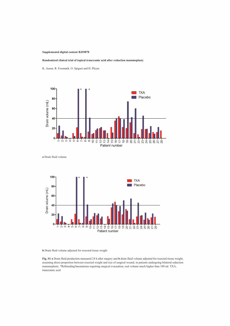
Supplemental digital content BJS9878
Randomized clinical trial of topical tranexamic acid after reduction mammoplasty
K. Ausen, R. Fossmark, O. Spigset and H. Pleym
a Drain fluid volume
b Drain fluid volume adjusted for resected tissue weight
Fig. S1 a Drain fluid production measured 24 h after surgery and b drain fluid volume adjusted for resected tissue weight, assuming direct proportion between resected weight and size of surgical wound, in patients undergoing bilateral reduction mammoplasty. *Rebleeding/haematoma requiring surgical evacuation; real volume much higher than 100 ml. TXA, tranexamic acid

Table S1 Drain fluid production measured 24 h after surgery in patients undergoing bilateral reduction mammoplasty
Patient no.
Drain fluid volume (ml) Weight-adjusted drain fluid volume
(ml) Resected tissue (g) Tranexamic acid Placebo Tranexamic acid Placebo Tranexamic acid Placebo
1 10.0 25.0 10.0 22.1 612 692 2 5.0 15.0 5.0 16.5 207 188 3 4.0 2.0 4.0 2.3 315 275 4 Excluded 5 0.0 0.0 0.0 0.0 635 600 6 Excluded 7 10.0 4.0 10.0 4.0 78 80 8 21.5 Rebleeding* 21.5 Rebleeding* 607 445 9 10.0 2.5 10.0 2.4 238 244 10 2.0 Rebleeding* 2.0 Rebleeding* 670 636 11 13.0 41.0 16.0 41.0 367 450 12 8.0 10.0 6.7 10.0 457 380 13 16.0 19.0 16.0 23.0 376 326 14 20.0 22.0 18.3 22.0 347 318 15 15.0 15.5 11.4 15.5 624 476 16 10.0 0.0 10.0 0.0 471 306 17 22.0 11.0 16.3 11.0 222 164 18 35.0 40.0 35.0 43.3 419 387 19 44.0 40.0 47.0 40.0 433 462 20 22.0 38.0 26.7 38.0 376 457 21 20.0 74.0 20.0 50.0 506 750 22 31.5 42.0 31.5 28.1 432 646 23 10.0 60.0 10.0 53.8 475 530 24 7.5 2.0 9.6 2.0 339 433 25 18.0 45.0 15.8 45.0 727 640 26 18.0 18.5 17.2 18.5 535 512 27 10.0 24.0 8.9 24.0 496 443 28 12.0 8.5 12.0 8.8 242 234 29 12.0 22.0 13.8 22.0 340 390 30 15.0 0.0 15.0 0.0 250 293 *Drain fluid volume considerably higher than 100 ml.

Comment and reply to Study !
Comments to Study 1:
We read with interest the randomised controlled trial regarding the use of topical
tranexamic acid in breast reduction surgery by Ausen et al. (1), but found some
inconsistencies in the statistical analyses that may mislead readers. Firstly, the authors
appropriately analysed the efficacy of the intervention over control using a paired Wilcoxon
signed-rank test, but incorrectly used an unpaired method to estimate its magnitude. They
reported the unpaired 8ml difference of medians of drain output between the two groups
(39% reduction). However, the paired median difference was only 2.5ml (12% reduction).
Analysis of a paired study must not ignore the level of dependency within each pair.
ermine the trial sample size
was much underestimated. Using the trial data we conservatively calculated a bootstrap
estimate of the actual variance as 1.09 (95% confidence interval (CI): 0.26-1.93), excluding
the three placebo group cases with 0ml. The assumed variance was far below the 95% CI of
around 30%. If we conservatively assume a variance of 0.9, then the required study size is
113 patients, 4 times larger. The 2.5ml median reduction in drain output in this study may
not accurately represent the true effect size, since the variance was very large and the study
very underpowered. Based on the promising preliminary data from this study, an adequately
powered study to definitively answer the hypothesis should now be conducted.
Alexander HR Varey
Serigne N Lo
University of New South Wales
University of Sydney, Melanoma Institute Australia
40 Rocklands Road
North Sydney
2060 NSW
Australia
References:
1. Ausen K, Fossmark R, Spigset O, Pleym H. Randomized clinical trial of topical tranexamic
acid after reduction mammoplasty. Br J Surg 2015; 102: 1348-1353.
Reply:
Thank you for giving us the opportunity to further clarify some aspects of our study (1). This
small study attempted to provide a proof of concept, and there was a significant difference
in drain production between wounds treated with tranexamic acid and placebo (p=0.038).
Reduction mammoplasties generally bleed little and are not in need of haemostatic
interventions, but it is one of few rare models where two almost identical wounds coexist in
the same patient. We agree that median difference should be calculated from the paired
numbers. However, to report the paired median difference in this material clearly
underreports the magnitude of the difference that exists in favour of tranexamic acid. If one
sorts the patients in the study according to difference in drain volume between their two
breasts, the differences in drain volume to the right of the median far exceeds the
differences to the left of the median.
Looking at the paired difference between a treated and untreated breast here, the absolute
paired difference is generally considerably larger in breasts with larger bleeding volumes
and then in favour of tranexamic acid. In 13 of the 15 patients in whom one or both breasts
had more than 20 ml drain production, the difference was in favour of tranexamic acid. Our
power calculations were performed in advance, and it should again be emphasized that we
found a statistically significant difference. In future studies, new power calculations based
upon the findings in the present study should obviously be carried out. Larger studies are
needed; preferably in patient groups with expected larger bleeding volumes.
Kjersti Ausen
Reidar Fossmark
Olav Spigset
Hilde Pleym
Department of Plastic and Reconstructive Surgery

PO Box 3250 Sluppen
N-7006 Trondheim
Norway
Reference:
1 Ausen K, Fossmark R, Spigset O, Pleym H. Randomized clinical trial of topical tranexamic
acid after reduction mammoplasty. BJS 2015; 102: 1348-1353.


Paper II


1
Randomized clinical trial of topical moistening of mastectomy wounds with diluted tranexamic acid
to reduce bleeding
Kjersti Ausen1,6 MD, Anne I. Hagen2 MD, PhD, Heidi S. Østbyhaug2 MD, Sverrir Olafsson3 MD, Berit J.
Kvalsund3 MD, Olav Spigset4,7 MD, PhD, Hilde Pleym5,6, MD, PhD
Author affiliations:
1
Trondheim, Norway
2
Norway
3Section for Breast and Endocrine Surgery, Department of Surgery, Ålesund Hospital, Møre and
Romsdal Hospital Trust, Ålesund, Norway
4Department
5
6Department of Circulation and Medical Imaging, Norwegian University of Science and Technology -
NTNU, Trondheim, Norway
7Department of Clinical and Molecular Medicine, Norwegian University of Science and Technology -
NTNU, Trondheim, Norway
Correspondence and requests for reprints to: Dr. K. Ausen, Section for Plastic and Reconstructive
Su -7006 Trondheim,
Norway (e-mail: [email protected] Phone: +47 92249693)

2
Source of Funding: Kjersti Ausen has received a PhD grant from The Liaison Committee between the
Central Norway Regional Health Authority (RHA) and the Norwegian University of Science and
Technology-NTNU, Trondheim, Norway (2016/29014). For the remaining authors none source of
funding or conflicts of interest were declared.
Manuscript category: Randomized clinical trial. ClinicalTrials.gov (NCT02627560)
Previous presentation of the trial: An abstract entitled «Topical moistening of the wound surface
with tranexamic acid 25 mg/ml reduces bleeding equal to intravenous administration and may reduce
need for re-operation of postoperative haemorrhage» was presented as an abstract and a single
slide/four minute oral presentation at the European Association of Plastic Surgeons Research Council
in Helsinki, Finland on May 21st 2019. This abstract included results both from this study and our
former pilot study.

3
Abstract
Background Topical administration of tranexamic acid (TXA) may be an alternative to intravenous
administration to reduce bleeding with less risk of systemic adverse events. The objective of this
study was to investigate whether moistening a surgical wound with TXA 25 mg/ml prior to closure,
leaving a thin film of drug only, reduces postoperative bleeding.
Methods This was a two-centre stratified, parallel-group, placebo-controlled, double-blind
randomized clinical trial. Patients undergoing mastectomy with/without axillary lymph node
clearance were randomized 1:1 to moistening of wound surface prior to closure with either TXA 25
mg/ml or NaCl 0.9% (placebo). Primary endpoint was postoperative bleeding as measured by drain
production first 24 hours. Secondary endpoints were early hematoma, total drain production,
postoperative complications, and late seroma aspirations within 3 months.
Results Between January 1st 2016 and August 31st 2018, 208 patients were randomized. Two
patients were converted to a different surgical procedure perioperatively, and four patients did not
receive intervention due to technical errors. Thus, 202 patients were included in the study (101
received TXA and 101 received placebo). TXA reduced drain production at 24 hours (110 vs. 144 ml,
effect size 34 ml, 95% CI 8 to 60 ml, P=0.010). Early hematoma occurred in one TXA patient versus
seven placebo patients (OR 0.13, 95% CI 0.02 to 1.07, P=0.057). There was no significant difference in
postoperative complications between TXA and placebo (13 vs 10, OR 1.11, 95% CI 0.45 to 2.73,
P=0.824) or need for late seroma aspirations (79.3 vs 66.6%, OR 1.83, 95% CI 0.91 to 3.68, P=0.089).
Conclusion Moistening the wound prior to closure with TXA 25 mg/ml reduces postoperative
bleeding within first 24 hours in patients undergoing mastectomy.
ClinicalTrials.gov (NCT02627560)

4
Introduction
In healthy patients without coagulation deficiencies, the only systemically administered
pharmacological intervention to universally reduce surgical bleeding with an acceptable safety profile
is antifibrinolytic drugs1. Tranexamic acid (TXA) has been the drug of choice since the withdrawal of
aprotinin from the market in 20072. Intravenous (iv) use of TXA reduces surgical bleeding and need
for blood transfusion with about one third3 and is routinely used in surgery associated with
significant blood loss. Even high doses of iv TXA has not been associated with increased incidence of
thromboembolic events, but large prospective studies designed to particularly evaluate this risk are
lacking4. A dose-dependent increase in non-ischemic seizures has been described after iv TXA in
cardiac surgery5-7. Although minimization of blood loss is desirable in all surgery, general use of iv
TXA has not been advocated due to unease regarding possible systemic adverse effects8, 9.
A prophylactic intervention to minimize postoperative blood loss should be low-cost, easy, efficient
and safe. Topical use of TXA may provide a high drug concentration on the wound surface with
negligible systemic concentrations10. Topical use is still off-label with no consensus as to optimal TXA
concentration in the solution applied, mode of application, or duration of contact11, 12. Most
publications come from joint replacement surgery, where instilling TXA as a bolus into the joint
reduces bleeding equal to iv administration13-15. We have previously introduced a novel procedure
where the wound surface is simply moistened with 25 mg/ml tranexamic acid prior to closure,
finding a mean 39% reduction in drain volume in a small -of- randomized placebo-
controlled study in bilateral reduction mammoplasties16. Such moistening exposes the wound surface
to TXA for a considerably shorter time than topical boluses10.
The aim of this study was to investigate the effect of our novel moistening method in a larger
population and in a different study model. Mastectomy was chosen as a suitable study model for
bleeding as it is a common and standardized surgical procedure with homogenous wounds and little

5
occult blood loss. As postoperative seroma is a major adverse event after mastectomy17, 18, we also
wanted to investigate whether topical TXA would influence seroma formation.
Methods
The study was a two-centre stratified, parallel-group, placebo-controlled, double-blind randomized
clinical trial. The study was registered in ClinicalTrials.gov (NCT02627560) and approved by the
involved departments, the Regional Committee for Medical and Health Research Ethics in Mid
Norway (2015/1722) and the Norwegian Medicines Agency (15/11405-7). The trial was performed in
accordance with the principles of the Declaration of Helsinki and was monitored in accordance with
the Good Clinical Practice directive of the European Medicines Agency.
Study population
Patients above 18 years of age who were to undergo simple mastectomy (SM), mastectomy with
sentinel node biopsy (SNB) or axillary lymph node clearance (AC) were consecutively identified from
the operation planning registries at
(Centre B) between January 1st 2016 and August 31st 2018. Exclusion criteria were 1) known
thromboembolic disease or high risk of thromboembolism warranting extra anticoagulation in
connection with the procedure, 2) pregnancy/nursing and 3) known allergy to TXA. Eligible patients
were informed about the study after diagnosis but prior to surgery by nurses and doctors connected
to the respective breast cancer centres. Patients were enrolled if written informed consent was
obtained.
Treatment allocation

6
Computer generated randomization was done in permuted blocks of 10, 20 or 50 patients stratified
according to study centre. Sealed and numbered opaque randomization envelopes were produced
accordingly. Details on screening and randomization are shown in Fig.1. All randomization and
organization of electronical Case Report Forms was performed by the Unit of Applied Clinical
Research19 at the Norwegian University of Science and Technology-NTNU, Trondheim, Norway.
Participants and all personnel involved in surgery and postoperative follow-up, data collection and
statistical analysis were blinded to the randomization.
Study interventions
Participants were randomly assigned in a 1:1 ratio to moistening of their wound surface prior to
closure with either 20 ml of TXA 25 mg/ml or NaCl 0.9% (placebo). Opening of the randomization
envelope and drug preparation was done by personnel not connected to the surgery, postoperative
follow-up, data collection or statistical analyses. Envelopes were opened in numerical order. TXA 25
mg/ml was prepared by extracting 5 ml from a 20 ml bottle of saline (NaCl 0.9%) and adding 5 ml of
tranexamic acid 100 mg/ml to the same bottle, thus obtaining 20 ml of TXA 25 mg/ml. Placebo was
an identical 20 ml bottle of saline, which was perforated by a needle for identical appearance.
The moistening method is illustrated in this video: https://www.youtube.com/watch?v=-
8MAE3NAHfQ. All involved surgeons were visually instructed on the details of the moistening
technique by watching this video, and also instructed to cover all surfaces and use the entire 20 ml
although much would spill. Breast specimen weight, height and width were measured during surgery.
Wound surface area was calculated as an ellipse (height/2 x width/2 ). Patients received active
vacuum drains (Exudrain FG 14, Wellspect, Oslo, Norway) marked with the exact timepoint to
register drain production 24 h after completion of the surgery. Preoperative evaluation, performance
of the surgery, postoperative treatment and drain removal were otherwise in accordance with

7
prevailing routine at the participating study centres and randomization was therefore stratified
according to study centre.
It was discussed whether to sub-stratify the randomization according to type of surgical procedure as
well, as the presence or absence of lymph node clearance is known to have a significant impact on
drain output. As randomization for practical purposes was done preoperatively and a planned
procedure might be converted perioperatively, we chose to rather adjust for type of surgery in the
statistical analyses.
Study outcomes
The primary outcome was postoperative bleeding as defined by the volume of drain production the
first 24 hours after surgery. Secondary outcomes were total drain production and drain time, early
hematoma, postoperative complications and seroma formation. Seroma was defined as fluid
accumulation warranting aspiration after drain removal. Chronic seroma was defined as persistent
seroma at three months. Patients experiencing postoperative complications defined as hematomas,
infections and wound ruptures were excluded from the seroma analyses as these may influence
seroma formation.
Drain production was recorded at 24 hours after termination of surgery and on a daily basis
thereafter until drain removal. Variables which according to protocol could influence the defined
outcomes were obtained from the medical record (Table 1). The staff was instructed to accurately
document seroma aspirations throughout the study period, and patients were instructed to request
accurate measuring should they have aspirations performed elsewhere. Patient follow-up lasted for
three months and all patients received a final phone call to ensure or correct registered data and
identify unregistered adverse events.

8
An evaluation of the surgical techniques at the two centres had been performed before study
initiation. Use of tumescence, dissection with diathermia and choice of surgical levels had been
found to be comparable. However, during data collection it was noted that Centre B seemed to have
notably lower drain productions. A post-hoc re-evaluation revealed that Centre B routinely applied a
circular compression bandage for the first 24 h postoperatively, which had not been recognized
during the study design phase. Postoperative compression is an active intervention to reduce
bleeding but was not accurately registered in the medical records nor accurately remembered by
patients in retrospect. It could therefore not be added as a post-hoc variable at the individual
patient level but had to be regarded an inherent factor when adjusting data for study centre.
Statistical analyses
A between-group difference in mean drain production of 25% was considered clinically significant.
Estimating a standard deviation of 0.620, alpha of 0.05 and power 0.80, a sample size of 92 patients in
each group was needed21. We planned to include a total of 210 patients to ensure additional power.
Continuous patient characteristics data were calculated as mean ± standard deviation (SD) and
compared using independent samples t-tests. Categorical patient characteristics data were
calculated as frequency counts and percentages and compared using chi-square tests, but variables
with 5 cases were analysed using Fisher exact tests.
Continuous outcome data are presented as mean (95% Confidence Interval (CI)). Categorical
outcome data are presented as frequency counts and percentages. For presentation of unadjusted
effect size of continuous data, mean difference (95% CI) between groups is used. Effect size of
categorical data is presented as odds ratio (OR) (95% CI). For statistical analyses of unadjusted effect
size, continuous outcome data were analysed using independent samples t-tests while categorical
outcome data were compared using chi-square tests or Fisher exact tests where appropriate.

9
For adjusted statistical analyses, non-normally distributed continuous data underwent logarithmic
(ln) transformation. Data were then analysed using a general linear model. We included all variables
registered in Table 1 and performed a stepwise removal of non-significant variables (P
obtain a final model that included only individually significant variables. Results are calculated as
differences between means as percentage (95% CI). Non-normally distributed continuous data that
could not undergo logarithmic transformation due to the appearance of zero values were
dichotomized and analysed using a logistic regression model adjusting for significant variables.
Results are given as mean difference or odds ratio (OR) with 95% CI. Two-tailed P-values < 0.05 were
considered statistically significant. Analyses were performed in accordance to
principle. Analyses were performed using SPSS version 25 (IBM SPSS Statistics for Windows, IBM
Corp., Armonk, NY, USA).
Results
Between January 1st 2016 and August 31st 2018, 208 patients were randomized, and 202 were
included in the study (Fig. 1). Of these, 101 received TXA and 101 received placebo. Patient
characteristics are presented in Table 1. Patients receiving TXA were on average 3.9 years older (P =
0.033), otherwise there were no differences between the groups. Patient data stratified for study
centre and type of surgery defined as AC or SM/SNB are presented in Tables S1-S2.
Outcome Data
Outcome data are presented in Table 2. Data stratified for type of surgery and study centre are
presented in Tables S3 S5. Mean drain production at 24 h was 110 ± 67 ml in the TXA group versus
144 ± 113 ml in the placebo group (mean difference 34 ml (95% 8 to 60 ml, P = 0.011).
10
The following patient variables independently and significantly affected outcome: 1) Administration
of TXA, 2) type of surgery, 3) study centre, 4) wound surface area and 5) surgeon seniority.
Perioperative anticoagulation did not affect the outcome variables. The independent significances of
effect of all patient variables on the primary outcome is presented in Table S6.
Outcome variables adjusted for the significant patient variables showed that moistening the wound
surface with TXA significantly reduced 24 h drain production with 32.4% (P < 0.001) (Table 2) . While
total drain volume was not significantly reduced in the unadjusted analyses, adjusted outcome
showed that total drain volume was reduced with 33.0% (P = 0.003). Patients in the TXA group were
significantly more likely to have drains removed on the first day (OR 3.0, P = 0.003) and had a
significantly shorter drain period (-15.6%, P = 0.017).
Early hematomas warranting re-operation occurred in seven placebo patients and one TXA patient (P
= 0.057). There were no differences between groups regarding other complications. No
thromboembolic events were registered.
Subgroup analyses
Prior to study initiation, AC was assumed to significantly influence drain production and subgroup
analyses were planned according to type of surgery. The recognition of the significant influence of
study centre led to an additional post-hoc subgroup analysis according to study centre. The adjusted
effect of type of surgery and study centre on outcome variables are presented in Table S7.
Subgroup analysis for type of surgery showed that patients undergoing AC and receiving TXA had
significant effect on drain production at 24 h (-26.7%, P = 0.026) albeit less than in the SM/SNB group
(-33.4%, P=0.001) (Table 3). Total drain production was not significantly reduced in patients
undergoing AC (-27.3%, P = 0.244) while it was significantly reduced in the SM/SNB patients (-32.3%,
P=0.005). The subpopulation who had undergone lymph node clearance had significantly increased

11
odds compared to placebo of both needing seroma aspiration (OR 5.7, P = 0.032) and of a cumulative
OR 5.7, P = 0.011) but no increased risk of chronic seroma (OR
1.5, P = 0.671).
Patients operated at Centre A had 60.3% higher drain production at 24 h than patients from Centre B
(P <0.001) (Table S7). Centre A practiced a significantly higher threshold value for ongoing drain
production at removal (97.4% more; P < 0.001) and kept drains significantly shorter (-29.8%, P <0
.001) than Centre B, yet Centre A had a 33.5% larger cumulative drain volume at the time of removal
(P = 0.013). This difference was postulated to be influenced by the postoperative compression used
at Centre B.
Patients who neither received compression nor underwent lymph node clearance were the SM/SNB
patients at Centre A (n=108, TXA=52, Placebo=56). To illustrate the isolated effect of TXA we
separately analysed this subpopulation (Table 4), and found a 39% reduction in 24-hour drain
production (P = 0.001), and a 46% reduction in total drain output (P = 0.001).
Discussion
To our knowledge, we are the first to examine the effect of merely leaving a thin film of TXA fluid on
the wound surface before closure16. This confirmatory study demonstrates that a simple single
moistening of a wound surface with TXA 25 mg/ml significantly reduces both postoperative bleeding
and total drain production with about one third. This is comparable to the effect of established
intravenous prophylactic use of TXA22 and is thus an alternative mode of administration. We also
postulate that topical TXA may reduce the risk of postoperative hematomas warranting reoperations,
as 7 out of 8 re-bleedings occurred in placebo patients, and in our previous pilot study16 the two re-
bleedings observed were both in the placebo group. Hence, in our two intervention studies in 258
breasts in In
12
the few existing studies on use of iv TXA in breast surgery, a reduction in bleeding and also
haematomas has been suggested20, 23, 24.
An unexpected finding was a possible negative effect of TXA on lymph leakage. The subgroup of TXA
patients who underwent lymph node clearance had less beneficial effect of TXA on postoperative
drain production, and they were later significantly more likely to need seroma aspiration and had an
increased cumulative seroma volume, although we saw no increase in chronic seromas.
The purpose of this study was not to demonstrate reduced bleeding in mastectomies per se.
Minimizing perioperative bleeding is fundamental, and the method has relevance in many
procedures other than breast surgery. Mastectomy is however a suitable study model where
homogenous wounds and little occult bleeding will provide relatively unbiased results. A major
strength of our study was the identification of significant effect modifying variables such as wound
surface area, performance of lymph node clearance and inherent differences between study centres.
Drain production is a surrogate variable for bleeding and could be considered a weakness of this
study. Topical application of TXA at the end of surgery will naturally not affect perioperative
bleeding and hence comparing pre- and postoperative haemoglobin concentrations would not be an
appropriate measure on the effect of this mode of administration. Drain fluids consist of both blood
and transudate with a transition to more serous fluids over time. We therefore regarded drain
production at 24 h as a more appropriate measure of postoperative bleeding than total drain
production.
A majo
as a separate variable, as this was recognized in retrospect to be a routine procedure at Centre B.
Closing of dead space reduces bleeding25. Compression is thus an active intervention which may
interact with the effect of TXA, possibly contributing to the reduced drain production and the lower
beneficial effect of TXA observed at Centre B. Use of compression to reduce bleeding and seroma has
been somewhat forfeited in breast surgery the few existing studies showing little effect particularly

13
on late seroma26, 27. However, these studies describe ongoing compression for several days, which
may induce both discomfort and secondary reactions to ischemia. Moreover, seroma is a different
endpoint than postoperative bleeding. A proper randomized evaluation of the effect of a 24 h
postoperative compression bandage as practiced at Centre B on postoperative bleeding and
haematoma is warranted.
When analysing only the subpopulation which had neither lymph node clearance nor postoperative
compression (SM/SNB patients at Centre A), the isolated effect of TXA increased to a 39% reduction
in 24-hour drain production, and a 46% reduction in total drain output. This may be the most
appropriate model for the true isolated effect of topical TXA.
Studies on topical use of TXA have been published since the 19 13. Topical administration has
however mostly been done by instilling boluses into closed spaces such as joints or the mediastinum,
application of soaked gauze or repeated irrigation13. As topical use is off-label and the possible local
effects of prolonged tissue exposure are largely unknown, keeping drug concentration and contact
time as low as possible would be a sensible precaution when advocating routine prophylactic use.
While bolus and gauze administrations may lead to prolonged exposure to TXA, we have in a
previous study demonstrated that our topical moistening both has predictable and swift elimination
and renders systemic concentrations negligible10.
Although there was no increase in wound ruptures, wound infections or chronic seromas in the TXA
group, the increase in seroma volume after TXA exposure in lymph node clearance patients raise the
question of whether tissue adhesion or healing of lymph vessels may be delayed from topical
application of TXA. As our population may have presented with exceedingly high rates of seroma due
to the practice of early drain removal (>70% needed at least one seroma aspiration thereafter), our
findings need confirmation from other populations with different drain protocols.
Whether TXA may have unrecognised antiadhesive properties need further exploration. TXA inhibits
plasminogen, which is ubiquitous in tissue matrix and has numerous functions beyond cleavage of

14
fibrin. Studies have suggested that TXA may affect cell adhesion28, 29, and inhibition of TXA on tumour
growth and spread was investigated in the 1970s and 80s30. We made the unexpected observation in
a previous study that prolonged exposure to high concentrations of topical TXA caused lack of re-
epithelialization and even epithelial detachment in an ex vivo human skin wound model31.
TXA does not only bind to plasminogen but may act as a competitive antagonist to the inhibitory
neurotransmitters GABAA and glycine5, 32 in the central nervous system (CNS). Both topical
application of TXA to the brain in animal experimental settings33, 34 and intrathecal accidental
administration of TXA in humans35 have been shown to cause seizures, and topical TXA should not be
used in surgery with exposed CNS.
These findings demonstrate that the local effect of TXA is insufficiently explored and that caution
may be warranted with regard to dose and exposure time.
In conclusion, we propose simple moistening of most surgical wound surface prior to closure with
TXA 25 mg/ml as a low-cost, simple and quick routine prophylactic measure to reduce bleeding and
possibly prevent re-operation due to haemorrhage. Further research must determine the lowest
effective topical dose when using a moistening technique. Adequately powered studies or meta-
analyses on the ability of topical TXA to prevent reoperations due to bleeding are also lacking, and
possible adverse effects of topical application need further exploration.
Acknowledgements
The authors thank the Unit of Applied Clinical Research at the Norwegian University of Science and
Technology-NTNU for invaluable help with randomization, data handling and statistical analyses.
15
References
1. Ferraris VA, Brown JR, Despotis GJ, Hammon JW, Reece TB, Saha SP, Song HK, Clough ER, Shore-Lesserson LJ, Goodnough LT, Mazer CD, Shander A, Stafford-Smith M, Waters J, Baker RA, Dickinson TA, FitzGerald DJ, Likosky DS, Shann KG. 2011 update to the Society of Thoracic Surgeons and the Society of Cardiovascular Anesthesiologists blood conservation clinical practice guidelines. The Annals of thoracic surgery 2011;91(3): 944-982. 2. Murkin JM. Lessons learned in antifibrinolytic therapy: The BART trial. Seminars in
cardiothoracic and vascular anesthesia 2009;13(2): 127-131. 3. Ker K, Prieto-Merino D, Roberts I. Systematic review, meta-analysis and meta-regression of the effect of tranexamic acid on surgical blood loss. The British journal of surgery 2013;100(10): 1271-1279. 4. Ker K, Roberts I. Tranexamic acid for surgical bleeding. BMJ (Clinical research ed) 2014;349: g4934. 5. Lecker I, Wang DS, Whissell PD, Avramescu S, Mazer CD, Orser BA. Tranexamic acid-associated seizures: Causes and treatment. Annals of neurology 2016;79(1): 18-26. 6. Sharma V, Katznelson R, Jerath A, Garrido-Olivares L, Carroll J, Rao V, Wasowicz M, Djaiani G. The association between tranexamic acid and convulsive seizures after cardiac surgery: a multivariate analysis in 11 529 patients. Anaesthesia 2014;69(2): 124-130. 7. Takagi H, Ando T, Umemoto T. Seizures associated with tranexamic acid for cardiac surgery: a meta-analysis of randomized and non-randomized studies. The Journal of cardiovascular surgery
2017;58(4): 633-641. 8. Gerstein NS, Kelly SP, Brierley JK. Yet Another Tranexamic Acid-Related Thrombotic Complication. Journal of cardiothoracic and vascular anesthesia 2016;30(3): e21-22. 9. Myers SP, Kutcher ME, Rosengart MR, Sperry JL, Peitzman AB, Brown JB, Neal MD. Tranexamic acid administration is associated with an increased risk of posttraumatic venous thromboembolism. The journal of trauma and acute care surgery 2019;86(1): 20-27. 10. Ausen K, Pleym H, Liu J, Hegstad S, Nordgard HB, Pavlovic I, Spigset O. Serum Concentrations and Pharmacokinetics of Tranexamic Acid after Two Means of Topical Administration in Massive Weight Loss Skin-Reducing Surgery. Plast Reconstr Surg 2019;143(6): 1169e-1178e. 11. Montroy J, Hutton B, Moodley P, Fergusson NA, Cheng W, Tinmouth A, Lavallee LT, Fergusson DA, Breau RH. The efficacy and safety of topical tranexamic acid: A systematic review and meta-analysis. Transfusion medicine reviews 2018. 12. Nouraei SM. What Are the Optimal Dose of Administration and Time of Drainage for Topical Tranexamic Acid in Patients Undergoing Cardiac Surgery? The Korean journal of thoracic and
cardiovascular surgery 2017;50(6): 477-478. 13. Ker K, Beecher D, Roberts I. Topical application of tranexamic acid for the reduction of bleeding. The Cochrane database of systematic reviews 2013;7: Cd010562. 14. Li J, Zhang Z, Chen J. Comparison of efficacy and safety of topical versus intravenous tranexamic acid in total hip arthroplasty: A meta-analysis. Medicine 2016;95(36): e4689. 15. Lin C, Qi Y, Jie L, Li HB, Zhao XC, Qin L, Jiang XQ, Zhang ZH, Ma L. Is combined topical with intravenous tranexamic acid superior than topical, intravenous tranexamic acid alone and control groups for blood loss controlling after total knee arthroplasty: A meta-analysis. Medicine
2016;95(51): e5344. 16. Ausen K, Fossmark R, Spigset O, Pleym H. Randomized clinical trial of topical tranexamic acid after reduction mammoplasty. The British journal of surgery 2015;102(11): 1348-1353. 17. Kuroi K, Shimozuma K, Taguchi T, Imai H, Yamashiro H, Ohsumi S, Saito S. Pathophysiology of seroma in breast cancer. Breast cancer (Tokyo, Japan) 2005;12(4): 288-293. 18. van Bemmel AJ, van de Velde CJ, Schmitz RF, Liefers GJ. Prevention of seroma formation after axillary dissection in breast cancer: a systematic review. European journal of surgical oncology : the
16
journal of the European Society of Surgical Oncology and the British Association of Surgical Oncology
2011;37(10): 829-835. 19. https://www.ntnu.edu/mh/akf/randomisering [February 15th 2019]. 20. Oertli D, Laffer U, Haberthuer F, Kreuter U, Harder F. Perioperative and postoperative tranexamic acid reduces the local wound complication rate after surgery for breast cancer. The
British journal of surgery 1994;81(6): 856-859. 21. http://www.biomath.info/power/prt.htm. [15.01 2012]. 22. Ker K, Edwards P, Perel P, Shakur H, Roberts I. Effect of tranexamic acid on surgical bleeding: systematic review and cumulative meta-analysis. BMJ (Clinical research ed) 2012;344: e3054. 23. Knight H, Banks J, Muchmore J, Ives C, Green M. Examining the use of intraoperative tranexamic acid in oncoplastic breast surgery. The breast journal 2019. 24. Wolter A, Scholz T, Pluto N, Diedrichson J, Arens-Landwehr A, Liebau J. Subcutaneous mastectomy in female-to-male transsexuals: Optimizing perioperative and operative management in 8 years clinical experience. J Plast Reconstr Aesthet Surg 2018;71(3): 344-352. 25. Kuroi K, Shimozuma K, Taguchi T, Imai H, Yamashiro H, Ohsumi S, Saito S. Effect of mechanical closure of dead space on seroma formation after breast surgery. Breast cancer (Tokyo,
Japan) 2006;13(3): 260-265. 26. Chen CY, Hoe AL, Wong CY. The effect of a pressure garment on post-surgical drainage and seroma formation in breast cancer patients. Singapore medical journal 1998;39(9): 412-415. 27. O'Hea BJ, Ho MN, Petrek JA. External compression dressing versus standard dressing after axillary lymphadenectomy. Am J Surg 1999;177(6): 450-453. 28. Cox S, Cole M, Mankarious S, Tawil N. Effect of tranexamic acid incorporated in fibrin sealant clots on the cell behavior of neuronal and nonneuronal cells. Journal of neuroscience research
2003;72(6): 734-746. 29. Lishko VK, Novokhatny VV, Yakubenko VP, Skomorovska-Prokvolit HV, Ugarova TP. Characterization of plasminogen as an adhesive ligand for integrins alphaMbeta2 (Mac-1) and alpha5beta1 (VLA-5). Blood 2004;104(3): 719-726. 30. Peterson H, Sundbeck A. Mechanisms Behind the Inhibiting Effect of Tranexamic Acid on Tumour Growth and Spread. In: Metastasis Developments in Oncology, Hellman K, Hilgard P, Eccles S (eds), vol 4. Springer: Dordrecht, 1980. 31. Eikebrokk TA, Vassmyr BS, Ausen K, Gravastrand C, Spigset O, Pukstad B. Cytotoxicity and effect on wound re-epithelialization after topical administration of tranexamic acid. BJSOpen In Press. 32. Furtmuller R, Schlag MG, Berger M, Hopf R, Huck S, Sieghart W, Redl H. Tranexamic acid, a widely used antifibrinolytic agent, causes convulsions by a gamma-aminobutyric acid(A) receptor antagonistic effect. The Journal of pharmacology and experimental therapeutics 2002;301(1): 168-173. 33. Pellegrini A, Giaretta D, Chemello R, Zanotto L, Testa G. Feline generalized epilepsy induced by tranexamic acid (AMCA). Epilepsia 1982;23(1): 35-45. 34. Schlag MG, Hopf R, Redl H. Convulsive seizures following subdural application of fibrin sealant containing tranexamic acid in a rat model. Neurosurgery 2000;47(6): 1463-1467. 35. Mahmoud K, Ammar A. Accidental intrathecal injection of tranexamic Acid. Case reports in
anesthesiology 2012;2012: 646028.

17
Legend to figure
Figure 1 Flow diagram of enrolment and randomization
List of supporting information
Project Protocol
Table S1 Patient characteristics at baseline stratified according to type of surgery and study
centre
Table S2 Patient characteristics at baseline stratified according to type of surgery at Centre A
and Centre B
Table S3 Primary and secondary outcome measures stratified for type of surgery crude data
Table S4 Centre A. Primary and secondary outcome measures stratified for type of surgery
crude data
Table S5 Centre B. Primary and secondary outcome measures stratified for type of surgery
crude data
Table S6 Independent effect of patient variables as main effect in a general linear model on
primary outcome
Table S7 Subgroup analyses. Adjusted primary and secondary outcomes according to type of
surgery and study centre

18
Figure 1 Flow diagram of enrolment and randomization
Centre A n=239
Centre B n=81
Excluded n=79 Did not meet inclusion criteria n=48 Declined to participate n=10 Other reason n=21
Excluded n=33 Did not meet inclusion criteria n=21 Declined to participate n=1 Other reason n=11
Allocated to tranexamic acid n=105 Received tranexamic acid n=101 Did not receive intervention n=4 - due to surgical anomalies n=1 - due to technical errors n=3
Allocated to placebo n=103 Received placebo n=101 Did not receive intervention n=2 - due to surgical anomalies n=1 - due to technical errors n=1
Analysed primary outcome (drain production first 24 h) n=101 Excluded from further analysis n=7
- due to evacuation of postoperative hematoma n=7
Analysed secondary outcome (drain output and late complications) n=94 Excluded from seroma analyses due to late complications n=10 Analysed secondary outcome (seroma) n=84
Analysed primary outcome (drain production first 24 h) n=101 Excluded from further analysis n=1
- due to evacuation of postoperative hematoma n=1
Analysed secondary outcome (drain output and late complications) n=100 Excluded from seroma analyses due to late complications n=13 Analysed secondary outcome (seroma) n=87
Randomized n=208
Assessed for eligibility n=320
Lost to follow-up n=0 Lost to follow-up n=0
Enro
lmen
t A
lloca
tio
n
Follo
w-u
p A
nal
ysis

19
Table 1. Patient characteristics at baseline
TXA Placebo
Characteristic (n=101) (n=101)
Female sex 98 (97.0) 100 (99.0)
Age* (yr) 66.2 (13.3) 62.3 (12.8)
Body mass index (kg/m2)* 26.9 (4.9) 27.1 (4.7)
Active smoker 19 (18.8) 13 (12.9)
Recruited at Centre A 77 (76.2) 77 (76.2)
Operated by senior surgeon 50 (49.5) 50 (49.5)
Neoadjuvant treatment 32 (31.7) 41 (40.6)
Irradiated tissue 14 (13.9) 11 (10.9)
Perioperative anticoagulation 32 (31.7) 37 (36.6)
Axillary clearance 34 (33.7) 31 (30.7)
Weight of breast specimen* (g) 780 (450) 746 (359)
Wound surface* (cm2) 292 (117) 279 (84)
TXA = tranexamic acid Data are number (%) unless stated otherwise * Data are mean (standard deviation)
Axillary component included

20
Table 2. Outcome data
Primary outcome TXA (n=101) Placebo (n=101) Effect size
(95% CI)
TXA vs. Placebo
Adjusted difference
(95% CI)
TXA vs. Placebo
Drain production first 24h (ml) Mean (95% CI)
110 (97 to 123)
144 (122 to 167)
-34 ml (-60 to -8) P=0.011
-32.4% (-51.4 to -15.8) P<0.001
Secondary outcomes* TXA (n=100) Placebo (n=94)
Early hematoma* 1 (1.0) 7 (6.9) OR 0.13 (0.02 to 1.11)
P=0.065§
OR 0.13 (0.02 to 1.07)
P=0.057# Total drain production (ml) Mean (95% CI)
189 (145 to 234)
214 (165 to 264)
-25 ml (-91 to 41)
P=0.461
-33.0% (-60.0 to -10.4)
P=0.003 Days with drain (d) Mean (95% CI)
1.7 (1.4 to 1.9)
1.8 (1.6 to 2.1)
-0.1 d (-0.7 to 0.2)
P=0.341
-15.6% (-30.2 to -2.6)
P=0.017
Drain removed at 24h 65 (65.0) 47 (50.0)
OR 1.86 (1.04 to 3.31) P=0.035¶
OR 3.00 (1.44 to 6.22)
P=0.003# Drain production last 24h before removal (ml) Mean (95% CI)
93 (82 to 105)
91 (80 to 102)
2 ml (-13 to 18) P=0.783
-6.4% (-22.3 to 8.1) P=0.083
Late hematoma/postoperative infection/wound rupture
13 (13.0) 10 (10.6)
OR 1.26 (0.52 to 3.02) P=0.721¶
OR 1.11 (0.45 to 2.73)
P=0.824#
Thromboembolic event 0 (0.0) 0 (0.0)
Seroma TXA (n=87) Placebo (n=84)
Seroma aspiration was needed 69 (79.3) 56 (66.6) OR 1.92 (0.96 to 3.82)
P=0.062¶ OR 1.83 (0.91 to 3.68)
P=0.089#
19 (21.8) 17 (20.2) OR 1.10 (0.53 to 2.30)
P=0.797¶ OR 0.87 (0.38 to 2.00)
P=0.740#
34 (39.1) 20 (23.8) OR 2.05 (1.06 to 3.98)
P=0.033¶ OR 1.99 (0.94 to 4.23)
P=0.073#
Chronic seroma 6 (6.9) 11 (13.1) OR 0.49 (0.17 to 1.40)
P=0.182¶ OR 0.41 (0.14 to 1.21)
P=0.107#
Data are number (%) unless stated otherwise. TXA = tranexamic acid * Patients with early hematoma calculated from all patients; patients with early hematoma excluded from other secondary postoperative outcome registrations.
-test. Difference between means (95% CI).
§ exact test. Odds ratio comparing TXA to placebo (95% CI)
¶ Chi square test. Odds ratio comparing TXA to placebo (95% CI) Regression analyses- adjusted according to axillary clearance, study centre, wound surface area and surgeon seniority. General linear model. Ratio between ln mean as percentage difference (95% CI).
# Logistic regression model. Odds ratio comparing TXA to placebo (95% CI).

21
Table 3. Exploratory subgroup analysis. Adjusted primary and secondary outcomes according to administration of TXA vs. placebo, stratified for type of surgery.
SM/SNB (n=137) AC (n=65)
Outcome TXA (n=67) vs placebo (n=70) TXA (n=34) vs placebo (n=31)
Primary outcome
Drain production first
-33.4% (-58.1 to -12.6)
P=0.001
-26.7% (- 55.9 to -2.9)
P=0.026 Secondary outcomes
Early hematoma §
n=1 vs n=3 P=0.620
n=0 vs n=4
P=0.046
-32.3% (-62.4 to -9.4) P=0.005
-27.3% (-91.9 to 18.4)
P=0.244
Days with drain*
-11.6% (-24.9 to -0.2) P=0.054
-23.0% (-65.4 to 9.2)
P= 0.166
Drain removed at 24 h
2.41 (1.02 to 5.68) P=0.044
5.70 (1.27 to 25.6)
P=0.023
Drain volume last 24 h before removal*
-11.3% (-30.3 to 5.3)
P=0.185
5.2% (-39.9 to 26.4)
P=0.721 Late hematoma/ postoperative infection/ wound rupture
1.01 (0.33 to 3.06)
P=0.985
1.27 (0.24 to 6.62)
P=0.778 Seroma¶
Seroma aspiration needed§
1.32 (0.59 to 2.97)
P=0.500
5.71 (1.16 to 28.2)
P=0.032
§
0.47 (0.15 to 1.50) P=0.203
1.88 (0.54 to 6.49)
P=0.320
ml§
1.08 (0.41 to 2.81)
P=0.880
5.72 (1.50 to 21.9)
P=0.011
Chronic seroma§
0.18 (0.04 to 0.89) P=0.035
1.50 (0.23 to 9.73)
P=0.671 TXA = tranexamic acid, AC = axillary clearance, SM/SNB = simple mastectomy/sentinel node biopsy * Ratio between ln mean as percentage difference comparing group I to group II (95% CI). Univariate general linear model adjusted for study centre and wound surface area.
Cases with early hematoma calculated from all patients; patients with early hematoma excluded from
other secondary postoperative outcome registrations. § Odds ratio comparing group I to group II (95% CI). Logistic regression model adjusted for study centre and wound surface area. ¶ Patients with early or late hematoma, infection or wound rupture excluded from seroma registrations.

22
Table 4. Exploratory subgroup analysis. Adjusted primary and secondary outcomes according to administration of TXA vs. placebo in SM/SNB patients at Centre A.
Outcome TXA (n=52) vs placebo (n=56)
Primary outcome
Drain production first
-39.1% (-69.2 to -14.3)
P=0.001 Secondary outcomes
Early hematoma §
n=0 vs n=3
P=0.244
-46.1% (-80.9 to -17.9) P=0.001
Days with drain*
-23.0% (-83.0 to -9.5) P=0.001
Drain removed at 24 h
6.6 (2.07 to 21.22) P=0.001
Drain volume last 24 h before removal*
-9.4% (-30.2 to 8.8)
P=0.309 Late hematoma/ postoperative infection/ wound rupture
n=5 vs n=6
P=1.000 Seroma¶
Seroma aspiration needed§
1.11 (0.44 to 2.79)
P=0.821
§
0.47 (0.15 to 1.50) P=0.203
Cumulative ml§
1.05 (0.38 to 2.90)
P=0.930
Chronic seroma§
0.18 (0.04 to 0.89) P=0.035
TXA = tranexamic acid, SM/SNB = simple mastectomy/sentinel node biopsy * Ratio between ln mean as percentage difference comparing group I to group II (95% CI). Univariate general linear model adjusted for drug wound surface area.
all patients;
patients with early hematoma excluded from other secondary postoperative outcome registrations. § Odds ratio comparing group I to group II (95% CI). Logistic regression model adjusted for wound surface area. ¶ Patients with early or late hematoma, infection or wound rupture excluded from seroma registrations.
analysis.

Ta
ble
S1
Pa
tie
nt
cha
ract
eri
stic
s a
t b
ase
line
str
ati
fie
d a
cco
rdin
g t
o t
ype
of
surg
ery
an
d s
tud
y ce
ntr
e
Str
ati
fie
d a
cco
rdin
g t
o t
yp
e o
f su
rge
ry
All
ca
ses
(n=
20
2)
S
M/S
NB
(n
=1
37
)
AC
(n
=6
5)
C
om
pa
riso
n s
urg
ery
(n
=2
02
)
Ch
ara
cte
rist
ic
TX
A (
n=
10
1)
Pla
ceb
o (
n=
10
1)
T
XA
(n
=6
7)
Pla
ceb
o (
n=
70
)
TX
A (
n=
34
) P
lace
bo
(n
=3
1)
S
M/S
NB
(n
=1
37
) A
C (
n=
65
)
Fem
ale
se
x
98
(9
7.0
) 1
00
(9
9.0
)
65
(9
7.0
) 6
9 (
98
.6)
3
3 (
97
.1)
31
(1
00
)
13
4 (
97
.8)
64
(9
8.5
)
Ag
e*
(yr
)
66
.2 (
13
.3)†
6
2.3
(1
2.8
)
66
.2 (
11
.6)
62
.3 (
1.9
)
66
.2 (
15
.9)
62
.3 (
14
.8)
6
4.2
(1
1.9
) 6
4.3
(1
5.4
)
Bo
dy
ma
ss in
de
x (k
g/m
2)*
2
6.9
(4
.9)
27
.1 (
4.7
)
26
.6 (
4.7
) 2
6.7
(4
.4)
2
7.5
(5
.2)
28
.0 (
5.5
)
26
.7 (
4.5
) 2
7.7
(5
.3)
Act
ive
sm
oke
r
19
(1
8.8
) 1
3 (
12
.9)
1
3 (
19
.4)
9 (
12
.9)
6
(1
7.6
) 4
(1
2.9
)
22
(1
6.1
) 1
0 (
15
.4)
Re
cru
ite
d a
t C
en
tre
A
77
(7
6.2
) 7
7 (
76
.2)
1
5 (
22
.4)
14
(2
0.0
)
9 (
26
.5)
10
(3
2.3
)
29
(2
1.2
) 1
9 (
29
.2)
Op
era
ted
by
sen
ior
surg
eo
n
50
(4
9.5
) 5
0 (
49
.5)
3
6 (
53
.7)
37
(5
2.8
)
14
(4
1.2
) 1
3 (
41
.9)
7
3 (
53
.3)
27
(4
1.5
)
Ne
oa
dju
van
t tr
ea
tme
nt
3
2 (
31
.7)
41
(4
0.6
)
14
(2
0.9
) 2
0 (
28
.6)
1
8 (
52
.9)
21
(6
7.7
)
34
(2
4.8
)†
39
(6
0.0
)
Irra
dia
ted
tis
sue
14
(1
3.9
) 1
1 (
10
.9)
1
2 (
17
.9)
10
(1
4.3
)
2 (
5.9
) 1
(3
.2)
2
2 (
16
.1)†
3
(4
.6)
Pe
rio
pe
rati
ve a
nti
coa
gu
lati
on
3
2 (
31
.7)
37
(3
6.6
)
15
(2
2.3
) 2
1 (
30
.0)
1
7 (
50
.0)
16
(5
1.6
)
36
(2
6.3
)†
33
(5
0.8
)
We
igh
t o
f b
rea
st s
pe
cim
en
*‡
(g
)
78
0 (
45
0)
74
6 (
35
9)
7
32
(4
54
) 6
98
(3
39
)
87
5 (
43
2)
85
7 (
38
3)
7
15
(3
98
)†
86
6 (
40
7)
Wo
un
d s
urf
ace
*‡
(cm
2)
2
92
(1
17
) 2
79
(8
4)
2
62
(1
00
) 2
58
(7
3)
3
50
(1
27
) 3
28
(8
7)
2
60
± 8
7†
3
40
(1
09
)
Str
ati
fie
d a
cco
rdin
g t
o s
tud
y c
en
tre
A
ll c
ase
s (n
=2
02
)
Ce
ntr
e A
(n
=1
54
)
Ce
ntr
e B
(n
=4
8)
C
om
pa
riso
n s
tud
y c
en
tre
(n
=2
02
)
Ch
ara
cte
rist
ic
TX
A (
n=
10
1)
Pla
ceb
o (
n=
10
1)
T
XA
(n
=6
7)
Pla
ceb
o (
n=
70
)
TX
A (
n=
34
) P
lace
bo
(n
=3
1)
C
en
tre
A (
n=
15
4)
Ce
ntr
e B
(n
=4
8)
Fem
ale
se
x
98
(9
7.0
) 1
00
(9
9.0
)
76
(9
8.7
) 7
6 (
98
.7)
2
2 (
91
.7)
24
(1
00
.0)
1
52
(9
8.7
) 4
6 (
95
.8)
Ag
e*
(yr
)
66
.2 (
13
.3)
62
.3 (
12
.8)
6
5.6
(1
3.6
) 6
2.9
(1
3.0
)
68
.2 (
11
.3)
60
.3 (
12
.2)
6
4.2
(1
3.3
) 6
4.3
(1
2.3
)
Bo
dy
ma
ss in
de
x (k
g/m
2)*
2
6.9
(4
.9)
27
.1 (
4.7
)
26
.8 (
4.9
) 2
7.1
(4
.7)
2
7.4
(5
.0)
27
.1 (
4.9
)
26
.9 (
4.8
) 2
7.2
(4
.9)
Act
ive
sm
oke
r
19
(1
8.8
) 1
3 (
12
.9)
1
3 (
16
.7)
9 (
11
.7)
6
(2
5.0
) 4
(1
6.7
)
22
(1
4.3
) 1
0 (
20
.8)
Op
era
ted
by
sen
ior
surg
eo
n
50
(4
9.5
) 5
0 (
49
.5)
3
3 (
42
.9)
32
(4
1.6
)
17
(7
0.8
) 1
8 (
75
.0)
6
5 (
42
.2)†
3
5 (
72
.9)
Ne
oa
dju
van
t tr
ea
tme
nt
3
2 (
31
.7)
41
(4
0.6
)
26
(3
3.8
) 3
2 (
41
.6)
6
(2
5.0
) 9
(3
7.5
)
58
(3
7.7
) 1
5 (
31
.3)
Irra
dia
ted
tis
sue
14
(1
3.9
) 1
1 (
10
.9)
1
2 (
15
.6)
6 (
7.8
)
2 (
8.3
) 5
(2
0.8
)
18
(1
1.7
) 7
(1
4.6
)
Pe
rio
pe
rati
ve a
nti
coa
gu
lati
on
3
2 (
31
.7)
37
(3
6.6
)
26
(3
3.8
) 3
3 (
42
.9)
6
(2
5.0
) 4
(1
6.7
)
59
(3
8.3
)†
10
(2
0.8
)
Axi
llary
cle
ara
nce
3
4 (
33
.7)
31
(3
0.7
)
21
(2
7.3
) 2
5 (
32
.5)
9
(3
7.5
) 1
0 (
41
.7)
4
6 (
29
.9)
19
(3
9.6
)
We
igh
t o
f b
rea
st s
pe
cim
en
*‡
(g
)
78
0 (
45
0)
74
6 (
35
9)
7
56
(3
89
) 7
46
(3
64
)
86
0 (
60
8)
74
8 (
35
0)
7
51
(3
76
) 8
04
(4
94
)
Wo
un
d s
urf
ace
*‡
(cm
2)
2
92
(1
17
) 2
79
(8
4)
2
89
(1
14
) 2
77
(8
4)
3
01
(1
26
) 2
87
(8
3)
2
83
(1
00
) 2
94
(1
06
)
TX
A =
tra
ne
xam
ic a
cid
, A
C =
axi
llary
cle
ara
nce
, SM
/SN
B =
sim
ple
ma
ste
cto
my/
sen
tin
el n
od
e b
iop
sy
Da
ta a
re n
um
be
r (%
) u
nle
ss s
tate
d o
the
rwis
e
*D
ata
are
me
an
(st
an
da
rd d
evi
ati
on
)
†Si
gn
ific
an
t d
iffe
ren
ce
‡A
xilla
ry c
om
po
ne
nt
incl
ud
ed
Ta
ble
S2
P
ati
en
t ch
ara
cte
rist
ics
at
ba
selin
e s
tra
tifi
ed
acc
ord
ing
to
typ
e o
f su
rge
ry a
t C
en
tre
A a
nd
Ce
ntr
e B
C
en
tre
A:
Str
ati
fie
d a
cco
rdin
g t
o t
yp
e o
f su
rge
ry
All
ca
ses
(n=
15
4)
S
M/S
NB
(n
=1
08
)
AC
(n
=4
6)
C
om
pa
riso
n s
urg
ery
(n
=1
54
)
Ch
ara
cte
rist
ic
TX
A (
n=
77
) P
lace
bo
(n
=7
7)
T
XA
(n
=5
2)
Pla
ceb
o (
n=
56
)
TX
A (
n=
25
) P
lace
bo
(n
=2
1)
S
M/S
NB
(n
=1
08
) A
C (
n=
46
)
Fem
ale
se
x
76
(9
8.7
) 7
6 (
98
.7)
5
1 (
98
.1)
55
(9
8.2
)
25
(1
00
.0)
21
(1
00
.0)
1
06
(9
8.1
) 4
6 (
10
0.0
)
Ag
e*
(yr
)
65
.6 (
13
.6)
62
.9 (
13
.0)
6
5.8
(1
1.9
) 6
2.7
(1
1.8
)
65
.2 (
16
.9)
63
.4 (
16
.1)
6
4.2
(1
1.9
) 6
4.4
(1
6.4
)
Bo
dy
ma
ss in
de
x (k
g/m
2)*
2
6.8
(4
.9)
27
.1 (
4.7
)
26
.3 (
4.5
) 2
6.8
(4
.5)
2
7.9
(5
.5)
28
.0 (
5.4
)
26
.5 (
4.4
) 2
8.0
(5
.4)
Act
ive
sm
oke
r
13
(1
6.7
) 9
(1
1.7
)
8 (
15
.4)
6 (
10
.7)
5
(2
0.0
) 3
(1
4.3
)
14
(1
3.0
) 8
(1
7.4
)
Op
era
ted
by
sen
ior
surg
eo
n
33
(4
2.9
) 3
2 (
41
.6)
2
4 (
46
.2)
25
(4
4.6
)
9 (
36
.0)
7 (
33
.3)
4
9 (
45
.4)
16
(3
4.8
)
Ne
oa
dju
van
t tr
ea
tme
nt
2
6 (
33
.8)
32
(4
1.6
)
12
(2
3.1
) 1
6 (
28
.6)
1
4 (
56
.0)
16
(7
6.2
)
28
(2
5.9
)†
30
(6
5.2
)
Irra
dia
ted
tis
sue
12
(1
5.6
) 6
(7
.8)
1
0 (
19
.2)
6 (
10
.7)
2
(8
.7)
0 (
0.0
)
16
(1
4.8
) 2
(4
.3)
Pe
rio
pe
rati
ve a
nti
coa
gu
lati
on
2
6 (
33
.8)
33
(4
2.9
)
14
(2
6.9
) 2
0 (
35
.7)
1
2 (
48
.0)
13
(6
1.9
)
34
(3
1.5
)†
25
(5
4.3
)
We
igh
t o
f b
rea
st s
pe
cim
en
*‡
(g
)
75
6 (
38
9)
74
6 (
36
4)
6
86
(3
47
) 6
98
(3
39
)
90
0 (
43
9)
87
4 (
40
4)
6
92
(3
41
)†
88
8 (
41
8)
Wo
un
d s
urf
ace
*‡
(cm
2)
28
9 (
11
4)
27
7 (
84
)
25
4 (
89
) 2
53
(7
2)
3
63
(1
27
) 3
38
(8
6)
2
54
(8
0)†
3
52
(1
10
)
C
en
tre
B:
Str
ati
fie
d a
cco
rdin
g t
o t
yp
e o
f su
rge
ry
All
ca
ses
(n=
48
)
SM
/SN
B (
n=
29
)
AC
(n
=1
9)
C
om
pa
riso
n s
urg
ery
(n
=4
8)
Ch
ara
cte
rist
ic
TX
A (
n=
24
) P
lace
bo
(n
=2
4)
T
XA
(n
=1
5)
Pla
ceb
o (
n=
14
)
TX
A (
n=
9)
Pla
ceb
o (
n=
10
)
SM
/SN
B (
n=
29
) A
C (
n=
19
)
Fem
ale
se
x
22
(9
1.7
) 2
4 (
10
0.0
)
14
(9
3.3
) 1
4 (
10
0.0
)
8 (
88
.9)
10
(1
00
.0)
2
8 (
96
.6)
18
(9
4.7
)
Ag
e*
(yr
)
68
.2 (
11
.3)†
6
0.3
(1
2.2
)
67
.6 (
10
.8)
60
.6 (
12
.8)
6
9.1
(1
2.9
) 5
9.9
(1
2.0
)
64
.2 (
12
.1)
64
.3 (
13
.0)
Bo
dy
ma
ss in
de
x (k
g/m
2)*
2
7.4
(5
.0)
27
.1 (
4.9
)
27
.9 (
5.6
) 2
6.5
(4
.2)
2
6.6
(4
.2)
28
.0 (
5.8
)
27
.2 (
4.9
) 2
7.3
(5
.0)
Act
ive
sm
oke
r
6 (
25
.0)
4 (
16
.7)
5
(3
3.3
) 3
(2
1.4
)
1 (
11
.1)
1 (
10
.0)
8
(2
7.6
) 2
(1
0.5
)
Op
era
ted
by
sen
ior
surg
eo
n
17
(7
0.8
) 1
8 (
75
.0)
1
2 (
80
.0)
12
(8
5.7
)
5 (
55
.6)
6 (
60
.0)
2
4 (
82
.6)
11
(5
7.9
)
Ne
oa
dju
van
t tr
ea
tme
nt
6
(2
5.0
) 9
(3
7.5
)
2 (
13
.3)
4 (
28
.6)
4
(4
4.4
) 5
(5
0.0
)
6 (
20
.7)
9 (
47
.4)
Irra
dia
ted
tis
sue
2 (
8.3
) 5
(2
0.8
)
2 (
13
.3)
4 (
28
.6)
0
(0
.0)
1 (
10
.0)
6
(2
0.7
) 1
(5
.3)
Pe
rio
pe
rati
ve a
nti
coa
gu
lati
on
6
(2
5.0
) 4
(1
6.7
)
1 (
6.7
) 1
(7
.1)
5
(5
5.6
) 3
(3
0.0
)
2 (
6.7
)†
8 (
42
.1)
We
igh
t o
f b
rea
st s
pe
cim
en
*‡
(g
)
86
0 (
60
8)
74
8 (
35
0)
8
93
(7
05
) 6
95
(3
51
)
80
5 (
43
1)
82
2 (
35
4)
7
98
(5
62
) 8
13
(3
81
)
Wo
un
d s
urf
ace
*‡
(cm
2)
30
1 (
12
6)
28
7 (
83
)
29
2 (
12
9)
27
4 (
79
)
31
4 (
12
7)
30
5 (
88
)
28
4 (
10
6)
31
0 (
10
5)
TX
A =
tra
ne
xam
ic a
cid
, A
C =
axi
llary
cle
ara
nce
, SM
/SN
B =
sim
ple
ma
ste
cto
my/
sen
tin
el n
od
e b
iop
sy
Da
ta a
re n
um
be
r (%
) u
nle
ss s
tate
d o
the
rwis
e
*D
ata
are
me
an
(st
an
da
rd d
evi
ati
on
)
†Si
gn
ific
an
t d
iffe
ren
ce
‡A
xilla
ry c
om
po
ne
nt
incl
ud
ed
Table S3 Primary and secondary outcome measures stratified for type of surgery – crude data
All cases (n=202) SM/SNB (n=137) AC (n=65)
Primary outcome TXA (N=101) Placebo (N=101) TXA (N=67) Placebo (N=70) TXA (N=34) Placebo (N=31)
Drain production first 24h (ml)
Mean (SD)
95% CI
110 (67)
97 to 123
144 (113)
122 to 167
81 (47)
70 to 93
118 (110)
91 to 144
167 (64)
145 to 190
205 (96)
170 to 240
Secondary outcomes* TXA (n=100) Placebo (n=94) TXA (n=66) Placebo (n=67) TXA (n=34) Placebo (n=27)
Early hematoma* 1 (1.0) 7 (6.9) 1 (1.5) 3 (4.3) 0 (0.0) 4 (12.9)
Total drain production (ml)
Mean (SD)
95% CI
190 (223)
145 to 234
214 (242)
165 to 264
98 (79)
78 to 117
130 (97)
106 to 154
368 (295)
265 to 471
423 (349)
285 to 561
Days with drain (d)
Mean (SD)
95% CI
1.7 ± 1.2
1.4 to 1.9
1.8 ±1.1
1.6 to 2.1
1.3 (0.6)
1.1 to 1.4
1.4 (0.6)
1.3 to 1.6
2.5 (1.6)
1.9 to 3.0
2.9 (1.5)
2.3 to 3.5
Drain removal after first 24h 65 (65.0) 47 (50.0) 52 (78.8) 44 (65.7) 13 (38.2) 3 (11.1)
Drain volume last 24h (ml)
Mean (SD)
95% CI
93 (57)
82 to 105
91 (54)
80 to 102
73 (43)
63 to 84
80 (42)
70 to 90
132 (61)
111 to 153
119 (69)
92 to 147
Late hematoma 3 (3.0) 2 (2.1) 2 (3.0) 2 (3.0) 1 (2.9) 0 (0.0)
Postoperative infection/wound
rupture 10 (10.0) 8 (8.5) 5 (7.6) 5 (7.5) 5 (14.7) 3 (11.1)
Thromboembolic event 0 (0.0) 0 (0.0) 0 (0.0) 0 (0.0) 0 (0.0) 0 (0.0)
Seroma† TXA (n=87) Placebo (n=84) TXA (n=59) Placebo (n=60) TXA (n=28) Placebo (n=24)
Number of seroma aspirations
Mean (SD)
95% CI
3.2 (4.0)
2.4 to 4.1
2.6 (3.1)
1.9 to 3.2
1.9 (1.7)
1.5 to 2.4
2.3 (2.7)
1.6 to 3.0
5.9 (5.9)
3.6 to 8.2
3.3 (4.0)
1.6 to 5.0
Cumulative seroma volume (ml)
Mean (SD)
95% CI
688 (971)
481 to 895
429 (690)
279 to 5
326 (370)
230 to 422
295 (406)
191 to 400
1450 (1350)
926 to 1973
762 (1064)
313 to 1211
Seroma aspiration was needed 69 (79.3) 56 (66.6) 44 (74.6) 41 (68.3) 25 (89.3) 15 (62.5)
19 (21.8) 17 (20.2) 6 (10.2) 10 (16.7) 13 (46.4) 7 (29.2)
34 (39.1) 20 (23.8) 13 (22.0) 11 (18.3) 21 (75.0) 9 (37.5)
Chronic seroma 6 (6.9) 11 (13.1) 2 (3.4) 9 (15.0) 4 (14.3) 2 (8.3)
TXA = tranexamic acid
Data are number (%) unless stated otherwise.
*Patients with early hematoma calculated from all patients; patients with early hematoma excluded from other secondary postoperative outcome
registrations.
†Patients with early or late hematoma, infection or wound rupture excluded from seroma registrations.

Ta
ble
S4
Ce
ntr
e A
. P
rim
ary
an
d s
eco
nd
ary
ou
tco
me
me
asu
res
stra
tifi
ed
fo
r ty
pe
of
surg
ery
– c
rud
e d
ata
A
ll c
ase
s
S
M/S
NB
A
C
Pri
ma
ry o
utc
om
e
TX
A (
n=
77
) P
lace
bo
(n
=7
7)
T
XA
(n
=5
2)
Pla
ceb
o (
n=
56
)
TX
A (
n=
25
)
Pla
ceb
o (
n=
21
)
Dra
in p
rod
uct
ion
fir
st 2
4h
(m
l)
M
ea
n (
SD
)
9
5%
CI
11
9 (
68
)
10
4 t
o 1
35
15
7 (
12
1)
13
0 t
o 1
85
89
(4
8)
75
to
10
2
13
1 (
11
7)
10
1 t
o 1
64
18
2 (
60
)
15
6 t
o 2
07
22
3 (
10
7)
17
5 t
o 2
72
Se
con
da
ry o
utc
om
es*
T
XA
(n
=7
7)
Pla
ceb
o (
n=
70
)
TX
A (
n=
52
) P
lace
bo
(n
=5
3)
T
XA
(n
=2
5)
Pla
ceb
o (
n=
17
)
Ea
rly
he
ma
tom
a*
0
(0
.0)
7 (
9.1
)
0 (
0.0
) 3
(5
.4)
0
(0
.0)
4 (
19
.0)
To
tal d
rain
pro
du
ctio
n (
ml)
M
ea
n (
SD
)
9
5%
CI
18
1 (
21
1)
13
3 t
o 2
29
23
0 (
26
8)
16
6 t
o 2
94
97
(6
8)
79
to
11
6
14
3 (
98
)
11
6 t
o 1
70
35
5 (
29
0)
23
6 t
o 4
75
50
1 (
41
8)
28
6 t
o 7
16
Da
ys w
ith
dra
in (
d)
M
ea
n (
SD
)
9
5%
CI
1.4
(0
.9)
1.2
to
1.6
1.8
(1
.1)
1.5
to
2.0
1.1
(0
.3)
1.0
to
1,2
1.4
(0
.6)
1.3
to
1.6
2.1
(1
.4)
1.5
to
2.6
2.8
(1
.6)
2.0
to
3.7
Dra
in r
em
ove
d a
t 2
4h
5
9 (
76
.6)
37
(5
2.9
)
47
(9
0.4
) 3
4 (
64
.2)
1
2 (
48
.0)
3 (
17
.6)
Dra
in v
olu
me
last
24
h b
efo
re r
em
ova
l (m
l)
M
ea
n (
SD
)
9
5%
CI
10
3 (
56
)
91
to
11
6
10
1 (
52
)
89
to
11
4
82
(4
3)
71
to
94
88
(4
1)
77
to
10
0
14
7 (
56
)
12
4 t
o 1
70
14
1 (
64
)
10
8 t
o 1
74
Late
he
ma
tom
a
3 (
3.9
) 2
(2
.9)
2
(3
.8)
2 (
3.8
)
1 (
4.0
) 0
(0
.0)
Po
sto
pe
rati
ve in
fect
ion
/wo
un
d r
up
ture
8
(1
0.4
) 7
(1
0.0
)
3 (
5.8
) 4
(7
.5)
5
(2
0.0
) 3
(1
7.6
)
Th
rom
bo
em
bo
lic e
ven
t 0
(0
.0)
0 (
0.0
)
0 (
0.0
) 0
(0
.0)
0
(0
.0)
0 (
0.0
)
Se
rom
a†
T
XA
(n
=6
6)
Pla
ceb
o (
n=
61
)
TX
A (
n=
47
) P
lace
bo
(n
=4
7)
T
XA
(n
=1
9)
Pla
ceb
o (
n=
14
)
Nu
mb
er
of
sero
ma
asp
ira
tio
ns
M
ea
n (
SD
)
9
5%
CI
3.4
(4
.0)
2.4
to
4.4
2.9
(3
.4)
2.1
to
3.8
2.0
(1
.8)
1.5
to
2.6
2.5
(2
.9)
1.6
to
3.4
6.7
(5
.8)
3.9
to
9.5
4.4
(4
.5)
1.8
to
6.9
Cu
mu
lati
ve s
ero
ma
vo
lum
e (
ml)
M
ea
n (
SD
)
9
5%
CI
73
9 (
98
3)
49
8 t
o 9
81
50
2 (
77
0)
30
5 t
o 7
00
34
2 (
38
6)
22
9 t
o 4
55
32
4 (
43
7)
19
5 t
o 4
52
17
23
(1
29
5)
10
99
to
23
47
11
02
(1
25
0)
38
1 t
o 1
82
4
Se
rom
a a
spir
ati
on
wa
s n
ee
de
d
53
(8
0.3
) 4
3 (
70
.5)
3
4 (
72
.3)
33
(7
0.2
)
19
(1
00
.0)
10
(7
1.4
)
1
7 (
25
.8)
16
(2
6.2
)
6 (
12
.8)
10
(2
1.3
)
11
(5
7.9
) 6
(4
2.9
)
2
8 (
42
.2)
17
(2
7.9
)
11
(2
3.4
) 1
0 (
21
.3)
1
7 (
89
.5)
7 (
50
.0)
Ch
ron
ic s
ero
ma
5
(7
.6)
10
(1
6.4
)
2 (
4.3
) 9
(1
9.1
)
3 (
15
.8)
1 (
7.1
)
TX
A =
tra
ne
xam
ic a
cid
Da
ta a
re n
um
be
r (%
) u
nle
ss s
tate
d o
the
rwis
e.
*P
ati
en
ts w
ith
ea
rly
he
ma
tom
a c
alc
ula
ted
fro
m a
ll p
ati
en
ts;
pa
tie
nts
wit
h e
arl
y h
em
ato
ma
exc
lud
ed
fro
m o
the
r se
con
da
ry p
ost
op
era
tive
ou
tco
me
re
gis
tra
tio
ns.
†P
ati
en
ts w
ith
ea
rly
or
late
he
ma
tom
a,
infe
ctio
n o
r w
ou
nd
ru
ptu
re e
xclu
de
d f
rom
se
rom
a r
eg
istr
ati
on
s.

T
ab
le S
5 C
en
tre
B.
Pri
ma
ry a
nd
se
con
da
ry o
utc
om
e m
ea
sure
s st
rati
fie
d f
or
typ
e o
f su
rge
ry –
cru
de
da
ta
A
ll C
ase
s
S
M/S
NB
A
C
Pri
ma
ry o
utc
om
e
TX
A (
n=
24
) P
lace
bo
(n
=2
4)
T
XA
(n
=1
5)
Pla
ceb
o (
n=
14
)
TX
A (
n=
9)
Pla
ceb
o (
n=
10
)
Dra
in p
rod
uct
ion
fir
st 2
4h
(m
l)
M
ea
n (
SD
)
9
5%
CI
81
(5
5)
58
to
10
5
10
2 (
66
)
74
to
13
0
55
(3
1)
38
to
72
57
(2
8)
41
to
73
12
5 (
60
)
79
to
17
1
16
6 (
49
)
13
1 t
o 2
00
Se
con
da
ry o
utc
om
es*
T
XA
(n
=2
3)
Pla
ceb
o (
n=
24
)
TX
A (
n=
14
) P
lace
bo
(n
=1
4)
T
XA
(n
=9
) P
lace
bo
(n
=1
0)
Ea
rly
he
ma
tom
a*
1
(4
.2)
0 (
0.0
)
1 (
6.7
) 0
(0
.0)
0
(0
.0)
0 (
0.0
)
To
tal d
rain
pro
du
ctio
n (
ml)
M
ea
n (
SD
)
9
5%
CI
21
8 (
26
4)
10
4 t
o 3
32
16
8 (
13
7)
11
0 t
o 2
25
98
(1
14
)
33
to
16
4
80
(7
7)
35
to
12
4
40
3 (
32
6)
15
3 t
o 6
54
29
0 (
10
2)
21
7 t
o 3
64
Da
ys w
ith
dra
in (
d)
M
ea
n (
SD
)
9
5%
CI
2.5
(1
.6)
1.9
to
3.2
2.0
(1
.2)
1.5
to
2.6
1.9
(1
.0)
1.3
to
2.5
1.4
(0
.6)
1.0
to
1.7
3.6
(1
.7)
2.2
to
4.9
3.0
(1
.2)
2.2
to
3.8
Dra
in r
em
ove
d a
t 2
4h
6
(2
6.1
) 1
0 (
41
.7)
5
(3
5.7
) 1
0 (
71
.4)
1
(1
1.1
) 0
(0
.0)
Dra
in v
olu
me
last
24
h b
efo
re r
em
ova
l (m
l)
M
ea
n (
SD
)
9
5%
CI
60
(4
7)
40
to
80
62
(4
7)
42
to
81
39
(2
0)
27
to
51
47
(2
3)
33
to
60
92
(5
8)
47
to
13
7
83
(6
3)
38
to
12
7
Late
he
ma
tom
a
0 (
0.0
) 0
(0
.0)
0
(0
.0)
0 (
0.0
)
0 (
0.0
) 0
(0
.0)
Po
sto
pe
rati
ve in
fect
ion
/wo
un
d r
up
ture
2
(8
.7)
1 (
4.2
)
2 (
14
.3)
1 (
7.1
)
0 (
0.0
) 0
(0
.0)
Th
rom
bo
em
bo
lic e
ven
t 0
(0
.0)
0 (
0.0
)
0 (
0.0
) 0
(0
.0)
0
(0
.0)
0 (
0.0
)
Se
rom
a†
T
XA
(n
=2
1)
Pla
ceb
o (
n=
23
)
TX
A (
n=
12
) P
lace
bo
(n
=1
3)
T
XA
(n
=9
) P
lace
bo
(n
=1
0)
Nu
mb
er
of
sero
ma
asp
ira
tio
ns
M
ea
n (
SD
)
9
5%
CI
2.7
(4
.1)
0.8
to
4.6
1.6
(2
.0)
0.7
to
2.4
1.7
(1
.1)
1.0
to
2.3
1.4
(1
.4)
0.5
to
2.2
4.1
(6
.1)
0.0
to
8.8
1.8
(2
.6)
0.0
to
3.7
Cu
mu
lati
ve s
ero
ma
vo
lum
e (
ml)
M
ea
n (
SD
)
9
5%
CI
52
5 (
93
6)
99
to
95
1
23
4 (
34
9)
83
to
38
5
26
5 (
30
7)
69
to
46
0
19
4 (
25
4)
41
to
34
7
87
2 (
13
50
)
0 t
o 1
91
0
28
6 (
45
5)
0 t
o 6
11
Se
rom
a a
spir
ati
on
wa
s n
ee
de
d
16
(7
6.2
) 1
3 (
56
.5)
1
0 (
83
.3)
8 (
61
.5)
6
(6
6.6
) 5
(5
0.0
)
2
(9
.5)
1 (
4.3
)
0 (
0.0
) 0
(0
.0)
2
(2
2.2
) 1
(1
0.0
)
6
(2
8.6
) 3
(1
3.0
)
2 (
16
.7)
1 (
7.7
)
4 (
44
.4)
2 (
20
.0)
Ch
ron
ic s
ero
ma
1
(4
.8)
1 (
4.3
)
0 (
0.0
) 0
(0
.0)
1
(1
1.1
) 1
(1
0.0
)
TX
A =
tra
ne
xam
ic a
cid
Da
ta a
re n
um
be
r (%
) u
nle
ss s
tate
d o
the
rwis
e.
*P
ati
en
ts w
ith
ea
rly
he
ma
tom
a c
alc
ula
ted
fro
m a
ll p
ati
en
ts;
pa
tie
nts
wit
h e
arl
y h
em
ato
ma
exc
lud
ed
fro
m o
the
r se
con
da
ry p
ost
op
era
tive
ou
tco
me
re
gis
tra
tio
ns.
†P
ati
en
ts w
ith
ea
rly
or
late
he
ma
tom
a,
infe
ctio
n o
r w
ou
nd
ru
ptu
re e
xclu
de
d f
rom
se
rom
a r
eg
istr
ati
on
s.

Ta
ble
S6
In
de
pe
nd
en
t e
ffe
ct o
f p
ati
en
t va
ria
ble
s a
s m
ain
eff
ect
in
a g
en
era
l lin
ea
r
mo
de
l on
pri
ma
ry o
utc
om
e
Pa
tie
nt
va
ria
ble
s D
rain
pro
du
ctio
n f
irst
24
h
Eff
ect
siz
e (
95
% C
I)*
, p
art
ial
Eta
2 †
, P
Tra
ne
xam
ic a
cid
-3
1.8
% (
-51
.0 t
o -
15
), 0
.07
7, P
<0
.00
1
Axi
lla
ry c
lea
ran
ce
80
.2%
(5
2.8
to
11
2.3
), 0
.20
6, P
<0
.00
1
Re
cru
ite
d a
t C
en
tre
A
57
.1%
(3
3.1
to
85
.3),
0.1
31
, P
<0
.00
1
Wo
un
d s
urf
ace
(cm
2)‡
P
art
ial E
ta2
0.1
07
, P
<0
.00
1
Op
era
ted
by
sen
ior
surg
eo
n
-16
.6%
(-3
4.6
to
-1
.1 %
), 0
.02
3, P
= 0
.03
5
Pe
rio
pe
rati
ve a
nti
coa
gu
lati
on
1
0.8
% (
-6.7
to
31
.1),
0.0
08
, P
= 0
.22
6
Ne
oa
dju
van
t tr
ea
tme
nt
-6.3
% (
-27
.5 t
o 1
2.5
), 0
.00
2,
P=
0.4
96
Irra
dia
ted
tis
sue
-2
2.0
% (
-50
.7 t
o 1
.2),
0.0
18
, P
= 0
.06
4
Act
ive
sm
oke
r
-3.2
% (
-24
.9 t
o 1
2.1
), 0
.00
1, P
= 0
.73
8
Ag
e‡
P
art
ial E
ta2
0.0
01
, P
= 0
.70
1
* R
ati
o b
etw
ee
n l
n m
ea
n a
s p
erc
en
tag
e d
iffe
ren
ce (
95
% C
I).
† R
sq
ua
red
fo
r a
ll a
bo
ve v
ari
ab
les
= 0
.51
0
‡ O
nly
Eta
2 p
rese
nte
d a
s e
ffe
ct s
ize
fo
r co
nti
no
us
vari
ab
les

Table S7. Subgroup analyses. Adjusted primary and secondary outcomes according to
type of surgery and study centre
Outcome AC (n=65)
vs. SM/SNB (n=137) Centre A (n=154)
vs. Centre B (n=48)
Primary outcome
Drain production first
24h*†
83.7% (57.3 to 114)
P<0.001
60.3% (36.1 to 88.9)
P<0.001
Secondary outcomes‡
Early hematoma‡§
2.89 (0.57 to 14.6)
P=0.19
2.68 (0.31 to 23.4)
P=0.373
Total drain
production*†
169%, (116 to 234)
P<0.001
33.5% (6.5 to 67.4)
P=0.013
Days with drain*
64.2% (43.0 to 88.5)
P<0.001
-29.8% (-49.2 to -13.0)
P<0.001
Drain removed at 24 h‡
0.17 (0.08 to 0.38)
P<0.001
3.90 (1.74 to 8.74)
P=0.001
Drain volume last 24 h
before removal*
60.5% (36.6 to 88.7)
P<0.001
97.4% (67.7 to 132)
P<0.001
Late hematoma/
postoperative
infection/
wound rupture‡
1.01 (0.36 to 2.80)
P=0.989
2.52 (0.70 to 9.05)
P=0.157
Seroma¶
Seroma aspiration
needed§
1.25 (0.55 to 2.84)
P=0.593
1.76 (0.81 to 3.83)
P=0.154
§
4.05 (1.67 to 9.85)
P=0.002
8.81 (2.33 to 33.3)
P=0.001
Cumulative seroma §
5.61 (2.45 to 12.8)
P<0.001
4.24 (1.60 to 11.3)
P=0.004
Chronic seroma§
1.06 (0.33 to 3.42)
P=0.929
3.59 (0.76 to 17.1)
P=0.108
TXA = tranexamic acid, AC = axillary clearance, SM/SNB = simple mastectomy/sentinel
node biopsy
*Ratio between ln mean as percentage difference comparing group I to group II (95%
CI). Univariate general linear model adjusted for drug application, type of surgery, study
centre and wound surface area as appropriate.
† Additionally adjusted for surgeon seniority
‡ Cases with early hematoma calculated from all patients; patients with early
hematoma excluded from other secondary postoperative outcome registrations. § Odds ratio comparing group I to group II (95% CI). Logistic regression model adjusted
for type of surgery, study centre and wound surface area as appropriate. ¶ Patients with early or late hematoma, infection or wound rupture excluded from
seroma registrations.


Paper III

www.PRSJournal.com 1169e
The antifibrinolytic drug tranexamic acid is routinely used for blood conservation in surgery with high risk of significant bleed-
ing.1 Tranexamic acid prevents clot breakdown by inhibiting the activation of plasminogen to plas-min, and intravenous use reduces bleeding and
Disclosure: None of the authors has a financial in-terest in any of the products, devices, or drugs men-tioned in this article.
Copyright © 2019 The Authors. Published by Wolters Kluwer Health, Inc. on behalf of the American Society of Plastic Sur-geons. All rights reserved. This is an open-access article dis-tributed under the terms of the Creative Commons Attribution-Non Commercial-No Derivatives License 4.0 (CCBY-NC-ND), where it is permissible to download and share the work provided it is properly cited. The work cannot be changed in any way or used commercially without permission from the journal.
DOI: 10.1097/PRS.0000000000005620
Kjersti Ausen, M.D.Hilde Pleym, M.D., Ph.D.
Jiayin Liu, Ph.D.Solfrid Hegstad, Ph.D.
Håvard Bersås Nordgård, M.D., Ph.D.
Ivan Pavlovic, M.D.Olav Spigset, M.D., Ph.D.
Trondheim, Norway
Background: Topical administration of tranexamic acid to reduce bleeding is receiving increasing attention, as it is inexpensive, simple, and possibly benefi-cial in most surgery. Concerns regarding potential systemic adverse effects such as thromboembolic events and seizures may prevent general use of tranexamic acid. Although serum concentrations after topical application are assumed to be low, proper pharmacokinetic studies of tranexamic acid after topical application are lacking.Methods: The authors have investigated systemic absorption of tranexamic acid after two means of topical administration in patients undergoing abdominoplasty after massive weight loss: a bolus of 200 ml of 5 mg/ml into the wound cavity versus moistening the wound surface with 20 ml of 25 mg/ml. Twelve patients were re-cruited in each group. Serum concentrations achieved were compared with those after administration of 1 g as an intravenous bolus to arthroplasty patients. Serial blood samples for tranexamic acid analysis were obtained for up to 24 hours. Results: After intravenous administration, the peak serum concentration was 66.1 ± 13.0 µg/ml after 6 ± 2 minutes. Peak serum concentration after topical moisten-ing was 5.2 ± 2.6 µg/ml after 80 ± 33 minutes, and in the topical bolus group, it was 4.9 ± 1.8 µg/ml after 359 ± 70 minutes. Topical moistening resulted in homog-enous and predictable absorption across the individuals included, whereas topical bolus administration caused variable and unpredictable serum concentrations.Conclusion: Topical administration of tranexamic acid in patients undergoing abdominoplasty results in low serum concentrations, which are highly unlikely to cause systemic effects. (Plast. Reconstr. Surg. 143: 1169e, 2019.)
From the Section for Plastic and Reconstructive Surgery, Clinic of Surgery, the Clinic of Anesthesia and Intensive Care, and the Department of Clinical Pharmacology, St. Olav’s University Hospital; the Departments of Circulation and Medical Imaging and Clinical and Molecular Medi-cine, Norwegian University of Science and Technology; and the Section for Plastic Surgery, Aleris Medical Center.Received for publication June 3, 2018; accepted October 17, 2018.This trial is registered under the name “Serum Concentra-tion of Tranexamic Acid After Topical Administration in Massive Weight Loss Skin Reducing Surgery,” ClinicalTri-als.gov registration number NCT03101124 (https://www.clinicaltrials.gov/ct2/show/NCT03101124).
Serum Concentrations and Pharmacokinetics of Tranexamic Acid after Two Means of Topical Administration in Massive Weight Loss Skin-Reducing Surgery
Supplemental digital content is available for this article. Direct URL citations appear in the text; simply type the URL address into any Web browser to access this content. Clickable links to the material are provided in the HTML text of this article on the Journal’s website (www.PRSJournal.com).
COSMETIC
1170e
Plastic and Reconstructive Surgery • June 2019
transfusion needs by approximately one-third.2 Fear of unrecognized adverse effects has so far limited routine use of intravenous tranexamic acid to high-risk surgery.
A drug that prevents bleeding may also pro-mote thrombosis.3 However, no increased risk of vascular occlusive events has been shown after intravenous use4–8 and this worry may be unwar-ranted. In contrast, increasing attention is given to reports of a tranexamic acid–associated dose-dependent increased risk of nonischemic convul-sive seizures, particularly in cardiac surgery.9–13
In vitro studies suggest that the minimum plasma concentration that significantly inhibits fibrinolysis is approximately 5 µg/ml in children and 10 µg/ml in adults.14–16 In clinical practice, doses vary greatly, and some regimens advocate doses causing plasma levels above 150 µg/ml.17–20 Tranexamic acid passes the blood-brain barrier and results in cerebrospinal fluid concentrations of approximately 10% of the plasma concentra-tions. Tranexamic acid may cause central nervous system hyperexcitability by blocking the action of the inhibitory neurotransmitters gamma-amino-butyric acid and glycine,21–23 and a cerebrospinal fluid concentration of 15 µg/ml has been postu-lated as a threshold value for a potentially excit-atory effect.22–24
Topical application of tranexamic acid can provide adequate concentrations in the wound with a low systemic concentration and thus lessen the risk of systemic adverse events. Topical appli-cation is receiving increasing attention, as it is inexpensive and simple, and may reduce bleed-ing from all surgical surfaces.25 Large studies from joint replacement surgery have confirmed that topical use of tranexamic acid reduces blood loss at least as well as intravenous administration.26–30 Studies from cardiac and thoracic surgery are fewer, smaller, and not unambiguous.31–38 Studies on topical use of tranexamic acid from other sur-gical areas have so far been scarce.39–46
Topical use of tranexamic acid in surgery con-sists mostly of administration as a bolus into a con-fined space or by adding it to the irrigation fluid.26 Moistening a wound surface can be performed with a small volume with a high drug concentration,46 whereas irrigation or local bolus administration needs larger volumes with lower drug concentra-tions.26 The lowest tranexamic acid concentration that can be administered in a solution and still have a topical hemostatic effect is unknown, but concen-trations below 5 mg/ml have been shown to be effective.37,38,47 In oral and dental surgery, a high-concentration mouthwash (48 mg/ml) has been
reported but is not commercially available.43,48,49 Only a few studies have measured systemic tranexamic acid concentrations after topical use in surgery, and then mostly at a single time point, rendering peak levels uncertain and precluding a complete pharmacokinetic analysis.33,41,50,51
The aim of this study was to investigate the degree of systemic absorption after two means of topical routine prophylactic application in patients having large wound surface areas: (1) moistening the wound surface before closure with 20 ml of tranexamic acid 25 mg/ml46,52 or (2) instilling a bolus of 200 ml tranexamic acid 5 mg/ml into the wound cavity retrogradely by means of drains after closure. We also compared the sys-temic tranexamic acid concentrations achieved by these two methods with standard intravenous pro-phylactic administration of 1 g of tranexamic acid in hip replacement surgery.
PATIENTS AND METHODSPatients older than 18 years undergoing skin-
reducing abdominoplasty after massive weight loss were consecutively recruited from two plastic surgical clinics in Trondheim, Norway. St. Olav’s University Hospital routinely moistens the wound surfaces with 20 ml of 25 mg/ml tranexamic acid (the topical moistening group), based on a pre-vious study from our group showing the efficacy of this method.46 The application is demonstrated in a video.52 Twenty milliliters is a sufficient vol-ume to moisten even large wounds, and 25 mg/ml is unlikely to be toxic.53,54 Aleris Medical Center instills a bolus of 200 ml of 5 mg/ml tranexamic acid mixed with local anesthesia into the wound cavity retrogradely by means of the drains after wound closure (the topical bolus group). This con-centration is lower than in the topical moistening group, but 5 mg/ml has had effect in published studies.37,47 Both clinics practice prophylactic topi-cal tranexamic acid in all surgery, but abdomino-plasties have the largest wound surfaces, which would allow for maximum absorption and thus constitute a good model for a pharmacokinetic study. Patients undergoing hip replacement sur-gery and routinely receiving 1 g tranexamic acid intravenously constituted the reference group (the intravenous bolus group) and were consecu-tively recruited from the Department of Orthope-dics at St. Olav’s University Hospital.
Patients were not eligible for inclusion if they (1) were pregnant or nursing, (2) had a known allergy to tranexamic acid, (3) had a known his-tory of a thromboembolic event, or (4) had an
Volume 143, Number 6 • Systemic Tranexamic Acid Concentration
1171e
estimated glomerular filtration rate less than 60 ml/minute. Twelve patients were recruited in each of the three groups. The Regional Commit-tee for Medical and Health Research Ethics in Mid Norway and the Norwegian Medicines Agency approved the study. Written informed consent was obtained from all participants.
InterventionsAge, sex, height, body weight, body mass
index, serum creatinine concentration, and esti-mated glomerular filtration rate were registered for all participants. The weight of the resected tis-sue was registered for the abdominoplasty groups and the maximum width and length of the wound were measured to allow calculation of an elliptical wound surface area (in square centimeters) as π × (length/2) × (width/2).
In the topical moistening group, the wound surface was moistened with 20 ml of tranexamic acid 25 mg/ml (total dose, 500 mg) after com-pletion of hemostasis and directly before wound closure, with no further swabbing of the wound. Drains were activated after completion of the wound closure, which was at least 45 minutes after tranexamic acid application. The dose of 500 mg is half of that given in the other two groups. However, 20 ml is enough to moisten even larger surfaces,52 and doubling the volume would only cause more spill without increasing the absorbed dose. Doubling the concentration was not done, as potential local toxic effects of 50 mg/ml are not yet clarified, and 25 mg/ml has proven efficient.46 In the topical bolus group, 200 ml of tranexamic acid 5 mg/ml (total dose, 1 g) was instilled into the wound cavity by means of the drains after wound closure. Drains were clamped for 1 hour thereafter. In the intrave-nous bolus group, 1 g tranexamic acid diluted in 100 mg 0.9% sodium chloride was administered intravenous immediately before surgery.
Blood samples for the analysis of tranexamic acid were obtained before drug administration, and after 10 minutes, 30 minutes, 1 hour, 2 hours, 4 hours, and 6 hours. The intravenous bolus group also had a sample taken at 5 minutes, whereas the two topical groups had an additional sample taken the next morning. In the topical bolus group, the last eight patients had an additional sample obtained after 8 hours, as early analyses suggested that the peak serum concentration of tranexamic acid in this group could take place later than 6 hours. All blood samples were centrifuged at 2000 relative centrifugal force for 10 minutes within 15 to 30 minutes after sampling. Thereafter, serum
was pipetted off, transferred to polypropylene tubes, and stored at −80°C until analysis.
Analysis of Tranexamic Acid in SerumTranexamic acid concentrations in serum
were determined by an ultra-high performance liquid chromatography tandem mass spectrom-etry method specifically developed for sensitive and precise analysis of low tranexamic acid con-centrations. (See Appendix, Supplemental Digital Content 1, for details of the analysis of tranexamic acid in serum, http://links.lww.com/PRS/D466.)
Pharmacokinetic AnalysisMaximum measured peak serum concentra-
tion and the times to achieve these concentrations were obtained directly from the measured values. Other pharmacokinetic variables were calculated using the pharmacokinetic program package Kinetica, version 5.0 (ThermoFisher Scientific, Waltham, Mass.).
Area under the time–serum concentration curve was calculated using a mixed log-linear model with extrapolation to infinity. Clearance (Cl) was calculated as dose per area under the time–serum concentration curve. By applying a noncompartment model, the parameter estimate describing the decrease of the log-concentration (λz) was calculated using the best-fit log-linear regression line of the samples representing the elimination phase. The elimination half-life was calculated as ln2/λz. Volume of distribution was calculated as Cl/λz. Mean residence time was calcu-lated as area under the serum concentration-time product versus time curve from zero to infinity/area under the time–serum concentration curve.
Statistical AnalysisStatistical analysis was performed using IBM
SPSS Version 25 (IBM Corp., Armonk, N.Y.). Descriptive data are presented as mean ± 1 SD or median (interquartile range) as appropri-ate. Categorical variables were compared using Fisher’s exact test, and continuous variables were compared using an independent samples t test. Associations between continuous variables were analyzed using the Pearson correlation coeffi-cient. Values of p < 0.05 were considered statisti-cally significant.
RESULTSPatient characteristics are summarized in
Table 1. There were no significant differences between the two topical groups with regard to age,

1172e
Plastic and Reconstructive Surgery • June 2019
sex, body mass index, estimated glomerular filtra-tion rate, weight of resected tissue, and wound surface area. Patients in the intravenous group were significantly older (p < 0.001), had signifi-cantly lower estimated glomerular filtration rate (p = 0.002), and had a more homogenous male-to-female ratio than the two topical groups com-bined (p = 0.036).
Pharmacokinetic data in the three groups are summarized in Table 1. Average serum concentra-tions over time are presented in Figure 1, with exact concentrations at identical time points shown in Supplemental Digital Content 2. (See Table, Sup-plemental Digital Content 2, which shows serum concentrations of tranexamic acid at comparable
selected sampling times in the intravenous bolus group, the topical bolus group, and the topical moistening group. Data are presented as means ± SD. All concentrations are in micrograms per milliliter, http://links.lww.com/PRS/D467.) Peak serum concentration was considerably lower and occurred later in the two topical groups than in the intravenous bolus group (Table 1). Elimina-tion half-life and mean residence time were also longer in the two topical groups than in the intra-venous bolus group (Table 1).
Serum concentration over time data for each patient are presented. [See Figure, Supplemen-tal Digital Content 3, which shows individual val-ues for serum concentration (in micrograms per
Table 1. Demographic Characteristics and Pharmacokinetic Data for 36 Patients Receiving Routine Prophylactic Tranexamic Acid When Undergoing Hip Arthroplasty (Intravenous Bolus Group) and Abdominoplasty (Topical Bolus Group and Topical Moistening Group)
Intravenous Bolus Group (1 g TXA)
Topical Bolus Group (1 g TXA)
Topical Moistening Group (500 mg TXA)
Patients No. 12 12 12 Male-to-female ratio 6:6 1:11 2:10Age, yr Mean ± SD 62 ± 11 41 ± 11 43 ± 13 Range 45–81 25–63 22–68Body weight, kg Mean ± SD 82.3 ± 17.4 74.9 ± 10.3 73.8 ± 7.3 Range 54–107 59–95 60–83BMI, kg/m2 Mean ± SD 27.2 ± 5.4 25.7 ± 2.4 25.9 ± 2.7 Range 16.9–34.9 20.7–31.0 22.0–30.5eGFR, ml/min Mean ± SD 98.1 ± 16.9 122.4 ± 14.7 113.1 ± 16.3 Range 74–120 103–153 85–136Wound area, cm2 Mean ± SD — 1091 ± 388 879 ± 383 Range — 491–1802 346–1571Pannus weight, g Mean ± SD — 1420 ± 623 1630 ± 756 Range — 635–2495 581–3065Mean Cmax ± SD, μg/ml 66.1 ± 13.0 4.9 ± 1.8 5.2 ± 2.6Mean tmax ± SD, min 6.2 ± 2.2 359 ± 70 80 ± 33t1/2, min Mean ± SD 114 ± 12 253 ± 32 Median (IQR) 500 (415–823)* AUC, (μg/ml) × hr Mean ± SD 99.1 ± 20.0 31.3 ± 9.7 Median (IQR) 92.6 (63.7–130.8)* Clearance, ml/min Mean ± SD 174 ± 33 292 ± 96† Median (IQR) 181 (129–263)*† MRT, min Mean ± SD 151 ± 19 377 ± 57 Median (IQR) 902 (768–1312)* Volume of distribution, liters Mean ± SD 28.5 ± 5.0 107.6 ± 38.5 Median (IQR) 186.6 (131.4–206.9)* TXA, tranexamic acid; BMI, body mass index; eGFR, estimated glomerular filtration rate; Cmax, maximum (peak) serum concentration; tmax, time to maximum concentration; t1/2, elimination half-life; IQR, interquartile range; AUC, area under the concentration-time curve; MRT, mean residence time.*Median value (interquartile range) given instead of mean ± SD because the distribution was extremely skewed, with four subjects having improbably high values (improbably low values for clearance).†Apparent clearance (i.e., Cl/F, where F is the fraction absorbed).

Volume 143, Number 6 • Systemic Tranexamic Acid Concentration
1173e
Fig. 1. Mean serum concentration versus time (minutes) after (above) intravenous bolus administration of 1 g of tranexamic acid (TXA); (center) topical bolus admin-istration of 1 g of tranexamic acid; or (below) topical moistening with 500 mg of tranexamic acid. Error bars = 1 SD. The dotted line represents a concentration of 10 µg/ml, which is considered a threshold value for inhibition of fibrinolysis in adults. Individual patient curves are presented in Supplemental Digital Content 3 through 5.

1174e
Plastic and Reconstructive Surgery • June 2019
milliliter) over time (in minutes) in 12 patients receiving intravenous bolus administration of 1 g of tranexamic acid (TXA), http://links.lww.com/PRS/D468. See Figure, Supplemental Digital Con-tent 4, which shows individual values for serum concentration (in micrograms per milliliter) over time (in minutes) in 12 patients receiving topical bolus administration of 1 g of tranexamic acid (TXA), http://links.lww.com/PRS/D469. See Figure, Supplemental Digital Content 5, which shows individual values for serum concentration (in micrograms per milliliter) over time (in min-utes) in 12 patients receiving topical moistening with 500 mg of tranexamic acid (TXA), http://links.lww.com/PRS/D470.] When using a serum concentration of 10 µg/ml as a threshold for clinical antifibrinolytic effect in adults, intrave-nous bolus administration maintained above-threshold values for at least 150 minutes in all patients. In contrast, serum levels above 10 µg/ml were not seen in any patients in the topical bolus group and in only one patient in the topi-cal moistening group.
Peak serum concentration was inversely corre-lated to body weight in the intravenous bolus group (r = −0.695; p = 0.012). In the topical moistening group, a similar albeit nonsignificant inverse cor-relation was seen (r = −0.454; p = 0.138), whereas no correlation was seen in the topical bolus group (r = 0.130; p = 0.688). Peak serum concentration was not correlated to wound surface area in either the topical bolus group (r = −0.278; p = 0.382) or the moistening group (r = −0.219; p = 0.494).
Adverse events were registered for the topical groups, as topical administration is still off-label. There was one postoperative hematoma in the topical moistening group that was managed con-servatively, and one postoperative wound infec-tion requiring antibiotics in the topical bolus group. There were no cases of thromboembolic events in either group.
DISCUSSIONThis study demonstrates that moistening a
large wound surface with a 25-mg/ml tranexamic acid solution, or instilling a bolus of 200 ml tranexamic acid 5 mg/ml into a large wound cavity, results in very low serum tranexamic acid levels compared to an intravenous bolus of 1 g of tranexamic acid. The mode of administra-tion clearly accounts for the differences between the intravenous bolus group and the two topi-cal groups. Although patients in the intravenous bolus group were on average 20 years older than
those in the topical groups and had significantly lower estimated glomerular filtration rate, group interdiversity should not influence the general descriptive observations of this study.
The tranexamic acid concentration needed to inhibit fibrinolysis in vitro starts at approxi-mately 10 µg/ml in adults and approximately 5 µg/ml in children.15,16 Fibrinolysis is inhibited by more than 90 percent at tranexamic acid con-centrations of approximately 20 µg/ml, and a concentration of 100 µg/ml provides a 98 per-cent inhibition.14 In our study, topical applica-tion gave a mean peak serum concentration of 4.9 µg/ml in the bolus group and 5.2 µg/ml in the moistening group. The systemic antifibri-nolytic effect should therefore be negligible. It would therefore appear safe to use these topical methods also in patients with increased risk of thromboembolic events55 or at the donor sites for free flaps. However, we have not found any pub-lished studies on the effect of topical application directly onto microvascular anastomoses, and we have personally not used topical tranexamic acid at recipient sites.
Tranexamic acid passes the blood-brain bar-rier, reaching a concentration in cerebrospinal fluid of approximately 10% of that in plasma, although the degree of passage may vary consid-erably.22,24 A plasma level of 5 µg/ml after topical application may thus cause a concentration of approximately 0.5 to 1 µg/ml in the brain.24 As a cerebrospinal fluid concentration of at least 15 µg/ml has been necessary in experimental settings to increase the excitatory potential of tranexamic acid,22 it is highly unlikely that a concentration of approximately 0.5 to 1 µg/ml may precipitate sei-zures. However, caution may be warranted should topical solutions come in direct contact with the central nervous system. Studies from topical use in spine surgery have not addressed this issue,56 and the possibility of seizures is not common knowledge outside of the cardiac surgery commu-nity.8 Any topical use in neurosurgery should be discouraged, as accidental intrathecal administra-tion in humans57–60 and direct topical application to the central nervous system in animal studies61–63 have caused seizures.
Our findings after intravenous administration of 1 g of tranexamic acid are in accordance with earlier pharmacokinetic data.64–67 Concentrations remained above 10 µg/ml for approximately 2.5 hours, which was well beyond the end of surgery in all patients.
The topical bolus group presented hetero-geneous results both for the total amount of

Volume 143, Number 6 • Systemic Tranexamic Acid Concentration
1175e
absorbed drug and for its elimination (Fig. 1, cen-ter) (see Figure, Supplemental Digital Content 4, http://links.lww.com/PRS/D469). Interindividual differences regarding the extent to which the drains actually eliminated the instilled fluids, patient mobility to stir up and distribute fluids, and wound cavity topography with nooks and crevices where fluid deposits reside are all factors that may add to the heterogeneity of this group. In four subjects in this group, the absorption was particularly low and irregular during the approx-imately 20 hours we followed them with serum concentrations measurements. Consequently, the area under the time–serum concentration curve calculations were uncertain because of a considerable degree of extrapolation; also, the elimination half-life, clearance, mean residence time, and volume of distribution values were correspondingly affected. We therefore present median values for these variables in this group in Table 1.
In the topical moistening group, tranexamic acid was smeared manually onto the wound sur-face. Moistening the entire wound surface was thus ensured under visual supervision, which may be beneficial for large wounds. A film is left on the wound surface and surplus volume is left to spill. We used a volume of 20 ml tranexamic acid 25 mg/ml (i.e., the total administered dose was 500 mg, which is half of the dose given to the other two groups). Abdominoplasties create large wounds, but 20 ml is still enough to moisten even larger surfaces.52 Doubling the volume would only cause more spill without increasing the absorbed dose. We chose not to double the drug concentration for this pharmacokinetic study, as our published routine method has shown that a concentration of 25 mg/ml46 is sufficient for an adequate clinical effect and because poten-tial local toxic effects of higher concentrations are not yet clarified. We assume that a doubling of the drug concentration would have caused a doubling of the serum concentration, as dem-onstrated by Wong et al.,51 who reported that an equal volume (100-ml) bolus of either 15 or 30 mg/ml tranexamic acid intraarticularly after knee arthroplasty resulted in serum concentra-tions of 4.5 µg/ml versus 8.5 µg/ml.
We crudely estimated the true net dose admin-istered (i.e., the absorbed dose) in the topical moistening group by comparing the area under the time–serum concentration curve values in this group with the area under the time–serum con-centration curve values in the intravenous bolus group, assuming that the true clearance in the
two groups was the same. Mean net administered dose in the moistening group could then be esti-mated to be 316 ± 98 mg. Drains were not acti-vated until at least 45 minutes after application, as closing of abdominoplasties takes time. Much of the absorption had presumably occurred at drain activation, and as the drug is applied as an evenly distributed film, little can be expected to have escaped through the drains, as time to maxi-mum concentration had taken place already at 80 ± 33 minutes. Elimination half-life was 253 ± 32 minutes and mean residence time was 377 ± 57 minutes, with a small interindividual variability. Elimination is thus slower than after intravenous bolus administration, and also somewhat slower but comparable to the elimination reported after intramuscular injection.68 Drug applied as a film would be expected to be quickly absorbed because of its short diffusion distance, with corre-spondingly little drug acting as a depot within the wound cavity. In contrast, the prolonged elimina-tion is probably attributable to a certain extent of tissue drug deposition (e.g., subcutaneously). Unabsorbed film will be diluted by wound effu-sions, and whether concentrations lower than 25 mg/ml may be effective in a moistening tech-nique is not known.
Systemic absorption of topically applied drugs is a product of concentration, contact surface area, volume, and time.69 In our topical moistening group, neither the absorbed dose (as measured by the area under the time–serum concentra-tion curve) nor the peak serum concentration was related to the wound surface area. One may speculate whether microstructural topographic differences or tissue vascularization may affect absorption and contact area to a larger extent than the surface area.
This study has some limitations, but also some strengths, that should be acknowledged. As intravenous tranexamic acid is not used rou-tinely for bleeding prophylaxis in abdominoplas-ties and this was a descriptive study of methods already used for routine prophylaxis, we had to choose a completely different patient group (hip arthroplasties) to describe pharmacokinetics after intravenous use, with resulting differences in age, estimated glomerular filtration rate, and sex distribution.70,71 Group interdiversity would, however, not be expected to significantly influ-ence the general descriptive observations of this study. According to standard methodology, we have derived pharmacokinetic data from serum concentrations, but topical administra-tion also allows for various nonbiological routes
1176e
Plastic and Reconstructive Surgery • June 2019
of elimination (e.g., through the drains and into absorbent materials). We did not collect fluids from these alternative external pathways, and thus the true amount of absorbed drug is uncer-tain. In the topical moistening group, a collection and analysis of all absorbing material in the oper-ating field could have been of value, whereas in the bolus group, both drain fluid analysis and not least prolonged blood sampling would have given more accurate results. Blood sampling beyond 24 hours was, however, not practically feasible in our routine surgery setting. It could also be consid-ered a weakness that we have included only 12 patients in each group; however, such a number is generally regarded sufficient to provide a rep-resentative pharmacokinetic picture. Neverthe-less, the topical bolus group could have benefited from a larger population because of the hetero-geneity of the data in this group.
Strengths of the study include the frequent and timely blood sampling from the subjects (with the possible exception of the topical bolus group), allowing us to estimate reliable pharma-cokinetic data. The sensitive and precise ana-lytical method developed to accurately describe the low serum tranexamic acid levels expected from topical administration is also a significant strength. Finally, we consider it being a strength that we have studied patients with very large surgi-cal wounds; thus, our study most likely represents a “worst case” scenario regarding drug absorption after topical administration.
Topical use of tranexamic acid is becoming widespread but is still off-label. The optimum dose and mode of administration for topical use of tranexamic acid are uncertain, and more effi-cacy studies are needed. Moistening of the wound surface before closure under visual and manual control ensures that a homogenous film of drug is applied to the entire wound surface. Pharma-cokinetics in the topical moistening group was homogenous and predictable, and thus this mode of drug administration can be considered stan-dardized and reproducible. When instilling a topi-cal bolus into a closed wound cavity, the volume of the bolus must be adjusted to the size of the cav-ity. In large wound cavities, the bolus may reside in various locations, and contact with the entire wound surface is not ensured. This is reflected by the unpredictable and highly variable phar-macokinetics we observed in the topical bolus group. In patients with large wound cavities, we would thus advocate the use of topical moisten-ing of the wound surface rather than topical bolus instillation.
CONCLUSIONSIn patients undergoing abdominoplasty, topi-
cal application of tranexamic acid—either with moistening with 20 ml of 25 mg/ml solution or by administration of a bolus of 200 ml of 5 mg/ml into the wound cavity—resulted in mean maximum (peak) serum concentration values of approximately 5 µg/ml, which is below the 10-µg/ml limit considered to cause any systemic antifibri-nolytic effect in adults. Moreover, these concentra-tions are much lower than those being associated with a possible risk of seizures.
Kjersti Ausen, M.D.Section for Plastic and Reconstructive Surgery
Clinic of SurgerySt. Olav’s University Hospital
P.O. Box 3250 TorgardenN-7006 Trondheim, Norway
ACKNOWLEDGMENTSKjersti Ausen, M.D., has received a Ph.D. grant
from the Liaison Committee between the Central Norway Regional Health Authority and the Norwegian Univer-sity of Science and Technology, and funding from the Aleris Grant for Research and Development. The authors thank the Unit for Applied Clinical Research and the Clinical Research Facility at St. Olav’s University Hos-pital for invaluable help with the study.
REFERENCES 1. Hunt BJ. The current place of tranexamic acid in the man-
agement of bleeding. Anaesthesia 2015;70(Suppl 1):50–53, e18.
2. Ker K, Edwards P, Perel P, Shakur H, Roberts I. Effect of tranexamic acid on surgical bleeding: Systematic review and cumulative meta-analysis. BMJ 2012;344:e3054.
3. Ker K, Roberts I. Tranexamic acid for surgical bleeding. BMJ 2014;349:g4934.
4. WOMAN Trial Collaborators. Effect of early tranexamic acid administration on mortality, hysterectomy, and other morbidities in women with post-partum haemorrhage (WOMAN): An international, randomised, double-blind, placebo-controlled trial. Lancet 2017;389:2105–2116.
5. Hutton B, Joseph L, Fergusson D, Mazer CD, Shapiro S, Tinmouth A. Risks of harms using antifibrinolytics in car-diac surgery: Systematic review and network meta-analysis of randomised and observational studies. BMJ 2012;345: e5798.
6. Poeran J, Rasul R, Suzuki S, et al. Tranexamic acid use and postoperative outcomes in patients undergoing total hip or knee arthroplasty in the United States: Retrospective analysis of effectiveness and safety. BMJ 2014;349:g4829.
7. Shakur H, Roberts I, Bautista R, et al. Effects of tranexamic acid on death, vascular occlusive events, and blood trans-fusion in trauma patients with significant haemorrhage (CRASH-2): A randomised, placebo-controlled trial. Lancet 2010;376:23–32.

Volume 143, Number 6 • Systemic Tranexamic Acid Concentration
1177e
8. Brown S, Yao A, Taub PJ. Antifibrinolytic agents in plastic sur-gery: Current practices and future directions. Plast Reconstr Surg. 2018;141:937e–949e.
9. Couture P, Lebon JS, Laliberté É, et al. Low-dose versus high-dose tranexamic acid reduces the risk of nonischemic seizures after cardiac surgery with cardiopulmonary bypass. J Cardiothorac Vasc Anesth. 2017;31:1611–1617.
10. Lin Z, Xiaoyi Z. Tranexamic acid-associated seizures: A meta-analysis. Seizure 2016;36:70–73.
11. Myles PS, Smith JA, Forbes A, et al.; ATACAS Investigators of the ANZCA Clinical Trials Network. Tranexamic acid in patients undergoing coronary-artery surgery. N Engl J Med. 2017;376:136–148.
12. Sharma V, Katznelson R, Jerath A, et al. The association between tranexamic acid and convulsive seizures after cardiac surgery: A multivariate analysis in 11 529 patients. Anaesthesia 2014;69:124–130.
13. Takagi H, Ando T, Umemoto T; All-Literature Investigation of Cardiovascular Evidence (ALICE) group. Seizures associ-ated with tranexamic acid for cardiac surgery: A meta-analy-sis of randomized and non-randomized studies. J Cardiovasc Surg (Torino) 2017;58:633–641.
14. Andersson L, Nilsoon IM, Colleen S, Granstrand B, Melander B. Role of urokinase and tissue activator in sustaining bleed-ing and the management thereof with EACA and AMCA. Ann N Y Acad Sci. 1968;146:642–658.
15. Rozen L, Faraoni D, Sanchez Torres C, et al. Effective tranexamic acid concentration for 95% inhibition of tissue-type plasminogen activator induced hyperfibrinolysis in children with congenital heart disease: A prospective, con-trolled, in-vitro study. Eur J Anaesthesiol. 2015;32:844–850.
16. Yee BE, Wissler RN, Zanghi CN, Feng C, Eaton MP. The effec-tive concentration of tranexamic acid for inhibition of fibrinoly-sis in neonatal plasma in vitro. Anesth Analg. 2013;117:767–772.
17. Dowd NP, Karski JM, Cheng DC, et al. Pharmacokinetics of tranexamic acid during cardiopulmonary bypass. Anesthe-siology 2002;97:390–399.
18. Fergusson DA, Hébert PC, Mazer CD, et al.; BART Investi-gators. A comparison of aprotinin and lysine analogues in high-risk cardiac surgery. N Engl J Med. 2008;358:2319–2331.
19. Karski JM, Teasdale SJ, Norman PH, Carroll JA, Weisel RD, Glynn MF. Prevention of postbypass bleeding with tranexamic acid and epsilon-aminocaproic acid. J Cardiothorac Vasc Anesth. 1993;7:431–435.
20. Ngaage DL, Bland JM. Lessons from aprotinin: Is the routine use and inconsistent dosing of tranexamic acid prudent? Meta-analysis of randomised and large matched observa-tional studies. Eur J Cardiothorac Surg. 2010;37:1375–1383.
21. Furtmüller R, Schlag MG, Berger M, et al. Tranexamic acid, a widely used antifibrinolytic agent, causes convulsions by a gamma-aminobutyric acid(A) receptor antagonistic effect. J Pharmacol Exp Ther. 2002;301:168–173.
22. Lecker I, Wang DS, Romaschin AD, Peterson M, Mazer CD, Orser BA. Tranexamic acid concentrations associated with human seizures inhibit glycine receptors. J Clin Invest. 2012;122:4654–4666.
23. Lecker I, Wang DS, Whissell PD, Avramescu S, Mazer CD, Orser BA. Tranexamic acid-associated seizures: Causes and treatment. Ann Neurol. 2016;79:18–26.
24. Abou-Diwan C, Sniecinski RM, Szlam F, et al. Plasma and cerebral spinal fluid tranexamic acid quantitation in car-diopulmonary bypass patients. J Chromatogr B Analyt Technol Biomed Life Sci. 2011;879:553–556.
25. Rohrich RJ, Cho MJ. The role of tranexamic acid in plastic surgery: Review and technical considerations. Plast Reconstr Surg. 2018;141:507–515.
26. Ker K, Beecher D, Roberts I. Topical application of trane-xamic acid for the reduction of bleeding. Cochrane Database Syst Rev. 2013;7:Cd010562.
27. Li J, Zhang Z, Chen J. Comparison of efficacy and safety of topical versus intravenous tranexamic acid in total hip arthro-plasty: A meta-analysis. Medicine (Baltimore) 2016;95:e4689.
28. Lin C, Qi Y, Jie L, et al. Is combined topical with intrave-nous tranexamic acid superior than topical, intravenous tranexamic acid alone and control groups for blood loss controlling after total knee arthroplasty: A meta-analysis. Medicine (Baltimore) 2016;95:e5344.
29. Shemshaki H, Nourian SM, Nourian N, Dehghani M, Mokhtari M, Mazoochian F. One step closer to sparing total blood loss and transfusion rate in total knee arthroplasty: A meta-analysis of different methods of tranexamic acid administration. Arch Orthop Trauma Surg. 2015;135:573–588.
30. Sridharan K, Sivaramakrishnan G. Tranexamic acid in total knee arthroplasty: Mixed treatment comparisons and recursive cumulative meta-analysis of randomized, con-trolled trials and cohort studies. Basic Clin Pharmacol Toxicol. 2018;122:111–119.
31. Abrishami A, Chung F, Wong J. Topical application of anti-fibrinolytic drugs for on-pump cardiac surgery: A systematic review and meta-analysis. Can J Anaesth. 2009;56:202–212.
32. Ali Shah MU, Asghar MI, Siddiqi R, Chaudhri MS, Janjua AM, Iqbal A. Topical application of tranexamic acid reduces postoperative bleeding in open-heart surgery: Myth or fact? J Coll Physicians Surg Pak. 2015;25:161–165.
33. De Bonis M, Cavaliere F, Alessandrini F, et al. Topical use of tranexamic acid in coronary artery bypass operations: A double-blind, prospective, randomized, placebo-controlled study. J Thorac Cardiovasc Surg. 2000;119:575–580.
34. Dell’Amore A, Caroli G, Nizar A, et al. Can topical applica-tion of tranexamic acid reduce blood loss in thoracic sur-gery? A prospective randomised double blind investigation. Heart Lung Circ. 2012;21:706–710.
35. Fawzy H, Elmistekawy E, Bonneau D, Latter D, Errett L. Can local application of tranexamic acid reduce post-coronary bypass surgery blood loss? A randomized controlled trial. J Cardiothorac Surg. 2009;4:25.
36. Mirmohammadsadeghi A, Mirmohammadsadeghi M, Kheiri M. Does topical tranexamic acid reduce postcoronary artery bypass graft bleeding? J Res Med Sci. 2018;23:6.
37. Nouraei M, Baradari AG, Ghafari R, Habibi MR, Zeydi AE, Sharifi N. Decreasing blood loss and the need for transfusion after CABG surgery: A double-blind randomized clinical trial of topical tranexamic acid. Turk J Med Sci. 2013;43:273–278.
38. Nouraei SM. What are the optimal dose of administration and time of drainage for topical tranexamic acid in patients undergoing cardiac surgery? Korean J Thorac Cardiovasc Surg. 2017;50:477–478.
39. Athanasiadis T, Beule AG, Wormald PJ. Effects of topical antifibrinolytics in endoscopic sinus surgery: A pilot random-ized controlled trial. Am J Rhinol. 2007;21:737–742.
40. Kinugasa M, Tamai H, Miyake M, Shimizu T. Uterine bal-loon tamponade in combination with topical administration of tranexamic acid for management of postpartum hemor-rhage. Case Rep Obstet Gynecol. 2015;2015:195036.
41. Krohn CD, Sorensen R, Lange JE, Riise R, Bjornsen S, Brosstad F. Tranexamic acid given into the wound reduces postoperative blood loss by half in major orthopaedic sur-gery. Eur J Surg Suppl. 2003;588:57–61.
42. Sarris I, Arafa A, Konaris L, Kadir RA. Topical use of tranexamic acid to control perioperative local bleeding in gynaecology patients with clotting disorders: Two cases. Haemophilia 2007;13:115–116.
1178e
Plastic and Reconstructive Surgery • June 2019
43. Sindet-Pedersen S, Ramström G, Bernvil S, Blombäck M. Hemostatic effect of tranexamic acid mouthwash in antico-agulant-treated patients undergoing oral surgery. N Engl J Med. 1989;320:840–843.
44. Winter SF, Santaguida C, Wong J, Fehlings MG. Systemic and topical use of tranexamic acid in spinal surgery: A systematic review. Global Spine J. 2016;6:284–295.
45. Zahed R, Moharamzadeh P, Alizadeharasi S, Ghasemi A, Saeedi M. A new and rapid method for epistaxis treatment using injectable form of tranexamic acid topically: A random-ized controlled trial. Am J Emerg Med. 2013;31:1389–1392.
46. Ausen K, Fossmark R, Spigset O, Pleym H. Randomized clini-cal trial of topical tranexamic acid after reduction mammo-plasty. Br J Surg. 2015;102:1348–1353.
47. Abdullah A, Javed A. Does topical tranexamic acid reduce post-TURP hematuria: A double blind randomized control trial. Urology. 2012;80(Suppl):S221–S222.
48. Zirk M, Zinser M, Buller J, et al. Supportive topical tranexamic acid application for hemostasis in oral bleeding events: Retrospective cohort study of 542 patients. J Craniomaxillofac Surg. 2018;46:932–936.
49. Ambados F. Preparing tranexamic acid 4.8% mouthwash. Austr Prescriber 2003;26:75–77.
50. Nadeau R, Howard J, Ralley F, Somerville L, Naudie D. Systemic absorption of intravenous and topical tranexamic acid in primary total hip arthroplasty. Orthop Proc. 2016;98-B(Suppl 20):56.
51. Wong J, Abrishami A, El Beheiry H, et al. Topical application of tranexamic acid reduces postoperative blood loss in total knee arthroplasty: A randomized, controlled trial. J Bone Joint Surg Am. 2010;92:2503–2513.
52. Kjersti A. Moistening a wound surface with 25 mg/ml tranexamic acid. Available at: https://youtu.be/-8MAE-3NAHfQ. Accessed April 14, 2019.
53. Parker JD, Lim KS, Kieser DC, Woodfield TBF, Hooper GJ. Is tranexamic acid toxic to articular cartilage when adminis-tered topically? Bone Joint J. 2018;100:404–412.
54. Tuttle JR, Feltman PR, Ritterman SA, Ehrlich MG. Effects of tranexamic acid cytotoxicity on in vitro chondrocytes. Am J Orthop (Belle Mead NJ) 2015;44:E497–E502.
55. Spanyer J, Patel J, Emberton E, Smith LS, Malkani AL. Topical tranexamic acid in total knee arthroplasty patients with increased thromboembolic risk. J Knee Surg. 2017;30:474–478.
56. Luo W, Sun RX, Jiang H, Ma XL. The efficacy and safety of topical administration of tranexamic acid in spine surgery: A meta-analysis. J Orthop Surg Res. 2018;13:96.
57. Butala BP, Shah VR, Bhosale GP, Shah RB. Medication error: Subarachnoid injection of tranexamic acid. Indian J Anaesth. 2012;56:168–170.
58. Mahmoud K, Ammar A. Accidental intrathecal injection of tranexamic acid. Case Rep Anesthesiol. 2012;2012:646028.
59. Mohseni K, Jafari A, Nobahar MR, Arami A. Polymyoclonus seizure resulting from accidental injection of tranexamic acid in spinal anesthesia. Anesth Analg. 2009;108:1984–1986.
60. Yeh HM, Lau HP, Lin PL, Sun WZ, Mok MS. Convulsions and refractory ventricular fibrillation after intrathecal injec-tion of a massive dose of tranexamic acid. Anesthesiology 2003; 98:270–272.
61. Pellegrini A, Giaretta D, Chemello R, Zanotto L, Testa G. Feline generalized epilepsy induced by tranexamic acid (AMCA). Epilepsia 1982;23:35–45.
62. Schlag MG, Hopf R, Redl H. Convulsive seizures following subdural application of fibrin sealant containing tranexamic acid in a rat model. Neurosurgery 2000;47:1463–1467.
63. Yamaura A, Nakamura T, Makino H, Hagihara Y. Cerebral complication of antifibrinolytic therapy in the treatment of ruptured intracranial aneurysm: Animal experiment and a review of literature. Eur Neurol. 1980;19:77–84.
64. Eriksson O, Kjellman H, Pilbrant A, Schannong M. Phar-macokinetics of tranexamic acid after intravenous admin-istration to normal volunteers. Eur J Clin Pharmacol. 1974; 7:375–380.
65. Lanoiselée J, Zufferey PJ, Ollier E, Hodin S, Delavenne X; PeriOpeRative Tranexamic acid in hip arthrOplasty (PORTO) study investigators. Is tranexamic acid expo-sure related to blood loss in hip arthroplasty? A pharma-cokinetic-pharmacodynamic study. Br J Clin Pharmacol. 2018;84:310–319.
66. Nilsson IM. Clinical pharmacology of aminocaproic and tranexamic acids. J Clin Pathol Suppl (R Coll Pathol.) 1980;14:41–47.
67. Pilbrant A, Schannong M, Vessman J. Pharmacokinetics and bioavailability of tranexamic acid. Eur J Clin Pharmacol. 1981;20:65–72.
68. Puigdellívol E, Carral ME, Moreno J, Plà-Delfina JM, Jané F. Pharmacokinetics and absolute bioavailability of intramuscu-lar tranexamic acid in man. Int J Clin Pharmacol Ther Toxicol. 1985;23:298–301.
69. Xu R, Shi D, Ge W, Jiang Q. Quantitative efficacy of topi-cal administration of tranexamic acid on postoperative bleeding in total knee arthroplasty. Br J Clin Pharmacol. 2017;83:2485–2493.
70. Ardehali B, Fiorentino F. A meta-analysis of the effects of abdominoplasty modifications on the incidence of postop-erative seroma. Aesthet Surg J. 2017;37:1136–1143.
71. Sun Y, Jiang C, Li Q. A systematic review and meta-analysis comparing combined intravenous and topical tranexamic acid with intravenous administration alone in THA. PLoS One 2017;12:e0186174.


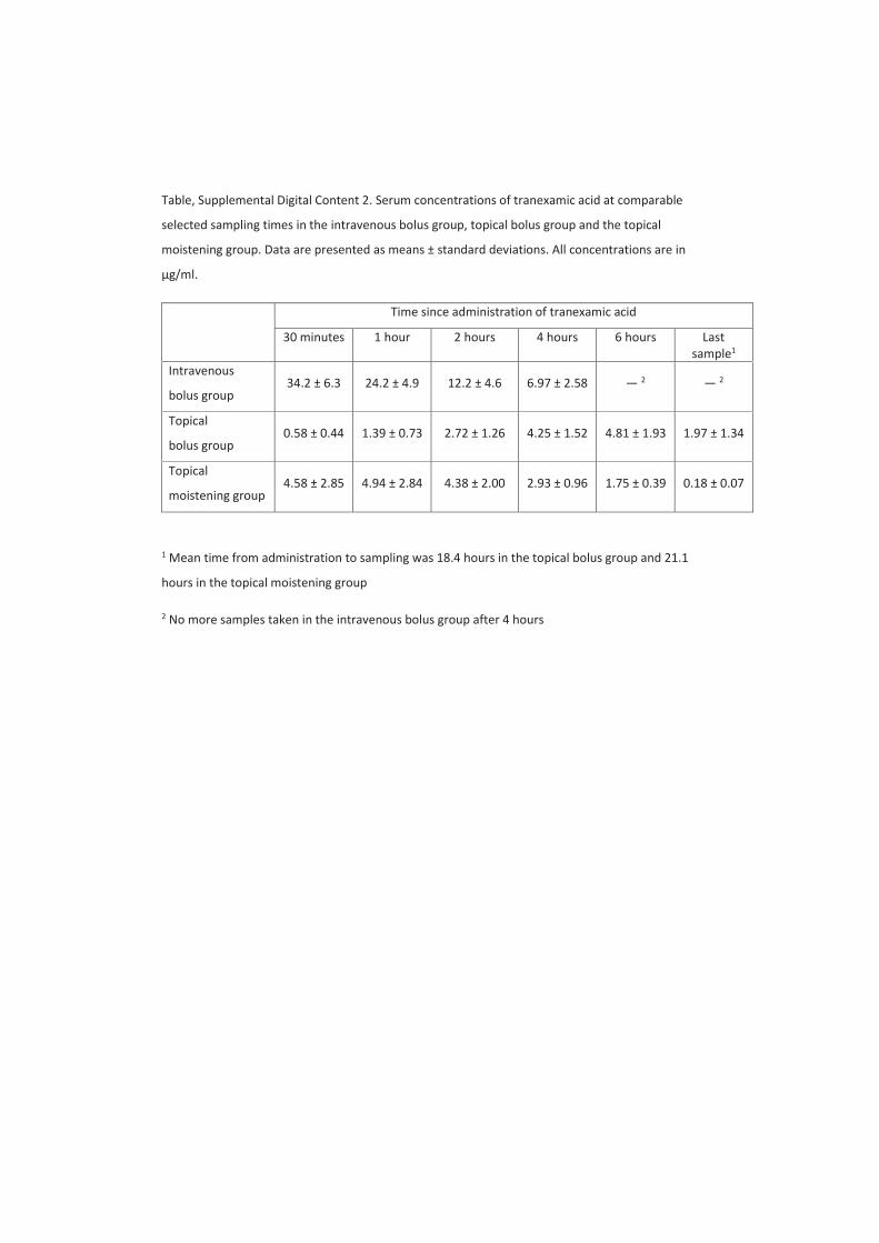
Table, Supplemental Digital Content 2. Serum concentrations of tranexamic acid at comparable
selected sampling times in the intravenous bolus group, topical bolus group and the topical
moistening group. Data are presented as means ± standard deviations. All concentrations are in
μg/ml.
Time since administration of tranexamic acid
30 minutes 1 hour 2 hours 4 hours 6 hours Last
sample1
Intravenous
bolus group 34.2 ± 6.3 24.2 ± 4.9 12.2 ± 4.6 6.97 ± 2.58 2 2
Topical
bolus group 0.58 ± 0.44 1.39 ± 0.73 2.72 ± 1.26 4.25 ± 1.52 4.81 ± 1.93 1.97 ± 1.34
Topical
moistening group 4.58 ± 2.85 4.94 ± 2.84 4.38 ± 2.00 2.93 ± 0.96 1.75 ± 0.39 0.18 ± 0.07
1 Mean time from administration to sampling was 18.4 hours in the topical bolus group and 21.1
hours in the topical moistening group
2 No more samples taken in the intravenous bolus group after 4 hours

Figure, Supplemental Digital Content 3

Figure, Supplemental Digital Content 4

Figure, Supplemental Digital Content 5