Toll-like receptor–mediated cytokine production is differentially regulated by glycogen synthase...
16
Toll-like receptor—mediated cytokine production is differentially regulated by glycogen synthase kinase 3 Michael Martin 1 , Kunal Rehani 2 , Richard S Jope 3 , and Suzanne M Michalek 2 1Department of Oral Biology, University of Alabama at Birmingham, Birmingham, Alabama 35294-2170, USA. 2Department of Microbiology, University of Alabama at Birmingham, Birmingham, Alabama 35294-2170, USA. 3Department of Psychiatry, University of Alabama at Birmingham, Birmingham, Alabama 35294-2170, USA. Abstract The cellular mechanisms that directly regulate the inflammatory response after Toll-like receptor (TLR) stimulation are unresolved at present. Here we report that glycogen synthase kinase 3 (GSK3) differentially regulates TLR-mediated production of pro- and anti-inflammatory cytokines. Stimulation of monocytes or peripheral blood mononuclear cells with TLR2, TLR4, TLR5 or TLR9 agonists induced substantial increases in interleukin 10 production while suppressing the release of proinflammatory cytokines after GSK3 inhibition. GSK3 regulated the inflammatory response by differentially affecting the nuclear amounts of transcription factors NF-κB subunit p65 and CREB interacting with the coactivator CBP. Administration of a GSK3 inhibitor potently suppressed the proinflammatory response in mice receiving lipopolysaccharide and mediated protection from endotoxin shock. These findings demonstrate a regulatory function for GSK3 in modulating the inflammatory response. The ability of the innate immune system to recognize and respond to microbial components has been largely attributed to the family of type I transmembrane receptors called Toll-like receptors (TLRs) 1,2 . TLRs are expressed mainly on antigen-presenting cells such as monocytes-macrophages and dendritic cells and show an ability to discriminate among distinct molecular patterns associated with microbial components. Recognition of microbial products by TLRs leads to a variety of signal transduction pathways that regulate the nature, magnitude and duration of the inflammatory response 3 . Although the production of proinflammatory cytokines is important for mediating the initial host defense against invading pathogens 4 , an inability to regulate the nature or duration of the host’s inflammatory response can be detrimental, as in chronic inflammatory diseases. The underlying cellular mechanisms that directly control pro- versus anti-inflammatory cytokine production after TLR stimulation are unresolved, but studies have shown the TLR signaling pathway can activate phosphatidylinositol 3-OH kinase (PI(3)K) 5 to limit production of tumor necrosis factor (TNF) and interleukin 12 (IL-12) 6–8 . Moreover, TLR2-induced activation of the PI(3)K pathway enhances IL-10 production, whereas IL-12 production is reduced 9 . Those studies suggested critical involvement of the PI(3)K pathway in differentially controlling pro- and anti- inflammatory cytokine production, but it is unclear at present if this pathway is limited to TLR2 signaling and how production of other inflammatory mediators induced by other TLR ligands is affected. Correspondence should be addressed to M.M. ([email protected]).. Accession codes. BIND (http://bind.ca): 296781 and 296782. COMPETING INTERESTS STATEMENT The authors declare that they have no competing financial interests. NIH Public Access Author Manuscript Nat Immunol. Author manuscript; available in PMC 2007 July 26. Published in final edited form as: Nat Immunol. 2005 August ; 6(8): 777–784. NIH-PA Author Manuscript NIH-PA Author Manuscript NIH-PA Author Manuscript
-
Upload
independent -
Category
Documents
-
view
5 -
download
0
Transcript of Toll-like receptor–mediated cytokine production is differentially regulated by glycogen synthase...
Toll-like receptor—mediated cytokine production is differentiallyregulated by glycogen synthase kinase 3
Michael Martin1, Kunal Rehani2, Richard S Jope3, and Suzanne M Michalek2
1Department of Oral Biology, University of Alabama at Birmingham, Birmingham, Alabama 35294-2170,USA.
2Department of Microbiology, University of Alabama at Birmingham, Birmingham, Alabama 35294-2170,USA.
3Department of Psychiatry, University of Alabama at Birmingham, Birmingham, Alabama 35294-2170, USA.
AbstractThe cellular mechanisms that directly regulate the inflammatory response after Toll-like receptor(TLR) stimulation are unresolved at present. Here we report that glycogen synthase kinase 3 (GSK3)differentially regulates TLR-mediated production of pro- and anti-inflammatory cytokines.Stimulation of monocytes or peripheral blood mononuclear cells with TLR2, TLR4, TLR5 or TLR9agonists induced substantial increases in interleukin 10 production while suppressing the release ofproinflammatory cytokines after GSK3 inhibition. GSK3 regulated the inflammatory response bydifferentially affecting the nuclear amounts of transcription factors NF-κB subunit p65 and CREBinteracting with the coactivator CBP. Administration of a GSK3 inhibitor potently suppressed theproinflammatory response in mice receiving lipopolysaccharide and mediated protection fromendotoxin shock. These findings demonstrate a regulatory function for GSK3 in modulating theinflammatory response.
The ability of the innate immune system to recognize and respond to microbial componentshas been largely attributed to the family of type I transmembrane receptors called Toll-likereceptors (TLRs)1,2. TLRs are expressed mainly on antigen-presenting cells such asmonocytes-macrophages and dendritic cells and show an ability to discriminate among distinctmolecular patterns associated with microbial components. Recognition of microbial productsby TLRs leads to a variety of signal transduction pathways that regulate the nature, magnitudeand duration of the inflammatory response3. Although the production of proinflammatorycytokines is important for mediating the initial host defense against invading pathogens4, aninability to regulate the nature or duration of the host’s inflammatory response can bedetrimental, as in chronic inflammatory diseases. The underlying cellular mechanisms thatdirectly control pro- versus anti-inflammatory cytokine production after TLR stimulation areunresolved, but studies have shown the TLR signaling pathway can activatephosphatidylinositol 3-OH kinase (PI(3)K)5 to limit production of tumor necrosis factor (TNF)and interleukin 12 (IL-12)6–8. Moreover, TLR2-induced activation of the PI(3)K pathwayenhances IL-10 production, whereas IL-12 production is reduced9. Those studies suggestedcritical involvement of the PI(3)K pathway in differentially controlling pro- and anti-inflammatory cytokine production, but it is unclear at present if this pathway is limited to TLR2signaling and how production of other inflammatory mediators induced by other TLR ligandsis affected.
Correspondence should be addressed to M.M. ([email protected])..Accession codes. BIND (http://bind.ca): 296781 and 296782.COMPETING INTERESTS STATEMENT The authors declare that they have no competing financial interests.
NIH Public AccessAuthor ManuscriptNat Immunol. Author manuscript; available in PMC 2007 July 26.
Published in final edited form as:Nat Immunol. 2005 August ; 6(8): 777–784.
NIH
-PA Author Manuscript
NIH
-PA Author Manuscript
NIH
-PA Author Manuscript
Here we have characterized how intracellular TLR signaling affected ‘downstream’ effectormolecules of the PI(3)K pathway to determine if a central effector molecule is responsible formediating the ability of this pathway to differentially dictate the host’s inflammatory response.Inhibition of glycogen synthase kinase 3 (GSK3), the constitutively active downstream kinaseof the PI(3)K pathway, mediated the ability of this pathway to selectively augment anti-inflammatory cytokine production while concurrently suppressing proinflammatory cytokineproduction in response to TLR stimulation.
RESULTSTLR4-mediated phosphorylation of GSK3-β
PI(3)K is a heterodimeric enzyme that consists of a regulatory subunit (p85) and a catalyticsubunit (p110)10. PI(3)K activation occurs through binding of its regulatory subunit to adaptorproteins bound to phosphotyrosine residues present in activated cellular receptors located onthe plasma membrane, including TLRs10–12. Activation of PI(3)K can mediate the recruitmentand subsequent activation of signaling proteins with pleckstrin homology domains, includingthe serine-threonine kinase Akt13–15. After recruitment, Akt is activated by phosphorylationat Thr308 and Ser473 (refs. 13–15). Akt is a key physiologic mediator of the PI(3)K pathway.Activated Akt phosphorylates several downstream targets of the PI(3)K pathway, includingthe constitutively active serine-threonine kinase GSK3-β (Ser9). Phosphorylation of GSK3-β(Ser9) results in its inhibition16. GSK3 can both positively and negatively affect a variety oftranscription factors that are critical in regulating pro- and anti-inflammatory cytokineproduction17–22. Therefore, we initially determined if LPS stimulation of human monocytesmediated Ser9 phosphorylation of GSK3 in a PI(3)K-Akt-dependent way. LPS stimulation ofmonocytes mediated the phosphorylation of Akt (Ser473; Fig. 1a). Use of the PI(3)K inhibitorLY294002 abolished the ability of Escherichia coli LPS to induce Ser473 phosphorylation ofAkt (P < 0.05; Fig. 1a). We obtained similar results for the phosphorylation of Akt at Thr308(data not shown). We next investigated if LPS stimulation of monocytes induced Ser9phosphorylation of the constitutively active kinase GSK3-β in a PI(3)K-Akt-dependent way.Human monocytes stimulated with E. coli LPS demonstrated increased Ser9 phosphorylationof GSK3-β at multiple time points (Fig. 1b). In contrast, LY294002 significantly reduced theability of E. coli LPS to phosphorylate GSK3-β on Ser9 (P < 0.05; Fig. 1b). We next used aselective Akt inhibitor to determine if this kinase was responsible for GSK3-β phosphorylationby E. coli LPS (Fig. 1c). The ability of E. coli LPS to induce Ser9 phosphorylation of GSK3-β was Akt dependent, because the Akt inhibitor abrogated LPS-induced phosphorylation ofGSK3-β on Ser9 to nearly unstimulated amounts (P < 0.05; Fig. 1c). Phosphorylation of GSK3-β on Ser9 results in GSK3-β inactivation; thus, we next assessed how LPS-mediated Ser9phosphorylation of GSK3-β affected its downstream target, glycogen synthase16. Glycogensynthase was phosphorylated on Ser641 in unstimulated human monocytes, but this wassubstantially reduced after LPS stimulation. Moreover, treatment of monocytes with the GSK3inhibitor SB216763 alone completely abrogated Ser641 phosphorylation of glycogen synthase(Fig. 1d). Thus, whereas LPS-mediated Ser9 phosphorylation of GSK3 did reduce theconstitutive ability of GSK3 to phosphorylate Ser641 of glycogen synthase, it did notcompletely abolish the abiliaty of GSK3 to phosphorylate glycogen synthase, as noted withthe GSK3 inhibitor SB216763 (Fig. 1d).
GSK3 modulates IL-10 and IL-12 productionAlthough the involvement of GSK3 in regulating a variety of cellular functions, including theregulation of glycogen synthesis, has been described in considerable detail23, the ability ofGSK3 to regulate the production of inflammatory mediators has not been demonstrated. Inunstimulated cells, GSK3 constitutively phosphorylates β-catenin and thus targets β-cateninfor degradation23. Therefore, to initially determine the ability of a panel of various GSK3
Martin et al. Page 2
Nat Immunol. Author manuscript; available in PMC 2007 July 26.
NIH
-PA Author Manuscript
NIH
-PA Author Manuscript
NIH
-PA Author Manuscript
inhibitors to inhibit GSK3 activity in human monocytes, we assessed β-cateninphosphorylation in the presence or absence of the GSK3 inhibitors LiCl, SB216763,azakenpaullone and BIO24–27. Unstimulated monocytes had detectable β-cateninphosphorylation (Fig. 1e). In contrast, monocytes treated with the GSK3 inhibitors showed nodetectable phosphorylated β-catenin (Fig. 1e), thus demonstrating their ability to inhibit GSK3activity in human monocytes.
Because inhibition of the PI(3)K pathway augments IL-12 production while concurrentlysuppressing IL-10 in response to a TLR2 ligand, we next investigated whether inactivation ofGSK3 mediated a functional effect on pro- and anti-inflammatory cytokine production byhuman monocytes after TLR4 stimulation (Fig. 2). In the absence of LPS stimulation, inhibitorsof Akt, PI(3)K and GSK3 had no discernible effect on pro- or anti-inflammatory cytokineproduction compared with that of untreated controls (data not shown). Production of the anti-inflammatory cytokine IL-10 was increased by three-to fivefold when human monocytes werestimulated with E. coli LPS in the presence of any of the GSK3 inhibitors tested comparedwith LPS-stimulated cultures (Fig. 2a,Fig. 2c). In contrast, inhibition of PI(3)K or Akt usingLY294002 or the Akt inhibitor, respectively, both of which abrogated the ability of E. coli LPSto inactivate GSK3-β by phosphorylating Ser9 (Fig. 1b,Fig. 1c), resulted in a substantialreduction in IL-10 compared with that of monocytes treated with E. coli LPS (Fig. 2a). In sharpcontrast to IL-10, IL-12p40 was increased by greater than 50% when we used LY294002 orthe Akt inhibitor (Fig. 2b). We obtained similar results with the PI(3)K inhibitor wortmanninand cells transfected with small interfering RNA (siRNA) targeting Akt (data not shown).However, IL-12p40 production was reduced more than 70% when human monocytes werepretreated with a GSK3 inhibitor and stimulated with LPS, compared with LPS-treated controls(Fig. 2b,Fig. 2d) or LPS-treated controls containing 0.1% DMSO or 10 mM NaCl (Fig. 2d).In LPS-stimulated monocytes, GSK3 inhibition resulted in a 60–80% reduction in IL-6 andTNF production compared with that of LPS-stimulated control cells (Fig. 2e,Fig. 2f). UsingGSK3-β-deficient mouse embryonic fibroblasts (MEFs)17, we also found that LPS stimulationresulted in a reduction in TNF and IL-6 production of more than 70%, whereas IL-10production was significantly enhanced compared with that of wild-type MEFs (P < 0.05; Fig.2g–Fig. 2i). These data demonstrate that the inhibition of GSK3 differentially affects the abilityof the TLR4 signaling pathway to induce pro- and anti-inflammatory cytokines.
The two mammalian GSK3 isoforms, GSK3-α and GSK3-β, are encoded by separategenes28. Although the GSK3 isoforms share 95% identity in their kinase domains, GSK3-αand GSK3-β share only 36% identity in the 76 amino residues at the C terminus28. Thisdivergence in homology has functional relevance, as deletion of the gene encoding GSK3-β isembryonically lethal and GSK3-α is unable to ‘rescue’ GSK3-β-null mice17. Probing for bothisoforms of GSK3 using a phosphorylation-specific antibody to GSK3-α and GSK-β (Ser21and Ser9, respectively; Fig. 1b,Fig. 1c) demonstrated that only Ser9 phosphorylation of GSK3-β was evident in response to LPS stimulation; GSK3-α was not phosphorylated on Ser21. Todefinitively demonstrate that GSK3-β was responsible for differentially regulating IL-10 andIL-12 production after LPS stimulation, we next used siRNA specific for GSK3-β (Fig. 3).‘Silencing’ of GSK3-β by RNA interference for 96 h reduced GSK3-β by more than 70%compared with that of untransfected cells or cells transfected with control siRNA (Fig. 3a). Inthe absence of LPS stimulation, cells pretreated with control siRNA or siRNA targeting GSK3-β showed no substantial increase in IL-10 or IL-12 production compared with that ofuntransfected control cells (data not shown). In contrast, monocytes pretreated for 96 h withsiRNA specific for GSK3-β and subsequently stimulated with E. coli LPS showed an increasein IL-10 production of more than twofold compared with that of untransfected cells or cellstransfected with control siRNA (Fig. 3b). Moreover, IL-12p40 production by LPS-stimulatedcultures pretreated for 96 h with siRNA against GSK3-β was reduced by more than 60%compared with that of untransfected cells or cells transfected with control siRNA (Fig. 3c).
Martin et al. Page 3
Nat Immunol. Author manuscript; available in PMC 2007 July 26.
NIH
-PA Author Manuscript
NIH
-PA Author Manuscript
NIH
-PA Author Manuscript
These results demonstrate that GSK3-β inhibition is responsible for enhancing IL-10 whilesuppressing IL-12 by LPS-stimulated monocytes.
GSK3 regulates TLR-induced inflammatory responseNext we determined if the ability of GSK3 to regulate ‘classical’ proand anti-inflammatorycytokine production was more global by assessing the production of a variety of inflammatorycytokines. We also determined whether GSK3 was able to differentially modulate proand anti-inflammatory cytokine production mediated by multiple TLR signaling pathways. Therefore,we used selective agonists for TLR2 (lipoteichoic acid from Streptococcus pneumoniae), TLR4(synthetic lipid A; compound 506), TLR5 (flagellin protein FljB from Salmonellatyphimurium) and TLR9 (human CpG) and assessed how inhibition of GSK3 in conjunctionwith a specific TLR agonist affected the inflammatory response. Human peripheral bloodmononuclear cells (PBMCs) stimulated with a TLR2, TLR4, TLR5 or TLR9 agonist in thepresence of the specific GSK3 inhibitor SB216763 showed a selective reduction of 50–90%in the production of the proinflammatory cytokines IL-1β, interferon-γ (IFN-γ), IL-12p40 andIL-6 (Fig. 4a–Fig. 4d). In contrast, production of the anti-inflammatory cytokine IL-10 wasincreased by three- to eightfold compared with that of controls regardless of which TLR agonistwe used (Fig. 4e). These data demonstrate that GSK3 potently suppresses the production ofproinflammatory cytokines while concurrently augmenting production of anti-inflammatoryIL-10 in response to multiple TLR signaling pathways.
GSK3 differentially regulates transcription factor activityTo investigate the underlying cellular mechanism responsible for the ability of GSK3 tosuppress proinflammatory cytokine production while augmenting anti-inflammatory cytokineproduction, we assessed how GSK3 influenced the activation of downstream transcriptionfactors involved in the inflammatory response. GSK3-β has been linked to regulation of themain eukaryotic transcription factor NF-κB, which regulates many diverse cellular processes,including proinflammatory cytokine responses17–21. Because NF-κB regulation can bemediated at multiple steps, including degradation of IκB inhibitory molecules, processing ofthe NF-κB p105 and p100 molecules and phosphorylation-dependent association with cellularcoactivators, including cAMP response element—binding protein (CREB)—binding protein(CBP)29, we sought to elucidate what step(s) of the NF-κB pathway could be affected by GSK3inhibition. IκBα degradation induced by E. coli LPS was evident at 30 min after exposure (Fig.5a). Presence of the GSK3 inhibitor SB216763 failed to alter the rate or extent of degradationor resynthesis of IκBα (Fig. 5a). We obtained similar results for the degradation and resynthesisof IκBα (data not shown). Because GSK3 can mediate p65 phosphorylation21, we determinedif GSK3 inhibition was exerting an effect on the phosphorylation status of the p65 subunit.Neither the amount nor the duration of p65 phosphorylation (Ser276 or Ser536) was affectedafter stimulation of human monocytes with E. coli LPS in the presence of the GSK3 inhibitorSB216763 compared with that of cultures stimulated with E. coli LPS alone (Fig. 5b).Therefore, we measured the nuclear NF-κB subunits p50 and p65 in the presence and absenceof the GSK3 inhibitor SB216763 (Fig. 5c,Fig. 5d). We found no alterations in the amount ofnuclear p50 or p65 in unstimulated cells treated with SB216763 alone compared with that ofuntreated control cells (data not shown). Additionally, the DNA binding of nuclear p50 or p65in LPS-stimulated cultures was not influenced by the presence of SB216763 compared withthat in monocytes stimulated with LPS alone (Fig. 5c,Fig. 5d).
Optimal transcriptional activity of the NF-κB p65 subunit is mediated by its association withthe nuclear coactivator CBP30,31. Additionally, nuclear amounts of CBP are limiting, andphosphorylated CREB (Ser133) and NF-κB p65 (Ser276) compete for CBP binding. Increasedassociation of CREB and CBP suppress NF-κB activity30–33. Because GSK3 can negativelyregulate the activation and DNA-binding activity of CREB22, we investigated how GSK3
Martin et al. Page 4
Nat Immunol. Author manuscript; available in PMC 2007 July 26.
NIH
-PA Author Manuscript
NIH
-PA Author Manuscript
NIH
-PA Author Manuscript
inhibition affected nuclear CREB (Ser133) binding (Fig. 5e). In the absence of LPS stimulation,the GSK3 inhibitor SB216763 did not substantially affect nuclear phosphorylated CREB(Ser133) compared with that of untreated control cells (data not shown). In contrast, the DNA-binding properties of CREB (Ser133) were significantly ‘upregulated’ when LPS-stimulatedmonocytes were treated with the GSK3 inhibitor SB216763 or with siRNA targeting GSK3-β(Fig. 5e). Because the CREB DNA-binding activity was augmented by GSK3 inhibition, wealso sought to determine if GSK3 inhibition affected the ability of CREB (Ser133) and NF-κB p65 (Ser276) to associate with CBP (Fig. 5f,Fig. 5g). LPS-stimulated monocytes had moreassociation of p65 with CBP than did unstimulated control cells (Fig. 5f,Fig. 5g). However,LPS-stimulated cultures that were pretreated with SB216763 showed a considerable decreasein association of p65 with CBP (Fig. 5f), whereas the binding of CREB to CBP was potentlyaugmented (Fig. 5g). These results demonstrate that GSK3-β inhibition augments the bindingof CREB (Ser133) and suppresses the binding of NF-κB p65 (Ser276) to the nuclear coactivatorCBP.
GSK3 regulates inflammatory response through CREBStudies characterizing the transcription factors important for IL-10 production in humanmonocytes have identified CREB as a critical component34. Moreover, because we haddemonstrated enhanced association of CREB with CBP, we investigated whether the abilityof GSK3 inhibition to enhance CREB activity was responsible for the differential regulationof pro- and anti-inflammatory cytokine production. Treatment of monocytes with siRNAtargeting CREB for 96 h reduced CREB by more than 80% compared with that of untransfectedcells or cells transfected with control siRNA (Fig. 5h). To determine what function CREB wasmediating in the ability of GSK3 inhibition to suppress the inflammatory response, wecompared IL-10 production in LPS-stimulated monocytes pretreated for 96 h with siRNAspecific for CREB and in the presence or absence of the GSK3 inhibitor SB216763 (Fig. 5i).LPS-stimulated cultures pretreated with siRNA targeting CREB had a decrease of more than30% in IL-10 compared with that of cultures stimulated with LPS alone or with LPS inconjunction with control siRNA (Fig. 5i). LPS stimulation of cells pretreated with siRNAtargeting CREB and in the presence of SB216763 did not produce any discernible increase inIL-10 release compared with that of LPS-stimulated control cells (Fig. 5i). In contrast,stimulation of cells pretreated with siRNA against CREB resulted in an increase in IL-12p40production of approximately 20% compared with that of LPS-stimulated cells or LPS-stimulated cells transfected with control siRNA (Fig. 5j). Moreover, the production ofIL-12p40 by LPS-stimulated cultures pretreated for 96 h with siRNA against CREB did notseem to be altered by the presence of SB216763 (Fig. 5j). These data demonstrate that theability of GSK3 to regulate the production of pro- and anti-inflammatory cytokines by LPS-stimulated monocytes is dependent on regulation of CREB activity.
GSK3 inhibition protects mice from endotoxin shockNext we defined the function of GSK3 inhibition in mediating protection against endotoxinshock and its effects on modulating the inflammatory response in vivo. Mice given the GSK3inhibitor SB216763 2 h before receiving a 100% lethal dose (LD100) of LPS showedsignificantly improved survival of more than 70%, compared with 0% for the LPS-only controlgroup (P < 0.001; Fig. 6a). Next we determined if a ‘delayed’ dose of SB216763 could stillmediate protective effects against LPS-induced lethality. SB216763 given 2 h after LPSchallenge conferred a survival rate of more than a 50%, compared with 0% for the control micegiven only LPS containing 0.1% DMSO. (Fig. 6b). Monitoring the survival rates of micechallenged with a LD100 of LPS over a 10-day period further demonstrated that mice givenSB216763 did not have any ‘late’ deaths (Fig. 6a,Fig. 6b). Thus, GSK3 inhibition did notsimply prolong LPS-induced lethality. We next assessed how GSK3 inhibition regulated theinflammatory response in mice given an LD100 of LPS. Assessment of proinflammatory
Martin et al. Page 5
Nat Immunol. Author manuscript; available in PMC 2007 July 26.
NIH
-PA Author Manuscript
NIH
-PA Author Manuscript
NIH
-PA Author Manuscript
cytokine profiles in mice given SB216763 and challenged with LPS showed there was asignificant reduction in IL-12p40, IFN-γ and IL-6 (P < 0.001; Fig. 6c–Fig. 6e). In contrast,production of the anti-inflammatory cytokine IL-10 was significantly enhanced by more thantwofold in LPS-challenged mice given SB216763 compared with that of control mice receivingonly LPS (P < 0.001; Fig. 6f). Thus, GSK3 inhibition conferred significant protection againstLPS-induced lethality when given before or after LPS insult (P < 0.001). Moreover, thesefindings further demonstrate targeting GSK3 in vivo can potently suppress the production ofproinflammatory cytokines, whereas production of the anti-inflammatory cytokine IL-10 issignificantly augmented.
DISCUSSIONStudies directly comparing the biological activity of some TLR agonists have indicated asubstantial divergence in their ability to influence the nature (pro- versus anti-inflammatory)and magnitude (absolute amounts) of the inflammatory response35–41. Indeed, the ability ofthe TLR2 and TLR4 signaling pathways to mediate the relative production of theimmunoregulatory cytokines IL-10 and IL-12, known to be key in the development of cell-mediated and humoral immunity as well as in controlling the amount of inflammatorymediators, differs widely36,38,42–44. Whereas the TLR4 signaling pathway is typically apotent mediator of IL-12p70, TLR2 agonists induce mainly IL-12p40 with little to no detectableIL-12p70 (refs. 38,43). The ability of a TLR2 agonist to induce IL-12p70 from humanmonocytes is dependent on inhibition of the PI(3)K pathway, but this reduces IL-10production9. Our findings now demonstrate the ability of the PI(3)K-Akt pathway todifferentially regulate IL-10 and IL-12 production is through inhibition of GSK3-β.Furthermore, the ability of GSK3 inhibition to reduce IL-1β, IL-6, TNF-α, IL-12 and IFN-γsubstantially while augmenting IL-10 in response to a variety of TLR-agonists demonstratesthe broad ability of GSK3 to attenuate the inflammatory response after TLR stimulation.
An inability to regulate the nature, magnitude or duration of the host’s inflammatory responsecan be detrimental to the host. Our study has shown how GSK3 inhibition can modulate boththe nature and magnitude of pro- versus anti-inflammatory cytokine production after TLRstimulation by differentially regulating the association of NF-κB p65 and CREB with thenuclear coactivator CBP. These findings are consistent with published studies demonstratingthat the relative amounts of active nuclear CREB and NF-κB p65 determine subsequentassociation with CBP30–33. A notable property of GSK3 after TLR stimulation was that GSK3inhibition selectively influenced the amount of nuclear CREB (Ser133) DNA-binding activitywithout any discernible effects on the amount of nuclear NF-κB p65. This is in agreement withpublished work showing CREB activity is potently suppressed by active GSK3 (ref. 22).Moreover, the finding that GSK3 inhibition augmented nuclear CREB activity and enhancedits association with CBP while reducing interactions between NF-κB p65 and CBP providesmolecular understanding of how TLR-mediated GSK3 inhibition suppresses the production ofproinflammatory cytokines while concurrently augmenting IL-10.
The onset of sepsis has been associated with predominant production of proinflammatorycytokines that are believed to contribute to the pathology. Exogenous IL-10 can substantiallyreduce the toxicity associated with endotoxin shock35. Moreover, various strategies targetingproinflammatory cytokines, including IL-1β, TNF, IFN-γ and IL-12, in animal models haveindicated these cytokines are key mediators of endotoxin lethality45. Our study hasdemonstrated that GSK3 inhibition substantially reduced proinflammatory cytokineproduction induced after challenge of mice with an LD100 of LPS while augmenting IL-10release. Moreover, the GSK3 inhibitor SB216763 mediated significant protection againstendotoxin lethality when given before or after LPS challenge. Mice fed a diet comprising 0.4%of the GSK3 inhibitor LiCl for 10 d (ref. 46) had substantially reduced proinflammatory
Martin et al. Page 6
Nat Immunol. Author manuscript; available in PMC 2007 July 26.
NIH
-PA Author Manuscript
NIH
-PA Author Manuscript
NIH
-PA Author Manuscript
cytokines and approached survival rates of 60% when challenged with an LD100 of E. coliLPS, compared with mice fed normal mouse chow, which had survival rates of 0% (M.M. andR.J., unpublished observations). Thus, because of the general ability of GSK3 to modulate theinflammatory response after TLR activation, GSK3 could potentially serve as a therapeutictarget against sepsis or other inflammatory diseases.
In summary, we have identified and characterized a central mechanism by which the inhibitionof GSK3 differentially affects the nature and magnitude of the inflammatory response.Inhibition of GSK3 resulted in a profound increase in IL-10 production after TLR stimulation,whereas the concurrent production of proinflammatory cytokines, including IL-1β, IL-6, TNF,IL-12 and IFN-γ, by human monocytes and PBMCs was substantially reduced. Our findingsidentify a critical function for GSK3 in modulating pro- versus anti-inflammatory cytokinesin vivo and provide a rationale for regulating the nature and severity of inflammation.
METHODSReagents
Protein-free E. coli (K235) LPS was prepared as described47,48. Lipoteichoic acid from S.pneumoniae was obtained from M. Nahm (University of Alabama at Birmingham). Bacterialflagellin (S. typhimurium FljB) was obtained from A. Gewirtz (Emory University, Atlanta,Georgia). E. coli synthetic lipid A (compound 506) was obtained from T. Ogawa (AsahiUniversity, Gifu, Japan). The CpG oligodeoxynucleotides 2216 (5′-GGGGGACGATCGTCGGGGGG-3′) and 2216 control (5′-GGGGGAGCATGCTGCGGGGG-3′) were purchased from InvivoGen. LiCl was purchased from Sigma. ‘Prevalidated’siRNA kits targeting CREB and GSK3-β were purchased from Upstate Biotechnology, andassays were done according to the manufacturer’s protocol. The GSK3 inhibitors SB216763,azakenpaullone and BIO were obtained from Sigma. Wild-type and GSK3-β-deficient MEFswere obtained from J. Woodgett (Ontario Cancer Institute, Division of ExperimentalTherapeutics, Toronto, Ontario, Canada)17. The Akt inhibitor II Akt-i was purchased fromCalbiochem. Antibodies to CBP, total p38 and total GSK3-β and phosphorylation-specificantibodies to Akt (Ser473), Akt (Thr308), GSK3-α and GSK3β (Ser21 and Ser9, respectively),β-catenin (Ser33, Ser37, Thr41), glycogen synthase (Ser641), NF-κB p65 (Ser276 or Ser536)and CREB (Ser133) were purchased from Cell Signaling Technology, Biosource Internationaland Santa Cruz Biotechnology.
Endotoxin shock modelMale C57BL/6 mice (8–12 weeks of age; 18–23 g body weight) were injected intraperitoneallywith an LD100 (10 µg/g) of E. coli K235 LPS in 200 µl of PBS containing 0.1% DMSO. Micewere monitored over a 10-day period for survival. All studies were approved by the InstitutionalAnimal Care and Use Committee of the University of Alabama at Birmingham.
Measurement of cytokinesMouse and human IL-1β, IL-6, IL-10, TNF, IL-12p40 and IFN-γ in culture supernatants weremeasured with enzyme-linked immunosorbent assay (ELISA) kits from R&D Systems oreBioscience according to manufacturer’s instructions.
Immunoprecipitation analysisHuman monocytes (2 × 10⁶/ml) in 24-well plates were pretreated with medium, 0.01% DMSO,the Akt inhibitor, LiCl, SB216763, azakenpaullone and BIO before the addition of medium orLPS. At various time points, cells were washed with PBS and analyzed by immunoblot asdescribed9. The AlphaImager 2000 documentation and analysis system (Alpha Innotech) was
Martin et al. Page 7
Nat Immunol. Author manuscript; available in PMC 2007 July 26.
NIH
-PA Author Manuscript
NIH
-PA Author Manuscript
NIH
-PA Author Manuscript

used for densitometer scans of the blots. For immunoprecipitation of CBP (Santa CruzBiotechnology) and subsequent probing for associated NF-κB p65 (Ser276) or CREB (Ser133;obtained from Cell Signaling Technologies), the Catch and Release ImmunoprecipitationSystem (Upstate Biotechnology) was used according to the manufacturer’s instructions.
NF-κB p50, NF-κB p65 and CREB activityHuman monocytes in 24-well polystyrene tissue culture plates were pretreated for 1 h withSB216763 or 0.01% DMSO and then were incubated with medium alone or LPS for varioustimes. Cells were collected and washed two times in PBS and then were assayed for activitywith the TransAM kit specific for each transcription factor (Active Motif). The amount ofnuclear NF-κB p50, NF-κB p65 or CREB was normalized by the absorbance at 450 nm from2 mg NF-κB p50 or NF-κB p65 or 20 µg of nuclear lysate (CREB).
Cell cultureHeparinized venous blood from healthy adult donors was used to obtain PBMCs by isolationof the buffy coat and elimination of red blood cell contamination using Histopaque (SG-1.077;Sigma) density gradients. Human monocytes were purified from PBMC samples by negativeselection with a monocyte isolation kit purchased from Miltenyi Biotech. This procedureroutinely resulted in samples that were more than 95% pure CD14+ cells, as shown by flowcytometry. Human monocytes or PBMCs were cultured in 24-well (2 × 10⁶ cells/well) or 96-well (2 × 10⁵ cells/well) plates containing RPMI 1640 medium supplemented with 10% FBS,50 µM 2-mercaptoethanol, 1 mM sodium pyruvate, 2 mM L-glutamine, 20 mM HEPES, 50 U/ml of penicillin and 50 µg/ml of streptomycin. Wild-type or GSK3-β-deficient MEFs werecultured as described17.
Statistical analysisData are expressed as mean ± s.d. Statistical significance between groups was evaluated byanalysis of variance and the Tukey multiple-comparison test using the InStat program(GraphPad Software). Differences between groups were considered significant at P < 0.05.
ACKNOWLEDGMENTS
Supported in part by the US Public Health Service (DE 08182, DE 14215, DE 09081 and AI 056460).
References1. Medzhitov R, Preston-Hurlburt P, Janeway CA. A human homologue of the Drosophila Toll protein
signals activation of adaptive immunity. Nature 1997;388:394–397. [PubMed: 9237759]2. Yang RB, et al. Toll-like receptor-2 mediates lipopolysaccharide-induced cellular signalling. Nature
1998;395:284–288. [PubMed: 9751057]3. Dinarello CA. Proinflammatory cytokines. Chest 2000;118:503–508. [PubMed: 10936147]4. O’Neill LA, Dinarello CA. The IL-1 receptor/Toll-like receptor superfamily: crucial receptors for
inflammation and host defense. Immunol. Today 2000;21:206–209. [PubMed: 10782049]5. Monick MM, et al. Lipopolysaccharide activates Akt in human alveolar macrophages resulting in
nuclear accumulation and transcriptional activity of beta-catenin. J. Immunol 2001;166:4713–4720.[PubMed: 11254732]
6. Fukao T, et al. PI3K-mediated negative feedback regulation of IL-12 production in DCs. Nat. Immunol2002;3:875–881. [PubMed: 12154357]
7. Fukao T, et al. Selective loss of gastrointestinal mast cells and impaired immunity in PI3K-deficientmice. Nat. Immunol 2002;3:295–304. [PubMed: 11850627]
8. Guha M, Mackman N. The phosphatidylinositol 3-kinase-Akt pathway limits lipopolysaccharideactivation of signaling pathways and expression of inflammatory mediators in human monocytic cells.J. Biol. Chem 2002;277:32124–32132. [PubMed: 12052830]
Martin et al. Page 8
Nat Immunol. Author manuscript; available in PMC 2007 July 26.
NIH
-PA Author Manuscript
NIH
-PA Author Manuscript
NIH
-PA Author Manuscript
9. Martin M, et al. Role of the phosphatidylinositol 3 kinase-Akt pathway in the regulation of IL-10 andIL-12 by Porphyromonas gingivalis lipopolysaccharide. J. Immunol 2003;171:717–725. [PubMed:12847238]
10. Cantley LC. The phosphoinositide 3-kinase pathway. Science 2002;296:1655–1657. [PubMed:12040186]
11. Arbibe L, et al. Toll-like receptor 2-mediated NF-κB activation requires a Rac1 - dependent pathway.Nat. Immunol 2000;1:533–540. [PubMed: 11101877]
12. Toker A, Cantley LC. Signalling through the lipid products of phosphoinositide-3-OH kinase. Nature1997;387:673–676. [PubMed: 9192891]
13. Franke TF, Kaplan DR, Cantley LC, Toker A. Direct regulation of the Akt protooncogene productby phosphatidylinositol-3,4-bisphosphate. Science 1997;275:665–668. [PubMed: 9005852]
14. Lawlor MA, Alessi DR. PKB/Akt: a key mediator of cell proliferation, survival and insulin responses?J. Cell Sci 2001;114:2903–2910. [PubMed: 11686294]
15. Stokoe DLR, et al. Dual role of phosphatidylinositol-3,4,5-trisphosphate in the activation of proteinkinase B. Science 1997;277:567–570. [PubMed: 9228007]
16. Cross DA, Alessi DR, Cohen P, Andjelkovich M, Hemmings BA. Inhibition of glycogen synthasekinase-3 by insulin mediated by protein kinase B. Nature 1995;378:785–789. [PubMed: 8524413]
17. Hoeflich KP, et al. Requirement for glycogen synthase kinase-3β in cell survival and NF-κBactivation. Nature 2000;406:86–90. [PubMed: 10894547]
18. Demarchi F, Bertoli C, Sandy P, Schneider C. Glycogen synthase kinase-3 β regulates NF-κB1/p105stability. J. Biol. Chem 2003;278:39583–39590. [PubMed: 12871932]
19. Demarchi F, Verardo R, Varnum B, Brancolini C, Schneider C. Gas6 antiapoptotic signaling requiresNF-κB activation. J. Biol. Chem 2001;276:31738–31744. [PubMed: 11425860]
20. Nemeth ZH, et al. Lithium induces NF-κB activation and interleukin-8 production in human intestinalepithelial cells. J. Biol. Chem 2002;277:7713–7719. [PubMed: 11756416]
21. Schwabe RF, Brenner DA. Role of glycogen synthase kinase-3 in TNF-α-induced NF-κB activationand apoptosis in hepatocytes. Am. J. Physiol. Gastrointest. Liver Physiol 2002;283:G204–G211.[PubMed: 12065308]
22. Grimes CA, Jope RS. CREB DNA binding activity is inhibited by glycogen synthase kinase-3 β andfacilitated by lithium. J. Neurochem 2001;78:1219–1232. [PubMed: 11579131]
23. Doble BW, Woodgett JR. GSK-3: tricks of the trade for a multi-tasking kinase. J. Cell Sci2003;116:1175–1186. [PubMed: 12615961]
24. Kunick C, Lauenroth K, Leost M, Meijer L, Lemcke T. 1-Azakenpaullone is a selective inhibitor ofglycogen synthase kinase-3 β. Bioorg. Med. Chem. Lett 2004;19:413–416. [PubMed: 14698171]
25. Cross DA, et al. Selective small-molecule inhibitors of glycogen synthase kinase-3 activity protectprimary neurones from death. J. Neurochem 2001;77:94–102. [PubMed: 11279265]
26. Meijer L, et al. GSK-3-selective inhibitors derived from Tyrian purple indirubins. Chem. Biol2003;10:1255–1266. [PubMed: 14700633]
27. Stambolic V, Ruel L, Woodgett JR. Lithium inhibits glycogen synthase kinase-3 activity and mimicswingless signalling in intact cells. Curr. Biol 1996;6:1664–1668. [PubMed: 8994831]
28. Woodgett JR. Molecular cloning and expression of glycogen synthase kinase-3/factor A. EMBO J1990;9:2431–2438. [PubMed: 2164470]
29. Ghosh S, May MJ, Kopp EB. NF-κB and rel proteins: evolutionary conserved mediators of immuneresponses. Annu. Rev. Immunol 1998;16:225–260. [PubMed: 9597130]
30. Sheppard KA, et al. Transcriptional activation by NF-κB requires multiple coactivators. Mol. Cell.Biol 1999;19:6367–6378. [PubMed: 10454583]
31. Zhong H, Voll RE, Ghosh S. Phosphorylation of NF-κB p65 by PKA stimulates transcriptional activityby promoting a novel bivalent interaction with the coactivator CBP/p300. Mol. Cell 1998;1:661–671. [PubMed: 9660950]
32. Parker D, et al. Phosphorylation of CREB at Ser-133 induces complex formation with CREB-bindingprotein via a direct mechanism. Mol. Cell. Biol 1996;16:694–703. [PubMed: 8552098]
33. Parry GC, Mackman N. Role of cyclic AMP response element-binding protein in cyclic AMPinhibition of NF-κB-mediated transcription. J. Immunol 1997;159:5450–5456. [PubMed: 9548485]
Martin et al. Page 9
Nat Immunol. Author manuscript; available in PMC 2007 July 26.
NIH
-PA Author Manuscript
NIH
-PA Author Manuscript
NIH
-PA Author Manuscript
34. Platzer C, et al. Cyclic adenosine monophosphate-responsive elements are involved in thetranscriptional activation of the human IL-10 gene in monocytic cells. Eur. J. Immunol1999;29:3098–3104. [PubMed: 10540320]
35. Berg DJ, et al. Interleukin-10 is a central regulator of the response to LPS in murine models ofendotoxic shock and the Shwartzman reaction but not endotoxin tolerance. J. Clin. Invest1995;96:2339–2347. [PubMed: 7593621]
36. Hirschfeld M, et al. Signaling by Toll-like receptor 2 and 4 agonists results in differential geneexpression in murine macrophages. Infect. Immun 2001;69:1477–1482. [PubMed: 11179315]
37. Jones BW, et al. Different Toll-like receptor agonists induce distinct macrophage responses. J. Leuk.Biol 2001;69:1036–1044.
38. Re F, Strominger JL. Toll-like receptor 2 (TLR2) and TLR4 differentially activate human dendriticcells. J. Biol. Chem 2001;276:37692–37699. [PubMed: 11477091]
39. Rhee SH, Jones BW, Toshchakov V, Vogel SN, Fenton MJ. Toll-like receptors 2 and 4 activateSTAT1 serine phosphorylation by distinct mechanisms in macrophages. J. Biol. Chem2003;278:22506–22512. [PubMed: 12686553]
40. Schilling D, Thomas K, Nixdorff K, Vogel SN, Fenton MJ. Toll-like receptor 4 and Toll-IL-1 receptordomain-containing adapter protein (TIRAP)/myeloid differentiation protein 88 adapter-like (Mal)contribute to maximal IL-6 expression in macrophages. J. Immunol 2002;169:5874–5880. [PubMed:12421970]
41. Toshchakov V, et al. TLR4, but not TLR2, mediates IFN-β-induced STAT1α/β-dependent geneexpression in macrophages. Nat. Immunol 2002;3:392–398. [PubMed: 11896392]
42. Dillon S, et al. A Toll-like receptor 2 ligand stimulates Th2 responses in vivo, via induction ofextracellular signal-regulated kinase mitogen-activated protein kinase and c-Fos in dendritic cells. J.Immunol 2004;172:4733–4743. [PubMed: 15067049]
43. Re F, Strominger JL. IL-10 released by concomitant TLR2 stimulation blocks the induction of a subsetof Th1 cytokines that are specifically induced by TLR4 or TLR3 in human dendritic cells. J. Immunol2004;173:7548–7555. [PubMed: 15585882]
44. Pulendran B, et al. Lipopolysaccharides from distinct pathogens induce different classes of immuneresponses in vivo. J. Immunol 2001;167:5067–5076. [PubMed: 11673516]
45. Cohen J. The immunopathogenesis of sepsis. Nature 2002;420:885–891. [PubMed: 12490963]46. O’Brien WT, et al. Glycogen synthase kinase-3β haploinsufficiency mimics the behavioral and
molecular effects of lithium. J. Neurosci 2004;24:6791–6798. [PubMed: 15282284]47. Hirschfeld M, Ma Y, Weis JH, Vogel SN, Weis JJ. Cutting edge: repurification of lipopolysaccharide
eliminates signaling through both human and murine toll-like receptor 2. J. Immunol 2000;165:18–22.
48. Tapping RI, Akashi S, Miyake K, Godowski PJ, Tobias PS. Toll-like receptor 4, but not Toll-likereceptor 2, is a signaling receptor for Escherichia and Salmonella lipopolysaccharides. J. Immunol2000;165:5780–5787. [PubMed: 11067937]
Martin et al. Page 10
Nat Immunol. Author manuscript; available in PMC 2007 July 26.
NIH
-PA Author Manuscript
NIH
-PA Author Manuscript
NIH
-PA Author Manuscript
Figure 1.E. coli LPS induces the phosphorylation of Akt (Ser473) and GSK3-β (Ser9) through the PI(3)K-Akt pathway in human peripheral blood monocytes. Human monocytes werepreincubated with 20 µM LY294002 (LY), 1 µM Akt inhibitor (Akt-i), 10 mM SB216763 or10 µM LiCl for 1 h before stimulation with 1 µg/ml of E. coli LPS. Unstimulated and LPS-stimulated cells not pretreated with an inhibitor were pretreated for 1 h with culture mediacontaining 0.1% DMSO or 10 mM NaCl. Whole-cell lysates (10 µg) were analyzed byimmunoblot with phosphorylationspecific antibodies (left margins) followed by enhancedchemiluminescence detection to assess phosphorylation of Akt (Ser473; a), GSK3-α andGSK3-β (Ser21 and Ser9, respectively; b,c), glycogen synthase (Ser641; d) and β-catenin(Ser33, Ser37 and Thr41; e). Data are representative of five separate experiments.
Martin et al. Page 11
Nat Immunol. Author manuscript; available in PMC 2007 July 26.
NIH
-PA Author Manuscript
NIH
-PA Author Manuscript
NIH
-PA Author Manuscript
Figure 2.The PI(3)K pathway differentially modulates pro- and anti-inflammatory cytokine productionby inhibition of GSK3. (a–f) Cytokine production in monocytes preincubated for 1 h withmedium only, 10 µM SB216763, 10 mM LiCl, 200 nM azakenpaullone, BIO (50 nM), 1 µMAkt inhibitor or 20 µM LY294002 (keys) and then left unstimulated or stimulated with 1 µg/ml of LPS for 20 h. Cell-free supernatants were analyzed by ELISA for production of IL-10(a,c), IL-12p40 (b,d), TNF (e) or IL-6 (f). (g–i) Cytokine production by wild-type (WT) orGSK3-β-deficient MEFs left unstimulated or stimulated with LPS for 20 h; cell-freesupernatants were analyzed by ELISA. *, P < 0.05, **, P < 0.01, and ***, P < 0.001, comparedwith LPS (a–f) or wild-type MEFs (g–i). Results represent mean ± s.d. of five separateexperiments.
Martin et al. Page 12
Nat Immunol. Author manuscript; available in PMC 2007 July 26.
NIH
-PA Author Manuscript
NIH
-PA Author Manuscript
NIH
-PA Author Manuscript
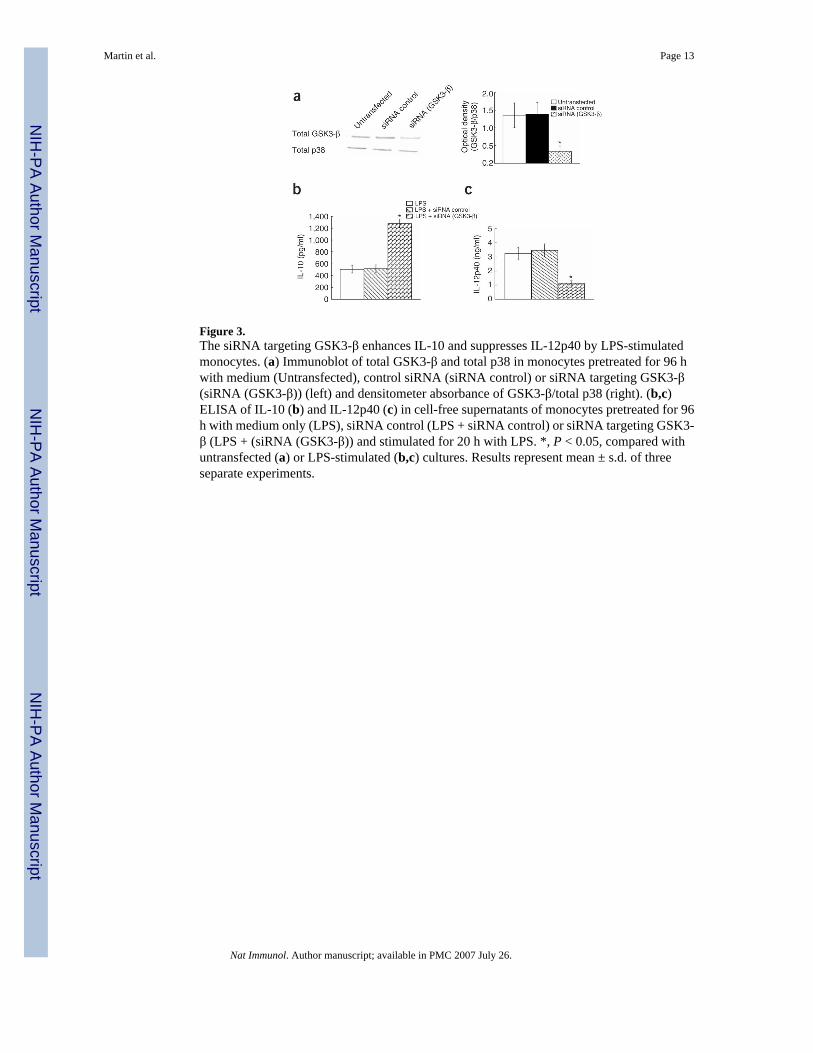
Figure 3.The siRNA targeting GSK3-β enhances IL-10 and suppresses IL-12p40 by LPS-stimulatedmonocytes. (a) Immunoblot of total GSK3-β and total p38 in monocytes pretreated for 96 hwith medium (Untransfected), control siRNA (siRNA control) or siRNA targeting GSK3-β(siRNA (GSK3-β)) (left) and densitometer absorbance of GSK3-β/total p38 (right). (b,c)ELISA of IL-10 (b) and IL-12p40 (c) in cell-free supernatants of monocytes pretreated for 96h with medium only (LPS), siRNA control (LPS + siRNA control) or siRNA targeting GSK3-β (LPS + (siRNA (GSK3-β)) and stimulated for 20 h with LPS. *, P < 0.05, compared withuntransfected (a) or LPS-stimulated (b,c) cultures. Results represent mean ± s.d. of threeseparate experiments.
Martin et al. Page 13
Nat Immunol. Author manuscript; available in PMC 2007 July 26.
NIH
-PA Author Manuscript
NIH
-PA Author Manuscript
NIH
-PA Author Manuscript
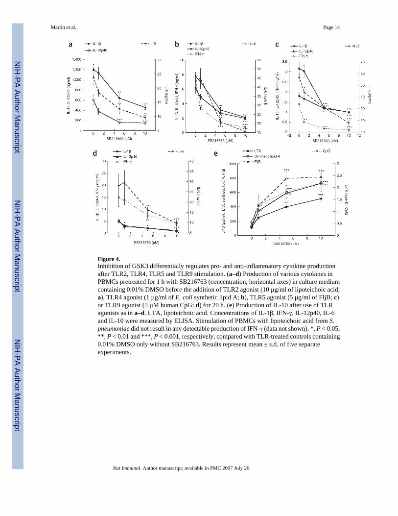
Figure 4.Inhibition of GSK3 differentially regulates pro- and anti-inflammatory cytokine productionafter TLR2, TLR4, TLR5 and TLR9 stimulation. (a–d) Production of various cytokines inPBMCs pretreated for 1 h with SB216763 (concentration, horizontal axes) in culture mediumcontaining 0.01% DMSO before the addition of TLR2 agonist (10 µg/ml of lipoteichoic acid;a), TLR4 agonist (1 µg/ml of E. coli synthetic lipid A; b), TLR5 agonist (5 µg/ml of FljB; c)or TLR9 agonist (5 µM human CpG; d) for 20 h. (e) Production of IL-10 after use of TLRagonists as in a–d. LTA, lipoteichoic acid. Concentrations of IL-1β, IFN-γ, IL-12p40, IL-6and IL-10 were measured by ELISA. Stimulation of PBMCs with lipoteichoic acid from S.pneumoniae did not result in any detectable production of IFN-γ (data not shown). *, P < 0.05,**, P < 0.01 and ***, P < 0.001, respectively, compared with TLR-treated controls containing0.01% DMSO only without SB216763. Results represent mean ± s.d. of five separateexperiments.
Martin et al. Page 14
Nat Immunol. Author manuscript; available in PMC 2007 July 26.
NIH
-PA Author Manuscript
NIH
-PA Author Manuscript
NIH
-PA Author Manuscript
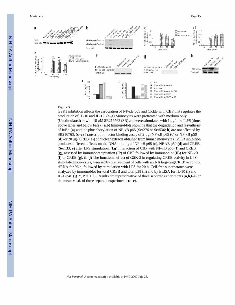
Figure 5.GSK3 inhibition affects the association of NF-κB p65 and CREB with CBP that regulates theproduction of IL-10 and IL-12. (a–g) Monocytes were pretreated with medium only(Unstimulated) or with 10 µM SB216763 (SB) and were stimulated with 1 µg/ml of LPS (time,above lanes and below bars). (a,b) Immunoblots showing that the degradation and resynthesisof IκBα (a) and the phosphorylation of NF-κB p65 (Ser276 or Ser536; b) are not affected bySB216763. (c–e) Transcription factor binding assay of 2 µg (NF-κB p65 (c) or NF-κB p50(d)) or 20 µg (CREB (e)) of nuclear extracts obtained from human monocytes. GSK3 inhibitionproduces different effects on the DNA binding of NF-κB p65 (c), NF-κB p50 (d) and CREB(Ser133; e) after LPS stimulation. (f,g) Interaction of CBP with NF-κB p65 (f) and CREB(g), assessed by immunoprecipitation (IP) of CBP followed by immunoblot (IB) for NF-κB(f) or CREB (g). (h–j) The functional effect of GSK-3 in regulating CREB activity in LPS-stimulated monocytes, assessed by pretreatment of cells with siRNA targeting CREB or controlsiRNA for 96 h, followed by stimulation with LPS for 20 h. Cell-free supernatants wereanalyzed by immunoblot for total CREB and total p38 (h) and by ELISA for IL-10 (i) andIL-12p40 (j). *, P < 0.05. Results are representative of three separate experiments (a,b,f–i) orthe mean ± s.d. of three separate experiments (c–e).
Martin et al. Page 15
Nat Immunol. Author manuscript; available in PMC 2007 July 26.
NIH
-PA Author Manuscript
NIH
-PA Author Manuscript
NIH
-PA Author Manuscript
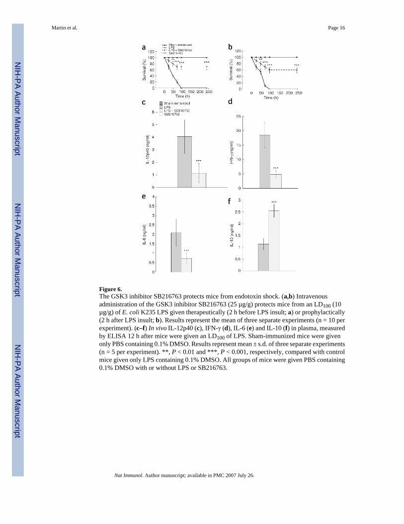
Figure 6.The GSK3 inhibitor SB216763 protects mice from endotoxin shock. (a,b) Intravenousadministration of the GSK3 inhibitor SB216763 (25 µg/g) protects mice from an LD100 (10µg/g) of E. coli K235 LPS given therapeutically (2 h before LPS insult; a) or prophylactically(2 h after LPS insult; b). Results represent the mean of three separate experiments (n = 10 perexperiment). (c–f) In vivo IL-12p40 (c), IFN-γ (d), IL-6 (e) and IL-10 (f) in plasma, measuredby ELISA 12 h after mice were given an LD100 of LPS. Sham-immunized mice were givenonly PBS containing 0.1% DMSO. Results represent mean ± s.d. of three separate experiments(n = 5 per experiment). **, P < 0.01 and ***, P < 0.001, respectively, compared with controlmice given only LPS containing 0.1% DMSO. All groups of mice were given PBS containing0.1% DMSO with or without LPS or SB216763.
Martin et al. Page 16
Nat Immunol. Author manuscript; available in PMC 2007 July 26.
NIH
-PA Author Manuscript
NIH
-PA Author Manuscript
NIH
-PA Author Manuscript