Title of Study: THE BIOLOGY OF THE COMMON STARFISH ...
45
Name: Paul D. Akers Date of Degree: August 10, 1963 Institution: Oklahoma State University Location: Stillwater, Oklahoma Title of Study: THE BIOLOGY OF THE COMMON STARFISH AS A SOURCE OF STUDY IN THE HIGH SCHOOL BIOLOGY CLASSROOM AND LABORATORY Pages in Study: 38 Candidate for Degree of Master of Science Major Field: Natural Science Nature of Study: Many high school biology teachers find it necessary to use supplementary materials in presenting the starfish biology to the students. This report deals with such items as: the anatomy and physiology of the starfish; diagrams which can be used to present the material; methods for preparing laboratory specimens of the organism; outlines for laboratory exercises using the starfish; and a glossary of terms associated with. the biology of this animal. Use of Study: It is desired that the information presented in this report will be of value to secondary biology teachers in introducing the biology of the starfish to the high school student. Also, that some teachers who are not satisfied with the materials they currently employ may be assisted by the information or references presented.
-
Upload
khangminh22 -
Category
Documents
-
view
0 -
download
0
Transcript of Title of Study: THE BIOLOGY OF THE COMMON STARFISH ...
Name: Paul D. Akers Date of Degree: August 10, 1963
Institution: Oklahoma State University
Location: Stillwater, Oklahoma
Title of Study: THE BIOLOGY OF THE COMMON STARFISH AS A SOURCE OF STUDY IN THE HIGH SCHOOL BIOLOGY CLASSROOM AND LABORATORY
Pages in Study: 38 Candidate for Degree of Master of Science
Major Field: Natural Science
Nature of Study: Many high school biology teachers find it necessary to use supplementary materials in presenting the starfish biology to the students. This report deals with such items as: the anatomy and physiology of the starfish; diagrams which can be used to present the material; methods for preparing laboratory specimens of the organism; outlines for laboratory exercises using the starfish; and a glossary of terms associated with. the biology of this animal.
Use of Study: It is desired that the information presented in this report will be of value to secondary biology teachers in introducing the biology of the starfish to the high school student. Also, that some teachers who are not satisfied with the materials they currently employ may be assisted by the information or references presented.
THE BIOLOGY OF THE COMMON STARFISH AS A
SOURCE OF STUDY IN THE HIGH SCHOOL
BIOLOGY CLASSROOM .AND LABORATORY
By
PAUL D. AKERS
Bachelor of Science in Education
Arkansas State Teachers College
Conway, Arkansas
1960
Submitted to the faculty of the Graduate School of the Oklahoma State University
in partial fulfillment of the requirements for the degree of MASTER OF SCIENCE
August, 1963

THE BIOLOGY OF THE COMMON STARFISH AS A
SOURCE OF STUDY IN THE HIGH SCHOOL
BIOLOGY CLASSROOM AND LABORATORY
Report Approved:
c1~~ ~~~~
Dean of the Graduate 9cho9J, ...
11
ACKNOWLEDGEMENTS
The writer wishes to express his sincere appreciation
and gratitude for the assistance and many valuable suggestions
given him by Dr. James H. Zant and Dr. L. H. Bruneau, in the
planning and writing of this report.
tii
Chapter
I.
II.
III.
IV.
v.
TABLE OF CONTENT
INTRODUCTION • • • • • • • • • • • • • • • • • • • • • • • • • • • • • • • • • • •
HISTORICAL DEVELOPMENT • • • • • • • • • • • • • • • • • • • • • • • • •
ANATOMY AND PHYSIOLOGY • • • • • • • • • • • • • • • • • • • • • • • • •
Characteristic Features••••••••••••••••••• Body Wall and Endoskeleton •••••••••••••••• Muscular System••••••••••••••••••••••••••• The Coelom •••••••••••••••••••••••••••••••• Digestive System•••••••••••••••••••••••••• Water-Vascular System••••••••••••••••••••• Nervous System•••••••••••••••••••••••••••• Reproduction•••••••••••••••••••••••••••••• Haemal (Circulatory) System••••••••••••••• Excretory System•••••••••••••••••••••••••• Respiratory System••••••••••••••••••••••••
ECOLOGY AND BEHAVIOR•••••••••••••••••••••••••••
OUTLINE FOR LABORATORY DISSECTION AND STUDY••••
Page
l
4
6
6 9
10 11 12 14 16 19 20 21 22
23
25
Preparing Material•••••••••••••••••••••••• 25 Laboratory Exercises Using Dry Specimens •• 26 Laboratory Exercises Using Injected
Specimens•••••••••••••••••••••••••••• 27 Laboratory Exercises Using Fixed Secti-ons. 28
VI. INSTRUCTIONAL DIAGRAMS ••••••••••••••••••••••••• 29
VII. GLOSSARY••-••••••••••••••••••••••••••••••••••••• 34
SELECTED BIBLIOGRAPHY••••••••••••••••••••••••••••••••••• 37
iv

LIST OF FIGURES
Figure Page
6-1. Crossed Pedicellaria of Asterias •••••••••••••••• 29
6-2. Diagrammatic Cross Section of the Ray of a Starfish••••••••••••••••••••••••••••••••••• 30
6-3. Diagrammatic Section Through the Disc and one Arm of a Starfish•••••••••••••••••••••••••• 31
6-4. Water-Vascular System of the Starfish ••••••••••• 32
6-5. Nervous System of the Starfish•••••••••••••••••• 33
V
CHAPTER I
INTRODUCTION
To the untrained eye the starfish seems to "have little
excuse for being alive."l The animal possesses no true excre
tory system, no true circulatory system, and only the most
primitive respiratory system. Regardless of these unusual
characteristics, most biologists agree that the phylum
Chordata arose in evolution directly from the phylum Echino
dermata, which contains the starfish as one of its principal
members.
Thus, the biology of the starfish offers the high school
student an opportunity to compare the transition in form and
structure combined with simplicity and complexity which is
characteristic of many echinoderms. For this purpose the
writer has chosen the common starfish, Asterias forbesi, as
a representative of this group of invertebrates.
Compared to the other invertebrates, the starfish
appears to be on a relatively low level of organization.
However, it possesses one of the most complex nervous sys
tems to be found in the invertebrate kingdom; its feeding
mechanism is unique in the animal kingdom; its method of
lAllison L. Burnett, "Enigma of an Echinoderm," Natural History, November,1961, p. 11.
1
locom:>tion is extremely complex, and is not duplicated out
side of the phylum Echinodermata; and finally, the animal's
ability to drop an arm when it is distur~ed and to regene
rate into a complete animal from only a small portion of the
central disc is not equaled by any of the higher inverte
brates.
2
The starfish has been studied thoroughly by modern
scientists from the physiological and anatomical point of
view. Yet less is known regarding the physiology of the star
fish than any other major invertebrate animal. The nature of
its digestive enzymes, the mechanism employed by the animal
to drop an arm, the physiology of the nervous system, the
cellular changes that occur during the regenerating process,
the regulation of its locomotory apparatus, and the trans
portation of food materials within its body are still matters
of highest speculation. It is only within the last decade
that the mechanism involved in the animal's feeding process
has been elucidated to any considerable extent.
The purpose of this study is three fold: (1) to provide
the writer with a satisfactory knowledge of the biology of
the starfish that may easily be communicated to the high
school biology student; (2) to present a general guide which
dan serve as resource material for teachers in the teaching
of the starfish biology at the high school level, and (3)
to provide the teacher with a source of information for con
ducting laboratory exercises with this animal.
However, a detailed and extensive study of the star
fish biology is beyond the scope of this report. With this
fact in mind, the writer has attempted to present an ele
mentary, yet informative, view of the biology of Asterias
forbesi, the common starfish. Included with this are se
lected references that the writer considers the most im
portant, an outline for the dissection and study of this
animal in the high school biology laboratory, a section of
diagrams showing the important structures of the starfish,
and a glossary of terms associated with the anatomy and
physiology of this animal.
3
CHAPTER II
HISTORICAL DEVELOPMENT
The stijrfish belongs to the class Asteroidea of the
phylum Echinodermata. The starfish have been known from the
earliest times, and the Greeks applied to them the name
Aster, meaning star.1 Aristotle was one of the first to ob
serve and record the action of the starfish attacking and
opening a clam. Linnaeus confused all the stellate echino
derms under the one name Asterias, and this confusion per
sisted for many years under names that were raised to fa
milial or even ordinal rank. Lamarck, 1792, includeG the
Asterias in the family Stelleridae. In 1837 Burmeister ap
plied the name Asteroidea to the combined sea stars and ser-
pent stars.
Valu~ble accounts of the asteroids were recorded by
Sladen, 1889, during the voyage of the Challenger, by Ludwig,
1897, in his works on the asteroids of the Mediterranean,
and by Fisher, 1911, in his purely taxonomic works. Preyer,
1886-7, made extensive observational and experimental studies
on many echinoderms and contributed very accurate information
on the starfish. Agassiz, 1877, is given much credit on the
!Libbie H. Hyman,~ Invertebrates: Echinoderma,ta,(New York, 1955), p. 245.
the embryology of asteroids with his studies on Asterias
forbesi.
In the present century Jennings, 1907, made a series of
observations on the biological behavior of A!. forbesi, and
Vevers, 1949, contributed extensive information on the biology
of this starfish. Anderson, 1954, demonstrated the anatomy
of the cardiac stomach of the starfish. 2 Smith, 1937, has
furnished the best description of the asteroid nervous
system. The ecology of!.:.. forbesi has been studied extensive
ly by Galtsoff and Loosanoff, 1939, in which they observed the
behavior reactions and ecology of this invertebrate in its
natural habitat.
Much of the knowledge of the physiology of!.:_ forbesi
is due to the work of Buddington, 1942, in his studies on
the cilia water currents, and by Paine, 1926, in her studies
on the function of the tube feet.3 Still further work was
contributed by Maloeuf, 1937, on the respiratory function and
by Van der Heyde, 1922, on the physiology of the digestive
system in this starfish.
2Ibid, pp. 276-281.
3virg1nia Paine, "Adhesion of the Tube Feet in Starfish," Journal .Qf. Experimental Zoology, XLV (1926), pp. 361-366.
CHAPTER III
ANATOMY AND PHYSIOLOGY
The anatomy and physiology of the starfish offers the
biology teacher an excellent source of material for class
room discussion. It gives the biology student an opportunity
to comp?re the structure and body functions of this inverte
brate with the other groups of animals, both vertebrate and
invertebrate. The writer has attempted to include in this
1e~tion a generalized view of the starfish biology which can
be conveyed to the biology student.
Characteristic Features
Asterias forbesi, like all echinoderms shows radial
symmetry inasmuch as each of its five arms contains the same
set of organs. But this radial symmetry is not the original
plan of structure and a closer examination of the starfish
reveals the fact that it is built on the principle of bi
lateral symmetry. This plan is determined by the calcerous
madreporic plate which is situated on the dorsal surface of
the central disc between two of the arms. The plane of symme
try bisects therefore the madreporic plate, the anus which is
situated in the middle of the dorsal surface of the central
disc, and the arm or radius opposite to the madreporic plate.
The two arms between which the madreporic plate is situated
6

7
form the bivium, the remaining three form the trivium.
The external surface of the madreporic plate is cut by
grooves which radiate from the center of the plate. Within
the substance of the madreporite these grooves communicate
with pores which in turn open into minute flagellated canals
that unite to form collecting canals. These collecting canals
empty into a dilated area, the ampulla, which gives rise to
the stone canal of the water-vascular system. The flagella
in the canals maintain a constant flow of water into the
water-vascular system.
On the lower or oral surface of the central disc is
situated the mouth, utilized as a point of reference in de
termining the ventral surface of the animal. The mouth is
surrounded by a soft membrane, the peristome, immediately
adjacent to which are five groups of oral spines. Extending
radially from the vicinity of the peristome along the ventral
surface of each ray is a groove, called the ambulacral groove,
which contains four rows of tube feet or poda. The distal end
of each podum is modified into a terminal disc or sucker,
which works in the manner of a suction cup and enables the
animal to attain a firmly adhesive grasp on the surface with
which it is in contact.
At the distal end of each ambulacral groove there occurs
a single terminal tentacle which contains a small red spot,
the eyespot. This eyespot is a photoreceptor organ, and an
important part of the nervous system of this animal. The
edges of the ambulacral grooves are covered by a double row
of spines, the ambulacral spines, that can be brought to-
gether to form a protective latticework over the tube feet
in the ambulacral grooves.
8
The body surface is very thick and hard owing to the
presence of calcified plates or ossicles in its inner layer.
The ossicles of the dorsal surface are more or less irregular
in shape and are held together by connective tissue and
muscle fibers. The outer surface of the animal is pimpled with
numerous white spines which are an elevation of the calcerous
ossices in the dermis below. Around the bases of these spines,
and between them, occur numerous small pincerlike mechanisms
called pedicellariae. Each pedicellaria consists of two blades
or jaws which have serrated-opposed surfaces. In general the
pedicellariae, all of which are stalked, keep the surface of
the animal devoid of debris and clear of organisms prone to
settle on the softer parts of the body wall.l
The ventral surface is formed by articulated ossicles
arranged in four rows in each arm. The two middle rows are
formei by the ambulacral ossicles. Between these are the
ambulacral pores through which the ambulacral feet project.
The peristome is surrounded by a pentagon of oral ossicles.
Outside of the adambulacral plates on the ventral surface and
on the dorsal surface are numerous branchiae, protruding
through the interstices between the ossicles. They are short,
tube like projections of the coelomic cavity which serve for
the purpose of respiration.
1Hyman, p. 264.
The whole external surface of the starfish, even the
exterior surfaces of such ~ppendages as the tube feet, the
pedicellariae, the spines, and the branchiae, is ciliated.
This ciliation aids in ridding the surface of debris, and
possible aids in the transportation of food to the mouth. 2
Body Wall and Endoskeleton ·
9
From exterior to interior the body wall comprises epi
dermis, dermis, muscle layer, and parietal peritoneum. The
epidermis is composed of ciliated columnar epithelium, with
scattered spindle-shaped neurosensory cells, and mucous gland
cells intermingled with muriform cells. The epidermis contains
the pigment granules responsible for the external color. In
the base of the epidermis is found a nervous layer, varying
in thickness. A delicate basement membrane separates the epi
dermis from the underlying dermis.
The dermis, the thickest layer in the body wall, is made
up of fibrous connective tissue. The dermis la}~r is per
meated with a system of canalicular spaces that form a ring
space around the base of each papula.
The skeleton is made up of calcerous plates or ossicles
bound together by connective tissue contained in the dermis.
The ossicles, as mentioned earlier, are regularly arranged
about the mouth and in the ambulacral grooves and often
2w1111am T. Taylor and R.J. Weber, General Biology, (Princeton, New Jersey, 1961), p. 603.
10
around the sides of the arms, but are more or less scattered
elsewhere. The shape of the ossicles is such that when they
are bound together they form a reticulate skeleton, leaving
spaces for the emergence of the papulae. The endoskeleton is
divided into the main supporting system, embedded in the
dermis, and the more superficial skeleton of projecting spines.
Muscular System
The narrow layer of smooth muscle, which comprises the
muscular system, is divided into an outer circular layer and
an inner longitudinal layer. These layers are rather thin
and weak, resulting in somewhat flexible rays which are not
rigid. A system of muscles operates the ambulacral grooves.
Each pair of ambulacral ossicles is connected by an upper and
a lower transverse muscle. The contracting and relaxing of the
muscles regulates the size of the ambulacral grooves. The
lower transverse muscle is found between the hyponeural
canals and the radial water vessel. There are other short
upper and lower longitudinal muscles between adjacent aml:n
lacral ossicles along the whole length of each row. Their
contraction tends to shorten the ambulacral grooves. The
lateral transverse ambulacral muscles connect the outer end
of each ambulacral ossicle with the adjacent adambulacral
ossicle. Their contraction widens the ambulacral grooves.
Finally, there are longitudinal muscles between adjacent
adambulacral ossicles that aid in lateral movement of the rays.
11
The Coelom
The body cavity is a true body cavity, or coelom, being
enclosed on all sides by coelomic epithelium. The epithelium
is of the simple cuboidal type and is ciliated. The coelom is
filled with an albuminous fluid which is kept in a continu
ous state of circulation by the cilia of the coelomic epi
thelium.3 This fluid receives soluble nutrients from the
digestive system and distributes them to the tissue~ which it
bathes.4 It also receives the carbon dioxide and nitrogenous
wastes from the tissues. The peritoneum that is applied to the
organs is called visceral peritoneum, and that which lines
thre body wall is termed the parietal peritoneum. The body
cavity is quite spacious and is often referred to as the peri
visceral cavity since it surrounds the viscera. This coelom,
however, is subdivided into a number of coelomic channels or
sinuses. These are important because they enclose most of the
haemal or circulatory system of this animal. One of these
sinuses is the vertical axial sinus which encloses the stone
canal of the water-vascular system. The axial sinus communi-
cates dorsally with the atoral sip.us which lies immediatley
under the dorsal body wall of the central disc. Below the
axial sinus joins with the inner division of the oral ring
31. Irving, "Ciliary Currents in Starfish," Journal 91. E,xperimental Zoologi, XLI (1924), pp. 115-118.
4R.A. Buddington, "The Ciliary Transport System of Asterias," Biological Bulletin, LXXX (1942) p. 1+38.
12
sinus; the oral ring sinus being divided by a septum into an
outer and inner division. The outer division of the oral ring
gives rise to the hyponeural radial sinuses, each of which
extends into a ray and subtends the radial nerve cord in its
course through the arm. The coelom also has an excretory
function, to be discussed later.
Digestive System
The mouth occupies the central portion of the perioral
membrane and is controlled by a sphincter muscle. From the
mouth there arises a short esophagus which opens above into a
spacious cardiac stomach that occupies the greater part of the
central disc. This stomach is held in place by two mesenteries
originating from the peritoneum on the ambulacral ridges. The
lateral wall of the cardiac stomach is evaginated into ten
pouches, two of which project into each arm for a short
distance. The stomach is eversible through the mouth during
feeding and can be retracted by the previously mentioned
sphincter muscle.5 Dorsad of the cardiac stomach lies the
pyloric stomach. This portion extends into each arm as a pair
of glandular appendages called the pyloric ceca. These are
suspended in each arm by mesenteries extending from the upper
portion of the arm. Each pyloric cecum, brownish or greenish
in color, is a hollow glandular structure with greatly evagi
nated walls. Each cecum is lined with ciliated columnar epi-
5A. Reese, "The Old Starfish-Clam Question," Science, XCVI (1942), p. 514.
13
thelium, some of which are believed to secrete the digestive
enzymes. A duct arises from each cecum and then unites with
the ducts from the other cecae to form a main pyloric duct
that opens into the pyloric stomach. Cilia in the pouches
direct the flow of the enzymes with the currents created by
their beating. The enzymes digest the food in the pyloric and
cardiac stomachs and the digested food is then directed into
the pyloric cecae for absorption. The pyloric cecae also
secrete such enzymes as protease, amylase, and lipase.
A short narrow intestine arises from the pyloric stomach
and extends to the anus on the dorsal surface of the central
disc located above. The intestine diverticulates into a bi-
lobed intestinal cecum lined with mucous and glandular cells.
The following tissues comprise the digestive system: a
ciliated columnar epithelium, a layer of nervous tissue, a
connective tissue, and a muscular layer. There are variations
in thickness in the layers going from one organ to another.
In the feeding process, the starfish seizes the prey,
usually bivalve molluscs, so that the free edges of the shell
are brought into close association with the mouth of the star
fish.6 The pressure exerted by the tube feet of the starfish
force the valves apart; at the same time a part of the cardiac
stomach is everted through the mouth of the starfish by a
constant pressure exerted on the coelomic fluid and cavity,
and by the musculature of the arms and body. The stomach is
!hen inserted into the opening between the valves of the
mollusc. Potent protolytic enzymes from the pyloric ceca
then pass through the cardiac stomach into the softer part of
the bivalve's viscera. These enzymes bring about almost com
plete disolution of the viscera. The cilia of the cardiac and
pyloric stomachs produce currents to convey the digested
products upward to the digestive system of the starfish where
further digestion occurs. The starfish then withdraws its
stomach by the contraction of the retractor muscles and the
mouth is closed by the contraction of the oral sphincter
muscles.
Besides the bivalve molluscs, the starfish is predatory
on other animals such as: sea urchins, snails, worms, fish,
and small crustaceans. However, its favorite food seems to be
bivalve molluscs such as clams and oysters.
Water-Vascular System
This system, embryologically derived from the coelom,
begins with the madreporite (madreporic plate). It communi
cates below with the dilated upper end of the stone canal, the
ampulla. The stone canal extends from the a~pulla ventrad to
join with the ring canal which circles around the inner sur
face of the peripheral area of the perioral membrane. The
stone canal is ciliated and contains calcerous deposits which
give it a rigid texture. From the inner aspect of the ring
canal arise nine small vesicles called Tiedermann's bodies.7
?Taylor and Weber, p. 608.
15
It is thought that the phagocytic coelomocytes occuring in the
water-vascular system are formed in these vesicles. Five radial
canals arise from the ring canal, one of which traverses the
length of each arm dorsad of the ambulacral groove to end in/
the terminal tentacle. As each radial canal extends through
the ray, it gives off podial canals which connect with the
tube feet on the ventral side of the ambulacral pores. Each
tube foot has a sac-like component, the ampulla, which lies
within the arm on the dorsal surface of the ambulacral pore.
This ampulla is lined with longitudinal muscle fibers. This
organ plays an important part in the regulation of the water
pressure in the tube feet.a Longitudinal muscles also occur
in the tube feet themselves.
In locomotion, the tube feet push the animal forward. The
ampullae contract, forcing fluid into the tube feet. The hydro
static pressure resulting from this intake of fluid causes
the extension of the feet in the direction of movement.9 The
terminal discs on the tube feet serve as suction cups as they
come in contact with the substrate. The feet then push for
ward, resulting in the pushing of the animal forward. The
longitudinal muscles in the tube feet then contract forcing
the fluid into the ampullae. The ampullae then contract,causing
the fluid to enter the tube feet, causing their extension, and
then the cycle described is repeated. The backflow of fluid
8 Paine, p. 362.
9G. Kerkut, "The Forces Exerted by the Tube Feet During Locomotion, 11 Journal .Qf. Experimental Biology, XXX (1953), pp. 575-583.
16
into the lateral canals is prevented by small valves in these
canals.
In the mechanism of locomotion there is involved a very
complex system of coordination between the nervous system and
the tube feet. Apparently most of the reflex coordination of
locomotion is accomplished by the circumoral ring, to be dis
cussed later.
The water-vascular system, so important in locomotion
and feeding, must be kept filled with fluid. Water intake
through the madreporite is a continuous process and is due
principally to currents set up by the cilia in the canals.
The fact that there is a steady fluid intake through the
madreporite implies that there may be some leakag.g through
the tube feet.
Structurally, the individual tube foot is a very weak
organ, but the combined effect of the entire number provide
the mechanism of locomotion and the opening of the shells. On
soft surfaces, such as sand and mud, the suckers on the feet
are of little use and they then act as legs.
Nervous System
The nervous system is described as being composed of
three interrelated systems.10 The main part is the oral or
ectoneural system, situated just beneath the epidermis. This
is composed of the nerve ring, the radial nerves, and the
general subepidermal plexus. The circumoral nerve ring, pen-
lOHyman, p. 270.
17
tagonal in shape, is s:ttuated in the peristomal membrane
near its periphery. It supplies nerve fibers into the peri
stomal membrane and the esophagus, and gives off radial ne:cves
that run the length of each arm in the bottom of the ambul ....
acral groove~ It then terminates in the eye spot of the ter;:ni
nal tentacle~ The radial nerve consists of fibrillae arranged
in layers. It is continuous with a general subepidermal plexus
spread throughout the entire body i,,rnll and supplying all of
the body wall appendages. 'l'he fibers of the radial nerve syn
apse with the cell bodies in the necks of the ampullae of' the
tube feet. As mentioned previously, these coordinate the many
activities of the feet in locomotion and feeding.
There is an extensive subepidermal neural plr:;xus which
comprises fi be1' s of sensory, association, and motor neurons.
It connects with the central nervous system, · especially by
way of the radial nerves. The subepidermal ple.xus has been
demorrntrated to be capable of localized reflex ac vi ty. The
:Plexus is thickened into a cord extending the length of each
a1·m on each side, called the marginal nerve cord. 'l'his in
turn gives off a longitudinal series of motor nerves called
the lateral motor nerves .. A pair of these extend to each
ambulacral ossicle. These eventually reach the coelornic lining
where they form a plexus. This plexus controls the muscular
layer of the body wall and gonads. This system is referred to
as the entoneural system.
The third portion of the nervous system is the hypo
neural system which occurs as a nervous layer in the lateral
part of the oral uall of hyponeural sinus, beneath the
18
coelomic epithelium lining the sinus. This nervous layer,
known as Lang's nerve is separated from the lateral part of
the radial nerve only by a thin layer of dermal connective.
tissue. Lang's nerve gives off a series of nerves along the
arm into the adjacent lower transverse muscle that extends
between the ambulacral ossicles in the roof of the hyponeural
sinus. The hyponeural nervous system is primarily motor.
The circumoral ring is of great importance in the neural
coordination of the starfish. This is especially true in the
actions of the tube feet during locomotion. Severance of the
ring from the radial nerve cords results in the rays acting
independently of each other.
The epidermis is permeated with neurosensory cells which
serve as tactile and chemoreceptors. Tactile receptors are
quite numerous in the terminal discs of the tube feet and on
the terminal tentacles. These neurons are slender cells, more
or less spindle shaped, which possess a nucleus, a distal
thread-like process reaching to the cuticle, and a proximal
fiber entering the subepidermal nerve plexus.
The most complex sense organ is the eyespot, occur at
the base of the terminal tentacle. Each eyespot is composed
of a number of cup-shaped concavities. The epidermis in these
concavities is modified into alternating pigmented and retinal
cells. The pigmented cells contain an orange pigment, while
each retinal cell bears distally a knobbed visual rod that
extends into the concavity.11
11Ib1d, p. 275
19
Reproduction
The starfish exhibits sexual, ~sexual, and regenerative
types of reproduction. To perform the sexual processes of
reproduction the starfish possesses a pair of gonads in each
arm. They are located aborally in the proximal portions of
the ray. They lie free in the perivisceral coelom except for
attachment near the genital pores, a pore for each gonad
being located close to the interradial line. The gonads begin
their existence as outgrowths of the aboral sinus. Each gonad,
resembling a grapelike cluster, extends from its point of
attachment at the base of the ray where it lies lateral to a
pyloric cecum. Close to the time of spawning the gonad gener
ally extends through the whole length of the ray. The male
gonads are usually pale gray in color, while the female gonads
vary in color from pink to orange. The sexes are separate,
although hermaphroditic specimens have been found. The sexes
usually cannot be distinguished externally except in the case
of brooding females.
Both male and female starfish discharge their gametes in
to the water. Fertilization of the ova follows. Each zygote
then undergoes total and equal cleavage into a bipinnaria
( bilaterally symmetrical larval organism). Metamorphosis
into a mature adult usually requires 1-3 years. In many cases
the starfish is able to reproduce after one year of growth.
Starfish have been observed to live for as long as 20-25
years.
In certain cases starfish undergo asexual reproduction
by a process known as autotomy. This process occurs by a
20
splitting of the disc along a more or less preformed line
that avoids ossicles and that leaves the arms intact. The
individual parts then reproduce into an entire organism. A•
sexual reproduction is affected by environmental and physio
logical factors.
The third type of reproduction, regeneration, is closely
associated with autotom.y in that a lost part is replaced. This
part may be lost by shedding (autotomy) or by external forces.
The most common body part lost and regenerated is the ray. The
regeneration of the ray involves the formation of the terminal
tentacle, the terminal ossicles, and the tissue of the eye
spot followed by the pyloric cecae, the radial canal of the
water-vascular system and,the radial nerves. In some isolated
cases an entire central disc has regenerated all of the miss
ing parts.
Haemal (Circulatory) System
The entire circulatory system follows the course of the
ambulacral system under which it is situated. It is composed
of a system of sinuses and a system of lacunae, with the
axial organ. Immediately below the ambulacral ring is the
oral ring sinus divided by a septum into an external and an
internal oral sinus. The external sinus gives rise to five
radial sinuses. Each radial sinus is divided longitudinally
by a septum inclosing the radial lacuna. The radial sinus
runs under the radial canal of the ambulacral system to the
end of the arm, the left and right channels uniting in the
tentacle. The radial sinuses give off transverse sinuses to
21
the ambulacral feet. The internal oral sinus gives rise to
the axial sinus which runs along the hydrophoric canal. The
aboral ring-sinus connects with the dorsal end of the axial
sinus; the ring-sinus giving rise to five genital sinuses.
The axial organ is a glandular part of the axial lacuna, in
which the lacunae form a plexus. Functionally, the dorsal
portion of the axial gland has been shown to be contractile.
This is regarded as the force responsible for the movement of
coelomocytes and fluid in the haemal system. The axial sinus,
axial lacunae with the axial organ, and the hydrophoric canal
are all together inclosed in a peritoneum and form an axial
complex.
As already mentioned, the coelomic fluid receives solu
ble nutrients from the digestive system and distributes them
to the tissues. It also functions to remove wastes from these
tissues.
Excretory System
The absence of a true excretory system in the starfish 12 is one of the biological mysteries of nature. It is very
strange that an animal, high up in the evolutionary tree, with
a complex nervous system and many other complicated structures
would possess only a reminant of an excretory mechanism. The
function of excretion is accomplished by the amoebocytes in
the coelomic fluid, aided by the rectal cecae. These amoeba
like bodies transport the matabolic wastes to the dermo-
12 Burnett, p. 11.
22
branchiae where they pass out into the surrounding water by
diffusion. These phagocytic cells are derived by budding from
the cells of the coelomic epithelium. The amoebocytes are of
two main types; those with ordinary slender pseudopods and
those with petaloid pseudopods. These cells are highly phago
cytic and ingest the particles to be excreted.
Respiratory System
The respiratory function is the responsibility of the
dermal branchiae, through which oxygen is absorbed. The intake
of oxygen is also performed by the water currents moving into
the starfish. These currents are kept in motion by the flagella
of the coelomic epithelium. There is a general internal flow
toward the tips of the rays with a return current along the
inner surface of the ray sides. As it flows along this path,
it presumbably picks up oxygen from the dermal branchiae for
distribution to the various tissues and organs.
CHAPTER IV
ECOLOGY AND BEHAVIOR
Asterias forbesi is found on most of the eastern sea
shore, ranging from Maine to the Gulf of Mexico and may be
found from lowtide level to a depth of atout 50 meters. As is
typical of the more specialized groups of Asteroidea, this
species integrates with closely related species. Specimens
are found either singly or in large aggregations on rocky or
shelly bottoms. This starfish has been the source of extensive
study because of its economic importance as a predaceous
oyster-eating organism. Each year the oyster industry suffers
tremendous losses due to this animal. In addition to feeding
on oysters, the starfish includes in its diet such organisms
as clams, mussels, sea snails, dead fish, worms, and in rare
cases, other starfish.
It is understood that some sort of seasonal migration
occurs among the starfish, but the character and extent of
this migration is unknown.1 The rate of locomotion in this
organism has been observed to be very slow, an average rate
of 6 inches per minute. However, according to the isolated
geographic distribution of this species, facts seem to indi-
1A.D. Mead, ''The Natural History of the Starfish,'' Bulle~ of~ U.S. Bureau of Fisheries, XVIX (1899), p. 205.
23
cate that the wanderings of these animals are rather limited
in extent.
Like all other forms of life, the starfish has natural
enemies. Some of these destructive agents and natural enemies
are cold and fresh water, various fishes, gulls and crows, and
parasites. The enemy which is doubtless the most destructive
to the starfish is the menhaden. This fish feeds exclusively
upon the minute organisms which swim or float in the water.
The larvae of the starfish are safe from this fish only after
they have set. The setting involves a process of metamorpho
sis and attachment to some object on the bottom. The diet of
the free swimming larvae consists of minute algae and other
microscopic forms of plankton.
As mentioned above, the starfish is subject to attack by
parasites. The parasitic organisms attack the gonads of the
starfish, destroying the tissue and rendering it partially or
fully sterile.
CHAPTER V
OUTLINE FOR LABORATORY DISSECTION AND STUDY
As a supplement to the classroom discussion of the star
fish, the body structures of this animal provide ample source
for laboratory exercises in which dissection and observation
of body parts are performed by the students. The following
section provides the teacher with an outline for the prepa
ration of specimens and instructions for laboratory exercises
using these specimens.
Preparing Material
Preparing dry specimens: A perfect dry specimen may be
obtained by placing the starfish in a dish in a perfect radial
symmetrical position and then pouring boiling water into the
dish. This tends to relax the body and allows for dehydration
to follow after removing from the bath. This process is excel
lent for studying the skeleton of the starfish.
Preparing injected specimens: This type of specimen is
especially useful in the study of internal organs and the
ambulacral system. The injection may be accomplished through
one of the radial canals and should be continued until the
ambulacral feet of all radi are injected. The specimen may
then be preserved in formalin or alcohol.
Preparing fixed specimens: Very small specimens may be
25'
26
fixed for sections in Perenyi•s fluid. 1 Decalcifying before
embedding.is necessary. Sodium hypochlorite is useful for this
decalcification. The sections should be prepared and then
stained in'haemotoxylin and eosin. 2 This type of specimen is
excellent for the study of the nervous system.
Preparing living specimens: It is desirable to have,
living specimens to demonstrate external structures and activi
ties of the animal. Dissection may be carried out on a living
form after anesthetizing it with about 15 ml. of 10% MgCl.
Laboratory Exercises Using Dry Specimens
Instructions: (a) Make a full size drawing of the dorsal
surface of the starfish showing the central disc and all arms,
madreporic plate, tubercles, and pe¢1.icellariae. Indicate the
plane of symmetry and label the bivium and trivium. Draw the
details only in one arm. (b) Remove with a forceps the ambul
acral feet of one arm. Make a full size drawing of the ventral
surface, shwoing mouth, oral spines, ambulacral grooves with
rows of pores between the ambulacral ossicles, spines on the
adambulacral ossicles, ectoneural nerve ring, and ectoneural
radial nerve. Draw the details only in one arm. (d) Examine
under low power the madreporic plate and make a drawing show
ing its structure. (d) Remove with a scape! some of the small
pedicellariae, place them in a drop of water on a slide, ex-
!Alexander Petrunkevitch, Morphology Q!, Invertebrate Types, (New York, 1929), p. 174.
2Ibid.
amine under a microscope, and make a drawing showing both
types of pedicellariae.
Laboratory Exercises Using Injected Specimens
27
Instructions: (a) Place the specimen in a dissecting pan
with water; examine the oral surface and sketch a drawing of
the ambulacral feet, eyespot, tentacle, mouth, oral spines,
and ambulacral grooves. (b) Examine the dorsal surface and
draw the madreproic plate, tubercles, pedicellariae, and the
branchiae. (c) With strong scissors make a lateral incision
around the whole animal taking care not to ruin the internal
organs. Make a circular incision around the madreporic plate.
Lift the dorsal wall, with forceps, at the end of each arm
and press the digestive glands down, tearing the mesenteries
with the scapel. Cut the intestine near the anus and remove
the entire body wall. Make a drawing of its inner surface
showing the network of ossicles and spaces between them. (d)
V1a.ke an enlarged drawing of the digestive system showing the
cardial pouches, pyloric portion of the stomach with the 5
pyloric cecae, and the intestine with its intestinalcecae.
Label the parts indicated. (e) Using forceps, remove the entire
digestive system, using care not to tear the peristomal
membrane. Make a drawing of the reproductive organs. (f) Remove
the reproductive organs (gonads). Make a full size drawing of
the ambulacral system shwoing m.adreporite, hydrophoric canal,
Tiedemann's bodies, the five pairs of ampullae, and the four
rows of ampullae in one of the arms. Include in the drawing
the peristomal membrane and the axial organ of the circulatory
28
system.
Laboratory Exercises Using Fixed Sections
Instructions: (a) Using a microscope or hand lens, study
the external characteristics. Make a drawing showing the cen
tral disc, arms, madreproic plate, tubercles, and the small
pedicellariae. (b) Observe the cross section of a ray, indi
cating on a drawing the reproductive organs, radial nerve, the
tube feet, coelomic cavity, and any other visible organs. (c)
Observe the slides under low and high magnification. Label all
parts indicated a?Qove.
CHAPTER VI
INSTRUCTIONAL DIAGRAMS
The teaching of the starfish anatomy and physiology can
best be facilitated by the use of diagrams ro drawings show
ing the major divisions and structures of the organism. The
writer has attempted to include in this section some illus
trations which will be helpful to the teacher in presenting
the material to the biology student.
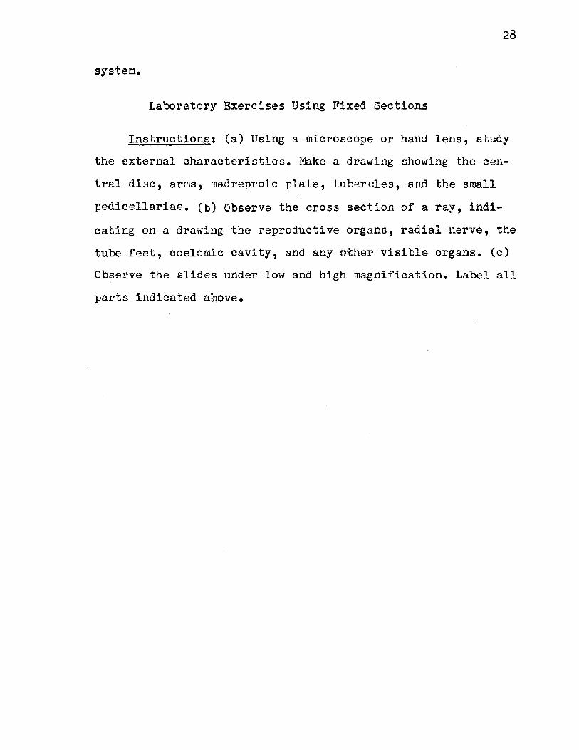
Fig. 6-1 Crossed pedicellaria of Asterias. am. adductor muscle;~-!!!· abductor muscle; :em,. basilar muscle;f12.. fibrous peduncle.
29
30
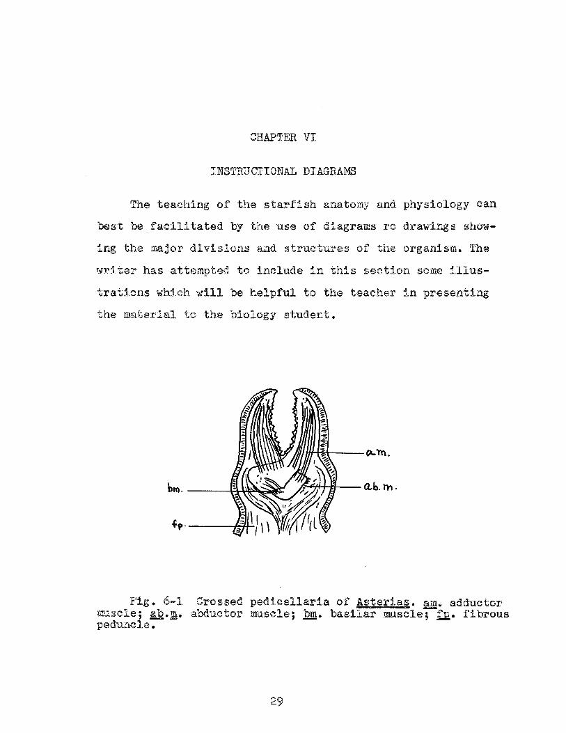
Fig. 6-2. Diagrammatic cross section of the ray of a starfish.~· ampulla; !!l• apical nerve; !2• ambulacral ossicle; 9J2.• dermal papula; K!• genital aperture!· g,. gonad; h.£. hepatic cecum; 1ml. longitudinal muscle layer;...!!• Lang's nerve;~· pedicellaria; R£.• perivisceral cavity; I!!• radial nerve;~· spine; tf. tube foot; ts. terminal sucker.
"'-S. --~ ........ +
~c.-----·
br.
b. ,s.- o..
,.
31
Mt. e. ST.
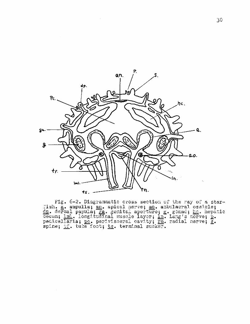
Fig. 6-3. Diagrammatic section through the disc and one arm of a starfish.~- anus;!.!• axial sinus; R• branchiae; bvr. blood-vascular ring;~· epidermis; m• rrouth; ~· madreporite; !!!:• nerve ring; 2.• ossicle; 12.• peristome; :Q.£• pyloric cecum; ¥ca. perivisceral cavity; .fill• pyloric sac;~- rectum; !!!• rad al nerve;~· stomach; sc. stone canal; §1. sensory tentacle;~· water-vascular ring.

32
re.
Fig. 6-4. Water-vascular system of the starfish.~· ampulla; I.£• radial canal;!:£.!• ring canal;!£• stone canal; .fil2.• sieve plate (madreporite); !f. tube foot.
33
th.
enor. ¥h..
etrn.
dl\.. hn..
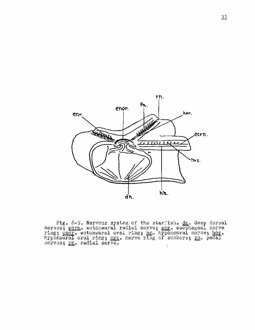
Fig. 6-5. Nervous system of the starfish. gn. deep dorsal nerves; !£!'..!l• ectoneural radial nerve; .fillr.• esophageal nerve ring;~· ectoneural oral ring; h,s. hyponeural nerve; hor. hyponeural oral ring;£!:!• nerve ring of suckers; 12!!• pedal nerves;!'.!!• radial nerve.
CHAPTER VII
GLOSSARY
Ambulacral feet: The tube feet which lie in the ambulacral grooves.
Ambulacral groove: A groove, extending radially from the vicinity of the peristome along the ventral surface of each ray, which contains the four rows of tube feet.
Ambulacral ossicles: Calcified plates located in the ambulacral region of the body wall.
Ambulacral pores: Pores or openings through which the tube feet project.
Ambulacral spines: Tiny projections of the body wall in the ambulacral region.
Ampullae: Bulb-like sacs extending into the coelom which connect directly with the tube feet. They function in regulating water pressure in the feet.
Autotomy: The process whereby the starfish sheds an injured ray.
Axial sinus: A cavity, involved in circulation, which runs along the hydrophoric canal.
Bilateral symmetry: The symmetrical arr~ngement whereby the body can be divided into two corresponding sides by a line drawn through the center of the disc.
Bipinnaria: The bilaterally symmetrical larval stage of the inmature starfish. This is the free-swimming stage.
Bivium: The two rays closest to the madreporite.
Branchiae: Short, tube-like projections of the coelom which protrude through the outer body covering. They serve for the purpose of respiration.
Circumoral ~: The ne~ve ring encircling the mouth. It is
35
situated in the peristomal membrane near its periphery.
Coelom: The body cavity of the starfish.
Coelomocytes: Phagocytic cells found in the coelomic fluid which aid in excretion and transportation of food particles. They are somtimes called amebocytes.
Ectoneural system:
Entoneural system:
Hydrophoric canal:
Superficial portion of the nervous system which follows the plan of the ambulacral and circulatory system. It functions in sensory perception.
The deep dorsal portion of the nervous system which consists of five nerves radiating from the central disc. This system belongs to the dorsal coelomic epithelium~
A short, stout, and somewhat curved canal extending from the madreporic plate ventrad where it connects with the ring canal. It is sometimes called the stone canal.
Hyponeural system: The motor system which is separated from the ectoneural only by a thin membrane of connective tissue.
Langns nerve: The dorsal portion of each radial nerve cord containing motor nerve fibers.
Madre~oric ~late: A circulart grooved plate situated on the dorsal surrace of the central disc. It functions in water intake.
Ossicles: Calcified plates found in the inner layer of the body which give the characteristic hardness of the skin.
Pedicellariae: Small, pincerlike mechanisms found projecting from the epidermis of the starfish. They are used to keep the surface clean.
Peristome: The soft membrane surrounding the mouth.
Perivisceral cavity: The body cavity which surrounds the viscera.
l:,Yloric ~: Digestive glands situated in each arm which are sac-like portions of the pyloric stomach.
Radial canal: Water-vascular canals which extend into each arm from the ring canal.

36
Radial nerve .£2.!:g,: The nerve cord running along the ambulacral groove in each ray.
Radial symmetrz: The body having parts arranged like rays, with each part having the same set of organs.
Ring canal: The water-vascular canal located in the central disc and surrounding the mouth. This gives rise to the five radial canals in the rays.
Subepidermal plexus: The plexus of sensory, association, and motor fibers which connect with the central nervous system by way of the radial nerves.
Tiedeman.n's bodies: Minute glands which produce amoebocytes. These glands open into the ring canal and consist of nine separate structures.
Trivium: The three rays furtherest from the madreporite.
SELECTED BIBLIOGRAPHY
Borradaile, L.A. The Invertebrata. New York: MacMillan Company, 193,;-pp. 634-637.
37
Brown, F.A. Selected Invertebrate Types. New York: John Wiley and Sons, 1950, pp. 515-523. ·
Buddington, R.A. ''The Ciliary Transport System of Asterias. 11
Biological Bulletin, LXXX (December, 1942), 1+38.
Burnett, Allison L. "Enigma of an Echinoderm." Natural History, LXX (November, 1961), 11-19.
Galtsoff, P.S. and V.L. Loosanoff. "Natural History and Methods of Controlling the Starfish." Bulletin .Q!'.. the U.S. Bureau .Q!. Fisheries, XLIX (1939), 75-132.
Hegner1 R.W. Invertebrate Zoology. New York: MacMillan Company, 1~43, pp. 513-514.
Hym~n, Libbie H. The Invertebrates:Echinodermata. New York: McGraw-Hill Book Company, 1955, pp. 245-414.
Irving, L. "Ciliary Currents in Starfish. 11 Journal of Experimental Zoology, XLI (November, 1924), 115-123.~
Kerkut, G. "The F'orces Exerted by the Tube Feet During Locomotion.11 Journal .Q.!: Experimental Biology, XXX( April, 1953), 575-5'83.
MacGurdy, H. "Reaction of Asterias to Light. 11 Science, XXXVIII (July, 1912), 98-100.
Mead, A.D. "The Natural History of the Starfish. 11 Bulletin of the U.S. Bureau 2£. Fish.eries,XVIX (1899), 203-224.
Moore .1 A.R. "Righting Movements in Starfish." Biological HU.lletin, XVIX {June,1910), 235-239.
Paine, Virginia.. "The Tube Feet of Starfishes as Autonomous Organs." American Naturalist, LXIII (December, 1926), 517-529 •
• "Adhesion of the Tube Feet in Starfishes. 11 Journal .Qf --Experimental Zoology,XLV (May, 1926), 361-366.
Petrunkevitch, Alexander. Morphology of Invertebrate Types. New York: MacMillan Company, 1929. pp. 174-181.
Reese, A. 11The Old Starfish-Clam Question." Science, XCVI tDecember, 1942), 514.
38
Taylor, W.T. and R.J. Weber. General Zoology. New York: D. Van Nostrand Company, 1961, pp. 599-613.
VITA
Paul D. Akers
Candidate for the Degree of
Master of Science
Report: THE BIOLOGY OF THE COMMON STARFISH AS A SOURCE OF STUDY IN THE HIGH SCHOOL BIOLOGY CLASSROOM AND LABORATORY
Major Field: Natural Science
Biographical:
Personal Data: Born at McGehee, Arkansas, November 12, 1936, the son of Sam and Geneva Akers.
Education: Attended grade school in McGehee and Little Rock, Arkansas; graduated from Mablevale High School, a rural school in Pulaski County, Arkansas, in 1955; received the Bachelor of Science in Education degree from Arkansas State Teachers College, Conway, Arkansas, with a major in biology, in ~~Y, 1960; did graduate work at the University of Arkansas during the summer of 1961; attended Oklahoma State University during the academic year of 1962-1963; completed requirements for the Masters of Science degree in August, 1963.
Professional experience: Taught biology in Little Rock, Arkansas, 1960-1962.
Professional organizations: Member of the National Education Association; Arkansas Education Association; Arkansas Academy of Sciences; Arkansas Science Fair Association.