Tissue Exposures to Free and Glucuronidated Monobutylyphthalate in the Pregnant and Fetal Rat...
19
TOXICOLOGICAL SCIENCES 103(2), 241–259 (2008) doi:10.1093/toxsci/kfn054 Advance Access publication March 15, 2008 Tissue Exposures to Free and Glucuronidated Monobutylyphthalate in the Pregnant and Fetal Rat following Exposure to Di-n-butylphthalate: Evaluation with a PBPK Model Rebecca A. Clewell,* , † ,1 John J. Kremer,† ,2 Carla C. Williams,† Jerry L. Campbell, Jr.,† Melvin E. Andersen,† and Susan J. Borghoff† ,3 *The University of North Carolina, Chappell Hill, North Carolina 27599; and †The Hamner Institutes for Health Sciences, Research Triangle Park, North Carolina 27709 Received November 21, 2007; accepted March 9, 2008 Human exposure to phthalic acid diesters occurs through a variety of pathways as a result of their widespread use in plastics. Repeated doses of di-n-butylphthalate (DBP) from gestation day (GD) 12 to 19 disrupt testosterone synthesis and male sexual development in the fetal rat. To gain a better understanding of the relationship of the target tissue (testes) dose to observed developmental effects, the pharmacokinetics of monobutyl phthalate (MBP) and its glucuronide (MBP-G) were examined in pregnant and fetal rats following single and repeated administration of DBP from GD 12219. These data, together with results from previously published studies, were used to develop a physiologically based pharmacokinetic model for DBP and its metabolites in the male, pregnant and fetal rat. The model structure accounts for the major metabolic (hydrolysis, glucur- onidation, oxidative metabolism) and transport processes (enter- ohepatic recirculation, urinary and fecal excretion, placental transfer). Extrapolation of the validated adult male rat model to gestation successfully predicts MBP and MBP-G levels in maternal plasma, placenta and urine, as well as the fetal plasma and testes. Sensitivity analysis indicates that plasma MBP kinetics are particularly sensitive to glucuronidation and enterohepatic recirculation: a decrease in the uridine 5#-diphospho-glucurono- syltransferase (UDPGT) capacity during gestation results in an increased MBP residence time, and saturation of UDPGT at the highest doses (> 100 mg/kg/day) causes a flattening out of the plasma time course data. Oxidative metabolism plays a significant role in elimination only at low doses (< 50 mg/kg DBP). Insights gained from modeling of the rat data will be used to support development of a human PBPK model for DBP. Key Words: PBPK model; phthalate; DBP; glucuronide; gestation; rat; kinetics. Phthalate esters are found in drinking water, food, personal care products and are used in the coating of some medications (ATSDR, 2001; Heudorf et al., 2007; Wormuth et al., 2006). Although the relative source contributions have not been well characterized in the United States, a recent study in Europeans found that oral exposure through food sources accounted for 40–90% of the overall di-n-butylphthalate (DBP) exposure (Wormuth et al., 2006). Metabolites of several phthalates have been found in the urine of the U.S. population, including adults, children, infants, and pregnant women (Brock et al., 2002; Koch et al., 2005; Kohn et al., 2000; Latini et al., 2003; Silva et al., 2004), with the highest levels in women of child- bearing age. Some of these phthalates, including di-n- butylphthalate (DBP), are developmental toxicants in rats and mice (Ema et al., 1998, 2000; Fisher et al., 2003; Gray et al., 1999, 2000; Mylchreest et al., 1998, 1999). Exposure of pregnant rats to DBP impairs development of male reproduc- tive tissues, as evidenced by reduced anogenital distance, nipple retention, hypospadias, delayed testes descent, and vaginal pouch development in male pups (Mylchreest et al., 2000, 2002). Although the mechanism of action of DBP has not been fully elucidated, these antiandrogenic effects result, at least in part, from the ability of the monoester metabolite (MBP) to inhibit testosterone production in the fetal rat testes (Akingbemi et al., 2004; Gray et al., 2000; Oishi and Hiraga, 1980; Parks et al., 2000). Repeated doses of DBP have been shown to reduce testosterone levels in the blood of adult male rats (Akingbemi et al., 2004; Kim et al., 2003, 2004; Parks et al., 2000) and in the testes of male fetuses from pregnant rats exposed to doses as low as 50 mg/kg/day from gestation day (GD) 12–19 (Lehmann et al., 2004). In the environment, phthalates exist primarily as their dialkyl esters (ATSDR, 2001). After ingestion, they are rapidly hydrolyzed to the monoester and released into systemic circu- lation (Rowland et al., 1977; White et al., 1980). Monobutyl phthalate (MBP) is the most prevalent metabolite of DBP in rat and is thought to be the principal biologically active species 1 To whom correspondence should be addressed at The Hamner Institutes for Health Sciences, 6 Davis Drive, Research Triangle Park, NC 27709. Fax: (919) 558-1300. E-mail: [email protected]. 2 Current address: Boston Scientific Corporation, Maple Grove, MN 55311. 3 Current address: Integrated Laboratory Systems, Inc., Research Triangle Park, NC 27707. Ó The Author 2008. Published by Oxford University Press on behalf of the Society of Toxicology. All rights reserved. For Permissions, please email: [email protected] by guest on June 3, 2013 http://toxsci.oxfordjournals.org/ Downloaded from
-
Upload
independent -
Category
Documents
-
view
0 -
download
0
Transcript of Tissue Exposures to Free and Glucuronidated Monobutylyphthalate in the Pregnant and Fetal Rat...
TOXICOLOGICAL SCIENCES 103(2), 241–259 (2008)
doi:10.1093/toxsci/kfn054
Advance Access publication March 15, 2008
Tissue Exposures to Free and Glucuronidated Monobutylyphthalate inthe Pregnant and Fetal Rat following Exposure to Di-n-butylphthalate:
Evaluation with a PBPK Model
Rebecca A. Clewell,*,†,1 John J. Kremer,†,2 Carla C. Williams,† Jerry L. Campbell, Jr.,† Melvin E. Andersen,† and Susan
J. Borghoff†,3
*The University of North Carolina, Chappell Hill, North Carolina 27599; and †The Hamner Institutes for Health Sciences, Research Triangle Park,
North Carolina 27709
Received November 21, 2007; accepted March 9, 2008
Human exposure to phthalic acid diesters occurs through
a variety of pathways as a result of their widespread use in
plastics. Repeated doses of di-n-butylphthalate (DBP) from
gestation day (GD) 12 to 19 disrupt testosterone synthesis and
male sexual development in the fetal rat. To gain a better
understanding of the relationship of the target tissue (testes) dose
to observed developmental effects, the pharmacokinetics of
monobutyl phthalate (MBP) and its glucuronide (MBP-G) were
examined in pregnant and fetal rats following single and repeated
administration of DBP from GD 12219. These data, together with
results from previously published studies, were used to develop
a physiologically based pharmacokinetic model for DBP and its
metabolites in the male, pregnant and fetal rat. The model
structure accounts for the major metabolic (hydrolysis, glucur-
onidation, oxidative metabolism) and transport processes (enter-
ohepatic recirculation, urinary and fecal excretion, placental
transfer). Extrapolation of the validated adult male rat model to
gestation successfully predicts MBP and MBP-G levels in
maternal plasma, placenta and urine, as well as the fetal plasma
and testes. Sensitivity analysis indicates that plasma MBP kinetics
are particularly sensitive to glucuronidation and enterohepatic
recirculation: a decrease in the uridine 5#-diphospho-glucurono-syltransferase (UDPGT) capacity during gestation results in an
increased MBP residence time, and saturation of UDPGT at the
highest doses (> 100 mg/kg/day) causes a flattening out of the
plasma time course data. Oxidative metabolism plays a significant
role in elimination only at low doses (< 50 mg/kg DBP). Insights
gained from modeling of the rat data will be used to support
development of a human PBPK model for DBP.
Key Words: PBPK model; phthalate; DBP; glucuronide;
gestation; rat; kinetics.
Phthalate esters are found in drinking water, food, personal
care products and are used in the coating of some medications
(ATSDR, 2001; Heudorf et al., 2007; Wormuth et al., 2006).
Although the relative source contributions have not been well
characterized in the United States, a recent study in Europeans
found that oral exposure through food sources accounted for
40–90% of the overall di-n-butylphthalate (DBP) exposure
(Wormuth et al., 2006). Metabolites of several phthalates have
been found in the urine of the U.S. population, including
adults, children, infants, and pregnant women (Brock et al.,2002; Koch et al., 2005; Kohn et al., 2000; Latini et al., 2003;
Silva et al., 2004), with the highest levels in women of child-
bearing age. Some of these phthalates, including di-n-
butylphthalate (DBP), are developmental toxicants in rats and
mice (Ema et al., 1998, 2000; Fisher et al., 2003; Gray et al.,1999, 2000; Mylchreest et al., 1998, 1999). Exposure of
pregnant rats to DBP impairs development of male reproduc-
tive tissues, as evidenced by reduced anogenital distance,
nipple retention, hypospadias, delayed testes descent, and
vaginal pouch development in male pups (Mylchreest et al.,2000, 2002). Although the mechanism of action of DBP has
not been fully elucidated, these antiandrogenic effects result, at
least in part, from the ability of the monoester metabolite
(MBP) to inhibit testosterone production in the fetal rat testes
(Akingbemi et al., 2004; Gray et al., 2000; Oishi and Hiraga,
1980; Parks et al., 2000). Repeated doses of DBP have been
shown to reduce testosterone levels in the blood of adult male
rats (Akingbemi et al., 2004; Kim et al., 2003, 2004; Parks
et al., 2000) and in the testes of male fetuses from pregnant rats
exposed to doses as low as 50 mg/kg/day from gestation day
(GD) 12–19 (Lehmann et al., 2004).
In the environment, phthalates exist primarily as their dialkyl
esters (ATSDR, 2001). After ingestion, they are rapidly
hydrolyzed to the monoester and released into systemic circu-
lation (Rowland et al., 1977; White et al., 1980). Monobutyl
phthalate (MBP) is the most prevalent metabolite of DBP in rat
and is thought to be the principal biologically active species
1 To whom correspondence should be addressed at The Hamner Institutes for
Health Sciences, 6 Davis Drive, Research Triangle Park, NC 27709. Fax: (919)
558-1300. E-mail: [email protected] Current address: Boston Scientific Corporation, Maple Grove, MN 55311.3 Current address: Integrated Laboratory Systems, Inc., Research Triangle
Park, NC 27707.
� The Author 2008. Published by Oxford University Press on behalf of the Society of Toxicology. All rights reserved.For Permissions, please email: [email protected]
by guest on June 3, 2013http://toxsci.oxfordjournals.org/
Dow
nloaded from
(Mylchreest et al., 2000; Tanaka et al., 1978). MBP can be
excreted unchanged in the urine, further metabolized via
oxidation, or conjugated to glucuronic acid by uridine 5#-diphospho-glucuronosyltransferase (UDPGT). Oxidation of
MBP occurs primarily through x and x-1 sidechain cleavage
(Albro and Moore, 1974). The resulting oxidative metabolites
may be further oxidized (with a terminal product of phthalic
acid), conjugated by UDPGT, or excreted in urine (Albro and
Moore, 1974). Glucuronidation appears to be a major route of
clearance for MBP in the rat: MBP-glucuronide (MBP-G)
accounted for greater than 20% of the total urinary metabolites
and approximately 17% of the administered dose in rats after
1–10 mg/kg DBP iv (Payan et al., 2001). Both free MBP and
MBP-G have been identified in the fetal rat plasma after
maternal exposure to DBP (Fennell et al., 2004, Kremer et al.,2005; Saillenfait et al., 1998), though the processes driving
fetal MBP kinetics (placental transport, glucuronide conjuga-
tion) are not well characterized. Although placental transfer of
glucuronidated xenobiotics is typically inefficient (Dickinson
et al., 1989; Fowler et al., 1988; Reynolds and Knott, 1989),
the presence of MBP-G in the rat fetus suggests that it may be
important to understanding fetal MBP exposure.
The combination of multiple nonlinear metabolic processes
leads to rather complex behavior of DBP and its metabolites
in vivo, making it difficult to predict a priori the pharmaco-
kinetics of these chemicals at different doses. Inherent changes
in physiology and biochemistry during pregnancy further com-
pound this issue. Yet, in order to evaluate the risk associated
with exposure to DBP, it is necessary to have a quantitative
understanding of the dose–response of the active compound
(MBP) at the target organ (fetal testis). The purpose of this
research was twofold: (1) to gain a quantitative understanding
of the relationship of fetal plasma and testes metabolite con-
centrations to external DBP doses, and (2) to identify the
biochemical processes that are the major determinants of
metabolite kinetics. In order to accomplish these goals, two
in vivo studies were performed in which the distribution of
MBP and MBP-G in the pregnant rat and fetus was measured
after single or repeated doses of DBP. These data were then
used to inform the development of a physiologically based
pharmacokinetic (PBPK) model for DBP and its metabolites
during gestation.
The structure of the PBPK model was determined based on
three primary considerations: (1) its intended use, (2) the available
data, and (3) previous investigations into DBP kinetics using
PBPK models. The ultimate goal of our work was to develop
a single model description that could be used with confidence
to predict DBP, MBP, and MBP-G distribution and elimination
in the pregnant and fetal rat under a variety of dosing scenarios,
particularly those associated with observed developmental
effects (repeated oral dosing). Although a few studies were
available in the literature with information on DBP disposition
during gestation (Calafat et al., 2006; Fennell et al., 2004;
Kremer et al., 2005; Saillenfait et al., 1998), some significant
data gaps still existed. In particular, urinary, fecal, and biliary
excretion and the potential differences in kinetics after multiple
exposures had not been characterized in the pregnant rat.
Because more complete information on metabolite kinetics
(including bile, urine, and feces) was available in the adult
male than the pregnant rat, the model was initially parameter-
ized for the adult male and then extended to gestation using the
published and newly obtained kinetic data.
A previous PBPK model for DBP was also available in the
literature (Keys et al. 2000) and was used to inform the current
model structure. Keys et al. (2000) evaluated the ability of four
different models (flow limited, diffusion limited, pH-trapping,
and enterohepatic recirculation) to recapitulate blood levels of
MBP after iv and oral dosing. The data available to Keys et al.(2000) and the goal of their work required that the models be
kept relatively simple in structure (few adjustable parameters),
precluding any description of the MBP metabolism or
characterization of excreted products. In order to describe not
only DBP and MBP, but also the glucuronide conjugate and
oxidative metabolites of MBP in the plasma, urine, feces and
developing fetus, a more comprehensive model is needed. By
including all major metabolic (hydrolysis, oxidation, glucruo-
nidation) and transport (biliary excretion, tissue partitioning,
urinary and fecal excretion) processes, the current model
provides both a means for investigating the processes driving
kinetics and a platform for future use in risk assessment
applications including extrapolation across doses, exposure
routes and eventually species.
METHODS
Pharmacokinetic Studies In Vivo
Pregnant Sprague–Dawley (Crl:CD(SD)) rats (Charles River Laboratories,
Raleigh, NC) were housed in a temperature- and humidity-controlled, high
efficiency particulate air-filtered environment on a 12-h light–dark cycle. Rats
were provided NIH rodent diet (NIH-07, Zeigler Bros., Gardner, PA) and
reverse-osmosis water ad libitum. Dosing solutions were prepared by mixing
DBP and corn oil (Sigma-Aldrich, St Louis, MO). DBP concentrations in the
dosing solutions were verified to be 48 ± 0.4, 89 ± 10, and 502 ± 9 mg/ml
(mean ± SE) by gas chromatography with the method of Fennell et al. (2004).
In the single dose study, GD 19 pregnant Sprague–Dawley rats (sperm
positive on GD 0) were administered a single oral dose of DBP (0 or 500 mg/
kg) in corn oil (1.0 ml/kg). Rats were euthanized with CO2 at 0.5-, 1-, 2-, and
24-h postdosing. In the repeated dose study, pregnant rats were given a daily
dose of DBP (0, 50, 100, or 500 mg/kg) dissolved in corn oil (1.0 ml/kg) from
GD 12 through 19. At 0.25, 0.5, 0.75, 1, 2, 4, 8, 12, 24, and 48 h after the final
dose, rats were euthanized by CO2. Maternal blood was drawn by cardiac
puncture. Fetal blood was collected using heparinized capillary tubes and
pooled by litter in glass centrifuge tubes. Plasma was separated from whole
blood by centrifugation at 1900 3 g at 4�C and stored at �80�C. Placenta
samples and amniotic fluid that was visibly clear of blood or other contaminants
were pooled by litter. Testes from each male fetus were stored separately. All
amniotic fluid and tissue samples were snap frozen in liquid nitrogen and stored
at �80�C. Tissues were prepared and analyzed for MBP and MBP-G as
described in the Supplementary Data.
Area under the curve (AUCt-inf), half-life (t1/2), and mean residence time
(MRTt-inf) for MBP and MBP-G in the maternal and fetal plasma data from the
242 CLEWELL ET AL.
by guest on June 3, 2013http://toxsci.oxfordjournals.org/
Dow
nloaded from
repeated dosing study were calculated in WinNonlin (Pharsight, Mountain
View, CA). Data were averaged by time point and dose group and modeled
using a noncompartmental analysis with extravascular administration and no
weighting. The data from the single dose study did not contain a sufficient
number of time points to carry out a comparable analysis.
PBPK Model Structure
Keys et al. (2000) showed that a simple flow-limited model was sufficient
for the description of DBP in the blood, whereas transport-limited tissue
distribution and enterohepatic recirculation were important determinants of
short-term MBP kinetics. These insights were used to inform the initial model
structure. However, as the current model was intended to describe several
aspects of DBP metabolite kinetics that were not addressed in the Keys model
(MBP-G in plasma, fecal and urinary excretion products, and most importantly,
fetal exposure), decisions regarding the current model structure were ultimately
based on available disposition data. Defining the structure of the model was an
iterative process, wherein the behavior of the data was used to develop an initial
model, which was then tested by simulating additional data sets. Failure to
recapitulate data led to revisions of the mechanistic hypotheses and model
restructuring. For example, in the most preliminary version, only DBP, MBP,
and MBP-G were specified in the model. However, when simulations were
performed for the data of Payan et al. (2001), it was clear that urinary clearance
and total plasma radioactivity could not be accurately described without
including a description of the oxidative metabolites. Thus, the model was
extended to include a simple description of the combined oxidative metabolites
in the plasma and urine, which allowed the whole data set to be reproduced.
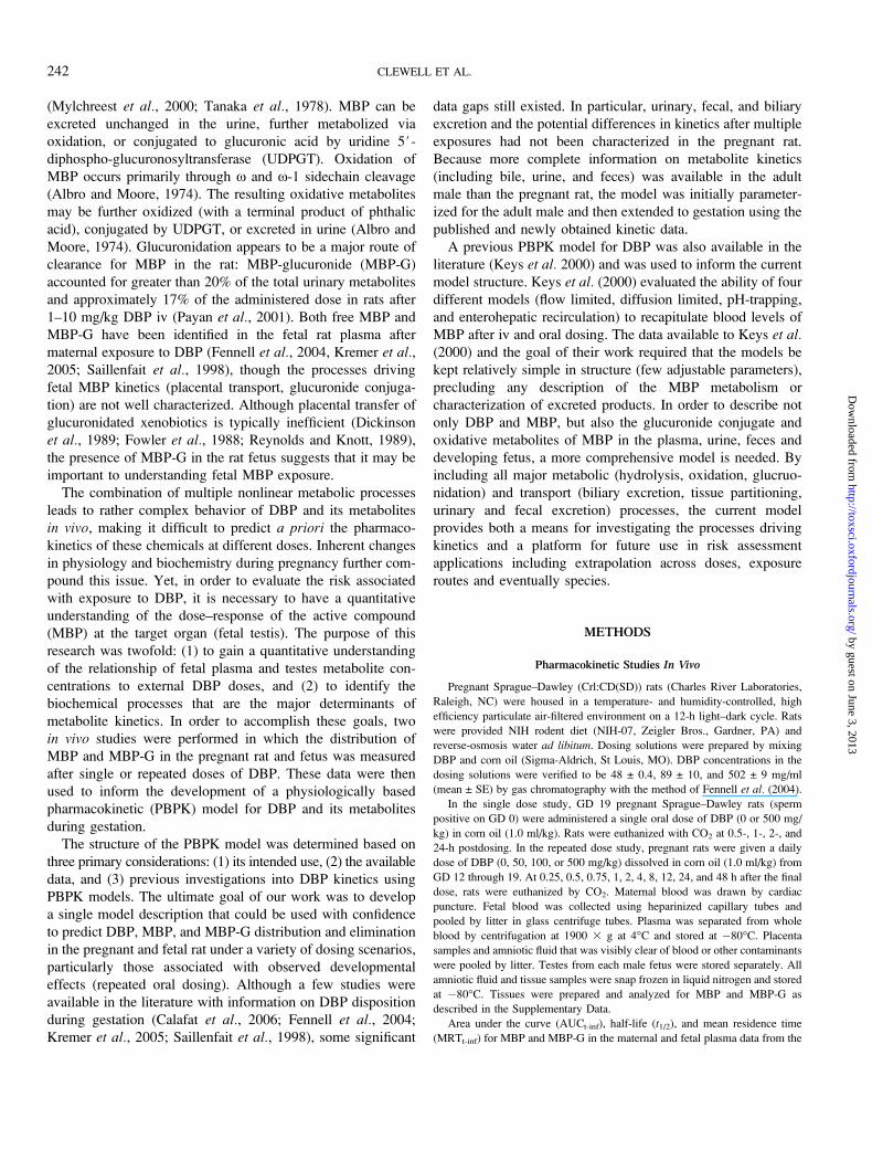
The final model (Fig. 1) contains four interconnected submodels, each with
the necessary amount of detail to adequately describe its chemical species:
DBP, MBP, MBP-G, and the combined oxidative metabolites (MBP-O). The
individual submodels interact at sites of metabolism (hydrolysis of the diester,
glucuronidation, hydrolysis of the glucuronide, and oxidation). The models for
each chemical species in the adult rat are described below, followed by the
modifications made to describe gestation. PBPK model simulations were
performed using ACSL 11.0 (Advanced Continuous Simulation Language,
AEgis Technologies Group, Inc., Huntsville, AL). Examples of model
equations are shown in the Supplementary Data.
Adult Male Rat Model
Intact DBP. Enzymes responsible for the hydrolysis of DBP are present in
the intestinal mucosa, plasma and liver (Rowland et al., 1977; Tanaka et al.,
1978). Hydrolysis of DBP in the plasma (kbc) and liver (kc) is described as a first
order rate as none of the tested doses were sufficient to overwhelm hydrolysis.
Hydrolysis of the DBP in the upper GI (stomach þ small intestine; GC1), on the
other hand, is described as a saturable process based on the in vitro data of
Rowland et al. (1977) and the apparent saturation of oral uptake at the highest
doses (~500 mg/kg) (NIEHS, 1994). In vitro studies suggest that DBP is poorly
absorbed in the gut, though a small amount may enter circulation intact,
particularly at doses where hydrolysis is overwhelmed (White et al., 1980).
Unabsorbed DBP may also be passed in to the lower intestine (GC2) and cleared
in the feces. Oral absorption is described as a first order process (kad). Transport
between the upper intestine (GC1) and lower intestine (GC2) compartments (kgic)
is a first order rate. Fecal excretion (kfc) is described using a clearance rate (l/h).
DBP that is taken up into the gut wall is passed to the liver via the portal blood
where it is hydrolyzed, released into systemic circulation with the plasma or
excreted into the bile (Tanaka et al., 1978). Biliary transfer of DBP into the
duodenum is modeled as a clearance rate (kbdc) from the liver to the upper
intestine (GC1). Efficient hydrolysis ensures that essentially 100% of the DBP is
de-esterified before it can be taken up into the bile. However, competition
between these processes (particularly after saturation of intestinal lipases at � 500
mg/kg doses) is included in the model in order to describe the wide range of
published data. Transport of DBP into the tissues is assumed to be flow limited.
Free MBP. Oral absorption is described as a first order process (kam).
Movement through the GI and fecal excretion are clearance rates. Unlike DBP,
MBP is readily absorbed in the gut wall and passed to liver via the portal blood.
Glucuronidation and oxidation of free MBP in the liver are described using
saturable kinetics. Free MBP may also be excreted into the bile or released into
systemic circulation. Transfer of MBP in the bile is described in the same manner
FIG. 1. Model structure for DBP kinetics in the pregnant rat. Adult male rat model is identical with the exception of the placenta/fetus compartments. Dashed
arrows indicate first order processes and clearance rates. Bold dashed arrows represent saturable processes. Solid arrows represent blood flows to the tissue
compartments. The solid arrows represent flow-limited (DBP) and diffusion-limited (MBP, MBP-G) transport into the tissues.
DBP PBPK MODEL FOR THE PREGNANT AND FETAL RAT 243
by guest on June 3, 2013http://toxsci.oxfordjournals.org/
Dow
nloaded from
as DBP, using a clearance rate (kbmc) from the liver to the upper intestine (GC1).
The MBP may then be reabsorbed or transported (kgic) into the lower intestine
(GC2). Absorption may occur in both the upper and lower intestine compart-
ments. Free MBP that is not absorbed in the intestine is cleared via the feces
(kfc). Transport of MBP into the tissues from the plasma is modeled using
diffusion-limitation. Secretion of free MBP into the urine is a saturable process,
based on nonlinear behavior of excretion data at low doses (Payan et al., 2001).
MBP-glucuronide. MBP-G formed in the liver (VmaxLc, KmL) may be
excreted into the bile or released into systemic circulation. Biliary transfer of
MBP-G is modeled as described for free MBP above. MBP-G then travels
through the intestine (kgic) and is either hydrolyzed to MBP via b-glucuronidase
(khydrc) in the lower intestine (GC2) or passed in the feces using first order
clearance rates. In the model, the hydrolysis of MBP-G is the rate limiting step
for reabsorption of conjugated MBP from the bile. Distribution of MBP-G into
the tissues is modeled using flow limitation, assuming distribution with body
water. Urinary excretion of MBP-G is modeled as a first order clearance rate
from the plasma compartment.
Oxidative metabolites of MBP. MBP-O formed by P450 metabolism
(VmaxOc, KmO) in the liver is released into the body via the venous blood.
A one-compartment volume of distribution model is used to describe the
combined oxidative metabolites. Distribution is assumed to be the body water
compartment. Urinary excretion is modeled as a first order clearance rate from
the central compartment.
Modifications for Gestation
During gestation, both MBP and MBP-G are allowed to move freely
between the arterial and placental plasma. Transfer of MBP between the
placental plasma and the fetal plasma is described as diffusion-limited (Gentry
et al., 2003). Versions of the model were tested which did or did not allow
MBP-G to cross the placenta (described below). Based on the fit of the model
simulations to available data, the final model does not include placental transfer
of MBP-G. Based on fetal MBP kinetic data, as well as published data on
UDPGT and b-glucuronidase activities in fetal tissues (Lucier et al., 1975;
Lucier and McDaniel, 1977; Wishart, 1978), glucuronidation of MBP and
hydrolysis of MBP-G are included in both dam and fetus. Transfer of MBP and
MBP-G between the fetus and amniotic fluid are described as first order
processes. Transfer between the fetal plasma and testes tissue is described using
diffusion-limited transport.
Parameterization in the Adult Male Rat PBPK Model
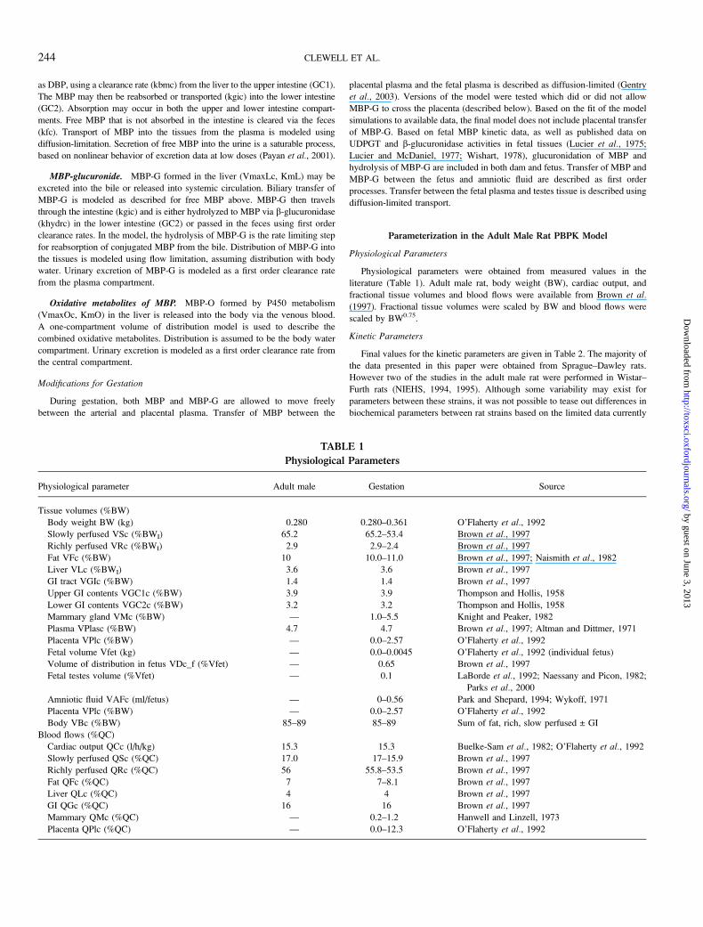
Physiological Parameters
Physiological parameters were obtained from measured values in the
literature (Table 1). Adult male rat, body weight (BW), cardiac output, and
fractional tissue volumes and blood flows were available from Brown et al.(1997). Fractional tissue volumes were scaled by BW and blood flows were
scaled by BW0.75.
Kinetic Parameters
Final values for the kinetic parameters are given in Table 2. The majority of
the data presented in this paper were obtained from Sprague–Dawley rats.
However two of the studies in the adult male rat were performed in Wistar–
Furth rats (NIEHS, 1994, 1995). Although some variability may exist for
parameters between these strains, it was not possible to tease out differences in
biochemical parameters between rat strains based on the limited data currently
TABLE 1
Physiological Parameters
Physiological parameter Adult male Gestation Source
Tissue volumes (%BW)
Body weight BW (kg) 0.280 0.280–0.361 O’Flaherty et al., 1992
Slowly perfused VSc (%BWI) 65.2 65.2–53.4 Brown et al., 1997
Richly perfused VRc (%BWI) 2.9 2.9–2.4 Brown et al., 1997
Fat VFc (%BW) 10 10.0–11.0 Brown et al., 1997; Naismith et al., 1982
Liver VLc (%BWI) 3.6 3.6 Brown et al., 1997
GI tract VGIc (%BW) 1.4 1.4 Brown et al., 1997
Upper GI contents VGC1c (%BW) 3.9 3.9 Thompson and Hollis, 1958
Lower GI contents VGC2c (%BW) 3.2 3.2 Thompson and Hollis, 1958
Mammary gland VMc (%BW) — 1.0–5.5 Knight and Peaker, 1982
Plasma VPlasc (%BW) 4.7 4.7 Brown et al., 1997; Altman and Dittmer, 1971
Placenta VPlc (%BW) — 0.0–2.57 O’Flaherty et al., 1992
Fetal volume Vfet (kg) — 0.0–0.0045 O’Flaherty et al., 1992 (individual fetus)
Volume of distribution in fetus VDc_f (%Vfet) — 0.65 Brown et al., 1997
Fetal testes volume (%Vfet) — 0.1 LaBorde et al., 1992; Naessany and Picon, 1982;
Parks et al., 2000
Amniotic fluid VAFc (ml/fetus) — 0–0.56 Park and Shepard, 1994; Wykoff, 1971
Placenta VPlc (%BW) — 0.0–2.57 O’Flaherty et al., 1992
Body VBc (%BW) 85–89 85–89 Sum of fat, rich, slow perfused ± GI
Blood flows (%QC)
Cardiac output QCc (l/h/kg) 15.3 15.3 Buelke-Sam et al., 1982; O’Flaherty et al., 1992
Slowly perfused QSc (%QC) 17.0 17–15.9 Brown et al., 1997
Richly perfused QRc (%QC) 56 55.8–53.5 Brown et al., 1997
Fat QFc (%QC) 7 7–8.1 Brown et al., 1997
Liver QLc (%QC) 4 4 Brown et al., 1997
GI QGc (%QC) 16 16 Brown et al., 1997
Mammary QMc (%QC) — 0.2–1.2 Hanwell and Linzell, 1973
Placenta QPlc (%QC) — 0.0–12.3 O’Flaherty et al., 1992
244 CLEWELL ET AL.
by guest on June 3, 2013http://toxsci.oxfordjournals.org/
Dow
nloaded from
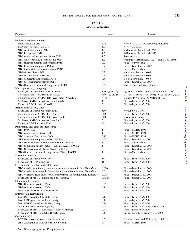
TABLE 2
Kinetic Parameters
Parameter Value Source
Partition coefficients (unitless)
DBP liver:plasma PL 21.8 Keys et al., 2000; personal communication
DBP body (tissues):plasma PT 1.0 Keys et al., 2000;
MBP gut tissue:plasma PMG 1.0 Williams and Blanchfield, 1975
MBP liver:plasma PML 1.3 Williams and Blanchfield, 1975
MBP richly perfused tissues:plasma PMR 1.3 Same as liver
MBP slowly perfused tissues:plasma PMS 1.2 Williams & Blanchfield, 1975; Tanaka et al., 1978
MBP maternal placenta tissue:plasma PMPl 0.2 Fitted—Current data
MBP fetus:maternal plasma PMFet 0.7 Fitted—Fennell et al., 2004
MBP testes:fetal central compartment PMFT 0.3 Fitted—Current data
MBP-G liver:plasma PGL 0.3 Vol of distribution ¼ 0.65
MBP-G body tissue:plasma PGT 0.3 Vol of distribution ¼ 0.65
MBP-G placenta tissue:plasma PGPl 0.3 Vol of distribution ¼ 0.65
MBP-G fetus:maternal plasma PGFet — Fitted—Fennell et al., 2004
MBP-G testess:fetal central compartment PGFT 0.3 Same as maternal tissue:plasma
Max capacity, Vmaxc (mg/h/kg)
Hydrolysis of DBP in GI lumen VmaxGc 150 (#), 90 ($) (#) Fitted—NIEHS, 1994, ($) White et al., 1980.
Glucuronidation of MBP in liver VmaxLc 300 (N); 150 (P) (N) Fitted—Payan et al., 2001; (P) Lucier et al., 1975
Glucuronidation of MBP in fetal liver (/fetus) VmaxLFc 0–44 Wishart, 1978; Lucier & McDaniel, 1977
Oxidation of MBP in maternal liver VmaxOc 1 Fitted—Payan et al., 2001
Uptake of MBP in urine VmaxUc 5 Fitted—Payan et al., 2001
Affinity constants, Km (mg/l)
Hydrolysis of DBP in GI lumen KmG 350 Rowland et al., 1977
Glucuronidation of MBP in liver KmL 300 Fitted—Payan et al., 2001
Glucuronidation of MBP in fetal liver KmLF 300 Sane as adult value
Oxidation of MBP in maternal liver KmO 5 Fitted—Payan et al., 2001
Uptake of MBP into urine KmU 45 Fitted—Payan et al., 2001
Permeability area cross products (l/h/kg)
MBP liver PALc 1 Fitted—NIEHS, 1994
MBP richly perfused tissue PARc 1 Fitted—NIEHS, 1994
MBP slowly perfused tissue PASc 0.15 Fitted—NIEHS, 1994
MBP fetus:maternal plasma (/fetus) PAFetc 0.05 Fitted—Fennell et al., 2004
MBP testes:fetal central compartment (/fetus) PAFTc 1 Fitted—Current data
MBP-G maternal tissues (/fetus) (PAGSc, PAGLc, PAGPlc) 1 Fitted—Fennell et al., 2004
MBP-G fetus:maternal plasma (/fetus) PAGFTc — Fitted—Fennell et al., 2004
MBP-G testes:fetal central compartment (/fetus) PAGFTc 1 Fitted—Current data
Metabolism rates (/h)
Hydrolysis of DBP in blood kbc 16 Fitted—Payan et al., 2001
Hydrolysis of DBP in liver kc 16 Fitted—Payan et al., 2001
Fetal-amniotic fluid transport (l/h/kg/fetus)
MBP transfer from fetus (central compartment) to amniotic fluid KtransM1c 0.006 Fitted—Fennell et al., 2004
MBP transfer from amniotic fluid to fetus (central compartment) KtransM2c 0.01 Fitted—Fennell et al., 2004
MBP-G transfer from fetus (central compartment) to amniotic fluid KtransGc 0.003 Fitted—Fennell et al., 2004
Hydrolysis of MBP-G in amniotic fluid KhydrAc 0.0025 Fitted—Fennell et al., 2004
Clearance rates (l/h/kg)
MBP-G urinary excretion ClGc 0.35 Fitted—Payan et al., 2001
MBP-O urinary excretion ClOc 0.1 Fitted—Payan et al., 2001
DBP, MBP, MBP-G fecal excretion kfc 0.012 Fitted—Payan et al., 2001
Enterohepatic recirculation
Liver DBP passed to bile kbdc (l/h/kg) 0.1 Fitted—Payan et al., 2001
Liver MBP passed to bile kbmc (l/h/kg) 0.1 Fitted—Payan et al., 2001
Liver MBP-G passed to bile kbgc (l/h/kg) 0.76 Fitted—Payan et al., 2001
Movement in GI contents kgic (/h) 0.01 Fitted—Payan et al., 2001; NIEHS 1994
Hydrolysis of MBP-G in maternal GI khydrc (l/h/kg) 0.3 Fitted—Payan et al., 2001
Hydrolysis of MBP-G in fetus khydrfc (l/h/kg) 0.13 Lucier et al., 1975; Soucy et al., 2006
Oral uptake (/h)
DBP Absorption in stomach and intestine kad 0.3 Calculated: kam and White et al., 1980
MBP Absorption in stomach and intestine kam 20 Fitted—NIEHS, 1994
Note. N ¼ nonpregnant rat, P ¼ pregnant rat.
DBP PBPK MODEL FOR THE PREGNANT AND FETAL RAT 245
by guest on June 3, 2013http://toxsci.oxfordjournals.org/
Dow
nloaded from
available. Therefore kinetic parameters were kept the same for both strains and
scaled allometrically as is typical for intra- and interspecies extrapolation
(Dedrick, 1973). Permeability area cross product (PA), Vmax, and clearance
constants were scaled by BW0.75. With the exception of metabolism
parameters, fetal parameters were scaled in a similar manner to the maternal
parameters: PAs were scaled by vfet0.75 and then multiplied by the total number
of fetuses (n ¼ 8) to obtain the value for the litter. Metabolism parameters
(glucuronide conjugation and hydrolysis; VmaxLf and khydrfc) were scaled by
adjusting the adult value by the ratio of the fetal:adult liver weight, because the
fetal liver weight is not linearly correlated with BW. The value for the total
litter was then calculated by multiplying by the number of fetuses (n ¼ 8).
Whenever possible parameters were taken from published values or calculated
from in vitro studies. Nonetheless, the lack of specific tissue and metabolism
data required that many of the model parameters be fitted to in vivo kinetic data.
Measured parameters. Tissue:plasma partition coefficients were obtained
from radiolabeled DBP studies (Williams and Blanchfield, 1975), and as such,
represent total phthalate rather than a specific compound. However, because
free MBP is expected to be the primary chemical form in the tissues, the total
radioactivity should be a reasonable predictor of MBP partitioning.
Tissue:plasma ratios obtained from early time points (4 h postdosing) of
radiolabeled studies were generally similar to the values obtained from vial
equilibration studies performed by Keys et al. (2000) (partition coefficient for
MBP in the richly perfused tissues (PMR) 1.3 vs. 1.2), with the exception of the
partition coefficient for the slowly perfused tissue (muscle). Because their vial
equilibration studies yielded questionable results for the muscle, Keys et al.(2000) used their published values for monoethylhexyl phthalate (MEHP) as
surrogate for MBP partitioning in the slowly perfused tissue (PMS). Although
muscle was not measured in Williams and Blanchfield (1975), another study
indicated that muscle:blood ratios were similar to the other richly perfused
tissues 24 h postdosing (Tanaka et al., 1978). Both the in vitro value for MEHP
and the value described above for MBP in the richly perfused tissue at 4-h
postdosing were tried as surrogates for the PMS by running the model against
iv plasma data (NIEHS, 1994) and slowly perfused tissue data (Tanaka et al.,
1978; not shown). The value for PMR (1.3) provided a better visual fit to the
data and was therefore used in the model.
The affinity constant (Km) for hydrolysis in the gut was calculated from the
data of Rowland et al. (1977). Rowland and coauthors spiked 1-ml aliquots of
rat stomach contents with various concentrations of DBP and measured
remaining DBP after 16 h. The Km for hydrolyzing enzymes (KmG ¼ 350 mg/l)
in the intestine was calculated by fitting a one-compartment model to the data in
Berkeley Madonna (University of California, Berkeley, CA).
Fitted and calculated parameters. For parameters that were not measured
experimentally, the values were estimated by adjusting the parameters to obtain
the best visual fit of the model to time course data. At this stage of model
development and evaluation a statistical program was not used to estimate model
parameters, although such an exercise would be particularly useful in conjunction
with further applications (e.g., extrapolation to the human). For the duration of
this paper, fitting of parameters refers to the manual process of determining one set
of parameters that could consistently recapitulate a large base of diverse data. In
order to minimize uncertainty in parameters, a sequential approach was followed
so that the most pertinent data sets were used for each parameter. This approach to
model parameterization is illustrated in Figure 2 and is described below.
Time course data on plasma MBP levels after a single iv dose (8 mg/kg) of
the monoester (NIEHS, 1994) were used to develop initial estimates of values
for the parameters governing liver metabolism and diffusion-limitation in the
richly and slowly perfused tissues (PARc and PASc). First approximations of
these parameters were based on this iv data set because it represented the
simplest possible dosing scenario, and was therefore dependent upon the least
number of adjustable parameters. However, as this study measured only plasma
MBP concentrations, additional data were needed to refine the parameters
driving elimination and enterohepatic recirculation.
Payan et al. (2001) provided a more comprehensive data set, which was
ideal for estimating elimination and metabolism parameters. Both iv and dermal
exposure data were presented. However, as the dermal data could not be used in
the model without significant changes to the structure that were not pertinent to
the current work, only the iv data were used. The iv study included plasma
DBP, MBP, MBP-G, and total 14C levels after a single iv dose of either 1 or
10 mg/kg 14C-DBP to adult male rats. Elimination of MBP, MBP-G, and total14C was measured over 72 h in the urine. Bile cannulation or a sham operation
(controls) was performed on a subset of rats dosed with 1 mg/kg 14C-DBP and
the total amount of 14C excreted in the bile and/or feces and urine was
measured at 30 h. Metabolism parameters (glucuronide conjugation, oxidation),
and urinary clearance rates were adjusted to fit DBP, MBP, MBP-G and total14C in the plasma and urine at the 1 mg/kg dose. Based on the increased water
solubility of glucuronide conjugates, it was assumed for the sake of simplicity
that MBP-G would be distributed with the water (volume of distribution
(VD) ¼ 0.65). The partition coefficient for MBP-G in the tissue (PGT) was
then calculated from the VD, blood volume (VB), and combined tissue volume
(VT) to be 0.3 [PGT ¼ (VD � VB)/VT]. Bile cannula studies were simulated
by turning off transfer of DBP, MBP, and MBP-G from the liver to the upper
intestine and biliary transport parameters (kbg, kbm, kbd) were fit to the bile
excretion data. The rates of MBP-G hydrolysis in the gut (khydrc) and fecal
excretion (kfgc, kfdc, kfmc) and movement in the gut (kgic) were then adjusted
to reproduce fecal excretion and plasma data in sham-operated rats.
With the above parameters constrained, the first order rate of MBP oral
absorption (kam) was determined. Kam was fit to plasma MBP data obtained
from adult male rats after a single oral bolus of 34 mg/kg MBP (NIEHS, 1994).
Absorption of unchanged DBP was then calculated based on in vitro data using
everted gut preparations (White et al., 1980). The measured rate for DBP
absorption was 67-fold less than MBP. Using this measured ratio and the
previously determined value for MBP absorption (20/h), a value of 0.3/h was
calculated for kad.
PBPK Model Validation in the Adult Male Rat
Validation of the model structure and the final parameters was performed
using separate data sets from those used for parameterization (NIEHS, 1995; 10
mg/kg from Payan et al., 2001). No parameters were changed to improve model
fits to the ‘‘validation’’ data.
Application of Adult Male Rat DBP PBPK Model to Gestation
Physiological Parameters
During gestation, mammary gland (VM), and fat (VF) tissue growth were
described as a linear processes based on the data of Hanwell and Linzell (1973),
Knight and Peaker (1982), and Naismith et al. (1982), as described in Clewell
et al. (2003). Placental volume (VPl) was described as the sum of the yolk sac
and chorioallantoic placenta based on the model of O’Flaherty et al. (1992).
Growth equations were available in the cited papers. The total BW of the dam
was made equal to the initial BW plus the change in volume of the uterus, fat,
mammary gland, placenta, and fetus. Fetal volume (Vfet) was described using
the equations of O’Flaherty et al. (1992). Growth of fetal testes is proportional
to the total BW, accounting for approximately 0.1% of the total fetal volume
from GD 16 through the end of gestation (LaBorde et al., 1992; Naessany and
Picon, 1982; Parks et al., 2000). Changes in amniotic fluid volume were
described using a TABLE function in ACSL, with linear interpolation between
data points (Park and Shepard, 1994; Wykoff, 1971).
Maternal cardiac output was described as the sum of initial cardiac output
(Brown et al., 1997) and the change in blood flow to the placenta, mammary
and fat tissues, per the approach of O’Flaherty et al. (1992). Changes in the
fractional cardiac output to the mammary gland, fat and yolk sac were assumed
to be proportional to changes in tissue volumes, with the exception of the
chorioallantoic placenta which increased more rapidly than the tissue volume.
Chemical transport within the fetus was modeled using diffusion, rather than
blood flow limitation. Thus, no assumptions were made as to proportional
blood flows to fetal tissues.
246 CLEWELL ET AL.
by guest on June 3, 2013http://toxsci.oxfordjournals.org/
Dow
nloaded from
Kinetic Parameters
Two kinetic parameters were adjusted before using the model in the
pregnant dam based on published in vitro studies: VmaxGc and VmaxLc.
VmaxGc, the maximum capacity for DBP hydrolysis in the intestine, was
decreased based on in vitro metabolism studies. Rowland et al. (1977)
measured the disappearance of DBP in 1-ml aliquots of small intestine contents
from adult male and female rats (33–40 days old). Their studies showed a sex
difference in the ability to hydrolyze DBP, with the females metabolizing only
60% as much as the males. This sex difference was accounted for in the model
by reducing the unscaled value for VmaxGc (90 vs. 150 mg/h/kg) for all
simulations of female rats. The parameter was then scaled by BW0.75 to account
for differences in BW.
VmaxLc, the maximum capacity for glucuronide conjugation in the liver,
was also reduced in simulations of pregnant rats. In vitro studies in livers of
nonpregnant female and pregnant rats showed that UDPGT activity for
a variety of substrates (steroidal and nonsteroidal) was decreased by
approximately 50% during gestation (GD 19–20) (Lucier et al., 1975; Luquita
et al., 2001). This difference in activity was included in the model by reducing
the unscaled value of VmaxLc by 50% (150 vs. 300 mg/h/kg) during gestation,
which was scaled by BW0.75 to account for changes in BW. All other maternal
parameters were scaled allometrically from the adult male rat.
Parameters describing transfer between the dam and fetus, and the fetus and
amniotic fluid were fit to published placenta, fetal plasma, and amniotic fluid
time course data (Fennell et al., 2004). Because no previous data were available
for MBP concentrations in the placenta tissue or fetal testes, the partition
coefficients (Pmpl, Pmft) for these tissues were fit to the data from the single
dose data from the current studies (500 mg/kg).
Two possible explanations were considered for the presence of MBP-G in
the fetus (Fennell et al, 2004): (1) MBP-G is not significantly transported in the
placenta and fetal MBP-G must be formed in the fetus itself, or (2) maternally
formed MBP-G is able to cross the placenta. Alternative versions of the model
were developed and tested with the MBP-G time course data to determine
which one described the data more accurately. In the first version, glucruonide
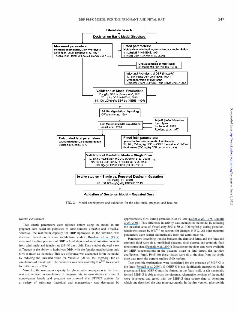
FIG. 2. Model development and validation for the adult male, pregnant and fetal rat.
DBP PBPK MODEL FOR THE PREGNANT AND FETAL RAT 247
by guest on June 3, 2013http://toxsci.oxfordjournals.org/
Dow
nloaded from
conjugation and hydrolysis of MBP-G was described in the central
compartment of the fetus (plasma). Fetal metabolism parameters were estimated
from in vitro data described below. In the second version of the model,
placental transfer of MBP-G was described in the same manner as free MBP, in
addition to fetal UGT and b-glucuronidase activity. The partition coefficient
and permeability area cross product were adjusted to achieve the best visual fit
to the fetal plasma MBP-G time course data.
During fetal development, rat UDPGTs are classified into two distinct
classes based on the preferred substrates. Steroidal UDPGTs are characterized
by a low activity in the fetal liver, with a rapid increase to adult levels after
birth. Nonsteroidal UDPGTs show a peak in activity on GD 19 at nearly adult
levels, followed by a drop in activity at birth and a subsequent increase in
adolescent rats (Lucier and McDaniel, 1977; Wishart, 1978). Although studies
have not been performed specifically on glucuronidation of MBP in fetal livers,
studies with a similar chemical, monoethylhexyl phthalate, showed that it is
metabolized by steroidal UDPGTs (Sjoberg et al., 1999). Therefore, the fetal
UDPGT activity in the model was estimated from the ratio of measured
fetal:adult UDPGT activities toward several steroidal substrates from measured
in vitro values. The in vitro activity averaged for the measured steroidal
substrates was 0.6, 1.3, 2.5, 11.8, and 11.4% of the adult male rat value per mg
microsomal protein on GD 17, 18, 19, 20, and 21 (Lucier and McDaniel, 1977;
Wishart, 1978). These relative activities were used to calculate the value for
maximum capacity of fetal UDPGT according to Equation 1. The amount of
microsomal protein in the liver of the Sprague–Dawley fetus (~7.3 mg/g liver)
was obtained from Alcorn et al. (2007), as it was not reported in the Lucier and
McDaniel (1977) or Wishart (1978) studies. The final values for VmaxLf (0.05,
0.12, 0.6, 3.0, and 4.4 mg/h) were coded into the model using a TABLE
function, which employs linear interpolation to estimate parameter values
between defined points. Fetal b-glucuronidase activity (khydrfc) was found to
be 20% of the adult value based on measured activities per mg microsomal
protein in vitro for various substrates (Lucier et al., 1975; Soucy et al., 2006).
Khydrf was estimated from the adult male rat value in the same manner as
VmaxLf.
VmaxLf ¼ VmaxL3RAfL 3MPC3 LW3 numfet ð1Þ
where VmaxL is the maximum capacity for glucuronide in the adult male rat(after scaling for BW), RAfL is the in vitro relative activity expressed as theratio of fetal to maternal activity (per mg microsomal protein), MPC is themicrosomal protein content of the fetal liver (mg/g liver), LW is the liverweight (g), and numfet is the number of fetuses per litter.
After setting the parameters, testing the model against the data of Fennell
et al. (2004) and deciding on a final model for the fetal MBP-G, the gestation
model was then tested with additional data sets from single dose studies on
different days of gestation and alternate dose routes: current studies (GD 19,
po); Saillenfait et al. (1998) (GD 14, po), and Kremer et al. (2005) (GD 19, iv).
Saillenfait et al. (1998) treated GD 14 Sprague–Dawley rats with a single oral
dose of 500 or 1500 mg/kg 14C-DBP in mineral oil. Total radioactivity, 14C-
MBP and 14C-MBP-G were measured in the maternal plasma, placenta,
amniotic fluid and whole fetus at 0.5-, 1-, 2-, 4-, 6-, 8-, 24-, and 48-h
postdosing. Total radioactivity in the maternal urine and feces was also
measured at 24- and 48-h postdosing. Kremer et al. (2005) performed a time
course study in GD 19 Sprague–Dawley rats after a single iv dose of 10, 30, or
50 mg/kg MBP. Serial samples were collected from cannulated rats, and MBP
and MBP-G plasma concentrations were measured at 0.08-, 0.25-, 0.5-, 0.75-,
1-, 1.5-, 2-, 4-, and 8-h postdosing.
Extrapolation of Acute DBP Gestation PBPK Model to Multiple
Day Exposures
The ability to describe repeated dosing scenarios was tested by running the
model against the repeated dosing data from the current studies without
adjusting any of the model parameters determined from the single dose data.
The model was also tested with the data of Calafat et al. (2006), who measured
free MBP in the amniotic fluid of GD 18 Sprague–Dawley rats. Dams received
an oral dose of 0, 100, or 250 mg/kg DBP once daily from GD 12–17.
Amniotic fluid was pooled by litter and collected at the time of sacrifice (GD
18). Urine catch samples were also collected on GD 17 (approximately 6 h
postdosing). However, because the urine samples were presented as
concentrations and volumes were not included, they could not be used
straightforwardly in the model.
Sensitivity Analysis of Model Parameters
A normalized sensitivity analysis was run on the gestation model to examine
the relative influence of each of the parameters on model output. The model
was run to determine the change in the average plasma MBP concentrations
(24 h AUC) resulting from a 1% change in the value of each kinetic parameter.
In an effort to determine the effect of metabolic saturation on relative parameter
importance, the sensitivity analysis was performed at two nominal doses of
DBP, representing unsaturated and saturated states (10 and 500 mg/kg,
respectively). The following equation shows the calculation of the sensitivity
coefficient.
Sensitivity coefficient ¼ ðA� BÞ=BðC � DÞ=D ð2Þ
where A is the plasma AUC with 1% increased parameter value, B is the plasmaAUC at the starting parameter value, C is the parameter value after 1% increaseand D is the original parameter value.
RESULTS
In order to relate tissue concentration of DBP metabolites to
observed effects during fetal development, the distribution of
MBP and MBP-G in the pregnant rat and fetus were
determined after a single dose of 500 mg/kg DBP and repeated
doses of DBP at a low (50 mg/kg), medium (100 mg/kg) and
high (500 mg/kg) doses. In addition to allowing direct
comparison of external and tissue dose, these data also
supported the development of a PBPK model that was used
to evaluate the relative importance of transport and metabolic
processes in the pharmacokinetic behavior of DBP across
doses, providing a robust platform for future risk assessment
applications.
Pharmacokinetic Studies In Vivo
The repeated dosing study was designed to reproduce the
experimental conditions of the published DBP effects studies in
order to gain a better understanding of internal dose associated
with observed effects. In this case, maternal and fetal plasma,
placenta and amniotic fluid MBP levels may be used as
surrogates for fetal dose. Concentration time course data
collected in this present study appear in the Supplementary
Data (Tables S1–4). Some key aspects of disposition of DBP
and its metabolites were noted from the initial PK analysis of
the data (Table S5).
Dose-dependent differences in AUC/D, Cmax, t1/2, and MRT
illustrate the nonlinear behavior of MBP in the repeated dosing
study (Supplementary Data), leading to some preliminary
hypotheses about dose-dependent kinetics. For example, the
fact that the apparent parameter differences in maternal plasma
248 CLEWELL ET AL.
by guest on June 3, 2013http://toxsci.oxfordjournals.org/
Dow
nloaded from
MBP do not take place at the same dose may suggest that more
that more than one saturable process is driving MBP dose–
response. Differences (~two-fold) in Cmax/D, t1/2 and MRT
between the 100 and 500 mg/kg/day groups, may be explained
by saturation of DBP intestinal hydrolysis and prolonged
absorption of MBP. AUC/D, on the other hand, increases from
2.6 to 4.8 between the 50 and 100 mg/kg/day groups,
suggesting that a second saturable process (possibly clearance)
may be involved. Nonetheless, the parameters are certainly
affected by a combination of many factors and these simple PK
models do not lend themselves to in-depth analysis of the
processes driving dose-dependent kinetics. PBPK models that
incorporate the various biochemical pathways into their
description are better suited to mechanistic investigations.
The model described in this paper was specifically designed for
this purpose. The PBPK model also allows comparison of
studies with fewer data points (i.e., the single dose study, testes
data), where classical PK analysis cannot be performed.
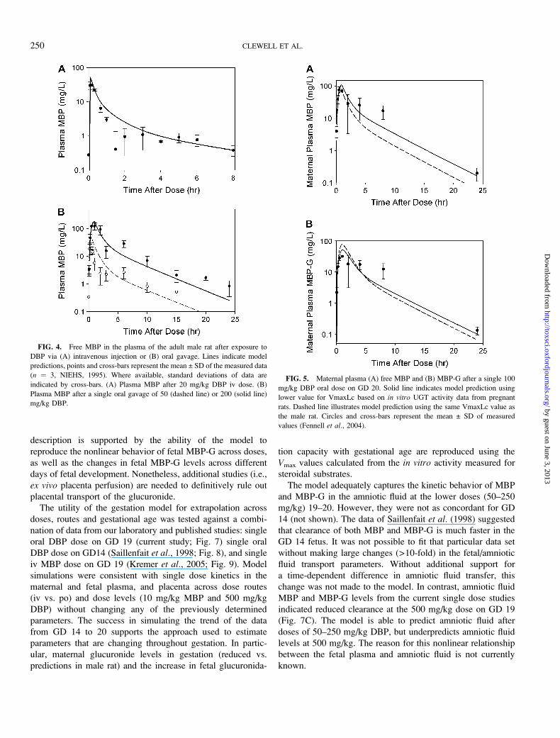
PBPK Model Validation in the Adult Male Rat
The results of model development and parameterization in
the adult male rate are provided in the Supplementary Data
(Figs. S1 and S2, Table S7). The final model differed
significantly in structure from the previous model of Keys
et al. (2000), primarily as a result of the current need to
describe additional metabolites in tissues and urine and to
facilitate extension to gestation. Differences between the two
models are summarized in the Supplementary Data (Table S6).
Before extending the model to gestation, the final model
structure and parameter values were tested by simulating data
in the male rat that were not used for parameterization. No
parameters were altered to fit the validation data. Plasma and
urine metabolite kinetics were well-described by the model
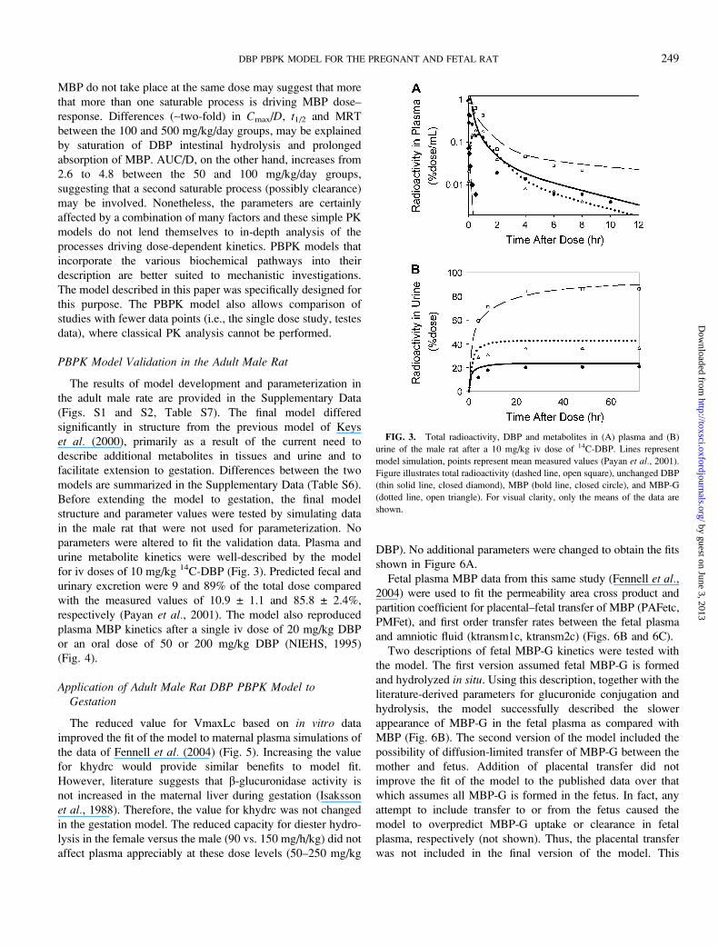
for iv doses of 10 mg/kg 14C-DBP (Fig. 3). Predicted fecal and
urinary excretion were 9 and 89% of the total dose compared
with the measured values of 10.9 ± 1.1 and 85.8 ± 2.4%,
respectively (Payan et al., 2001). The model also reproduced
plasma MBP kinetics after a single iv dose of 20 mg/kg DBP
or an oral dose of 50 or 200 mg/kg DBP (NIEHS, 1995)
(Fig. 4).
Application of Adult Male Rat DBP PBPK Model toGestation
The reduced value for VmaxLc based on in vitro data
improved the fit of the model to maternal plasma simulations of
the data of Fennell et al. (2004) (Fig. 5). Increasing the value
for khydrc would provide similar benefits to model fit.
However, literature suggests that b-glucuronidase activity is
not increased in the maternal liver during gestation (Isaksson
et al., 1988). Therefore, the value for khydrc was not changed
in the gestation model. The reduced capacity for diester hydro-
lysis in the female versus the male (90 vs. 150 mg/h/kg) did not
affect plasma appreciably at these dose levels (50–250 mg/kg
DBP). No additional parameters were changed to obtain the fits
shown in Figure 6A.
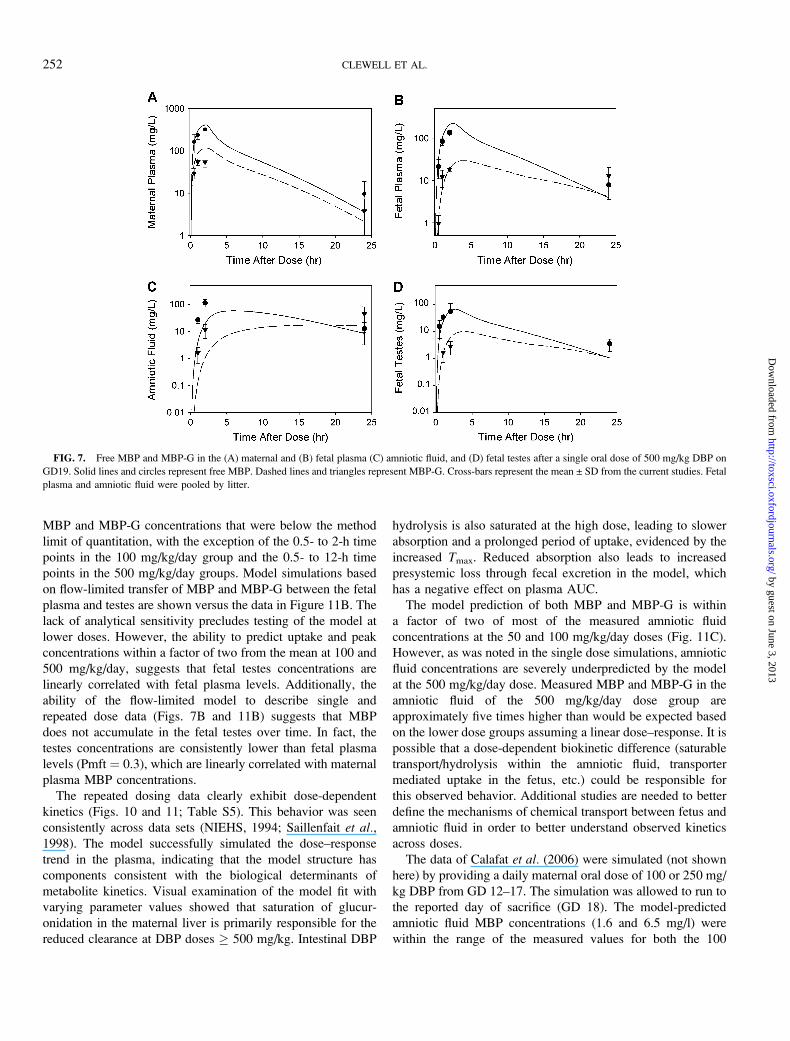
Fetal plasma MBP data from this same study (Fennell et al.,2004) were used to fit the permeability area cross product and
partition coefficient for placental–fetal transfer of MBP (PAFetc,
PMFet), and first order transfer rates between the fetal plasma
and amniotic fluid (ktransm1c, ktransm2c) (Figs. 6B and 6C).
Two descriptions of fetal MBP-G kinetics were tested with
the model. The first version assumed fetal MBP-G is formed
and hydrolyzed in situ. Using this description, together with the
literature-derived parameters for glucuronide conjugation and
hydrolysis, the model successfully described the slower
appearance of MBP-G in the fetal plasma as compared with
MBP (Fig. 6B). The second version of the model included the
possibility of diffusion-limited transfer of MBP-G between the
mother and fetus. Addition of placental transfer did not
improve the fit of the model to the published data over that
which assumes all MBP-G is formed in the fetus. In fact, any
attempt to include transfer to or from the fetus caused the
model to overpredict MBP-G uptake or clearance in fetal
plasma, respectively (not shown). Thus, the placental transfer
was not included in the final version of the model. This
FIG. 3. Total radioactivity, DBP and metabolites in (A) plasma and (B)
urine of the male rat after a 10 mg/kg iv dose of 14C-DBP. Lines represent
model simulation, points represent mean measured values (Payan et al., 2001).
Figure illustrates total radioactivity (dashed line, open square), unchanged DBP
(thin solid line, closed diamond), MBP (bold line, closed circle), and MBP-G
(dotted line, open triangle). For visual clarity, only the means of the data are
shown.
DBP PBPK MODEL FOR THE PREGNANT AND FETAL RAT 249
by guest on June 3, 2013http://toxsci.oxfordjournals.org/
Dow
nloaded from
description is supported by the ability of the model to
reproduce the nonlinear behavior of fetal MBP-G across doses,
as well as the changes in fetal MBP-G levels across different
days of fetal development. Nonetheless, additional studies (i.e.,
ex vivo placenta perfusion) are needed to definitively rule out
placental transport of the glucuronide.
The utility of the gestation model for extrapolation across
doses, routes and gestational age was tested against a combi-
nation of data from our laboratory and published studies: single
oral DBP dose on GD 19 (current study; Fig. 7) single oral
DBP dose on GD14 (Saillenfait et al., 1998; Fig. 8), and single
iv MBP dose on GD 19 (Kremer et al., 2005; Fig. 9). Model
simulations were consistent with single dose kinetics in the
maternal and fetal plasma, and placenta across dose routes
(iv vs. po) and dose levels (10 mg/kg MBP and 500 mg/kg
DBP) without changing any of the previously determined
parameters. The success in simulating the trend of the data
from GD 14 to 20 supports the approach used to estimate
parameters that are changing throughout gestation. In partic-
ular, maternal glucuronide levels in gestation (reduced vs.
predictions in male rat) and the increase in fetal glucuronida-
tion capacity with gestational age are reproduced using the
Vmax values calculated from the in vitro activity measured for
steroidal substrates.
The model adequately captures the kinetic behavior of MBP
and MBP-G in the amniotic fluid at the lower doses (50–250
mg/kg) 19–20. However, they were not as concordant for GD
14 (not shown). The data of Saillenfait et al. (1998) suggested
that clearance of both MBP and MBP-G is much faster in the
GD 14 fetus. It was not possible to fit that particular data set
without making large changes (>10-fold) in the fetal/amniotic
fluid transport parameters. Without additional support for
a time-dependent difference in amniotic fluid transfer, this
change was not made to the model. In contrast, amniotic fluid
MBP and MBP-G levels from the current single dose studies
indicated reduced clearance at the 500 mg/kg dose on GD 19
(Fig. 7C). The model is able to predict amniotic fluid after
doses of 50–250 mg/kg DBP, but underpredicts amniotic fluid
levels at 500 mg/kg. The reason for this nonlinear relationship
between the fetal plasma and amniotic fluid is not currently
known.
FIG. 5. Maternal plasma (A) free MBP and (B) MBP-G after a single 100
mg/kg DBP oral dose on GD 20. Solid line indicates model prediction using
lower value for VmaxLc based on in vitro UGT activity data from pregnant
rats. Dashed line illustrates model prediction using the same VmaxLc value as
the male rat. Circles and cross-bars represent the mean ± SD of measured
values (Fennell et al., 2004).
FIG. 4. Free MBP in the plasma of the adult male rat after exposure to
DBP via (A) intravenous injection or (B) oral gavage. Lines indicate model
predictions, points and cross-bars represent the mean ± SD of the measured data
(n ¼ 3, NIEHS, 1995). Where available, standard deviations of data are
indicated by cross-bars. (A) Plasma MBP after 20 mg/kg DBP iv dose. (B)
Plasma MBP after a single oral gavage of 50 (dashed line) or 200 (solid line)
mg/kg DBP.
250 CLEWELL ET AL.
by guest on June 3, 2013http://toxsci.oxfordjournals.org/
Dow
nloaded from
Fetal testes parameters were fitted to the data from the
current single dose study (Fig. 7D), as no previous data were
available for this tissue. Both MBP and MBP-G appear to be
distributed with body water. Partition coefficients were 0.3 and
permeability area cross products were 1 l/h/kg, suggesting
rapid equilibration with the plasma.
Model predictions for 24 h urinary (77% of dose) and fecal
(15% of dose) excretion in the GD 20 rat were similar to the
measured values in the naı̈ve female rat (77 ± 8 and 7 ± 3.5%
of dose, respectively) after a 100 mg/kg oral DBP dose
(Fennell et al., 2004). Model-predicted excretion did not differ
between the nonpregnant and GD 20 rat.
Extrapolation of Acute DBP Gestation PBPK Model toMultiple Day Exposures
In general, the model was able to predict maternal and fetal
plasma and placenta kinetic behavior at the 50, 100, and
500 mg/kg/day doses from the current repeated dose studies
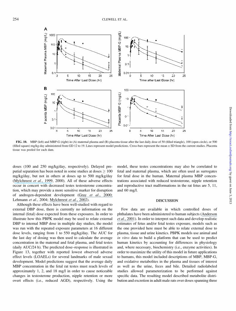
(Figs. 10 and 11) without changing any kinetic parameters.
Although the model falls outside of the standard deviations for
some time points, it consistently captures the kinetic behavior
of MBP and MBP-G, including the slowed uptake and
clearance of MBP at high doses (Fig. 10A) and the apparent
accumulation of MBP-G in the fetal plasma and amniotic fluid
over time (Fig. 11). The majority of the fetal testes samples had
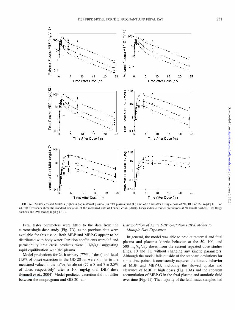
FIG. 6. MBP (left) and MBP-G (right) in (A) maternal plasma (B) fetal plasma, and (C) amniotic fluid after a single dose of 50, 100, or 250 mg/kg DBP on
GD 20. Crossbars show the standard deviation of the measured data of Fennell et al. (2004). Lines indicate model predictions at 50 (small dashed), 100 (large
dashed) and 250 (solid) mg/kg DBP.
DBP PBPK MODEL FOR THE PREGNANT AND FETAL RAT 251
by guest on June 3, 2013http://toxsci.oxfordjournals.org/
Dow
nloaded from
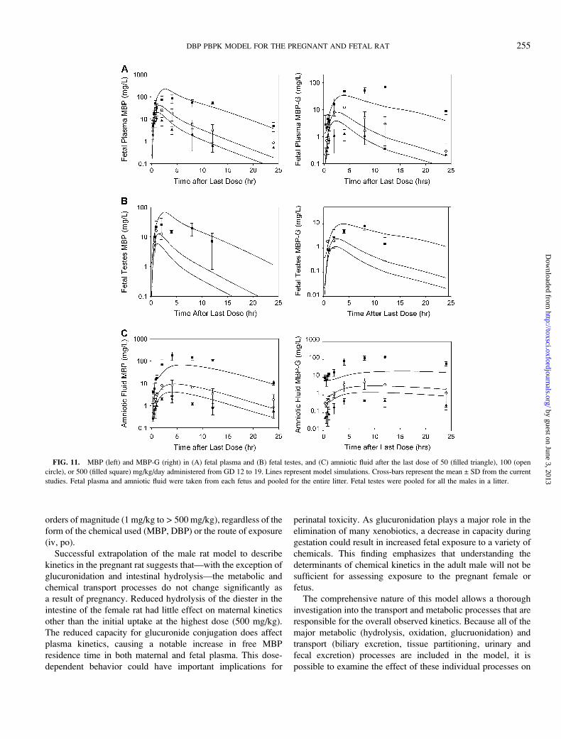
MBP and MBP-G concentrations that were below the method
limit of quantitation, with the exception of the 0.5- to 2-h time
points in the 100 mg/kg/day group and the 0.5- to 12-h time
points in the 500 mg/kg/day groups. Model simulations based
on flow-limited transfer of MBP and MBP-G between the fetal
plasma and testes are shown versus the data in Figure 11B. The
lack of analytical sensitivity precludes testing of the model at
lower doses. However, the ability to predict uptake and peak
concentrations within a factor of two from the mean at 100 and
500 mg/kg/day, suggests that fetal testes concentrations are
linearly correlated with fetal plasma levels. Additionally, the
ability of the flow-limited model to describe single and
repeated dose data (Figs. 7B and 11B) suggests that MBP
does not accumulate in the fetal testes over time. In fact, the
testes concentrations are consistently lower than fetal plasma
levels (Pmft ¼ 0.3), which are linearly correlated with maternal
plasma MBP concentrations.
The repeated dosing data clearly exhibit dose-dependent
kinetics (Figs. 10 and 11; Table S5). This behavior was seen
consistently across data sets (NIEHS, 1994; Saillenfait et al.,1998). The model successfully simulated the dose–response
trend in the plasma, indicating that the model structure has
components consistent with the biological determinants of
metabolite kinetics. Visual examination of the model fit with
varying parameter values showed that saturation of glucur-
onidation in the maternal liver is primarily responsible for the
reduced clearance at DBP doses � 500 mg/kg. Intestinal DBP
hydrolysis is also saturated at the high dose, leading to slower
absorption and a prolonged period of uptake, evidenced by the
increased Tmax. Reduced absorption also leads to increased
presystemic loss through fecal excretion in the model, which
has a negative effect on plasma AUC.
The model prediction of both MBP and MBP-G is within
a factor of two of most of the measured amniotic fluid
concentrations at the 50 and 100 mg/kg/day doses (Fig. 11C).
However, as was noted in the single dose simulations, amniotic
fluid concentrations are severely underpredicted by the model
at the 500 mg/kg/day dose. Measured MBP and MBP-G in the
amniotic fluid of the 500 mg/kg/day dose group are
approximately five times higher than would be expected based
on the lower dose groups assuming a linear dose–response. It is
possible that a dose-dependent biokinetic difference (saturable
transport/hydrolysis within the amniotic fluid, transporter
mediated uptake in the fetus, etc.) could be responsible for
this observed behavior. Additional studies are needed to better
define the mechanisms of chemical transport between fetus and
amniotic fluid in order to better understand observed kinetics
across doses.
The data of Calafat et al. (2006) were simulated (not shown
here) by providing a daily maternal oral dose of 100 or 250 mg/
kg DBP from GD 12–17. The simulation was allowed to run to
the reported day of sacrifice (GD 18). The model-predicted
amniotic fluid MBP concentrations (1.6 and 6.5 mg/l) were
within the range of the measured values for both the 100
FIG. 7. Free MBP and MBP-G in the (A) maternal and (B) fetal plasma (C) amniotic fluid, and (D) fetal testes after a single oral dose of 500 mg/kg DBP on
GD19. Solid lines and circles represent free MBP. Dashed lines and triangles represent MBP-G. Cross-bars represent the mean ± SD from the current studies. Fetal
plasma and amniotic fluid were pooled by litter.
252 CLEWELL ET AL.
by guest on June 3, 2013http://toxsci.oxfordjournals.org/
Dow
nloaded from
(0.3–2.4 mg/l) and the 250 mg/kg/day (3.8–22.3 mg/l) dose
groups.
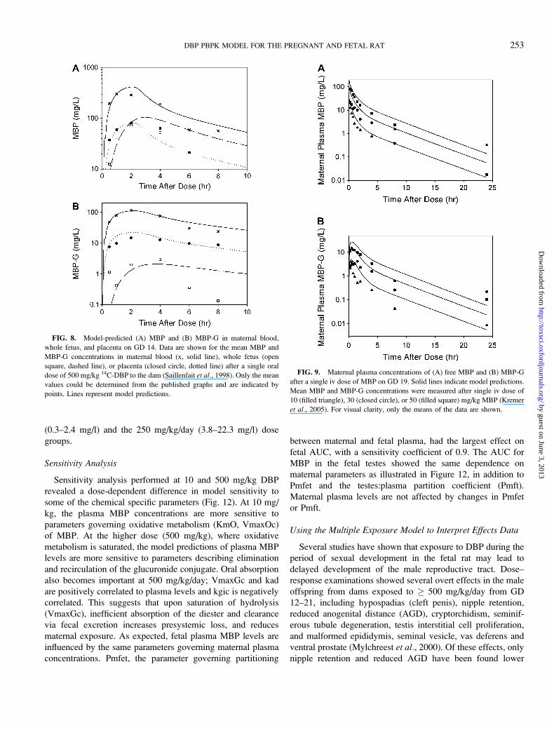
Sensitivity Analysis
Sensitivity analysis performed at 10 and 500 mg/kg DBP
revealed a dose-dependent difference in model sensitivity to
some of the chemical specific parameters (Fig. 12). At 10 mg/
kg, the plasma MBP concentrations are more sensitive to
parameters governing oxidative metabolism (KmO, VmaxOc)
of MBP. At the higher dose (500 mg/kg), where oxidative
metabolism is saturated, the model predictions of plasma MBP
levels are more sensitive to parameters describing elimination
and recirculation of the glucuronide conjugate. Oral absorption
also becomes important at 500 mg/kg/day; VmaxGc and kad
are positively correlated to plasma levels and kgic is negatively
correlated. This suggests that upon saturation of hydrolysis
(VmaxGc), inefficient absorption of the diester and clearance
via fecal excretion increases presystemic loss, and reduces
maternal exposure. As expected, fetal plasma MBP levels are
influenced by the same parameters governing maternal plasma
concentrations. Pmfet, the parameter governing partitioning
between maternal and fetal plasma, had the largest effect on
fetal AUC, with a sensitivity coefficient of 0.9. The AUC for
MBP in the fetal testes showed the same dependence on
maternal parameters as illustrated in Figure 12, in addition to
Pmfet and the testes:plasma partition coefficient (Pmft).
Maternal plasma levels are not affected by changes in Pmfet
or Pmft.
Using the Multiple Exposure Model to Interpret Effects Data
Several studies have shown that exposure to DBP during the
period of sexual development in the fetal rat may lead to
delayed development of the male reproductive tract. Dose–
response examinations showed several overt effects in the male
offspring from dams exposed to � 500 mg/kg/day from GD
12–21, including hypospadias (cleft penis), nipple retention,
reduced anogenital distance (AGD), cryptorchidism, seminif-
erous tubule degeneration, testis interstitial cell proliferation,
and malformed epididymis, seminal vesicle, vas deferens and
ventral prostate (Mylchreest et al., 2000). Of these effects, only
nipple retention and reduced AGD have been found lower
FIG. 8. Model-predicted (A) MBP and (B) MBP-G in maternal blood,
whole fetus, and placenta on GD 14. Data are shown for the mean MBP and
MBP-G concentrations in maternal blood (x, solid line), whole fetus (open
square, dashed line), or placenta (closed circle, dotted line) after a single oral
dose of 500 mg/kg 14C-DBP to the dam (Saillenfait et al., 1998). Only the mean
values could be determined from the published graphs and are indicated by
points. Lines represent model predictions.
FIG. 9. Maternal plasma concentrations of (A) free MBP and (B) MBP-G
after a single iv dose of MBP on GD 19. Solid lines indicate model predictions.
Mean MBP and MBP-G concentrations were measured after single iv dose of
10 (filled triangle), 30 (closed circle), or 50 (filled square) mg/kg MBP (Kremer
et al., 2005). For visual clarity, only the means of the data are shown.
DBP PBPK MODEL FOR THE PREGNANT AND FETAL RAT 253
by guest on June 3, 2013http://toxsci.oxfordjournals.org/
Dow
nloaded from
doses (100 and 250 mg/kg/day, respectively). Delayed pre-
putial separation has been noted in some studies at doses � 100
mg/kg/day, but not in others at doses up to 500 mg/kg/day
(Mylchreest et al., 1999, 2000). All of these adverse effects
occur in concert with decreased testes testosterone concentra-
tion, which may provide a more sensitive marker for disruption
of androgen-dependent development (Gray et al., 2000;
Lehmann et al., 2004; Mylchreest et al., 2002).
Although these effects have been well-studied with regard to
external DBP dose, there is currently no information on the
internal (fetal) dose expected from these exposures. In order to
illustrate how this PBPK model may be used to relate external
DBP to internal MBP dose in multiple day studies, the model
was run with the repeated exposure parameters at 16 different
dose levels, ranging from 1 to 550 mg/kg/day. The AUC for
the last day of dosing was then used to calculate the average
concentration in the maternal and fetal plasma, and fetal testes
(daily AUC/24 h). The predicted dose–response is illustrated in
Figure 13, together with reported lowest observed adverse
effect levels (LOAELs) for several landmarks of male sexual
development. Model predictions suggest that the average daily
MBP concentration in the fetal rat testes must reach levels of
approximately 1, 2, and 18 mg/l in order to cause noticeable
changes in testosterone production, nipple retention or more
overt effects (i.e., reduced AGD), respectively. Using the
model, these testes concentrations may also be correlated to
fetal and maternal plasma, which are often used as surrogates
for fetal dose in the human. Maternal plasma MBP concen-
trations associated with reduced testosterone, nipple retention
and reproductive tract malformations in the rat fetus are 5, 11,
and 60 mg/l.
DISCUSSION
Few data are available in which controlled doses of
phthalates have been administered to human subjects (Anderson
et al., 2001). In order to interpret such data and develop realistic
estimates of fetus and/or fetal testes exposure, models such as
the one provided here must be able to relate external dose to
plasma, tissue and urine kinetics. PBPK models use animal and
in vitro data to build a platform that can be used to predict
human kinetics by accounting for differences in physiology
and, where necessary, biochemistry (i.e., enzyme activities). In
order to maximize the utility of this model in future applications
to humans, this model included descriptions of MBP, MBP-G,
and oxidative metabolites in the plasma and tissues of interest
as well as the urine, feces and bile. Detailed radiolabeled
studies allowed parameterization to be performed against
specific data. The resulting model described metabolite distri-
bution and excretion in adult male rats over doses spanning three
FIG. 10. MBP (left) and MBP-G (right) in (A) maternal plasma and (B) placenta tissue after the last daily dose of 50 (filled triangle), 100 (open circle), or 500
(filled square) mg/kg-day administered from GD 12 to 19. Lines represent model predictions. Cross-bars represent the mean ± SD from the current studies. Placenta
tissue was pooled for each dam.
254 CLEWELL ET AL.
by guest on June 3, 2013http://toxsci.oxfordjournals.org/
Dow
nloaded from
orders of magnitude (1 mg/kg to > 500 mg/kg), regardless of the
form of the chemical used (MBP, DBP) or the route of exposure
(iv, po).
Successful extrapolation of the male rat model to describe
kinetics in the pregnant rat suggests that—with the exception of
glucuronidation and intestinal hydrolysis—the metabolic and
chemical transport processes do not change significantly as
a result of pregnancy. Reduced hydrolysis of the diester in the
intestine of the female rat had little effect on maternal kinetics
other than the initial uptake at the highest dose (500 mg/kg).
The reduced capacity for glucuronide conjugation does affect
plasma kinetics, causing a notable increase in free MBP
residence time in both maternal and fetal plasma. This dose-
dependent behavior could have important implications for
perinatal toxicity. As glucuronidation plays a major role in the
elimination of many xenobiotics, a decrease in capacity during
gestation could result in increased fetal exposure to a variety of
chemicals. This finding emphasizes that understanding the
determinants of chemical kinetics in the adult male will not be
sufficient for assessing exposure to the pregnant female or
fetus.
The comprehensive nature of this model allows a thorough
investigation into the transport and metabolic processes that are
responsible for the overall observed kinetics. Because all of the
major metabolic (hydrolysis, oxidation, glucruonidation) and
transport (biliary excretion, tissue partitioning, urinary and
fecal excretion) processes are included in the model, it is
possible to examine the effect of these individual processes on
FIG. 11. MBP (left) and MBP-G (right) in (A) fetal plasma and (B) fetal testes, and (C) amniotic fluid after the last dose of 50 (filled triangle), 100 (open
circle), or 500 (filled square) mg/kg/day administered from GD 12 to 19. Lines represent model simulations. Cross-bars represent the mean ± SD from the current
studies. Fetal plasma and amniotic fluid were taken from each fetus and pooled for the entire litter. Fetal testes were pooled for all the males in a litter.
DBP PBPK MODEL FOR THE PREGNANT AND FETAL RAT 255
by guest on June 3, 2013http://toxsci.oxfordjournals.org/
Dow
nloaded from
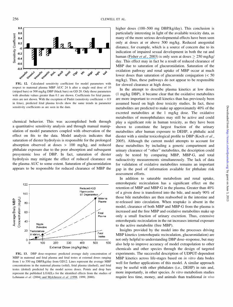
chemical behavior. This was accomplished both through
a quantitative sensitivity analysis and through manual manip-
ulation of model parameters coupled with observation of the
effect on fits to the data. Model analysis indicates that
saturation of diester hydrolysis is responsible for the prolonged
absorption observed at doses > 100 mg/kg, and reduced
phthalate exposure due to the poor absorption and subsequent
presystemic loss of DBP. In fact, saturation of diester
hydrolysis may mitigate the effect of reduced clearance on
the plasma AUC to some extent. Saturation of glucuronidation
appears to be responsible for reduced clearance of MBP the
higher doses (100–500 mg DBP/kg/day). This conclusion is
particularly interesting in light of the available toxicity data, as
many of the more serious developmental effects have been seen
only at doses at or above 500 mg/kg. Reduced anogenital
distance, for example, which is a source of concern due to its
indication of impaired sexual development in both the rat and
human (Fisher et al., 2003) is only seen at doses � 250 mg/kg/
day. This effect may in fact be a result of reduced clearance of
MBP due to saturation of glucuronidation. Saturation of the
oxidation pathway and renal uptake of MBP occur at much
lower doses than saturation of glucuronide conjugation (< 50
mg/kg). Thus, these pathways do not appear to be responsible
for slowed clearance at high doses.
In the attempt to describe plasma kinetics at low doses
(1 mg/kg DBP), it became clear that the oxidative metabolites
are more important to overall kinetics than has previously been
assumed based on high dose toxicity studies. In fact, these
metabolites are predicted to make up approximately 40% of the
excreted metabolites at the 1 mg/kg dose. The oxidative
metabolites of monophthalates may still be active and could
play a significant role in human toxicity, as they have been
shown to constitute the largest fraction of the urinary
metabolites after human exposure to DEHP, a phthalic acid
diester with a similar toxicological profile to DBP (Koch et al.,2005). Although the current model attempts to account for
these metabolites by including a generic compartment and
urinary clearance of ‘‘other’’ metabolites, the description could
only be tested by comparing MBP, MBP-G, and total
radioactivity measurements simultaneously. The lack of data
for validation of oxidative metabolites remains an important
gap in the pool of information available for phthalate risk
assessment efforts.
In addition to saturable metabolism and renal uptake,
enterohepatic recirculation has a significant effect on the
retention of MBP and MBP-G in the plasma. Greater than 40%
of a given dose is transferred into the bile, and nearly 90% of
those bile metabolites are then reabsorbed in the intestine and
re-released into circulation. When reuptake is absent in the
model, clearance of both MBP and MBP-G from the plasma is
increased and the free MBP and oxidative metabolites make up
only a small fraction of urinary excretion. Thus, extensive
enterohepatic recirculation in the rat increases internal exposure
to the active metabolite (free MBP).
Insights provided by the model into the processes driving
MBP kinetics (enterohepatic recirculation, glucuronidation) are
not only helpful to understanding DBP dose–response, but may
also help to improve accuracy of model extrapolation to other
chemicals and other species through the design of targeted
experiments. The successful description of UDPGT-dependent
MBP kinetics across life-stages based on in vitro data bodes
well for further applications of this model. A similar approach
may be useful with other phthalates (i.e., DEHP) in rats and,
more importantly, in other species. In vitro metabolism studies
require less time, money, and animals than traditional in vivo
FIG. 12. Calculated sensitivity coefficient for model parameters with
respect to maternal plasma MBP AUC 24 h after a single oral dose of 10
(striped bars) or 500 mg/kg DBP (black bars) on GD 20. Only those parameters
with absolute values greater than 0.1 are shown. Coefficients for fetal param-
eters are not shown. With the exception of Pmfet (sensitivity coefficient ¼ 0.9
in fetus), predicted fetal plasma levels show the same trends in parameter
sensitivity coefficients as are seen in the dam.
FIG. 13. DBP dose–response: predicted average daily concentration of
MBP in maternal and fetal plasma and fetal testes at external doses ranging
from 1 to 550 mg DBP/kg/day from GD12. Lines represent the average MBP
concentrations in the maternal plasma (solid), fetal plasma (dashed), and fetal
testes (dotted) predicted by the model across doses. Points and drop bars
represent the published LOAELs for the identified effects from the studies of
Lehmann et al. (2004) and Mylchreest et al. (1998, 1999, 2000).
256 CLEWELL ET AL.
by guest on June 3, 2013http://toxsci.oxfordjournals.org/
Dow
nloaded from
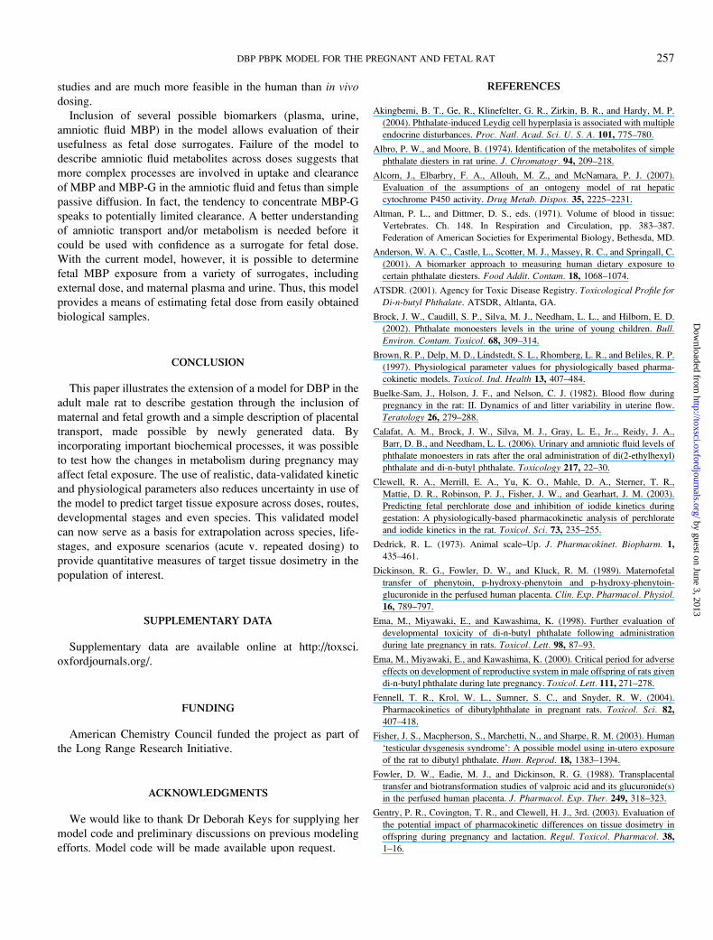
studies and are much more feasible in the human than in vivodosing.
Inclusion of several possible biomarkers (plasma, urine,
amniotic fluid MBP) in the model allows evaluation of their
usefulness as fetal dose surrogates. Failure of the model to
describe amniotic fluid metabolites across doses suggests that
more complex processes are involved in uptake and clearance
of MBP and MBP-G in the amniotic fluid and fetus than simple
passive diffusion. In fact, the tendency to concentrate MBP-G
speaks to potentially limited clearance. A better understanding
of amniotic transport and/or metabolism is needed before it
could be used with confidence as a surrogate for fetal dose.
With the current model, however, it is possible to determine
fetal MBP exposure from a variety of surrogates, including
external dose, and maternal plasma and urine. Thus, this model
provides a means of estimating fetal dose from easily obtained
biological samples.
CONCLUSION
This paper illustrates the extension of a model for DBP in the
adult male rat to describe gestation through the inclusion of
maternal and fetal growth and a simple description of placental
transport, made possible by newly generated data. By
incorporating important biochemical processes, it was possible
to test how the changes in metabolism during pregnancy may
affect fetal exposure. The use of realistic, data-validated kinetic
and physiological parameters also reduces uncertainty in use of
the model to predict target tissue exposure across doses, routes,
developmental stages and even species. This validated model
can now serve as a basis for extrapolation across species, life-
stages, and exposure scenarios (acute v. repeated dosing) to
provide quantitative measures of target tissue dosimetry in the
population of interest.
SUPPLEMENTARY DATA
Supplementary data are available online at http://toxsci.
oxfordjournals.org/.
FUNDING
American Chemistry Council funded the project as part of
the Long Range Research Initiative.
ACKNOWLEDGMENTS
We would like to thank Dr Deborah Keys for supplying her
model code and preliminary discussions on previous modeling
efforts. Model code will be made available upon request.
REFERENCES
Akingbemi, B. T., Ge, R., Klinefelter, G. R., Zirkin, B. R., and Hardy, M. P.
(2004). Phthalate-induced Leydig cell hyperplasia is associated with multiple
endocrine disturbances. Proc. Natl. Acad. Sci. U. S. A. 101, 775–780.
Albro, P. W., and Moore, B. (1974). Identification of the metabolites of simple
phthalate diesters in rat urine. J. Chromatogr. 94, 209–218.
Alcorn, J., Elbarbry, F. A., Allouh, M. Z., and McNamara, P. J. (2007).
Evaluation of the assumptions of an ontogeny model of rat hepatic
cytochrome P450 activity. Drug Metab. Dispos. 35, 2225–2231.
Altman, P. L., and Dittmer, D. S., eds. (1971). Volume of blood in tissue:
Vertebrates. Ch. 148. In Respiration and Circulation, pp. 383–387.
Federation of American Societies for Experimental Biology, Bethesda, MD.
Anderson, W. A. C., Castle, L., Scotter, M. J., Massey, R. C., and Springall, C.
(2001). A biomarker approach to measuring human dietary exposure to
certain phthalate diesters. Food Addit. Contam. 18, 1068–1074.
ATSDR. (2001). Agency for Toxic Disease Registry. Toxicological Profile for
Di-n-butyl Phthalate. ATSDR, Altlanta, GA.
Brock, J. W., Caudill, S. P., Silva, M. J., Needham, L. L., and Hilborn, E. D.
(2002). Phthalate monoesters levels in the urine of young children. Bull.
Environ. Contam. Toxicol. 68, 309–314.
Brown, R. P., Delp, M. D., Lindstedt, S. L., Rhomberg, L. R., and Beliles, R. P.
(1997). Physiological parameter values for physiologically based pharma-
cokinetic models. Toxicol. Ind. Health 13, 407–484.
Buelke-Sam, J., Holson, J. F., and Nelson, C. J. (1982). Blood flow during
pregnancy in the rat: II. Dynamics of and litter variability in uterine flow.
Teratology 26, 279–288.
Calafat, A. M., Brock, J. W., Silva, M. J., Gray, L. E., Jr.., Reidy, J. A.,
Barr, D. B., and Needham, L. L. (2006). Urinary and amniotic fluid levels of
phthalate monoesters in rats after the oral administration of di(2-ethylhexyl)
phthalate and di-n-butyl phthalate. Toxicology 217, 22–30.
Clewell, R. A., Merrill, E. A., Yu, K. O., Mahle, D. A., Sterner, T. R.,
Mattie, D. R., Robinson, P. J., Fisher, J. W., and Gearhart, J. M. (2003).
Predicting fetal perchlorate dose and inhibition of iodide kinetics during
gestation: A physiologically-based pharmacokinetic analysis of perchlorate
and iodide kinetics in the rat. Toxicol. Sci. 73, 235–255.
Dedrick, R. L. (1973). Animal scale–Up. J. Pharmacokinet. Biopharm. 1,
435–461.
Dickinson, R. G., Fowler, D. W., and Kluck, R. M. (1989). Maternofetal
transfer of phenytoin, p-hydroxy-phenytoin and p-hydroxy-phenytoin-
glucuronide in the perfused human placenta. Clin. Exp. Pharmacol. Physiol.
16, 789–797.
Ema, M., Miyawaki, E., and Kawashima, K. (1998). Further evaluation of
developmental toxicity of di-n-butyl phthalate following administration
during late pregnancy in rats. Toxicol. Lett. 98, 87–93.
Ema, M., Miyawaki, E., and Kawashima, K. (2000). Critical period for adverse
effects on development of reproductive system in male offspring of rats given
di-n-butyl phthalate during late pregnancy. Toxicol. Lett. 111, 271–278.
Fennell, T. R., Krol, W. L., Sumner, S. C., and Snyder, R. W. (2004).
Pharmacokinetics of dibutylphthalate in pregnant rats. Toxicol. Sci. 82,
407–418.
Fisher, J. S., Macpherson, S., Marchetti, N., and Sharpe, R. M. (2003). Human
‘testicular dysgenesis syndrome’: A possible model using in-utero exposure
of the rat to dibutyl phthalate. Hum. Reprod. 18, 1383–1394.
Fowler, D. W., Eadie, M. J., and Dickinson, R. G. (1988). Transplacental
transfer and biotransformation studies of valproic acid and its glucuronide(s)
in the perfused human placenta. J. Pharmacol. Exp. Ther. 249, 318–323.
Gentry, P. R., Covington, T. R., and Clewell, H. J., 3rd. (2003). Evaluation of
the potential impact of pharmacokinetic differences on tissue dosimetry in
offspring during pregnancy and lactation. Regul. Toxicol. Pharmacol. 38,
1–16.
DBP PBPK MODEL FOR THE PREGNANT AND FETAL RAT 257
by guest on June 3, 2013http://toxsci.oxfordjournals.org/
Dow
nloaded from
Gray, L. E., Jr., Ostby, J., Furr, J., Price, M., Veeramachaneni, D. N., and
Parks, L. (2000). Perinatal exposure to the phthalates DEHP, BBP, and
DINP, but not DEP, DMP, or DOTP, alters sexual differentiation of the male
rat. Toxicol. Sci. 58, 350–365.
Gray, L. E., Jr., Wolf, C., Lambright, C., Mann, P., Price, M., Cooper, R. L.,
and Ostby, J. (1999). Administration of potentially antiandrogenic pesticides
(procymidone, linuron, iprodione, chlozolinate, p,p#-DDE, and ketocona-
zole) and toxic substances (dibutyl- and diethylhexyl phthalate, PCB 169,
and ethane dimethane sulphonate) during sexual differentiation produces
diverse profiles of reproductive malformations in the male rat. Toxicol. Ind.
Health 15, 94–118.
Hanwell, A., and Linzell, J. L. (1973). The time course of cardiovascular
changes in the rat. J. Physiol. 233, 99–109.
Heudorf, U., Mersch-Sundermann, V., and Angerer, J. (2007). Phthalates:
Toxicology and exposure. Int. J. Hyg. Environ. Health 210, 623–634.
Isaksson, A., Hultberg, B., and Bergenfeldt, M. (1988). Lysosomal enzymes in
pregnant and steroid treated rats. Horm. Metab. Res. 20, 274–277.
Keys, D. A., Wallace, D. G., Kepler, T. B., and Conolly, R. B. (2000).
Quantitative evaluation of alternative mechanisms of blood disposition of
di(n-butyl) phthalate and mono(n-butyl) phthalate in rats. Toxicol. Sci. 53,
173–184.
Kim, H. S., Kim, T. S., Shin, J. H., Moon, H. J., Kang, I. H., Kim, I. Y.,
Oh, J. Y., and Han, S. Y. (2004). Neonatal exposure to di(n-butyl) phthalate
(DBP) alters male reproductive-tract development. J. Toxicol. Environ.
Health A 67, 2045–2060.
Kim, H. S., Saito, K., Ishizuka, M., Kazusaka, A., and Fujita, S. (2003). Short
period exposure to di-(2-ethylhexyl) phthalate regulates testosterone
metabolism in testis of prepubertal rats. Arch. Toxicol. 77, 446–451.
Knight, C. H., and Peaker, M. (1982). Mammary cell proliferation in mice
during pregnancy and lactation in relation to milk yield. Q. J. Exp. Physiol.
67, 165–177.
Koch, H. M., Bolt, H. M., Preuss, R., and Angerer, J. (2005). New metabolites
of di(2-ethylhexyl) phthalate (DEHP) in human urine and plasma after single
oral doses of deuterium-labelled DEHP. Arch. Toxicol. 79, 367–376.
Kohn, M. C., Parham, F., Masten, S. A., Portier, C. J., Shelby, M. D.,
Brock, J. W., and Needham, L. L. (2000). Human exposure estimates for
phthalates. Environ. Health Perspect. 108, A440–A442.
Kremer, J. J., Williams, C. C., Parkinson, H. D., and Borghoff, S. J. (2005).
Pharmacokinetics of monobutylphthalate, the active metabolite of di-n-
butylphthalate, in pregnant rats. Toxicol. Lett. 159, 144–153.
LaBorde, J. B., Hansen, D. K., Young, J. F., Sheehan, D. M., and Holson, R. R.
(1992). Prenatal dexamethasone exposure in rats: Effects of dose, age at
exposure, and drug-induced hypophagia on malformations and fetal organ
weights. Fundam. Appl. Toxicol. 19, 545–554.
Latini, G., De Felice, C., Presta, G., Del Vecchio, A., Paris, I., Ruggieri, F., and
Mazzeo, P. (2003). In utero exposure to di-(2-ethylhexyl) phthalate and
duration of human pregnancy. Environ. Health Perspect. 111, 1783–1785.
Lehmann, K. P., Phillips, S., Sar, M., Foster, P. M., and Gaido, K. W. (2004).
Dose-dependent alterations in gene expression and testosterone synthesis in
the fetal testes of male rats exposed to di (n-butyl) phthalate. Toxicol. Sci. 81,
60–68.
Lucier, G. W., and McDaniel, O. S. (1977). Steroid and non-steroid UDP
glucuronyltransferase: Glucuronidation of synthetic estrogens as steroids. J.
Steroid Biochem. 8, 867–872.
Lucier, G. W., Sonawane, B. R., McDaniel, O. S., and Hook, G. E. (1975).
Postnatal stimulation of hepatic microsomal enzymes following administra-
tion of TCDD to pregnant rats. Chem. Biol. Interact. 11, 15–26.
Luquita, M. G., Catania, V. A., Pozzi, E. J., Veggi, L. M., Hoffman, T.,
Pellegrino, J. M., Ikushiro, S. I., Emi, Y., Iyanagi, T., Vore, M., et al. (2001).
Molecular basis of perinatal changes in UDP-glucuronosyltransferase
activity in maternal rat liver. J. Pharmacol. Exp. Ther. 298, 49–56.
Mylchreest, E., Cattley, R. C., and Foster, P. M. D. (1998). Male reproductive
tract malformations in rats following gestational and lactational exposure to
di(n-butyl) phthalate: An antiandrogenic mechanism? Toxicol. Sci. 43,
47–60.
Mylchreest, E., Sar, M., Cattley, R. C., and Foster, P. M. D. (1999). Disruption
of androgen-regulated male reproductive development by di(n-butyl)
phthalate during late gestation in rats is different from flutamide. Toxicol.
Appl. Pharmacol. 156, 81–95.
Mylchreest, E., Sar, M., Wallace, D. G., and Foster, P. M. (2002). Fetal
testosterone insufficiency and abnormal proliferation of Leydig cells and
gonocytes in rats exposed to di(n-butyl) phthalate. Reprod. Toxicol. 16,
19–28.
Mylchreest, E., Wallace, D. G., Cattley, R. C., and Foster, P. M. D. (2000).
Dose-dependent alterations in androgen-regulated male reproductive de-
velopment in rats exposed to di(n-butyl) phthalate during late gestation.
Toxicol. Sci. 55, 143–151.
Naessany, S., and Picon, R. (1982). Onset of a feedback inhibition by
testosterone in male rat fetuses. Biol. Neonate 41, 234–239.
Naismith, D. J., Richardson, D. P., and Pritchard, A. E. (1982). The utilization
of protein and energy during lactation in the rat, with particular regard to the
use of fat accumulated during pregnancy. Br. J. Nutr. 48, 433–441.
NIEHS. (1994). National Toxicology Program (NTP). Prestart toxicokinetic
study report: Di-n-butyl phthalate in rodent plasma, June 30, 1994 NIH
Contract NO: N01-ES-15307. NTP Chem Task No: HEM00324.
NIEHS. (1995). National Toxicology Program (NTP). toxicokinetic study
report: The toxicokinetics and metabolism of di-n-butyl phthalate March 29,
1995. .NIEHS, Research Triangle Park, NC. NIH Contract NO: N01-ES-
15307. NTP Chem Task No: CHEM00324.
O’Flaherty, E. J., Scott, W., Schreiner, C., and Beliles, R. P. (1992). A
physiologically based kinetic model of rat and mouse gestation: Disposition
of a weak acid. Toxicol. Appl. Pharmacol. 112, 245–256.
Oishi, S., and Hiraga, K. (1980). Effects of phthalic acid monoesters on mouse
testes. Toxicol. Lett. 6, 239–242.
Park, H. W., and Shepard, T. H. (1994). Volume and glucose concentration of
rat amniotic fluid: Effects on embryo nutrition and axis rotation. Teratology
49, 465–469.
Parks, L. G., Ostby, J. S., Lambright, C. R., Abbott, B. D., Klinefelter, G. R.,
Barlow, N. J., and Gray, L. E., Jr. (2000). The plasticizer diethylhexyl
phthalate induces malformations by decreasing fetal testosterone synthesis
during sexual differentiation in the male rat. Toxicol. Sci. 58, 339–349.
Payan, J. P., Marty, J. P., Fabry, J. P., Beydon, D., Boudry, I., Ferrari, E.,
Canel, F., Grandclaude, M. C., and Vincent, C. M. (2001). In vivo and in
vitro percutaneous absorption of [(14)C]di-n-butylphthalate in rat. Drug
Metab. Dispos. 29, 843–854.
Reynolds, F., and Knott, C. (1989). Pharmacokinetics in pregnancy and
placental drug transfer. Oxf. Rev. Reprod. Biol. 11, 389–449.
Rowland, I. R., Cottrell, R. C., and Phillips, J. C. (1977). Hydrolysis of
phthalate esters by the gastro-intestinal contents of the rat. Food Cosmet.
Toxicol. 15, 17–21.
Saillenfait, A. M., Payan, J. P., Fabry, J. P., Beydon, D., Langonne, I.,
Gallissot, F., and Sabate, J. P. (1998). Assessment of the developmental
toxicity, metabolism, and placental transfer of Di-n-butyl phthalate
administered to pregnant rats. Toxicol. Sci. 45, 212–224.
Silva, M. J., Barr, D. B., Reidy, J. A., Malek, N. A., Hodge, C. C.,
Caudill, S. P., Brock, J. W., Needham, L. L., and Calafat, A. M. (2004).
Urinary levels of seven phthalate metabolites in the U.S. population from the
National Health and Nutrition Examination Survey (NHANES) 1999-2000.
Environ. Health Perspect. 112, 331–338.
Sjoberg, P., Egestad, B., Klasson-Wehler, E., and Gustafsson, J. (1999).
Glucuronidation of mono(2-ethylhexyl)phthalate. Some enzyme character-
istics and inhibition by bilirubin. Biochem. Pharmacol. 41, 1493–1496.
258 CLEWELL ET AL.
by guest on June 3, 2013http://toxsci.oxfordjournals.org/
Dow
nloaded from
Soucy, N. V., Parkinson, H. D., Sochaski, M. A., and Borghoff, S. J. (2006).
Kinetics of genistein and its conjugated metabolites in pregnant Sprague-
Dawley rats following single and repeated genistein administration. Toxicol.
Sci. 90, 230–240.
Tanaka, A., Matsumoto, A., and Yamaha, T. (1978). Biochemical studies on
phthalic esters. III. Metabolism of dibutyl phthalate (DBP) in animals.
Toxicology 9, 109–123.
Thompson, R. C., and Hollis, O. L. (1958). Irradiation of the gastro-
intestinal tract of the rat by ingested ruthenium-96. Am. J. Physiol. 194,308–312.
White, R. D., Carter, D. E., Earnest, D., and Mueller, J. (1980). Absorption and
metabolism of three phthalate diesters by rat small intestine. Food Cosmet.
Toxicol. 18, 383–386.
Williams, D. T., and Blanchfield, B. J. (1975). The retention, distribution,
excretion, and metabolism of dibutyl phthalate-7-14 C in the rat. J. Agric.
Food Chem. 23, 854–858.
Wishart, G. J. (1978). Functional heterogeneity of UDP-glucuronosyltransfer-
ase as indicated by its differential development and inducibility by
glucocorticoids. Demonstration of two groups within the enzyme’s activity
towards twelve substrates. Biochem. J. 174, 485–489.
Wormuth, M., Scheringer, M., Vollenweider, M., and Hungerbuhler, K. (2006).
What are the sources of exposure to eight frequently used phthalic acid esters
in Europeans? Risk Anal. 26, 803–824.
Wykoff, M. H. (1971). Weight changes of the developing rat conceptus. Am. J.
Vet. Res. 32, 1633–1635.
DBP PBPK MODEL FOR THE PREGNANT AND FETAL RAT 259
by guest on June 3, 2013http://toxsci.oxfordjournals.org/
Dow
nloaded from