Thyrotropin modulates low density lipoprotein binding activity in FRTL-5 thyroid cells
7
THE Joumu~ CIF BKII.OGICA~ CHE~~ISTRY Vol. 265, No. 31, Issue of November 5, pp. 19336-19342,199O Printed in CL SA. Thyrotropin Modulates Low Density Lipoprotein Binding Activity in FRTL-5 Thyroid Cells* (Received for publication, November 8, 1989) Maurizio Bifulco$$, Mariarosaria Santillo$, Idolo Tedesco$, Raffaele Zarrilli$, Chiara Laezzas, and Salvatore M. Aloj$q From the $Centro di Endocrinologia ed Oncologia Sperimentale/Consiglio Nazionale delle Ricerche, and Dipartimento di Biologia e Patologia Cellulnre e Molecolure “L. Califano”, University of Naples, Via S. Pansini 5, 80131 Naples, the @tit&o de Oncologia Sperimentale e Clinica, University of Reggio Calubria, Catanzaro, Ztaly, and the Wection on Biochemistry of Cell Regulntion; Laboratory of Biochemistry and Metabolism, National Znstitute of Diabetes and Digestive and Kidney Dkeases, National institutes of Health, Bethesda, Maryland 20892 FRTL-S cells possess high affinity low density lipo- protein (LDL) receptors which bind, internalize, and degrade LDL. When FRTL-5 cells are deprived of thy- rotropin (TSH) the binding of LDL increases more than 2-fold. Upon addition of TSH, at a concentration of 1 X 10-l’ M or greater, LDL binding decreases rapidly and within 24 h reaches the level which is typical of FRTL-5 cells chronically stimulated by TSH. The data available suggest that TSH-dependent down-regula- tion of LDL receptor activity is exerted through a reduction of the number of active LDL receptors, with no change in affinity. It is unlikely that the synthesis of LDL receptors is impaired, since LDL receptor mes- senger RNA is not decreased by TSH. The effect of the hormone on LDL receptor activity can be mimicked by &Br-CAMP and is completely abolished by the protein synthesis inhibitor cycloheximide but not by actino- mycin D. TSH regulation of LDL receptor activity is lost in v- ras Ki-transformed FRTL-5 cells (Ki Mol) which also have lost TSH dependence for adenylate cyclase acti- vation and growth. However, 8-Br-CAMP decreases LDL binding in Ki Mol FRTL-5 cells. The reduced availability of LDL receptor in TSH-stimulated FRTL- 6 cells may be related to the increased membrane flu- idity (Beguinot, F., Beguinot, L., Tramontano, D., Dui- lio, C., Formisano, S., Bifulco, M., Ambesi-Impiom- bato, F. S., and Aloj, S. M. (1987) J. Biol. Chem. 262, 1575-1582) or may reflect increased degradation of LDL receptors. We propose that a lower cholesterol uptake is needed in an actively proliferating cell pop- ulation, to increase the production of isoprenoids whether it be for cholesterol biosynthesis or for the synthesis of other compounds requiring isoprenoid pre- cursors. Thyrotropin is a potent physiological regulator of thyroid cell function and growth. In the FRTL-5 rat thyroid cell line *This study was supported in part by the Progetto Finalizzato “Invecchiamento” of the Consiglio Nazionale delle Ricerche and hy the exchange program between Italian scientists and the National Institutes oi Gealih, funded by the Italian Department of Education (Minister0 della Pubblica Istruzione). The costs of publication of this article were defrayed in part hy the payment of page charges. This article must therefore be hereby marked “advertisement” in accord- ance with 18 U.S.C. Section 1734 solely to indicate this fact. (l-3) TSH’ affects thyroglobulin synthesis and secretion (1, 3), iodine metabolism (4,) and growth (1,2, 5). When TSH is deleted from the culture medium FRTL-5 become quiescent, but resume duplication upon addition of the hormone (5). Studies of the effect of TSH on the physico-chemical prop- erties of the plasma membrane in FRTL-5 cells have revealed that membrane fluidity and lipid composition are markedly affected by TSH (6). Thus, in the absence of the hormone membrane fluidity undergoes a major decrease which is caused by changes in membrane lipids of which the most notable is a more than 2-fold increase in total cholesterol. Readdition of TSH reverts the lipid changes and reestablishes cell dupli- cation (6). While the changes in membrane fluidity are con- sistent with those observed in other cell systems, in which a more fluid membrane has been associated with active cell division (7-9), the changes in cholesterol were surprising. Indeed membrane biogenesis, in a proliferating cell popula- tion, would require a larger availability of cholesterol. This puzzling observation has prompted us to investigate the mech- anism by which TSH affects cholesterol content in FRTL-5 cells. Since the source of cell cholesterol is either exogenous, through the low density lipoprotein (LDL) receptor pathway (lo), or endogenous, through cholesterol biosynthesis, we have studied the effect of TSH on both processes in FRTL-5 cells. In this report we present evidence showing that these cells possess specific LDL receptors which are highly sensitive to TSH. We demonstrate that TSH, through a mechanism which involves CAMP production, and requires active protein syn- thesis, markedly reduces LDL binding while increasing the internalization index (i.e. internalization + degradation per binding) (11). We also show that a v-ras Ki-transformed strain of FRTL-5 cells (12, 13), which has lost the ability to augment CAMP production upon TSH challenge (14), does not show any effect of TSH on LDL receptor activity. EXPERIMENTAL PROCEDURES Material-TSH (26 k 3 units/mg) was purified from hovine pituitary extracts (I5,16). Culture media and hormonal supplements were ohtained as reported previously (6). Bovine serum albumin (crystalline), salmon sperm DNA, 2-mercaptoethanol, 8-bromo cyclic AMP (8-Br-CAMP), and cycloheximide were obtained from Sigma. Octyl p-D-glucopyranoside and actinomycin D were from Calbiochem; guanidine thiocyanate was from Fluka Chemie AG. Carrier-free Na’*‘I was from Amersham Corp. and [3zP]dCTP (3,000 Ci/mmol) was from ’ The abbreviations are: TSH, thyrotropin or thyroid stimulating hormone; LDL, low density lipoproteins; LPDS, lipoprotein-deficient serum; HEPES, 4-(2-hydroxymethyl)-l-piperazineethanesulfonic aci4 MOPS, 4-(morpholino)propane sulfonic acid. 19336 by guest, on July 10, 2011 www.jbc.org Downloaded from
Transcript of Thyrotropin modulates low density lipoprotein binding activity in FRTL-5 thyroid cells

THE Joumu~ CIF BKII.OGICA~ CHE~~ISTRY Vol. 265, No. 31, Issue of November 5, pp. 19336-19342,199O Printed in CL SA.
Thyrotropin Modulates Low Density Lipoprotein Binding Activity in FRTL-5 Thyroid Cells*
(Received for publication, November 8, 1989)
Maurizio Bifulco$$, Mariarosaria Santillo$, Idolo Tedesco$, Raffaele Zarrilli$, Chiara Laezzas, and Salvatore M. Aloj$q From the $Centro di Endocrinologia ed Oncologia Sperimentale/Consiglio Nazionale delle Ricerche, and Dipartimento di Biologia e Patologia Cellulnre e Molecolure “L. Califano”, University of Naples, Via S. Pansini 5, 80131 Naples, the @tit&o de Oncologia Sperimentale e Clinica, University of Reggio Calubria, Catanzaro, Ztaly, and the Wection on Biochemistry of Cell Regulntion; Laboratory of Biochemistry and Metabolism, National Znstitute of Diabetes and Digestive and Kidney Dkeases, National institutes of Health, Bethesda, Maryland 20892
FRTL-S cells possess high affinity low density lipo- protein (LDL) receptors which bind, internalize, and degrade LDL. When FRTL-5 cells are deprived of thy- rotropin (TSH) the binding of LDL increases more than 2-fold. Upon addition of TSH, at a concentration of 1 X 10-l’ M or greater, LDL binding decreases rapidly and within 24 h reaches the level which is typical of FRTL-5 cells chronically stimulated by TSH. The data available suggest that TSH-dependent down-regula- tion of LDL receptor activity is exerted through a reduction of the number of active LDL receptors, with no change in affinity. It is unlikely that the synthesis of LDL receptors is impaired, since LDL receptor mes- senger RNA is not decreased by TSH. The effect of the hormone on LDL receptor activity can be mimicked by &Br-CAMP and is completely abolished by the protein synthesis inhibitor cycloheximide but not by actino- mycin D.
TSH regulation of LDL receptor activity is lost in v- ras Ki-transformed FRTL-5 cells (Ki Mol) which also have lost TSH dependence for adenylate cyclase acti- vation and growth. However, 8-Br-CAMP decreases LDL binding in Ki Mol FRTL-5 cells. The reduced availability of LDL receptor in TSH-stimulated FRTL- 6 cells may be related to the increased membrane flu- idity (Beguinot, F., Beguinot, L., Tramontano, D., Dui- lio, C., Formisano, S., Bifulco, M., Ambesi-Impiom- bato, F. S., and Aloj, S. M. (1987) J. Biol. Chem. 262, 1575-1582) or may reflect increased degradation of LDL receptors. We propose that a lower cholesterol uptake is needed in an actively proliferating cell pop- ulation, to increase the production of isoprenoids whether it be for cholesterol biosynthesis or for the synthesis of other compounds requiring isoprenoid pre- cursors.
Thyrotropin is a potent physiological regulator of thyroid cell function and growth. In the FRTL-5 rat thyroid cell line
*This study was supported in part by the Progetto Finalizzato “Invecchiamento” of the Consiglio Nazionale delle Ricerche and hy the exchange program between Italian scientists and the National Institutes oi Gealih, funded by the Italian Department of Education (Minister0 della Pubblica Istruzione). The costs of publication of this article were defrayed in part hy the payment of page charges. This article must therefore be hereby marked “advertisement” in accord- ance with 18 U.S.C. Section 1734 solely to indicate this fact.
(l-3) TSH’ affects thyroglobulin synthesis and secretion (1, 3), iodine metabolism (4,) and growth (1,2, 5). When TSH is deleted from the culture medium FRTL-5 become quiescent, but resume duplication upon addition of the hormone (5). Studies of the effect of TSH on the physico-chemical prop- erties of the plasma membrane in FRTL-5 cells have revealed that membrane fluidity and lipid composition are markedly affected by TSH (6). Thus, in the absence of the hormone membrane fluidity undergoes a major decrease which is caused by changes in membrane lipids of which the most notable is a more than 2-fold increase in total cholesterol. Readdition of TSH reverts the lipid changes and reestablishes cell dupli- cation (6). While the changes in membrane fluidity are con- sistent with those observed in other cell systems, in which a more fluid membrane has been associated with active cell division (7-9), the changes in cholesterol were surprising. Indeed membrane biogenesis, in a proliferating cell popula- tion, would require a larger availability of cholesterol. This puzzling observation has prompted us to investigate the mech- anism by which TSH affects cholesterol content in FRTL-5 cells. Since the source of cell cholesterol is either exogenous, through the low density lipoprotein (LDL) receptor pathway (lo), or endogenous, through cholesterol biosynthesis, we have studied the effect of TSH on both processes in FRTL-5 cells. In this report we present evidence showing that these cells possess specific LDL receptors which are highly sensitive to TSH. We demonstrate that TSH, through a mechanism which involves CAMP production, and requires active protein syn- thesis, markedly reduces LDL binding while increasing the internalization index (i.e. internalization + degradation per binding) (11). We also show that a v-ras Ki-transformed strain of FRTL-5 cells (12, 13), which has lost the ability to augment CAMP production upon TSH challenge (14), does not show any effect of TSH on LDL receptor activity.
EXPERIMENTAL PROCEDURES
Material-TSH (26 k 3 units/mg) was purified from hovine pituitary extracts (I5,16). Culture media and hormonal supplements were ohtained as reported previously (6). Bovine serum albumin (crystalline), salmon sperm DNA, 2-mercaptoethanol, 8-bromo cyclic AMP (8-Br-CAMP), and cycloheximide were obtained from Sigma. Octyl p-D-glucopyranoside and actinomycin D were from Calbiochem; guanidine thiocyanate was from Fluka Chemie AG. Carrier-free Na’*‘I was from Amersham Corp. and [3zP]dCTP (3,000 Ci/mmol) was from
’ The abbreviations are: TSH, thyrotropin or thyroid stimulating hormone; LDL, low density lipoproteins; LPDS, lipoprotein-deficient serum; HEPES, 4-(2-hydroxymethyl)-l-piperazineethanesulfonic aci4 MOPS, 4-(morpholino)propane sulfonic acid.
19336
by guest, on July 10, 2011w
ww
.jbc.orgD
ownloaded from

TSH Regulation of LDL Binding
Du Pont-New England Nuclear. All chemicals were of the highest quality.
Human lipoproteins were isolated from a pool of normal human sera by sequential ultracentrifugal flotation according to established techniques (17). LDL was radiolabeled using the iodine monochloride method of McFarlane as modified by Bilheimer (18), to a specific activity of 400 cpm/ng. The monoclonil antibody to the rat live; LDL recentor PlB3 (19) was kindlv nrovided bv Dr. A. D. Cooner (Research Institute, Palo Alto Medical boundatioi, Palo Alto, kA),’ and was iodinated by the Iodo-Gen method (Bio-Rad) to a specific activity of 400 cpm/ng.
CeUs-The FRTL-5 cells (ATCC CRL 8305) used in this study are functioning thyroid cells whose characteristics and culture condiiions have been extensively described (1. 2.6.20, 21). Thev were routinely grown in Coon’s mo&fied Ham’s ‘F-12’ midium sup-plemented with 5% calf serum and a mixture of six hormones (6H), including TSH and insulin (1,2). When the effect of TSH was assayed the cells were maintained in medium lacking TSH (5H) or thi entire hormone comnlement (no H) and 5% linonrotein-deficient serum (LPDS). The lattir was prhpareh followini tie procedure of Goldstein et al: (22). When it was necessary to minimize the contribution of hormones physiologically present in 5% calf serum, 48 h before starting the experiment, the medium was changed to one containing 0.2% calf serum.
The Ki Mol cells are FRTL-5 cells infected with the Kirsten sarcoma virus (12, 13).
?&dies of I..DL Me&o&m-Binding of ‘*‘I-LDL to cell surface was measured according to the procedure of Goldstein et ul. (22). Semi confluent FRTL-5 or Ki Mol cells were maintained for 72 h in medium from which the hormone supplement was removed (no H medium). During the 24-h period preceding the binding assay the medium was replaced with oie contiining 5% LPDS witior wiihout 1 X lo-’ M TSH. Binding assays were carried out at 37 ‘C in 24-well tissue culture plates, i; 0.5 hl of medium containing the noted concentrations of “‘1-LDL and a 50-IOO-fold excess of unlabeled lipoproteins or bovine TSH. Surface-bound Y-LDL was released by treating the cells with 0.5 ml of a buffer containing 50 mM NaCl, 10 rnM HEPES, and 10 mg/ml heparin. For measurements of internal- ized and degraded LDL, cells were incubated with 0.5 ml of warm (37 “C) medium containing various concentrations of I?-LDL in the presence or in the absence of a 50-loo-fold excess unlabeled LDL. The procedures were those described by Goldstein et ul. (22). Protein concentration was determined by the method of Bradford (23) using crystalline bovine serum albumin as the standard.
Analysis of LDL Receptor Messenger RNA-For Northern blot analysis, poly(A)+-rich RNA was preiared by affinity chromatogra- phy on oligo(dT)-Sepharose columns, from FRTL-5 cells grown under condition; identical-to those used fir LDL binding studyes, i.e. cells were maintained for 5 days in 5H medium (without TSH) and 5% LPDS, then either treated with or not exposed to TSH for 24 h. Poly(A)+ RNA was fractionated on a 1% iormaldehyde-agarose gel in MOPS buffer and transferred to Nvtran membranes (Schleicher & Schuell) in 1.5 M NaCl, 150 mM sod&m citrate (10 X S&Z) and 10 mM EDTA, pH 7.4, as described previously (24). After baking the filters at 80 ‘C for 1 h, they were nre-hvbridized for 3-5 h with buffer containing 50% formimiie, 5 X-SSPh (SSPE consists of 150 mM NaCl, 10 mM sodium phosphate, and 1 mM EDTA, pH 7.4), 5 X Denhardt’s solution (25), 200 Fg/ml sonicated salmon sperm DNA, and 0.1% sodium dodecyl sulfate. The hybridization was carried out for 18 h at 42 ‘C in the pre-hybridization buffer containing 2-5 x 10⁶ cpm of “P-labeled, nick-translated, purified probe (l-2 x 10’ cpm/ pg DNA). The LDL receptor probe was a EcoRI/jVoeI restriction fragment excised from the plasmid pLDLR3 (26) (supplied by the American Type Culture Collection, Rockville, MD). The &actin cDNA probe (27) was a generous gift of Dr. B. Paterson (iational Cancer Institute). The filters were washed in two changes of 2 x SSC and 0.1% sodium dodecyl sulfate at room temperature for 15 min each time and at 60 “C for 1 h. The fIna wash was carried out in 0.1 x SSC for 1 h at 55 ‘C. A RNA ladder was included in each experiment as a size marker.
The filters were exposed to Kodak XAR-5 film (Eastman Kodak) with Du Pant-Cronex intensifying screens at -70 ‘C. Quantitative analysis of autoradiographs was performed bv laser densitometrv.
Solubilizahon and ~nknobloking of LDi Receptor-To visuilize LDL receptor, FRTL-5 cells were maintained as described above for studies of LDL metabolism. Following the TSH starvation period, cells were divided in two groups (20 x 106 cells each), one group was exposed to medium containing 1 x lo-’ M TSH, and the other to
medium lacking this hormone for 24 h; during this time both media were supplemented with 5% LPDS. Each group of cells was then washed three times with phosphate-buffered saline, scraped with a rubber policeman, disrupted by sonication in 15 ml buffer (50 mM Tris, 150 mM NaCl, 1 mM CaCIZ, pH 6.8), and centrifuged at 1000 X g for I5 min. The pellets were suspended in buffer containing 40 nM octyl O-D-glycopyranoside to solubilize the LDL receptor (28, 29). The suspension was stirred at 4 ‘C for 10 min, and centrifuged at 1 x 10’ x g for 60 min. The clear supernatants were electrophoresed on 7% polyacrylamide gels prepared by the method of Laemmli (30). Samples were not heated or added with reducing agents. Between 150 and 200 gg of protein were loaded onto the gels. Protein bands were transferred to nitrocellulose strips using the Bio-Rad blotting device (Trans-Blot), in a buffer containing 20 mM Tris, 150 mM glycine, and 20% (v:v) methanol, at 200 mA for 16 h at 4 OC, according to the procedure described by Beisiegel et ul. (31). Incubation of the nitro- celluose strips for immunoblotting and development, using the mono- clonal anti-rat LDL receptor antibody PlB3 and horseradish perox- idase, were performed following closely the procedure of Cooper et al. (191.
RESULTS
Surface Binding of lz51-LDL to FRTL-5 CeL!.s--Although FRTL-5 cells require the presence of TSH in the culture medium for growth and expression of thyroid-specific func- tions, they survive TSH withdrawal for prolonged periods (5, 6). When maintained in the absence of TSH, FRTL-5 cells undergo profound changes which affect most dramatically DNA replication and, adenylate cyclase activity (5, 6), lipid composition and membrane fluidity (6). Under these condi- tions, i.e. in the absence of TSH, FRTL-5 cells bind specifi-
lz5 cally I- LDb after 2 h incubation at 37 ‘C, the amount of ‘251-LDL which could be released by heparin was 130 k 40 ng/mg cell protein. Binding was saturable and could be com- peted for by cold LDL, but not by cold HDL (Fig. 1). FRTL- 5 cells also bound with typical saturation kinetics 1z51-PlB3, a monoclonal antibody specific for the rat liver LDL receptor (19) (see below).
The Effect of TSH on the Surface Binding of ‘251-LDL and 1251-P1B3-When FRTL-5 cells are exposed to TSH, the above described structural and functional changes produced
100 Zoo 300 UNLABELED LIPOPROTEIN lpg protein/ml1
FIG. 1. The effect of unlabeled lipoproteins on the binding of ?-LDL to FRTL-5 cells. Cells were grown to semiconfluence in 24-well culture plates in Coon’s modified Ham’s F-12 medium, supplemented with 5% calf serum and a mixture of six hormones and growth factors (6H medium) (1, 2). Seventy-two hours before the binding assay, the medium was replaced with one containing no hormonal supplement (No H medium); 24 h before the assay, the medium was changed again with No H medium in which calf serum was replaced with 5% LPDS (see “Experimental Procedures”). Bind- ing was carried out at 37 ‘C in 0.5 ml of No H/5% LPDS medium containing 25 pg/ml lz51-LDL and the noted concentrations of unla- beled HDL (@) or unlabeled LDL (0). After 2 h incubation medium was aspirated, wells were rinsed with phosphate-buffered saline, pH 7.4, and 0.5 ml of a solution containing 50 mM NaCl, 10 mM HEPES, 2% bovine serum albumin, and 10 mg/ml heparin, were added. After incubation for 60 min, the solution was aspirated and the radioactivity measured. Cells were dissolved in 0.1 N NaOH, and total cell proteins were assayed (23). Protein concentration values were used to nor- malize the heparin released radioactivity. Each data point is the mean of four experimental values whose difference was smaller than 10%.
by guest, on July 10, 2011w
ww
.jbc.orgD
ownloaded from

19338 TSH Regulation of LDL Binding
by TSH withdrawal are reversed with different time depend- encies for each change. For instance, challenging FRTL-5 cells with TSH causes a nearly 3-fold decrease of heparin- releasable ‘*‘I-LDL binding. The decrease is time-dependent being half-maximal around 2 h and maximal 24 h after TSH addition (Fig. 2) when LDL binding is at the level typical of FRTL-5 cells chronically exposed to TSH. Also the binding of ‘*‘I-PlB3 is similarly affected by TSH with essentially superimposable kinetics (Fig. 2). The effect of TSH on lz51- LDL binding is concentration dependent (Fig. 3): it becomes measurable at 1 X lo-” M and is nearly maximal at 1 x lo-’ M, which is the concentration of TSH necessary to maintain optimal growth of FRTL-5 cells (6, 20, 21).
When the opposite situation is determined, i.e. when FRTL- 5 cells maintained in a medium containing TSH are exposed to a medium from which this hormone has been removed, binding of 1251-PlB3 increases with time. However, the changes of 1*51-PlB3 binding caused by TSH withdrawal take somewhat longer to reach a new steady state (Fig. 4). The effect of TSH is reversed by one-half after 24 h and completely by 48 h.
The data reported in Fig. 5 show that FRTL-5 cells, main- tained in the absence or in the presence of TSH, exhibit saturable lz51-LDL binding. Scatchard analysis of the binding data (irzset, Fig. 5) revealed that TSH did not change signifi- cantly the apparent binding affinity for LDL, whereas it decreased the number of LDL binding sites by nearly 3-fold. Binding inhibition was not the consequence of cross-reaction of TSH with the LDL receptor since when unlabeled TSH, at 1 x 10e6 M, was present in the binding assay, its effect on 1251-
. , . 0 . *
FIG. 2. Time dependence of the effect of TSH on the specific binding of lz61-LDL and ‘*‘I-PlB3 to FRTL-5 cells. Cells were grown to semiconfluence, in 35mm dishes, in 6H medium/5% calf serum. Five days prior to the experiment medium was replaced with one containing no hormone supplement (No H medium), and 24 h before the experiment, medium was changed again with No H medium containing 5% LPDS. At time 0, 1 x lo-’ M bovine TSH was added and incubation continued for the noted times, at 37 ‘C, in a humidi- fied atmosphere composed of 95% air and 5% CO*. At the noted times, triplicate dishes were used for binding assays of either lz51- LDL (0) or 1*51-PlB3 (0). Medium was replaced with 1 ml of identical medium containing either 25 fig of lz51-LDL, or 2.5 pg of ‘z51-PlB3 with or without unlabeled LDL or IgG. Incubation was for 2 h at 37 OC! for LDL binding and at 4 ‘C for PlB3 binding. Heparin releasable lz51-LDL binding was measured as detailed in the legend to Fig. 1. For measurements of 1z51-PlB3 binding, following incuba- tion with the labeled ligand, cells were thoroughly washed and scraped, collected, and centrifuged into Eppendorf tubes; radioactivity was measured on cell pellets which were dissolved in 0.1 N NaOH for total cell protein assay (23). When lz51-LDL binding was measured in the presence of 2.5 mg/ml unlabeled LDL, 5 k 1 ng of ‘*‘I-LDL/ mg cell protein were bound. Binding of 1z51-PlB3 in the presence of 1 mg/ml unlabeled IgG was 130 k 50 fmol/mg cell protein. The nonsaturable fractions of both LDL and PlB3 binding were not affected by TSH.
$f L/ 1 I I 0 11 Q 7
-Log [TSH]
FIG. 3. TSH concentration dependence of specific 1’61-LDL binding to FRTL-5 cells. Cells were grown to semiconfluence in 35-mm dishes under conditions identical to those described in the legend to Fig. 2. The noted concentrations of TSH were added to triplicate dishes, and incubation was continued for 24 h at 37 ‘C, in a humidified atmosphere composed of 95% air and 5% COZ. For binding assays medium was replaced with one containing 50 @g/ml lz51-LDL with or without 2.5 mg/ml of unlabeled LDL. Binding was measured as described in the legend to Fig. 1; nonsaturable “‘I-LDL binding was 6 k I.5 ng/mg cell protein and was not affected signifi- cantly by TSH.
5?++-++ HOURS AFWR TSH REMOVAL
FIG. 4. Time dependence of TSH removal on the specific binding of “‘1-PlB3 to FRTL-5 cells. Cells were grown to semiconfluence in 35-mm dishes in 6H medium/5% calf serum. At time 0, medium was replaced with one containing no hormone sup- plement (No H medium) and 0.2% calf serum, and incubation was continued at 37 ‘C in a humidified atmosphere composed of 95% air and 5% CO*. At the noted times binding of ‘*51-PlB3 was measured as detailed in the legend to Fig. 2.
LDL binding was negligible (data not shown). To confirm that the decrease of LDL binding caused by TSH was indeed the consequence of a reduced availability of LDL receptor sites, binding assays were performed in which the labeled ligand was the monoclonal antibody specific for the LDL receptor of rat liver PlB3 (19). This antibody recognizes epitopes other than the LDL binding site on the LDL receptor and it does not inhibit LDL binding (19). Data reported in Fig. 6 show that FRTL-5 cells maintained in the absence of TSH bind nearly 5-fold as much ‘*‘I-PlB3 compared to cells maintained in the presence of the hormone. Also in the case of I?-PlB3 binding Scatchard analysis is consistent with TSH causing negligible changes in binding affinity and a major decrease in total binding capacity (inset, Fig. 6).
TSH Znhibition of lz5Z-LDL Binding Zs Mimi&d by 8-Br- CAMP and Does Not Occur in u-r-as-transformed FRTL-Ki Mel CeUs-When FRTL-5 maintained in the absence of TSH for more than 48 h are challenged with 1 X lo-’ M TSH they undergo nearly maximal activation of adenylate cyclase and CAMP production (5, 21). It was possible, therefore, that the inhibition of lz51-LDL binding caused by TSH could be me-
by guest, on July 10, 2011w
ww
.jbc.orgD
ownloaded from

TSH Regulution of LDL Binding
7 , I
’ ’ ’ ’ I -TSH
‘=I-hLDL 1~ protein/mli
FIG. 5. Specific binding of “‘1-LDL to FRTL-6 cells main- tained in the absence or in the presence of TSH. Semiconfluent cells, maintained for 72 h in medium supplemented with 0.2% calf serum, with (+Z?W) (0) or without (-Z’Sm (0) 1 X lo-’ M TSH, were incubated with the noted concentrations of “‘I-LDL for 2 h at 37 C, in the absence or in the presence of a 50-fold excess unlabeled LDL for each concentration of labeled LDL. Heparin-releasable bound LDL was measured as detailed in the legend to Fig. 1. Radio- activity bound in the presence of excess unlabeled LDL (~15% of specific binding) has been subtracted. The kset shows the data replotted as a Scatchard representation of specific binding, using the same symbols to refer to the + or - TSH condition.
L, ; + TSH
5 10 15
FIG. 6. Specific binding of “‘1-PlB3 to FRTL-6 cells main- tained in the absence or in the presence of TSH. Semiconfluent cells, maintained for 72 h in medium supplemented with 0.2% calf serum, with (+Z’Sif) (0) or without (-T5X) (0) 1 x lo-’ M TSH, were incubated with the noted concentrations of iz51-PlB3 for 2 h at 4 ‘C, in the absence or in the presence of a 30-fold excess unlabeled IgG for each concentration of labeled PlB3. Binding was assayed as detailed in the legend to Fig. 2. Radioactivity bound in the presence of excess unlabeled IgG (<lo% of specific binding) has been sub- tracted. The k.set shows the data replotted as a Scatchard represen- tation of specific binding, using the same symbols to refer to the + or - TSH condition.
diated by a CAMP signal. We compared the effect of exposing for 24 h TSH-starved FRTL-5 cells to either 1 X lo-’ M TSH or 1 X 10m3 M 8-Br-CAMP on their ability to bind specifically “‘1-LDL. The data reported in the left panel of Fig. 7 show that both TSH and 8-Br-CAMP reduce LDL binding to the same extent, thus supporting the contention that the effect of TSH on LDL binding is mediated via CAMP production. Additional evidence in support of the role of CAMP as a mediator of the inhibitory effect of TSH on LDL binding to FRTL-5 cells, comes from experiments in which FRTL-5 cells
FRTL-5 Cells r FRTL- KiMol Cells
19339
FIG. 7. Effect of TSH and 8-Br-CAMP on the specific bind- ing of “‘1-LDL to FRTL-5 cells and to FRTL-Ki Mol cells. Cells were grown in 35-mm dishes as described in the legend to Fig. 2. Twenty-four hours before the binding assay medium was changed with one containing 0.2% calf serum without added TSH (control), or with 1 x lo-’ M TSH (!f’SII) or without added TSH and with 1 X 10e3 M 8Br-CAMP. Incubation was continued at 37 ‘C, in humidified atmosphere composed of 95% air and 5% COz. The assay for lz51- LDL binding was the same as the one described in the legend to Fig. 1 using 25 pg/ml “‘1-LDL with or without 2.5 mg/ml of unlabeled LDL. All values shown represent mean k SE. of quadruplicate determinations. Nonsaturable “‘1-LDL bound was 5.2 ng/mg cell protein and was not affected significantly by either TSH or 8-Br- CAMP.
transformed by the Kirsten sarcoma virus (12, 13) were used. In the v-ras Ki-transformed cell line used in this study (Ki Mol) TSH is not required for DNA synthesis and proliferation nor does it stimulate adenylate cyclase activity (14). The Ki Mol cell line also exhibits specific lz51-LDL binding (and an active LDL receptor pathway, see below) which, as it is shown on the right panel of Fig. 7, is not affected by TSH. However, S-Br-CAMP inhibits significantly LDL binding in Ki Mol cells as it does in the nontransformed parental strain.
The Effect of TSH on LDL Receptor Pathway in FRTL-5 Celk and in v-ras-transformed FRTL-Ki Mel-We have in- vestigated whether the inhibitory effect of TSH on LDL receptor activity in FRTL-5 cells affected the entire LDL receptor pathway or was merely limited to surface binding. Studies of TSH effect on lz51-LDL binding and degradation were performed at 37 “C, using both FRTL-5 cells and the virally transformed TSH-independent Ki Mol cells, and a constant concentration of lz51-LDL (25 pg/ml). The results reported in Table I show that, in the absence of TSH, FRTL- 5 cells’ surface binding is 3-fold higher than in the presence of the hormone; the effect of TSH on the internalization of lz51-LDL is much less dramatic, and the fraction of lz51-LDL degraded is increased 75% by TSH. It appears that TSH affects the entire LDL receptor pathway; however, the phases of this pathway appear to be affected to a very different extent. Thus, the most notable change caused by TSH with- drawal appears to be the disproportion of the fractional dis- tribution of LDL in the three components: bound, internal- ized, and degraded. The data of Table I also show that in the Ki Mol cells viral transformation does not seem to have affected the ability of these cells to bind, internalize, and degrade LDL; however, the TSH dependence of these proc- esses has been completely lost.
TSH Down-regulation of LDL Binding in FRTL-5 CelLs Does Not Entail Modification of LDL Receptor mRNA but Requires Protein Synthesis-In order to gain insight into the mechanism underlying the effect of TSH on LDL binding in FRTL-5 cells we have measured the steady state level of LDL
by guest, on July 10, 2011w
ww
.jbc.orgD
ownloaded from
19340 TSH Regulation of LDL Binding
TABLE I LDL specific binding and degradation in FRTL-5 cells and in v-ras
Ki-transformed (Ki Mel) cells Semiconfluent FRTL-5 cells were maintained for 3 days in medium
containing no hormone supplement (No H medium) and 5% calf serum; 24 h hefore the assay medium was replaced with a similar medium in which calf serum was replaced with 5% LPDS, without or with 1 X lo-’ M TSH and incubation continued in humidified 95% air, 5% CO*. Specific binding, internalization, and degradation of ‘*‘I- LDL were measured as detailed in the legend to Fig. 1 and under “Experimental Procedures” with the only difference that incubation was for 5 h rather than 2 h. I + D/B = internalization index, i.e. LDL internalized + LDL degraded per LDL bound (11).
LDL (ng) specifically bound or degraded/mg cell protein
Cells TSH Binding (heparin
I+D
released) Internalized Degraded T
FRTL-5 - 115 k 7.5 125 k 5.6 400 k 11 4.6 + 30 * 4.1 103 k 9.5 300 * 10 13.4
Ki Mol - 38 + 6.2 158 2 8.5 220 k 8 9.9 + 32 k 3.8 141 k 7.3 198 k 10 10.6
@a -253s
l -18s
TSH - 1 + ,Gq
FIG. 8. Northern blot analysis of FRTL-5 cell poly(A)+ RNA. Poly(A)+ RNA was prepared hy affinity chromatography on oligo(dT)-Sepharose column from total RNA extracted from either FRTL-5 cells maintained for 5 days in 5 H medium (-TSH) contain- ing 5% calf serum and three additional days in 5 H medium containing 0.2% calf serum, or cells treated similarly but exposed to 1 X lo-’ M TSH during the last 24 h before RNA extraction. Details on the cDNA probes used and the experimental procedures are given under “Experimental Procedures.” The data presented are representative of four experiments yielding very similar or identical results.
receptor messenger RNA in FRTL-5 cells maintained in the absence or in the presence of TSH. Poly(A)+-rich RNA was isolated by oligo(dT)-Sepharose affinity columns from either TSH-starved or TSH-challenged FRTL-5 cells and analyzed by Northern blot after fractionation on denaturing agarose gels. Hybridization to ‘*P-labeled cDNA probes for LDL receptor or fi-actin (Fig. 8) revealed single bands migrating close to the 28 S and 18 S markers respectively, consistent with the size of LDL receptor and @actin messenger RNAs described in other cell systems (26, 27). Although TSH ap- peared to increase the intensity of the band hybridizing with the LDL receptor cDNA probe, when the results were nor- malized to the @-actin signal the effect of TSH did not appear to be important, thus suggesting that LDL-receptor message was not modified significantly after TSH challenge. Therefore it is unlikely that TSH down-regulation of LDL binding in FRTL-5 cells is exerted through a negative control on LDL receptor gene expression. It is also unlikely that LDL receptor synthesis is increased during TSH starvation since in the presence of the protein synthesis inhibitor cycloheximide, LDL binding to FRTL-5 cells, maintained without TSH, did not change significantly (see legend to Fig. 9). However, when cycloheximide was present, during TSH stimulation, the down-regulation of LDL binding was completely lost (Fig. 9), suggesting that TSH modulates LDL receptor activity through the synthesis of one or more protein species. Since the effect of cycloheximide on the TSH induced decrease of LDL binding was not duplicated by the transcription inhibitor actinomycin D (Fig. 9), the message or messages activated by
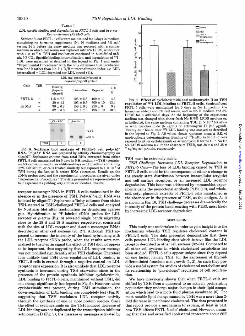
FIG. 9. Effect of cycloheximide and actinomycin D on TSH regulation of lz51-LDL binding to FRTL-5 cells. Semiconfluent FRTL-5 cells were maintained for 5 days in No H medium (no hormones added) and 5% calf serum, and in No H medium and 5% LPDS for 3 additional days. At the beginning of the experiment medium was changed with either fresh No H/5% LPDS medium or, as indicated, the same medium containing TSH (1 x lo-’ M) alone or with cycloheximide (5 pg/ml) or actinomycin D (2.5 fig/ml). Twenty-four hours later ‘*‘I-LDL binding was assayed as described in the legend to Fig. 2. All values shown represent mean k S.E. of quadruplicate determinations. Binding of ‘*‘I-LDL to FRTL-5 cells exposed to either cycloheximide or actinomycin D for 24 h, in No H/ 5% LPDS medium (i.e. in the absence of TSH), was 38 k 6 and 35 k 7 ng/mg cell protein, respectively.
TSH must be extremely stable. TSH Challenge Increases LDL Receptor Degradation in
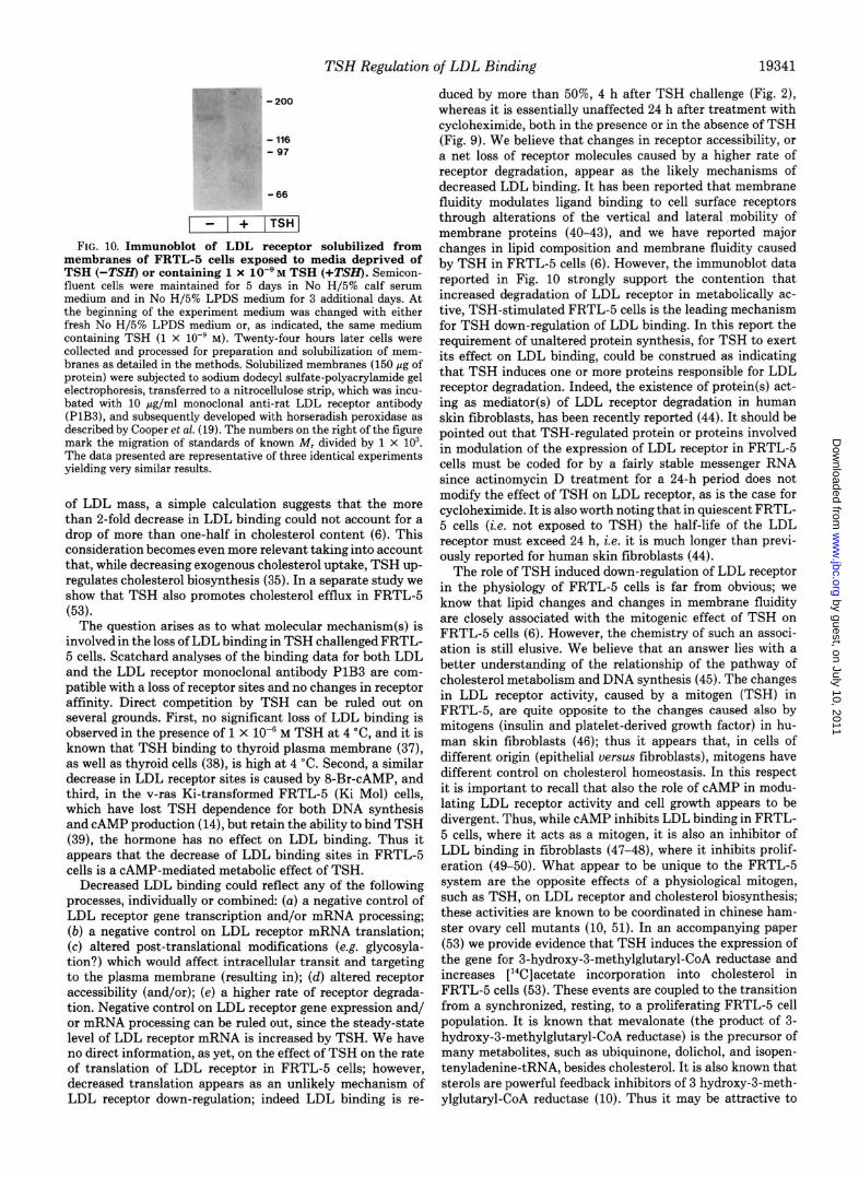
FRTL-5 CeU.s-The loss of LDL binding caused by TSH in FRTL-5 cells could be the consequence of either a change in the steady state distribution between intracellular (cryptic) and cell surface receptors, or an increased LDL receptor degradation. This issue was addressed by immunoblot exper- iments using the monoclonal antibody PlB3 (19), and whole- cell, octyl glucoside extracts of FRTL-5 cells maintained in the absence or in the presence of TSH, as the antigen. As it is shown in Fig. 10, TSH challenge decreases dramatically the intensity of the protein band reacting with PlB3, most likely by increasing LDL receptor degradation.
DISCUSSION
This study was undertaken in order to gain insight into the mechanism whereby TSH regulates cholesterol content in FRTL-5 cells. The data presented demonstrate that these cells possess LDL binding sites which behave like the LDL receptor described in other cell systems (32-34). Compared to all other cell systems in which cholesterol metabolism has been studied, FRTL-5 cells appear unique since they depend on one factor, namely TSH, for the expression of thyroid- differentiated functions and growth (1, 2). As such they pro- vide a useful system for studies of cholesterol metabolism and its relationship to “physiologic” regulation of cell prolifera- tion.
We have previously shown that when FRTL-5 cells are shifted by TSH from a quiescent to an actively proliferating population they undergo major changes in their lipid compo- sition which lead to a more fluid plasma membrane (6). The most notable lipid change caused by TSH was a more than 2- fold decrease in membrane cholesterol. The data presented in this report provide a mechanism to explain, at least in part, how TSH affects FRTL-5 cells’ cholesterol. However, assum- ing that free and esterified cholesterol represents about 50%
by guest, on July 10, 2011w
ww
.jbc.orgD
ownloaded from
- 116 G ,-97
-66
- 1 + ITSHI
FIG. 10. Immunoblot of LDL receptor solubilized from membranes of FRTL-5 cells exposed-to media deprived of TSH t-TSm or containing 1 x lo-'M TSH t+TSH. Semicon- fluent ‘cells iere maintained for 5 days in Non H/5% ‘calf serum medium and in No H/5% LPDS medium for 3 additional days. At the beginning of the experiment medium was changed with either fresh No H/5% LPDS medium or, as indicated, the same medium containing TSH (1 x lo-’ M). Twenty-four hours later cells were collected and processed for preparation and solubilization of mem- branes as detailed in the methods. Solubilized membranes (150 pg of protein) were subjected to sodium dodecyl sulfate-polyacrylamide gel electrophoresis, transferred to a nitrocellulose strip, which was incu- bated with 10 pg/ml monoclonal anti-rat LDL receptor antibody (PlB3), and subsequently developed with horseradish peroxidase as
TSH Regulation of LDL Binding 19341
duced by more than 50%, 4 h after TSH challenge (Fig. 2), whereas it is essentially unaffected 24 h after treatment with cycloheximide, both in the presence or in the absence of TSH (Fig. 9). We believe that changes in receptor accessibility, or a net loss of receptor molecules caused by a higher rate of receptor degradation, appear as the likely mechanisms of decreased LDL binding. It has been reported that membrane fluidity modulates ligand binding to cell surface receptors through alterations of the vertical and lateral mobility of membrane proteins (40-43), and we have reported major changes in lipid composition and membrane fluidity caused by TSH in FRTL-5 cells (6). However, the immunoblot data reported in Fig. 10 strongly support the contention that increased degradation of LDL receptor in metabolically ac- tive, TSH-stimulated FRTL-5 cells is the leading mechanism for TSH down-regulation of LDL binding. In this report the requirement of unaltered protein synthesis, for TSH to exert its effect on LDL binding, could be construed as indicating that TSH induces one or more proteins responsible for LDL receptor degradation. Indeed, the existence of protein(s) act- ing as mediator(s) of LDL receptor degradation in human skin fibroblasts, has been recently reported (44). It should be pointed out that TSH-regulated protein or proteins involved in modulation of the expression of LDL receptor in FRTL-5 cells must be coded for by a fairly stable messenger RNA since actinomycin D treatment for a 24-h period does not modify the effect of TSH on LDL receptor, as is the case for cycloheximide. It is also worth noting that in quiescent FRTL- 5 cells (i.e. not exposed to TSH) the half-life of the LDL receptor must exceed 24 h, i.e. it is much longer than previ- ously reported for human skin fibroblasts (44).
described by Cooper et al. (19). The numbers on the right of the figure mark the migration of standards of known &f, divided bv 1 X 10’. The data preiented are representative of three identical eiperiments yielding very similar results.
of LDL mass, a simple calculation suggests that the more than 2-fold decrease in LDL binding could not account for a drop of more than one-half in cholesterol content (6). This consideration becomes even more relevant taking into account that, while decreasing exogenous cholesterol uptake, TSH up- regulates cholesterol biosynthesis (35). In a separate study we show that TSH also promotes cholesterol efflux in FRTL-5 (531.
The question arises as to what molecular mechanism(s) is involved in the loss of LDL binding in TSH challenged FRTL- 5 cells. Scatchard analyses of the binding data for both LDL and the LDL receptor monoclonal antibody PlB3 are com- patible with a loss of receptor sites and no changes in receptor affinity. Direct competition by TSH can be ruled out on several grounds. First, no significant loss of LDL binding is observed in the presence of 1 x 10m6 M TSH at 4 “C, and it is known that TSH binding to thyroid plasma membrane (37), as well as thyroid cells (38), is high at 4 ‘C. Second, a similar decrease in LDL receptor sites is caused by 8-Br-CAMP, and third, in the v-ras Ki-transformed FRTL-5 (Ki Mol) cells, which have lost TSH dependence for both DNA synthesis and CAMP production (14), but retain the ability to bind TSH (39), the hormone has no effect on LDL binding. Thus it appears that the decrease of LDL binding sites in FRTL-5 cells is a CAMP-mediated metabolic effect of TSH.
Decreased LDL binding could reflect any of the following processes, individually or combine& (u) a negative control of LDL receptor gene transcription and/or mRNA processing; (b) a negative control on LDL receptor mRNA translation; (c) altered post-translational modifications (e.g. glycosyla- tion?) which would affect intracellular transit and targeting to the plasma membrane (resulting in); (d) altered receptor accessibility (and/or); (e) a higher rate of receptor degrada- tion. Negative control on LDL receptor gene expression and/ or mRNA processing can be ruled out, since the steady-state level of LDL receptor mRNA is increased by TSH. We have no direct information, as yet, on the effect of TSH on the rate of translation of LDL receptor in FRTL-5 cells; however, decreased translation appears as an unlikely mechanism of LDL receptor down-regulation; indeed LDL binding is re-
The role of TSH induced down-regulation of LDL receptor in the physiology of FRTL-5 cells is far from obvious; we know that lipid changes and changes in membrane fluidity are closely associated with the mitogenic effect of TSH on FRTL-5 cells (6). However, the chemistry of such an associ- ation is still elusive. We believe that an answer lies with a better understanding of the relationship of the pathway of cholesterol metabolism and DNA synthesis (45). The changes in LDL receptor activity, caused by a mitogen (TSH) in FRTL-5, are quite opposite to the changes caused also by mitogens (insulin and platelet-derived growth factor) in hu- man skin fibroblasts (46); thus it appears that, in cells of different origin (epithelial uersus fibroblasts), mitogens have different control on cholesterol homeostasis. In this respect it is important to recall that also the role of CAMP in modu- lating LDL receptor activity and cell growth appears to be divergent. Thus, while CAMP inhibits LDL binding in FRTL- 5 cells, where it acts as a mitogen, it is also an inhibitor of LDL binding in fibroblasts (47-48), where it inhibits prolif- eration (49-50). What appear to be unique to the FRTL-5 system are the opposite effects of a physiological mitogen, such as TSH, on LDL receptor and cholesterol biosynthesis; these activities are known to be coordinated in Chinese ham- ster ovary cell mutants (10, 51). In an accompanying paper (53) we provide evidence that TSH induces the expression of the gene for 3-hydroxy-3-methylglutaryl-CoA reductase and increases [14C]acetate incorporation into cholesterol in FRTL-5 cells (53). These events are coupled to the transition from a synchronized, resting, to a proliferating FRTL-5 cell population. It is known that mevalonate (the product of 3- hydroxy-3-methylglutaryl-CoA reductase) is the precursor of many metabolites, such as ubiquinone, dolichol, and isopen- tenyladenine-tRNA, besides cholesterol. It is also known that sterols are powerful feedback inhibitors of 3 hydroxy-3-meth- ylglutaryl-CoA reductase (10). Thus it may be attractive to
by guest, on July 10, 2011w
ww
.jbc.orgD
ownloaded from

TSH Regulation of LDL Binding
speculate that the transition of a cell population from a resting to a proliferating state would require an active mevalonate synthesis, more than cholesterol itself (52). It is conceivable that a mitogenic stimulus, such as TSH for FRTL-5 cells, would reduce cholesterol uptake while stimulating mevalonate and cholesterol synthesis.
Finally, it would be of interest to investigate whether down- regulation of LDL receptor activity by TSH is confined to thyroid cells or is exerted also on other cell types. If the latter would be true, a mechanism could be provided for the hyper- cholesterolemia of primary hypothyroidism, which is charac- terized by very high levels of circulating TSH. It is known that hypercholesterolemia does not occur in secondary hypo- thyroidism, when circulating TSH is extremely low or unde- tectable (36).
Acknowledgments-We are indebted to Dr. Leonard D. Kohn for his constant support and encouragement throughout the course of this study and to Dr. Joseph E. Rail for critical review of the manuscript. We should like to thank Dr. Heather M. Bond for helping with the immunoblot experiments.
1.
2.
3.
4.
5.
6.
7. 8.
9.
10.
11.
12.
13.
14.
15.
16.
17.
18.
19.
REFERENCES
Ambesi-Impiombato, F. S., Parks, L. A. M., and Coon, H. G. (1980) Proc. Natl. Acad. Sci. U. S. A. 77, 3455-3459
Ambesi-Impiombato, F. S., Picone, R., and Tramontano, D. (1982) Cold Snrins Harbor Conf. Cell Proliferation 9.483-492
Santisteban, P.: Kahn, L. D., and Di Laura, R. (1987) J. Biol. Chem. 262,4048-4052
Weiss, S. J., Philp, N. J., Amhesi-Impiombato, F. S., and Groll- man, E. F. (1984) Endocrmology 114,1099-1107
Valente, W. A., Vitti, P., Kahn, L. D., Brandi, M. L., Rotella, C. M., Toccafondi, R., Tramontano, D., Aloj, S. M., and Ambesi- Impiombato, F. S. (1983) Endocrinofogy 112,71-79
Beguinot, F., Beguinot, L., Tramontano, D., Duilio, C., Formi- sano. S.. Bifulco. M.. Ambesi-Imniomhato. F. S.. and Aloi. S. M. (i987) J. Rioi. Chm. 262,1575-X82 ’ “’
Inbar, I., Yuli, I., and Raz, A. (1977) Exp. Cell Res. 105,325-335 Muller, C. P., Volloch, Z, and Shinitzky, M. (1980) Cell Eiophys.
2,233-240 Reichert, L., Castagna, M., Beck, J. P., Pong, S., Lun, B., and
Ourisson, G. (1984) Biochem. Biophys. Res. Commun. 120, 192-197
Brown, M. S., and Goldstein, J. L. (1980) J. Lipid Res. 21, 505- 517
Tomita, K., Yoshida, T., Yoshimura, A., Ono, M., and Kuwano, M. (1987) J. Biol. Chem. 262,1398-1404
Fusco, A., Pinto, A., Ambesi-Impiombato, F. S., Vecchio, G., and Tsushida. N. (1981) Int. J. Cuncer 28,655-662
Colletta, G.; Pinto, Al, Di Fiore, P. P., Fusco, A., Ferrentino, M., Awedimento, V. E., Tsushida, N., and Vecchio, G. (1983) Mol. Cell Biol. 3, 2099-2109
40.
41.
42.
43.
44.
45. 46.
47.
Colletta, G., Corda, D., Schettini, G., Cirafici, A. M., Kahn, L. D., and Consiglio, E. (1988) FEBS Lett. 228, 37-41
Winand, R. J., and Kahn, L. D. (1970) J. Btil. Chem. 245,967- 48.
Coetzee, G. A. (i988) J. Lipid Res. 29,1481-1489 Siperstein, M. D. (1984) J. Lipid Res. 25, 1462-1468 Mazzone, T., Basheeruddin, K., Ping, L., Frazer, S., and Getz, G.
S. (1989) J. Biol. Chem. 264. 1787-1792 Mazier&, J. C., Mazier&, C., Gardette, J., Mora, L., and Polon-
owski, J. (1983) Biochem. Biophys. Res. Commun. 112, 795- 800
Stout, R. W., and Bierman, E. L. (1983) Atherosclerosis 46, 13- -- 975
xl
Kohn, L. D., and Winand, R. J. (1975) J. Biol. Chem. 250,6503- 49. Johnson, G. S., Friedman, R. M., and Pastan, I. (1971) Proc.
6508 Natl. Acad Sci. U. S. A. 68,425-429
Havel, R. J., Eder, H. A., and Bragdon, J. H. (1955) J. Clin. 50. Hsie, A. W., and Puck, T. T. (1971) Proc. Natl. Acad Sci. U. S.
A. 68,358-361 Inuest. 34,1345-1353
Bilheimer, D. W., Eisenberg, S., and Levy, R. I. (1972) Biochim. 51. Chin, J., and Chang, T.-Y. (1981) J. Biol. Chem. 256,6304-6310 52. Bifulco, M., Tedesco, I., Laezza, C., Papaccio, G., Garbi, C., and
Biophys. Actu 260,212-221 Aloj, S. M. (1989) Ann. Endocrinol. 50, 11 Cooper, A. D., Nutik, R., and Chen, J. (1987) J. Lipid Res. 28, 53. Grieco, D., Beg, Z. H., Romano, A., Bifulco, M., and Aloj, S. M.
59-68 (1990) J. Biot. Chem. 265, 19343-19350
20.
21.
22.
23. 24.
25.
26.
27.
28.
29.
30. 31.
32.
33.
34.
35.
36.
37.
38.
39.
Ambesi-Impiombato, F. S. (August 26, 1986) U. S. Patent 4,608,341
Kahn, L. D., Valente, W. A.. Grollman, E. F., Alai, S. M., and Vi&, P. (September 2,1986) U. S. Patent 4,609,&2
Goldstein, J. L., Basu, S. K., and Brown, M. S. (19830 Metkods Enzymol. 92, 241-260
Bradford. M. M. (1976) Anal. Biochem. 72. 248-254 Thomas,‘P. S. (lb80) Proc. Natl. Acad, Sc>. U. S. A. 77, 5201-
5205 Denhardt, D. T. (1966) Biochem. Biophys. Res. Commun. 23,
641-646 Yamamoto, T., Davis, C. G., Brown, M. S., Schneider, W. J.,
Casey, L. M., Goldstein, J. L., and Russel, D. W. (1984) Cell 39,27-38
Levy, A., Eldridge, J. D., and Paterson, B. M. (1985) Science 229,393-395
Schneider, W. J., Goldstein, J. L., and Brown, M. S. (1980) J. Biol. &em. 255,11442-11447
Schneider, W. J., Beisiegel, U., Goldstein, J. L., and Brown, M. S. (1982) J. Biol. Chem. 267,2664-2673
Laemmli. U. K. (1970) Nature 227.680-685 Beisiegeli U., Schneider, W. J., Brown, M. S., and Goldstein, J.
L. (1982) J. Biol. Chem. 257.13150-13156 Goldstein,.J. L., and Brown, M. S. (1974) J. Biol. Chem. 249,
5153-5162 Goldstein, J. L., Basu, S. K., Brunschede, G. Y., and Brown, M.
S. (1976) Cell 7,85-95 Goldstein, J. L., and Brown, M. S. (1977) Annu. Reu. B&hem.
46,897-930 Aloj, S. M. (1989) in PRTL-5 Today (Perrild, H., and Ambesi-
Impiombato, F. S., eds) pp. 117-119, Elsevier, Amsterdam Ingbar, S. H. (1987) in Harrisons’ Principles of Internal Medicine
(Braunwald, E., Isselbacher, K., Petersdorf, R. G., Wilson, J. D., Martin, J. B., and Faucci, A. S., eds) p. 1742, McGraw-Hill Book Co., New York
Bolonkin, D., Tate, R. L., Luber, J. H., Kohn, L. D., and Winand, R. J. (1975) J. Biol. Chem. 250, 6516-6521
Tramontano, D., and Ingbar, S. H. (1986) Endocrinology 118, 1945-1951
Colletta, G., Pinto, A., Schettini, G., Cirafici, A. M., Marinaccio, M., and Consiglio, E. (1985) in Cell Membrane and Cancer (Galeotti, T., Cittadini, A., Neri, G., Papa, S., and Smets, L. A,, eds) pp. 101-106, Elsevier, Amsterdam
Heron, D. S., Shinitzky, M., Hershkowitz, M., and Samuel, D. (1980) Proc. Natl. Acad Sci. U. S. A. 77,7463-7467
Strittmatter, W. J., Hirata, F., and Axelrod, J. (1979) Science 204,1205-1207
Hanski, E., Rimon, G., and Levitzki, A. (1979) Biochemistry 18, 846-853
Salesse, R., Garnier, J., Leterrier, F., Daveloose, D., and Viret, J. (1982) Biochemistry, 21,1581-1586
Casciola. L. A. F.. van der Westhuyzen, D. R., Gerves, W., and
by guest, on July 10, 2011w
ww
.jbc.orgD
ownloaded from