This electronic thesis or dissertation has been downloaded ...
133
This electronic thesis or dissertation has been downloaded from Explore Bristol Research, http://research-information.bristol.ac.uk Author: Songra, Goldie Tashfeen Butt Title: A randomised controlled clinical trial to investigate the rates of alignment of the upper and lower labial segments, rates of extraction space closure in the upper and lower buccal segments and treatment efficiency using conventional, active and passive self- ligating orthodontic brackets General rights Access to the thesis is subject to the Creative Commons Attribution - NonCommercial-No Derivatives 4.0 International Public License. A copy of this may be found at https://creativecommons.org/licenses/by-nc-nd/4.0/legalcode This license sets out your rights and the restrictions that apply to your access to the thesis so it is important you read this before proceeding. Take down policy Some pages of this thesis may have been removed for copyright restrictions prior to having it been deposited in Explore Bristol Research. However, if you have discovered material within the thesis that you consider to be unlawful e.g. breaches of copyright (either yours or that of a third party) or any other law, including but not limited to those relating to patent, trademark, confidentiality, data protection, obscenity, defamation, libel, then please contact [email protected] and include the following information in your message: • Your contact details • Bibliographic details for the item, including a URL • An outline nature of the complaint Your claim will be investigated and, where appropriate, the item in question will be removed from public view as soon as possible.
-
Upload
khangminh22 -
Category
Documents
-
view
1 -
download
0
Transcript of This electronic thesis or dissertation has been downloaded ...

This electronic thesis or dissertation has beendownloaded from Explore Bristol Research,http://research-information.bristol.ac.uk
Author:Songra, Goldie Tashfeen Butt
Title:A randomised controlled clinical trial to investigate the rates of alignment of the upperand lower labial segments, rates of extraction space closure in the upper and lowerbuccal segments and treatment efficiency using conventional, active and passive self-ligating orthodontic brackets
General rightsAccess to the thesis is subject to the Creative Commons Attribution - NonCommercial-No Derivatives 4.0 International Public License. Acopy of this may be found at https://creativecommons.org/licenses/by-nc-nd/4.0/legalcode This license sets out your rights and therestrictions that apply to your access to the thesis so it is important you read this before proceeding.
Take down policySome pages of this thesis may have been removed for copyright restrictions prior to having it been deposited in Explore Bristol Research.However, if you have discovered material within the thesis that you consider to be unlawful e.g. breaches of copyright (either yours or that ofa third party) or any other law, including but not limited to those relating to patent, trademark, confidentiality, data protection, obscenity,defamation, libel, then please contact [email protected] and include the following information in your message:
•Your contact details•Bibliographic details for the item, including a URL•An outline nature of the complaint
Your claim will be investigated and, where appropriate, the item in question will be removed from public view as soon as possible.

A RANDOMISED CONTROLLED CLINICAL TRIAL TO
INVESTIGATE THE RATES OF ALIGNMENT OF THE UPPER AND
LOWER LABIAL SEGMENTS, RATES OF EXTRACTION SPACE
CLOSURE IN THE UPPER AND LOWER BUCCAL SEGMENTS AND
TREATMENT EFFICIENCY USING CONVENTIONAL, ACTIVE AND
PASSIVE SELF-LIGATING ORTHODONTIC BRACKETS
Goldie Tashfeen Butt Songra
BDS, MFDS RCSEng, MOrth RCSEng
A dissertation submitted to the University of Bristol in accordance with the requirements of the degree of Doctorate of Dental Surgery in Orthodontics
in the School of Oral and Dental Sciences, Faculty of Medicine and Dentistry
July 2011
Word Count: 26,248
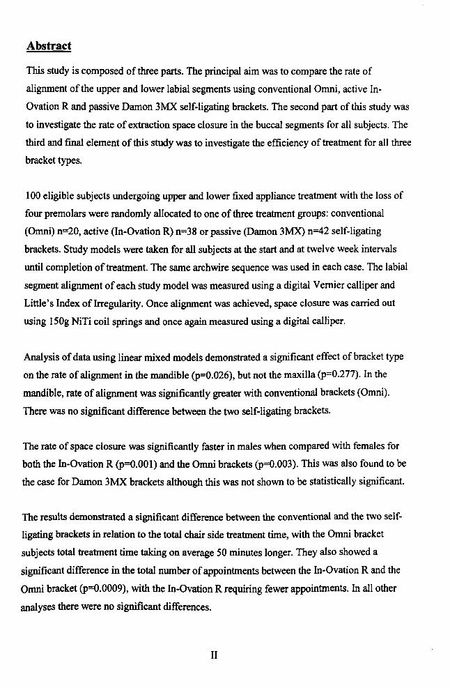
Abstract
This study is composed of three parts. The principal aim was to compare the rate of
alignment of the upper and lower labial segments using conventional Omni, active In
Ovation R and passive Damon 3MX self-ligating brackets. The second part of this study was
to investigate the rate of extraction space closure in the buccal segments for all subjects. The
third and final element of this study was to investigate the efficiency of treatment for all three
bracket types.
100 eligible subjects undergoing upper and lower fixed appliance treatment with the loss of
four premolars were randomly allocated to one of three treatment groups: conventional
(Omni) n=20, active (In-Ovation R) n=38 or passive (Damon 3MX) n=42 self-ligating
brackets. Study models were taken for all subjects at the start and at twelve week intervals
until completion of treatment. The same archwire sequence was used in each case. The labial
segment alignment of each study model was measured using a digital Vernier calliper and
Little's Index of Irregularity. Once alignment was achieved, space closure was carried out
using 150g NiTi coil springs and once again measured using a digital calliper.
Analysis of data using linear mixed models demonstrated a significant effect of bracket type
on the rate of alignment in the mandible (p=0.026), but not the maxilla (p=0.277). In the
mandible, rate of alignment was significantly greater with conventional brackets (Omni).
There was no significant difference between the two self-ligating brackets.
The rate of space closure was significantly faster in males when compared with females for
both the In-Ovation R (p=0.001) and the Omni brackets (p=0.003). This was also found to be
the case for Damon 3MX brackets although this was not shown to be statistically significant.
The results demonstrated a significant difference between the conventional and the two self
ligating brackets in relation to the total chair side treatment time, with the Omni bracket
subjects total treatment time taking on average 50 minutes longer. They also showed a
significant difference in the total number of appointments between the In-Ovation R and the
Omni bracket (p=O.0009), with the In-Ovation R requiring fewer appointments. In all other
analyses there were no significant differences.
II

Acknowledgements
I would like to take this opportunity to thank everybody who has helped me with this
dissertation.
Firstly, I would like express my appreciation to Matt Clover, who worked so hard to lay the
foundations for this project.
My sincere thanks go to my supervisors, Dr. Tony Ireland and Ms. Nikki Atack whose help,
support and guidance have been invaluable in both the clinical and academic elements of this
research project.
I am also very grateful for the participation of Nick Mitchell, Helen Griffiths, Nick Wenger,
Rebecca Wilson and Michael Dawson, who have recruited and treated the subjects in this
study.
Enormous gratitude goes to the department's nurses, Helen Winters, Lynne Farkas, Lois
Grainger, Jane Ledsham and Sarah Lofthouse whose support and experience was invaluable.
Thank you to my colleagues, Tony, Caroline, Sonica and Murray in the dental laboratory, for
all their hard work in processing the hundreds of study models required.
Additionally, I would like to thank all of the reception staff for their efforts in organising and
managing the appointments for this investigation.
I would like to thank Paul Ewings and Dr. Martyn Sherriff for their help and advice with the
statistical analysis for this study.
Richard Long ofOrmco and David Rees ofTOC, generously provided brackets and
archwires used in this study, for which I am most appreciative.
Finally, all this would not be possible without the incalculable support, patience and
encouragement from the two special ladies in my life, especially my mother, Tara Songra for
which I am eternally grateful.
I would like to dedicate this thesis to my late father Basharat A. Butt.
III

Author's Declaration
Orthodontic Departmen"t,
Musgrove Park Hospital,
Taunton
TAl5DA
I declare that the work within this dissertation was carried out in accordance with the
Regulations of the University of Bristol. The work is original, except where indicated by
special reference in the text, and no part of the dissertation has been submitted for any other
academic award.
Any views expressed in the dissertation are those of the author and in no way represent those
of the University of Bristol.
The dissertation has not been presented to any other University for examination either in the
United Kingdom or overseas.
Goldie T B Songra
IV

List of Contents
Title
Abstract
Acknowledgements
Author's Declaration
List of Contents
List of Tables
List of Figures
List of Appendices
1 Review of the literature
1.1 Introduction
1.2 History of orthodontic appliances
1.3 Methods of ligation
1.4 Self-ligating brackets
1.4.1 History of the Damon bracket
1.4.2 History of the In-Ovation R bracket
1.5 Active versus passive
1.6 Friction
1.6.1 Clinical implications of friction
1.6.1.1 Factors influencing resistance to sliding
1.6.1.1.1 Masticatory forces
1.6.1.1.2 Angulation of archwire
1.6.1.1.3 Size of bracket
1.6.1.1.4 Size of archwire
1.6.1.1.5 Effect oflubrication
1.6.1.1.6 Archwire material
v
Page
II
III
IV
V-VIII
IX
X-XII
XIII
1
5
6
8
10
12
14
15
15
15
16
16
17
17
18

1.6.1.1.7 Bracket material
1.6.1.1.8 Method of ligation
1.7 Space closure
1.7.1 Closing loops
1. 7.2 Sliding mechanics
1.7.2.1 Elastomeric powerchain
1.7.2.2 Active ligatures
1.7.2.3 Patient applied intra- and inter-arch elastics
1.7.2.4 Coil springs
1.7.3 Optimum force for tooth movement
1.8 Measuring alignment
1.9 Measuring space closure
1.10 Comparison of conventional and self-ligating brackets
1.10.1 Ligation, chairside time and efficiency
1.10.2 Alignment
1.10.3 Space closure
2 Aims
2.1 Null hypotheses
3 Materials and Methods
3.1 Materials used in the clinical environment
3.2 Materials used in the non-clinica1 environment
3.3 Power calculation
3.4 Method of investigation
3.5 The bond-up appointment
3.6 Routine appointments
VI
19
19
21
22
23
23
24
25
26
27
29
32
35
35
37
39
42
42
43
43
45
46
46
48
50

3.7 Emergency appointments
3.8 Measurement of labial segment alignment
3.9 Measurement of space closure
3.10 Measurement of appointment treatment times
4 Consort diagram
5 Results
5.1 Age and sex distribution for the trial
5.2 Bracket distribution
5.3 Frankfort mandibular planes angle
5.4 Repeatability of measurement using the vernier callipers
5.4.1 Little's index
5.4.2 Space closure
5.5 Labial segment alignment
5.5.1 The effect of bracket type on alignment
5.6 The effect of bracket type on space closure
5.6.1 The effect of sex on space closure
5.7 Treatment times
5.7.1 Total chair side time in minutes
5.7.2 Mean treatment time per visit in minutes
5.7.3 The overall treatment time in days
5.7.4 The total number of appointments to debond
S.7.S The total number of emergency appointments
6 Discussion
6.1 Extraction vs. non-extraction
VII
51
52
52
53
S4
SS
55
56
57
60
60
61
62
63
67
71
75
75
77
78
79
80
81
81
6.2 Exclusions 81
6.3 Practical clinical issues 81
6.4 Archwire sequence 82
6.5 Frankfort mandibular planes angle 83
6.6 Bracket Prescription 84
6.7 Pre-treatment crowding 84
6.8 Measurement period 85
6.9 Effect of bracket on alignment 85
6.10 Effect of bracket on space closure 87
6.11 Treatment times 88
7 Conclusions 92
8 Future work 93
9 References 94
VIII

List of Tables Page
Table 1
Table 2
Table 3
Table 4
Table 5
Table 6
Table 7
Table 8
Table 9
Table 10
Table 11
Table 12
Chronology of the self-ligating bracket
Summary statistics for age and sex distribution of the study subjects
Bracket distribution within male and female subjects
FMP A within male and female subjects
FMP A within bracket type
Pairwise comparison showing there is no statistically significant
difference in the rate of initial alignment between the two self-ligating
brackets. There was a statistically significant difference in the rate of
initial alignment between these two brackets and the conventional Omni
bracket
7
55
56
57
58
66
Summary table of the effects and interactions tested using linear mixed 68
models within each bracket type
Exact probabilities of the effect of bracket type on space closure 68
Data summary table for space closure for sex (data from all three brackets 72
pooled and for both maxilla and mandible)
Summary data of total chair side time for patients who had completed
treatment
76
Summary data of chair side time per visit for patients who had completed 77
treatment
Summary data of total treatment time in days 78
IX

List of Figures
Figure 1
Figure 2
Figure 3
Figure 4
Figure 5
Figure 6
Figure 7
Figure 8
Figure 9
Figure 10
Figure 11
Figure 12
Figure 13
Figure 14
Damon 3MX brackets in open and closed positions
In-Ovation R brackets in open and closed positions
Side view of In-Ovation R bracket
Elastomeric powerchain being used in buccal space closure
Active ligatures being used for buccal space closure
Patient applied Class 2 elastics being used in the buccal segments
NiTi coil springs being used for buccal space closure
Consort diagram charting the flow of subjects through the trial
Histogram illustrating the bracket distribution within sex, Damon 3MX,
In-Ovation R and conventional Omni bracket
Histogram illustrating the FMPA distribution within sex. Code L = Low,
Code A = Average and Code H ;; High
Histogram illustrating the FMP A distribution within bracket, Damon
3MX, In-Ovation R and conventional Omni bracket
Histogram illustrating the FMP A distribution within sex and bracket,
Damon 3MX, In-Ovation R and conventional Omni bracket
Lin's concordance correlation coefficient plot for Little's Index
measured using digital Vernier callipers at T1 and T2
Lin's concordance correlation coefficient plot for Space Closure
measured using digital Vernier Callipers at T I and T2
x
Page
10
II
13
23
24
25
26
54
56
57
58
59
60
61

Figure 15 Means, 95% confidence intervals of the mean and raw data for the 62
combined upper and lower labial segment Little's Index scores for the
patients in each of the three bracket treatment groups
Figure 16 Distributional plot of the initial alignment over time (days) for all the 63
patients with all three bracket types in both the mandible and maxilla
(200 observations)
Figure 17 Distributional plot of the initial alignment over time (days) for all the 64
patients with the Damon 3MX bracket both mandible and maxilla
Figure 18 Distributional plot of the initial alignment over time (days) for all the 64
patients with the In-Ovation R bracket both mandible and maxilla
Figure 19' Distributional plot of the initial alignment over time (days) for all the 65
patients with the Omni bracket both mandible and maxilla
Figure 20 Plot of the q-values against bracket pairing. In this plot anything above 67
the 0.05 line is a significant difference (Pairings: Code 1 == Damon 3MX,
Code 2 == In-Ovation R and Code 3 = conventional Omni bracket)
Figure 21 Plot of the q-values against bracket pairing. In this plot anything above 69
the red 0.05 line is a significant difference (D = Damon 3MX, I = In-
Ovation Rand 0 = conventional Omni bracket)
Figure 22 Plot of space closure (mm) against time (days) for the Damon 3MX 70
brackets
Figure 23 Plot of space closure (mm) against time (days) for the In-Ovation R 70
bracket
Figure 24 Plot of space closure (mm) against time (days) for the Omni bracket 71
Figure 25 Means, 95% confidence intervals of the mean and raw data for the 72
combined space closure (mm) for males and females
Figure 26 Restricted cubic spline plot of rate of space closure for the Damon 3 MX 73
bracket
XI

Figure 27 Restricted cubic spline plot of rate of space closure for the In-Ovation R 73
bracket
Figure 28 Restricted cubic spline plot of rate of space closure for the Omni bracket 74
Figure 29 Means, 95% confidence intervals of the mean and raw data for total chair 75
side time in minutes for each of the three brackets under test
Figure 30 Means, 95% confidence intervals of the mean and raw data for chair side 77
time in minutes per visit for each of the three brackets under test
Figure 31 Means, 95% confidence intervals of the mean and raw data for total time 78
in days for each of the three brackets under test
Figure 32 Frequency distribution plots for the number of appointments for each of 79
the three brackets under test
Figure 33 Frequency distribution plots for the number of emergency appointments 80
for each of the three brackets under test
XII

List of Appendices
Appendix 1 Information sheet for participant (child)
Appendix 2 Information sheet for parents! guardians
Appendix 3 Consent form
XIII
Page
114
116
119
1 Review of the Literature
1.1 Introduction
The human race has been fascinated with moving teeth for a very long time, with some form
of 'orthodontics' and tooth movement having been practised for centuries (Weinberger 1934).
For example, Ancient Egyptian mummies have been discovered with metal bands wrapped
around individual teeth and with catgut used to close spaces (Paladin 2005). Both
Hippocrates and Aristotle contemplated the various dental conditions and ways to straighten
teeth, as far back as 400 to 500 Be. However, it was not until Roman times and Aurelius
Cornelius Celsus that the fIrSt orthodontic treatment plan was recorded, where digit pressure
was noted as being able to guide an aberrantly erupting tooth into its correct position (Wahl
2005a).
1.2 History of Orthodontic Appliances
The practice of fixed orthodontic appliances can be traced back as early as the 18th Century.
Pierre Fauchard published his book "The Surgeon Dentist" in 1728 with a whole chapter
dedicated to methods used to straighten teeth (Wahl2005a). Fauchard is also credited with
the development of the 'bandeau'. This was a horseshoe shaped piece of precious metal for
the maxilla to which teeth were ligated. This was the first expansion appliance and was to go
onto influence, inspire and ignite modem mans' desire to have straight teeth.
It was not until the late 19th Century that Edward Angle, regarded by many as the father of
modem orthodontics, developed his E or expansion-arch, where a partially threaded wire was
attached to banded molar teeth (Angle 1907).
During use the wire was lengthened by the advancement of a nut on the threaded portion of
the wire. Since the teeth were only ligated to the expansion arch the tooth movement
achieved was tipping (Angle 1907).
The E-arch was then followed by Angle's Pin and Tube appliance (Angle 1916). This
appliance was designed to overcome some of the problems associated with the original E
arch, allowing all of the teeth to be banded using a tube soldered to the labial surfaces. The
archwire was constructed with corresponding soldered pins which would engage the tubes
and therefore allow more controlled tooth movement. There was however a major drawback
1

in the use of this appliance, as the soldered pins required repositioning every time the teeth
moved. Consequently it was not a popular appliance and only Angle and one of his students
ever mastered its clinical use (proffit 2007).
Nevertheless the era of modem orthodontics had begun and soon a more advanced appliance,
the 'Ribbon Arch', was introduced by Angle (Angle 1920). This was a modification of the
earlier pin and tube appliance which overcame the problem of resoldering new pins onto the
archwire, or making a new appliance every time the teeth had moved. It did so by allowing a
0.010" x 0.020" sized ribbon of gold wire to be placed in a vertical slot that was cut behind
the tube on each band. The wire could then be held in place by pins. Although easier to use
than the Pin and Tube, this appliance still only resulted in partial alignment of the teeth due to
considerable movement of the archwire within the band slot or 'slop'. Root torquing was not
particularly efficient and additional adjustments to the wire would need to be performed by
the operator, despite the use of what was a rectangular cross-sectional arch wire (Proffit
2007).
The next significant step in the development of orthodontic appliances and techniques was
the introduction by Angle of the Edgewise appliance (Angle 1928). This appliance was
capable of allowing three dimensional control of tooth position, overcoming many of the
shortcomings of the ribbon arch appliance. Angle also made additional modifications to this
new appliance. He changed the dimensions of the slot to 0.022" x 0.028" and re-orientated
the slot position from the vertical to the horizontal. This allowed the wire to be rotated by 90
degrees and inserted 'Edgewise' into the newly positioned horizontal slot. The Edgewise
appliance allowed the clinician for the first time to have a positive three dimensional control
over the position of the teeth. Different archwires could be used to carry out different
movements. Initially a round wire could be used to allow tipping of the crowns of the teeth in
the buccall labial direction and achieve initial alignment. This was then followed by a
rectangular wire which would allow the torquing of the roots of the teeth in the buccall labial
direction. The Edgewise appliance then relied on the clinician undertaking the final detailing
of positioning of the teeth by placing specific bends in the wire to provide the individual in!
out, tip and torque for each tooth, i.e. three-dimensional position of each tooth. This was
necessary as each bracket was of a universal standard design and, as such, was not specific or
capable of individual tooth positioning (Thurow 1973).
2

Angle advocated the expansion of the dental arches to relieve crowding rather than extraction
of teeth (Wahl2005b). One of Angles' students was Raymond Begg. Begg returned to his
native Australia after learning the Angle technique, and after a period of research examining
the indigenous population, concluded that extractions were often necessary. Subsequently he
developed his own bracket in 1933 and then his own appliance based on Angle's ribbon arch
appliance, namely the Begg appliance in 1965 (Wahl 200Sb). This appliance was different to
Angle's ribbon arch in three main ways:
1. A high strength single 0.016" stainless steel wire replaced the precious metal ribbon
arch.
2. Inverting the bracket slot upside down so that the slot faced gingivally rather than
occlusally.
3. The addition of auxiliary springs to control root position, i.e. torque.
Begg's new appliance utilised the concept of differential tooth movement, which was a low
friction means of crown tipping, followed by root uprighting and allowed for the correction
and closure of extraction spaces (Wahl2005b).
The Edgewise and Begg appliances dominated the orthodontic world for the next 35 years
and formed the bastion of fixed appliance treatment. That was until 1965 when Larry
Andrews developed the Straight Wirec appliance. This was introduced onto the market a few
years later and soon became widely accepted (Andrews 1972). This revolutionary new
appliance was the first of what would be known as the preadjusted edgewise system, and
would form the foundation of modem orthodontic appliances.
Andrews, in developing this new appliance, had removed the requirement of all the first,
second and third order bends needed in the original Edgewise appliance archwire. This was
facilitated by incorporating the individual three dimensional 'prescription' for each tooth, in
each tooth specific bracket. The Straight Wirec appliance was based on the investigation of
the occlusions of 120 non-orthodontic subjects with normal occlusions. Andrews was able to
assess these occlusions and from this develop his "six keys to normal occlusion" (Andrews
1972). Andrews felt these keys were critical if a normal occlusion was desired at the end of
fixed orthodontic appliance treatment, and was found to be present in all of the 120 normal
occlusion subjects.
3

The six keys were as follows:
1. Molar relationship: The distal surface of the distobuccal cusp of the upper first molar,
should make contact and occlude with the mesial surface of the mesiobuccal cusp of
the lower second molar.
2. Crown Angulation: The gingival portion of the long axis of each crown should be
distal to the incisal portion, varying with the individual tooth type.
3. Crown Inclination: Anterior teeth- upper and lower crown inclination should be
sufficient to resist overeruption of the anterior teeth. Upper posterior teeth- there
should be constant lingual crown inclination from the canines to the second premolars
with slightly more inclination in the molars. Lower posterior teeth- there should be a
progressive increase in lingual crown inclination from the canines to the second
molars.
4. Rotations: There should be no rotations.
5. Spaces: There should no spaces between the teeth.
6. Occlusal Plane: There should be a level plane of occlusion or a slight curve of Spee.
Since the introduction of the Straight Wirec appliance various bracket designs have been
introduced to the orthodontic market with differing 'prescriptions' for each tooth. The
rationale has been to allow specific treatment mechanics and! or to achieve a better and more
stable final position of the teeth at the end of treatment (Bennett & McLaughlin 1994).
The major advantage of the preadjusted Edgewise appliance, compared to the original
Edgewise appliance, is the lack of adjustment required by the clinician, particularly a
reduction in the need for chair side wire bending. In addition, the use of the 'straight
archwire' has meant the principle of sliding mechanics could be more readily employed
during treatment. This enabled teeth to be more easily moved bodily, or space closure to be
carried out by moving teeth 'en masse' along the archwire (Bennett & Mclaughlin 1994).
This added to the popularity of the system. However, the major disadvantage of this
appliance when compared with the more traditional Edgewise mechanics of closing loops,
was the high level of friction between the slot and the wire. This can result in slower tooth
movement and particularly a loss of anchorage during treatment (Kesling 1988).
4
To overcome this limitation, Peter Kesling and his team developed the Tip-Edge appliance
(Kesling 1988). Tip-Edge utilised the Begg philosophy of free tipping followed by
uprighting, and combined it with the improved finishing of the preadjusted Edgewise
appliance. By using the Begg mechanics of differential tooth movements, only very light
forces are needed to translate the teeth into their pre-finishing positions. The subsequent
uprighting is achieved with the use of auxiliaries such as sidewinder springs, and finally large
dimension rectangular wires in a rectangular slot.
Over the last century a large number of appliances, systems and philosophies have been
introduced to treat malocclusion, each with their own advantages and disadvantages. Current
modem orthodontic treatment aims to provide the most efficient, safe, reliable and beneficial
treatment to the patient. It does so by utilising parts and! or modifications of each of the
appliances mentioned above.
1.3 Methods of Ligation
Whether clinicians prefer to use the Edgewise, Straight Wirec, Begg or Tip-Edge appliances,
a requirement in each case is that the archwire must be securely held within the bracket slot.
Traditionally this was carried out using ligatures made of Stainless Steel, or brass pins in the
case of the Begg appliance. These methods ofligation were cheap, robust and biologically
inert. However, they were not without their weaknesses namely, the time required to tie in
each archwire and the risk of trauma to the patient from the free ends (Shivapuja & Berger
1994).
Elastomeric modules were introduced in the late 1960's and even though they had their
limitations, they were generally accepted by the whole profession because of their ease of
use, and the speed of cbanging an archwire compared with the use of Stainless Steel ligatures
(Eliades & Pandis 2009).
A number of disadvantages of elastomeric modules have been identified which include;
microbial accumulation, archwires not seating fully during torquing or rotational movements
and the occurrence of binding during sliding mechanics (Taloumis et al. 1997). A number of
in vitro studies also found that the use of modules significantly increased friction and thus
resistance to sliding (Edwards et al. 1995; Hain et al. 2003; Khambay et al. 2004,2005). The
5

oral environment has also been found to have an adverse effect on elastomerics (Ash &
Nikolai 1978; Iwasaki et al. 2003). Water absorption accelerates the force decay of
elastomerics (Taloumis et al. 1997). Moisture uptake leads to a weakening of non covalent
forces and then degradation of the material. This along with heat causes a decrease in the
dimensional stability of the elastomeric, causing force to be lost in the first 24 hours by up to
50 to 70% (Huget et al. 1990; Taloumis et al. 1997).
Self-ligating brackets have more recently been introduced into clinical practise. This is due,
in part, to the increased friction associated with ligation and also the potential infection
control issues.
1.4 Self-Ligating Brackets
As the name suggests, self-ligating (SL) brackets do not require any external form of ligation
i.e. Stainless Steel ligature or elastomeric module, but have an inbuilt ligation mechanism to
hold the archwire in the slot. The self-ligating bracket is not a new development with the first
such bracket, the Russell Lock attachment, described approximately 70 years ago by
Stolzenburg (1935). It comprised a nut and bolt apparatus that was attached to a band. The
archwire was held in place by the adjustable nut, which screwed into the labial surface of the
slot. The flat lingual surface of the nut created a 'fourth' wall, and by varying the position of
the nut one could control the interaction between the archwire and the bracket. It was
reported that using the Russell attachment was considerably more comfortable for the patient,
required shorter visits and that the overall treatment time was shorter than when using the
conventional brackets of the day (Stolzenburg 1946). These benefits have been re-examined
more recently by Maijer & Smith (1990) who also claim that SL brackets have improved
aesthetics, are more comfortable, smoother and less traumatic. Further advantages of SL
brackets have been reported to include; reduced friction, full and secure wire ligation,
improved oral hygiene and anchorage conservation (Turnbull & Birnie 2007).
There are a number of SL brackets available with differing techniques for the secure ligation
of the archwire (Table 1).
6

Table 1 Chronology of the self-ligating bracket (Harradine & Birnie 2010)
Bracket Year of Development
Russell Lock 1935
Onnco EdgeLok 1972
Forestadent Mobil-Lock 1980
OrecSpeed 1980
"A" Company Activa 1986
Adenta Time 1994
"A" Company Damon SL 1996
Onnco TwinLock 1998
Onncol "A" Company Damon 2 2000
GAC In-Ovation 2000
Gestenco Oyster 2001
GAC In-Ovation R 2002
Adenta Evolution L T 2002
Ultradent Opal 2004
SDS Onnco Damon 3 2004
3M Unitek SmartCIip 2004
SDS Ormco Damon 3MX 2005
Class One Carriere SLB 2005
GAC In-Ovation C 2006
Forestadent Quick 2006
7
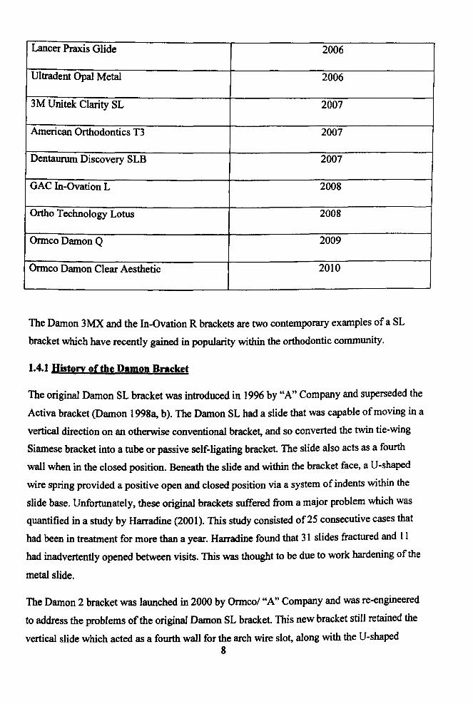
Lancer Praxis Glide 2006
Ultradent Opal Metal 2006
3M Unitek Clarity SL 2007
American Orthodontics T3 2007
Dentaurum Discovery SLB 2007
GAC In-Ovation L 2008
Ortho Technology Lotus 2008
Ormco Damon Q 2009
Ormco Damon Clear Aesthetic 2010
The Damon 3MX and the In-Ovation R brackets are two contemporary examples of a SL
bracket which have recently gained in popularity within the orthodontic community.
1.4.1 History of the Damon Bracket
The original Damon SL bracket was introduced in 1996 by "A" Company and superseded the
Activa bracket (Damon I 998a, b). The Damon SL had a slide that was capable of moving in a
vertical direction on an otherwise conventional bracket, and so converted the twin tie-wing
Siamese bracket into a tube or passive self-ligating bracket. The slide also acts as a fourth
wall when in the closed position. Beneath the slide and within the bracket face, a V-shaped
wire spring provided a positive open and closed position via a system of indents within the
slide base. Unfortunately, these original brackets suffered from a major problem which was
quantified in a study by Harradine (2001). This study consisted of25 consecutive cases that
had been in treatment for more than a year. Harradine found that 31 slides fractured and 11
had inadvertently opened between visits. This was thought to be due to work hardening of the
metal slide.
The Damon 2 bracket was launched in 2000 by Ormco/ "A" Company and was re-engineered
to address the problems of the original Damon SL bracket. This new bracket still retained the
vertical slide which acted as a fourth wall for the arch wire slot, along with the V-shaped 8

spring to control its opening and closing. The slide was now housed within the shelter of the
twin tie wings and this, in combination with metal injection moulding manufacture used to
generate closer tolerances, virtually eliminated any accidental slide opening or breakage. An
unfortunate side effect however, was that this also made the brackets more difficult to open
and close (Harradine 2003).
The next stage in the evolutionary process was the Damon 3 bracket, released in 2004. Like
the Damon 2 bracket the slide was housed within the tie wings. Similar to its predecessors, it
allowed an unrestricted view of the archwire slot in both arches as the slide opened in a
downwards direction, but the new bracket design had a small hole incorporated into the
surface of the slide to permit access to the opening mechanism. A special tool could then be
used to depress the Stainless Steel catch and open the slide in one continuous motion. Unlike
its predecessor, the Damon 3 slide could be closed using light finger pressure and did not
require the use of any special instruments. These new brackets were also semi aesthetic as
the bracket base and half of the tie wings were made of a composite material. However, this
led to significant problems including a high rate of bond failures, separation of the metal from
the composite base and fracture of the composite tie wings (Eliades & Pandis 2009). To
eliminate these problems, in 2005 Onnco launched the all new and all metal Damon 3MX
bracket (Figure I). This was similar to the Damon 3 bracket except that it was made
completely from injection moulded Stainless Steel. The Damon 3MX has been recently
superseded by the Damon Q bracket which is similar in many respects to the previous bracket
design, but differs in the mode of the slide opening mechanism. The Q bracket has a new
slide design and a special slide opening tool that requires a twisting action rather than a
downward force, transferring reciprocal forces to the bracket rather than the tooth.
9

Figure 1 Damon 3MX brackets in open and closed positions
1.4.2 History of In-Ovation R Bracket
The In-Ovation bracket was introduced by GAC International in 2000.
The design and concept of the In-Ovation bracket is very similar to that of the original Speed
bracket invented by Herbert Hanson in 1975 (Hanson 1980). Hanson combined his own
concept of a dynamic and self-ligating appliance, with Angle' s edgewise appliance resulting
in the so called 'active' self-ligating bracket. The 'active' component was due to a spring
loaded, self adjusting ligature-less design, which actively influenced the control of the
archwire within the archwire slot (Hanson 1980). It featured a curved flexible spring clip
made from a highly resilient super-elastic Nickel-Titanium that wrapped occlusally
gingivalJy around a miniaturised bracket body. The labial arm of the spring clip formed a
fourth and flexible outer wall of the archwire slot (Graber et al. 2005).
The In-Ovation bracket possesses an active spring clip similar to the Speed bracket, which
pushes the archwire into the base of the bracket slot once it is above a certain dimension. The
principal difference between the In-Ovation and the Speed bracket is in the clip which is
constructed from Cobalt Chromium alloy. Other differences include its twin configuration
and the presence of tie wings. The spring clip once closed converts the arch wire slot into a
rhomboid shape rather than a traditional rectangular shape tube, causing it to become
10

interactive with archwires above 0.018" . This allows torque control and expression of the
bracket prescription (Graber et al. 2005).
The In-Ovation R bracket was introduced in 2002. (Figure 2) The principal difference
between the two versions being the reduced mesio-distal width of the anterior brackets in the
"R" series, with the "R" standing for 'reduced', and therefore allowing for a greater inter
bracket span.
Figure 2 In-Ovation R brackets in open and closed positions
The spring clip is opened in a gingivally-occlusal direction. In the upper arch this is in a
downward motion whereas in the lower arch is in an upward direction. It can be opened with
gentle force using any instrument with a sharp edge, however this carries a risk of debonding
the bracket if you inadvertently catch the bracket base rather than the latch. Therefore GAC
manufactured the 'R' tool which looks like a periodontal probe with a ball on the tip. This
allows the operator to slide to the edge of the bracket pad and then slip onto the latch which
allows the clip to pop open. The other end of the instrument has a double ligature direction
design and can be used to seat the wire into the bracket slot. The two prongs of the instrument
are slightly wider than the widest In-Ovation bracket (Graber et al. 2005). Similar to the
Damon 3MX slide, light finger pressure can be applied to close the spring clip.
Even though these brackets have been described as robust (Harradine 2003), some
disadvantages have been reported. Visualisation of the gingival end of the spring clip can be
difficult, especially in the lower arch. Also excess composite at the gingival aspect of the
bracket can be difficult to see, and if not removed before setting may hinder access to the
11

spring clip opening latch. The spring clip can also inadvertently close before the archwire has
been placed into position.
A common problem found with both the Damon SL and the In-Ovation SL brackets is the
build up of calculus, which can result in the slide and spring clip opening mechanisms being
difficult to access and open. A modification was introduced to the In-Ovation spring clip in
2006, where a channel was made at the point where the clip enters the slot blocker. The
purpose of this hole is to allow clinicians to insert a probe and open the clip without
accessing the 'whale's tail' at the gingival part of the bracket where the calculus is most
likely to build up.
1.5 Active venus Passive
The major difference between the two designs of SL brackets is whether the brackets have
active clips or passive slides. The In-Ovation R system is an active SL bracket whereas the
Damon 3MX system is a passive SL bracket. Active spring clips physically invade the
bracket slot from the labial side and have the potential to generate an additional force onto the
archwire in a labio-lingual direction (Figure 3). Whereas the passive slides open and close
vertically and essentially transform the SL bracket archwire slot into a tube, with no power to
exert any additional force to the archwire. The potential advantages and disadvantages of
each system have been the topic of considerable debate in recent years. (Sims et al. 1993;
Matasa 1996; Read-Ward et al. 1997)
Active spring clips reduce the archwire slot depth from 0.028" to approximately 0.018", and
will therefore only become active once an archwire of dimension greater than 0.018" is
inserted into the slot (Figure 3). However, an active clip can also generate additional force
from the archwire, independent of its size, when a tooth or part of it is rotated or is lingually
displaced. As the spring clip does not reduce the slot dimension in a uniform manner, i.e. it
encroaches more into the gingival rather than the occlusal part of the slot, the active force
from the clip is also dependent on the vertical position of the archwire within the slot.
12

Figure 3 Side view of In-Ovation R bracket
An arch wire greater than 0.018" in size will have a continuous lingually directed force
applied to it by the spring clip, which will force the wire further into the base of the slot. This
feature has led many to speculate that active SL brackets are much more effective at
expressing their torque and from an earlier treatment stage (Graber et al. 2005). A study by
Badawi et al. (2008) demonstrated that the lingual force derived from the active clip did
indeed contribute to the reduction of the ' slop' of the archwire within the slot by 7 degrees
and therefore influenced 3 rd order tooth position.
The potential disadvantage of having an active force pushing down on the archwire is that an
active SL bracket can mimic a conventional bracket tied with a Stainless Steel ligature or an
elastomeric module, and can introduce additional friction compared to a passive SL bracket.
This may be undesirable if ' sliding mechanics' are to be used. Laboratory studies carried out
by Thorstenson & Kusy (2002) confirmed this hypothesis and demonstrated that active SL
brackets generate more friction than passive SL brackets, and in some cases frictional forces
were found to be 50 times greater with the active brackets. These results may need to be
treated with caution, as laboratory findings do not necessarily translate to the clinical
environment (Turpin 2009).
13

1.6 Friction
Friction is defined as "A force that retards or resists the relative motion of two objects in
contact, and its direction is tangential to the common boundary of the two surfaces in
contact" (Drescher et a/. 1989).
The frictional force (FR) between the objects is directly proportional to the force (F) with
which the objects are pushed together and the coefficient of friction (Il) which is a constant
and depends exclusively on the objects in contact and their roughness, texture and hardness
(Besancon 1985). Therefore:
There are two different types of frictional forces to consider. Static friction, which is the
smallest amount of force required to initiate sliding between two objects that were previously
at rest, and kinetic friction which is the amount of force that resists the sliding motion of the
two objects over each other at a constant speed (Omana et at. 1992a). In clinical orthodontics,
static friction is much more important, as teeth do not necessarily slide along the archwire
(kinetic), but instead may 'walk' along with a combination of tipping and uprighting
movements. Hence the force needed to overcome the initial static force is probably more
relevant to tooth movement. (Omana et at. 1992b)
Friction can also affect the net amount of force being delivered to the tooth via the active
components of the appliance. This is because the active forces have to first overcome the
static friction, and only then can any remaining force be utilised to affect tooth movement.
The applied force lost due to friction ranges between 12% and 60% (Kusy & Whitley 1997).
Resistance to sliding (RS) is another common term used when discussing tooth movement
and this can be described as RS = Friction + Binding + Notching (Kusy & Whitley 1997).
When the archwire first contacts and binds against the edges of the bracket slot, the angle at
which this occurs is called the critical contact angle (Kusy & Whitley 1999). As this angle
between the archwire and bracket increases, it may lead to deformation of the wire and
potentially notching which will further increase the resistance to sliding (Articolo et af.
2000).
14

1.6.1 Clinical implications of friction
1.6.1.1 Factors influencing resistance to sliding
A number of individual factors can affect friction, binding and notching in the clinical
environment and in turn the rate of tooth movement, and these are listed below:
• Masticatory Forces
• Angulation of Archwire
• Size of Bracket
• Size of Archwire
• Effect of Lubrication
• Archwire Material
• Bracket Material
• Method of Ligation
Reviewing these in turn:
1.6.1.1.1 Masticatory Forces
Frictional resistance has been primarily studied in vitro (Braun et af. 1999), whereas the
clinical situation that it is trying to represent or mimic can be very different. Different factors
have been shown to reduce friction at the bracket and archwire interface by 85% (Liew
1993). These phenomena include chewing, speaking, swallowing and food particles in
contact with the fixed appliance and all periodic and repetitive motions which cause minute
movements of the teeth and the orthodontic appliance. These vibrations or oscillations in the
system can momentarily decrease the surface contacts, which provide an associated
instantaneous elimination of the normal force. Braun et al. (1999) carried out experiments in
vitro, which showed that vibrations caused frictional resistance to momentarily fall to zero in
95.8% of cases. Whilst a number of authors have shown in vitro that masticatory forces
consistently and predictably reduce friction (Frank & Nikolai 1980; Huffinan & Way 1983;
Kapila et af. 1990; Yamaguchi et af. 1996; Braun et al. 1999), some authors have not taken
into account the force of ligation as a factor (Ziedenberg 1997). Whereas others have shown
that there is no difference to friction with mastication, and an in vivo study carried out by
Iwasaki et al. (2003) showed that it does not eliminate friction associated with sliding
mechanics.
IS

It has therefore been suggested that in vitro vibration simulation of an in vivo occlusal and
masticatory force can lack validity (Harradine 2003).
1.6.1.1.2 Angulation of Archwire
It has been shown by a number of workers that the greater the angulation, i. e. critical contact
angle between the archwire and the bracket, the greater the friction and hence resistance to
sliding (Andreasen & Quevedo 1970; Articolo et al. 2000; Redlich et al. 2003; Moore et al.
2004). The effect of binding and notching due to the critical contact angle of each bracket and
archwire has been tested by many authors. At a zero degree angulation or when angulation
was not taken into account, then SL brackets demonstrated lower friction than conventional
brackets (Sims et al. 1993; Read-Ward et al. 1997). Although when tipped relative to the
archwire, SL brackets demonstrated similar frictional forces to those observed with
conventional brackets with both Stainless Steel ligatures and elastomeric modules (Bednar et
al. 1991). This was later confirmed by Loftus et al. (1999) who showed that there was no
difference in resistance to sliding with SL brackets when tipped.
A further multi bracket study found that inter-bracket distance was inversely proportional to
resistance to sliding (Kusy & Whitley 2000). This suggests that as inter-bracket distance
increases, the resistance to sliding decreases and vice versa. The main reason for this is
probably due to the smaller critical contact angle between the archwire and the bracket slot in
the case of wider brackets which in tum results in less binding or notching.
To summarise, studies carried out predominantly in vitro, have shown that if the arch is level
and aligned then using SL brackets may result in lower friction and therefore resistance to
sliding. However, this advantage is soon lost if the critical contact angle is reached, even for a
SL bracket.
1.6.1.1.3 Size of Bracket
The overall size of the orthodontic bracket itself has also been investigated and its effect on
friction. This has provided some conflicting results.
Tidy (1989) showed that friction was greatest for narrow brackets, as narrow brackets allow
tipping movements (Drescher et al. 1989), and therefore increase the critical contact angle of
the archwire relative to the bracket. In contrast, other studies have shown that friction is
greatest with wider brackets (Frank 1979; Frank & Nikolai 1980), and speculated that not 16

only was the normal force on the archwire an important factor, but the contact area between
the arch wire and the bracket slot was a major feature in determining the frictional resistance.
1.6.1.1.4 Size of Archwire
A large number of investigations have concluded that an increase in archwire size, going
from a round wire to a large rectangular wire increases the friction in the experiment
(Andreasen & Quevedo 1970; Kapila et al. 1990; Loftus et al. 1999; Redlich et al. 2003;
Moore et al. 2004). If bracket tip and critical contact angles are negligible then this effect can
be explained by considering two different sizes of archwires. A round 0.016" wire in a
rectangular 0.022" x 0.028" slot will have a large amount of , slop' to move around within the
bracket slot. Therefore, the archwire will not be subject to high frictional forces unless certain
ligation methods are being used, which apply a normal force onto the wire hence seating it in
the slot. Whereas contrast that with a second much larger 0.019" x 0.025" rectangular
archwire being placed into the same slot, the amount of 'slop' has diminished significantly
and therefore the resistance to sliding is much more likely with all types of bracket systems
(Andreasen & Quevedo 1970; Frank & Nikolai 1980; Angolkar et al. 1990; Kapila et al.
1990; Loftus et al. 1999; Moore et al. 2004).
1.6.1.1.5 Effect of Lubrication
The role of saliva on friction is controversial as it has produced conflicting results (Stannard
et al. 1986; Baker et al. 1987; Read-Ward et al. 1997). The different experimental methods
used in the literature make it difficult to compare results directly (Hain et al. 2003). Baker et
al. (1987) showed that saliva decreased friction. as it acted like a lubricant and aided the
sliding of the archwire within the bracket slot. Other studies conversely demonstrated that
saliva increased friction as it offers more resistance to sliding, and a greater amount of force
is required to overcome the static friction between the archwire, saliva and bracket interface
(Stannard et al. 1986; Angolkar et al. 1990; Pratten et al. 1990). Whilst Andreasen &
Quevedo (1970) concluded that the effect of saliva is insignificant.
Looking at the effect of lubrication with specific materials, Edwards et al. (1995) showed that
under wet conditions both Stainless Steel ligatures and elastomeric modules increased
friction. Although Hain et al. (2003) found that friction was reduced when modules were
lubricated with saliva. These contrasting results support the finding that there is little
difference between dry samples and saliva samples (Andreasen & Quevedo 1970). 17

Kusy (1991) and Downing et al. (1995) have questioned the validity of experiments carried
out using artificial saliva, as they suggested that artificial saliva is merely an alternative to,
rather than a true and accurate substitute for human saliva.
1.6.1.1.6 Arc:hwire Material
A review of the literature regarding archwire materials and friction once again demonstrates
the conflicting nature of the many laboratory studies on the subject.
It has been suggested by a number of workers that Stainless Steel archwires show the least
amount of friction (Drescher et al. 1989; Tidy 1989; Kapila et al. 1990; Kusy & Whitley
199O). However, Tselepsis et al. (1994) have found that friction is greater with Stainless Steel
archwires. Other studies have reported that Nitinol archwires produce the highest frictional
forces (Omana et al. 1992; Karamouzos et al. 1997; Damon 1998a, b), or conversely the
lowest (Peterson et al. 1982).
A hierarchy of archwire material and friction has been suggested by a number of researchers,
who showed that friction was similar between Stainless Steel and Nitinol wires, which in
both cases were less than that seen with Titanium Molybdenum Alloy (TMA) (Drescher et al.
1989; Angolkar et al. 1990; Kapila el al. 1990; Kusy & Whitley 1990; Kusy el al. 1991;
Prososki el al. 1991; Articolo & Kusy 1999; Loftus et al. 1999).
When this hierarchy was investigated further it was felt that Nitinol and TMA wires gave rise
to significantly greater frictional forces than Stainless Steel, with TMA resulting in higher
frictional forces than Nitinol (Gamer el al. 1986; Drescher el a11989; Tidy 1989; Bednar et
al. 1991). Laboratory experiments carried out by Kusy and Whitley (1990) found that this
was due to the surface of the TMA wire becoming cold-welded to the Stainless Steel of the
bracket and adding to the resistance to sliding. Kusyand co-workers (1992) investigated this
matter further, and found that if Nitrogen ions were implanted into the surface of the wire it
would cause the archwire surface to harden and thereby decrease frictional force by as much
as 70%. A randomised clinical trial conducted by Kula el al. (1998) used a split mouth design
to determine whether space closure would be facilitated using an ion implanted TMA
archwire. They concluded that there was no difference between the ion implanted TMA and
the normal TMA archwire.
18

Ireland et 01. (1991) suggested that sliding mechanics should be undertaken on a Stainless
Steel archwire, rather than Nitinol wires if friction is to be minimised. Whereas some
investigations have failed to detect a difference between archwire materials and friction
(Omana et 01. 1992b; Tselepsis et 01. 1994).
1.6.1.1.7 Bracket Material
The inter relationship between the bracket and archwire is also an important factor to
consider and should not be overlooked when discussing friction. Materials with the same or
similar coefficient of friction will demonstrate lower frictional forces when in contact, i. e. a
Stainless Steel bracket, with a Stainless Steel wire, ligated with a Stainless Steel ligature,
have a very favourable resistance to sliding. Whereas other materials have been shown to
increase the friction in the system. Plastic and ceramic bracket materials have been shown on
a number of occasions to demonstrate the highest levels of resistance to sliding (Angolkar et
01. 1990; Pratten et 01. 1990; Keith et 01. 1993; Downing et 01. 1994).
1.6.1.1.8 Method of Ligation
Traditionally the archwire has been ligated with StainIess Steel ligatures, which has been
shown by Shivapuja and Berger (1994), to increase the amount of added friction to the
system whilst moving teeth. Khambay et 01. (2005) in contrast, were able to demonstrate that
this form of ligation produced the lowest frictional force.
Riley et 01. (1979) have claimed that elastomeric modules produce less friction than Stainless
Steel ligatures. A divergent view was taken by Bednar et 01. (1991), who reported that
elastomeric modules cause more friction than StainIess Steel ligatures. However, a study by
Matarese et 01. (2008) concluded that there was no difference between elastomerics and
Stainless Steel ligatures.
Friction has also been attributed to how tightly the StainIess Steel ligature is tied to the
bracket whilst holding the archwire in place. Some studies have shown that loose Stainless
Steel ligatures have less frictional resistance (Bednar et 01. 1991; Taylor & Ison 1996; Hain et
01. 2003), whilst some disagree showing no difference between tight and loose Stainless Steel
ligatures (Iwasaki et 01. 2003).
Although studies have shown conflicting results regarding the use of elastomerics and
Stainless Steel ligatures, a number of investigators concur that tying an elastomeric module in 19

a figure of '8' increases the friction considerably compared to the normal '0' ring shape
(Sims et al. 1993; Edwards et al. 1995; Voudouris 1997).
The introduction of SL brackets has led to a number of in vitro studies which have compared
this type of ligation with the more traditional methods. The majority of studies investigating
ligation methods and friction found that SL brackets produced less friction than conventional
brackets with elastomeric modules and Stainless Steel ligatures (Berger 1990; Bednar et al.
1991; Sims et al. 1993; Shivapuja& Berger 1994; Voudouris 1997; Thomas et al. 1998).
More specifically, it has been demonstrated that passive SL brackets produce the least friction
(Berger 1990; Sims et al. 1993; Shivapuja & Berger 1994; Taylor & Ison 1996; Pizzoni et al.
1998; Thomas et al. 1998; Khambay et al. 2004; Griffiths et al. 2005; Budd et al. 2008; Kim
et al. 2008).
Investigators found and confirmed that active SL generated higher frictional forces in
comparison to passive SL brackets. This ranged from five times greater (Franchi & Baccetti
2006) to approximately fifty times greater (Thorstenson & Kusy 2002) frictional resistance
than passive SL brackets. This is in contrast to a study by Henao & Kusy (2004) which
showed no difference between active and passive SL brackets as wire size increased or
archwire clearance reduced. These studies are also in contrast to results which showed there
was no difference between SL brackets and conventional brackets, either with elastomeric
modules or Stainless Steel ligatures (Bednar et al. 1991; Shivapuja & Berger 1994; Loftus et
al. 1999), although some only demonstrated this finding when looking at larger dimensions
of archwires (Read-Ward et al. 1997).
In summary, the majority of the experiments, which were conducted in vitro, tell us that all of
the traditional methods of ligating the archwire, including the use of active SL brackets have
shown to increase the friction within the appliance. Only passive SL brackets have been
shown to have reduced or negligible amounts of friction whilst securing the archwire within
the bracket slot.
Overall and after considering all of the factors affecting friction, Thorstenson & Kusy (2001)
found that the SL brackets allowed more of the applied force to be used for sliding, rather
than overcoming friction and resistance to sliding when compared with conventional
brackets. Although it is important to remember that this statement is based on in vitro
laboratory studies and it is known that friction and the resultant effect on tooth movement is a
20

multifactorial combination of the bracket, archwire and ligature interface (Ireland et al.
1991).
In addition, in vitro studies can lack validity due to their inability to simulate biological
responses, as well as the difficulty of mimicking the clinical situation in the laboratory
(Rinchuse & Miles 2007). Therefore in vitro laboratory experiments assessing the clinical
effects of friction are difficult to carry out and to compare with the real in vivo clinical
situation.
1.7 Space Closure
Edward Angle was a 'non-extractionist' and believed in the philosophy of arch expansion to
create space for the misaligned teeth. In contrast, people like Raymond Begg, Charles Tweed
and their followers were 'extractionists' and felt that dental extractions were necessary to
achieve a more stable occlusion at the end of treatment. Tweed was disappointed with the
amount of relapse that cases suffered after being treated with the expansionist approach and
therefore retreated 100 of his cases with extractions (Wahl2005b). The results he achieved
convinced not only him, but many other of his fellow orthodontists that extractions were an
acceptable and essential approach to treatment in many instances. This debate is still
continuing today with various treatment philosophies being based on arch preservation and
non-extraction treatment plans, whilst others advocate the use of extractions as part of their
treatment regime. The pendulum of opinion within the orthodontic community continues to
swing from either standpoint and is one of many 'controversies' within our profession
(Proffit 2007).
If extractions are carried out to alleviate dental crowding, then at some point during
orthodontic treatment space closure usually needs to be undertaken. This is due to the fact
that few cases require the full space created by extractions for their treatment mechanics.
Space closure as part of the orthodontic treatment normally begins after the initial levelling
and aligning phase is complete. There are many ways of closing spaces in orthodontics, when
selecting an appropriate method it is important that consideration is given to some
fundamental goals:
21

1. Differential space closure i. e. anterior retraction, posterior protraction or both
2. Minimum patient co-operation e.g. headgear or elastics
3. Axial inclination control
4. Control of rotations and arch width
5. Optimum biological response e.g. minimum tooth resorption
6. Operator convenience i.e. simple to use
(Burstone 1982)
Historically silk ligatures and elastic bands were used to close space. However, more recently
there have been two major ways to close spaces in orthodontics, namely to use closing loops
or to use the concept of sliding mechanics.
1.7.1 Closing Loops
These can either be incorporated into the continuous archwire itself or a section of archwire
(Burstone & Koenig 1976) and provide an intramaxillary force (Brown 1984). Friction is
eliminated because the teeth ligated to the wire stay in the same position on the wire. As the
activated loop automatically tries to revert to its passive position the teeth move with, rather
than along the wire. A wide range of different closing loops or springs can be incorporated
into the archwire. Their specific design features can affect the forces that they produced, i. e.
reduction in the cross section of wire, increasing the height of the loop, the strategic
placement of helices and the placement of the loop closer to one tooth than the other to
increase the range of activation. These are just a few of the many subtle differences that can
be incorporated whilst carrying out space closure (Burstone & Koenig 1976; Burstone 1982).
In the past sectional arches were also popular when closing buccal extraction spaces, with a
variety of different closing loop designs being available. These were again thought to be
'friction-less' systems (Burstone & Koenig 1976) providing closure of the remaining
unwanted space effectively and efficiently. However, using the closing loop technique
increased the amount of wire bending required and often resulted in the application of
superfluous high forces (Bennett & McLaughlin 1994) with unwanted effects on the anchor
teeth. 22

1.7.2 Sliding Mechanics
Unlike the Edgewise system, the preferred method of space closure using the preadjusted
Straight Wireo system was to use the principle of sliding mechanics, ideally on a 0.019" x
0.025" dimension Stainless Steel archwire (Bennett & McLaughlin 1994; Damon 1998b).
Tbis technique according to its advocates offered the clinician the opportunity of minimal
wire bending along with the added benefi ts of easier treatment, better efficiency and a bigher
quality of finish compared to using the standard Edgewise or Begg systems (Bennett &
McLaughlin 1994).
Sliding mechanics uses the ability to move each individual tooth or a group of teeth ' en
masse ' if required and slide it along the archwire (Bennett & McLaughlin 1994). The biggest
benefit of using this technique compared to the use of closing loops is the minimal wire
bending required. It can be carried out in a number of different ways, a few of the most
popular methods of force delivery are featured below:
• Elastomeric Powerchain
• Active Ligatures
• Elastics
• Coil Springs
1. 7.2.1 Elastomeric Powerchain
Figure 4 Elastomeric powerchain being used in buccal space closure
The synthetic elastomeric chain (powerchain) was introduced to the orthodontic market in the
1960' s (Andreasen & Bisbara 1970). Elastomeric powerchain is a polyurethane, a bigh
23

weight polymer which is predominantly bonded by covalent bonds. Polyurethane is a generic
name that is given to elastic polymers that contain the urethane linkage. Polyurethanes are not
direct polymers of urethane, but are synthesised through a process of reactions to produce a
complex structure with a urethane linkage (Young & Sandrik 1979).
The advantages of an elastomeric powerchain are the same as those of an elastomeric
module. This is because in essence the powerchain is made from a number of modules joined
together at varied intervals. These ad antages include quick application, patient comfort and
availability in a variety of colours (Taloumis et af. 1997). Their disadvantages include
microbial accumulation (Forsberg et af. 1991) and water absorption, which in combination
can lead to a weakening of the non-covalent forces and polymer degradation (Huget et af.
1990). There is certainly a rapid loss of force within the first 24 hours after placement
(Taloumis et al. 1997).
1.7.2.2 Active Ligatures
Figure 5 Active ligatures being used for buccal space closure
These are stretched elastomeric modules combined with a ligature wire (Bennett &
McLaughlin 1994). They work by using the properties of the elastomeric module to move the
teeth. As the material is stretched, some of the covalent bonds break and the polymer chains
unfold. Once the stress is removed the polymer should return to its original shape, provided
the primary bonds within the molecular chains remain intact. However, if these bonds are
broken, the elastomer' s elastic limit will have been exceeded and permanent deformation will
occur.
24

The advantages of active ligatures include low cost and ease of use. Disadvantages include
the fact that it can be difficuJt to accuratel determine the amount of force being applied and
like other elastomerics they lose a large amount of their force during their use (Andreasen &
Bishara 1970; Killiany & Duplesis 1985; Nattrass et al. 1998), particularly in the first 24
hours in the oral environment (Huget et af. 1990' Taloumis el al. 1997).
1.7.2.3 Patient applied Intra- and Inter-arch Elastics
Figure 6 Patient applied Class 2 elastics being used in the buccal segments
Historically these have been employed since the late 19th Century and were pioneered by
Calvin Case and H. A. Baker (Eliades & Pandis 2009). They are used with the Begg or Tip
Edge systems but can also be used witb conventional preadjusted bracket systems. They work
by moving blocks of teeth and also affecting their tip and torque.
They were initially made from natural rubber, but modern elastics are made from a simiLar
polyurethane material as used in modules and powerchain, and are availabLe in a variety of
different sizes, thicknesses and strengths.
Their advantages are simi Lar to other eLastomeric materials and include ease of use and cost.
However, their disadvantages are also the same once again including water saturation and
hysteresis loss leading to rapid force Loss in the first 24 bours. Unlike the other elastomerics
so far mentioned these are rep.laced each da by tbe patient in order to overcome this
limitation.
25

1.7.2.4 Coil Springs
Figure 7 NiTi coil springs being used for buccal space closure
Coil springs can be divided into two groups namel springs that have open coils and springs
that have closed coils. They are usually found on a reel and are also available as individual
springs in differing lengths with eyelets on either end. Open coils are activated by
compression and normally push teeth apart and hence are used to create space. Whereas
closed coils work under tension and are used to close spaces.
They can be made from a variety of different metals. Steel coil springs became available in
the early 20th
century (Ander on 1931) followed by Cobalt Chromium coil springs. The
development of Nickel Titanium came in the 1980 s from the Naval Ordinance Laboratories
(Muira et al. 1986) hence the name NITINOL, an acronym of nickel-titanium naval
ordinance laboratory.
Nitinol (NiTi) coils springs have been shown to retain more of their force over a given time
period and provide a constant force compared to elastomeric modules and latex elastics
(Muira el a/. 1986; Angolkar et al. 1992). This was confirmed by both Samuels et al. (1993)
and Dixon et al. (2002) who showed that closed coil (NiTi) springs have been found to be
more consistent and more effective in carrying out space closure than active elastic modules.
Another advantage is the lack of reaction with moisture in the oral cavity. They have also
been shown to produce a light continuous force over a long range of action (Muira et aT.
1986, 1988a, b). The 150g and 200g springs showed similar rates of space closure but
produce a faster rate of space closure than a 100g spring (Samuels et al. 1998). It was also
shown, once again, that NiTi coil springs are more effective than intra oral elastics (Sonis
1994).
26

Further research in vivo carried out by Nightingale and Jones (2003) compared NiTi coil
springs with active ligatures and powerchain, and concluded by showing that there was no
significant difference between the rates of space closure between the coil spring and
powerchain. However, other studies have shown there was a difference between these two
methods and the active ligatures (Samuels et al. 1993; Dixon et aJ. 2002). Even though NiTi
springs gave the most rapid rate of space closure in vivo, Dixon et al. (2002) felt that
powerchain provided a cheaper treatment option that was as effective, given that the main
disadvantage of the coil spring is its expense. This topic is presently under investigation as a
Cochrane review (Ye et al. 2009).
Currently a pack of intra oral elastics can be purchased for £0.57 plus VAT, compared to one
metre of elastomeric powerchain which costs £6.91 plus VAT. Whereas a single coil spring
with eyelets is supplied at £2.15 plus VAT (personal Communication).
Since their development, all of the products mentioned above have been modified and refined
to aid space closure and treatment mechanics.
Even though sliding mechanics was thought to improve the efficiency of treatment,
occasionally over activation of the force delivery methods can cause a loss of tip and torque
control on the incisors and molars (Bennett & Mclaughlin 1994). This would obviously have
to be corrected at the end of treatment and would usually require adjustment in the finishing
archwire.
1.7.3 Optimum Foree for tooth movement
For sliding mechanics to occur and friction to be overcome, a force needs to be used to move
the teeth. This leads to the question, what is the optimal force for tooth movement?
It has been suggested that the optimum force levels for orthodontic tooth movement should
be just high enough to stimulate cellular activity, without completely occluding the blood
vessels in the periodontal ligament (Proffit 2007). Therefore, as vascularity is critical to tooth
movement, Proffit (2007) has suggested that light continuous forces produce the most
efficient tooth movement, and that heavy forces should be avoided. In addition, the use of
light forces can help to avoid patient discomfort and tissue damage (Rygh 1986) and to
conserve anchorage (Storey & Smith 1952; Smith & Storey 1952). 27

A study carried out by Owman-Moll el al. (1995) showed that tooth movement with a
continuous force was more effective when compared with an interrupted force after seven
weeks. They also showed that there was no difference in the degree or severity of root
resorption, which differs from the findings of Weiland (2003) who described increased
resorption in teeth treated with continuous forces.
Levander el al. (1994) radiographically evaluated the effect of a treatment pause of 2 to 3
months on teeth in which external root resorption had been discovered after an initial
treatment period of 6 months with fixed appliances. The amount of root resorption was
significantly less in patients treated with a pause than in those treated without interruption.
The interruption of the forces facilitated reorganisation of damaged periodontal tissue and
reduced root shortening. Weiland (2003) compared the amount of root resorption when teeth
were subjected to constant or dissipating forces. Constant force was induced by a superelastic
wire for 12 weeks, whereas dissipating forces were induced by Stainless Steel wires that were
activated every 4 weeks. The results showed that the resorption craters on the teeth receiving
constant force were 140% greater than the teeth in the group with dissipating forces.
It has also been postulated that the optimal force for tooth movement is not the same for
everyone and may actually differ for each tooth and for each individual (Noda 2007; Proffit
2007).
It was originally suggested that 150g was an appropriate amount of force for space closure
during sliding mechanics (Mclaughlin & Bennett 1989). They also went onto suggest that a
force of 100 to 150g is required bilaterally to retract the upper labial segment during overjet
reduction (Bennett & McLaughlin 1992). Bennett and Mclaughlin (1994) later found that
active ligatures that delivered 50 to 1 SOg of force for space closure were ideal.
Quinn and Yoshikawa (1985) suggested a force of 100 to 200g as optimal for the retraction of
canines. However, Proffit (2007) claimed that you need at least 200g of force to slide a single
canine tooth along an archwire, with l00g needed just to overcome the friction.
A study comparing a 150g spring with a 200g spring found no clinical difference in the rate
of space closure (Samuels el aI. 1998). While a systematic review on the subject concluded
by showing that there was no strong evidence for the optimal force level for tooth movement
(Ren el al. 2003).
28

1.8 Measuring Alignment
A number of methods of assessing alignment or crowding of the incisors have been proposed
in the past (Little 1975).
Barrow and White (1952) described mandibular crowding in terms of fractions of permanent
central incisor width. Mild mandibular crowding could be described as 'one third of a lower
incisor' while 'four thirds ofa lower incisor or more' would describe severe crowding.
Moorrees and Reed (1954) stated that crowding could be visualised as the numerical
difference between mesiodistal crown width and the space available. However, this was an
arch length assessment rather than a crowding index (Little 1975).
Another numerical method of assessing crowding was suggested using the amount of
displacement and rotation of each incisor, adding up the cumulative scores (VanKirk &
Pennel 1959). For example, ideal alignment scored zero, less than 45° of incisor rotation or
less than 1.5mm of incisor displacement scored 1, and greater than 45° of rotation or greater
than 1.5mm of displacement scored 2. This was modified by Bjork et al. (1964) by using 15°
as the division between normal and crowded incisors. They also assessed crowding by
recording the incisors which were deviated from the 'midline of the alveolar process' by
more than 2mm. Although these methods were numerical, they were not truly quantitative
(Little 1975).
Crowding or 'labiolingual spread' has also been described as the deviation (measured in
millimetres) of each incisor from the normal arch (Draker 1960). Another assessment of
crowding involved the rating of severity on a scale of 0 to 5 (proffit & Ackerman 1973).
However, the subjective nature of both of these methods causes them to be lacking in
accuracy and consistency, therefore a more reliable and truly quantitative method of
assessing alignment or crowding was needed (Little 1975).
Little (1975) described his index as a quantitative method of assessing mandibular anterior
irregularity. It measured the actual linear distance between adjacent anatomic contact points
of the anterior labial segment. A score of zero would equate to perfect alignment measured
from the mesial anatomic contact point of the left canine, to the mesial anatomic contact point
of the right canine. Deviation from this would be as a result of displacement of the anatomic
contact points of adjacent incisor teeth and the sum of these five measurements would
29
UtlVERSITY arBRISTOL ... --. ..... -,,~

represent the relative anterior irregularity. A higher numerical index score represented greater
displacements and therefore increased crowding.
Little (1975) obtained the five measurements directly from a cast of the arch rather than
intraorally, as this allowed proper positioning and greater accuracy and consistency. The cast
was viewed from above and each displacement was measured using callipers. He originally
recommended using dial callipers as they were more accurate and easier to read to the nearest
O.1mm rather than Vernier callipers. However, the development of digital Vernier callipers
which allow more accurate measurements to the nearest O.Olmm has meant that they have
now replaced the use of dial callipers in the measurement of Little's Index of Irregularity.
This method does have some limitations. An arch can be assessed as having an Irregularity
Index of zero as there may be no adjacent contact point displacements, however, the incisor
teeth could be rotated in relation to the ideal archform and therefore assessed as not 'aligned'.
The index also does not take into consideration any spacing or any vertical discrepancy
between adjacent anatomic contact points if there is no labio-lingual displacement. Even with
these limitations, Little's Index ofIrregularity is still widely used and there have been no
alternatives developed to improve the accuracy of the index (Clover 2008).
Numerous methods of measuring Little's Index ofIrregularity have been reported in the
literature. Some researchers have shown that it can be measured intraorally by the operator
using digital callipers (Miles et al. 2006; Pandis et al. 2009), whereas most of the studies
have recorded the scores directly from study models as described by Little himself
(Almasoud & Beam 2010).
However, other methods have also been suggested and can be divided into 2-dimensional or
3-dimensional methods. Two-dimensional methods include occlusal scanning of the study
models with a flatbed scanner (Tran et al. 2003), photocopying the study models (Yen 1991),
taking extra-oral photographs of study models or intra-oral photographs of patients' arches
(Almasoud & Beam 2010) and then either directly measuring or digiti sing the images.
It was felt that measurements using the 2-dimensional images would be easier to carry out,
whilst eliminating the high potential of errors and variability associated with measuring
directly from a 3-dimensional study model. It has also been shown that photocopying and
scanning have good reliability and validity compared with measurements from study models
(Tran et al. 2003). 30

However, this was contrary to the findings of Yen (1991) who showed that any significant
deviation of the tooth from the perpendicular axis, such as severe tipping, or the angulation of
the photograph could influence the accuracy of this method. The accuracy of these methods is
also dependent on what is being measured. While arch width measurements have been shown
to be similar, magnification produced by the photocopier of particular segments of an arch
can affect arch length and space analysis assessment. This is caused by the study model being
raised from the surface of the glass due to a curve of Spee. This along with other factors such
as crowded tooth positions, differences in tooth inclinations and the convex surface of the
teeth prevent accurate or reproducible measurements (Champagne 1992; Schinner &
Wiltshire 1997). This inability to accurately represent a 3-dimensional object as a 2-
dimensional image limits the use of these methods for general observations, comparing pre
and post- treatment changes and individual tooth movements.
The range of 3-dimensional measurement methods include the reflex metrograph (Butcher &
Stephens 1981; Takada et al. 1983), reflex microscope (West et al. 1995) and the 3-
dimensional digitiser (Heiser et al. 2008).
O'Brien et al. (1990) investigated the efficiency of initial aligning archwires. They measured
the 3-dimensional contact point displacement of the upper labial segment, and conducted
error analyses to investigate the 3-dimensional plotting capabilities of the reflex metrograph.
They found that measurements carried out on study models using the reflex metrograph were
as accurate as the plaster models.
Similar error analysis was also carried out by West et al. (1995) who used a reflex
microscope to digitise their study models. They also used Little's Index to measure alignment
of the labial segments, and again found no statistically significant difference in measurement
error in identifying points on the casts.
However these 3-dimensional methods are prone to errors because the distance between the
contact points is recorded in both the horizontal and vertical planes, and so might not be a
valid representation of LittIe's Index of Irregularity. They also rely on the availability of
study models, produced from dental impressions (Almasuod & Beam 2010).
To overcome this, the use of direct optical scanning of the mouth has been suggested to
produce a 3-dimensional digital study model, which can then be used instead of the
traditional plaster study cast for assessments, measurements, treatment planning and the 31

fabrication of orthodontic appliances and archwires. The most obvious advantage of digital
study models as a substitute for plaster models is that they can be stored electronically, thus
saving space. They also cannot degrade with repeated measurements, break, fracture or be
damaged during storage, transportation and actual use (Tran et af. 2003). Digital study
models also eliminate the time consuming procedures of fabrication, cataloguing, storage and
retrieval that are associated with plaster study models (Ireland et al. 2008).
Current intra-oral optical scanning techniques can take up to 45 minutes per mouth and
therefore in a busy orthodontic clinic do not offer a feasible alternative to an alginate
impression. However, newer systems are currently being developed that are likely to
substantially reduce the scanning time, making the routine acquisition of digital images rather
than conventional impression taking a possibility (Ireland et al. 2008).
The use of a 3-dimensionallaser scanner (Renishaw Cyclone Series 2) was investigated by a
previous study into the accuracy of measuring Little's Index of Irregularity compared with
digital Vernier callipers (Clover 2008). Two operators under similar conditions measured the
contact point displacements on 25 sets of study models using digital Vernier callipers on two
separate occasions, a week apart. The same 25 sets of study models were then scanned using
the laser scanning machine, and the resulting 3D images were then measured by the same
operators, again on two separate occasions one week apart under similar conditions. The
mean intra- and inter-operator error was calculated for each measuring technique. It was
found that the Vernier callipers proved to be more accurate than the laser scanner when used
to measure labial segment irregularity (Clover 2008).
1.9 Measuring Space Closure
Studies investigating the rate of space closure in orthodontics have looked at a number of
variables. These have ranged from bracket design (Lotzof et af. 1996), different ligation
methods (Burrow 2010; Mezomo et al. 2011) and archwire properties (Huffinan & Way
1983; Kula et al. 1998). However the variable that has been investigated most frequently is
the method of force delivery (Sonis et aJ. 1986; Samuels et al. 1993; Sonis 1994, Samuels et
al. 1998; Dixon et al. 2002; Nightingale & Jones 2003; Bokas & Woods 2006).
32

The methods used to measure the amount of space closure often differ between
investigations. Traditionally researchers have taken lateral cephalograms and superimposed
the pre and post space closure images, and measured between specific dental anatomical
points (Dincer & Iscan 1994; Dincer et al. 2000; Thiruvenkatachari et aJ. 2006; Xu et al.
2010). Cha et al. (2007) found that the difficulty with this method was in identifying inherent
landmarks and differentiating between the left and right hand sides of the patient, as well as
evaluating 3-dimensional dental movements. Other drawbacks of serial cephalograms are
tracing errors, high costs and the frequent exposure of the patient to radiation (Ghafari et al.
1998).
Some researchers have actually carried out their measurements intra-orally using digital
callipers from the mesial aspect of the canine bracket to the distal aspect of the molar bracket
(Miles 2007). Others have measured intra-orally using a flexible ruler and looking at the
distance from the maxillary midline to the mesial of the canine, and measuring the amount of
space opening between the two points to then calculate the rate of canine retraction (Burrow
2010). Both of these methods have to be questioned regarding their validity as they are prone
to errors, they do not take into account bracket debonding failures and the inaccuracy of
carrying out measurements intra-orally, with soft tissues potentially causing an obstruction.
To overcome this, Yee et al. (2009) carried out numerous measurements intra-orally but also
measured study models. They measured from the bracket of the canine to the bracket of the
molar, as well as the distance from the cusp tip of the canine to the mesiobuccal cusp of the
first molar. They also took images of the study models using a flatbed scanner marked with
reference points, and measured superimposed images to calculate the amount of canine
retraction.
Study models appear to be the most popular method of measuring changes intra-orally and
documenting malocclusion (Cha et al. 2007). These have been used to measure the distance
between the canine cusp tip to the buccal groove of the first molar (Dixon et af. 2002;
Nightingale & Jones 2003), or the distance between the distal contact point of the canine to
the mesial contact point of the second premolar (Mezomo et al. 2011). It is important that
points which are reproducible are used when repeated measurements need to be carried out.
The use of the canine cusp tip needs to be evaluated, as it has been shown that cusp tips can
be damaged and eroded during retraction (McGuinness 1992).
33

Study models also need to be stored and can be damaged, therefore alternatives to plaster
models have been suggested. These include the use of photography (Schirmer & Wiltshire
1997), holographic technology (Ryden et al. 1982) and laser scanning to digitise the dental
arch (Motohashi & Kuroda 1999).
Samuels et al. (1993) carried out measurements of study models using an x-y digitizer and
looked at the distance between a point between the central incisors and a clear reproducible
landmark on the first molar. They also used a reflex microscope and measured the study
models once again using similar landmarks (Samuels et al. 1998).
3-dimensional computerised digital study models have been shown to be accurate and
reproducible when measuring the amount of space closure compared to lateral cephalograms
(Cha et al. 2007) and allow the researchers to measure not only distance moved but also
rotations, vertical movement and arch changes (Cho et al. 2010). The disadvantages
associated with digital models are the slow rate of data acquisition, difficulties in image
projection and the high cost (Ireland et al. 2008).
Recent studies have shown that 3D digital models made using cone beam CT can be used as
an alternative to conventional stone study models (Damstra et a/. 2010; El-Zanaty et al.
2010). However, not only are patients subjected to radiation which may not be justified
(Isaacson et al. 2008), but studies have also shown that using anatomic landmarks on the
surface models were subject to identification errors in the segmentation process (Peri ago et
al. 2008; Brown et al. 2009). CBCT also tends to underestimate the reference values and
measurements and this ranges from 60.7% (Damstra et al. 2010) to 94.4% (Ballrick el a/.
2008) when compared to direct digital calliper measurements (Lascala et al. 2004).
Therefore it can be said that either the use of direct measurements using study models, or the
use of3-dimensional digital study models using a range of image capture techniques, are the
most popular methods of measuring orthodontic tooth movement and space closure.
34

1.10 Comparison of conventional and self-ligating brackets
1.10.1 Ligation. chair side time and efficiency
A number of in vivo studies have looked at and compared the treatment efficiency of using
SL brackets compared to conventional brackets and have reported mixed results.
A study by Maijer and Smith (1990) showed that it was three times faster to use SL brackets
than conventional brackets for the placement and removal of archwires. They also concluded
that this resulted in a reduced chair side time of an average of seven minutes per patient. This
could be used to schedule more patients, increase practice efficiency and improve patient
clinician relationships.
Shivapuja and Berger (1994) also showed a shorter treatment time and reduced chair time
when using SL brackets compared to conventional brackets, due to a decrease in time taken
for archwire placement and removal. They showed that the mean time taken to open and
close a SL bracket was 41 seconds compared to tying and untying a conventional bracket
with a metal ligature or a rubber ligature which took either 480 seconds or 140 seconds
respectively.
This type of time saving was again shown by Berger and Byloff (200 1). They compared four
types of SL brackets with conventional brackets tied with Stainless Steel ligatures and
elastomerics. Their study found that SL brackets saved on average 10 to 12 minutes per
patient compared to tying Stainless Steel ligatures for both upper and lower arches. They also
found that SL brackets saved 2 to 3 minutes per patient compared to elastomerics.
Turnbull and Birnie (2007) showed similar findings, however their time reduction was a little
more modest at only 1.5 minutes per patient. The reduced friction generated by SL brackets
has also been proposed as the reason for a reduction in treatment time (Hanson 1980; Berger
1994; Harradine & Birnie 1996; Damon 1998b).
It has also been suggested that the labour intensive, tedious and ergonomically unsatisfactory
process of passing, placing and removing elastomeric modules is threatened with the ever
growing popularity of SL brackets where a single operator can do everything (Turnbull &
Birnie 2007).
35

Retrospective studies by Harradine (200 1) and Eberting et al. (2001) both found that they had
made significant time savings with patients who had been treated with SL appliances.
Harradine (200 I) looked at patients treated with Damon SL brackets and compared them with
patients treated with conventional brackets, with an equivalent level of occlusal irregularity
as measured by the Peer Assessment Rating (Richmond et al. 1992). He found that there was
a mean reduction of four months (from 23.5 to 19.4) in treatment time and four visits (from
16 to 12) during active treatment for the Damon group. He also found that there was an
average time saving of24 seconds in archwire removal and placement whilst using SL
brackets per arch per patient. Even though attempts were made by the author to reduce bias
by comparing similar patients, due to its retrospective nature it is not clear whether similar
techniques were used as well as if all variables were controlled.
Eberting et al. (2001) again found that SL cases treated using Damon brackets had an average
reduction of six months (from 31 to 25) and seven visits (from 28 to 21) when compared to
conventional cases. Again it is not clear what treatment techniques were used or which
variables were controlled. Another criticism of this study was that the selection criteria were
not well detailed, and pre-treatment characteristics of the sample were not tested for
equivalence (Ong et al. 2010). It is also pertinent to remember that retrospective studies can
be potentially biased because there are many uncontrolled factors that could affect the
outcome. These include greater experience, differing archwires, altered wire sequences,
change in treatment mechanics, modified appointment intervals as well as observer bias
(Miles et al. 2006).
More recently a large retrospective comparison of active SL brackets (Hamilton el al. 2008)
as well as a prospective randornised clinical trial investigating passive SL brackets with
conventional brackets (DiBiase et al. 2011) both reported that overall there was no reduction
in treatment time, total number of visits or time spent in initial alignment.
36

1.10.2 Alignment
A number of other studies have also recently investigated the treatment efficiency of SL
brackets compared to conventional brackets.
One of the first published studies which investigated this was by Miles (2005). This study
compared a passive SL bracket (Smartclip) with conventional brackets in the mandibular
arches of patients, either having undergone extractions or being treated on a non extraction
basis, and measured the rate of initial reduction of crowding over the first twenty weeks. All
patients were treated with identical wire sequences, which was an initial archwire of 0.014"
NiTi followed by a 0.016" x 0.025" NiTi. Lower labial alignment was found to be better for
the conventional brackets at ten weeks, however there was no difference between the bracket
types at twenty weeks. Although this study had been reported as a clinical trial, there was no
mention of sample size calculation, no blinding of measurement and no mention of standard
deviations (Fleming & Johal2010). Although it could be questioned whether it was
appropriate to include patients requiring extractions, and comparing these to patients being
treated on a non-extraction basis.
Miles et al. (2006) conducted another study which compared the rate of alignment of Damon
2 brackets with conventional brackets in the mandibles of patients using a split mouth design.
In this study of 58 patients, one quadrant of the mandible was bonded with Damon 2 brackets
and the other quadrant with conventional brackets and vice versa. Once again all of the
patients had a similar archwire sequence and patients were recalled at 10 and 20 weeks.
Labial alignment was measured using Little's Index ofIrreguJarity and measured intra-orally
using digital callipers. Contact point displacements of just three teeth were taken in each
quadrant of the arch to represent the irregularity index for each bracket type. It was found that
Damon 2 brackets were less effective at reducing irregularity at both 10 and 20 week
intervals and that the conventional brackets achieved a 0.2mm reduction in irregularity than
Damon 2 brackets. Although a statistically significant difference was found between the two
brackets, once again not all of the patients were similar. The cohort were a mixture of either
extraction or non extraction cases, there was no mention of sample size, standard deviations
or blinding to measurements which can all lead to a higher risk of bias (Chen et al. 2010).
The suitability of using a split mouth design to analyse irregularity and efficiency of
alignment has also been questioned (Fleming et al. 2009), as having one half of the arch
37

ligated with elastomerics may have stopped the free sliding of the archwire through the
Damon 2 brackets past the midline.
This was in contrast to a study again comparing Damon 2 brackets with conventional
brackets (pandis et al. 2007). All of the patients were treated on a non-extraction basis and
only the mandibular arches were investigated. They found that there was a statistically
significant difference in the rate of alignment for patients with moderate crowding i.e.
Irregularity Index <5mm, as these patients completed alignment 2.7 times faster with SL
brackets than compared to conventional brackets. Severe crowding i.e. Irregularity Index
>5mm showed a similar but less powerful tendency as treatment time was 1.37 times faster
with SL brackets but this was not significant. Overall, it was reported that there was no
difference between the brackets in time required to alleviate mandibular crowding. These
results need to be considered with caution though, as complete alleviation of crowding was
not measured but in fact judged clinically by the operator which could be thought of as
SUbjective and increasing the risk of bias, as well as the study actually using different
archwire sequences for either bracket group. The Damon 2 patients had an initial archwire of
0.014" NiTi follOwed by a 0.014" x 0.025" NiTi, whereas the conventional bracket patients
started with a 0.016" NiTi followed by a 0.020" Sentalloy archwire. The use of a thicker and
rectangular archwire with the Damon 2 brackets could explain the quicker alignment rates,
than compared to the round wires used with the conventional brackets.
More robust randomised controlled trials investigating passive SL brackets with conventional
brackets have all concluded by showing that there was no difference in alleviating irregularity
initially or overall (Scott et al. 2008; Fleming et al. 2009).
Fleming et al. (2009) analysed the 3-dimensional contact point displacement of the
mandibular arch in non-extraction patients with mild crowding and looked at the whole arch
from molar to molar rather than just the anterior labial segment. They also only looked at the
first eight weeks. This study concluded by showing no difference between the two bracket
types, however differences may only become apparent in cases with severe crowding. The
other study looked at the mandibular labial segment of patients requiring premolar extractions
and measured Little's Index until the placement ofa 0.019" x 0.025" Stainless Steel archwire
(Scott et al. 2008). Once again they concluded that the SL brackets were no more clinically
effective than conventional brackets during the alignment phase of orthodontic treatment.
38

All of the studies so far had only investigated incisor irregularity in the mandibular arch,
whereas Pandis et af. (2010) compared the time required to complete alignment of the
maxillary anterior teeth. They compared a passive SL bracket (Damon 3MX) with an active
SL bracket (In-Ovation R) in patients treated on a non-extraction basis. It was found that
there was no difference between the two bracket systems. Even though initial irregularity
was measured on study models using digital callipers, complete alignment was judged
clinically by the operator without taking any further study models or measurements. The rate
of alignment was measured by calculating the time taken in days. Another criticism of this
randomised control trial was that it did not have a control group, i. e. a conventional bracket
group with which they could compare the two SL brackets.
With all of these studies, one can surmise that a potential time saving with SL brackets is not
during initial alignment, but could be at a later stage of treatment. Perhaps in extraction
patients, the reduced normal force of friction with SL brackets allows faster sliding
mechanics during space closure and therefore reduces treatment time (Miles 2007).
1.10.3 Space Closure
Studies investigating the rate of space closure have also reported no difference between SL
brackets and conventional brackets (Miles 2007; Ong et af. 2010; Mezomo et al. 2011). Miles
(2007) published the first study to evaluate the rate of en-masse space closure in extraction
cases, comparing the SmartClip SL bracket with conventional brackets tied with Stainless
Steel ligatures. A split mouth design was used on 13 patients, all having conventional
brackets bonded anteriorly and with one side of the arch the premolar and molar teeth being
bonded with the SL brackets, whilst the other side with the conventional brackets and vice
versa on consecutive cases. Measurements were made intra-orally from the mesial of the
canine bracket to the distal of the molar bracket and taken until one space in the arch had
closed. The difference between the first and the final measurement was calculated and
divided by the number of months to give the rate of space closure. It was found to be 1.1 mm
per month for the SmartClip and 1.2mm per month for the conventional bracket which was
not statistically significant.
Although Miles (2007) did note that there was a time saving in ligating and untying the
archwires i.e. chair time between the SL bracket and the conventional bracket. It can be 39

argued that this investigation's methodology was flawed in a number of ways. The method of
measuring space closure does not take into account the difference in mesio-distal width of the
SL and conventional brackets. There was a small sample with no sample size calculation and
no blinding of measurement. It has also been argued that the use of a posted archwire during
space closure could have meant that tooth movement on one side of the arch may not have
been independent of the other (Fleming & Johal2010).
Another study with a small sample of only 15 patients found that there was no difference in
the rates of tooth movement between SmartClip SL and conventional brackets (Mezomo et
al. 2011). Once again they used a split mouth design and compared the rate of canine
retraction in the maxilla Space closure was carried out on a 0.018" Stainless Steel archwire
and measurements were only taken for a three month period and not until complete space
closure. This result does not conclusively prove that there is no difference between the two
bracket types, as there could have been differences which may only have been obvious after
three months or been achieved with a larger sample size. A way to overcome this would have
been to undertake a power calculation, which was not carried out in this study.
A study that did undertake a power calculation in order to consider their appropriate sample
size was an investigation by Ong et al. (2010). This study compared Damon 3MX with
conventional brackets and investigated the efficiency of anterior tooth alignment and the
amount of passive space closure during the first 20 weeks of treatment. Over the investigative
period, it was found that there was no difference in irregularity score reduction between the
SL and conventional brackets. There was also no difference between the maxilla and
mandible. In addition, over the 20 week period there was no difference found between the SL
and conventional brackets in tenns of passive extraction space closure, and again no
difference was found between the maxilla and mandible. This is an interesting study,
cOnfinning what has been shown by a number of previous researchers. However, due to its
retrospective nature it was not a randomised study sample, therefore there is a possibility of
sampling bias. A prospective study with randomised or consecutive assignment is preferred
(Miles 2009). The study only measured each patient until week 20 rather than until the end of
treatment or complete alignment, which may not be long enough to show any significant
differences between the different bracket types, as differences may only become apparent in
cases after a longer time interval (Fleming el al. 2009).
40

To conclude, some retrospective research shows reduced treatment times and fewer
appointments with the use of SL brackets. The other available research which is
predominantly prospective and of a high level of evidence, has so far shown there to be no
difference between treatment times, treatment efficiency, time taken for initial alignment and
time taken for 'en-masse' space closure between SL and conventional brackets.
41

2 Aims -
The aims of this investigation were:
1. To investigate the rates of initial alignment of the labial segments of the maxilla and
mandible with Damon 3MX passive self-ligating brackets, In-Ovation R active self
ligating brackets and OMNI conventional brackets.
2. To investigate the rate of extraction space closure in the buccal segments of the
maxilla and mandible with Damon 3MX passive self-ligating brackets, In-Ovation R
active self-ligating brackets and OMNI conventional brackets.
3. To investigate the efficiency of treatment with Damon 3MX passive self-ligating
brackets, In-Ovation R active self-ligating brackets and OMNI conventional brackets.
2.1 Null Hypotheses
The null hypotheses state that:
1. There is no difference between the rates of alignment of the labial segments of the
maxilla and mandible with passive self-ligating, active self-ligating and conventional
brackets.
2. There is no difference between the rates of extraction space closure in the buccal
segments of the maxilla and mandible with passive self-ligating brackets, active self
ligating brackets and conventional brackets.
3. There is no difference in the efficiency of treatment with passive self-ligating
brackets, active self-ligating brackets and conventional brackets.
42

3 Materials and Methods
3.1 Materials used in the clinical environment
• Information sheet for participant (Appendix 1)
• Information sheet for parenti guardian (Appendix 2)
• Consent fonn (Appendix 3)
• Orthoprint alginate impression material (Zbennack, Rovigo, Italy)
• Plastic impression trays (Dentsply DeTrey Gmbh, Konstantz, Germany)
• Fix tray adhesive (Dentsply DeTrey Gmbh, Konstanz, Germany)
• Alginator 2 alginate mixer (Cadco, Utrecht, Holland)
• Safety glasses
• Elastomeric separators (Ortbocare, Bradford, West Yorkshire, UK)
• Lipl Cheek retractors
• Patient and assistant suction
• Disposable dappens pots
• Microbrushes (Microbrush Products, Co. Waterford, Ireland)
• 37% o-phosphoric acid (Hawley Russell Ltd, Potters Bar, UK)
• 3 way air and water syringes
• Sterile water
• Transbond TM XT light cure adhesive primer (3M Unitek, Dental Products Division,
Monrovia. CA, USA)
• Transbond TM XT light cure orthodontic adhesive paste (3M Unitek, Dental Products
Division, Monrovia. CA, USA)
• Litex 695C LED light curing gun (DentAmerica. City of Industry, CA, USA)
• Mini LED Ortho (Satelec, Merignac, France)
• Reverse action bracket holder
• GAC Omni conventional brackets 0.022" slot Roth prescription (GAC Orthodontics,
Bohemia, NY, USA)
• In-Ovation R GAC brackets 0.022" slot Roth prescription (GAC Orthodontics,
Bohemia. NY, USA)
• Damon 3MX brackets 0.022" slot Roth prescription (ORMCO, Glendora, CA, USA)
• Mirror
43

• Probe
• Mitchell's trimmer
• Molar bands (GAC Orthodontics, Bohemia, NY, USA)
• Intact glass ionomer cement (OrthoCare, Bradford, West Yorkshire, UK)
• Second molar tubes (American Orthodontics, Wisconsin, USA)
• Second molar tube holder (TP Orthodontics Inc., La Porte, IN, USA)
• Direct Bond eyelet brackets (TOC, Bristol, UK)
• 0.014" copper nickel titanium archwire (ORMCO, Glendora, CA, USA)
• 0.018" copper nickel titanium archwire (ORMCO, Glendora, CA, USA)
• 0.016" x 0.022" stainless steel archwire (GAC Orthodontics, Bohemia, NY, USA)
• 0.019" x 0.025" stainless steel archwire (GAC Orthodontics, Bohemia, NY, USA)
• Archwire stop (2mm) (ORMCO, Glendora, CA, USA)
• Weingart's utility pliers
• Distal end cutter
• Artery forceps
• Brit-ties (OrthoCare, Bradford, West Yorkshire, UK)
• Stainless steel ligatures
• Long ligatures
• NiTi Coil Springs 150g (GAC Orthodontics, Bohemia, NY, USA)
• Open coil spring pushcoil (SS) (Orthocare, Bradford, West Yorkshire, UK)
• Damon opening and closing tool
• System R opening and closing tool
• Cool tool
• Crimpable hook holder
• Crimpable hooks (TP Orthodontics Inc., La Porte, IN, USA)
• Tru-Force elastics (TP Orthodontics Inc., La Porte, IN, USA)
• Debonding Pliers
• Composite removal bur
• Contra angle slow handpiece (W &H, St. Albans, Hertfordshire, UK)
• Wax for bite and blocking out
• Perform-ID disinfectant for impressions (Schulke & Mayr UK Ltd, Sheffield, UK)
• 0.05% Fluoride mouthwash (Orthocare, Bradford, West Yorkshire, UK)
44

• Stopwatch
3.2 Materials used in the non-clinieal environment
• Novadur extra hard dental stone (South West Industrial Plasters, Wiltshire, UK)
• Plaster bowl
• Spatula
• Dual Wheel model trimmer (Gamberini SRL, Casalecchio di Reno, Italy)
• Digital Vernier callipers (Fred V. Fowler Co., Inc., Newton, MA, USA)
• Study model storage boxes
45

3.3 Power Calculation
This study is a continuation of an investigation which commenced in 2006 (Clover 2008).
Therefore when this study was designed, there were no existing studies reporting the standard
deviation (SO) for the rate of initial alignment comparing self-ligating and conventional
brackets. Consequently a power calculation was undertaken based on previous results of a
study investigating space closure using NiTi coil springs with conventional brackets (Dixon
et 01. 2002). Here the mean rate of space closure with NiTi coil springs with conventional
brackets was 0.81 DUD per month and with a standard deviation of 0.51 mm.
Using this data it was estimated that 90 subjects, 18 with conventional brackets and 36 with
both the passive and active self-ligating brackets were necessary to give an 80% power and a
level of significance of 0.05 to detect a 0.3 mm difference in space closure between the
different bracket types and therefore on any outcome. To allow for a 10% dropout rate, it was
therefore planned to recruit 100 patients into the trial.
3.4 Method of Investigation
Local Research Ethics Committee (06/0220216) and Research and Development (N23270/1)
approvals were obtained from the Taunton and Somerset's Clinical Ethics Committee and the
Research and Development Executive Group respectively prior to commencement of this
clinical trial.
Patients who were ready to commence orthodontic treatment at the Orthodontic Department,
Musgrove Park Hospital, Taunton, were assessed on their eligibility for inclusion in the trial.
The flow of patients is shown in the CONSORT diagram (Figure 8) and the inclusion criteria
were as follows:
• No medical history to contraindicate the placement of orthodontic bonds and bands
• Upper and lower premolar extractions required in all four quadrants
• Upper and lower fixed appliances required
• Under the age of 18 years at the start of treatment
46

The principal exclusion criteria were as follows:
• SUbjects with learning difficulties
• Subjects who did not understand English
• Subjects with incomplete labial segments
All eligible subjects were given an infonnation sheet describing the purpose of the trial
(Appendix 1) at the treatment planning appointment as were accompanying parents or
guardians (Appendix 2). This was done at least six weeks prior to the bond up appointment in
order to allow the patient and or their parents! guardians the opportunity to ask the researcher
any relevant questions regarding the trial.
Interested eligible subjects and their accompanying parent or guardians, were then asked to
give their written consent at the start of their bond-up appointment (Appendix 3) in order to
participate.
Just prior to the bond-up the local Research and Development department was contacted to
determine the subject's study number and allocated bracket type. Each subject was randomly
allocated to one of three groups: the control group (n=20) with conventional brackets, or one
of the two experimental groups: Damon 3MX (n=42) or In-Ovation R (n=38). The
randomisation process also took into consideration the following patient variables:
Patient Age:
Two groups: 11 to 14 years old
IS to 18 years old
Frankfort Mandibular Planes Angle:
Three groups:
47

Cephalometric analysis of the pre·treatment lateral skull radiograph was used to determine
the Frankfort Mandibular Planes Angle.
Block randomisation was used to ensure subjects were randomly allocated into the three arms
of the trial in order to facilitate stratified randomisation. This process was carried out solely
by the research and development department in Taunton, to allow blinding of the researchers
and to prevent the possibility of prediction of the next randomisation within each block.
All of the subjects were treated at Musgrove Park Hospital by named operators. These
included three consultants (CNM, NEA, HSO) and five specialist registrars (MJC, NAW,
RW, MPD, OS).
Initial pre·treatment impressions were obtained prior to bond·up using Orthoprint alginate
impression material (Zhermack, Rovigo, Italy). The dental laboratory was infonned and the
impressions were cast up within one hour, using extra hard dental stone (Novadur, South
West Industrial Plasters, Wiltshire, UK). They were labelled with the date of the impression,
the subject's study number and then stored in study model boxes.
3.5 The bond-up appointment
Following the randomisation process into one of the three bracket groups, each subject had a
full mouth bond up carried out using their assigned bracket type.
The bond·up was carried out following a standardised protocol:
1. Retraction of the soft tissues was achieved with the use of lip/ cheek retractors and
moisture control was maintained using intra-oral suction.
2. All of the teeth were etched for 30 seconds each using 37% o·phosphoric acid,
followed by rinsing with copious amounts of sterile water. Finally the enamel was
dried until it was frosty white in appearance.
3. Transbond™ XT light cure adhesive primer (3M Unitek, Dental Products Division,
Monrovia, CA, USA) was applied to the etched surfaces of the teeth. This was then
either light cured or left uncured at the discretion of the operator.
48

4. Transbond™ XT light cure adhesive paste (3M Unitek, Dental Products Division,
Monrovia, CA, USA) was applied to the appropriate bracket base by the dental
assistant and each bracket was placed on the tooth by the operator. The bracket was
pushed onto the tooth surface and any excess adhesive was removed using either a
Mitchell's trimmer or probe. The order in which the brackets were placed was
according to operator preference. Each tooth was light cured for 20 seconds (10
seconds per interproximal space) using either a Litex 695C light curing unit
(DentAmerica, City of Industry, CA, USA) or a Mini LED Ortho (Satelec, Merignac,
France). Where self-ligating brackets were used, there was no particular protocol
regarding whether they were open or closed during bond up. However, they were all
closed once the initial bond-up appointment was complete.
S. Following bond placement and curing, elastomeric separators (OrthoCare, Bradford,
West Yorkshire, UK) were placed mesially and distally to the first molars in all
quadrants, as required. All subjects were given routine aftercare information and
advised to use a 0.05% Fluoride mouthwash daily. An initial bottle of Fluoride
mouthwash was provided to every patient.
6. Subjects returned to the department for the band and archwire placement one week
after the initial bond placement and separation. First molar bands (GAC Orthodontics,
Bohemia, NY, USA) were cemented using Intact glass ionomer cement (OrthoCare,
Bradford, West Yorkshire, UK). The order in which these were placed was again left
to the discretion of the operator. If specific treatment plans required the placement of
a transpalatal arch, lingual arch or quadhelix then bands were placed in situ,
impressions obtained and elastomeric separators replaced. These appliances were then
cemented one week later using Intact glass ionomer cement and initial archwires
placed at this appointment.
49

If it was not possible to place a bracket in the ideal position on the labial surface of a tooth
(e.g. in cases of severe crowding), then clinical discretion was used to reach a decision,
options included;
1. To bond the tooth and accept that the bracket would have to be repositioned at a later
date.
2. To bond an eyelet on the tooth surface and ligate it to the archwire with a Stainless
Steel ligature (with or without pushcoil).
3. To place pushcoil initially and make space to allow the placement of the bracket in
the ideal position on the labial surface of the tooth at a later date.
The specific mechanics chosen had no impact on the inclusion or exclusion of the subject to
the trial.
The first archwire for all subjects (following bonding and banding) was a 0.014" Copper
Nickel Titanium (ORMCO, Glendora, CA, USA). This was ligated to conventional brackets
using elastomeric modules (Brit-Ties) (Orthocare, Bradford, West Yorkshire, UK). When
self-ligating brackets were used all brackets were fully closed. To prevent the archwire from
swivelling, 2mm archwire stops (ORMCO, Glendora, CA, USA) were placed on both the
upper and lower archwires. These were placed in sites where they would not interfere! hinder
alignment. The most common position for the lower archwire was a single stop placed in
between the lower central incisors, and in the upper archwire two stops were usually placed
either side of an incisor bracket. Lacebacks were not permitted during the initial alignment
phase as "active" lacebacks, which could not be reproducibly placed, would retract the
canines and affect the rate of labial segment alignment.
3.6 Routine Appointments
Subjects in the conventional group were recalled every 6 weeks, as is custom and practice.
Whereas the remaining subjects in both of the self-ligation groups were recalled every 12
weeks. This time frame was selected to reflect the recommended recall period suggested by
the manufacturers of the Damon 3MX brackets.
Archwires were changed when clinically indicated, and the following archwire sequence was
used for all of the subjects within the study:
so

• 0.014" Copper Nickel Titanium archwire (ORMCO, Glendora, CA, USA)
• 0.018" Copper Nickel Titanium archwire (ORMCO, Glendora, CA, USA)
• 0.016" x 0.022" Stainless Steel archwire (OAC Orthodontics, Bohemia, NY,
USA)
• 0.019" x 0.025" Stainless Steel archwire (OAC Orthodontics, Bohemia, NY,
USA)
Active space closure was carried out on a 0.019" x 0.025" Stainless Steel archwire in both the
upper and lower arches. Crimpable archwire hooks were placed between the lateral incisor
and canine bracket in all four quadrants when required and space closure was carried out
using a 150g NiTi coil springs and sliding mechanics. When space closure in a quadrant was
complete, a passive long ligature was placed from the first molar to the crimpable hook to
prevent the space from reopening.
The use of intra-oral elastics was pennitted where clinically justified. This was usually once
the SUbjects were in a Stainless Steel archwire.
The second molars were not routinely included in the initial bond-up. However, if it was
clinically justified as part of the treatment plan, then upper and lower second molars were
bonded with molar tubes following the same bonding protocol as used in the initial bond-up
appointment.
Upper and lower alginate impressions were taken of all subjects every 12 weeks until the end
of treatment. Beading wax was used to block out the brackets andarchwire to facilitate the
easy removal of the impression. This also enabled the bracket type to remain anonymous
When impressions were subsequently cast in stone. All impressions were cast within one hour
in the dental laboratory.
3.7 Emergency AppointmenCl
If a subject presented for an emergency visit, the appliance was repaired using the same
bracket type and archwire. This could be undertaken by any named operator in the study
(CNM, NEA, HSG, MJC, NA W, RW, MPD and OS). The same bonding protocol was used
as in the initial bond-up appointment, and if possible, the same archwire replaced. If complete
51

engagement of the archwire could not be achieved, then the previous wire size within the
agreed archwire sequence that could be fully engaged was fitted.
3.8 Measurement of Labial segment alignment
A previous study looking at the accuracy of measuring labial segment alignment
recommended the use of digital Vernier callipers (Clover 2008). Therefore, Little's Index of
Irregularity was measured for each subject using their study models, at each 12 week period
of the study using a digital Vernier calliper (Fred V. Fowler Co., Inc., Newton, MA, USA) by
a single researcher (GS). Prior to this part of the study a reproducibility study was
undertaken. The Little's Index on 10 randomly selected models were measured on two
occasions one week apart by the same operator (GS) using the Vernier callipers.
FollOwing the repeatability study the contact point displacements were measured to within
O.Olmm from the mesial of the right canine to the mesial of the left canine in the upper dental
arch and from the mesial of the left canine to the mesial of the right canine in the lower dental
arch. In order to ensure consistency each model set was measured with the bases placed on a
flat surface and back to back. All measurements were made from above in a clockwise
direction starting from the upper right canine and ending at the lower right canine. The sum
of contact point displacements was calculated for each dental arch, for each subject, at every
12 week period until the end of treatment The data were ttansferred into an Access Database
2007 (Microsoft Corp., Seattle, USA).
3.9 Measurement of Space Closure
Extraction space in all four buccal quadrants was measured using the subject's study models
at the start and at each 12 week period until the end of treatment, using the same digital
Vernier calliper again by a single researcher (OS). It was measured from the buccal groove of
the first molar to the distal contact point of the corresponding canine in each quadrant. To
ensure repeatability, each model was placed on a flat surface and the space measured from
above and to within O.Olmm under similar conditions. For every subject the measurements
were carried out in a clockwise direction starting with the upper right quadrant. All of the
measurements were then entered into an Access Database 2007 (Microsoft Corp., Seattle,
52

USA). As with the Little's Index a repeatability study was perfonned by measuring the space
on 10 randomly selected models by the same operator (OS) on two occasions one week apart.
3.10 Measurement of appoiatmeDt treatment times
Along with the measurements of labial segment alignment and buccal space closure, a
number of other factors were investigated and measured during this study. Each appointment
for every subject was timed using a stopwatch from the point of 'probe to tooth' to 'ligation
of the last bracket'. This was measured to the nearest second using a stopwatch and recorded
in the patients notes. Each appointment was also logged in a study book for every subject
including all emergency appointments.
53

4 Consort Diagram
Figure 8 Consort diagram charting the flow of subjects through the trial
54

5 Results
The data were analysed using Stata Version 11 (Stata Corp., College Station, Texas, USA)
with a predetermined level of significance in most cases of a=O.05. Age distribution was
tested using the Koimogorov-Smimov test for equality and bracket distribution using the X2
test. Initial alignment and subsequent space closure was investigated using linear mixed
models and False Discovery Rate (Simes FOR) multiple comparison tests. The advantage of
using linear mixed models over conventional repeated measures ANOV A is that the mixed
model allows missing data within a patient, whereas in conventional ANOV A any missing
data means that patient will be excluded from the analysis. Repeatability of measurement of
Little's Index of incisor alignment and subsequent space closure were tested using Lin's
concordance correlation coefficient.
Treatment times and breakage data demonstrated residuals that were not normally distributed
and so a non-parametric Kruskal Wallis one way analysis of variance was used in each case.
5.1 Age and sex distribution for the trial
The summary statistics for age and sex distribution are illustrated in Table 2. Approximately
two thirds of the patients enrolled into the study were female and one third male. Using the
Koimogorov-Smimov test for equality there was no significant difference in the age
distribution within each of the two groups (p=O.11).
Sex N Mean SD Min Max
Female 63 13.96 1.31 11.00 16.92
Male 31 14.21 1.20 11.75 17.08
Total 100 14.05 1.31 11.00 17.08
Table 2 Summary statistics for age and sex distribution of the study subjects
55

5.2 Bracket distribution
There were three brackets under test within this trial, namely the non self-ligating Siamese
Omni brackets, the Damon 3MX and the In-Ovation R brackets. Using the Chi squared test
there was no statistically significant difference in the bracket distribution within the two
groups, male or female (p=0.68). The distribution is shown in Table 3 and illustrated in the
histogram, Figure 9.
Bracket Female Male
Damon3MX 25 17
In-Ovation R 26 12
Omni 12 8
Probability < X2 = 0.68
Table 3 Bracket distribution within male and female subjects
Female In-Ovation R
OMNI
Damon
Male In-Ovation R
OMNI
o 5 10 15 20 25 frequency
Figure 9 Histogram illustrating the bracket distribution within sex, Damon 3MX, In
Ovation R and conventional Omni bracket
56

5.3 Frankfort Mandibular Planes Angle (FMP A)
As rate of tooth movement is influenced by the FMP A, it was decided to test the relationship
between FMP A (low average or high) and sex (Table 4 and Figure 10) using the Chi squared
test and also between FMPA and bracket type using the same test (Table 5 and Figure 11). In
both cases there was no statistically significant difference in the distribution ofFMPA with
sex (p=O.44) or bracket type (p=O.99).
FMPA Female Male
Low 5 5
Average 40 25
High 18 7
Probability < X2 = 0.44
Table 4 FMP A within male and female subjects
Female A
H
L
Male A
H
o 10 20 frequency
30 40
Figure 10 Histogram illustrating the FMP A distribution within sex. Code L = Low,
Code A = Average and Code H = High
57

Brackets
FMPA Damon3MX In-Ovation R Omrli
Low 4 4 2
Average 27 25 13
High 11 9 5
.. · 2 ProbabJIJty < Chi - 0.99
Table 5 FMPA within bracket type
L.II Damon
L
In-Ovation R A
H
L
OMNI A
H
0 10 frequency
20 30
Figure 11 Histogram illustrating the FMP A distribution within bracket, Damon 3MX,
in-Ovation R and conventional Omni bracket
58

L t---_ .....
Female A ~---______ 5!'!~::z;JI'[]~rtJ~~:za::~
H L_. Male A
• ~ -'. .J. - ;. - ." ." • ~ ..... - :.:
o 5 10 15 frequency
Damon In-Ovation R I OMNI
Figure 12 Histogram illustrating the FMP A distribution within sex and
bracket, Damon 3MX. In-Ovation R and conventional Omni bracket
59

5.4 Repeatability of measurement using tbe Vernier callipers
5.4.1 Little's Index
Repeatability of measurement of Little ' s Index of Irregularity was tested using Lin' s
concordance correlation coefficient (Lin 1989, 2000), which showed almost perfect
agreement between the measurements at T1 and T2 by the single operator (GS). The
concordance plot is shown in Figure 13 .
~
ro
o N
> o
5 10
--- reduced major axis
15 20 var2
--- line of perfect concordance
Figure 13 Lin's concordance correlation coefficient plot for Little's Index measured
using digital Vernier callipers at T1 and T2
rho c SE(rho_c) Obs [ 95% CI ] p Cl type
1.000 0.000 20
Difference
Average Std Dev.
-0.004 0.095
J .000 1.000 0.000 asymptotic 1.000 1.000 0.000 z-transform
95% Limits Of Agreement
(Bland & Altman, 1986)
-0.190 0.182
60

5.4.2 Space Closure
Repeatability of measurement of space closure was also tested using Lin' s concordance
correlation coefficient (Lin 1989 2000) and again showed almost perfect agreement between
the measurements at T1 and T2 by the single operator (OS). The concordance plot is shown
in Figure 14.
14 16
--- reduced major axis
18 var2
20
--- line of perfect concordance
22
Figure 14 Lin' s concordance correlation coefficient plot for Space Closure measured
using digital Vernier Callipers at T1 and T2
rho c SE(rho_c) Obs [95% Cl] P C) type
0.999 0.000 40 0.998 0.999 0.000 asymptotic
0.997 0.999 0.000 z-transform
Difference 95% Limits Of Agreement
Average Std Dev. (Bland & Altman., 1986)
0.002 0.076 -0.146 0.151
61

5.5 Labial segment alignment
An important aspect of any trial comparing initial alignment is the degree of malalignment of
the teeth prior to treatment. It is particularly important this malalignment is similar in each
treatment group. Using a one way analysis of variance there was no statjstically significant
difference in the pre-treatment Little ' s Index between the groups (p=0.234). This is illustrated
by the means and 95% confidence intervals of the mean in Figure 15.
a
lO N
a N
lO
a .....
lO
a
f
0
0 0 0 0
8 §
I f I I 8 0
Damon
0
8 0
0 0
0 0 0
~ ~ 0
! 8 0
Ii ~
f 8
I ~ 0
9 0 ~ 0 6
In-Ovation Omni
Figure 15 Means 95% confidence intervals of the mean and raw data for the
combined upper and lower labial segment Little' s Index scores for the patients in eacb
of the three bracket treatment groups
62

5.5.1 The effect of bracket type on alignment
The effect of bracket type on alignment was performed by measuring Little ' s Index from
permanent canine to canine in both the maxilla and the mandible using study models taken at
intervals of 12 weeks. The data were analysed using linear mixed models (West et al. 2007).
The follOwing are the distributional plots for the combined brackets in the maxilla and
mandible and then separate plots each of the brackets under test (maxillary and mandibular
data combined). In each case these are the plots for all the patients and both upper and lower
arches.
u c
1.0 N
a N
o
All data
500 1000 1500 Elaspsed time I days
Figure 16 Distributional plot of the initial alignment over time (days) for all the
patients with all three bracket types in both the mandible and maxilla (200
observations)
63

Damon lO N
a N
lO ..-U
.~
a ..-
lO .-------a
a 500 1000 1500 Elaspsed time I days
Figure 17 Distributional plot of the initial alignment over time (days) for all the
patients with the Damon 3MX bracket both mandible and maxilla
In-Ovation lO N
a N
l()
u C
a
o 500 1000 1500 Elaspsed time I days
Figure 18 Distributional plot of the initial alignment over time (days) for all the
patients with the In-Ovation R bracket both mandible and maxilla
64
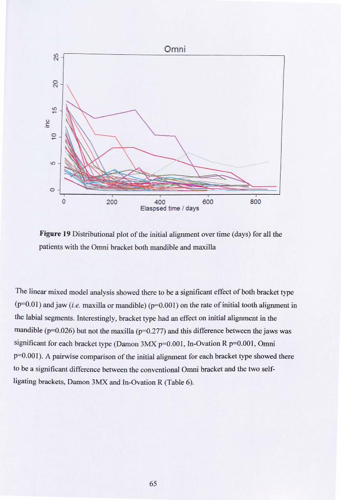
Omni
o 200 400 600 800 Elaspsed time I days
Figure 19 Distributional plot of the initial alignment over time (days) for all the
patients with the Omni bracket both mandible and maxilla
The linear mixed model analysis showed there to be a significant effect of both bracket type
(P==O.Ol) and jaw (i.e. maxilla or mandible) (p=O.OOl) on the rate of initial tooth alignment in
the labial segments. Interestingly, bracket type had an effect on initial alignment in the
mandible (p==O.026) but not the maxilla (p==O.277) and this difference between the jaws was
significant for each bracket type (Damon 3MX p=O.OOl , In-Ovation R p=O.OOI , Omni
P==O.OOl). A pairwise comparison of the initial alignment for each bracket type showed there
to be a significant difference between the conventional Omni bracket and the two self
ligating brackets, Damon 3MX and In-Ovation R (Table 6).
65

Comparison Coefficient Std Err Z p>[z] 95%CI
Damon 3MX vs. In-Ovation R 0.158 0.277 0.571 0.568 -0.384 to 0.701
Damon 3MX vs. Omni 1.023 0.344 2.97 0.003 0.348 to 1.699
In-Ovation R vs. Omni 0.866 0.350 2.47 0.013 0.179 to 1.552
Table 6 Pairwise comparison showing there is no statistically significant difference in the
rate of initial alignment between the two self-ligating brackets. There was a statistically
significant difference in the rate of initial alignment between these two brackets and the
conventional Omni bracket
In order to test the effect of bracket type further a multiple comparison test was used. Instead
of using a more traditional approach such as Family Wise Error Rate (FWER) e.g.
Bonferroni, which can be rather conservative, it was decided to use the more recent False
Discovery Rate (FOR). This approach controls the number of false positives in those tests
that result in a significant result (discovery) and means they have a greater ability (power) to
detect truly significant results. The approach uses q-values which are the adjusted p-values.
In these tests we have used Simes FDR (1986) method for multiple comparisons and it shows
there to be a significant difference between the Omni bracket and the two self-ligating
brackets (Figure 20) with the rate of initial alignment being significantly faster with the Omni
bracket and no statistically significant difference between the two self-ligating brackets.
66

---If) Q)
E Ci5 '-'
Brackets
• •
Q)
~ ro >
------------------------------------------------------------------------I
0-
~ co •
Pair
Figure 20 Plot of the q-values against bracket pairing. In this plot anything above the 0.05
line is a significant difference (pairings: Code 1 = Damon 3MX, Code 2 = In-Ovation R and
Code 3 = conventional Omni bracket)
5.6 The effect of bracket type on space closure
It was decided to determine the effect of factors and their interactions on the observed space
closure along with a direct comparison of bracket type ignoring aIJ other factors. The results
indicated there was no significant effect of FMP A so it was omitted from the model. Table 7
shows the summary of the interactions for space closure within each bracket. With the
Damon 3MX bracket there was a significant interaction withjaw in the female group but no
significant effect overall with sex. With the In-Ovation R there was no significant effect of
jaw but there was with sex. With the Omni bracket there was a significant effect of both sex
and jaw.
67

Effect Damon3MX In-Ovation R OMNI
jaw 0.015 0.873 0.001
sex 0.155 0.001 0.003
jaW#sex 0.670 0.001 0.096
sex at mandible 0.139 0.001 0.015
sex at maxilla 0.189 0.001 0.001
jaw at female 0.023 0.001 0.272
jaw at male 0.198 0.001 0.001
Table 7 Summary table of the effects and interactions tested using linear mixed
models within each bracket type
Table 8 shows the probabilities for the pairwise comparisons for the effect of bracket on
space closure. The q-plot using the linear mixed model is illustrated in Figure 21. The exact
probabilities in Table 8 and the q-plot demonstrate there is no statistically significant
difference in the rate of buccal segment space closure for each of the three brackets under
test.
Bracket pair Probability
Damon 3MX - In-Ovation R 0.797
Damon 3MX - OMNI 0.751
In-Ovation R - OMNI 0.917
Overall probability p = 0.941
Table 8 Exact probabilities of the effect of bracket type on space closure
68

C>
~
C'!
UJ <D ~ E
-(ij
:U- "'I: 0 £; <DII) E ->-..c co (I) ~
Iii > r--& -
OC!
~ •
0 - 1
•
0 - 0 Bracket pair
•
1-0
Figure 21 Plot of the q-values against bracket pairing. In this plot anything above the red
0.05 line is a significant difference (D = Damon 3MX, I = In-Ovation R and 0 =
conventional Omni bracket)
Looking at each bracket separately, space closure was significantly faster in males than in
females in the case of both the In-Ovation R (p=0.001) and the Omni brackets (p=0.003).
AJthough not statistically significant in the case of the Damon 3MX brackets, the plot of
space closure with time in each case still shows space closure to be quicker in males than in
females. lbis is illustrated in Figures 22, 23 and 24.
69

Damon 3MX
00
N
o 500 1000 1500 Time 1 days
• Man/M MaxlF _ MaxIM
Figure 22 Plot of space closure (mm) against time (days) for the Damon 3MX brackets
In-Ovation R N N
• -0 N
(I.) '-:::JOO VI .,.... 0
U
<0
II -,- e
"O:t"
0 500 1000 1500 Time 1 days
• ManlF • ManlM • MaxlF • MaxIM
Figure 23 Plot of space closure (mm) against time (days) for the In-Ovation R bracket
70

N N
• •
o
OMNI
500 1000 1500 Time I days
• Man/F • Man/M • MaxlF • MaxiM
Figure 24 Plot of space closure (mm) against time (days) for the Omni bracket
In the case of the Damon 3MX bracket the effect of sex and jaw appears to be more
pronounced at longer times and the maxilla appears to reach a steady state before the
mandible. With both the In-Ovation R bracket and the Omni bracket the effect of sex appears
more pronounced than with the Damon 3MX bracket.
5.6.1 The effect of sex on space closure
In order to further investigate the effect of sex on space closure the results of all three bracket
types were pooled. The summary data are illustrated in Table 9. A student t-test showed there
to be a significant difference between males and females (p=O.OOI), with space closure faster
in males. This is illustrated in Figure 25.
71
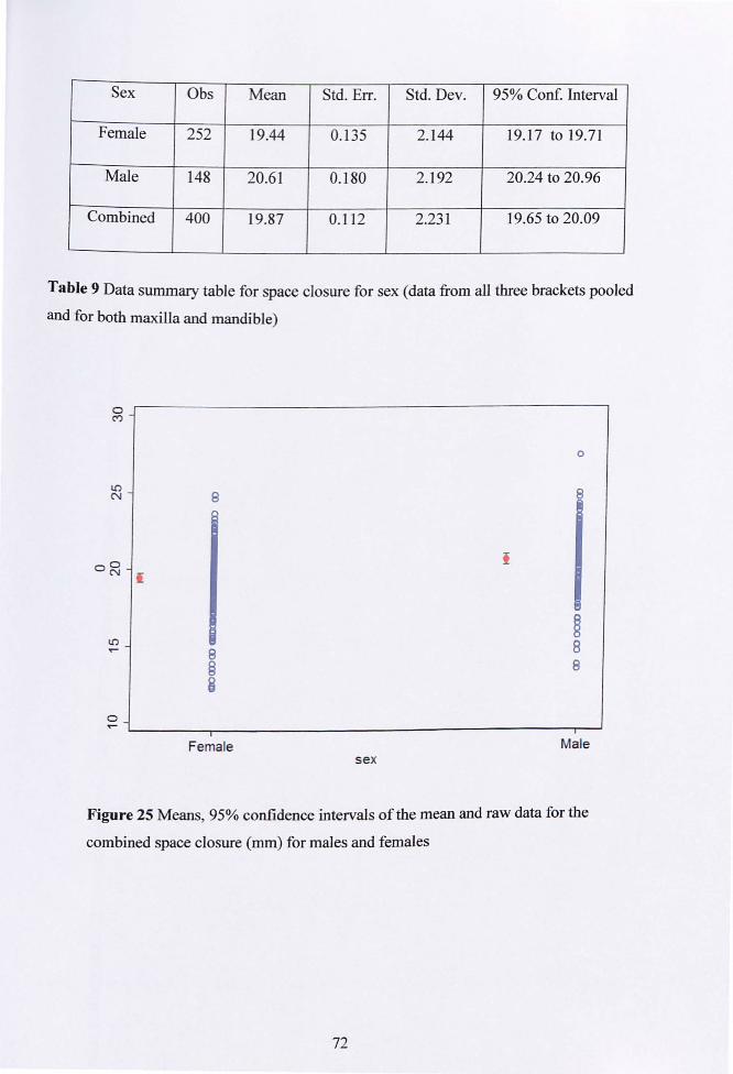
Sex Obs Mean Std. Err. Std. Dev. 95% Conf. Interval
Female 252 19.44 0.135 2.144 19.17 to 19.71
Male 148 20.61 0.180 2.192 20.24 to 20.96
Combined 400 19.87 0.112 2.231 19.65 to 20.09
Table 9 Data summary table for space closure for sex (data from all three brackets pooled
and for both maxilla and mandible)
l() N
00 N I
l()
0 .,...
8
8 § 8
Female sex
o
8
~ 8 8
Male
Figure 25 Means, 95% confidence intervals of the mean and raw data for the
combined space closure (mm) for males and females
72

A further illustration and investigation of the effect of sex was sort using restricted cubic
spline plots to fit the data (HarreU 2001). It was then differentiated numerically in order to
determine the rate of closure. Within each plot the steeper the curve the faster the rate of
space closure. The plots for each bracket type are shown in Figures 26, 27 and 28.
a
1.0 a a ..
1.0 N a ..
• o 500
Damon
.......... -""'---- - -~.,.--.. -.. - -
1000 Time I days
- Mandible, Female - Mandible, Male
- Maxilla, Female - Maxilla , Male
1500
Figure 26 Restricted cubic spline plot of rate of space closure for the Damon 3MX bracket
1.0 a C!
a
In-Ovation
.-.- _ .. ---: .-.- .. . -. -.
~ . " Lr--------------.---------------.---------------~
o 500 1000 1500 TIme I days
• Mandible, Female • Mandible, Male
• Maxilla, Female - Maxilla, Male
Figure 27 Restricted cubic spline plot of rate of space closure for the In-Ovation R bracket
73

a
Omni ..... '.... :..-. ., .. • •
~ ... : .-.-. P " "\ 1" ~o -, ' .\..~ ~ .. I ,~~~ ~ .. /. . " - ...
N a •
" ~--------------'---------------r-------------~ o 500 1000 1500 TIme / days
• Mandible, Female • Mandible, Male
• Maxilla, Female - Maxilla , Male
Figure 28 Restricted cubic spline plot of rate of space closure for the Omni bracket
LOOking at the cubic spline plots it can be seen that the rate of space closure during treatment
in the case of all three brackets is generally greater towards the earlier stages of treatment.
However, the plots show that the rate of space closure is far from linear at all times. Indeed at
some points there is a reversal of the rate of space closure which seems to be a consistent
finding with all three brackets. This could be the time between initial alignment, when
simultaneous space closure is taking place, and the placement of active space closing
mechanics. What is also interesting from the cubic spline plots is that the effect of sex is more
pronounced in the case of the In-Ovation R and the Omni brackets.
74

5.7 Treatment Times
The following treatment time parameters were studied and the data analysed in each case
using the Kruskal Wallis non-parametric one way analysis of variance:
• • • • •
Total chair side time in minutes
Mean treatment time per visit in minutes
The overall treatment time in days
The total number of appointments to debond
The total number of emergency appointments
5.7.1 Total chair side time in minutes
350 Total time 'chair side'
300
0
250 0 0
Time/min 0 0 0
~ 200 § §
f ~
I 8 150
~ ~ ~
0 0
100 8 § 0 0 0 0
Damon 3MX In-Ovation R
0
8
I § @ 0
@
0
0
0
0
OMNI
Figure 29 Means, 95% confidence intervals ofthe mean and raw data for total chair
side time in minutes for each of the three brackets under test
75

r
Bracket N Mean SD Min Max
Damon3MX 30 164.39 50.88 79.77 251.68
In-Ovation R 31 168.82 49.40 81.48 284.90
OMNI 16 220.37 71.67 104.63 332.10
Table 10 Summary data of Total Chair Side time for patients who had completed treatment
The Kruskal Wallis non-parametric one way analysis of variance demonstrated a statistically
significant difference between the groups (p=0.013). Further analysis using the Mann
Whitney test demonstrated no significant difference between the Damon 3MX and In
Ovation R brackets (p=0.39). There was a statistically significant difference between the
Damon 3MX and the Omni brackets (p=O.003) and the In-Ovation R and the Omni brackets
(P=0.005) with the total chair side treatment being greater in the case of the Omni bracket and
by around 50 minutes.
76

5.7.2 Mean treatment time per visit in minutes
Mean time 'chair side' It) 0 C"')
0 0
0 0
§ 0 0 C"') 0 0 0
0 8 0
c 8 §
I 0
·E It) 0
f 0
~N @
8 0
E 0 0
i=
f § 0 0
8 8 0 § N
~ 0
~ § 0
8 0 0 It) .,.... 0
0
Damon In-Ovation OMNI
Figure 30 Means, 95% confidence intervals of the mean and raw data for chair side
time in minutes per visit for each of the three brackets under test
Bracket N Mean SD Min Max
Damon3MX 30 21.42 4.99 13.61 34.57
In-Ovation R 31 23.89 5.99 15.] 9 35.68
OMNI 16 24.59 5.94 15.98 34.96
Table 11 Summary data of chair side time per visit for patients who had completed treatment
77

The Kruskal Wallis non-parametric one way analysis of variance showed there to be no
statistically significant difference in the chair side time per visit for each of the three brackets
under test (p=0 .154) even though the overall treatment time was longer in the case of the
Ornni bracket.
5.7.3 The overall treatment time in days
0 Total treatment time 0 N
0
0 0 0 .... 0 0
0
8 0
8 0 0
>.0 R @ coco
f ! "0 8 ......
! f Q)
I 8 .So 1-0 0 § co e €t 8 0
0 0 0 8 0 ~
0 0 0
0 0
0 0 N
Damon In-Ovation OMNI
Figure 31 Means, 95% confidence intervals of the mean and raw data for total time in
days for each of the three brackets under test
Bracket N Mean SD Min Max
Damon 3MX 30 698.27 179.65 256.00 1141.00
In-Ovation R 31 667.10 144.89 302.00 951.00
OMNI 16 627.63 154.41 372.00 883.00
Table 12 Summary data of total treatment time in days
78

The Kruskal Wallis non-parametric one way analysis of variance showed there to be no
statistically significant difference in the total overall treatment time in days for each of the
three brackets under test (p=O.318)
5.7.4 The total number of appointments to debond
Number of appointments frequency
5 6
1 0
2 1 1 c: 12 Q)
.5 13 c: ·0 14 a. a. 15 III -- 16 0 '- 17 Q)
.Q 18 E ::l 19 Z
20 21 22 25
Damon In-Ovation OMNI maximum: 6
Figure 32 Frequency distribution plots for the number of appointments for each of the
three brackets under test
The Kruskal Wallis non-parametric one way analysis of variance demonstrated a statistically
significant difference between the groups (p=O.007) with an adjusted p value of 0.008.
Further analysis using the Mann-Whitney test demonstrated no significant difference between
the Damon 3MX and In-Ovation R brackets (p=0.106). There was also no statistically
significant difference between the Damon 3MX and the Omni brackets (p=O.O 19), but there
was between the In-Ovation R and the Omni brackets (p=O.0009).
79

5.7.5 The total number of emergency appointments
Number of 'SOS' appointments frequency
1 ~ c Q) 2 E C '0 0.. 3 0.. m
'<-0 L.. 4 Q)
..c E ~ 5 z
6
9 Damon In-Ovation OMNI
maximum: 11
Figure 33 Frequency distribution plots for the number of emergency appointments for
each of the three brackets under test
When the total number of unplanned emergency appointments were considered the Kruskal
Wallis non-parametric one way analysis of variance demonstrated no statistically significant
difference between the three brackets under test (p=O.6).
80

6 Discussion
6.1 Extraction VS. Non-Extraction
As part of the inclusion criteria of this investigation, all of the subjects required a loss of a
premolar unit in each quadrant. This allowed all the subjects within each bracket group to be
closely matched to ensure consistency. This is in contrast to a number of previous studies
investigating similar variables. Harradine (2001) only had 40% of extraction cases in his
study, Miles (2005) only 14% and Miles et af. (2006) 20%. These different rates of extraction
may have influenced the rate of alignment and therefore their results should be interpreted
with this in mind. Other researchers have carried out alignment studies on arches that were
treated on a non-extraction basis (Pandis et af. 2007; Fleming et af. 2009; Pandis et af. 2010).
The use of a non-extraction treatment plan may also have influenced the rate of alignment.
Therefore their results cannot be used in direct comparison with this investigation.
6.2 Exclusions
There were three subjects within the current investigation who failed to complete treatment.
One subject withdrew from the study, another moved away and the final subject had their
treatment plan altered during treatment which meant that it did not match the investigation
protocol. All of the subjects' results were still included within the data analysis as it was felt
that their inclusion until their removal from the study would help with the power of the final
results. The inclusion of their results was possible by the use of the linear mixed models, as
using this analysis instead of conventional ANOV A allowed the inclusion of subjects with
missing data.
6.3 Practical Clinical Issues
A number of clinical difficulties were encountered during this investigation. Due to the large
numbers of participants in this study, it was not possible for all of the subjects to be treated
by the same individual. This may have lead to differences in treatment times for certain
clinicians. However, the clinicians involved in the investigation were not familiar with either
81

the passive or active SL brackets, and therefore they all experienced a similar learning curve
which ensured that treatment times were comparable.
It was discovered that subjects who had undergone the extraction of the second premolars,
sometimes had to have their initial aligning archwires (0.014" and 0.018") annealed and
cinched distal to the molar bands. This was carried out to reduce the risk of these archwires
dislodging from the molar bands. This risk could have been reduced by the placement of
lacebacks. However, as lacebacks were prohibited as part of the treatment protocol, this was
not an option. In certain situations, where the archwires repeatedly shifted, it was agreed that
these archwires could be cut distal to the first premolar brackets.
It was felt that the transition from 0.018" CuNiTi to a 0.016" x 0.022" Stainless Steel was
sometimes difficult to carry out. This was especially the case for the passive SL brackets
(Damon 3MX). This was due to the inadequate derotation of the teeth with the 0.018"
archwire, especially in the canine region. Therefore, it was necessary to sometimes place an
elastomeric module over the SL bracket and archwire. This would then allow the closure of
the passive slide at the next appointment. This problem may have been overcome by the use
ofan intermediate rectangular NiTi archwire, as recommended by the manufacturers of the
self-ligating brackets, which could have enabled the bracket slot to be better filled, allowing
for quicker alignment. However, this was not possible in this study as all subjects were
treated using an identical archwire sequence.
6.4 Arehwire Sequence
All of the manufacturers of the SL brackets advise on using a specific archwire sequence as
part of their system. The manufacturers of the active SL bracket recommend the following
archwire sequence:
0.014" CuNiTi
0.018" CuNiTi
0.020" x 0.020" CuNiTi
0.019" x 0.025" Stainless Steel
82

The square archwire is used to complete the alignment phase, as it is felt that the Cobalt
Chromium clip of the active SL bracket becomes interactive and uses the arch wire to fully
align the teeth.
The manufacturers of the passive SL bracket recommend a completely different archwire
sequence as part of their treatment system. They advise the following archwire sequence:
0.014" CuNiTi
0.014" x 0.025" CuNiTi
0.018" x 0.025" CuNiTi
0.019" x 0.025" Stainless Steel
The use of the rectangular CuNiTi archwire is advocated because it is felt that the archwire
can fill the slot and allow the bracket to derotate the teeth.
To make this investigation uniform and consistent, a standard archwire sequence was used.
This was based on the orthodontic department's standard archwire sequence for the
conventional brackets. It could be argued that the different SL brackets could have performed
better if their respective archwire sequences were used.
6.S Frankfort Mandibular Planes Angle
It has been reported in the literature that space closure is more difficult in subjects with a low
FMPA « 22°) due to higher muscular forces (Moller 1966). Conversely, it is also widely
thought that space closure or tooth movement is quicker in subjects with a high FMP A (>
32), although there is no evidence to support this. This study found that there was no
statistically significant difference in the distribution ofFMPA with either gender (p=0.44) or
bracket type (p=O.99). It also found that there was no significant effect of FMPA on space
closure.
83

6.6 Bracket Prescription
A number of studies investigating the rate of initial alignment and space closure have not
mentioned whether the different brackets under investigation had similar bracket
prescriptions (Miles et al. 2006; Burrow 2010; Dng et al. 201 O). The study by Scott et al.
(2008) highlighted the use of differing prescriptions as a potential criticism of their study.
Graber et al. (2005) have speculated that active SL brackets can express their torque from an
earlier treatment stage due to the active spring clip pushing the archwire into the base of the
slot. Badawi et al. (2008) have shown that the active clip did influence 3rd order tooth
position by reducing the 'slop' within the slot by 7 degrees. To avoid this problem and to
have true equivalence, this investigation used the same bracket prescription for all three
bracket types. Although, it has to be noted that the final archwire in our sequence was a
0.019" x 0.025" Stainless Steel and therefore the full torque values may not have been fully
expressed.
6.7 Pre-treatment crowding
Shivapuja and Berger (1994) have suggested that pre-treatment initial malalignment is an
important factor when detennining the rate of initial alignment. It is therefore logical to
assume that the greater the initial malalignment, the faster the rate of alignment, as the
archwires will be more efficient when ligated to grossly displaced teeth.
Pandis et al. (2007) found that there was a statistically significant difference in alignment for
patients with moderate crowding <5 mm. They showed that the SL bracket group completed
alignment 2.7 times faster than the conventional bracket group. A similar but less powerful
tendency was shown with severe crowding, as alignment was 1.37 times faster with the SL
bracket compared to the conventional bracket, but this was not significant. These results may
be due to the fact that they used an initial 0.016" NiTi and then 0.020" Sentalloy for the
conventional bracket group and an initial 0.014" NiTi and followed by a 0.014" x 0.025"
NiTi for the SL bracket group.
This investigation showed no statistically significant difference in the pre-treatment Little's
Index ofIrregularity for all of the three bracket groups (p=O.234). Therefore, pre-treatment
84

crowding was not an important factor in the initial alignment findings where alignment was
fastest with the Omni bracket.
It has also been shown by Leighton and Hunter (1982) that patients with a larger FMPA were
more likely to have increased crowding. However, this study showed no statistically
significant difference in the distribution ofFMPA and bracket group (p=0.99) and as there
was no difference detected between pre-treatment crowding and bracket group (p=0.234), this
suggests that there is no relationship between FMPA and crowding.
6.8 Measurement Period
Many studies investigating the rates of initial alignment and space closure using conventional
and SL brackets have only recorded values at certain dates i.e. week 10 and week 20 (Miles
2005; Miles el 01.2006) and have not continued measurement until the end of treatment (Ong
et 01. 2010; Mezomo et 01. 2011). These results need to be evaluated with caution as the
changes observed may not have been comparable for the entire period of treatment. Also
some changes between different bracket types may only become apparent after a longer
period of time (Fleming et 01. 2009). Pandis et 01. (2007) have shown that the active clip in
In-Ovation R brackets can relax during use and therefore could contribute to a lack of
consistent engagement of the archwire throughout treatment. Therefore this investigation
measured the changes in alignment and space closure of subjects in all three bracket types till
the end of their treatment.
6.9 Effe~t of bra~ket on alignment
This investigation demonstrated a significant difference in the rate of initial alignment of the
labial segments in relation to both the bracket type (p=O.Ol) and the jaw (p=O.OOl). It found
that the initial alignment was faster with all three bracket types in the mandible (p=O.026) and
that there was a significant difference between the conventional bracket and both the self
ligating brackets. These results are similar to the study by Miles et 01. (2006) who also
showed that there was a significant difference in alignment between conventional and passive
SL brackets in the mandible. However, their study was flawed as it was only carried out for
twenty weeks, it was a split mouth design which may have influenced the rate of alignment
85

and it was not clear whether the patients were extraction or non-extraction cases. The results
of this study are in contrast to a number of other studies that show there is no difference
between the rate of initial alignment between different bracket types (Scott et af. 2008;
Fleming et af. 2009; Ong et af. 2010).
The faster rate of alignment in the mandible for all three brackets could be explained by the
smaller inter-bracket distances between the lower labial teeth. This would cause the archwire
to be in contact with more of the bracket over shorter distances and allow less deflection i.e.
more active forces to align the teeth. The fact that there was no difference in the rate of
alignment between either of the bracket groups, particularly the passive and active SL
brackets in the maxilla confinns this. It is also in agreement with Pandis et af. (2010) who
showed no difference in the alleviation of crowding between an active or passive SL bracket
used in the maxilla
In addition, the faster alignment with the use of the conventional bracket, when compared to
both of the self-ligating brackets could be due to a number of other factors. The archwire,
even ifit is a 0.014" in the conventional bracket is ligated firmly into the base of the slot
every time, ensuring good rotational control. The same archwire within a passive SL bracket
is not positively pushed into the base of the slot and therefore has the ability to move freely
within the 0.022" x 0.028" tube. The same argument can be made for the active SL bracket,
as it has been shown that only archwires above a certain dimension, namely 0.018", will
cause the clip to become interactive, and enable the resulting archwire to be pushed into the
back of the slot, thus allowing further control in the first and second order positions of the
teeth. These results differ to those previously published by a number of other researchers
(pandis et af. 2007; Scott et af. 2008; Fleming et al. 2009; Ong et af. 2010).
It has also been postulated that the differing widths of the brackets could have an effect on
the rate of alignment. Narrower brackets generate higher moments which lead to higher
forces at the edge of the bracket when compared to wider brackets (Drescher et at. 1989).
These increased forces could increase the critical contact angle and lead to increased notching
and binding (Ong et al. 2010), which can affect the resistance to sliding and hence the rate of
alignment. This may be one of the explanations for the differing rates of initial alignment
between the brackets under investigation in this study.
The frequent appointments and shorter intervals for the conventional bracket may also have
allowed faster progression up the recommended archwire sequence, causing a faster rate of
86

alignment. It has been shown by Taloumis et a/. (1997) that elastomeric modules undergo a
rapid force loss within the first 24 hours and that they can also be affected by the oral
environment. This is one of the main reasons that the conventional bracket group subjects
were recalled every 6 weeks. This was in contrast to both of the SL bracket groups which
were seen every 12 weeks. The appointment interval was a recommendation by the SL
bracket manufacturers. This increase in appointment interval time whilst using SL brackets,
may have had an effect on the rate of alignment. Even if the teeth had aligned fully and the
specific archwire was passive after six weeks, the archwire would not be changed for another
six weeks thus delaying the progression to the next archwire in the sequence.
An interesting finding of this investigation was that the majority of subjects with the passive
SL bracket did not fulJy achieve a Little's Index oflrregularity of zero, even at the end of
treatment. This was despite the placement of the final 0.019" x 0.025" Stainless Steel
archwire. This can only be explained by the 'slop' of the SL brackets being larger than the
other brackets under investigation, due to a larger dimension of the bracket slot (Bhalla et a/.
2010) and therefore allowing minute movements of the labial teeth in the first order
(Harradine 2003).
6.10 Effect of bracket on space elosure
The different bracket types demonstrated a number of significant differences in relation to
extraction space closure. The conventional bracket showed a significant interaction of both
gender and jaw, whilst the passive SL bracket only showed a significant effect with jaw in
females. This was in contrast to the active SL bracket which showed a significant interaction
with gender but not withjaw.
Investigating this further, led to the discovery that extraction space closure was significantly
faster in males for both the conventional (p=O.003) and active SL bracket groups (p=O.OOl)
and whilst not being statistically significant for the passive SL group, space closure was still
faster in males in comparison to females.
This interesting discovery was again confirmed by the student t-test carried out on all of the
pooled data, which confirmed that space closure was faster in males than in females
(p=O.OOl).
87

It has been recommended by a number of researchers that treatments, especially involving
extractions, should be carried out during a period of maximal growth (Stephens & Houston
1985; Nanda & Nanda 1992). They have suggested that to obtain maximum space closure,
both males and females should be treated during their pubertal growth spurt. Tanner et al.
(1976) has shown that on average males have their pubertal growth spurt at 14 years +1- 2
years, whereas females have their pubertal growth spurt at 12 years +/- 2 years. Considering
the sample in this investigation, it showed the mean age of the male and female subjects to be
14.21 years and 13.96 years respectively. This could explain the faster rate of space closure in
males as they were treated during their pubertal growth spurt, whereas the females still
experienced space closure but at a slower rate as they had already undergone much their
pubertal growth spurt. To our knowledge this is the first time a positive relationship between
sex and rate of tooth movement has been demonstrated so conclusively in a clinical trial.
Previous workers have merely suggested this might be the case based on anecdotal evidence.
Another consistent finding with all three bracket types was a reversal in the rate of space
closure during treatment as illustrated in the cubic spline plots (Figures 26 to 28). An
explanation for this change could be the difference between passive extraction space closure
phase and active extraction space closure phase. Ong et al. (20 I 0) showed passive extraction
space closure can occur during the initial alignment phase of treatment. This could explain
the initial faster rate of space closure observed in this study. However, once the arches have
been aligned and levelled which is shown by a lag phase, then active extraction space closure
can commence. The application of an active force to allow sliding mechanics can then
facilitate further space closure, which was represented by the second increased rate of space
closure. The interim transition period between completing the initial alignment phase and
prior to commencing the active space closing phase may explain the change in the rate of
extraction space closure for all three bracket types.
6.11 Treatment times
The efficiency of treatment with the different bracket types was also investigated and once
again showed some significant differences.
There was a significant difference in total chair side time between the conventional bracket
and both of the SL brackets (p=O.013). The conventional bracket group took an average of 50
88

minutes longer than both the passive and active SL bracket groups. There was also a
significant difference in the total number of appointments to debond between the
conventional group and the active SL group (p=0.0009). Both of these results can be
explained by the shorter six week interval between appointments for the conventional bracket
group. The SL bracket group subjects were only recalled every 12 weeks. This meant that the
majority of the conventional group subjects had double the number of appointments when
compared to their fellow counterparts in both the SL bracket groups over the same time
period. Both of these results are contrary to the conclusions reached by Hamilton et al.
(2008), who found in their retrospective study that there was no measurable difference
between active SL brackets and conventional brackets when investigating the number of
visits.
The conventional Omni bracket is priced at £3.05, the Damon 3MX is £11.12 and the In
Ovation R is £9.75 (personal Communication). It has been suggested that the increased cost
of these SL brackets could be offset by the reduction in chair side time (Maijer & Smith
1990; Berger & Byloff 200 1), which would allow more patients to be seen and treated.
Turnbull and Birnie (2007) have also shown a small financial saving related to elastomeric
modules not being required. However, as Fleming et al. (2009) discussed, it remains unclear
whether the time and money saved in terms of number of appointments and reduced chair
side time justify the extra cost.
This study has shown that there was no significant difference in the mean chair side time i.e.
appointment time at each individual visit between either bracket group when the subjects
were seen for appointments (p=O.154). This is in contrast to Shivapuja and Berger (1994)
who found a dramatically shorter chair side time for the removal and insertion of the
archwires in SL brackets when compared to conventional brackets. This was again the case
with Turnbull and Birnie (2007), who found a significant difference between the time taken
for the placement and removal of archwires using a passive SL and a conventional bracket,
with the passive being quicker. This tendency was not observed in this study as the whole
treatment appointment was timed rather than just the untying and ligation of the archwires.
This investigation also found there was no difference in the total overall treatment time in
days (p=O.318). This is similar to results published by DiBiase et al. (20 11) who compared
the duration of treatment using a passive SL bracket with a conventional bracket group and
found no overall reduction in treatment time.
89

Finally, it was shown that there was no statistically significant difference between all three
bracket types when considering the total number of unplanned emergency appointments
(p=O.6). This is in contrast to studies carried out by Miles et al. (2006) and Hamilton et al.
(2008), which both showed significantly higher breakages associated with the SL brackets
and which they attributed to the bracket profile. Both, Damon 3MX and the In-Ovation R
brackets are larger inciso-gingivally and labio-lingually when compared to the conventional
Omni bracket and in addition they both have a smaller bonding base. These factors could
have increased the likelihood of these brackets interfering with the occlusion and leading to
bracket bonding failures (Chen et al. 2010). However, it is worthwhile noting that the study
by Hamilton et al. (2008) was retrospective and that the patients were not treated using a
similar archwire sequence. While the study by Miles et al. (2006) only reported first time
failures for each tooth and no other subsequent failures. The results from this current
investigation confirm the fmdings reported by a number of other researchers who also found
no difference between conventional and SL brackets (Pandis et al. 2006; Chen et al. 2010;
DiBiase et al. 2011).
Proponents of the SL brackets, especially the passive Damon 3MX may well disagree with
the majority of the results in this investigation. This is because, it could be argued that the full
benefits of these brackets were not expressed. The subjects in this study were not treated
using the Damon philosophy, i.e. a low friction system to achieve physiologic adaptation, by
allowing light forces to align crowded arches without the need of extractions. Or in other
words the Damon archwire sequence on a non-extraction basis. In answer to this, firstly all of
the subjects included in this study presented with sufficient crowding in both arches, to
necessitate extractions. In any case Pandis et al. (2007) have shown that patients with severe
crowding showed no difference in the rate of alignment with the Damon SL bracket.
Secondly, the use of anchorage reinforcement for some subjects could also have hindered the
SL brackets from demonstrating their advantages. However, it is important to reiterate that
this study was carried out to investigate the brackets and not the archwire system or
extraction! non-extraction philosophies. Therefore it was appropriate to carry out accepted
treatment plans that would be generally advised for all of the subjects presenting for
treatment at any other time.
Although all of the results regarding space closure and treatment times are very interesting
and show a number of significant differences, it has to be remembered that at the time of data
analysis the final twenty three subjects were still undergoing treatment, notably space closure
90

and had not completed the full trial. It is envisaged that their results will eventually be
incorporated into the existing study and further strengthen the power of the results.
91
7 Conclusions
The following conclusions can be drawn from this investigation:
1. There was a statistically significant difference in the rate of initial alignment with the
use of OMNI conventional brackets when compared to the Damon 3MX passive and
C'-" In-Ovation R active self-ligating brackets. The conventional brackets aligned the
labial segment teeth quicker.
2. There Was a statistically significant difference in the rate of initial alignment in the
mandible with the use of all three bracket types when compared to the maxilla. All
three bracket types aligned the mandibular labial segment quicker than the maxillary
labial segment.
3. There was a statistically significant difference in the rate of space closure in the
buccal segments in males with OMNI conventional brackets and In-Ovation R active
self-ligating brackets. Although not statistically significant, the rate of buccal space
closure was also faster in males with Damon 3MX passive brackets.
4. There was a statistically significant difference in the total chair side time between the
OMNI conventional brackets and both the Damon 3MX passive and In-Ovation R
active self-ligating brackets. The conventional bracket group took an average of fifty
minutes longer to complete treatment which is explained by the shorter appointment
intervals with the Omni brackets.
5. There was a statistically significant difference in the total number of appointments
between the OMNI conventional bracket and the In-Ovation R active self-ligating
bracket. The conventional bracket group took more appointments to complete
treatment which is again explained by the shorter appointment intervals with the
Omni brackets.
6. The rate of space closure was greater in males than females, which appears to be
related to the time of the pubertal growth spurt.
92

8 Future Work
As stated earlier, this clinical trial still has a number of patients undergoing final buccal
segment space closure. These remaining subjects within the study will be followed up until
the end of their treatment and the data reanalysed in order to confirm these interim findings.
This will further improve the power of the trial.
The follOwing also merit further investigation:
1. A repeat of the study using the manufacturers' recommended archwire sequences for
each bracket type to see if this has any additional effect on the rate of initial alignment
and space closure.
2. A repeat of the study with subjects treated on a non-extraction basis in order to
establish if the results observed are similar to the present investigation.
3. A repeat of the study with the inclusion of other popular self-ligating brackets in order
to discern any differences in the rate of initial alignment or extraction space closure
with different bracket designs.
93

9 References
Almasoud, N. & Beam, D. (2010). Little's irregularity index: Photographic assessment vs.
study model assessment. American Journal of Orthodontics & DentofacialOrthopedics 138:
787-794.
Anderson, G.M. (1931). One use for the Arnold coil spring. International Journal
Orthodontia, Oral Surgery & Radiography 17: 688.
Andreasen, G.F. & Bishara, S. (1970). Comparison of Alastik chains with elastics involved
with intra-arch molar to molar forces. The Angle Orthodontist 40: 151-158.
Andreasen, O.F. & Quevedo, F.R. (1970). Evaluation offliction forces in the 0.022 x 0.028
edgewise bracket in vitro. Journal of Biomechanics 3: 151-160.
Andrews, L.F. (1972). The six keys to normal occlusion. American Journal of Orthodontics
62: 296-309.
Angle, E.H. (1907) Treatment of malocclusion of the teeth. 7th Ed. SS White Dental
Manufacturing Company, Philadelphia.
Angle, E.H. (1916). Some new form of orthodontic mechanism and the reasons for their
introduction. Dental Cosmos 58: 969-994.
Angle, E.H. (1920). Orthodontia - ribbon arch mechanism and some new auxiliary
instruments. Dental Cosmos 62: 1157-1176, 1279-1294.
Angle, E.H. (1928). The latest and best in orthodontic mechanism. Dental Cosmos 70: 1143-
1158.
Angolkar, P.V., Kapila, S., Duncanson, M.G., & Nanda, R.S. (1990). Evaluation of friction
between ceramic brackets and orthodontic wires of four alloys. American Journal of
Orthodontics & Dentofacial Orthopedics 98: 499-506.
Angolkar, P.V., Arnold, J.V., Nanda, R.S., & Duncanson, M.O. (1992). Force degradation of
closed coil springs. An in vitro evaluation. American Journal of Orthodontics & Dentofacia/
Orthopedics 102: 127-133.
94
Articolo, L.C. & Kusy, R.P. (1999). Influence of angulation on the resistance to sliding in
fixed appliances. American Journal of Orthodontics & Dentofacial Orthopedics 115: 39-51.
Articolo, L.C., Kusy, K., Saunders, C.R., & Kusy, R.P. (2000). Influence of ceramic and
stainless steel brackets on the notching of archwires during clinical treatment. European
Journal of Orthodontics 22: 409-424.
Ash, J.L. & Nikolai, R.J. (1978). Relaxation of orthodontic elastomeric chains and modules
in vitro and in vivo. Journal of Dental Research 57: 685-690.
Badawi, H.M., Toogod, R.W., Carey, J.P.R., Heo, G., Major, P.W. (2008). Torque
expression of self-ligating brackets. American Journal of Orthodontics & Dento/acial
Orthopedics 133: 721-728.
Baker, K.L., Nieberg, L.G., Weimer, A.D., & Hanna, M. (1987). Frictional changes in force
values caused by saliva substitution. American Journal of Orthodontics & Dento/adal
Orthopedics 91: 316-320.
Ballrick, J.W., Palomo, J.M., Ruch, E., Ambennan, B.D., & Hans, M.G. (2008). Image
distortion and spatial resolution of a commercially available cone-beam computed
tomography machine. American Journal of Orthodontics & Dentofacial Orthopedics 134:
573-582.
Barrow, G. & White, 1. (1952). Developmental changes of the maxillary and mandibular
dental arches. The Angle Orthodontist 12: 41-46.
Bednar, J.R., Gruendeman, G.W., & Sandrik, J.L. (1991). A comparative study of frictional
forces between orthodontic brackets and arch wires. American Journal of Orthodontics &
DentofacialOrthopedics 100: 513-522.
Bennett, J.C. & McLaughlin, R.P. (1992). Overjet reduction with a preadjusted appliance
system. Journal of Clinical Orthodontics 26: 293-309.
Bennett, lC. & McLaughlin, R.P. (1994). Orthodontic treatment mechanics and the
preadjusted appliance. Mosby-Wolfe.
95

Berger, J.L. (1990). The influence of the SPEED bracket's self-ligating design on force
levels in tooth movement: a comparative in vitro study. American Journal o/Orthodontics &
Dento/acialOrthopedics 97: 219-228.
Berger, J.L. (1994). The SPEED appliance: a 14-year update on this unique self-ligating
orthodontic mechanism. American Journal 0/ Orthodontics & Dento/acial Orthopedics lOS:
217-223.
Berger,1. & Byloff, F.K. (2001). The clinical efficiency of self-ligated brackets. Journal 0/ Clinical Orthodontics 35: 304-308.
Besancon, R.M. (1985). The encyclopedia o/physics. 3rd Ed. Van Nostrand Reinhold
Company, New York.
Bhalla, N.B., Good, S.A., McDonald, F., Sherriff, M., & Cash, A.C. (2010). Assessment of
slot sizes in self-ligating brackets using electron microscopy. Australian Orthodontic Journal
26: 38-41.
Bjork, A., Krebs, A., & Solow, B. (1964). A method for epidemiological registration of
malocclusion. Acta Odontologica Scandinavica 22: 27-41.
Bokas, J. & Woods, M. (2006). A clinical comparison between nickel titanium springs and
elastomeric chains. Australian Orthodontic Journal 22: 39-46.
Braun, S., Bluestein, M., Keith Moore, B., & Benson, G. (1999). Friction in perspective.
American Journal o/Orthodontics & Dento/acial Orthopedics US: 619-627.
Brown, A.A., Scarfe, W.C., Scheetz, J.P., Silveira, A.M., & Farman, A.G. (2009). Linear
accuracy of cone beam CT 3D images. The Angle Orthodontist 79: 150-157.
Brown, M. (1984). Controlled space closure with the edgewise appliance. British Journal 0/
Orthodontics U: 92-99.
Budd, S., Daskalogiannakis, J., & Tompson, B.D. (2008). A study of the frictional
characteristics of four commercially available self· ligating bracket systems. European
Journal o/Orthodontics 30: 645-653.
96

Burrow, SJ. (2010). Canine retraction rate with self-ligating brackets vs. conventional
edgewise brackets. The Angle Orthodontist 80: 626-633.
Burstone, CJ. & Koenig, H.A. (1976). Optimising anterior and canine retraction. American
Journal of Orthodontics 70 (1): 1-19.
Burstone, C.J. (1982). The segmental arch approach to space closure. American Journal of
Orthodontics 82 (5): 361-378.
Butcher, G.W. & Stephens, C.D. (1981). The reflex optical plotter. A preliminary report.
British Dental Journal lSI: 304-305.
Cha, B.K., Lee, J.Y., Jost-Brinkmann, P.G., & Yoshida, N. (2007). Analysis of tooth
movement in extraction cases using three dimensional reverse engineering technology.
European Journal of Orthodontics 29: 325-331.
Champagne, M. (1992). Reliability of measurements from photocopies of study models.
Journal o/Clinical Orthodontics 26: 648-650.
Chen, S.S.H., Greenlee, G.M., Kim, J.E., Smith, C.L., & Huang, GJ. (2010). Systematic
review of self-ligating brackets. American Journal 0/ Orthodontics & Dento/adal
Orthopedics 137: 726.el-726.eI8.
Cho, M.Y., Choi, J.H., Lee, S.P., & Baek, S.H. (2010). Three dimensional analysis of the
tooth movement and arch dimension changes in Class I malocclusions treated with first
premolar extractions: A guideline for virtual treatment planning. American Journal of
Orthodontics & Dentofacial Orthopedics 138: 747-757.
Clover, MJ. (2008). A randomised controlled clinical trial to investigate the rates of
alignment 0/ the upper and lower labial segments using conventional, active and passive self
ligating orthodontic brackets. DDS Thesis. University of Bristol, Bristol, England.
Damon, D.H. (1998a). The rationale, evolution and clinical application of the self ligating
bracket. Clinical Orthodontics and Research 1: 52-61.
97

Damon, D.H. (1998b). The Damon low friction bracket: a biologically compatible straight
wire system. Journal of Clinical Orthodontics 32: 670-680.
Damstra, J., Fourie, Z., Huddleston Slater, J.J.R., & Ren, Y. (2010). Accuracy of linear
measurements from cone-beam computed tomography -derived surface models of different
voxel sizes. American Journal of Orthodontics & DentofacialOrthopedics 137: 16.el-16.e6.
DiBiase, A.T., Nasr, I.H., Scott, P., & Coboume, M.T. (2011). Duration of treatment and
occlusal outcome using Damon 3 self-ligated and conventional orthodontic bracket systems
in extraction patients: A prospective randomised clinical trial. American Journal of
Orthodontics & Dentofacial Orthopedics 139: e III-e 116.
Dincer, M. & Iscan, H.N. (1994). The effects of different sectional arches in canine
retraction. European Journal of Orthodontics 16: 317-323.
Dincer, M., Gulsen, A., & Turk, T. (2000). The retraction of upper incisors with the PG
retraction system. European Journal of Orthodontics 22: 33-41.
Dixon, Y., Read, M.J., O'Brien, K.D., Worthington, H.Y., & Mandell, M.A. (2002). A
randomised clinical trial to compare three methods of orthodontic space closure. Journal of
Orthodontics 29: 31-36.
Downing, A., McCabe, J., & Gordon, P. (1994). A study of frictional forces between
orthodontic brackets and archwires. British Journal of Orthodontics 21: 349-357.
Downing, A., McCabe, J.F., & Gordon, P.H. (1995). The effect of artificial saliva on the
frictional forces between orthodontic brackets and archwires. British Journal of Orthodontics
22: 41-46.
Draker, H. (1960). Handicapping labio-lingual deviations: A proposed index for Public
Health purposes. American Journal of Orthodontics 46: 295-305.
Drescher, D., Bourauel, C., & Schumacher, H.A. (1989). Frictional forces between bracket
and arch wire. American Journal of Orthodontics & Dentofacial Orthopedics 96: 397-404.
98

Eberting, J.J., Straja, S.R., & Tuncay, O.C. (2001). Treatment time, outcome and patient
satisfaction comparisons of Damon and conventional brackets. Clinical Orthodontics and
Research 4: 228-234.
Edwards, G.D., Davies, E.H., & Jones, S.P. (1995). The ex vivo effect of ligation technique
on the static frictional resistance of stainless steel brackets and arch wires. British Journal of
Orthodontics 22: 145-153.
Eliades, T. & Pandis, N. (eds.) (2009). Self-ligation in Orthodontics. Wiley-Blackwell.
EI-Zanaty, H.M., EI-Beialy, A.R., EI-Ezz, A.M.A., Attia, K.H., EI-Bialy, A.R., & Mostafa,
Y.A. (2010). Three dimensional dental measurements: An alternative to plaster models.
American Journal of Orthodontics & Dento/acial Orthopedics 137: 259-265.
Fleming, P.S., DiBiase, A.T., Sarri, G., & Lee, R.T. (2009). Efficiency of mandibular arch
alignment with 2 preadjusted edgewise appliances. American Journal 0/ Orthodontics &
Denlo/acialOrthopedics 135: 597-602.
Fleming, P.S. & Johal, A. (2010). Self-ligating brackets in orthodontics. A systematic review.
The Angle Orthodontist 80: 575-584.
Forsberg, C.M., Brattstrom, V., & Malmberg Nord, C.E. (1991). Ligature wires and
elastomeric rings: two methods of ligature, and their association with microbial colonisation
of Streptococcus mutans and Lactobacilli. European Journal o/Orthodontics 13: 416-420.
Franchi, L. & Baccetti, T. (2006). Forces released during alignment with a preadjusted
appliance with different types of elastomeric ligatures. American Journal 0/ Orthodontics &
DentofacialOrthopedics 129: 687-690 ..
Frank, C.A. (1979). An evaluation ofthefrictionalforce generated between orthodontic
bracket and arch wire. Master's thesis. St. Louis University, Missouri, USA.
Frank, C.A. & Nikolai, R.J. (1980). A comparative study of frictional resistances between
orthodontic bracket and archwire. American Journal o/Orthodontics & Dento/acial
Orthopedics 78: 593-609.
99

Gamer, lL., Allai, W.W., & Moore, B.K. (1986). A comparison offrictional forces during
simulated canine retraction of a continuous edgewise archwire. American Journal 0/ Orthodontics & Dento/acialOrthopedics 90: 199-203.
Ghafari, J., Baumrind, S., & Efstratiadis, S.S. (1998). Misinterpreting growth and treatment
outcome from serial cephalographs. Clinical Orthodontic and Research 1: 102-106.
Graber, T.M., Vanarsdall, R.L. Jr., & Vig, K.W.L. (eds.) (2005). Orthodontics Current
Principles and Techniques. 4th Edition. Elsevier-Mosby.
Griffiths, H.S., Sherriff, M., & Ireland, AJ. (2005). Resistance to sliding with 3 types of
elastomeric modules. American Journal 0/ Orthodontics & Dento/acial Orthopedics 127:
670-675.
Hain, M., Dhopatker, A., & Rock, P. (2003). The effect of ligation method on friction in
sliding mechanics. American Journal 0/ Orthodontics & Dento/acial Orthopedics 123: 416-
422.
Hamilton, R., Goonewardene, M.S., & Murray, K. (2008). Comparison of active self-ligating
brackets and conventional pre-adjusted brackets. Australian Orthodontic Journal 24: 102-
109.
Hanson, G.H. (1980). The SPEED system: a report on the development of a new edgewise
appliance. American Journal 0/ Orthodontics 78: 243-265.
Harradine, N.W.T. & Birnie, OJ. (1996). The clinical use of Activa self-ligating brackets.
American Journal 0/ Orthodontics & Dento/acial Orthopedics 109: 319-328.
Harradine, N. W. T. (200 I). Self-ligating brackets and treatment efficiency. Clinical
Orthodontics and Research 4: 220-227.
Harradine, N.W.T. (2003). Self-ligating brackets: where are we now? Journal 0/ Orthodontics 30: 262-273.
Harradine, N. W. T. (2008). The history and development of self-ligating brackets. Seminars
in Orthodontics 14: 5-18.
100

Harradine, N.W.T. & Birnie, D.l (2010). Self-ligating brackets: theory and practice. In:
Harradine, N.W.T. & Birnie, D.J. (cds.). Excellence in Orthodontics p194.
Harrell, F.E. (2001). Regression modelling strategies with applications to linear models,
lOgistic regression and survival analysis. Springer-Verlag.
Heiser, W., Richter, M., Niederwanger, A., Neunteufel, N., & Kulmer, S. (2008). Association
of the canine guidance angle with maxillary and mandibular intercanine widths and anterior
alignment relapse: extraction vs. nonextraction treatment. American Journal 0/ Orthodontics
& Dento/acial Orthopedics 133: 669-680.
Henao, S.P. & Kusy, R.P. (2004). Frictional evaluations of dental typodont models using four
self-ligating designs and a conventional design. The Angle Orthodontist 75: 75-85.
Huffman, J.D. & Way, D.C. (1983). A clinical evaluation of tooth movement along arch
wires of two different sizes. American Journal o/Orthodontics 83: 453-459.
Huget, E.F., Patrick, K.S., & Nunez, L.J. (1990). Observations on the elastic behaviour of a
synthetic orthodontic elastomer. Journal o/Dental Research 69: 496-501.
Ireland, AJ., Sherriff, M., & McDonald, F. (1991). Effect of bracket and wire composition
on frictional forces. European Journal o/Orthodontics 13: 322-328.
Ireland, AJ., McNamara, C., Clover, MJ., House, K., Wenger, N., Barbour, M.E.,
Alemzadeh, K., & Sandy, J.R. (2008). 3D surface imaging in dentistry-what we are looking
at. British Dental Journal 204(1 1): 639-644.
Isaacson, K.G., Thorn, A.R., Homer, K., & Whaites E. (2008). Orthodontic Radiographs
Guidelines. 3n1 Edition. British Orthodontic Society. London.
Iwasaki, L.R., Beatty, M.W., Jared Randall, C., & Nickel, J.C. (2003). Clinical ligation
forces and intraoral friction during sliding on a stainless steel archwire. American Journal 0/ Orthodontics & Dento/acialOrthopedics 123: 408-415.
101

Kapila, S., Angolkar, P.V., Duncanson, M.G. Jr., & Nanda, R.S. (1990). Evaluation of
friction between edgewise stainless steel brackets and orthodontic wires of four alloys.
American Journal of Orthodontics & Dentofacial Orthopedics 98: 117-126.
Karamouzos, A., Athanasiou, A.E., & Papadopoulos, M.A. (1997). Clinical characteristics
and properties of ceramic brackets: a comprehensive review. American Journal of
Orthodontics & Dentofacial Orthopedics 112: 34-40.
Keith, 0., Jones, S.P., & Davies, E.H. (1993). The influence of bracket material, ligation
force and wear on frictional resistance of orthodontic brackets. British Journal of
Orthodontics 20: 109-115.
Kesling, P.C. (1988). Expanding the horizons of the edgewise slot. American Journal of
Orthodontics 94: 26-37.
Khambay, B., Millett, D., & McHugh, S. (2004). Evaluation of methods of arch wire ligation
on frictional resistance. European Journal of Orthodontics 26: 327-332.
Khambay, B., Millett, D., & McHugh, S. (2005). Archwire seating forces produced by
different ligation methods and their effect on frictional resistance. European Journal of
Orthodontics 27: 302-308.
Killiany, D.M. & Duplesis, I. (1985). Relaxation ofelastomeric chains. Journal of Clinical
Orthodontics 19: 592-593.
Kim, T.K., Kim, K.D., & Baek, S.H. (2008). Comparison of frictional forces during the
initial levelling stage in various combinations of self-ligating brackets and archwires with a
custom-designed typodont system. American Journal of Orthodontics & Dentofacial
Orthopedics 133: 187.e15-187.e24.
Kula, K., Phillips, C., Gibilaro, A., & Proffit, W.R. (1998). Effect of ion implantation of
TMA archwires on the rate of orthodontic sliding space closure. American Journal of
Orthodontics & Dentofacial Orthopedics 114: 577-581.
102

Kusy, R.P. & Whitley, J.Q. (1990). Coefficients of friction for arch wires in stainless steel
and polycrystalline alumina brackets slots: the dry state. American Journal 0/ Orthodontics
& Dentofacial Orthopedics 98: 300-312.
Kusy, R.P., Whitley, J.Q., & Prewitt, MJ. (1991). Comparison of the frictional coefficients
for selected archwire-bracket slot combinations in the dry and wet states. The Angle
Orthodontist 61: 293-302.
Kusy, R.P. (1991). Materials and appliances in orthodontics: brackets, archwires and friction.
Current Opinion in Dentistry 1: 634-644.
Kusy, R.P., Tobin, EJ., Whitley, J.Q., & Sioshansi, P. (1992). Frictional coefficients of ion
implanted alumina against ion implanted beta-titanium in the low load, low velocity, single
pass regime. Dental Materials 8: 167-172.
Kusy, R.P. & Whitley, J.Q. (1997). Friction between different wire-bracket configurations
and materials. Seminars in Orthodontics 3: 166-177.
Kusy, R.P. & Whitley, J.Q. (1999). Assessment of second-order clearances between
orthodontic archwires and bracket slots via the critical contact angle for binding. The Angle
Orthodontist 69: 71-80.
Kusy, R.P. & Whitley, J.Q. (1999). Influence of arch wire and bracket dimensions on sliding
mechanics: derivations and determinations of the critical contact angles for binding.
European Journal of Orthodontics 21: 199-208.
Kusy, R.P. & Whitley, J.Q. (2000). Resistance to sliding of orthodontic appliances in the dry
and wet states: influence of arch wire alloy, interbracket distance and bracket engagement.
Journal o/Biomedical Materials Research 52: 797-811.
Lascala, C.A., Panella, J., & Marques, M.M. (2004). Analysis of the accuracy of linear
measurements obtained by cone beam computed tomography (CBCT-NewTom).
Dentomaxillofacial Radiology 33: 291-294.
Leighton, B.C. & Hunter, W.S. (1982). Relationship between lower arch spacing! crowding
and facial height and depth. American Journal 0/ Orthodontics 82 (5): 418-425
103

Levander, E., Malmgren, 0., & Eliasson, S. (1994). Evaluation of root resorption in relation
to two orthodontic treatment regimes. A clinical experimental study. European Journal of
Orthodontics 16: 223-228.
Liew, C.F. (1993). The effect of repeated displacement on slidingfriction between
orthodontic bracket and archwire. Unpublished Thesis. University of Queensland, Australia.
Lin, L. (1989). A concordance correlation coefficient to evaluate reproducibility. Biometrics
45: 255-268.
Lin, L. (2000). A note on the concordance correlation coefficient. Biometrics 56: 324-325.
Little, R.M. (1975). The Irregularity Index: A quantitative score of mandibular anterior
alignment. American Journal of Orthodontics 68 (5): 554-563.
Loftus, B.P., Artun, J., Nicholls, 1.1., Alonzo, T.A., & Stoner, J.A. (1999). Evaluation of
friction during sliding tooth movement in various bracket-arch wire combinations. American
Journal of Orthodontics & Dentofadal Orthopedics 116: 336-345.
Lotzof, L.P., Fine, H.A., & Cisneros, GJ. (1996). Canine retraction: a comparison of two
preadjusted bracket systems. American Journal of Orthodontics & Dentofacial Orthopedics
110: 191-196.
Maijer, R. & Smith, D.C. (1990). Time savings with self-ligating brackets. Journal of
Clinical Orthodontics 24: 29-31.
Matarese, G., Nucera, R., Militi, A., Mazza, M., Portelli, M., Festa, F., & Cordasco, G.
(2008). Evaluation of frictional forces during dental alignment: An experimental model with
3 non leveled brackets. American Journal of Orthodontics & Dentofacial Orthopedics 133:
708-715.
Matasa, C.G. (1996). Self engaging brackets - passive versus active. The Orthodontic
Materials Insider 9: 5-11.
McGuinness, N.J. (1992). Prevention in Orthodontics-A Review. Dental Update 19: 168-
175.
104

McLaughlin, R.P. & Bennett, J.C. The transition from standard edgewise to preadjusted
appliance systems. Journal of Clinical Orthodontics 23: 142-153.
Mezomo, M., de Lima, E.S., de Menezes, L.M., Weissheimer, A., & Allgayer, S. (2011).
Maxillary canine retraction with self-ligating and conventional brackets. The Angle
Orthodontist 81: 292-297.
Miles, P.G. (2005). Smartclip versus conventional twin brackets for initial alignment: is there
a difference. Australian Orthodontic Journal 21: 123-127.
Miles, P.G., Weyant, R.J., & Rustveld, L. (2006). A clinical trial of Damon 2 vs.
Conventional twin brackets during initial alignment. The Angle Orthodontist 76: 480-485.
Miles, P.G. (2007). Self-ligating vs. conventional twin brackets during en-masse space
closure with sliding mechanics. American Journal of Orthodontics & Dentofacial
Orthopedics 132: 223-225.
Miles, P.G. (2009). Self-ligating brackets in orthodontics: do they deliver what they claim?
Australian Dental Journal 54: 9-11.
Moller, E. (1966). The chewing apparatus. Acta Physiologica 61: 571-574.
Moore, M.M., Harrington, E., & Rock, W.P. (2004). Factors affecting friction in the pre
adjusted appliance. European Journal of Orthodontics 26: 579-583.
Moorrees, C. & Reed, B. (1954). Biometrics of crowding and spacing of the teeth of the
mandible. American Journal of Physical Anthropology 12: 77-88.
Motohashi, N. & Kuroda, T. A 3D computer aided design system applied to diagnosis and
treatment planning in orthodontics and orthognathic surgery. European Journal of
Orthodontics 21: 263-274.
Muira, F., Mogi, M., Ohura, Y., & Hamanaka, H. (1986). The super elastic property of the
Japanese NiTi alloy wire for the use in orthodontics. American Journal of Orthodontics &
Dentofacial Orthopedics 90: 1-10.
105

-'
I "-
Muira, F., Mogi, M., & Ohura, Y. (I 988a). Japanese NiTi alloy wire: use of the direct
electric resistance heat treatment method. European Journal of Orthodontics 10: 187-191.
Muira, F., Mogi, M., Ohura, Y., & Karlbe, M. (l988b). The super elastic Japanese NiTi alloy
wire for the use in orthodontics. Part III. Studies on the Japanese NiTi alloy coil springs.
American Journal 0/ Orthodontics & Dento/acial Orthopedics 94: 89-96.
Nattrass, C., Ireland, A.J., & Sherriff, M. (1998). The effect of environmental factors on
elastomeric chain and nickel titanium coil springs. European Journal 0/ Orthodontics 20:
169-176.
Nightingale, C. & Jones, S.P. (2003). A clinical investigation offorce delivery systems for
orthodontic space closure. Journal of Orthodontics 30: 229-236.
Noda, K., Nakamura, Y., Oikawa, T., Shimpo, S., Kogure, K., & Hirashita, A. (2007). A new
idea and method of tooth movement using a ratchet bracket. European Journal of
Orthodontics 29: 225-231.
O'Brien, K., Lewis, D., Shaw, W., & Combe, E. (1990) A clinical trial of aligning archwires.
European Journal o/Orthodontics 17: 1-19.
Omana, H., Moore, R., Bagby, M., & Erikson, L. (l992a). Frictional properties of ceramic
brackets during simulated cuspid retraction (abstract). Journal of Dental Research 71: A500.
Omana, H.M., Moore, R.N., & Bagby, M.D. (1992b). Frictional properties of metal and
ceramic brackets. Journal o/Clinical Orthodontics 26: 425-432.
Ong, E., McCallum, H., Griffin, M.P., & Ho, C. (2010). Efficiency of self-ligating vs.
conventionally ligated brackets during initial alignment. American Journal 0/ Orthodontics
& Dento/acialOrthopedics 138: 138.el-138.e7.
Onnco n.d. http://www.ormco.comlindexldamon-products-damong-featuresbenefits-2
(October 2010)
Paladin, P. (ed) (2005). Orthodontic History; braced/ora century. St. Louis, MO: American
Association of Orthodontics.
106
Owrnan-Moll, P., Kurol, J., & Lundgren, D. (1995). Continuous versus interrupted
continuous orthodontic force related to early tooth movement and root resorption. The Angle
Orthodontist 65(6): 395-402.
Pandis, N., Polychronopoulou, A., & Eliades, T. (2006). Failure rate of self-ligating and
edgewise brackets bonded with conventional acid etching and a self-etching primer: a
prospective in-vivo study. The Angle Orthodontist 76: 119-122.
Pandis, N., Bourauel, C., & Eliades, T. (2007). Changes in the stifthess ofthe ligating
mechanism in retrieved active self-ligating brackets. American Journal of Orthodontics &
DentofacialOrthopedics 132: 834-837.
Pandis, N., Polychronopoulou, A., & Eliades, T. (2007). Self-ligating vs. conventional
brackets in the treatment of mandibular crowding: A prospective clinical trial of treatment
duration and dental effects. American Journal of Orthodontics & Dentofacial Orthopedics
131: 208-215.
Pandis, N., Polychronopoulou, A., & Eliades, T. (2009). Alleviation of mandibular anterior
crowding with copper-nickel-titanium vs. nickel-titanium wires: A double-blind randomised
control trial. American Journal of Orthodontics & Dentofacial Orthopedics 136: 152.e 1-
1 52.e7.
Pandis, N., Polychronopoulou, A., & Eliades, T. (2010). Active or passive self-ligating
brackets? A randomised controlled trial of comparative efficiency in resolving maxillary
anterior crowding in adolescents. American Journal of Orthodontics & Dentofacial
Orthopedics 137: 12.el-12.e6.
Peri ago, D.R., Scarle, W.C., Moshiri, M., Scheetz, J.P., Silveira, A.M., & Farman, A.G.
(2008). Linear accuracy and reliability of cone beam CT derived 3.-dimensional images using
an orthodontic volumetric rendering program. The Angle Orthodontist 78: 387-395.
Peterson, L., Spencer, R., & Andreasen, G.F. (1982). A comparison of friction resistance for
Nitinol and stainless steel wire in edgewise brackets. Quintessence InternationalS: 563-565.
107
Pizzoni, L., Ravnholt, G., & Melsen, B. (1998). Frictional forces related to self-ligating
brackets. European Journal 0/ Orthodontics 20: 283-291.
Pratten, D.H., Popli, K., Germane, N., & Gunsolley, lC. (1990). Frictional resistance of
ceramic and stainless steel orthodontic brackets. American Journal 0/ Orthodontics &
Dentofacial Orthopedics 98: 398-403.
Proffit, W.R. & Ackerman, I. (1973). Rating the characteristics of malocclusion: A
systematic approach for planning treatment. American Journal of Orthodontics 64: 258-269.
Proffit, W.R., Fields, Jr., H.W., & Sarver D. (eds.) (2007). Contemporary Orthodontics. 4th
Edition. Mosby-Elsevier.
Prososki, R.R., Bagby, M.D., & Erikson, L.C. (1991). Static frictional force and surface
roughness of nickel-titanium arch-wires. American Journal of Orthodontics & Dento/acial
Orthopedics 100: 341-348.
QUinn, R.S. & Yoshikawa, D.K. (1985). A reassessment offorce magnitude in orthodontics.
American Journal of Orthodontics 83: 252-260.
Read-Ward, G.E., Jones, S.P., & Davies, E.H. (1997). A comparison of self-ligating and
conventional orthodontic bracket systems. British Journal o/Orthodontics 24: 309-317.
Redlich, M., Mayer, Y., Harari, D., & Lewinstein, I. (2003). In vitro study of frictional forces
during sliding mechanics of "reduced-friction" brackets. American Journal o/Orthodontics
& DentofacialOrthopedics 124: 69-73.
Ren, Y., Iaap, C.M., & Kuijpers-Iagtman, A.M. (2003). Optimum force magnitude for
orthodontic tooth movement: A systematic literature review. The Angle Orthodontist 73: 86-
92.
Richmond, S., Shaw, W.C., O'Brien, K.D., Buchanan, I.B., Jones, R., Stephens, C.D.,
Roberts, C.T., & Andrews, M. (1992). The development of the PAR index (peer Assessment
Rating): reliability and validity. European Journal o/Orthodontics 14: 125-139.
108

Rinchuse, D.J. & Miles, P.G. (2007). Self-ligating brackets: Present and future. American
Journal o/Orthodontics & Dento/acialOrthopedics 132: 216-222.
Riley, J.L., Garrett, S.G., & Moon, P.C. (1979). Frictional forces 0/ ligated plastic and metal
edgewise braclcets. Master's Thesis. Richmond, Virginia. USA.
Ryden, H., Bjelkhagen, H., & Martensson, B. (1982). Tooth position measurements on dental
casts using holographic images. American Journal 0/ Orthodontics 81: 310-313.
Rygh, P. (1986). Periodontal response to tooth moving force: is trauma necessary in
orthodontics? In: Graber, L. W. (ed) State o/the art, essence o/the science. CV Mosby, St.
Louis, pp. 100-115.
Samuels, R.H.A., Rudge, S.J., & Mair, L.H. (1993). A comparison of the rate of space
closure using a nickel titanium spring and an elastic module: A clinical study. American
Journal o/Orthodontics & Dento/acialOrthopedics 103: 464-467.
Samuels, R.H.A., Rudge, S.J., & Mair, L.H. (1998). A clinical study of space closure with
nickel-titanium closed coil springs and an elastic module. American Journal 0/ Orthodontics
& Dento/acialOrthopedics 114: 73-79.
Schirmer, U.R. & Wiltshire, W.A. (1997). Manual and computer-aided space analysis: A
comparative study. American Journal o/Orthodontics & Dento/acialOrthopedics 112: 676-
680.
Scott, P., DiBiase, A.T., Sherriff, M., & Cobourne, M.T. (2008). Alignment efficiency of
Damon 3 self-ligating and conventional orthodontic bracket systems: A randomised clinical
trial. American Journal o/Orthodontics & Dento/acial OrthopediCS 134: 470.el-470.e8.
Shivapuja, P.K. & Berger, J. (1994). A comparative study of conventional ligation and self
ligation bracket systems. American Journal of Orthodontics & Dentofacial Orthopedics 106:
472-480.
Simes, R.J. (1986). An improved Bonferroni procedure for mUltiple tests of significance.
Biometrika 73: 751-754.
109

Sims, A.P.T., Waters, N.E., Birnie, DJ., & Pethybridge, R.I. (1993). A comparison of the
forces required to produce tooth movement in vitro using two self-ligating brackets and a
pre-adjusted bracket employing two types of ligation. European Journal of Orthodontics 15:
377-385.
Sonis, A.L., Van der Plas, E., & Gianelly, A. (1986). A comparison ofelastomeric auxiliaries
versus elastic thread on premolar extraction site closure: an in vivo study. American Journal
of Orthodontics 89: 73-78.
Sonis, A.L. (l994). Comparison ofNiTi coil springs vs. elastics in canine retraction. Journal
of Clinical Orthodontics 28: 293-295.
Stannard, J.G., Gau, J.M., & Hanna, M.A. (1986). Comparative friction of orthodontic wires
under dry and wet conditions. American Journal of Orthodontics 89: 485-491.
Stolzenberg, J. (l935). The Russell attachment and its improved advantages. International
Journal of Orthodontia and Dentistry for Children 21: 837-840.
Stolzenberg, J. The efficiency of the Russell attachment. (l946). American Journal of
Orthodontics and Oral Surgery 32: 572-582.
Storey, E. & Smith, R. (1952). Force in orthodontics and its relation to tooth movement.
Australian Dental Journal 56: 11-18.
Smith, R. & Storey, E. (1952). The importance of force in Orthodontics. Australian Dental
Journal 56: 291-304.
Takada, K., Lowe, A.A., & Decou, R. (1983). Operational performance of the reflex optical
metrograph and its applicability to the three dimensional analysis of dental casts. American
Journal of Orthodontics 83: 195-199.
Taloumis, L.J., Smith, T.M., Hondrum, S.D., & Lorton, L. (1997). Force decay and
deformation of orthodontic elastomeric ligatures. American Journal of Orthodontics &
DentofacialOrthopedics 111: 1-11.
110

Taylor, N.G. & Ison, K. (1996). Frictional resistance between orthodontic brackets and
archwires in the buccal segments. The Angle Orthodontist 66: 215-222.
Thiruvenkatachari, B., Pavithranand, A., Rajasigamani, K., & Kyung, H.M. (2006).
Comparison and measurement of the amount of anchorage loss of the molars with and
without the use of implant anchorage during canine retraction. American Journal 0/ Orthodontics & Dentofacial Orthopedics 129: 551-554.
Thomas, S., Sherriff, M., & Birnie, D. (1998). A comparative in vitro study of the frictional
characteristics of two types of self-ligating brackets and two types of pre-adjusted edgewise
brackets tied with elastomeric ligatures. European Journal o/Orthodontics 20: 589-596.
Thorstenson, G.A. & Kusy, R.P. (2001). Resistance to sliding of self-ligating brackets versus
conventional stainless steel twin brackets with second-order angulation in the dry and wet
(saliva) states. American Journal 0/ Orthodontics & DentofacialOrthopedics 120: 361-370.
Thorstenson, G.A. & Kusy, R.P. (2002). Comparison of resistance to sliding between
different self-ligating brackets with second-order angulation in the dry and saliva states.
American Journa/ o/Orthodontics & Dento/acial Orthopedics 121: 472-482.
Thurow, R. (1973). Edgewise Orthodontics. 3rd Edition. Mosby Inc.
Tidy, D.C. (1989). Frictional forces in fixed appliances. American Journal of Orthodontics &
Den/ofacial Orthopedics 96: 249-254.
Torres, C.B., CabrilIa, M.C.P., & Quintanilla, D.S. (2005). Comparative assessment o/the
effectiveness of dental alignment between low friction conventional ligated and self-closing
brackets on the maxillary arch in 18 patients. Proceedings of the European Orthodontic
Society. European Orthodontic Society. p294.
Tran, A.M., Rugh, 1.0., Chacon, I.A., & Hatch, J.P. (2003). Reliability and validity ofa
computer-based Little Irregularity Index. American Journal 0/ Orthodontics & Dento/acial
Orthopedics 123(3): 349-351.
III

Tselepsis, M., Brockhurst, P., & West, V.C. (1994). The dynamic frictional resistance
between orthodontic brackets and archwires. American Journal 0/ Orthodontics &
Dento/acialOrthopedics 106: 131-138.
Turnbull, N.R. & Birnie, 0.1. (2007). Treatment efficiency of conventional vs. self-ligating
brackets: effects of archwire size and material. American Journal 0/ Orthodontics &
DentofacialOrthopedics 131: 395-399.
Turpin, D.L. (2009). In-vivo studies offer best measure of self-ligation. American Journal of
Orthodontics & Dento/acial Orthopedics 136: 141-142.
Van Kirk, L. & Pennel, E. (1959). Assessment of malocclusion in population groups.
American Journal o/Public Health 49: 1157-1163.
Voudouris, J.C. (1997). Interactive edgewise mechanisms: form and function comparison
with conventional edgewise brackets. American Journal 0/ Orthodontics & Dentofacial
Orthopedics 111: 119-140.
Wahl N. (2005a). Orthodontics in 3 millennia. Chapter 1: Antiquity to the mid-19th century.
American Journa/ of Orthodontics & Dento/ada/ Orthopedics 127: 255-259.
Wahl N. (2005b). Orthodontics in 3 millennia. Chapter 6: More early 20th century appliances
and the extraction controversy. American Journal of Orthodontics & Dentofacial
Orthopedics 128: 795-800.
Weiland, F. (2003). Constant versus dissipating forces in orthodontics: the effect on initial
tooth movement and root resorption. European Journal o/Orthodontics 25: 335-342.
Weinberger, B. W. (1934). Historical resume of the evolution and growth of orthodontia. The
Journal o/the American Denta/ Association 21: 2001-2021.
West, A.E., Jones, M.L., & Newcombe, R.O. (1995). Multiflex versus supereJastic: a
randomised clinical trial of the tooth alignment ability of initial archwires. American Journal
o/Orthodontics & Dentofacia/ Orthopedics 108: 464-471.
112

West, B.T., Welch, K.B., & Galecki, A.T. (2007). Linear mixed models. a practical guide
using statistical software. Chapman & Hall.
Xu, T.M., Zhang, X., Oh, H.S., Boyd, R.L., Kom, E.L., & Baumrind, S. (2010). Randomised
clinical trial comparing control of maxillary anchorage with 2 retraction techniques.
American Journal o/Orthodontics & Dento/acial Orthopedics 138: 544.el-544.e9.
Yee, J.A., Turk, T., Elekdag-Turk, S., Cheng, L.L., & Darendeliler, M.A. (2009). Rate of
tooth movement under heavy and light continuous orthodontic forces. American Journal 0/ Orthodontics & Dento/acial Orthopedics 136: 150.el-150.e9.
Yen, C.H. (1991). Computer aided space analysis. Journalo/Clinical Orthodontics 25: 236-
238.
Yamaguchi, K., Nanda, R.S., Morimoto, N., & Oda, Y. (1996). A study of force application,
amount of retarding force, and bracket width in sliding mechanics. American Journal 0/ Orthodontics & Dento/acial Orthopedics 109: 50-56.
Ye, Q., He, Y., Luo, J., Zou, S., Zhao, Z., & Ren, Y. (2009). Interventions/or space closure
in Orthodontic treatment. (Protocol). Cochrane Database of Systematic Reviews, Issue 3. Art
No.: CDOO7671. 001: 10.1002114651858. CD007671.pub2.
Yeh, C.L., Kusnoto, B., Viana, G., Evans, C.A., & Drummond, J.L. (2007). In-vitro
evaluation of frictional resistance between brackets with passive-ligation designs. American
Journal o/Orthodontics & Dento/acialOrthopedics 131: 704.ell-704.e22.
Young, J. & Sandrik, J.L. (1979). The influence of preloading on stress relaxation of
orthodontic elastic polymers. The Angle Orthodontist 49: 104-108.
Ziedenberg, M.J. (1997). In vivo testing of friction on orthodontic archwire. Thesis.
University of Manitoba, Winnipeg.
113

Appendix 1
Taunton and Somerset ,.'l:bj NHS Trust
Department of Orthodontics Taunton and Somerset Hospital
Musgrove Park TAUNTON
Somerset T A1 SDA
Telephone: 01823344702 Facsimile: 01823342169
Information Sheets for Participants (Child)
Study title: Comparing 3 types of metal orthodontic brackets
We are asking if you would agree to take part in a research project to find the answer to the question: 'Is treatment quicker with self-clipping orthodontic brackets?'
Before you decide if you want to join in it's important to understand why the research is being done and what it will involve for you. So please read this leaflet carefully and talk about it with your family, friends, dentist if you want to. Ask us if there is anything you do not understand or if you would like more information.
Thank you for reading this.
Why are we doing this research? Some orthodontic metal braces do not need coloured elastic rings to hold the wires in place they have a clip instead. Some people think that these braces are faster and need fewer appointments to complete orthodontic treatment. We do not know for sure this is the case. This study aims to look at whether one type of fixed-on orthodontic brace is faster than another.
What is the device or procedure that is being tested? We are looking at 3 different types of metal orthodontic brackets.
Why have I been asked to take part? Approximately 100 people will be invited to join the study and they are all patients who are about to start orthodontic treatment (fixed-on braces) in the hospital at the moment.
Do I have to take part? No! It is up to you, taking part is entirely voluntary. If you do not wish to take part then you do not have to. You will not have to give a reason why and it will not affect your treatment if you don't.
What will happen to me if I take part? Your orthodontist will ask you to sign a form giving your consent. You will be given a copy of this information sheet and your signed form to keep. You are free to stop taking part at any time during the research without giving a reason. If you decide to stop, this will not affect the care you receive.
What will I be asked to do? You will have one of 3 types of orthodontic metal brackets (fixed-on brace) placed at the start of your treatment. This choice of bracket will be made randomly by the Hospital and not by your orthodontist. You will need to have dental impressions (moulds of your teeth) taken every 12 weeks during treatment. These impressions will be taken at your routine orthodontic appointment and you will not be asked to attend for any extra visits.
What are the side effects of the types of appliances? The side effects are only those that we would expect as part of routine orthodontic treatment and will be explained to you as part of the normal consent to treatment process.
114

Is there anything else to be worried about if I take part? There is nothing to worry about. The only difference with taking part in the study is that extra sets of dental impressions (moulds of your teeth) will be taken every 12 weeks at your routine appointments.
What are the possible benefits of taking part? Two of the types of orthodontic brackets (braces) are reported to give quicker treatment times and need fewer appointments.
What happens if new infonnation about the research appliance comes along? The appliances to be used in the present study have been in routine use for about 5 years and, hence, no new information is expected to arise that will affect your treatment.
What if there is a problem or something goes wrong? If the brace breaks during treatment then you should contact the Orthodontic Department on 01823 342175 and will be advised on what to do. This is the same whether you were or were not taking part in the study.
Will anyone else know I'm doing this? It is normal for research inspectors to look at research medical files to make sure the research is being done properly. If you agree to take part in the research, any of your medical records may be looked at to check that the study is being carried out correctly. Your name, however, will not be disclosed outside the hospital. Your dentist will be told as a matter of routine that you are undergoing orthodontic treatment but not that you are taking part in the study unless you give permission. All information that is collected about you during the course of the research will be kept strictly confidential.
What will happen to the dental moulds taken for this study? These moulds will be measured as part of the research study. They will be kept in a locked room when stored.
After your treatment is finished all the moulds of your mouth will be kept for 9 years before being destroyed. This is a legal requirement.
All information which is collected about you during the course of the research will be kept strictly confidential.
Who is organising and funding the research? Miss N E Atack, Cons~ltant Orthodontist will be organising the research. Neither your orthodontist nor the hospital department will be paid for including you in this study.
Who has reviewed the study? Before any research goes ahead it has to be checked by an Ethics Committee. They make sure that the research is OK to do. Your project has been checked by the Somerset Research Ethics Committee and an external reviewer.
Thank you for reading this - please ask any questions if you need to.
Contact Details:
For further information you can contact Nikki Atack or Nick Mitchell, Orthodontic Department, Taunton and Somerset NHS Trust, Musgrove Park, Taunton , TA1 5DA
Telephone Number: 01823344702
e-mail: [email protected] or [email protected]
Taunton and Somerset 'cl:kj Thank you for reading NHS Trust
115

i
i Appendix 2 Department of Orthodontics Taunton and Somerset Hospital
Musgrove Park TAUNTON
Somerset TAISDA
Telephone: 01823 344702 Facsimile: 01823342169
Infonnatlon sheets for parents/guardians
Study title: Comparing 3 tvpe! of metal orthodontic brackets
We are asking your child if they would agree to take part in a research project to find the answer to the question: 'Is treatment quicker with self-clipping orthodontic brackets?'
Before they decide it's important to understand why the research is being done and what it will involve for them. This leaflet will give you information regarding the study. Ask us if there is anything you do not understand or if you would like more information.
Thank you for reading this.
What Is the purpose of the research project? Some orthodontic metal braces do not need coloured elastic rings to hold the wires in place they have a clip instead. Some people think that these braces are faster and need fewer appointments to complete orthodontic treatment. We do not know for sure this is the case. This study aims to look at whether one type of fixed~n orthodontic brace is faster than another.
Why has my child been chosen? Approximately 100 people will be invited to join the study and they are all patients who are about to start orthodontic treatment (fixed~n braces) in the hospital at the moment.
Does my child have to take part? Nol It is up to you and your child, taking part is entirely voluntary. If you do not wish to take part then you do not have to. You will not have to give a reason why and it will not affect your treatment if you don't. You are both free to withdraw from the research at any time and without giving a reason. Your decisions about this will not affect the standard of care your child will receive.
What will happen to my child If we agree to take part? Your orthodontist will ask you to sign a form giving your consent. You will be given a copy of this information sheet and your signed form to keep. You are free to stop taking part at any time during the ""arch without giving a reason. If you decide to stop, this will not affect the care you receive.
What does my child have to do If we agree to take part? They will have one of 3 types of orthodontic metal brackets (fixed-on brace) placed at the start of their treatment This choice of bracket will be made randomly by the hospital and not by the orthodontist. They will need to have dental impreSSions (moulds of their teeth) taken every 12 weeks during treatment. These impressions will be taken at their routine orthodontic appointment and you will not have to attend for any extra visits.
What Is the appliance (Orthodontic fixed-on brace) that Is being tested? 3 different types of orthodontic fixed~n braces are being investigated: 2 have clips to hold the wire in place and one uses elastic bands.
What are the altematlves for treatment? Your child will require fixed~n braces to straighten their teeth. If you do not wish to participate in the study the conventional brackets with elastic bands will be used.
What are the side effects of any treatment receIved when taking part?
116

The side effects are only those that we would expect as part of routine orthodontic treatment and will be explained to you and your child as part of the normal consent to treatment process.
What are the other possible disadvantages of taking part? Compared with not taking part in the study, the only difference with taking part is that extra sets of dental impreSSions (moulds of your child's teeth) will need to be taken every 12 weeks at the routine appointments.
What are the possible benefits of taking part? Two of the types of orthodontic brackets (braces) are reported to give quicker treatment times.
What If there Is a problem? If the brace breaks during treatment then you should contact the Orthodontic Department on 01823 342175 and will be advised on what to do. This is the same whether or not your child was taking part in the study.
Will my child's taking part In the research project be kept confidential? It is normal for research inspectors to look at your child's research medical files to make sure the research is being done properly. If you agree to take part in the research, any of your child's medical records may be looked at to check that the study is being carried out correctly. Their name, however, will not be disclosed outside the hospital. Your dentist will be told as a matter of routine that your child is undergoing orthodontic treatment but not that your child is taking part in the study unless they have agreed and consented for this to happen. All information that is collected about your child during the course of the research will be kept strictly confidential.
What If relevant new Infonnatlon becomes available? The ~pplian~s ~ be used in the present study have been in routine use for about 5 years and, hence, no new Information IS expected to arise that will affect the treatment.
What will happen If my child or I don't want to carry on with the research? Your ch~ld Is free to ~~ taking part at any time during the research without giving a reason. If you or they decide to stop, thiS Will not affect the care they receive.
Involvement of the Dentist?
Your dentist will be told as a matt~r of routine that your child is undergoing orthodontic treatment but not, as a matter of course, that they are taking part in the study. The dentist will be informed that your child is taking part in the study if they agree to this.
What will happen to the dental moulds my child gives? These moulds will be measured as part of the research study. They will be kept in a locked room when stored. After their treatment is finished all the moulds of their mouth will be kept for 9 years before being destroyed. This is a legal requirement. All information which is collected about your child during the course of the research will be kept strictly confidential.
What will happen to the results of the research study? The results will be presented at Dental Conferences and published in orthodontic journals but all the patients will remain anonymous.
Who Is organising and funding the research? Miss N E Atack Consultant Orthodontist will be organiSing the research. Neither your orthodontist nor the hospital department will be paid for Including you in this study.
Who has reviewed the study? .' Before any research goes ahead it has to be checked by an EthiCS Committee. Th~y make s.ure that the research is OK to do. Your project has been checked by the Somerset Research EthiCS Committee and an external reviewer.
Thank you for reading this - plea.e ask any questions If you need to.
117

Contact Details:
For further information you can contact Nikki Atack or Nick Mitchell, Orthodontic Department, Taunton and Somerset NHS Trust, Musgrove Park, Taunton, TA1 5DA
Telephone Number: 01823344702
e-mail: nikki.atack@bristol. ac.uk or [email protected]
Thank you for reading
118
Taunton and Somerset '~l:kj NHS Trust

AppeDdix3 Department of Orthodontics Taunton and Somerset Hospital
Musgrove Park TAUNTON
Somerset TAl 5DA
Telepbone: 01823344702 Facsimile: 01823342169
Centre Name: Orthodontic Dept, Taunton and Somerset, NHS Trust Study Number: Patient Identification Number for this trial:
CONSENT FORM Title of Project: Comparing 3 type! of orthodontic meta' brackets
Name of Researcher: Mise N E Atack
Please Inltl.' box
1. I confirm that I have read and understand the information sheet dated 16/01/2006 (version 2) for the above study. I have had the opportunity to consider the information,
ask questions and have had these answered satisfactorily.
2. I understand that my participation is voluntary and that J am free to withdraw at any time, without giving any reason, Without my medical care or legal rights being affected.
3. I understand that relevant sections of any of my medical notes and data collected
during the study, may be looked at by responsible individuals from the NHS Trust where it is relevant to my taking part in this research.
J give permission for these individuals to have access to my records.
D
o o
4. I give permission for my dentist to be informed that I am involved in this research. D 5. I agree to taking part in the above study. D Name of Patient
Name of Parent/Guardian
Name of Person taking consent (If different from researcher)
Researcher
Date
Date
Date
Date
Signature (Child)
Signature (Parent/Guardian)
Signature
Signature
When completed; I for patient; I for researcher site file; I (original) to be kept in medical notes
LIIVERSITY 119 OFBRB1'OL . _~"_,