Thesis - UvA-DARE (Digital Academic Repository ...
202
UvA-DARE is a service provided by the library of the University of Amsterdam (https://dare.uva.nl) UvA-DARE (Digital Academic Repository) Orbital decompression in Graves’ orbitopathy: state of the art and novel perspectives Baldeschi, L. Publication date 2011 Document Version Final published version Link to publication Citation for published version (APA): Baldeschi, L. (2011). Orbital decompression in Graves’ orbitopathy: state of the art and novel perspectives. General rights It is not permitted to download or to forward/distribute the text or part of it without the consent of the author(s) and/or copyright holder(s), other than for strictly personal, individual use, unless the work is under an open content license (like Creative Commons). Disclaimer/Complaints regulations If you believe that digital publication of certain material infringes any of your rights or (privacy) interests, please let the Library know, stating your reasons. In case of a legitimate complaint, the Library will make the material inaccessible and/or remove it from the website. Please Ask the Library: https://uba.uva.nl/en/contact, or a letter to: Library of the University of Amsterdam, Secretariat, Singel 425, 1012 WP Amsterdam, The Netherlands. You will be contacted as soon as possible. Download date:30 Jan 2022
-
Upload
khangminh22 -
Category
Documents
-
view
0 -
download
0
Transcript of Thesis - UvA-DARE (Digital Academic Repository ...

UvA-DARE is a service provided by the library of the University of Amsterdam (https://dare.uva.nl)
UvA-DARE (Digital Academic Repository)
Orbital decompression in Graves’ orbitopathy: state of the art and novelperspectives
Baldeschi, L.
Publication date2011Document VersionFinal published version
Link to publication
Citation for published version (APA):Baldeschi, L. (2011). Orbital decompression in Graves’ orbitopathy: state of the art and novelperspectives.
General rightsIt is not permitted to download or to forward/distribute the text or part of it without the consent of the author(s)and/or copyright holder(s), other than for strictly personal, individual use, unless the work is under an opencontent license (like Creative Commons).
Disclaimer/Complaints regulationsIf you believe that digital publication of certain material infringes any of your rights or (privacy) interests, pleaselet the Library know, stating your reasons. In case of a legitimate complaint, the Library will make the materialinaccessible and/or remove it from the website. Please Ask the Library: https://uba.uva.nl/en/contact, or a letterto: Library of the University of Amsterdam, Secretariat, Singel 425, 1012 WP Amsterdam, The Netherlands. Youwill be contacted as soon as possible.
Download date:30 Jan 2022
THE AMSTERDAM DECLARATION (Thyroid 2010;3:351-2)
Graves’ orbitopathy affects hundreds of thousands of people in the world every year. It causes pain, discomfort, double vision, disfi-gurement, and sometimes blindness. People suffering with Graves’ orbitopathy have a poor quality of life and long-term psychosocial morbidity. The quality of care received by the majority of people affected by this condition can be improved. Conventional treat-ments are effective when used appropriately and by centers with expertise. Not all patients are offered effective treatments either because most are not referred early or are not referred at all. Peo-ple at high risk of developing Graves’ orbitopathy can be identified, and effective risk management can potentially lessen the severity of the disease. The care of people with Graves’ orbitopathy can be vastly impro-ved by making centers of excellence more accessible to them.
In October 2009, international experts on Graves’ orbitopathy, representatives of professional organizations and patient representatives met in Amsterdam and unanimously agreed on the following:
Health care providers and professional organizations should recognize the need to improve the care of people with Graves’ orbitopathy and support plans for implementing better care and prevention.
The general objectives are• To minimize the morbidity associated with Graves’ orbitopathy
and improve the patients’ experience and quality of life• To prevent the development of Graves’ orbitopathy in people at
high risk
The 5-year targets are• Raise awareness of this condition among health care professionals and managers• Establish pathways of referral and care• Support existing centers of excellence in management of this
condition• Create new centers of excellence in localities where they are
lacking• Establish audit and monitoring mechanisms of quality assurance of provision of care to people with Graves’ orbitopathy• Implement measures to reduce the incidence and morbidity of
the disease by - Halving the time from presentation to diagnosis - Halving the time from diagnosis to referral to a center of
excellence - Appropriate management of thyroid dysfunction including use
of radioiodine - Vigorous antismoking measures in patients at risk of or with
Graves’ orbitopathy• Improve the existing research networks and develop further
international collaborative research
ORBITAL DECOMPRESSION
IN
GRAVES’ ORBITOPATHY
STATE OF THE ART AND NOVEL PERSPECTIVES
Lelio Baldeschi
OR
BITA
L D
EC
OM
PRE
SSION
IN G
RAV
ES’ O
RB
ITOPAT
HY
L
elio Baldeschi
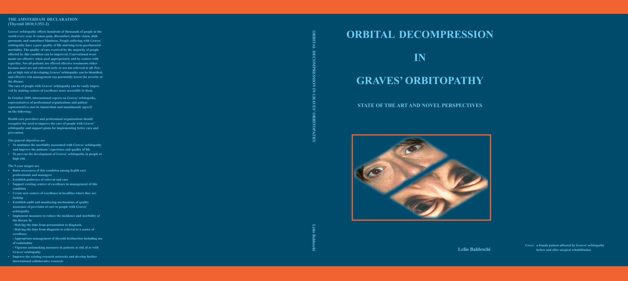
Cover: a female patient affected by Graves’ orbitopathy before and after surgical rehabilitation
100379_Baldeschi_Omslag Thesis.indd 1 woensdag15-december-2010 9:13

Orbital Decompression in Graves’ Orbitopathy:
State of the Art and Novel Perspectives
100379 Thesis Baldeschi.indd 1 vrijdag17-december-2010 12:07
© L. Baldeschi, Amsterdam, 2010
All rights reserved. No part of this book may be produced, stored in a retrivial system, or
transmitted in any form or by any means, electronic, mechanical, photocopying or otherwise,
without the prior permission of the holder of the copyright.
ISBN:
Author: Lelio Baldeschi
Lay-out: Lelio Baldeschi
Printed by: Buijten & Schipperheijn, Amsterdam
Orbital Decompression in Graves’ Orbitopathy:
State of the Art and Novel Perspectives
ACADEMISCH PROEFSCHRIFT
ter verkrijging van de graad van doctor
aan de Universiteit van Amsterdam
op gezag van de Rector Magnificus
prof. dr. D.C. van den Boom
ten overstaan van een door het college voor promoties ingestelde commissie,
in het openbaar te verdedigen in de Agnietenkapel
op vrijdag 14 januari 2011, te 10.00 uur
door
LELIO BALDESCHI
geboren te Pisa, Italië
978-90-9025951-2
100379 Thesis Baldeschi.indd 2 vrijdag17-december-2010 12:07
© L. Baldeschi, Amsterdam, 2010
All rights reserved. No part of this book may be produced, stored in a retrivial system, or
transmitted in any form or by any means, electronic, mechanical, photocopying or otherwise,
without the prior permission of the holder of the copyright.
ISBN:
Author: Lelio Baldeschi
Lay-out: Lelio Baldeschi
Printed by: Buijten & Schipperheijn, Amsterdam
Orbital Decompression in Graves’ Orbitopathy:
State of the Art and Novel Perspectives
ACADEMISCH PROEFSCHRIFT
ter verkrijging van de graad van doctor
aan de Universiteit van Amsterdam
op gezag van de Rector Magnificus
prof. dr. D.C. van den Boom
ten overstaan van een door het college voor promoties ingestelde commissie,
in het openbaar te verdedigen in de Agnietenkapel
op vrijdag 14 januari 2011, te 10.00 uur
door
LELIO BALDESCHI
geboren te Pisa, Italië
100379 Thesis Baldeschi.indd 3 vrijdag17-december-2010 12:07
Promotiecommissie
Promotores
Prof. dr. W.M. Wiersinga
Prof. dr. M.J.D. de Smet
Overige leden
Prof. dr. D.A. Bosch
Dr. G. Rose
Prof. dr. G.P.M. Luyten
Prof. dr. H.P. van den Akker
Prof. dr. P.T.V.M. de Jong
Faculteit der Geneeskunde
This thesis is dedicated to Leo Koornneef one of the most bright minded and talented surgeons in the field of orbit and ophthalmic plastic surgery
“behind every problem lies an opportunity”
Galileo Galilei Pisa 15 February 1564 - Florence 8 January 1642
100379 Thesis Baldeschi.indd 4 vrijdag17-december-2010 12:07
Promotiecommissie
Promotores
Prof. dr. W.M. Wiersinga
Prof. dr. M.J.D. de Smet
Overige leden
Prof. dr. D.A. Bosch
Dr. G. Rose
Prof. dr. G.P.M. Luyten
Prof. dr. H.P. van den Akker
Prof. dr. P.T.V.M. de Jong
Faculteit der Geneeskunde
This thesis is dedicated to Leo Koornneef one of the most bright minded and talented surgeons in the field of orbit and ophthalmic plastic surgery
“behind every problem lies an opportunity”
Galileo Galilei Pisa 15 February 1564 - Florence 8 January 1642
100379 Thesis Baldeschi.indd 5 vrijdag17-december-2010 12:07
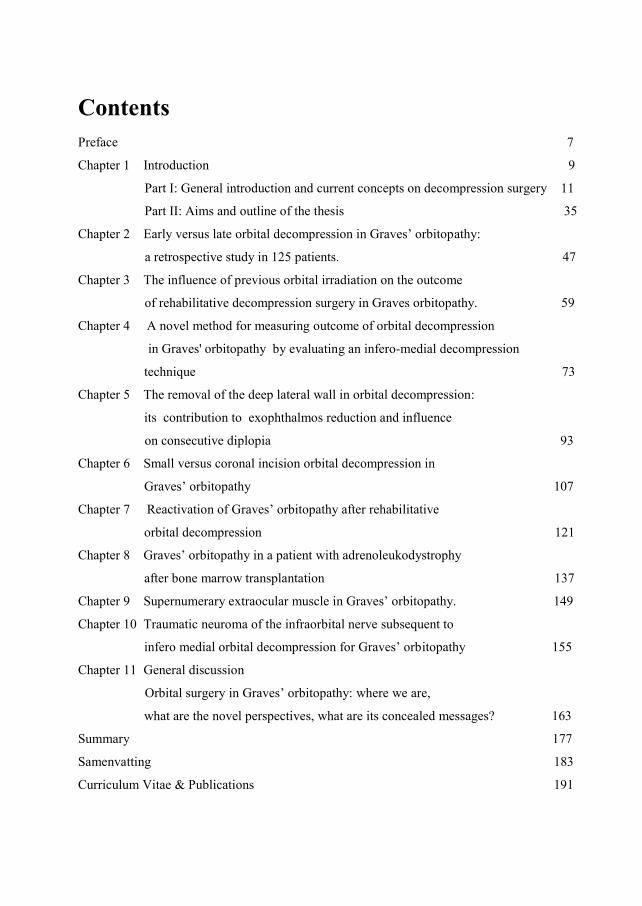
Contents Preface 7
Chapter 1 Introduction 9
Part I: General introduction and current concepts on decompression surgery 11
Part II: Aims and outline of the thesis 35
Chapter 2 Early versus late orbital decompression in Graves‟ orbitopathy:
a retrospective study in 125 patients. 47
Chapter 3 The influence of previous orbital irradiation on the outcome
of rehabilitative decompression surgery in Graves orbitopathy. 59
Chapter 4 A novel method for measuring outcome of orbital decompression
in Graves' orbitopathy by evaluating an infero-medial decompression
technique 73
Chapter 5 The removal of the deep lateral wall in orbital decompression:
its contribution to exophthalmos reduction and influence
on consecutive diplopia 93
Chapter 6 Small versus coronal incision orbital decompression in
Graves‟ orbitopathy 107
Chapter 7 Reactivation of Graves‟ orbitopathy after rehabilitative
orbital decompression 121
Chapter 8 Graves‟ orbitopathy in a patient with adrenoleukodystrophy
after bone marrow transplantation 137
Chapter 9 Supernumerary extraocular muscle in Graves‟ orbitopathy. 149
Chapter 10 Traumatic neuroma of the infraorbital nerve subsequent to
infero medial orbital decompression for Graves‟ orbitopathy 155
Chapter 11 General discussion
Orbital surgery in Graves‟ orbitopathy: where we are,
what are the novel perspectives, what are its concealed messages? 163
Summary 177
Samenvatting 183
Curriculum Vitae & Publications 191
100379 Thesis Baldeschi.indd 6 vrijdag17-december-2010 12:07
Contents Preface 7
Chapter 1 Introduction 9
Part I: General introduction and current concepts on decompression surgery 11
Part II: Aims and outline of the thesis 35
Chapter 2 Early versus late orbital decompression in Graves‟ orbitopathy:
a retrospective study in 125 patients. 47
Chapter 3 The influence of previous orbital irradiation on the outcome
of rehabilitative decompression surgery in Graves orbitopathy. 59
Chapter 4 A novel method for measuring outcome of orbital decompression
in Graves' orbitopathy by evaluating an infero-medial decompression
technique 73
Chapter 5 The removal of the deep lateral wall in orbital decompression:
its contribution to exophthalmos reduction and influence
on consecutive diplopia 93
Chapter 6 Small versus coronal incision orbital decompression in
Graves‟ orbitopathy 107
Chapter 7 Reactivation of Graves‟ orbitopathy after rehabilitative
orbital decompression 121
Chapter 8 Graves‟ orbitopathy in a patient with adrenoleukodystrophy
after bone marrow transplantation 137
Chapter 9 Supernumerary extraocular muscle in Graves‟ orbitopathy. 149
Chapter 10 Traumatic neuroma of the infraorbital nerve subsequent to
infero medial orbital decompression for Graves‟ orbitopathy 155
Chapter 11 General discussion
Orbital surgery in Graves‟ orbitopathy: where we are,
what are the novel perspectives, what are its concealed messages? 163
Summary 177
Samenvatting 183
Curriculum Vitae & Publications 191
Preface
I am grateful to my patients and to many people who have helped me in different ways.
I am grateful to my promoters whose constant advice and encouragement kept me going,
and I am also grateful to those who could not be my promoters, but whose attitude
provided me with positive motivations as well.
I am also indebted to colleagues, ortoptists, paramedical and administrative personnel, and
former fellows of the Department of Ophthalmology AMC all of them have never failed to
lend me their support. I particularly thank my former secretary Mrs Ineke Ompi whose hard
secretarial work was behind most of my publications.
I wish to express my feelings of gratitude to all the friends of the NVGP, the EUGOGO,
and the AMORE group AMC, all of whom I shared years of work and commitments. Marc
Prummel has a special place among them.
Twenty years back my ophthalmic plastic adventure started in London with Mr. Richard
Collin and Mr. Geoffrey Rose and thereafter continued in Amsterdam with Professor Leo
Koornneef. At that time I met other fellows and new colleagues, Professor Christoph
Hintschich, Dr. Christopher Neoh and Dr. Jane Dickinson who have become friends for
life. All these people have been instrumental in my professional and human development
with their culture and friendship. Thank you! Finally, I of course thank my wife Antonella, our son Giovanni and my parents for their
patience during the production of this thesis, and for being the never ending source of
support and confidence which made my life so happy.
100379 Thesis Baldeschi.indd 7 vrijdag17-december-2010 12:07

Chapter 1
Introduction
Part I:
General Introduction and Current Concepts on Decompression
Surgery for Graves’ Orbitopathy 1. A MISNAMED DISEASE WHICH CONTINUES TO REQUIRE SURGICAL ATTENTION
2. AN OVERVIEW OF GRAVES‟ ORBITOPATHY AND ITS CURRENT MODALITIES OF TREATMENT: THE
PLACE OF SURGERY
3. SURGERY FOR GRAVES‟ ORBITOPATHY: THE PLACE OF ORBITAL DECOMPRESSION from: Baldeschi L. Rehabilitative surgery. In: Wiersinga WM, Kahaly GJ, editors. Graves‟ Orbitopathy A Multidisciplinary Approach - Questions and Answers, 2nd edition. Basel: Karger, 2010:167-70.
4. ORBITAL DECOMPRESSION IN GRAVES‟ ORBITOPATHY: STATE OF THE ART
from: Baldeschi L. Orbital decompression. In: Wiersinga WM, Kahaly GJ, editors. Graves‟ Orbitopathy A Multidisciplinary Approach - Questions and Answers, 2nd edition. Basel: Karger, 2010:171-88.
Part II:
Outline and aims of this thesis
100379 Thesis Baldeschi.indd 8 vrijdag17-december-2010 12:07

Chapter 1
Introduction
Part I:
General Introduction and Current Concepts on Decompression
Surgery for Graves’ Orbitopathy 1. A MISNAMED DISEASE WHICH CONTINUES TO REQUIRE SURGICAL ATTENTION
2. AN OVERVIEW OF GRAVES‟ ORBITOPATHY AND ITS CURRENT MODALITIES OF TREATMENT: THE
PLACE OF SURGERY
3. SURGERY FOR GRAVES‟ ORBITOPATHY: THE PLACE OF ORBITAL DECOMPRESSION from: Baldeschi L. Rehabilitative surgery. In: Wiersinga WM, Kahaly GJ, editors. Graves‟ Orbitopathy A Multidisciplinary Approach - Questions and Answers, 2nd edition. Basel: Karger, 2010:167-70.
4. ORBITAL DECOMPRESSION IN GRAVES‟ ORBITOPATHY: STATE OF THE ART
from: Baldeschi L. Orbital decompression. In: Wiersinga WM, Kahaly GJ, editors. Graves‟ Orbitopathy A Multidisciplinary Approach - Questions and Answers, 2nd edition. Basel: Karger, 2010:171-88.
Part II:
Outline and aims of this thesis
100379 Thesis Baldeschi.indd 9 vrijdag17-december-2010 12:07

1. A MISNAMED DISEASE WHICH CONTINUES TO REQUIRE SURGICAL ATTENTION
The multi-systemic disease which may include autoimmune thyroid disorders, goitre,
exophthalmos, pretibial myxedema, and acropachy has been recognized as such for a very
long time.
According to Jan-Gustaf Ljunggren1, a Persian physician, Sayyid Ismail Al-Jurjani was
the first to have noted the association of goitre and exophthalmos in the 12th century, and
reported his observation in Thesaurus of the Shah of Khwarazm, the most famous of his
five books, and the major medical dictionary of its period.
In more recent times, the Italian anatomist and surgeon Giuseppe Flajani in 18022, followed
by another Italian physician Antonio Giuseppe Testa in 18103 described “exophthalmic
goiter”, though it was earlier re cognized by Caleb Hillier Parry in 1786 and reported in
his “unpublished medical writings” which appeared 3 years after his death in 1825.4
Robert James Graves in 18355, and the German Karl Adolph von Basedow in 18406
independently reported the constellation of signs and symptoms which typifies the disease.
Long before any description of the association between exophthalmos and goitre appeared
in the medical literature, a clear iconographic report of the disease was already available.
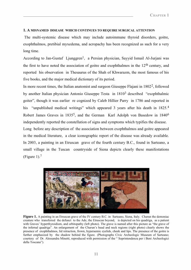
In 2003, a painting in an Etruscan grave of the fourth century B.C., found in Sarteano, a
small village in the Tuscan countryside of Siena depicts clearly these manifestations
(Figure 1).7
Figure 1. A painting in an Etruscan grave of the IV century B.C. in Sarteano, Siena, Italy. Charun the demoniac creature who transferred the defunct to the Ade, the Etruscan beyond, is depicted on his quadriga, as a patient with Graves‟ hyperthyroidism, and orbitopathy (left photo). The grave is named after this picture as “the grave of the infernal quadriga”. An enlargement of the Charun‟s head and neck regions (right photo) clearly shows the presence of exophthalmos, lid retraction, frown, hyperaemic eyelids, cheek and lips. The presence of the goitre is further emphasized by the shadow behind the figure. (Photographs Civic Archeologic Museum of Sarteano; courtesy of Dr. Alessandra Minetti, reproduced with permission of the “ Soprintendenza per i Beni Archeologici della Toscana”).
100379 Thesis Baldeschi.indd 10 vrijdag17-december-2010 12:07
11
Chapter 1
1. A MISNAMED DISEASE WHICH CONTINUES TO REQUIRE SURGICAL ATTENTION
The multi-systemic disease which may include autoimmune thyroid disorders, goitre,
exophthalmos, pretibial myxedema, and acropachy has been recognized as such for a very
long time.
According to Jan-Gustaf Ljunggren1, a Persian physician, Sayyid Ismail Al-Jurjani was
the first to have noted the association of goitre and exophthalmos in the 12th century, and
reported his observation in Thesaurus of the Shah of Khwarazm, the most famous of his
five books, and the major medical dictionary of its period.
In more recent times, the Italian anatomist and surgeon Giuseppe Flajani in 18022, followed
by another Italian physician Antonio Giuseppe Testa in 18103 described “exophthalmic
goiter”, though it was earlier re cognized by Caleb Hillier Parry in 1786 and reported in
his “unpublished medical writings” which appeared 3 years after his death in 1825.4
Robert James Graves in 18355, and the German Karl Adolph von Basedow in 18406
independently reported the constellation of signs and symptoms which typifies the disease.
Long before any description of the association between exophthalmos and goitre appeared
in the medical literature, a clear iconographic report of the disease was already available.
In 2003, a painting in an Etruscan grave of the fourth century B.C., found in Sarteano, a
small village in the Tuscan countryside of Siena depicts clearly these manifestations
(Figure 1).7
Figure 1. A painting in an Etruscan grave of the IV century B.C. in Sarteano, Siena, Italy. Charun the demoniac creature who transferred the defunct to the Ade, the Etruscan beyond, is depicted on his quadriga, as a patient with Graves‟ hyperthyroidism, and orbitopathy (left photo). The grave is named after this picture as “the grave of the infernal quadriga”. An enlargement of the Charun‟s head and neck regions (right photo) clearly shows the presence of exophthalmos, lid retraction, frown, hyperaemic eyelids, cheek and lips. The presence of the goitre is further emphasized by the shadow behind the figure. (Photographs Civic Archeologic Museum of Sarteano; courtesy of Dr. Alessandra Minetti, reproduced with permission of the “ Soprintendenza per i Beni Archeologici della Toscana”).
100379 Thesis Baldeschi.indd 11 vrijdag17-december-2010 12:07
12
Chapter 1
As manifestation of the disease, orbital involvement has been given a multitude of names,
including: thyroid eye disease, autoimmune orbitopathy, thyroid related or dysthyroid
orbitopathy / ophthalmopathy, Basedow‟s5 or Graves‟6 orbitopathy / ophthalmopathy. All
of them are misnomers as they lack etiopathogenetic precision or eponymic priority. Until
the pathogenesis of the disease is known and a more specific nosologic nomenclature is
available, this bizarre orbital disorder should at least respect eponymic priority. For this
reason, the adjective “Etruscan” should be mentioned when defining this orbitopathy and
conversely the thyroid disease. This logical but regionalistic suggestion, however, might
not sound appropriate to the international character of the two large consortia which at
present are committed to research in the field of this disease, and which are both already
named after it as European Group on Graves‟ Orbitopathy (EUGOGO)8 and International
Thyroid Eye Disease Society (ITEDS).9 Additionally, just for a coincidence..., it might
appear parochially motivated by the Tuscan birth and Etruscan ancestry of the writer,
together with the medieval but still strongly deep seated Ghibelline vicinity of Pisa, the
author‟s home town, to Siena.
By an astonishing twist of fate, and the serendipitous location of the Etruscan painting,
Graves‟ orbitopathy (GO), fulfils the writer‟s proposal in part, that such a definition is
maintained in this PhD thesis. This is also consistent with the writer being a member of
EUGOGO.8
The painting in the Sarteano grave in which Charun is depicted resembling a patient with
Graves‟ disease and GO7 (Figure 1) suggests that the somatic alterations and psychosis due
to untreated hyperthyroidism might have already in that ancient culture been a source of
prejudices resulting in social isolation for the affected patients.
Visible deformity, particularly involving the face, has always induced society‟s aversion.
Patients with facial disfigurement suffer from intrusions such as staring or comments. At
the root of the patient‟s distress lies a social pressure to conform to an idealized appearance.
This obsession with appearance in our culture devalues those who do not match the
perceived ideal and stigmatizes those with visible disfigurement.10
Despite the long-lasting social implications and medical recognition of GO, its exact
pathogenesis remains unknown. As a consequence a specific medical therapy is lacking,
and for many patients orbital surgery continues to represent an essential cornerstone in the
treatment of their disabling and socially alienating disease.11
2. AN OVERVIEW OF GRAVES’ ORBITOPATHY AND ITS CURRENT MODALITIES OF
TREATMENT: THE PLACE OF SURGERY
GO is an autoimmune disorder representing the most frequent and important extrathyroidal
expression of Graves‟ disease. It may also be found, although less frequently, in patients
with no present or past history of hyperthyroidism (so-called euthyroid or ophthalmic
Graves‟ disease) or in patients who are hypothyroid due to chronic autoimmune
(Hashimoto‟s) thyroiditis.12, 13 In most affected individuals GO is mild and self-limiting,
and only in 3-5% of cases, it is severe and potentially sight-threatening.12, 14
The exact pathogenesis of GO is unknown.15-18 It is, however, worth highlighting the clear-
cut link between the orbit and the thyroid, because this has important clinical and
therapeutic implications.19 In addition to endogenous (non-preventable) factors, such as
genetics, age- and gender-related factors20, GO occurrence and progression are influenced
by environmental (preventable) factors, such as cigarette smoking, thyroid dysfunction, and
different treatments for hyperthyroidism.14, 21 This implies that control or correction of
these risk factors is an integral part of GO management.
Independently of the complex association with thyroid dysfunction and its treatment,
management of GO is difficult: decisions need to be made regarding the need for specific
treatment or whether spontaneous regression is likely.
The natural history of GO is of gradual increase in severity followed by a plateau phase
then gradual improvement.22 These are the active phases. The inactive phase follows with
no change in severity. GO is thus self-limiting, although it often does not return to baseline.
Treatment is aimed at accelerating recovery, preventing serious sequelae, and eventually
functional and cosmetic rehabilitation.
Therapeutic options consist of medical therapy, radiotherapy, surgery, or frequently a
combination of these. Consensus as to indications and timing of these options has been
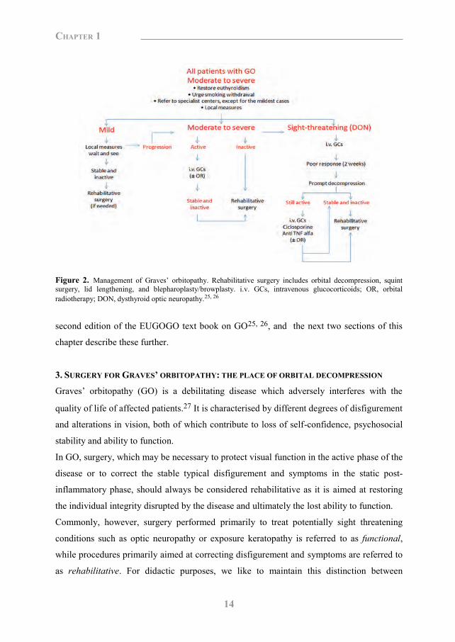
reached by the EUGOGO consortium (Figure 2).23, 24 More specifically the role of orbital
decompressions in the state of the art rehabilitative surgery has been published in the
100379 Thesis Baldeschi.indd 12 vrijdag17-december-2010 12:07

13
Chapter 1
As manifestation of the disease, orbital involvement has been given a multitude of names,
including: thyroid eye disease, autoimmune orbitopathy, thyroid related or dysthyroid
orbitopathy / ophthalmopathy, Basedow‟s5 or Graves‟6 orbitopathy / ophthalmopathy. All
of them are misnomers as they lack etiopathogenetic precision or eponymic priority. Until
the pathogenesis of the disease is known and a more specific nosologic nomenclature is
available, this bizarre orbital disorder should at least respect eponymic priority. For this
reason, the adjective “Etruscan” should be mentioned when defining this orbitopathy and
conversely the thyroid disease. This logical but regionalistic suggestion, however, might
not sound appropriate to the international character of the two large consortia which at
present are committed to research in the field of this disease, and which are both already
named after it as European Group on Graves‟ Orbitopathy (EUGOGO)8 and International
Thyroid Eye Disease Society (ITEDS).9 Additionally, just for a coincidence..., it might
appear parochially motivated by the Tuscan birth and Etruscan ancestry of the writer,
together with the medieval but still strongly deep seated Ghibelline vicinity of Pisa, the
author‟s home town, to Siena.
By an astonishing twist of fate, and the serendipitous location of the Etruscan painting,
Graves‟ orbitopathy (GO), fulfils the writer‟s proposal in part, that such a definition is
maintained in this PhD thesis. This is also consistent with the writer being a member of
EUGOGO.8
The painting in the Sarteano grave in which Charun is depicted resembling a patient with
Graves‟ disease and GO7 (Figure 1) suggests that the somatic alterations and psychosis due
to untreated hyperthyroidism might have already in that ancient culture been a source of
prejudices resulting in social isolation for the affected patients.
Visible deformity, particularly involving the face, has always induced society‟s aversion.
Patients with facial disfigurement suffer from intrusions such as staring or comments. At
the root of the patient‟s distress lies a social pressure to conform to an idealized appearance.
This obsession with appearance in our culture devalues those who do not match the
perceived ideal and stigmatizes those with visible disfigurement.10
Despite the long-lasting social implications and medical recognition of GO, its exact
pathogenesis remains unknown. As a consequence a specific medical therapy is lacking,
and for many patients orbital surgery continues to represent an essential cornerstone in the
treatment of their disabling and socially alienating disease.11
2. AN OVERVIEW OF GRAVES’ ORBITOPATHY AND ITS CURRENT MODALITIES OF
TREATMENT: THE PLACE OF SURGERY
GO is an autoimmune disorder representing the most frequent and important extrathyroidal
expression of Graves‟ disease. It may also be found, although less frequently, in patients
with no present or past history of hyperthyroidism (so-called euthyroid or ophthalmic
Graves‟ disease) or in patients who are hypothyroid due to chronic autoimmune
(Hashimoto‟s) thyroiditis.12, 13 In most affected individuals GO is mild and self-limiting,
and only in 3-5% of cases, it is severe and potentially sight-threatening.12, 14
The exact pathogenesis of GO is unknown.15-18 It is, however, worth highlighting the clear-
cut link between the orbit and the thyroid, because this has important clinical and
therapeutic implications.19 In addition to endogenous (non-preventable) factors, such as
genetics, age- and gender-related factors20, GO occurrence and progression are influenced
by environmental (preventable) factors, such as cigarette smoking, thyroid dysfunction, and
different treatments for hyperthyroidism.14, 21 This implies that control or correction of
these risk factors is an integral part of GO management.
Independently of the complex association with thyroid dysfunction and its treatment,
management of GO is difficult: decisions need to be made regarding the need for specific
treatment or whether spontaneous regression is likely.
The natural history of GO is of gradual increase in severity followed by a plateau phase
then gradual improvement.22 These are the active phases. The inactive phase follows with
no change in severity. GO is thus self-limiting, although it often does not return to baseline.
Treatment is aimed at accelerating recovery, preventing serious sequelae, and eventually
functional and cosmetic rehabilitation.
Therapeutic options consist of medical therapy, radiotherapy, surgery, or frequently a
combination of these. Consensus as to indications and timing of these options has been
reached by the EUGOGO consortium (Figure 2).23, 24 More specifically the role of orbital
decompressions in the state of the art rehabilitative surgery has been published in the
100379 Thesis Baldeschi.indd 13 vrijdag17-december-2010 12:07
14
Chapter 1
Figure 2. Management of Graves‟ orbitopathy. Rehabilitative surgery includes orbital decompression, squint surgery, lid lengthening, and blepharoplasty/browplasty. i.v. GCs, intravenous glucocorticoids; OR, orbital radiotherapy; DON, dysthyroid optic neuropathy.25, 26
second edition of the EUGOGO text book on GO25, 26, and the next two sections of this
chapter describe these further.
3. SURGERY FOR GRAVES’ ORBITOPATHY: THE PLACE OF ORBITAL DECOMPRESSION
Graves‟ orbitopathy (GO) is a debilitating disease which adversely interferes with the
quality of life of affected patients.27 It is characterised by different degrees of disfigurement
and alterations in vision, both of which contribute to loss of self-confidence, psychosocial
stability and ability to function.
In GO, surgery, which may be necessary to protect visual function in the active phase of the
disease or to correct the stable typical disfigurement and symptoms in the static post-
inflammatory phase, should always be considered rehabilitative as it is aimed at restoring
the individual integrity disrupted by the disease and ultimately the lost ability to function.
Commonly, however, surgery performed primarily to treat potentially sight threatening
conditions such as optic neuropathy or exposure keratopathy is referred to as functional,
while procedures primarily aimed at correcting disfigurement and symptoms are referred to
as rehabilitative. For didactic purposes, we like to maintain this distinction between
functional and rehabilitative surgery, although it is necessary to admit that besides the
above-listed semantic considerations, a clear-cut distinction between the two does not exist
as surgery aimed primarily at restoring function also has positive effects on disfigurement
and vice versa.
The definition cosmetic surgery, which does not stress the impact of the orbital disease in
affected patients, appears inadequate and should be avoided. Surgery is in fact aimed at
restoring a patient‟s appearance as closely as possible to that preceding the onset of GO,
and not at changing his or her somatic tracts to make them more beautiful.
Cosmetic/aesthetic rehabilitation has often been used and can be considered an acceptable
compromise when defining surgery mainly aimed at correcting disfigurement due to GO.
Figure 3. A patient affected by left, moderate severe, not-active GO with exophthalmos, mild restrictive esotropia and large angle restrictive hypotropia. An upper lid aponeurotic ptosis was also present left side. The general health condition of the patient and his scarce cooperation suggested to reduce as much as possible the number of surgical interventions. (a) Upon admittance for simultaneous orbital decompression and strabismus correction. (b) Five weeks after left deep lateral wall decompression through an upper skin crease approach followed by recession of the left inferior rectus muscle in the same surgical session. An adequate reduction of exophthalmos was achieved and the field of binocular single vision extended for more than 20° around primary position of gaze.
100379 Thesis Baldeschi.indd 14 vrijdag17-december-2010 12:07

15
Chapter 1
Figure 2. Management of Graves‟ orbitopathy. Rehabilitative surgery includes orbital decompression, squint surgery, lid lengthening, and blepharoplasty/browplasty. i.v. GCs, intravenous glucocorticoids; OR, orbital radiotherapy; DON, dysthyroid optic neuropathy.25, 26
second edition of the EUGOGO text book on GO25, 26, and the next two sections of this
chapter describe these further.
3. SURGERY FOR GRAVES’ ORBITOPATHY: THE PLACE OF ORBITAL DECOMPRESSION
Graves‟ orbitopathy (GO) is a debilitating disease which adversely interferes with the
quality of life of affected patients.27 It is characterised by different degrees of disfigurement
and alterations in vision, both of which contribute to loss of self-confidence, psychosocial
stability and ability to function.
In GO, surgery, which may be necessary to protect visual function in the active phase of the
disease or to correct the stable typical disfigurement and symptoms in the static post-
inflammatory phase, should always be considered rehabilitative as it is aimed at restoring
the individual integrity disrupted by the disease and ultimately the lost ability to function.
Commonly, however, surgery performed primarily to treat potentially sight threatening
conditions such as optic neuropathy or exposure keratopathy is referred to as functional,
while procedures primarily aimed at correcting disfigurement and symptoms are referred to
as rehabilitative. For didactic purposes, we like to maintain this distinction between
functional and rehabilitative surgery, although it is necessary to admit that besides the
above-listed semantic considerations, a clear-cut distinction between the two does not exist
as surgery aimed primarily at restoring function also has positive effects on disfigurement
and vice versa.
The definition cosmetic surgery, which does not stress the impact of the orbital disease in
affected patients, appears inadequate and should be avoided. Surgery is in fact aimed at
restoring a patient‟s appearance as closely as possible to that preceding the onset of GO,
and not at changing his or her somatic tracts to make them more beautiful.
Cosmetic/aesthetic rehabilitation has often been used and can be considered an acceptable
compromise when defining surgery mainly aimed at correcting disfigurement due to GO.
Figure 3. A patient affected by left, moderate severe, not-active GO with exophthalmos, mild restrictive esotropia and large angle restrictive hypotropia. An upper lid aponeurotic ptosis was also present left side. The general health condition of the patient and his scarce cooperation suggested to reduce as much as possible the number of surgical interventions. (a) Upon admittance for simultaneous orbital decompression and strabismus correction. (b) Five weeks after left deep lateral wall decompression through an upper skin crease approach followed by recession of the left inferior rectus muscle in the same surgical session. An adequate reduction of exophthalmos was achieved and the field of binocular single vision extended for more than 20° around primary position of gaze.
100379 Thesis Baldeschi.indd 15 vrijdag17-december-2010 12:07
16
Chapter 1
3.1. What Are the Steps and Timing of Rehabilitative Surgery?
During the post-inflammatory phase of GO, after a 6- to 8-month stable endocrinological
and ophthalmic clinical picture, surgical rehabilitation can be performed if required.
Depending on the severity of the disease, surgical rehabilitation can be more or less
extensive, the full treatment consisting of decompression surgery, squint surgery, eyelid
lengthening, blepharoplasty and eyebrow plasty.
The first rehabilitative step mainly consists of orbital bony decompression and early
intervention soon after stabilisation has been advocated.28 Fibrosis due to long-lasting
orbital disease or as a possible consequence of retrobulbar irradiation administrated in the
early phase of GO has been questioned as a possible cause of poor distensibility and
plasticity of the soft orbital tissues, resulting in scarce effectiveness of orbital expansion
surgery.29-32 Recent studies, however, have demonstrated that long-lasting GO or
preoperative radiotherapy do not adversely interfere with the results of orbital bony
decompression; thus, when the stabilisation of Graves‟ disease and orbitopathy has
occurred, rehabilitative surgery can be started at any time and no adverse effects from
common preceding treatments such as retrobulbar irradiation are to be expected. 33, 34
Decompression surgery causes a reduction in exophthalmos as well as reduction in upper
and lower eyelid displacement.33 It may positively influence extra-ocular muscle
restriction, but displacement of the soft orbital tissues caused by decompression procedures
may also cause strabismus. Possible squint surgery should therefore follow orbital
decompressions, but considering that vertical tropias may influence eyelid position, squint
surgery should precede possible correction of eyelid position. Finally, when necessary, the
finishing touch can be given by eyebrow lift, forehead plasty and blepharoplasty.
In short, surgical rehabilitation needs to respect the given order since the preceding step
may influence the necessity and the extent of the step that follows. When all the steps are
necessary, the entire rehabilitation may require between 1.5 and 2 years. In particular cases,
exceptions are possible and the rehabilitation can be favourably speeded-up by carrying out
more than one procedure at the same time (Figure 3). The traditional management
algorithm has not been respected in only a few series, and simultaneous decompression and
strabismus surgery (in severe orbitopathy35) or simultaneous decompression and correction
of upper eyelid retraction (in mild to moderate36, 37, or in moderate to severe orbitopathy38,
39) have been performed routinely. The only series aimed at retrospectively comparing the
outcome of surgical correction of upper eyelid retraction after staged or simultaneous
decompression and eyelid surgery did not conclusively prove the superiority of either
approach37, and the concept of performing decompression and upper eyelid lengthening
during the same surgical intervention met with vigorous criticism.40
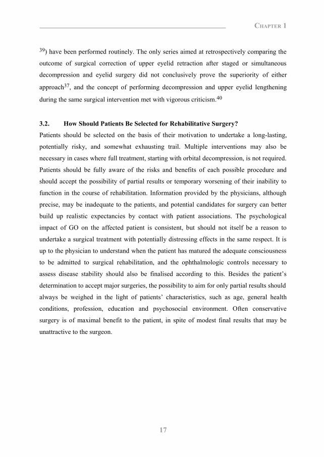
3.2. How Should Patients Be Selected for Rehabilitative Surgery?
Patients should be selected on the basis of their motivation to undertake a long-lasting,
potentially risky, and somewhat exhausting trail. Multiple interventions may also be
necessary in cases where full treatment, starting with orbital decompression, is not required.
Patients should be fully aware of the risks and benefits of each possible procedure and
should accept the possibility of partial results or temporary worsening of their inability to
function in the course of rehabilitation. Information provided by the physicians, although
precise, may be inadequate to the patients, and potential candidates for surgery can better
build up realistic expectancies by contact with patient associations. The psychological
impact of GO on the affected patient is consistent, but should not itself be a reason to
undertake a surgical treatment with potentially distressing effects in the same respect. It is
up to the physician to understand when the patient has matured the adequate consciousness
to be admitted to surgical rehabilitation, and the ophthalmologic controls necessary to
assess disease stability should also be finalised according to this. Besides the patient‟s
determination to accept major surgeries, the possibility to aim for only partial results should
always be weighed in the light of patients‟ characteristics, such as age, general health
conditions, profession, education and psychosocial environment. Often conservative
surgery is of maximal benefit to the patient, in spite of modest final results that may be
unattractive to the surgeon.
100379 Thesis Baldeschi.indd 16 vrijdag17-december-2010 12:07
17
Chapter 1
3.1. What Are the Steps and Timing of Rehabilitative Surgery?
During the post-inflammatory phase of GO, after a 6- to 8-month stable endocrinological
and ophthalmic clinical picture, surgical rehabilitation can be performed if required.
Depending on the severity of the disease, surgical rehabilitation can be more or less
extensive, the full treatment consisting of decompression surgery, squint surgery, eyelid
lengthening, blepharoplasty and eyebrow plasty.
The first rehabilitative step mainly consists of orbital bony decompression and early
intervention soon after stabilisation has been advocated.28 Fibrosis due to long-lasting
orbital disease or as a possible consequence of retrobulbar irradiation administrated in the
early phase of GO has been questioned as a possible cause of poor distensibility and
plasticity of the soft orbital tissues, resulting in scarce effectiveness of orbital expansion
surgery.29-32 Recent studies, however, have demonstrated that long-lasting GO or
preoperative radiotherapy do not adversely interfere with the results of orbital bony
decompression; thus, when the stabilisation of Graves‟ disease and orbitopathy has
occurred, rehabilitative surgery can be started at any time and no adverse effects from
common preceding treatments such as retrobulbar irradiation are to be expected. 33, 34
Decompression surgery causes a reduction in exophthalmos as well as reduction in upper
and lower eyelid displacement.33 It may positively influence extra-ocular muscle
restriction, but displacement of the soft orbital tissues caused by decompression procedures
may also cause strabismus. Possible squint surgery should therefore follow orbital
decompressions, but considering that vertical tropias may influence eyelid position, squint
surgery should precede possible correction of eyelid position. Finally, when necessary, the
finishing touch can be given by eyebrow lift, forehead plasty and blepharoplasty.
In short, surgical rehabilitation needs to respect the given order since the preceding step
may influence the necessity and the extent of the step that follows. When all the steps are
necessary, the entire rehabilitation may require between 1.5 and 2 years. In particular cases,
exceptions are possible and the rehabilitation can be favourably speeded-up by carrying out
more than one procedure at the same time (Figure 3). The traditional management
algorithm has not been respected in only a few series, and simultaneous decompression and
strabismus surgery (in severe orbitopathy35) or simultaneous decompression and correction
of upper eyelid retraction (in mild to moderate36, 37, or in moderate to severe orbitopathy38,
39) have been performed routinely. The only series aimed at retrospectively comparing the
outcome of surgical correction of upper eyelid retraction after staged or simultaneous
decompression and eyelid surgery did not conclusively prove the superiority of either
approach37, and the concept of performing decompression and upper eyelid lengthening
during the same surgical intervention met with vigorous criticism.40
3.2. How Should Patients Be Selected for Rehabilitative Surgery?
Patients should be selected on the basis of their motivation to undertake a long-lasting,
potentially risky, and somewhat exhausting trail. Multiple interventions may also be
necessary in cases where full treatment, starting with orbital decompression, is not required.
Patients should be fully aware of the risks and benefits of each possible procedure and
should accept the possibility of partial results or temporary worsening of their inability to
function in the course of rehabilitation. Information provided by the physicians, although
precise, may be inadequate to the patients, and potential candidates for surgery can better
build up realistic expectancies by contact with patient associations. The psychological
impact of GO on the affected patient is consistent, but should not itself be a reason to
undertake a surgical treatment with potentially distressing effects in the same respect. It is
up to the physician to understand when the patient has matured the adequate consciousness
to be admitted to surgical rehabilitation, and the ophthalmologic controls necessary to
assess disease stability should also be finalised according to this. Besides the patient‟s
determination to accept major surgeries, the possibility to aim for only partial results should
always be weighed in the light of patients‟ characteristics, such as age, general health
conditions, profession, education and psychosocial environment. Often conservative
surgery is of maximal benefit to the patient, in spite of modest final results that may be
unattractive to the surgeon.
100379 Thesis Baldeschi.indd 17 vrijdag17-december-2010 12:07
18
Chapter 1
4. ORBITAL DECOMPRESSION IN GRAVES’ ORBITOPATHY: STATE OF THE ART
4.1. What Is Orbital Decompression?
The autoimmune process at the basis of Graves‟ orbitopathy (GO) induces swelling of the
soft tissues contained within the boundary of the bony orbit, this causes impairment of the
venous out flux towards the cavernous sinus and reverses the flux in direction of facial
circulation.
This positive feedback circle leads to an increase in the intraorbital pressure which is first
responsible for the progression of GO and later for its typical signs and symptoms.16 Any
surgical procedure aimed at decreasing the raised intraorbital pressure and its effects by
means of enlargement of the bony orbit and/or removal of the orbital fat is defined as
orbital decompression.
4.2. What Are the Aims of Orbital Decompression?
For one century decompression surgery has been used to treat GO. First it was used only to
address sight-threatening conditions such as optic neuropathy refractory to medical therapy,
or exposure keratopathy unresponsive to local measures, and/or minor eyelid surgeries.
More recently, the indications of orbital decompression were extended to the treatment of
disfiguring exophthalmos and symptoms. Eyeball subluxation (which may be a possible
cause of acute optic neuropathy and exposure keratopathy41), postural visual obscuration in
patients with congestive inactive GO42 and choroidal folds due to eyeball indentation by
enlarged extraocular muscles represent other more recently recognised functional
indications for decompression surgery.
Functional Aims
According to a large retrospective study, decompression surgery can offer a rapid solution
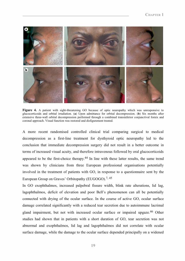
to dysthyroid optic neuropathy with an acceptable list of adverse effects (Figure 4).43
Figure 4. A patient with sight-threatening GO because of optic neuropathy which was unresponsive to glucocorticoids and orbital irradiation. (a) Upon admittance for orbital decompression. (b) Six months after extensive three-wall orbital decompression performed through a combined transinferior conjunctival fornix and coronal approach. Visual function was restored and disfigurement treated. A more recent randomised controlled clinical trial comparing surgical to medical
decompression as a first-line treatment for dysthyroid optic neuropathy led to the
conclusion that immediate decompression surgery did not result in a better outcome in
terms of increased visual acuity, and therefore intravenous followed by oral glucocorticoids
appeared to be the first-choice therapy.44 In line with these latter results, the same trend
was shown by clinicians from three European professional organisations potentially
involved in the treatment of patients with GO, in response to a questionnaire sent by the
European Group on Graves‟ Orbitopathy (EUGOGO).7, 45
In GO exophthalmos, increased palpebral fissure width, blink rate alterations, lid lag,
lagophthalmos, deficit of elevation and poor Bell‟s phenomenon can all be potentially
connected with drying of the ocular surface. In the course of active GO, ocular surface
damage correlated significantly with a reduced tear secretion due to autoimmune lacrimal
gland impairment, but not with increased ocular surface or impaired upgaze.46 Other
studies had shown that in patients with a short duration of GO, tear secretion was not
abnormal and exophthalmos, lid lag and lagophthalmos did not correlate with ocular
surface damage, while the damage to the ocular surface depended principally on a widened
100379 Thesis Baldeschi.indd 18 vrijdag17-december-2010 12:07
19
Chapter 1
4. ORBITAL DECOMPRESSION IN GRAVES’ ORBITOPATHY: STATE OF THE ART
4.1. What Is Orbital Decompression?
The autoimmune process at the basis of Graves‟ orbitopathy (GO) induces swelling of the
soft tissues contained within the boundary of the bony orbit, this causes impairment of the
venous out flux towards the cavernous sinus and reverses the flux in direction of facial
circulation.
This positive feedback circle leads to an increase in the intraorbital pressure which is first
responsible for the progression of GO and later for its typical signs and symptoms.16 Any
surgical procedure aimed at decreasing the raised intraorbital pressure and its effects by
means of enlargement of the bony orbit and/or removal of the orbital fat is defined as
orbital decompression.
4.2. What Are the Aims of Orbital Decompression?
For one century decompression surgery has been used to treat GO. First it was used only to
address sight-threatening conditions such as optic neuropathy refractory to medical therapy,
or exposure keratopathy unresponsive to local measures, and/or minor eyelid surgeries.
More recently, the indications of orbital decompression were extended to the treatment of
disfiguring exophthalmos and symptoms. Eyeball subluxation (which may be a possible
cause of acute optic neuropathy and exposure keratopathy41), postural visual obscuration in
patients with congestive inactive GO42 and choroidal folds due to eyeball indentation by
enlarged extraocular muscles represent other more recently recognised functional
indications for decompression surgery.
Functional Aims
According to a large retrospective study, decompression surgery can offer a rapid solution
to dysthyroid optic neuropathy with an acceptable list of adverse effects (Figure 4).43
Figure 4. A patient with sight-threatening GO because of optic neuropathy which was unresponsive to glucocorticoids and orbital irradiation. (a) Upon admittance for orbital decompression. (b) Six months after extensive three-wall orbital decompression performed through a combined transinferior conjunctival fornix and coronal approach. Visual function was restored and disfigurement treated. A more recent randomised controlled clinical trial comparing surgical to medical
decompression as a first-line treatment for dysthyroid optic neuropathy led to the
conclusion that immediate decompression surgery did not result in a better outcome in
terms of increased visual acuity, and therefore intravenous followed by oral glucocorticoids
appeared to be the first-choice therapy.44 In line with these latter results, the same trend
was shown by clinicians from three European professional organisations potentially
involved in the treatment of patients with GO, in response to a questionnaire sent by the
European Group on Graves‟ Orbitopathy (EUGOGO).7, 45
In GO exophthalmos, increased palpebral fissure width, blink rate alterations, lid lag,
lagophthalmos, deficit of elevation and poor Bell‟s phenomenon can all be potentially
connected with drying of the ocular surface. In the course of active GO, ocular surface
damage correlated significantly with a reduced tear secretion due to autoimmune lacrimal
gland impairment, but not with increased ocular surface or impaired upgaze.46 Other
studies had shown that in patients with a short duration of GO, tear secretion was not
abnormal and exophthalmos, lid lag and lagophthalmos did not correlate with ocular
surface damage, while the damage to the ocular surface depended principally on a widened
100379 Thesis Baldeschi.indd 19 vrijdag17-december-2010 12:07
20
Chapter 1
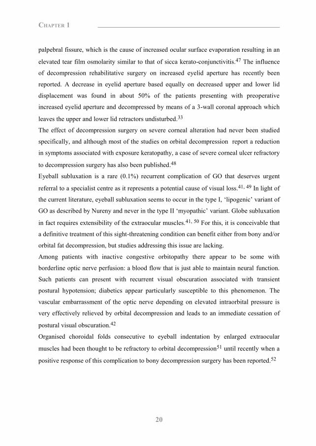
palpebral fissure, which is the cause of increased ocular surface evaporation resulting in an
elevated tear film osmolarity similar to that of sicca kerato-conjunctivitis.47 The influence
of decompression rehabilitative surgery on increased eyelid aperture has recently been
reported. A decrease in eyelid aperture based equally on decreased upper and lower lid
displacement was found in about 50% of the patients presenting with preoperative
increased eyelid aperture and decompressed by means of a 3-wall coronal approach which
leaves the upper and lower lid retractors undisturbed.33
The effect of decompression surgery on severe corneal alteration had never been studied
specifically, and although most of the studies on orbital decompression report a reduction
in symptoms associated with exposure keratopathy, a case of severe corneal ulcer refractory
to decompression surgery has also been published.48
Eyeball subluxation is a rare (0.1%) recurrent complication of GO that deserves urgent
referral to a specialist centre as it represents a potential cause of visual loss.41, 49 In light of
the current literature, eyeball subluxation seems to occur in the type I, „lipogenic‟ variant of
GO as described by Nureny and never in the type II „myopathic‟ variant. Globe subluxation
in fact requires extensibility of the extraocular muscles.41, 50 For this, it is conceivable that
a definitive treatment of this sight-threatening condition can benefit either from bony and/or
orbital fat decompression, but studies addressing this issue are lacking.
Among patients with inactive congestive orbitopathy there appear to be some with
borderline optic nerve perfusion: a blood flow that is just able to maintain neural function.
Such patients can present with recurrent visual obscuration associated with transient
postural hypotension; diabetics appear particularly susceptible to this phenomenon. The
vascular embarrassment of the optic nerve depending on elevated intraorbital pressure is
very effectively relieved by orbital decompression and leads to an immediate cessation of
postural visual obscuration.42
Organised choroidal folds consecutive to eyeball indentation by enlarged extraocular
muscles had been thought to be refractory to orbital decompression51 until recently when a
positive response of this complication to bony decompression surgery has been reported.52
Rehabilitative Symptomatic Aims
In GO, severe functional complications due to increased intraocular pressure are rare,
different degrees of venous congestion, strabismus, eyelid puffiness and retraction,
exophthalmos, and symptoms such as retroocular pressure and/or grittiness due to chronic
corneal exposure are more frequent. Decompression surgery is the mainstay method to treat
stable disfiguring alterations and/or symptoms that can typify the inactive post-
inflammatory phase of the disease (Figure 5). Decompression surgery is not necessarily
required only when exophthalmos exceeds the normal reference range. Patients with a flat
forehead, scarce brow bossing, and scarce anterior projection of the zygomatic eminence or
patients with deep-set eyes before GO may be or feel disfigured at normal
exophthalmometric values (Figure 6). Evaluation of pre-GO facial photographs may help
the surgeon to restore a patient‟s appearance as close as possible to how it was before the
onset of orbital disease. Most of the studies dealing with orbital decompressions have
indicated that this type of surgery is associated with lessening of the subjective perception
of retro-ocular tension. In the early 1990s, Khan et al.53, using the McGill pain
questionnaire and visual analogue scales, specifically addressed this issue, and, although
their study was not free of biases, it seemed to confirm that orbital discomfort significantly
responded to orbital decompression.
4.3. Which Surgical Technique Should Be Preferred?
The raised intraorbital pressure and its consequences can be surgically addressed by
expansion of the bony orbital boundary and/or by means of fat removal. For about one
century, the two possibilities developed through parallel routes; only recently did it become
clear that they should no longer be considered alternatives but complementary approaches
concurring in tailoring the most adequate treatment to the specific patient‟s needs (Figures
7, 8).54-56 Through the years, there have been many proposed techniques and variations.
This has been largely due to the multifaceted nature of the disease, the different indications
for decompression surgery, surgeon preferences and expertise, variations in orbital
osteology, and patients‟ expectations and attitude towards intervention. Furthermore, the
constant attempt to implement the beneficial effects of this type of surgery while
simultaneously decreasing the aesthetic impact of surgical scars, convalescence periods and
100379 Thesis Baldeschi.indd 20 vrijdag17-december-2010 12:07

21
Chapter 1
palpebral fissure, which is the cause of increased ocular surface evaporation resulting in an
elevated tear film osmolarity similar to that of sicca kerato-conjunctivitis.47 The influence
of decompression rehabilitative surgery on increased eyelid aperture has recently been
reported. A decrease in eyelid aperture based equally on decreased upper and lower lid
displacement was found in about 50% of the patients presenting with preoperative
increased eyelid aperture and decompressed by means of a 3-wall coronal approach which
leaves the upper and lower lid retractors undisturbed.33
The effect of decompression surgery on severe corneal alteration had never been studied
specifically, and although most of the studies on orbital decompression report a reduction
in symptoms associated with exposure keratopathy, a case of severe corneal ulcer refractory
to decompression surgery has also been published.48
Eyeball subluxation is a rare (0.1%) recurrent complication of GO that deserves urgent
referral to a specialist centre as it represents a potential cause of visual loss.41, 49 In light of
the current literature, eyeball subluxation seems to occur in the type I, „lipogenic‟ variant of
GO as described by Nureny and never in the type II „myopathic‟ variant. Globe subluxation
in fact requires extensibility of the extraocular muscles.41, 50 For this, it is conceivable that
a definitive treatment of this sight-threatening condition can benefit either from bony and/or
orbital fat decompression, but studies addressing this issue are lacking.
Among patients with inactive congestive orbitopathy there appear to be some with
borderline optic nerve perfusion: a blood flow that is just able to maintain neural function.
Such patients can present with recurrent visual obscuration associated with transient
postural hypotension; diabetics appear particularly susceptible to this phenomenon. The
vascular embarrassment of the optic nerve depending on elevated intraorbital pressure is
very effectively relieved by orbital decompression and leads to an immediate cessation of
postural visual obscuration.42
Organised choroidal folds consecutive to eyeball indentation by enlarged extraocular
muscles had been thought to be refractory to orbital decompression51 until recently when a
positive response of this complication to bony decompression surgery has been reported.52
Rehabilitative Symptomatic Aims
In GO, severe functional complications due to increased intraocular pressure are rare,
different degrees of venous congestion, strabismus, eyelid puffiness and retraction,
exophthalmos, and symptoms such as retroocular pressure and/or grittiness due to chronic
corneal exposure are more frequent. Decompression surgery is the mainstay method to treat
stable disfiguring alterations and/or symptoms that can typify the inactive post-
inflammatory phase of the disease (Figure 5). Decompression surgery is not necessarily
required only when exophthalmos exceeds the normal reference range. Patients with a flat
forehead, scarce brow bossing, and scarce anterior projection of the zygomatic eminence or
patients with deep-set eyes before GO may be or feel disfigured at normal
exophthalmometric values (Figure 6). Evaluation of pre-GO facial photographs may help
the surgeon to restore a patient‟s appearance as close as possible to how it was before the
onset of orbital disease. Most of the studies dealing with orbital decompressions have
indicated that this type of surgery is associated with lessening of the subjective perception
of retro-ocular tension. In the early 1990s, Khan et al.53, using the McGill pain
questionnaire and visual analogue scales, specifically addressed this issue, and, although
their study was not free of biases, it seemed to confirm that orbital discomfort significantly
responded to orbital decompression.
4.3. Which Surgical Technique Should Be Preferred?
The raised intraorbital pressure and its consequences can be surgically addressed by
expansion of the bony orbital boundary and/or by means of fat removal. For about one
century, the two possibilities developed through parallel routes; only recently did it become
clear that they should no longer be considered alternatives but complementary approaches
concurring in tailoring the most adequate treatment to the specific patient‟s needs (Figures
7, 8).54-56 Through the years, there have been many proposed techniques and variations.
This has been largely due to the multifaceted nature of the disease, the different indications
for decompression surgery, surgeon preferences and expertise, variations in orbital
osteology, and patients‟ expectations and attitude towards intervention. Furthermore, the
constant attempt to implement the beneficial effects of this type of surgery while
simultaneously decreasing the aesthetic impact of surgical scars, convalescence periods and
100379 Thesis Baldeschi.indd 21 vrijdag17-december-2010 12:07
22
Chapter 1
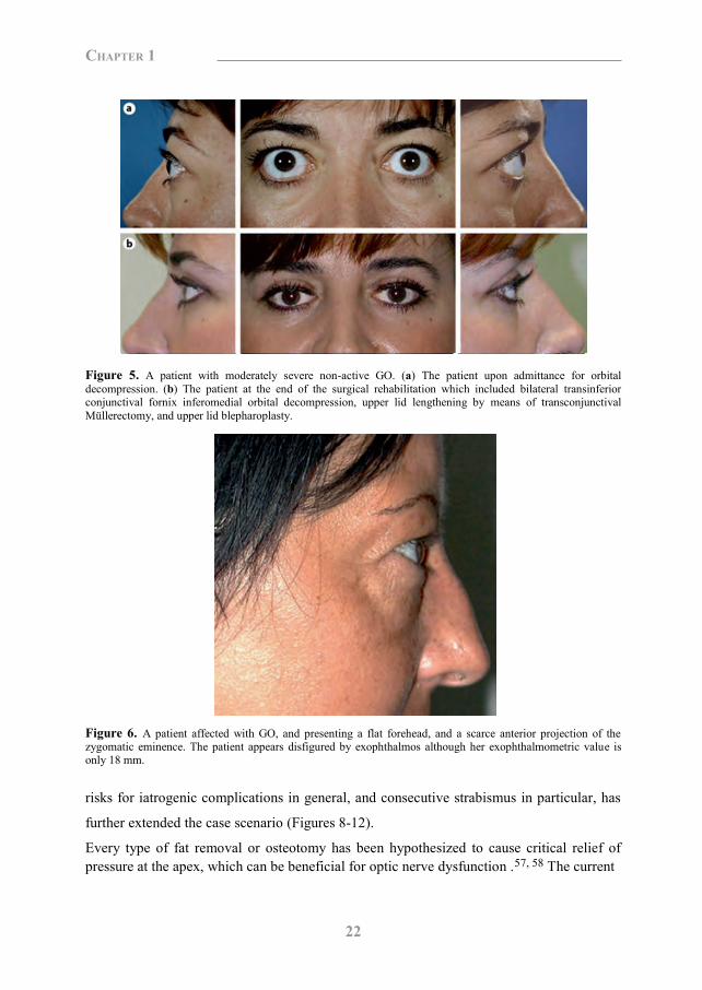
Figure 5. A patient with moderately severe non-active GO. (a) The patient upon admittance for orbital decompression. (b) The patient at the end of the surgical rehabilitation which included bilateral transinferior conjunctival fornix inferomedial orbital decompression, upper lid lengthening by means of transconjunctival Müllerectomy, and upper lid blepharoplasty.
Figure 6. A patient affected with GO, and presenting a flat forehead, and a scarce anterior projection of the zygomatic eminence. The patient appears disfigured by exophthalmos although her exophthalmometric value is only 18 mm.
risks for iatrogenic complications in general, and consecutive strabismus in particular, has
further extended the case scenario (Figures 8-12).
Every type of fat removal or osteotomy has been hypothesized to cause critical relief of pressure at the apex, which can be beneficial for optic nerve dysfunction .57, 58 The current
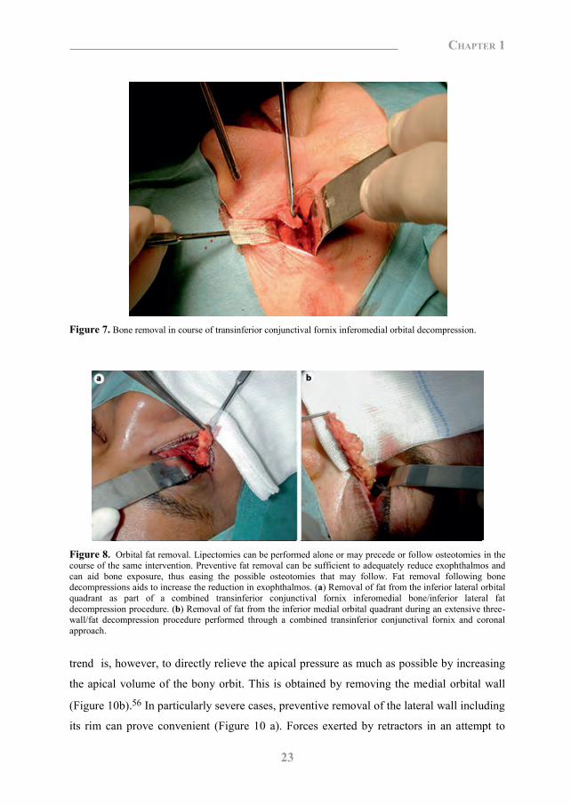
Figure 7. Bone removal in course of transinferior conjunctival fornix inferomedial orbital decompression.
Figure 8. Orbital fat removal. Lipectomies can be performed alone or may precede or follow osteotomies in the course of the same intervention. Preventive fat removal can be sufficient to adequately reduce exophthalmos and can aid bone exposure, thus easing the possible osteotomies that may follow. Fat removal following bone decompressions aids to increase the reduction in exophthalmos. (a) Removal of fat from the inferior lateral orbital quadrant as part of a combined transinferior conjunctival fornix inferomedial bone/inferior lateral fat decompression procedure. (b) Removal of fat from the inferior medial orbital quadrant during an extensive three-wall/fat decompression procedure performed through a combined transinferior conjunctival fornix and coronal approach.
trend is, however, to directly relieve the apical pressure as much as possible by increasing
the apical volume of the bony orbit. This is obtained by removing the medial orbital wall
(Figure 10b).56 In particularly severe cases, preventive removal of the lateral wall including
its rim can prove convenient (Figure 10 a). Forces exerted by retractors in an attempt to
100379 Thesis Baldeschi.indd 22 vrijdag17-december-2010 12:07
23
Chapter 1
Figure 5. A patient with moderately severe non-active GO. (a) The patient upon admittance for orbital decompression. (b) The patient at the end of the surgical rehabilitation which included bilateral transinferior conjunctival fornix inferomedial orbital decompression, upper lid lengthening by means of transconjunctival Müllerectomy, and upper lid blepharoplasty.
Figure 6. A patient affected with GO, and presenting a flat forehead, and a scarce anterior projection of the zygomatic eminence. The patient appears disfigured by exophthalmos although her exophthalmometric value is only 18 mm.
risks for iatrogenic complications in general, and consecutive strabismus in particular, has
further extended the case scenario (Figures 8-12).
Every type of fat removal or osteotomy has been hypothesized to cause critical relief of pressure at the apex, which can be beneficial for optic nerve dysfunction .57, 58 The current
Figure 7. Bone removal in course of transinferior conjunctival fornix inferomedial orbital decompression.
Figure 8. Orbital fat removal. Lipectomies can be performed alone or may precede or follow osteotomies in the course of the same intervention. Preventive fat removal can be sufficient to adequately reduce exophthalmos and can aid bone exposure, thus easing the possible osteotomies that may follow. Fat removal following bone decompressions aids to increase the reduction in exophthalmos. (a) Removal of fat from the inferior lateral orbital quadrant as part of a combined transinferior conjunctival fornix inferomedial bone/inferior lateral fat decompression procedure. (b) Removal of fat from the inferior medial orbital quadrant during an extensive three-wall/fat decompression procedure performed through a combined transinferior conjunctival fornix and coronal approach.
trend is, however, to directly relieve the apical pressure as much as possible by increasing
the apical volume of the bony orbit. This is obtained by removing the medial orbital wall
(Figure 10b).56 In particularly severe cases, preventive removal of the lateral wall including
its rim can prove convenient (Figure 10 a). Forces exerted by retractors in an attempt to
100379 Thesis Baldeschi.indd 23 vrijdag17-december-2010 12:07
24
Chapter 1
achieve apex decompression along the medial orbital wall can increase the already high
retro-bulbar pressure up to critical levels for optic nerve fibres and vasculature. The
preventive removal of the lateral orbital wall permits surgeons to address the deepest orbit
more smoothly, reducing the risk of adding an iatrogenic component to the pathologically
high orbital pressure at the basis of the neuropathy. There are several possible options to
remove the medial orbital wall, but transconjunctival routes (either transcaruncular or
transinferior fornix), which leave no visible scars are currently preferred.59, 60 The
endoscopic transnasal approach, described first and relatively recently by Kennedy et al.61,
addressing the orbital apex without any substantial increase in the intraorbital pressure can
also be a valid alternative that can even be performed under local anesthesia.62, 63
During the last three decades, when the number of rehabilitative orbital decompressions
started to rise64, it became of primary importance to balance a given technique in terms of
not only effectiveness in reducing exophthalmos, but also (and mostly) in terms of safety.
In the early 1980s, the antral-ethmoidal decompression by a transantral approach, as
described by Walsh and Ogura in 1957, was the mainstay technique.64-66 The major
disadvantage reported with transantral surgery was a subsequent motility imbalance as high
as 52%67, and therefore alternative procedures were sought in an attempt to decrease the
risk of decompression-induced diplopia. In cases of mild exophthalmos, trans-lid antral-
ethmoidal decompression appeared to be a valid alternative, with a risk of iatrogenic
diplopia in only 4.6% of patients.64 For more severe exophthalmos, infero-medial
decompression was used in combination with lateral decompression. Such procedures were
also related with a low incidence of consecutive diplopia.66 In 1989, Leone et al.68, in an
attempt to further reduce post-decompression strabismus, proposed balancing the
decompression by removing the medial and lateral orbital walls while sparing the floor.68
This technique, which theoretically should have minimised the risk of iatrogenic diplopia,
later appeared to be associated with a higher risk of such a complication compared with
removal of the lateral orbital wall alone, or with inferomedial and three-wall surgeries.69, 70
At present the medial wall, the orbital floor and the lateral wall continue to be addressed
during bony decompression surgery (Figure 10), while orbital roof removal has been
abandoned due to the fact that its contribution to orbital expansion is minimal and
associated with potential complications and side effects. Although orbital floor removal in
the course of orbital decompression is currently not favoured in North America, a recent
prospective survey of the European Group on Graves‟ Orbitopathy (EUGOGO) showed
that inferomedial bone decompression is still a widely used procedure in Europe.71
Depending on the severity of exophthalmos, the effect of inferomedial decompression can
be implemented by adding lateral wall decompression and/or removal of the fat, usually of
the inferior lateral orbital quadrant (Figures 8, 11). In view of reducing postoperative
diplopia, an opposite sequence which involves firstly the removal of the lateral wall,
associated or not to fat excision, and secondly, if necessary, the removal of the medial and
inferior walls has been suggested.54 This strategy represents a significant conceptual
departure from the traditional approach, which began with inferomedial decompression,
and suggests regarding the lateral orbital wall and in particular its deep portion as being the
region of first choice for orbital decompression in the case of rehabilitative surgery (Figure
10a). Removal of the lateral orbital wall - which appears to be connected with a low risk of
consecutive diplopia or severe complications, such as cerebrospinal fluid leak - perfectly
fits the needs of the increasingly demanding patient population.54-56 It was recently
reported that removal of the deep lateral wall as part of a rehabilitative coronal-approach 3-
wall decompression gives a 32% enhancement in exophthalmos reduction without
increasing the risk of consecutive diplopia as compared with traditional more conservative
3-wall decompression.72 The same study, however, confirmed the known high
interindividual variability in the volume of the deep lateral wall. In light of this, the deep
lateral wall is to be considered an effective although not always available zone of possible
orbital volume expansion when dealing with rehabilitative decompression surgery.72 The
effect of pure lateral wall decompression on exophthalmos reduction may be modest if not
associated with medial wall removal, but in this case the risk of consecutive diplopia arises,
while the result of lateral wall decompression can be augmented by intraconal fat removal
without substantially increasing the risk of iatrogenic strabismus. On the contrary, removal
of the lateral orbital wall and intraconal fat was beneficial in reducing preoperative primary
gaze diplopia.36
100379 Thesis Baldeschi.indd 24 vrijdag17-december-2010 12:07
25
Chapter 1
achieve apex decompression along the medial orbital wall can increase the already high
retro-bulbar pressure up to critical levels for optic nerve fibres and vasculature. The
preventive removal of the lateral orbital wall permits surgeons to address the deepest orbit
more smoothly, reducing the risk of adding an iatrogenic component to the pathologically
high orbital pressure at the basis of the neuropathy. There are several possible options to
remove the medial orbital wall, but transconjunctival routes (either transcaruncular or
transinferior fornix), which leave no visible scars are currently preferred.59, 60 The
endoscopic transnasal approach, described first and relatively recently by Kennedy et al.61,
addressing the orbital apex without any substantial increase in the intraorbital pressure can
also be a valid alternative that can even be performed under local anesthesia.62, 63
During the last three decades, when the number of rehabilitative orbital decompressions
started to rise64, it became of primary importance to balance a given technique in terms of
not only effectiveness in reducing exophthalmos, but also (and mostly) in terms of safety.
In the early 1980s, the antral-ethmoidal decompression by a transantral approach, as
described by Walsh and Ogura in 1957, was the mainstay technique.64-66 The major
disadvantage reported with transantral surgery was a subsequent motility imbalance as high
as 52%67, and therefore alternative procedures were sought in an attempt to decrease the
risk of decompression-induced diplopia. In cases of mild exophthalmos, trans-lid antral-
ethmoidal decompression appeared to be a valid alternative, with a risk of iatrogenic
diplopia in only 4.6% of patients.64 For more severe exophthalmos, infero-medial
decompression was used in combination with lateral decompression. Such procedures were
also related with a low incidence of consecutive diplopia.66 In 1989, Leone et al.68, in an
attempt to further reduce post-decompression strabismus, proposed balancing the
decompression by removing the medial and lateral orbital walls while sparing the floor.68
This technique, which theoretically should have minimised the risk of iatrogenic diplopia,
later appeared to be associated with a higher risk of such a complication compared with
removal of the lateral orbital wall alone, or with inferomedial and three-wall surgeries.69, 70
At present the medial wall, the orbital floor and the lateral wall continue to be addressed
during bony decompression surgery (Figure 10), while orbital roof removal has been
abandoned due to the fact that its contribution to orbital expansion is minimal and
associated with potential complications and side effects. Although orbital floor removal in
the course of orbital decompression is currently not favoured in North America, a recent
prospective survey of the European Group on Graves‟ Orbitopathy (EUGOGO) showed
that inferomedial bone decompression is still a widely used procedure in Europe.71
Depending on the severity of exophthalmos, the effect of inferomedial decompression can
be implemented by adding lateral wall decompression and/or removal of the fat, usually of
the inferior lateral orbital quadrant (Figures 8, 11). In view of reducing postoperative
diplopia, an opposite sequence which involves firstly the removal of the lateral wall,
associated or not to fat excision, and secondly, if necessary, the removal of the medial and
inferior walls has been suggested.54 This strategy represents a significant conceptual
departure from the traditional approach, which began with inferomedial decompression,
and suggests regarding the lateral orbital wall and in particular its deep portion as being the
region of first choice for orbital decompression in the case of rehabilitative surgery (Figure
10a). Removal of the lateral orbital wall - which appears to be connected with a low risk of
consecutive diplopia or severe complications, such as cerebrospinal fluid leak - perfectly
fits the needs of the increasingly demanding patient population.54-56 It was recently
reported that removal of the deep lateral wall as part of a rehabilitative coronal-approach 3-
wall decompression gives a 32% enhancement in exophthalmos reduction without
increasing the risk of consecutive diplopia as compared with traditional more conservative
3-wall decompression.72 The same study, however, confirmed the known high
interindividual variability in the volume of the deep lateral wall. In light of this, the deep
lateral wall is to be considered an effective although not always available zone of possible
orbital volume expansion when dealing with rehabilitative decompression surgery.72 The
effect of pure lateral wall decompression on exophthalmos reduction may be modest if not
associated with medial wall removal, but in this case the risk of consecutive diplopia arises,
while the result of lateral wall decompression can be augmented by intraconal fat removal
without substantially increasing the risk of iatrogenic strabismus. On the contrary, removal
of the lateral orbital wall and intraconal fat was beneficial in reducing preoperative primary
gaze diplopia.36
100379 Thesis Baldeschi.indd 25 vrijdag17-december-2010 12:07
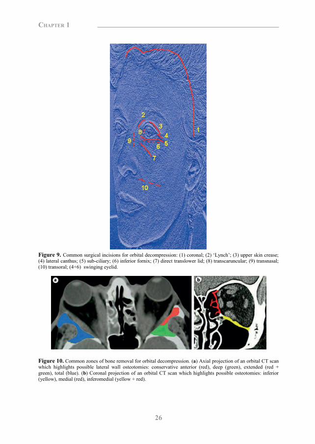
26
Chapter 1
Figure 9. Common surgical incisions for orbital decompression: (1) coronal; (2) „Lynch‟; (3) upper skin crease; (4) lateral canthus; (5) sub-ciliary; (6) inferior fornix; (7) direct translower lid; (8) transcaruncular; (9) transnasal; (10) transoral; (4+6) swinging eyelid.
Figure 10. Common zones of bone removal for orbital decompression. (a) Axial projection of an orbital CT scan which highlights possible lateral wall osteotomies: conservative anterior (red), deep (green), extended (red + green), total (blue). (b) Coronal projection of an orbital CT scan which highlights possible osteotomies: inferior (yellow), medial (red), inferomedial (yellow + red).
Figure 11. Schematic representation of sites for orbital lipectomies reachable (yellow) or not (blue) through hidden transconjunctival incisions (red line) superimposed on a periorbital region photograph of a patient with GO (a) and on a coronal projection of an orbital magnetic resonance scan taken at the level of the middle orbit in a patient with GO (b). The fat compartment of the superior lateral quadrant (blue) can be exposed only through transcutaneous incisions; however, it is not used as an elective site for lipectomy because it hosts delicate structures and its removal gives a minimal contribution to fat decompression surgery.
Figure 12. A patient with moderate non-active GO and documentation of his intervention. (a) The patient upon admission for orbital decompression. Exophthalmos, hypoglobus, and lower lid displacement right side were present. (b) The patient 3 months after superomedial transcutaneous-septal fat decompression performed through an upper skin crease approach. Exophthalmos, hypoglobus, and lower lid displacement were adequately treated. (c) Surgical site. The skin and the orbicularis oculi muscle are retracted. An aperture through the orbital septum (white arrow heads), which is kept open with a cotton tip applicator following partial removal of the superomedial extraconal fat, shows (from left to right): medial margin of the levator palpebrae superioris muscle aponeurosis (blue arrow), anterior margin of the superior oblique muscle tendon (white arrow), intraconal fat (yellow arrow), and belly of the medial rectus muscle (red arrow). (d) Fat removed from the superomedial orbital quadrant at the end of surgery.
100379 Thesis Baldeschi.indd 26 vrijdag17-december-2010 12:07
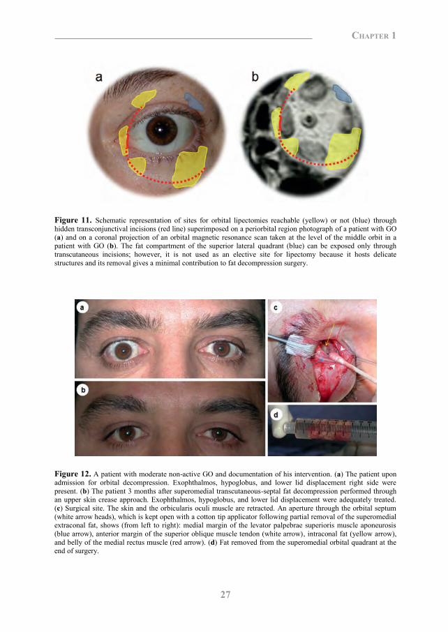
27
Chapter 1
Figure 9. Common surgical incisions for orbital decompression: (1) coronal; (2) „Lynch‟; (3) upper skin crease; (4) lateral canthus; (5) sub-ciliary; (6) inferior fornix; (7) direct translower lid; (8) transcaruncular; (9) transnasal; (10) transoral; (4+6) swinging eyelid.
Figure 10. Common zones of bone removal for orbital decompression. (a) Axial projection of an orbital CT scan which highlights possible lateral wall osteotomies: conservative anterior (red), deep (green), extended (red + green), total (blue). (b) Coronal projection of an orbital CT scan which highlights possible osteotomies: inferior (yellow), medial (red), inferomedial (yellow + red).
Figure 11. Schematic representation of sites for orbital lipectomies reachable (yellow) or not (blue) through hidden transconjunctival incisions (red line) superimposed on a periorbital region photograph of a patient with GO (a) and on a coronal projection of an orbital magnetic resonance scan taken at the level of the middle orbit in a patient with GO (b). The fat compartment of the superior lateral quadrant (blue) can be exposed only through transcutaneous incisions; however, it is not used as an elective site for lipectomy because it hosts delicate structures and its removal gives a minimal contribution to fat decompression surgery.
Figure 12. A patient with moderate non-active GO and documentation of his intervention. (a) The patient upon admission for orbital decompression. Exophthalmos, hypoglobus, and lower lid displacement right side were present. (b) The patient 3 months after superomedial transcutaneous-septal fat decompression performed through an upper skin crease approach. Exophthalmos, hypoglobus, and lower lid displacement were adequately treated. (c) Surgical site. The skin and the orbicularis oculi muscle are retracted. An aperture through the orbital septum (white arrow heads), which is kept open with a cotton tip applicator following partial removal of the superomedial extraconal fat, shows (from left to right): medial margin of the levator palpebrae superioris muscle aponeurosis (blue arrow), anterior margin of the superior oblique muscle tendon (white arrow), intraconal fat (yellow arrow), and belly of the medial rectus muscle (red arrow). (d) Fat removed from the superomedial orbital quadrant at the end of surgery.
100379 Thesis Baldeschi.indd 27 vrijdag17-december-2010 12:07
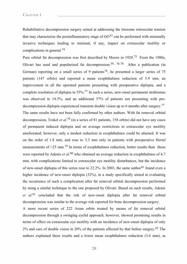
28
Chapter 1
Rehabilitative decompression surgery aimed at addressing the tiresome retroocular tension
that may characterise the postinflammatory stage of GO53 can be performed with minimally
invasive techniques leading to minimal, if any, impact on extraocular motility or
complications in general.74
Pure orbital fat decompression was first described by Moore in 1920.75 From the 1980s,
Olivari has used and popularized fat decompression.39, 76-78 After a publication (in
German) reporting on a small series of 9 patients76, he presented a larger series of 75
patients (147 orbits) and reported a mean exophthalmos reduction of 5.9 mm, an
improvement in all the operated patients presenting with preoperative diplopia, and a
complete resolution of diplopia in 55%.77 In such a series, new-onset permanent strabismus
was observed in 14.3%, and an additional 57% of patients not presenting with pre-
decompression diplopia experienced transient double vision up to 6 months after surgery.77
The same results have not been fully confirmed by other authors. With fat removal orbital
decompression, Trokel et al.79 (in a series of 81 patients, 158 orbits) did not have any cases
of permanent induced diplopia and on average restrictions in extraocular eye motility
ameliorated; however, only a modest reduction in exophthalmos could be attained. It was
on the order of 1.8 mm, and rose to 3.3 mm only in patients with preoperative Hertel
measurements of >25 mm.79 In terms of exophthalmos reduction, better results than those
were reported by Adenis et al.80 who obtained an average reduction in exophthalmos of 4.7
mm, with complications limited to extraocular eye motility disturbances, but the incidence
of new-onset diplopia of this series rose to 22.2%. In 2003, the same author81 found even a
higher incidence of new-onset diplopia (32%), in a study specifically aimed at evaluating
the occurrence of such a complication after fat removal orbital decompression performed
by using a similar technique to the one proposed by Olivari. Based on such results, Adenis
et al.81 concluded that the risk of new-onset diplopia after fat removal orbital
decompression was similar to the average risk reported for bone decompression surgery.
A more recent series of 222 Asian orbits treated by means of fat removal orbital
decompression through a swinging eyelid approach, however, showed promising results in
terms of effect on extraocular eye motility with an incidence of new-onset diplopia of only
2% and cure of double vision in 20% of the patients affected by that before surgery.82 The
authors explained these results and a lower mean exophthalmos reduction (3.6 mm), as
compared with the series of Adenies et al.81, on the basis of differences in surgical
techniques used and in anatomy between the Asian and Caucasian orbits.82
Simultaneously with the Asian series, the group of Olivari reported on their 20-year
experience with trans-palpebral fat decompression by presenting a monumental series of
2,697 operations.78 Although the mean exophthalmos reduction remained similar to the
earliest study of the mentor of this group77, persistent new-onset diplopia requiring surgical
or prismatic correction later than 6 months after surgery rose to 22.2%.78
Despite the discrepancy in the amount of exophthalmos reduction and effects on
extraocular eye motility, overall fat removal orbital decompression has proven to be a safe
procedure through the years, and its positive effects also include improvement in visual
function and reduction in intraocular tension when elevated secondary to raised intraorbital
pressure.39, 58, 83 It appears to be an effective procedure mostly indicated in moderate-to-
severe cases of lipogenic exophthalmos and periocular disfigurement due to fat prolapse
and venous congestion, although it has also been used as a standard approach to any type of
functional or rehabilitative orbital decompression (Figures 12, 13).39, 76-83
During recent years, the combination of bone decompression associated with fat removal
(Figure 8) has been gaining popularity in view of its reported safety and increased
effectiveness as compared with bone or fat decompression alone.54-56
Beveled osteotomies and onlay alloplastic periorbital implants - although sporadically used
to camouflage more than to reduce exophthalmos - remain of uncertain effectiveness84-86,
and their edges, not infrequently visible, may devalue the final rehabilitative cosmetic
result.
Most of the techniques currently used seem to be effective either in restoring impaired
vision, and eye position or reversing congestive symptomatology and disfigurement of the
periorbital region. An unbiased analysis of the current literature in terms of effectiveness
versus safety, however, is extremely difficult because of the great heterogeneity of the
patients included in the published studies; differences in definitions and methodologies
used to study patients before decompression and to assess results after surgery; variations
applied to surgical techniques that while falling under the same general definition may be
completely different concerning surgical route, modality, location, and amount of removed
bone or fat. Furthermore, it should be noted that the evidence of the literature concerning
100379 Thesis Baldeschi.indd 28 vrijdag17-december-2010 12:07
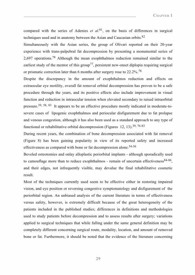
29
Chapter 1
Rehabilitative decompression surgery aimed at addressing the tiresome retroocular tension
that may characterise the postinflammatory stage of GO53 can be performed with minimally
invasive techniques leading to minimal, if any, impact on extraocular motility or
complications in general.74
Pure orbital fat decompression was first described by Moore in 1920.75 From the 1980s,
Olivari has used and popularized fat decompression.39, 76-78 After a publication (in
German) reporting on a small series of 9 patients76, he presented a larger series of 75
patients (147 orbits) and reported a mean exophthalmos reduction of 5.9 mm, an
improvement in all the operated patients presenting with preoperative diplopia, and a
complete resolution of diplopia in 55%.77 In such a series, new-onset permanent strabismus
was observed in 14.3%, and an additional 57% of patients not presenting with pre-
decompression diplopia experienced transient double vision up to 6 months after surgery.77
The same results have not been fully confirmed by other authors. With fat removal orbital
decompression, Trokel et al.79 (in a series of 81 patients, 158 orbits) did not have any cases
of permanent induced diplopia and on average restrictions in extraocular eye motility
ameliorated; however, only a modest reduction in exophthalmos could be attained. It was
on the order of 1.8 mm, and rose to 3.3 mm only in patients with preoperative Hertel
measurements of >25 mm.79 In terms of exophthalmos reduction, better results than those
were reported by Adenis et al.80 who obtained an average reduction in exophthalmos of 4.7
mm, with complications limited to extraocular eye motility disturbances, but the incidence
of new-onset diplopia of this series rose to 22.2%. In 2003, the same author81 found even a
higher incidence of new-onset diplopia (32%), in a study specifically aimed at evaluating
the occurrence of such a complication after fat removal orbital decompression performed
by using a similar technique to the one proposed by Olivari. Based on such results, Adenis
et al.81 concluded that the risk of new-onset diplopia after fat removal orbital
decompression was similar to the average risk reported for bone decompression surgery.
A more recent series of 222 Asian orbits treated by means of fat removal orbital
decompression through a swinging eyelid approach, however, showed promising results in
terms of effect on extraocular eye motility with an incidence of new-onset diplopia of only
2% and cure of double vision in 20% of the patients affected by that before surgery.82 The
authors explained these results and a lower mean exophthalmos reduction (3.6 mm), as
compared with the series of Adenies et al.81, on the basis of differences in surgical
techniques used and in anatomy between the Asian and Caucasian orbits.82
Simultaneously with the Asian series, the group of Olivari reported on their 20-year
experience with trans-palpebral fat decompression by presenting a monumental series of
2,697 operations.78 Although the mean exophthalmos reduction remained similar to the
earliest study of the mentor of this group77, persistent new-onset diplopia requiring surgical
or prismatic correction later than 6 months after surgery rose to 22.2%.78
Despite the discrepancy in the amount of exophthalmos reduction and effects on
extraocular eye motility, overall fat removal orbital decompression has proven to be a safe
procedure through the years, and its positive effects also include improvement in visual
function and reduction in intraocular tension when elevated secondary to raised intraorbital
pressure.39, 58, 83 It appears to be an effective procedure mostly indicated in moderate-to-
severe cases of lipogenic exophthalmos and periocular disfigurement due to fat prolapse
and venous congestion, although it has also been used as a standard approach to any type of
functional or rehabilitative orbital decompression (Figures 12, 13).39, 76-83
During recent years, the combination of bone decompression associated with fat removal
(Figure 8) has been gaining popularity in view of its reported safety and increased
effectiveness as compared with bone or fat decompression alone.54-56
Beveled osteotomies and onlay alloplastic periorbital implants - although sporadically used
to camouflage more than to reduce exophthalmos - remain of uncertain effectiveness84-86,
and their edges, not infrequently visible, may devalue the final rehabilitative cosmetic
result.
Most of the techniques currently used seem to be effective either in restoring impaired
vision, and eye position or reversing congestive symptomatology and disfigurement of the
periorbital region. An unbiased analysis of the current literature in terms of effectiveness
versus safety, however, is extremely difficult because of the great heterogeneity of the
patients included in the published studies; differences in definitions and methodologies
used to study patients before decompression and to assess results after surgery; variations
applied to surgical techniques that while falling under the same general definition may be
completely different concerning surgical route, modality, location, and amount of removed
bone or fat. Furthermore, it should be noted that the evidence of the literature concerning
100379 Thesis Baldeschi.indd 29 vrijdag17-december-2010 12:07
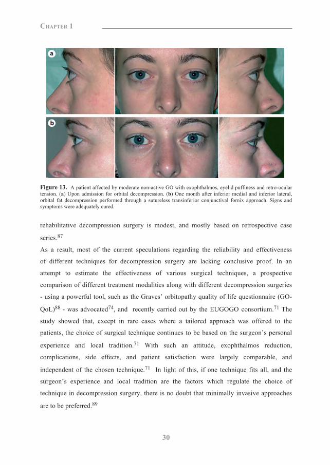
30
Chapter 1
Figure 13. A patient affected by moderate non-active GO with exophthalmos, eyelid puffiness and retro-ocular
tension. (a) Upon admission for orbital decompression. (b) One month after inferior medial and inferior lateral,
orbital fat decompression performed through a sutureless transinferior conjunctival fornix approach. Signs and
symptoms were adequately cured.
rehabilitative decompression surgery is modest, and mostly based on retrospective case
series.87
As a result, most of the current speculations regarding the reliability and effectiveness
of different techniques for decompression surgery are lacking conclusive proof. In an
attempt to estimate the effectiveness of various surgical techniques, a prospective
comparison of different treatment modalities along with different decompression surgeries
- using a powerful tool, such as the Graves’ orbitopathy quality of life questionnaire (GO-
QoL)88 - was advocated74, and recently carried out by the EUGOGO consortium.71 The
study showed that, except in rare cases where a tailored approach was offered to the
patients, the choice of surgical technique continues to be based on the surgeon’s personal
experience and local tradition.71 With such an attitude, exophthalmos reduction,
complications, side effects, and patient satisfaction were largely comparable, and
independent of the chosen technique.71 In light of this, if one technique fits all, and the
surgeon’s experience and local tradition are the factors which regulate the choice of
technique in decompression surgery, there is no doubt that minimally invasive approaches
are to be preferred.89
Although the average results may be largely comparable regardless of the surgical
technique used, forcing patients into standardised surgical frames is suboptimal as a
standardised approach may produce unsuitable results for the single patient. Ideally, the
planning of decompression surgery should comply with the patient’s expectations and
needs, and should be matched to the patient’s anatomical and pathological substrates and
possible previous surgeries.11
In clinical practice, this desirable approach to orbital decompression, which may involve
the use of several different surgical techniques, is possible only in centres where adequate
referral and a long-lasting tradition in orbital surgery favour transmission of expertise and
warrant adequate back-up, whilst offering the possibility to develop new techniques or to
master ongoing variations of others. As this cannot be the case in most of the centres
dealing with GO, the use of a standardised versatile approach is to be regarded as an
acceptable although suboptimal alternative.89
The swinging eyelid approach described first by McCord in the early 1980s60 - with the
conjunctival incision that can be extended medially as much as necessary, offering an
adequate access to the bony orbit and to the orbital fat compartments - is a versatile
technique that can be used for the majority of patients needing decompression surgery.89 As
an alternative, inferior and medial conjunctival fornix and upper skin crease incisions can
be used separately or in combination (Figures 9-12).
In 2003, Cruz and Leme questioned whether the coronal approach continues to have a role
in orbital decompression and concluded that there is little, if any, need for this technique.90
Although the study design and the results of their paper could not offer strong support to
their conclusions, it is easy to share their doubts about using an invasive approach, such as
the coronal incision, as a standard route for orbital decompression.
Minimally invasive approaches and hidden periorbital incisions are currently preferred
(Figures 5, 9, 11-13); nevertheless, the coronal incision is not to be abandoned as it can be
an additional tool in surgeons’ hands when dealing with patients who can benefit more
from a tailored approach than a standardised one. There are many circumstances in which
this may happen. These include the presence of remarkable periorbital swelling or
conjunctival chemosis, the necessity of minimising the number of periorbital incisions, or
the necessity of extensive manipulation of the lateral wall (including its rim).89, 91 Through
100379 Thesis Baldeschi.indd 30 vrijdag17-december-2010 12:07
31
Chapter 1
Figure 13. A patient affected by moderate non-active GO with exophthalmos, eyelid puffiness and retro-ocular
tension. (a) Upon admission for orbital decompression. (b) One month after inferior medial and inferior lateral,
orbital fat decompression performed through a sutureless transinferior conjunctival fornix approach. Signs and
symptoms were adequately cured.
rehabilitative decompression surgery is modest, and mostly based on retrospective case
series.87
As a result, most of the current speculations regarding the reliability and effectiveness
of different techniques for decompression surgery are lacking conclusive proof. In an
attempt to estimate the effectiveness of various surgical techniques, a prospective
comparison of different treatment modalities along with different decompression surgeries
- using a powerful tool, such as the Graves’ orbitopathy quality of life questionnaire (GO-
QoL)88 - was advocated74, and recently carried out by the EUGOGO consortium.71 The
study showed that, except in rare cases where a tailored approach was offered to the
patients, the choice of surgical technique continues to be based on the surgeon’s personal
experience and local tradition.71 With such an attitude, exophthalmos reduction,
complications, side effects, and patient satisfaction were largely comparable, and
independent of the chosen technique.71 In light of this, if one technique fits all, and the
surgeon’s experience and local tradition are the factors which regulate the choice of
technique in decompression surgery, there is no doubt that minimally invasive approaches
are to be preferred.89
Although the average results may be largely comparable regardless of the surgical
technique used, forcing patients into standardised surgical frames is suboptimal as a
standardised approach may produce unsuitable results for the single patient. Ideally, the
planning of decompression surgery should comply with the patient’s expectations and
needs, and should be matched to the patient’s anatomical and pathological substrates and
possible previous surgeries.11
In clinical practice, this desirable approach to orbital decompression, which may involve
the use of several different surgical techniques, is possible only in centres where adequate
referral and a long-lasting tradition in orbital surgery favour transmission of expertise and
warrant adequate back-up, whilst offering the possibility to develop new techniques or to
master ongoing variations of others. As this cannot be the case in most of the centres
dealing with GO, the use of a standardised versatile approach is to be regarded as an
acceptable although suboptimal alternative.89
The swinging eyelid approach described first by McCord in the early 1980s60 - with the
conjunctival incision that can be extended medially as much as necessary, offering an
adequate access to the bony orbit and to the orbital fat compartments - is a versatile
technique that can be used for the majority of patients needing decompression surgery.89 As
an alternative, inferior and medial conjunctival fornix and upper skin crease incisions can
be used separately or in combination (Figures 9-12).
In 2003, Cruz and Leme questioned whether the coronal approach continues to have a role
in orbital decompression and concluded that there is little, if any, need for this technique.90
Although the study design and the results of their paper could not offer strong support to
their conclusions, it is easy to share their doubts about using an invasive approach, such as
the coronal incision, as a standard route for orbital decompression.
Minimally invasive approaches and hidden periorbital incisions are currently preferred
(Figures 5, 9, 11-13); nevertheless, the coronal incision is not to be abandoned as it can be
an additional tool in surgeons’ hands when dealing with patients who can benefit more
from a tailored approach than a standardised one. There are many circumstances in which
this may happen. These include the presence of remarkable periorbital swelling or
conjunctival chemosis, the necessity of minimising the number of periorbital incisions, or
the necessity of extensive manipulation of the lateral wall (including its rim).89, 91 Through
100379 Thesis Baldeschi.indd 31 vrijdag17-december-2010 12:07
32
Chapter 1
a coronal incision, brow lift, and correction of frontal/glabellar rhytids, which are often
necessary in patients with GO, can be performed simultaneously with orbital
decompression; thus, favourably speeding-up the timing of rehabilitative surgery.89, 92
4.4. What Are the Possible Complications of Orbital Decompression?
Orbital fat decompression has never reached the popularity of bone decompression due to
the theoretical (more than real) complications that may potentially be connected with this
approach and which encompass damages to oculomotor ciliary and lacrimal nerves, orbital
vasculature, extraocular eye muscles, optic nerve and the eyeball itself.80 Also, in the case
of bone orbital decompression, in spite of theoretical expectations, severe complications are
rare in clinical practice. Common complications of this surgical approach are consecutive
strabismus, infraorbital hypoaesthesia and sinusitis93, lower lid entropion94, and eyeball
dystopia95, while leakage of cerebrospinal fluid, infections involving the central nervous
system, damage to the eye and optic nerve or their vasculature, cerebral vasospasm,
ischaemia and infarction are severe but rare events.42, 96 Reactivation of GO after
rehabilitative bony orbital decompression is another rare complication very recently
described in 3 of 239 patients not treated with perioperative glucocorticoids. The
phenomenon consisted of the onset of typical signs and symptoms of active GO with
radiologic evidence of extraocular muscle enlargement a few weeks after surgery and
following a normal convalescence period. Based on its clinical characteristics, the
observation was named delayed decompression-related reactivation (DDRR). The incidence
of DDRR appeared to be in the order of 1.3% and could be controlled with systemic
immunosuppression or retrobulbar irradiation.97
In addition to complications common to orbital decompressions in general, different
surgical approaches and routes may carry the risk of specific complications. The second
branch of the trigeminal nerve may be damaged in the course of orbital floor removal
decompression. This may potentially induce the formation of traumatic or amputation
neuromas. Such lesions, although rare, should be included in the potential complications of
decompressions when counselling patients about to undergo this type of surgery, as they are
difficult to treat and may cause persistent and disabling pain.98
into the nasal cavity and scarring of the nasal mucosa with meatal and sinus obstruction,
mucocele and sinusitis can occur with endoscopic endonasal approaches.99, 100
The coronal approach leaving the eyelid undisturbed is less likely than periorbital incisions
to create complications which may potentially be harmful to the eye. Periorbital scarring
with iatrogenic lid retraction and cicatricial lagophthalmos, eyelid margin malpositions and
ptosis (although rare) are more likely to occur with periorbital incisions. On the other hand,
temporal bossing, damage to the frontalis nerve, scarring and alopecia at the site of the
scalp incision, or effects upon ischemic areas of the frontal flap after healing by secondary
intention may complicate the coronal approach.89
The coronal approach should not be considered a more hazardous technique as compared
with the less invasive periorbital incisions. The more serious complications connected with
the coronal approach have been reported in small series where less than 3 patients per year
were operated upon.90, 101
4.5. Can Complications Be Predicted or Prevented?
Most of the possible complications cannot be predicted and their prevention is based on
recommendations which are not specific in nature, and which include careful manipulation
of the orbital content, accurate dissection of the orbital fat, and avoidance of expandable
haemostatic agents and/or extensive use of diathermy within the orbit.
Other complications with known pathogenesis such as sinusitis can be simply prevented
by taking care to create an adequate sinus aeration as a part of the surgical procedure at the
time of bone decompression.
The occurrence of other complications, namely infraorbital hypoesthesia or pain, eyeball
dystopia leakage of cerebrospinal fluid and possible consecutive infectious involvement of
the central nervous system, can be reduced by means of accurate evaluation of preoperative
imaging, adequate planning of surgical intervention and the use of prophylactic antibiotics.
Strict observance of this methodology helps detecting patients at an increased risk of
possible complications. A low lamina cribrosa for instance should be regarded as a possible
source of cerebrospinal fluid leakage for those patients planned for medial wall
decompression. Late-onset enophthalmos and hypoglobus can be prevented simply by
avoiding inferomedial decompression when dealing with patients recognised to be at higher
risk due to their anatomical substrate. The complication had in fact been described as
100379 Thesis Baldeschi.indd 32 vrijdag17-december-2010 12:07
33
Chapter 1
a coronal incision, brow lift, and correction of frontal/glabellar rhytids, which are often
necessary in patients with GO, can be performed simultaneously with orbital
decompression; thus, favourably speeding-up the timing of rehabilitative surgery.89, 92
4.4. What Are the Possible Complications of Orbital Decompression?
Orbital fat decompression has never reached the popularity of bone decompression due to
the theoretical (more than real) complications that may potentially be connected with this
approach and which encompass damages to oculomotor ciliary and lacrimal nerves, orbital
vasculature, extraocular eye muscles, optic nerve and the eyeball itself.80 Also, in the case
of bone orbital decompression, in spite of theoretical expectations, severe complications are
rare in clinical practice. Common complications of this surgical approach are consecutive
strabismus, infraorbital hypoaesthesia and sinusitis93, lower lid entropion94, and eyeball
dystopia95, while leakage of cerebrospinal fluid, infections involving the central nervous
system, damage to the eye and optic nerve or their vasculature, cerebral vasospasm,
ischaemia and infarction are severe but rare events.42, 96 Reactivation of GO after
rehabilitative bony orbital decompression is another rare complication very recently
described in 3 of 239 patients not treated with perioperative glucocorticoids. The
phenomenon consisted of the onset of typical signs and symptoms of active GO with
radiologic evidence of extraocular muscle enlargement a few weeks after surgery and
following a normal convalescence period. Based on its clinical characteristics, the
observation was named delayed decompression-related reactivation (DDRR). The incidence
of DDRR appeared to be in the order of 1.3% and could be controlled with systemic
immunosuppression or retrobulbar irradiation.97
In addition to complications common to orbital decompressions in general, different
surgical approaches and routes may carry the risk of specific complications. The second
branch of the trigeminal nerve may be damaged in the course of orbital floor removal
decompression. This may potentially induce the formation of traumatic or amputation
neuromas. Such lesions, although rare, should be included in the potential complications of
decompressions when counselling patients about to undergo this type of surgery, as they are
difficult to treat and may cause persistent and disabling pain.98
into the nasal cavity and scarring of the nasal mucosa with meatal and sinus obstruction,
mucocele and sinusitis can occur with endoscopic endonasal approaches.99, 100
The coronal approach leaving the eyelid undisturbed is less likely than periorbital incisions
to create complications which may potentially be harmful to the eye. Periorbital scarring
with iatrogenic lid retraction and cicatricial lagophthalmos, eyelid margin malpositions and
ptosis (although rare) are more likely to occur with periorbital incisions. On the other hand,
temporal bossing, damage to the frontalis nerve, scarring and alopecia at the site of the
scalp incision, or effects upon ischemic areas of the frontal flap after healing by secondary
intention may complicate the coronal approach.89
The coronal approach should not be considered a more hazardous technique as compared
with the less invasive periorbital incisions. The more serious complications connected with
the coronal approach have been reported in small series where less than 3 patients per year
were operated upon.90, 101
4.5. Can Complications Be Predicted or Prevented?
Most of the possible complications cannot be predicted and their prevention is based on
recommendations which are not specific in nature, and which include careful manipulation
of the orbital content, accurate dissection of the orbital fat, and avoidance of expandable
haemostatic agents and/or extensive use of diathermy within the orbit.
Other complications with known pathogenesis such as sinusitis can be simply prevented
by taking care to create an adequate sinus aeration as a part of the surgical procedure at the
time of bone decompression.
The occurrence of other complications, namely infraorbital hypoesthesia or pain, eyeball
dystopia leakage of cerebrospinal fluid and possible consecutive infectious involvement of
the central nervous system, can be reduced by means of accurate evaluation of preoperative
imaging, adequate planning of surgical intervention and the use of prophylactic antibiotics.
Strict observance of this methodology helps detecting patients at an increased risk of
possible complications. A low lamina cribrosa for instance should be regarded as a possible
source of cerebrospinal fluid leakage for those patients planned for medial wall
decompression. Late-onset enophthalmos and hypoglobus can be prevented simply by
avoiding inferomedial decompression when dealing with patients recognised to be at higher
risk due to their anatomical substrate. The complication had in fact been described as
100379 Thesis Baldeschi.indd 33 vrijdag17-december-2010 12:07
34
Chapter 1
dependent on prolapse of orbital fat into the ethmoidal infundibulum in the presence of a
predisposing anatomy which includes septal deviation to the affected side and eventual
abnormal middle turbinate whose inferior part, directed laterally, also crowds the maxillary
infundibulum.95
Diplopia has a considerable impact on the quality of life of patients with GO, and is a
feared complication that often prevents patients and physicians from undertaking
decompression rehabilitative surgery. In light of the current literature, strabismus
subsequent to decompression surgery has been linked to mechanical and neurological
implications connected with the „lipogenic or myopathic‟ types of GO50, the surgical route,
the extension and location of the osteotomy, and the preservation of structures such as the
maxillary ethmoidal strut or the anterior periorbit. Differing types of motor and/or sensory
capacities for compensation of induced muscle imbalances may also play a role.33 A better
understanding of all of the possible factors involved in the pathogenesis of diplopia
consecutive to decompression surgery may help its prevention.
Outline and Aims of this Thesis In 1999 the European Group on Graves Orbitopathy (EUGOGO)7 was established in
Amsterdam in an attempt to contribute to a better understanding of the
immunopathogenesis of Graves‟ orbitopathy and to improve the frequently unsatisfactory
outcomes in patients affected by this disease. This was followed by years of preparation
and extensive internal discussions before any multicentric clinical studies could be started.
Since the beginning, as ophthalmic delegate from one of the involved centres (University of
Amsterdam) and founding member of the Group, I had the privilege to participate in the
debates. The limitations in methodology and the scarce evidence of the available literature
concerning the ophthalmic surgical management emerged frequently during that period,
and were later confirmed in published reviews on the subject.23, 24, 87 “Time honoured
axioms” and misleading statements also appeared in this same literature.
The studies which are included in this thesis were designed and conducted with the
purpose of highlighting and possibly amending some limitations, flaws, or unproven
concepts in the accepted literature within the field of decompression surgery (chapters 2-
4); to critically evaluate and review its current trends regarding osteotomies and approaches
(chapters 5, 6); to analyse the abnormal “situations” affecting GO patients about to undergo
or who have undergone decompression surgery and whose investigations might have
contributed to a better understanding (chapters 7, 8) and management (chapters 9-10) of
this debilitating orbital disease.
This latter purpose was specifically included into the present thesis to show that the value
of orbital surgery can go beyond its practical results and most importantly as a tribute to
the far-sightedness of Professor Leo Koornneef, the mentor of Dutch orbital surgery, to
whom this thesis is dedicated. Long ago, he recognised the value of multidisciplinarity,
which continues to inspire the work of the EUGOGO consortium and to motivate
initiatives such as the Amsterdam declaration.102-104 In his words: „„The pathogenesis of
Graves‟ orbitopathy is still poorly understood. Through research into the normal and
abnormal situations, approached by different disciplines, might elucidate the problems and
enable better management to prevent this disfiguring disease.‟‟ 102
100379 Thesis Baldeschi.indd 34 vrijdag17-december-2010 12:07
35
Chapter 1
dependent on prolapse of orbital fat into the ethmoidal infundibulum in the presence of a
predisposing anatomy which includes septal deviation to the affected side and eventual
abnormal middle turbinate whose inferior part, directed laterally, also crowds the maxillary
infundibulum.95
Diplopia has a considerable impact on the quality of life of patients with GO, and is a
feared complication that often prevents patients and physicians from undertaking
decompression rehabilitative surgery. In light of the current literature, strabismus
subsequent to decompression surgery has been linked to mechanical and neurological
implications connected with the „lipogenic or myopathic‟ types of GO50, the surgical route,
the extension and location of the osteotomy, and the preservation of structures such as the
maxillary ethmoidal strut or the anterior periorbit. Differing types of motor and/or sensory
capacities for compensation of induced muscle imbalances may also play a role.33 A better
understanding of all of the possible factors involved in the pathogenesis of diplopia
consecutive to decompression surgery may help its prevention.
Outline and Aims of this Thesis In 1999 the European Group on Graves Orbitopathy (EUGOGO)7 was established in
Amsterdam in an attempt to contribute to a better understanding of the
immunopathogenesis of Graves‟ orbitopathy and to improve the frequently unsatisfactory
outcomes in patients affected by this disease. This was followed by years of preparation
and extensive internal discussions before any multicentric clinical studies could be started.
Since the beginning, as ophthalmic delegate from one of the involved centres (University of
Amsterdam) and founding member of the Group, I had the privilege to participate in the
debates. The limitations in methodology and the scarce evidence of the available literature
concerning the ophthalmic surgical management emerged frequently during that period,
and were later confirmed in published reviews on the subject.23, 24, 87 “Time honoured
axioms” and misleading statements also appeared in this same literature.
The studies which are included in this thesis were designed and conducted with the
purpose of highlighting and possibly amending some limitations, flaws, or unproven
concepts in the accepted literature within the field of decompression surgery (chapters 2-
4); to critically evaluate and review its current trends regarding osteotomies and approaches
(chapters 5, 6); to analyse the abnormal “situations” affecting GO patients about to undergo
or who have undergone decompression surgery and whose investigations might have
contributed to a better understanding (chapters 7, 8) and management (chapters 9-10) of
this debilitating orbital disease.
This latter purpose was specifically included into the present thesis to show that the value
of orbital surgery can go beyond its practical results and most importantly as a tribute to
the far-sightedness of Professor Leo Koornneef, the mentor of Dutch orbital surgery, to
whom this thesis is dedicated. Long ago, he recognised the value of multidisciplinarity,
which continues to inspire the work of the EUGOGO consortium and to motivate
initiatives such as the Amsterdam declaration.102-104 In his words: „„The pathogenesis of
Graves‟ orbitopathy is still poorly understood. Through research into the normal and
abnormal situations, approached by different disciplines, might elucidate the problems and
enable better management to prevent this disfiguring disease.‟‟ 102
100379 Thesis Baldeschi.indd 35 vrijdag17-december-2010 12:07
36
Chapter 1
Chapter 2 discusses the effect of early versus late intervention on the outcomes of
rehabilitative decompression surgery. The study aimed at investigating whether a group of
patients with long standing disease and possible subsequent fibrosis of the soft orbital
tissues had different surgical outcomes as compared with a group of patients decompressed
by means of the same surgical technique and with similar baseline characteristics except for
having received earlier decompression surgery.
Chapter 3 is concerned with the effect of pre-decompression administered orbital
radiotherapy on the outcome of rehabilitative decompression surgery. The study aimed at
investigating whether a group of patients who received pre-decompression radiotherapy
(which could have been a possible cause of subsequent fibrosis of the soft orbital tissues)
had different surgical outcomes as compared with two other groups of patients
decompressed by the same surgical technique and with similar baseline characteristics
except for having received systemic glucocorticoids or a combination of orbital
radiotherapy and systemic glucocorticoids in the active phase of the disease preceding
rehabilitative decompression surgery.
In chapter 4, a prospective observational case series is presented regarding a novel
evaluation for the outcome of a graded bone removal of the infero-medial orbital wall in
patients with GO. Outcomes of the novel appraisal were also compared with the results
which could have been detected in the same cohort using a traditional methodology.
The novel evaluation method consisted in scoring whether the targets (amount of desired
exophthalmos reduction, improvement of retroocular tension, reduction of peri-orbital
swelling / oedema, resolution of lagophthalmos) established before surgery were indeed
achieved; in characterizing the cohort of included orbits and patients, and their sub-groups
with indices which could readily identify: a) invasiveness of surgery to which they were
exposed, b) score in target achievement, c) influence of surgery on central binocular
vision; in performing comparisons after stratification of the included orbits for amount of
desired exophthalmos reduction and invasiveness of surgery.
Traditional methodology examined the entire series as a single homogeneous cohort which
underwent rehabilitative surgery with infero-medial decompression independently of the
different invasiveness of surgery.
In chapter 5, a case control study is presented on the contribution of the popular deep
lateral wall orbital osteotomy on rehabilitative exophthalmos reduction in GO and the
influence of such a procedure on the onset of consecutive diplopia.
In chapter 6, a review is presented on the indications, advantages, disadvantages, reported
complications and their possible causes, of small versus coronal incisions in orbital
decompression surgery. Practical implications associated with the choice of a standardized
surgical approach as opposed to customized one are also discussed.
In chapter 7, an observational case series and retrospective follow-up study is presented
on the incidence, clinical presentation, clinical course and treatment of previously
unreported decompression induced reactivation of GO. Eventual pathogenetic hypothesis
of this phenomenon are discussed and correlated to those of GO.
In chapter 8, the case of a patient who was a possible candidate for orbital decompression
is presented. He was affected by X-linked childhood cerebral adrenoleukodystrophy and
developed Graves‟ hyperthyroidism and GO after a whole bone marrow transplantation
from his sister. She was not affected by Graves‟ hyperthyroidism at the time of bone
marrow transplantation, but developed the condition thereafter. As in the preceding
chapter, pathogenetic hypotheses are discussed and correlated to those of GO.
In chapter 9, the case of a patient affected by GO, and presenting with a CT and biopsy
proven presence of a supernumerary extraocular muscle enlarged because of the
orbitopathy and co-determinant of restrictive strabismus, is presented underscoring the
importance of an accurate preoperative assessment to avoid diagnostic and surgical
mistakes.
In chapter 10, the case of a patient affected by GO who developed a traumatic neuroma
of the infraorbital nerve subsequent to inferomedial orbital decompression is presented. The
causative mechanism of traumatic neuromas are discussed and, the relevance to mention
versus
100379 Thesis Baldeschi.indd 36 vrijdag17-december-2010 12:07
37
Chapter 1
Chapter 2 discusses the effect of early versus late intervention on the outcomes of
rehabilitative decompression surgery. The study aimed at investigating whether a group of
patients with long standing disease and possible subsequent fibrosis of the soft orbital
tissues had different surgical outcomes as compared with a group of patients decompressed
by means of the same surgical technique and with similar baseline characteristics except for
having received earlier decompression surgery.
Chapter 3 is concerned with the effect of pre-decompression administered orbital
radiotherapy on the outcome of rehabilitative decompression surgery. The study aimed at
investigating whether a group of patients who received pre-decompression radiotherapy
(which could have been a possible cause of subsequent fibrosis of the soft orbital tissues)
had different surgical outcomes as compared with two other groups of patients
decompressed by the same surgical technique and with similar baseline characteristics
except for having received systemic glucocorticoids or a combination of orbital
radiotherapy and systemic glucocorticoids in the active phase of the disease preceding
rehabilitative decompression surgery.
In chapter 4, a prospective observational case series is presented regarding a novel
evaluation for the outcome of a graded bone removal of the infero-medial orbital wall in
patients with GO. Outcomes of the novel appraisal were also compared with the results
which could have been detected in the same cohort using a traditional methodology.
The novel evaluation method consisted in scoring whether the targets (amount of desired
exophthalmos reduction, improvement of retroocular tension, reduction of peri-orbital
swelling / oedema, resolution of lagophthalmos) established before surgery were indeed
achieved; in characterizing the cohort of included orbits and patients, and their sub-groups
with indices which could readily identify: a) invasiveness of surgery to which they were
exposed, b) score in target achievement, c) influence of surgery on central binocular
vision; in performing comparisons after stratification of the included orbits for amount of
desired exophthalmos reduction and invasiveness of surgery.
Traditional methodology examined the entire series as a single homogeneous cohort which
underwent rehabilitative surgery with infero-medial decompression independently of the
different invasiveness of surgery.
In chapter 5, a case control study is presented on the contribution of the popular deep
lateral wall orbital osteotomy on rehabilitative exophthalmos reduction in GO and the
influence of such a procedure on the onset of consecutive diplopia.
In chapter 6, a review is presented on the indications, advantages, disadvantages, reported
complications and their possible causes, of small versus coronal incisions in orbital
decompression surgery. Practical implications associated with the choice of a standardized
surgical approach as opposed to customized one are also discussed.
In chapter 7, an observational case series and retrospective follow-up study is presented
on the incidence, clinical presentation, clinical course and treatment of previously
unreported decompression induced reactivation of GO. Eventual pathogenetic hypothesis
of this phenomenon are discussed and correlated to those of GO.
In chapter 8, the case of a patient who was a possible candidate for orbital decompression
is presented. He was affected by X-linked childhood cerebral adrenoleukodystrophy and
developed Graves‟ hyperthyroidism and GO after a whole bone marrow transplantation
from his sister. She was not affected by Graves‟ hyperthyroidism at the time of bone
marrow transplantation, but developed the condition thereafter. As in the preceding
chapter, pathogenetic hypotheses are discussed and correlated to those of GO.
In chapter 9, the case of a patient affected by GO, and presenting with a CT and biopsy
proven presence of a supernumerary extraocular muscle enlarged because of the
orbitopathy and co-determinant of restrictive strabismus, is presented underscoring the
importance of an accurate preoperative assessment to avoid diagnostic and surgical
mistakes.
In chapter 10, the case of a patient affected by GO who developed a traumatic neuroma
of the infraorbital nerve subsequent to inferomedial orbital decompression is presented. The
causative mechanism of traumatic neuromas are discussed and, the relevance to mention
versus
100379 Thesis Baldeschi.indd 37 vrijdag17-december-2010 12:07
38
Chapter 1
this previously unreported cause of trigeminal neuralgia when counselling patients about to
undergo inferomedial decompression is stressed.
In chapter 11, a historical perspective, associated with an overview on the most recent
technical innovations in the field of orbital decompressions, and an analysis of the current
possibilities of implementation for this type of surgery, are used as a framework for the
discussion of the previous chapters. Their contribution to patient care, research and new
perspectives in the field of orbital decompression surgery, along with their input towards a
basic science approach to GO are critically reviewed.
REFERENCES 1. Ljunggren JG. Who was the man behind the syndrome: Ismail al-Jurjani, Testa, Flagani, Parry, Graves
or Basedow? Use the term hyperthyreosis instead. Lakartidningen 1983;80:2902.
2. Flajani G. Sopra un tumor freddo nell'anterior parte del collo broncocele. (Osservazione LXVII). In
Ripa MA, editor. Collezione d'osservazioni e reflessioni di chirurgia. Rome: Lino Contedini, 1802:270-
3.
3. Testa AG. Delle malattie del cuore, loro cagioni, specie, segni e cura, 2nd edition. Bologna: 1810.
4. Parry CH. Enlargement of the thyroid gland in connection with enlargement or palpitations of the heart.
Posthumous, collections from the unpublished medical writings of C. H. Parry. London, 1825:111-29.
5. Graves RJ. New observed affection of the thyroid gland in females. (Clinical lectures.) London
Medical and Surgical Journal (Renshaw) 1835;7:516-7. Reprinted in Medical Classics 1940;5:33-6.
6. Von Basedow KA. Exophthalmus durch Hypertrophie des Zellgewebes in der Augenhöhle. [Casper's]
Wochenschrift für die gesammte Heilkunde Berlin 1840;6:197-204; 220-8.
7. Martinelli M. Demone di Sarteano. Una Patologia? (Demon of Sarteno. Was it a disease?).
Archeologia Viva 2005;113:7. Ed. Giunti, Florence, Italy. ISSN 0392-9426.
8. www.eugogo.eu
9. www.thyroideyedisease.org
10. Terwee CB, Dekker FW, Bonsel GJ, Heisterkamp SH, Prummel MF, Baldeschi L, Wiersinga WM.
Facial disfigurement: is it in the eye of the beholder? A study in patients with Graves' ophthalmopathy.
Clin Endocrinol (Oxf) 2003;58:192-8.
11. Baldeschi L. Orbital decompressions: status of the art and unresolved issues. In: R. Guthoff, J.
Katowitz editors. Essential in ophthalmology: Orbit and Oculoplastic. Berlin: Springer, 2007:143-51.
12. Bartalena L, Pinchera A, Marcocci C. Management of Graves' ophthalmopathy: reality and
perspectives. Endocr Rev 2000;21:168-99.
13. Perros P, Dickinson AJ. Ophthalmopathy. In: Braverman LE, Utiger RD, editors. Werner‟s and
Ingbar‟s The Thyroid - A Fundamental and Clinical Text, 9th edition. Philadelphia: Lippincott
Williams & Wilkins, 2005:474-87.
14. Wiersinga WM, Bartalena L. Epidemiology and prevention of Graves' ophthalmopathy. Thyroid
2002;12:855-60.
15. Prabhakar BS, Bahn RS, Smith TJ. Current perspective on the pathogenesis of Graves' disease and
ophthalmopathy. Endocr Rev 2003;24:802-35.
16. Bahn RS. Clinical review 157: Pathophysiology of Graves' ophthalmopathy: the cycle of disease. J Clin
Endocrinol Metab 2003;88:1939-46.
17. Ludgate M, Baker G. Inducing Graves' ophthalmopathy. J Endocrinol Invest 2004;27:211-5.
18. Ajjan RA, Weetman AP. New understanding of the role of cytokines in the pathogenesis of Graves'
ophthalmopathy. J Endocrinol Invest 2004;27:237-45.
19. Bartalena L, Wiersinga WM, Pinchera A. Graves' ophthalmopathy: state of the art and perspectives. J
Endocrinol Invest 2004;27:295-301.
100379 Thesis Baldeschi.indd 38 vrijdag17-december-2010 12:07
39
Chapter 1
this previously unreported cause of trigeminal neuralgia when counselling patients about to
undergo inferomedial decompression is stressed.
In chapter 11, a historical perspective, associated with an overview on the most recent
technical innovations in the field of orbital decompressions, and an analysis of the current
possibilities of implementation for this type of surgery, are used as a framework for the
discussion of the previous chapters. Their contribution to patient care, research and new
perspectives in the field of orbital decompression surgery, along with their input towards a
basic science approach to GO are critically reviewed.
REFERENCES 1. Ljunggren JG. Who was the man behind the syndrome: Ismail al-Jurjani, Testa, Flagani, Parry, Graves
or Basedow? Use the term hyperthyreosis instead. Lakartidningen 1983;80:2902.
2. Flajani G. Sopra un tumor freddo nell'anterior parte del collo broncocele. (Osservazione LXVII). In
Ripa MA, editor. Collezione d'osservazioni e reflessioni di chirurgia. Rome: Lino Contedini, 1802:270-
3.
3. Testa AG. Delle malattie del cuore, loro cagioni, specie, segni e cura, 2nd edition. Bologna: 1810.
4. Parry CH. Enlargement of the thyroid gland in connection with enlargement or palpitations of the heart.
Posthumous, collections from the unpublished medical writings of C. H. Parry. London, 1825:111-29.
5. Graves RJ. New observed affection of the thyroid gland in females. (Clinical lectures.) London
Medical and Surgical Journal (Renshaw) 1835;7:516-7. Reprinted in Medical Classics 1940;5:33-6.
6. Von Basedow KA. Exophthalmus durch Hypertrophie des Zellgewebes in der Augenhöhle. [Casper's]
Wochenschrift für die gesammte Heilkunde Berlin 1840;6:197-204; 220-8.
7. Martinelli M. Demone di Sarteano. Una Patologia? (Demon of Sarteno. Was it a disease?).
Archeologia Viva 2005;113:7. Ed. Giunti, Florence, Italy. ISSN 0392-9426.
8. www.eugogo.eu
9. www.thyroideyedisease.org
10. Terwee CB, Dekker FW, Bonsel GJ, Heisterkamp SH, Prummel MF, Baldeschi L, Wiersinga WM.
Facial disfigurement: is it in the eye of the beholder? A study in patients with Graves' ophthalmopathy.
Clin Endocrinol (Oxf) 2003;58:192-8.
11. Baldeschi L. Orbital decompressions: status of the art and unresolved issues. In: R. Guthoff, J.
Katowitz editors. Essential in ophthalmology: Orbit and Oculoplastic. Berlin: Springer, 2007:143-51.
12. Bartalena L, Pinchera A, Marcocci C. Management of Graves' ophthalmopathy: reality and
perspectives. Endocr Rev 2000;21:168-99.
13. Perros P, Dickinson AJ. Ophthalmopathy. In: Braverman LE, Utiger RD, editors. Werner‟s and
Ingbar‟s The Thyroid - A Fundamental and Clinical Text, 9th edition. Philadelphia: Lippincott
Williams & Wilkins, 2005:474-87.
14. Wiersinga WM, Bartalena L. Epidemiology and prevention of Graves' ophthalmopathy. Thyroid
2002;12:855-60.
15. Prabhakar BS, Bahn RS, Smith TJ. Current perspective on the pathogenesis of Graves' disease and
ophthalmopathy. Endocr Rev 2003;24:802-35.
16. Bahn RS. Clinical review 157: Pathophysiology of Graves' ophthalmopathy: the cycle of disease. J Clin
Endocrinol Metab 2003;88:1939-46.
17. Ludgate M, Baker G. Inducing Graves' ophthalmopathy. J Endocrinol Invest 2004;27:211-5.
18. Ajjan RA, Weetman AP. New understanding of the role of cytokines in the pathogenesis of Graves'
ophthalmopathy. J Endocrinol Invest 2004;27:237-45.
19. Bartalena L, Wiersinga WM, Pinchera A. Graves' ophthalmopathy: state of the art and perspectives. J
Endocrinol Invest 2004;27:295-301.
100379 Thesis Baldeschi.indd 39 vrijdag17-december-2010 12:07
40
Chapter 1
20. Perros P, Crombie AL, Matthews JN, Kendall-Taylor P. Age and gender influence the severity of
thyroid-associated ophthalmopathy: a study of 101 patients attending a combined thyroid-eye clinic.
Clin Endocrinol (Oxf) 1993;38:367-72.
21. Bartalena L, Marcocci C, Pinchera A. Graves' ophthalmopathy: a preventable disease? Eur J
Endocrinol 2002;146:457-61.
22. Rundle FF. Development and course of exophthalmos and ophthalmoplegia in Graves‟ disease with
special reference to the effect of thyroidectomy Cli Sci 1945;5:177-94.
23. Bartalena L, Baldeschi L, Dickinson A, Eckstein A, Kendall-Taylor P, Marcocci C, Mourits M, Perros
P, Boboridis K, Boschi A, Currò N, Daumerie C, Kahaly GJ, Krassas GE, Lane CM, Lazarus JH,
Marinò M, Nardi M, Neoh C, Orgiazzi J, Pearce S, Pinchera A, Pitz S, Salvi M, Sivelli P, Stahl M, von
Arx G, Wiersinga WM; European Group on Graves' Orbitopathy (EUGOGO). Consensus statement of
the European Group on Graves‟ orbitopathy (EUGOGO) on management of Graves‟ orbitopathy. Eur J
Endocrinol 2008;158:273-85.
24. Bartalena L, Baldeschi L, Dickinson JA, Eckstein A, Kendall-Taylor P, Marcocci C, Mourits MP,
Perros P, Boboridis K, Boschi A, Currò N, Daumerie C, Kahaly GJ, Krassas G, Lane CM, Lazarus JH,
Marinò M, Nardi M, Neoh C, Orgiazzi J, Pearce S, Pinchera A, Pitz S, Salvi M, Sivelli P, Stahl M, von
Arx G, Wiersinga WM. Consensus statement of the European Group on Graves‟ orbitopathy
(EUGOGO) on management of Graves‟ orbitopathy. Thyroid 2008;18:333-46.
25. Baldeschi L. Rehabilitative Surgery. In: Wiersinga WM, Kahaly GJ, editors. Graves‟ Orbitopathy A
Multidisciplinary Approach - Questions and Answers, 2nd edition. Basel: Karger, 2010:167-70.
26. Baldeschi L. Orbital Decompression. In: Wiersinga WM, Kahaly GJ, editors. Graves‟ Orbitopathy: A
Multidisciplinary Approach - Questions and Answers, 2nd edition. Basel: Karger, 2010:171-88.
27. Terwee CB, Dekker FW, Prummel MF, Wiersinga WM. Graves‟ ophthalmopathy through the eyes of
the patient: a state of the art on health-related quality of life assessment. Orbit 2001;20:281-90.
28. Härting F, Koornneef L, Peeters HJF, Gillisen JPA. Fourteen years of orbital decompression in Graves‟
disease: a review of technique, results and complications. Orbit 1986;5:123-9.
29. Wilson WB, Manke WF. Orbital decompression in Graves‟ disease: the predictability of reduction of
proptosis. Arch Opthalmol 1991;109:343-5.
30. Goldberg RA, Weinberg DA, Shorr N, Wirta D. Maximal three-wall orbital decompression through a
coronal approach. Ophthalmic Surg Lasers 1997;28:832- 43.
31. Kazim M, Trokel S, Moore S. Treatment of acute Graves‟ orbitopathy. Ophthalmology 1991;98:1443-
8.
32. Clauser L, Galie M, Sarti E, Dallera V. Rationale of treatment in Graves‟ ophthalmopathy. Plast
Reconstr Surg 2001;108:1880-94.
33. Baldeschi L, Wakelkamp IMMJ, Lindeboom R, Prummel MF, Wiersinga WM. Early versus late orbital
decompression in Graves‟ orbitopathy: a retrospective study in 125 patients. Ophthalmology
2006;113:874-8.
34. Baldeschi L, MacAndie K, Koetsier E, Blank L,Wiersinga WM. The influence of previous orbital
irradiation on the outcome of rehabilitative decompression surgery in Graves‟ orbitopathy. Am J
Ophthalmol 2008;145:534-40.
35. Michel O, Oberländer N, Neugebauer P, Neugebauer A, Rüssmann W. Follow-up of transnasal orbital
decompression in severe Graves‟ ophthalmopathy. Ophthalmology 2001;108:400-4.
36. Tremolada C, Tremolada MA. The “triple technique” for treating stable Graves‟ ophthalmopathy. Plast
Reconstr Surg 1997;100:40-8.
37. Ben Simon GJ, Mansury AM, Schwarcz RM, Lee S, McCann JD, Goldberg RA. Simultaneous orbital
decompression and correction of upper eyelid retraction versus staged procedures in thyroid-related
orbitopathy. Ophthalmology 2005;112:923-32.
38. Richter DF, Stoff A, Olivari N. Transpalpebral decompression of endocrine ophthalmopathy by
intraorbital fat removal (Olivari technique): experience and progression after more than 3000
operations over 20 years. Plast Reconstr Surg 2007;120:109-23.
39. Olivari N. Endocrine ophthalmopathy: Surgical treatment. HNO 2010;58:8-10, 12-4.
40. Chang EL, Rubin PAD. Thyroid orbitopathy. Ophthalmology 2006;113:718-9.
41. Rubin PAD, Watkins LM, Rumelt S, Sutula FC, Dallow RL. Orbital computed tomographic
characteristics of globe subluxation in thyroid orbitopathy. Ophthalmology 1998;105:2061-64.
42. Rose GE. Postural visual obscurations in patients with inactive thyroid eye disease; a variant of
“hydraulic” disease. Eye 2006;20:1178-85.
43. Soares-Welch CV, Fatourechi V, Bartley GB, Beartty CW, Gorman CA, Bahn RS, Bergstralh EJ,
Schlech CD, Garrithy BS and JA. Optic neuropathy of Graves disease: results of transantral orbital
decompression and long-term follow-up in 215 patients. Am J Ophthalmol 2003;136:433-41.
44. Wakelkamp IMMJ, Baldeschi L, Saeed P, Mourits MP, Prummel MF, Wiersinga WM. Surgical or
medical decompression as a first line treatment of optic neuropathy in Graves' ophthalmopathy? A
randomized controlled trial. Clin Endocrinol (Oxf) 2005;63:323-8.
45. P. Perros, L. Baldeschi, K. Boboridis, A.J. Dickinson, A. Hullo, G.J. Kahaly, P. Kendall-Taylor, G.E.
Krassas, C.M. Lane, J.H. Lazarus, C. Marcocci, M.P. Mourits, M. Nardi, J. Orgiazzi, A. Pinchera, S.
Pitz, M.F. Prummel, W.M. Wiersinga. A questionnaire survey on the management of Graves‟
orbitopathy in Europe. Eur J Endocrinol 2006;155:207-11.
46. Eckstein AK, Finkenrath A, Heiligenhaus A, Renzing-Kohler K, Esser J, Kruger C, Quadbeck B,
Steuhl KP, Gieseler RK. Dry eye syndrome in thyroid-associated ophthalmopathy: lacrimal
expression of TSH receptor suggests involvement of TSHR-specific autoantibodies. Acta Ophthalmol
Scand 2004;82:291-7.
47. Gilbard JP, Farris RL. Ocular surface drying and ocular tear film osmolarity in thyroid eye disease.
Acta Ophthalmol (Copenh) 1983;61:108-16.
48. Heinz C, Eckstein A, Steuhl KP, Meller D. Amniotic membrane transplantation for reconstruction of
corneal ulcer in Graves‟ ophthalmopathy. Cornea 2004;23:524-6.
49. Wiersinga WM, Perros P, Kahaly GJ, Mourits MP, Baldeschi L, Boboridis K, Boschi A, Dickinson
AJ, Hullo A, Kendall-Taylor P, Krassas GE, Lane CM, Lazarus JH, Marcocci C, Marino M, Nardi M,
Neho C, Orgiazzi J, Pinchera A, Pitz S, Prummel MF, Sartini MS, Stahl M, G. von Arx G. Clinical
assessment of patients with Graves‟orbitopathy: the European group for Graves‟ orbitopathy
recommendations to generalists, specialists and clinical researchers. Eur J Endocrinol 2006;155:387-
9.
100379 Thesis Baldeschi.indd 40 vrijdag17-december-2010 12:07

41
Chapter 1
20. Perros P, Crombie AL, Matthews JN, Kendall-Taylor P. Age and gender influence the severity of
thyroid-associated ophthalmopathy: a study of 101 patients attending a combined thyroid-eye clinic.
Clin Endocrinol (Oxf) 1993;38:367-72.
21. Bartalena L, Marcocci C, Pinchera A. Graves' ophthalmopathy: a preventable disease? Eur J
Endocrinol 2002;146:457-61.
22. Rundle FF. Development and course of exophthalmos and ophthalmoplegia in Graves‟ disease with
special reference to the effect of thyroidectomy Cli Sci 1945;5:177-94.
23. Bartalena L, Baldeschi L, Dickinson A, Eckstein A, Kendall-Taylor P, Marcocci C, Mourits M, Perros
P, Boboridis K, Boschi A, Currò N, Daumerie C, Kahaly GJ, Krassas GE, Lane CM, Lazarus JH,
Marinò M, Nardi M, Neoh C, Orgiazzi J, Pearce S, Pinchera A, Pitz S, Salvi M, Sivelli P, Stahl M, von
Arx G, Wiersinga WM; European Group on Graves' Orbitopathy (EUGOGO). Consensus statement of
the European Group on Graves‟ orbitopathy (EUGOGO) on management of Graves‟ orbitopathy. Eur J
Endocrinol 2008;158:273-85.
24. Bartalena L, Baldeschi L, Dickinson JA, Eckstein A, Kendall-Taylor P, Marcocci C, Mourits MP,
Perros P, Boboridis K, Boschi A, Currò N, Daumerie C, Kahaly GJ, Krassas G, Lane CM, Lazarus JH,
Marinò M, Nardi M, Neoh C, Orgiazzi J, Pearce S, Pinchera A, Pitz S, Salvi M, Sivelli P, Stahl M, von
Arx G, Wiersinga WM. Consensus statement of the European Group on Graves‟ orbitopathy
(EUGOGO) on management of Graves‟ orbitopathy. Thyroid 2008;18:333-46.
25. Baldeschi L. Rehabilitative Surgery. In: Wiersinga WM, Kahaly GJ, editors. Graves‟ Orbitopathy A
Multidisciplinary Approach - Questions and Answers, 2nd edition. Basel: Karger, 2010:167-70.
26. Baldeschi L. Orbital Decompression. In: Wiersinga WM, Kahaly GJ, editors. Graves‟ Orbitopathy: A
Multidisciplinary Approach - Questions and Answers, 2nd edition. Basel: Karger, 2010:171-88.
27. Terwee CB, Dekker FW, Prummel MF, Wiersinga WM. Graves‟ ophthalmopathy through the eyes of
the patient: a state of the art on health-related quality of life assessment. Orbit 2001;20:281-90.
28. Härting F, Koornneef L, Peeters HJF, Gillisen JPA. Fourteen years of orbital decompression in Graves‟
disease: a review of technique, results and complications. Orbit 1986;5:123-9.
29. Wilson WB, Manke WF. Orbital decompression in Graves‟ disease: the predictability of reduction of
proptosis. Arch Opthalmol 1991;109:343-5.
30. Goldberg RA, Weinberg DA, Shorr N, Wirta D. Maximal three-wall orbital decompression through a
coronal approach. Ophthalmic Surg Lasers 1997;28:832- 43.
31. Kazim M, Trokel S, Moore S. Treatment of acute Graves‟ orbitopathy. Ophthalmology 1991;98:1443-
8.
32. Clauser L, Galie M, Sarti E, Dallera V. Rationale of treatment in Graves‟ ophthalmopathy. Plast
Reconstr Surg 2001;108:1880-94.
33. Baldeschi L, Wakelkamp IMMJ, Lindeboom R, Prummel MF, Wiersinga WM. Early versus late orbital
decompression in Graves‟ orbitopathy: a retrospective study in 125 patients. Ophthalmology
2006;113:874-8.
34. Baldeschi L, MacAndie K, Koetsier E, Blank L,Wiersinga WM. The influence of previous orbital
irradiation on the outcome of rehabilitative decompression surgery in Graves‟ orbitopathy. Am J
Ophthalmol 2008;145:534-40.
35. Michel O, Oberländer N, Neugebauer P, Neugebauer A, Rüssmann W. Follow-up of transnasal orbital
decompression in severe Graves‟ ophthalmopathy. Ophthalmology 2001;108:400-4.
36. Tremolada C, Tremolada MA. The “triple technique” for treating stable Graves‟ ophthalmopathy. Plast
Reconstr Surg 1997;100:40-8.
37. Ben Simon GJ, Mansury AM, Schwarcz RM, Lee S, McCann JD, Goldberg RA. Simultaneous orbital
decompression and correction of upper eyelid retraction versus staged procedures in thyroid-related
orbitopathy. Ophthalmology 2005;112:923-32.
38. Richter DF, Stoff A, Olivari N. Transpalpebral decompression of endocrine ophthalmopathy by
intraorbital fat removal (Olivari technique): experience and progression after more than 3000
operations over 20 years. Plast Reconstr Surg 2007;120:109-23.
39. Olivari N. Endocrine ophthalmopathy: Surgical treatment. HNO 2010;58:8-10, 12-4.
40. Chang EL, Rubin PAD. Thyroid orbitopathy. Ophthalmology 2006;113:718-9.
41. Rubin PAD, Watkins LM, Rumelt S, Sutula FC, Dallow RL. Orbital computed tomographic
characteristics of globe subluxation in thyroid orbitopathy. Ophthalmology 1998;105:2061-64.
42. Rose GE. Postural visual obscurations in patients with inactive thyroid eye disease; a variant of
“hydraulic” disease. Eye 2006;20:1178-85.
43. Soares-Welch CV, Fatourechi V, Bartley GB, Beartty CW, Gorman CA, Bahn RS, Bergstralh EJ,
Schlech CD, Garrithy BS and JA. Optic neuropathy of Graves disease: results of transantral orbital
decompression and long-term follow-up in 215 patients. Am J Ophthalmol 2003;136:433-41.
44. Wakelkamp IMMJ, Baldeschi L, Saeed P, Mourits MP, Prummel MF, Wiersinga WM. Surgical or
medical decompression as a first line treatment of optic neuropathy in Graves' ophthalmopathy? A
randomized controlled trial. Clin Endocrinol (Oxf) 2005;63:323-8.
45. P. Perros, L. Baldeschi, K. Boboridis, A.J. Dickinson, A. Hullo, G.J. Kahaly, P. Kendall-Taylor, G.E.
Krassas, C.M. Lane, J.H. Lazarus, C. Marcocci, M.P. Mourits, M. Nardi, J. Orgiazzi, A. Pinchera, S.
Pitz, M.F. Prummel, W.M. Wiersinga. A questionnaire survey on the management of Graves‟
orbitopathy in Europe. Eur J Endocrinol 2006;155:207-11.
46. Eckstein AK, Finkenrath A, Heiligenhaus A, Renzing-Kohler K, Esser J, Kruger C, Quadbeck B,
Steuhl KP, Gieseler RK. Dry eye syndrome in thyroid-associated ophthalmopathy: lacrimal
expression of TSH receptor suggests involvement of TSHR-specific autoantibodies. Acta Ophthalmol
Scand 2004;82:291-7.
47. Gilbard JP, Farris RL. Ocular surface drying and ocular tear film osmolarity in thyroid eye disease.
Acta Ophthalmol (Copenh) 1983;61:108-16.
48. Heinz C, Eckstein A, Steuhl KP, Meller D. Amniotic membrane transplantation for reconstruction of
corneal ulcer in Graves‟ ophthalmopathy. Cornea 2004;23:524-6.
49. Wiersinga WM, Perros P, Kahaly GJ, Mourits MP, Baldeschi L, Boboridis K, Boschi A, Dickinson
AJ, Hullo A, Kendall-Taylor P, Krassas GE, Lane CM, Lazarus JH, Marcocci C, Marino M, Nardi M,
Neho C, Orgiazzi J, Pinchera A, Pitz S, Prummel MF, Sartini MS, Stahl M, G. von Arx G. Clinical
assessment of patients with Graves‟orbitopathy: the European group for Graves‟ orbitopathy
recommendations to generalists, specialists and clinical researchers. Eur J Endocrinol 2006;155:387-
9.
100379 Thesis Baldeschi.indd 41 vrijdag17-december-2010 12:07
42
Chapter 1
50. Nunery WR. Ophthalmic Graves‟ disease: a dual theory of pathogenesis. Ophthalmol Clin North Am
1991;4:73-87.
51. Mourits MPh, Rose GE, Garrity JA, Nardi M, Matton G, Koornneef. Surgical management of Graves‟
ophthalmopathy. In: Prummel MF, editor. Recent developments in Graves‟ ophthalmopathy. Boston:
Kluwer Academic Publishers 2000:133-69.
52. Jorge R, Scott IU, Akaishi PM, Velasco Cruz AA, Flynn HW Jr. Resolution of choroidal folds and
improvement in visual acuity after orbital decompression for Graves orbitopathy. Retina
2003;23:563-5.
53. Khan JA, Doane JF, Whitare MM. Does decompression diminish the discomfort of severe dysthyroid
orbitopathy? Ophthal Plast Reconstr Surg 1995;11:109-12.
54. Goldberg RA. The evolving paradigm of orbital decompression surgery. Arch Ophthalmol
1998;116:95-6.
55. Unal M, Leri F, Konuk O, Hasanreisoglu B. Balanced orbital decompression combined with fat
removal in Graves ophthalmopathy: do we really need to remove the third wall? Ophthal Plast
Reconstr Surg 2003;19:112-8.
56. McCann JD, Goldberg RA, Anderson RL, Burroughs JR, Ben Simon GJ. Medial wall decompression
for optic neuropathy but lateral wall decompression with fat removal for non vision-threatening
indications. Am J Ophthalmol 2006;141:916-7.
57. Dollinger J. Die Druckentlastung der Augenhöhle durch Entfernung der äußeren Orbitawand bei
hochgradigem Exophthalmos (Morbus Basedowii) und konsekutiver Hauterkrankung. Dtsch med
Wschr 1911;37:1888-90.
58. Kazim M, Trokel SL, Acaroglu G, Elliot A. Reversal of dysthyroid optic neuropathy following orbital
fat decompression. Br J Ophthalmol 2000;84:600-5.
59. Liao SL, Chang TC, Lin LL. Transcaruncular orbital decompression: an alternate procedure for
Graves ophthalmopathy with compressive optic neuropathy. Am J Ophthalmol 2006;14: 810-8.
60. McCord CD. Orbital decompression for Graves‟ disease: exposure through lateral canthal and
inferior fornix incision. Ophthalmology 1981;88:533-41.
61. Kennedy DW, Goldstein ML, Miller NR, Zinreich SJ. Endoscopic transnasal orbital decompression,
Arch Otolaryngol Head Neck Surg 1990;116:275-82.
62. Metson R, Dallow RL, Shore JW. Endoscopic orbital decompression. Laryngoscope 1994;104:950-7.
63. Metson R, Shore JW, Gliklich RE, Dallow RL. Endoscopic orbital decompression under local
anesthesia.Otolaryngol Head Neck Surg 1995;113:661-7.
64. Mc Cord CD. Current trends in orbital decompression. Ophthalmology 1985;92:21-33.
65. Walsch TE, Ogura JH. Transantral orbital decompression for malignant exophthalmos. Laryngoscope
1957;67:544-68.
66. Mourits MP, Koornneef L, Wiersinga WM, Prummel MF, Berghout A, van der Gaag R. Orbital
decompression for Graves‟ ophthalmopathy by inferomedial, by inferomedial plus lateral, and by
coronal approach. Ophthalmology 1990;97:636-41.
67. De Santo LW. Transantral orbital decompression. In: Gorman CA, Waller RR, Dyer JA, editors. The
Eye and Orbit in Thyroid disease. New York: Raven Press, 1984:231-51.
68. Leone CR, Piest KL, Newman RJ. Medial and lateral wall decompression for thyroid
ophthalmopathy. Am J Ophthalmol 1989;108:160-6.
69. Goldberg RA, Perry JD, Hortoleza V, Tong JT. Strabismus after balanced medial plus lateral wall
versus lateral wall only orbital decompression for disthyroid orbitopathy. Ophthal Plast Reconstr
Surg 2000;16:271-7.
70. Paridaens D, Hans K, van Buiten S, Mourits MP. The incidence of diplopia following coronal and
translid orbital decompression in Graves‟ orbitopathy. Eye 1998;12:800-5.
71. European Group on Graves' Orbitopathy (EUGOGO), Mourits MP, Bijl H, Altea MA, Baldeschi L,
Boboridis K, Currò N, Dickinson AJ, Eckstein A, Freidel M, Guastella C, Kahaly GJ, Kalmann R,
Krassas GE, Lane CM, Lareida J, Marcocci C, Marino M, Nardi M, Mohr Ch, Neoh C, Pinchera A,
Orgiazzi J, Pitz S, Saeed P, Salvi M, Sellari-Franceschini S, Stahl M, von Arx G, Wiersinga WM.
Outcome of orbital decompression for disfiguring proptosis in patients with Graves' orbitopathy using
various surgical procedures. Br J Ophthalmol 2009;93:1518-23.
72. Baldeschi L, MacAndie K, Hintschich C, Wakelkamp IMMJ, Prummel MF, Wiersinga WM. The
removal of the deep lateral wall in decompression surgery: its contribution to exophthalmos
reduction and influence on consecutive diplopia. Am J Ophthalmol 2005;140:642-7.
73. Ben Simon GJ, Wang L, McCann JD, Goldberg RA. Primary-gaze diplopia in patients with thyroid-
related orbitopathy undergoing deep lateral orbital decompression with intraconal fat debulking: a
retrospective analysis of treatment outcome. Thyroid 2004;14:379-83.
74. Ben Simon GJ, Schwarcz RM, Mansury AM, Wang L, McCann JD, Goldberg RA. Minimally
invasive orbital decompression: local anesthesia and hand-carved bone. Arch Ophthalmol
2005;123:1671-5.
75. Moore RF. Exophthalmos and limitation of eye movements of Graves‟ disease. Lancet 1920;2:701.
76. Olivari N. Transpalpebral decompression-operation bei Endokriner orbitopathy (exophthalmus). Wien
Med Wochenschr 1988;18:138-42.
77. Olivari N.Transpalpebral decompression of endocrine ophthalmopathy (Graves' disease) by removal
of intraorbital fat: experience with 147 operations over 5 years. Plast Reconstr Surg 1991;87:627-41.
78. Richter DF, Stoff A, Olivari N. Transpalpebral decompression of endocrine ophthalmopathy by
intraorbital fat removal (Olivari technique): experience and progression after more than 3000
operations over 20 years. Plast Reconstr Surg 2007;120:109-23.
79. Trokel S, Kazim M, Moore S. Orbital fat removal, decompression for Graves ophthalmopathy.
Ophthalmology 1993;100:674-82.
80. Adenis JP, Rober PY, Lasudry JGH, Dalloul Z. Treatment of proptosis with fat removal orbital
decompression in Graves‟ ophthalmopathy. Eur J Ophthalmol 1998;8:246-52.
81. Adenis JP, Camezind P, Robert PY.Is incidence of diplopia after Fat Removal Orbital Decompression
a predictive factor of choice of surgical technique for Graves' ophthalmopathy? Bull Acad Natl Med
2003;187:1649-58.
82. Wu CH, Chang TC, Liao SL. Results and predictability of fat-removal orbital decompression for
disfiguring Graves exophthalmos in an Asian patient population. Am J Ophthalmol 2008;145:755-9.
100379 Thesis Baldeschi.indd 42 vrijdag17-december-2010 12:07
43
Chapter 1
50. Nunery WR. Ophthalmic Graves‟ disease: a dual theory of pathogenesis. Ophthalmol Clin North Am
1991;4:73-87.
51. Mourits MPh, Rose GE, Garrity JA, Nardi M, Matton G, Koornneef. Surgical management of Graves‟
ophthalmopathy. In: Prummel MF, editor. Recent developments in Graves‟ ophthalmopathy. Boston:
Kluwer Academic Publishers 2000:133-69.
52. Jorge R, Scott IU, Akaishi PM, Velasco Cruz AA, Flynn HW Jr. Resolution of choroidal folds and
improvement in visual acuity after orbital decompression for Graves orbitopathy. Retina
2003;23:563-5.
53. Khan JA, Doane JF, Whitare MM. Does decompression diminish the discomfort of severe dysthyroid
orbitopathy? Ophthal Plast Reconstr Surg 1995;11:109-12.
54. Goldberg RA. The evolving paradigm of orbital decompression surgery. Arch Ophthalmol
1998;116:95-6.
55. Unal M, Leri F, Konuk O, Hasanreisoglu B. Balanced orbital decompression combined with fat
removal in Graves ophthalmopathy: do we really need to remove the third wall? Ophthal Plast
Reconstr Surg 2003;19:112-8.
56. McCann JD, Goldberg RA, Anderson RL, Burroughs JR, Ben Simon GJ. Medial wall decompression
for optic neuropathy but lateral wall decompression with fat removal for non vision-threatening
indications. Am J Ophthalmol 2006;141:916-7.
57. Dollinger J. Die Druckentlastung der Augenhöhle durch Entfernung der äußeren Orbitawand bei
hochgradigem Exophthalmos (Morbus Basedowii) und konsekutiver Hauterkrankung. Dtsch med
Wschr 1911;37:1888-90.
58. Kazim M, Trokel SL, Acaroglu G, Elliot A. Reversal of dysthyroid optic neuropathy following orbital
fat decompression. Br J Ophthalmol 2000;84:600-5.
59. Liao SL, Chang TC, Lin LL. Transcaruncular orbital decompression: an alternate procedure for
Graves ophthalmopathy with compressive optic neuropathy. Am J Ophthalmol 2006;14: 810-8.
60. McCord CD. Orbital decompression for Graves‟ disease: exposure through lateral canthal and
inferior fornix incision. Ophthalmology 1981;88:533-41.
61. Kennedy DW, Goldstein ML, Miller NR, Zinreich SJ. Endoscopic transnasal orbital decompression,
Arch Otolaryngol Head Neck Surg 1990;116:275-82.
62. Metson R, Dallow RL, Shore JW. Endoscopic orbital decompression. Laryngoscope 1994;104:950-7.
63. Metson R, Shore JW, Gliklich RE, Dallow RL. Endoscopic orbital decompression under local
anesthesia.Otolaryngol Head Neck Surg 1995;113:661-7.
64. Mc Cord CD. Current trends in orbital decompression. Ophthalmology 1985;92:21-33.
65. Walsch TE, Ogura JH. Transantral orbital decompression for malignant exophthalmos. Laryngoscope
1957;67:544-68.
66. Mourits MP, Koornneef L, Wiersinga WM, Prummel MF, Berghout A, van der Gaag R. Orbital
decompression for Graves‟ ophthalmopathy by inferomedial, by inferomedial plus lateral, and by
coronal approach. Ophthalmology 1990;97:636-41.
67. De Santo LW. Transantral orbital decompression. In: Gorman CA, Waller RR, Dyer JA, editors. The
Eye and Orbit in Thyroid disease. New York: Raven Press, 1984:231-51.
68. Leone CR, Piest KL, Newman RJ. Medial and lateral wall decompression for thyroid
ophthalmopathy. Am J Ophthalmol 1989;108:160-6.
69. Goldberg RA, Perry JD, Hortoleza V, Tong JT. Strabismus after balanced medial plus lateral wall
versus lateral wall only orbital decompression for disthyroid orbitopathy. Ophthal Plast Reconstr
Surg 2000;16:271-7.
70. Paridaens D, Hans K, van Buiten S, Mourits MP. The incidence of diplopia following coronal and
translid orbital decompression in Graves‟ orbitopathy. Eye 1998;12:800-5.
71. European Group on Graves' Orbitopathy (EUGOGO), Mourits MP, Bijl H, Altea MA, Baldeschi L,
Boboridis K, Currò N, Dickinson AJ, Eckstein A, Freidel M, Guastella C, Kahaly GJ, Kalmann R,
Krassas GE, Lane CM, Lareida J, Marcocci C, Marino M, Nardi M, Mohr Ch, Neoh C, Pinchera A,
Orgiazzi J, Pitz S, Saeed P, Salvi M, Sellari-Franceschini S, Stahl M, von Arx G, Wiersinga WM.
Outcome of orbital decompression for disfiguring proptosis in patients with Graves' orbitopathy using
various surgical procedures. Br J Ophthalmol 2009;93:1518-23.
72. Baldeschi L, MacAndie K, Hintschich C, Wakelkamp IMMJ, Prummel MF, Wiersinga WM. The
removal of the deep lateral wall in decompression surgery: its contribution to exophthalmos
reduction and influence on consecutive diplopia. Am J Ophthalmol 2005;140:642-7.
73. Ben Simon GJ, Wang L, McCann JD, Goldberg RA. Primary-gaze diplopia in patients with thyroid-
related orbitopathy undergoing deep lateral orbital decompression with intraconal fat debulking: a
retrospective analysis of treatment outcome. Thyroid 2004;14:379-83.
74. Ben Simon GJ, Schwarcz RM, Mansury AM, Wang L, McCann JD, Goldberg RA. Minimally
invasive orbital decompression: local anesthesia and hand-carved bone. Arch Ophthalmol
2005;123:1671-5.
75. Moore RF. Exophthalmos and limitation of eye movements of Graves‟ disease. Lancet 1920;2:701.
76. Olivari N. Transpalpebral decompression-operation bei Endokriner orbitopathy (exophthalmus). Wien
Med Wochenschr 1988;18:138-42.
77. Olivari N.Transpalpebral decompression of endocrine ophthalmopathy (Graves' disease) by removal
of intraorbital fat: experience with 147 operations over 5 years. Plast Reconstr Surg 1991;87:627-41.
78. Richter DF, Stoff A, Olivari N. Transpalpebral decompression of endocrine ophthalmopathy by
intraorbital fat removal (Olivari technique): experience and progression after more than 3000
operations over 20 years. Plast Reconstr Surg 2007;120:109-23.
79. Trokel S, Kazim M, Moore S. Orbital fat removal, decompression for Graves ophthalmopathy.
Ophthalmology 1993;100:674-82.
80. Adenis JP, Rober PY, Lasudry JGH, Dalloul Z. Treatment of proptosis with fat removal orbital
decompression in Graves‟ ophthalmopathy. Eur J Ophthalmol 1998;8:246-52.
81. Adenis JP, Camezind P, Robert PY.Is incidence of diplopia after Fat Removal Orbital Decompression
a predictive factor of choice of surgical technique for Graves' ophthalmopathy? Bull Acad Natl Med
2003;187:1649-58.
82. Wu CH, Chang TC, Liao SL. Results and predictability of fat-removal orbital decompression for
disfiguring Graves exophthalmos in an Asian patient population. Am J Ophthalmol 2008;145:755-9.
100379 Thesis Baldeschi.indd 43 vrijdag17-december-2010 12:07
44
Chapter 1
83. Robert PY, Rivas M, Camezind P, Rulfi JY, Adenis JP. Decrease of intraocular pressure after fat-
removal orbital decompression in Graves disease. Ophthal Plast Reconstr Surg 2006;22:92-5.
84. Wulc AE, Popp JC, Bartlett SP. Lateral wall advancement in orbital decompression. Ophthalmology
1990;97:1358-67.
85. Goldberg RA, Soroudi AE, McCann JD. Treatment of prominent eyes with orbital rim onlay
implants: four-year experience. Ophthal Plast Reconstr Surg 2003;19:38-45.
86. Gilliland GD, Clayton E. Beaveled osteotomy with lateral wall advancement and interpositional
bone grafting for severe thyroid orbitopathy. Ophthal Plast Reconstr Surg 2007;23:192-6.
87. Leong SC, Karkos PD, Macewen CJ, White PS. A systematic review of outcomes following surgical
decompression for dysthyroid orbitopathy. Laryngoscope 2009;119:1106-15.
88. Terwee CB, Dekker FW, Mourits MPh, Gerdings MN, Baldeschi L, Kalmann R, Prummel MF,
Wiersinga WM. Interpretation and validity of changes in scores on the Graves‟ ophthalmopathy
quality of life questionnaire (GO-QOL) after different treatments. Clin Endocrinol 2001;54:391-8.
89. Baldeschi L. Small versus coronal incision orbital decompression in Graves‟ orbitopathy. Orbit
2010;29:177-82.
90. Cruz AA, Leme VR. Orbital decompression: a comparison between trans-fornix/transcaruncular
inferomedial and coronal inferomedial plus lateral approaches. Ophthal Plast Reconstr Surg
2003;19:440-5
91. van der Wal KG, de Visscher JG, Boukes RJ, Smeding B. Surgical treatment of Graves orbitopathy: a
modified balanced technique. Int J Oral Maxillofac Surg 2001;30:254-8.
92. Saks ND, Burnstine MA, Putterman AM. Glabellar rhytids in thyroid-associated orbitopathy. Ophthal
Plast Reconstr Surg 2001;17:91-5.
93. Carrasco JR, Castillo I, Bilyk JR, Pribitkin EA, Savino PJ. Incidence of infraorbital hypesthesia and
sinusitis after orbital decompression for thyroid-related orbitopathy: a comparison of surgical
techniques. Ophthal Plast Reconstr Surg 2005;21:188-91.
94. Goldberg RA, Christenbury JD, Shorr N. Medial entropion following orbital decompression for
dysthyroid ophthalmopathy. Ophthal Plast Reconstr Surg 1988;4:81-5.
95. Rose GE, Lund VJ. Clinical features and treatment of late enophthalmos after orbital decompression:
a condition suggesting cause for idiopathic "imploding antrum" (silent sinus) syndrome.
Ophthalmology 2003;110:819-26.
96. McCormick CD, Bearden WH, Hunts JH, Anderson RL. Cerebral vasospasm and ischemia after
orbital decompression for graves ophthalmopathy. Ophthal Plast Reconstr Surg 2004;20:347-51.
97. Baldeschi L, Lupetti A, Vu P, Wakelkamp IMMJ, Prummel MF, Wiersinga WM. Reactivation of
Graves‟ orbitopathy after rehabilitative orbital decompression. Ophthalmology 2007;114:1395-402.
98. Baldeschi L, Saeed P, Regensburg NI, Zacharopoulos I, Wiersinga WM. Traumatic neuroma of the
infraorbital nerve subsequent to infero medial orbital decompression for Graves‟ orbitopathy. Eur J
Ophthalmol 2010;20:481-4.
99. Sellari-Franceschini S, Muscatello L, Seccia V, Lenzi R, Santoro A, Nardi M, Mazzi B, Pinchera A,
Marcocci C. Reasons for revision surgery after orbital decompression for Graves' orbitopathy. Clin
Ophthalmol 2008;2:283-90.
100. Michel O, Oberländer N, Neugebauer P, Neugebauer A, Rüssmann W. Follow-up of transnasal orbital
decompression in severe Graves' ophthalmopathy. Ophthalmology 2001;108:400-4.
101. Leatherbarrow B, Lendrum J, Mahaffey PJ, Noble JL, Kwartz J, Davies H. Three wall orbital
decompression for Graves' ophthalmopathy via a coronal approach. Eye 1991;5:456-65.
102. Koornneef L, Schmidt ED, van der Gaag R. The orbit: structure, autoantigens and pathology. In:
Wall JR, How J, editors. Graves‟ Ophthalmopathy. Philadelphia: Blackwell Scientific Publications,
1990:1-16.
103. Graves‟ orbitopathy: improving outcomes for thyroid eye disease -The Amsterdam declaration.
Thyroid 2010;20:351-2.
104. Perros P, Wiersinga WM. The Amsterdam Declaration on Graves' orbitopathy. Thyroid 2010;20:245-
6.
100379 Thesis Baldeschi.indd 44 vrijdag17-december-2010 12:07
45
Chapter 1
83. Robert PY, Rivas M, Camezind P, Rulfi JY, Adenis JP. Decrease of intraocular pressure after fat-
removal orbital decompression in Graves disease. Ophthal Plast Reconstr Surg 2006;22:92-5.
84. Wulc AE, Popp JC, Bartlett SP. Lateral wall advancement in orbital decompression. Ophthalmology
1990;97:1358-67.
85. Goldberg RA, Soroudi AE, McCann JD. Treatment of prominent eyes with orbital rim onlay
implants: four-year experience. Ophthal Plast Reconstr Surg 2003;19:38-45.
86. Gilliland GD, Clayton E. Beaveled osteotomy with lateral wall advancement and interpositional
bone grafting for severe thyroid orbitopathy. Ophthal Plast Reconstr Surg 2007;23:192-6.
87. Leong SC, Karkos PD, Macewen CJ, White PS. A systematic review of outcomes following surgical
decompression for dysthyroid orbitopathy. Laryngoscope 2009;119:1106-15.
88. Terwee CB, Dekker FW, Mourits MPh, Gerdings MN, Baldeschi L, Kalmann R, Prummel MF,
Wiersinga WM. Interpretation and validity of changes in scores on the Graves‟ ophthalmopathy
quality of life questionnaire (GO-QOL) after different treatments. Clin Endocrinol 2001;54:391-8.
89. Baldeschi L. Small versus coronal incision orbital decompression in Graves‟ orbitopathy. Orbit
2010;29:177-82.
90. Cruz AA, Leme VR. Orbital decompression: a comparison between trans-fornix/transcaruncular
inferomedial and coronal inferomedial plus lateral approaches. Ophthal Plast Reconstr Surg
2003;19:440-5
91. van der Wal KG, de Visscher JG, Boukes RJ, Smeding B. Surgical treatment of Graves orbitopathy: a
modified balanced technique. Int J Oral Maxillofac Surg 2001;30:254-8.
92. Saks ND, Burnstine MA, Putterman AM. Glabellar rhytids in thyroid-associated orbitopathy. Ophthal
Plast Reconstr Surg 2001;17:91-5.
93. Carrasco JR, Castillo I, Bilyk JR, Pribitkin EA, Savino PJ. Incidence of infraorbital hypesthesia and
sinusitis after orbital decompression for thyroid-related orbitopathy: a comparison of surgical
techniques. Ophthal Plast Reconstr Surg 2005;21:188-91.
94. Goldberg RA, Christenbury JD, Shorr N. Medial entropion following orbital decompression for
dysthyroid ophthalmopathy. Ophthal Plast Reconstr Surg 1988;4:81-5.
95. Rose GE, Lund VJ. Clinical features and treatment of late enophthalmos after orbital decompression:
a condition suggesting cause for idiopathic "imploding antrum" (silent sinus) syndrome.
Ophthalmology 2003;110:819-26.
96. McCormick CD, Bearden WH, Hunts JH, Anderson RL. Cerebral vasospasm and ischemia after
orbital decompression for graves ophthalmopathy. Ophthal Plast Reconstr Surg 2004;20:347-51.
97. Baldeschi L, Lupetti A, Vu P, Wakelkamp IMMJ, Prummel MF, Wiersinga WM. Reactivation of
Graves‟ orbitopathy after rehabilitative orbital decompression. Ophthalmology 2007;114:1395-402.
98. Baldeschi L, Saeed P, Regensburg NI, Zacharopoulos I, Wiersinga WM. Traumatic neuroma of the
infraorbital nerve subsequent to infero medial orbital decompression for Graves‟ orbitopathy. Eur J
Ophthalmol 2010;20:481-4.
99. Sellari-Franceschini S, Muscatello L, Seccia V, Lenzi R, Santoro A, Nardi M, Mazzi B, Pinchera A,
Marcocci C. Reasons for revision surgery after orbital decompression for Graves' orbitopathy. Clin
Ophthalmol 2008;2:283-90.
100. Michel O, Oberländer N, Neugebauer P, Neugebauer A, Rüssmann W. Follow-up of transnasal orbital
decompression in severe Graves' ophthalmopathy. Ophthalmology 2001;108:400-4.
101. Leatherbarrow B, Lendrum J, Mahaffey PJ, Noble JL, Kwartz J, Davies H. Three wall orbital
decompression for Graves' ophthalmopathy via a coronal approach. Eye 1991;5:456-65.
102. Koornneef L, Schmidt ED, van der Gaag R. The orbit: structure, autoantigens and pathology. In:
Wall JR, How J, editors. Graves‟ Ophthalmopathy. Philadelphia: Blackwell Scientific Publications,
1990:1-16.
103. Graves‟ orbitopathy: improving outcomes for thyroid eye disease -The Amsterdam declaration.
Thyroid 2010;20:351-2.
104. Perros P, Wiersinga WM. The Amsterdam Declaration on Graves' orbitopathy. Thyroid 2010;20:245-
6.
100379 Thesis Baldeschi.indd 45 vrijdag17-december-2010 12:07

Chapter 2
Early versus Late Orbital Decompression in Graves’ Orbitopathy
A Retrospective Study in 125 Patients
Lelio Baldeschi1, Iris M. M. J. Wakelkamp2, Robert Lindeboom3, Marc F. Prummel2,Wilmar M. Wiersinga2
1 Orbital Centre, Department of Ophthalmology, University of Amsterdam, Amsterdam, The Netherlands. 2 Department of Endocrinology, University of Amsterdam, Amsterdam, The Netherlands. 3 Department of Clinical Epidemiology and Biostatistics, University of Amsterdam, Amsterdam, The Netherlands. Ophthalmology 2006;113:874-8
100379 Thesis Baldeschi.indd 46 vrijdag17-december-2010 12:07
Chapter 2
Early versus Late Orbital Decompression in Graves’ Orbitopathy
A Retrospective Study in 125 Patients
Lelio Baldeschi1, Iris M. M. J. Wakelkamp2, Robert Lindeboom3, Marc F. Prummel2,Wilmar M. Wiersinga2
1 Orbital Centre, Department of Ophthalmology, University of Amsterdam, Amsterdam, The Netherlands. 2 Department of Endocrinology, University of Amsterdam, Amsterdam, The Netherlands. 3 Department of Clinical Epidemiology and Biostatistics, University of Amsterdam, Amsterdam, The Netherlands. Ophthalmology 2006;113:874-8
100379 Thesis Baldeschi.indd 47 vrijdag17-december-2010 12:07
48
Chapter 2
Abstract
Purpose: To determine if early rehabilitative orbital decompression in Graves‟ orbitopathy
(GO) leads to a more effective postoperative outcome than the same intervention performed
at a later, more likely, fibrotic stage.
Design: Retrospective comparative case series.
Participants: The medical records of all GO patients treated with a 3-wall orbital
decompression at our institution between 1990 and 2000 were reviewed retrospectively.
Only patients operated bilaterally for aesthetic rehabilitation, without preoperative diplopia,
were included. They were divided into group 1 (duration of GO < 4 years) and group 2
(duration ≥ 4 years).
Methods and Main Outcome Measures: The 2 groups were compared for demographics,
smoking habits, preoperative characteristics (immunosuppressive treatments, Hertel values,
score in NOSPECS [no signs or symptoms, only signs, soft tissue involvement with
symptoms and signs, proptosis, extraocular muscle involvement, corneal involvement, sight
involvement] class 2, degree of extraocular muscle enlargement), and surgical outcome
(mean reduction of exophthalmos, symmetry of exophthalmos reduction, reduction in upper
and lower lid retraction, any persistent periorbital swelling requiring cosmetic eyelid
surgery, postdecompression diplopia).
Results: The medical records of 125 of 376 patients were selected for this study. There
were no differences between group 1 (n = 70) mean GO duration (2.2±0.8 years) and group
2 (n = 55) mean GO duration (9.0±5.4 years) with respect to demographics, smoking habits,
and preoperative characteristics except for the degree of extraocular muscle enlargement,
which was significantly greater in group 1 (P = 0.039). There was no difference in surgical
outcomes between the 2 groups, with the exception of postdecompression diplopia, which
was significantly more frequent in group 1 than in group 2 (29% vs. 13%, P = 0.033).
Conclusions: In GO, early rehabilitative orbital decompression does not improve surgical
outcome and is associated with a higher risk of postdecompression diplopia.
Introduction
Graves‟ orbitopathy (GO) is an inflammatory disorder characterized by autoimmune
activity against orbital fibroblasts and adipocytes.1-3 The increased volume of the
extraocular muscles, orbital connective tissues, and adipose tissues that develops leads to a
mass effect, and may cause venous obstruction and congestion. Orbital fibrosis, which
characterizes the final stage of GO4-7, is a consequence of the long-lasting inflammatory
and congestive involvement of the soft orbital tissues.
Prompt restoration of euthyroidism and, when necessary, immunosuppression represent the
front line in the management of GO.1,8 Unfortunately, medical therapy alone is insufficient
for many patients, and surgical intervention may be required for psychosocial rehabilitation
and/or functional reasons. Rehabilitative surgery mostly consists of minor eyelid
procedures, but in more severely affected patients, orbital decompression has to be
considered.
Orbital compliance, which refers to the distensibility and plasticity of the orbital tissues, is
reduced by orbital fibrosis in patients with GO.9 Reduced orbital compliance more likely
occurs in the later stages of GO and may diminish the effectiveness of orbital expansion
surgery due to the fact that orbital soft tissues can fail to prolapse maximally into the newly
created spaces.10 The relationship between duration of GO at time of surgery and results of
decompressive procedures, although incidentally mentioned11,12, has never been
specifically studied.
This study aimed to determine whether early orbital decompression is associated with a
better postoperative outcome in terms of reduction of exophthalmos, symmetry in reduction
of exophthalmos, reduction of periorbital swelling, reduction of eyelid retraction, and
occurrence of the most frequent complication—namely, postdecompression diplopia.
Patients and Methods
Our study is a retrospective comparative case series. The medical records of all the patients
affected by GO and treated with a 3-wall orbital decompression performed via a coronal
approach at the Orbital Centre, Department of Ophthalmology, University of Amsterdam
between January 1990 and December 2000 were evaluated retrospectively. Institutional
review board approval was not required for this study.
vs.
100379 Thesis Baldeschi.indd 48 vrijdag17-december-2010 12:07
49
Chapter 2
Abstract
Purpose: To determine if early rehabilitative orbital decompression in Graves‟ orbitopathy
(GO) leads to a more effective postoperative outcome than the same intervention performed
at a later, more likely, fibrotic stage.
Design: Retrospective comparative case series.
Participants: The medical records of all GO patients treated with a 3-wall orbital
decompression at our institution between 1990 and 2000 were reviewed retrospectively.
Only patients operated bilaterally for aesthetic rehabilitation, without preoperative diplopia,
were included. They were divided into group 1 (duration of GO < 4 years) and group 2
(duration ≥ 4 years).
Methods and Main Outcome Measures: The 2 groups were compared for demographics,
smoking habits, preoperative characteristics (immunosuppressive treatments, Hertel values,
score in NOSPECS [no signs or symptoms, only signs, soft tissue involvement with
symptoms and signs, proptosis, extraocular muscle involvement, corneal involvement, sight
involvement] class 2, degree of extraocular muscle enlargement), and surgical outcome
(mean reduction of exophthalmos, symmetry of exophthalmos reduction, reduction in upper
and lower lid retraction, any persistent periorbital swelling requiring cosmetic eyelid
surgery, postdecompression diplopia).
Results: The medical records of 125 of 376 patients were selected for this study. There
were no differences between group 1 (n = 70) mean GO duration (2.2±0.8 years) and group
2 (n = 55) mean GO duration (9.0±5.4 years) with respect to demographics, smoking habits,
and preoperative characteristics except for the degree of extraocular muscle enlargement,
which was significantly greater in group 1 (P = 0.039). There was no difference in surgical
outcomes between the 2 groups, with the exception of postdecompression diplopia, which
was significantly more frequent in group 1 than in group 2 (29% vs. 13%, P = 0.033).
Conclusions: In GO, early rehabilitative orbital decompression does not improve surgical
outcome and is associated with a higher risk of postdecompression diplopia.
Introduction
Graves‟ orbitopathy (GO) is an inflammatory disorder characterized by autoimmune
activity against orbital fibroblasts and adipocytes.1-3 The increased volume of the
extraocular muscles, orbital connective tissues, and adipose tissues that develops leads to a
mass effect, and may cause venous obstruction and congestion. Orbital fibrosis, which
characterizes the final stage of GO4-7, is a consequence of the long-lasting inflammatory
and congestive involvement of the soft orbital tissues.
Prompt restoration of euthyroidism and, when necessary, immunosuppression represent the
front line in the management of GO.1,8 Unfortunately, medical therapy alone is insufficient
for many patients, and surgical intervention may be required for psychosocial rehabilitation
and/or functional reasons. Rehabilitative surgery mostly consists of minor eyelid
procedures, but in more severely affected patients, orbital decompression has to be
considered.
Orbital compliance, which refers to the distensibility and plasticity of the orbital tissues, is
reduced by orbital fibrosis in patients with GO.9 Reduced orbital compliance more likely
occurs in the later stages of GO and may diminish the effectiveness of orbital expansion
surgery due to the fact that orbital soft tissues can fail to prolapse maximally into the newly
created spaces.10 The relationship between duration of GO at time of surgery and results of
decompressive procedures, although incidentally mentioned11,12, has never been
specifically studied.
This study aimed to determine whether early orbital decompression is associated with a
better postoperative outcome in terms of reduction of exophthalmos, symmetry in reduction
of exophthalmos, reduction of periorbital swelling, reduction of eyelid retraction, and
occurrence of the most frequent complication—namely, postdecompression diplopia.
Patients and Methods
Our study is a retrospective comparative case series. The medical records of all the patients
affected by GO and treated with a 3-wall orbital decompression performed via a coronal
approach at the Orbital Centre, Department of Ophthalmology, University of Amsterdam
between January 1990 and December 2000 were evaluated retrospectively. Institutional
review board approval was not required for this study.
100379 Thesis Baldeschi.indd 49 vrijdag17-december-2010 12:07
50
Chapter 2
Inclusion Criteria
We included all patients (1) who were operated bilaterally for aesthetic rehabilitation and
(2) who did not present with preoperative diplopia within 20° of the central position of
gaze.
Exclusion Criteria
We excluded patients (1) who were operated unilaterally; (2) who were decompressed for
optic neuropathy or exposure keratopathy; (3) who presented with preoperative diplopia;
(4) who had been treated with iodine 131 in the 6 months preceding decompression or in
the 6 months preceding postoperative evaluation; and (5) whose records did not clearly
state (a) duration of GO at decompression, (b) smoking habits at surgery, (c) use of
predecompression immunosuppressive treatments such as systemic corticosteroids and/or
orbital radiotherapy, (d) results of an immediate preoperative clinical examination,
including standard ophthalmological evaluation, NOSPECS (no signs or symptoms, only
signs, soft tissue involvement with symptoms and signs, proptosis, extraocular muscle
involvement, corneal involvement, sight involvement) classification, Hertel
exophthalmometry, measurements of eyelid fissure, and the respective upper and lower
eyelid positions, (e) results of at least one postoperative clinical examination performed ≥ 6
months after surgery, and (f) results of preoperative and postoperative orthoptic evaluations
performed respectively not earlier than 6 months before decompression and between 3 and
6 months after surgery.
The following data were retrieved: (1) gender, (2) age at surgery, (3) duration of GO at
decompression, (4) smoking habits at surgery, (5) use of predecompression
immunosuppressive treatments, (6) preoperative Hertel values, (7) preoperative score in
NOSPECS class 2, and (8) preoperative presence of upper and/or lower lid retraction with
measurements of eyelid fissure and the respective upper and lower eyelid position. In
addition to these, the degree of preoperative extraocular muscle enlargement was evaluated
on coronal projections of orbital computed tomography scans, when available, and scored
by a panel of 3 independent observers using a semiquantitative scale as absent, minimal,
moderate, or severe. In case of discrepancy, the highest score was considered.
We considered as smokers those patients who were smokers on admission to the hospital at
the time of surgery. The rationale for this choice is that in our center the clinical workup
preceding rehabilitative orbital decompression consists of several ophthalmologic and
endocrinologic examinations. At all times, patients who smoke are strongly invited to give
up smoking. We assume that those patients who maintain their habits up to the time of
decompression despite multiple health warnings will not refrain from smoking after
surgery.
The postoperative data that we used to assess the surgical outcome were (1) reduction of
exophthalmos, (2) symmetry of exophthalmos reduction, (3) occurrence of reduction of
upper and/or lower lid retraction when these signs were present before surgery, (4)
persistence of periorbital swelling requiring cosmetic eyelid surgery, and (5) occurrence of
postdecompression diplopia.
Based on the duration of the orbitopathy before decompression, the included patients were
divided into 2 groups. Patients were included in group 1 when the duration of the
orbitopathy before decompression was < 4 years and in group 2 when it was ≥ 4 years. The
4-year duration was chosen arbitrarily as a cutoff point because, in our view, such an
interval may represent a reasonable border between the dynamic inflammatory phase and
the static fibrotic stage of GO.
Demographics, smoking habits, and preoperative and postoperative data were compared
between the groups by means of a database created in SPSS 11.5.2 Statistics UK (SPSS
Inc., Chicago, IL). Right and left sides were not evaluated separately. Preoperative Hertel
readings, NOSPECS class 2 score, and preoperative eyelid positions were the values
measured at hospitalization for orbital decompression.
We considered as postoperative Hertel readings and postoperative eyelid positions the
values measured at the first clinical examination carried out ≥ 6 months after surgery. For
those patients presenting with a predecompression score ≥ a (mild) in NOSPECS class 2,
persistence of periorbital swelling, implying the necessity for postdecompression cosmetic
surgery, was defined as evidence in the medical record of upper or lower lid blepharoplasty
proposed or performed at any time after orbital decompression. The presence of
postoperative diplopia was assessed from the first orthoptic examination performed
between 3 and 6 months after surgery.
100379 Thesis Baldeschi.indd 50 vrijdag17-december-2010 12:07
51
Chapter 2
Inclusion Criteria
We included all patients (1) who were operated bilaterally for aesthetic rehabilitation and
(2) who did not present with preoperative diplopia within 20° of the central position of
gaze.
Exclusion Criteria
We excluded patients (1) who were operated unilaterally; (2) who were decompressed for
optic neuropathy or exposure keratopathy; (3) who presented with preoperative diplopia;
(4) who had been treated with iodine 131 in the 6 months preceding decompression or in
the 6 months preceding postoperative evaluation; and (5) whose records did not clearly
state (a) duration of GO at decompression, (b) smoking habits at surgery, (c) use of
predecompression immunosuppressive treatments such as systemic corticosteroids and/or
orbital radiotherapy, (d) results of an immediate preoperative clinical examination,
including standard ophthalmological evaluation, NOSPECS (no signs or symptoms, only
signs, soft tissue involvement with symptoms and signs, proptosis, extraocular muscle
involvement, corneal involvement, sight involvement) classification, Hertel
exophthalmometry, measurements of eyelid fissure, and the respective upper and lower
eyelid positions, (e) results of at least one postoperative clinical examination performed ≥ 6
months after surgery, and (f) results of preoperative and postoperative orthoptic evaluations
performed respectively not earlier than 6 months before decompression and between 3 and
6 months after surgery.
The following data were retrieved: (1) gender, (2) age at surgery, (3) duration of GO at
decompression, (4) smoking habits at surgery, (5) use of predecompression
immunosuppressive treatments, (6) preoperative Hertel values, (7) preoperative score in
NOSPECS class 2, and (8) preoperative presence of upper and/or lower lid retraction with
measurements of eyelid fissure and the respective upper and lower eyelid position. In
addition to these, the degree of preoperative extraocular muscle enlargement was evaluated
on coronal projections of orbital computed tomography scans, when available, and scored
by a panel of 3 independent observers using a semiquantitative scale as absent, minimal,
moderate, or severe. In case of discrepancy, the highest score was considered.
We considered as smokers those patients who were smokers on admission to the hospital at
the time of surgery. The rationale for this choice is that in our center the clinical workup
preceding rehabilitative orbital decompression consists of several ophthalmologic and
endocrinologic examinations. At all times, patients who smoke are strongly invited to give
up smoking. We assume that those patients who maintain their habits up to the time of
decompression despite multiple health warnings will not refrain from smoking after
surgery.
The postoperative data that we used to assess the surgical outcome were (1) reduction of
exophthalmos, (2) symmetry of exophthalmos reduction, (3) occurrence of reduction of
upper and/or lower lid retraction when these signs were present before surgery, (4)
persistence of periorbital swelling requiring cosmetic eyelid surgery, and (5) occurrence of
postdecompression diplopia.
Based on the duration of the orbitopathy before decompression, the included patients were
divided into 2 groups. Patients were included in group 1 when the duration of the
orbitopathy before decompression was < 4 years and in group 2 when it was ≥ 4 years. The
4-year duration was chosen arbitrarily as a cutoff point because, in our view, such an
interval may represent a reasonable border between the dynamic inflammatory phase and
the static fibrotic stage of GO.
Demographics, smoking habits, and preoperative and postoperative data were compared
between the groups by means of a database created in SPSS 11.5.2 Statistics UK (SPSS
Inc., Chicago, IL). Right and left sides were not evaluated separately. Preoperative Hertel
readings, NOSPECS class 2 score, and preoperative eyelid positions were the values
measured at hospitalization for orbital decompression.
We considered as postoperative Hertel readings and postoperative eyelid positions the
values measured at the first clinical examination carried out ≥ 6 months after surgery. For
those patients presenting with a predecompression score ≥ a (mild) in NOSPECS class 2,
persistence of periorbital swelling, implying the necessity for postdecompression cosmetic
surgery, was defined as evidence in the medical record of upper or lower lid blepharoplasty
proposed or performed at any time after orbital decompression. The presence of
postoperative diplopia was assessed from the first orthoptic examination performed
between 3 and 6 months after surgery.
100379 Thesis Baldeschi.indd 51 vrijdag17-december-2010 12:07
52
Chapter 2
Statistical analysis
For univariate comparisons, Student‟s t test was used to compare continuous variables. In
case of ordinal or non-normally distributed variables, the Mann-Whitney U test was used.
Categorical preoperative and postoperative data were investigated using the χ² test.
Surgical Technique
All the patients included in this study underwent standardized 3-wall decompression via a
coronal approach, as is performed traditionally at our institution.11
A coronal incision was made with a no. 10 blade from ear to ear, 3 to 4 cm behind the
hairline. In the central portion of the skull, a subperiosteal plane was created by blunt
dissection, and laterally, a surgical plane was developed bluntly between the deep and
superficial temporalis fasciae. The forehead flap thus created was then turned down to
expose the superior and lateral orbital rims. The supraorbital nerve was set free by chiseling
its bony foramen, when present, and the periorbita, including the trochlea, was dissected off
the orbital bones. After this, the temporalis muscle was dissected from its anterior origin
with a no. 10 blade and periosteal elevators, leaving sufficient tissue for suturing at the end
of surgery. In this way, the lateral orbital wall was exposed. A small osteotomy was
chiseled behind the lateral orbital rim, and bone-nibbling rongeurs were used to remove the
middle portion of the lateral orbital wall. After this, a Frazier suction tip was used to
fracture the delicate bone of the medial orbital wall and the floor, and Blakesley forceps
nos. 1 and 2 were used to remove bony fragments and sinuses‟ mucosa. The bulla
ethmoidalis beneath the frontoethmoidal suture was opened towards the orbit from the
posterior lacrimal crest up to the orbital apex, and then the orbital floor medial to the
infraorbital canal was removed from 0.5 cm behind the inferior orbital rim up to the
posterior wall of the maxillary sinus. The posterior two thirds of the maxillary ethmoidal
strut was removed, creating a wide antrostomy, whereas the anterior one third of the strut
was left intact to prevent globe displacement and the possibility of medial entropion or
hypoglobus.
Finally, the periorbita was incised to promote maximal prolapsed of the orbital tissues into
the newly created spaces, the temporalis muscle was sutured back into position with 4 to 5
interrupted 2/0 Mersilene sutures (Ethicon, Inc., Somerville, NJ), and after the insertion of a
3.3-mm-diameter end-perforated wound drain into each temporalis fossa, the scalp incision
was closed with iron staples.
Results
Medical records of 125 of 376 patients were selected for this study; 251 were excluded
because they did not meet the inclusion criteria or the necessary data were not recorded.
Demographics, Smoking Habits, and Preoperative Characteristics
There were no differences between group 1 (n = 70) and group 2 (n = 55) with respect to
demographics, smoking habits, or preoperative characteristics, except for the degree of
extraocular muscle enlargement, which was significantly greater in group 1 (P = 0.039)
(Table 1).
Table 1. Demographics, Smoking Habits, and Preoperative Characteristics in Group 1 and 2
<4 yrs
(n=70)
>4 yrs
(n=55)
Significance
Female 93% 93% P = 0.978 Age at surgery (yrs ± SD) 40.0 (9.2) 40.4 (10.9) P = 0.836 Duration of GO at surgery (yrs) (± SD)
2.2 (0.8) 9.0 (5.4)
Smokers 56% 54% P = 0.471 Preoperative immunosuppression: Corticosteroids Orbital radiotherapy Corticosteroids and orbital radiotherapy Neither corticosteroids nor orbital radiotherapy
14% 27% 16% 43%
11% 18% 11% 60%
P = 0.301
Hertel values (mm) (± SD)
21.8 (2.5)
22.5 (2.5)
P = 0.415
Score in NOSPECS class 2* 0 a b c
6% 63% 30% 1%
15% 55% 27% 3%
P = 0.312
Degree of extraocular muscle enlargement:† absent minimal moderate severe
54% 27% 16% 3%
79% 8% 13% 0%
P = 0.039
GO = Graves‟ orbitopathy; NOSPECS = no signs or symptoms (class 0); only signs (class 1); soft tissue involvement with symptoms and signs (class 2); proptosis (class 3), extraocular muscle involvement (class 4), corneal involvement (class 5), sight involvement (class 6); SD = standard deviation. *On the basis of a semiquantitative scale (0, absent; a, mild; b, moderate; c, severe). †Only when coronal projections of orbital CT scan were available: <4yrs (n=56); ≥4yrs (n=39).
Surgical Outcome
There were no statistically significant differences in mean reduction of exophthalmos and
symmetry of exophthalmos reduction between the 2 groups. When the predecompression
score in NOSPECS class 2 was ≥ a, there were no statistically significant differences
100379 Thesis Baldeschi.indd 52 vrijdag17-december-2010 12:07
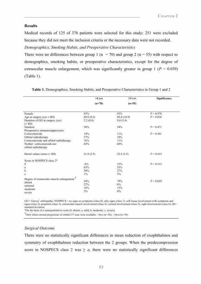
53
Chapter 2
Statistical analysis
For univariate comparisons, Student‟s t test was used to compare continuous variables. In
case of ordinal or non-normally distributed variables, the Mann-Whitney U test was used.
Categorical preoperative and postoperative data were investigated using the χ² test.
Surgical Technique
All the patients included in this study underwent standardized 3-wall decompression via a
coronal approach, as is performed traditionally at our institution.11
A coronal incision was made with a no. 10 blade from ear to ear, 3 to 4 cm behind the
hairline. In the central portion of the skull, a subperiosteal plane was created by blunt
dissection, and laterally, a surgical plane was developed bluntly between the deep and
superficial temporalis fasciae. The forehead flap thus created was then turned down to
expose the superior and lateral orbital rims. The supraorbital nerve was set free by chiseling
its bony foramen, when present, and the periorbita, including the trochlea, was dissected off
the orbital bones. After this, the temporalis muscle was dissected from its anterior origin
with a no. 10 blade and periosteal elevators, leaving sufficient tissue for suturing at the end
of surgery. In this way, the lateral orbital wall was exposed. A small osteotomy was
chiseled behind the lateral orbital rim, and bone-nibbling rongeurs were used to remove the
middle portion of the lateral orbital wall. After this, a Frazier suction tip was used to
fracture the delicate bone of the medial orbital wall and the floor, and Blakesley forceps
nos. 1 and 2 were used to remove bony fragments and sinuses‟ mucosa. The bulla
ethmoidalis beneath the frontoethmoidal suture was opened towards the orbit from the
posterior lacrimal crest up to the orbital apex, and then the orbital floor medial to the
infraorbital canal was removed from 0.5 cm behind the inferior orbital rim up to the
posterior wall of the maxillary sinus. The posterior two thirds of the maxillary ethmoidal
strut was removed, creating a wide antrostomy, whereas the anterior one third of the strut
was left intact to prevent globe displacement and the possibility of medial entropion or
hypoglobus.
Finally, the periorbita was incised to promote maximal prolapsed of the orbital tissues into
the newly created spaces, the temporalis muscle was sutured back into position with 4 to 5
interrupted 2/0 Mersilene sutures (Ethicon, Inc., Somerville, NJ), and after the insertion of a
3.3-mm-diameter end-perforated wound drain into each temporalis fossa, the scalp incision
was closed with iron staples.
Results
Medical records of 125 of 376 patients were selected for this study; 251 were excluded
because they did not meet the inclusion criteria or the necessary data were not recorded.
Demographics, Smoking Habits, and Preoperative Characteristics
There were no differences between group 1 (n = 70) and group 2 (n = 55) with respect to
demographics, smoking habits, or preoperative characteristics, except for the degree of
extraocular muscle enlargement, which was significantly greater in group 1 (P = 0.039)
(Table 1).
Table 1. Demographics, Smoking Habits, and Preoperative Characteristics in Group 1 and 2
<4 yrs
(n=70)
>4 yrs
(n=55)
Significance
Female 93% 93% P = 0.978 Age at surgery (yrs ± SD) 40.0 (9.2) 40.4 (10.9) P = 0.836 Duration of GO at surgery (yrs) (± SD)
2.2 (0.8) 9.0 (5.4)
Smokers 56% 54% P = 0.471 Preoperative immunosuppression: Corticosteroids Orbital radiotherapy Corticosteroids and orbital radiotherapy Neither corticosteroids nor orbital radiotherapy
14% 27% 16% 43%
11% 18% 11% 60%
P = 0.301
Hertel values (mm) (± SD)
21.8 (2.5)
22.5 (2.5)
P = 0.415
Score in NOSPECS class 2* 0 a b c
6% 63% 30% 1%
15% 55% 27% 3%
P = 0.312
Degree of extraocular muscle enlargement:† absent minimal moderate severe
54% 27% 16% 3%
79% 8% 13% 0%
P = 0.039
GO = Graves‟ orbitopathy; NOSPECS = no signs or symptoms (class 0); only signs (class 1); soft tissue involvement with symptoms and signs (class 2); proptosis (class 3), extraocular muscle involvement (class 4), corneal involvement (class 5), sight involvement (class 6); SD = standard deviation. *On the basis of a semiquantitative scale (0, absent; a, mild; b, moderate; c, severe). †Only when coronal projections of orbital CT scan were available: <4yrs (n=56); ≥4yrs (n=39).
Surgical Outcome
There were no statistically significant differences in mean reduction of exophthalmos and
symmetry of exophthalmos reduction between the 2 groups. When the predecompression
score in NOSPECS class 2 was ≥ a, there were no statistically significant differences
≥4 yrs
100379 Thesis Baldeschi.indd 53 vrijdag17-december-2010 12:07

54
Chapter 2
between the 2 groups in persistence of postdecompression periorbital swelling requiring
upper or lower lid blepharoplasty. The occurrence of reduction of upper and lower lid
retraction also did not differ statistically when these signs were present before surgery. In
contrast, the occurrence of postdecompression diplopia was significantly (P = 0.033) more
frequent in group 1 (Table 2), the relative risk and risk difference for
decompressioninduced diplopia being 2.3 and 18.1%, respectively.
Table 2. Postoperative Outcome
<4 yrs
(n=70) >4 yrs (n=55)
Significance
Exophthalmos reduction (mm ± SD) 4.5 (1.8) 4.2 (1.9) P = 0.456 Symmetry in exophthalmos reduction <1mm (87%)
>1mm (13%) <1mm (89%) >1mm (11%)
P = 0.739
Occurrence of reduction of upper lid retraction * 58%
50%
P = 0.502
Occurrence of reduction of lower lid retraction† 65%
62%
P = 0,771
Persistence of post-decompression periorbital swelling requiring cosmetic surgery‡
50% 49% P = 0.911
Post-decompression diplopia 29% 13% P = 0,033
SD = standard deviation. * Only patients who presented with this sign before surgery: <4yrs (n = 48); ≥4yrs (n = 24). †Only patients who presented with this sign before surgery: <4yrs (n = 48); ≥4yrs (n = 47). ‡Only patients who presented with predecompression score in NOSPECS (no signs or symptoms, only signs, soft tissue involvement with symptoms and signs, proptosis, extraocular muscle involvement, corneal involvement, sight involvement) Class 2 > a: <4yrs (n=66); ≥4yrs (n=47).
Discussion
To the authors‟ knowledge, the literature that evaluates the relationship between results of
decompressive procedures and duration of the orbitopathy at surgery is scarce. At present,
the assumption that links early intervention with more attractive results is hypothetical,
often mentioned at meetings, and based on the natural course of the orbital disease.
McCord14 quantified the result of aesthetic orbital decompression by measuring the
retrodisplacement of the globe in 23 patients. They concluded that the amount of
retrodisplacement varied and depended strictly on orbital fibrosis, but neither duration of
the orbitopathy at surgery nor explanations of the method to determine orbital fibrosis were
given. Härting et al.11 advocated early intervention without providing data to support their
preference. Kalmann et al.12 reported no significant relation between duration of GO and
the amount of proptosis reduction after 3-wall orbital decompression in 250 orbits.
However, because that study was not carried out specifically to evaluate this particular
aspect, patient characteristics according to duration of the orbitopathy were not given, and
the mean duration of the orbital disease was only 4.35 years.
Male gender, old age, and heavy smoking habits are patient characteristics that are known
to be associated with a more severe orbital disease15-17, whereas preoperative Hertel values
have been regarded as a factor influencing the amount of exophthalmos reduction.12
The 2 groups compared in this survey did not differ with respect to gender, mean age at
surgery, or smoking habits. Because other predecompression characteristics, such as Hertel
values, NOSPECS class 2 score, and use of immunosuppressive treatments also did not
differ, it is conceivable that the 2 groups were not dissimilar in either severity of GO at
surgery or its activity during the active phase of the disease. In addition, based on our
inclusion criteria, only patients decompressed for aesthetic reasons and not presenting with
diplopia within 20° of the central position of gaze were included. Therefore, groups 1 and 2
can be considered to be composed of a homogeneous population of Graves‟ patients, only
differing in duration of the orbitopathy at decompression. The observed difference in
preoperative volume of the extraocular muscles is, in fact, just the tomographic
representation of the different pathophysiological periods that characterize the 2 groups.
Therefore, it should be regarded as a consequence of the duration of the orbitopathy in
group 1 and 2, rather than a self-standing characteristic of the groups.
Our retrospective observations did not show any statistically significant correlation between
duration of the orbitopathy at decompression and (1) reduction of exophthalmos, (2)
symmetry of exophthalmos reduction, (3) persistence of postdecompression periorbital
swelling requiring postdecompression cosmetic surgery, and (4) amelioration of upper or
lower lid retraction, whereas in our series patients decompressed within 4 years from the
onset of the orbitopathy seemed to run a higher relative risk of postdecompression diplopia
than patients decompressed later than 4 years.
The higher frequency of postdecompression diplopia in group 1 can be explained by the
greater preoperative enlargement of the extraocular muscles in this group as compared with
group 2, despite similar preoperative Hertel values in the 2 groups. This implies a more
consistent contribution of the extraocular muscle mass to exophthalmos in group 1.
Therefore, it is feasible to deduce from the similar reduction of exophthalmos that was
achieved in the 2 groups that, for group 1, there was a more consistent shift of extraocular
≥4 yrs
≤1mm ≤1mm
100379 Thesis Baldeschi.indd 54 vrijdag17-december-2010 12:07
55
Chapter 2
between the 2 groups in persistence of postdecompression periorbital swelling requiring
upper or lower lid blepharoplasty. The occurrence of reduction of upper and lower lid
retraction also did not differ statistically when these signs were present before surgery. In
contrast, the occurrence of postdecompression diplopia was significantly (P = 0.033) more
frequent in group 1 (Table 2), the relative risk and risk difference for
decompressioninduced diplopia being 2.3 and 18.1%, respectively.
Table 2. Postoperative Outcome
<4 yrs
(n=70) >4 yrs (n=55)
Significance
Exophthalmos reduction (mm ± SD) 4.5 (1.8) 4.2 (1.9) P = 0.456 Symmetry in exophthalmos reduction <1mm (87%)
>1mm (13%) <1mm (89%) >1mm (11%)
P = 0.739
Occurrence of reduction of upper lid retraction * 58%
50%
P = 0.502
Occurrence of reduction of lower lid retraction† 65%
62%
P = 0,771
Persistence of post-decompression periorbital swelling requiring cosmetic surgery‡
50% 49% P = 0.911
Post-decompression diplopia 29% 13% P = 0,033
SD = standard deviation. * Only patients who presented with this sign before surgery: <4yrs (n = 48); ≥4yrs (n = 24). †Only patients who presented with this sign before surgery: <4yrs (n = 48); ≥4yrs (n = 47). ‡Only patients who presented with predecompression score in NOSPECS (no signs or symptoms, only signs, soft tissue involvement with symptoms and signs, proptosis, extraocular muscle involvement, corneal involvement, sight involvement) Class 2 > a: <4yrs (n=66); ≥4yrs (n=47).
Discussion
To the authors‟ knowledge, the literature that evaluates the relationship between results of
decompressive procedures and duration of the orbitopathy at surgery is scarce. At present,
the assumption that links early intervention with more attractive results is hypothetical,
often mentioned at meetings, and based on the natural course of the orbital disease.
McCord14 quantified the result of aesthetic orbital decompression by measuring the
retrodisplacement of the globe in 23 patients. They concluded that the amount of
retrodisplacement varied and depended strictly on orbital fibrosis, but neither duration of
the orbitopathy at surgery nor explanations of the method to determine orbital fibrosis were
given. Härting et al.11 advocated early intervention without providing data to support their
preference. Kalmann et al.12 reported no significant relation between duration of GO and
the amount of proptosis reduction after 3-wall orbital decompression in 250 orbits.
However, because that study was not carried out specifically to evaluate this particular
aspect, patient characteristics according to duration of the orbitopathy were not given, and
the mean duration of the orbital disease was only 4.35 years.
Male gender, old age, and heavy smoking habits are patient characteristics that are known
to be associated with a more severe orbital disease15-17, whereas preoperative Hertel values
have been regarded as a factor influencing the amount of exophthalmos reduction.12
The 2 groups compared in this survey did not differ with respect to gender, mean age at
surgery, or smoking habits. Because other predecompression characteristics, such as Hertel
values, NOSPECS class 2 score, and use of immunosuppressive treatments also did not
differ, it is conceivable that the 2 groups were not dissimilar in either severity of GO at
surgery or its activity during the active phase of the disease. In addition, based on our
inclusion criteria, only patients decompressed for aesthetic reasons and not presenting with
diplopia within 20° of the central position of gaze were included. Therefore, groups 1 and 2
can be considered to be composed of a homogeneous population of Graves‟ patients, only
differing in duration of the orbitopathy at decompression. The observed difference in
preoperative volume of the extraocular muscles is, in fact, just the tomographic
representation of the different pathophysiological periods that characterize the 2 groups.
Therefore, it should be regarded as a consequence of the duration of the orbitopathy in
group 1 and 2, rather than a self-standing characteristic of the groups.
Our retrospective observations did not show any statistically significant correlation between
duration of the orbitopathy at decompression and (1) reduction of exophthalmos, (2)
symmetry of exophthalmos reduction, (3) persistence of postdecompression periorbital
swelling requiring postdecompression cosmetic surgery, and (4) amelioration of upper or
lower lid retraction, whereas in our series patients decompressed within 4 years from the
onset of the orbitopathy seemed to run a higher relative risk of postdecompression diplopia
than patients decompressed later than 4 years.
The higher frequency of postdecompression diplopia in group 1 can be explained by the
greater preoperative enlargement of the extraocular muscles in this group as compared with
group 2, despite similar preoperative Hertel values in the 2 groups. This implies a more
consistent contribution of the extraocular muscle mass to exophthalmos in group 1.
Therefore, it is feasible to deduce from the similar reduction of exophthalmos that was
achieved in the 2 groups that, for group 1, there was a more consistent shift of extraocular
100379 Thesis Baldeschi.indd 55 vrijdag17-december-2010 12:07
56
Chapter 2
muscles into the spaces newly created by decompression surgery. This could have led to a
greater decompensation of extraocular muscle balance in group 1. Differing motor and/or
sensory capacities for compensation of induced muscle imbalance between the 2 groups
also may have played a role. Further investigations are required to clarify this.
In conclusion, in contrast to the assumption that early intervention is related to a more
desirable surgical outcome, our observation links early intervention with a higher risk of
postdecompression diplopia. Our results, however, are far from conclusive, because the
study might have been biased by systemic corticosteroid use, orbital radiotherapy, and
relatively low preoperative Hertel values. The immunosuppressive effect of corticosteroids
and/or radiotherapy has influenced the pathophysiological course of the disease and might
have reduced the final orbital fibrosis in about half of our patients. On the other hand, the
toxicity of radiation therapy to soft tissues, which includes fibrotic sequelae, might have
reduced the distensibility and plasticity of the soft orbital tissues, decreasing the effect of
decompression surgery. In addition, the low mean preoperative Hertel values in the 2
groups can be regarded as the epiphenomenon of a minimally to moderately increased
intraorbital pressure, which reasonably induced only mild congestion, leading to scarce
final fibrosis.
We acknowledge that, to overcome the shortcomings of our retrospective analysis, a
prospective clinical trial would be needed, with some design difficulties. If only patients
who did not require treatment with systemic corticosteroids or orbital radiotherapy were
included, the study would enroll only those patients with milder disease who, presumably,
are less prone to develop late fibrosis. On the other hand, it would be ethically questionable
to design a study in which immunosuppression is not administrated to reduce biases, and
patients are randomized to early or late decompression.
Patients with GO who do not have prompt access to diagnosis and medical treatment and
who finally present to the orbital surgeon with advanced disease could offer an adequate
population for a prospective study aimed to validate our major conclusions.
References
1. Bartalena L, Pinchera A, Marcocci C. Management of Graves‟ ophthalmopathy: reality and perspectives.
Endocr Rev 2000;21:168 -99.
2. Burch HB, Wartofsky L. Graves‟ ophthalmopathy: current comcepts regarding patogenesis and management.
Endocr Rev 1993;14:747-93.
3. Sorisky A, Pardasani D, Gagnon A, Smith TJ. Evidence of adipocyte differentiation in human orbital fibroblasts
in primary culture. J Clin Endocrinol Metab 1996;81:3428-31.
4. Naffzinger HC. Pathologic changes in the orbit in progressive exophthalmos. Arch Ophthalmol 1933;9:1-12.
5. Brain RW. Exophthalmic ophthalmoplegia. Q J Med 1938;31:293-323.
6. Daicker B. Das gewebliche substrat der verdickten ausseren augenmuskelen bei der endocrinen orbitopathie.
Klin Monatsbl Augenheilkd 1979;174:843-7.
7. Tallstedt L, Norberg R. Immunohistochemical staining of normal and Graves‟ extraocular muscle. Invest
Ophthalmol Vis Sci 1988;29:175-84.
8. Prummel MF, Wiersinga WM, Mourits MP, Koornneef L, Berghout A, van der Gaag R. Effect of abnormal
thyroid function on the severity of Graves‟ ophthalmopathy. Arch Intern Med 1990;150:1098-101.
9. Wilson WB, Manke WF. Orbital decompression in Graves‟ disease: the predictability of reduction of proptosis.
Arch Ophthalmol 1991;109:343-5.
10. Goldberg RA, Weinberg DA, Shorr N, Wirta D. Maximal three-wall orbital decompression through a coronal
approach. Ophthalmic Surg Lasers 1997;28:832-43.
11. Härting F, Koornneef L, Peeters HJF, Gillisen JPA. Fourteen years of orbital decompression in Graves‟
disease. A review of technique, results and complications. Orbit 1986;5:123-9.
12. Kalmann R, Mourits MP, van der Pol JP, Koornneef L. Coronal approach for rehabilitative orbital
decompression in Graves‟ ophthalmopathy. Br J Ophthalmol 1997;81:41-5.
13. Werner SC. Modifications of the classification of the eye changes of Graves‟ disease: recommendations of the
Ad Hoc Committee of the American Thyroid Association. J Clin Endocrinol Metab 1977;44:203-4.
14. McCord CD. Orbital decompression for Graves‟ disease exposure through lateral cantal and inferior fornix
incision. Ophthalmology 1981;88:533- 41.
15. Kendler DL, Lippa J, Rootman J. The initial characteristics of Graves‟ orbitopathy vary with age and sex. Arch
Ophthalmol 1993;111:197-201.
16. Perros P, Cromble AL, Matthews JN, Kendall-Taylor P. Age and gender influence the severity of thyroid-
associated ophthalmopathy: a study of 101 patients attending a combined thyroid-eye clinic. Clin Endocrinol
1993;38:367-72.
17. Prummel MF, Wiersinga WM. Smoking and risk of Graves‟ disease. JAMA 1993;269:479-82.
100379 Thesis Baldeschi.indd 56 vrijdag17-december-2010 12:07
57
Chapter 2
muscles into the spaces newly created by decompression surgery. This could have led to a
greater decompensation of extraocular muscle balance in group 1. Differing motor and/or
sensory capacities for compensation of induced muscle imbalance between the 2 groups
also may have played a role. Further investigations are required to clarify this.
In conclusion, in contrast to the assumption that early intervention is related to a more
desirable surgical outcome, our observation links early intervention with a higher risk of
postdecompression diplopia. Our results, however, are far from conclusive, because the
study might have been biased by systemic corticosteroid use, orbital radiotherapy, and
relatively low preoperative Hertel values. The immunosuppressive effect of corticosteroids
and/or radiotherapy has influenced the pathophysiological course of the disease and might
have reduced the final orbital fibrosis in about half of our patients. On the other hand, the
toxicity of radiation therapy to soft tissues, which includes fibrotic sequelae, might have
reduced the distensibility and plasticity of the soft orbital tissues, decreasing the effect of
decompression surgery. In addition, the low mean preoperative Hertel values in the 2
groups can be regarded as the epiphenomenon of a minimally to moderately increased
intraorbital pressure, which reasonably induced only mild congestion, leading to scarce
final fibrosis.
We acknowledge that, to overcome the shortcomings of our retrospective analysis, a
prospective clinical trial would be needed, with some design difficulties. If only patients
who did not require treatment with systemic corticosteroids or orbital radiotherapy were
included, the study would enroll only those patients with milder disease who, presumably,
are less prone to develop late fibrosis. On the other hand, it would be ethically questionable
to design a study in which immunosuppression is not administrated to reduce biases, and
patients are randomized to early or late decompression.
Patients with GO who do not have prompt access to diagnosis and medical treatment and
who finally present to the orbital surgeon with advanced disease could offer an adequate
population for a prospective study aimed to validate our major conclusions.
References
1. Bartalena L, Pinchera A, Marcocci C. Management of Graves‟ ophthalmopathy: reality and perspectives.
Endocr Rev 2000;21:168 -99.
2. Burch HB, Wartofsky L. Graves‟ ophthalmopathy: current comcepts regarding patogenesis and management.
Endocr Rev 1993;14:747-93.
3. Sorisky A, Pardasani D, Gagnon A, Smith TJ. Evidence of adipocyte differentiation in human orbital fibroblasts
in primary culture. J Clin Endocrinol Metab 1996;81:3428-31.
4. Naffzinger HC. Pathologic changes in the orbit in progressive exophthalmos. Arch Ophthalmol 1933;9:1-12.
5. Brain RW. Exophthalmic ophthalmoplegia. Q J Med 1938;31:293-323.
6. Daicker B. Das gewebliche substrat der verdickten ausseren augenmuskelen bei der endocrinen orbitopathie.
Klin Monatsbl Augenheilkd 1979;174:843-7.
7. Tallstedt L, Norberg R. Immunohistochemical staining of normal and Graves‟ extraocular muscle. Invest
Ophthalmol Vis Sci 1988;29:175-84.
8. Prummel MF, Wiersinga WM, Mourits MP, Koornneef L, Berghout A, van der Gaag R. Effect of abnormal
thyroid function on the severity of Graves‟ ophthalmopathy. Arch Intern Med 1990;150:1098-101.
9. Wilson WB, Manke WF. Orbital decompression in Graves‟ disease: the predictability of reduction of proptosis.
Arch Ophthalmol 1991;109:343-5.
10. Goldberg RA, Weinberg DA, Shorr N, Wirta D. Maximal three-wall orbital decompression through a coronal
approach. Ophthalmic Surg Lasers 1997;28:832-43.
11. Härting F, Koornneef L, Peeters HJF, Gillisen JPA. Fourteen years of orbital decompression in Graves‟
disease. A review of technique, results and complications. Orbit 1986;5:123-9.
12. Kalmann R, Mourits MP, van der Pol JP, Koornneef L. Coronal approach for rehabilitative orbital
decompression in Graves‟ ophthalmopathy. Br J Ophthalmol 1997;81:41-5.
13. Werner SC. Modifications of the classification of the eye changes of Graves‟ disease: recommendations of the
Ad Hoc Committee of the American Thyroid Association. J Clin Endocrinol Metab 1977;44:203-4.
14. McCord CD. Orbital decompression for Graves‟ disease exposure through lateral cantal and inferior fornix
incision. Ophthalmology 1981;88:533- 41.
15. Kendler DL, Lippa J, Rootman J. The initial characteristics of Graves‟ orbitopathy vary with age and sex. Arch
Ophthalmol 1993;111:197-201.
16. Perros P, Cromble AL, Matthews JN, Kendall-Taylor P. Age and gender influence the severity of thyroid-
associated ophthalmopathy: a study of 101 patients attending a combined thyroid-eye clinic. Clin Endocrinol
1993;38:367-72.
17. Prummel MF, Wiersinga WM. Smoking and risk of Graves‟ disease. JAMA 1993;269:479-82.
-99
100379 Thesis Baldeschi.indd 57 vrijdag17-december-2010 12:07

Chapter 3
The Influence of Previous Orbital Irradiation on
the Outcome of Rehabilitative Decompression Surgery
in Graves’ Orbitopathy
Lelio Baldeschi1, Kerr MacAndie1, Eva Kotsier1, Leo E.C.M. Blank2, Wilmar M. Wiersinga3
1 Orbital Centre, Department of Ophthalmology, University of Amsterdam, Amsterdam, The Netherlands. 2 Department of Radiotherapy, University of Amsterdam, Amsterdam, The Netherlands. 3 Department of Endocrinology, University of Amsterdam, Amsterdam, The Netherlands. American Journal of Ophthalmology 2008;145:534-40
100379 Thesis Baldeschi.indd 58 vrijdag17-december-2010 12:07
Chapter 3
The Influence of Previous Orbital Irradiation on
the Outcome of Rehabilitative Decompression Surgery
in Graves’ Orbitopathy
Lelio Baldeschi1, Kerr MacAndie1, Eva Kotsier1, Leo E.C.M. Blank2, Wilmar M. Wiersinga3
1 Orbital Centre, Department of Ophthalmology, University of Amsterdam, Amsterdam, The Netherlands. 2 Department of Radiotherapy, University of Amsterdam, Amsterdam, The Netherlands. 3 Department of Endocrinology, University of Amsterdam, Amsterdam, The Netherlands. American Journal of Ophthalmology 2008;145:534-40
100379 Thesis Baldeschi.indd 59 vrijdag17-december-2010 12:07
60
Chapter 3
Abstract
Purpose: To investigate whether orbital irradiation influences the outcome of
decompression surgery in Graves‟ orbitopathy.
Design: Retrospective, comparative case series.
Methods: The medical records of all the patients with Graves‟ orbitopathy treated with a
three-wall orbital decompression through a coronal approach at our institution between
January 1, 1990 and December 31, 2000 were reviewed. Only patients who underwent
bilateral surgery for aesthetic rehabilitation, without preoperative diplopia, and who, in the
active phase of the disease, had received orbital radiotherapy alone (20 Grays (Gy) in 10
daily fractions of two Gy over a period of two weeks; group R), systemic glucocorticoids
alone (daily administration for more than three months independently from the dosage;
group G), or both radiotherapy and glucocorticoids (group RG) were selected. Groups were
compared for demographics, smoking habits, preoperative characteristics, and surgical
outcome (mean reduction of exophthalmos, reduction of lid retraction, persistence of
periorbital swelling requiring cosmetic eyelid surgery, onset of diplopia within 20 degrees
of the central position of gaze, and variations in the peripheral field of diplopia).
Results: Sixty-one of 376 patients were selected for this study. There were no differences
between group R (n = 29), group G (n = 15), and group RG (n = 17) with respect to
demographics or predecompression characteristics, whereas the number of smokers was
significantly greater in group RG (P = 0.019). We could not find differences in surgical
outcome by comparing the three groups.
Conclusions: The total radiation dose, fraction size, and irradiated volume commonly used
to treat active Graves‟ orbitopathy do not adversely interfere with the outcome of
rehabilitative decompression surgery.
Introduction
Since 1913, external beam irradiation therapy has been used for the treatment of Graves‟
orbitopathy.1 Initially, when exophthalmos was thought to be the result of dysfunction of
the hypothalamus and the pituitary gland, radiotherapy was aimed at these structures.
It later became clear that only those patients with iatrogenic orbital irradiation resulting
from poorly collimated radiation delivery systems improved. Radiotherapy subsequently
was directed to the orbital tissues behind the globe.2
Although its efficacy was questioned recently3, orbital radiotherapy continues to represent a
mainstay in the management of active Graves‟ orbitopathy. This is largely the result of its
superiority of optimal tolerance and almost no short-term side effects, as opposed to the
alternative of systemic immunosuppression with glucocorticoids.4 Issues of concern with
radiotherapy in benign diseases are the long-term adverse effects of ionizing radiation to
tissue, which in the case of Graves‟ orbitopathy may lead to an increased risk of
malignancy in the head and neck region, retinopathy, cataract formation, and diffuse
fibrosis of the retroocular soft tissues. So far, no secondary tumors have been observed in
long-term follow-up studies, and ocular complications, such as retinopathy or cataract, do
not seem to represent real aftermaths of orbital irradiation for Graves' orbitopathy.5 Fibrosis
of the orbital soft tissues secondary to radiation therapy may influence the results of later
surgical procedures such as squint or decompression surgery. Preoperative radiotherapy
was demonstrated not to affect squint surgery adversely6, but its effect on orbital
decompressions remains undetermined. Although this issue has been mentioned
incidentally in the literature7,8 and is a source of debate at meetings, we are unaware of
previous reports that link orbital radiotherapy to the effects of decompression surgery in
patients with Graves‟ orbitopathy, and a computerized search using MEDLINE failed to
reveal any references. This study sought to determine whether orbital irradiation in Graves‟
orbitopathy influences the outcome of rehabilitative decompression surgery.
100379 Thesis Baldeschi.indd 60 vrijdag17-december-2010 12:07

61
Chapter 3
Abstract
Purpose: To investigate whether orbital irradiation influences the outcome of
decompression surgery in Graves‟ orbitopathy.
Design: Retrospective, comparative case series.
Methods: The medical records of all the patients with Graves‟ orbitopathy treated with a
three-wall orbital decompression through a coronal approach at our institution between
January 1, 1990 and December 31, 2000 were reviewed. Only patients who underwent
bilateral surgery for aesthetic rehabilitation, without preoperative diplopia, and who, in the
active phase of the disease, had received orbital radiotherapy alone (20 Grays (Gy) in 10
daily fractions of two Gy over a period of two weeks; group R), systemic glucocorticoids
alone (daily administration for more than three months independently from the dosage;
group G), or both radiotherapy and glucocorticoids (group RG) were selected. Groups were
compared for demographics, smoking habits, preoperative characteristics, and surgical
outcome (mean reduction of exophthalmos, reduction of lid retraction, persistence of
periorbital swelling requiring cosmetic eyelid surgery, onset of diplopia within 20 degrees
of the central position of gaze, and variations in the peripheral field of diplopia).
Results: Sixty-one of 376 patients were selected for this study. There were no differences
between group R (n = 29), group G (n = 15), and group RG (n = 17) with respect to
demographics or predecompression characteristics, whereas the number of smokers was
significantly greater in group RG (P = 0.019). We could not find differences in surgical
outcome by comparing the three groups.
Conclusions: The total radiation dose, fraction size, and irradiated volume commonly used
to treat active Graves‟ orbitopathy do not adversely interfere with the outcome of
rehabilitative decompression surgery.
Introduction
Since 1913, external beam irradiation therapy has been used for the treatment of Graves‟
orbitopathy.1 Initially, when exophthalmos was thought to be the result of dysfunction of
the hypothalamus and the pituitary gland, radiotherapy was aimed at these structures.
It later became clear that only those patients with iatrogenic orbital irradiation resulting
from poorly collimated radiation delivery systems improved. Radiotherapy subsequently
was directed to the orbital tissues behind the globe.2
Although its efficacy was questioned recently3, orbital radiotherapy continues to represent a
mainstay in the management of active Graves‟ orbitopathy. This is largely the result of its
superiority of optimal tolerance and almost no short-term side effects, as opposed to the
alternative of systemic immunosuppression with glucocorticoids.4 Issues of concern with
radiotherapy in benign diseases are the long-term adverse effects of ionizing radiation to
tissue, which in the case of Graves‟ orbitopathy may lead to an increased risk of
malignancy in the head and neck region, retinopathy, cataract formation, and diffuse
fibrosis of the retroocular soft tissues. So far, no secondary tumors have been observed in
long-term follow-up studies, and ocular complications, such as retinopathy or cataract, do
not seem to represent real aftermaths of orbital irradiation for Graves' orbitopathy.5 Fibrosis
of the orbital soft tissues secondary to radiation therapy may influence the results of later
surgical procedures such as squint or decompression surgery. Preoperative radiotherapy
was demonstrated not to affect squint surgery adversely6, but its effect on orbital
decompressions remains undetermined. Although this issue has been mentioned
incidentally in the literature7,8 and is a source of debate at meetings, we are unaware of
previous reports that link orbital radiotherapy to the effects of decompression surgery in
patients with Graves‟ orbitopathy, and a computerized search using MEDLINE failed to
reveal any references. This study sought to determine whether orbital irradiation in Graves‟
orbitopathy influences the outcome of rehabilitative decompression surgery.
100379 Thesis Baldeschi.indd 61 vrijdag17-december-2010 12:07

62
Chapter 3
Patients and Methods
Our study was a retrospective, comparative case series. The medical records of all the
patients with Graves‟ orbitopathy who underwent three-wall orbital decompression
performed via a coronal approach at the Orbital Center, Department of Ophthalmology,
University of Amsterdam, between January 1, 1990 and December 31, 2000 were evaluated
retrospectively.
Inclusion Criteria
We included all patients who: 1) had undergone bilateral surgery for aesthetic
rehabilitation, 2) did not have preoperative diplopia within 20 degrees of the central
position of gaze, and 3) had been treated, in the active phase of Graves‟ orbitopathy, with
bilateral orbital radiotherapy alone (total dose of 20 Grays (Gy) in 10 daily fractions of two
Gy over a period of two weeks), systemic glucocorticoids alone (for a period of at least
three consecutive months independently from dosage regimens), or with both retrobulbar
radiotherapy and systemic glucocorticoids.
Exclusion Criteria
We excluded patients who: 1) had undergone unilateral surgery; 2) were decompressed for
optic neuropathy or exposure keratopathy; 3) did not receive bilateral orbital radiotherapy,
systemic glucocorticoids or both the treatments in the active phase of Graves‟ orbitopathy
or did not receive these treatments as specified in the inclusion criteria; 4) had preoperative
diplopia; 5) had been treated with thyroidectomy or iodine 131 in the six months preceding
decompression or in the six months preceding postoperative evaluation; 6) had concurrent
orbital or periorbital diseases, injuries, or surgeries (except for postdecompression
blepharoplasty) within six months after decompression; and 7) whose records did not
clearly state the duration of Graves‟ orbitopathy at decompression; smoking habits at
surgery; the date and method of administration of predecompression orbital radiotherapy or
systemic corticosteroids; the results of an immediate preoperative clinical examination,
including standard ophthalmologic evaluation, NOSPECS (no signs or symptoms, only
signs, soft tissues involvement with symptoms and signs, proptosis, extraocular muscle
involvement, corneal involvement, sight involvement) classification,9 Hertel
exophthalmometry, measurements of eyelid fissure and respective upper and lower eyelid
positions; the results of at least one postoperative clinical examination performed six
months or more after surgery; the results of preoperative and postoperative orthoptic
evaluations performed, respectively, not earlier than six months before decompression and
between three and six months after surgery.
The following data were retrieved: 1) gender; 2) age at surgery; 3) duration of Graves‟
orbitopathy at decompression; 4) smoking habits at surgery; 5) type of immunosuppression
used in the active phase of Graves‟ orbitopathy; 6) interval between end of
immunosuppression by steroids or radiation and time of decompression (in the case of
treatment with radiotherapy and glucocorticoids, such an interval was calculated based on
the last of the two administered treatments); 7) predecompression thyroidectomy or use of
iodine 131; and 8) predecompression Hertel readings, score in NOSPECS class 2, presence
of upper or lower lid retraction, or both, with measurements of eyelid fissure and the
respective upper and lower eyelid position. Predecompression Hertel readings, score in
NOSPECS class 2, and preoperative eyelid positions were those measured at hospitalization
for orbital decompression. Data concerning the activity of Graves‟ orbitopathy according to
the seven-point clinical activity score (CAS)10 at baseline also were retrieved or were
calculated retrospectively based on recorded signs of inflammation. We considered baseline
to be the last ophthalmologic evaluation preceding the immunosuppressive treatment (the
first immunosuppressive treatment when both orbital radiotherapy and systemic
glucocorticoids had been administered).
We considered smokers to be those patients who were smokers on admission to the hospital
at the time of surgery. In our center, the clinical work-up preceding rehabilitative orbital
decompression consists of several ophthalmologic and endocrinologic examinations.
Patients who smoke are strongly encouraged to give up smoking at all times. We assume
that those patients who maintain their habit up to the time of decompression, despite
multiple health warnings, will not refrain from smoking after surgery.
The postoperative data we used to assess the surgical outcome were: 1) reduction of
exophthalmos; 2) reduction of upper or lower lid retraction, or both, when these signs were
present before surgery; 3) persistence of periorbital swelling requiring cosmetic eyelid
surgery; 4) occurrence of postdecompression diplopia within 20 degrees of the central
position of gaze; and 5) variations in the peripheral field of diplopia. Postoperative Hertel
readings and postoperative eyelid positions were those measured at the first clinical
examination carried out six months or more after surgery.
100379 Thesis Baldeschi.indd 62 vrijdag17-december-2010 12:07
63
Chapter 3
Patients and Methods
Our study was a retrospective, comparative case series. The medical records of all the
patients with Graves‟ orbitopathy who underwent three-wall orbital decompression
performed via a coronal approach at the Orbital Center, Department of Ophthalmology,
University of Amsterdam, between January 1, 1990 and December 31, 2000 were evaluated
retrospectively.
Inclusion Criteria
We included all patients who: 1) had undergone bilateral surgery for aesthetic
rehabilitation, 2) did not have preoperative diplopia within 20 degrees of the central
position of gaze, and 3) had been treated, in the active phase of Graves‟ orbitopathy, with
bilateral orbital radiotherapy alone (total dose of 20 Grays (Gy) in 10 daily fractions of two
Gy over a period of two weeks), systemic glucocorticoids alone (for a period of at least
three consecutive months independently from dosage regimens), or with both retrobulbar
radiotherapy and systemic glucocorticoids.
Exclusion Criteria
We excluded patients who: 1) had undergone unilateral surgery; 2) were decompressed for
optic neuropathy or exposure keratopathy; 3) did not receive bilateral orbital radiotherapy,
systemic glucocorticoids or both the treatments in the active phase of Graves‟ orbitopathy
or did not receive these treatments as specified in the inclusion criteria; 4) had preoperative
diplopia; 5) had been treated with thyroidectomy or iodine 131 in the six months preceding
decompression or in the six months preceding postoperative evaluation; 6) had concurrent
orbital or periorbital diseases, injuries, or surgeries (except for postdecompression
blepharoplasty) within six months after decompression; and 7) whose records did not
clearly state the duration of Graves‟ orbitopathy at decompression; smoking habits at
surgery; the date and method of administration of predecompression orbital radiotherapy or
systemic corticosteroids; the results of an immediate preoperative clinical examination,
including standard ophthalmologic evaluation, NOSPECS (no signs or symptoms, only
signs, soft tissues involvement with symptoms and signs, proptosis, extraocular muscle
involvement, corneal involvement, sight involvement) classification,9 Hertel
exophthalmometry, measurements of eyelid fissure and respective upper and lower eyelid
positions; the results of at least one postoperative clinical examination performed six
months or more after surgery; the results of preoperative and postoperative orthoptic
evaluations performed, respectively, not earlier than six months before decompression and
between three and six months after surgery.
The following data were retrieved: 1) gender; 2) age at surgery; 3) duration of Graves‟
orbitopathy at decompression; 4) smoking habits at surgery; 5) type of immunosuppression
used in the active phase of Graves‟ orbitopathy; 6) interval between end of
immunosuppression by steroids or radiation and time of decompression (in the case of
treatment with radiotherapy and glucocorticoids, such an interval was calculated based on
the last of the two administered treatments); 7) predecompression thyroidectomy or use of
iodine 131; and 8) predecompression Hertel readings, score in NOSPECS class 2, presence
of upper or lower lid retraction, or both, with measurements of eyelid fissure and the
respective upper and lower eyelid position. Predecompression Hertel readings, score in
NOSPECS class 2, and preoperative eyelid positions were those measured at hospitalization
for orbital decompression. Data concerning the activity of Graves‟ orbitopathy according to
the seven-point clinical activity score (CAS)10 at baseline also were retrieved or were
calculated retrospectively based on recorded signs of inflammation. We considered baseline
to be the last ophthalmologic evaluation preceding the immunosuppressive treatment (the
first immunosuppressive treatment when both orbital radiotherapy and systemic
glucocorticoids had been administered).
We considered smokers to be those patients who were smokers on admission to the hospital
at the time of surgery. In our center, the clinical work-up preceding rehabilitative orbital
decompression consists of several ophthalmologic and endocrinologic examinations.
Patients who smoke are strongly encouraged to give up smoking at all times. We assume
that those patients who maintain their habit up to the time of decompression, despite
multiple health warnings, will not refrain from smoking after surgery.
The postoperative data we used to assess the surgical outcome were: 1) reduction of
exophthalmos; 2) reduction of upper or lower lid retraction, or both, when these signs were
present before surgery; 3) persistence of periorbital swelling requiring cosmetic eyelid
surgery; 4) occurrence of postdecompression diplopia within 20 degrees of the central
position of gaze; and 5) variations in the peripheral field of diplopia. Postoperative Hertel
readings and postoperative eyelid positions were those measured at the first clinical
examination carried out six months or more after surgery.
100379 Thesis Baldeschi.indd 63 vrijdag17-december-2010 12:07
64
Chapter 3
The included patients were divided into three groups based on the immunosuppression
treatments administered in the active phase of Graves‟ orbitopathy: those who had received
orbital radiotherapy alone were included in group R, those who had received systemic
glucocorticoids alone were included in group G, and those who had received both orbital
radiotherapy and systemic glucocorticoids were included in group RG. Patients in group R
were compared with those of groups G and RG for demographics, smoking habits, and
preoperative and postoperative data by means of a database created using version 11.5.2 of
the SPSS statistical analysis software package (SPSS, Inc, Chicago, Illinois, USA.). Right
and left sides were not evaluated separately except for reduction of upper and lower lid
retraction. For those patients presenting with a predecompression score ≥ a in NOSPECS
class 2, persistence of periorbital swelling implying the necessity for postdecompression
cosmetic surgery was defined as evidence in the medical record of upper or lower lid
blepharoplasty proposed or performed at any time after orbital decompression.
Postoperative orthoptic data were based on the first orthoptic examination performed
between three and six months after decompression.
Statistical Analysis
Univariate comparisons were carried out using a one-way analysis of variance to compare
continuous variables. In case of ordinal or non normally distributed variables, the Mann-
Whitney U test was used. Categorical preoperative and postoperative data were investigated
using the χ² test.
Orbital Radiotherapy
Orbital irradiation was delivered with a 5- or 6-mV accelerator in 10 daily fractions of two
Gy over a period of two weeks. Localization and verification films with lead markers on the
lateral canthus of each eye were performed for each patient on a simulator. The dose was
calculated at the midline and given by two three degree anterior angled lateral portals of 5 ×
5 cm, with the patient‟s head fixed by an individual full-head shell.
Surgical Technique
All the patients included in this study underwent a standardized three-wall decompression
via a coronal approach as is traditionally performed at our institution.11 A coronal incision
was made with a no. 10 blade from ear to ear, 3 to 4 cm behind the hairline. In the central
portion of the cranium, a subperiosteal plane was created by blunt dissection, and laterally,
a surgical plane was developed bluntly between the deep and the superficial temporalis
fascia. The forehead flap thus created was then turned down to expose the superior and
lateral orbital rims. The supraorbital nerve was set free by chiselling its bony foramen when
present and the periorbita, including the trochlea, was dissected off the orbital bones. The
temporalis muscle then was dissected from its anterior origin with a no. 10 blade and
periosteal elevators, leaving sufficient tissue for suturing at the end of surgery. In this way,
the lateral orbital wall was exposed. A small osteotomy was chiselled behind the lateral
orbital rim, and bone-nibbling rongeurs were used to remove the mid portion of the lateral
orbital wall. A Frazier suction tip was used to fracture the delicate bone of the medial
orbital wall and the floor, and Blakesley forceps nos. 1 and 2 were used to remove bony
fragments and mucosa of the sinuses. The bulla ethmoidalis beneath the frontoethmoidal
suture was opened toward the orbit from the posterior lacrimal crest up to the orbital apex,
and then the orbital floor medial to the infraorbital canal was removed from 0.5 cm behind
the inferior orbital rim up to the posterior wall of the maxillary sinus. The posterior two-
thirds of the maxillary ethmoidal strut were removed creating a wide antrostomy, whereas
the anterior one-third of the strut was left intact to prevent globe displacement and the
possibility of medial entropion or hypoglobus. Finally, the periorbita was incised
extensively to promote maximal prolapse of the orbital tissues into the newly created
spaces. The temporalis muscle was sutured back into position with four to five interrupted
2-0 Mersilene sutures and, after the insertion of a 3.3-mm diameter end-perforated wound
drain into each temporalis fossa, the scalp incision was closed with steel staples.
Results
The medical records of 61 of 376 patients were selected for this study, whereas 315 were
excluded because they did not meet the inclusion criteria or the necessary data were not
recorded.
Demographics, Smoking Habits, and Preoperative Characteristics
There were no differences between group R (n = 29), group G (n = 15), and group RG (n =
17) with respect to demographics or preoperative characteristics, whereas the number of
smokers was significantly greater in group RG (P = 0.019). It was not possible to compare
group RG with the other two groups with regard to CAS at baseline because we could not
retrieve enough data from the medical records of the patients included in this group (Table
1).
100379 Thesis Baldeschi.indd 64 vrijdag17-december-2010 12:07
65
Chapter 3
The included patients were divided into three groups based on the immunosuppression
treatments administered in the active phase of Graves‟ orbitopathy: those who had received
orbital radiotherapy alone were included in group R, those who had received systemic
glucocorticoids alone were included in group G, and those who had received both orbital
radiotherapy and systemic glucocorticoids were included in group RG. Patients in group R
were compared with those of groups G and RG for demographics, smoking habits, and
preoperative and postoperative data by means of a database created using version 11.5.2 of
the SPSS statistical analysis software package (SPSS, Inc, Chicago, Illinois, USA.). Right
and left sides were not evaluated separately except for reduction of upper and lower lid
retraction. For those patients presenting with a predecompression score ≥ a in NOSPECS
class 2, persistence of periorbital swelling implying the necessity for postdecompression
cosmetic surgery was defined as evidence in the medical record of upper or lower lid
blepharoplasty proposed or performed at any time after orbital decompression.
Postoperative orthoptic data were based on the first orthoptic examination performed
between three and six months after decompression.
Statistical Analysis
Univariate comparisons were carried out using a one-way analysis of variance to compare
continuous variables. In case of ordinal or non normally distributed variables, the Mann-
Whitney U test was used. Categorical preoperative and postoperative data were investigated
using the χ² test.
Orbital Radiotherapy
Orbital irradiation was delivered with a 5- or 6-mV accelerator in 10 daily fractions of two
Gy over a period of two weeks. Localization and verification films with lead markers on the
lateral canthus of each eye were performed for each patient on a simulator. The dose was
calculated at the midline and given by two three degree anterior angled lateral portals of 5 ×
5 cm, with the patient‟s head fixed by an individual full-head shell.
Surgical Technique
All the patients included in this study underwent a standardized three-wall decompression
via a coronal approach as is traditionally performed at our institution.11 A coronal incision
was made with a no. 10 blade from ear to ear, 3 to 4 cm behind the hairline. In the central
portion of the cranium, a subperiosteal plane was created by blunt dissection, and laterally,
a surgical plane was developed bluntly between the deep and the superficial temporalis
fascia. The forehead flap thus created was then turned down to expose the superior and
lateral orbital rims. The supraorbital nerve was set free by chiselling its bony foramen when
present and the periorbita, including the trochlea, was dissected off the orbital bones. The
temporalis muscle then was dissected from its anterior origin with a no. 10 blade and
periosteal elevators, leaving sufficient tissue for suturing at the end of surgery. In this way,
the lateral orbital wall was exposed. A small osteotomy was chiselled behind the lateral
orbital rim, and bone-nibbling rongeurs were used to remove the mid portion of the lateral
orbital wall. A Frazier suction tip was used to fracture the delicate bone of the medial
orbital wall and the floor, and Blakesley forceps nos. 1 and 2 were used to remove bony
fragments and mucosa of the sinuses. The bulla ethmoidalis beneath the frontoethmoidal
suture was opened toward the orbit from the posterior lacrimal crest up to the orbital apex,
and then the orbital floor medial to the infraorbital canal was removed from 0.5 cm behind
the inferior orbital rim up to the posterior wall of the maxillary sinus. The posterior two-
thirds of the maxillary ethmoidal strut were removed creating a wide antrostomy, whereas
the anterior one-third of the strut was left intact to prevent globe displacement and the
possibility of medial entropion or hypoglobus. Finally, the periorbita was incised
extensively to promote maximal prolapse of the orbital tissues into the newly created
spaces. The temporalis muscle was sutured back into position with four to five interrupted
2-0 Mersilene sutures and, after the insertion of a 3.3-mm diameter end-perforated wound
drain into each temporalis fossa, the scalp incision was closed with steel staples.
Results
The medical records of 61 of 376 patients were selected for this study, whereas 315 were
excluded because they did not meet the inclusion criteria or the necessary data were not
recorded.
Demographics, Smoking Habits, and Preoperative Characteristics
There were no differences between group R (n = 29), group G (n = 15), and group RG (n =
17) with respect to demographics or preoperative characteristics, whereas the number of
smokers was significantly greater in group RG (P = 0.019). It was not possible to compare
group RG with the other two groups with regard to CAS at baseline because we could not
retrieve enough data from the medical records of the patients included in this group (Table
1).
100379 Thesis Baldeschi.indd 65 vrijdag17-december-2010 12:07
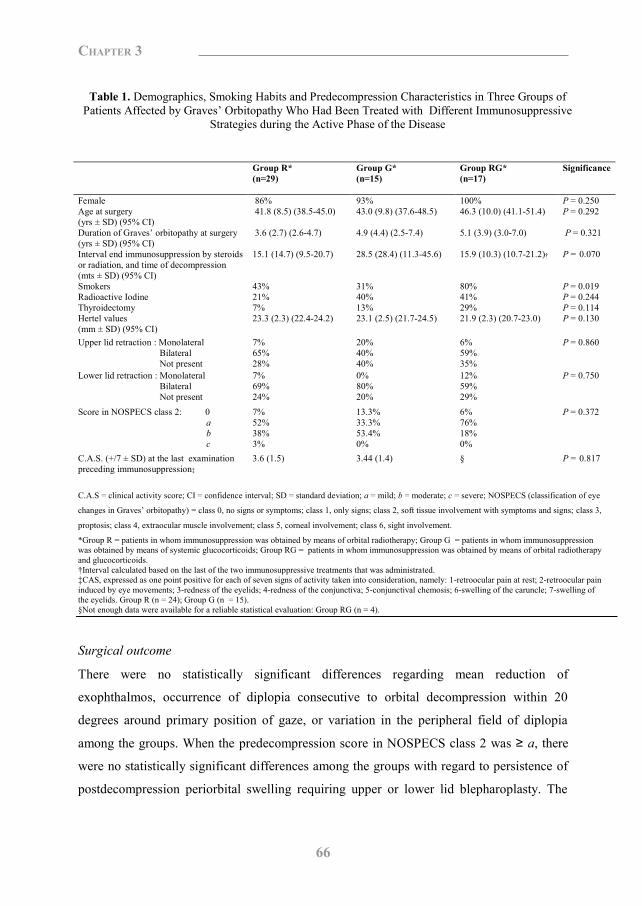
66
Chapter 3
Table 1. Demographics, Smoking Habits and Predecompression Characteristics in Three Groups of
Patients Affected by Graves‟ Orbitopathy Who Had Been Treated with Different Immunosuppressive Strategies during the Active Phase of the Disease
C.A.S = clinical activity score; CI = confidence interval; SD = standard deviation; a = mild; b = moderate; c = severe; NOSPECS (classification of eye
changes in Graves‟ orbitopathy) = class 0, no signs or symptoms; class 1, only signs; class 2, soft tissue involvement with symptoms and signs; class 3,
proptosis; class 4, extraocular muscle involvement; class 5, corneal involvement; class 6, sight involvement.
*Group R = patients in whom immunosuppression was obtained by means of orbital radiotherapy; Group G = patients in whom immunosuppression was obtained by means of systemic glucocorticoids; Group RG = patients in whom immunosuppression was obtained by means of orbital radiotherapy and glucocorticoids. †Interval calculated based on the last of the two immunosuppressive treatments that was administrated. ‡CAS, expressed as one point positive for each of seven signs of activity taken into consideration, namely: 1-retroocular pain at rest; 2-retroocular pain induced by eye movements; 3-redness of the eyelids; 4-redness of the conjunctiva; 5-conjunctival chemosis; 6-swelling of the caruncle; 7-swelling of the eyelids. Group R (n = 24); Group G (n = 15). §Not enough data were available for a reliable statistical evaluation: Group RG (n = 4).
Surgical outcome
There were no statistically significant differences regarding mean reduction of
exophthalmos, occurrence of diplopia consecutive to orbital decompression within 20
degrees around primary position of gaze, or variation in the peripheral field of diplopia
among the groups. When the predecompression score in NOSPECS class 2 was ≥ a, there
were no statistically significant differences among the groups with regard to persistence of
postdecompression periorbital swelling requiring upper or lower lid blepharoplasty. The
Group R* (n=29)
Group G* (n=15)
Group RG* (n=17)
Significance
Female 86% 93% 100% P = 0.250 Age at surgery (yrs ± SD) (95% CI)
41.8 (8.5) (38.5-45.0) 43.0 (9.8) (37.6-48.5) 46.3 (10.0) (41.1-51.4) P = 0.292
Duration of Graves‟ orbitopathy at surgery (yrs ± SD) (95% CI)
3.6 (2.7) (2.6-4.7) 4.9 (4.4) (2.5-7.4) 5.1 (3.9) (3.0-7.0) P = 0.321
Interval end immunosuppression by steroids or radiation, and time of decompression (mts ± SD) (95% CI)
15.1 (14.7) (9.5-20.7) 28.5 (28.4) (11.3-45.6) 15.9 (10.3) (10.7-21.2)† P = 0.070
Smokers 43% 31% 80% P = 0.019 Radioactive Iodine 21% 40% 41% P = 0.244 Thyroidectomy 7% 13% 29% P = 0.114 Hertel values (mm ± SD) (95% CI)
23.3 (2.3) (22.4-24.2) 23.1 (2.5) (21.7-24.5) 21.9 (2.3) (20.7-23.0) P = 0.130
Upper lid retraction : Monolateral Bilateral Not present
7% 65% 28%
20% 40% 40%
6% 59% 35%
P = 0.860
Lower lid retraction : Monolateral Bilateral Not present
7% 69% 24%
0% 80% 20%
12% 59% 29%
P = 0.750
Score in NOSPECS class 2: 0 a b c
7% 52% 38% 3%
13.3% 33.3% 53.4% 0%
6% 76% 18% 0%
P = 0.372
C.A.S. (+/7 ± SD) at the last examination preceding immunosuppression‡
3.6 (1.5) 3.44 (1.4) § P = 0.817
reduction of upper and lower lid retraction also was not statistically different among groups
when these signs were present before surgery (Table 2).
Table 2. Outcome of Three-Wall Rehabilitative Orbital Decompression Through a Coronal Approach in Three Groups of Patients Affected by Graves‟ Orbitopathy
Who Had Been Treated with Different Immunosuppressive Strategies during the Active Phase of the Disease
CI = confidence interval; SD = standard deviation. *Group R = patients in whom immunosuppression was obtained by means of orbital radiotherapy; Group G = patients in whom immunosuppression was obtained by means of systemic glucocorticoids; Group RG = patients in whom immunosuppression was obtained by means of orbital radiotherapy and glucocorticoids. †Only patients with upper lid retraction before surgery: Group R, 21 right 19 left; Group G, six right nine left; Group RG, 11 right 10 left. ‡Only patients with lower lid retraction before surgery: Group R, 21 right 21 left; Group G, 12 right 12 left; Group RG, 12 right 10 left. §Only patients with predecompression score in NOSPECS class 2 = a: Group R, 27; Group G, 13; Group RG, 16. ||Only patients who did have postdecompression diplopia within 20 degrees around the primary position of gaze: Group R, 18; Group G, 7; Group RG, 8. Discussion
Orbital irradiation is a common method of treatment of Graves‟ orbitopathy in its early,
inflammatory, active phase. It seems to be effective on soft tissue inflammatory changes,
recent extraocular muscle involvement, and optic neuropathy by inactivating orbital
lymphocytes and fibroblasts, which are, respectively, the mediators and the effectors of the
inflammatory reaction in the orbit.12 Ionizing radiation, however, may not be without
negligible side effects even when low doses are used.
Histopathologic radiation-induced alterations mainly occur in a repetitive form in different
structures, namely, epithelia and parenchymatous tissues, stroma, and blood vessels. Some
alterations are characteristic of particular organs. All of them are delayed radiation injuries
Group R* (n=29)
Group G* (n=15)
Group RG* (n=17)
Significance
Mean exophthalmos reduction ± SD, mm (95% CI)
4.9 (1.8) (4.2-5.6)
4.6 (1.9) (3.5-5.7)
5.3 (1.9) (4.3-6.2)
P = 0.598
Reduction of upper lid retraction right/left† 52% / 47% 50% / 33% 36% / 40% P = 0.683/P = 0.772
Reduction of lower lid retraction right/left‡ 62% / 52% 67% / 67% 67% / 60% P = 0.946/P = 0.719
Persistence of postdecompression periorbital swelling requiring cosmetic surgery§
48% 46% 31% P = 0.536
Onset of diplopia consecutive to decompression within 20° around primary position of gaze
28% 40% 41% P = 0.563
Field of diplopia beyond 20° from primary position of gaze||: Increased Decreased Unchanged
50% 17% 33%
72% 14% 14%
25% 25% 50%
P = 0.494
100379 Thesis Baldeschi.indd 66 vrijdag17-december-2010 12:07
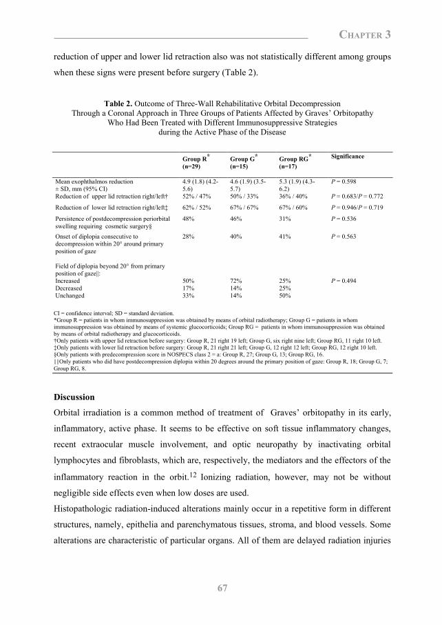
67
Chapter 3
Table 1. Demographics, Smoking Habits and Predecompression Characteristics in Three Groups of
Patients Affected by Graves‟ Orbitopathy Who Had Been Treated with Different Immunosuppressive Strategies during the Active Phase of the Disease
C.A.S = clinical activity score; CI = confidence interval; SD = standard deviation; a = mild; b = moderate; c = severe; NOSPECS (classification of eye
changes in Graves‟ orbitopathy) = class 0, no signs or symptoms; class 1, only signs; class 2, soft tissue involvement with symptoms and signs; class 3,
proptosis; class 4, extraocular muscle involvement; class 5, corneal involvement; class 6, sight involvement.
*Group R = patients in whom immunosuppression was obtained by means of orbital radiotherapy; Group G = patients in whom immunosuppression was obtained by means of systemic glucocorticoids; Group RG = patients in whom immunosuppression was obtained by means of orbital radiotherapy and glucocorticoids. †Interval calculated based on the last of the two immunosuppressive treatments that was administrated. ‡CAS, expressed as one point positive for each of seven signs of activity taken into consideration, namely: 1-retroocular pain at rest; 2-retroocular pain induced by eye movements; 3-redness of the eyelids; 4-redness of the conjunctiva; 5-conjunctival chemosis; 6-swelling of the caruncle; 7-swelling of the eyelids. Group R (n = 24); Group G (n = 15). §Not enough data were available for a reliable statistical evaluation: Group RG (n = 4).
Surgical outcome
There were no statistically significant differences regarding mean reduction of
exophthalmos, occurrence of diplopia consecutive to orbital decompression within 20
degrees around primary position of gaze, or variation in the peripheral field of diplopia
among the groups. When the predecompression score in NOSPECS class 2 was ≥ a, there
were no statistically significant differences among the groups with regard to persistence of
postdecompression periorbital swelling requiring upper or lower lid blepharoplasty. The
Group R* (n=29)
Group G* (n=15)
Group RG* (n=17)
Significance
Female 86% 93% 100% P = 0.250 Age at surgery (yrs ± SD) (95% CI)
41.8 (8.5) (38.5-45.0) 43.0 (9.8) (37.6-48.5) 46.3 (10.0) (41.1-51.4) P = 0.292
Duration of Graves‟ orbitopathy at surgery (yrs ± SD) (95% CI)
3.6 (2.7) (2.6-4.7) 4.9 (4.4) (2.5-7.4) 5.1 (3.9) (3.0-7.0) P = 0.321
Interval end immunosuppression by steroids or radiation, and time of decompression (mts ± SD) (95% CI)
15.1 (14.7) (9.5-20.7) 28.5 (28.4) (11.3-45.6) 15.9 (10.3) (10.7-21.2)† P = 0.070
Smokers 43% 31% 80% P = 0.019 Radioactive Iodine 21% 40% 41% P = 0.244 Thyroidectomy 7% 13% 29% P = 0.114 Hertel values (mm ± SD) (95% CI)
23.3 (2.3) (22.4-24.2) 23.1 (2.5) (21.7-24.5) 21.9 (2.3) (20.7-23.0) P = 0.130
Upper lid retraction : Monolateral Bilateral Not present
7% 65% 28%
20% 40% 40%
6% 59% 35%
P = 0.860
Lower lid retraction : Monolateral Bilateral Not present
7% 69% 24%
0% 80% 20%
12% 59% 29%
P = 0.750
Score in NOSPECS class 2: 0 a b c
7% 52% 38% 3%
13.3% 33.3% 53.4% 0%
6% 76% 18% 0%
P = 0.372
C.A.S. (+/7 ± SD) at the last examination preceding immunosuppression‡
3.6 (1.5) 3.44 (1.4) § P = 0.817
reduction of upper and lower lid retraction also was not statistically different among groups
when these signs were present before surgery (Table 2).
Table 2. Outcome of Three-Wall Rehabilitative Orbital Decompression Through a Coronal Approach in Three Groups of Patients Affected by Graves‟ Orbitopathy
Who Had Been Treated with Different Immunosuppressive Strategies during the Active Phase of the Disease
CI = confidence interval; SD = standard deviation. *Group R = patients in whom immunosuppression was obtained by means of orbital radiotherapy; Group G = patients in whom immunosuppression was obtained by means of systemic glucocorticoids; Group RG = patients in whom immunosuppression was obtained by means of orbital radiotherapy and glucocorticoids. †Only patients with upper lid retraction before surgery: Group R, 21 right 19 left; Group G, six right nine left; Group RG, 11 right 10 left. ‡Only patients with lower lid retraction before surgery: Group R, 21 right 21 left; Group G, 12 right 12 left; Group RG, 12 right 10 left. §Only patients with predecompression score in NOSPECS class 2 = a: Group R, 27; Group G, 13; Group RG, 16. ||Only patients who did have postdecompression diplopia within 20 degrees around the primary position of gaze: Group R, 18; Group G, 7; Group RG, 8. Discussion
Orbital irradiation is a common method of treatment of Graves‟ orbitopathy in its early,
inflammatory, active phase. It seems to be effective on soft tissue inflammatory changes,
recent extraocular muscle involvement, and optic neuropathy by inactivating orbital
lymphocytes and fibroblasts, which are, respectively, the mediators and the effectors of the
inflammatory reaction in the orbit.12 Ionizing radiation, however, may not be without
negligible side effects even when low doses are used.
Histopathologic radiation-induced alterations mainly occur in a repetitive form in different
structures, namely, epithelia and parenchymatous tissues, stroma, and blood vessels. Some
alterations are characteristic of particular organs. All of them are delayed radiation injuries
Group R* (n=29)
Group G* (n=15)
Group RG* (n=17)
Significance
Mean exophthalmos reduction ± SD, mm (95% CI)
4.9 (1.8) (4.2-5.6)
4.6 (1.9) (3.5-5.7)
5.3 (1.9) (4.3-6.2)
P = 0.598
Reduction of upper lid retraction right/left† 52% / 47% 50% / 33% 36% / 40% P = 0.683/P = 0.772
Reduction of lower lid retraction right/left‡ 62% / 52% 67% / 67% 67% / 60% P = 0.946/P = 0.719
Persistence of postdecompression periorbital swelling requiring cosmetic surgery§
48% 46% 31% P = 0.536
Onset of diplopia consecutive to decompression within 20° around primary position of gaze
28% 40% 41% P = 0.563
Field of diplopia beyond 20° from primary position of gaze||: Increased Decreased Unchanged
50% 17% 33%
72% 14% 14%
25% 25% 50%
P = 0.494
100379 Thesis Baldeschi.indd 67 vrijdag17-december-2010 12:07
68
Chapter 3
that appear months to years after exposure to ionizing radiation. Acute radiation injury is
rare.
Fibrosis, one of the most common delayed injuries of stroma, may affect soft orbital tissues,
especially the fat and, in turn, may have an adverse effect on bone removal orbital
rehabilitative decompression surgery. Fibrosis occurs in practically all tissues and organs
exposed to ionizing irradiation, but the extent and severity vary from site to site and are
dependent on the total radiation dose, fraction size, and treated volume. The typical
radiation-induced fibrosis is inhomogeneous: some areas have very dense, acellular
collagen, whereas next to them, other areas have only a few fibrous bands. The fibrous
areas are within the irradiated field but do not fill it entirely. Such alterations are typical of
irradiated adipose tissue.13
Classic theories of radiation effects not only indicate deoxyribonucleic acid double-strand
breaks as the early lethal event, but also assert that tissues that are prone to late radiation
fibrosis have slow reproductive rates that delay cell death and result in late reactions.14
Adipose tissue perfectly fits such characteristics.
Eventual late radiation toxicity to orbital fat in Graves‟ orbitopathy is not likely to be solely
the result of delayed cell death and slow cell turnover. Orbital irradiation is administered in
the active, inflammatory phase of Graves‟ orbitopathy, and it has long been observed that
inflammation worsens delayed radiation fibrosis. Inflammation is mediated largely by
proinflammatory cytokines, including tumor necrosis factor α, which in injured tissue has
been shown to be involved in the recruitment and activation of macrophages with the
release of downstream fibrogenic cytokines. These cytokines include fibroblast growth
factor 2 (FGF2) and transforming growth factor β 1 (TGFβ1).15,16 FGF2 is chemotactic
and mitotic for fibroblasts17, and TGFβ1 stimulates fibroblast proliferation and premature
end differentiation, leading to excessive extracellular matrix glycoprotein production.18-22
Thus, irradiation dramatically alters the ability of macrophages to stimulate normal
fibroblast populations, and fibrosis induced by radiation is a multicellular event in which
the macrophage and fibroblast cell systems interact through intracellular communication
mediated by specific growth factors such as FGF2 and TGFβ1.13,23
Orbital compliance, which refers to the distensibility and plasticity of the orbital tissues,
may be reduced by fibrosis of the orbital soft tissues24, and reduced orbital compliance may
diminish the effectiveness of orbital expansion surgery because orbital soft tissues can fail
to prolapse maximally into the spaces newly created by surgery.25 In view of this
possibility, orbital radiotherapy may be expected to have a negative effect on
decompression surgery in patients with Graves‟ orbitopathy.
Because of the extreme heterogeneity of patients with Graves‟ orbitopathy who need
rehabilitative decompression surgery, the variation applied to surgical techniques, and the
use of perioperative glucocorticoids, it may prove difficult to verify whether this affects the
results of orbital surgery. The paucity of data available in literature seems to be in line with
this viewpoint, which our results confirm further. In our retrospective survey, conducted in
a tertiary referral center with an established tradition for the treatment of Graves‟
orbitopathy11 and encompassing a period of 11 years, among 376 patients decompressed
using the same technique, we could recruit only 61, meeting the criteria that we regarded as
essential for a comparative case series.
Male gender, older age, and heavy smoking habits are patient characteristics that are known
to be associated with more severe orbital disease26-28, whereas extension and location of the
osteotomy performed during decompression surgery, differences in aperture of the
periorbit, and preoperative Hertel values have been regarded as factors influencing the
amount of exophthalmos reduction.25,29-36 In addition, the timing for assessment of surgical
outcome can be an additional source of bias when evaluating the results of decompression
surgery. As a consequence, any meaningful comparison of the results of rehabilitative
decompression surgery in different cohorts of patients with Graves‟ orbitopathy
necessitates a careful evaluation of their baseline characteristics and a careful control of
confounding factors by means of the methodology of investigation. We therefore compared
the groups for baseline characteristics and we tried to control confounding factors and bias
by means of our strict inclusion and exclusion criteria, timing for assessment of the results,
and clear definitions for important symptoms such as diplopia or signs such as persistence
of postdecompression periorbital swelling.
The three groups of patients included in our study were basically homogeneous at baseline,
although smoking was a most common habit among the patients in the RG group.
Considering the well-known negative influence of smoking on the course of Graves
orbitopathy and its capacity to impair the response of Graves orbitopathy to radiotherapy
and glucocorticoids37, it was not unexpected that group RG required the most intensive
100379 Thesis Baldeschi.indd 68 vrijdag17-december-2010 12:07
69
Chapter 3
that appear months to years after exposure to ionizing radiation. Acute radiation injury is
rare.
Fibrosis, one of the most common delayed injuries of stroma, may affect soft orbital tissues,
especially the fat and, in turn, may have an adverse effect on bone removal orbital
rehabilitative decompression surgery. Fibrosis occurs in practically all tissues and organs
exposed to ionizing irradiation, but the extent and severity vary from site to site and are
dependent on the total radiation dose, fraction size, and treated volume. The typical
radiation-induced fibrosis is inhomogeneous: some areas have very dense, acellular
collagen, whereas next to them, other areas have only a few fibrous bands. The fibrous
areas are within the irradiated field but do not fill it entirely. Such alterations are typical of
irradiated adipose tissue.13
Classic theories of radiation effects not only indicate deoxyribonucleic acid double-strand
breaks as the early lethal event, but also assert that tissues that are prone to late radiation
fibrosis have slow reproductive rates that delay cell death and result in late reactions.14
Adipose tissue perfectly fits such characteristics.
Eventual late radiation toxicity to orbital fat in Graves‟ orbitopathy is not likely to be solely
the result of delayed cell death and slow cell turnover. Orbital irradiation is administered in
the active, inflammatory phase of Graves‟ orbitopathy, and it has long been observed that
inflammation worsens delayed radiation fibrosis. Inflammation is mediated largely by
proinflammatory cytokines, including tumor necrosis factor α, which in injured tissue has
been shown to be involved in the recruitment and activation of macrophages with the
release of downstream fibrogenic cytokines. These cytokines include fibroblast growth
factor 2 (FGF2) and transforming growth factor β 1 (TGFβ1).15,16 FGF2 is chemotactic
and mitotic for fibroblasts17, and TGFβ1 stimulates fibroblast proliferation and premature
end differentiation, leading to excessive extracellular matrix glycoprotein production.18-22
Thus, irradiation dramatically alters the ability of macrophages to stimulate normal
fibroblast populations, and fibrosis induced by radiation is a multicellular event in which
the macrophage and fibroblast cell systems interact through intracellular communication
mediated by specific growth factors such as FGF2 and TGFβ1.13,23
Orbital compliance, which refers to the distensibility and plasticity of the orbital tissues,
may be reduced by fibrosis of the orbital soft tissues24, and reduced orbital compliance may
diminish the effectiveness of orbital expansion surgery because orbital soft tissues can fail
to prolapse maximally into the spaces newly created by surgery.25 In view of this
possibility, orbital radiotherapy may be expected to have a negative effect on
decompression surgery in patients with Graves‟ orbitopathy.
Because of the extreme heterogeneity of patients with Graves‟ orbitopathy who need
rehabilitative decompression surgery, the variation applied to surgical techniques, and the
use of perioperative glucocorticoids, it may prove difficult to verify whether this affects the
results of orbital surgery. The paucity of data available in literature seems to be in line with
this viewpoint, which our results confirm further. In our retrospective survey, conducted in
a tertiary referral center with an established tradition for the treatment of Graves‟
orbitopathy11 and encompassing a period of 11 years, among 376 patients decompressed
using the same technique, we could recruit only 61, meeting the criteria that we regarded as
essential for a comparative case series.
Male gender, older age, and heavy smoking habits are patient characteristics that are known
to be associated with more severe orbital disease26-28, whereas extension and location of the
osteotomy performed during decompression surgery, differences in aperture of the
periorbit, and preoperative Hertel values have been regarded as factors influencing the
amount of exophthalmos reduction.25,29-36 In addition, the timing for assessment of surgical
outcome can be an additional source of bias when evaluating the results of decompression
surgery. As a consequence, any meaningful comparison of the results of rehabilitative
decompression surgery in different cohorts of patients with Graves‟ orbitopathy
necessitates a careful evaluation of their baseline characteristics and a careful control of
confounding factors by means of the methodology of investigation. We therefore compared
the groups for baseline characteristics and we tried to control confounding factors and bias
by means of our strict inclusion and exclusion criteria, timing for assessment of the results,
and clear definitions for important symptoms such as diplopia or signs such as persistence
of postdecompression periorbital swelling.
The three groups of patients included in our study were basically homogeneous at baseline,
although smoking was a most common habit among the patients in the RG group.
Considering the well-known negative influence of smoking on the course of Graves
orbitopathy and its capacity to impair the response of Graves orbitopathy to radiotherapy
and glucocorticoids37, it was not unexpected that group RG required the most intensive
100379 Thesis Baldeschi.indd 69 vrijdag17-december-2010 12:07
70
Chapter 3
immunosuppression regimen. By comparing the results of rehabilitative decompression
surgery of the three groups, we did not find that patients treated with retrobulbar irradiation
in the active phase of Graves‟ orbitopathy had a different outcome than those treated with
systemic glucocorticoids alone or with both retrobulbar irradiation and systemic
glucocorticoids in the same phase of the disease.
We acknowledge that the results of our study should be weighed in light of its retrospective
nature and its limitations. Our survey was a retrospective comparative case series, and the
results of this type of study need to be confirmed by investigations with stronger study
designs. The number of patients included was relatively small, the mean interval between
retrobulbar irradiation and orbital decompression was relatively short, and patients were not
allocated randomly to either of the three treatment groups. Despite these shortcomings, our
results seem to suggest that the total radiation dose, fraction size, and irradiated volume
commonly used to treat active Graves‟ orbitopathy do not adversely interfere with the
outcome of rehabilitative decompression surgery.
References 1. Juler FA. Acute purulent keratitis in exophthalmic Goiter treated by repeated tarsorrhaphy resection of the
cervical sympathetic and x-rays: retention of vision in one eye. Trans Ophthalmol Soc UK 1913;33:58-62.
2. Beierwaltes WH. X-ray treatment of malignant exophthalmos. A report of 28 patients. J Clin Endocrinol Metab
1953;13:1090-100.
3. Gorman CA, Garrity JA, Fatourechi V, Bahn RS, Petersen IA, Stafford SL, Earle JD, Forbes GS, Kline RW,
Bergstralh EJ, Offord KP, Rademacher DM, Stanley NM, Bartley GB. A prospective, randomised double blind,
placebo-controlled study of orbital radiotherapy for Graves‟ ophthalmopathy. Ophthalmology 2001;108:1523-34.
4. Prummel MF, Mourits MP, Blank L, Berghout A, Koornneef L, Wiersinga WM. Randomized double-blind trial
of prednisone versus radiotherapy in Graves‟ ophthalmopathy. Lancet 1993;342:949-54.
5. Wakelkamp IM, Tan H, Saeed P, Schlingemann RO, Verbraak FD, Blank LE, Prummel MF, Wiersinga WM.
Orbital irradiation for Graves‟ ophthalmopathy: is it safe? A long-term follow-up study. Ophthalmology
2004;111:1557-62.
6. Mourits MP, Koornneef L, van Mourik-Noordenbos AM, van der Meulen-Schot HM, Prummel MF, Wiersinga
WM, Berghout A. Extraocular muscle surgery for Graves‟ ophthalmopathy: does prior treatment influence
surgical outcome? Br J Ophthalmol 1990;74:481-3.
7. Kazim M, Trokel S, Moore S. Treatment of acute Graves‟ orbitopathy. Ophthalmology 1991;98:1443-1448.
8. Clauser L, Galie M, Sarti E, Dallera V. Rationale of treatment in Graves‟ ophthalmopathy. Plast Reconstr Surg
2001;108:1880-94.
9. Werner SC. Modifications of the classification of the eye changes of Graves‟ disease: recommendations of the
Ad Hoc Committee of the American Thyroid association. J Clin Endocrinol Metab 1977;44:203-4.
10. Mourits MP, Prummel MF, Wiersinga WM, Koornneef L. Clinical activity score as a guide in management of
patients with Graves‟ ophthalmopathy. Clin Endocrinol (Oxf) 1997;47:9-14.
11. Härting F, Koornneef, Peeters HJF, Gillisen JPA. Fourteen years of orbital decompression in Graves‟ disease.
A review of technique, results and complications. Orbit 1986;5:123-9.
12. Kahaly GJ, Gorman CA, Kal HB, Mourits MP, Pinchera A, Tan HS, Prummel MF. Radiotherapy for Graves‟
ophthalmopathy. In: Prummel MF, editor. Recent developments in Graves‟ ophthalmopathy. Boston: Kluwer
Academic Publishers, 2000:115-31.
13. Fajardo L. The pathology of ionizing radiation as defined by morphologic patterns. Acta Oncol 2005;44:13-22.
14. Hellman S. Principles of cancer management: radiation therapy. In: Devita VT Jr, Hellman S, Rosemberg SA,
editors. Cancer: principles and practice of oncology, 3rd edition. Philadelphia: Lippincott-Raven, 1997:307-22.
15. Hallahan DE, Haimovitz-Friedman A, Kufe DW, Fuks Z, Weichselbaum RR. The role of cytokines in
radiation oncology. In: De Vita VT, Hellman S, Rosenberg SA, editors. Important advances in oncology.
Philadelphia: JB Lippincott, 1993:71-80.
16. Neta R, Okunieff P. Cytokine-induced radiation protection and sensitization. Semin Radiat Oncol 1996;6:306-
20.
17. Schreier T, Degen E, Baschong W. Fibroblast migration and proliferation during in vitro wound healing. A
quantitative comparison between various growth factors and a low molecular weight blood dialysate used in the
clinic to normalize impaired wound healing. Res Exp Med (Berl) 1993;193:195-205.
18. Rodemann HP, Bamberg M. Cellular basis of radiationinduced fibrosis. Radiother Oncol 1995;35:83-90.
100379 Thesis Baldeschi.indd 70 vrijdag17-december-2010 12:07
71
Chapter 3
immunosuppression regimen. By comparing the results of rehabilitative decompression
surgery of the three groups, we did not find that patients treated with retrobulbar irradiation
in the active phase of Graves‟ orbitopathy had a different outcome than those treated with
systemic glucocorticoids alone or with both retrobulbar irradiation and systemic
glucocorticoids in the same phase of the disease.
We acknowledge that the results of our study should be weighed in light of its retrospective
nature and its limitations. Our survey was a retrospective comparative case series, and the
results of this type of study need to be confirmed by investigations with stronger study
designs. The number of patients included was relatively small, the mean interval between
retrobulbar irradiation and orbital decompression was relatively short, and patients were not
allocated randomly to either of the three treatment groups. Despite these shortcomings, our
results seem to suggest that the total radiation dose, fraction size, and irradiated volume
commonly used to treat active Graves‟ orbitopathy do not adversely interfere with the
outcome of rehabilitative decompression surgery.
References 1. Juler FA. Acute purulent keratitis in exophthalmic Goiter treated by repeated tarsorrhaphy resection of the
cervical sympathetic and x-rays: retention of vision in one eye. Trans Ophthalmol Soc UK 1913;33:58-62.
2. Beierwaltes WH. X-ray treatment of malignant exophthalmos. A report of 28 patients. J Clin Endocrinol Metab
1953;13:1090-100.
3. Gorman CA, Garrity JA, Fatourechi V, Bahn RS, Petersen IA, Stafford SL, Earle JD, Forbes GS, Kline RW,
Bergstralh EJ, Offord KP, Rademacher DM, Stanley NM, Bartley GB. A prospective, randomised double blind,
placebo-controlled study of orbital radiotherapy for Graves‟ ophthalmopathy. Ophthalmology 2001;108:1523-34.
4. Prummel MF, Mourits MP, Blank L, Berghout A, Koornneef L, Wiersinga WM. Randomized double-blind trial
of prednisone versus radiotherapy in Graves‟ ophthalmopathy. Lancet 1993;342:949-54.
5. Wakelkamp IM, Tan H, Saeed P, Schlingemann RO, Verbraak FD, Blank LE, Prummel MF, Wiersinga WM.
Orbital irradiation for Graves‟ ophthalmopathy: is it safe? A long-term follow-up study. Ophthalmology
2004;111:1557-62.
6. Mourits MP, Koornneef L, van Mourik-Noordenbos AM, van der Meulen-Schot HM, Prummel MF, Wiersinga
WM, Berghout A. Extraocular muscle surgery for Graves‟ ophthalmopathy: does prior treatment influence
surgical outcome? Br J Ophthalmol 1990;74:481-3.
7. Kazim M, Trokel S, Moore S. Treatment of acute Graves‟ orbitopathy. Ophthalmology 1991;98:1443-1448.
8. Clauser L, Galie M, Sarti E, Dallera V. Rationale of treatment in Graves‟ ophthalmopathy. Plast Reconstr Surg
2001;108:1880-94.
9. Werner SC. Modifications of the classification of the eye changes of Graves‟ disease: recommendations of the
Ad Hoc Committee of the American Thyroid association. J Clin Endocrinol Metab 1977;44:203-4.
10. Mourits MP, Prummel MF, Wiersinga WM, Koornneef L. Clinical activity score as a guide in management of
patients with Graves‟ ophthalmopathy. Clin Endocrinol (Oxf) 1997;47:9-14.
11. Härting F, Koornneef, Peeters HJF, Gillisen JPA. Fourteen years of orbital decompression in Graves‟ disease.
A review of technique, results and complications. Orbit 1986;5:123-9.
12. Kahaly GJ, Gorman CA, Kal HB, Mourits MP, Pinchera A, Tan HS, Prummel MF. Radiotherapy for Graves‟
ophthalmopathy. In: Prummel MF, editor. Recent developments in Graves‟ ophthalmopathy. Boston: Kluwer
Academic Publishers, 2000:115-31.
13. Fajardo L. The pathology of ionizing radiation as defined by morphologic patterns. Acta Oncol 2005;44:13-22.
14. Hellman S. Principles of cancer management: radiation therapy. In: Devita VT Jr, Hellman S, Rosemberg SA,
editors. Cancer: principles and practice of oncology, 3rd edition. Philadelphia: Lippincott-Raven, 1997:307-22.
15. Hallahan DE, Haimovitz-Friedman A, Kufe DW, Fuks Z, Weichselbaum RR. The role of cytokines in
radiation oncology. In: De Vita VT, Hellman S, Rosenberg SA, editors. Important advances in oncology.
Philadelphia: JB Lippincott, 1993:71-80.
16. Neta R, Okunieff P. Cytokine-induced radiation protection and sensitization. Semin Radiat Oncol 1996;6:306-
20.
17. Schreier T, Degen E, Baschong W. Fibroblast migration and proliferation during in vitro wound healing. A
quantitative comparison between various growth factors and a low molecular weight blood dialysate used in the
clinic to normalize impaired wound healing. Res Exp Med (Berl) 1993;193:195-205.
18. Rodemann HP, Bamberg M. Cellular basis of radiationinduced fibrosis. Radiother Oncol 1995;35:83-90.
100379 Thesis Baldeschi.indd 71 vrijdag17-december-2010 12:07

72
Chapter 3
19. Martin M, Remy J, Daburon F. In vitro growth potential of fibroblasts isolated from pigs with radiation-
induced fibrosis. Int J Radiat Biol 1986;49:821-8.
20. Bernstein EF, Harisiadis L, Salomon G, Harrington F, Mitchell J, Uitto J, Glatstein E, Russo A. Transforming
growth factor-beta improves healing of radiation-impaired wounds. J Invest Dermatol 1991;97:430-4.
21. Massague J. The transforming growth factor-beta family. Annu Rev Cell Biol 1990;6:541-97.
22. Roberts AB, McCune BK, Sporn MB. TGF-β: regulation of extracellular matrix. Kidney Int 1992;41:557-559.
23. Okunieff P, Augustine E, Hicks JE, Cornelison TL, Altemus RM, Naydich BG, Ding I, Huser AK, Abraham
EH, Smith JJ, Coleman N, Gerber LH. Pentoxifylline in the treatment of radiation-induced fibrosis. J Clin Oncol
2004;22:2207-13.
24. Wilson WB, Manke WF. Orbital decompression in Graves‟ disease: the predictability of reduction of
proptosis. Arch Ophthalmol 1991;109:343-5.
25. Goldberg RA, Weinberg DA, Shorr N, Wirta D. Maximal three-wall orbital decompression through a coronal
approach. Ophthalmic Surg Lasers 1997;28:832-43.
26. Kendler DL, Lippa J, Rootman J. The initial characteristics of Graves‟ orbitopathy vary with age and sex. Arch
Ophthalmol 1993;111:197-201.
27. Perros P, Cromble AL, Matthews JN, Kendall-Taylor P. Age and gender influence the severity of thyroid-
associated ophthalmopathy: a study of 101 patients attending a combined thyroid-eye clinic. Clin Endocrinol
1993;38:367-72.
28. Prummel MF, Wiersinga WM. Smoking and risk of Graves‟ disease. Jama 1993;269:479-82.
29. Baldeschi L, MacAndie K, Hintschich C, Wakelkamp IM, Prummel MF, Wiersinga WM. The removal of the
deep lateral wall in orbital decompression: its contribution to exophthalmos reduction and influence on
consecutive diplopia. Am J Ophthalmol 2005;140:642-47.
30. Linberg JV, Anderson LA. Transorbital decompression. Indications and results. Arch Ophthalmol
1981;99:113-19.
31. Warren JD, Spector JG, Burde R. Long-term follow-up and recent observations on 305 cases of orbital
decompression for dysthyroid orbitopathy. Laryngoscope 1989;99:35-40.
32. Leatherbarrow B, Lendrum J, Mahaffey PJ, Noble JL, Kwartz J, Davies H. Three wall orbital decompression
for Graves‟ ophthalmopathy via a coronal approach. Eye 1991;5:456-65.
33. Garrity JA, Fatourechi V, Bergstralh EJ, Bartley GB, Beatty CW, DeSanto LW, Gorman CA. Results of
transantral orbital decompression in 428 patients with severe Graves‟ ophthalmopathy. Am J Ophthalmol
1993;116:533-47.
34. Lyons CJ, Rootman J. Orbital decompression for disfiguring exophthalmos in thyroid orbitopathy.
Ophthalmology 1994;101:223-30.
35. Fatourechi V, Garrity JA, Bartley GB, Bergstralh EJ, De-Santo LW, Gorman CA. Graves‟ ophthalmopathy.
Results of transantral orbital decompression performed primarily for cosmetic indications. Ophthalmology
1994;101:938-42.
36. Kalmann R, Mourits MP, van der Pol JP, Koornneef L. Coronal approach for rehabilitative orbital
decompression in Graves‟ ophthalmopathy. Br J Ophthalmol 1997;81:41-5.
37. Bartalena L, Pinchera A, Marcocci C. Management of Graves‟ ophthalmopathy: reality and perspectives.
Endocr Rev 2000;21:168-99.
Chapter 4
A Novel Method for Measuring Outcome
of Orbital Decompression in Graves' Orbitopathy
by Evaluating an Infero-medial Decompression
Technique
Lelio Baldeschi1, Annesa Cosma1, 2, Robert Lindeboom3, Marc D. de Smet1,
Wilmar M. Wiersinga4
1 Department of Ophthalmology, University of Amsterdam, Amsterdam, The Netherlands. 2 Department of Ophthalmology, University of Foggia, Foggia, Italy. 3 Department of Clinical Epidemiology and Biostatistics, University of Amsterdam, Amsterdam, The Netherlands. 4 Department of Endocrinology, University of Amsterdam, Amsterdam, The Netherlands. Manuscript in preparation
100379 Thesis Baldeschi.indd 72 vrijdag17-december-2010 12:07
19. Martin M, Remy J, Daburon F. In vitro growth potential of fibroblasts isolated from pigs with radiation-
induced fibrosis. Int J Radiat Biol 1986;49:821-8.
20. Bernstein EF, Harisiadis L, Salomon G, Harrington F, Mitchell J, Uitto J, Glatstein E, Russo A. Transforming
growth factor-beta improves healing of radiation-impaired wounds. J Invest Dermatol 1991;97:430-4.
21. Massague J. The transforming growth factor-beta family. Annu Rev Cell Biol 1990;6:541-97.
22. Roberts AB, McCune BK, Sporn MB. TGF-β: regulation of extracellular matrix. Kidney Int 1992;41:557-559.
23. Okunieff P, Augustine E, Hicks JE, Cornelison TL, Altemus RM, Naydich BG, Ding I, Huser AK, Abraham
EH, Smith JJ, Coleman N, Gerber LH. Pentoxifylline in the treatment of radiation-induced fibrosis. J Clin Oncol
2004;22:2207-13.
24. Wilson WB, Manke WF. Orbital decompression in Graves‟ disease: the predictability of reduction of
proptosis. Arch Ophthalmol 1991;109:343-5.
25. Goldberg RA, Weinberg DA, Shorr N, Wirta D. Maximal three-wall orbital decompression through a coronal
approach. Ophthalmic Surg Lasers 1997;28:832-43.
26. Kendler DL, Lippa J, Rootman J. The initial characteristics of Graves‟ orbitopathy vary with age and sex. Arch
Ophthalmol 1993;111:197-201.
27. Perros P, Cromble AL, Matthews JN, Kendall-Taylor P. Age and gender influence the severity of thyroid-
associated ophthalmopathy: a study of 101 patients attending a combined thyroid-eye clinic. Clin Endocrinol
1993;38:367-72.
28. Prummel MF, Wiersinga WM. Smoking and risk of Graves‟ disease. Jama 1993;269:479-82.
29. Baldeschi L, MacAndie K, Hintschich C, Wakelkamp IM, Prummel MF, Wiersinga WM. The removal of the
deep lateral wall in orbital decompression: its contribution to exophthalmos reduction and influence on
consecutive diplopia. Am J Ophthalmol 2005;140:642-47.
30. Linberg JV, Anderson LA. Transorbital decompression. Indications and results. Arch Ophthalmol
1981;99:113-19.
31. Warren JD, Spector JG, Burde R. Long-term follow-up and recent observations on 305 cases of orbital
decompression for dysthyroid orbitopathy. Laryngoscope 1989;99:35-40.
32. Leatherbarrow B, Lendrum J, Mahaffey PJ, Noble JL, Kwartz J, Davies H. Three wall orbital decompression
for Graves‟ ophthalmopathy via a coronal approach. Eye 1991;5:456-65.
33. Garrity JA, Fatourechi V, Bergstralh EJ, Bartley GB, Beatty CW, DeSanto LW, Gorman CA. Results of
transantral orbital decompression in 428 patients with severe Graves‟ ophthalmopathy. Am J Ophthalmol
1993;116:533-47.
34. Lyons CJ, Rootman J. Orbital decompression for disfiguring exophthalmos in thyroid orbitopathy.
Ophthalmology 1994;101:223-30.
35. Fatourechi V, Garrity JA, Bartley GB, Bergstralh EJ, De-Santo LW, Gorman CA. Graves‟ ophthalmopathy.
Results of transantral orbital decompression performed primarily for cosmetic indications. Ophthalmology
1994;101:938-42.
36. Kalmann R, Mourits MP, van der Pol JP, Koornneef L. Coronal approach for rehabilitative orbital
decompression in Graves‟ ophthalmopathy. Br J Ophthalmol 1997;81:41-5.
37. Bartalena L, Pinchera A, Marcocci C. Management of Graves‟ ophthalmopathy: reality and perspectives.
Endocr Rev 2000;21:168-99.
Chapter 4
A Novel Method for Measuring Outcome
of Orbital Decompression in Graves' Orbitopathy
by Evaluating an Infero-medial Decompression
Technique
Lelio Baldeschi1, Annesa Cosma1, 2, Robert Lindeboom3, Marc D. de Smet1,
Wilmar M. Wiersinga4
1 Department of Ophthalmology, University of Amsterdam, Amsterdam, The Netherlands. 2 Department of Ophthalmology, University of Foggia, Foggia, Italy. 3 Department of Clinical Epidemiology and Biostatistics, University of Amsterdam, Amsterdam, The Netherlands. 4 Department of Endocrinology, University of Amsterdam, Amsterdam, The Netherlands. Manuscript in preparation
100379 Thesis Baldeschi.indd 73 vrijdag17-december-2010 12:07
74
Chapter 4
Abstract
Purpose: To evaluate outcome of a graded bone removal infero-medial orbital
decompression in Graves‟ orbitopathy (GO) by means of a novel stratified appraisal (NSA)
versus a traditional group analysis (TGA).
Design: Prospective observational case series.
Participants: All the orbits decompressed at our institution, for rehabilitative reasons by
one surgeon (L.B.), using the technique under evaluation, between June 1999 and
December 2005.
Methods and main outcome measures: NSA assessed or quantified: (1) the invasiveness
of surgery from minimally (25%) to maximally (100%) invasive, allowing to calculate a
mean index of invasiveness per orbit (MIIO) and per patient (MIIP) (25%≤values≤100%);
(2) at ≥ 6 months postoperatively the surgical targets recorded prior to surgery (amount of
desired exophthalmos reduction, improvement of retroocular tension, reduction of peri-
orbital swelling / aedema, resolution of lagophthalmos) were scored allowing to calculate a
mean index of targets achieved per orbit (MITAO) and per patient (MITAP) (0≤values≤1);
(3) an index of diplopia (ID) (ratio of central 20° of gaze decompression-induced / cured
diplopia) was calculated based on orthoptics at ≥ 3 months postoperatively and before any
strabismus surgery; (4) demographics and preoperative characteristics were compared after
stratification of the included orbits for surgical target; surgical outcomes were assessed
after stratification for amount of desired exophthalmos reduction and invasiveness of
surgery.
TGA examined the entire series as a single homogeneous cohort independently of the
different targets and invasiveness of surgery.
NSA and TGA were compared and complications noted.
Results: 151 orbits of 84 patients (78% female, mean age 45.5±9,7 years) were included.
The NSA detected differences among groups within the studied cohort, the overall
achievement of surgical targets was very high, indices precisely quantified the extent of
applied surgery and surgical results, at variance with those obtained with TGA. A
negligible complication rate was recorded.
Conclusions: The NSA may represent a step forward towards a more comprehensive and
accurate evaluation of decompression surgery outcomes.
Introduction
For roughly a century decompression surgery has been used to treat Graves‟ orbitopathy
(GO).1 Initially it was utilized to address sight threatening conditions such as optic
neuropathy, or exposure keratopathy. More recently the indications for orbital
decompression were extended to the treatment of the typical disfigurement, and symptoms
caused by the disease, which include globe and eyelid displacement, periorbital swelling,
and raised retroocular tension. At present, it is also used as a preliminary step before squint
surgery when space is required for recession of enlarged extraocular muscles, and
functional indications of orbital decompression have been further expanded for the
treatment of globe subluxation, postural visual obscuration in patients with congestive
inactive GO2 and choroidal folds of recent onset due to globe indentation by enlarged
extraocular muscles.3, 4
Despite the multiple indications for rehabilitative decompression surgery, mean
exophthalmos reduction and incidence of new onset diplopia have been the main outcome
measures reported in most of the published series. Through the years most authors have
linked effectiveness and reliability of the reported techniques to the highest extent of
exophthalmos reduction and the lowest incidence of induced diplopia.
We perceive this perspective as suboptimal since the data which have been generated /
reported are incomplete, often imprecise and most likely misleading.
Data are incomplete because most of the effects of decompression surgery other than those
on exophthalmos and binocular single vision have remained unmeasured.
Data are also often imprecise because many series inappropriately mix patients
decompressed for disfigurement, with those presenting with compressive dysthyroid optic
neuropathy, in whom the reduction of exophthalmos was not the reason to undertake
decompression surgery and in whom the true incidence of decompression induced diplopia
could have been confounded by the low pre-decompression visual acuity.
Data are most likely misleading because it is assumed that decompression surgery is always
performed to obtain the maximal possible exophthalmos reduction when in reality any
decompression technique can be used with different extent of osteotomies, fat removal or
periorbita manipulation to achieve any degree of desired reduction of exophthalmos in the
single orbit. In addition, high exophthalmos reduction is not an index of effectiveness as
any excessive exophthalmos reduction is to be regarded as a complication of
100379 Thesis Baldeschi.indd 74 vrijdag17-december-2010 12:07
75
Chapter 4
Abstract
Purpose: To evaluate outcome of a graded bone removal infero-medial orbital
decompression in Graves‟ orbitopathy (GO) by means of a novel stratified appraisal (NSA)
versus a traditional group analysis (TGA).
Design: Prospective observational case series.
Participants: All the orbits decompressed at our institution, for rehabilitative reasons by
one surgeon (L.B.), using the technique under evaluation, between June 1999 and
December 2005.
Methods and main outcome measures: NSA assessed or quantified: (1) the invasiveness
of surgery from minimally (25%) to maximally (100%) invasive, allowing to calculate a
mean index of invasiveness per orbit (MIIO) and per patient (MIIP) (25%≤values≤100%);
(2) at ≥ 6 months postoperatively the surgical targets recorded prior to surgery (amount of
desired exophthalmos reduction, improvement of retroocular tension, reduction of peri-
orbital swelling / aedema, resolution of lagophthalmos) were scored allowing to calculate a
mean index of targets achieved per orbit (MITAO) and per patient (MITAP) (0≤values≤1);
(3) an index of diplopia (ID) (ratio of central 20° of gaze decompression-induced / cured
diplopia) was calculated based on orthoptics at ≥ 3 months postoperatively and before any
strabismus surgery; (4) demographics and preoperative characteristics were compared after
stratification of the included orbits for surgical target; surgical outcomes were assessed
after stratification for amount of desired exophthalmos reduction and invasiveness of
surgery.
TGA examined the entire series as a single homogeneous cohort independently of the
different targets and invasiveness of surgery.
NSA and TGA were compared and complications noted.
Results: 151 orbits of 84 patients (78% female, mean age 45.5±9,7 years) were included.
The NSA detected differences among groups within the studied cohort, the overall
achievement of surgical targets was very high, indices precisely quantified the extent of
applied surgery and surgical results, at variance with those obtained with TGA. A
negligible complication rate was recorded.
Conclusions: The NSA may represent a step forward towards a more comprehensive and
accurate evaluation of decompression surgery outcomes.
Introduction
For roughly a century decompression surgery has been used to treat Graves‟ orbitopathy
(GO).1 Initially it was utilized to address sight threatening conditions such as optic
neuropathy, or exposure keratopathy. More recently the indications for orbital
decompression were extended to the treatment of the typical disfigurement, and symptoms
caused by the disease, which include globe and eyelid displacement, periorbital swelling,
and raised retroocular tension. At present, it is also used as a preliminary step before squint
surgery when space is required for recession of enlarged extraocular muscles, and
functional indications of orbital decompression have been further expanded for the
treatment of globe subluxation, postural visual obscuration in patients with congestive
inactive GO2 and choroidal folds of recent onset due to globe indentation by enlarged
extraocular muscles.3, 4
Despite the multiple indications for rehabilitative decompression surgery, mean
exophthalmos reduction and incidence of new onset diplopia have been the main outcome
measures reported in most of the published series. Through the years most authors have
linked effectiveness and reliability of the reported techniques to the highest extent of
exophthalmos reduction and the lowest incidence of induced diplopia.
We perceive this perspective as suboptimal since the data which have been generated /
reported are incomplete, often imprecise and most likely misleading.
Data are incomplete because most of the effects of decompression surgery other than those
on exophthalmos and binocular single vision have remained unmeasured.
Data are also often imprecise because many series inappropriately mix patients
decompressed for disfigurement, with those presenting with compressive dysthyroid optic
neuropathy, in whom the reduction of exophthalmos was not the reason to undertake
decompression surgery and in whom the true incidence of decompression induced diplopia
could have been confounded by the low pre-decompression visual acuity.
Data are most likely misleading because it is assumed that decompression surgery is always
performed to obtain the maximal possible exophthalmos reduction when in reality any
decompression technique can be used with different extent of osteotomies, fat removal or
periorbita manipulation to achieve any degree of desired reduction of exophthalmos in the
single orbit. In addition, high exophthalmos reduction is not an index of effectiveness as
any excessive exophthalmos reduction is to be regarded as a complication of
100379 Thesis Baldeschi.indd 75 vrijdag17-december-2010 12:07
76
Chapter 4
decompression surgery. Similarly the incidence of decompression induced diplopia is not
itself most appropriate to describe surgical outcome because it does not take into account
the cases of preoperative diplopia that are cured by decompression.
Based on these considerations it appears evident that the results of decompression surgery
deserve a more adequate, comprehensive and reliable evaluation. We tried to make a move
towards this goal by evaluating the outcome of a graded, bone inferomedial rehabilitative
decompression technique performed through a minimally invasive suture-less trans inferior
fornix approach, under a novel perspective which possibly overcomes the existing “time
honoured” limitations.
Patients and methods
Our survey is a prospective observational case series. The study was carried out following
the tenets of the Declaration of Helsinki and of the “CODE of CONDUCT” of the Dutch
Federation of Biomedical Scientific Societies (http://www.federa.org/) as recommended by
the Review Board of the Academic Medical Center University of Amsterdam. The same
Review Board ruled that for this type of study its permission need not to be requested, nor
was patients‟ informed consent necessary if data are collected anonymously from the
medical records used by the authors in their every day practice.
Inclusion criteria:
We included all the patients decompressed mono or bilaterally for rehabilitative reasons,
who were clinically and biochemically euthyroid for at least 3 months, and who had
undergone a graded bone inferomedial rehabilitative decompression performed through a
minimally invasive suture less trans inferior fornix approach by one surgeon (L.B.),
between June 1999 and December 2005.
Exclusion criteria:
We excluded all the patients 1) who had been treated with thyroidectomy or I131 in the 6
months preceding decompression or in the 6 months preceding postoperative evaluation; 2)
who had concurrent orbital or periorbital diseases, injuries or surgeries (except for
postdecompression blepharoplasties) within 6 months after decompression; 3) whose
surgical targets were not listed in the respective medical files and whose pre and post
operative notes were not filled out by means of the GO case record form in use in our
clinic at the time of the study. It included all the necessary information for an adequate
clinical assessement of patients with GO, and was very similar to the EUGOGO case record
form5 being one of its precursors.
Surgical technique
In bilateral cases, surgery was always initially performed on the more protruding eye if
any. After the exposure of the inferior fornix by mean of a Desmarres‟ retractor and a
malleable orbital retractor, both electrically insulated, the conjunctiva and lower lid
retractor complex were severed en-bloc with a Colorado needle and the inferior orbital rim
was exposed. The periorbita was incised at this level and the medial and inferior orbital
walls exposed by developing a subperiosteal plane. In order to obtain the best possible
exposure of the medial wall, the bone insertion of the inferior oblique muscle was partially
detached, and the conjunctival incision extended up to the caruncle.
Once the medial orbital wall and the orbital floor were exposed a Frazier suction tip was
used to fracture the delicate bone of the medial orbital wall and of the floor and Blakesley
forceps were used to remove bony fragments and mucosa of the sinuses. If necessary bone-
nibbling rongeurs were also used. The bulla ethmoidalis underneath the fronto-ethmoidal
suture was opened towards the orbit from the posterior lacrimal crest up to the orbital apex,
and then the orbital floor medial to the infra-orbital canal was removed from about 1 cm
behind the inferior orbital rim up to the posterior wall of the maxillary sinus.
Starting from this point, on the basis of intra operative estimations of eye position and
resistance to retropulsion of the globe, and considering the targets to be pursued in the
single orbit, surgery was graded to the specific patient‟s needs. Clear surgical notes were
prepared so that 4 progressively invasive surgical classes could be defined in the single
orbit (A = minimally invasive to D maximally invasive, with an attributed index of
invasiveness orbit (IIO) ranging from 25% for class A to 100% for class D).
Surgical class A: Osteotomies were terminated at this stage, and the periorbita left intact.
Surgical class B: The periobita was opened inferiorly and medially sparing the anterior
portion of its inferior and medial surfaces for roughly 1.5 cm behind the arcus marginalis.
Surgical class C: The bony infraorbital canal and the orbital floor lateral to it were
completely removed, the periorbita was addressed as for class B.
Surgical class D: The posterior two thirds of the maxillary ethmoidal strut were removed
creating a wide antrostomy between the maxillary sinus and the nasal cavity. The bony
infraorbital canal, the orbital floor lateral to it, and periorbita were addressed as for class C.
100379 Thesis Baldeschi.indd 76 vrijdag17-december-2010 12:07
77
Chapter 4
decompression surgery. Similarly the incidence of decompression induced diplopia is not
itself most appropriate to describe surgical outcome because it does not take into account
the cases of preoperative diplopia that are cured by decompression.
Based on these considerations it appears evident that the results of decompression surgery
deserve a more adequate, comprehensive and reliable evaluation. We tried to make a move
towards this goal by evaluating the outcome of a graded, bone inferomedial rehabilitative
decompression technique performed through a minimally invasive suture-less trans inferior
fornix approach, under a novel perspective which possibly overcomes the existing “time
honoured” limitations.
Patients and methods
Our survey is a prospective observational case series. The study was carried out following
the tenets of the Declaration of Helsinki and of the “CODE of CONDUCT” of the Dutch
Federation of Biomedical Scientific Societies (http://www.federa.org/) as recommended by
the Review Board of the Academic Medical Center University of Amsterdam. The same
Review Board ruled that for this type of study its permission need not to be requested, nor
was patients‟ informed consent necessary if data are collected anonymously from the
medical records used by the authors in their every day practice.
Inclusion criteria:
We included all the patients decompressed mono or bilaterally for rehabilitative reasons,
who were clinically and biochemically euthyroid for at least 3 months, and who had
undergone a graded bone inferomedial rehabilitative decompression performed through a
minimally invasive suture less trans inferior fornix approach by one surgeon (L.B.),
between June 1999 and December 2005.
Exclusion criteria:
We excluded all the patients 1) who had been treated with thyroidectomy or I131 in the 6
months preceding decompression or in the 6 months preceding postoperative evaluation; 2)
who had concurrent orbital or periorbital diseases, injuries or surgeries (except for
postdecompression blepharoplasties) within 6 months after decompression; 3) whose
surgical targets were not listed in the respective medical files and whose pre and post
operative notes were not filled out by means of the GO case record form in use in our
clinic at the time of the study. It included all the necessary information for an adequate
clinical assessement of patients with GO, and was very similar to the EUGOGO case record
form5 being one of its precursors.
Surgical technique
In bilateral cases, surgery was always initially performed on the more protruding eye if
any. After the exposure of the inferior fornix by mean of a Desmarres‟ retractor and a
malleable orbital retractor, both electrically insulated, the conjunctiva and lower lid
retractor complex were severed en-bloc with a Colorado needle and the inferior orbital rim
was exposed. The periorbita was incised at this level and the medial and inferior orbital
walls exposed by developing a subperiosteal plane. In order to obtain the best possible
exposure of the medial wall, the bone insertion of the inferior oblique muscle was partially
detached, and the conjunctival incision extended up to the caruncle.
Once the medial orbital wall and the orbital floor were exposed a Frazier suction tip was
used to fracture the delicate bone of the medial orbital wall and of the floor and Blakesley
forceps were used to remove bony fragments and mucosa of the sinuses. If necessary bone-
nibbling rongeurs were also used. The bulla ethmoidalis underneath the fronto-ethmoidal
suture was opened towards the orbit from the posterior lacrimal crest up to the orbital apex,
and then the orbital floor medial to the infra-orbital canal was removed from about 1 cm
behind the inferior orbital rim up to the posterior wall of the maxillary sinus.
Starting from this point, on the basis of intra operative estimations of eye position and
resistance to retropulsion of the globe, and considering the targets to be pursued in the
single orbit, surgery was graded to the specific patient‟s needs. Clear surgical notes were
prepared so that 4 progressively invasive surgical classes could be defined in the single
orbit (A = minimally invasive to D maximally invasive, with an attributed index of
invasiveness orbit (IIO) ranging from 25% for class A to 100% for class D).
Surgical class A: Osteotomies were terminated at this stage, and the periorbita left intact.
Surgical class B: The periobita was opened inferiorly and medially sparing the anterior
portion of its inferior and medial surfaces for roughly 1.5 cm behind the arcus marginalis.
Surgical class C: The bony infraorbital canal and the orbital floor lateral to it were
completely removed, the periorbita was addressed as for class B.
Surgical class D: The posterior two thirds of the maxillary ethmoidal strut were removed
creating a wide antrostomy between the maxillary sinus and the nasal cavity. The bony
infraorbital canal, the orbital floor lateral to it, and periorbita were addressed as for class C.
100379 Thesis Baldeschi.indd 77 vrijdag17-december-2010 12:07
78
Chapter 4
At all times possible sources of bleeding were controlled with cautious bipolar diathermy.
In all cases, at the end of surgery the fornix incision was not sutured and a pressure bandage
was applied on the operated eye, after absence of bleeding was assessed.
Immediate post operative care:
Pupil reflexes, visual function, ductions and versions were checked when the patient was
awake postoperatively, then daily up until the morning of post operative day 2 when
patients were discharged from the hospital. Pressure bandages were removed and reapplied
daily after clinical examination up until discharge. At surgery, post operative day 1 and 2
intravenous ceftriaxone (1 g/day) was administered; patients left hospital without
medications except for artificial tears.
Evaluated data
The following data were evaluated: 1) gender; 2) age at surgery; 3) duration of GO at
decompression; 4) smoking habits at surgery; 5) possible administration of systemic
immunosuppressive treatments, and / or orbital radiotherapy before decompression;
possible treatments with I131 or thyroidectomy before and after decompression; 8) pre and
post operative best corrected visual acuity, exophthalmometric values, measure of possible
lagophthalmos, NOSPECS, and CAS scores6,7; 9) expected targets of infero medial
decompression surgery; 10) information useful for defining IIO; 11) pre and postoperative
orthoptic evaluations including information on central diplopia defined as double vision
within 20° of the central position of gaze, field of peripheral diplopia, namely extension of
diplopia beyond 20 degrees of the central position of gaze, measures of ductions in degrees
performed with a modified perimeter8; 12) any information concerning intraoperative,
perioperative or medium term complications other than strabismus.
Clinical pre operative data were those of the last ophthalmic examination before
decompression, while post operative data and scoring of the single targets as achieved or
not achieved referred to the first ophthalmic examination performed 6 or more months
after surgery. In the case of post decompression blepharoplasties performed earlier than
post decompression month 6 the achievement of the target “reduction of periorbital
oedema” referred to the last ophthalmic examination preceding blepharoplasty.
Pre and postoperative data concerning eye motility were those collected at the orthoptic
evaluations performed respectively not earlier than 6 months before decompression and not
earlier than 3 months, but before any strabismus surgery.
Methodology
Predecompression characteristics and outcome by a novel stratified appraisal (NSA) were
weigh ed against that of a traditional group analysis (TGA).
Novel Stratified Appraisal
Before surgery the targets to be achieved by the graded inferomedial decompression
technique in the single orbit, were discussed with the patients and listed in their medical
records.
Orbital targets were scored as achieved as desired or not, and were: (1) amount of desired
exophthalmos reduction specified as a) none to minimal, i.e. ≤ 2 mm; b) moderate to
maximal, i.e. ≥ 3 mm without causing enophthalmos and / or hypoglobus namely
excessive reduction of exophthalmos, and / or eyeball dystopia for which surgical
correction was advised or performed; (2) resolution of retroocular tension, defined as
patient‟s subjective perception of disappearance of such a symptom after decompression;
(3) reduction of periorbital swelling oedema, defined as decrease of at least 1 point in
NOSPECS class 2 after decompression; (4) cure of lagophthalmos, defined as
disappearance or reduction of ≥ 3 mm of preoperative lagophthalmos.
In addition symmetry in exophthalmos reduction was considered as a constant patient target
to be pursued. We considered symmetry as achieved when post operative Hertel readings
were right = left±1 mm.
In order to characterize different groups according to the invasiveness of surgery a mean
IIO, named mean index of invasiveness orbit (MIIO) was calculated, as well as a mean
index of invasiveness patient (MIIP) which was based on the formula 0.5 x Σ IIO / number
of patients included into the group (the coefficient 0.5 in the formula was chosen because in
a patient the contribution of each orbit to invasiveness is 50%). IIO, MIIO, and MIIP
express invasiveness in percentages from 25-100%.
For measuring in different groups the average amount of achievement of the desired
surgical targets, a mean index of targets achieved orbit (MITAO) and a mean index of
targets achievement patient (MITAP) were calculated, respectively based on the formulas:
Σ 1 or 0 point for each target respectively achieved as desired or not in the single orbit /
overall number of desired targets in a group of orbits; Σ 1 or 0 point for each target
respectively achieved as desired or not for targets concerning only one of the two orbits,
and for target symmetry (constant) + Σ 0.5 or 0 points for each target respectively achieved
100379 Thesis Baldeschi.indd 78 vrijdag17-december-2010 12:08

79
Chapter 4
At all times possible sources of bleeding were controlled with cautious bipolar diathermy.
In all cases, at the end of surgery the fornix incision was not sutured and a pressure bandage
was applied on the operated eye, after absence of bleeding was assessed.
Immediate post operative care:
Pupil reflexes, visual function, ductions and versions were checked when the patient was
awake postoperatively, then daily up until the morning of post operative day 2 when
patients were discharged from the hospital. Pressure bandages were removed and reapplied
daily after clinical examination up until discharge. At surgery, post operative day 1 and 2
intravenous ceftriaxone (1 g/day) was administered; patients left hospital without
medications except for artificial tears.
Evaluated data
The following data were evaluated: 1) gender; 2) age at surgery; 3) duration of GO at
decompression; 4) smoking habits at surgery; 5) possible administration of systemic
immunosuppressive treatments, and / or orbital radiotherapy before decompression;
possible treatments with I131 or thyroidectomy before and after decompression; 8) pre and
post operative best corrected visual acuity, exophthalmometric values, measure of possible
lagophthalmos, NOSPECS, and CAS scores6,7; 9) expected targets of infero medial
decompression surgery; 10) information useful for defining IIO; 11) pre and postoperative
orthoptic evaluations including information on central diplopia defined as double vision
within 20° of the central position of gaze, field of peripheral diplopia, namely extension of
diplopia beyond 20 degrees of the central position of gaze, measures of ductions in degrees
performed with a modified perimeter8; 12) any information concerning intraoperative,
perioperative or medium term complications other than strabismus.
Clinical pre operative data were those of the last ophthalmic examination before
decompression, while post operative data and scoring of the single targets as achieved or
not achieved referred to the first ophthalmic examination performed 6 or more months
after surgery. In the case of post decompression blepharoplasties performed earlier than
post decompression month 6 the achievement of the target “reduction of periorbital
oedema” referred to the last ophthalmic examination preceding blepharoplasty.
Pre and postoperative data concerning eye motility were those collected at the orthoptic
evaluations performed respectively not earlier than 6 months before decompression and not
earlier than 3 months, but before any strabismus surgery.
Methodology
Predecompression characteristics and outcome by a novel stratified appraisal (NSA) were
weigh ed against that of a traditional group analysis (TGA).
Novel Stratified Appraisal
Before surgery the targets to be achieved by the graded inferomedial decompression
technique in the single orbit, were discussed with the patients and listed in their medical
records.
Orbital targets were scored as achieved as desired or not, and were: (1) amount of desired
exophthalmos reduction specified as a) none to minimal, i.e. ≤ 2 mm; b) moderate to
maximal, i.e. ≥ 3 mm without causing enophthalmos and / or hypoglobus namely
excessive reduction of exophthalmos, and / or eyeball dystopia for which surgical
correction was advised or performed; (2) resolution of retroocular tension, defined as
patient‟s subjective perception of disappearance of such a symptom after decompression;
(3) reduction of periorbital swelling oedema, defined as decrease of at least 1 point in
NOSPECS class 2 after decompression; (4) cure of lagophthalmos, defined as
disappearance or reduction of ≥ 3 mm of preoperative lagophthalmos.
In addition symmetry in exophthalmos reduction was considered as a constant patient target
to be pursued. We considered symmetry as achieved when post operative Hertel readings
were right = left±1 mm.
In order to characterize different groups according to the invasiveness of surgery a mean
IIO, named mean index of invasiveness orbit (MIIO) was calculated, as well as a mean
index of invasiveness patient (MIIP) which was based on the formula 0.5 x Σ IIO / number
of patients included into the group (the coefficient 0.5 in the formula was chosen because in
a patient the contribution of each orbit to invasiveness is 50%). IIO, MIIO, and MIIP
express invasiveness in percentages from 25-100%.
For measuring in different groups the average amount of achievement of the desired
surgical targets, a mean index of targets achieved orbit (MITAO) and a mean index of
targets achievement patient (MITAP) were calculated, respectively based on the formulas:
Σ 1 or 0 point for each target respectively achieved as desired or not in the single orbit /
overall number of desired targets in a group of orbits; Σ 1 or 0 point for each target
respectively achieved as desired or not for targets concerning only one of the two orbits,
and for target symmetry (constant) + Σ 0.5 or 0 points for each target respectively achieved
100379 Thesis Baldeschi.indd 79 vrijdag17-december-2010 12:08
80
Chapter 4
as desired or not for targets concerning both the orbits / overall number of desired targets
in a group of patients (0.5 points for targets desirable in both orbits was chosen because in a
patient the contribution of each orbit to the achievement of that target is 50%). MITAO and
MITAP are numbers between 0 and 1; MITAO=1 and MITAP=1 when all the targets are
achieved in a group of orbits or in a group of patients respectively.
Based on the ratio frequency decompression induced diplopia / frequency decompression
cured diplopia, an index of diplopia (ID) was calculated. ID is =1 when the frequency of
induced diplopia is the same as that of cured, is > 1 when the frequency of induced diplopia
is higher than that of cured, is < 1 viceversa.
Orbits grouped for target “amount of desired exophthalmos reduction” and stratified for
class of surgical invasiveness (IIO) were compared within groups and within classes in
respect to mean exophthalmos reduction, mean decompression induced change in ductions.
Central and peripheral fields of diplopia were assessed in patients in whom preoperative
binocular vision was present and who did not have anomalous retinal correspondence.
Patients stratified for decompression induced or cured central diplopia were compared.
Traditional Group Analysis
Surgical outcomes (mean post operative exophthalmos, mean exophthalmos reduction,
decompression induced change in duction) of the entire cohort of orbits were evaluated as
a whole in the TGA. Incidence of decompression induced diplopia was evaluated in the
same cohort of patients as for NSA.
Statistical analysis
Pre and post operative data were inserted in a data base created in SPSS 16.0 Statistics UK
and analysed.
Univariate comparisons: one-way ANOVA and Student's t test were used to compare
continuous variables. In case of ordinal or not normally distributed variables the Mann-
Whitney U test was used. Categorical preoperative and post-operative data were
investigated using χ² test. Significance was assumed if P ≤ 0.05.
Multiple regression analysis was used to assess the independent influence of preoperative
Hertel values and IIO on exophthalmos reduction, and decompression induced mean
changes in ductions.
Results
Eighty-four patients (151 orbits) out of 295 patients decompressed within the study period
were selected for this survey, 210 patients were excluded because they did not meet the
inclusion criteria or the necessary data were not recorded. One patient, previously reported,
was excluded because of delayed decompression related reactivation (DDRR)9, requiring
high dose oral glucocorticoids starting from 1 month after surgery.
Preoperative characteristics
The 151 orbits had the following characteristics: 77.5% were feminine, mean age ± SD at
decompression was 45.5±9.7 years, mean duration of GO ± SD at decompression
45.7±44.6 months; 51.3% were smokers, and 34.2% had received I131 and 11.6%
thyroidectomy. Steroids, orbital radiotherapy, or combined steroids and orbital radiotherapy
had been administered in 12.2%, 17.6%, and 10.1% respectively.
The NSA disclosed the following differences among orbits stratified for surgical target:
The orbits that required “moderate to maximal exophthalmos reduction” were younger and
had higher mean preoperative Hertel values than those that required “no to minimal
exophthalmos reduction” (mean± SD age at decompression (44.4±9.6 vs 52.3±7.6 years, P
= 0.000) (mean± SD preoperative Hertel 21.6±2,2 vs 19.6±1,5 mm, P = 0.000). In addition
the orbits whose target was “no to minimal exophthalmos reduction” had received orbital
radiotherapy less frequently than those whose target was “moderate to maximal
exophthalmos reduction”(9.5% vs 30.7% P = 0.001). The orbits that required treatment of
lagophthalmos had received combined radiotherapy and glucocorticoids less frequently
(0%) as compared with those who did not (12%) (P = 0.000). The duration of GO ± SD at
decompression was longer in those orbits that did not require reduction of the periorbital
oedema (70.3±67.5 months) as compared with those who required it (40.7±36.8 months)
(P = 0.003).
Surgical outcome
After decompression surgery there was no clinically relevant change in visual acuity in any
patient.
The achievement of the desired targets in the operated orbits are outlined in table 1.
Calculated MITAO and MITAP were 0.95±0.16 and 0.94±0.15 respectively, attained by
100379 Thesis Baldeschi.indd 80 vrijdag17-december-2010 12:08
81
Chapter 4
as desired or not for targets concerning both the orbits / overall number of desired targets
in a group of patients (0.5 points for targets desirable in both orbits was chosen because in a
patient the contribution of each orbit to the achievement of that target is 50%). MITAO and
MITAP are numbers between 0 and 1; MITAO=1 and MITAP=1 when all the targets are
achieved in a group of orbits or in a group of patients respectively.
Based on the ratio frequency decompression induced diplopia / frequency decompression
cured diplopia, an index of diplopia (ID) was calculated. ID is =1 when the frequency of
induced diplopia is the same as that of cured, is > 1 when the frequency of induced diplopia
is higher than that of cured, is < 1 viceversa.
Orbits grouped for target “amount of desired exophthalmos reduction” and stratified for
class of surgical invasiveness (IIO) were compared within groups and within classes in
respect to mean exophthalmos reduction, mean decompression induced change in ductions.
Central and peripheral fields of diplopia were assessed in patients in whom preoperative
binocular vision was present and who did not have anomalous retinal correspondence.
Patients stratified for decompression induced or cured central diplopia were compared.
Traditional Group Analysis
Surgical outcomes (mean post operative exophthalmos, mean exophthalmos reduction,
decompression induced change in duction) of the entire cohort of orbits were evaluated as
a whole in the TGA. Incidence of decompression induced diplopia was evaluated in the
same cohort of patients as for NSA.
Statistical analysis
Pre and post operative data were inserted in a data base created in SPSS 16.0 Statistics UK
and analysed.
Univariate comparisons: one-way ANOVA and Student's t test were used to compare
continuous variables. In case of ordinal or not normally distributed variables the Mann-
Whitney U test was used. Categorical preoperative and post-operative data were
investigated using χ² test. Significance was assumed if P ≤ 0.05.
Multiple regression analysis was used to assess the independent influence of preoperative
Hertel values and IIO on exophthalmos reduction, and decompression induced mean
changes in ductions.
Results
Eighty-four patients (151 orbits) out of 295 patients decompressed within the study period
were selected for this survey, 210 patients were excluded because they did not meet the
inclusion criteria or the necessary data were not recorded. One patient, previously reported,
was excluded because of delayed decompression related reactivation (DDRR)9, requiring
high dose oral glucocorticoids starting from 1 month after surgery.
Preoperative characteristics
The 151 orbits had the following characteristics: 77.5% were feminine, mean age ± SD at
decompression was 45.5±9.7 years, mean duration of GO ± SD at decompression
45.7±44.6 months; 51.3% were smokers, and 34.2% had received I131 and 11.6%
thyroidectomy. Steroids, orbital radiotherapy, or combined steroids and orbital radiotherapy
had been administered in 12.2%, 17.6%, and 10.1% respectively.
The NSA disclosed the following differences among orbits stratified for surgical target:
The orbits that required “moderate to maximal exophthalmos reduction” were younger and
had higher mean preoperative Hertel values than those that required “no to minimal
exophthalmos reduction” (mean± SD age at decompression (44.4±9.6 vs 52.3±7.6 years, P
= 0.000) (mean± SD preoperative Hertel 21.6±2,2 vs 19.6±1,5 mm, P = 0.000). In addition
the orbits whose target was “no to minimal exophthalmos reduction” had received orbital
radiotherapy less frequently than those whose target was “moderate to maximal
exophthalmos reduction”(9.5% vs 30.7% P = 0.001). The orbits that required treatment of
lagophthalmos had received combined radiotherapy and glucocorticoids less frequently
(0%) as compared with those who did not (12%) (P = 0.000). The duration of GO ± SD at
decompression was longer in those orbits that did not require reduction of the periorbital
oedema (70.3±67.5 months) as compared with those who required it (40.7±36.8 months)
(P = 0.003).
Surgical outcome
After decompression surgery there was no clinically relevant change in visual acuity in any
patient.
The achievement of the desired targets in the operated orbits are outlined in table 1.
Calculated MITAO and MITAP were 0.95±0.16 and 0.94±0.15 respectively, attained by
100379 Thesis Baldeschi.indd 81 vrijdag17-december-2010 12:08
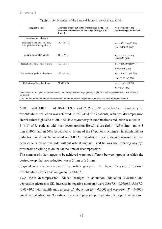
82
Chapter 4
Table 1. Achievement of the surgical target in the operated orbit
Surgical targets Operated orbits out of the whole series (n=151) in which the achievement of the surgical target was desired
Achievement of the surgical target as desired
Exophthalmos reduction:
moderate to maximal (≥ 3mm, <enophthalmos/hypoglobus*)
130 (86.1%)
Yes = 127/130 (97,7%) No = 3/130 (2.3%)□
none to minimal (≤ 2mm)
21 (13.9%)
Yes = 21/21 (100%) No = 0/21 (0%)
Reduction of retroocular tension 100 (66.2%) Yes = 100/100 (100%)
No = 0/100 (0%)
Reduction of periorbital aedema 122 (80.8%) Yes = 110/122 (90.2%)
No = 12/122 (9.8%)
Reduction of lagophthalmos 24 (15.9%) Yes = 24/24 (100%)
No = 0/24 (0%)
*enophthalmos / hypoglobus = excessive reduction of exophthalmos or eye globe dystopia for which surgical correction was advised or performed. □ one patient operated bilaterally had monolateral enophthalmos / hypoglobus, another had bilateral hypocorrection.
MIIO and MIIP of 86.4±21,9% and 78.2±26.1% respectively. Symmetry in
exophthalmos reduction was achieved in 78 (94%) of 83 patients, with post decompression
Hertel values right side = left in 56.4%; asymmetry in exophthalmos reduction resulted in
5 (6%) of 83 patients with post decompression Hertel values right = left ± 2mm and ± 3
mm in 40% and in 60% respectively. In one of the 84 patients symmetry in exophthalmos
reduction could not be assessed nor MITAP calculated. Prior to decompression, he had
been enucleated on one side without orbital implant, and he was not wearing any eye
prosthesis or willing to do that at the time of decompression.
The number of other targets to be achieved were not different between groups in which the
desired exophthalmos reduction was ≤ 2 mm or ≥ 3 mm.
Surgical outcome measures of the orbits grouped for target “amount of desired
exophthalmos reduction” are given in table 2.
TGA mean decompression induced changes in abduction, adduction, elevation and
depression (degrees ± SD, increase in negative numbers) were 3.8±7.8; -0.05±6.6; 5.6±7.7;
-0.03±10.6 with significant decrease of abduction (P = 0.000) and elevation (P = 0.000),
could be calculated on 93 orbits for which pre- and postoperative orthoptic evaluations
Table 2. Invasiveness of applied decompression surgery and its effect on exophthalmos and overall achievement of surgical targets in a cohort of 151 orbits grouped for target “amount of desired
exophthalmos reduction”
Orbits which required moderate to maximal exophthalmos reduction (≥3mm,<enophthalmos/hypoglobus*) (n=130/151)
Orbits which required none to minimal exophthalmos reduction: (≤ 2mm) (n=21/151)
Significance
Mean index of invasiveness orbits (MIIO) ± SD, %** Mean index target achievement orbit (MITAO) ± SD□ Mean post decompression Hertel values ± SD, mm Mean exophthalmos reduction ± SD, mm
91.3±15.8 0.96±0.15 16.5±2.3□□ 5.0±1.9 □□
55.9±29.4 0.90±0.17 18.1±1.4 1.5±0.6
P = 0.000 P = 0.075 P = 0.002 P = 0.000
*enophthalmos / hypoglobus = excessive reduction of exophthalmos or eye globe dystopia for which surgical correction was advised or performed. **Four progressively invasive surgical classes were defined in the single orbit (A = minimally invasive to D maximally invasive, with an attributed index of invasiveness orbit (IIO) ranging from 25% for class A to 100% for class D), MIIO is the mean index of invasiveness for the orbits of a given group. □The mean index of targets achievement orbit (MITAO) is the average amount of achievement of the desired surgical targets for the orbits of a group. It was calculated based on the formula: Σ 1 or 0 point for each target respectively achieved as desired or not in the single orbit / overall number of desired targets for the orbits of a group. □□Calculated excluding in this group the 3 orbits in which the target was no achieved (in one because of enophthalmos, in the o ther two because of hypo-correction) and which were considered failed surgeries in respect to desired exophthalmos reduction.
were available and were attained with a MIIO of 85±23.4%. The same measures stratified
for target “amount of desired exophthalmos reduction” are given in table 3.
In table 4 orbits are grouped for target “amount of desired exophthalmos reduction” and,
stratified for class of surgical invasiveness. Within and between group comparisons are
specified for exophthalmos reduction, and decompression induced changes in ductions.
Multiple linear regression analysis demonstrated that the amount of exophthalmos
reduction was proportional to mean preoperative Hertel readings and IIO for the entire
cohort of orbits (P = 0.000 and P = 0.000) and for those whose target was “moderate to
maximal exophthalmos reduction” (P = 0.000 and P = 0.002), but not for those where “no
to conservative exophthalmos reduction” was desired.
Multiple linear regression analysis demonstrated that decompression induced changes in
abduction, elevation and depression were proportional to IIO (P = 0.022; P = 0.047; P =
0.001) for those orbits where “moderate to maximal exophthalmos reduction” was desired
but not for those whose target was “no to conservative exophthalmos reduction”.
Achievement of the Surgical Target in the Operated Orbit
100379 Thesis Baldeschi.indd 82 vrijdag17-december-2010 12:08
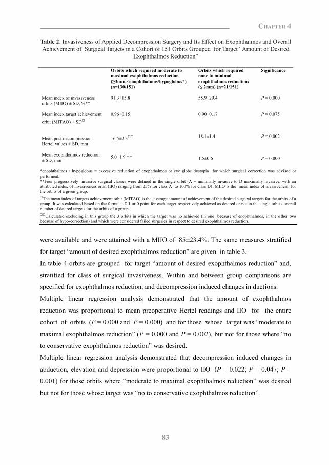
83
Chapter 4
Table 1. Achievement of the surgical target in the operated orbit
Surgical targets Operated orbits out of the whole series (n=151) in which the achievement of the surgical target was desired
Achievement of the surgical target as desired
Exophthalmos reduction:
moderate to maximal (≥ 3mm, <enophthalmos/hypoglobus*)
130 (86.1%)
Yes = 127/130 (97,7%) No = 3/130 (2.3%)□
none to minimal (≤ 2mm)
21 (13.9%)
Yes = 21/21 (100%) No = 0/21 (0%)
Reduction of retroocular tension 100 (66.2%) Yes = 100/100 (100%)
No = 0/100 (0%)
Reduction of periorbital aedema 122 (80.8%) Yes = 110/122 (90.2%)
No = 12/122 (9.8%)
Reduction of lagophthalmos 24 (15.9%) Yes = 24/24 (100%)
No = 0/24 (0%)
*enophthalmos / hypoglobus = excessive reduction of exophthalmos or eye globe dystopia for which surgical correction was advised or performed. □ one patient operated bilaterally had monolateral enophthalmos / hypoglobus, another had bilateral hypocorrection.
MIIO and MIIP of 86.4±21,9% and 78.2±26.1% respectively. Symmetry in
exophthalmos reduction was achieved in 78 (94%) of 83 patients, with post decompression
Hertel values right side = left in 56.4%; asymmetry in exophthalmos reduction resulted in
5 (6%) of 83 patients with post decompression Hertel values right = left ± 2mm and ± 3
mm in 40% and in 60% respectively. In one of the 84 patients symmetry in exophthalmos
reduction could not be assessed nor MITAP calculated. Prior to decompression, he had
been enucleated on one side without orbital implant, and he was not wearing any eye
prosthesis or willing to do that at the time of decompression.
The number of other targets to be achieved were not different between groups in which the
desired exophthalmos reduction was ≤ 2 mm or ≥ 3 mm.
Surgical outcome measures of the orbits grouped for target “amount of desired
exophthalmos reduction” are given in table 2.
TGA mean decompression induced changes in abduction, adduction, elevation and
depression (degrees ± SD, increase in negative numbers) were 3.8±7.8; -0.05±6.6; 5.6±7.7;
-0.03±10.6 with significant decrease of abduction (P = 0.000) and elevation (P = 0.000),
could be calculated on 93 orbits for which pre- and postoperative orthoptic evaluations
Table 2. Invasiveness of applied decompression surgery and its effect on exophthalmos and overall achievement of surgical targets in a cohort of 151 orbits grouped for target “amount of desired
exophthalmos reduction”
Orbits which required moderate to maximal exophthalmos reduction (≥3mm,<enophthalmos/hypoglobus*) (n=130/151)
Orbits which required none to minimal exophthalmos reduction: (≤ 2mm) (n=21/151)
Significance
Mean index of invasiveness orbits (MIIO) ± SD, %** Mean index target achievement orbit (MITAO) ± SD□ Mean post decompression Hertel values ± SD, mm Mean exophthalmos reduction ± SD, mm
91.3±15.8 0.96±0.15 16.5±2.3□□ 5.0±1.9 □□
55.9±29.4 0.90±0.17 18.1±1.4 1.5±0.6
P = 0.000 P = 0.075 P = 0.002 P = 0.000
*enophthalmos / hypoglobus = excessive reduction of exophthalmos or eye globe dystopia for which surgical correction was advised or performed. **Four progressively invasive surgical classes were defined in the single orbit (A = minimally invasive to D maximally invasive, with an attributed index of invasiveness orbit (IIO) ranging from 25% for class A to 100% for class D), MIIO is the mean index of invasiveness for the orbits of a given group. □The mean index of targets achievement orbit (MITAO) is the average amount of achievement of the desired surgical targets for the orbits of a group. It was calculated based on the formula: Σ 1 or 0 point for each target respectively achieved as desired or not in the single orbit / overall number of desired targets for the orbits of a group. □□Calculated excluding in this group the 3 orbits in which the target was no achieved (in one because of enophthalmos, in the o ther two because of hypo-correction) and which were considered failed surgeries in respect to desired exophthalmos reduction.
were available and were attained with a MIIO of 85±23.4%. The same measures stratified
for target “amount of desired exophthalmos reduction” are given in table 3.
In table 4 orbits are grouped for target “amount of desired exophthalmos reduction” and,
stratified for class of surgical invasiveness. Within and between group comparisons are
specified for exophthalmos reduction, and decompression induced changes in ductions.
Multiple linear regression analysis demonstrated that the amount of exophthalmos
reduction was proportional to mean preoperative Hertel readings and IIO for the entire
cohort of orbits (P = 0.000 and P = 0.000) and for those whose target was “moderate to
maximal exophthalmos reduction” (P = 0.000 and P = 0.002), but not for those where “no
to conservative exophthalmos reduction” was desired.
Multiple linear regression analysis demonstrated that decompression induced changes in
abduction, elevation and depression were proportional to IIO (P = 0.022; P = 0.047; P =
0.001) for those orbits where “moderate to maximal exophthalmos reduction” was desired
but not for those whose target was “no to conservative exophthalmos reduction”.
Table 2. Invasiveness of Applied Decompression Surgery and Its Effect on Exophthalmos and Overall Achievement of Surgical Targets in a Cohort of 151 Orbits Grouped for Target “Amount of Desired
Exophthalmos Reduction”
100379 Thesis Baldeschi.indd 83 vrijdag17-december-2010 12:08
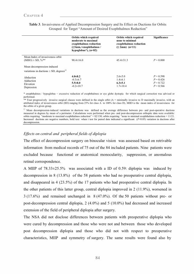
84
Chapter 4
Table 3. Invasiveness of applied decompression surgery and its effect on ductions for orbits grouped for target “amount of desired exophthalmos reduction”
Orbits which required
moderate to maximal exophthalmos reduction (≥3mm,<enophthalmos / hypoglobus*), (n=82)
Orbits which required none to minimal exophthalmos reduction (≤ 2mm) (n=11)
Significance
Mean Index of invasiveness orbit (MIIO) ± SD, %** Mean decompression-induced variations in ductions ± SD, degrees□ Abduction Adduction Elevation Depression
90.4±16.0 4.0±8.2 -0.3±6.7 5.5±8.0 -0.2±10.7
45.4±31.3 2.6±5.0 1.4±6.1 6.3±5.1 1.7±10.4
P = 0.000 P = 0.598 P = 0.426 P = 0.722 P = 0.566
* enophthalmos / hypoglobus = excessive reduction of exophthalmos or eye globe dystopia for which surgical correction was advised or performed. ** Four progressively invasive surgical classes were defined in the single orbit (A = minimally invasive to D maximally invasive, with an attributed index of invasiveness orbit (IIO) ranging from 25% for class A to 100% for class D), MIIO is the mean index of invasiveness for the orbits of a given group. □ Mean decompression-induced variations in ductions was defined as the average difference between pre- and post-operative ductions measured in degrees by mean of a perimeter. Calculations were performed when pre- and post-decompression orthoptic data were available: orbits requiring “moderate to maximal exophthalmos reduction” = 82/130; orbits requiring “none to minimal exophthalmos reduction = 11/21. Increased ductions are negative numbers, bold text when t test for paired data indicated a significant (P<0.05) variation in ductions after decompression.
Effects on central and peripheral fields of diplopia
The effect of decompression surgery on binocular vision was assessed based on retrivable
information from medical records of 75 out of the 84 included patients. Nine patients were
excluded because functional or anatomical monocularity, suppression, or anomalous
retinal correspondence.
A MIIP of 78.33±25.5% was associated with a ID of 0.59: diplopia was induced by
decompression in 8 (13.8%) of the 58 patients who had no preoperative central diplopia,
and disappeared in 4 (23.5%) of the 17 patients who had preoperative central diplopia. In
the other patients of this latter group, central diplopia improved in 2 (11.9%), worsened in
3 (17.6%) and remained unchanged in 8 (47.0%). Of the 50 patients without pre- or
post-decompression central diplopia, 2 (4.0%) and 5 (10.0%) had decreased and increased
extension of the field of peripheral diplopia after surgery.
The NSA did not disclose differences between patients with preoperative diplopia who
were cured by decompression and those who were not and between those who developed
post decompression diplopia and those who did not with respect to preoperative
characteristics, MIIP and symmetry of surgery. The same results were found also by
Table 4. Orbits grouped for target “amount of desired exophthalmos reduction” and stratified for classes of surgical invasiveness are compared for the effect of surgery on exophthalmos reduction and
decompression induced changes in ductions
Class of surgical invasiveness
A IIO*=25%
Class of surgical invasiveness
B IIO*=50%
Class of surgical invasiveness
C IIO*=75%
Class of surgical invasiveness
D IIO*=100%
Significance**
Mean exophthalmos reduction ± SD, mm: orbits which required moderate to maximal exophthalmos reduction (n.127/151)** orbits which required none to minimal exophthalmos reduction (n..21/151) Significance***
1.5±0.5
3.4±0.5
1.5±0.6
4.2±1.2
1.8±0.4
5.4±2.0
1.2± 0.9
P = 0.000 P = 0.627
P = 0.000 P = 0.000 P = 0.000
Mean decompression-induced variations in ductions ± SD, degrees□: orbits which required moderate to maximal exophthalmos reduction (n.82) Abduction Adduction Elevation Depression orbits which required none to minimal exophthalmos reduction (n.11) Abduction Adduction Elevation Depression
Significance***
4.6±3.9 1.9±6.6 6.4±4.6 3.3±9.1
-3.1±10.3 -7.4±6.8
2.1±9.6 -16.7±16.8
4.0 1.0 11.0 0.0
2.1±6.8 3.0±8.1 3.4±6.9 -1.6±9.5
-8.0 5.0 1.0 6.0
5.4±7.8 -0.3±5.6 6.5±8.0 2.2±8.2
0.5±3.5 -1.5±9.2 6.5±9.2
-5.0±21.2
P = 0.017 P = 0.002 P = 0.199 P = 0.000 P = 0.084 P = 0.886 P = 0.665 P = 0.813
P = 0.541 P = 0.291 P = 0.420 P = 0.390
P = 0.165 P = 0.813 P = 0.740 P = 0.609
P = 0.387 P = 0.779 P = 0.998 P = 0.251
* Four progressively invasive surgical classes were defined in the single orbit (A = minimally invasive to D maximally invasive, with an attributed index of invasiveness orbit (IIO) ranging from 25% for class A to 100% for class D) **Significance refers to comparisons among different classes of invasiveness, within 2 groups of orbits which required exophthalmos reduction “moderate to maximal” (≥ 3 mm) or “none to minimal” (≤ 2 mm). ***Significance refers to comparisons between 2 groups of orbits which required exophthalmos reduction “moderate to maximal” (≥ 3 mm) or “none to minimal” (≤ 2 mm), within different classes of invasiveness. □Mean decompression-induced variations in ductions was defined as the average difference between pre- and postoperative ductions measured in degrees by mean of a perimeter. Calculations were performed when pre- and post-decompression orthoptic data were available: orbits requiring “moderate to maximal exophthalmos reduction” = 82/130; orbits requiring “none to minimal exophthalmos reduction” = 11/21. Increased ductions are negative numbers, bold text when t test for paired data indicated a significant (P<0.05) variation in ductions after decompression.
comparing patients affected by preoperative diplopia and cured by decompression with
those who presented decompression induced diplopia (Table 5).
Table 3. Invasiveness of Applied Decompression Surgery and Its Effect on Ductions for Orbits Grouped for Target “Amount of Desired Exophthalmos Reduction”
100379 Thesis Baldeschi.indd 84 vrijdag17-december-2010 12:08
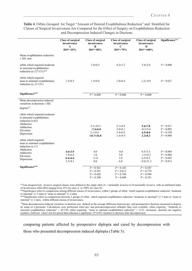
85
Chapter 4
Table 3. Invasiveness of applied decompression surgery and its effect on ductions for orbits grouped for target “amount of desired exophthalmos reduction”
Orbits which required
moderate to maximal exophthalmos reduction (≥3mm,<enophthalmos / hypoglobus*), (n=82)
Orbits which required none to minimal exophthalmos reduction (≤ 2mm) (n=11)
Significance
Mean Index of invasiveness orbit (MIIO) ± SD, %** Mean decompression-induced variations in ductions ± SD, degrees□ Abduction Adduction Elevation Depression
90.4±16.0 4.0±8.2 -0.3±6.7 5.5±8.0 -0.2±10.7
45.4±31.3 2.6±5.0 1.4±6.1 6.3±5.1 1.7±10.4
P = 0.000 P = 0.598 P = 0.426 P = 0.722 P = 0.566
* enophthalmos / hypoglobus = excessive reduction of exophthalmos or eye globe dystopia for which surgical correction was advised or performed. ** Four progressively invasive surgical classes were defined in the single orbit (A = minimally invasive to D maximally invasive, with an attributed index of invasiveness orbit (IIO) ranging from 25% for class A to 100% for class D), MIIO is the mean index of invasiveness for the orbits of a given group. □ Mean decompression-induced variations in ductions was defined as the average difference between pre- and post-operative ductions measured in degrees by mean of a perimeter. Calculations were performed when pre- and post-decompression orthoptic data were available: orbits requiring “moderate to maximal exophthalmos reduction” = 82/130; orbits requiring “none to minimal exophthalmos reduction = 11/21. Increased ductions are negative numbers, bold text when t test for paired data indicated a significant (P<0.05) variation in ductions after decompression.
Effects on central and peripheral fields of diplopia
The effect of decompression surgery on binocular vision was assessed based on retrivable
information from medical records of 75 out of the 84 included patients. Nine patients were
excluded because functional or anatomical monocularity, suppression, or anomalous
retinal correspondence.
A MIIP of 78.33±25.5% was associated with a ID of 0.59: diplopia was induced by
decompression in 8 (13.8%) of the 58 patients who had no preoperative central diplopia,
and disappeared in 4 (23.5%) of the 17 patients who had preoperative central diplopia. In
the other patients of this latter group, central diplopia improved in 2 (11.9%), worsened in
3 (17.6%) and remained unchanged in 8 (47.0%). Of the 50 patients without pre- or
post-decompression central diplopia, 2 (4.0%) and 5 (10.0%) had decreased and increased
extension of the field of peripheral diplopia after surgery.
The NSA did not disclose differences between patients with preoperative diplopia who
were cured by decompression and those who were not and between those who developed
post decompression diplopia and those who did not with respect to preoperative
characteristics, MIIP and symmetry of surgery. The same results were found also by
Table 4. Orbits grouped for target “amount of desired exophthalmos reduction” and stratified for classes of surgical invasiveness are compared for the effect of surgery on exophthalmos reduction and
decompression induced changes in ductions
Class of surgical invasiveness
A IIO*=25%
Class of surgical invasiveness
B IIO*=50%
Class of surgical invasiveness
C IIO*=75%
Class of surgical invasiveness
D IIO*=100%
Significance**
Mean exophthalmos reduction ± SD, mm: orbits which required moderate to maximal exophthalmos reduction (n.127/151)** orbits which required none to minimal exophthalmos reduction (n..21/151) Significance***
1.5±0.5
3.4±0.5
1.5±0.6
4.2±1.2
1.8±0.4
5.4±2.0
1.2± 0.9
P = 0.000 P = 0.627
P = 0.000 P = 0.000 P = 0.000
Mean decompression-induced variations in ductions ± SD, degrees□: orbits which required moderate to maximal exophthalmos reduction (n.82) Abduction Adduction Elevation Depression orbits which required none to minimal exophthalmos reduction (n.11) Abduction Adduction Elevation Depression
Significance***
4.6±3.9 1.9±6.6 6.4±4.6 3.3±9.1
-3.1±10.3 -7.4±6.8
2.1±9.6 -16.7±16.8
4.0 1.0 11.0 0.0
2.1±6.8 3.0±8.1 3.4±6.9 -1.6±9.5
-8.0 5.0 1.0 6.0
5.4±7.8 -0.3±5.6 6.5±8.0 2.2±8.2
0.5±3.5 -1.5±9.2 6.5±9.2
-5.0±21.2
P = 0.017 P = 0.002 P = 0.199 P = 0.000 P = 0.084 P = 0.886 P = 0.665 P = 0.813
P = 0.541 P = 0.291 P = 0.420 P = 0.390
P = 0.165 P = 0.813 P = 0.740 P = 0.609
P = 0.387 P = 0.779 P = 0.998 P = 0.251
* Four progressively invasive surgical classes were defined in the single orbit (A = minimally invasive to D maximally invasive, with an attributed index of invasiveness orbit (IIO) ranging from 25% for class A to 100% for class D) **Significance refers to comparisons among different classes of invasiveness, within 2 groups of orbits which required exophthalmos reduction “moderate to maximal” (≥ 3 mm) or “none to minimal” (≤ 2 mm). ***Significance refers to comparisons between 2 groups of orbits which required exophthalmos reduction “moderate to maximal” (≥ 3 mm) or “none to minimal” (≤ 2 mm), within different classes of invasiveness. □Mean decompression-induced variations in ductions was defined as the average difference between pre- and postoperative ductions measured in degrees by mean of a perimeter. Calculations were performed when pre- and post-decompression orthoptic data were available: orbits requiring “moderate to maximal exophthalmos reduction” = 82/130; orbits requiring “none to minimal exophthalmos reduction” = 11/21. Increased ductions are negative numbers, bold text when t test for paired data indicated a significant (P<0.05) variation in ductions after decompression.
comparing patients affected by preoperative diplopia and cured by decompression with
those who presented decompression induced diplopia (Table 5).
Table 4. Orbits Grouped for Target “Amount of Desired Exophthalmos Reduction” and Stratified for Classes of Surgical Invasiveness Are Compared for the Effect of Surgery on Exophthalmos Reduction
and Decompression Induced Changes in Ductions
100379 Thesis Baldeschi.indd 85 vrijdag17-december-2010 12:08
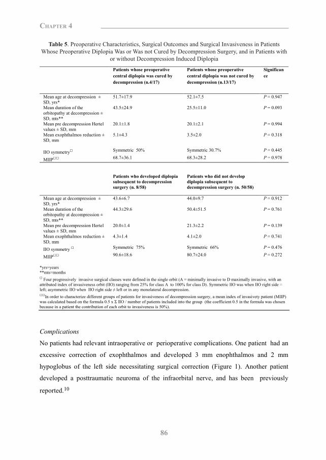
86
Chapter 4
Table 5. Preoperative characteristics, surgical outcomes and surgical invasiveness in patients whose preoperative diplopia was or was not cured by decompression surgery, and in patients with or without
decompression induced diplopia
Patients whose preoperative central diplopia was cured by decompression (n.4/17)
Patients whose preoperative central diplopia was not cured by decompression (n.13/17)
Significance
Mean age at decompression ± SD, yrs*
51.7±17.9 52.1±7.5 P = 0.947
Mean duration of the orbitopathy at decompression ± SD, mts**
43.5±24.9 25.5±11.0 P = 0.093
Mean pre decompression Hertel values ± SD, mm
20.1±1.8 20.1±2.1 P = 0.994
Mean exophthalmos reduction ± SD, mm
5.1±4.3 3.5±2.0 P = 0.318
IIO symmetry□ Symmetric 50% Symmetric 30.7% P = 0.445
MIIP□□ 68.7±36.1 68.3±28.2 P = 0.978
Patients who developed diplopia subsequent to decompression surgery (n. 8/58)
Patients who did not develop diplopia subsequent to decompression surgery (n. 50/58)
Mean age at decompression ± SD, yrs*
43.6±6.7 44.0±9.7 P = 0.912
Mean duration of the orbitopathy at decompression ± SD, mts**
44.3±29.6 50.4±51.5 P = 0.761
Mean pre decompression Hertel values ± SD, mm
20.0±1.4 21.3±2.2 P = 0.139
Mean exophthalmos reduction ± SD, mm
4.3±1.4 4.1±2.0 P = 0.741
IIO symmetry □ Symmetric 75% Symmetric 66% P = 0.476
MIIP□□ 90.6±18.6 80.7±24.0 P = 0.272
*yrs=years **mts=months □ Four progressively invasive surgical classes were defined in the single orbit (A = minimally invasive to D maximally invasive, with an attributed index of invasiveness orbit (IIO) ranging from 25% for class A to 100% for class D). Symmetric IIO was when IIO right side = left; asymmetric IIO when IIO right side ≠ left or in any monolateral decompression. □□In order to characterize different groups of patients for invasiveness of decompression surgery, a mean index of invasivety patient (MIIP) was calculated based on the formula 0.5 x Σ IIO / number of patients included into the group (the coefficient 0.5 in the formula was chosen because in a patient the contribution of each orbit to invasiveness is 50%).
Complications
No patients had relevant intraoperative or perioperative complications. One patient had an
excessive correction of exophthalmos and developed 3 mm enophthalmos and 2 mm
hypoglobus of the left side necessitating surgical correction (Figure 1). Another patient
developed a posttraumatic neuroma of the infraorbital nerve, and has been previously
reported.10
Figure 1. The patient who developed left enophthalmos subsequent to inferomedial orbital decompression. (Top) The patient at first ophthalmologic evaluation. (Bottom) The patient after bilateral orbital decompression, correction of strabismus, and correction of left hypoglobus and enophthalmos. Left lower lid retraction was still present and amenable of possible surgical correction.
Discussion
Different techniques are used for decompression surgery in GO4 and each of them can be
personalized at partient‟s and surgeon‟s convenience for invasiveness and technical
variations. Most of the study up until now provide a careful description of the surgical
procedures, but invasiveness and technical variations have been left undetermined. In this
study, the uniform execution of surgery secured by selecting patients operated by one
surgeon, and indices such IIO, MIIO and MIIP were aimed at a better classification of
applied decompression surgery for the benefit of study comparisons.
There are multiple indications for decompression surgery in GO.4 Hence the evaluation of
its results should be inseparable from the assessment of the achievement of the targets,
for which it has been performed. Only a precise, quantitative assessment of target
achievement can allow meaningful and straightforward comparisons of the effectiveness of
decompression surgery within and among studies. Our input to this goal was represented by
clear definitions of targets, scoring of target achievement and corresponding MITAO and
MITAP indices. The NSA which includes descriptive figures (Table 1) and easily
interpretable quantitative indices is specific for decompression surgery and can be
regarded as a further step in defining the overall response to treatment as proposed by
EUGOGO.11
Table 5. Preoperative Characteristics, Surgical Outcomes and Surgical Invasiveness in Patients Whose Preoperative Diplopia Was or Was not Cured by Decompression Surgery, and in Patients with
or without Decompression Induced Diplopia
100379 Thesis Baldeschi.indd 86 vrijdag17-december-2010 12:08

87
Chapter 4
Table 5. Preoperative characteristics, surgical outcomes and surgical invasiveness in patients whose preoperative diplopia was or was not cured by decompression surgery, and in patients with or without
decompression induced diplopia
Patients whose preoperative central diplopia was cured by decompression (n.4/17)
Patients whose preoperative central diplopia was not cured by decompression (n.13/17)
Significance
Mean age at decompression ± SD, yrs*
51.7±17.9 52.1±7.5 P = 0.947
Mean duration of the orbitopathy at decompression ± SD, mts**
43.5±24.9 25.5±11.0 P = 0.093
Mean pre decompression Hertel values ± SD, mm
20.1±1.8 20.1±2.1 P = 0.994
Mean exophthalmos reduction ± SD, mm
5.1±4.3 3.5±2.0 P = 0.318
IIO symmetry□ Symmetric 50% Symmetric 30.7% P = 0.445
MIIP□□ 68.7±36.1 68.3±28.2 P = 0.978
Patients who developed diplopia subsequent to decompression surgery (n. 8/58)
Patients who did not develop diplopia subsequent to decompression surgery (n. 50/58)
Mean age at decompression ± SD, yrs*
43.6±6.7 44.0±9.7 P = 0.912
Mean duration of the orbitopathy at decompression ± SD, mts**
44.3±29.6 50.4±51.5 P = 0.761
Mean pre decompression Hertel values ± SD, mm
20.0±1.4 21.3±2.2 P = 0.139
Mean exophthalmos reduction ± SD, mm
4.3±1.4 4.1±2.0 P = 0.741
IIO symmetry □ Symmetric 75% Symmetric 66% P = 0.476
MIIP□□ 90.6±18.6 80.7±24.0 P = 0.272
*yrs=years **mts=months □ Four progressively invasive surgical classes were defined in the single orbit (A = minimally invasive to D maximally invasive, with an attributed index of invasiveness orbit (IIO) ranging from 25% for class A to 100% for class D). Symmetric IIO was when IIO right side = left; asymmetric IIO when IIO right side ≠ left or in any monolateral decompression. □□In order to characterize different groups of patients for invasiveness of decompression surgery, a mean index of invasivety patient (MIIP) was calculated based on the formula 0.5 x Σ IIO / number of patients included into the group (the coefficient 0.5 in the formula was chosen because in a patient the contribution of each orbit to invasiveness is 50%).
Complications
No patients had relevant intraoperative or perioperative complications. One patient had an
excessive correction of exophthalmos and developed 3 mm enophthalmos and 2 mm
hypoglobus of the left side necessitating surgical correction (Figure 1). Another patient
developed a posttraumatic neuroma of the infraorbital nerve, and has been previously
reported.10
Figure 1. The patient who developed left enophthalmos subsequent to inferomedial orbital decompression. (Top) The patient at first ophthalmologic evaluation. (Bottom) The patient after bilateral orbital decompression, correction of strabismus, and correction of left hypoglobus and enophthalmos. Left lower lid retraction was still present and amenable of possible surgical correction.
Discussion
Different techniques are used for decompression surgery in GO4 and each of them can be
personalized at partient‟s and surgeon‟s convenience for invasiveness and technical
variations. Most of the study up until now provide a careful description of the surgical
procedures, but invasiveness and technical variations have been left undetermined. In this
study, the uniform execution of surgery secured by selecting patients operated by one
surgeon, and indices such IIO, MIIO and MIIP were aimed at a better classification of
applied decompression surgery for the benefit of study comparisons.
There are multiple indications for decompression surgery in GO.4 Hence the evaluation of
its results should be inseparable from the assessment of the achievement of the targets,
for which it has been performed. Only a precise, quantitative assessment of target
achievement can allow meaningful and straightforward comparisons of the effectiveness of
decompression surgery within and among studies. Our input to this goal was represented by
clear definitions of targets, scoring of target achievement and corresponding MITAO and
MITAP indices. The NSA which includes descriptive figures (Table 1) and easily
interpretable quantitative indices is specific for decompression surgery and can be
regarded as a further step in defining the overall response to treatment as proposed by
EUGOGO.11
100379 Thesis Baldeschi.indd 87 vrijdag17-december-2010 12:08
88
Chapter 4
In the NSA, all targets chosen for the assessment of rehabilitative surgery outcomes are
direct consequences of decompression induced decrease of intra-orbital pressure and globe
displacement; decompression allows to reach these goals based on intraoperative
estimations of resistance to retropulsion of the globe, eye position and aspect of the peri-
orbital region. Decompressions may influence eyelid positions and lid fissure width
modifying vertical ductions, eyeball position, or as a consequence of eyelid scarring. Lid
fissure width is therefore not controllable during decompression surgery and for this reason
we did not consider it as a target in our study. Conversely dysthyroid lagophthalmos is a
static consequence of eyelid retractions or of the relative eyelid shortening due to the
presence of a protruding eye; its reduction can be assessed based on intraoperative
estimations. The minimally invasive transinferior conjunctival fornix approach that we
used in the present series, by minimally disrupting tissues, ease intra operative estimations.
Similar exophthalmometric values can cause different degrees of disfigurement when
anthropomorphic characteristics like gender, ethnicity, shull and face conformation are
considered. This has implications on the extent of required exophthalmos reduction4; hence
it is within the context of the targets to be achieved, and not, as it was previously done, on
the basis of preoperative Hertel values per se12, that traditionally used measures such as the
amount of exophthalmos reduction assumes a specific means. High values of mean
exophthalmos reduction, which for decades has been linked to effectiveness of
decompression surgery, may, on the contrary, represent a complication when excessive or
not desired.
Data concerning the potential of a given approach for exophthalmos reduction can be
relevant when counseling patients, but are of less interest to those patients about to be
operated for other targets. The latter patients in fact might be more interested in opposite
informations, or rather the possibilities to use a particular type of decompression avoiding
the induction of unwanted reduction of Hertel values, while still taking advantage of its
other possible beneficial effects.
A similar perspective applies to the effects of decompressions on extraocular eye motility.
Till now the incidence of decompression induced diplopia was considered a good safety
marker. Although extremely valuable when counseling patients unaffected by diplopia
prior to surgery it is unhelpful to patients who already have diplopia, and who would be
more interested about the potential benefits of decompression to their double vision. The
ID, which merges in one single number information concerning frequency of
decompression induced and cured diplopia, represents an easy means to describe diplopia-
related intervals. This can have distinct advantages for comparisons within and among
studies.
Decompression induced changes in ductions which quantify the influences of surgery on
extra-ocular monocular movements, are very valuable measures not only for study
comparisons but also for counseling purposes. With respect to induced changes in ductions
the NSA is specific for decompression surgery and consistent with EUGOGO suggestion
for clinical trials.11
The above considerations were the rationale behind the NSA. Most of the novel
observations are liable to speculative interpretations and might be cues for future studies.
We found that orbits for which “no to conservative exophthalmos reduction” was desired,
had the lowest pre operative Hertel reading and belonged to the oldest patients, but did not
have a longer duration of GO. This suggests a particular sub set of GO developing at older
age. Additionally we showed that when combined steroids and radiotherapy were
administered before decompression, orbits were less likely to need reduction of
lagophthalmos. Reduction of periorbital aedema was needed less frequently in orbits with a
longer duration of GO. This suggests that the use of medical options may reduce the need
of surgical treatment of lagophthalmos, and that simply “tincture of time” may reduce the
necessity of surgery for periorbital aedema.
Overall the inferomedial decompression approach with graded degree of invasiveness
described in this paper resulted in a rather high degree of achievement of desired targets,
with negligible occurrence of complications. In about 10% of the cases orbital
decompression was not effective in reducing preoperative periorbital oedema, and indeed
this target was achieved less frequently than the other.
When “no to conservative exophthalmos reduction” was desired, the observed MIIO was
the lowest, and exophthalmos reduction was not related to the degree of surgical
invasiveness. This latter finding is a substantial departure from the traditional concept that
more extensive osteotomies result in higher mean exophthalmos reduction.13 Explanation
of our finding can only be speculative; it can be linked (as observed at surgery) to the
distinct lack of compliance of the soft tissues in many of the orbits included in the group
whose target was “no to conservative exophthalmos reduction”. The statistically
100379 Thesis Baldeschi.indd 88 vrijdag17-december-2010 12:08
89
Chapter 4
In the NSA, all targets chosen for the assessment of rehabilitative surgery outcomes are
direct consequences of decompression induced decrease of intra-orbital pressure and globe
displacement; decompression allows to reach these goals based on intraoperative
estimations of resistance to retropulsion of the globe, eye position and aspect of the peri-
orbital region. Decompressions may influence eyelid positions and lid fissure width
modifying vertical ductions, eyeball position, or as a consequence of eyelid scarring. Lid
fissure width is therefore not controllable during decompression surgery and for this reason
we did not consider it as a target in our study. Conversely dysthyroid lagophthalmos is a
static consequence of eyelid retractions or of the relative eyelid shortening due to the
presence of a protruding eye; its reduction can be assessed based on intraoperative
estimations. The minimally invasive transinferior conjunctival fornix approach that we
used in the present series, by minimally disrupting tissues, ease intra operative estimations.
Similar exophthalmometric values can cause different degrees of disfigurement when
anthropomorphic characteristics like gender, ethnicity, shull and face conformation are
considered. This has implications on the extent of required exophthalmos reduction4; hence
it is within the context of the targets to be achieved, and not, as it was previously done, on
the basis of preoperative Hertel values per se12, that traditionally used measures such as the
amount of exophthalmos reduction assumes a specific means. High values of mean
exophthalmos reduction, which for decades has been linked to effectiveness of
decompression surgery, may, on the contrary, represent a complication when excessive or
not desired.
Data concerning the potential of a given approach for exophthalmos reduction can be
relevant when counseling patients, but are of less interest to those patients about to be
operated for other targets. The latter patients in fact might be more interested in opposite
informations, or rather the possibilities to use a particular type of decompression avoiding
the induction of unwanted reduction of Hertel values, while still taking advantage of its
other possible beneficial effects.
A similar perspective applies to the effects of decompressions on extraocular eye motility.
Till now the incidence of decompression induced diplopia was considered a good safety
marker. Although extremely valuable when counseling patients unaffected by diplopia
prior to surgery it is unhelpful to patients who already have diplopia, and who would be
more interested about the potential benefits of decompression to their double vision. The
ID, which merges in one single number information concerning frequency of
decompression induced and cured diplopia, represents an easy means to describe diplopia-
related intervals. This can have distinct advantages for comparisons within and among
studies.
Decompression induced changes in ductions which quantify the influences of surgery on
extra-ocular monocular movements, are very valuable measures not only for study
comparisons but also for counseling purposes. With respect to induced changes in ductions
the NSA is specific for decompression surgery and consistent with EUGOGO suggestion
for clinical trials.11
The above considerations were the rationale behind the NSA. Most of the novel
observations are liable to speculative interpretations and might be cues for future studies.
We found that orbits for which “no to conservative exophthalmos reduction” was desired,
had the lowest pre operative Hertel reading and belonged to the oldest patients, but did not
have a longer duration of GO. This suggests a particular sub set of GO developing at older
age. Additionally we showed that when combined steroids and radiotherapy were
administered before decompression, orbits were less likely to need reduction of
lagophthalmos. Reduction of periorbital aedema was needed less frequently in orbits with a
longer duration of GO. This suggests that the use of medical options may reduce the need
of surgical treatment of lagophthalmos, and that simply “tincture of time” may reduce the
necessity of surgery for periorbital aedema.
Overall the inferomedial decompression approach with graded degree of invasiveness
described in this paper resulted in a rather high degree of achievement of desired targets,
with negligible occurrence of complications. In about 10% of the cases orbital
decompression was not effective in reducing preoperative periorbital oedema, and indeed
this target was achieved less frequently than the other.
When “no to conservative exophthalmos reduction” was desired, the observed MIIO was
the lowest, and exophthalmos reduction was not related to the degree of surgical
invasiveness. This latter finding is a substantial departure from the traditional concept that
more extensive osteotomies result in higher mean exophthalmos reduction.13 Explanation
of our finding can only be speculative; it can be linked (as observed at surgery) to the
distinct lack of compliance of the soft tissues in many of the orbits included in the group
whose target was “no to conservative exophthalmos reduction”. The statistically
100379 Thesis Baldeschi.indd 89 vrijdag17-december-2010 12:08
90
Chapter 4
significant less frequent administration of predecompression radiotherapy could have
contributed to the stiffness of the soft orbital tissue observed in this group. Its differences
in IIO were not relevant to decompression induced changes in ductions. Interestingly unlike
for orbits whose target was exophthalmos reduction ≥ 3mm, a maximal surgical
invasiveness of 100% was not associated with any change ductions. The explanation of this
is again speculative and in line with the previous one. In this group of orbits, despite
maximally sized osteotomies, reduced compliance of the soft orbital tissue might have
resulted in minimal if any displacement (and scarring) of the soft orbital tissues, including
extraocular muscles. In line with this perspective, a lower surgical invasiveness of 25%
which was presumably associated with better compliance of soft orbital tissues, was
related to reduction of preoperative abduction and elevation.
In all groups, independent of the amount of desired exophthalmos reduction, IIO 75% was
neutral to ductions, while invasiveness 50% (which is a commonly used pattern of
osteotomies for the treatment of disfiguring exophthalmos either through a trans cutaneous,
or a swinging eyelid approach) was beneficial to altered adduction and depression.
Apart from the observed novel figures the main value of the NSA is that of providing a
more comprehensive outlook. Although the NSA may appear more complete, precise and
reliable than the TGA, the NSA it is far from being ideal. Variations in morphology of the
orbital cavity and in orbital osteology might significantly influence the degree of
exophthalmos reduction which can be obtained by standardized decompression
procedures.14 In our study, the influence of different osteotomies weighed for invasiveness
of applied surgery on outcomes of decompression surgery remains unexplored.
Additionally in our analysis the lipogenic or myopathic nature of the orbital disease was not
taken into account, nor was the orbital compliance which is not directly measurable before
decompression. For these reasons the figures given by our method cannot be considered
absolute when counseling patients about to undergo surgery. Depending on the soft tissue
response to surgery, the invasiveness of the planned approach, if not the surgical technique
itself, may need to be changed during surgery. Nevertheless when counseling patients, the
observations generated by our novel approach can be offered to them as a possible case
scenario. The same observation are also invaluable to the surgeon faced with intraoperative
decision making for balancing risk benefits of his / her eventual choices.
Finally the time frame that we chose to study the outcome of decompression surgery was
relatively short and therefore suboptimal for assessing long-term complications such as
permanent dysaesthesias of the infraorbital nerve or sinus obstructions. We however regard
our choice as justifiable because it was aimed at reducing the influence of confounding
factors linked to further post decompression rehabilitative procedures.
Despite its shortcomings the value of this study lies in its innovative perspectives and in
having pioneered a new path linking specific needs, and background characteristics of the
patients to a detailed graded surgical approach. This therefore has the potential to be a step
towards the final goal of a desirable, and as much as possible, evidence based customized
approach to orbital decompression.
100379 Thesis Baldeschi.indd 90 vrijdag17-december-2010 12:08
91
Chapter 4
significant less frequent administration of predecompression radiotherapy could have
contributed to the stiffness of the soft orbital tissue observed in this group. Its differences
in IIO were not relevant to decompression induced changes in ductions. Interestingly unlike
for orbits whose target was exophthalmos reduction ≥ 3mm, a maximal surgical
invasiveness of 100% was not associated with any change ductions. The explanation of this
is again speculative and in line with the previous one. In this group of orbits, despite
maximally sized osteotomies, reduced compliance of the soft orbital tissue might have
resulted in minimal if any displacement (and scarring) of the soft orbital tissues, including
extraocular muscles. In line with this perspective, a lower surgical invasiveness of 25%
which was presumably associated with better compliance of soft orbital tissues, was
related to reduction of preoperative abduction and elevation.
In all groups, independent of the amount of desired exophthalmos reduction, IIO 75% was
neutral to ductions, while invasiveness 50% (which is a commonly used pattern of
osteotomies for the treatment of disfiguring exophthalmos either through a trans cutaneous,
or a swinging eyelid approach) was beneficial to altered adduction and depression.
Apart from the observed novel figures the main value of the NSA is that of providing a
more comprehensive outlook. Although the NSA may appear more complete, precise and
reliable than the TGA, the NSA it is far from being ideal. Variations in morphology of the
orbital cavity and in orbital osteology might significantly influence the degree of
exophthalmos reduction which can be obtained by standardized decompression
procedures.14 In our study, the influence of different osteotomies weighed for invasiveness
of applied surgery on outcomes of decompression surgery remains unexplored.
Additionally in our analysis the lipogenic or myopathic nature of the orbital disease was not
taken into account, nor was the orbital compliance which is not directly measurable before
decompression. For these reasons the figures given by our method cannot be considered
absolute when counseling patients about to undergo surgery. Depending on the soft tissue
response to surgery, the invasiveness of the planned approach, if not the surgical technique
itself, may need to be changed during surgery. Nevertheless when counseling patients, the
observations generated by our novel approach can be offered to them as a possible case
scenario. The same observation are also invaluable to the surgeon faced with intraoperative
decision making for balancing risk benefits of his / her eventual choices.
Finally the time frame that we chose to study the outcome of decompression surgery was
relatively short and therefore suboptimal for assessing long-term complications such as
permanent dysaesthesias of the infraorbital nerve or sinus obstructions. We however regard
our choice as justifiable because it was aimed at reducing the influence of confounding
factors linked to further post decompression rehabilitative procedures.
Despite its shortcomings the value of this study lies in its innovative perspectives and in
having pioneered a new path linking specific needs, and background characteristics of the
patients to a detailed graded surgical approach. This therefore has the potential to be a step
towards the final goal of a desirable, and as much as possible, evidence based customized
approach to orbital decompression.
100379 Thesis Baldeschi.indd 91 vrijdag17-december-2010 12:08
92
Chapter 4
References 1. Dollinger J. Die Druckentlastung der Augenhöhle durch Entfernung der äußeren Orbitawand bei
hochgradigem Exophthalmos (Morbus Basedowii) und konsekutiver Hauterkrankung. Dtsch med Wschr
1911;37:1888-90.
2. Rose GE. Postural visual obscurations in patients with inactive thyroid eye disease; a variant of “hydraulic”
disease. Eye 2006;20:1178-85.
3. Jorge R, Scott IU, Akaishi PM, Velasco Cruz AA, Flynn HW Jr. Resolution of choroidal folds and
improvement in visual acuity after orbital decompression for Graves orbitopathy. Retina 2003;23:563-5.
4. Baldeschi L. Orbital decompression. In: Wiersinga WM, Kahaly GJ, editors. Graves‟ Orbitopathy A
Multidisciplinary Approach - Questions and Answers, 2nd edition. Basel: Karger, 2010:171-88.
5. www.eugogo.eu
6. Werner SC. Modifications of the classification of the eye changes of Graves‟ disease: Recommendations of
the Ad Hoc Committee of the American Thyroid association. J Clin Endocrinol Metab 1977;44:203-4.
7. Mourits MP, Prummel MF, Wiersinga WM, Koornneef L. Clinical activity score as a guide in management
of patients with Graves‟ ophthalmopathy. Clin Endocrinol (Oxf) 1997;47:9-14.
8. Mourits MP, Prummel MF, Wiersinga WM, Koornneef L. Measuring eye movements in Graves
ophthalmopathy. Ophthalmology 1994;101:1341-6.
9. Baldeschi L, Lupetti A, Vu P, Wakelkamp IMMJ, Prummel MF, Wiersinga WM. Reactivation of Graves‟
orbitopathy after rehabilitative orbital decompression. Ophthalmology 2007;114:1395-402.
10. Baldeschi L, Saeed P, Regensburg NI, Zacharopoulos I, Wiersinga WM. Traumatic neuroma of the
infraorbital nerve subsequent to infero medial orbital decompression for Graves‟ orbitopathy. Eur J
Ophthalmol 2010;20:481-4.
11. European Group on Graves' Orbitopathy (EUGOGO), Wiersinga WM, Perros P, Kahaly GJ, Mourits MP,
Baldeschi L, Boboridis K, Boschi A, Dickinson AJ, Kendall-Taylor P, Krassas GE, Lane CM, Lazarus JH,
Marcocci C, Marino M, Nardi M, Neoh C, Orgiazzi J, Pinchera A, Pitz S, Prummel MF, Sartini MS, Stahl M,
von Arx G. Clinical assessment of patients with Graves' orbitopathy: the European Group on Graves'
Orbitopathy recommendations to generalists, specialists and clinical researchers. Eur J Endocrinol
2006;155:387-9.
12. Sasim IV, de Graaf ME, Berendschot TT, Kalmann R, van Isterdael C, Mourits MP. Coronal or swinging
eyelid decompression for patients with disfiguring proptosis in Graves‟ orbitopathy? Comparison of results in
one center. Ophthalmology 2005;112:1310-5.
13. Mourits, MP, Bijl HM, Baldeschi L, Boboridis K, Curro N, Dickinson J, Eckstein AK, Guastella C, Altea M,
Kahaly G, Freidel M, Kalmann R, Krassas GE, Lane C, Lareida J, Marcocci C, Marino M, Nardi M, Mohr
Ch, Neoh C, Pinchera A, Orgiazzi J, Pitz S, Saeed P, Salvi M, Sellari-Franceschini S, Stahl M, von Arx G,
Wiersinga W. Outcome of orbital decompression for disfiguring proptosis in patients with Graves‟
orbitopathy using various surgical procedures. Br J Ophthalmol 2009;93:1518-23.
14. Kamer L, Noser H, Schramm A, Hammer B, Kirsch E. Anatomy-based surgical concepts for individualized
orbital decompression surgery in graves orbitopathy. I. Orbital size and geometry. Ophthal Plast Reconstr
Surg 2010;26:348-52.
Chapter 5
The Removal of the Deep Lateral Wall in Orbital Decompression: Its Contribution to Exophthalmos Reduction and Influence on
Consecutive Diplopia
Lelio Baldeschi1, Kerr MacAndie1, Christoph Hintschich2,
Iris M.M.J. Wakelkamp3, Mark F Prumme3, Wilmar M. Wiersinga3
1 Orbital Centre, Department of Ophthalmology, University of Amsterdam, Amsterdam, The Netherlands. 2 University Eye Hospital, Ludvig Maximilians University of Munich, Munich, Germany. 3 Department of Endocrinology, University of Amsterdam, Amsterdam, The Netherlands. American Journal of Ophthalmology 2005;140:642-7
100379 Thesis Baldeschi.indd 92 vrijdag17-december-2010 12:08
References 1. Dollinger J. Die Druckentlastung der Augenhöhle durch Entfernung der äußeren Orbitawand bei
hochgradigem Exophthalmos (Morbus Basedowii) und konsekutiver Hauterkrankung. Dtsch med Wschr
1911;37:1888-90.
2. Rose GE. Postural visual obscurations in patients with inactive thyroid eye disease; a variant of “hydraulic”
disease. Eye 2006;20:1178-85.
3. Jorge R, Scott IU, Akaishi PM, Velasco Cruz AA, Flynn HW Jr. Resolution of choroidal folds and
improvement in visual acuity after orbital decompression for Graves orbitopathy. Retina 2003;23:563-5.
4. Baldeschi L. Orbital decompression. In: Wiersinga WM, Kahaly GJ, editors. Graves‟ Orbitopathy A
Multidisciplinary Approach - Questions and Answers, 2nd edition. Basel: Karger, 2010:171-88.
5. www.eugogo.eu
6. Werner SC. Modifications of the classification of the eye changes of Graves‟ disease: Recommendations of
the Ad Hoc Committee of the American Thyroid association. J Clin Endocrinol Metab 1977;44:203-4.
7. Mourits MP, Prummel MF, Wiersinga WM, Koornneef L. Clinical activity score as a guide in management
of patients with Graves‟ ophthalmopathy. Clin Endocrinol (Oxf) 1997;47:9-14.
8. Mourits MP, Prummel MF, Wiersinga WM, Koornneef L. Measuring eye movements in Graves
ophthalmopathy. Ophthalmology 1994;101:1341-6.
9. Baldeschi L, Lupetti A, Vu P, Wakelkamp IMMJ, Prummel MF, Wiersinga WM. Reactivation of Graves‟
orbitopathy after rehabilitative orbital decompression. Ophthalmology 2007;114:1395-402.
10. Baldeschi L, Saeed P, Regensburg NI, Zacharopoulos I, Wiersinga WM. Traumatic neuroma of the
infraorbital nerve subsequent to infero medial orbital decompression for Graves‟ orbitopathy. Eur J
Ophthalmol 2010;20:481-4.
11. European Group on Graves' Orbitopathy (EUGOGO), Wiersinga WM, Perros P, Kahaly GJ, Mourits MP,
Baldeschi L, Boboridis K, Boschi A, Dickinson AJ, Kendall-Taylor P, Krassas GE, Lane CM, Lazarus JH,
Marcocci C, Marino M, Nardi M, Neoh C, Orgiazzi J, Pinchera A, Pitz S, Prummel MF, Sartini MS, Stahl M,
von Arx G. Clinical assessment of patients with Graves' orbitopathy: the European Group on Graves'
Orbitopathy recommendations to generalists, specialists and clinical researchers. Eur J Endocrinol
2006;155:387-9.
12. Sasim IV, de Graaf ME, Berendschot TT, Kalmann R, van Isterdael C, Mourits MP. Coronal or swinging
eyelid decompression for patients with disfiguring proptosis in Graves‟ orbitopathy? Comparison of results in
one center. Ophthalmology 2005;112:1310-5.
13. Mourits, MP, Bijl HM, Baldeschi L, Boboridis K, Curro N, Dickinson J, Eckstein AK, Guastella C, Altea M,
Kahaly G, Freidel M, Kalmann R, Krassas GE, Lane C, Lareida J, Marcocci C, Marino M, Nardi M, Mohr
Ch, Neoh C, Pinchera A, Orgiazzi J, Pitz S, Saeed P, Salvi M, Sellari-Franceschini S, Stahl M, von Arx G,
Wiersinga W. Outcome of orbital decompression for disfiguring proptosis in patients with Graves‟
orbitopathy using various surgical procedures. Br J Ophthalmol 2009;93:1518-23.
14. Kamer L, Noser H, Schramm A, Hammer B, Kirsch E. Anatomy-based surgical concepts for individualized
orbital decompression surgery in graves orbitopathy. I. Orbital size and geometry. Ophthal Plast Reconstr
Surg 2010;26:348-52.
Chapter 5
The Removal of the Deep Lateral Wall in Orbital Decompression: Its Contribution to Exophthalmos Reduction and Influence on
Consecutive Diplopia
Lelio Baldeschi1, Kerr MacAndie1, Christoph Hintschich2,
Iris M.M.J. Wakelkamp3, Mark F Prumme3, Wilmar M. Wiersinga3
1 Orbital Centre, Department of Ophthalmology, University of Amsterdam, Amsterdam, The Netherlands. 2 University Eye Hospital, Ludvig Maximilians University of Munich, Munich, Germany. 3 Department of Endocrinology, University of Amsterdam, Amsterdam, The Netherlands. American Journal of Ophthalmology 2005;140:642-7
100379 Thesis Baldeschi.indd 93 vrijdag17-december-2010 12:08

94
Chapter 5
Abstract
Purpose: To evaluate the contribution of maximal removal of the deep lateral wall of the
orbit to exophthalmos reduction in Graves‟ orbitopathy and its influence on the onset of
consecutive diplopia.
Design: Case-control study.
Methods: The medical records of two cohorts of patients affected by Graves‟ orbitopathy
with exophthalmos >23 mm, without preoperative diplopia, were retrieved at random from
the pool of patients decompressed for rehabilitative reasons at our institution (01/1990 to
12/2003), and retrospectively reviewed. They had been treated with an extended (cases,
group 1, n = 15) or conservative (controls, group 2, n = 15) 3-wall orbital decompression
performed through a coronal approach. The deep portion of the lateral wall had been
removed in the extended decompression group while preserved in the conservative
decompression group. Demographics, preoperative characteristics, and surgical outcome
were compared. The difference in mean exophthalmos reduction between groups 1 and 2
was considered to be the contribution of the deep lateral wall to reduction of exophthalmos.
Results: Groups 1 and 2 were drawn from a pool of 37 and 335 patients, respectively.
Demographics and preoperative characteristics of the two groups were not significantly
different. The mean contribution of the deep lateral wall to exophthalmos reduction was 2.3
mm. The onset of consecutive diplopia was not significantly different between the two
groups (case n = 2/15, controls n = 5/15; P = 0.203). Diplopia resolved spontaneously in all
the patients of group 1, while all the patients of group 2 required surgery.
Conclusions: Removal of the deep lateral orbital wall as part of a coronal-approach, 3-wall
decompression, enhances the degree of exophthalmos reduction without increasing the risk
of consecutive diplopia.
Introduction
The natural history of Graves‟ orbitopathy has been known for many years1 and yet, the
etiopathogenesis of this autoimmune disease remains unknown and consequently no
specific medical therapy currently exists. Corticosteroids or radiotherapy may modulate the
initial inflammatory dynamic phase by reducing its duration and by reducing the tendency
to progression towards more severe signs and symptoms. Nevertheless, a considerable
proportion of patients require decompression surgery either for functional reasons or for
esthetic rehabilitation.
The history of orbital decompression surgery can be dated back to 1911 when Dollinger2
first proposed orbital enlargement by removing the lateral wall for the cure of
exophthalmos. Since then, various osteotomies involving one or more of the other orbital
walls have been proposed3-6,and recently the lateral wall, and in particular its deeper
portion, has been described as an elective zone of possible orbital volume expansion.7 The
potential volume for soft orbital tissue expansion after the removal of the lateral wall or its
deeper part has been measured8,9 and in terms of exophthalmos reduction, the possible
advantage of the strategic location of the deeper portion, just behind the globe, has been
emphasized.9,10 Additionally, in contrast to medial or inferior orbital decompression, deep
lateral wall osteotomy is not at the mercy of potential limitations imposed by coexisting
pathology of the paranasal sinuses.
The contribution of any osteotomy to clinical decompression is not only dependent on its
volume and location: many patient-related characteristics such as the stage of the
orbitopathy at the time of surgery, orbital compliance, and preoperative Hertel readings can
play a role in the final reduction of the exophthalmos.11-13 Consequently, controlled studies
are necessary to validate and quantify the actual effect of any given bone removal and
although deep lateral wall osteotomy has been described in several clinical studies, its
contribution to exophthalmos reduction remains undetermined.9,10,14-16 We, therefore,
found it interesting to measure the contribution of the deep lateral wall to exophthalmos
reduction and secondarily to evaluate its influence on the onset of consecutive diplopia.
100379 Thesis Baldeschi.indd 94 vrijdag17-december-2010 12:08
95
Chapter 5
Abstract
Purpose: To evaluate the contribution of maximal removal of the deep lateral wall of the
orbit to exophthalmos reduction in Graves‟ orbitopathy and its influence on the onset of
consecutive diplopia.
Design: Case-control study.
Methods: The medical records of two cohorts of patients affected by Graves‟ orbitopathy
with exophthalmos >23 mm, without preoperative diplopia, were retrieved at random from
the pool of patients decompressed for rehabilitative reasons at our institution (01/1990 to
12/2003), and retrospectively reviewed. They had been treated with an extended (cases,
group 1, n = 15) or conservative (controls, group 2, n = 15) 3-wall orbital decompression
performed through a coronal approach. The deep portion of the lateral wall had been
removed in the extended decompression group while preserved in the conservative
decompression group. Demographics, preoperative characteristics, and surgical outcome
were compared. The difference in mean exophthalmos reduction between groups 1 and 2
was considered to be the contribution of the deep lateral wall to reduction of exophthalmos.
Results: Groups 1 and 2 were drawn from a pool of 37 and 335 patients, respectively.
Demographics and preoperative characteristics of the two groups were not significantly
different. The mean contribution of the deep lateral wall to exophthalmos reduction was 2.3
mm. The onset of consecutive diplopia was not significantly different between the two
groups (case n = 2/15, controls n = 5/15; P = 0.203). Diplopia resolved spontaneously in all
the patients of group 1, while all the patients of group 2 required surgery.
Conclusions: Removal of the deep lateral orbital wall as part of a coronal-approach, 3-wall
decompression, enhances the degree of exophthalmos reduction without increasing the risk
of consecutive diplopia.
Introduction
The natural history of Graves‟ orbitopathy has been known for many years1 and yet, the
etiopathogenesis of this autoimmune disease remains unknown and consequently no
specific medical therapy currently exists. Corticosteroids or radiotherapy may modulate the
initial inflammatory dynamic phase by reducing its duration and by reducing the tendency
to progression towards more severe signs and symptoms. Nevertheless, a considerable
proportion of patients require decompression surgery either for functional reasons or for
esthetic rehabilitation.
The history of orbital decompression surgery can be dated back to 1911 when Dollinger2
first proposed orbital enlargement by removing the lateral wall for the cure of
exophthalmos. Since then, various osteotomies involving one or more of the other orbital
walls have been proposed3-6,and recently the lateral wall, and in particular its deeper
portion, has been described as an elective zone of possible orbital volume expansion.7 The
potential volume for soft orbital tissue expansion after the removal of the lateral wall or its
deeper part has been measured8,9 and in terms of exophthalmos reduction, the possible
advantage of the strategic location of the deeper portion, just behind the globe, has been
emphasized.9,10 Additionally, in contrast to medial or inferior orbital decompression, deep
lateral wall osteotomy is not at the mercy of potential limitations imposed by coexisting
pathology of the paranasal sinuses.
The contribution of any osteotomy to clinical decompression is not only dependent on its
volume and location: many patient-related characteristics such as the stage of the
orbitopathy at the time of surgery, orbital compliance, and preoperative Hertel readings can
play a role in the final reduction of the exophthalmos.11-13 Consequently, controlled studies
are necessary to validate and quantify the actual effect of any given bone removal and
although deep lateral wall osteotomy has been described in several clinical studies, its
contribution to exophthalmos reduction remains undetermined.9,10,14-16 We, therefore,
found it interesting to measure the contribution of the deep lateral wall to exophthalmos
reduction and secondarily to evaluate its influence on the onset of consecutive diplopia.
100379 Thesis Baldeschi.indd 95 vrijdag17-december-2010 12:08
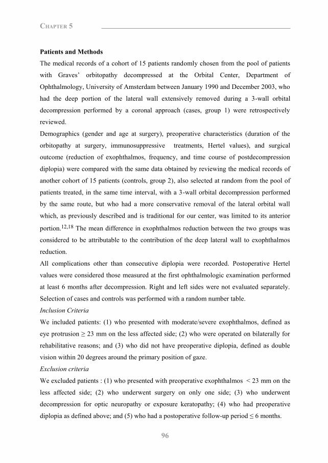
96
Chapter 5
Patients and Methods
The medical records of a cohort of 15 patients randomly chosen from the pool of patients
with Graves‟ orbitopathy decompressed at the Orbital Center, Department of
Ophthalmology, University of Amsterdam between January 1990 and December 2003, who
had the deep portion of the lateral wall extensively removed during a 3-wall orbital
decompression performed by a coronal approach (cases, group 1) were retrospectively
reviewed.
Demographics (gender and age at surgery), preoperative characteristics (duration of the
orbitopathy at surgery, immunosuppressive treatments, Hertel values), and surgical
outcome (reduction of exophthalmos, frequency, and time course of postdecompression
diplopia) were compared with the same data obtained by reviewing the medical records of
another cohort of 15 patients (controls, group 2), also selected at random from the pool of
patients treated, in the same time interval, with a 3-wall orbital decompression performed
by the same route, but who had a more conservative removal of the lateral orbital wall
which, as previously described and is traditional for our center, was limited to its anterior
portion.12,18 The mean difference in exophthalmos reduction between the two groups was
considered to be attributable to the contribution of the deep lateral wall to exophthalmos
reduction.
All complications other than consecutive diplopia were recorded. Postoperative Hertel
values were considered those measured at the first ophthalmologic examination performed
at least 6 months after decompression. Right and left sides were not evaluated separately.
Selection of cases and controls was performed with a random number table.
Inclusion Criteria
We included patients: (1) who presented with moderate/severe exophthalmos, defined as
eye protrusion ≥ 23 mm on the less affected side; (2) who were operated on bilaterally for
rehabilitative reasons; and (3) who did not have preoperative diplopia, defined as double
vision within 20 degrees around the primary position of gaze.
Exclusion criteria
We excluded patients : (1) who presented with preoperative exophthalmos < 23 mm on the
less affected side; (2) who underwent surgery on only one side; (3) who underwent
decompression for optic neuropathy or exposure keratopathy; (4) who had preoperative
diplopia as defined above; and (5) who had a postoperative follow-up period ≤ 6 months.
Statistical analysis
The analysis was conducted by means of a database created using version 11.5 of the SPSS
statistical analysis software package (http://www.spss.com). Student t test was used to
compare continuous variables showing no marked deviations from the normal distribution.
For other continuous variables or ordinal variables, the Mann-Whitney U-test was used.
Categorical preoperative and postoperative data were investigated using the χ² test.
Surgical technique
The surgical approach to orbital decompression was the same for cases and controls: a
coronal incision was made with a no. 10 blade from ear to ear, 3 to 4 cm behind the
hairline. The incision extended down to the periosteum of the parietal bones centrally, and
to the deep temporalis fascia laterally. Bleeding from the wound edges was controlled with
Raney scalp clips. In the central portion of the skull, a subperiosteal plane was created by
blunt dissection and laterally a surgical plane was bluntly developed between the deep and
the superficial temporalis fascia. Laterally and inferiorly, where the deep temporalis fascia
divides into a deeper and a more superficial layer to enclose Yasargil‟s superficial temporal
fat pad, the surgical dissection was carried out directly against the deeper division of the
fascia.
The forehead flap thus created was then turned down to expose the superior and lateral
orbital rims. The supraorbital nerve was set free by chiselling its bony foramen when
present, and the periorbita, including the trochlea, was dissected off the orbital bones and
the temporalis muscle was dissected from its anterior origin with a no. 10 blade and
periosteal elevators, exposing the lateral orbital wall while leaving sufficient tissue for
suturing at the end of surgery.
In group 2, only the anterior portion of the lateral orbital wall behind the orbital rim (which
was left intact) was removed, “allowing a fingertip to pass through”12,18 (Figure 1). In
group 1, the osteotomy of the lateral wall was started as for patients of group 2 but was then
extended inferiorly up to the inferior orbital fissure with bone punches and posteriorly up to
the dura of the middle cranial fossa by mean of bone punches and a surgical high-speed
drill equipped with a cutting-burr or a diamond-burr tip (Figure 2). While removing the
lateral orbital wall, the soft orbital tissues and the temporalis muscle were retracted and
protected with malleable orbital retractors.
100379 Thesis Baldeschi.indd 96 vrijdag17-december-2010 12:08
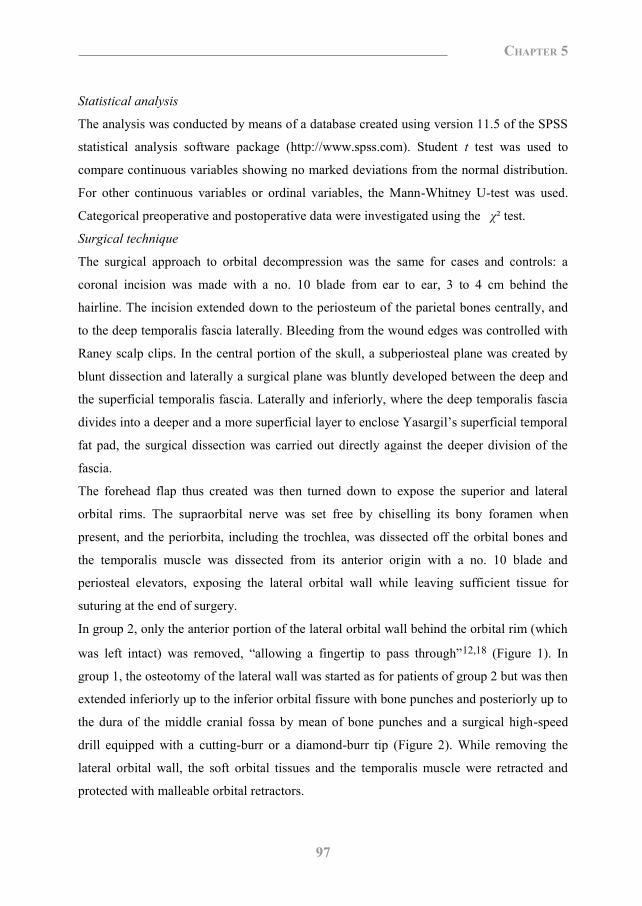
97
Chapter 5
Patients and Methods
The medical records of a cohort of 15 patients randomly chosen from the pool of patients
with Graves‟ orbitopathy decompressed at the Orbital Center, Department of
Ophthalmology, University of Amsterdam between January 1990 and December 2003, who
had the deep portion of the lateral wall extensively removed during a 3-wall orbital
decompression performed by a coronal approach (cases, group 1) were retrospectively
reviewed.
Demographics (gender and age at surgery), preoperative characteristics (duration of the
orbitopathy at surgery, immunosuppressive treatments, Hertel values), and surgical
outcome (reduction of exophthalmos, frequency, and time course of postdecompression
diplopia) were compared with the same data obtained by reviewing the medical records of
another cohort of 15 patients (controls, group 2), also selected at random from the pool of
patients treated, in the same time interval, with a 3-wall orbital decompression performed
by the same route, but who had a more conservative removal of the lateral orbital wall
which, as previously described and is traditional for our center, was limited to its anterior
portion.12,18 The mean difference in exophthalmos reduction between the two groups was
considered to be attributable to the contribution of the deep lateral wall to exophthalmos
reduction.
All complications other than consecutive diplopia were recorded. Postoperative Hertel
values were considered those measured at the first ophthalmologic examination performed
at least 6 months after decompression. Right and left sides were not evaluated separately.
Selection of cases and controls was performed with a random number table.
Inclusion Criteria
We included patients: (1) who presented with moderate/severe exophthalmos, defined as
eye protrusion ≥ 23 mm on the less affected side; (2) who were operated on bilaterally for
rehabilitative reasons; and (3) who did not have preoperative diplopia, defined as double
vision within 20 degrees around the primary position of gaze.
Exclusion criteria
We excluded patients : (1) who presented with preoperative exophthalmos < 23 mm on the
less affected side; (2) who underwent surgery on only one side; (3) who underwent
decompression for optic neuropathy or exposure keratopathy; (4) who had preoperative
diplopia as defined above; and (5) who had a postoperative follow-up period ≤ 6 months.
Statistical analysis
The analysis was conducted by means of a database created using version 11.5 of the SPSS
statistical analysis software package (http://www.spss.com). Student t test was used to
compare continuous variables showing no marked deviations from the normal distribution.
For other continuous variables or ordinal variables, the Mann-Whitney U-test was used.
Categorical preoperative and postoperative data were investigated using the χ² test.
Surgical technique
The surgical approach to orbital decompression was the same for cases and controls: a
coronal incision was made with a no. 10 blade from ear to ear, 3 to 4 cm behind the
hairline. The incision extended down to the periosteum of the parietal bones centrally, and
to the deep temporalis fascia laterally. Bleeding from the wound edges was controlled with
Raney scalp clips. In the central portion of the skull, a subperiosteal plane was created by
blunt dissection and laterally a surgical plane was bluntly developed between the deep and
the superficial temporalis fascia. Laterally and inferiorly, where the deep temporalis fascia
divides into a deeper and a more superficial layer to enclose Yasargil‟s superficial temporal
fat pad, the surgical dissection was carried out directly against the deeper division of the
fascia.
The forehead flap thus created was then turned down to expose the superior and lateral
orbital rims. The supraorbital nerve was set free by chiselling its bony foramen when
present, and the periorbita, including the trochlea, was dissected off the orbital bones and
the temporalis muscle was dissected from its anterior origin with a no. 10 blade and
periosteal elevators, exposing the lateral orbital wall while leaving sufficient tissue for
suturing at the end of surgery.
In group 2, only the anterior portion of the lateral orbital wall behind the orbital rim (which
was left intact) was removed, “allowing a fingertip to pass through”12,18 (Figure 1). In
group 1, the osteotomy of the lateral wall was started as for patients of group 2 but was then
extended inferiorly up to the inferior orbital fissure with bone punches and posteriorly up to
the dura of the middle cranial fossa by mean of bone punches and a surgical high-speed
drill equipped with a cutting-burr or a diamond-burr tip (Figure 2). While removing the
lateral orbital wall, the soft orbital tissues and the temporalis muscle were retracted and
protected with malleable orbital retractors.
100379 Thesis Baldeschi.indd 97 vrijdag17-december-2010 12:08
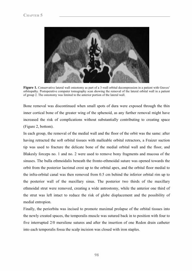
98
Chapter 5
Figure 1. Conservative lateral wall osteotomy as part of a 3-wall orbital decompression in a patient with Graves‟ orbitopathy. Postoperative computer tomography scan showing the removal of the lateral orbital wall in a patient of group 2. The osteotomy was limited to the anterior portion of the lateral wall.
Bone removal was discontinued when small spots of dura were exposed through the thin
inner cortical bone of the greater wing of the sphenoid, as any further removal might have
increased the risk of complications without substantially contributing to creating space
(Figure 2, bottom).
In each group, the removal of the medial wall and the floor of the orbit was the same: after
having retracted the soft orbital tissues with malleable orbital retractors, a Fraizer suction
tip was used to fracture the delicate bone of the medial orbital wall and the floor, and
Blakesly forceps no. 1 and no. 2 were used to remove bony fragments and mucosa of the
sinuses. The bulla ethmoidalis beneath the fronto-ethmoidal suture was opened towards the
orbit from the posterior lacrimal crest up to the orbital apex, and the orbital floor medial to
the infra-orbital canal was then removed from 0.5 cm behind the inferior orbital rim up to
the posterior wall of the maxillary sinus. The posterior two thirds of the maxillary
ethmoidal strut were removed, creating a wide antrostomy, while the anterior one third of
the strut was left intact to reduce the risk of globe displacement and the possibility of
medial entropion.
Finally, the periorbita was incised to promote maximal prolapse of the orbital tissues into
the newly created spaces, the temporalis muscle was sutured back in to position with four to
five interrupted 2/0 mersilene sutures and after the insertion of one Redon drain catheter
into each temporalis fossa the scalp incision was closed with iron staples.
Figure 2. Extended lateral wall osteotomy as part of a 3-wall orbital decompression in a patient with Graves‟ orbitopathy. (Top) Postoperative computer tomography scan showing the removal of the lateral orbital wall in a patient of group 1. The osteotomy was extended to the more posterior portion of the lateral wall. (Bottom) Intraoperative photo showing the removal of the deep lateral orbital wall by a coronal approach. A spot of exposed dura is evident (arrow).
Figure 3. A patient of group 1. (Top) The patient at admittance for orbital decompression. (Bottom) The patient 4 months after surgery. An adequate decompression had been achieved, esophoria, and deficit of accommodation which were present soon after surgery were disappeared, a mild anisocoria persisted.
100379 Thesis Baldeschi.indd 98 vrijdag17-december-2010 12:08
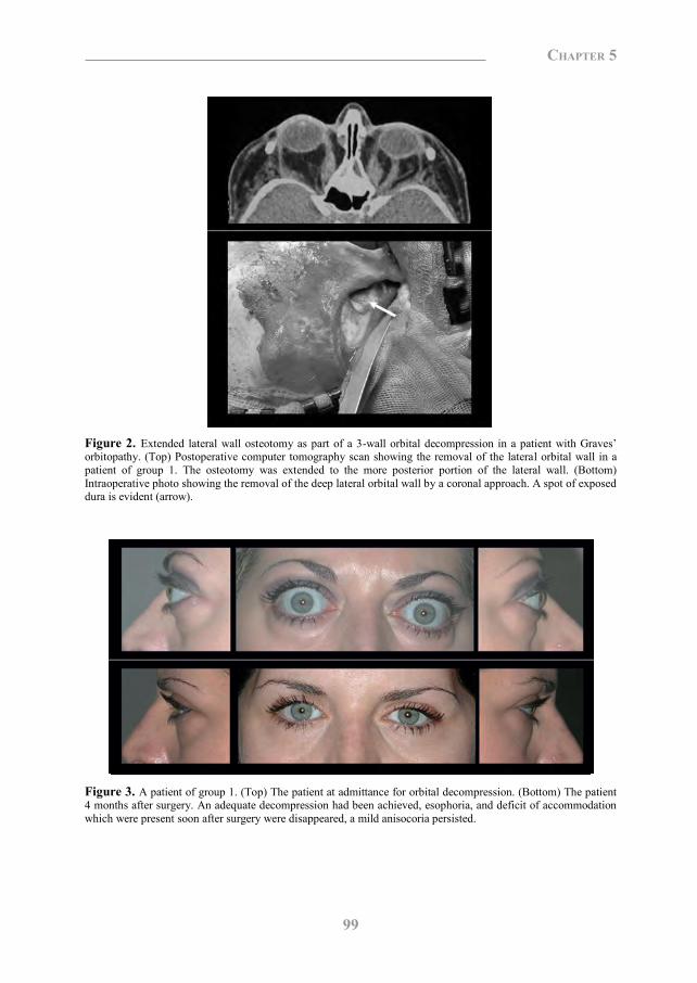
99
Chapter 5
Figure 1. Conservative lateral wall osteotomy as part of a 3-wall orbital decompression in a patient with Graves‟ orbitopathy. Postoperative computer tomography scan showing the removal of the lateral orbital wall in a patient of group 2. The osteotomy was limited to the anterior portion of the lateral wall.
Bone removal was discontinued when small spots of dura were exposed through the thin
inner cortical bone of the greater wing of the sphenoid, as any further removal might have
increased the risk of complications without substantially contributing to creating space
(Figure 2, bottom).
In each group, the removal of the medial wall and the floor of the orbit was the same: after
having retracted the soft orbital tissues with malleable orbital retractors, a Fraizer suction
tip was used to fracture the delicate bone of the medial orbital wall and the floor, and
Blakesly forceps no. 1 and no. 2 were used to remove bony fragments and mucosa of the
sinuses. The bulla ethmoidalis beneath the fronto-ethmoidal suture was opened towards the
orbit from the posterior lacrimal crest up to the orbital apex, and the orbital floor medial to
the infra-orbital canal was then removed from 0.5 cm behind the inferior orbital rim up to
the posterior wall of the maxillary sinus. The posterior two thirds of the maxillary
ethmoidal strut were removed, creating a wide antrostomy, while the anterior one third of
the strut was left intact to reduce the risk of globe displacement and the possibility of
medial entropion.
Finally, the periorbita was incised to promote maximal prolapse of the orbital tissues into
the newly created spaces, the temporalis muscle was sutured back in to position with four to
five interrupted 2/0 mersilene sutures and after the insertion of one Redon drain catheter
into each temporalis fossa the scalp incision was closed with iron staples.
Figure 2. Extended lateral wall osteotomy as part of a 3-wall orbital decompression in a patient with Graves‟ orbitopathy. (Top) Postoperative computer tomography scan showing the removal of the lateral orbital wall in a patient of group 1. The osteotomy was extended to the more posterior portion of the lateral wall. (Bottom) Intraoperative photo showing the removal of the deep lateral orbital wall by a coronal approach. A spot of exposed dura is evident (arrow).
Figure 3. A patient of group 1. (Top) The patient at admittance for orbital decompression. (Bottom) The patient 4 months after surgery. An adequate decompression had been achieved, esophoria, and deficit of accommodation which were present soon after surgery were disappeared, a mild anisocoria persisted.
100379 Thesis Baldeschi.indd 99 vrijdag17-december-2010 12:08
100
Chapter 5
Results
Patients in group 1 were drown from a pool of 37, while patients in group 2 were drawn
from a pool of 335. The patients‟ demographics and preoperative characteristics are
summarized in the table.
The mean reduction of exophthalmos was 7.2 mm (SD = 2.3) in group 1 and 4.9 mm (SD =
1.1) in group 2 and the mean reduction of exophthalmos in group 1 was significantly higher
than in group 2 (P = 0.001). The contribution of the deep lateral wall to exophthalmos
reduction was 2.3 mm, giving the patients of group 1, on average, a 32.0% greater degree of
exophthalmos reduction. Decompression-induced diplopia arose in two (13.3%) of the 15
patients of group 1 and in five (33.3%) of the 15 patients of group 2, although the
difference in onset of decompression-induced diplopia between the two groups was not
statistically significant (P = 0.203). Both of the patients of group 1 with postoperative
diplopia presented with an esotropia and a deficit of abduction that resolved spontaneously
4 to 6 months after surgery, while all five patients of group 2 with postoperative diplopia
presented with esotropia and in two cases there was associated vertical strabismus: all of
these patients required subsequent surgical correction once the squint angle became stabile.
One of the two patients of group 1 with postoperative diplopia presented with mild
persistent anisocoria and a transient deficit of accommodation of the left eye (Figure 3).
Discussion
In the 1980s, when the number of orbital decompression procedures being performed
started to rise as surgery was being undertaken not only for functional reasons, but also for
the esthetic/psychosocial rehabilitation of patients with Graves‟ orbitopathy15, the antral-
ethmoidal decompression by a transantral approach, as described by Walsh and Ogura in
1957, was the mainstay technique.5,12 The major disadvantage reported with transantral
surgery was subsequent motility imbalance as high as 52%19 and, therefore, alternative
procedures were sought in an attempt to decrease the risk of decompression-induced
diplopia. In cases of mild exophthalmos, trans-lid antral-ethmoidal decompression appeared
to be a valid alternative, with a risk of iatrogenic diplopia in only 4.6% of patients15 and for
more severe exophthalmos, inferomedial decompression was used in combination with
lateral decompression. Such procedures, whether performed with separate periorbital
incisions or through a coronal approach, were also related with a low incidence of
Table. Demographics and Preoperative Characteristics in Patients Treated With Extended (Cases, Group 1) or Conservative (Controls, Group 2) Removal of the Lateral Orbital Wall
as Part of a Coronal-approach, 3-wall Decompression
Cases
(n=15)
Controls
(n=15)
Significance
Female 80% 86.66% P = 0 .624
Age at surgery (yrs, ± SD)
40.9 (9.7) 40.1 (12.4) P = 0.852
Duration of Graves‟ orbitopathy at surgery (yrs, ± SD)
5.43 (5.8) 3.78 (2.1) P = 0.314
Preop. immunosuppressive therapy: a) corticosteroids b) orbital radiotherapy c) corticosteroids and orbital radiotherapy d) neither corticosteroids nor orbital radiotherapy
20% 27% 13%
40%
13% 47% 7%
33%
P = 0.500 P = 0.225 P = 0.500
P = 0.500
Preoperative Hertel values (mm, ± SD)
26.3 (2.4)
25.0 (1.4)
P = 0.123
consecutive diplopia.12 In 1989, Leone and coauthors, in an attempt to further reduce the
effect of decompression surgery on extra ocular muscle motility disturbance, proposed
balancing the decompression by removing the medial and lateral orbital walls while sparing
the floor.17 This technique, which theoretically should have minimized the risk of
iatrogenic diplopia, later appeared to be associated with a higher risk of such a complication
compared with removal of the lateral orbital wall alone, or with inferomedial and 3-wall
techniques.20,21
Recently, the lateral orbital wall has been promoted as being the region of first choice for
orbital decompression. Its removal, which is connected with a low risk of consecutive
diplopia or severe complications such as cerebrospinal fluid leak, perfectly fits the needs of
the increasingly demanding patient population.7 The lateral wall of the orbit is the thickest
orbital wall and is composed of the zygomatic bone anteriorly and the greater wing of the
sphenoid posteriorly. The thinnest part of the lateral wall is at the zygomaticosphenoid
suture, approximately 1 cm behind the orbital rim, and approximately 1 cm behind the
100379 Thesis Baldeschi.indd 100 vrijdag17-december-2010 12:08
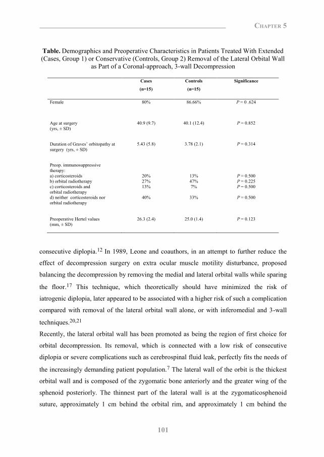
101
Chapter 5
Results
Patients in group 1 were drown from a pool of 37, while patients in group 2 were drawn
from a pool of 335. The patients‟ demographics and preoperative characteristics are
summarized in the table.
The mean reduction of exophthalmos was 7.2 mm (SD = 2.3) in group 1 and 4.9 mm (SD =
1.1) in group 2 and the mean reduction of exophthalmos in group 1 was significantly higher
than in group 2 (P = 0.001). The contribution of the deep lateral wall to exophthalmos
reduction was 2.3 mm, giving the patients of group 1, on average, a 32.0% greater degree of
exophthalmos reduction. Decompression-induced diplopia arose in two (13.3%) of the 15
patients of group 1 and in five (33.3%) of the 15 patients of group 2, although the
difference in onset of decompression-induced diplopia between the two groups was not
statistically significant (P = 0.203). Both of the patients of group 1 with postoperative
diplopia presented with an esotropia and a deficit of abduction that resolved spontaneously
4 to 6 months after surgery, while all five patients of group 2 with postoperative diplopia
presented with esotropia and in two cases there was associated vertical strabismus: all of
these patients required subsequent surgical correction once the squint angle became stabile.
One of the two patients of group 1 with postoperative diplopia presented with mild
persistent anisocoria and a transient deficit of accommodation of the left eye (Figure 3).
Discussion
In the 1980s, when the number of orbital decompression procedures being performed
started to rise as surgery was being undertaken not only for functional reasons, but also for
the esthetic/psychosocial rehabilitation of patients with Graves‟ orbitopathy15, the antral-
ethmoidal decompression by a transantral approach, as described by Walsh and Ogura in
1957, was the mainstay technique.5,12 The major disadvantage reported with transantral
surgery was subsequent motility imbalance as high as 52%19 and, therefore, alternative
procedures were sought in an attempt to decrease the risk of decompression-induced
diplopia. In cases of mild exophthalmos, trans-lid antral-ethmoidal decompression appeared
to be a valid alternative, with a risk of iatrogenic diplopia in only 4.6% of patients15 and for
more severe exophthalmos, inferomedial decompression was used in combination with
lateral decompression. Such procedures, whether performed with separate periorbital
incisions or through a coronal approach, were also related with a low incidence of
Table. Demographics and Preoperative Characteristics in Patients Treated With Extended (Cases, Group 1) or Conservative (Controls, Group 2) Removal of the Lateral Orbital Wall
as Part of a Coronal-approach, 3-wall Decompression
Cases
(n=15)
Controls
(n=15)
Significance
Female 80% 86.66% P = 0 .624
Age at surgery (yrs, ± SD)
40.9 (9.7) 40.1 (12.4) P = 0.852
Duration of Graves‟ orbitopathy at surgery (yrs, ± SD)
5.43 (5.8) 3.78 (2.1) P = 0.314
Preop. immunosuppressive therapy: a) corticosteroids b) orbital radiotherapy c) corticosteroids and orbital radiotherapy d) neither corticosteroids nor orbital radiotherapy
20% 27% 13%
40%
13% 47% 7%
33%
P = 0.500 P = 0.225 P = 0.500
P = 0.500
Preoperative Hertel values (mm, ± SD)
26.3 (2.4)
25.0 (1.4)
P = 0.123
consecutive diplopia.12 In 1989, Leone and coauthors, in an attempt to further reduce the
effect of decompression surgery on extra ocular muscle motility disturbance, proposed
balancing the decompression by removing the medial and lateral orbital walls while sparing
the floor.17 This technique, which theoretically should have minimized the risk of
iatrogenic diplopia, later appeared to be associated with a higher risk of such a complication
compared with removal of the lateral orbital wall alone, or with inferomedial and 3-wall
techniques.20,21
Recently, the lateral orbital wall has been promoted as being the region of first choice for
orbital decompression. Its removal, which is connected with a low risk of consecutive
diplopia or severe complications such as cerebrospinal fluid leak, perfectly fits the needs of
the increasingly demanding patient population.7 The lateral wall of the orbit is the thickest
orbital wall and is composed of the zygomatic bone anteriorly and the greater wing of the
sphenoid posteriorly. The thinnest part of the lateral wall is at the zygomaticosphenoid
suture, approximately 1 cm behind the orbital rim, and approximately 1 cm behind the
100379 Thesis Baldeschi.indd 101 vrijdag17-december-2010 12:08
102
Chapter 5
zygomaticosphenoid suture the sphenoid bone thickens where it divides to form the anterior
corner of the middle cranial fossa.22 Here compact bone passes into thick cancellous bone
to form a trigonus, nicknamed the “door-jamb” by Goldberg who, more than any other
author, has stressed the importance of this zone of possible orbital expansion.9,10
Experimental studies on the basis of dry skulls and clinical radiologic surveys have
quantified the absolute volume expected from the lateral orbital wall to clinical
decompressions.8,9 Although there is considerable interindividual variability, the doorjamb
represents a mean volume of 2.9 cm3 and because of its anatomical position located directly
behind the globe, an almost millimeter-for-millimeter relationship between door-jamb
removal and exophthalmos reduction was hypothesized.9,10 However, even by the use of a
coronal approach where excellent access to the deep lateral wall can be obtained, thus
permitting the possibility of performing the widest possible osteotomy, the volume of the
doorjamb should be regarded as an idealized measurement that is impossible to be used
fully for clinical decompressions.9 In addition to the volume and location of a given orbital
wall, several disease-dependent variables can, in clinical practice, influence the possible
reduction of exophthalmos. The stage of the orbitopathy at the time of surgery, the
distensibility and plasticity of the soft orbital tissues and the degree of preoperative
exophthalmos have all been highlighted as possible factors determining the final outcome
of decompression surgery.11-13
Although removal of the deep lateral wall has been reported in several clinical trials9, 10, 14-
17, its contribution to exophthalmos reduction has never been specifically studied.
Additionally, the extreme heterogeneity of the patients included in each series and the
variation of applied surgical techniques do not permit any attempt of its quantification on
the basis of the data already available in the literature. In our study, the calculated
contribution of the deep lateral wall to exophthalmos reduction can be regarded as maximal
and on the basis of a homogeneous population of patients with Graves‟ orbitopathy. We
selected patients operated by the coronal approach, which permitted maximal removal of
the deep lateral wall in the group 1 patients, and we chose to consider the reduction of
exophthalmos later than 6 months after decompression as exophthalmos reduction is
expected to be maximal in most of the decompressed patients by this time.18 Furthermore,
in our study, surgical access and inclusion and exclusion criteria were identical for the two
groups, and we found no difference in the demographics or preoperative characteristics
between groups 1 and 2.
We found that removal of the deep lateral wall contributed a mean of 2.3 mm to
exophthalmos reduction. Interestingly, the standard deviation of the exophthalmos
reduction in group 1 was approximately twice that of group 2 and we interpret this finding
as a manifestation of the known9 interindividual variability of the volume of the deep
lateral wall.
Removal of the deep lateral wall during 3-wall orbital decompression was not found to be
associated with an increased risk of consecutive diplopia when compared with the more
conservative 3-wall orbital decompression applied in group 2. Furthermore, both patients in
group 1 who presented with consecutive esotropia and deficit of abduction after removal of
the deep lateral wall experienced a spontaneous resolution of their strabismus 4 to 6 months
after surgery. Transient strabismus after deep lateral wall removal has previously been
reported.10,16 Deep mechanical contusions of the soft orbital tissues associated with the
wide exposure that is necessary to accomplish the osteotomy with a cutting-burr can be
regarded as a possible cause of this transient strabismus that most probably has a paralytic,
rather than a restrictive, etiology and this hypothesis is supported by the simultaneous, mild
anisocoria, and transient deficit of accommodation demonstrated by one of our patients.
Given the multifaceted nature of Graves‟ orbitopathy, the numerous indications for
decompression surgery and the many variations in surgical techniques, an ideal attempt to
quantify the contribution of a specific osteotomy to exophthalmos reduction and
consecutive diplopia must take the influence of these variables into account. We attempted
to do this by selecting patients at random from a homogeneous group of patients with
Graves‟ orbitopathy undergoing surgery for the same indication and by utilizing a
consistent surgical approach in each group. Nevertheless, we wish to acknowledge the
limitations of our study. Our observations are retrospective, and patients were not randomly
allocated to either of the two treatment groups. Additionally, our findings are only
applicable to the specific population of patients with Graves‟ orbitopathy as selected in this
study, although our population, which was composed of individuals affected by
moderate/severe exophthalmos, with no preoperative diplopia and treated exclusively for
esthetic reasons, may be considered to be representative of a numerically significant and,
100379 Thesis Baldeschi.indd 102 vrijdag17-december-2010 12:08
103
Chapter 5
zygomaticosphenoid suture the sphenoid bone thickens where it divides to form the anterior
corner of the middle cranial fossa.22 Here compact bone passes into thick cancellous bone
to form a trigonus, nicknamed the “door-jamb” by Goldberg who, more than any other
author, has stressed the importance of this zone of possible orbital expansion.9,10
Experimental studies on the basis of dry skulls and clinical radiologic surveys have
quantified the absolute volume expected from the lateral orbital wall to clinical
decompressions.8,9 Although there is considerable interindividual variability, the doorjamb
represents a mean volume of 2.9 cm3 and because of its anatomical position located directly
behind the globe, an almost millimeter-for-millimeter relationship between door-jamb
removal and exophthalmos reduction was hypothesized.9,10 However, even by the use of a
coronal approach where excellent access to the deep lateral wall can be obtained, thus
permitting the possibility of performing the widest possible osteotomy, the volume of the
doorjamb should be regarded as an idealized measurement that is impossible to be used
fully for clinical decompressions.9 In addition to the volume and location of a given orbital
wall, several disease-dependent variables can, in clinical practice, influence the possible
reduction of exophthalmos. The stage of the orbitopathy at the time of surgery, the
distensibility and plasticity of the soft orbital tissues and the degree of preoperative
exophthalmos have all been highlighted as possible factors determining the final outcome
of decompression surgery.11-13
Although removal of the deep lateral wall has been reported in several clinical trials9, 10, 14-
17, its contribution to exophthalmos reduction has never been specifically studied.
Additionally, the extreme heterogeneity of the patients included in each series and the
variation of applied surgical techniques do not permit any attempt of its quantification on
the basis of the data already available in the literature. In our study, the calculated
contribution of the deep lateral wall to exophthalmos reduction can be regarded as maximal
and on the basis of a homogeneous population of patients with Graves‟ orbitopathy. We
selected patients operated by the coronal approach, which permitted maximal removal of
the deep lateral wall in the group 1 patients, and we chose to consider the reduction of
exophthalmos later than 6 months after decompression as exophthalmos reduction is
expected to be maximal in most of the decompressed patients by this time.18 Furthermore,
in our study, surgical access and inclusion and exclusion criteria were identical for the two
groups, and we found no difference in the demographics or preoperative characteristics
between groups 1 and 2.
We found that removal of the deep lateral wall contributed a mean of 2.3 mm to
exophthalmos reduction. Interestingly, the standard deviation of the exophthalmos
reduction in group 1 was approximately twice that of group 2 and we interpret this finding
as a manifestation of the known9 interindividual variability of the volume of the deep
lateral wall.
Removal of the deep lateral wall during 3-wall orbital decompression was not found to be
associated with an increased risk of consecutive diplopia when compared with the more
conservative 3-wall orbital decompression applied in group 2. Furthermore, both patients in
group 1 who presented with consecutive esotropia and deficit of abduction after removal of
the deep lateral wall experienced a spontaneous resolution of their strabismus 4 to 6 months
after surgery. Transient strabismus after deep lateral wall removal has previously been
reported.10,16 Deep mechanical contusions of the soft orbital tissues associated with the
wide exposure that is necessary to accomplish the osteotomy with a cutting-burr can be
regarded as a possible cause of this transient strabismus that most probably has a paralytic,
rather than a restrictive, etiology and this hypothesis is supported by the simultaneous, mild
anisocoria, and transient deficit of accommodation demonstrated by one of our patients.
Given the multifaceted nature of Graves‟ orbitopathy, the numerous indications for
decompression surgery and the many variations in surgical techniques, an ideal attempt to
quantify the contribution of a specific osteotomy to exophthalmos reduction and
consecutive diplopia must take the influence of these variables into account. We attempted
to do this by selecting patients at random from a homogeneous group of patients with
Graves‟ orbitopathy undergoing surgery for the same indication and by utilizing a
consistent surgical approach in each group. Nevertheless, we wish to acknowledge the
limitations of our study. Our observations are retrospective, and patients were not randomly
allocated to either of the two treatment groups. Additionally, our findings are only
applicable to the specific population of patients with Graves‟ orbitopathy as selected in this
study, although our population, which was composed of individuals affected by
moderate/severe exophthalmos, with no preoperative diplopia and treated exclusively for
esthetic reasons, may be considered to be representative of a numerically significant and,
100379 Thesis Baldeschi.indd 103 vrijdag17-december-2010 12:08
104
Chapter 5
for obvious reasons, demanding subpopulation of patients with Graves‟ orbitopathy. We,
therefore, believe that our findings are of practical relevance when planning decompression
surgery.
References 1. Rundle FF, Wilson CW. Development and course of exophthalmos and ophthalmoplegia in Graves‟ disease
with special reference to the effect of thyroidectomy. Cli Sci 1945;5:177- 94.
2. Dollinger J. Die Druckentlastung der Augenhöhle durch Entfernung der äu_eren Orbitawand bei hochgradigem
Exophthalmos (Morbus Basedowii) und konsekutiver Hauterkrankung. Dtsch med Wschr 1911;37:1888-90.
3. Naffziger HC. Progressive exophthalmos following thyroidectomy; its pathology and treatment. Ann Surg
1931;94:582-6.
4. Sewall EC. Operative control of progressive exophthalmos. Arch Otolaryngol 1936;24:621- 4.
5. Walsh TE, Ogura JH. Transantral orbital decompression for malignant exophthalmos. Laryngoscope
1957;67:544-68.
6. Tessier P. Expansion chirurgicale de l‟orbit. Ann Chir Plast 1969;14:207-14.
7. Goldberg RA. The evolving paradigm of orbital decompression surgery. Arch Ophthalmol 1998;116:95-96.
8. Stabile JR, Trokel SM. Increase in orbital volume obtained by decompression in dried skulls. Am J Ophthalmol
1983;95:327-31.
9. Goldberg RA, Kim AJ, Kerivan KM. The lacrimal keyhole, orbital door jamb, and basin of the inferior orbital
fissure. Three areas of deep bone in the lateral orbit. Arch Ophthalmol 1998;116:1618-24.
10. Goldberg RA, Weinberg DA, Schorr N, Wirta D. Maximal 3-wall orbital decompression through a coronal
approach. Ophthalmic Surg Lasers 1997;28:832-43.
11. Härting F, Koornneef L, Peeters HJF, Gillissen JPA. Fourteen years of orbital decompression in Graves‟
disease. A review of technique, results, and complications. Orbit 1986;5:123-9.
12. Mourits MPh, Koornneef L, Wiersinga WM, Prummel MF, Berghout A, van der Gaag R. Orbital
decompression for Graves‟ ophthalmopathy by inferomedial, by inferomedial plus lateral, and by coronal
approach. Ophthalmology 1990;97:636-41.
13. Wilson WB, Manke WF. Orbital decompression in Graves‟ disease: the predictability of reduction of
proptosis. Arch Opthalmol 1991;109:343-5.
14. Kennerdell JS, Maroon JC. An orbital decompression for severe dysthyroid exophthalmos. Ophthalmology
1982;89:467-72.
15. Mc Cord CD. Current trends in orbital decompression. Ophthalmology 1985;92:21-33.
16. Hurwitz JJ, Birt D. An individualized approach to orbital decompression in Graves‟ orbitopathy. Arch
Ophthalmol 1985;103:660-5.
17. Leone CR, Piest KL, Newman RJ. Medial and lateral wall decompression for thyroid ophthalmopathy. Am J
Ophthalmol 1989;108:160-6.
18. Kalmann R, Mourits MPh, van der Pol J, Koornneef L. Coronal approach for rehabilitative orbital
decompression in Graves‟ ophthalmopathy. Br J Ophthalmol 1997;81:41-5.
19. De Santo LW. Transantral orbital decompression. In: Gorman CA, Waller RR, Dyer JA, editors. The eye and
orbit in thyroid disease. New York: Raven Press, 1984:231-51.
20. Goldberg RA, Perry JD, Hortoleza V, Tong JT. Strabismus after balanced medial plus lateral wall versus
lateral wall only orbital decompression for disthyroid orbitopathy. Ophthal Plast Reconstr Surg 2000;16:271-7.
21. Paridaens D, Hans K, van Buiten S, Mourits MPh. The incidence of diplopia following coronal and trans lid
orbital decompression in Graves‟ orbitopathy. Eye 1998;12:800-05.
100379 Thesis Baldeschi.indd 104 vrijdag17-december-2010 12:08

105
Chapter 5
for obvious reasons, demanding subpopulation of patients with Graves‟ orbitopathy. We,
therefore, believe that our findings are of practical relevance when planning decompression
surgery.
References 1. Rundle FF, Wilson CW. Development and course of exophthalmos and ophthalmoplegia in Graves‟ disease
with special reference to the effect of thyroidectomy. Cli Sci 1945;5:177- 94.
2. Dollinger J. Die Druckentlastung der Augenhöhle durch Entfernung der äu_eren Orbitawand bei hochgradigem
Exophthalmos (Morbus Basedowii) und konsekutiver Hauterkrankung. Dtsch med Wschr 1911;37:1888-90.
3. Naffziger HC. Progressive exophthalmos following thyroidectomy; its pathology and treatment. Ann Surg
1931;94:582-6.
4. Sewall EC. Operative control of progressive exophthalmos. Arch Otolaryngol 1936;24:621- 4.
5. Walsh TE, Ogura JH. Transantral orbital decompression for malignant exophthalmos. Laryngoscope
1957;67:544-68.
6. Tessier P. Expansion chirurgicale de l‟orbit. Ann Chir Plast 1969;14:207-14.
7. Goldberg RA. The evolving paradigm of orbital decompression surgery. Arch Ophthalmol 1998;116:95-96.
8. Stabile JR, Trokel SM. Increase in orbital volume obtained by decompression in dried skulls. Am J Ophthalmol
1983;95:327-31.
9. Goldberg RA, Kim AJ, Kerivan KM. The lacrimal keyhole, orbital door jamb, and basin of the inferior orbital
fissure. Three areas of deep bone in the lateral orbit. Arch Ophthalmol 1998;116:1618-24.
10. Goldberg RA, Weinberg DA, Schorr N, Wirta D. Maximal 3-wall orbital decompression through a coronal
approach. Ophthalmic Surg Lasers 1997;28:832-43.
11. Härting F, Koornneef L, Peeters HJF, Gillissen JPA. Fourteen years of orbital decompression in Graves‟
disease. A review of technique, results, and complications. Orbit 1986;5:123-9.
12. Mourits MPh, Koornneef L, Wiersinga WM, Prummel MF, Berghout A, van der Gaag R. Orbital
decompression for Graves‟ ophthalmopathy by inferomedial, by inferomedial plus lateral, and by coronal
approach. Ophthalmology 1990;97:636-41.
13. Wilson WB, Manke WF. Orbital decompression in Graves‟ disease: the predictability of reduction of
proptosis. Arch Opthalmol 1991;109:343-5.
14. Kennerdell JS, Maroon JC. An orbital decompression for severe dysthyroid exophthalmos. Ophthalmology
1982;89:467-72.
15. Mc Cord CD. Current trends in orbital decompression. Ophthalmology 1985;92:21-33.
16. Hurwitz JJ, Birt D. An individualized approach to orbital decompression in Graves‟ orbitopathy. Arch
Ophthalmol 1985;103:660-5.
17. Leone CR, Piest KL, Newman RJ. Medial and lateral wall decompression for thyroid ophthalmopathy. Am J
Ophthalmol 1989;108:160-6.
18. Kalmann R, Mourits MPh, van der Pol J, Koornneef L. Coronal approach for rehabilitative orbital
decompression in Graves‟ ophthalmopathy. Br J Ophthalmol 1997;81:41-5.
19. De Santo LW. Transantral orbital decompression. In: Gorman CA, Waller RR, Dyer JA, editors. The eye and
orbit in thyroid disease. New York: Raven Press, 1984:231-51.
20. Goldberg RA, Perry JD, Hortoleza V, Tong JT. Strabismus after balanced medial plus lateral wall versus
lateral wall only orbital decompression for disthyroid orbitopathy. Ophthal Plast Reconstr Surg 2000;16:271-7.
21. Paridaens D, Hans K, van Buiten S, Mourits MPh. The incidence of diplopia following coronal and trans lid
orbital decompression in Graves‟ orbitopathy. Eye 1998;12:800-05.
100379 Thesis Baldeschi.indd 105 vrijdag17-december-2010 12:08

106
Chapter 5
22. Dutton JJ. Osteology of the orbit. In: Dutton JJ, editor. Clinical and surgical anatomy of the orbit. Philadelphia:
WB Saunders Company 1994:1-12.
Chapter 6
Small Versus Coronal Incision Orbital Decompression In Graves’ Orbitopathy
Lelio Baldeschi
Orbital Centre, Department of Ophthalmology, University of Amsterdam, Amsterdam, The Netherlands. Orbit 2010;29:177-82
100379 Thesis Baldeschi.indd 106 vrijdag17-december-2010 12:08
22. Dutton JJ. Osteology of the orbit. In: Dutton JJ, editor. Clinical and surgical anatomy of the orbit. Philadelphia:
WB Saunders Company 1994:1-12.
Chapter 6
Small Versus Coronal Incision Orbital Decompression In Graves’ Orbitopathy
Lelio Baldeschi
Orbital Centre, Department of Ophthalmology, University of Amsterdam, Amsterdam, The Netherlands. Orbit 2010;29:177-82
100379 Thesis Baldeschi.indd 107 vrijdag17-december-2010 12:08
108
Chapter 6
Abstract
Ideally the planning of decompression surgery should be adequate to the severity of the
orbitopathy, its possible “lipogenic” or “myopathic” variants, the patient‟s specific orbital
osteology and possible previous surgeries. Due to surgeon‟s experience and local traditions,
however, a standardized rather than a tailored approach is often offered to the patient.
An inferior fornix incision can be used for infero medial bony decompression and/or for
removing fat from the medial and lateral inferior orbital quadrants. Through the same route
a lateral osteotomy can also be performed although an upper skin crease incision offers a
wider access to the lateral orbital wall. As an alternative the swinging eyelid technique,
offering an adequate access to the bony orbit and to the orbital fat compartments is a
versatile technique that can virtually be used as a standard approach for the greatest
majority of patients needing decompression surgery.
Orbital decompression by coronal incision is an invasive technique and for this not to be
used as a standard approach to orbital decompression. Nevertheless, it is not to be
abandoned as it can be an additional tool in surgeons‟ hands when dealing with patients
who can better benefit out of a particular, tailored rather than a standardised approach.
Many are the circumstances in which this may happen.
Major complications associated with the coronal approach have been mainly described in
small series, where only a few patients per year were operated. In this respect it is therefore
unavoidable to emphasize that each technique has its own learning curve and it may be
difficult to differentiate the effects of each technique from the experience of the surgeon.
For almost one century decompression surgery has been used to treat Graves‟ orbitopathy
(GO), and through the years it has been subjected to a paradigm shift in respect to
indications, approaches, and surgical routes. This has been largely due to a better
understanding of the multifaceted nature of the disease, a constant attempt to implement the
beneficial effects of this type of surgery simultaneously decreasing the aesthetic impact of
surgical scars, hospitalisation time, reconvalescence periods and risks for iatrogenic
complications in general, and consecutive strabismus in particular. Patients‟ increased
expectations and a more critical attitude towards surgical interventions are also not
negligible aspects that have driven the shift of orbital decompression surgery.
Orbital decompression was first used only to address sight threatening conditions, such as
optic neuropathy refractory to medical therapy, or exposure keratopathy unresponsive to
local measures and/or to minor eyelid surgeries. More recently the indications for orbital
decompression were extended to the treatment of disfiguring exophthalmos and symptoms.
Eyeball subluxation, which may be a possible cause of acute optic neuropathy and exposure
keratopathy, postural visual obscuration in patients with congestive inactive GO and
choroidal folds due to eyeball indentation by enlarged extraocular muscles represent other,
more recently recognised functional indications for decompression surgery.1
Expansion of the bony orbital boundary and fat removal had been the surgical methods to
reduce the raised intra-orbital pressure. The two approaches, which developed through
parallel routes up until recently, are no longer to be considered alternatives, but
complementary possibilities concurring in tailoring the most adequate treatment to the
specific patient‟s needs.2
As indications and approaches also surgical routes to orbital decompression have been
changed through the years. In the 1950s, when decompression surgery started to be used
routinely for functional purposes or rehabilitation of patients with GO, transbuccal incisions
were preferred to former visible and more invasive periorbital trans-cutaneous incisions.
The coronal approach to orbital decompression introduced by Tessier (1969)3, used by
Krastinova and Rodallec (1985)4 and later popularized by Leo Koornneef and his fellows
beginning in the 1980s5-8, although virtually invisible in patients who do not present
baldness, is a rather invasive access to the orbit. It is a common incision for plastic,
maxillofacial and neurosurgical procedures, which gives a wide access to the bony orbit,
but in general is not the surgical access to the orbit preferred by ophthalmologists, who are
100379 Thesis Baldeschi.indd 108 vrijdag17-december-2010 12:08
109
Chapter 6
Abstract
Ideally the planning of decompression surgery should be adequate to the severity of the
orbitopathy, its possible “lipogenic” or “myopathic” variants, the patient‟s specific orbital
osteology and possible previous surgeries. Due to surgeon‟s experience and local traditions,
however, a standardized rather than a tailored approach is often offered to the patient.
An inferior fornix incision can be used for infero medial bony decompression and/or for
removing fat from the medial and lateral inferior orbital quadrants. Through the same route
a lateral osteotomy can also be performed although an upper skin crease incision offers a
wider access to the lateral orbital wall. As an alternative the swinging eyelid technique,
offering an adequate access to the bony orbit and to the orbital fat compartments is a
versatile technique that can virtually be used as a standard approach for the greatest
majority of patients needing decompression surgery.
Orbital decompression by coronal incision is an invasive technique and for this not to be
used as a standard approach to orbital decompression. Nevertheless, it is not to be
abandoned as it can be an additional tool in surgeons‟ hands when dealing with patients
who can better benefit out of a particular, tailored rather than a standardised approach.
Many are the circumstances in which this may happen.
Major complications associated with the coronal approach have been mainly described in
small series, where only a few patients per year were operated. In this respect it is therefore
unavoidable to emphasize that each technique has its own learning curve and it may be
difficult to differentiate the effects of each technique from the experience of the surgeon.
For almost one century decompression surgery has been used to treat Graves‟ orbitopathy
(GO), and through the years it has been subjected to a paradigm shift in respect to
indications, approaches, and surgical routes. This has been largely due to a better
understanding of the multifaceted nature of the disease, a constant attempt to implement the
beneficial effects of this type of surgery simultaneously decreasing the aesthetic impact of
surgical scars, hospitalisation time, reconvalescence periods and risks for iatrogenic
complications in general, and consecutive strabismus in particular. Patients‟ increased
expectations and a more critical attitude towards surgical interventions are also not
negligible aspects that have driven the shift of orbital decompression surgery.
Orbital decompression was first used only to address sight threatening conditions, such as
optic neuropathy refractory to medical therapy, or exposure keratopathy unresponsive to
local measures and/or to minor eyelid surgeries. More recently the indications for orbital
decompression were extended to the treatment of disfiguring exophthalmos and symptoms.
Eyeball subluxation, which may be a possible cause of acute optic neuropathy and exposure
keratopathy, postural visual obscuration in patients with congestive inactive GO and
choroidal folds due to eyeball indentation by enlarged extraocular muscles represent other,
more recently recognised functional indications for decompression surgery.1
Expansion of the bony orbital boundary and fat removal had been the surgical methods to
reduce the raised intra-orbital pressure. The two approaches, which developed through
parallel routes up until recently, are no longer to be considered alternatives, but
complementary possibilities concurring in tailoring the most adequate treatment to the
specific patient‟s needs.2
As indications and approaches also surgical routes to orbital decompression have been
changed through the years. In the 1950s, when decompression surgery started to be used
routinely for functional purposes or rehabilitation of patients with GO, transbuccal incisions
were preferred to former visible and more invasive periorbital trans-cutaneous incisions.
The coronal approach to orbital decompression introduced by Tessier (1969)3, used by
Krastinova and Rodallec (1985)4 and later popularized by Leo Koornneef and his fellows
beginning in the 1980s5-8, although virtually invisible in patients who do not present
baldness, is a rather invasive access to the orbit. It is a common incision for plastic,
maxillofacial and neurosurgical procedures, which gives a wide access to the bony orbit,
but in general is not the surgical access to the orbit preferred by ophthalmologists, who are
100379 Thesis Baldeschi.indd 109 vrijdag17-december-2010 12:08
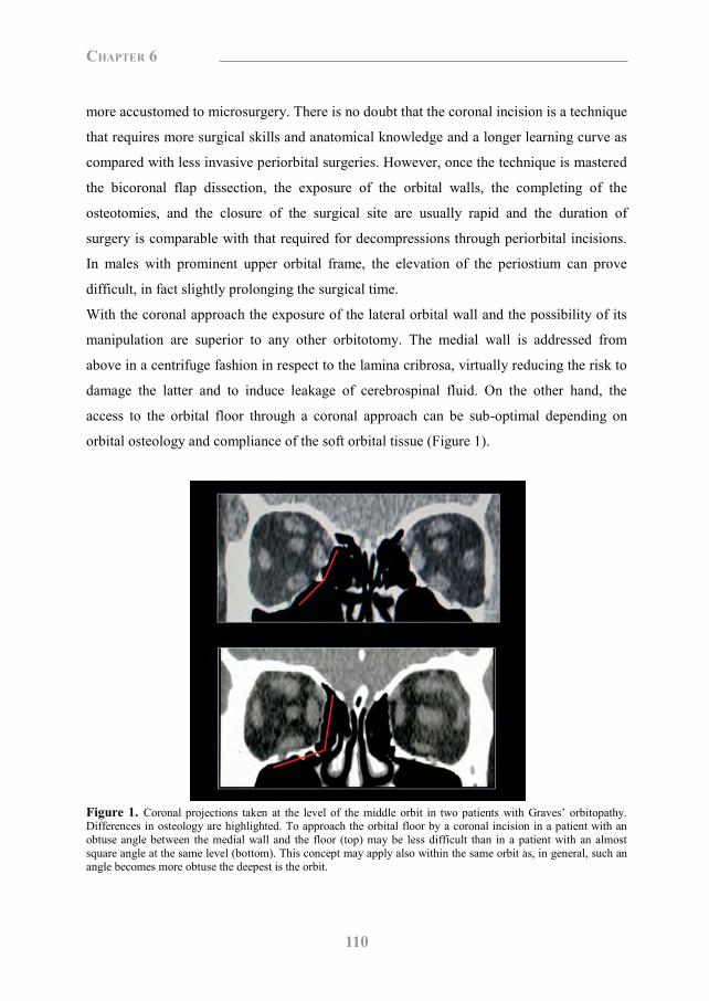
110
Chapter 6
more accustomed to microsurgery. There is no doubt that the coronal incision is a technique
that requires more surgical skills and anatomical knowledge and a longer learning curve as
compared with less invasive periorbital surgeries. However, once the technique is mastered
the bicoronal flap dissection, the exposure of the orbital walls, the completing of the
osteotomies, and the closure of the surgical site are usually rapid and the duration of
surgery is comparable with that required for decompressions through periorbital incisions.
In males with prominent upper orbital frame, the elevation of the periostium can prove
difficult, in fact slightly prolonging the surgical time.
With the coronal approach the exposure of the lateral orbital wall and the possibility of its
manipulation are superior to any other orbitotomy. The medial wall is addressed from
above in a centrifuge fashion in respect to the lamina cribrosa, virtually reducing the risk to
damage the latter and to induce leakage of cerebrospinal fluid. On the other hand, the
access to the orbital floor through a coronal approach can be sub-optimal depending on
orbital osteology and compliance of the soft orbital tissue (Figure 1).
Figure 1. Coronal projections taken at the level of the middle orbit in two patients with Graves‟ orbitopathy. Differences in osteology are highlighted. To approach the orbital floor by a coronal incision in a patient with an obtuse angle between the medial wall and the floor (top) may be less difficult than in a patient with an almost square angle at the same level (bottom). This concept may apply also within the same orbit as, in general, such an angle becomes more obtuse the deepest is the orbit.
The coronal incision continues to be used as the standard approach for orbital
decompression in some centres, while only for selected patients in others.9 Because of its
invasiveness and technical difficulties, not rarely, issues of concern are raised at meetings
on whether or not this technique is to be abandoned, and if it can be fully replaced by other
less invasive, but equally effective approaches. Based on the current literature, it is difficult
to establish if the coronal approach continues to have a place in modern decompression
surgery. My opinion can neither aim at increasing the level of evidence on the argument
nor, due to my education as a fellow of Leo Koorneef, may it be a totally unbiased
overview on the issue. Thus acknowledging these shortcomings, I hope that my experience
with the coronal approach orbital decompression may result useful to highlight its possible
advantages and limitations. The literature regarding the use of the coronal route to orbital
decompression is limited, and only a few papers compare the coronal approach to other less
invasive incisions.9-12 In addition, the evidence of the current literature on decompression
surgery is impaired by its retrospective nature, the heterogeneity of the patient populations
included in the published case series, the inhomogeneous perioperative medical regimens,
which may or may not include glucocorticoids, the use of various surgical techniques
which, thus falling under a given definition, (e.g., coronal incision) are virtually different
concerning planes of dissection and, therefore, potentially connected with a different
morbidity.7-9, 12, 13 As a result, most of the speculations available in the current literature
regarding reliability and effectiveness of different techniques for decompression surgery are
not proven conclusively.
In an attempt to estimate the effectiveness of various surgical techniques, a prospective
comparison of different treatment modalities along with different decompression surgeries,
using a powerful tool, such as the Graves‟ orbitopathy quality-of-life questionnaire (GO-
QoL)14, has been advocated15, and recently published by the EUGOGO consortium.9 The
study showed that, except in rare cases where a tailored approach was offered to the
patients, the choice of surgical technique continues to be based on personal experience and
local tradition.
With such an attitude, exophthalmos reduction, complications, side effects, and patient
satisfaction resulted grossly comparable and independent from the chosen technique. In
light of this, if one technique should fit all, and personal experience and local tradition are
the factors which regulate the choice of technique in decompression surgery, there is no
100379 Thesis Baldeschi.indd 110 vrijdag17-december-2010 12:08

111
Chapter 6
more accustomed to microsurgery. There is no doubt that the coronal incision is a technique
that requires more surgical skills and anatomical knowledge and a longer learning curve as
compared with less invasive periorbital surgeries. However, once the technique is mastered
the bicoronal flap dissection, the exposure of the orbital walls, the completing of the
osteotomies, and the closure of the surgical site are usually rapid and the duration of
surgery is comparable with that required for decompressions through periorbital incisions.
In males with prominent upper orbital frame, the elevation of the periostium can prove
difficult, in fact slightly prolonging the surgical time.
With the coronal approach the exposure of the lateral orbital wall and the possibility of its
manipulation are superior to any other orbitotomy. The medial wall is addressed from
above in a centrifuge fashion in respect to the lamina cribrosa, virtually reducing the risk to
damage the latter and to induce leakage of cerebrospinal fluid. On the other hand, the
access to the orbital floor through a coronal approach can be sub-optimal depending on
orbital osteology and compliance of the soft orbital tissue (Figure 1).
Figure 1. Coronal projections taken at the level of the middle orbit in two patients with Graves‟ orbitopathy. Differences in osteology are highlighted. To approach the orbital floor by a coronal incision in a patient with an obtuse angle between the medial wall and the floor (top) may be less difficult than in a patient with an almost square angle at the same level (bottom). This concept may apply also within the same orbit as, in general, such an angle becomes more obtuse the deepest is the orbit.
The coronal incision continues to be used as the standard approach for orbital
decompression in some centres, while only for selected patients in others.9 Because of its
invasiveness and technical difficulties, not rarely, issues of concern are raised at meetings
on whether or not this technique is to be abandoned, and if it can be fully replaced by other
less invasive, but equally effective approaches. Based on the current literature, it is difficult
to establish if the coronal approach continues to have a place in modern decompression
surgery. My opinion can neither aim at increasing the level of evidence on the argument
nor, due to my education as a fellow of Leo Koorneef, may it be a totally unbiased
overview on the issue. Thus acknowledging these shortcomings, I hope that my experience
with the coronal approach orbital decompression may result useful to highlight its possible
advantages and limitations. The literature regarding the use of the coronal route to orbital
decompression is limited, and only a few papers compare the coronal approach to other less
invasive incisions.9-12 In addition, the evidence of the current literature on decompression
surgery is impaired by its retrospective nature, the heterogeneity of the patient populations
included in the published case series, the inhomogeneous perioperative medical regimens,
which may or may not include glucocorticoids, the use of various surgical techniques
which, thus falling under a given definition, (e.g., coronal incision) are virtually different
concerning planes of dissection and, therefore, potentially connected with a different
morbidity.7-9, 12, 13 As a result, most of the speculations available in the current literature
regarding reliability and effectiveness of different techniques for decompression surgery are
not proven conclusively.
In an attempt to estimate the effectiveness of various surgical techniques, a prospective
comparison of different treatment modalities along with different decompression surgeries,
using a powerful tool, such as the Graves‟ orbitopathy quality-of-life questionnaire (GO-
QoL)14, has been advocated15, and recently published by the EUGOGO consortium.9 The
study showed that, except in rare cases where a tailored approach was offered to the
patients, the choice of surgical technique continues to be based on personal experience and
local tradition.
With such an attitude, exophthalmos reduction, complications, side effects, and patient
satisfaction resulted grossly comparable and independent from the chosen technique. In
light of this, if one technique should fit all, and personal experience and local tradition are
the factors which regulate the choice of technique in decompression surgery, there is no
100379 Thesis Baldeschi.indd 111 vrijdag17-december-2010 12:08
112
Chapter 6
doubt that minimally invasive approaches are to be preferred to more invasive ones.
However, to force patients into standardised surgical frames is suboptimal. Although on
average results may be grossly comparable despite the used surgical technique, a
standardised approach may be inadequate to the specific needs, and to the anatomic and
pathologic substrate of the single patient. Ideally the planning of decompression surgery
should be adequate to the severity of the orbitopathy, its possible “lipogenic” or
“myopathic” variants, the patient‟s specific orbital osteology, and possible previous
surgeries.2
In clinical practice, this desirable approach to orbital decompression, which may imply the
use of several different surgical techniques, is possible only in centres where adequate
referral and a long-lasting tradition in orbital surgery favour transmission of expertise,
warrant adequate back-up whilst giving the possibility to develop new techniques or to
master ongoing variations of others. As this cannot be the case in most of the centres
dealing with GO, the use of a standardised versatile approach is to be regarded as an
acceptable although suboptimal alternative.
The swinging eyelid technique described first by McCord in the early 1980s16, with the
conjunctival incision that can be extended medially as much as necessary, offering an
adequate access to the bony orbit and to the orbital fat compartments is a versatile
technique that can virtually be used for the vast majority of patients needing decompression
surgery, independently from the orbital osteology and the compliance of the orbital soft
tissues. As an alternative inferior fornix and upper skin crease incisions can be used
separately or in association.
Sasim and co-authors in their retrospective survey of 200512 showed that the use of the
swinging eyelid technique in 28 patients or coronal incision in 46 as a standardised
approach for rehabilitative three wall orbital decompression could produce similar effects in
terms of reduction of exophthalmos, induction of new onset diplopia and patient
satisfaction. In a preceding series the incidence of diplopia following coronal and trans-lid
orbital decompression was also not different.11
In 2003 Cruz and Leme had questioned if coronal approach continues to have a role in
orbital decompression and concluded that there is little, if any need for this technique.10
Although the study design and the results of this paper could not offer a strong support to
its conclusions, it is conceivable to share with Cruz and Leme their doubts with respect to
using an invasive approach, such as the coronal incision, as a standard route for orbital
decompression. Nevertheless the coronal incision is not to be abandoned as it can be an
additional tool in surgeons‟ hands when dealing with patients who can better benefit out of
a particular, tailored rather than a standardised approach. Many are the circumstances in
which this may happen.
The coronal incision can be easily performed also in those GO patients presenting
remarkable periorbital swelling or conjunctival chemosis in whom periorbital routes,
including the swinging eyelid, may result inconvenient (Figure 2). The same applies to
those patients in whom previous surgery might have adversely interfered with the
periorbital lymphatic drainage. Possible scarring at the outer cantus, as it can occur with the
swinging eyelid technique, can cause a complete impairment of the lymphatic drainage of
the lower lid if a previously performed direct excision of lower lid bags, had already
interrupted the lymphatic drainage towards the submandibular lymph nodes (Figure 3).
The coronal incision may be useful to minimise the number of periorbital incisions which
may be necessary to accomplish a full rehabilitation in those patients, whose initial severe
clinical picture suggests the necessity of multiple surgeries. This may result particularly
advantageous in young or black patients who are more prone to develop evident scars.
Through a coronal incision frontal lift, correction of glabellar rhytids17or brow plasty,
which are often necessary in patients with GO, can be performed simultaneously with
orbital decompression and thus contribute to earlier surgical rehabilitation. Another elective
use of the coronal incision is when extensive manipulation of the lateral wall is necessary18
or when the lateral wall including the lateral orbital rim is completely removed. The coronal
approach implies the elevation of a periorbital subperiosteal plane which, different than
with any direct periorbital incision, does not disrupt, full-thickness, the anatomical planes
of the region. Consequently, depressed disfiguring iatrogenic scars due to adhesions
between deep and more superficial layers, which are possible with periorbital incisions
(Figure 4), are prevented by the use of a coronal approach (Figure 5).
100379 Thesis Baldeschi.indd 112 vrijdag17-december-2010 12:08
113
Chapter 6
doubt that minimally invasive approaches are to be preferred to more invasive ones.
However, to force patients into standardised surgical frames is suboptimal. Although on
average results may be grossly comparable despite the used surgical technique, a
standardised approach may be inadequate to the specific needs, and to the anatomic and
pathologic substrate of the single patient. Ideally the planning of decompression surgery
should be adequate to the severity of the orbitopathy, its possible “lipogenic” or
“myopathic” variants, the patient‟s specific orbital osteology, and possible previous
surgeries.2
In clinical practice, this desirable approach to orbital decompression, which may imply the
use of several different surgical techniques, is possible only in centres where adequate
referral and a long-lasting tradition in orbital surgery favour transmission of expertise,
warrant adequate back-up whilst giving the possibility to develop new techniques or to
master ongoing variations of others. As this cannot be the case in most of the centres
dealing with GO, the use of a standardised versatile approach is to be regarded as an
acceptable although suboptimal alternative.
The swinging eyelid technique described first by McCord in the early 1980s16, with the
conjunctival incision that can be extended medially as much as necessary, offering an
adequate access to the bony orbit and to the orbital fat compartments is a versatile
technique that can virtually be used for the vast majority of patients needing decompression
surgery, independently from the orbital osteology and the compliance of the orbital soft
tissues. As an alternative inferior fornix and upper skin crease incisions can be used
separately or in association.
Sasim and co-authors in their retrospective survey of 200512 showed that the use of the
swinging eyelid technique in 28 patients or coronal incision in 46 as a standardised
approach for rehabilitative three wall orbital decompression could produce similar effects in
terms of reduction of exophthalmos, induction of new onset diplopia and patient
satisfaction. In a preceding series the incidence of diplopia following coronal and trans-lid
orbital decompression was also not different.11
In 2003 Cruz and Leme had questioned if coronal approach continues to have a role in
orbital decompression and concluded that there is little, if any need for this technique.10
Although the study design and the results of this paper could not offer a strong support to
its conclusions, it is conceivable to share with Cruz and Leme their doubts with respect to
using an invasive approach, such as the coronal incision, as a standard route for orbital
decompression. Nevertheless the coronal incision is not to be abandoned as it can be an
additional tool in surgeons‟ hands when dealing with patients who can better benefit out of
a particular, tailored rather than a standardised approach. Many are the circumstances in
which this may happen.
The coronal incision can be easily performed also in those GO patients presenting
remarkable periorbital swelling or conjunctival chemosis in whom periorbital routes,
including the swinging eyelid, may result inconvenient (Figure 2). The same applies to
those patients in whom previous surgery might have adversely interfered with the
periorbital lymphatic drainage. Possible scarring at the outer cantus, as it can occur with the
swinging eyelid technique, can cause a complete impairment of the lymphatic drainage of
the lower lid if a previously performed direct excision of lower lid bags, had already
interrupted the lymphatic drainage towards the submandibular lymph nodes (Figure 3).
The coronal incision may be useful to minimise the number of periorbital incisions which
may be necessary to accomplish a full rehabilitation in those patients, whose initial severe
clinical picture suggests the necessity of multiple surgeries. This may result particularly
advantageous in young or black patients who are more prone to develop evident scars.
Through a coronal incision frontal lift, correction of glabellar rhytids17or brow plasty,
which are often necessary in patients with GO, can be performed simultaneously with
orbital decompression and thus contribute to earlier surgical rehabilitation. Another elective
use of the coronal incision is when extensive manipulation of the lateral wall is necessary18
or when the lateral wall including the lateral orbital rim is completely removed. The coronal
approach implies the elevation of a periorbital subperiosteal plane which, different than
with any direct periorbital incision, does not disrupt, full-thickness, the anatomical planes
of the region. Consequently, depressed disfiguring iatrogenic scars due to adhesions
between deep and more superficial layers, which are possible with periorbital incisions
(Figure 4), are prevented by the use of a coronal approach (Figure 5).
100379 Thesis Baldeschi.indd 113 vrijdag17-december-2010 12:08
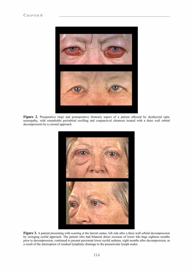
114
Chapter 6
Figure 2. Preoperative (top) and postoperative (bottom) aspect of a patient affected by dysthyroid optic neuropathy, with remarkable periorbital swelling and conjunctival chemosis treated with a three wall orbital decompression by a coronal approach.
Figure 3. A patient presenting with scarring at the lateral cantus, left side after a three wall orbital decompression by swinging eyelid approach. The patient who had bilateral direct excision of lower lids bags eighteen months prior to decompression, continued to present persistent lower eyelid aedema, eight months after decompression, as a result of the interruption of residual lymphatic drainage to the preauricular lymph nodes.
Figure 4. A patient with Graves‟ orbitopathy, who visited our clinic after being treated elsewhere with a complete lateral wall removal orbital decompression by a direct periorbital incision. The iatrogenic deformity caused to the lateral orbital rim is evident.
Figure 5. Preoperative (top) and postoperative (bottom) aspect of a patient affected by dysthyroid optic neuropathy, treated with an extensive 3-wall orbital decompression by a coronal approach, which included the total removal of the lateral orbital wall. No deformities of the lateral cantus or of the lateral orbital rim are evident in spite of the fact that the bony lateral frame was removed.
100379 Thesis Baldeschi.indd 114 vrijdag17-december-2010 12:08
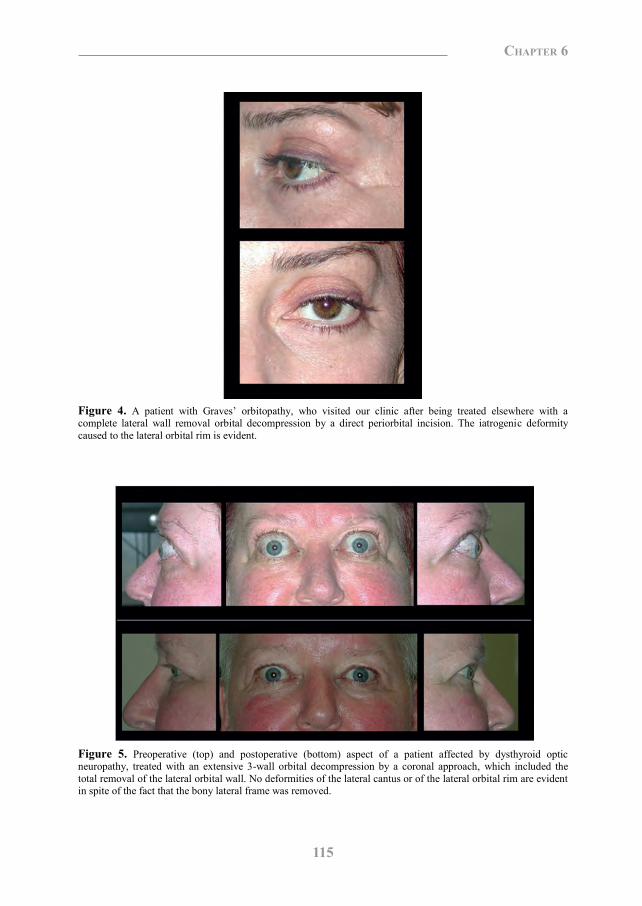
115
Chapter 6
Figure 2. Preoperative (top) and postoperative (bottom) aspect of a patient affected by dysthyroid optic neuropathy, with remarkable periorbital swelling and conjunctival chemosis treated with a three wall orbital decompression by a coronal approach.
Figure 3. A patient presenting with scarring at the lateral cantus, left side after a three wall orbital decompression by swinging eyelid approach. The patient who had bilateral direct excision of lower lids bags eighteen months prior to decompression, continued to present persistent lower eyelid aedema, eight months after decompression, as a result of the interruption of residual lymphatic drainage to the preauricular lymph nodes.
Figure 4. A patient with Graves‟ orbitopathy, who visited our clinic after being treated elsewhere with a complete lateral wall removal orbital decompression by a direct periorbital incision. The iatrogenic deformity caused to the lateral orbital rim is evident.
Figure 5. Preoperative (top) and postoperative (bottom) aspect of a patient affected by dysthyroid optic neuropathy, treated with an extensive 3-wall orbital decompression by a coronal approach, which included the total removal of the lateral orbital wall. No deformities of the lateral cantus or of the lateral orbital rim are evident in spite of the fact that the bony lateral frame was removed.
100379 Thesis Baldeschi.indd 115 vrijdag17-december-2010 12:08
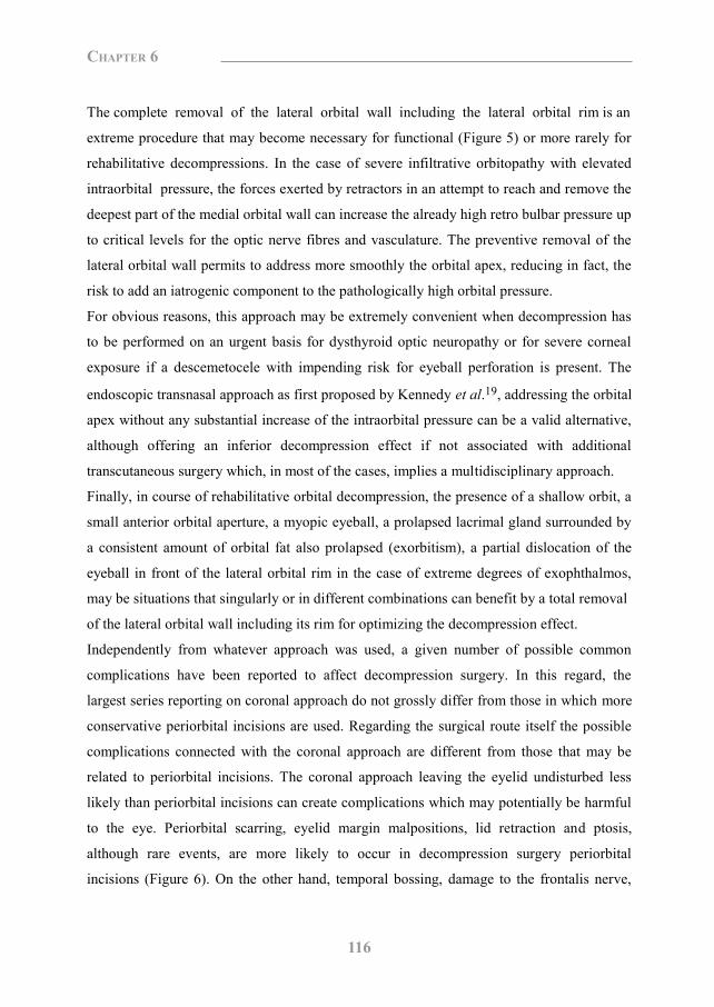
116
Chapter 6
The complete removal of the lateral orbital wall including the lateral orbital rim is an
extreme procedure that may become necessary for functional (Figure 5) or more rarely for
rehabilitative decompressions. In the case of severe infiltrative orbitopathy with elevated
intraorbital pressure, the forces exerted by retractors in an attempt to reach and remove the
deepest part of the medial orbital wall can increase the already high retro bulbar pressure up
to critical levels for the optic nerve fibres and vasculature. The preventive removal of the
lateral orbital wall permits to address more smoothly the orbital apex, reducing in fact, the
risk to add an iatrogenic component to the pathologically high orbital pressure.
For obvious reasons, this approach may be extremely convenient when decompression has
to be performed on an urgent basis for dysthyroid optic neuropathy or for severe corneal
exposure if a descemetocele with impending risk for eyeball perforation is present. The
endoscopic transnasal approach as first proposed by Kennedy et al.19, addressing the orbital
apex without any substantial increase of the intraorbital pressure can be a valid alternative,
although offering an inferior decompression effect if not associated with additional
transcutaneous surgery which, in most of the cases, implies a multidisciplinary approach.
Finally, in course of rehabilitative orbital decompression, the presence of a shallow orbit, a
small anterior orbital aperture, a myopic eyeball, a prolapsed lacrimal gland surrounded by
a consistent amount of orbital fat also prolapsed (exorbitism), a partial dislocation of the
eyeball in front of the lateral orbital rim in the case of extreme degrees of exophthalmos,
may be situations that singularly or in different combinations can benefit by a total removal
of the lateral orbital wall including its rim for optimizing the decompression effect.
Independently from whatever approach was used, a given number of possible common
complications have been reported to affect decompression surgery. In this regard, the
largest series reporting on coronal approach do not grossly differ from those in which more
conservative periorbital incisions are used. Regarding the surgical route itself the possible
complications connected with the coronal approach are different from those that may be
related to periorbital incisions. The coronal approach leaving the eyelid undisturbed less
likely than periorbital incisions can create complications which may potentially be harmful
to the eye. Periorbital scarring, eyelid margin malpositions, lid retraction and ptosis,
although rare events, are more likely to occur in decompression surgery periorbital
incisions (Figure 6). On the other hand, temporal bossing, damage to the frontalis nerve,
scarring and alopecia at the site of scalp incision, or affecting ischemic areas of the frontal
flap after healing by secondary intention, may complicate the coronal approach (Figure 7).
Major complications connected with the coronal approach have been mainly described in
small series where three10 or less13 patients per year were operated. In this respect it is
therefore unavoidable to emphasize that each technique has its own learning curve and it
may be difficult to differentiate the effects of each technique from the experience of the
surgeon.10
Besides the patient‟s determination to accept major surgeries, and surgeon‟s skills to
perform them, the possibility of aiming at attaining only partial results should always be
weighed in light of patients‟ characteristics such as age, general health conditions,
profession, education and psychosocial environment. Often conservative surgery is of
maximal benefit to the patient in spite of modest final results that may be unattractive to the
surgeon.20
100379 Thesis Baldeschi.indd 116 vrijdag17-december-2010 12:08
117
Chapter 6
The complete removal of the lateral orbital wall including the lateral orbital rim is an
extreme procedure that may become necessary for functional (Figure 5) or more rarely for
rehabilitative decompressions. In the case of severe infiltrative orbitopathy with elevated
intraorbital pressure, the forces exerted by retractors in an attempt to reach and remove the
deepest part of the medial orbital wall can increase the already high retro bulbar pressure up
to critical levels for the optic nerve fibres and vasculature. The preventive removal of the
lateral orbital wall permits to address more smoothly the orbital apex, reducing in fact, the
risk to add an iatrogenic component to the pathologically high orbital pressure.
For obvious reasons, this approach may be extremely convenient when decompression has
to be performed on an urgent basis for dysthyroid optic neuropathy or for severe corneal
exposure if a descemetocele with impending risk for eyeball perforation is present. The
endoscopic transnasal approach as first proposed by Kennedy et al.19, addressing the orbital
apex without any substantial increase of the intraorbital pressure can be a valid alternative,
although offering an inferior decompression effect if not associated with additional
transcutaneous surgery which, in most of the cases, implies a multidisciplinary approach.
Finally, in course of rehabilitative orbital decompression, the presence of a shallow orbit, a
small anterior orbital aperture, a myopic eyeball, a prolapsed lacrimal gland surrounded by
a consistent amount of orbital fat also prolapsed (exorbitism), a partial dislocation of the
eyeball in front of the lateral orbital rim in the case of extreme degrees of exophthalmos,
may be situations that singularly or in different combinations can benefit by a total removal
of the lateral orbital wall including its rim for optimizing the decompression effect.
Independently from whatever approach was used, a given number of possible common
complications have been reported to affect decompression surgery. In this regard, the
largest series reporting on coronal approach do not grossly differ from those in which more
conservative periorbital incisions are used. Regarding the surgical route itself the possible
complications connected with the coronal approach are different from those that may be
related to periorbital incisions. The coronal approach leaving the eyelid undisturbed less
likely than periorbital incisions can create complications which may potentially be harmful
to the eye. Periorbital scarring, eyelid margin malpositions, lid retraction and ptosis,
although rare events, are more likely to occur in decompression surgery periorbital
incisions (Figure 6). On the other hand, temporal bossing, damage to the frontalis nerve,
scarring and alopecia at the site of scalp incision, or affecting ischemic areas of the frontal
flap after healing by secondary intention, may complicate the coronal approach (Figure 7).
Major complications connected with the coronal approach have been mainly described in
small series where three10 or less13 patients per year were operated. In this respect it is
therefore unavoidable to emphasize that each technique has its own learning curve and it
may be difficult to differentiate the effects of each technique from the experience of the
surgeon.10
Besides the patient‟s determination to accept major surgeries, and surgeon‟s skills to
perform them, the possibility of aiming at attaining only partial results should always be
weighed in light of patients‟ characteristics such as age, general health conditions,
profession, education and psychosocial environment. Often conservative surgery is of
maximal benefit to the patient in spite of modest final results that may be unattractive to the
surgeon.20
100379 Thesis Baldeschi.indd 117 vrijdag17-december-2010 12:08
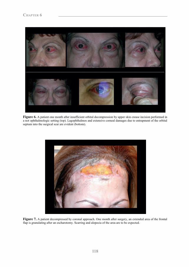
118
Chapter 6
Figure 6. A patient one month after insufficient orbital decompression by upper skin crease incision performed in a not ophthalmologic setting (top). Lagophthalmos and extensive corneal damages due to entrapment of the orbital septum into the surgical scar are evident (bottom).
Figure 7. A patient decompressed by coronal approach. One month after surgery, an extended area of the frontal flap is granulating after an escharotomy. Scarring and alopecia of the area are to be expected.
References 1. Baldeschi L. Orbital decompression. In: Wiersinga WM, Kahaly GJ, editors. Graves‟ Orbitopathy A
Multidisciplinary Approach. Basel: Karger, 2007:163-75.
2. Baldeschi L. Orbital decompressions: status of the art and unresolved issues. In: Guthoff RF, Katowitz JA,
editors. Essential in Ophthalmology, Oculoplastics and Orbit. Berlin: Springer, 2007:143-51..
3. Tessier P. Expansion chirurgicale de l‟orbit. Ann Chir Plast 1969;14:207-14.
4. Krastinova D, Rodallec A. Basedowian orbitopathy. Ann Chir Plast Esthet 1985;30:351-8.
5. Härting F, Koornneef, L, Peeters HJF, Gillisen JPA. Fourteen years of orbital decompression in Graves‟
disease.A review of technique, results and complications. Orbit 1986;5:123-9.
6. Mourits MP, Koornneef L, Wiersinga WM, Prummel MF, Berghout A, van der Gaag R. Orbital decompression
for Graves‟ ophthalmopathy by inferomedial, by inferomedial plus lateral, and by coronal approach.
Ophthalmology 1990;97:636-41.
7. Kalmann R, Mourits MP, van der Pol JP, Koornneef L. Coronal approach for rehabilitative orbital
decompression in Graves‟ ophthalmopathy. Br J Ophthalmol 1997;81:41-5.
8. Baldeschi L, MacAndie K, Hintschich C, Wakelkamp IM, Prummel MF, Wiersinga WM. The removal of the
deep lateral wall in orbital decompression: its contribution to exophthalmos reduction and influence on
consecutive diplopia. Am J Ophthalmol 2005;140:642-7.
9. Mourits, MP, Bijl HM, Baldeschi L, Boboridis K, Curro N, Dickinson J, Eckstein AK, Guastella C, Altea M,
Kahaly G, Freidel M, Kalmann R, Krassas GE, Lane C, Lareida J, Marcocci C, Marino M, Nardi M, Mohr Ch,
Neoh C, Pinchera A, Orgiazzi J, Pitz S, Saeed P, Salvi M, Sellari-Franceschini S, Stahl M, von Arx G, Wiersinga
W. Outcome of orbital decompression for disfiguring g proptosis in patients with Graves‟ orbitopathy using
various surgical procedures. Br J Ophthalmol 2009;93:1518-23.
10. Cruz AA, Leme VR. Orbital decompression: a comparison between trans-fornix/transcaruncular inferomedial
and coronal inferomedial plus lateral approaches. Ophthal Plast Reconstr Surg 2003;19:440-45.
11. Paridaens D, Hans K, van Buitenen S, Mourits MPh. The incidence of diplopia following coronal and trans lid
orbital decompression in Graves‟ orbitopathy. Eye 1998;12:800-05.
12. Sasim IV, de Graaf ME, Berendschot TT, Kalmann R, van Isterdael C, Mourits MP. Coronal or swinging
eyelid decompression for patients with disfiguring proptosis in Graves‟ orbitopathy? Comparison of results in one
center. Ophthalmology 2005;112:1310-15.
13. Leatherbarrow B, Lendrum J, Mahaffey PJ, Noble JL, Kwartz J, Davies H. Three wall orbital decompression
for Graves‟ ophthalmopathy via a coronal approach. Eye 1991;5:456-65.
14. Terwee CB, Dekker FW, Mourits MPh, Gerdings MN, Baldeschi L, Kalmann R, Prummel MF, Wiersinga
WM. Interpretation and validity of changes in scores on the Graves‟ ophthalmopathy quality of life questionnaire
(GO-QOL) after different treatments. Clin Endocrinol (Oxf) 2001;54:391-8.
15. Ben Simon GJ, Schwarcz RM, Mansury AM, Wang L, McCann JD, Goldberg RA. Minimally invasive orbital
decompression: local anaesthesia and hand-carved bone. Arch Ophthalmol 2005;123:1671-5.
16. McCord CD Jr. Orbital decompression for Graves‟ disease:exposure through lateral canthal and inferior fornix
incision. Ophthalmology 1981;88:533-41.
17. Saks ND, Burnstine MA, Putterman AM. Glabellar rhytids in thyroid-associated orbitopathy. Ophthal Plast
Reconstr Surg 2001;17:91-5.
100379 Thesis Baldeschi.indd 118 vrijdag17-december-2010 12:08

119
Chapter 6
Figure 6. A patient one month after insufficient orbital decompression by upper skin crease incision performed in a not ophthalmologic setting (top). Lagophthalmos and extensive corneal damages due to entrapment of the orbital septum into the surgical scar are evident (bottom).
Figure 7. A patient decompressed by coronal approach. One month after surgery, an extended area of the frontal flap is granulating after an escharotomy. Scarring and alopecia of the area are to be expected.
References 1. Baldeschi L. Orbital decompression. In: Wiersinga WM, Kahaly GJ, editors. Graves‟ Orbitopathy A
Multidisciplinary Approach. Basel: Karger, 2007:163-75.
2. Baldeschi L. Orbital decompressions: status of the art and unresolved issues. In: Guthoff RF, Katowitz JA,
editors. Essential in Ophthalmology, Oculoplastics and Orbit. Berlin: Springer, 2007:143-51..
3. Tessier P. Expansion chirurgicale de l‟orbit. Ann Chir Plast 1969;14:207-14.
4. Krastinova D, Rodallec A. Basedowian orbitopathy. Ann Chir Plast Esthet 1985;30:351-8.
5. Härting F, Koornneef, L, Peeters HJF, Gillisen JPA. Fourteen years of orbital decompression in Graves‟
disease.A review of technique, results and complications. Orbit 1986;5:123-9.
6. Mourits MP, Koornneef L, Wiersinga WM, Prummel MF, Berghout A, van der Gaag R. Orbital decompression
for Graves‟ ophthalmopathy by inferomedial, by inferomedial plus lateral, and by coronal approach.
Ophthalmology 1990;97:636-41.
7. Kalmann R, Mourits MP, van der Pol JP, Koornneef L. Coronal approach for rehabilitative orbital
decompression in Graves‟ ophthalmopathy. Br J Ophthalmol 1997;81:41-5.
8. Baldeschi L, MacAndie K, Hintschich C, Wakelkamp IM, Prummel MF, Wiersinga WM. The removal of the
deep lateral wall in orbital decompression: its contribution to exophthalmos reduction and influence on
consecutive diplopia. Am J Ophthalmol 2005;140:642-7.
9. Mourits, MP, Bijl HM, Baldeschi L, Boboridis K, Curro N, Dickinson J, Eckstein AK, Guastella C, Altea M,
Kahaly G, Freidel M, Kalmann R, Krassas GE, Lane C, Lareida J, Marcocci C, Marino M, Nardi M, Mohr Ch,
Neoh C, Pinchera A, Orgiazzi J, Pitz S, Saeed P, Salvi M, Sellari-Franceschini S, Stahl M, von Arx G, Wiersinga
W. Outcome of orbital decompression for disfiguring g proptosis in patients with Graves‟ orbitopathy using
various surgical procedures. Br J Ophthalmol 2009;93:1518-23.
10. Cruz AA, Leme VR. Orbital decompression: a comparison between trans-fornix/transcaruncular inferomedial
and coronal inferomedial plus lateral approaches. Ophthal Plast Reconstr Surg 2003;19:440-45.
11. Paridaens D, Hans K, van Buitenen S, Mourits MPh. The incidence of diplopia following coronal and trans lid
orbital decompression in Graves‟ orbitopathy. Eye 1998;12:800-05.
12. Sasim IV, de Graaf ME, Berendschot TT, Kalmann R, van Isterdael C, Mourits MP. Coronal or swinging
eyelid decompression for patients with disfiguring proptosis in Graves‟ orbitopathy? Comparison of results in one
center. Ophthalmology 2005;112:1310-15.
13. Leatherbarrow B, Lendrum J, Mahaffey PJ, Noble JL, Kwartz J, Davies H. Three wall orbital decompression
for Graves‟ ophthalmopathy via a coronal approach. Eye 1991;5:456-65.
14. Terwee CB, Dekker FW, Mourits MPh, Gerdings MN, Baldeschi L, Kalmann R, Prummel MF, Wiersinga
WM. Interpretation and validity of changes in scores on the Graves‟ ophthalmopathy quality of life questionnaire
(GO-QOL) after different treatments. Clin Endocrinol (Oxf) 2001;54:391-8.
15. Ben Simon GJ, Schwarcz RM, Mansury AM, Wang L, McCann JD, Goldberg RA. Minimally invasive orbital
decompression: local anaesthesia and hand-carved bone. Arch Ophthalmol 2005;123:1671-5.
16. McCord CD Jr. Orbital decompression for Graves‟ disease:exposure through lateral canthal and inferior fornix
incision. Ophthalmology 1981;88:533-41.
17. Saks ND, Burnstine MA, Putterman AM. Glabellar rhytids in thyroid-associated orbitopathy. Ophthal Plast
Reconstr Surg 2001;17:91-5.
100379 Thesis Baldeschi.indd 119 vrijdag17-december-2010 12:08

120
Chapter 6
18. van der Wal KG, de Visscher JG, Boukes RJ, Smeding B. Surgical treatment of Graves orbitopathy: a
modified balanced technique. Int J Oral Maxillofac Surg 2001;30:254-8.
19. Kennedy DW, Goldstein ML, Miller NR, Zinreich SJ. Endoscopic transnasal orbital decompression. Arch
Otolaryngol Head Neck Surg 1990;116:275-82.
20. Baldeschi L. Rehabilitative surgery. In: Wiersinga WM, Kahaly GJ, editors. Graves‟ Orbitopathy A
Multidisciplinary Approach. Basel: Karger, 2007:160-2.
Chapter 7
Reactivation of Graves’ Orbitopathy after Rehabilitative Orbital Decompression
Lelio Baldeschi1, Antonella Lupetti2, Phung Vu1, Iris M. M. J. Wakelkamp4,
Robert Lindeboom3, Marc F. Prummel4, Wilmar M. Wiersinga4
1 Orbital Centre, Department of Ophthalmology, University of Amsterdam, Amsterdam, The Netherlands. 2 Department of Experimental Pathology, Medical Biotechnologies, Infectivology and Epidemiology, University of Pisa, Pisa, Italy. 3 Department of Clinical Epidemiology and Biostatistics, University of Amsterdam, Amsterdam, The Netherlands. 4 Department of Endocrinology, University of Amsterdam, Amsterdam, The Netherlands. Ophthalmology 2007;114:1395-402
100379 Thesis Baldeschi.indd 120 vrijdag17-december-2010 12:08
18. van der Wal KG, de Visscher JG, Boukes RJ, Smeding B. Surgical treatment of Graves orbitopathy: a
modified balanced technique. Int J Oral Maxillofac Surg 2001;30:254-8.
19. Kennedy DW, Goldstein ML, Miller NR, Zinreich SJ. Endoscopic transnasal orbital decompression. Arch
Otolaryngol Head Neck Surg 1990;116:275-82.
20. Baldeschi L. Rehabilitative surgery. In: Wiersinga WM, Kahaly GJ, editors. Graves‟ Orbitopathy A
Multidisciplinary Approach. Basel: Karger, 2007:160-2.
Chapter 7
Reactivation of Graves’ Orbitopathy after Rehabilitative Orbital Decompression
Lelio Baldeschi1, Antonella Lupetti2, Phung Vu1, Iris M. M. J. Wakelkamp4,
Robert Lindeboom3, Marc F. Prummel4, Wilmar M. Wiersinga4
1 Orbital Centre, Department of Ophthalmology, University of Amsterdam, Amsterdam, The Netherlands. 2 Department of Experimental Pathology, Medical Biotechnologies, Infectivology and Epidemiology, University of Pisa, Pisa, Italy. 3 Department of Clinical Epidemiology and Biostatistics, University of Amsterdam, Amsterdam, The Netherlands. 4 Department of Endocrinology, University of Amsterdam, Amsterdam, The Netherlands. Ophthalmology 2007;114:1395-402
100379 Thesis Baldeschi.indd 121 vrijdag17-december-2010 12:08

122
Chapter 7
Abstract
Objective: To present and discuss three cases of apparent reactivation of Graves‟
orbitopathy (GO) after orbital decompression and to evaluate the incidence of this
phenomenon.
Design: Observational case series and retrospective follow-up study.
Participants: A few weeks after surgery 2 patients with GO (patients 1 and 2), treated at
our institution with rehabilitative bony orbital decompression during the static phase of the
disease showed clinical and radiologic evidence of reactivated orbitopathy. After this
observation, a sample of 249 patients who had consecutively undergone the same treatment
for the same reason before the second of the 2 observed patients was selected for this study.
Methods: The records of the selected patients were retrospectively reviewed searching for
cases presenting with clinical and radiologic evidence of GO reactivated as a consequence
of any type of bony orbital decompression. Patients treated with perioperative systemic
glucocorticoids or who had concurrent periorbital diseases, injuries, or surgeries, or who
had immunocompromised conditions or a follow-up of ≤ 2 months, were excluded.
Main Outcome Measures: Incidence of reactivation. Clinical history, clinical and
radiologic characteristics, treatment modalities, and time course of the reactivation in
patients presenting with this phenomenon.
Results: Decompression surgery took place between 1994 and 2000. Eleven patients were
excluded for having been treated with perioperative glucocorticoids. Only 1 patient (patient
3) presented with reactivation. The incidence of the phenomenon that we regard as
reactivation of GO after rehabilitative bony orbital decompression was therefore 1.3%
(3/239). In all 3 patients, the reactivation took place a few weeks after surgery, after an
early normal convalescence period and could be controlled with systemic
immunosuppression or orbital radiotherapy. None of the patients we report developed
further episodes of reactivation during the follow-up period (mean, 7.5 years).
Conclusions: Based on its clinical characteristics, we suggest naming our observation
delayed decompression related reactivation and we propose using its acronym DDRR when
referring to it. Although DDRR appears to be a rare event, it is important for physicians and
patients to be aware of its possible occurrence with rehabilitative decompression surgery.
Introduction
In Graves‟ orbitopathy (GO), signs and symptoms progressively increase in severity during
an early dynamic, active, inflammatory phase; later, they become milder and relatively
stable during a consecutive inactive, postinflammatory static phase.1 Prompt restoration of
stable euthyroidism and immunosuppression, when necessary, may decrease the duration of
the dynamic phase and reverse the tendency to progress toward more severe
symptomatology.2-4 During the dynamic active phase, patients with functional
complications such as optic neuropathy or exposure keratopathy can benefit from surgical
expansion of the bony orbit when medical therapy fails.5
Bony orbital decompression is the mainstay therapy for the treatment of stable alterations
such as exophthalmos, periorbital congestion, and retro-ocular tension that typify the
inactive postinflammatory phase of the disease. Worsening of the disease after bony
decompression surgery performed for functional reasons during the inflammatory phase has
been described6, but it is debatable whether the phenomenon should be considered the
natural progression of the active phase despite surgical intervention or a direct consequence
of surgery.
In this paper, we present and discuss clinical history, clinical and radiologic characteristics,
treatment modalities, and time course of a few cases of apparent reactivation of GO after
orbital decompression performed for aesthetic rehabilitation during the inactive phase of the
disease. Because we are unaware of previous reports of this phenomenon and could not find
references to it in a computerized search using MEDLINE, we chose to evaluate its
incidence by means of a retrospective survey.
Patients and methods
Our survey is an observational case series and retrospective follow-up study. A few weeks
after surgery 2 patients (patients 1 and 2) treated with bony orbital decompression surgery
at our institution for aesthetic rehabilitation during the inactive, postinflammatory phase of
GO started showing clinical signs and symptoms and computed tomography (CT) evidence
of reactivated orbitopathy. A sample of 249 records of patients with GO who had
consecutively undergone the same treatment for the same reason before the second of the 2
observed patients were retrospectively reviewed to assess the incidence and clinical
characteristics of the phenomenon, which appeared subsequent to orbital decompression.
100379 Thesis Baldeschi.indd 122 vrijdag17-december-2010 12:08
123
Chapter 7
Abstract
Objective: To present and discuss three cases of apparent reactivation of Graves‟
orbitopathy (GO) after orbital decompression and to evaluate the incidence of this
phenomenon.
Design: Observational case series and retrospective follow-up study.
Participants: A few weeks after surgery 2 patients with GO (patients 1 and 2), treated at
our institution with rehabilitative bony orbital decompression during the static phase of the
disease showed clinical and radiologic evidence of reactivated orbitopathy. After this
observation, a sample of 249 patients who had consecutively undergone the same treatment
for the same reason before the second of the 2 observed patients was selected for this study.
Methods: The records of the selected patients were retrospectively reviewed searching for
cases presenting with clinical and radiologic evidence of GO reactivated as a consequence
of any type of bony orbital decompression. Patients treated with perioperative systemic
glucocorticoids or who had concurrent periorbital diseases, injuries, or surgeries, or who
had immunocompromised conditions or a follow-up of ≤ 2 months, were excluded.
Main Outcome Measures: Incidence of reactivation. Clinical history, clinical and
radiologic characteristics, treatment modalities, and time course of the reactivation in
patients presenting with this phenomenon.
Results: Decompression surgery took place between 1994 and 2000. Eleven patients were
excluded for having been treated with perioperative glucocorticoids. Only 1 patient (patient
3) presented with reactivation. The incidence of the phenomenon that we regard as
reactivation of GO after rehabilitative bony orbital decompression was therefore 1.3%
(3/239). In all 3 patients, the reactivation took place a few weeks after surgery, after an
early normal convalescence period and could be controlled with systemic
immunosuppression or orbital radiotherapy. None of the patients we report developed
further episodes of reactivation during the follow-up period (mean, 7.5 years).
Conclusions: Based on its clinical characteristics, we suggest naming our observation
delayed decompression related reactivation and we propose using its acronym DDRR when
referring to it. Although DDRR appears to be a rare event, it is important for physicians and
patients to be aware of its possible occurrence with rehabilitative decompression surgery.
Introduction
In Graves‟ orbitopathy (GO), signs and symptoms progressively increase in severity during
an early dynamic, active, inflammatory phase; later, they become milder and relatively
stable during a consecutive inactive, postinflammatory static phase.1 Prompt restoration of
stable euthyroidism and immunosuppression, when necessary, may decrease the duration of
the dynamic phase and reverse the tendency to progress toward more severe
symptomatology.2-4 During the dynamic active phase, patients with functional
complications such as optic neuropathy or exposure keratopathy can benefit from surgical
expansion of the bony orbit when medical therapy fails.5
Bony orbital decompression is the mainstay therapy for the treatment of stable alterations
such as exophthalmos, periorbital congestion, and retro-ocular tension that typify the
inactive postinflammatory phase of the disease. Worsening of the disease after bony
decompression surgery performed for functional reasons during the inflammatory phase has
been described6, but it is debatable whether the phenomenon should be considered the
natural progression of the active phase despite surgical intervention or a direct consequence
of surgery.
In this paper, we present and discuss clinical history, clinical and radiologic characteristics,
treatment modalities, and time course of a few cases of apparent reactivation of GO after
orbital decompression performed for aesthetic rehabilitation during the inactive phase of the
disease. Because we are unaware of previous reports of this phenomenon and could not find
references to it in a computerized search using MEDLINE, we chose to evaluate its
incidence by means of a retrospective survey.
Patients and methods
Our survey is an observational case series and retrospective follow-up study. A few weeks
after surgery 2 patients (patients 1 and 2) treated with bony orbital decompression surgery
at our institution for aesthetic rehabilitation during the inactive, postinflammatory phase of
GO started showing clinical signs and symptoms and computed tomography (CT) evidence
of reactivated orbitopathy. A sample of 249 records of patients with GO who had
consecutively undergone the same treatment for the same reason before the second of the 2
observed patients were retrospectively reviewed to assess the incidence and clinical
characteristics of the phenomenon, which appeared subsequent to orbital decompression.
100379 Thesis Baldeschi.indd 123 vrijdag17-december-2010 12:08
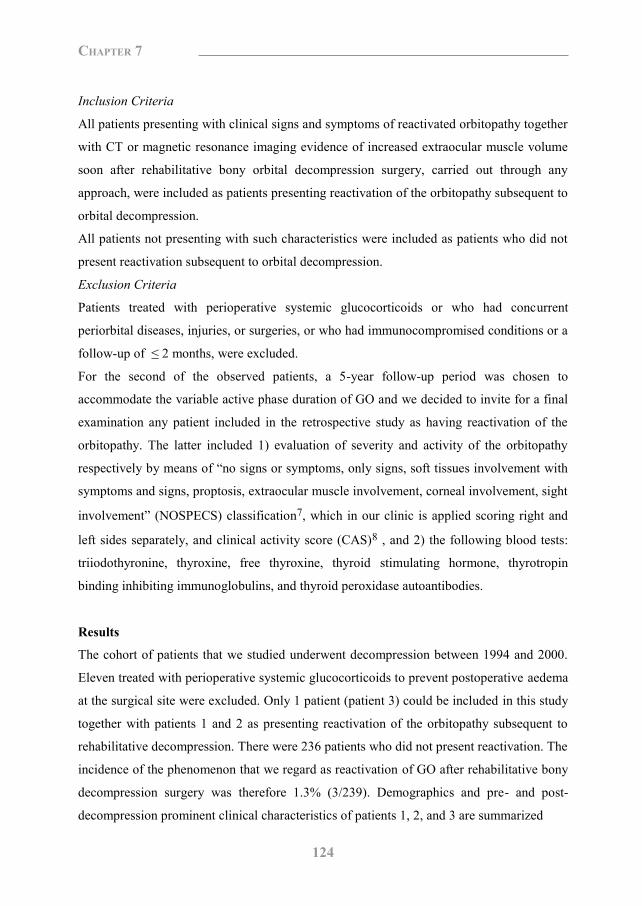
124
Chapter 7
Inclusion Criteria
All patients presenting with clinical signs and symptoms of reactivated orbitopathy together
with CT or magnetic resonance imaging evidence of increased extraocular muscle volume
soon after rehabilitative bony orbital decompression surgery, carried out through any
approach, were included as patients presenting reactivation of the orbitopathy subsequent to
orbital decompression.
All patients not presenting with such characteristics were included as patients who did not
present reactivation subsequent to orbital decompression.
Exclusion Criteria
Patients treated with perioperative systemic glucocorticoids or who had concurrent
periorbital diseases, injuries, or surgeries, or who had immunocompromised conditions or a
follow-up of ≤ 2 months, were excluded.
For the second of the observed patients, a 5-year follow-up period was chosen to
accommodate the variable active phase duration of GO and we decided to invite for a final
examination any patient included in the retrospective study as having reactivation of the
orbitopathy. The latter included 1) evaluation of severity and activity of the orbitopathy
respectively by means of “no signs or symptoms, only signs, soft tissues involvement with
symptoms and signs, proptosis, extraocular muscle involvement, corneal involvement, sight
involvement” (NOSPECS) classification7, which in our clinic is applied scoring right and
left sides separately, and clinical activity score (CAS)8 , and 2) the following blood tests:
triiodothyronine, thyroxine, free thyroxine, thyroid stimulating hormone, thyrotropin
binding inhibiting immunoglobulins, and thyroid peroxidase autoantibodies.
Results
The cohort of patients that we studied underwent decompression between 1994 and 2000.
Eleven treated with perioperative systemic glucocorticoids to prevent postoperative aedema
at the surgical site were excluded. Only 1 patient (patient 3) could be included in this study
together with patients 1 and 2 as presenting reactivation of the orbitopathy subsequent to
rehabilitative decompression. There were 236 patients who did not present reactivation. The
incidence of the phenomenon that we regard as reactivation of GO after rehabilitative bony
decompression surgery was therefore 1.3% (3/239). Demographics and pre- and post-
decompression prominent clinical characteristics of patients 1, 2, and 3 are summarized
Table 1. Demographics, Pre- and Post-decompression Clinical Characteristics of Patients Presenting with Reactivation of Graves‟ Orbitopathy (GO) Subsequent to Rehabilitative Orbital Decompression
surgery
Anti-TPO = thyroid peroxidase autoantibodies (normal values, 0-60 kU/l); CAS = clinical activity score; TBII = thyrotropin binding inhibiting immunoglobulins (normal values, 0.0 - 1.5 E/l); TSI = thyroid-stimulating immunoglobulin (normal values, 0-10 E/l).
in table 1. The clinical history as well as characteristics and time course of the phenomenon
presented by the 3 patients are reported below.
Patient 1 Patient 2 Patient 3
Gender Female Female Female Age at decompression followed by reactivation (yrs) 48 71 51 Duration of GO at decompression (yrs) 3 29 1.3 Duration of GO inactivity at decompression (yrs) 0.5 6 0.6 CAS at decompression 0+/7 0+/7 0+/7 Smoker at decompression Yes No Yes Type of metabolic abnormality of the thyroid at decompression
compensated autoimmune hypothyroidism
compensated hypothyroidism consecutive to thyroidectomy
compensated autoimmune hyperthyroidism
Duration of thyroid disease at decompression (yrs)
16
32
3
Duration of thyroid metabolic stability at decompression (yrs)
16 29 2.5
Thyroid autoimmunity at decompression
Anti T.P.O. = 80 kU/l T.B.I.I. = 0.0
Anti T.P.O. < 30 kU/l T.S.I. = 58 E/l
Unknown
Thyroid autoimmunity at the last evaluation
Anti T.P.O. = 90 kU/l T.B.I.I. = 31.1 E/l
Anti T.P.O. < 30 kU/l T.B.I.I. = 2.5 E/l
Anti T.P.O. = 1410 kU/l T.B.I.I. < 1.0
Signs and symptoms at reactivation following decompression: Active peri-orbital aedema Inadequate decrease of exophthalmos Increased eyelid aperture Worsening of restrictive myopathy Onset of diplopia within 20° around central position of gaze Profuse tearing
Yes Yes Yes Yes Yes Yes
Yes Yes No Yes No Yes
Yes No No Yes Yes Yes
CAS at reactivation following decompression:
7+/10 7+/10 5+/10
Duration of reactivation following decompression (mts) 20 12 11 Treatment of reactivation following decompression
oral glucocorticoids IV and oral glucocorticoids associated with orbital irradiation
orbital irradiation
Follow-up after reactivation (yrs) 6.3 5.7 10.6
100379 Thesis Baldeschi.indd 124 vrijdag17-december-2010 12:09
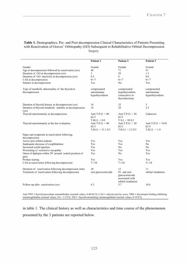
125
Chapter 7
Inclusion Criteria
All patients presenting with clinical signs and symptoms of reactivated orbitopathy together
with CT or magnetic resonance imaging evidence of increased extraocular muscle volume
soon after rehabilitative bony orbital decompression surgery, carried out through any
approach, were included as patients presenting reactivation of the orbitopathy subsequent to
orbital decompression.
All patients not presenting with such characteristics were included as patients who did not
present reactivation subsequent to orbital decompression.
Exclusion Criteria
Patients treated with perioperative systemic glucocorticoids or who had concurrent
periorbital diseases, injuries, or surgeries, or who had immunocompromised conditions or a
follow-up of ≤ 2 months, were excluded.
For the second of the observed patients, a 5-year follow-up period was chosen to
accommodate the variable active phase duration of GO and we decided to invite for a final
examination any patient included in the retrospective study as having reactivation of the
orbitopathy. The latter included 1) evaluation of severity and activity of the orbitopathy
respectively by means of “no signs or symptoms, only signs, soft tissues involvement with
symptoms and signs, proptosis, extraocular muscle involvement, corneal involvement, sight
involvement” (NOSPECS) classification7, which in our clinic is applied scoring right and
left sides separately, and clinical activity score (CAS)8 , and 2) the following blood tests:
triiodothyronine, thyroxine, free thyroxine, thyroid stimulating hormone, thyrotropin
binding inhibiting immunoglobulins, and thyroid peroxidase autoantibodies.
Results
The cohort of patients that we studied underwent decompression between 1994 and 2000.
Eleven treated with perioperative systemic glucocorticoids to prevent postoperative aedema
at the surgical site were excluded. Only 1 patient (patient 3) could be included in this study
together with patients 1 and 2 as presenting reactivation of the orbitopathy subsequent to
rehabilitative decompression. There were 236 patients who did not present reactivation. The
incidence of the phenomenon that we regard as reactivation of GO after rehabilitative bony
decompression surgery was therefore 1.3% (3/239). Demographics and pre- and post-
decompression prominent clinical characteristics of patients 1, 2, and 3 are summarized
Table 1. Demographics, Pre- and Post-decompression Clinical Characteristics of Patients Presenting with Reactivation of Graves‟ Orbitopathy (GO) Subsequent to Rehabilitative Orbital Decompression
surgery
Anti-TPO = thyroid peroxidase autoantibodies (normal values, 0-60 kU/l); CAS = clinical activity score; TBII = thyrotropin binding inhibiting immunoglobulins (normal values, 0.0 - 1.5 E/l); TSI = thyroid-stimulating immunoglobulin (normal values, 0-10 E/l).
in table 1. The clinical history as well as characteristics and time course of the phenomenon
presented by the 3 patients are reported below.
Patient 1 Patient 2 Patient 3
Gender Female Female Female Age at decompression followed by reactivation (yrs) 48 71 51 Duration of GO at decompression (yrs) 3 29 1.3 Duration of GO inactivity at decompression (yrs) 0.5 6 0.6 CAS at decompression 0+/7 0+/7 0+/7 Smoker at decompression Yes No Yes Type of metabolic abnormality of the thyroid at decompression
compensated autoimmune hypothyroidism
compensated hypothyroidism consecutive to thyroidectomy
compensated autoimmune hyperthyroidism
Duration of thyroid disease at decompression (yrs)
16
32
3
Duration of thyroid metabolic stability at decompression (yrs)
16 29 2.5
Thyroid autoimmunity at decompression
Anti T.P.O. = 80 kU/l T.B.I.I. = 0.0
Anti T.P.O. < 30 kU/l T.S.I. = 58 E/l
Unknown
Thyroid autoimmunity at the last evaluation
Anti T.P.O. = 90 kU/l T.B.I.I. = 31.1 E/l
Anti T.P.O. < 30 kU/l T.B.I.I. = 2.5 E/l
Anti T.P.O. = 1410 kU/l T.B.I.I. < 1.0
Signs and symptoms at reactivation following decompression: Active peri-orbital aedema Inadequate decrease of exophthalmos Increased eyelid aperture Worsening of restrictive myopathy Onset of diplopia within 20° around central position of gaze Profuse tearing
Yes Yes Yes Yes Yes Yes
Yes Yes No Yes No Yes
Yes No No Yes Yes Yes
CAS at reactivation following decompression:
7+/10 7+/10 5+/10
Duration of reactivation following decompression (mts) 20 12 11 Treatment of reactivation following decompression
oral glucocorticoids IV and oral glucocorticoids associated with orbital irradiation
orbital irradiation
Follow-up after reactivation (yrs) 6.3 5.7 10.6
Surgery
100379 Thesis Baldeschi.indd 125 vrijdag17-december-2010 12:09
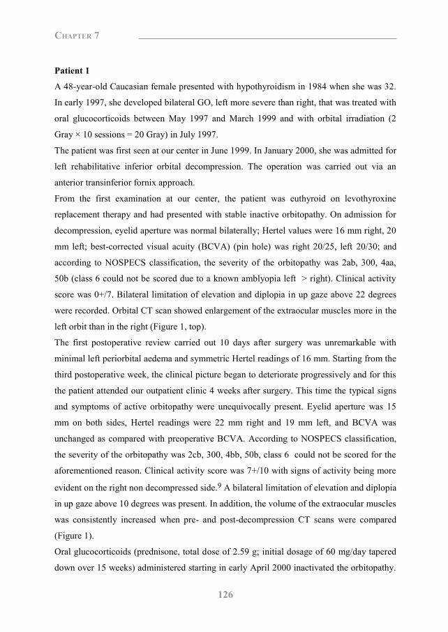
126
Chapter 7
Patient 1
A 48-year-old Caucasian female presented with hypothyroidism in 1984 when she was 32.
In early 1997, she developed bilateral GO, left more severe than right, that was treated with
oral glucocorticoids between May 1997 and March 1999 and with orbital irradiation (2
Gray × 10 sessions = 20 Gray) in July 1997.
The patient was first seen at our center in June 1999. In January 2000, she was admitted for
left rehabilitative inferior orbital decompression. The operation was carried out via an
anterior transinferior fornix approach.
From the first examination at our center, the patient was euthyroid on levothyroxine
replacement therapy and had presented with stable inactive orbitopathy. On admission for
decompression, eyelid aperture was normal bilaterally; Hertel values were 16 mm right, 20
mm left; best-corrected visual acuity (BCVA) (pin hole) was right 20/25, left 20/30; and
according to NOSPECS classification, the severity of the orbitopathy was 2ab, 300, 4aa,
50b (class 6 could not be scored due to a known amblyopia left > right). Clinical activity
score was 0+/7. Bilateral limitation of elevation and diplopia in up gaze above 22 degrees
were recorded. Orbital CT scan showed enlargement of the extraocular muscles more in the
left orbit than in the right (Figure 1, top).
The first postoperative review carried out 10 days after surgery was unremarkable with
minimal left periorbital aedema and symmetric Hertel readings of 16 mm. Starting from the
third postoperative week, the clinical picture began to deteriorate progressively and for this
the patient attended our outpatient clinic 4 weeks after surgery. This time the typical signs
and symptoms of active orbitopathy were unequivocally present. Eyelid aperture was 15
mm on both sides, Hertel readings were 22 mm right and 19 mm left, and BCVA was
unchanged as compared with preoperative BCVA. According to NOSPECS classification,
the severity of the orbitopathy was 2cb, 300, 4bb, 50b, class 6 could not be scored for the
aforementioned reason. Clinical activity score was 7+/10 with signs of activity being more
evident on the right non decompressed side.9 A bilateral limitation of elevation and diplopia
in up gaze above 10 degrees was present. In addition, the volume of the extraocular muscles
was consistently increased when pre- and post-decompression CT scans were compared
(Figure 1).
Oral glucocorticoids (prednisone, total dose of 2.59 g; initial dosage of 60 mg/day tapered
down over 15 weeks) administered starting in early April 2000 inactivated the orbitopathy.
Exophthalmos decreased on the left to 17 mm; the right side remained 22 mm. In March
2002, the patient underwent a right rehabilitative inferomedial orbital decompression
through an inferior fornix approach. This time the postoperative course was uneventful and
right exophthalmos decreased to 17 mm. Bilateral eyebrow ptosis correction and upper lid
blepharoplasty followed in February and July 2003, respectively.
The orbitopathy remained stable without signs of reactivation up to the last examination
performed in May 2006, 6.3 years after the phenomenon that we regard as reactivation
subsequent to rehabilitative orbital decompression. The patient has been euthyroid with
levothyroxine replacement therapy, and thyroid autoimmunity has been abnormal for the
entire review period at our institution (Table 1).
Patient 2 A 71-year-old Caucasian female was diagnosed with hyperthyroidism in 1969 at the age of
43. In September 1972, a few weeks after subtotal thyroidectomy, she developed GO with
optic neuropathy. For this she was first treated with oral glucocorticoids and then with
lateral orbital decompression bilaterally.
She first presented to our center in December 1981 (at age 52) with a disfiguring clinically
inactive orbitopathy. The severity of the orbitopathy scored as 2cc, 3bb, 4aa, 500, and 600
with NOSPECS classification. Eyelid aperture was 14 mm right, 16 mm left; Hertel
readings were 26 mm right, 28 mm left; and BCVA was 20/20 both sides.
In July 1994, clinical signs of active orbitopathy became evident. Hertel values rose to 30
mm right and 31 mm left, and BCVA dropped to 20/30 bilaterally. For this she was treated
with a bilateral inferomedial orbital decompression through a translid approach in August
1994.
In 1995, several functional and aesthetic eyelid surgical procedures were carried out.
Thereafter, for some years, the orbitopathy remained stable. Her BCVA was 20/30 and
Hertel values 28 mm bilaterally; CAS was 0+/7. The patient was euthyroid and disturbed
by a retro-ocular pressure feeling and symptoms related to exposure keratopathy.
In an attempt to improve her symptoms in September 2000, it was decided to proceed with
a third orbital decompression, as despite 2 previous decompression procedures, most of the
medial and lateral bony orbit was still intact (Figure 2, top). The procedure was carried out
bilaterally by means of a combined transcaruncular and upper skin-crease approach.
100379 Thesis Baldeschi.indd 126 vrijdag17-december-2010 12:09
127
Chapter 7
Patient 1
A 48-year-old Caucasian female presented with hypothyroidism in 1984 when she was 32.
In early 1997, she developed bilateral GO, left more severe than right, that was treated with
oral glucocorticoids between May 1997 and March 1999 and with orbital irradiation (2
Gray × 10 sessions = 20 Gray) in July 1997.
The patient was first seen at our center in June 1999. In January 2000, she was admitted for
left rehabilitative inferior orbital decompression. The operation was carried out via an
anterior transinferior fornix approach.
From the first examination at our center, the patient was euthyroid on levothyroxine
replacement therapy and had presented with stable inactive orbitopathy. On admission for
decompression, eyelid aperture was normal bilaterally; Hertel values were 16 mm right, 20
mm left; best-corrected visual acuity (BCVA) (pin hole) was right 20/25, left 20/30; and
according to NOSPECS classification, the severity of the orbitopathy was 2ab, 300, 4aa,
50b (class 6 could not be scored due to a known amblyopia left > right). Clinical activity
score was 0+/7. Bilateral limitation of elevation and diplopia in up gaze above 22 degrees
were recorded. Orbital CT scan showed enlargement of the extraocular muscles more in the
left orbit than in the right (Figure 1, top).
The first postoperative review carried out 10 days after surgery was unremarkable with
minimal left periorbital aedema and symmetric Hertel readings of 16 mm. Starting from the
third postoperative week, the clinical picture began to deteriorate progressively and for this
the patient attended our outpatient clinic 4 weeks after surgery. This time the typical signs
and symptoms of active orbitopathy were unequivocally present. Eyelid aperture was 15
mm on both sides, Hertel readings were 22 mm right and 19 mm left, and BCVA was
unchanged as compared with preoperative BCVA. According to NOSPECS classification,
the severity of the orbitopathy was 2cb, 300, 4bb, 50b, class 6 could not be scored for the
aforementioned reason. Clinical activity score was 7+/10 with signs of activity being more
evident on the right non decompressed side.9 A bilateral limitation of elevation and diplopia
in up gaze above 10 degrees was present. In addition, the volume of the extraocular muscles
was consistently increased when pre- and post-decompression CT scans were compared
(Figure 1).
Oral glucocorticoids (prednisone, total dose of 2.59 g; initial dosage of 60 mg/day tapered
down over 15 weeks) administered starting in early April 2000 inactivated the orbitopathy.
Exophthalmos decreased on the left to 17 mm; the right side remained 22 mm. In March
2002, the patient underwent a right rehabilitative inferomedial orbital decompression
through an inferior fornix approach. This time the postoperative course was uneventful and
right exophthalmos decreased to 17 mm. Bilateral eyebrow ptosis correction and upper lid
blepharoplasty followed in February and July 2003, respectively.
The orbitopathy remained stable without signs of reactivation up to the last examination
performed in May 2006, 6.3 years after the phenomenon that we regard as reactivation
subsequent to rehabilitative orbital decompression. The patient has been euthyroid with
levothyroxine replacement therapy, and thyroid autoimmunity has been abnormal for the
entire review period at our institution (Table 1).
Patient 2 A 71-year-old Caucasian female was diagnosed with hyperthyroidism in 1969 at the age of
43. In September 1972, a few weeks after subtotal thyroidectomy, she developed GO with
optic neuropathy. For this she was first treated with oral glucocorticoids and then with
lateral orbital decompression bilaterally.
She first presented to our center in December 1981 (at age 52) with a disfiguring clinically
inactive orbitopathy. The severity of the orbitopathy scored as 2cc, 3bb, 4aa, 500, and 600
with NOSPECS classification. Eyelid aperture was 14 mm right, 16 mm left; Hertel
readings were 26 mm right, 28 mm left; and BCVA was 20/20 both sides.
In July 1994, clinical signs of active orbitopathy became evident. Hertel values rose to 30
mm right and 31 mm left, and BCVA dropped to 20/30 bilaterally. For this she was treated
with a bilateral inferomedial orbital decompression through a translid approach in August
1994.
In 1995, several functional and aesthetic eyelid surgical procedures were carried out.
Thereafter, for some years, the orbitopathy remained stable. Her BCVA was 20/30 and
Hertel values 28 mm bilaterally; CAS was 0+/7. The patient was euthyroid and disturbed
by a retro-ocular pressure feeling and symptoms related to exposure keratopathy.
In an attempt to improve her symptoms in September 2000, it was decided to proceed with
a third orbital decompression, as despite 2 previous decompression procedures, most of the
medial and lateral bony orbit was still intact (Figure 2, top). The procedure was carried out
bilaterally by means of a combined transcaruncular and upper skin-crease approach.
100379 Thesis Baldeschi.indd 127 vrijdag17-december-2010 12:09

128
Chapter 7
After surgery, periorbital aedema did not subside as expected, and starting from the third
postoperative week typical clinical signs of active orbitopathy were clearly evident (Figure
3). NOSPECS classification and CAS worsened to 2cc, 3cc, 4cc, 5bb, 6aa, and 7+/10
respectively. In addition, postoperative orbital CT scans showed an increased enlargement
of extraocular muscles as compared with preoperative scans (Figure 2). Her BCVA did not
worsen and Hertel values remained unchanged despite the extensive osteotomies.
In April 2001, the patient underwent simultaneous treatment with IV and oral
glucocorticoids (IV methylprednisolone, total dose 6 g, 1 g/day at days 1, 2, 3, 8, 9, and 10
followed by oral prednisone, total dose, 2.73 g, initial dosage of 40 mg/day, starting at day
15 and tapered down over 17 weeks) combined with orbital irradiation (2 Gray × 10
sessions).
By the end of 2001, her orbitopathy was inactive, Hertel values decreased to 22 mm right
and 23 mm left, eyelid swelling was greatly reduced, and BCVA was 20/20 bilaterally.
Ophthalmic signs and symptoms remained stable, and evidence of active orbitopathy has
been absent through the last examination performed in May 2006, 5.7 years after the
phenomenon that we regard as decompression-related reactivation. Since the time of the
last decompression, the patient has remained biochemically euthyroid with levothyroxine
replacement therapy and presented abnormal thyroid autoimmunity (Table 1).
Patient 3
A Caucasian female who was 51 years old at the time of our retrospective observation
presented with hyperthyroidism and diffuse goiter in 1993 at the age of 48. She was
initially treated with antithyroid drugs and later with block replacement therapy.
In September 1994, the patient developed GO; she was treated with low-dose oral
glucocorticoids until May 1995, when she was first seen at our institution. At that time, she
presented with a moderately severe, inactive orbitopathy. Her NOSPECS classification was
2bb, 300, 400, 5aa, 600, and her CAS 0+/7. She presented an increased eyelid aperture (14
mm right, 17 mm left), Hertel readings were 21 mm right, 22 mm left, and BCVA was right
20/20, left 20/25. Ocular motility and the field of binocular vision were normal. The patient
also presented a mild form of psoriasis. The clinical picture was unchanged when in
December she was admitted for bilateral inferomedial orbital decompression. A coronal
approach was used and on postoperative day 10, exophthalmos was reduced to 19 mm
bilaterally; no double vision was present.
Starting from postoperative week 3, the patient experienced increased retro-ocular pressure
feeling, double vision in primary position of gaze, foreign body sensation, and profuse
tearing; psoriasis worsened simultaneously. When she was examined 4 weeks after surgery
NOSPECS classification was 2cc, 300, 4bb, 500, 600 and CAS 5+/10. Eye movements
were reduced in every direction of gaze, Hertel readings were not worsened, and BCVA
was unchanged as compared with preoperative acuity. The volume of extraocular muscles
was increased on postoperative CT when compared with preoperative (Figure 4).
Orbital irradiation (2 Gray × 10 sessions) administered in May 1996 produced inactivation
of the orbitopathy. In March 1997, the patient underwent successful horizontal squint
surgery, and 1 month later she was treated with bilateral upper lid lengthening.
At the end of 1997, Hertel values of 14 mm right, 15 mm left, persistent bilateral upper lid
lateral flare, and the presence of puffy eyelids were recorded. Despite the eyelid alterations,
the patient refused any further rehabilitative surgical procedure.
Signs and symptoms of the orbitopathy remained stable, and evidence of reactivation has
been absent through the last examination performed in June 2006, 10.6 years after the
phenomenon that we regard as decompression-related reactivation.
From the first examination at our institution to April 1999, when she was treated with I131,
the patient had been euthyroid under antithyroid drugs and levothyroxine replacement
therapy. After April 1999 and to the last examination, the patient remained metabolically
stable with levothyroxine replacement therapy. The patient‟s thyroid autoimmunity had
never been investigated until the most recent follow-up, when it was found to be grossly
abnormal (Table 1).
100379 Thesis Baldeschi.indd 128 vrijdag17-december-2010 12:09
129
Chapter 7
After surgery, periorbital aedema did not subside as expected, and starting from the third
postoperative week typical clinical signs of active orbitopathy were clearly evident (Figure
3). NOSPECS classification and CAS worsened to 2cc, 3cc, 4cc, 5bb, 6aa, and 7+/10
respectively. In addition, postoperative orbital CT scans showed an increased enlargement
of extraocular muscles as compared with preoperative scans (Figure 2). Her BCVA did not
worsen and Hertel values remained unchanged despite the extensive osteotomies.
In April 2001, the patient underwent simultaneous treatment with IV and oral
glucocorticoids (IV methylprednisolone, total dose 6 g, 1 g/day at days 1, 2, 3, 8, 9, and 10
followed by oral prednisone, total dose, 2.73 g, initial dosage of 40 mg/day, starting at day
15 and tapered down over 17 weeks) combined with orbital irradiation (2 Gray × 10
sessions).
By the end of 2001, her orbitopathy was inactive, Hertel values decreased to 22 mm right
and 23 mm left, eyelid swelling was greatly reduced, and BCVA was 20/20 bilaterally.
Ophthalmic signs and symptoms remained stable, and evidence of active orbitopathy has
been absent through the last examination performed in May 2006, 5.7 years after the
phenomenon that we regard as decompression-related reactivation. Since the time of the
last decompression, the patient has remained biochemically euthyroid with levothyroxine
replacement therapy and presented abnormal thyroid autoimmunity (Table 1).
Patient 3
A Caucasian female who was 51 years old at the time of our retrospective observation
presented with hyperthyroidism and diffuse goiter in 1993 at the age of 48. She was
initially treated with antithyroid drugs and later with block replacement therapy.
In September 1994, the patient developed GO; she was treated with low-dose oral
glucocorticoids until May 1995, when she was first seen at our institution. At that time, she
presented with a moderately severe, inactive orbitopathy. Her NOSPECS classification was
2bb, 300, 400, 5aa, 600, and her CAS 0+/7. She presented an increased eyelid aperture (14
mm right, 17 mm left), Hertel readings were 21 mm right, 22 mm left, and BCVA was right
20/20, left 20/25. Ocular motility and the field of binocular vision were normal. The patient
also presented a mild form of psoriasis. The clinical picture was unchanged when in
December she was admitted for bilateral inferomedial orbital decompression. A coronal
approach was used and on postoperative day 10, exophthalmos was reduced to 19 mm
bilaterally; no double vision was present.
Starting from postoperative week 3, the patient experienced increased retro-ocular pressure
feeling, double vision in primary position of gaze, foreign body sensation, and profuse
tearing; psoriasis worsened simultaneously. When she was examined 4 weeks after surgery
NOSPECS classification was 2cc, 300, 4bb, 500, 600 and CAS 5+/10. Eye movements
were reduced in every direction of gaze, Hertel readings were not worsened, and BCVA
was unchanged as compared with preoperative acuity. The volume of extraocular muscles
was increased on postoperative CT when compared with preoperative (Figure 4).
Orbital irradiation (2 Gray × 10 sessions) administered in May 1996 produced inactivation
of the orbitopathy. In March 1997, the patient underwent successful horizontal squint
surgery, and 1 month later she was treated with bilateral upper lid lengthening.
At the end of 1997, Hertel values of 14 mm right, 15 mm left, persistent bilateral upper lid
lateral flare, and the presence of puffy eyelids were recorded. Despite the eyelid alterations,
the patient refused any further rehabilitative surgical procedure.
Signs and symptoms of the orbitopathy remained stable, and evidence of reactivation has
been absent through the last examination performed in June 2006, 10.6 years after the
phenomenon that we regard as decompression-related reactivation.
From the first examination at our institution to April 1999, when she was treated with I131,
the patient had been euthyroid under antithyroid drugs and levothyroxine replacement
therapy. After April 1999 and to the last examination, the patient remained metabolically
stable with levothyroxine replacement therapy. The patient‟s thyroid autoimmunity had
never been investigated until the most recent follow-up, when it was found to be grossly
abnormal (Table 1).
100379 Thesis Baldeschi.indd 129 vrijdag17-december-2010 12:09
130
Chapter 7
Figure 1. Coronal computer tomography scans of the orbits in patient 1. (Top) Predecompression scan. The left extraocular muscles were enlarged more than the right.( Bottom) Postdecompression scan 1 month after surgery. The right orbital floor was removed, and a consistent enlargement of the extraocular muscles was present bilaterally.
Figure 2. Coronal computer tomography scans of the orbits in patient 2. (Top) Scan before the third orbital decompression. The extraocular muscles were moderately enlarged, and the medial orbital wall and part of the lateral were intact bilaterally. (Bottom) Scan 3 weeks after the third decompression. The medial and the lateral orbital walls were removed, and the extraocular muscles were severely enlarged bilaterally.
Figure 3. Patient 2. (Top) The patient before the third orbital decompression. No signs of active orbitopathy were present. (Bottom) The patient 3 weeks after the third orbital decompression. Signs of active Graves‟ orbitopathy were evident.
Figure 4. Coronal computed tomography scans of the orbits in patient 3. (Top) Predecompression scan. A moderate enlargement of the extraocular muscles was present bilaterally. (Bottom) Postdecompression scan 3 weeks after surgery. Right and left medial orbital wall, and right orbital floor osteotomies were evident; the extraocular muscles were severely enlarged bilaterally.
100379 Thesis Baldeschi.indd 130 vrijdag17-december-2010 12:09
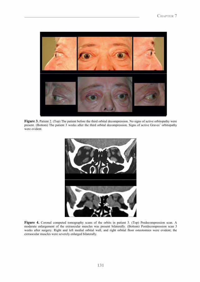
131
Chapter 7
Figure 1. Coronal computer tomography scans of the orbits in patient 1. (Top) Predecompression scan. The left extraocular muscles were enlarged more than the right.( Bottom) Postdecompression scan 1 month after surgery. The right orbital floor was removed, and a consistent enlargement of the extraocular muscles was present bilaterally.
Figure 2. Coronal computer tomography scans of the orbits in patient 2. (Top) Scan before the third orbital decompression. The extraocular muscles were moderately enlarged, and the medial orbital wall and part of the lateral were intact bilaterally. (Bottom) Scan 3 weeks after the third decompression. The medial and the lateral orbital walls were removed, and the extraocular muscles were severely enlarged bilaterally.
Figure 3. Patient 2. (Top) The patient before the third orbital decompression. No signs of active orbitopathy were present. (Bottom) The patient 3 weeks after the third orbital decompression. Signs of active Graves‟ orbitopathy were evident.
Figure 4. Coronal computed tomography scans of the orbits in patient 3. (Top) Predecompression scan. A moderate enlargement of the extraocular muscles was present bilaterally. (Bottom) Postdecompression scan 3 weeks after surgery. Right and left medial orbital wall, and right orbital floor osteotomies were evident; the extraocular muscles were severely enlarged bilaterally.
100379 Thesis Baldeschi.indd 131 vrijdag17-december-2010 12:09
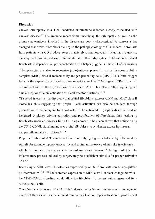
132
Chapter 7
Discussion
Graves‟ orbitopathy is a T-cell-mediated autoimmune disorder, closely associated with
Graves‟ disease.10 The immune mechanisms underlying the orbitopathy as well as the
primary autoantigens involved in the disease are poorly characterized. A consensus has
emerged that orbital fibroblasts are key to the pathophysiology of GO. Indeed, fibroblasts
from patients with GO produce excess matrix glycosaminoglycans, including hyaluronan,
are very proliferative, and can differentiate into fatlike adipocytes. Proliferation of orbital
fibroblasts is dependent on proper activation of T helper (TH) cells. These CD4+-expressing
T lymphocytes are able to recognize (auto)antigens present in major histocompatibility
complex (MHC) class II molecules by antigen presenting cells (APC). This initial trigger
leads to the expression of T-cell surface receptors, such as CD40 ligand (CD40L), which
can interact with CD40 expressed on the surface of APC. This CD40-CD40L signaling is a
crucial step for efficient activation of T-cell effector functions.11,12
Of special interest is the discovery that orbital fibroblasts express CD40 and MHC class II
molecules, thus suggesting that proper T-cell activation can also be achieved through
presentation of autoantigens by fibroblasts.13 The activated T lymphocytes then produce
increased cytokines driving activation and proliferation of fibroblasts, thus leading to
fibroblast-associated diseases like GO. In agreement, it has been shown that activation by
the CD40-CD40L signaling induces orbital fibroblasts to synthesize excess hyaluronan
and proinflammatory cytokines.13-15
Proper activation of APC can be achieved not only by TH cells but also by inflammatory
stimuli, for example, lipopolysaccharide and proinflammatory cytokines like interferon-γ,
which is produced during an infection/inflammatory process.16 In light of this, the
inflammatory process induced by surgery may be a sufficient stimulus for proper activation
of APC.
Interestingly, MHC class II molecules expressed by orbital fibroblasts can be upregulated
by interferon- γ.13,17,18 The increased expression of MHC class II molecules together with
the CD40-CD40L signaling would allow the fibroblasts to present autoantigens and fully
activate the T cells.
Therefore, the exposure of soft orbital tissues to pathogen components / endogenous
microbial flora as well as the surgical trauma may lead to proper activation of professional
APC, and perhaps orbital fibroblasts. These events singularly, or in combination, might
have induced the processing and presentation of normally sequestered self-antigens with
possible reactivation of GO. In this respect, genetic factors may have also played a role.19
Clearly, more research is required to decipher the mechanisms underlying the phenomenon
that we described as related to surgical decompression. Investigation of patients presenting
this phenomenon may also be useful in uncovering valuable information regarding the
etiology of GO itself.
We could directly or retrospectively observe reactivation of the disease after orbital bony
decompression in a restricted number of patients. We described 3 of a sample of 239
Graves‟ patients decompressed for rehabilitative reasons and not treated with perioperative
systemic glucocorticoids. At the time of surgery, 2 of these patients had alteration of
thyroid autoimmunity; information regarding the third patient was not available. In all of
them, thyroid autoimmunity was altered at the time of the last evaluation.
The incidence of the complication that we described appears to be on the order of 1.3%. It
is a rare event that in our series of patients could be controlled with systemic
immunosuppression or orbital radiotherapy. None of the patients we reported developed
further episodes of reactivation during the period of our observation. Our estimated
incidence, however, could have been biased. Our calculation was based on patients referred
to a tertiary center who can be expected to have a somewhat more problematic disease,
resulting in an overestimation. We do not think that this was the case because the entire
case scenario of patients affected by GO is referred to our institution. On the other hand, we
excluded11 patients treated with perioperative glucocorticoids, which may have resulted in
an underestimation; we cannot exclude that in some of them glucocorticoids could have
prevented the trigger of the reactivation or jeopardized its clinical manifestations. This is
unlikely, however, because all received glucocorticoids to prevent postoperative aedema
and not to treat a disease suspected for reactivation. Besides these considerations, we found
the exclusion of patients treated with perioperative glucocorticoids correct from a
methodologic viewpoint. Our purpose was in fact to evaluate the incidence of reactivation
in a cohort of patients as similar as possible to cases 1 and 2, who represented our
prototypes of reactivation and who did not receive corticosteroids perioperatively.
The phenomenon that we described consists in the onset of the typical signs and symptoms
of active GO with radiologic evidence of extraocular muscles enlargement after
100379 Thesis Baldeschi.indd 132 vrijdag17-december-2010 12:09
133
Chapter 7
Discussion
Graves‟ orbitopathy is a T-cell-mediated autoimmune disorder, closely associated with
Graves‟ disease.10 The immune mechanisms underlying the orbitopathy as well as the
primary autoantigens involved in the disease are poorly characterized. A consensus has
emerged that orbital fibroblasts are key to the pathophysiology of GO. Indeed, fibroblasts
from patients with GO produce excess matrix glycosaminoglycans, including hyaluronan,
are very proliferative, and can differentiate into fatlike adipocytes. Proliferation of orbital
fibroblasts is dependent on proper activation of T helper (TH) cells. These CD4+-expressing
T lymphocytes are able to recognize (auto)antigens present in major histocompatibility
complex (MHC) class II molecules by antigen presenting cells (APC). This initial trigger
leads to the expression of T-cell surface receptors, such as CD40 ligand (CD40L), which
can interact with CD40 expressed on the surface of APC. This CD40-CD40L signaling is a
crucial step for efficient activation of T-cell effector functions.11,12
Of special interest is the discovery that orbital fibroblasts express CD40 and MHC class II
molecules, thus suggesting that proper T-cell activation can also be achieved through
presentation of autoantigens by fibroblasts.13 The activated T lymphocytes then produce
increased cytokines driving activation and proliferation of fibroblasts, thus leading to
fibroblast-associated diseases like GO. In agreement, it has been shown that activation by
the CD40-CD40L signaling induces orbital fibroblasts to synthesize excess hyaluronan
and proinflammatory cytokines.13-15
Proper activation of APC can be achieved not only by TH cells but also by inflammatory
stimuli, for example, lipopolysaccharide and proinflammatory cytokines like interferon-γ,
which is produced during an infection/inflammatory process.16 In light of this, the
inflammatory process induced by surgery may be a sufficient stimulus for proper activation
of APC.
Interestingly, MHC class II molecules expressed by orbital fibroblasts can be upregulated
by interferon- γ.13,17,18 The increased expression of MHC class II molecules together with
the CD40-CD40L signaling would allow the fibroblasts to present autoantigens and fully
activate the T cells.
Therefore, the exposure of soft orbital tissues to pathogen components / endogenous
microbial flora as well as the surgical trauma may lead to proper activation of professional
APC, and perhaps orbital fibroblasts. These events singularly, or in combination, might
have induced the processing and presentation of normally sequestered self-antigens with
possible reactivation of GO. In this respect, genetic factors may have also played a role.19
Clearly, more research is required to decipher the mechanisms underlying the phenomenon
that we described as related to surgical decompression. Investigation of patients presenting
this phenomenon may also be useful in uncovering valuable information regarding the
etiology of GO itself.
We could directly or retrospectively observe reactivation of the disease after orbital bony
decompression in a restricted number of patients. We described 3 of a sample of 239
Graves‟ patients decompressed for rehabilitative reasons and not treated with perioperative
systemic glucocorticoids. At the time of surgery, 2 of these patients had alteration of
thyroid autoimmunity; information regarding the third patient was not available. In all of
them, thyroid autoimmunity was altered at the time of the last evaluation.
The incidence of the complication that we described appears to be on the order of 1.3%. It
is a rare event that in our series of patients could be controlled with systemic
immunosuppression or orbital radiotherapy. None of the patients we reported developed
further episodes of reactivation during the period of our observation. Our estimated
incidence, however, could have been biased. Our calculation was based on patients referred
to a tertiary center who can be expected to have a somewhat more problematic disease,
resulting in an overestimation. We do not think that this was the case because the entire
case scenario of patients affected by GO is referred to our institution. On the other hand, we
excluded11 patients treated with perioperative glucocorticoids, which may have resulted in
an underestimation; we cannot exclude that in some of them glucocorticoids could have
prevented the trigger of the reactivation or jeopardized its clinical manifestations. This is
unlikely, however, because all received glucocorticoids to prevent postoperative aedema
and not to treat a disease suspected for reactivation. Besides these considerations, we found
the exclusion of patients treated with perioperative glucocorticoids correct from a
methodologic viewpoint. Our purpose was in fact to evaluate the incidence of reactivation
in a cohort of patients as similar as possible to cases 1 and 2, who represented our
prototypes of reactivation and who did not receive corticosteroids perioperatively.
The phenomenon that we described consists in the onset of the typical signs and symptoms
of active GO with radiologic evidence of extraocular muscles enlargement after
100379 Thesis Baldeschi.indd 133 vrijdag17-december-2010 12:09
134
Chapter 7
rehabilitative orbital decompression and after a normal convalescence period of a few
weeks soon after surgery. Based on its clinical characteristics, we propose naming our
observation delayed decompression-related reactivation, or DDRR.
A similar phenomenon had been previously described by Wai et al.20 after cataract
extraction. Severe reactivation of GO took place 3 weeks after surgery in a patient who had
presented inactive orbitopathy for 24 years. The authors hypothesized that trauma and
pressure in the retrobulbar space induced by retrobulbar anesthesia triggered local
inflammatory and immune responses, which in turn caused progression of GO. That
conclusion was in keeping with the concept of Rapoport et al.21 that trauma and/or pressure
explains, at least in part, the distribution of the extrathyroidal manifestations of Graves‟
disease.
Before this case, Hamed and Lingua22 reported previously unsuspected GO in 8 patients
out of a series of 58 who developed dysthyroid strabismus after cataract extraction. If in
some of these cases it appeared that the diplopia may have been masked before surgery by
the impaired vision, in others there was a clear progression of GO in the following months,
suggesting that the surgical procedure may have contributed to aggravating the disease.
To our knowledge, reactivation of GO after rehabilitative orbital decompression has not
been reported. The findings we described present several similarities to those described as
subsequent to cataract extraction, which is by far a more common ophthalmologic
intervention. In our cases, as in Wai et al.‟s and Hamed and Lingua‟s, GO worsened a few
weeks after surgical trauma, and the phenomenon was invariably bilateral despite unilateral
cataract extraction in all their cases and despite unilateral orbital decompression in one of
ours.
Although DDRR is a rare complication, we feel that it is an event which deserves to be
known by physicians and to be mentioned to those patients with GO undertaking surgical
rehabilitation by means of orbital decompression.
References 1. Rundle FF, Wilson CW. Development and course of exophthalmos and ophthalmoplegia in Graves‟
disease with special reference to the effect of thyroidectomy. Clin Sci 1945;5:177-94.
2. Prummel MF, Wiersinga WM, Mourits MP, Koornneef L, Berghout A, van der Gaag. Effect of
abnormal thyroid function on the severity of Graves‟ ophthalmopathy. Arch Intern Med
1990;150:1098-101.
3. Prummel MF, Mourits MP, Berghout A, Krenning EP, van der Gaag R, Koornneef L, Wiersinga WM.
Prednisone and cyclosporine in the treatment of severe Graves‟ ophthalmopathy. N Engl J Med
1989;321:1353-9.
4. Bartalena L, Pinchera A, Marcocci C. Management of Graves‟ ophthalmopathy: reality and
perspectives. Endocr Rev 2000;21:168-99.
5. Wakelkamp IM, Baldeschi L, Saeed P, Mourits MP, Prummel MF, Wiersinga WM. Surgical or medical
decompression as a first-line treatment of optic neuropathy in Graves‟ ophthalmopathy? A randomized
controlled trial. Clin Endocrinol (Oxf) 2005;63:323-8.
6. Wenz R, Levine MR, Putterman A, Bersani T, Feldman K. Extraocular muscle enlargement after
orbital decompression for Graves‟ ophthalmopathy. Ophthal Plast Reconstr Surg 1994;10:34-41.
7. Werner SC. Modifications of the classification of the eye changes of Graves‟ disease:
recommendations of the Ad Hoc Committee of the American Thyroid Association. J Clin Endocrinol
Metab 1977;44:203- 4.
8. Mourits MP, Prummel MF, Wiersinga WM, Koornneef L. Clinical activity score as a guide in the
management of patients with Graves‟ ophthalmopathy. Clin Endocrinol (Oxf) 1997;47:9-14.
9. Mourits MP, Koornneef L, Wiersinga WM, Prummel MF, Berghout A, van der Gaag R. Clinical
criteria for the assessment of disease activity in Graves‟ ophthalmopathy: a novel approach. Br J
Ophthalmol 1989;73: 639-44.
10. Wall JR, Salvi M, Bernard NF, Boucher A, Haegert D. Thyroid-associated ophthalmopathy: a model
for the association of organ-specific autoimmune disorders. Immunol Today 1991;12:150-3.
11. Grewal IS, Flavell RA. A central role of CD40 ligand in the regulation of CD4+ T-cell responses.
Immunol Today 1996;17:410-4.
12. Noelle RJ. CD40 and its ligand in host defense. Immunity 1996;4:415-9.
13. Sempowski GD, Rozenblit J, Smith TJ, Phipps RP. Human orbital fibroblasts are activated through
CD40 to induce proinflammatory cytokine production. Am J Physiol 1998;274: C707-14.
14. Cao HJ, Wang HS, Zhang Y, Lin HY, Phipps RP, Smith TJ. Activation of human orbital fibroblasts
through CD40 engagement results in a dramatic induction of hyaluronan synthesis and prostaglandin
endoperoxide H synthase-2 expression. Insights into potential pathogenic mechanisms of thyroid-
associated ophthalmopathy. J Biol Chem 1998;273:29615-25.
15. Zhang Y, Cao HJ, Graf B, Meekins H, Smith TJ, Phipps RP. CD40 engagement upregulates
cyclooxygenase-2 expression and prostaglandin E2 production in human lung fibroblasts. J Immunol
1998;160:1053-7.
100379 Thesis Baldeschi.indd 134 vrijdag17-december-2010 12:09
135
Chapter 7
rehabilitative orbital decompression and after a normal convalescence period of a few
weeks soon after surgery. Based on its clinical characteristics, we propose naming our
observation delayed decompression-related reactivation, or DDRR.
A similar phenomenon had been previously described by Wai et al.20 after cataract
extraction. Severe reactivation of GO took place 3 weeks after surgery in a patient who had
presented inactive orbitopathy for 24 years. The authors hypothesized that trauma and
pressure in the retrobulbar space induced by retrobulbar anesthesia triggered local
inflammatory and immune responses, which in turn caused progression of GO. That
conclusion was in keeping with the concept of Rapoport et al.21 that trauma and/or pressure
explains, at least in part, the distribution of the extrathyroidal manifestations of Graves‟
disease.
Before this case, Hamed and Lingua22 reported previously unsuspected GO in 8 patients
out of a series of 58 who developed dysthyroid strabismus after cataract extraction. If in
some of these cases it appeared that the diplopia may have been masked before surgery by
the impaired vision, in others there was a clear progression of GO in the following months,
suggesting that the surgical procedure may have contributed to aggravating the disease.
To our knowledge, reactivation of GO after rehabilitative orbital decompression has not
been reported. The findings we described present several similarities to those described as
subsequent to cataract extraction, which is by far a more common ophthalmologic
intervention. In our cases, as in Wai et al.‟s and Hamed and Lingua‟s, GO worsened a few
weeks after surgical trauma, and the phenomenon was invariably bilateral despite unilateral
cataract extraction in all their cases and despite unilateral orbital decompression in one of
ours.
Although DDRR is a rare complication, we feel that it is an event which deserves to be
known by physicians and to be mentioned to those patients with GO undertaking surgical
rehabilitation by means of orbital decompression.
References 1. Rundle FF, Wilson CW. Development and course of exophthalmos and ophthalmoplegia in Graves‟
disease with special reference to the effect of thyroidectomy. Clin Sci 1945;5:177-94.
2. Prummel MF, Wiersinga WM, Mourits MP, Koornneef L, Berghout A, van der Gaag. Effect of
abnormal thyroid function on the severity of Graves‟ ophthalmopathy. Arch Intern Med
1990;150:1098-101.
3. Prummel MF, Mourits MP, Berghout A, Krenning EP, van der Gaag R, Koornneef L, Wiersinga WM.
Prednisone and cyclosporine in the treatment of severe Graves‟ ophthalmopathy. N Engl J Med
1989;321:1353-9.
4. Bartalena L, Pinchera A, Marcocci C. Management of Graves‟ ophthalmopathy: reality and
perspectives. Endocr Rev 2000;21:168-99.
5. Wakelkamp IM, Baldeschi L, Saeed P, Mourits MP, Prummel MF, Wiersinga WM. Surgical or medical
decompression as a first-line treatment of optic neuropathy in Graves‟ ophthalmopathy? A randomized
controlled trial. Clin Endocrinol (Oxf) 2005;63:323-8.
6. Wenz R, Levine MR, Putterman A, Bersani T, Feldman K. Extraocular muscle enlargement after
orbital decompression for Graves‟ ophthalmopathy. Ophthal Plast Reconstr Surg 1994;10:34-41.
7. Werner SC. Modifications of the classification of the eye changes of Graves‟ disease:
recommendations of the Ad Hoc Committee of the American Thyroid Association. J Clin Endocrinol
Metab 1977;44:203- 4.
8. Mourits MP, Prummel MF, Wiersinga WM, Koornneef L. Clinical activity score as a guide in the
management of patients with Graves‟ ophthalmopathy. Clin Endocrinol (Oxf) 1997;47:9-14.
9. Mourits MP, Koornneef L, Wiersinga WM, Prummel MF, Berghout A, van der Gaag R. Clinical
criteria for the assessment of disease activity in Graves‟ ophthalmopathy: a novel approach. Br J
Ophthalmol 1989;73: 639-44.
10. Wall JR, Salvi M, Bernard NF, Boucher A, Haegert D. Thyroid-associated ophthalmopathy: a model
for the association of organ-specific autoimmune disorders. Immunol Today 1991;12:150-3.
11. Grewal IS, Flavell RA. A central role of CD40 ligand in the regulation of CD4+ T-cell responses.
Immunol Today 1996;17:410-4.
12. Noelle RJ. CD40 and its ligand in host defense. Immunity 1996;4:415-9.
13. Sempowski GD, Rozenblit J, Smith TJ, Phipps RP. Human orbital fibroblasts are activated through
CD40 to induce proinflammatory cytokine production. Am J Physiol 1998;274: C707-14.
14. Cao HJ, Wang HS, Zhang Y, Lin HY, Phipps RP, Smith TJ. Activation of human orbital fibroblasts
through CD40 engagement results in a dramatic induction of hyaluronan synthesis and prostaglandin
endoperoxide H synthase-2 expression. Insights into potential pathogenic mechanisms of thyroid-
associated ophthalmopathy. J Biol Chem 1998;273:29615-25.
15. Zhang Y, Cao HJ, Graf B, Meekins H, Smith TJ, Phipps RP. CD40 engagement upregulates
cyclooxygenase-2 expression and prostaglandin E2 production in human lung fibroblasts. J Immunol
1998;160:1053-7.
100379 Thesis Baldeschi.indd 135 vrijdag17-december-2010 12:09
136
Chapter 7
16. Schuurhuis DH, Laban S, Toes RE, Ricciardi-Castagnoli P, Kleijmeer MJ, van der Voort EI, Rea D,
Offringa R, Geuze HJ, Melief CJ, Ossendorp F. Immature dendritic cells acquire CD8(+) cytotoxic T
lymphocyte priming capacity upon activation by T helper-independent or -dependent stimuli. J Exp
Med 2000;192:145-50.
17. Koumas L, Smith TJ, Phipps RP. Fibroblast subsets in the human orbit: Thy-1+ and Thy-1-
subpopulations exhibit distinct phenotypes. Eur J Immunol 2002;32:477-85.
18. Shimojo N, Kohno Y, Yamaguchi K, Kikuoka S, Hoshioka A, Niimi H, Hirai A, Tamura Y, Saito Y,
Kohn LD, Tahara K. Induction of Graves-like disease in mice by immunization with fibroblasts
transfected with the thyrotropin receptor and a class II molecule. Proc Natl Acad Sci USA.
1996;93:11074-9.
19. Vaidya B, Imrie H, Perros P, Dickinson J, McCarthy MI, Kendall-Taylor P, Pearce SH. Cytotoxic T
lymphocyte antigen-4 (CTLA-4) gene polymorphism confers susceptibility to thyroid associated
orbitopathy. Lancet 1999;354:743-4.
20. Wai DC, Ho SC, Seah LL, Fong KS, Khoo DH. Severe Graves‟ ophthalmopathy after retrobulbar
anesthesia for cataract extraction in a patient with mild stable thyroid eye disease. Thyroid 2003;13:
823-6.
21. Rapoport B, Alsabeh R, Aftergood D, McLachlan SM. Elephantiasic pretibial myxedema: insight into
and a hypothesis regarding the pathogenesis of the extrathyroidal manifestations of Graves‟ disease.
Thyroid 2000;10:685-92.
22. Hamed LM, Lingua RW. Thyroid eye disease presenting after cataract surgery. J Pediatr Ophthalmol
Strabismus 1990;27:10-5.
Chapter 8
Graves’ Orbitopathy in a Patient with Adrenoleukodystrophy
after Bone Marrow Transplantation Yoav Vardizer1,2, Antonella Lupetti3, Sylvie Vandelanotte1,
Arjen C. Lankester4, Wilmar M. Wiersinga5, Lelio Baldeschi1
1 Orbital Centre, Department of Ophthalmology, University of Amsterdam, Amsterdam, The Netherlands. 2 Department of Ophthalmology, Haémek Medical Center, Haifa, Israel 3 Department of Experimental Pathology, Medical Biotechnologies, Infectivology and Epidemiology, University of Pisa, Pisa, Italy. 4 Bone Marrow Transplantation Unit, Department of Pediatrics, Leiden University Medical Center, Leiden,The Netherlands. 5 Department of Endocrinology, University of Amsterdam, Amsterdam, The Netherlands. European Journal of Endocrinology 2009;161:369-73
100379 Thesis Baldeschi.indd 136 vrijdag17-december-2010 12:09
16. Schuurhuis DH, Laban S, Toes RE, Ricciardi-Castagnoli P, Kleijmeer MJ, van der Voort EI, Rea D,
Offringa R, Geuze HJ, Melief CJ, Ossendorp F. Immature dendritic cells acquire CD8(+) cytotoxic T
lymphocyte priming capacity upon activation by T helper-independent or -dependent stimuli. J Exp
Med 2000;192:145-50.
17. Koumas L, Smith TJ, Phipps RP. Fibroblast subsets in the human orbit: Thy-1+ and Thy-1-
subpopulations exhibit distinct phenotypes. Eur J Immunol 2002;32:477-85.
18. Shimojo N, Kohno Y, Yamaguchi K, Kikuoka S, Hoshioka A, Niimi H, Hirai A, Tamura Y, Saito Y,
Kohn LD, Tahara K. Induction of Graves-like disease in mice by immunization with fibroblasts
transfected with the thyrotropin receptor and a class II molecule. Proc Natl Acad Sci USA.
1996;93:11074-9.
19. Vaidya B, Imrie H, Perros P, Dickinson J, McCarthy MI, Kendall-Taylor P, Pearce SH. Cytotoxic T
lymphocyte antigen-4 (CTLA-4) gene polymorphism confers susceptibility to thyroid associated
orbitopathy. Lancet 1999;354:743-4.
20. Wai DC, Ho SC, Seah LL, Fong KS, Khoo DH. Severe Graves‟ ophthalmopathy after retrobulbar
anesthesia for cataract extraction in a patient with mild stable thyroid eye disease. Thyroid 2003;13:
823-6.
21. Rapoport B, Alsabeh R, Aftergood D, McLachlan SM. Elephantiasic pretibial myxedema: insight into
and a hypothesis regarding the pathogenesis of the extrathyroidal manifestations of Graves‟ disease.
Thyroid 2000;10:685-92.
22. Hamed LM, Lingua RW. Thyroid eye disease presenting after cataract surgery. J Pediatr Ophthalmol
Strabismus 1990;27:10-5.
Chapter 8
Graves’ Orbitopathy in a Patient with Adrenoleukodystrophy
after Bone Marrow Transplantation Yoav Vardizer1,2, Antonella Lupetti3, Sylvie Vandelanotte1,
Arjen C. Lankester4, Wilmar M. Wiersinga5, Lelio Baldeschi1
1 Orbital Centre, Department of Ophthalmology, University of Amsterdam, Amsterdam, The Netherlands. 2 Department of Ophthalmology, Haémek Medical Center, Haifa, Israel 3 Department of Experimental Pathology, Medical Biotechnologies, Infectivology and Epidemiology, University of Pisa, Pisa, Italy. 4 Bone Marrow Transplantation Unit, Department of Pediatrics, Leiden University Medical Center, Leiden,The Netherlands. 5 Department of Endocrinology, University of Amsterdam, Amsterdam, The Netherlands. European Journal of Endocrinology 2009;161:369-73
100379 Thesis Baldeschi.indd 137 vrijdag17-december-2010 12:09
138
Chapter 8
Abstract
Objective: For many years, the treatment of X-linked childhood cerebral
adrenoleukodystrophy (XALD) consisted of hydrocortisone replacement and a mixture of
short chain-fatty acids, known as „Lorenzo‟s oil‟. Recently, bone marrow transplantation
(BMT) has also been used.
Case report: We report the case of a patient affected by XALD who developed Graves‟
hyperthyroidism (GH) and Graves‟ orbitopathy (GO) after BMT and who we could follow-
up for 6.5 years afterwards.
Evidence synthesis: A boy affected by XALD was treated at the age of 6 years, with a
whole BMT from his sister. One year after BMT, the transplanted patient presented TSH at
the lower normal value and 3 years later he developed thyrotoxicosis. After a further 2
years, the patient developed GO, which showed clinical evidence of reactivation 5 years
after its onset as a consequence of an attempt to treat thyrotoxicosis by means of I131 (300
MBq). Seven years after BMT, the donor showed alterations of thyroid autoimmunity and 1
year thereafter she developed GH. She never presented GO during a subsequent 5 year
follow-up.
Conclusions: This case illustrates that autoimmunity originating from a pre-symptomatic
donor can be transferred into the host during allogeneic stem cell transplantation. In cases
where autoimmune phenomena are recognized in the donor prior to donation, alternative
donors or T-cell manipulation of the graft might be considered.
Introduction
X-linked childhood cerebral adrenoleukodystrophy (XALD) is a peroxisomal disorder
involving defective β-oxidation of very long-chain fatty acids that accumulate in plasma,
brain, and adrenal cortex.1 Clinical symptoms include adrenal insufficiency and motor-
mental deterioration accompanied by visual and hearing impairment due to intracranial
demyelinization.1, 2
For many years treatment consisted of hydrocortisone replacement and administration of a
mixture of short-chain fatty acids, known as “Lorenzo‟s oil”.1
Recently, bone marrow transplantation (BMT) has been reported to decrease the elevated
blood levels of very long-chain fatty acids, to reduce the intracranial demyelinization, and
in turn to ameliorate the neurologic symptoms of the disease.2-5
BMT can be regarded as an in vivo model of inducing autoimmune reactions, as in this
procedure human leukocyte antigen (HLA) matched bone marrow cells are exposed to a
new set of pre-existing antigens.
Occasionally, BMT has been related to both transmission6-9 and cure10, 11 of autoimmune
diseases, and relapse of Graves‟ disease after BMT has also been described.11 Considering
that only 30% of the patients with Graves‟ hyperthyroidism (GH) may develop Graves‟
orbitopathy (GO)13, it is not surprising that publications that link BMT to GO are scarce.
We are aware of only one publication reporting concordant GH and GO after BMT14, and
could not find any other by means of a computerized search using MEDLINE.
In this article, we present and discuss the case of a patient affected by XALD, who
developed GH 4 years, and GO 6 years after BMT from his sister. Seven years after BMT,
the donor also showed alteration of thyroid autoimmunity and 1 year thereafter she
developed GH. Both patient and donor were followed up for 12 years after BMT.
Case report
A 12-year-old boy affected by XALD was referred to the Orbital Center, Department of
Ophthalmology, University of Amsterdam with a 3 month history of bilateral signs and
symptoms compatible with GO. The boy had been treated with supplementary „Lorenzo‟s
oil‟ and hydrocortisone replacement therapy since when, 6 years prior to our observation,
100379 Thesis Baldeschi.indd 138 vrijdag17-december-2010 12:09
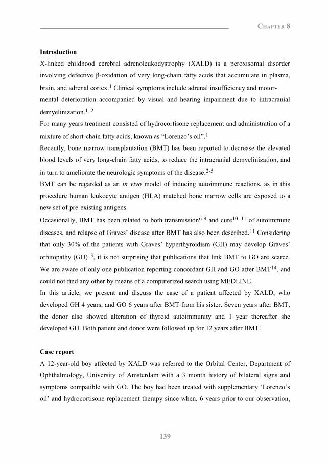
139
Chapter 8
Abstract
Objective: For many years, the treatment of X-linked childhood cerebral
adrenoleukodystrophy (XALD) consisted of hydrocortisone replacement and a mixture of
short chain-fatty acids, known as „Lorenzo‟s oil‟. Recently, bone marrow transplantation
(BMT) has also been used.
Case report: We report the case of a patient affected by XALD who developed Graves‟
hyperthyroidism (GH) and Graves‟ orbitopathy (GO) after BMT and who we could follow-
up for 6.5 years afterwards.
Evidence synthesis: A boy affected by XALD was treated at the age of 6 years, with a
whole BMT from his sister. One year after BMT, the transplanted patient presented TSH at
the lower normal value and 3 years later he developed thyrotoxicosis. After a further 2
years, the patient developed GO, which showed clinical evidence of reactivation 5 years
after its onset as a consequence of an attempt to treat thyrotoxicosis by means of I131 (300
MBq). Seven years after BMT, the donor showed alterations of thyroid autoimmunity and 1
year thereafter she developed GH. She never presented GO during a subsequent 5 year
follow-up.
Conclusions: This case illustrates that autoimmunity originating from a pre-symptomatic
donor can be transferred into the host during allogeneic stem cell transplantation. In cases
where autoimmune phenomena are recognized in the donor prior to donation, alternative
donors or T-cell manipulation of the graft might be considered.
Introduction
X-linked childhood cerebral adrenoleukodystrophy (XALD) is a peroxisomal disorder
involving defective β-oxidation of very long-chain fatty acids that accumulate in plasma,
brain, and adrenal cortex.1 Clinical symptoms include adrenal insufficiency and motor-
mental deterioration accompanied by visual and hearing impairment due to intracranial
demyelinization.1, 2
For many years treatment consisted of hydrocortisone replacement and administration of a
mixture of short-chain fatty acids, known as “Lorenzo‟s oil”.1
Recently, bone marrow transplantation (BMT) has been reported to decrease the elevated
blood levels of very long-chain fatty acids, to reduce the intracranial demyelinization, and
in turn to ameliorate the neurologic symptoms of the disease.2-5
BMT can be regarded as an in vivo model of inducing autoimmune reactions, as in this
procedure human leukocyte antigen (HLA) matched bone marrow cells are exposed to a
new set of pre-existing antigens.
Occasionally, BMT has been related to both transmission6-9 and cure10, 11 of autoimmune
diseases, and relapse of Graves‟ disease after BMT has also been described.11 Considering
that only 30% of the patients with Graves‟ hyperthyroidism (GH) may develop Graves‟
orbitopathy (GO)13, it is not surprising that publications that link BMT to GO are scarce.
We are aware of only one publication reporting concordant GH and GO after BMT14, and
could not find any other by means of a computerized search using MEDLINE.
In this article, we present and discuss the case of a patient affected by XALD, who
developed GH 4 years, and GO 6 years after BMT from his sister. Seven years after BMT,
the donor also showed alteration of thyroid autoimmunity and 1 year thereafter she
developed GH. Both patient and donor were followed up for 12 years after BMT.
Case report
A 12-year-old boy affected by XALD was referred to the Orbital Center, Department of
Ophthalmology, University of Amsterdam with a 3 month history of bilateral signs and
symptoms compatible with GO. The boy had been treated with supplementary „Lorenzo‟s
oil‟ and hydrocortisone replacement therapy since when, 6 years prior to our observation,
100379 Thesis Baldeschi.indd 139 vrijdag17-december-2010 12:09
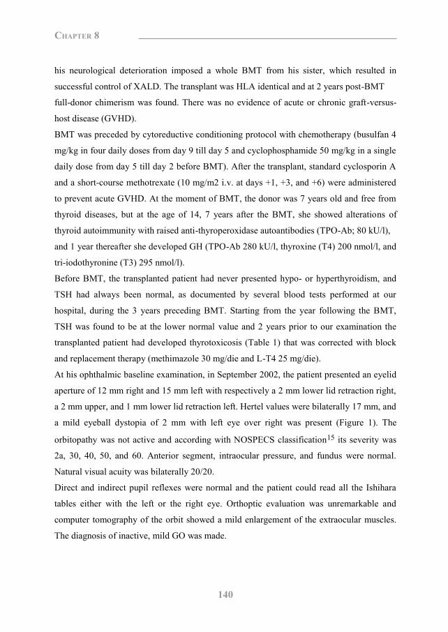
140
Chapter 8
his neurological deterioration imposed a whole BMT from his sister, which resulted in
successful control of XALD. The transplant was HLA identical and at 2 years post-BMT
full-donor chimerism was found. There was no evidence of acute or chronic graft-versus-
host disease (GVHD).
BMT was preceded by cytoreductive conditioning protocol with chemotherapy (busulfan 4
mg/kg in four daily doses from day 9 till day 5 and cyclophosphamide 50 mg/kg in a single
daily dose from day 5 till day 2 before BMT). After the transplant, standard cyclosporin A
and a short-course methotrexate (10 mg/m2 i.v. at days +1, +3, and +6) were administered
to prevent acute GVHD. At the moment of BMT, the donor was 7 years old and free from
thyroid diseases, but at the age of 14, 7 years after the BMT, she showed alterations of
thyroid autoimmunity with raised anti-thyroperoxidase autoantibodies (TPO-Ab; 80 kU/l),
and 1 year thereafter she developed GH (TPO-Ab 280 kU/l, thyroxine (T4) 200 nmol/l, and
tri-iodothyronine (T3) 295 nmol/l).
Before BMT, the transplanted patient had never presented hypo- or hyperthyroidism, and
TSH had always been normal, as documented by several blood tests performed at our
hospital, during the 3 years preceding BMT. Starting from the year following the BMT,
TSH was found to be at the lower normal value and 2 years prior to our examination the
transplanted patient had developed thyrotoxicosis (Table 1) that was corrected with block
and replacement therapy (methimazole 30 mg/die and L-T4 25 mg/die).
At his ophthalmic baseline examination, in September 2002, the patient presented an eyelid
aperture of 12 mm right and 15 mm left with respectively a 2 mm lower lid retraction right,
a 2 mm upper, and 1 mm lower lid retraction left. Hertel values were bilaterally 17 mm, and
a mild eyeball dystopia of 2 mm with left eye over right was present (Figure 1). The
orbitopathy was not active and according with NOSPECS classification15 its severity was
2a, 30, 40, 50, and 60. Anterior segment, intraocular pressure, and fundus were normal.
Natural visual acuity was bilaterally 20/20.
Direct and indirect pupil reflexes were normal and the patient could read all the Ishihara
tables either with the left or the right eye. Orthoptic evaluation was unremarkable and
computer tomography of the orbit showed a mild enlargement of the extraocular muscles.
The diagnosis of inactive, mild GO was made.
Tab
le 1
. Lab
orat
ory
findi
ngs i
n a
patie
nt w
ith a
dren
oleu
kody
stro
phy,
bef
ore
and
afte
r bon
e m
arro
w tr
ansp
lant
atio
n th
at to
ok p
lace
in 1
996
Dat
e 09
-199
3 04
-199
4 09
-199
7 03
-199
9 12
-200
0 04
-200
1 04
-200
2 07
-200
3 08
-200
4 06
-200
5 06
-200
6 01
-200
7 03
-200
7 07
-200
7 11
-200
7 06
-200
8
T4 n
mol
/L
135
155
13
0 95
80
12
5 90
95
100
190
180
60
T3 n
mol
/L
2.05
1.
85
2.
60
2.15
2.
40
1.10
2.
15
1.95
7.
50
2.80
7.
00
4.50
1.
05
TSH
mE/
L 2.
00
2.40
0.
49
0.71
<0
.005
0.
04
12.2
0 4.
20
4.10
4.
90
0.35
<
0.01
<
0.01
0.
03
< 0.
01
TB
G n
mol
/L
380
290
370
300
250
290
260
28
0
240
T4
free
pm
ol/L
16
.2
23.3
18
.00
13.1
52
.8
19.3
13
.1
15.2
18
.2
15.0
17
.5
> 70
.0
18.9
55
.8
57.5
10
.1
tireo
glob
ulin
pm
ol/L
32
2
< 1
TP
O-A
b kU
/L
posit
ive
520
220
110
280
90
90
> 30
00
> 30
00
> 30
00
> 30
00
> 30
00
T.B.
I.I.
E/L
2.9
< 1.
0 <
1.0
< 1.
0 <
1.0
< 1.
0 56
.0
31.2
10
.6
4.1
TSI
46
100379 Thesis Baldeschi.indd 140 vrijdag17-december-2010 12:09
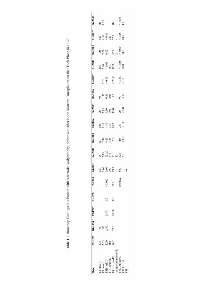
his neurological deterioration imposed a whole BMT from his sister, which resulted in
successful control of XALD. The transplant was HLA identical and at 2 years post-BMT
full-donor chimerism was found. There was no evidence of acute or chronic graft-versus-
host disease (GVHD).
BMT was preceded by cytoreductive conditioning protocol with chemotherapy (busulfan 4
mg/kg in four daily doses from day 9 till day 5 and cyclophosphamide 50 mg/kg in a single
daily dose from day 5 till day 2 before BMT). After the transplant, standard cyclosporin A
and a short-course methotrexate (10 mg/m2 i.v. at days +1, +3, and +6) were administered
to prevent acute GVHD. At the moment of BMT, the donor was 7 years old and free from
thyroid diseases, but at the age of 14, 7 years after the BMT, she showed alterations of
thyroid autoimmunity with raised anti-thyroperoxidase autoantibodies (TPO-Ab; 80 kU/l),
and 1 year thereafter she developed GH (TPO-Ab 280 kU/l, thyroxine (T4) 200 nmol/l, and
tri-iodothyronine (T3) 295 nmol/l).
Before BMT, the transplanted patient had never presented hypo- or hyperthyroidism, and
TSH had always been normal, as documented by several blood tests performed at our
hospital, during the 3 years preceding BMT. Starting from the year following the BMT,
TSH was found to be at the lower normal value and 2 years prior to our examination the
transplanted patient had developed thyrotoxicosis (Table 1) that was corrected with block
and replacement therapy (methimazole 30 mg/die and L-T4 25 mg/die).
At his ophthalmic baseline examination, in September 2002, the patient presented an eyelid
aperture of 12 mm right and 15 mm left with respectively a 2 mm lower lid retraction right,
a 2 mm upper, and 1 mm lower lid retraction left. Hertel values were bilaterally 17 mm, and
a mild eyeball dystopia of 2 mm with left eye over right was present (Figure 1). The
orbitopathy was not active and according with NOSPECS classification15 its severity was
2a, 30, 40, 50, and 60. Anterior segment, intraocular pressure, and fundus were normal.
Natural visual acuity was bilaterally 20/20.
Direct and indirect pupil reflexes were normal and the patient could read all the Ishihara
tables either with the left or the right eye. Orthoptic evaluation was unremarkable and
computer tomography of the orbit showed a mild enlargement of the extraocular muscles.
The diagnosis of inactive, mild GO was made.
Tab
le 1
. Lab
orat
ory
findi
ngs i
n a
patie
nt w
ith a
dren
oleu
kody
stro
phy,
bef
ore
and
afte
r bon
e m
arro
w tr
ansp
lant
atio
n th
at to
ok p
lace
in 1
996
Dat
e 09
-199
3 04
-199
4 09
-199
7 03
-199
9 12
-200
0 04
-200
1 04
-200
2 07
-200
3 08
-200
4 06
-200
5 06
-200
6 01
-200
7 03
-200
7 07
-200
7 11
-200
7 06
-200
8
T4 n
mol
/L
135
155
13
0 95
80
12
5 90
95
100
190
180
60
T3 n
mol
/L
2.05
1.
85
2.
60
2.15
2.
40
1.10
2.
15
1.95
7.
50
2.80
7.
00
4.50
1.
05
TSH
mE/
L 2.
00
2.40
0.
49
0.71
<0
.005
0.
04
12.2
0 4.
20
4.10
4.
90
0.35
<
0.01
<
0.01
0.
03
< 0.
01
TB
G n
mol
/L
380
290
370
300
250
290
260
28
0
240
T4
free
pm
ol/L
16
.2
23.3
18
.00
13.1
52
.8
19.3
13
.1
15.2
18
.2
15.0
17
.5
> 70
.0
18.9
55
.8
57.5
10
.1
tireo
glob
ulin
pm
ol/L
32
2
< 1
TP
O-A
b kU
/L
posit
ive
520
220
110
280
90
90
> 30
00
> 30
00
> 30
00
> 30
00
> 30
00
T.B.
I.I.
E/L
2.9
< 1.
0 <
1.0
< 1.
0 <
1.0
< 1.
0 56
.0
31.2
10
.6
4.1
TSI
46
Tabl
e 1.
Lab
orat
ory
Find
ings
in a
Pat
ient
with
Adr
enol
euko
dyst
roph
y, b
efor
e an
d af
ter B
one
Mar
row
Tra
nspl
anta
tion
that
Too
k Pl
ace
in 1
996
100379 Thesis Baldeschi.indd 141 vrijdag17-december-2010 12:09
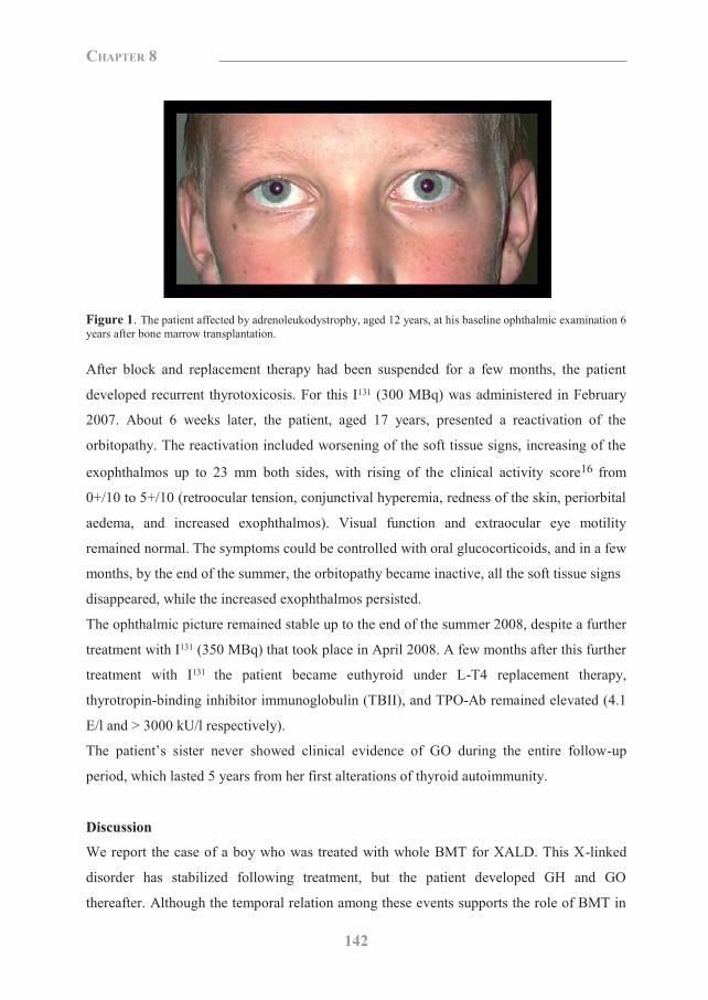
142
Chapter 8
Figure 1. The patient affected by adrenoleukodystrophy, aged 12 years, at his baseline ophthalmic examination 6 years after bone marrow transplantation.
After block and replacement therapy had been suspended for a few months, the patient
developed recurrent thyrotoxicosis. For this I131 (300 MBq) was administered in February
2007. About 6 weeks later, the patient, aged 17 years, presented a reactivation of the
orbitopathy. The reactivation included worsening of the soft tissue signs, increasing of the
exophthalmos up to 23 mm both sides, with rising of the clinical activity score16 from
0+/10 to 5+/10 (retroocular tension, conjunctival hyperemia, redness of the skin, periorbital
aedema, and increased exophthalmos). Visual function and extraocular eye motility
remained normal. The symptoms could be controlled with oral glucocorticoids, and in a few
months, by the end of the summer, the orbitopathy became inactive, all the soft tissue signs
disappeared, while the increased exophthalmos persisted.
The ophthalmic picture remained stable up to the end of the summer 2008, despite a further
treatment with I131 (350 MBq) that took place in April 2008. A few months after this further
treatment with I131 the patient became euthyroid under L-T4 replacement therapy,
thyrotropin-binding inhibitor immunoglobulin (TBII), and TPO-Ab remained elevated (4.1
E/l and > 3000 kU/l respectively).
The patient‟s sister never showed clinical evidence of GO during the entire follow-up
period, which lasted 5 years from her first alterations of thyroid autoimmunity.
Discussion
We report the case of a boy who was treated with whole BMT for XALD. This X-linked
disorder has stabilized following treatment, but the patient developed GH and GO
thereafter. Although the temporal relation among these events supports the role of BMT in
inducing GH and GO, other etiopathogenetic hypothesis cannot be neglected and should
therefore be included into the possible case scenario.
Although Laureti et al.17 reported that two out of five patients with adult onset of XALD
presented elevated titers of thyroid microsomal autoantibodies and one of them developed
clinical hypothyroidism, GO has never been described as part of XALD, and at present
there is no evidence that GH or GO and XALD are connected in one way or another.
Nevertheless, immunomodulation might play a role in the pathomechanism of
demyelinization as well as in the development of GH or GO. The positive familiar history
for alteration of thyroid autoimmunity presented by the transplanted patient that we report
may itself be a factor connected with a higher risk for developing other autoimmune
disorders such as autoimmune thyroid disease (ATD) or GO14, 18, although he had never
presented hypo- or hyperthyroidism or alterations of thyroid autoimmunity before BMT as
is documented by several blood tests performed in the years preceding such a procedure
(Table 1).
Occurrence and remission of GH after BMT have been reported.11, 12 Although thyroid
dysfunction is a common long-term complication associated with total body irradiation
given in the pre-BMT conditioning protocol, Slatter et al.9 reported thyroid dysfunction
also in patients who were given cytoreductive conditioning with chemotherapy, but without
total body irradiation as was the case for the patient we report. They found that 10.8% of
their patients had clinical and/or biochemical thyroid dysfunction at 4 months to 4.5 years
post-BMT, and 33% of these patients had positive antithyroid microsomal antibodies.
Both thyroid stimulating and blocking autoantibodies were detected after BMT.10
According to Sherer‟s and Shoenfeld‟s hypothesis, the induction of autoimmune diseases
post-BMT lays its foundations into the current understanding of GVHD19, and it could fit
to the case that we report, although the patient did not show typical evidences of GVHD.
Graves‟ disease is a multisystemic autoimmune disorder characterized in its complete form
by alteration of thyroid metabolism, orbitopathy, dermopathy, and acropachy. It involves
autoantigens common to the thyroid and other affected body districts. The autoimmune
attack on the thyroid leads, in the majority of cases, to alteration of thyroid hormones
homeostasis, and the autoimmune attack on the orbit causes the typical constellation of
signs and symptoms that characterize the clinical picture of GO.
100379 Thesis Baldeschi.indd 142 vrijdag17-december-2010 12:09

143
Chapter 8
Figure 1. The patient affected by adrenoleukodystrophy, aged 12 years, at his baseline ophthalmic examination 6 years after bone marrow transplantation.
After block and replacement therapy had been suspended for a few months, the patient
developed recurrent thyrotoxicosis. For this I131 (300 MBq) was administered in February
2007. About 6 weeks later, the patient, aged 17 years, presented a reactivation of the
orbitopathy. The reactivation included worsening of the soft tissue signs, increasing of the
exophthalmos up to 23 mm both sides, with rising of the clinical activity score16 from
0+/10 to 5+/10 (retroocular tension, conjunctival hyperemia, redness of the skin, periorbital
aedema, and increased exophthalmos). Visual function and extraocular eye motility
remained normal. The symptoms could be controlled with oral glucocorticoids, and in a few
months, by the end of the summer, the orbitopathy became inactive, all the soft tissue signs
disappeared, while the increased exophthalmos persisted.
The ophthalmic picture remained stable up to the end of the summer 2008, despite a further
treatment with I131 (350 MBq) that took place in April 2008. A few months after this further
treatment with I131 the patient became euthyroid under L-T4 replacement therapy,
thyrotropin-binding inhibitor immunoglobulin (TBII), and TPO-Ab remained elevated (4.1
E/l and > 3000 kU/l respectively).
The patient‟s sister never showed clinical evidence of GO during the entire follow-up
period, which lasted 5 years from her first alterations of thyroid autoimmunity.
Discussion
We report the case of a boy who was treated with whole BMT for XALD. This X-linked
disorder has stabilized following treatment, but the patient developed GH and GO
thereafter. Although the temporal relation among these events supports the role of BMT in
inducing GH and GO, other etiopathogenetic hypothesis cannot be neglected and should
therefore be included into the possible case scenario.
Although Laureti et al.17 reported that two out of five patients with adult onset of XALD
presented elevated titers of thyroid microsomal autoantibodies and one of them developed
clinical hypothyroidism, GO has never been described as part of XALD, and at present
there is no evidence that GH or GO and XALD are connected in one way or another.
Nevertheless, immunomodulation might play a role in the pathomechanism of
demyelinization as well as in the development of GH or GO. The positive familiar history
for alteration of thyroid autoimmunity presented by the transplanted patient that we report
may itself be a factor connected with a higher risk for developing other autoimmune
disorders such as autoimmune thyroid disease (ATD) or GO14, 18, although he had never
presented hypo- or hyperthyroidism or alterations of thyroid autoimmunity before BMT as
is documented by several blood tests performed in the years preceding such a procedure
(Table 1).
Occurrence and remission of GH after BMT have been reported.11, 12 Although thyroid
dysfunction is a common long-term complication associated with total body irradiation
given in the pre-BMT conditioning protocol, Slatter et al.9 reported thyroid dysfunction
also in patients who were given cytoreductive conditioning with chemotherapy, but without
total body irradiation as was the case for the patient we report. They found that 10.8% of
their patients had clinical and/or biochemical thyroid dysfunction at 4 months to 4.5 years
post-BMT, and 33% of these patients had positive antithyroid microsomal antibodies.
Both thyroid stimulating and blocking autoantibodies were detected after BMT.10
According to Sherer‟s and Shoenfeld‟s hypothesis, the induction of autoimmune diseases
post-BMT lays its foundations into the current understanding of GVHD19, and it could fit
to the case that we report, although the patient did not show typical evidences of GVHD.
Graves‟ disease is a multisystemic autoimmune disorder characterized in its complete form
by alteration of thyroid metabolism, orbitopathy, dermopathy, and acropachy. It involves
autoantigens common to the thyroid and other affected body districts. The autoimmune
attack on the thyroid leads, in the majority of cases, to alteration of thyroid hormones
homeostasis, and the autoimmune attack on the orbit causes the typical constellation of
signs and symptoms that characterize the clinical picture of GO.
100379 Thesis Baldeschi.indd 143 vrijdag17-december-2010 12:09
144
Chapter 8
It has been shown that orbital tissue from patients with GO is infiltrated with B and T cells,
and it has been reported that orbital fibroblasts from Graves‟ patients can stimulate
proliferation of autologous T cells20 and that autologous T cells from patients with GO can
stimulate proliferation of orbital fibroblasts, thus providing a mechanism by which
infiltration of orbital tissue by autoimmune lymphocytes can drive the pathogenic features
of GO.21 Another evidence of the involvement of T lymphocytes in the pathogenesis of GO
derives from the observation that newborns from mothers with GO can be hyperthyroid as a
result of possible passage of thyroid-stimulating IgG through the placental barrier, but they
never show GO at birth due to the placental barrier to T lymphocytes.22, 23
Here, we show that whole BMT, from a donor who was free from thyroid diseases at the
time of BMT, but who developed GH thereafter, may be involved in the induction of GO 6
years later in the transplanted patient. It is likely that the disease process is primarily
dependent upon altered function of the T lymphocyte. Although the immune mechanisms
underlying the orbitopathy as well as the primary autoantigens involved in the disease are
poorly characterized, it can be hypothesized that dormant autoreactive T cells once
transplanted could have been activated by nonprofessional antigen-presenting cells, such as
orbital fibroblasts.
Afterwards, T-cell-driven stimulation of orbital fibroblasts to upregulate expression of
MHC class II and the presentation of autoantigens could have occurred. This in turn could
have further activated T cells to produce surface and/or diffusible factors that drive
activation and proliferation of orbital fibroblasts, leading to expression of fibroblast-based
diseases, i.e. proliferation of fibroblasts and excess connective tissue, deposition of matrix
glycosaminoglycans, intramuscular fibrosis, and differentiation and proliferation of
adipocytes.
Another interesting issue regards the time between BMT and the onset of GH and GO. A
delay due to immune reconstitution of the recipient should be expected, but it can also be
hypothesized that an additional stimulus like an infection, even subclinical, or an
inflammation could have played a role in triggering the process. Indeed, orbital fibroblasts,
upon stimulation with IFN-γ produced during infections / inflammations, are able to
overexpress MHC class II molecules on their surfaces24 and to produce proinflammatory
cytokines, such as IL-8 and IL-6. This suggests that orbital fibroblasts may be crucial in the
trafficking of bone marrow-derived immune cells to the orbit in states of
infection/inflammation.
Here, we propose that the recipient passively acquired dormant autoreactive T cells from
his sister, although she was apparently healthy. This case report suggests to consider, if
available, the use of a matched unrelated donor when a potential HLA-identical sibling
donor presents relevant immune-mediated diseases. In the case where a matched unrelated
donor is not available, a T-cell depleted rather than whole BMT might be an alternative
when the donor is affected by ATDs or other T-cell-driven autoimmune disorders, although
the role of bone marrow graft engineering is still under study. T-cell depletion of the graft
might in theory change / prevent the occurrence of autoimmunity; however, we
acknowledge that the evidence is very limited to make this statement, and we are aware that
T-cell-depleted bone marrow has major drawbacks, such as an enhanced risk of graft
rejection as well as delayed immune reconstitution, with a consequent increased risk of
viral and fungal infections.
100379 Thesis Baldeschi.indd 144 vrijdag17-december-2010 12:09
145
Chapter 8
It has been shown that orbital tissue from patients with GO is infiltrated with B and T cells,
and it has been reported that orbital fibroblasts from Graves‟ patients can stimulate
proliferation of autologous T cells20 and that autologous T cells from patients with GO can
stimulate proliferation of orbital fibroblasts, thus providing a mechanism by which
infiltration of orbital tissue by autoimmune lymphocytes can drive the pathogenic features
of GO.21 Another evidence of the involvement of T lymphocytes in the pathogenesis of GO
derives from the observation that newborns from mothers with GO can be hyperthyroid as a
result of possible passage of thyroid-stimulating IgG through the placental barrier, but they
never show GO at birth due to the placental barrier to T lymphocytes.22, 23
Here, we show that whole BMT, from a donor who was free from thyroid diseases at the
time of BMT, but who developed GH thereafter, may be involved in the induction of GO 6
years later in the transplanted patient. It is likely that the disease process is primarily
dependent upon altered function of the T lymphocyte. Although the immune mechanisms
underlying the orbitopathy as well as the primary autoantigens involved in the disease are
poorly characterized, it can be hypothesized that dormant autoreactive T cells once
transplanted could have been activated by nonprofessional antigen-presenting cells, such as
orbital fibroblasts.
Afterwards, T-cell-driven stimulation of orbital fibroblasts to upregulate expression of
MHC class II and the presentation of autoantigens could have occurred. This in turn could
have further activated T cells to produce surface and/or diffusible factors that drive
activation and proliferation of orbital fibroblasts, leading to expression of fibroblast-based
diseases, i.e. proliferation of fibroblasts and excess connective tissue, deposition of matrix
glycosaminoglycans, intramuscular fibrosis, and differentiation and proliferation of
adipocytes.
Another interesting issue regards the time between BMT and the onset of GH and GO. A
delay due to immune reconstitution of the recipient should be expected, but it can also be
hypothesized that an additional stimulus like an infection, even subclinical, or an
inflammation could have played a role in triggering the process. Indeed, orbital fibroblasts,
upon stimulation with IFN-γ produced during infections / inflammations, are able to
overexpress MHC class II molecules on their surfaces24 and to produce proinflammatory
cytokines, such as IL-8 and IL-6. This suggests that orbital fibroblasts may be crucial in the
trafficking of bone marrow-derived immune cells to the orbit in states of
infection/inflammation.
Here, we propose that the recipient passively acquired dormant autoreactive T cells from
his sister, although she was apparently healthy. This case report suggests to consider, if
available, the use of a matched unrelated donor when a potential HLA-identical sibling
donor presents relevant immune-mediated diseases. In the case where a matched unrelated
donor is not available, a T-cell depleted rather than whole BMT might be an alternative
when the donor is affected by ATDs or other T-cell-driven autoimmune disorders, although
the role of bone marrow graft engineering is still under study. T-cell depletion of the graft
might in theory change / prevent the occurrence of autoimmunity; however, we
acknowledge that the evidence is very limited to make this statement, and we are aware that
T-cell-depleted bone marrow has major drawbacks, such as an enhanced risk of graft
rejection as well as delayed immune reconstitution, with a consequent increased risk of
viral and fungal infections.
100379 Thesis Baldeschi.indd 145 vrijdag17-december-2010 12:09
146
Chapter 8
References 1. Shapiro E, Krivit W, Lockman L, Jambaqué I, Cowan M, Harris R, Blanke S, Bordigoni P, Loes D, Ziegler R,
Crittenden M, Ris D, Berg B, Cox C, Moser H, Fischer A, Aubourg P. Long term effect of bone marrow
transplantation for childhood onset cerebral X linked adrenoleukodystrophy. Lancet 2000;356:713-8.
2. Hitomi T, Mezaki T, Tsujii T, Kinishita M, Tomimoto H, Ikeda A, Shimohama S, Okazaki T, Uchiyama T,
Shibasaki H. Improvement of central motor conduction after bone marrow transplantation in
adrenoleukodystrophy. J Neurol Neurosurg Psychiatry 2003;74:373-5.
3. Beam D, Poe MD, Provenzale JM, Szabolcs P, Martin PL, Prasad V, Parikh S, Driscoll T, Mukundan S,
Kurtzberg J, Escolar ML. Outcomes of unrelated umbilical cord blood transplantation for X-linked
adrenoleukodystrophy. Biol Blood Marrow Transplant 2007;13:665-74.
4. Peters C, Steward CG, National Marrow Donor Program, International Bone Marrow Transplant Registry,
Working Party on Inborn Errors, European Bone Marrow Transplant Group. Hematopoietic cell transplantation for
inherited metabolic diseases: an overview of outcomes and practice guidelines. Bone Marrow Transplant
2003;31:229-39.
5. Peters C, Charnas LR, Tan Y, Ziegler RS, Shapiro EG, DeFor T, Grewal SS, Orchard PJ, Abel SL, Goldman
AI, Ramsay NK, Dusenbery KE, Loes DJ, Lockman LA, Kato S, Aubourg PR, Moser HW, Krivit W. Cerebral X-
linked adrenoleukodystrophy: the international hematopoietic cell transplantation experience from 1982 to 1999.
Blood 2004;104:881-8.
6. Yammamori I, Kanie T, Maeda N, Kodera Y, Matsuyama T, Hasegawa H. Appearance of thyroid stimulating
and blocking immunoglobulins after bone marrow transplantation: presentation of two contrasting cases. Endocr J
2004;51:439-43.
7. Curtillet C, Cuadras P, Dore E, Chambost H, Thuret I, Michel G. Thyroid dysfunction after haematopoietic
stem cell transplantation during childhood. Arch Pediatr 2004;11:1326-32.
8. Marazuela M, Steegman JL. Transfer of autoimmune hypothyroidism following bone marrow transplantation.
Bone Marrow Transplant 2000;26:1217-20.
9. Slatter MA, Gennery AR, Cheetham TD, Bhattacharya A, Crooks BN, Flood TJ, Cant AJ, Abinun M. Thyroid
dysfunction after bone marrow transplantation for primary immunodeficiency without the use of total body
irradiation in conditioning. Bone Marrow Transplantat 2004;33:949-53.
10. Lee WY, Oh ES, Min CK, Kim DW, Lee JW, Kang MI, Min WS, Cha BY, Lee KW, Son HY, Kang SK, Kim
CC. Changes in autoimmune thyroid disease following allogeneic bone marrow transplantation. Bone Marrow
Transplantat 2001;28:63-6.
11. Kishimoto Y, Yamamoto Y, Ito T, Matsumoto N, Ichiyoshi H, Katsurada T, Date M, Ohga S, Kitajima H,
Ikehara S, Fukuhara S. Transfer of autoimmune thyroiditis and resolution of palmoplantar pustular psoriasis
following allogeneic bone marrow transplantation. Bone Marrow Transplantat 1997;19:1041-3.
12. Hsiao LT, Liu JH, Yen CC,Wang WS, Fan FS, Chiou TJ, Chen PM. Relapse of Graves‟ disease after
successful allogeneic bone marrow transplantation. Bone Marrow Transplantat 2001;28:1151-3.
13. Lazarus JH, Marinò M. Orbit-thyroid relationship. In: Wiersinga WM, Kahaly GJ, editors. Graves‟
Orbitopathy A Multidisciplinary Approach - Questions and Answers, Basel: Karger, 2007:27-33.
14. Holland FJ, McConnon JK, Volpé R , Saunders EF. Concordant Graves‟ disease after bone marrow
transplantation: implications for pathogenesis. J Clin Endocrinol Metab 1991;72:837-40.
15. Werner SC. Modifications of the classification of the eye changes of Graves‟ disease: recommendations of the
Ad Hoc Committee of the American Thyroid Association. J Clin Endocrinol Metab 1977;44:203-4.
16. Mourits MP, Koornnneef L, Wiersinga WM, Prummel MF, Berghout A , van der Gaag R. Clinical criteria for
the assessment of disease activity in Graves‟ ophthalmopathy: a novel approach. Br J Ophthalmol 1989;73:639-44.
17. Laureti S, Falorni A, Volpato M, Casucci G, Picchio ML, Angeletti G, Luthman H, Brunetti P, Betterle C,
Santeusanio F. Absence of circulating adrenal autoantibodies in adult-onset X-linked adrenoleukodystrophy. Horm
Metab Res1996;28: 319-22.
18. Vaidya B, Imrie H, Perros P, Dickinson J, McCarthy MI, Kendall- Taylor P, Pearce SH. Cytotoxic T
lymphocyte antigen-4 (CTLA-4) gene polymorphism confers susceptibility to thyroid associated orbitopathy.
Lancet 1999;354:743-44.
19. Sherer Y, Shoenfeld Y. Autoimmune disease and autoimmunity post bone marrow transplantation. Bone
Marrow Transplant 1998;22:873-81.
20. Eckstein AK, Quadbeck B, Tews S, Mann K, Kruger C, Mohr CH, Steuhl KP, Esser J, Gieseler RK. Thyroid
associated ophthalmopathy: evidence for CD4(+) gammadelta T cells; de novo differentiation of RFD7(+)
macrophages, but not of RFD1(+) dendritic cells; and loss of gammadelta and alphabeta T cell receptor
expression. Br J Ophthalmol 2004;88:803-8.
21. Feldon SE, Park DJ, O‟Loughlin CW, Nguyen VT, Landskroner-Eiger S, Chang D, Thatcher TH, Phipps RP.
Autologous T-lymphocytes stimulate proliferation of orbital fibroblasts derived from patients with Graves‟
ophthalmopathy. Invest Ophthalmol Vis Sci 2005;46:3913-21.
22. Orgiazzi J. Pathogenesis. In: Wiersinga WM, Kahaly GJ, editors. Graves‟ Orbitopathy A Multidisciplinary
Approach - Questions and Answers. Basel: Karger, 2007:41-56.
23. Zimmerman D. Fetal and neonatal hyperthyroidism. Thyroid 1999;9:727-33.
24. Heufelder AE, Smith TJ, Gorman CA, Bahn RS. Increased induction of HLA-DR by interferon-gamma in
cultured fibroblasts derived from patients with Graves‟ ophthalmopathy and pretibial dermopathy. J Clin
Endocrinol Metab 1991;73:307-13.
100379 Thesis Baldeschi.indd 146 vrijdag17-december-2010 12:09
147
Chapter 8
References 1. Shapiro E, Krivit W, Lockman L, Jambaqué I, Cowan M, Harris R, Blanke S, Bordigoni P, Loes D, Ziegler R,
Crittenden M, Ris D, Berg B, Cox C, Moser H, Fischer A, Aubourg P. Long term effect of bone marrow
transplantation for childhood onset cerebral X linked adrenoleukodystrophy. Lancet 2000;356:713-8.
2. Hitomi T, Mezaki T, Tsujii T, Kinishita M, Tomimoto H, Ikeda A, Shimohama S, Okazaki T, Uchiyama T,
Shibasaki H. Improvement of central motor conduction after bone marrow transplantation in
adrenoleukodystrophy. J Neurol Neurosurg Psychiatry 2003;74:373-5.
3. Beam D, Poe MD, Provenzale JM, Szabolcs P, Martin PL, Prasad V, Parikh S, Driscoll T, Mukundan S,
Kurtzberg J, Escolar ML. Outcomes of unrelated umbilical cord blood transplantation for X-linked
adrenoleukodystrophy. Biol Blood Marrow Transplant 2007;13:665-74.
4. Peters C, Steward CG, National Marrow Donor Program, International Bone Marrow Transplant Registry,
Working Party on Inborn Errors, European Bone Marrow Transplant Group. Hematopoietic cell transplantation for
inherited metabolic diseases: an overview of outcomes and practice guidelines. Bone Marrow Transplant
2003;31:229-39.
5. Peters C, Charnas LR, Tan Y, Ziegler RS, Shapiro EG, DeFor T, Grewal SS, Orchard PJ, Abel SL, Goldman
AI, Ramsay NK, Dusenbery KE, Loes DJ, Lockman LA, Kato S, Aubourg PR, Moser HW, Krivit W. Cerebral X-
linked adrenoleukodystrophy: the international hematopoietic cell transplantation experience from 1982 to 1999.
Blood 2004;104:881-8.
6. Yammamori I, Kanie T, Maeda N, Kodera Y, Matsuyama T, Hasegawa H. Appearance of thyroid stimulating
and blocking immunoglobulins after bone marrow transplantation: presentation of two contrasting cases. Endocr J
2004;51:439-43.
7. Curtillet C, Cuadras P, Dore E, Chambost H, Thuret I, Michel G. Thyroid dysfunction after haematopoietic
stem cell transplantation during childhood. Arch Pediatr 2004;11:1326-32.
8. Marazuela M, Steegman JL. Transfer of autoimmune hypothyroidism following bone marrow transplantation.
Bone Marrow Transplant 2000;26:1217-20.
9. Slatter MA, Gennery AR, Cheetham TD, Bhattacharya A, Crooks BN, Flood TJ, Cant AJ, Abinun M. Thyroid
dysfunction after bone marrow transplantation for primary immunodeficiency without the use of total body
irradiation in conditioning. Bone Marrow Transplantat 2004;33:949-53.
10. Lee WY, Oh ES, Min CK, Kim DW, Lee JW, Kang MI, Min WS, Cha BY, Lee KW, Son HY, Kang SK, Kim
CC. Changes in autoimmune thyroid disease following allogeneic bone marrow transplantation. Bone Marrow
Transplantat 2001;28:63-6.
11. Kishimoto Y, Yamamoto Y, Ito T, Matsumoto N, Ichiyoshi H, Katsurada T, Date M, Ohga S, Kitajima H,
Ikehara S, Fukuhara S. Transfer of autoimmune thyroiditis and resolution of palmoplantar pustular psoriasis
following allogeneic bone marrow transplantation. Bone Marrow Transplantat 1997;19:1041-3.
12. Hsiao LT, Liu JH, Yen CC,Wang WS, Fan FS, Chiou TJ, Chen PM. Relapse of Graves‟ disease after
successful allogeneic bone marrow transplantation. Bone Marrow Transplantat 2001;28:1151-3.
13. Lazarus JH, Marinò M. Orbit-thyroid relationship. In: Wiersinga WM, Kahaly GJ, editors. Graves‟
Orbitopathy A Multidisciplinary Approach - Questions and Answers, Basel: Karger, 2007:27-33.
14. Holland FJ, McConnon JK, Volpé R , Saunders EF. Concordant Graves‟ disease after bone marrow
transplantation: implications for pathogenesis. J Clin Endocrinol Metab 1991;72:837-40.
15. Werner SC. Modifications of the classification of the eye changes of Graves‟ disease: recommendations of the
Ad Hoc Committee of the American Thyroid Association. J Clin Endocrinol Metab 1977;44:203-4.
16. Mourits MP, Koornnneef L, Wiersinga WM, Prummel MF, Berghout A , van der Gaag R. Clinical criteria for
the assessment of disease activity in Graves‟ ophthalmopathy: a novel approach. Br J Ophthalmol 1989;73:639-44.
17. Laureti S, Falorni A, Volpato M, Casucci G, Picchio ML, Angeletti G, Luthman H, Brunetti P, Betterle C,
Santeusanio F. Absence of circulating adrenal autoantibodies in adult-onset X-linked adrenoleukodystrophy. Horm
Metab Res1996;28: 319-22.
18. Vaidya B, Imrie H, Perros P, Dickinson J, McCarthy MI, Kendall- Taylor P, Pearce SH. Cytotoxic T
lymphocyte antigen-4 (CTLA-4) gene polymorphism confers susceptibility to thyroid associated orbitopathy.
Lancet 1999;354:743-44.
19. Sherer Y, Shoenfeld Y. Autoimmune disease and autoimmunity post bone marrow transplantation. Bone
Marrow Transplant 1998;22:873-81.
20. Eckstein AK, Quadbeck B, Tews S, Mann K, Kruger C, Mohr CH, Steuhl KP, Esser J, Gieseler RK. Thyroid
associated ophthalmopathy: evidence for CD4(+) gammadelta T cells; de novo differentiation of RFD7(+)
macrophages, but not of RFD1(+) dendritic cells; and loss of gammadelta and alphabeta T cell receptor
expression. Br J Ophthalmol 2004;88:803-8.
21. Feldon SE, Park DJ, O‟Loughlin CW, Nguyen VT, Landskroner-Eiger S, Chang D, Thatcher TH, Phipps RP.
Autologous T-lymphocytes stimulate proliferation of orbital fibroblasts derived from patients with Graves‟
ophthalmopathy. Invest Ophthalmol Vis Sci 2005;46:3913-21.
22. Orgiazzi J. Pathogenesis. In: Wiersinga WM, Kahaly GJ, editors. Graves‟ Orbitopathy A Multidisciplinary
Approach - Questions and Answers. Basel: Karger, 2007:41-56.
23. Zimmerman D. Fetal and neonatal hyperthyroidism. Thyroid 1999;9:727-33.
24. Heufelder AE, Smith TJ, Gorman CA, Bahn RS. Increased induction of HLA-DR by interferon-gamma in
cultured fibroblasts derived from patients with Graves‟ ophthalmopathy and pretibial dermopathy. J Clin
Endocrinol Metab 1991;73:307-13.
100379 Thesis Baldeschi.indd 147 vrijdag17-december-2010 12:09

Chapter 9
Supernumerary Extraocular Muscle in Graves‟ Orbitopathy
Lelio Baldeschi1, Peter H.L.T. Bisschop2, Wilmar M. Wiersinga2
1 Orbital Centre, Department of Ophthalmology, University of Amsterdam, Amsterdam, The Netherlands. 2 Department of Endocrinology, University of Amsterdam, Amsterdam, The Netherlands. Thyroid 2007;17:479-80
100379 Thesis Baldeschi.indd 148 vrijdag17-december-2010 12:09
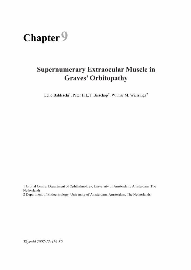
Chapter 9
Supernumerary Extraocular Muscle in Graves‟ Orbitopathy
Lelio Baldeschi1, Peter H.L.T. Bisschop2, Wilmar M. Wiersinga2
1 Orbital Centre, Department of Ophthalmology, University of Amsterdam, Amsterdam, The Netherlands. 2 Department of Endocrinology, University of Amsterdam, Amsterdam, The Netherlands. Thyroid 2007;17:479-80
Supernumerary Extraocular Muscle inGraves’ Orbitopathy
100379 Thesis Baldeschi.indd 149 vrijdag17-december-2010 12:09
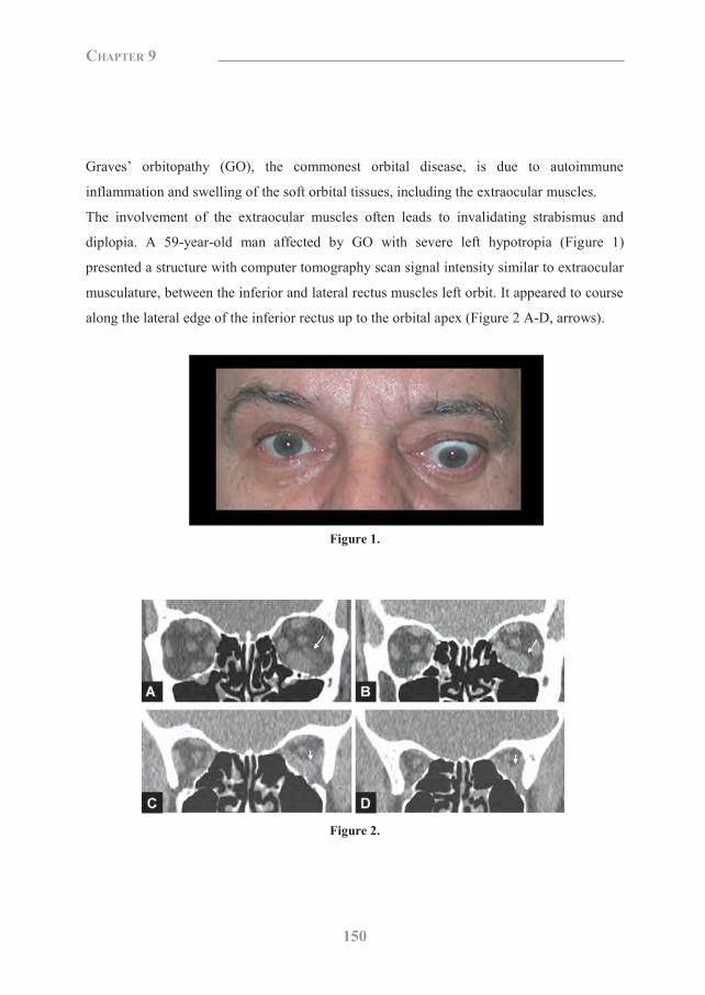
150
Chapter 9
Graves‟ orbitopathy (GO), the commonest orbital disease, is due to autoimmune
inflammation and swelling of the soft orbital tissues, including the extraocular muscles.
The involvement of the extraocular muscles often leads to invalidating strabismus and
diplopia. A 59-year-old man affected by GO with severe left hypotropia (Figure 1)
presented a structure with computer tomography scan signal intensity similar to extraocular
musculature, between the inferior and lateral rectus muscles left orbit. It appeared to course
along the lateral edge of the inferior rectus up to the orbital apex (Figure 2 A-D, arrows).
Figure 1.
Figure 2.
A better evaluation by means of magnetic tomography scan was not possible due to an
irremovable dental prosthesis. Surgical exploration of the area confirmed the presence of a
supernumerary extraocular muscle (SEM) (Figure 3, arrow).
Figure 3.
It was inserted onto the sclera by a 3-mm-long, 5-mm-wide tendon, 5 mm laterally to the
insertion of the inferior rectus muscle at 7 mm from the cornel limbus, and its belly was
swollen. Eight months after retrobulbar irradiation, the decreased swelling of the inferior
rectus and SEM allowed a better visualization of the two muscles on computer tomography
scans. Such an examination confirmed that the SEM was an autonomous structure that
originated from the orbital apex and not from the lateral edge of the inferior rectus muscle
(Figure 4 A, B, arrows).
100379 Thesis Baldeschi.indd 150 vrijdag17-december-2010 12:09
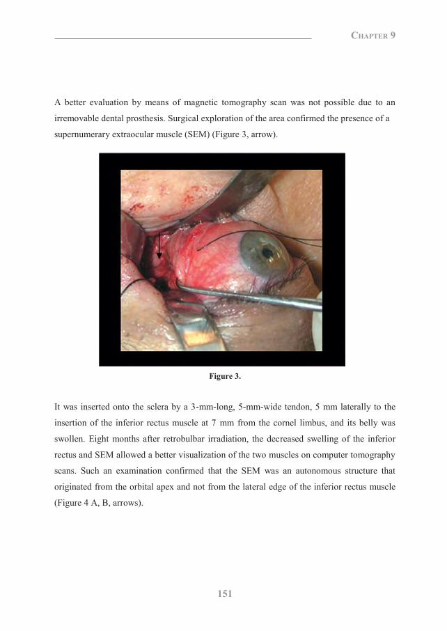
151
Chapter 9
Graves‟ orbitopathy (GO), the commonest orbital disease, is due to autoimmune
inflammation and swelling of the soft orbital tissues, including the extraocular muscles.
The involvement of the extraocular muscles often leads to invalidating strabismus and
diplopia. A 59-year-old man affected by GO with severe left hypotropia (Figure 1)
presented a structure with computer tomography scan signal intensity similar to extraocular
musculature, between the inferior and lateral rectus muscles left orbit. It appeared to course
along the lateral edge of the inferior rectus up to the orbital apex (Figure 2 A-D, arrows).
Figure 1.
Figure 2.
A better evaluation by means of magnetic tomography scan was not possible due to an
irremovable dental prosthesis. Surgical exploration of the area confirmed the presence of a
supernumerary extraocular muscle (SEM) (Figure 3, arrow).
Figure 3.
It was inserted onto the sclera by a 3-mm-long, 5-mm-wide tendon, 5 mm laterally to the
insertion of the inferior rectus muscle at 7 mm from the cornel limbus, and its belly was
swollen. Eight months after retrobulbar irradiation, the decreased swelling of the inferior
rectus and SEM allowed a better visualization of the two muscles on computer tomography
scans. Such an examination confirmed that the SEM was an autonomous structure that
originated from the orbital apex and not from the lateral edge of the inferior rectus muscle
(Figure 4 A, B, arrows).
100379 Thesis Baldeschi.indd 151 vrijdag17-december-2010 12:09
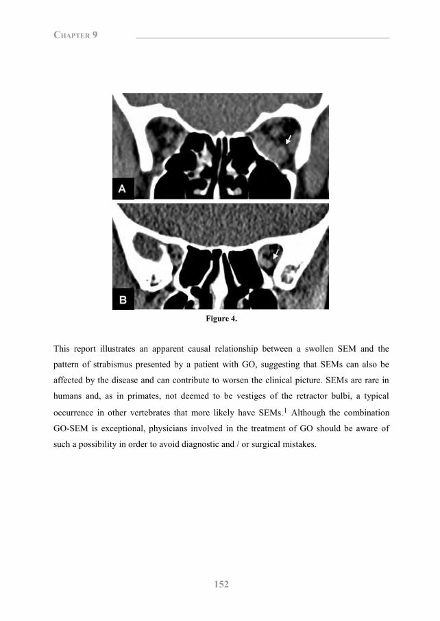
152
Chapter 9
Figure 4.
This report illustrates an apparent causal relationship between a swollen SEM and the
pattern of strabismus presented by a patient with GO, suggesting that SEMs can also be
affected by the disease and can contribute to worsen the clinical picture. SEMs are rare in
humans and, as in primates, not deemed to be vestiges of the retractor bulbi, a typical
occurrence in other vertebrates that more likely have SEMs.1 Although the combination
GO-SEM is exceptional, physicians involved in the treatment of GO should be aware of
such a possibility in order to avoid diagnostic and / or surgical mistakes.
Reference 1. von Ludinghausen M. Bilateral supernumerary rectus muscles of the orbit. Clin Anat 1998;11:271-7.
100379 Thesis Baldeschi.indd 152 vrijdag17-december-2010 12:09

153
Chapter 9
Figure 4.
This report illustrates an apparent causal relationship between a swollen SEM and the
pattern of strabismus presented by a patient with GO, suggesting that SEMs can also be
affected by the disease and can contribute to worsen the clinical picture. SEMs are rare in
humans and, as in primates, not deemed to be vestiges of the retractor bulbi, a typical
occurrence in other vertebrates that more likely have SEMs.1 Although the combination
GO-SEM is exceptional, physicians involved in the treatment of GO should be aware of
such a possibility in order to avoid diagnostic and / or surgical mistakes.
Reference 1. von Ludinghausen M. Bilateral supernumerary rectus muscles of the orbit. Clin Anat 1998;11:271-7.
100379 Thesis Baldeschi.indd 153 vrijdag17-december-2010 12:09

Chapter 10
Traumatic Neuroma of the Infraorbital Nerve Subsequent to
Inferomedial Orbital Decompression for Graves’ Orbitopathy
Lelio Baldeschi1, Peerooz Saeed1, Noortje I. Regensburg1,
Ioannis Zacharopoulos1, Wilmar M. Wiersinga2
1 Orbital Centre, Department of Ophthalmology, University of Amsterdam, Amsterdam, The Netherlands. 2 Department of Endocrinology, University of Amsterdam, Amsterdam, The Netherlands. European Journal of Ophthalmology 2009;140:642-7
100379 Thesis Baldeschi.indd 154 vrijdag17-december-2010 12:09
Chapter 10
Traumatic Neuroma of the Infraorbital Nerve Subsequent to
Inferomedial Orbital Decompression for Graves’ Orbitopathy
Lelio Baldeschi1, Peerooz Saeed1, Noortje I. Regensburg1,
Ioannis Zacharopoulos1, Wilmar M. Wiersinga2
1 Orbital Centre, Department of Ophthalmology, University of Amsterdam, Amsterdam, The Netherlands. 2 Department of Endocrinology, University of Amsterdam, Amsterdam, The Netherlands. European Journal of Ophthalmology 2009;140:642-7 2010:20:481-4
100379 Thesis Baldeschi.indd 155 vrijdag17-december-2010 12:09
156
Chapter 10
Abstract
Purpose: To present and discuss the occurrence of a traumatic neuroma subsequent to
inferomedial orbital decompression surgery in Graves‟ orbitopathy.
Method: Case report.
Results: Approximately 1 month after surgery, a patient who underwent bilateral
rehabilitative inferomedial orbital decompression developed a mass with clinical and
radiologic characteristics compatible with a traumatic neuroma of the left infraorbital nerve.
The lesion, which was thought to be the result of unnoticed nerve trauma at the time of
surgical dissection of the infraorbital canal, remained stable in shape and other imaging
characteristics during the 39-month follow-up period. Symptoms of trigeminal neuralgia
could be only partially controlled with medical therapy (oral pregabalin 75 mg 3 times
daily).
Conclusions: The second branch of the trigeminal nerve may be damaged in the course of
orbital floor removal decompression for Graves‟ orbitopathy. This may potentially induce
the formation of traumatic or amputation neuromas. Such lesions should be included in the
potential complications of decompressions when counseling patients about to undergo this
type of surgery, as they are difficult to treat and may cause persistent and disabling pain.
Introduction
Traumatic neuromas may be due to chronic nerve irritation or trauma that partially avulses
or totally disrupts the nerve. In the case of chronic irritation, an undisrupted nerve gives rise
to a focal fusiform swelling often referred to as “spindle neuroma.” Different degrees of
nerve disruption and subsequent external axon growth, which represents an attempt at
natural repair, leads to the formation of masses referred to as “lateral or terminal, traumatic
neuromas,” the latter also referred to as “amputation neuromas.”
Traumatic neuromas may cause persistent pain and have been described in several anatomic
locations including the orbit after enucleation.1 Orbital decompression surgery is a
currently accepted, effective, and generally safe treatment for Graves‟ orbitopathy.2 When
decompression surgery entails removal of the orbital floor, the infraorbital nerve may be
damaged by surgical maneuvers, by bone edges or fragments, or may be stretched by the
soft orbital tissues which prolapse into the maxillary antrum. This generally causes
hypoesthesia, rarely anesthesia or pain in the sensory distribution area of this nerve branch.
Herein we present and discuss a patient with Graves‟ orbitopathy presenting with clinical
and radiologic evidence of traumatic neuroma of the infraorbital nerve starting a few weeks
after rehabilitative inferomedial orbital decompression, and who has been followed up at
our department for 39 months afterwards.
We are unaware of previous reports that link traumatic neuromas of the infraorbital nerve to
orbital decompression.
Case report
A 45-year-old Caucasian woman with stable inactive Graves‟ orbitopathy for more than 5
years, on levothyroxine replacement therapy for 4 years following I131, was admitted for
bilateral inferomedial rehabilitative orbital decompression in June 2005. On admission, she
had best corrected visual acuity (pinhole) of 20/20 bilaterally, bilateral slight increase of the
eyelid aperture with 3 mm upper lid retraction, moderate periorbital aedema, consistent
retroocular tension, and Hertel readings of 23 mm bilaterally, and did not have diplopia.
Decompression was carried out uneventfully by an anterior trans-inferior-fornix approach
and included the complete removal of the orbital floor. The immediate postoperative period
was unremarkable. The patient left the hospital 2 days after surgery without any medication
100379 Thesis Baldeschi.indd 156 vrijdag17-december-2010 12:09
157
Chapter 10
Abstract
Purpose: To present and discuss the occurrence of a traumatic neuroma subsequent to
inferomedial orbital decompression surgery in Graves‟ orbitopathy.
Method: Case report.
Results: Approximately 1 month after surgery, a patient who underwent bilateral
rehabilitative inferomedial orbital decompression developed a mass with clinical and
radiologic characteristics compatible with a traumatic neuroma of the left infraorbital nerve.
The lesion, which was thought to be the result of unnoticed nerve trauma at the time of
surgical dissection of the infraorbital canal, remained stable in shape and other imaging
characteristics during the 39-month follow-up period. Symptoms of trigeminal neuralgia
could be only partially controlled with medical therapy (oral pregabalin 75 mg 3 times
daily).
Conclusions: The second branch of the trigeminal nerve may be damaged in the course of
orbital floor removal decompression for Graves‟ orbitopathy. This may potentially induce
the formation of traumatic or amputation neuromas. Such lesions should be included in the
potential complications of decompressions when counseling patients about to undergo this
type of surgery, as they are difficult to treat and may cause persistent and disabling pain.
Introduction
Traumatic neuromas may be due to chronic nerve irritation or trauma that partially avulses
or totally disrupts the nerve. In the case of chronic irritation, an undisrupted nerve gives rise
to a focal fusiform swelling often referred to as “spindle neuroma.” Different degrees of
nerve disruption and subsequent external axon growth, which represents an attempt at
natural repair, leads to the formation of masses referred to as “lateral or terminal, traumatic
neuromas,” the latter also referred to as “amputation neuromas.”
Traumatic neuromas may cause persistent pain and have been described in several anatomic
locations including the orbit after enucleation.1 Orbital decompression surgery is a
currently accepted, effective, and generally safe treatment for Graves‟ orbitopathy.2 When
decompression surgery entails removal of the orbital floor, the infraorbital nerve may be
damaged by surgical maneuvers, by bone edges or fragments, or may be stretched by the
soft orbital tissues which prolapse into the maxillary antrum. This generally causes
hypoesthesia, rarely anesthesia or pain in the sensory distribution area of this nerve branch.
Herein we present and discuss a patient with Graves‟ orbitopathy presenting with clinical
and radiologic evidence of traumatic neuroma of the infraorbital nerve starting a few weeks
after rehabilitative inferomedial orbital decompression, and who has been followed up at
our department for 39 months afterwards.
We are unaware of previous reports that link traumatic neuromas of the infraorbital nerve to
orbital decompression.
Case report
A 45-year-old Caucasian woman with stable inactive Graves‟ orbitopathy for more than 5
years, on levothyroxine replacement therapy for 4 years following I131, was admitted for
bilateral inferomedial rehabilitative orbital decompression in June 2005. On admission, she
had best corrected visual acuity (pinhole) of 20/20 bilaterally, bilateral slight increase of the
eyelid aperture with 3 mm upper lid retraction, moderate periorbital aedema, consistent
retroocular tension, and Hertel readings of 23 mm bilaterally, and did not have diplopia.
Decompression was carried out uneventfully by an anterior trans-inferior-fornix approach
and included the complete removal of the orbital floor. The immediate postoperative period
was unremarkable. The patient left the hospital 2 days after surgery without any medication
100379 Thesis Baldeschi.indd 157 vrijdag17-december-2010 12:09
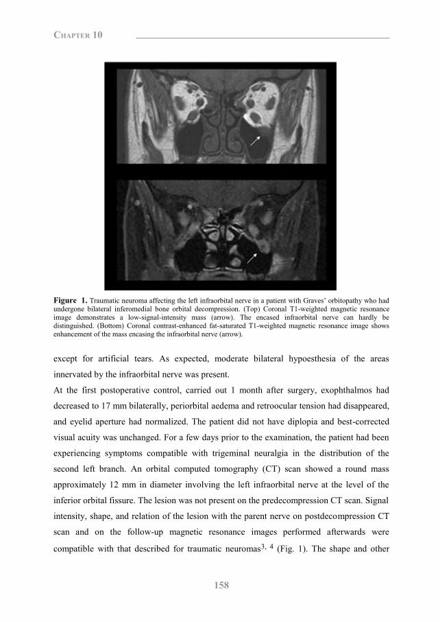
158
Chapter 10
Figure 1. Traumatic neuroma affecting the left infraorbital nerve in a patient with Graves‟ orbitopathy who had undergone bilateral inferomedial bone orbital decompression. (Top) Coronal T1-weighted magnetic resonance image demonstrates a low-signal-intensity mass (arrow). The encased infraorbital nerve can hardly be distinguished. (Bottom) Coronal contrast-enhanced fat-saturated T1-weighted magnetic resonance image shows enhancement of the mass encasing the infraorbital nerve (arrow).
except for artificial tears. As expected, moderate bilateral hypoesthesia of the areas
innervated by the infraorbital nerve was present.
At the first postoperative control, carried out 1 month after surgery, exophthalmos had
decreased to 17 mm bilaterally, periorbital aedema and retroocular tension had disappeared,
and eyelid aperture had normalized. The patient did not have diplopia and best-corrected
visual acuity was unchanged. For a few days prior to the examination, the patient had been
experiencing symptoms compatible with trigeminal neuralgia in the distribution of the
second left branch. An orbital computed tomography (CT) scan showed a round mass
approximately 12 mm in diameter involving the left infraorbital nerve at the level of the
inferior orbital fissure. The lesion was not present on the predecompression CT scan. Signal
intensity, shape, and relation of the lesion with the parent nerve on postdecompression CT
scan and on the follow-up magnetic resonance images performed afterwards were
compatible with that described for traumatic neuromas3, 4 (Fig. 1). The shape and other
radiologic characteristics of the lesion remained constant until September 2008, when the
patient attended her last follow-up examination.
When the lesion was detected, the patient refused systemic glucocorticoids and truncular
anesthesia of the infraorbital nerve. Persistent trigeminal neuralgia did not respond to
medical treatment with oral gabapentin 600 mg 2 to 3 times daily, but could be partially
controlled by oral pregabalin 75 mg 3 times daily until the present.
Discussion
Orbital decompression is mostly achieved by means of osteotomies which involve the
lateral and the medial orbital walls, the orbital floor, and when necessary fat is removed
possibly from the inferolateral orbital quadrant. The most effective and safest sequence of
osteotomies and lipectomies to be used to gradually implement the effects of
decompression surgery continues to be debated. Although orbital floor removal in course of
orbital decompression is currently not favored in North America, a recent prospective
survey of the European Group on Graves‟ Orbitopathy (EUGOGO) showed that
inferomedial bone decompression is still a widely used procedure in Europe.5
When clinical history and symptoms suggest the presence of a traumatic neuroma, the
diagnosis can be further supported by radiologic imaging evidence of an abnormal bulbous
appearance at the proximal end of or along a nerve which was respectively transected or
damaged. CT and magnetic resonance imaging are optimal in detecting the location and
shape of the lesion and its relation with the parent nerve, while their nonspecific signal
intensity and variable enhancement after administration of contrast are less important for
the diagnosis. Although we did not perform any confirmatory histopathology, in all
likelihood the lesion we reported represents a traumatic neuroma, and we regard an
unnoticed iatrogenic injury at the time of surgery as its most probable cause. As in the case
we described, traumatic neuromas have been reported to become evident at approximately 1
month after acute trauma.3 The patient we described started presenting symptoms of
trigeminal neuralgia and evidence of the lesion on imaging a few weeks after surgery, and
this does not permit us to include in the possible pathogenesis “chronic” causative
mechanisms such as nerve irritation due to aggressive bone edges left at the level of the
anterior margin of the inferior orbital fissure. Other trauma to the nerve such as stretching
due to the mechanical action of the soft orbital tissues prolapsing into the maxillary sinus,
100379 Thesis Baldeschi.indd 158 vrijdag17-december-2010 12:09
159
Chapter 10
Figure 1. Traumatic neuroma affecting the left infraorbital nerve in a patient with Graves‟ orbitopathy who had undergone bilateral inferomedial bone orbital decompression. (Top) Coronal T1-weighted magnetic resonance image demonstrates a low-signal-intensity mass (arrow). The encased infraorbital nerve can hardly be distinguished. (Bottom) Coronal contrast-enhanced fat-saturated T1-weighted magnetic resonance image shows enhancement of the mass encasing the infraorbital nerve (arrow).
except for artificial tears. As expected, moderate bilateral hypoesthesia of the areas
innervated by the infraorbital nerve was present.
At the first postoperative control, carried out 1 month after surgery, exophthalmos had
decreased to 17 mm bilaterally, periorbital aedema and retroocular tension had disappeared,
and eyelid aperture had normalized. The patient did not have diplopia and best-corrected
visual acuity was unchanged. For a few days prior to the examination, the patient had been
experiencing symptoms compatible with trigeminal neuralgia in the distribution of the
second left branch. An orbital computed tomography (CT) scan showed a round mass
approximately 12 mm in diameter involving the left infraorbital nerve at the level of the
inferior orbital fissure. The lesion was not present on the predecompression CT scan. Signal
intensity, shape, and relation of the lesion with the parent nerve on postdecompression CT
scan and on the follow-up magnetic resonance images performed afterwards were
compatible with that described for traumatic neuromas3, 4 (Fig. 1). The shape and other
radiologic characteristics of the lesion remained constant until September 2008, when the
patient attended her last follow-up examination.
When the lesion was detected, the patient refused systemic glucocorticoids and truncular
anesthesia of the infraorbital nerve. Persistent trigeminal neuralgia did not respond to
medical treatment with oral gabapentin 600 mg 2 to 3 times daily, but could be partially
controlled by oral pregabalin 75 mg 3 times daily until the present.
Discussion
Orbital decompression is mostly achieved by means of osteotomies which involve the
lateral and the medial orbital walls, the orbital floor, and when necessary fat is removed
possibly from the inferolateral orbital quadrant. The most effective and safest sequence of
osteotomies and lipectomies to be used to gradually implement the effects of
decompression surgery continues to be debated. Although orbital floor removal in course of
orbital decompression is currently not favored in North America, a recent prospective
survey of the European Group on Graves‟ Orbitopathy (EUGOGO) showed that
inferomedial bone decompression is still a widely used procedure in Europe.5
When clinical history and symptoms suggest the presence of a traumatic neuroma, the
diagnosis can be further supported by radiologic imaging evidence of an abnormal bulbous
appearance at the proximal end of or along a nerve which was respectively transected or
damaged. CT and magnetic resonance imaging are optimal in detecting the location and
shape of the lesion and its relation with the parent nerve, while their nonspecific signal
intensity and variable enhancement after administration of contrast are less important for
the diagnosis. Although we did not perform any confirmatory histopathology, in all
likelihood the lesion we reported represents a traumatic neuroma, and we regard an
unnoticed iatrogenic injury at the time of surgery as its most probable cause. As in the case
we described, traumatic neuromas have been reported to become evident at approximately 1
month after acute trauma.3 The patient we described started presenting symptoms of
trigeminal neuralgia and evidence of the lesion on imaging a few weeks after surgery, and
this does not permit us to include in the possible pathogenesis “chronic” causative
mechanisms such as nerve irritation due to aggressive bone edges left at the level of the
anterior margin of the inferior orbital fissure. Other trauma to the nerve such as stretching
due to the mechanical action of the soft orbital tissues prolapsing into the maxillary sinus,
100379 Thesis Baldeschi.indd 159 vrijdag17-december-2010 12:09
160
Chapter 10
which normally occurs after floor removal orbital decompression, are also unlikely to have
been the cause of the lesion.
Traumatic neuromas of the infraorbital nerve, in fact, have never been described before,
despite the elevated number of this type of surgery performed and reported in published
series up until now. Although the rapid onset nature of the orbital mass might have been
suggestive of an etiology ranging from inflammation to malignancy, with the patient‟s
consent, we elected not to perform incisional or excisional biopsies and histopathology on
the lesion, but only to treat it medically and to closely follow it up with imaging. This
choice, of course, implied accepting a diagnosis suggestive of traumatic neuroma and some
risks, but surgery would have not been without risk either. Although medical therapy could
offer only a partial control of the symptoms, we did not attempt to treat the lesion more
aggressively, as more invasive treatment alternatives including intralesional injections of
steroid or alcohol, phenolization, cryoapplications, and surgical resection do not offer any
superior chance of pain relief.6
Common complications of orbital decompression are consecutive strabismus, infraorbital
hypoesthesia and sinusitis, lower lid entropion, and eyeball dystopia, while leakage of
cerebrospinal fluid, infections involving the central nervous system, damage to the eye and
optic nerve or their vasculature, cerebral vasospasm, ischemia and infarction, and delayed
decompression-related reactivation are rare events.7 Among these latter complications,
traumatic neuromas deserve a special mention, because they can cause disabling trigeminal
neuralgia with limited possibility of treatment. This retrospective case report was carried
out following the tenets of the Declaration of Helsinki and of the Code of Conduct of the
Dutch Federation of Biomedical Scientific Societies (http://www.federa.org/) as
recommended by the Review Board of our institution. The Review Board of our institution
declared that for retrospective studies its permission is not needed, nor is patient informed
consent necessary if data are collected anonymously from the medical records used by the
authors in their everyday practice.
References 1. Messmer EP, Camara J, Boniuk M, Font RL. Amputation neuroma of the orbit. Report of two cases and review
of the literature. Ophthalmology 1984;9:1420-3.
2. Bahn RS. Is orbital decompression a safe and effective treatment for Graves‟ orbitopathy? Nat Clin Pract
Endocrinol Metab 2007;3:796-7.
3. Singson RD, Feldman F, Slipman CW, Gonzalez E, Rosenberg ZS, Kiernan H. Postamputation neuromas and
other symptomatic stump abnormalities: detection with CT. Radiology 1987;162: 743-5.
4. Singson RD, Feldman F, Staron R, Fechtner D, Gonzalez E, Stein J. MRI of post amputation neuromas. Skeletal
Radiol 1990;19:259-62.
5. Mourits, MP, Bijl HM, Baldeschi L, Boboridis K, Curro N, Dickinson J, Eckstein AK, Guastella C, Altea M,
Kahaly G, Freidel M, Kalmann R, Krassas GE, Lane C, Lareida J, Marcocci C, Marino M, Nardi M, Mohr Ch,
Neoh C, Pinchera A, Orgiazzi J, Pitz S, Saeed P, Salvi M, Sellari-Franceschini S, Stahl M, von Arx G, Wiersinga
W. Outcome of orbital decompression for disfiguring proptosis in patients with Graves‟ orbitopathy using various
surgical procedures. Br J Ophthalmol 2009;93:1518-23.
6. Ernberg LA, Adler RS, Lane J. Ultrasound in the detection and treatment of a painful stump neuroma. Skeletal
Radiol 2003;32:306-9.
7. Baldeschi L. Orbital decompression. In: Wiersinga WM, Kahaly GJ, editors. Graves‟ Orbitopathy A
Multidisciplinary Approach. Basel: Karger, 2007:163-75.
100379 Thesis Baldeschi.indd 160 vrijdag17-december-2010 12:09
161
Chapter 10
which normally occurs after floor removal orbital decompression, are also unlikely to have
been the cause of the lesion.
Traumatic neuromas of the infraorbital nerve, in fact, have never been described before,
despite the elevated number of this type of surgery performed and reported in published
series up until now. Although the rapid onset nature of the orbital mass might have been
suggestive of an etiology ranging from inflammation to malignancy, with the patient‟s
consent, we elected not to perform incisional or excisional biopsies and histopathology on
the lesion, but only to treat it medically and to closely follow it up with imaging. This
choice, of course, implied accepting a diagnosis suggestive of traumatic neuroma and some
risks, but surgery would have not been without risk either. Although medical therapy could
offer only a partial control of the symptoms, we did not attempt to treat the lesion more
aggressively, as more invasive treatment alternatives including intralesional injections of
steroid or alcohol, phenolization, cryoapplications, and surgical resection do not offer any
superior chance of pain relief.6
Common complications of orbital decompression are consecutive strabismus, infraorbital
hypoesthesia and sinusitis, lower lid entropion, and eyeball dystopia, while leakage of
cerebrospinal fluid, infections involving the central nervous system, damage to the eye and
optic nerve or their vasculature, cerebral vasospasm, ischemia and infarction, and delayed
decompression-related reactivation are rare events.7 Among these latter complications,
traumatic neuromas deserve a special mention, because they can cause disabling trigeminal
neuralgia with limited possibility of treatment. This retrospective case report was carried
out following the tenets of the Declaration of Helsinki and of the Code of Conduct of the
Dutch Federation of Biomedical Scientific Societies (http://www.federa.org/) as
recommended by the Review Board of our institution. The Review Board of our institution
declared that for retrospective studies its permission is not needed, nor is patient informed
consent necessary if data are collected anonymously from the medical records used by the
authors in their everyday practice.
References 1. Messmer EP, Camara J, Boniuk M, Font RL. Amputation neuroma of the orbit. Report of two cases and review
of the literature. Ophthalmology 1984;9:1420-3.
2. Bahn RS. Is orbital decompression a safe and effective treatment for Graves‟ orbitopathy? Nat Clin Pract
Endocrinol Metab 2007;3:796-7.
3. Singson RD, Feldman F, Slipman CW, Gonzalez E, Rosenberg ZS, Kiernan H. Postamputation neuromas and
other symptomatic stump abnormalities: detection with CT. Radiology 1987;162: 743-5.
4. Singson RD, Feldman F, Staron R, Fechtner D, Gonzalez E, Stein J. MRI of post amputation neuromas. Skeletal
Radiol 1990;19:259-62.
5. Mourits, MP, Bijl HM, Baldeschi L, Boboridis K, Curro N, Dickinson J, Eckstein AK, Guastella C, Altea M,
Kahaly G, Freidel M, Kalmann R, Krassas GE, Lane C, Lareida J, Marcocci C, Marino M, Nardi M, Mohr Ch,
Neoh C, Pinchera A, Orgiazzi J, Pitz S, Saeed P, Salvi M, Sellari-Franceschini S, Stahl M, von Arx G, Wiersinga
W. Outcome of orbital decompression for disfiguring proptosis in patients with Graves‟ orbitopathy using various
surgical procedures. Br J Ophthalmol 2009;93:1518-23.
6. Ernberg LA, Adler RS, Lane J. Ultrasound in the detection and treatment of a painful stump neuroma. Skeletal
Radiol 2003;32:306-9.
7. Baldeschi L. Orbital decompression. In: Wiersinga WM, Kahaly GJ, editors. Graves‟ Orbitopathy A
Multidisciplinary Approach. Basel: Karger, 2007:163-75.
100379 Thesis Baldeschi.indd 161 vrijdag17-december-2010 12:09

Chapter 11
General Discussion
Orbital Surgery in Graves’ Orbitopathy:
Where We Are, What Are the Novel Perspectives,
What Are Its Concealed Messages?
100379 Thesis Baldeschi.indd 162 vrijdag17-december-2010 12:09
Chapter 11
General Discussion
Orbital Surgery in Graves’ Orbitopathy:
Where We Are, What Are the Novel Perspectives,
What Are Its Concealed Messages?
100379 Thesis Baldeschi.indd 163 vrijdag17-december-2010 12:09

Where we are
In ancient times lacking any background experience, accurate tools, technology to guide the
comprehension of diseases and adequate prescription drugs our ancestors struggled to learn
how to repair the human body. Surgery came into being and with it, human life became
more sophisticated. Man lived longer, and having gained a knowledge of themselves
sufficient to break the boundaries built by ignorance, ultimately increased their quality of
life. Later in eras wherein religious views took precedence over medicine and logic,
surgical advancement was difficult. Some of the surgical technology developed in ancient
times surpassed anything available in the modern world until the 18th or 19th century.
Most of the knowledge we now have was obtained from prehistoric exploits.1
Skull trephination and craniotomy performed by abrasion, scraping, crosscut sawing, and
drilling are the oldest known surgical techniques used by primitive people for
decompression of the brain after fracture or epidural haemorrhages, to cure epilepsy or
osteomyelitis.2, 3
The indications for modern decompression surgery for GO are not too dissimilar from those
which motivated the invention of surgery in ancient times, and the greatest bulk of it, bone
decompression, conceptually continues to be based on principles and techniques similar to
those for cranial trephination from as early as 70003, or even 20000 years ago.4 Stone
lancets and obsidian curettes, already used successfully in up to 80% of such prehistoric
surgery2, 3, 5, have been substituted by more sophisticated equipment. Nevertheless,
abrasion, scraping, crosscut sawing, and drilling are still in current use in the great majority
of surgery for treating raised intraorbital pressure responsible of signs and symptoms of
GO.
Modern bone orbital decompression surgery in GO started in 1911 when Dollinger6
proposed the removal of the lateral orbital wall, which had been already used by Kroenlein7
for the excision of an orbital dermoid cyst, as a possibility to increase the volume of the
bony orbit in GO. Till since many other osteotomies and approaches have been proposed,
abandoned, modified7-26 and performed with minimally invasive techniques or more
extensive incisions each carrying its distinctive advantages and disadvantages as discussed
in the paper included in chapter 6.27 Starting from the end of the 1990s, the osteotomy
entailing the removal of the lateral orbital wall, and in particular its deep portion, has
100379 Thesis Baldeschi.indd 164 vrijdag17-december-2010 12:09
165
Chapter 11
Where we are
In ancient times lacking any background experience, accurate tools, technology to guide the
comprehension of diseases and adequate prescription drugs our ancestors struggled to learn
how to repair the human body. Surgery came into being and with it, human life became
more sophisticated. Man lived longer, and having gained a knowledge of themselves
sufficient to break the boundaries built by ignorance, ultimately increased their quality of
life. Later in eras wherein religious views took precedence over medicine and logic,
surgical advancement was difficult. Some of the surgical technology developed in ancient
times surpassed anything available in the modern world until the 18th or 19th century.
Most of the knowledge we now have was obtained from prehistoric exploits.1
Skull trephination and craniotomy performed by abrasion, scraping, crosscut sawing, and
drilling are the oldest known surgical techniques used by primitive people for
decompression of the brain after fracture or epidural haemorrhages, to cure epilepsy or
osteomyelitis.2, 3
The indications for modern decompression surgery for GO are not too dissimilar from those
which motivated the invention of surgery in ancient times, and the greatest bulk of it, bone
decompression, conceptually continues to be based on principles and techniques similar to
those for cranial trephination from as early as 70003, or even 20000 years ago.4 Stone
lancets and obsidian curettes, already used successfully in up to 80% of such prehistoric
surgery2, 3, 5, have been substituted by more sophisticated equipment. Nevertheless,
abrasion, scraping, crosscut sawing, and drilling are still in current use in the great majority
of surgery for treating raised intraorbital pressure responsible of signs and symptoms of
GO.
Modern bone orbital decompression surgery in GO started in 1911 when Dollinger6
proposed the removal of the lateral orbital wall, which had been already used by Kroenlein7
for the excision of an orbital dermoid cyst, as a possibility to increase the volume of the
bony orbit in GO. Till since many other osteotomies and approaches have been proposed,
abandoned, modified7-26 and performed with minimally invasive techniques or more
extensive incisions each carrying its distinctive advantages and disadvantages as discussed
in the paper included in chapter 6.27 Starting from the end of the 1990s, the osteotomy
entailing the removal of the lateral orbital wall, and in particular its deep portion, has
100379 Thesis Baldeschi.indd 165 vrijdag17-december-2010 12:09
166
Chapter 11
regained popularity23, 24, 26, roughly reiterating what had been proposed one century before
by Dollinger, when he described his pristine form of bone decompression surgery.6
In essence over time human reasoning has not produced any substantial conceptual
innovation in the field of decompression surgery, or of its execution. An exception was the
introduction of fat decompression in 192028, which, however, never reached the popularity
of its bone counterpart.
Real new short term conceptual breakthroughs in orbital surgery for GO appear to be very
unlikely. Collective awareness which also extended to the possible discovery of specific
immunological therapy, has been recently stated in “the Amsterdam declaration on GO” 29,
30, and it was also maintained that the overall quality of patient care can be implemented
by reducing the inequality in expertise and delivery of existing treatments.30-32
Recently introduced technical advances33, and most importantly implementation of the
scarce evidence and quality of literature concerning available surgical treatment for GO34-
36 can also play a positive key role in the amelioration of surgical care, including orbital
decompression.
Technical advances
Technical advance in the field of decompression surgery has undeniably been substantial,
and there is further potential.33 The current use of high-speed rotary instrumentation has
facilitated the possibility of deeper lateral wall osteotomies than that achievable by means
of conventional bone nibbling rongeurs.26 This has resulted in the possibility of more
effective exophthalmos reduction24, 26, but grinding with cutting-burrs or diamond-burr
tips in tight spaces, such as those available in orbital surgery, has a potential, not negligible,
risk of damage to soft orbital tissues or the dura. Ultrasonic bone removal recently
proposed for orbital surgery represents a promising field of technical development.37, 38
Hand pieces which transmit 25-29 kHz vibrations can easily selectively carve/cut only
mineralized tissues. The reliability of ultrasonic bone curettes in orbital surgery, however,
still needs to be tested against large numbers, and issues of concern about possible damage
to soft tissues when using such devices have been arisen in neurosurgery.39 Technical
innovations for decompression surgery, however are limited when compared with those
that are already available and constantly implemented for other rehabilitative surgical
procedures for GO such as blepharoplasty, forehead plasty, and eyebrow lift. This is the
result of the benefits of technical solutions from the commercial dynamic world of
cosmetic surgery.33
Decompression surgery: evidence and quality of the literature
A clear methodology is a basic requirement for any scientific paper. In clinical science a
plain definition of the treatment, the circumstances under which it is administered, and an
unambiguous description of the treated patient population are essential ingredients. These
criteria guarantee that results are repeatable and that meaningful comparisons can be made
among studies. Most articles on the outcome of decompression surgery does not fulfil these
criteria. They are chiefly based on retrospective case series with inhomogeneous
methodologies and definitions, heterogeneous anatomical and pathological substrates, none
standardized modalities and timing of pre- and postoperative clinical assessments,
differences in surgical techniques, modality, location, and amount of removed bone or
fat.27 Consequently surgical choices continue to be left to local tradition and surgeons‟
experience40 rather than being based on evidence that links the patient‟s specific needs,
and their background characteristics to one or another surgical approach.
At present patients‟ access to high standard surgical treatments is still not prompt or
available in many countries29, 30, and not many are the centres of excellence or the
international organizations with a referral adequate enough to produce randomized clinical
trials in the field of decompression surgery.40 Although a document such as the Amsterdam
declaration29 might instigate a medium term desirable implementation in surgical care for
patients with GO, it is not unforeseen that retrospective case series will continue to
remain the commonest study design for decompression surgery.
While the evidence of the literature might not be easily implemented, its quality can.
Limitations can be overcome, and overstatements and conceptual axioms are liable to
critical review, thus potentially generating novel data and perspectives useful for clinical
practice or future research.
The main ambition of this thesis was to stimulate a move towards a more systematic
reporting and analytical style which can, more reliably and comprehensively, allow present
100379 Thesis Baldeschi.indd 166 vrijdag17-december-2010 12:09
167
Chapter 11
regained popularity23, 24, 26, roughly reiterating what had been proposed one century before
by Dollinger, when he described his pristine form of bone decompression surgery.6
In essence over time human reasoning has not produced any substantial conceptual
innovation in the field of decompression surgery, or of its execution. An exception was the
introduction of fat decompression in 192028, which, however, never reached the popularity
of its bone counterpart.
Real new short term conceptual breakthroughs in orbital surgery for GO appear to be very
unlikely. Collective awareness which also extended to the possible discovery of specific
immunological therapy, has been recently stated in “the Amsterdam declaration on GO” 29,
30, and it was also maintained that the overall quality of patient care can be implemented
by reducing the inequality in expertise and delivery of existing treatments.30-32
Recently introduced technical advances33, and most importantly implementation of the
scarce evidence and quality of literature concerning available surgical treatment for GO34-
36 can also play a positive key role in the amelioration of surgical care, including orbital
decompression.
Technical advances
Technical advance in the field of decompression surgery has undeniably been substantial,
and there is further potential.33 The current use of high-speed rotary instrumentation has
facilitated the possibility of deeper lateral wall osteotomies than that achievable by means
of conventional bone nibbling rongeurs.26 This has resulted in the possibility of more
effective exophthalmos reduction24, 26, but grinding with cutting-burrs or diamond-burr
tips in tight spaces, such as those available in orbital surgery, has a potential, not negligible,
risk of damage to soft orbital tissues or the dura. Ultrasonic bone removal recently
proposed for orbital surgery represents a promising field of technical development.37, 38
Hand pieces which transmit 25-29 kHz vibrations can easily selectively carve/cut only
mineralized tissues. The reliability of ultrasonic bone curettes in orbital surgery, however,
still needs to be tested against large numbers, and issues of concern about possible damage
to soft tissues when using such devices have been arisen in neurosurgery.39 Technical
innovations for decompression surgery, however are limited when compared with those
that are already available and constantly implemented for other rehabilitative surgical
procedures for GO such as blepharoplasty, forehead plasty, and eyebrow lift. This is the
result of the benefits of technical solutions from the commercial dynamic world of
cosmetic surgery.33
Decompression surgery: evidence and quality of the literature
A clear methodology is a basic requirement for any scientific paper. In clinical science a
plain definition of the treatment, the circumstances under which it is administered, and an
unambiguous description of the treated patient population are essential ingredients. These
criteria guarantee that results are repeatable and that meaningful comparisons can be made
among studies. Most articles on the outcome of decompression surgery does not fulfil these
criteria. They are chiefly based on retrospective case series with inhomogeneous
methodologies and definitions, heterogeneous anatomical and pathological substrates, none
standardized modalities and timing of pre- and postoperative clinical assessments,
differences in surgical techniques, modality, location, and amount of removed bone or
fat.27 Consequently surgical choices continue to be left to local tradition and surgeons‟
experience40 rather than being based on evidence that links the patient‟s specific needs,
and their background characteristics to one or another surgical approach.
At present patients‟ access to high standard surgical treatments is still not prompt or
available in many countries29, 30, and not many are the centres of excellence or the
international organizations with a referral adequate enough to produce randomized clinical
trials in the field of decompression surgery.40 Although a document such as the Amsterdam
declaration29 might instigate a medium term desirable implementation in surgical care for
patients with GO, it is not unforeseen that retrospective case series will continue to
remain the commonest study design for decompression surgery.
While the evidence of the literature might not be easily implemented, its quality can.
Limitations can be overcome, and overstatements and conceptual axioms are liable to
critical review, thus potentially generating novel data and perspectives useful for clinical
practice or future research.
The main ambition of this thesis was to stimulate a move towards a more systematic
reporting and analytical style which can, more reliably and comprehensively, allow present
100379 Thesis Baldeschi.indd 167 vrijdag17-december-2010 12:09
168
Chapter 11
and future generations to use the data in clinical practice, the planning of new clinical trials
and meta-analysis.
Novel perspectives
If present and future studies are to be used and interpreted adequately, it is essential that
sufficient information be provided regarding the methods of data collection, and baseline
characteristics: male gender, older age and heavy smoking are patient characteristics that
are known to be associated with more severe orbital disease.41, 43 Preoperative Hertel
values, extent and location of the osteotomy performed during decompression surgery, and
differences in manipulation of the periorbita, are factors known to affect the extent of
exophthalmos reduction.26, 44-51 Precise data collection, listing of inclusion and exclusion
criteria followed by a careful evaluation of predecompression characteristics of the cohort
and groups under evaluation are therefore essential methodological requirements. Only in
this way can it be clear to whom the results of a survey apply, and whether or not the
groups under evaluation present differences or are homogeneous.
All these methodological limitations, and misleading statements regarding the negative
effects of long standing GO and previous orbital radiotherapy on the outcome of
decompression surgery were addressed in the study published in chapters 2, and 3. A clear
methodology and careful analysis of the results allowed us to compare homogeneous
patient populations and to conclude that a long duration of GO and orbital radiotherapy do
not have an adverse effect on decompression surgery52, 53, and that there was a decreased
risk for developing post decompression diplopia in long duration GO as compared with
shorter duration.52 Such strict methodology as in the papers included in chapters 2 and 352,
53 was used in the case control study included in chapter 5 aimed at quantifying, in clinical
practice, the previously unpublished contribution of popular lateral wall osteotomy to
exophthalmos reduction and the influence of such an osteotomy on consecutive diplopia.27
We concluded that a deep lateral wall is an effective although not always available zone of
possible orbital volume expansion, and that it is connected with a minimal incidence of
decompression induced diplopia when performed as part of a 3-wall decompression
through a coronal approach.
Despite the retrospective nature of these studies27, 52, 53, they had the merit of showing that
validity can be increased, by respecting as much as possible symmetry with strict
methodology, and that bias and confounding factors can be controlled by adopting clear
inclusion/exclusion criteria and by careful comparison of the groups under consideration for
demographics and preoperative characteristics.
The prospective trial included in chapter 4 was aimed at a better assessment of outcome
following decompression surgery. Demographics and pre decompression characteristics
were addressed similarly to the studies included in chapter 2, 3, and 5. The context of the
study allowed us to review the “time honoured axiom” which links the best results with the
highest exophthalmos reduction and the lowest incidence of induced diplopia, and to assess
the results of decompression surgery based on a novel methodology. The novel appraisal
method with descriptive figures (achievement or not of the preoperative target) and easily
interpretable quantitative indexes, precisely defined the extension of applied surgery and
its outcomes, to whom they pertain and, empowered the survey to disclose new findings
regarding pre-decompression characteristics and surgical results. The study showed that
different pre-decompression medical regimens and the duration of orbitopathy at
decompression may influence the targets to be pursued by decompression surgery and that
the development of GO at an older age, with associated low Hertel readings necessitates
targets in decompression surgery other than exophthalmos reduction, and that the “best
results” are linked to the achievement of the desired targets which may or may not include
reduction of exophthalmos. Additionally the paradigm that amount of exophthalmos
reduction is proportional to the extent of the osteotomies was shown not to be always valid.
Although the novel appraisal method of this study took into account a large proportion of
the variables which can play a role in determining the outcomes of decompression surgery,
it still remains sub optimal. Morphology of the orbital cavity and its osteology, compliance
of the soft orbital tissue, lipogenic versus the myogenic variants of the disease, all of which
are potential determinants of surgical outcome, were not considered. The novel appraisal
and the generated indexes, however, can already be of some benefit for straight forward
study comparisons, patient counselling, and can be a valuable tool for balancing risks
benefits when changing invasivety of the procedure during the course of surgery. This
then might represent a step towards the final goal of a desirable, and as much as possible,
100379 Thesis Baldeschi.indd 168 vrijdag17-december-2010 12:09

169
Chapter 11
and future generations to use the data in clinical practice, the planning of new clinical trials
and meta-analysis.
Novel perspectives
If present and future studies are to be used and interpreted adequately, it is essential that
sufficient information be provided regarding the methods of data collection, and baseline
characteristics: male gender, older age and heavy smoking are patient characteristics that
are known to be associated with more severe orbital disease.41, 43 Preoperative Hertel
values, extent and location of the osteotomy performed during decompression surgery, and
differences in manipulation of the periorbita, are factors known to affect the extent of
exophthalmos reduction.26, 44-51 Precise data collection, listing of inclusion and exclusion
criteria followed by a careful evaluation of predecompression characteristics of the cohort
and groups under evaluation are therefore essential methodological requirements. Only in
this way can it be clear to whom the results of a survey apply, and whether or not the
groups under evaluation present differences or are homogeneous.
All these methodological limitations, and misleading statements regarding the negative
effects of long standing GO and previous orbital radiotherapy on the outcome of
decompression surgery were addressed in the study published in chapters 2, and 3. A clear
methodology and careful analysis of the results allowed us to compare homogeneous
patient populations and to conclude that a long duration of GO and orbital radiotherapy do
not have an adverse effect on decompression surgery52, 53, and that there was a decreased
risk for developing post decompression diplopia in long duration GO as compared with
shorter duration.52 Such strict methodology as in the papers included in chapters 2 and 352,
53 was used in the case control study included in chapter 5 aimed at quantifying, in clinical
practice, the previously unpublished contribution of popular lateral wall osteotomy to
exophthalmos reduction and the influence of such an osteotomy on consecutive diplopia.27
We concluded that a deep lateral wall is an effective although not always available zone of
possible orbital volume expansion, and that it is connected with a minimal incidence of
decompression induced diplopia when performed as part of a 3-wall decompression
through a coronal approach.
Despite the retrospective nature of these studies27, 52, 53, they had the merit of showing that
validity can be increased, by respecting as much as possible symmetry with strict
methodology, and that bias and confounding factors can be controlled by adopting clear
inclusion/exclusion criteria and by careful comparison of the groups under consideration for
demographics and preoperative characteristics.
The prospective trial included in chapter 4 was aimed at a better assessment of outcome
following decompression surgery. Demographics and pre decompression characteristics
were addressed similarly to the studies included in chapter 2, 3, and 5. The context of the
study allowed us to review the “time honoured axiom” which links the best results with the
highest exophthalmos reduction and the lowest incidence of induced diplopia, and to assess
the results of decompression surgery based on a novel methodology. The novel appraisal
method with descriptive figures (achievement or not of the preoperative target) and easily
interpretable quantitative indexes, precisely defined the extension of applied surgery and
its outcomes, to whom they pertain and, empowered the survey to disclose new findings
regarding pre-decompression characteristics and surgical results. The study showed that
different pre-decompression medical regimens and the duration of orbitopathy at
decompression may influence the targets to be pursued by decompression surgery and that
the development of GO at an older age, with associated low Hertel readings necessitates
targets in decompression surgery other than exophthalmos reduction, and that the “best
results” are linked to the achievement of the desired targets which may or may not include
reduction of exophthalmos. Additionally the paradigm that amount of exophthalmos
reduction is proportional to the extent of the osteotomies was shown not to be always valid.
Although the novel appraisal method of this study took into account a large proportion of
the variables which can play a role in determining the outcomes of decompression surgery,
it still remains sub optimal. Morphology of the orbital cavity and its osteology, compliance
of the soft orbital tissue, lipogenic versus the myogenic variants of the disease, all of which
are potential determinants of surgical outcome, were not considered. The novel appraisal
and the generated indexes, however, can already be of some benefit for straight forward
study comparisons, patient counselling, and can be a valuable tool for balancing risks
benefits when changing invasivety of the procedure during the course of surgery. This
then might represent a step towards the final goal of a desirable, and as much as possible,
100379 Thesis Baldeschi.indd 169 vrijdag17-december-2010 12:09

170
Chapter 11
evidence based customized approach to orbital decompression. Customization may be
required in unconventional situations as well, such as the case of a patient with an enlarged
supernumerary extraocular muscle presented in chapter 9.54
While future studies on decompression surgery will likely fine tune and expand the
evaluation criteria, the existing list should stimulate reflection among practising orbital
surgeons.
Diplopia has a considerable impact on the patient‟s quality of life, is a feared, debilitating
eventual consequence of decompression surgery, and it often deters patients and physicians
from undertaking decompression rehabilitative surgery. In the vast majority of cases,
however it is correctable with straight forward surgery performed under topical
anesthesia. Induced diplopia should therefore be considered a treatable side effect rather
than a real complication of decompression surgery. Decompression surgery can also result
in improving diplopia and instruments such as the diplopia index (ID) as proposed in the
paper included in chapter 4 could prove to be valuable for study comparisons, or when
counselling both, patients without or with diplopia about to undergo decompressions.
Only the irreversible, disabling or unwanted consequences resulting from decompression
surgery should be considered real complications. Trigeminal neuralgia is one of them. In
chapter 10 the case of a patient with GO who developed a traumatic neuroma and
trigeminal neuralgia of the infraorbital nerve subsequent to inferomedial orbital
decompression is presented.55 This previously unreported complication is unlikely to
respond to any treatment and deserves to be mentioned when counselling patients about to
undergo inferomedial decompression.
Concealed messages of orbital surgery
Careful clinical observations, particularly in a multidisciplinary setting can lead to new
insights on pathogenesis56 as discussed in chapters 7 and 8.57-58 Orbital fibroblasts are key
to the pathogenesis of GO. Fibroblasts from GO patients produce excess matrix
glycosaminoglycans, and can differentiate into fatlike adipocytes. Proliferation of orbital
fibroblasts is dependent on appropriate activation of T helper (TH) cells. However,
effective T cell help requires the local expression of co-stimulatory molecules.59, 60 The
discovery that orbital fibroblasts can express co-stimulatory molecules, such as CD40, and
MHC class II molecules, suggests that T-cell activation in orbital tissue can occur without
the traditional antigen presenting cells. Activation or perpetuation of the inflammatory
cycle can be sustained through the presentation of autoantigens by orbital fibroblasts.61
Inflammatory stimuli, for example, lipopolysaccharide and proinflammatory cytokines like
interferon-γ (IFN-γ) can also drive the autoimmune process.62, 63 These and other
inflammatory signal mediators may be induced or released at the time of surgery leading to
reactivation of disease as seen in the patients discussed in chapter 7.
Inflammation and infection have been suspected to trigger GO. The case described in
chapter 8, extends and confirms that genetics play a role. A patient received a bone marrow
transplant from a donor who years later developed Gaves‟ hyperthyroidism. The recipient
patient developed Graves‟ hyperthyroidism and orbitopathy before the donor. While donor
and recipient were siblings, the earlier onset in the recipient suggests that a preferential
recruitment and expansion of orbital tissue responsive T cells occurred. Immunity
constituted after BMT is not the same as during embryogenesis. The checks and counter
check between self and non self are not as tightly established following BMT. Certain Treg
populations may be missing, predisposing the recipient to certain viral infections or
autoimmune disease. GO obviously is no exception to this state of affairs. The fact that
orbital fibroblasts may bypass normal control mechanisms may be a further aggravating
factor in the early onset of disease in this BMT patient.
While the exact mechanisms underlying the pathogenesis of GO will come from basic
research, critical clinical observations as those presented in chapters 7 and 8 may help to
drive such research and provide valuable information for basic scientists.
100379 Thesis Baldeschi.indd 170 vrijdag17-december-2010 12:09
171
Chapter 11
evidence based customized approach to orbital decompression. Customization may be
required in unconventional situations as well, such as the case of a patient with an enlarged
supernumerary extraocular muscle presented in chapter 9.54
While future studies on decompression surgery will likely fine tune and expand the
evaluation criteria, the existing list should stimulate reflection among practising orbital
surgeons.
Diplopia has a considerable impact on the patient‟s quality of life, is a feared, debilitating
eventual consequence of decompression surgery, and it often deters patients and physicians
from undertaking decompression rehabilitative surgery. In the vast majority of cases,
however it is correctable with straight forward surgery performed under topical
anesthesia. Induced diplopia should therefore be considered a treatable side effect rather
than a real complication of decompression surgery. Decompression surgery can also result
in improving diplopia and instruments such as the diplopia index (ID) as proposed in the
paper included in chapter 4 could prove to be valuable for study comparisons, or when
counselling both, patients without or with diplopia about to undergo decompressions.
Only the irreversible, disabling or unwanted consequences resulting from decompression
surgery should be considered real complications. Trigeminal neuralgia is one of them. In
chapter 10 the case of a patient with GO who developed a traumatic neuroma and
trigeminal neuralgia of the infraorbital nerve subsequent to inferomedial orbital
decompression is presented.55 This previously unreported complication is unlikely to
respond to any treatment and deserves to be mentioned when counselling patients about to
undergo inferomedial decompression.
Concealed messages of orbital surgery
Careful clinical observations, particularly in a multidisciplinary setting can lead to new
insights on pathogenesis56 as discussed in chapters 7 and 8.57-58 Orbital fibroblasts are key
to the pathogenesis of GO. Fibroblasts from GO patients produce excess matrix
glycosaminoglycans, and can differentiate into fatlike adipocytes. Proliferation of orbital
fibroblasts is dependent on appropriate activation of T helper (TH) cells. However,
effective T cell help requires the local expression of co-stimulatory molecules.59, 60 The
discovery that orbital fibroblasts can express co-stimulatory molecules, such as CD40, and
MHC class II molecules, suggests that T-cell activation in orbital tissue can occur without
the traditional antigen presenting cells. Activation or perpetuation of the inflammatory
cycle can be sustained through the presentation of autoantigens by orbital fibroblasts.61
Inflammatory stimuli, for example, lipopolysaccharide and proinflammatory cytokines like
interferon-γ (IFN-γ) can also drive the autoimmune process.62, 63 These and other
inflammatory signal mediators may be induced or released at the time of surgery leading to
reactivation of disease as seen in the patients discussed in chapter 7.
Inflammation and infection have been suspected to trigger GO. The case described in
chapter 8, extends and confirms that genetics play a role. A patient received a bone marrow
transplant from a donor who years later developed Gaves‟ hyperthyroidism. The recipient
patient developed Graves‟ hyperthyroidism and orbitopathy before the donor. While donor
and recipient were siblings, the earlier onset in the recipient suggests that a preferential
recruitment and expansion of orbital tissue responsive T cells occurred. Immunity
constituted after BMT is not the same as during embryogenesis. The checks and counter
check between self and non self are not as tightly established following BMT. Certain Treg
populations may be missing, predisposing the recipient to certain viral infections or
autoimmune disease. GO obviously is no exception to this state of affairs. The fact that
orbital fibroblasts may bypass normal control mechanisms may be a further aggravating
factor in the early onset of disease in this BMT patient.
While the exact mechanisms underlying the pathogenesis of GO will come from basic
research, critical clinical observations as those presented in chapters 7 and 8 may help to
drive such research and provide valuable information for basic scientists.
100379 Thesis Baldeschi.indd 171 vrijdag17-december-2010 12:09
172
Chapter 11
REFERENCES 1. Ancient surgery. http://www.123Helpme.com/view.asp?id=23410.
2. Marino R Jr, Gonzales-Portillo M. Preconquest Peruvian neurosurgeons: a study of Inca and pre-
Columbian trephination and the art of medicine in ancient Peru. Neurosurgery 2000;47:940-50.
3. Heidecker KM. Trepanation of the skull in classical antiquity.Wurzbg Medizinhist Mitt 2006;25:113-
31.
4. Rebeka Stevenson. Surgery in ancient times. In: Malanie Stapleton, editor., Proceedings of the 16th
Annual History of Medicine Days. Calgary: University of Calgary, 2007:237-43.
5. Neurosurgery 7000 years ago in central Italy. Lancet 2002;359:2206.
6. Dollinger J. Die Druckentlastung der Augenhöhle durch Entfernung der äußeren Orbitawand bei
hochgradigem Exophthalmos (Morbus Basedowii) und konsekutiver Hauterkrankung. Dtsch med
Wschr 1911;37:1888-90.
7. Kroenlein RU. Zur Pathologie und operativen Behandlung der Dermoidcysten der Orbita. Beitr Klin
Chir 1889;4:149.
8. Naffziger HC. Progressive exophthalmos following thyroidectomy: its pathology and treatment. Ann
Surg 1931;94:582.
9. Duke-Elder S, MacFaul PA. Orbital involvement in general diseases. I. Endocrine disorders
(Thyreotoxicosis and endocrine exophthalmos). In: Duke-Elder S, editor. System of Ophthalmogy. The
Ocular Adnexa, vol. XIII, chapt. XIV. London: Henry Kimpton; 1974:935.
10. Sewall EC. Operative control of progressive exophthalmos. Arch Otolaryngol Head Neck Surg
1936;24:621.
11. Hirsch O. Surgical decompression of exophthalmos. Arch Otolaryngol Head Neck Surg 1950;51:325.
12. Lewkowitz F. Antral approach for paranasal sinusitis. Z Laryngol 1932;22:320.
13. Walsch TE, Ogura JH. Transantral orbital decompression for malignant exophthalmos. Laryngoscope
1957;67:544-68.
14. MacBeth R. Caldwell, Luc, and their operation. Laryngoscope 2002;112:1378-83.
15. De Santo LW. Transantral orbital decompression. In: Gorman CA, Waller RR, Dyer JA, editors. The
Eye and Orbit in Thyroid disease. New York: Raven Press, 1984:231-51.
16. Carrasco JR, Castillo I, Bilyk JR, Pribitkin EA, Savino PJ. Incidence of infraorbital hypesthesia and
sinusitis after orbital decompression for thyroid-related orbitopathy: a comparison of surgical
techniques. Ophthal Plast Reconstr Surg 2005;21:188-91.
17. McCord CD Jr. Orbital decompression for Graves' disease. Exposure through lateral canthal and
inferior fornix incision. Ophthalmology 1981;88:533.
18. Krastinova D, Rodallec A. Basedowian orbitopathy. Ann Chir Plast Esthet 1985;30:351-8.
19. Kennedy DW, Goodstein ML, Miller NR, Zinreich SJ. Endoscopic transnasal orbital decompression.
Arch Otolaryngol Head Neck Surg 1990;116:275-82.
20. Graham SM, Chee L, Alford MA, Carter KD. New techniques for surgical decompression of thyroid-
related orbitopathy. Ann Acad Med Singapore1999;28:494-7.
21. Leone CR, Piest KL, Newman RJ. Medial and lateral wall decompression for thyroid ophthalmopathy.
Am J Ophthalmol 1989;108:160-6.
22. Goldberg RA, Perry JD, Hortoleza V, Tong JT. Strabismus after balanced medial plus lateral wall
versus lateral wall only orbital decompression for disthyroid orbitopathy. Ophthal Plast Reconstr Surg
2000;16:271-7.
23. Goldberg RA, Kim AJ, Kerivan KM. The lacrimal keyhole, orbital door jamb, and basin of the inferior
orbital fissure. Three areas of deep bone in the lateral orbit. Arch Ophthalmol 1998;116:1618-24.
24. Goldberg RA. The evolving paradigm of orbital decompression surgery. Arch Ophthalmol
1998;116:95-6.
25. Shorr N, Baylis HI, Goldberg RA, Perry JD. Transcaruncular approach to the medial orbit and orbital
apex. Ophthalmology 1999;28:494-7.
26. Baldeschi L, MacAndie K, Hintschich C, Wakelkamp IMMJ, Prummel MF, Wiersinga WM. The
removal of the deep lateral wall in decompression surgery: its contribution to exophthalmos reduction
and influence on consecutive diplopia. Am J Ophthalmol 2005;140:642-7.
27. Baldeschi L. Small versus coronal incision orbital decompression in Graves‟ orbitopathy. Orbit
2010;29:177-82.
28. Moore RF. Exophthalmos and limitation of eye movements of Graves‟ disease. Lancet 1920;2:701.
29. Graves‟ orbitopathy: improving outcomes for thyroid eye disease - The Amsterdam declaration.
Thyroid 2010;20:351-2.
30. Perros P, Wiersinga WM. The Amsterdam Declaration on Graves' orbitopathy. Thyroid 2010;20:245-6.
31. Perros P, Baldeschi L, Boboridis K, Dickinson AJ, Hullo A, Kahaly GJ, Kendall-Taylor P, Krassas GE,
Lane CM, Lazarus JH, Marcocci C, Marino M, Mourits MP, Nardi M, Orgiazzi J, Pinchera A, Pitz S,
Prummel MF, Wiersinga WM. A questionnaire survey on the management of Graves‟ orbitopathy in
Europe. Eur J Endocrinol 2006;155:207-11.
32. Ramos HE, Diehl LA, Camacho CP, Perros P, Graf H. Management of Graves‟ orbitopathy in Latin
America: an international questionnaire study compared with Europe. Clin Endocrinol (Oxf)
2008;69:951-6.
33. Salvi M, Baldeschi L. Future developments. In: Wiersinga WM, Kahaly GJ, editors. Graves‟
Orbitopathy: A Multidisciplinary Approach - Questions and Answers, 2nd edition. Basel: Karger,
2010:255-64.
34. Bartalena L, Baldeschi L, Dickinson A, Eckstein A, Kendall-Taylor P, Marcocci C, Mourits M, Perros
P, Boboridis K, Boschi A, Currò N, Daumerie C, Kahaly GJ, Krassas GE, Lane CM, Lazarus JH,
Marinò M, Nardi M, Neoh C, Orgiazzi J, Pearce S, Pinchera A, Pitz S, Salvi M, Sivelli P, Stahl M, von
Arx G, Wiersinga WM; European Group on Graves' Orbitopathy (EUGOGO). Consensus statement of
the European Group on Graves‟ orbitopathy (EUGOGO) on management of Graves‟ orbitopathy. Eur J
Endocrinol 2008;158:273-85.
35. Bartalena L, Baldeschi L, Dickinson JA, Eckstein A, Kendall-Taylor P, Marcocci C, Mourits MP,
Perros P, Boboridis K, Boschi A, Currò N, Daumerie C, Kahaly GJ, Krassas G, Lane CM, Lazarus JH,
Marinò M, Nardi M, Neoh C, Orgiazzi J, Pearce S, Pinchera A, Pitz S, Salvi M, Sivelli P, Stahl M, von
Arx G, Wiersinga WM. Consensus statement of the European Group on Graves‟ orbitopathy
(EUGOGO) on management of Graves‟ orbitopathy. Thyroid 2008;18:333-46.
100379 Thesis Baldeschi.indd 172 vrijdag17-december-2010 12:09
173
Chapter 11
REFERENCES 1. Ancient surgery. http://www.123Helpme.com/view.asp?id=23410.
2. Marino R Jr, Gonzales-Portillo M. Preconquest Peruvian neurosurgeons: a study of Inca and pre-
Columbian trephination and the art of medicine in ancient Peru. Neurosurgery 2000;47:940-50.
3. Heidecker KM. Trepanation of the skull in classical antiquity.Wurzbg Medizinhist Mitt 2006;25:113-
31.
4. Rebeka Stevenson. Surgery in ancient times. In: Malanie Stapleton, editor., Proceedings of the 16th
Annual History of Medicine Days. Calgary: University of Calgary, 2007:237-43.
5. Neurosurgery 7000 years ago in central Italy. Lancet 2002;359:2206.
6. Dollinger J. Die Druckentlastung der Augenhöhle durch Entfernung der äußeren Orbitawand bei
hochgradigem Exophthalmos (Morbus Basedowii) und konsekutiver Hauterkrankung. Dtsch med
Wschr 1911;37:1888-90.
7. Kroenlein RU. Zur Pathologie und operativen Behandlung der Dermoidcysten der Orbita. Beitr Klin
Chir 1889;4:149.
8. Naffziger HC. Progressive exophthalmos following thyroidectomy: its pathology and treatment. Ann
Surg 1931;94:582.
9. Duke-Elder S, MacFaul PA. Orbital involvement in general diseases. I. Endocrine disorders
(Thyreotoxicosis and endocrine exophthalmos). In: Duke-Elder S, editor. System of Ophthalmogy. The
Ocular Adnexa, vol. XIII, chapt. XIV. London: Henry Kimpton; 1974:935.
10. Sewall EC. Operative control of progressive exophthalmos. Arch Otolaryngol Head Neck Surg
1936;24:621.
11. Hirsch O. Surgical decompression of exophthalmos. Arch Otolaryngol Head Neck Surg 1950;51:325.
12. Lewkowitz F. Antral approach for paranasal sinusitis. Z Laryngol 1932;22:320.
13. Walsch TE, Ogura JH. Transantral orbital decompression for malignant exophthalmos. Laryngoscope
1957;67:544-68.
14. MacBeth R. Caldwell, Luc, and their operation. Laryngoscope 2002;112:1378-83.
15. De Santo LW. Transantral orbital decompression. In: Gorman CA, Waller RR, Dyer JA, editors. The
Eye and Orbit in Thyroid disease. New York: Raven Press, 1984:231-51.
16. Carrasco JR, Castillo I, Bilyk JR, Pribitkin EA, Savino PJ. Incidence of infraorbital hypesthesia and
sinusitis after orbital decompression for thyroid-related orbitopathy: a comparison of surgical
techniques. Ophthal Plast Reconstr Surg 2005;21:188-91.
17. McCord CD Jr. Orbital decompression for Graves' disease. Exposure through lateral canthal and
inferior fornix incision. Ophthalmology 1981;88:533.
18. Krastinova D, Rodallec A. Basedowian orbitopathy. Ann Chir Plast Esthet 1985;30:351-8.
19. Kennedy DW, Goodstein ML, Miller NR, Zinreich SJ. Endoscopic transnasal orbital decompression.
Arch Otolaryngol Head Neck Surg 1990;116:275-82.
20. Graham SM, Chee L, Alford MA, Carter KD. New techniques for surgical decompression of thyroid-
related orbitopathy. Ann Acad Med Singapore1999;28:494-7.
21. Leone CR, Piest KL, Newman RJ. Medial and lateral wall decompression for thyroid ophthalmopathy.
Am J Ophthalmol 1989;108:160-6.
22. Goldberg RA, Perry JD, Hortoleza V, Tong JT. Strabismus after balanced medial plus lateral wall
versus lateral wall only orbital decompression for disthyroid orbitopathy. Ophthal Plast Reconstr Surg
2000;16:271-7.
23. Goldberg RA, Kim AJ, Kerivan KM. The lacrimal keyhole, orbital door jamb, and basin of the inferior
orbital fissure. Three areas of deep bone in the lateral orbit. Arch Ophthalmol 1998;116:1618-24.
24. Goldberg RA. The evolving paradigm of orbital decompression surgery. Arch Ophthalmol
1998;116:95-6.
25. Shorr N, Baylis HI, Goldberg RA, Perry JD. Transcaruncular approach to the medial orbit and orbital
apex. Ophthalmology 1999;28:494-7.
26. Baldeschi L, MacAndie K, Hintschich C, Wakelkamp IMMJ, Prummel MF, Wiersinga WM. The
removal of the deep lateral wall in decompression surgery: its contribution to exophthalmos reduction
and influence on consecutive diplopia. Am J Ophthalmol 2005;140:642-7.
27. Baldeschi L. Small versus coronal incision orbital decompression in Graves‟ orbitopathy. Orbit
2010;29:177-82.
28. Moore RF. Exophthalmos and limitation of eye movements of Graves‟ disease. Lancet 1920;2:701.
29. Graves‟ orbitopathy: improving outcomes for thyroid eye disease - The Amsterdam declaration.
Thyroid 2010;20:351-2.
30. Perros P, Wiersinga WM. The Amsterdam Declaration on Graves' orbitopathy. Thyroid 2010;20:245-6.
31. Perros P, Baldeschi L, Boboridis K, Dickinson AJ, Hullo A, Kahaly GJ, Kendall-Taylor P, Krassas GE,
Lane CM, Lazarus JH, Marcocci C, Marino M, Mourits MP, Nardi M, Orgiazzi J, Pinchera A, Pitz S,
Prummel MF, Wiersinga WM. A questionnaire survey on the management of Graves‟ orbitopathy in
Europe. Eur J Endocrinol 2006;155:207-11.
32. Ramos HE, Diehl LA, Camacho CP, Perros P, Graf H. Management of Graves‟ orbitopathy in Latin
America: an international questionnaire study compared with Europe. Clin Endocrinol (Oxf)
2008;69:951-6.
33. Salvi M, Baldeschi L. Future developments. In: Wiersinga WM, Kahaly GJ, editors. Graves‟
Orbitopathy: A Multidisciplinary Approach - Questions and Answers, 2nd edition. Basel: Karger,
2010:255-64.
34. Bartalena L, Baldeschi L, Dickinson A, Eckstein A, Kendall-Taylor P, Marcocci C, Mourits M, Perros
P, Boboridis K, Boschi A, Currò N, Daumerie C, Kahaly GJ, Krassas GE, Lane CM, Lazarus JH,
Marinò M, Nardi M, Neoh C, Orgiazzi J, Pearce S, Pinchera A, Pitz S, Salvi M, Sivelli P, Stahl M, von
Arx G, Wiersinga WM; European Group on Graves' Orbitopathy (EUGOGO). Consensus statement of
the European Group on Graves‟ orbitopathy (EUGOGO) on management of Graves‟ orbitopathy. Eur J
Endocrinol 2008;158:273-85.
35. Bartalena L, Baldeschi L, Dickinson JA, Eckstein A, Kendall-Taylor P, Marcocci C, Mourits MP,
Perros P, Boboridis K, Boschi A, Currò N, Daumerie C, Kahaly GJ, Krassas G, Lane CM, Lazarus JH,
Marinò M, Nardi M, Neoh C, Orgiazzi J, Pearce S, Pinchera A, Pitz S, Salvi M, Sivelli P, Stahl M, von
Arx G, Wiersinga WM. Consensus statement of the European Group on Graves‟ orbitopathy
(EUGOGO) on management of Graves‟ orbitopathy. Thyroid 2008;18:333-46.
100379 Thesis Baldeschi.indd 173 vrijdag17-december-2010 12:09
174
Chapter 11
36. Leong SC, Karkos PD, Macewen CJ, White PS. A systematic review of outcomes following surgical
decompression for dysthyroid orbitopathy. Laryngoscope 2009;119:1106-15.
37. Sival Calcott JA, Limberg JV, Patel S. Ultrasonic bone removal with the sonopet omni: a new
instrument for orbital surgery. Arch Ophthalmol 2005;123:1595-7.
38. Cho RI, Choe CH, Elner VM. Ultrasonic bone removal versus high-speed burring for lateral orbital
decompression: comparison of surgical outcomes for the treatment of thyroid eye disease. Ophthal
Plast Reconstr Surg 2010;26:83-7.
39. Kim K, Isu T, Matsumoto R, Isobe M, Kogure K. Surgical pitfalls of an ultrasonic bone curette
(SONOPET) in spinal surgery. Neurosurgery 2006;59:390-3.
40. European Group on Graves' Orbitopathy (EUGOGO), Mourits MP, Bijl H, Altea MA, Baldeschi L,
Boboridis K, Currò N, Dickinson AJ, Eckstein A, Freidel M, Guastella C, Kahaly GJ, Kalmann R,
Krassas GE, Lane CM, Lareida J, Marcocci C, Marino M, Nardi M, Mohr Ch, Neoh C, Pinchera A,
Orgiazzi J, Pitz S, Saeed P, Salvi M, Sellari-Franceschini S, Stahl M, von Arx G, Wiersinga WM.
Outcome of orbital decompression for disfiguring proptosis in patients with Graves' orbitopathy using
various surgical procedures. Br J Ophthalmol 2009;93:1518-23.
41. Kendler DL, Lippa J, Rootman J. The initial characteristics of Graves‟ orbitopathy vary with age and
sex. Arch Ophthalmol 1993;111:197-201.
42. Perros P, Cromble AL, Matthews JN, Kendall-Taylor P. Age and gender influence the severity of
thyroid-associated ophthalmopathy: a study of 101 patients attending a combined thyroid-eye clinic.
Clin Endocrinol 1993;38:367-72.
43. Prummel MF, Wiersinga WM. Smoking and risk of Graves‟ disease. Jama 1993;269:479-82.
44. Goldberg RA, Weinberg DA, Shorr N, Wirta D. Maximal three-wall orbital decompression through a
coronal approach. Ophthalmic Surg Lasers 1997;28:832- 43.
45. Linberg JV, Anderson LA. Transorbital decompression. Indications and results. Arch Ophthalmol
1981;99:113-9.
46. Warren JD, Spector JG, Burde R. Long-term follow-up and recent observations on 305 cases of orbital
decompression for dysthyroid orbitopathy. Laryngoscope 1989;99:35-40.
47. Leatherbarrow B, Lendrum J, Mahaffey PJ, Noble JL, Kwartz J, Davies H. Three wall orbital
decompression for Graves‟ ophthalmopathy via a coronal approach. Eye 1991;5:456-65.
48. Garrity JA, Fatourechi V, Bergstralh EJ, Bartley GB, Beatty CW, DeSanto LW, Gorman CA. Results
of transantral orbital decompression in 428 patients with severe Graves‟ ophthalmopathy. Am J
Ophthalmol 1993;116:533-47.
49. Lyons CJ, Rootman J. Orbital decompression for disfiguring exophthalmos in thyroid orbitopathy.
Ophthalmology 1994;101:223-30.
50. Fatourechi V, Garrity JA, Bartley GB, Bergstralh EJ, De-Santo LW, Gorman CA. Graves‟
ophthalmopathy. Results of transantral orbital decompression performed primarily for cosmetic
indications. Ophthalmology 1994;101:938-42.
51. Kalmann R, Mourits MP, van der Pol JP, Koornneef L. Coronal approach for rehabilitative orbital
decompression in Graves‟ ophthalmopathy. Br J Ophthalmol 1997;81:41-5.
52. Baldeschi L, Wakelkamp IMMJ, Lindeboom R, Prummel MF, Wiersinga WM: Early versus late orbital
decompression in Graves‟ orbitopathy: a retrospective study in 125 patients. Ophthalmology
2006;113:874-8.
53. Baldeschi L, MacAndie K, Koetsier E, Blank L,Wiersinga WM. The influence of previous orbital
irradiation on the outcome of rehabilitative decompression surgery in Graves‟ orbitopathy. Am J
Ophthalmol 2008;145:534-40.
54. Baldeschi L, Bisschop PHLT, Wiersinga WM. Supernumerary extraocular muscle in Graves‟
orbitopathy. Thyroid 2007;17:479-80.
55. Baldeschi L, Saeed P, Regensburg NI, Zacharopoulos I, Wiersinga WM. Traumatic neuroma of the
infraorbital nerve subsequent to infero medial orbital decompression for Graves‟ orbitopathy. Eur J
Ophthalmol 2010;20:481-4.
56. Koornneef L, Schmidt ED, van der Gaag R. Graves‟ Ophthalmopathy. In: Wall JR, How J, editors.
The orbit: structure, autoantigens and pathology. Philadelphia: Blackwell Scientific Publications,
1990:1-16.
57. Baldeschi L, Lupetti A, Vu P, Wakelkamp IMMJ, Prummel MF, Wiersinga WM. Reactivation of
Graves‟ orbitopathy after rehabilitative orbital decompression. Ophthalmology 2007;114:1395-402.
58. Valdizer Y, Lupetti A, Vandelanotte S, Lankester AC, Wiersinga WM, Baldeschi L. Graves‟
orbitopathy in a patient with adrenoleukodystropy after bone marrow transplantation. Eur J Endocrinol
2009;161:369-73.
59. Grewal IS, Flavell RA. A central role of CD40 ligand in the regulation of CD4+ T cell responses.
Immunol Today 1996;17:410-4.
60. Noelle R. CD40 and its ligand in host defense. Immunity 1996;4:415-9.
61. Sempowski GD, Rozenblit J, Smith TJ, Phipps RP. Human orbital fibroblasts are activated through
CD40 to induce proinflammatory cytokine production. Am J Phisiol 1998;274:707-14.
62. Schuurhuis DH, Laban S, Toes RE, Ricciardi-Castagnoli P, Kleijmeer MJ, van der Voort EIH, Rea D,
Offringa R, Geuze HJ, Melief CJM, Ossendorp F. Immature DC‟s acquire CD8(+) citotoxic T
lymphocite priming capacity upon activation by T helper-independent or -dependent stimuli. J Exp
Med 2000;192:145-50.
63. Heufelder AE, Smith TJ, Gorman CA, Bahn RS. Increased induction of HLA-DR by interferon-
gamma in cultured fibroblasts derived from patients with Graves' ophthalmopathy and pretibial
dermopathy. Journal of Clinical Endocrinology and Metabolism 1991;73:307-13.
100379 Thesis Baldeschi.indd 174 vrijdag17-december-2010 12:09
175
Chapter 11
36. Leong SC, Karkos PD, Macewen CJ, White PS. A systematic review of outcomes following surgical
decompression for dysthyroid orbitopathy. Laryngoscope 2009;119:1106-15.
37. Sival Calcott JA, Limberg JV, Patel S. Ultrasonic bone removal with the sonopet omni: a new
instrument for orbital surgery. Arch Ophthalmol 2005;123:1595-7.
38. Cho RI, Choe CH, Elner VM. Ultrasonic bone removal versus high-speed burring for lateral orbital
decompression: comparison of surgical outcomes for the treatment of thyroid eye disease. Ophthal
Plast Reconstr Surg 2010;26:83-7.
39. Kim K, Isu T, Matsumoto R, Isobe M, Kogure K. Surgical pitfalls of an ultrasonic bone curette
(SONOPET) in spinal surgery. Neurosurgery 2006;59:390-3.
40. European Group on Graves' Orbitopathy (EUGOGO), Mourits MP, Bijl H, Altea MA, Baldeschi L,
Boboridis K, Currò N, Dickinson AJ, Eckstein A, Freidel M, Guastella C, Kahaly GJ, Kalmann R,
Krassas GE, Lane CM, Lareida J, Marcocci C, Marino M, Nardi M, Mohr Ch, Neoh C, Pinchera A,
Orgiazzi J, Pitz S, Saeed P, Salvi M, Sellari-Franceschini S, Stahl M, von Arx G, Wiersinga WM.
Outcome of orbital decompression for disfiguring proptosis in patients with Graves' orbitopathy using
various surgical procedures. Br J Ophthalmol 2009;93:1518-23.
41. Kendler DL, Lippa J, Rootman J. The initial characteristics of Graves‟ orbitopathy vary with age and
sex. Arch Ophthalmol 1993;111:197-201.
42. Perros P, Cromble AL, Matthews JN, Kendall-Taylor P. Age and gender influence the severity of
thyroid-associated ophthalmopathy: a study of 101 patients attending a combined thyroid-eye clinic.
Clin Endocrinol 1993;38:367-72.
43. Prummel MF, Wiersinga WM. Smoking and risk of Graves‟ disease. Jama 1993;269:479-82.
44. Goldberg RA, Weinberg DA, Shorr N, Wirta D. Maximal three-wall orbital decompression through a
coronal approach. Ophthalmic Surg Lasers 1997;28:832- 43.
45. Linberg JV, Anderson LA. Transorbital decompression. Indications and results. Arch Ophthalmol
1981;99:113-9.
46. Warren JD, Spector JG, Burde R. Long-term follow-up and recent observations on 305 cases of orbital
decompression for dysthyroid orbitopathy. Laryngoscope 1989;99:35-40.
47. Leatherbarrow B, Lendrum J, Mahaffey PJ, Noble JL, Kwartz J, Davies H. Three wall orbital
decompression for Graves‟ ophthalmopathy via a coronal approach. Eye 1991;5:456-65.
48. Garrity JA, Fatourechi V, Bergstralh EJ, Bartley GB, Beatty CW, DeSanto LW, Gorman CA. Results
of transantral orbital decompression in 428 patients with severe Graves‟ ophthalmopathy. Am J
Ophthalmol 1993;116:533-47.
49. Lyons CJ, Rootman J. Orbital decompression for disfiguring exophthalmos in thyroid orbitopathy.
Ophthalmology 1994;101:223-30.
50. Fatourechi V, Garrity JA, Bartley GB, Bergstralh EJ, De-Santo LW, Gorman CA. Graves‟
ophthalmopathy. Results of transantral orbital decompression performed primarily for cosmetic
indications. Ophthalmology 1994;101:938-42.
51. Kalmann R, Mourits MP, van der Pol JP, Koornneef L. Coronal approach for rehabilitative orbital
decompression in Graves‟ ophthalmopathy. Br J Ophthalmol 1997;81:41-5.
52. Baldeschi L, Wakelkamp IMMJ, Lindeboom R, Prummel MF, Wiersinga WM: Early versus late orbital
decompression in Graves‟ orbitopathy: a retrospective study in 125 patients. Ophthalmology
2006;113:874-8.
53. Baldeschi L, MacAndie K, Koetsier E, Blank L,Wiersinga WM. The influence of previous orbital
irradiation on the outcome of rehabilitative decompression surgery in Graves‟ orbitopathy. Am J
Ophthalmol 2008;145:534-40.
54. Baldeschi L, Bisschop PHLT, Wiersinga WM. Supernumerary extraocular muscle in Graves‟
orbitopathy. Thyroid 2007;17:479-80.
55. Baldeschi L, Saeed P, Regensburg NI, Zacharopoulos I, Wiersinga WM. Traumatic neuroma of the
infraorbital nerve subsequent to infero medial orbital decompression for Graves‟ orbitopathy. Eur J
Ophthalmol 2010;20:481-4.
56. Koornneef L, Schmidt ED, van der Gaag R. Graves‟ Ophthalmopathy. In: Wall JR, How J, editors.
The orbit: structure, autoantigens and pathology. Philadelphia: Blackwell Scientific Publications,
1990:1-16.
57. Baldeschi L, Lupetti A, Vu P, Wakelkamp IMMJ, Prummel MF, Wiersinga WM. Reactivation of
Graves‟ orbitopathy after rehabilitative orbital decompression. Ophthalmology 2007;114:1395-402.
58. Valdizer Y, Lupetti A, Vandelanotte S, Lankester AC, Wiersinga WM, Baldeschi L. Graves‟
orbitopathy in a patient with adrenoleukodystropy after bone marrow transplantation. Eur J Endocrinol
2009;161:369-73.
59. Grewal IS, Flavell RA. A central role of CD40 ligand in the regulation of CD4+ T cell responses.
Immunol Today 1996;17:410-4.
60. Noelle R. CD40 and its ligand in host defense. Immunity 1996;4:415-9.
61. Sempowski GD, Rozenblit J, Smith TJ, Phipps RP. Human orbital fibroblasts are activated through
CD40 to induce proinflammatory cytokine production. Am J Phisiol 1998;274:707-14.
62. Schuurhuis DH, Laban S, Toes RE, Ricciardi-Castagnoli P, Kleijmeer MJ, van der Voort EIH, Rea D,
Offringa R, Geuze HJ, Melief CJM, Ossendorp F. Immature DC‟s acquire CD8(+) citotoxic T
lymphocite priming capacity upon activation by T helper-independent or -dependent stimuli. J Exp
Med 2000;192:145-50.
63. Heufelder AE, Smith TJ, Gorman CA, Bahn RS. Increased induction of HLA-DR by interferon-
gamma in cultured fibroblasts derived from patients with Graves' ophthalmopathy and pretibial
dermopathy. Journal of Clinical Endocrinology and Metabolism 1991;73:307-13.
100379 Thesis Baldeschi.indd 175 vrijdag17-december-2010 12:09

Summary
100379 Thesis Baldeschi.indd 176 vrijdag17-december-2010 12:09

Summary
100379 Thesis Baldeschi.indd 177 vrijdag17-december-2010 12:09

The introductory Chapter 1 provides an overview of Graves‟ orbitopathy (GO) and its
current treatment modalities. GO is one of the phenotypic appearances of Graves‟disease,
a multisystem disorder which usually leads to hyperthyroidism and goiter, less frequently
to GO, and rarely to pretibial myxedema and acropachy. Its etiopathogenesis is poorly
characterized and consequently a specific therapy is lacking. The European Group On
Graves‟ Orbitopathy (EUGOGO) has published a consensus statement on indications and
timing of the various medical and surgical treatment options in GO (Figure 2, page 14).
The current role of orbital decompression in the context of rehabilitative surgery for GO
(the subject of this thesis) is reviewed.
The studies reported in chapters 2, 3 and 4 were designed to evaluate some unproven
concepts in the existing literature on orbital decompression surgery.
Chapter 2 evaluated the concept that outcome of orbital decompression is worse when
performed rather late in the course of the disease, because there is more fibrosis and less
compliance of orbital soft tissues in the late inactive stage of GO. In a retrospective study
among GO patients without preoperative diplopia, outcome of surgical decompression was
compared between 70 patients with GO duration <4 yr and 55 patients with GO duration of
4 yr or longer. Baseline characteristics of both groups were similar, except for greater
extraocular muscle enlargement in the patients with early decompression. The surgical
outcome (applying the same technique in all patients) was not different between both
groups, except for more postdecompression diplopia in the group undergoing early
decompression. It is concluded that a long duration of GO does not adversely interfere with
the results of surgical decompression, and is associated with a decreased risk for developing
postdecompression diplopia.
Chapter 3 evaluated the concept that outcome of orbital decompression is worse when
patients have been treated with orbital irradiation prior to surgery, because radiotherapy
might induce fibrosis and less compliant soft tissues. In a retrospective study among GO
patients without preoperative diplopia, outcomes of 3-wall orbital decompression through a
coronal approach was compared between 29 patients previously treated with radiotherapy
(20 Gy), 15 patients previously treated with glucocorticoids, and 17 patients previously
treated with radiotherapy and glucocorticoids. All preoperative characteristics except
smoking behaviour were similar in the three groups. Surgical outcome did not differ
100379 Thesis Baldeschi.indd 178 vrijdag17-december-2010 12:09
179
Summary
The introductory Chapter 1 provides an overview of Graves‟ orbitopathy (GO) and its
current treatment modalities. GO is one of the phenotypic appearances of Graves‟disease,
a multisystem disorder which usually leads to hyperthyroidism and goiter, less frequently
to GO, and rarely to pretibial myxedema and acropachy. Its etiopathogenesis is poorly
characterized and consequently a specific therapy is lacking. The European Group On
Graves‟ Orbitopathy (EUGOGO) has published a consensus statement on indications and
timing of the various medical and surgical treatment options in GO (Figure 2, page 14).
The current role of orbital decompression in the context of rehabilitative surgery for GO
(the subject of this thesis) is reviewed.
The studies reported in chapters 2, 3 and 4 were designed to evaluate some unproven
concepts in the existing literature on orbital decompression surgery.
Chapter 2 evaluated the concept that outcome of orbital decompression is worse when
performed rather late in the course of the disease, because there is more fibrosis and less
compliance of orbital soft tissues in the late inactive stage of GO. In a retrospective study
among GO patients without preoperative diplopia, outcome of surgical decompression was
compared between 70 patients with GO duration <4 yr and 55 patients with GO duration of
4 yr or longer. Baseline characteristics of both groups were similar, except for greater
extraocular muscle enlargement in the patients with early decompression. The surgical
outcome (applying the same technique in all patients) was not different between both
groups, except for more postdecompression diplopia in the group undergoing early
decompression. It is concluded that a long duration of GO does not adversely interfere with
the results of surgical decompression, and is associated with a decreased risk for developing
postdecompression diplopia.
Chapter 3 evaluated the concept that outcome of orbital decompression is worse when
patients have been treated with orbital irradiation prior to surgery, because radiotherapy
might induce fibrosis and less compliant soft tissues. In a retrospective study among GO
patients without preoperative diplopia, outcomes of 3-wall orbital decompression through a
coronal approach was compared between 29 patients previously treated with radiotherapy
(20 Gy), 15 patients previously treated with glucocorticoids, and 17 patients previously
treated with radiotherapy and glucocorticoids. All preoperative characteristics except
smoking behaviour were similar in the three groups. Surgical outcome did not differ
100379 Thesis Baldeschi.indd 179 vrijdag17-december-2010 12:09
180
Summary
between groups. It is concluded that previous orbital radiotherapy does not adversely
interfere with the results of surgical decompression.
Chapter 4 evaluated the concept that -according to a time-honoured axiom- the outcomes of
orbital decompression in GO for rehabilitative reasons can be described in a satisfactory
manner in terms of exophthalmos reduction and postoperative diplopia. These conventional
outcome measures were compared to a newly designed set of outcome criteria in a
prospective study among 84 GO patients who underwent a graded inferomedial bone
decompression of 151 orbits through a minimally invasive sutureless transinferior fornix
approach. The novel criteria allowed to assess surgical outcome in relation to the extent of
surgery. Surgical targets were recorded prior to surgery, and included:1) desired degree of
exophthalmos reduction, 2) resolution of retroocular tension, 3) reduction of periorbital
swelling, 4) cure of lagophthalmos; it allowed to calculate a mean index of targets achieved
per orbit (MITAO) and per patient (MITAP), with values ranging between 0 and 1. Also an
index of diplopia (ratio of decompression-induced de novo diplopia to decompression-
induced cure of diplopia) was calculated. Invasiveness of surgery was quantified from
minimally invasive (25%) to maximally invasive (100%), allowing to calculate a mean
index of invasiveness per orbit (MIIO) and per patient (MIIP) ranging from 25% to 100%.
The novel methodology described surgical outcomes in relation to the extent of surgery
more precisely and meaningfully as compared to traditional evaluation of surgical
outcomes.
The studies reported in chapters 5 and 6 were performed in order to evaluate and review
current trends in decompression surgery with regard to ostetomies and approaches.
Chapter 5 evaluated the -previously undetermined- contribution of maximal removal of the
deep lateral wall of the orbit to exophthalmos reduction, and its influence on the onset of
diplopia. In a case-control study among GO patients undergoing 3-wall decompression
through a coronal approach, 15 patients in whom the deep portion of the lateral wall had
been removed (cases, extended decompression) were compared to 15 patients in whom the
deep lateral wall had been preserved (controls, conservative decompression). There were no
differences in baseline characteristics between cases and controls, but mean exophthalmos
reduction was 2.3 mm greater in cases than controls; onset of diplopia was similar. It is
concluded that removal of the deep lateral orbital wall as part of a coronal approach 3-wall
decompression enhances the degree of exophthalmos reduction without increasing the risk
of diplopia.
Chapter 6 reviews the advantages and disadvantages of small versus coronal incisions to
orbital decompression surgery. An inferior fornix incision can be used for inferomedial
bony decompression and/or for removing fat from the medial and lateral inferior orbital
quadrants. Through the same route a lateral ostetomy can be performed, although an upper
skin crease incision offers a wider access to the lateral orbital wall. The swinging eyelid
technique provides adequate access to the bony orbit and to the orbital fat compartments; it
is a versatile technique that can virtually be used as a standard approach for the majority of
patients needing decompression surgery. Orbital decompression by coronal incision is an
invasive technique, not to be used as a standard approach. Nevertheless, it should not be
abandoned as it could be advantageous in particular patients who may benefit more from a
tailored rather than a standardised approach. Practical implications of the choice between a
standardized approach to orbital decompression surgery and a more desirable customized
approach are discussed.
The studies reported in chapters 7, 8, 9 and 10 deal with rare conditions which may be
encountered in GO patients who are about to undergo or who already had undergone orbital
decompression surgery.
Chapter 7 describes two patients who developed clinical and radiological evidence of
reactivated GO a few weeks after rehabilitative bony orbital decompression during the
static phase of the disease. We reviewed the charts of 249 consecutive GO patients who had
undergone the same treatment for the same reason (excluded were patients with
perioperative glucocorticoids, concurrent periorbital diseases, or who were
immunocompromised). The incidence of delayed decompression-related reactivitation of
GO (called DDRR) was 1.3%. The reactivation took place a few weeks after surgery, after
a normal convalescence period. No further episodes of reactivation were observed during a
mean follow-up of 7.5 yr.
Chapter 8 describes a patient with X-linked childhood cerebral adrenoleukodystrophy; he
was treated for this condition with whole bone marrow transplantation from his sister, and
developed Graves‟ hyperthyroidism and GO six years later. His sister was euthyroid when
she donated the bone marrow transplant, but developed Graves‟ hyperthyroidism
100379 Thesis Baldeschi.indd 180 vrijdag17-december-2010 12:09
181
Summary
between groups. It is concluded that previous orbital radiotherapy does not adversely
interfere with the results of surgical decompression.
Chapter 4 evaluated the concept that -according to a time-honoured axiom- the outcomes of
orbital decompression in GO for rehabilitative reasons can be described in a satisfactory
manner in terms of exophthalmos reduction and postoperative diplopia. These conventional
outcome measures were compared to a newly designed set of outcome criteria in a
prospective study among 84 GO patients who underwent a graded inferomedial bone
decompression of 151 orbits through a minimally invasive sutureless transinferior fornix
approach. The novel criteria allowed to assess surgical outcome in relation to the extent of
surgery. Surgical targets were recorded prior to surgery, and included:1) desired degree of
exophthalmos reduction, 2) resolution of retroocular tension, 3) reduction of periorbital
swelling, 4) cure of lagophthalmos; it allowed to calculate a mean index of targets achieved
per orbit (MITAO) and per patient (MITAP), with values ranging between 0 and 1. Also an
index of diplopia (ratio of decompression-induced de novo diplopia to decompression-
induced cure of diplopia) was calculated. Invasiveness of surgery was quantified from
minimally invasive (25%) to maximally invasive (100%), allowing to calculate a mean
index of invasiveness per orbit (MIIO) and per patient (MIIP) ranging from 25% to 100%.
The novel methodology described surgical outcomes in relation to the extent of surgery
more precisely and meaningfully as compared to traditional evaluation of surgical
outcomes.
The studies reported in chapters 5 and 6 were performed in order to evaluate and review
current trends in decompression surgery with regard to ostetomies and approaches.
Chapter 5 evaluated the -previously undetermined- contribution of maximal removal of the
deep lateral wall of the orbit to exophthalmos reduction, and its influence on the onset of
diplopia. In a case-control study among GO patients undergoing 3-wall decompression
through a coronal approach, 15 patients in whom the deep portion of the lateral wall had
been removed (cases, extended decompression) were compared to 15 patients in whom the
deep lateral wall had been preserved (controls, conservative decompression). There were no
differences in baseline characteristics between cases and controls, but mean exophthalmos
reduction was 2.3 mm greater in cases than controls; onset of diplopia was similar. It is
concluded that removal of the deep lateral orbital wall as part of a coronal approach 3-wall
decompression enhances the degree of exophthalmos reduction without increasing the risk
of diplopia.
Chapter 6 reviews the advantages and disadvantages of small versus coronal incisions to
orbital decompression surgery. An inferior fornix incision can be used for inferomedial
bony decompression and/or for removing fat from the medial and lateral inferior orbital
quadrants. Through the same route a lateral ostetomy can be performed, although an upper
skin crease incision offers a wider access to the lateral orbital wall. The swinging eyelid
technique provides adequate access to the bony orbit and to the orbital fat compartments; it
is a versatile technique that can virtually be used as a standard approach for the majority of
patients needing decompression surgery. Orbital decompression by coronal incision is an
invasive technique, not to be used as a standard approach. Nevertheless, it should not be
abandoned as it could be advantageous in particular patients who may benefit more from a
tailored rather than a standardised approach. Practical implications of the choice between a
standardized approach to orbital decompression surgery and a more desirable customized
approach are discussed.
The studies reported in chapters 7, 8, 9 and 10 deal with rare conditions which may be
encountered in GO patients who are about to undergo or who already had undergone orbital
decompression surgery.
Chapter 7 describes two patients who developed clinical and radiological evidence of
reactivated GO a few weeks after rehabilitative bony orbital decompression during the
static phase of the disease. We reviewed the charts of 249 consecutive GO patients who had
undergone the same treatment for the same reason (excluded were patients with
perioperative glucocorticoids, concurrent periorbital diseases, or who were
immunocompromised). The incidence of delayed decompression-related reactivitation of
GO (called DDRR) was 1.3%. The reactivation took place a few weeks after surgery, after
a normal convalescence period. No further episodes of reactivation were observed during a
mean follow-up of 7.5 yr.
Chapter 8 describes a patient with X-linked childhood cerebral adrenoleukodystrophy; he
was treated for this condition with whole bone marrow transplantation from his sister, and
developed Graves‟ hyperthyroidism and GO six years later. His sister was euthyroid when
she donated the bone marrow transplant, but developed Graves‟ hyperthyroidism
100379 Thesis Baldeschi.indd 181 vrijdag17-december-2010 12:09
182
Summary
afterwards. The case demonstrates passive transfer of thyroid autoimmunity from an
asymptomatic donor during allogeneic stem cell transplantation.
Chapter 9 describes a GO patient presenting with restrictive strabismus. Orbital CT
scanning showed an enlarged supernumerary extraocular muscle, confirmed by biopsy.
Supernumerary extraocular muscles are quite prevalent in cats but extremely rare in
humans. The case underscores the importance of an accurate preoperative assessment of
GO in order to avoid surgical mistakes.
Chapter 10 describes a GO patient who after bilateral rehabilitative inferomedial
decompression developed a traumatic neuroma of the left infraorbital nerve. This
previously unreported but disabling complication is likely the result of unnoticed nerve
trauma at the time of surgical dissection of the infraorbital canal. The lesion remained
stable during 39 months follow-up. Symptoms of neuralgia could be controlled only
partially with medical therapy.
The general discussion in Chapter 11 puts the presented findings in GO patients who
underwent rehabilitative orbital surgery into perspective. Sometimes astute clinical
observations may contribute to better understanding basic mechanisms in the pathogenesis
of GO. Systematic review of the results obtained with orbital surgical decompression
proves to be invaluable in refining surgical techniques and improving surgical outcomes.
Samenvatting
100379 Thesis Baldeschi.indd 182 vrijdag17-december-2010 12:09

afterwards. The case demonstrates passive transfer of thyroid autoimmunity from an
asymptomatic donor during allogeneic stem cell transplantation.
Chapter 9 describes a GO patient presenting with restrictive strabismus. Orbital CT
scanning showed an enlarged supernumerary extraocular muscle, confirmed by biopsy.
Supernumerary extraocular muscles are quite prevalent in cats but extremely rare in
humans. The case underscores the importance of an accurate preoperative assessment of
GO in order to avoid surgical mistakes.
Chapter 10 describes a GO patient who after bilateral rehabilitative inferomedial
decompression developed a traumatic neuroma of the left infraorbital nerve. This
previously unreported but disabling complication is likely the result of unnoticed nerve
trauma at the time of surgical dissection of the infraorbital canal. The lesion remained
stable during 39 months follow-up. Symptoms of neuralgia could be controlled only
partially with medical therapy.
The general discussion in Chapter 11 puts the presented findings in GO patients who
underwent rehabilitative orbital surgery into perspective. Sometimes astute clinical
observations may contribute to better understanding basic mechanisms in the pathogenesis
of GO. Systematic review of the results obtained with orbital surgical decompression
proves to be invaluable in refining surgical techniques and improving surgical outcomes.
Samenvatting
100379 Thesis Baldeschi.indd 183 vrijdag17-december-2010 12:09

Het inleidend Hoofdstuk 1 geeft een overzicht van Graves‟ Orbitopathie (GO) en de
huidige behandeling ervan. GO is een van de verschijningsvormen van de ziekte van
Graves‟, een aandoening die zich op verschillende wijzen kan manifesteren:
hyperthyreoidie en struma komen het meest voor, GO minder vaak, en pretibiaal
myxoedeem en acropachie het minst. De pathogenese van de ziekte van Graves‟ is niet
goed opgehelderd, en een specifieke behandeling ontbreekt. De “European Group On
Graves‟ Orbitopathy” (EUGOGO) heeft een consensusverklaring gepubliceerd over de
indicaties en de keuze van het juiste tijdstip van de verschillende medische en chirurgische
behandelingsopties in GO (Figuur 2, pagina 14). De huidige rol van oogkasdecompressie in
de context van rehabiliterende chirurgie voor GO (het onderwerp van dit proefschrift)
wordt besproken.
De studies in hoofdstukken 2, 3 en 4 werden opgezet om bepaalde onbewezen opvattingen
over oogkasdecompressie in de bestaande literatuur nader te onderzoeken.
Hoofdstuk 2 onderzoekt het idee dat de uitkomst van oogkasdecompressie slechter is als de
operatie op een laat tijdstip in het beloop van de ziekte plaatsvindt, omdat er meer fibrose
en minder compliantie van de weke weefsels in de oogkas zou bestaan in het late inactieve
stadium van GO. De uitkomst van oogkasdecompressie werd nagegaan in een
retrospectieve studie onder GO patienten zonder dubbelzien voor de operatie: de duur van
de oogziekte was korter dan 4 jaar in 70 patienten, en 4 jaar of langer in 55 patienten.
Preoperatieve kenmerken waren vergelijkbaar in beide groepen; alleeen de oogspieren
waren meer vergroot in de groep met een vroege decompressie. De uitkomst van de
operatie (waarbij steeds dezelfde techniek werd toegepast) was tussen beide groepen niet
verschillend, behalve dat dubbelzien na de operatie vaker voorkwam in de groep patienten
die binnen 4 jaar werd geopereerd. Conclusie: langbestaande GO heeft geen nadelig effect
op de uitkomst van oogkasdecompressie, en is geassocieerd met een afgenomen risico op
het ontstaan van dubbelzien na de operatie.
Hoofdstuk 3 onderzoekt het idee dat de uitkomst van oogkasdecompressie slechter is als
voorafgaand aan de operatie de oogkas is bestraald, omdat bestraling fibrose en minder
compliantie van de weke weefsels in de oogkas zou induceren. In een retrospectieve studie
onder GO patienten zonder preoperatief dubbelzien werd de uitkomst van een drie-wanden
oogkasdecompressie via een coronale snede vergeleken tussen 29 patienten die tevoren
100379 Thesis Baldeschi.indd 184 vrijdag17-december-2010 12:09
185
Samenvatting
Het inleidend Hoofdstuk 1 geeft een overzicht van Graves‟ Orbitopathie (GO) en de
huidige behandeling ervan. GO is een van de verschijningsvormen van de ziekte van
Graves‟, een aandoening die zich op verschillende wijzen kan manifesteren:
hyperthyreoidie en struma komen het meest voor, GO minder vaak, en pretibiaal
myxoedeem en acropachie het minst. De pathogenese van de ziekte van Graves‟ is niet
goed opgehelderd, en een specifieke behandeling ontbreekt. De “European Group On
Graves‟ Orbitopathy” (EUGOGO) heeft een consensusverklaring gepubliceerd over de
indicaties en de keuze van het juiste tijdstip van de verschillende medische en chirurgische
behandelingsopties in GO (Figuur 2, pagina 14). De huidige rol van oogkasdecompressie in
de context van rehabiliterende chirurgie voor GO (het onderwerp van dit proefschrift)
wordt besproken.
De studies in hoofdstukken 2, 3 en 4 werden opgezet om bepaalde onbewezen opvattingen
over oogkasdecompressie in de bestaande literatuur nader te onderzoeken.
Hoofdstuk 2 onderzoekt het idee dat de uitkomst van oogkasdecompressie slechter is als de
operatie op een laat tijdstip in het beloop van de ziekte plaatsvindt, omdat er meer fibrose
en minder compliantie van de weke weefsels in de oogkas zou bestaan in het late inactieve
stadium van GO. De uitkomst van oogkasdecompressie werd nagegaan in een
retrospectieve studie onder GO patienten zonder dubbelzien voor de operatie: de duur van
de oogziekte was korter dan 4 jaar in 70 patienten, en 4 jaar of langer in 55 patienten.
Preoperatieve kenmerken waren vergelijkbaar in beide groepen; alleeen de oogspieren
waren meer vergroot in de groep met een vroege decompressie. De uitkomst van de
operatie (waarbij steeds dezelfde techniek werd toegepast) was tussen beide groepen niet
verschillend, behalve dat dubbelzien na de operatie vaker voorkwam in de groep patienten
die binnen 4 jaar werd geopereerd. Conclusie: langbestaande GO heeft geen nadelig effect
op de uitkomst van oogkasdecompressie, en is geassocieerd met een afgenomen risico op
het ontstaan van dubbelzien na de operatie.
Hoofdstuk 3 onderzoekt het idee dat de uitkomst van oogkasdecompressie slechter is als
voorafgaand aan de operatie de oogkas is bestraald, omdat bestraling fibrose en minder
compliantie van de weke weefsels in de oogkas zou induceren. In een retrospectieve studie
onder GO patienten zonder preoperatief dubbelzien werd de uitkomst van een drie-wanden
oogkasdecompressie via een coronale snede vergeleken tussen 29 patienten die tevoren
100379 Thesis Baldeschi.indd 185 vrijdag17-december-2010 12:09
186
Samenvatting
retrobulbaire bestraling met 20 Gy hadden gekregen, 15 patienten die tevoren behandeld
waren met glucocorticoiden, en 17 patienten die tevoren zowel bestraling als
glucocorticoiden hadden gekregen. Preoperative kenmerken waren vergelijkbaar in de drie
groepen op het rookgedrag na. De chirurgische uitkomst verschilde niet tussen de groepen.
Conclusie: voorafgaande retrobulbaire bestraling heeft geen nadelig effect op de uitkomst
van oogkasdecompressie.
Hoofdstuk 4 onderzoekt het idee dat de uitkomst van oogkasdecompressie in GO om reden
van rehabilitatie bevredigend kan worden beschreven in termen van vermindering van
exophthalmus en postoperatief dubbelzien. Deze conventionele uitkomstmaten werden
vergeleken met een nieuw ontworpen reeks van uitkomstcriteria in een prospectieve studie
onder 84 GO patienten bij wie een inferomediale decompressie van 151 oogkassen werd
uitgevoerd via een minimaal invasieve transinferieure fornix benadering. De nieuwe criteria
maken het mogelijk de chirurgische uitkomst te relateren aan de mate van uitgebreidheid
van de operatie. Voorafgaand aan de operatie werden de chirurgische doelstellingen
opgetekend. Deze konden zijn: 1/ de gewenste mate van vermindering van exophthalmus,
2/ het verdwijnen van een drukkend gevoel achter de ogen, 3/ vermindering van zwelling
rond de ogen, 4/ het opheffen van lagophthalmus. Op grond hiervan kon een gemiddelde
index van bereikte doelstellingen per oogkas en per patient worden berekend (varierend
tussen 0 en 1). Ook een dubbelzien-index werd berekend als de verhouding van de
frequentie van nieuw ontstane dubbelbeelden na de operatie tot de frequentie van
verdwijnen van reeds voor de operatie aanwezige dubbelbeelden. De mate van
uitgebreidheid van chirurgie werd gekwantificeerd van minimaal invasief (25%) tot
maximaal invasief (100%), waardoor een gemiddelde index voor het invasieve karakter van
de operatie per oogkas en per patient kon worden berekend (variërend tussen 25% en
100%). De nieuwe criteria beschrijven de chirurgische uitkomst in relatie tot de
uitgebreidheid van de operatie nauwkeuriger en betekenisvoller dan de traditionele
uitkomstmaten.
De studies in hoofdstukken 5 en 6 werden uitgevoerd om huidige trends in
oogkasdecompressie nader te onderzoeken, met name wat betreft toegepaste osteotomieën
en incisies.
Hoofdstuk 5 onderzoekt de -niet eerder vastgestelde- bijdrage van maximale verwijdering
van de diepe laterale wand van de oogkas aan vermindering van de exophthalmus, en het
effect hiervan op het ontstaan van dubbelzien. In een case-control studie onder GO
patienten die een drie-wanden oogkasdecompressie ondergingen via een coronale snede,
vergeleken we 15 patienten bij wie het diepe deel van de laterale wand was verwijderd
(cases) met 15 patienten bij wie het diepe deel van de laterale wand was blijven staan
(controles). Beide groepen verschilden niet van elkaar in preoperatieve kenmerken, maar de
vermindering van exophthalmus was gemiddeld 2.3 mm groter in de cases dan in de
controles; er was geen verschil in het ontstaan van dubbelzien tussen beide groepen.
Conclusie: verwijdering van het diepe deel van de laterale wand als onderdeel van een drie-
wanden oogkasdecompressie vergroot de afname in exophthalmus zonder toename van het
risico op dubbelzien.
Hoofdstuk 6 geeft een overzicht van de voordelen en de nadelen van kleine incisies vs
coronale incisies bij oogkasdecompressie. Incisie van de onderste fornix kan worden
gebruikt voor inferomediale benige decompressie en/of voor verwijdering van vet uit de
mediale en laterale onderste kwadranten van de oogkas. Een laterale osteotomie kan via
dezelfde snede worden uitgevoerd, hoewel incisie van de bovenooglidplooi een ruimere
toegang tot de laterale oogkaswand biedt. De “swinging eyelid” (zwaaiend ooglid) techniek
verschaft voldoende toegang tot de benige oogkas en het oogkasvet; het is een veelzijdige
techniek die als standaardbenadering kan gelden voor de meerderheid van patienten die een
oogkasdecompressie moeten ondergaan. Oogkasdecompressie via een coronale snede is
een invasieve techniek, die niet geschikt is als standaardbenadering. De coronale incisie
dient echter niet geheel verlaten te worden, daar deze van voordeel kan zijn bij sommige
patienten bij wie een op het individu toegesneden benadering te verkiezen is boven de
standaardbenadering. Besproken worden de practische gevolgen van de keuze tussen een
gestandaardiseerde benadering en een op de patient aangepaste benadering, die meer
wenselijk lijkt.
De studies in hoofdstukken 7,8,9 en 10 handelen over zeldzame aandoeningen die zich
soms voordoen bij GO patienten voor of na oogkasdecompressie.
Hoofdstuk 7 beschrijft twee patienten bij wie een klinisch en radiologisch bewezen
reactivering van GO ontstond enkele weken na rehabiliterende oogkasdecompressie tijdens
100379 Thesis Baldeschi.indd 186 vrijdag17-december-2010 12:09
187
Samenvatting
retrobulbaire bestraling met 20 Gy hadden gekregen, 15 patienten die tevoren behandeld
waren met glucocorticoiden, en 17 patienten die tevoren zowel bestraling als
glucocorticoiden hadden gekregen. Preoperative kenmerken waren vergelijkbaar in de drie
groepen op het rookgedrag na. De chirurgische uitkomst verschilde niet tussen de groepen.
Conclusie: voorafgaande retrobulbaire bestraling heeft geen nadelig effect op de uitkomst
van oogkasdecompressie.
Hoofdstuk 4 onderzoekt het idee dat de uitkomst van oogkasdecompressie in GO om reden
van rehabilitatie bevredigend kan worden beschreven in termen van vermindering van
exophthalmus en postoperatief dubbelzien. Deze conventionele uitkomstmaten werden
vergeleken met een nieuw ontworpen reeks van uitkomstcriteria in een prospectieve studie
onder 84 GO patienten bij wie een inferomediale decompressie van 151 oogkassen werd
uitgevoerd via een minimaal invasieve transinferieure fornix benadering. De nieuwe criteria
maken het mogelijk de chirurgische uitkomst te relateren aan de mate van uitgebreidheid
van de operatie. Voorafgaand aan de operatie werden de chirurgische doelstellingen
opgetekend. Deze konden zijn: 1/ de gewenste mate van vermindering van exophthalmus,
2/ het verdwijnen van een drukkend gevoel achter de ogen, 3/ vermindering van zwelling
rond de ogen, 4/ het opheffen van lagophthalmus. Op grond hiervan kon een gemiddelde
index van bereikte doelstellingen per oogkas en per patient worden berekend (varierend
tussen 0 en 1). Ook een dubbelzien-index werd berekend als de verhouding van de
frequentie van nieuw ontstane dubbelbeelden na de operatie tot de frequentie van
verdwijnen van reeds voor de operatie aanwezige dubbelbeelden. De mate van
uitgebreidheid van chirurgie werd gekwantificeerd van minimaal invasief (25%) tot
maximaal invasief (100%), waardoor een gemiddelde index voor het invasieve karakter van
de operatie per oogkas en per patient kon worden berekend (variërend tussen 25% en
100%). De nieuwe criteria beschrijven de chirurgische uitkomst in relatie tot de
uitgebreidheid van de operatie nauwkeuriger en betekenisvoller dan de traditionele
uitkomstmaten.
De studies in hoofdstukken 5 en 6 werden uitgevoerd om huidige trends in
oogkasdecompressie nader te onderzoeken, met name wat betreft toegepaste osteotomieën
en incisies.
Hoofdstuk 5 onderzoekt de -niet eerder vastgestelde- bijdrage van maximale verwijdering
van de diepe laterale wand van de oogkas aan vermindering van de exophthalmus, en het
effect hiervan op het ontstaan van dubbelzien. In een case-control studie onder GO
patienten die een drie-wanden oogkasdecompressie ondergingen via een coronale snede,
vergeleken we 15 patienten bij wie het diepe deel van de laterale wand was verwijderd
(cases) met 15 patienten bij wie het diepe deel van de laterale wand was blijven staan
(controles). Beide groepen verschilden niet van elkaar in preoperatieve kenmerken, maar de
vermindering van exophthalmus was gemiddeld 2.3 mm groter in de cases dan in de
controles; er was geen verschil in het ontstaan van dubbelzien tussen beide groepen.
Conclusie: verwijdering van het diepe deel van de laterale wand als onderdeel van een drie-
wanden oogkasdecompressie vergroot de afname in exophthalmus zonder toename van het
risico op dubbelzien.
Hoofdstuk 6 geeft een overzicht van de voordelen en de nadelen van kleine incisies vs
coronale incisies bij oogkasdecompressie. Incisie van de onderste fornix kan worden
gebruikt voor inferomediale benige decompressie en/of voor verwijdering van vet uit de
mediale en laterale onderste kwadranten van de oogkas. Een laterale osteotomie kan via
dezelfde snede worden uitgevoerd, hoewel incisie van de bovenooglidplooi een ruimere
toegang tot de laterale oogkaswand biedt. De “swinging eyelid” (zwaaiend ooglid) techniek
verschaft voldoende toegang tot de benige oogkas en het oogkasvet; het is een veelzijdige
techniek die als standaardbenadering kan gelden voor de meerderheid van patienten die een
oogkasdecompressie moeten ondergaan. Oogkasdecompressie via een coronale snede is
een invasieve techniek, die niet geschikt is als standaardbenadering. De coronale incisie
dient echter niet geheel verlaten te worden, daar deze van voordeel kan zijn bij sommige
patienten bij wie een op het individu toegesneden benadering te verkiezen is boven de
standaardbenadering. Besproken worden de practische gevolgen van de keuze tussen een
gestandaardiseerde benadering en een op de patient aangepaste benadering, die meer
wenselijk lijkt.
De studies in hoofdstukken 7,8,9 en 10 handelen over zeldzame aandoeningen die zich
soms voordoen bij GO patienten voor of na oogkasdecompressie.
Hoofdstuk 7 beschrijft twee patienten bij wie een klinisch en radiologisch bewezen
reactivering van GO ontstond enkele weken na rehabiliterende oogkasdecompressie tijdens
vs
100379 Thesis Baldeschi.indd 187 vrijdag17-december-2010 12:09
188
Samenvatting
de inactieve fase van de ziekte. De ziektegeschiedenissen van 249 opeenvolgende GO
patienten werden bestudeerd die dezelfde behandeling bij dezelfde indicatie hadden
ondergaan; uitgesloten werden patienten die rond de operatie glucocorticoiden hadden
gekregen, of bij wie sprake was van andere periorbitale ziekten of bij wie het
immuunsysteem anderszins deficient was.De incidentie van uitgestelde decompressie-
gerelateerde reactivering was 1.3%. Reactivering trad op enkele weken na de operatie, na
een aanvankelijk voorspoedig verlopende herstelperiode. Bij het verder vervolgen van de
patienten gedurende gemiddeld 7.5 jaar werden geen nieuwe episoden van reactivering van
de oogziekte waargenomen.
Hoofdstuk 8 beschrijft een patient met X-gebonden adrenoleukodystrofie op kinderleeftijd;
hij werd voor deze ziekte behandeld met beenmergtransplantatie van zijn zuster, en
ontwikkelde Graves‟ hyperthyreoidie en GO 6 jaar nadien. Zijn zuster had een normale
schildklierfunctie toen zij haar beenmerg doneerde, maar kreeg later zelf Graves‟
hyperthyreoidie. De ziektegeschiedenis illustreert passieve overdracht van
schildklierimmuniteit van een asymptomatische donor tijdens allogene stamcel
transplantatie.
Hoofdstuk 9 beschrijft een GO patient met restrictief scheelzien. Op een CT scan van de
oogkas werd een vergrote boventallige extraoculaire oogspier gezien (bevestigd door
biopsie). Boventallige, extraoculaire oogspieren komen frequent voor bij katten, maar zijn
extreem zeldzaam bij de mens. De casus onderstreept het belang van een zorgvuldige
preoperatieve beoordeling van GO teneinde chirurgische fouten te voorkomen.
Hoofdstuk 10 beschrijft een GO patient bij wie een traumatisch neuroom van de linker
infraorbitale zenuw ontstond na een dubbelzijdige, rehabiliterende, inferomediale
oogkasdecompressie. Deze niet eerder beschreven maar invaliderende complicatie is
waarschijnlijk het gevolg van een onopgemerkt zenuwletsel tijdens de chirurgische
ontleding van het infraorbitale kanaal. De laesie bleef stabiel bij het vervolgen van de
patient gedurende 39 maanden. De klachten van neuralgie konden slechts gedeeltelijk met
medicijnen worden bestreden.
De algemene discussie in Hoofdstuk 11 plaatst de bevindingen bij GO patienten die een
rehabiliterende oogkasdecompressie ondergingen in perspectief. Een enkele maal dragen
scherpzinnige klinische waarnemingen bij aan een beter begrip van basale mechanismen in
de pathogenese van GO. Systematische beoordeling van de verkregen resultaten van
chirurgische oogkasdecompressie blijkt van grote waarde voor het verfijnen van
chirurgische technieken en het verbeteren van operatieve resultaten.
100379 Thesis Baldeschi.indd 188 vrijdag17-december-2010 12:09

189
Samenvatting
de inactieve fase van de ziekte. De ziektegeschiedenissen van 249 opeenvolgende GO
patienten werden bestudeerd die dezelfde behandeling bij dezelfde indicatie hadden
ondergaan; uitgesloten werden patienten die rond de operatie glucocorticoiden hadden
gekregen, of bij wie sprake was van andere periorbitale ziekten of bij wie het
immuunsysteem anderszins deficient was.De incidentie van uitgestelde decompressie-
gerelateerde reactivering was 1.3%. Reactivering trad op enkele weken na de operatie, na
een aanvankelijk voorspoedig verlopende herstelperiode. Bij het verder vervolgen van de
patienten gedurende gemiddeld 7.5 jaar werden geen nieuwe episoden van reactivering van
de oogziekte waargenomen.
Hoofdstuk 8 beschrijft een patient met X-gebonden adrenoleukodystrofie op kinderleeftijd;
hij werd voor deze ziekte behandeld met beenmergtransplantatie van zijn zuster, en
ontwikkelde Graves‟ hyperthyreoidie en GO 6 jaar nadien. Zijn zuster had een normale
schildklierfunctie toen zij haar beenmerg doneerde, maar kreeg later zelf Graves‟
hyperthyreoidie. De ziektegeschiedenis illustreert passieve overdracht van
schildklierimmuniteit van een asymptomatische donor tijdens allogene stamcel
transplantatie.
Hoofdstuk 9 beschrijft een GO patient met restrictief scheelzien. Op een CT scan van de
oogkas werd een vergrote boventallige extraoculaire oogspier gezien (bevestigd door
biopsie). Boventallige, extraoculaire oogspieren komen frequent voor bij katten, maar zijn
extreem zeldzaam bij de mens. De casus onderstreept het belang van een zorgvuldige
preoperatieve beoordeling van GO teneinde chirurgische fouten te voorkomen.
Hoofdstuk 10 beschrijft een GO patient bij wie een traumatisch neuroom van de linker
infraorbitale zenuw ontstond na een dubbelzijdige, rehabiliterende, inferomediale
oogkasdecompressie. Deze niet eerder beschreven maar invaliderende complicatie is
waarschijnlijk het gevolg van een onopgemerkt zenuwletsel tijdens de chirurgische
ontleding van het infraorbitale kanaal. De laesie bleef stabiel bij het vervolgen van de
patient gedurende 39 maanden. De klachten van neuralgie konden slechts gedeeltelijk met
medicijnen worden bestreden.
De algemene discussie in Hoofdstuk 11 plaatst de bevindingen bij GO patienten die een
rehabiliterende oogkasdecompressie ondergingen in perspectief. Een enkele maal dragen
scherpzinnige klinische waarnemingen bij aan een beter begrip van basale mechanismen in
de pathogenese van GO. Systematische beoordeling van de verkregen resultaten van
chirurgische oogkasdecompressie blijkt van grote waarde voor het verfijnen van
chirurgische technieken en het verbeteren van operatieve resultaten.
100379 Thesis Baldeschi.indd 189 vrijdag17-december-2010 12:09

Curriculum Vitae & Publications
100379 Thesis Baldeschi.indd 190 vrijdag17-december-2010 12:09

Curriculum Vitae & Publications
100379 Thesis Baldeschi.indd 191 vrijdag17-december-2010 12:09

192
CurriCulum vitae & publiCationS
Lelio Baldeschi was born in Pisa, Italy, on 24th March 1964, is married with Antonella and happy father of their little tiger, Giovanni.
He qualified in Medicine cum laude at the University of Pisa and graduated in
Ophthalmology cum laude at the University of Florence, before he sub-specialized in
oculoplastic and orbital surgery first under the guide of Richard Collin and Geoffrey Rose
at Moorfields Eye Hospital in London and then with Leo Koornneef at the Orbital Centre,
University of Amsterdam.
Afterwards he has been a Consultant surgeon to Orbital Centre, University of Amsterdam,
where he trained in oculoplastic, lacrimal and orbital surgery residents in ophthalmology
and nineteen international fellows, and had the opportunity to publish in the field of
adnexal diseases. He has been member of the scientific advisory board, invited speaker and
faculty member at a wide variety of international congresses, and courses, visiting professor
to several European and North American universities, and had organised national and
international congresses. Currently he is member of the Italian Society of Ophthalmology,
European Society of Ophthalmic Plastic and Reconstructive Surgery, serve in the steering
committee of the European Group on Graves‟ Orbitopathy, and is the President of the
Italian Society of Ophthalmic Plastic Surgery.
Referee to: Ophthalmology
American Journal of Ophthalmology
European Journal of Ophthalmology
British Journal of Ophthalmology
Clinical Ophthalmology
Orbit
Ophthalmic Plastic and Reconstructive Surgery
Expert review of clinical immunology
Clinical Anatomy
Clinical Medicine
Radiation oncology
Journal of endocrinological Investigation
Thyroid
“Cochrane Eyes and Vision Group” of the Cochrane Library
Thirty of 145 invited lectures 1. “The Jack Crawford Oculoplastics Lecture”: Graves‟ Orbitopathy. Mount Sinai Hospital, University of
Toronto,. Toronto, Canada, 9 December 2004.
2. Treatment of orbital fractures. III International Congress of the Italian Society of Ophthalmology,
Florence, Italy, 12-14 May 2005.
3. Surgical treatment of Graves‟ orbitopathy. 36th Congress of the Societat Catalana d‟Oftalmologia
(Catalan Society of Ophthalmology), Barcelona, Spain, 24-26 November 2005
4. Decompression surgery in Graves‟ orbitopathy: status of the art and open issues. Full Member Winter
Meeting of the European Society of Ophthalmic Plastic and Reconstructive Surgery (ESOPRS),
Cortina d‟Ampezzo, Belluno, Italy, 23-29 January 2006;.
5. Orbital rhabdomyosarcomas. Advanced in orbital diseases. World Congress of Ophthalmology, S.
Paulo, Brazil, 20-24 February 2006.
6. Orbital myositis. Advanced in orbital diseases. World Congress of Ophthalmology, S. Paulo, Brazil,
20-24 February 2006.
7. Benign and malignant paediatric orbital tumours. XVI Congress of the Sociedad Española de Cirugía
plástica Ocular y Orbitaria (Spanish Society of Ophthalmic Plastic and Orbital Surgery), Barcelona,
Spain, 16-17 June 2006.
8. Fine needle aspiration cytology versus open sky biopsy in orbital oncology. XVI Congress of the
Sociedad Española de Cirugía plástica Ocular y Orbitaria (Spanish Society of Ophthalmic Plastic and
Orbital Surgery), Barcelona, Spain, 16-17 June 2006.
9. The hot orbit and complication of immunesuppression. Cursus: Immunosup- pressive Therapy in
Adnexal and Ocular Surface Disorders. Indications, Protocols, Wanted and Unwanted Effects.
Congress of the European Society of Ophthalmology (SOE) Vienna, Austria, 12 June 2007.
10. Extraocular muscle changes in Dysthyroid Eye Disease. Symposium of the Academia Ophthalmologica
Internationalis (AOI) for the congress of the European Society of Ophthalmology (SOE) Vienna,
Austria, 9-12 June 2007
11. Featured guest lecture:Functional and rehabilitative surgery for Graves orbitopathy. XV Annual
congress of the Italian Society of Maxillo-Facial Surgery (SICMF), Bologna 20-23 June 2007.
12. Graves‟ orbitopathy: treatment of tear film alterations and corneal exposure. XXV Congress of the
European Society of Cataract and Refractive Surgeons (ESCR), Stockholm, Sweden 8-12 September
2007.
13. The lateral orbital wall and its role in decompression surgery for dysthyroid eye disease. Symposium:
“Current developments in ophthalmic plastic and reconstructive surgery" 105th Congress Deutschland
Oogheelkundig Gezelschap (DOG) 20-23 September 2007, Berlin, Germany.
14. New surgical approaches for orbital decompression surgery. Symposium: “Update Graves‟
orbitopathy: an interdisciplinary challange" 105th Congress Deutschland Oogheelkundig Gezelschap
(DOG) 20-23 September 2007, Berlin, Germany
15. Dysthyroid orbitopathy: indications and results of surgical treatment. VII Congress of the Italian
Society of MD Endocrinologists (AME) Bologna, Italy 2-4 Novembre 2007.
100379 Thesis Baldeschi.indd 192 vrijdag17-december-2010 12:10

193
CurriCulum vitae & publiCationS
Lelio Baldeschi was born in Pisa, Italy, on 24th March 1964, is married with Antonella and happy father of their little tiger, Giovanni.
He qualified in Medicine cum laude at the University of Pisa and graduated in
Ophthalmology cum laude at the University of Florence, before he sub-specialized in
oculoplastic and orbital surgery first under the guide of Richard Collin and Geoffrey Rose
at Moorfields Eye Hospital in London and then with Leo Koornneef at the Orbital Centre,
University of Amsterdam.
Afterwards he has been a Consultant surgeon to Orbital Centre, University of Amsterdam,
where he trained in oculoplastic, lacrimal and orbital surgery residents in ophthalmology
and nineteen international fellows, and had the opportunity to publish in the field of
adnexal diseases. He has been member of the scientific advisory board, invited speaker and
faculty member at a wide variety of international congresses, and courses, visiting professor
to several European and North American universities, and had organised national and
international congresses. Currently he is member of the Italian Society of Ophthalmology,
European Society of Ophthalmic Plastic and Reconstructive Surgery, serve in the steering
committee of the European Group on Graves‟ Orbitopathy, and is the President of the
Italian Society of Ophthalmic Plastic Surgery.
Referee to: Ophthalmology
American Journal of Ophthalmology
European Journal of Ophthalmology
British Journal of Ophthalmology
Clinical Ophthalmology
Orbit
Ophthalmic Plastic and Reconstructive Surgery
Expert review of clinical immunology
Clinical Anatomy
Clinical Medicine
Radiation oncology
Journal of endocrinological Investigation
Thyroid
“Cochrane Eyes and Vision Group” of the Cochrane Library
Thirty of 145 invited lectures 1. “The Jack Crawford Oculoplastics Lecture”: Graves‟ Orbitopathy. Mount Sinai Hospital, University of
Toronto,. Toronto, Canada, 9 December 2004.
2. Treatment of orbital fractures. III International Congress of the Italian Society of Ophthalmology,
Florence, Italy, 12-14 May 2005.
3. Surgical treatment of Graves‟ orbitopathy. 36th Congress of the Societat Catalana d‟Oftalmologia
(Catalan Society of Ophthalmology), Barcelona, Spain, 24-26 November 2005
4. Decompression surgery in Graves‟ orbitopathy: status of the art and open issues. Full Member Winter
Meeting of the European Society of Ophthalmic Plastic and Reconstructive Surgery (ESOPRS),
Cortina d‟Ampezzo, Belluno, Italy, 23-29 January 2006;.
5. Orbital rhabdomyosarcomas. Advanced in orbital diseases. World Congress of Ophthalmology, S.
Paulo, Brazil, 20-24 February 2006.
6. Orbital myositis. Advanced in orbital diseases. World Congress of Ophthalmology, S. Paulo, Brazil,
20-24 February 2006.
7. Benign and malignant paediatric orbital tumours. XVI Congress of the Sociedad Española de Cirugía
plástica Ocular y Orbitaria (Spanish Society of Ophthalmic Plastic and Orbital Surgery), Barcelona,
Spain, 16-17 June 2006.
8. Fine needle aspiration cytology versus open sky biopsy in orbital oncology. XVI Congress of the
Sociedad Española de Cirugía plástica Ocular y Orbitaria (Spanish Society of Ophthalmic Plastic and
Orbital Surgery), Barcelona, Spain, 16-17 June 2006.
9. The hot orbit and complication of immunesuppression. Cursus: Immunosup- pressive Therapy in
Adnexal and Ocular Surface Disorders. Indications, Protocols, Wanted and Unwanted Effects.
Congress of the European Society of Ophthalmology (SOE) Vienna, Austria, 12 June 2007.
10. Extraocular muscle changes in Dysthyroid Eye Disease. Symposium of the Academia Ophthalmologica
Internationalis (AOI) for the congress of the European Society of Ophthalmology (SOE) Vienna,
Austria, 9-12 June 2007
11. Featured guest lecture:Functional and rehabilitative surgery for Graves orbitopathy. XV Annual
congress of the Italian Society of Maxillo-Facial Surgery (SICMF), Bologna 20-23 June 2007.
12. Graves‟ orbitopathy: treatment of tear film alterations and corneal exposure. XXV Congress of the
European Society of Cataract and Refractive Surgeons (ESCR), Stockholm, Sweden 8-12 September
2007.
13. The lateral orbital wall and its role in decompression surgery for dysthyroid eye disease. Symposium:
“Current developments in ophthalmic plastic and reconstructive surgery" 105th Congress Deutschland
Oogheelkundig Gezelschap (DOG) 20-23 September 2007, Berlin, Germany.
14. New surgical approaches for orbital decompression surgery. Symposium: “Update Graves‟
orbitopathy: an interdisciplinary challange" 105th Congress Deutschland Oogheelkundig Gezelschap
(DOG) 20-23 September 2007, Berlin, Germany
15. Dysthyroid orbitopathy: indications and results of surgical treatment. VII Congress of the Italian
Society of MD Endocrinologists (AME) Bologna, Italy 2-4 Novembre 2007.
Advanced in orbital diseases.
Advanced in orbital diseases.
100379 Thesis Baldeschi.indd 193 vrijdag17-december-2010 12:10
194
CurriCulum vitae & publiCationS
16. Mechanical, not endoscopic endonasal dacryocystorhinostomy. IX Word Congress of the International
Society of Dacryology and Dry Eye (ISD&DE), Istanbul, Turkey, 16-18 May 2008.
17. Orbital Tumor management. Institutional pre-meeting course on eyelid and orbital disease of the XXVI
congress of the European Society of Ophthalmic Plastic and Reconstructive Surgery (ESOPRS),
Lucerne, Switzerland 12 June 2008.
18. Ocular surface alterations and their treatment in Graves‟ orbitopathy. XXVI Congress of the European
Society of Cataract and Refractive Surgeons (ESCRS), Berlin, Germany 13-17 September 2008.
19. Indications and techniques for orbital decompression in Graves‟ orbitopathy: new developments.
Symposium of the European Society of Ophthalmology (SOE) within the American Academy of
Ophthalmology (AAO): Update on orbital surgery. Atlanta, Georgia, USA, 8-11 Novembre 2008.
20. Surgical management of Graves‟ orbitopathy: active phase and rehabilitation. Symposium: Graves
orbitopathy - actual management strategies. 17th Congress of the European Society of Ophthalmology
(SOE) Amsterdam, The Netherlands, 13-16 June 2009.
21. Endonasal dacryocystorhinostomy in modern lacrimal surgery. Symposium: Lacrimal surgery: how to
deal with the watery patient. 17th Congress of the European Society of Ophthalmology (SOE)
Amsterdam, The Netherlands, 13-16 June 2009.
22. The treatment of eyelid retraction in Graves‟ orbitopathy and other diseases. XXVII Congress of the
European Society of Cataract and Refractive Surgeons (ESCR), Barcelona, Spain 12-13 September
2009.
23. The EUGOGO experience. Word congress Pediatric Ophthalmology and Strabismus
(WCPOS), Barcelona, Spain 12-13 September 2009.
24. Endonasal mechanical not endoscopic dacryocystorhinostomy. Symposium of the Section DOG-
Ophthalmoplastic and Reconstructive Surgery: “Recent development in Ophthalmic Plastic and
Reconstructive Surgery”. 107th Congress Deutschland Oogheelkundig Gezelschap (DOG), Leipzig,
Germany, 24-27 September 2009.
25. Bone orbital decompression. International Symposium on Graves‟ orbitopathy of the European Group
on Graves Orbitopathy (EUGOGO), Amsterdam, the Netherlands, 29-30 October 2009.
26. Featured guest symposium: “Graves orbitopathy: pathogenesis, clinical assessment, medical and
surgical therapy”. Philippines Academy of Ophthalmology (PAO) annual meeting 2009,
Cutting Edge Ophthalmology: The Best from Europe. 25-29 November 2009, Manila, Philippines.
27. “Orbital surgery”. Symposium on Graves‟ disease. World Congress of Ophthalmology, 5-9 June,
Berlin, Germany.
28. “Rehabilitative and functional decompressions”. Symposium of the European Group on Graves
Orbitopathy (EUGOGO), World Congress of Ophthalmology, 5-9 June, Berlin, Germany
29. “Graves‟ ophthalmopathy: surgical management”. Pre meeting course: Current concepts in ophthalmic
plastic surgery. 28th annual meeting of the European Society of Ophthalmic Plastic and Reconstructive
Surgery, 9-11 September 2010, Munich, Germany.
30. Featured speaker lecture: “The EUGOGO experience: take home messages for the oculofacial
specialist”. 41st Fall Annual Scientific Symposium of the American Society of Ophthalmic Plastic and
Reconstructive Surgery (ASOPRS),13-14 October 2010, Chicago (IL), USA.
Upcoming invited lectures at: Association for Research in Vision and Ophthalmology of Asia (Asia-ARVO) Conference, 20-22
January 2011 Singapore;
Congress of the African Society of Ophthalmic Plastic and Reconstructive Surgery (AFSOPRS), 8-9
April 2011, Cairo, Egypt.
EUGOGO Russian course, 12-15 May 2011, University of Moscow, Moscow, Russia.
Congress of the European Society of Ophthalmology (SOE), 4-7 June 2011, Genève, Switzerland.
10th World Congress of the International Society of Dacryology and Dry Eye (ISD&DE), Manila,
Philippines, 30 November - 02 December 2011
World Congress of Ophthalmology, 16-20 February 2012, Abu Dhabi, United Arab Emirates.
Publications 1. Barca L, Rizzo S, Fava PL, Baldeschi L, Torrazza C. Neovascolarizzazione retinica in paziente con
sclerosi multipla. [Retinal neovascolarization in a patient with multiple sclerosis]. Annali di
Ottalmologia e Clinica Oculistica 1993;2:133-8.
2. Rizzo S, Baldeschi L, Frezzotti P, Torrazza C, Nardi M, Rose GE. A case of a late complication
after naso-lacrimal duct intubation. Eye 1995;9:801-2.
3. Baldeschi L, Rizzo S, Nardi M. Damage of foldable intraocular lenses by incorrect folder forceps.
Am J Ophthalmol 1997;124:245-7.
4. Romani A, Baldeschi L, Genovesi-Ebert F, Gremigni E, Ragone MC, Rizzo S, Nardi M.
Ultrasonographic follow-up of primary choroideal malignant melanoma after proton beam irradiation
therapy. Ophthalmologica 1998;212:50-2.
5. Romani A, Baldeschi L, Genovesi-Ebert F, Gremigni E, Ragone MC, Rizzo S, Nardi M.
Ultrasonographic and angiographic follow-up of primary choroideal malignant melanoma after
proton beam irradiation therapy. Ophthalmologica 1998;212:47-9.
6. Romani A, Baldeschi L, Genovesi-Ebert F, Gremigni E, Ragone MC, Rizzo S, Nardi M. Sensitivity
and specificity of ultrasonography, fluorescein videoangiography, indocianine green
videoangiography, magnetic resonance and radioimmunoscintigraphy in the diagnosis of primary
choroidal malignant melanoma. Ophthalmologica 1998;212:44-6.
7. Hintschich CR, Reithel E, Willekens B, Craig G, Bernatzky G, Baldeschi L, Collin JRO. Glass-
ionomer implants and tissue aluminium levels: an animal study. Orbit 1997;16:181-191.
8. Baldeschi L, Lupetti A, Nardi M, Hintschich C, Collin JRO. A comparison of skin storage methods
for oculoplastic surgery. Eye 1998;12:714-6.
9. Baldeschi L, Nardi M, Hintschich CR, Koornneef L. Anterior suspended flaps: a modified approach
for external dacryocystorhinostomy. Br J Opthalmol 1998;82:790-2.
10. Hintschich C, Zonneveld FW, Baldeschi L, Koornneef L. Langzeitergebnisse sekundärer
orbitaimplantate (Long-term results of secondary orbital implant). Ophthalmologe 1999;96:34-9.
11. Baldeschi L, Nolst Trenité GJ, Hintschich C, Koornneef L. Intranasal ostium after external
dacryocystorhinostomy and internal opening of the lacrimal canaliculi. Orbit 2000;19:81-6.
16. Mechanical, not endoscopic endonasal dacryocystorhinostomy. IX Word Congress of the International
Society of Dacryology and Dry Eye (ISD&DE), Istanbul, Turkey, 16-18 May 2008.
17. Orbital Tumor management. Pre-meeting course: Eyelid and orbital diseases. 26th annual meeting of the
European Society of Ophthalmic Plastic and Reconstructive Surgery (ESOPRS), Lucerne, Switzerland
12 June 2008.
18. Ocular surface alterations and their treatment in Graves’ orbitopathy. XXVI Congress of the European
Society of Cataract and Refractive Surgeons (ESCRS), Berlin, Germany 13-17 September 2008.
19. Indications and techniques for orbital decompression in Graves’ orbitopathy: new developments.
Symposium of the European Society of Ophthalmology (SOE) within the American Academy of
Ophthalmology (AAO): Update on orbital surgery. Atlanta, GA, USA, 8-11 Novembre 2008.
20. Surgical management of Graves’ orbitopathy: active phase and rehabilitation. Symposium: Graves
orbitopathy - actual management strategies. 17th Congress of the European Society of Ophthalmology
(SOE) Amsterdam, The Netherlands, 13-16 June 2009.
21. Endonasal dacryocystorhinostomy in modern lacrimal surgery. Symposium: Lacrimal surgery: how to
deal with the watery patient. 17th Congress of the European Society of Ophthalmology (SOE)
Amsterdam, The Netherlands, 13-16 June 2009.
22. The treatment of eyelid retraction in Graves’ orbitopathy and other diseases. XXVII Congress of the
European Society of Cataract and Refractive Surgeons (ESCR), Barcelona, Spain 12-13 September
2009.
23. The EUGOGO experience. Word congress Pediatric Ophthalmology and Strabismus
(WCPOS), Barcelona, Spain 12-13 September 2009.
24. Endonasal mechanical not endoscopic dacryocystorhinostomy. Symposium of the Section DOG-
Ophthalmoplastic and Reconstructive Surgery: “Recent development in Ophthalmic Plastic and
Reconstructive Surgery”. 107th Congress Deutschland Oogheelkundig Gezelschap (DOG), Leipzig,
Germany, 24-27 September 2009.
25. Bone orbital decompression. International Symposium on Graves’ orbitopathy of the European Group
on Graves Orbitopathy (EUGOGO), Amsterdam, the Netherlands, 29-30 October 2009.
26. Featured guest symposium: Graves orbitopathy: pathogenesis, clinical assessment, medical and
surgical therapy. Philippines Academy of Ophthalmology (PAO) annual meeting 2009,
Cutting Edge Ophthalmology: The Best from Europe. 25-29 November 2009, Manila, Philippines.
27. “Orbital surgery”. Symposium on Graves’ disease. World Congress of Ophthalmology, 5-9 June,
Berlin, Germany.
28. Rehabilitative and functional decompressions. Symposium of the European Group on Graves
Orbitopathy (EUGOGO), World Congress of Ophthalmology, 5-9 June, Berlin, Germany
29. Graves’ ophthalmopathy: surgical management. Pre-meeting course: Current concepts in ophthalmic
plastic surgery. 28th annual meeting of the European Society of Ophthalmic Plastic and Reconstructive
Surgery, 9-11 September 2010, Munich, Germany.
30. Featured speaker lecture: The EUGOGO experience: take home messages for the oculofacial specialist.
41st Fall Annual Scientific Symposium of the American Society of Ophthalmic Plastic and
Reconstructive Surgery (ASOPRS),13-14 October 2010, Chicago (IL), USA.
100379 Thesis Baldeschi.indd 194 vrijdag17-december-2010 12:10
195
CurriCulum vitae & publiCationS
16. Mechanical, not endoscopic endonasal dacryocystorhinostomy. IX Word Congress of the International
Society of Dacryology and Dry Eye (ISD&DE), Istanbul, Turkey, 16-18 May 2008.
17. Orbital Tumor management. Institutional pre-meeting course on eyelid and orbital disease of the XXVI
congress of the European Society of Ophthalmic Plastic and Reconstructive Surgery (ESOPRS),
Lucerne, Switzerland 12 June 2008.
18. Ocular surface alterations and their treatment in Graves‟ orbitopathy. XXVI Congress of the European
Society of Cataract and Refractive Surgeons (ESCRS), Berlin, Germany 13-17 September 2008.
19. Indications and techniques for orbital decompression in Graves‟ orbitopathy: new developments.
Symposium of the European Society of Ophthalmology (SOE) within the American Academy of
Ophthalmology (AAO): Update on orbital surgery. Atlanta, Georgia, USA, 8-11 Novembre 2008.
20. Surgical management of Graves‟ orbitopathy: active phase and rehabilitation. Symposium: Graves
orbitopathy - actual management strategies. 17th Congress of the European Society of Ophthalmology
(SOE) Amsterdam, The Netherlands, 13-16 June 2009.
21. Endonasal dacryocystorhinostomy in modern lacrimal surgery. Symposium: Lacrimal surgery: how to
deal with the watery patient. 17th Congress of the European Society of Ophthalmology (SOE)
Amsterdam, The Netherlands, 13-16 June 2009.
22. The treatment of eyelid retraction in Graves‟ orbitopathy and other diseases. XXVII Congress of the
European Society of Cataract and Refractive Surgeons (ESCR), Barcelona, Spain 12-13 September
2009.
23. The EUGOGO experience. Word congress Pediatric Ophthalmology and Strabismus
(WCPOS), Barcelona, Spain 12-13 September 2009.
24. Endonasal mechanical not endoscopic dacryocystorhinostomy. Symposium of the Section DOG-
Ophthalmoplastic and Reconstructive Surgery: “Recent development in Ophthalmic Plastic and
Reconstructive Surgery”. 107th Congress Deutschland Oogheelkundig Gezelschap (DOG), Leipzig,
Germany, 24-27 September 2009.
25. Bone orbital decompression. International Symposium on Graves‟ orbitopathy of the European Group
on Graves Orbitopathy (EUGOGO), Amsterdam, the Netherlands, 29-30 October 2009.
26. Featured guest symposium: “Graves orbitopathy: pathogenesis, clinical assessment, medical and
surgical therapy”. Philippines Academy of Ophthalmology (PAO) annual meeting 2009,
Cutting Edge Ophthalmology: The Best from Europe. 25-29 November 2009, Manila, Philippines.
27. “Orbital surgery”. Symposium on Graves‟ disease. World Congress of Ophthalmology, 5-9 June,
Berlin, Germany.
28. “Rehabilitative and functional decompressions”. Symposium of the European Group on Graves
Orbitopathy (EUGOGO), World Congress of Ophthalmology, 5-9 June, Berlin, Germany
29. “Graves‟ ophthalmopathy: surgical management”. Pre meeting course: Current concepts in ophthalmic
plastic surgery. 28th annual meeting of the European Society of Ophthalmic Plastic and Reconstructive
Surgery, 9-11 September 2010, Munich, Germany.
30. Featured speaker lecture: “The EUGOGO experience: take home messages for the oculofacial
specialist”. 41st Fall Annual Scientific Symposium of the American Society of Ophthalmic Plastic and
Reconstructive Surgery (ASOPRS),13-14 October 2010, Chicago (IL), USA.
Upcoming invited lectures at: Association for Research in Vision and Ophthalmology of Asia (Asia-ARVO) Conference, 20-22
January 2011 Singapore;
Congress of the African Society of Ophthalmic Plastic and Reconstructive Surgery (AFSOPRS), 8-9
April 2011, Cairo, Egypt.
EUGOGO Russian course, 12-15 May 2011, University of Moscow, Moscow, Russia.
Congress of the European Society of Ophthalmology (SOE), 4-7 June 2011, Genève, Switzerland.
10th World Congress of the International Society of Dacryology and Dry Eye (ISD&DE), Manila,
Philippines, 30 November - 02 December 2011
World Congress of Ophthalmology, 16-20 February 2012, Abu Dhabi, United Arab Emirates.
Publications 1. Barca L, Rizzo S, Fava PL, Baldeschi L, Torrazza C. Neovascolarizzazione retinica in paziente con
sclerosi multipla. [Retinal neovascolarization in a patient with multiple sclerosis]. Annali di
Ottalmologia e Clinica Oculistica 1993;2:133-8.
2. Rizzo S, Baldeschi L, Frezzotti P, Torrazza C, Nardi M, Rose GE. A case of a late complication
after naso-lacrimal duct intubation. Eye 1995;9:801-2.
3. Baldeschi L, Rizzo S, Nardi M. Damage of foldable intraocular lenses by incorrect folder forceps.
Am J Ophthalmol 1997;124:245-7.
4. Romani A, Baldeschi L, Genovesi-Ebert F, Gremigni E, Ragone MC, Rizzo S, Nardi M.
Ultrasonographic follow-up of primary choroideal malignant melanoma after proton beam irradiation
therapy. Ophthalmologica 1998;212:50-2.
5. Romani A, Baldeschi L, Genovesi-Ebert F, Gremigni E, Ragone MC, Rizzo S, Nardi M.
Ultrasonographic and angiographic follow-up of primary choroideal malignant melanoma after
proton beam irradiation therapy. Ophthalmologica 1998;212:47-9.
6. Romani A, Baldeschi L, Genovesi-Ebert F, Gremigni E, Ragone MC, Rizzo S, Nardi M. Sensitivity
and specificity of ultrasonography, fluorescein videoangiography, indocianine green
videoangiography, magnetic resonance and radioimmunoscintigraphy in the diagnosis of primary
choroidal malignant melanoma. Ophthalmologica 1998;212:44-6.
7. Hintschich CR, Reithel E, Willekens B, Craig G, Bernatzky G, Baldeschi L, Collin JRO. Glass-
ionomer implants and tissue aluminium levels: an animal study. Orbit 1997;16:181-191.
8. Baldeschi L, Lupetti A, Nardi M, Hintschich C, Collin JRO. A comparison of skin storage methods
for oculoplastic surgery. Eye 1998;12:714-6.
9. Baldeschi L, Nardi M, Hintschich CR, Koornneef L. Anterior suspended flaps: a modified approach
for external dacryocystorhinostomy. Br J Opthalmol 1998;82:790-2.
10. Hintschich C, Zonneveld FW, Baldeschi L, Koornneef L. Langzeitergebnisse sekundärer
orbitaimplantate (Long-term results of secondary orbital implant). Ophthalmologe 1999;96:34-9.
11. Baldeschi L, Nolst Trenité GJ, Hintschich C, Koornneef L. Intranasal ostium after external
dacryocystorhinostomy and internal opening of the lacrimal canaliculi. Orbit 2000;19:81-6.
Upcoming invited lectures at:
• Association for Research in Vision and Ophthalmology of Asia (Asia-ARVO) Conference, 20-22
January 2011 Singapore;
• Congress of the African Society of Ophthalmic Plastic and Reconstructive Surgery (AFSOPRS), 8-9
April 2011, Cairo, Egypt.
• EUGOGO Russian course, 12-15 May 2011, University of Moscow, Moscow, Russia.
• Congress of the European Society of Ophthalmology (SOE), 4-7 June 2011, Genève, Switzerland.
• 10th World Congress of the International Society of Dacryology and Dry Eye (ISD&DE), Manila,
Philippines, 30 November - 02 December 2011
• World Congress of Ophthalmology, 16-20 February 2012, Abu Dhabi, United Arab Emirates.
Publications
1. Barca L, Rizzo S, Fava PL, Baldeschi L, Torrazza C. Neovascolarizzazione retinica in paziente con
sclerosi multipla. [Retinal neovascolarization in a patient with multiple sclerosis]. Annali di
Ottalmologia e Clinica Oculistica 1993;2:133-8.
2. Rizzo S, Baldeschi L, Frezzotti P, Torrazza C, Nardi M, Rose GE. A case of a late complication
after naso-lacrimal duct intubation. Eye 1995;9:801-2.
3. Baldeschi L, Rizzo S, Nardi M. Damage of foldable intraocular lenses by incorrect folder forceps.
Am J Ophthalmol 1997;124:245-7.
4. Romani A, Baldeschi L, Genovesi-Ebert F, Gremigni E, Ragone MC, Rizzo S, Nardi M.
Ultrasonographic follow-up of primary choroideal malignant melanoma after proton beam irradiation
therapy. Ophthalmologica 1998;212:50-2.
5. Romani A, Baldeschi L, Genovesi-Ebert F, Gremigni E, Ragone MC, Rizzo S, Nardi M.
Ultrasonographic and angiographic follow-up of primary choroideal malignant melanoma after
proton beam irradiation therapy. Ophthalmologica 1998;212:47-9.
6. Romani A, Baldeschi L, Genovesi-Ebert F, Gremigni E, Ragone MC, Rizzo S, Nardi M. Sensitivity
and specificity of ultrasonography, fluorescein videoangiography, indocianine green
videoangiography, magnetic resonance and radioimmunoscintigraphy in the diagnosis of primary
choroidal malignant melanoma. Ophthalmologica 1998;212:44-6.
7. Hintschich CR, Reithel E, Willekens B, Craig G, Bernatzky G, Baldeschi L, Collin JRO. Glass-
ionomer implants and tissue aluminium levels: an animal study. Orbit 1997;16:181-191.
8. Baldeschi L, Lupetti A, Nardi M, Hintschich C, Collin JRO. A comparison of skin storage methods
for oculoplastic surgery. Eye 1998;12:714-6.
9. Baldeschi L, Nardi M, Hintschich CR, Koornneef L. Anterior suspended flaps: a modified approach
for external dacryocystorhinostomy. Br J Opthalmol 1998;82:790-2.
10. Hintschich C, Zonneveld FW, Baldeschi L, Koornneef L. Langzeitergebnisse sekundärer
orbitaimplantate [Long-term results of secondary orbital implant]. Ophthalmologe 1999;96:34-9.
11. Baldeschi L, Nolst Trenité GJ, Hintschich C, Koornneef L. Intranasal ostium after external
dacryocystorhinostomy and internal opening of the lacrimal canaliculi. Orbit 2000;19:81-6.
January 2011, Singapore.
30 November - 02 December 2011, Manila, Philippines.
University of Moscow, 12-15 May 2011, Moscow Russia.
100379 Thesis Baldeschi.indd 195 vrijdag17-december-2010 12:10
196
CurriCulum vitae & publiCationS
12. Hintschich C, Baldeschi L. Rehabilitation of anophthalmic patients. Results of a survey.
Ophthalmologe 2001;98:74-80.
13. Hintschich C, Zonneveld F, Baldeschi L, Bunce C, Koornneef L. Bony orbital development after
early enucleation in humans. Br J Opthalmol 2001;85:205-8.
14. Terwee CB, Dekker FW, Mourits MP, Gerding MN, Baldeschi L, Kalmann R, Prummel MF,
Wiersinga WM. Interpretation and validity of changes in scores on the Graves‟ ophthalmopathy
quality of life questionnaire (GO-QOL) after different treatments. Clin Endocrinol 2001;54:391-8.
15. Terwee CB, Dekker FW, Bonsel GJ, Heisterkamp SH, Prummel MF, Baldeschi L, Wiersinga WM.
Facial disfigurement: is it in the eye of the beholder? A study in patients with Graves‟
ophthalmopathy. Clin Endocrinol (Oxf) 2003;58:192-8.
16. Wakelkamp IMMJ, Bakker O, Baldeschi L, Wiersinga WM, Prummel MF. TSH-R expression and
cytokine profile in orbital tissue of active vs. inactive Graves' patients. Clin Endocrinol (Oxf)
2003;58:280-7.
17. Prummel MF, Bakker A, Wiersinga WM, Baldeschi L, Mourits MP, Kendall-Taylor P, Perros P,
Neoh C, Dickinson AJ, Lazarus JH, Lane CM, Heufelder AE, Kahaly GJ, Pitz S, Orgiazzi J, Hullo A,
Pinchera A, Marcocci C, Sartini MS, Rocchi R, Nardi M, Krassas GE, Halkias A. Multi-center study
on the characteristics and treatment strategies of patients with Graves' orbitopathy: the first European
Group on Graves' Orbitopathy experience. Eur J Endocrinol 2003;148:491-5.
18. Prummel MF, Terwee CB, Gerding MN, Baldeschi L, Mourits MP, Blank L, Dekker FW, Wiersinga
WM. A randomised controlled trial of orbital radiotherapy versus sham irradiation in patients with
mild Graves‟ ophthalmopathy. J Clin Endocrinol Metab 2004;89:15-20.
19. Baldeschi L, Macandie K, Hintschich CR. The length of unsutured mucosal margins in external
dacryocystorhinostomy. Am J Ophthalmol 2004;138:840-4.
20. Wakelkamp IMMJ, Baldeschi L, Saeed P, Mourits MP, Prummel MF, Wiersinga WM. Surgical or
medical decompression as a first line treatment of optic neuropathy in Graves' ophthalmopathy? A
randomized controlled trial. Clin Endocrinol (Oxf) 2005; 63:323-8.
21. Baldeschi L, MacAndie K, Hintschich C, Wakelkamp IM, Prummel MF, Wiersinga WM. The
removal of the deep lateral wall in decompression surgery: its contribution to exophthalmos
reduction and influence on consecutive diplopia. Am J Ophthalmol 2005;140:642-7.
22. Baldeschi L, Wakelkamp IMMJ, Lindeboom R, Prummel MF, Wiersinga WM. Early versus late
orbital decompression in Graves‟ orbitopathy: a retrospective study in 125 patients. Ophthalmology
2006;113:874-8.
23. European Group of Graves' Orbitopathy, Perros P, Baldeschi L, Boboridis K, Dickinson AJ, Hullo
A, Kahaly GJ, Kendall-Taylor P, Krassas GE, Lane CM, Lazarus JH, Marcocci C, Marino M,
Mourits MP, Nardi M, Orgiazzi J, Pinchera A, Pitz S, Prummel MF, Wiersinga WM. A questionnaire
survey on the management of Graves‟ orbitopathy in Europe. Eur J Endocrinol 2006;155:207-11.
24. Baldeschi L, Lindeboom R, MacAndie K, Wakelkamp IMMJ, Wiersinga WM, Hintschich C. The
deep lateral wall in decompression surgery: reply of the author. Am J Ophthalmol 2006;142:353-4.
25. European Group on Graves' Orbitopathy (EUGOGO), Wiersinga WM, Perros P, Kahaly GJ, Mourits
MP, Baldeschi L, Boboridis K, Boschi A, Dickinson AJ, Kendall-Taylor P, Krassas GE, Lane CM,
Lazarus JH, Marcocci C, Marino M, Nardi M, Neoh C, Orgiazzi J, Pinchera A, Pitz S, Prummel MF,
Sartini MS, Stahl M, von Arx G. Clinical assessment of patients with Graves‟orbitopathy: the
European group for Graves‟ orbitopathy (EUGOGO) recommendations to generalists, specialists and
clinical researchers. Eur J Endocrinol 2006;155:387-9.
26. McKeag D, Lane C, Lazarus JH, Baldeschi L, Boboridis K, Dickinson AJ, Hullo AI, Kahaly G,
Krassas G, Marcocci C, Marinò M, Mourits MP, Nardi M, Neoh C, Orgiazzi J, Perros P, Pinchera A,
Pitz S, Prummel MF, Sartini MS, Wiersinga WM; European Group on Graves' Orbitopathy
(EUGOGO). Clinical Features of Dysthyroid Optic Neuropathy: A European Group on Graves'
Orbitopathy (EUGOGO) Survey. Br J Ophthalmol 2007;91:455-8.
27. Baldeschi L, Wakelkamp IMMJ, Lindeboom R, Prummel MF, Wiersinga WM. Timing of surgery
for Graves‟ orbitopathy: author reply. Ophthalmology 2007;114:393e1-2.
28. Baldeschi L, Lupetti A, Vu P, Wakelkamp IMMJ, Prummel MF, Wiersinga WM. Reactivation of
Graves‟ orbitopathy after rehabilitative orbital decompression. Ophthalmology 2007;114:1395-402.
29. Le Moli R, Baldeschi L, Saeed P, Regensburg N, Mourits MP, Wiersinga WM. Determinants of
liver function damage after high dosis IV corticosteroids for Graves‟ orbitopathy. Thyroid
2007;17:357-62.
30. Baldeschi L, Bisschop PH LT, Wiersinga WM. Supernumerary extraocular muscle in Graves‟
orbitopathy. Thyroid 2007;17:479-80.
31. Baldeschi L, Lupetti A, Wiersinga WM. Graves‟ orbitopathy presenting after botulinum toxin
injections. Ophth plast Rec Surg 2007;23:498-9.
32. Baldeschi L, MacAndie K, Koetsier E, Blank LE, Wiersinga WM. The influence of previous orbital
radiotherapy on the outcome of rehabilitative decompression surgery in Graves orbitopathy. Am J
Ophthalmol 2008;145:534-40.
33. Bartalena L, Baldeschi L, Dickinson A, Eckstein A, Kendall-Taylor P, Marcocci C, Mourits M,
Perros P, Boboridis K, Boschi A, Currò N, Daumerie C, Kahaly GJ, Krassas GE, Lane CM, Lazarus
JH, Marinò M, Nardi M, Neoh C, Orgiazzi J, Pearce S, Pinchera A, Pitz S, Salvi M, Sivelli P, Stahl
M, von Arx G, Wiersinga WM; European Group on Graves' Orbitopathy (EUGOGO). Consensus
statement of the European Group on Graves‟ orbitopathy (EUGOGO) on management of Graves‟
orbitopathy. Eur J Endocrinol 2008;158:273-85.
34. Bartalena L, Baldeschi L, Dickinson AJ, Eckstein A, Kendall-Taylor P, Marcocci C, Mourits MP,
Perros P, Boboridis K, Boschi A, Currò N, Daumerie C, Kahaly GJ, Krassas G, Lane CM, Lazarus
JH, Marinò M, Nardi M, Neoh C, Orgiazzi J, Pearce S, Pinchera A, Pitz S, Salvi M, Sivelli P, Stahl
M, von Arx G, Wiersinga WM. Consensus statement of the European Group on Graves‟ orbitopathy
(EUGOGO) on management of Graves‟ orbitopathy. Thyroid 2008;18:333-46.
35. Wiersinga WM, Bleumink M, Saeed P, Baldeschi L, Prummel MF. Is sleeping position related to
asymmetry in bilateral Graves‟ ophthalmopathy? Thyroid 2008;18:541-4.
36. Regensburg NI, Kok PH, Zonneveld FW, Baldeschi L, Saeed P, Wiersinga WM, Mourits MP. A
standardized CT-based method for the calculation of orbital soft tissue volumes. Invest Ophthalmol Vis
Sci 2008;49:1758-62.
37. Baldeschi L. Correction of lid retraction and exophthalmos. Dev Ophthalmol. 2008;41:103-26.
versus
100379 Thesis Baldeschi.indd 196 vrijdag17-december-2010 12:10
197
CurriCulum vitae & publiCationS
12. Hintschich C, Baldeschi L. Rehabilitation of anophthalmic patients. Results of a survey.
Ophthalmologe 2001;98:74-80.
13. Hintschich C, Zonneveld F, Baldeschi L, Bunce C, Koornneef L. Bony orbital development after
early enucleation in humans. Br J Opthalmol 2001;85:205-8.
14. Terwee CB, Dekker FW, Mourits MP, Gerding MN, Baldeschi L, Kalmann R, Prummel MF,
Wiersinga WM. Interpretation and validity of changes in scores on the Graves‟ ophthalmopathy
quality of life questionnaire (GO-QOL) after different treatments. Clin Endocrinol 2001;54:391-8.
15. Terwee CB, Dekker FW, Bonsel GJ, Heisterkamp SH, Prummel MF, Baldeschi L, Wiersinga WM.
Facial disfigurement: is it in the eye of the beholder? A study in patients with Graves‟
ophthalmopathy. Clin Endocrinol (Oxf) 2003;58:192-8.
16. Wakelkamp IMMJ, Bakker O, Baldeschi L, Wiersinga WM, Prummel MF. TSH-R expression and
cytokine profile in orbital tissue of active vs. inactive Graves' patients. Clin Endocrinol (Oxf)
2003;58:280-7.
17. Prummel MF, Bakker A, Wiersinga WM, Baldeschi L, Mourits MP, Kendall-Taylor P, Perros P,
Neoh C, Dickinson AJ, Lazarus JH, Lane CM, Heufelder AE, Kahaly GJ, Pitz S, Orgiazzi J, Hullo A,
Pinchera A, Marcocci C, Sartini MS, Rocchi R, Nardi M, Krassas GE, Halkias A. Multi-center study
on the characteristics and treatment strategies of patients with Graves' orbitopathy: the first European
Group on Graves' Orbitopathy experience. Eur J Endocrinol 2003;148:491-5.
18. Prummel MF, Terwee CB, Gerding MN, Baldeschi L, Mourits MP, Blank L, Dekker FW, Wiersinga
WM. A randomised controlled trial of orbital radiotherapy versus sham irradiation in patients with
mild Graves‟ ophthalmopathy. J Clin Endocrinol Metab 2004;89:15-20.
19. Baldeschi L, Macandie K, Hintschich CR. The length of unsutured mucosal margins in external
dacryocystorhinostomy. Am J Ophthalmol 2004;138:840-4.
20. Wakelkamp IMMJ, Baldeschi L, Saeed P, Mourits MP, Prummel MF, Wiersinga WM. Surgical or
medical decompression as a first line treatment of optic neuropathy in Graves' ophthalmopathy? A
randomized controlled trial. Clin Endocrinol (Oxf) 2005; 63:323-8.
21. Baldeschi L, MacAndie K, Hintschich C, Wakelkamp IM, Prummel MF, Wiersinga WM. The
removal of the deep lateral wall in decompression surgery: its contribution to exophthalmos
reduction and influence on consecutive diplopia. Am J Ophthalmol 2005;140:642-7.
22. Baldeschi L, Wakelkamp IMMJ, Lindeboom R, Prummel MF, Wiersinga WM. Early versus late
orbital decompression in Graves‟ orbitopathy: a retrospective study in 125 patients. Ophthalmology
2006;113:874-8.
23. European Group of Graves' Orbitopathy, Perros P, Baldeschi L, Boboridis K, Dickinson AJ, Hullo
A, Kahaly GJ, Kendall-Taylor P, Krassas GE, Lane CM, Lazarus JH, Marcocci C, Marino M,
Mourits MP, Nardi M, Orgiazzi J, Pinchera A, Pitz S, Prummel MF, Wiersinga WM. A questionnaire
survey on the management of Graves‟ orbitopathy in Europe. Eur J Endocrinol 2006;155:207-11.
24. Baldeschi L, Lindeboom R, MacAndie K, Wakelkamp IMMJ, Wiersinga WM, Hintschich C. The
deep lateral wall in decompression surgery: reply of the author. Am J Ophthalmol 2006;142:353-4.
25. European Group on Graves' Orbitopathy (EUGOGO), Wiersinga WM, Perros P, Kahaly GJ, Mourits
MP, Baldeschi L, Boboridis K, Boschi A, Dickinson AJ, Kendall-Taylor P, Krassas GE, Lane CM,
Lazarus JH, Marcocci C, Marino M, Nardi M, Neoh C, Orgiazzi J, Pinchera A, Pitz S, Prummel MF,
Sartini MS, Stahl M, von Arx G. Clinical assessment of patients with Graves‟orbitopathy: the
European group for Graves‟ orbitopathy (EUGOGO) recommendations to generalists, specialists and
clinical researchers. Eur J Endocrinol 2006;155:387-9.
26. McKeag D, Lane C, Lazarus JH, Baldeschi L, Boboridis K, Dickinson AJ, Hullo AI, Kahaly G,
Krassas G, Marcocci C, Marinò M, Mourits MP, Nardi M, Neoh C, Orgiazzi J, Perros P, Pinchera A,
Pitz S, Prummel MF, Sartini MS, Wiersinga WM; European Group on Graves' Orbitopathy
(EUGOGO). Clinical Features of Dysthyroid Optic Neuropathy: A European Group on Graves'
Orbitopathy (EUGOGO) Survey. Br J Ophthalmol 2007;91:455-8.
27. Baldeschi L, Wakelkamp IMMJ, Lindeboom R, Prummel MF, Wiersinga WM. Timing of surgery
for Graves‟ orbitopathy: author reply. Ophthalmology 2007;114:393e1-2.
28. Baldeschi L, Lupetti A, Vu P, Wakelkamp IMMJ, Prummel MF, Wiersinga WM. Reactivation of
Graves‟ orbitopathy after rehabilitative orbital decompression. Ophthalmology 2007;114:1395-402.
29. Le Moli R, Baldeschi L, Saeed P, Regensburg N, Mourits MP, Wiersinga WM. Determinants of
liver function damage after high dosis IV corticosteroids for Graves‟ orbitopathy. Thyroid
2007;17:357-62.
30. Baldeschi L, Bisschop PH LT, Wiersinga WM. Supernumerary extraocular muscle in Graves‟
orbitopathy. Thyroid 2007;17:479-80.
31. Baldeschi L, Lupetti A, Wiersinga WM. Graves‟ orbitopathy presenting after botulinum toxin
injections. Ophth plast Rec Surg 2007;23:498-9.
32. Baldeschi L, MacAndie K, Koetsier E, Blank LE, Wiersinga WM. The influence of previous orbital
radiotherapy on the outcome of rehabilitative decompression surgery in Graves orbitopathy. Am J
Ophthalmol 2008;145:534-40.
33. Bartalena L, Baldeschi L, Dickinson A, Eckstein A, Kendall-Taylor P, Marcocci C, Mourits M,
Perros P, Boboridis K, Boschi A, Currò N, Daumerie C, Kahaly GJ, Krassas GE, Lane CM, Lazarus
JH, Marinò M, Nardi M, Neoh C, Orgiazzi J, Pearce S, Pinchera A, Pitz S, Salvi M, Sivelli P, Stahl
M, von Arx G, Wiersinga WM; European Group on Graves' Orbitopathy (EUGOGO). Consensus
statement of the European Group on Graves‟ orbitopathy (EUGOGO) on management of Graves‟
orbitopathy. Eur J Endocrinol 2008;158:273-85.
34. Bartalena L, Baldeschi L, Dickinson AJ, Eckstein A, Kendall-Taylor P, Marcocci C, Mourits MP,
Perros P, Boboridis K, Boschi A, Currò N, Daumerie C, Kahaly GJ, Krassas G, Lane CM, Lazarus
JH, Marinò M, Nardi M, Neoh C, Orgiazzi J, Pearce S, Pinchera A, Pitz S, Salvi M, Sivelli P, Stahl
M, von Arx G, Wiersinga WM. Consensus statement of the European Group on Graves‟ orbitopathy
(EUGOGO) on management of Graves‟ orbitopathy. Thyroid 2008;18:333-46.
35. Wiersinga WM, Bleumink M, Saeed P, Baldeschi L, Prummel MF. Is sleeping position related to
asymmetry in bilateral Graves‟ ophthalmopathy? Thyroid 2008;18:541-4.
36. Regensburg NI, Kok PH, Zonneveld FW, Baldeschi L, Saeed P, Wiersinga WM, Mourits MP. A
standardized CT-based method for the calculation of orbital soft tissue volumes. Invest Ophthalmol Vis
Sci 2008;49:1758-62.
37. Baldeschi L. Correction of lid retraction and exophthalmos. Dev Ophthalmol. 2008;41:103-26.
100379 Thesis Baldeschi.indd 197 vrijdag17-december-2010 12:10

198
CurriCulum vitae & publiCationS
38. Baldeschi L, Saeed P. Commentary. Surg Neurol. 2008;70 Suppl 1:S1:91-2.
39. European Group on Graves' Orbitopathy (EUGOGO), Mourits MP, Bijl H, Altea MA, Baldeschi L,
Boboridis K, Currò N, Dickinson AJ, Eckstein A, Freidel M, Guastella C, Kahaly GJ, Kalmann R,
Krassas GE, Lane CM, Lareida J, Marcocci C, Marino M, Nardi M, Mohr Ch, Neoh C, Pinchera A,
Orgiazzi J, Pitz S, Saeed P, Salvi M, Sellari-Franceschini S, Stahl M, von Arx G, Wiersinga WM.
Outcome of orbital decompression for disfiguring proptosis in patients with Graves‟ orbitopathy using
various surgical procedures. Br J Ophthalmol 2009;93:1518-23.
40. Vardizer Y, Lupetti A, Vandelanotte S, Lankester AC, Wiersinga WM, Baldeschi L. Graves‟
orbitopathy in a patient with adrenoleukodystropy after bone marrow transplantation. Eur J Endocrinol
2009;161:369-73.
41. Saeed P, Tanck MW, Freling N, Baldeschi L, Mourits MP, Bennink RJ. Somatostatin receptor
scintigraphy for optic nerve sheat meningiomas. Ophthalmology 2009:116:1581-6.
42. Blank LE, Koedooder K, van der Grient HN, Wolffs NA, van de Kar M, Merks JH, Pieters BR, Saeed
P, Baldeschi L, Freling NJ, Koning CC. Brachytherapy as part of the multidisciplinary treatment of
childhood rhabdomyosarcomas of the orbit. Int J Radiat Oncol Biol Phys 2010;77:1463-9.
43. Regensburg NI, Wiersinga WM, van Velthoven ME, Berendschot TT, Zonneveld FW, Baldeschi L,
Saeed P, Mourits MP. Age and gender-specific reference values of orbital fat- and muscle volumes in
Caucasians. Br J Ophthalmol. 2010 Jun 7 [Epub ahead of print]
44. Baldeschi L, Saeed P, Regensburg NI, Zacharopoulos I, Wiersinga WM. Amputation neuroma of the
infraorbital nerve subsequent to inferomedial orbital decompression for Graves‟ orbitopathy. Eur J
Ophthalmol 2010;20:481-4.
45. Lazarus J, Bartalena L, Marcocci C, Kahaly GJ, Krassas G, Wiersinga WM, Baldeschi L, Boboridis K,
Boschi A, Currò N, Daumerie C, Dickinson AJ, Eckstein A, Kendall-Taylor P, Lane CM, Ludgate ME,
Mann K, Marinò M, Mourits MP, Nardi M, Neoh C, Orgiazzi J, Pearce S, Perros P, Pinchera A, Pitz S,
Salvi M, Sivelli P, Stahl M, von Arx G. Glucocorticoid administration for Graves' hyperthyroidism
treated by radioiodine. A questionnaire survey among members of the European Thyroid Association. J
Endocrinol Invest 2010;33:409-13.
46. Baldeschi L. Small versus coronal incision orbital decompression in Graves‟ orbitopathy. Orbit
2010;29:177-82.
*** *** ***
47. Baldeschi L, Nieuwendaal C. Kanker van de oog en oogkas. [Cancer of the eye and orbit]. In: Veenhof
CH, Voûte PA, editors. Behandeling van kanker [Treatment of the cancer]. Houten: Bohn Stafleu Van
Loghum, 2000:364-80.
48. Baldeschi L. Correction of lid retraction and exophthalmos. In Brewitt H, Geerling G, editors.
Scientific evidence and guidelines for the clinical management of dry eye associated ocular surface
disease. Basel: Karger, 2007:103-26.
49. Baldeschi L. Orbital decompressions: status of the art and unresolved issues. In: Guthoff RF, Katowitz
JA, editors. Essential in Ophthalmology, Oculoplastics and Orbit. Berlin: Springer, 2007:143-51.
50. Baldeschi L. Rehabilitative surgery. In: Wiersinga WM, Kahaly GJ, editors. Graves‟ Orbitopathy A
Multidisciplinary Approach. Basel: Karger, 2007:160-2.
51. Baldeschi L. Orbital decompression. In: Wiersinga WM, Kahaly GJ, editors. Graves‟ Orbitopathy A
Multidisciplinary Approach. Basel: Karger, 2007:163-75.
52. Salvi M, Baldeschi L. Future developments. In: Wiersinga WM, Kahaly GJ, editors. Graves‟
Orbitopathy A Multidisciplinary Approach. Basel: Karger, 2007:237-45.
53. Baldeschi L. Patologie di interesse orbito oftalmoplastico: indicazioni e principi di trattamento”
[Orbital diseases: indications and principles of treatment]. Rome: I.N.C. Innovation-News-
Communication, 2010:1-127.
54. Baldeschi L. Orbitopatia tiroide correlata. [Thyroid Related Orbitopathy] In: Baldeschi L, editor.
Patologie di interesse orbito oftalmoplastico: indicazioni e principi di trattamento, [Orbital diseases:
indications and principles of treatment]. Rome: I.N.C. Innovation-News-Communication, 2010:105-12.
55. Baldeschi L. Rehabilitative surgery. In: Wiersinga WM, Kahaly G, editors. Graves‟ Orbitopathy A
Multidisciplinary Approach - Questions and Answers, 2nd edition. Basel: Karger, 2010:167-70.
56. Baldeschi L. Orbital decompression. In: Wiersinga WM, Kahaly G, editors. Graves‟ Orbitopathy A
Multidisciplinary Approach - Questions and Answers, 2nd edition. Basel: Karger, 2010:171-88.
57. Baldeschi L. Future developments. In: Wiersinga WM, Kahaly G, editors. Graves‟ Orbitopathy A
Multidisciplinary Approach - Questions and Answers, 2nd edition. Basel: Karger, 2010:255-64.
58. Baldeschi L. Functional and rehabilitative surgery of Graves orbitopathy. In: Spaeth GL, editor.
Ophthalmic Surgery: Principles and Practice, Atlas and Text Book, 4th edition. Elsevier Science,
Philadelpia, USA. (in press)
versus
[Cancer of the eye and orbit].
[Treatment of cancer].
Traumatic
100379 Thesis Baldeschi.indd 198 vrijdag17-december-2010 12:10
199
CurriCulum vitae & publiCationS
38. Baldeschi L, Saeed P. Commentary. Surg Neurol. 2008;70 Suppl 1:S1:91-2.
39. European Group on Graves' Orbitopathy (EUGOGO), Mourits MP, Bijl H, Altea MA, Baldeschi L,
Boboridis K, Currò N, Dickinson AJ, Eckstein A, Freidel M, Guastella C, Kahaly GJ, Kalmann R,
Krassas GE, Lane CM, Lareida J, Marcocci C, Marino M, Nardi M, Mohr Ch, Neoh C, Pinchera A,
Orgiazzi J, Pitz S, Saeed P, Salvi M, Sellari-Franceschini S, Stahl M, von Arx G, Wiersinga WM.
Outcome of orbital decompression for disfiguring proptosis in patients with Graves‟ orbitopathy using
various surgical procedures. Br J Ophthalmol 2009;93:1518-23.
40. Vardizer Y, Lupetti A, Vandelanotte S, Lankester AC, Wiersinga WM, Baldeschi L. Graves‟
orbitopathy in a patient with adrenoleukodystropy after bone marrow transplantation. Eur J Endocrinol
2009;161:369-73.
41. Saeed P, Tanck MW, Freling N, Baldeschi L, Mourits MP, Bennink RJ. Somatostatin receptor
scintigraphy for optic nerve sheat meningiomas. Ophthalmology 2009:116:1581-6.
42. Blank LE, Koedooder K, van der Grient HN, Wolffs NA, van de Kar M, Merks JH, Pieters BR, Saeed
P, Baldeschi L, Freling NJ, Koning CC. Brachytherapy as part of the multidisciplinary treatment of
childhood rhabdomyosarcomas of the orbit. Int J Radiat Oncol Biol Phys 2010;77:1463-9.
43. Regensburg NI, Wiersinga WM, van Velthoven ME, Berendschot TT, Zonneveld FW, Baldeschi L,
Saeed P, Mourits MP. Age and gender-specific reference values of orbital fat- and muscle volumes in
Caucasians. Br J Ophthalmol. 2010 Jun 7 [Epub ahead of print]
44. Baldeschi L, Saeed P, Regensburg NI, Zacharopoulos I, Wiersinga WM. Amputation neuroma of the
infraorbital nerve subsequent to inferomedial orbital decompression for Graves‟ orbitopathy. Eur J
Ophthalmol 2010;20:481-4.
45. Lazarus J, Bartalena L, Marcocci C, Kahaly GJ, Krassas G, Wiersinga WM, Baldeschi L, Boboridis K,
Boschi A, Currò N, Daumerie C, Dickinson AJ, Eckstein A, Kendall-Taylor P, Lane CM, Ludgate ME,
Mann K, Marinò M, Mourits MP, Nardi M, Neoh C, Orgiazzi J, Pearce S, Perros P, Pinchera A, Pitz S,
Salvi M, Sivelli P, Stahl M, von Arx G. Glucocorticoid administration for Graves' hyperthyroidism
treated by radioiodine. A questionnaire survey among members of the European Thyroid Association. J
Endocrinol Invest 2010;33:409-13.
46. Baldeschi L. Small versus coronal incision orbital decompression in Graves‟ orbitopathy. Orbit
2010;29:177-82.
*** *** ***
47. Baldeschi L, Nieuwendaal C. Kanker van de oog en oogkas. [Cancer of the eye and orbit]. In: Veenhof
CH, Voûte PA, editors. Behandeling van kanker [Treatment of the cancer]. Houten: Bohn Stafleu Van
Loghum, 2000:364-80.
48. Baldeschi L. Correction of lid retraction and exophthalmos. In Brewitt H, Geerling G, editors.
Scientific evidence and guidelines for the clinical management of dry eye associated ocular surface
disease. Basel: Karger, 2007:103-26.
49. Baldeschi L. Orbital decompressions: status of the art and unresolved issues. In: Guthoff RF, Katowitz
JA, editors. Essential in Ophthalmology, Oculoplastics and Orbit. Berlin: Springer, 2007:143-51.
50. Baldeschi L. Rehabilitative surgery. In: Wiersinga WM, Kahaly GJ, editors. Graves‟ Orbitopathy A
Multidisciplinary Approach. Basel: Karger, 2007:160-2.
51. Baldeschi L. Orbital decompression. In: Wiersinga WM, Kahaly GJ, editors. Graves‟ Orbitopathy A
Multidisciplinary Approach. Basel: Karger, 2007:163-75.
52. Salvi M, Baldeschi L. Future developments. In: Wiersinga WM, Kahaly GJ, editors. Graves‟
Orbitopathy A Multidisciplinary Approach. Basel: Karger, 2007:237-45.
53. Baldeschi L. Patologie di interesse orbito oftalmoplastico: indicazioni e principi di trattamento”
[Orbital diseases: indications and principles of treatment]. Rome: I.N.C. Innovation-News-
Communication, 2010:1-127.
54. Baldeschi L. Orbitopatia tiroide correlata. [Thyroid Related Orbitopathy] In: Baldeschi L, editor.
Patologie di interesse orbito oftalmoplastico: indicazioni e principi di trattamento, [Orbital diseases:
indications and principles of treatment]. Rome: I.N.C. Innovation-News-Communication, 2010:105-12.
55. Baldeschi L. Rehabilitative surgery. In: Wiersinga WM, Kahaly G, editors. Graves‟ Orbitopathy A
Multidisciplinary Approach - Questions and Answers, 2nd edition. Basel: Karger, 2010:167-70.
56. Baldeschi L. Orbital decompression. In: Wiersinga WM, Kahaly G, editors. Graves‟ Orbitopathy A
Multidisciplinary Approach - Questions and Answers, 2nd edition. Basel: Karger, 2010:171-88.
57. Baldeschi L. Future developments. In: Wiersinga WM, Kahaly G, editors. Graves‟ Orbitopathy A
Multidisciplinary Approach - Questions and Answers, 2nd edition. Basel: Karger, 2010:255-64.
58. Baldeschi L. Functional and rehabilitative surgery of Graves orbitopathy. In: Spaeth GL, editor.
Ophthalmic Surgery: Principles and Practice, Atlas and Text Book, 4th edition. Elsevier Science,
Philadelpia, USA. (in press)
[Orbital diseases: indications and principles of treatment].
[Thyroid Related Orbitopathy]
[Orbital diseases:
indications and principles of treatment].
Graves’
100379 Thesis Baldeschi.indd 199 vrijdag17-december-2010 12:10

It‟s what you do next that counts, ad maiora!
100379 Thesis Baldeschi.indd 200 vrijdag17-december-2010 12:10