A Historical Context - UvA-DARE (Digital Academic Repository)
Upload
khangminh22Category
view
3download
0
UvA-DARE is a service provided by the library of the University of Amsterdam (https://dare.uva.nl)
UvA-DARE (Digital Academic Repository)
Development of simplified molecular tools for the diagnosis of kinetoplastdiseases
Mugasa, C.M.
Publication date2010
Link to publication
Citation for published version (APA):Mugasa, C. M. (2010). Development of simplified molecular tools for the diagnosis ofkinetoplast diseases.
General rightsIt is not permitted to download or to forward/distribute the text or part of it without the consent of the author(s)and/or copyright holder(s), other than for strictly personal, individual use, unless the work is under an opencontent license (like Creative Commons).
Disclaimer/Complaints regulationsIf you believe that digital publication of certain material infringes any of your rights or (privacy) interests, pleaselet the Library know, stating your reasons. In case of a legitimate complaint, the Library will make the materialinaccessible and/or remove it from the website. Please Ask the Library: https://uba.uva.nl/en/contact, or a letterto: Library of the University of Amsterdam, Secretariat, Singel 425, 1012 WP Amsterdam, The Netherlands. Youwill be contacted as soon as possible.
Download date:14 Feb 2022

Chapter 1
Kinetoplastids, the parasites Kinetoplastids are named as such because these protozoan parasites contain
two prominent organelles, the nucleus and a kinetoplast. The kinetoplast is a rod-shaped mitochondrial structure
consisting of a DNA network i.e. kinetoplast-DNA (kDNA) of about 10,000 minicircles and about 50 maxicircles. It
has been established that maxicircles encode for mitochondrial mRNAs while the minicircles play a role in the
editing process of the rRNAs (1). Kinetoplastids are traditionally subdivided into two suborders Bodonina and
Trypanosomatina (2, 3). The suborder Trypanosomatina (the trypanosomatids), comprise a single family
Trypanosomatidae, which includes obligatory parasitic organisms with a single flagellum and a small kinetoplast.
They are known to parasitize almost all classes of vertebrates, invertebrates and plants (4). The focus of this
thesis is kinetoplastids in the family Trypanosomatidae (specifically African trypanosomes and Leishmania).
These parasites are vector-borne and can cause zoonotic diseases (5, 6).
Kinetoplastid diseases Kinetoplastid parasites cause disease in humans and animals, as well as plants which
severely distresses human and animal health and retards agriculture development in developing countries (7). In
Africa, leishmaniasis (caused by Leishmania spp), and Human African trypanosomiasis (HAT or commonly called
sleeping sickness), caused by two pathogenic subspecies of Trypanosoma brucei, are the major human
diseases caused by kinetoplastids.
According to the World Health Organization (WHO), sleeping sickness may affect more than 60 million men,
women and children in 36 countries of sub-Saharan Africa, most of which are among the least developed
countries in the world. However, only a small fraction of the people in these countries is under surveillance for
HAT. Prevalence of HAT differs from one country to another, from one region to another in a given country and
the outbreaks tend to be sporadic. In 1998, almost 40,000 cases were reported and it was estimated that
300,000 or perhaps more cases remained undiagnosed and untreated due to poor disease surveillance (6). In
partnership with Aventis Pharma, WHO, in 2000 created a surveillance team that provided control measures as
well as free drugs for HAT treatment. As a result of this surveillance, the number of HAT patients was reported to
have reduced from 37,991 in 1998 to 17,616 in 2005 (8). However, it is estimated that annually 50,000 - 70,000
persons are infected with HAT (9) and by 2002, WHO global burden of disease studies estimated that HAT
caused a burden of 1.53 million Disability Adjusted Life Years (DALYs) (10). Furthermore, the case fatality rate in
untreated patients is 100%. This fact, combined with the focal nature of the disease, means that the DALYs
averted per infection cured or prevented are very high. Therefore, even if HAT claims comparatively few lives,
the risk of major epidemics means that the surveillance and control measures must be maintained (10).
Leishmania parasites may cause 3 different disease syndromes: visceral leishmaniasis (VL), also called “kala
azar” or “dum dum fever”; cutaneous leishmaniasis and muco-cutaneous leishmaniasis. Leishmaniasis
prevalence is estimated to be 12 million people and approximately 350 million people, mainly poor, in developing
countries, are at risk of contracting the disease. The disease is endemic in 88 countries around the world, 72 of
which are developing countries (11). The disease burden caused by leishmaniasis is estimated at 2,357,000
DALYs (946,000 for men and 1,410,000 for women) (7).
Chapter 1
General introduction
9
In addition to their medical importance, kinetoplastid parasites also cost developing nations millions of dollars in
lost potential agricultural production, since other kinetoplastids are major parasites of crops, fish and cattle.
Various Trypanosoma species cause nagana in livestock and lead to economic loss in cattle production
estimated at 4 billion USD per year due to death of up to three million cattle per year, widespread chronic ill-
health, abortion and reduced productivity of cattle herds (12). Losses incurred due to total tsetse infested farm
lands is estimated at 4.75 billion dollars per year (13)
History of African Trypanosomiasis Sleeping sickness is a disease caused by a trypanosome, a blood
parasite. The name “trypanosome” is derived from “trupan”, a Greek word that means “borer,” which refers to the
corkscrew-like appearance of the parasite. The first written record on sleeping sickness was by the historian Ibn
Khaldun who reported the death of King Mansa Diata II, sultan of Mali in West Africa as early as 1373 AD (14).
In 1803, a new disease was described by Winterbottom as “Negro lethargy” sighting characteristic enlargement
of glands in the neck, now referred to as Winterbottom’s sign (15). However, it was not until 1902 that Everett
Dutton identified the trypanosome for the first time in human blood, while working in Gambia, West Africa. Dutton
identified the trypanosome in blood of a man who had a fever that did not respond to malaria drugs, thus the
disease was referred to as “trypanosome fever” (16).
In West Africa, the disease was highly prevalent in vast areas of Guinea and Upper Volta (now Burkina Faso),
Gambia, Ivory coast, Ghana, Benin, Senegal and Mali and was probably due to population movements at the
beginning of the 20th century. Between 1906 and 1908, Paul Gouzin the head of the health service in then Niger
and Upper Senegal, reported that the disease had spread from the Volta river to the interior and that some
villages had been wiped out. (17). After the first world war, a medical examination every two weeks was
compulsory for people living in the region to prevent spread of the disease from the north to the south of the
Upper Volta (18). Between 1930 and 1950 sleeping sickness control programmes were instituted and the number
of patients in French West Africa was reported to have dropped to 373,012 in 1954. Immediately after
independence, the disease seemed to be under control and was then called “residual trypanosomiasis” since it
seemed to persist only in the Eastern region of West Africa where 80 new cases were reported in 1961 (19, 20).
However in the 1970s the disease re-emerged due to disorganization of surveillance activities and probably
changes of epidemiological parameters, and in 2001 WHO estimated 500,000 cases of sleeping sickness (21).
In East Africa, in 1903, David Bruce and David Nabarro arrived in Uganda to investigate rumors of a sleeping
sickness epidemic, and as a result the first outbreak was recorded in 1903 in Busoga (eastern Uganda). A third
of the population in the area had died from the disease and the surviving population was evacuated (22). In the
1940s, a second epidemic of sleeping sickness began in southeast Uganda (23), which was reduced to near
elimination in 1950-1960 and later re-emerged in the 1970s when it started to spread northwards due to political
Chapter 1
General introduction
10
turmoil in the country. As a result, tsetse control services as well as agricultural practices were severely affected,
and as a consequence vast land areas were left uncultivated and became a suitable habitat for the tsetse fly
(24). In the 1980s aerial spraying to control the tsetse flies was undertaken but with little success, so that in the
1990s cases of sleeping sickness rose (25). Outbreaks in the 1990s in southeastern Uganda have been linked to
abandonment of land, bush invasion, and increased risk of exposure for returning internally displaced people
(IDP) (26). In north-western Uganda (West Nile region), T. b. gambiense infection is believed to have been
introduced into the area by refugees returning from infected areas of Sudan following Uganda's civil war (27). Up
to date, the disease continues to pose a public health and economic burden (28, 29) in the affected populations.
History of Leishmaniasis In Africa and India, reports in the mid-19th century describe a disease called “kala-
azar”; the Urdu, Hindi and Hindustani phrase for black fever (kālā meaning black and āzār meaning fever or
disease) or “black fever.” The name is based on the observation that after a long duration of the disease the skin
of the patient becomes discoloured. The disease became known as leishmaniasis after William Leishman who
had served as a doctor in the British Army in India and who, in 1901 in Netley, UK, had discovered ovoid bodies
in the spleen of a British soldier who was repatriated from Dum Dum near Calcutta and who had died of fever,
anaemia, muscular atrophy and swelling of the spleen. Leishman published his findings in 1903 (30). Charles
Donovan found the organisms in the spleen of kala-azar patients, both alive and deceased, and published his
discovery a few weeks after Leishman (31). Sir Ronald Ross settled the dispute on the status and name of the
newly found organisms; according to him this was a new genus which he named after the discoverers:
Leishmania donovani. The amastigotes are often referred to as Leishman-Donovan bodies.
The history of cutaneous leishmaniasis dates back to early centuries (400-900) AD when it was displayed on
pottery from Peru and Ecuador depicting skin lesions and facial deformities that are typical of cutaneous and
muco-cutaneous leishmaniasis. Incan texts from the 15th and 16th century and accounts from Spanish
conquistadores noted the presence of skin lesions on agricultural workers returning from the Andes. These
ulcers resembled leprosy lesions and were labeled, “white leprosy,” “Andean sickness,” or “valley sickness.” (30)
In 1571, Pedro Pizarro reported that people who used to live in Peru were devastated by a disease that
disfigured the nose and face, later characterized as cutaneous leishmaniasis. The deformations were so
significant that they were painted in ceramic pieces by artists of the period (32). In 1903, Wright described the
parasite found in “oriental sore", which is presently known as Leishmania tropica (33). In Brazil, cutaneous
leishmaniasis was commonly known as "úlcera de Bauru" and the parasites causing the ulcers were also called
L. tropica. In 1909, Gaspar Vianna, at the Oswaldo Cruz Institute, renamed the species of Leishmania to
Leishmania braziliensis, because of morphological differences that were later disproved (34). Since then
approximately 20 Leishmania species are recognized as pathogens to humans.
Taxonomy of Trypanosoma and Leishmania
Chapter 1
General introduction
11
The genera Trypanosoma and Leishmania belong both to the Phylum Euglenozoa, Class Kinetoplastae, Order
Kinetoplastida and Family Trypanosomatidae.
The genus Trypanosoma can be further subdivided into 3 subgenera: Duttonella (T. vivax group), Nannomonas
(Congolense group) and Trypanozoon (T. brucei group). The subgenus Trypanozoon includes the species T.
brucei that includes two subspecies that are pathogenic to man i.e. T. b. gambiense and T. b. rhodesiense and
one subspecies (T. b. brucei) that causes disease predominantly in animals (35).
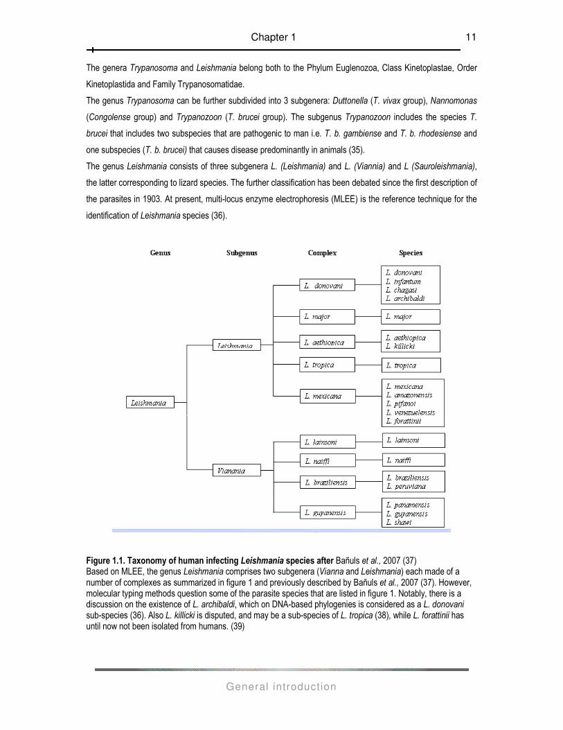
The genus Leishmania consists of three subgenera L. (Leishmania) and L. (Viannia) and L (Sauroleishmania),
the latter corresponding to lizard species. The further classification has been debated since the first description of
the parasites in 1903. At present, multi-locus enzyme electrophoresis (MLEE) is the reference technique for the
identification of Leishmania species (36).
Figure 1.1. Taxonomy of human infecting Leishmania species after Bañuls et al., 2007 (37) Based on MLEE, the genus Leishmania comprises two subgenera (Vianna and Leishmania) each made of a number of complexes as summarized in figure 1 and previously described by Bañuls et al., 2007 (37). However, molecular typing methods question some of the parasite species that are listed in figure 1. Notably, there is a discussion on the existence of L. archibaldi, which on DNA-based phylogenies is considered as a L. donovani sub-species (36). Also L. killicki is disputed, and may be a sub-species of L. tropica (38), while L. forattinii has until now not been isolated from humans. (39)
Chapter 1
General introduction
12
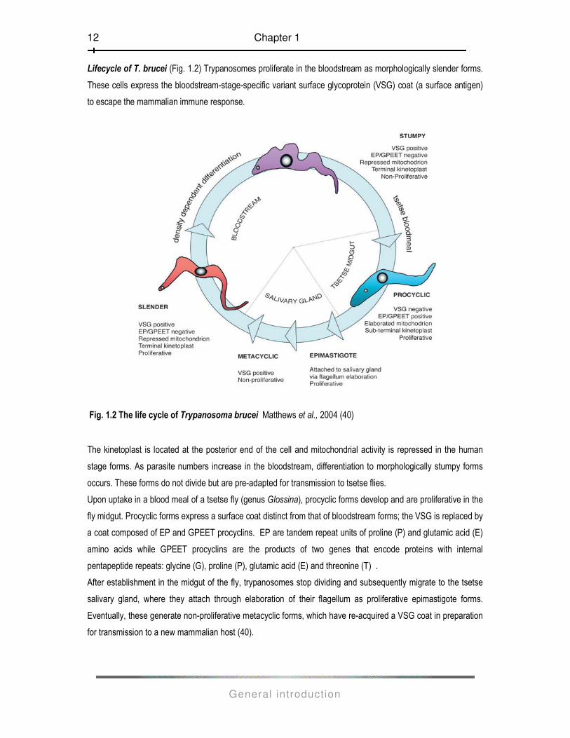
Lifecycle of T. brucei (Fig. 1.2) Trypanosomes proliferate in the bloodstream as morphologically slender forms.
These cells express the bloodstream-stage-specific variant surface glycoprotein (VSG) coat (a surface antigen)
to escape the mammalian immune response.
Fig. 1.2 The life cycle of Trypanosoma brucei Matthews et al., 2004 (40)
The kinetoplast is located at the posterior end of the cell and mitochondrial activity is repressed in the human
stage forms. As parasite numbers increase in the bloodstream, differentiation to morphologically stumpy forms
occurs. These forms do not divide but are pre-adapted for transmission to tsetse flies.
Upon uptake in a blood meal of a tsetse fly (genus Glossina), procyclic forms develop and are proliferative in the
fly midgut. Procyclic forms express a surface coat distinct from that of bloodstream forms; the VSG is replaced by
a coat composed of EP and GPEET procyclins. EP are tandem repeat units of proline (P) and glutamic acid (E)
amino acids while GPEET procyclins are the products of two genes that encode proteins with internal
pentapeptide repeats: glycine (G), proline (P), glutamic acid (E) and threonine (T) .
After establishment in the midgut of the fly, trypanosomes stop dividing and subsequently migrate to the tsetse
salivary gland, where they attach through elaboration of their flagellum as proliferative epimastigote forms.
Eventually, these generate non-proliferative metacyclic forms, which have re-acquired a VSG coat in preparation
for transmission to a new mammalian host (40).
Chapter 1
General introduction
13
The VSG coat is crucial in the survival of the trypanosome in the mammalian host. It allows the trypanosome to
escape the host immune system by regularly changing this surface antigen faster than the new antibodies can be
made. This is referred to as antigenic variation, and it is brought about by replacing the VSG gene in an active
expression site or by switching to another expression site such that a different VSG is expressed. In a
trypanosome, there are about 1000 potential VSG genes thus the parasite can make excessive numbers of the
different VSG coats. (41)
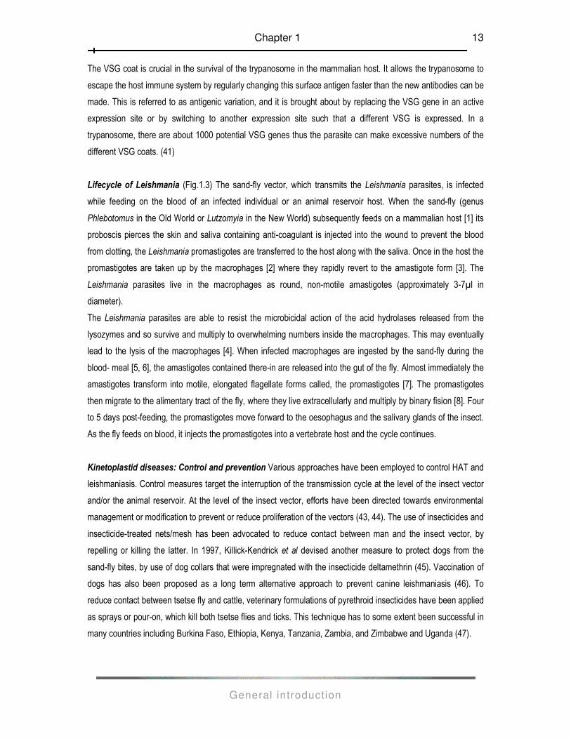
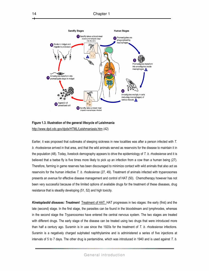
Lifecycle of Leishmania (Fig.1.3) The sand-fly vector, which transmits the Leishmania parasites, is infected
while feeding on the blood of an infected individual or an animal reservoir host. When the sand-fly (genus
Phlebotomus in the Old World or Lutzomyia in the New World) subsequently feeds on a mammalian host [1] its
proboscis pierces the skin and saliva containing anti-coagulant is injected into the wound to prevent the blood
from clotting, the Leishmania promastigotes are transferred to the host along with the saliva. Once in the host the
promastigotes are taken up by the macrophages [2] where they rapidly revert to the amastigote form [3]. The
Leishmania parasites live in the macrophages as round, non-motile amastigotes (approximately 3-7µl in
diameter).
The Leishmania parasites are able to resist the microbicidal action of the acid hydrolases released from the
lysozymes and so survive and multiply to overwhelming numbers inside the macrophages. This may eventually
lead to the lysis of the macrophages [4]. When infected macrophages are ingested by the sand-fly during the
blood- meal [5, 6], the amastigotes contained there-in are released into the gut of the fly. Almost immediately the
amastigotes transform into motile, elongated flagellate forms called, the promastigotes [7]. The promastigotes
then migrate to the alimentary tract of the fly, where they live extracellularly and multiply by binary fision [8]. Four
to 5 days post-feeding, the promastigotes move forward to the oesophagus and the salivary glands of the insect.
As the fly feeds on blood, it injects the promastigotes into a vertebrate host and the cycle continues.
Kinetoplastid diseases: Control and prevention Various approaches have been employed to control HAT and
leishmaniasis. Control measures target the interruption of the transmission cycle at the level of the insect vector
and/or the animal reservoir. At the level of the insect vector, efforts have been directed towards environmental
management or modification to prevent or reduce proliferation of the vectors (43, 44). The use of insecticides and
insecticide-treated nets/mesh has been advocated to reduce contact between man and the insect vector, by
repelling or killing the latter. In 1997, Killick-Kendrick et al devised another measure to protect dogs from the
sand-fly bites, by use of dog collars that were impregnated with the insecticide deltamethrin (45). Vaccination of
dogs has also been proposed as a long term alternative approach to prevent canine leishmaniasis (46). To
reduce contact between tsetse fly and cattle, veterinary formulations of pyrethroid insecticides have been applied
as sprays or pour-on, which kill both tsetse flies and ticks. This technique has to some extent been successful in
many countries including Burkina Faso, Ethiopia, Kenya, Tanzania, Zambia, and Zimbabwe and Uganda (47).
Chapter 1
General introduction
14
Figure 1.3. Illustration of the general lifecycle of Leishmania
http://www.dpd.cdc.gov/dpdx/HTML/Leishmaniasis.htm (42)
Earlier, it was proposed that outbreaks of sleeping sickness in new localities was after a person infected with T.
b. rhodesiense arrived in that area, and that the wild animals served as reservoirs for the disease to maintain it in
the population (48). Today, livestock demography appears to drive the epidemiology of T. b. rhodesiense and it is
believed that a tsetse fly is five times more likely to pick up an infection from a cow than a human being (27).
Therefore, farming in game reserves has been discouraged to minimize contact with wild animals that also act as
reservoirs for the human infective T. b. rhodesiense (27, 49). Treatment of animals infected with trypanosomes
presents an avenue for effective disease management and control of HAT (50). Chemotherapy however has not
been very successful because of the limited options of available drugs for the treatment of these diseases, drug
resistance that is steadily developing (51, 52) and high toxicity.
Kinetoplastid diseases: Treatment Treatment of HAT. HAT progresses in two stages: the early (first) and the
late (second) stage. In the first stage, the parasites can be found in the bloodstream and lymphnodes, whereas
in the second stage the Trypanosomes have entered the central nervous system. The two stages are treated
with different drugs. The early stage of the disease can be treated using two drugs that were introduced more
than half a century ago. Suramin is in use since the 1920s for the treatment of T. b. rhodesiense infections.
Suramin is a negatively charged sulphated naphthylamine and is administered a series of five injections at
intervals of 5 to 7 days. The other drug is pentamidine, which was introduced in 1940 and is used against T. b.
Chapter 1
General introduction
15
gambiense infections. It is a positively charged aromatic diamidine and administered by intramuscular injection
for seven consecutive days (53, 54). Suramin and pentamidine do not cross the blood-brain barrier and therefore
once the trypanosomes invade the central nervous system (second stage), other drugs have to be used instead.
Melarsoprol and eflornithine are the drugs available for treatment of second stage HAT. Melarsoprol, an organo-
arsenic compound, was developed in 1949 and was for a long time the only drug available for the treatment of
second stage HAT. It is highly toxic and may cause hypertension, myocardial damage and most importantly
reactive encephalopathy in 5 to 10 % of treated patients with a case fatality rate of about 50% (55). Moreover
since 1970s, resistance to melarsoprol has been reported (56, 57). Eflornithine (difluoromethylornithine [DFMO])
an inhibitor of ornithine decarboxylase, was originally developed as an anticancer drug in the 1980s, but was
later registered for treatment of second stage T. b. gambiense HAT. This drug appears to be safer than
melarsoprol and drug failure in treatment of T. b. gambiense HAT has not yet been reported.
Nifurtimox, an oral drug, was discovered in 1960 through veterinary research and developed to treat Chagas’
disease. While nifurtimox has been used to treat second stage sleeping sickness in patients who did not respond
to other treatments, it is currently not registered for use against HAT. It has been recommended for use in
combination with eflornithine (Nifurtimox-Eflornithine combination-NECT) after successful trials in Uganda and
Republic of Congo (58, 59, 60).
Recently, melarsoprol has been replaced with eflornithine (DFMO) as first line therapy for late stage T. b.
gambiense in north western Uganda, DR Congo, Southern Sudan and Angola due to occurring treatment failure
(52, 61, 62, 63). In attempt to minimize spread of drug resistance suggestions of alternation of melarsoprol and
DMFO coupled with the nifurtimox-eflornithine combinations are being studied(62, 63).
Treatment of leishmaniasis Various anti-leishmanial drugs are available for the treatment of the various clinical
manifestations of leishmaniasis. For over 70 years, the first line drugs for treatment of VL have been the
pentavalent antimonials sodium stibogluconate and meglumine antimoniate. Antimonials are toxic drugs with
frequent, sometimes life-threatening, adverse side effects, including cardiac arrhythmia and acute pancreatitis.
The prognosis of using these drugs is poor especially in patients under the age of 2 or aged 45 or over with signs
of adverse disease or severe malnutrition (65). In India, there have been reports of treatment failure of up to 60%
and in such areas conventional amphotericin B has replaced antimonials as the first line drug (66). Another drug
is Liposomal amphotericin B, which was initially only used as a first line drug in Europe and United States of
America due to its high market price. However in 2007 the WHO announced that the price would be drastically
reduced to enable its use in the public health sector in countries that are endemic with VL (67). Other drugs are
miltefosine that was developed as an anticancer drug; it is teratogenic and thus its use by pregnant women is
prohibited. Paromomycin, an aminoglycoside antibiotic, has shown promising results in Africa and India with high
efficacy and safety. It has been evaluated alone or in combination with sodium stibogluconate (68) and
amphotericin B (69). In order to prevent development of drug resistance and increase treatment efficacy
combination therapy has been suggested and studied (70).
Chapter 1
General introduction
16
For cutaneous and muco-cutaneous leishmaniasis the first line drugs are pentavalent antimonial compounds
such as sodium stibogluconate and meglumine antimoniate for 20 days. In case of treatment failure, the drug of
choice is amphotericin B; a polyene antibiotic that is given intravenously every other day up to eight weeks.
Alternate drugs include pentamidine isethionate, administered parenterally on alternate days for seven days, and
miltefosine administered orally for 28 days. Other treatments are paromomycin, allopurinol, dapsone, fluconazole
and ketoconazole (71).
Kinetoplastid diseases: Diagnosis Clinical diagnosis. In man, clinical diagnosis of HAT is multifaceted because
of presence of non-specific symptoms such as fever, anorexia, weight loss and distended abdomen (72). HAT
evolves through clinically distinct stages and if left untreated it leads to death. A local skin reaction (trypanosomal
chancre) at the sight of tsetse fly bite may be seen as well as swelling of regional lymph nodes. In the first stage
there is undulating fever that may last for weeks. Lymph node enlargement at the neck is characteristic, and
distension of the abdomen due to swelling of the liver and spleen. (hepato-splenomegaly) is also common in this
stage (73). The second stage occurs when the trypanosomes invade the central nervous system resulting in a
chronic encephalopathy characterized by headache and mental changes. The patient manifests signs of in-
coordination, a reversed sleep-wake cycle with daytime somnolence alternating with night time insomnia (thus
the name sleeping sickness) (74). Death is the inevitable outcome if HAT is not treated.
Diagnosis of leishmaniasis using clinical signs is challenging. There may be a localized erythematous reaction at
the site where an infected sand fly has bitten. Clinical manifestations of visceral leishmaniasis include fever,
lymphadenopathy, hepato-splenomegaly and wasting, features not specific for leishmaniasis. Cutaneous
leishmaniasis may present as ulcerative skin lesions (one or multiple) or as single or multiple non-ulcerative
nodules (localized and diffuse cutaneous leishmaniasis) and muco-cutaneous leishmaniasis is characterized by
destructive mucosal inflammation of nose, mouth, pharynx and larynx. For all three syndromes a differential
diagnosis needs to be considered and thus laboratory techniques have to be applied to confirm the diagnosis.
Parasitological diagnosis is by microscopic demonstration of parasites in clinical specimens. In the case of
leishmaniasis this is by examination of Giemsa stained lesion biopsies (CL and MCL) or lymph node, bone
marrow, and spleen aspirates (VL). This may be supplemented by culturing of parasites from clinical samples,
but this is laborious, time-consuming and prone to contamination. Both microscopy and culture can have a low
and variable sensitivity, depending on the number and distribution of the parasites in the samples, the sampling
procedure and the skills of the personnel (76, 77).
Microscopical examination of blood and CSF can be used for the direct detection of Trypanosoma parasites in
HAT. The presence of the parasite is a direct indication of active disease, but this benefit is down-played by the
fact that this conventional technique has a relatively low sensitivity (78), especially on blood, due to low parasite
count and fluctuating parasitaemia (79). To circumvent this problem, parasite concentration techniques such as
Chapter 1
General introduction
17
quantification buffy coat (80), hematocrit centrifugation, technique (81) and mini-anion-exchange centrifugation
technique (mAECT) (82, 83) have been used to increase sensitivity of microscopy to 500-100 parasites per ml
(83). Cerebrospinal fluid (CSF) has to be examined for staging of HAT as treatment in the two stages is
different(55, 84). Collecting CSF (for HAT) and aspirates of spleen, lymph node and bone marrow (for VL)
involves invasive, painful sampling techniques generally to be performed in hospital settings.
Immunological techniques. The majority of immunological methods that are being used to support the diagnosis
of leishmaniasis are serological techniques, which can be particularly useful in the case of VL (85). These tests
include enzyme-linked immunosorbent assay (ELISA), rK39 dipsticks (86) direct agglutination test (DAT) (87)
and the fast agglutination screening test (FAST) (88). Serology for CL or for VL in immuno-compromised
individuals is considered unreliable due to low or absent production of specific antibodies (89); although some
evidence shows successful serological diagnosis of VL in HIV co-infected patients from Ethiopia with the DAT
(90). Despite the large number of serological tests that have been developed for diagnosis of VL, there is still no
gold standard diagnostic test; because none of the tests is 100% specific and sensitive (91). Serological
diagnostic tests are prone to give false positive results because antibodies may remain detectable for many
years after successful chemotherapy (92).
The Montenegro or Leishmanin skin test (MST or LST) is a technique that reflects acquired cellular immunity
against leishmaniasis and is based on a crude parasite mixture. The MST or LST can be used in CL diagnosis
(particularly in epidemiological studies), because it is simple to use and it has a high sensitivity and specificity.
However the test cannot be used for the diagnosis of VL. The important disadvantages of the MST or LST are
that different antigen preparations have varying sensitivity and that the test cannot discriminate between past
and present infections (93).
A serological card agglutination test for trypanosomiasis (CATT) developed in late 1970s is used for mass
diagnosis of T. b. gambiense HAT, but there is no satisfactory screening tool for T. b. rhodesiense HAT. CATT is
a fast and simple agglutination assay for detection of T. b. gambiense-specific antibodies in the blood, plasma, or
serum of HAT patients (94). The draw backs include false positive test results after successful treatment of
patients and false negative results where the infecting T. b. gambiense do not express the LiTat 1.3 gene on
which the test is based (95). A LATEX test (latex particles coated with three variable surface glycoproteins of T.
b. gambiense) was found to have higher specificity than the CATT (96). The major limitation of such antibody
detection based tests is that they are of limited use in immune compromised patients with low antibody
production as well as in follow-up of treated patients since the anti-trypanosome antibodies persist in the body for
a significant period (97).
Molecular diagnosis A number of molecular tests have been developed for the diagnosis of leishmaniasis and
HAT and a selection is presented below.
Chapter 1
General introduction
18
Polymerase chain reaction (PCR). At present the polymerase chain reaction (PCR) constitutes the main
molecular diagnostic approach for both HAT and leishmaniasis and can, with some adaptations, also be used for
species identification, which is important for treatment decisions. PCR has been in use for almost 30 years and
was after its introduction in the early eighties of the last century rapidly considered as the ultimate tool for the
detection of infectious agents, because it combines sensitivity with specificity. Over the past two decades,
dozens of different types of PCRs, have been designed for the detection and speciation of Trypanosomatidae.
Since many research laboratories can assemble their own amplification tests by designing new primers and
optimizing in-house protocols, numerous PCRs and related techniques are currently in use for the detection and
identification of the parasites and a universal gold standard PCR for either diseases does not exist (98). Studies
in which head-to-head comparisons of the sensitivity/specificity of different PCR have been done are few, but
needed.
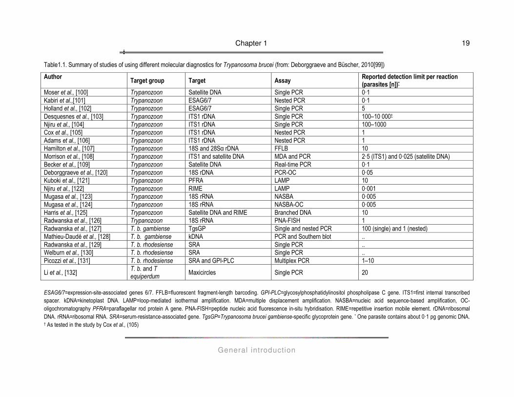
A recent publication by Deborggraeve and Büscher (99) provides an excellent overview of molecular diagnostics
for HAT that are currently available and a summary is presented in table 1.1. From the table it is obvious that
there is a lot of variability in the detection limit of the employed technologies. An important factor that is affecting
the sensitivity and specificity of a diagnostic PCR is the DNA sequence that is targeted by the primers. Hence,
sequences that are conserved but unique for the target group and that occur as multiple copies in the parasite's
genome are attractive as templates in diagnostic PCRs. In the case of diagnostic PCRs for trypanosomiasis, it
seems that the multicopy genes for 18S rDNA and rRNA are the most suitable targets to develop diagnostic
PCRs.
Differentiation of HAT causing T. brucei subspecies is important for T brucei gambiense and T brucei
rhodesiense as these human pathogens require a different therapeutic approach. This is particularly relevant for
Uganda given the imminent risk of overlap of both subspecies in the country (133). The molecular differentiation
of these subspecies is mainly based on two genes. The gene encoding T brucei gambiense-specific glycoprotein
(TgsGP) is only present in T brucei gambiense, whereas the gene encoding the serum-resistance-associated
protein (SRA) is specific for T brucei rhodesiense (134, 127, 129) and PCRs that can discriminate between the
two sub-species have been developed using primers designed from these targets.
Alongside diagnosis of HAT, PCR has been crucial in determining the stage of disease (135, 136) which is
important as treatment for the two stages is different, as mentioned (53).
PCR is used for detection and genotyping of Leishmania species by identification of a variety of targets, including
kinetoplast DNA, ribosomal RNA genes, internal transcribed spacers, spliced leader RNA (mini-exon) gene
repeats and regions of the glycoprotein 63 (gp63) gene locus. An overview of PCR assays developed for
detection and diagnosis of Leishmania infections in humans over the last decade is presented in Table 1.2. A
variety of clinical specimens is being used for diagnostic purposes, depending on the clinical presentation of
leishmaniasis under investigation: blood, bone marrow and lymph node aspirates for VL, blood and skin
exudates for PKDL, lesion biopsy for MCL, skin scrapings and biopsies, lesion aspirates and biopsies for CL.
(see table 1.2).
Chapter 1
General introduction
19
Table1.1. Summary of studies of using different molecular diagnostics for Trypanosoma brucei (from: Deborggraeve and Büscher, 2010[99])
Author Target group Target Assay
Reported detection limit per reaction (parasites [n])*
Moser et al., [100] Trypanozoon Satellite DNA Single PCR 0·1 Kabiri et al.,[101] Trypanozoon ESAG6/7 Nested PCR 0·1 Holland et al., [102] Trypanozoon ESAG6/7 Single PCR 5 Desquesnes et al., [103] Trypanozoon ITS1 rDNA Single PCR 100–10 000† Njiru et al., [104] Trypanozoon ITS1 rDNA Single PCR 100–1000 Cox et al., [105] Trypanozoon ITS1 rDNA Nested PCR 1 Adams et al., [106] Trypanozoon ITS1 rDNA Nested PCR 1 Hamilton et al., [107] Trypanozoon 18S and 28Sα rDNA FFLB 10 Morrison et al., [108] Trypanozoon ITS1 and satellite DNA MDA and PCR 2·5 (ITS1) and 0·025 (satellite DNA) Becker et al., [109] Trypanozoon Satellite DNA Real-time PCR 0·1 Deborggraeve et al., [120] Trypanozoon 18S rDNA PCR-OC 0·05 Kuboki et al., [121] Trypanozoon PFRA LAMP 10 Njiru et al., [122] Trypanozoon RIME LAMP 0·001 Mugasa et al., [123] Trypanozoon 18S rRNA NASBA 0·005 Mugasa et al., [124] Trypanozoon 18S rRNA NASBA-OC 0·005 Harris et al., [125] Trypanozoon Satellite DNA and RIME Branched DNA 10 Radwanska et al., [126] Trypanozoon 18S rRNA PNA-FISH 1 Radwanska et al., [127] T. b. gambiense TgsGP Single and nested PCR 100 (single) and 1 (nested) Mathieu-Daudé et al., [128] T. b. gambiense kDNA PCR and Southern blot .. Radwanska et al., [129] T. b. rhodesiense SRA Single PCR .. Welburn et al., [130] T. b. rhodesiense SRA Single PCR .. Picozzi et al., [131] T. b. rhodesiense SRA and GPI-PLC Multiplex PCR 1–10
Li et al., [132] T. b. and T equiperdum
Maxicircles Single PCR 20
ESAG6/7=expression-site-associated genes 6/7. FFLB=fluorescent fragment-length barcoding. GPI-PLC=glycosylphosphatidylinositol phospholipase C gene. ITS1=first internal transcribed spacer. kDNA=kinetoplast DNA. LAMP=loop-mediated isothermal amplification. MDA=multiple displacement amplification. NASBA=nucleic acid sequence-based amplification, OC- oligochromatography PFRA=paraflagellar rod protein A gene. PNA-FISH=peptide nucleic acid fluorescence in-situ hybridisation. RIME=repetitive insertion mobile element. rDNA=ribosomal DNA. rRNA=ribosomal RNA. SRA=serum-resistance-associated gene. TgsGP=Trypanosoma brucei gambiense-specific glycoprotein gene. * One parasite contains about 0·1 pg genomic DNA. † As tested in the study by Cox et al., (105)
Chapter 1
General introduction
20
Information on the infecting Leishmania species is very important to install adequate treatment (different clinical
forms caused by different species require different treatment), in particular in the case in regions where
Leishmania species are geographically overlapping. Species discrimination is also important for epidemiological
reasons, and with advances in molecular techniques, a number of PCR protocols and molecular markers have
been developed to detect or identify Leishmania on different taxonomical levels (137). A PCR based on mini
exon gene was developed to discriminate between major Leishmania complexes, that is Old world Leishmania,
New world Leishmania and new world Viannia (137, 138). Genotyping of mini exon sequences using PCR-
restriction fragment length polymorphism (RFLP) based assay has been reported by Murfart et al., 2003 (137)
and based on an uncharacterized repetitive genomic sequence of Old World Leishmania (139).
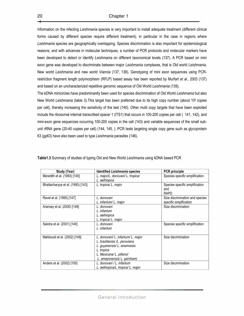
The kDNA minicircles have predominantly been used for species discrimination of Old World Leishmania but also
New World Leishmania (table 3).This target has been preferred due to its high copy number (about 104 copies
per cell), thereby increasing the sensitivity of the test (140). Other multi copy targets that have been exploited
include the ribosomal internal transcribed spacer 1 (ITS1) that occurs in 100-200 copies per cell ( 141, 142), and
mini-exon gene sequences occurring 100-200 copies in the cell (143) and variable sequences of the small sub-
unit rRNA gene (20-40 copies per cell) (144, 145, ). PCR tests targeting single copy gene such as glycoprotein
63 (gp63) have also been used to type Leishmania parasites (146).
Table1.3 Summary of studies of typing Old and New World Leishmania using kDNA based PCR
Study (Year) Identified Leishmania species PCR principle
Meredith et al. (1993) [145] L. major/L. donovani/ L. tropica/ L. aethiopica
Species specific amplification
Bhattacharyya et al. (1993) [143] L. tropica/ L. major Species specific amplification and RAPD
Ravel et al. (1995) [147] L. donovani L. infantum/ L. major
Size discrimination and species specific amplification
Aransay et al. (2000) [148] L. donovani L. infantum L. aethiopica L. tropica/ L. major
Size discrimination
Salotra et al. (2001) [140] L. donovani L. infantum
Species specific amplification
Mahboudi et al. (2002) [149] L. donovani/ L. infantum/ L. major L. braziliensis /L. peruviana L. guyanensis/ L. anamnesis L. tropica L. Mexicana/ L. pifanoi/ L. amazonensis/ L. garnhami
Size discrimination
Anders et al. (2002) [150] L. donovani / L. infantum L. aethiopica/L. tropica/ L. major
Size discrimination
Chapter 1
General introduction
21
Another single target gene is the cysteine proteinase B (cpb) gene that was first used by Hide and Bañuls in
2006 (151) to discriminate L. donovani from L. infantum based on both size discrimination and species specific
amplification. Shorter variants of cpb gene were identified and amplified in some strains in L. major, L. infantum
and L. donovani (152, 153). Laurent and colleagues later used the same target to identify 3 more species L.
aethiopica, L. tropica and L. major based on species specific amplification (154).
Of interest is the use of PCR to monitor leishmaniasis patients undergoing treatment and during follow-up up to
at least 8 weeks after treatment (155, 156, 157). The PCR test results generally have a high concordance value
with the clinical outcome for immuno-competent patients. There were however a few deviant results where one
patient relapsed 11 months after end of treatment (156) and in another study (157) where one patient had a
positive PCR 145 days after treatment initiation. On the other hand, studies (158, 159, 160, 161) involving
immune compromised leishmaniasis patients such as those co-infected with human immunodeficiency virus
(HIV) show that the majority of patients do not clear parasite DNA from blood, remain PCR positive, which may
be linked with relapses (159).
Although PCR-based protocols have increased the specificity and sensitivity of kinetoplastid diseases compared
to conventional techniques like microscopy, the implementation of this technology in disease endemic countries,
were resources are often limited, is little. This is due to need of sophisticated equipment in particular an
automated thermocycler that requires constant supply of electricity. Furthermore, electrophoresis, the
conservative tool for detection of PCR amplicons is laborious and involves the use of toxic ethidium bromide to
stain the gels and subsequent use of a hazardous ultraviolet transiluminator (162). Moreover, PCR is relatively
expensive (€2.60- 4.75 per sample excluding labour) (124) for mid-level laboratories and not affordable for low
income countries, such as Uganda. Finally, a risk of diagnostic tests based on nucleic acid amplification is
contamination leading to false positive results. These factors thus limit the use of PCR in regions that are
ravaged by leishmaniasis and HAT. To circumvent these hurdles, different new amplification technologies, which
are isothermal and possibly less prone to contamination, are currently being developed. These include loop
mediated amplification (LAMP) and nucleic acid sequence based amplification (NASBA). In addition, to simplify
detection systems, a single-step lateral flow dipstick has been developed based on the principle of
oligochromatography (OC), which is quicker, not producing toxic waist and as sensitive as conventional
systems. These new technologies are further presented below.
Loop Mediated Isothermal Amplification (LAMP) is an isothermal amplification method (at 60-65°C) developed
by Notomi et al., (2000) (176) that relies on autocycling strand displacement DNA synthesis by a Bst (Bacillus
stearothermophilus) thermostable DNA polymerase using 2 pairs of primers. LAMP synthesizes 10 to 20 µg of
target DNA within 30 to 60 min, and the LAMP reaction appears to be limited only by amount of deoxynucleoside
triphosphates and primers (176, 177). In the process, a large amount of pyrophosphate ion is produced, which
Chapter 1
General introduction
22
reacts with magnesium ions in the reaction to form magnesium pyrophosphate, a white precipitate by-product
(178). This phenomenon (turbidity) allows easy and rapid visual identification that the target DNA was amplified
by LAMP. The turbidity has been further modified by using a fluorescent dye (SYBR Green) for the detection of
T. b. rhodesiense (179). Therefore, LAMP can be an alternative for diagnosis of infectious diseases both in well-
equipped and in mid-level laboratories typical of HAT endemic countries.
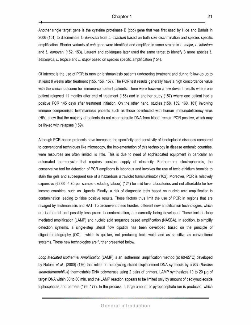
Nucleic acid sequence-based amplification (NASBA) is a relatively novel amplification assay that was first
described by Kievits et al., in 1991 (180) (Fig 4). Like LAMP, NASBA is a robust single-step isothermal (41°C)
assay not requiring a thermocycler, a simple water bath or heat block is sufficient to perform the amplification
reaction. NASBA is a RNA-specific amplification process that amplifies RNA in a DNA background (181), and this
test is less prone to contamination. It has been successfully used for detection and quantification of infectious
organisms in clinical samples from infections with Plasmodium falciparum, (182), Leishmania (183) and hepatitis
A virus (184). In this thesis, NASBA has been developed (123, 124) to detect the presence of T. brucei in blood
from HAT patients and Leishmania parasites in blood, and skin biopsies from VL and CL patients respectively
(124).
Fig.1.4 The NASBA amplification reaction process; it is initiated by the annealing of an oligonucleotide primer (P1)
to the RNA target. The 3’ end of the P1 primer is complementary to the target RNA and the 5’ end encodes the T7
RNA polymerase promoter. After annealing, the reverse transcriptase activity of AMV-RT is engaged and a cDNA
copy of the RNA target is produced. The RNA portion of the resulting hybrid molecule is hydrolyzed through the action
of RNase H. The second primer (P2) anneals to the remaining cDNA strand and the DNA-dependent polymerase
activity of AMV-RT is engaged again, producing a double stranded cDNA with a fully functional T7 RNA polymerase
promoter. This promoter is then recognized by the T7 RNA polymerase (T7RNA pol), which produces a large amount
(10 - 1000) of single stranded RNA transcripts corresponding to the original RNA target. These RNA transcripts can
then serve as templates for the amplification process, (in the cyclic phase) however the primers anneal in the reverse
order. (Kievits et al., in 1991)
Chapter 1
General introduction
23
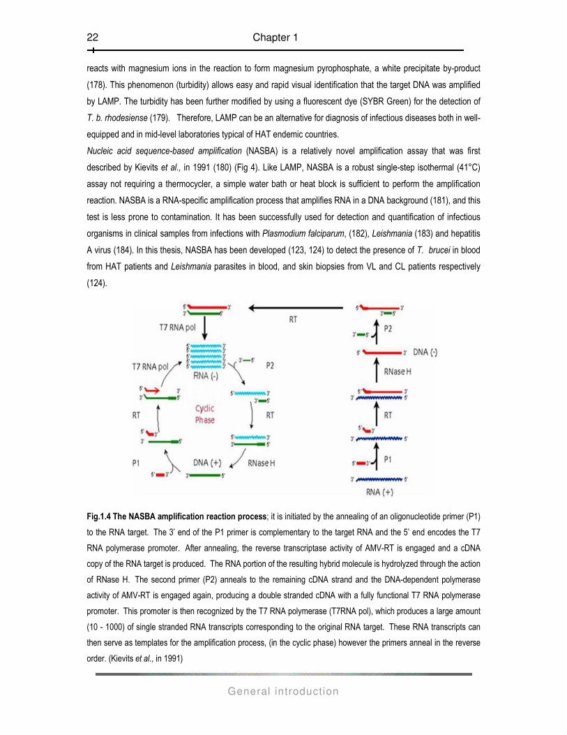
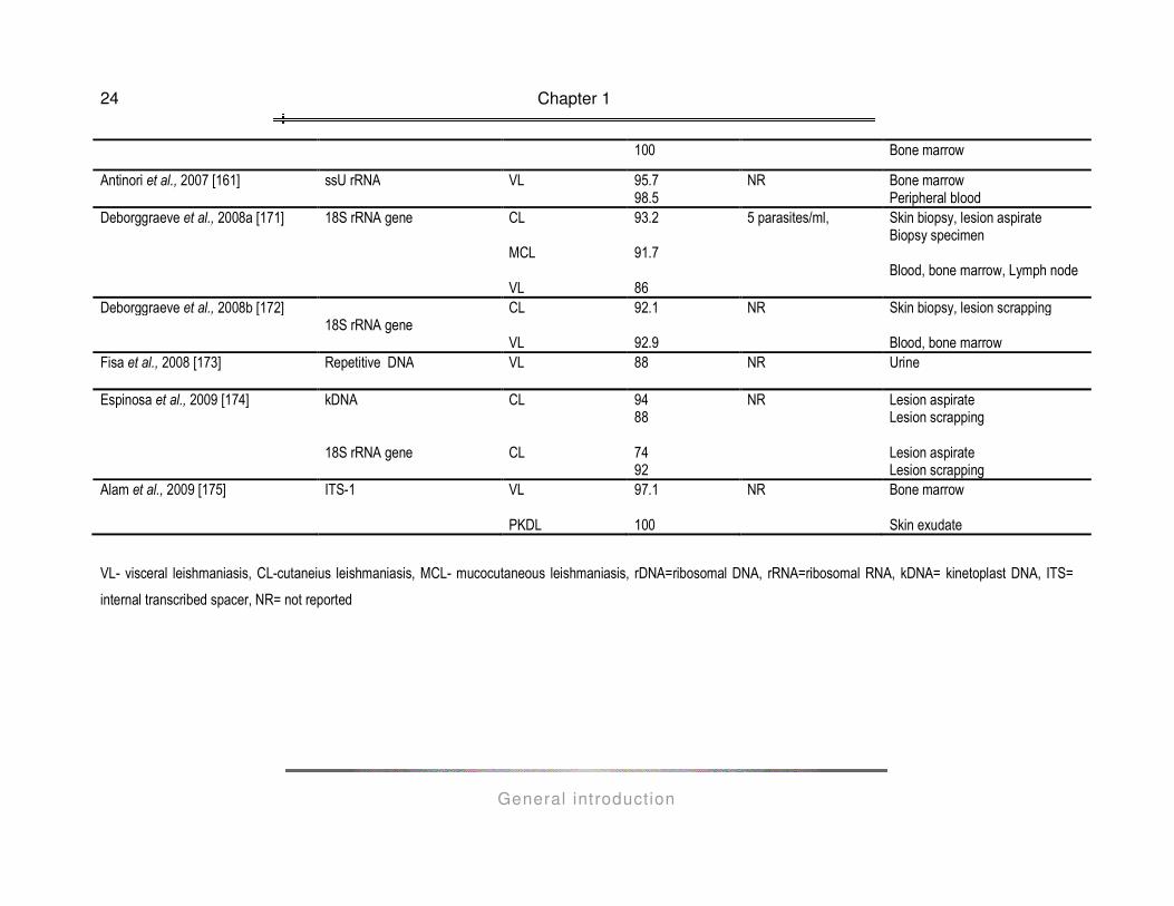
Table1.2 PCR assays developed for detection and diagnosis of Leishmania infections in humans over the last decade (Edited from Antinori et al., 2007)
Author(s) Target sequence Leishmaniasis form
Sensitivity (%) Detection limit Clinical sample
Lachaud et al., 2000 [163] ssU-rRNA VL 97 100
NR Peripheral blood Bone marrow
Salotra et al., 2001[140]
minicircle kDNA VL PKDL
96 93.8
1 parasite Blood
Pizzuto et al., 2001[160] ssu rRNA gene VL 100 20-40 copies per cell Blood Isaza et al., 2002 [164] kDNA CL 94 NR Skin scrapping and biopsy
Cruz et al., 2002 [159] ssu rRNA gene VL 95.5
100 NR Blood
Bone marrow Fisa et al., 2002 [165] Repetitive DNA VL 100 NR Blood
de Oliviera et al., 2003 [166] Minicircle kDNA CL 100 NR Skin biopsy
Da Salva et al., 2004 [167] Minicircle kDNA VL 71.7
100 NR Blood
Bone marrow De Doncker et al., 2005 [168] 18S rRNA gene
kDNA VL 73.2
67.8 NR Blood
Maurya et al., 2005 [156] Minicircle kDNA VL 99 NR Peripheral blood
Chargui et al., 2005 [169] 18S rRNA gene CL 99.3 NR Skin
Bensoussan et al., 2006 [170]
Mini-exon gene ITS KDNA
CL
53.8 91.0 98.7
about 104 copies per cell
Skin biopsy
Author(s) Target sequence Leishmaniasis form
Sensitivity (%) Detection limit Clinical sample
Cruz et al., 2006 [157] ssu rRNA gene VL 79 NR Blood
(continued)
Chapter 1
General introduction
24
100 Bone marrow
Antinori et al., 2007 [161] ssU rRNA VL 95.7 98.5
NR Bone marrow Peripheral blood
Deborggraeve et al., 2008a [171]
18S rRNA gene
CL MCL VL
93.2 91.7 86
5 parasites/ml,
Skin biopsy, lesion aspirate Biopsy specimen Blood, bone marrow, Lymph node
Deborggraeve et al., 2008b [172] 18S rRNA gene
CL VL
92.1 92.9
NR Skin biopsy, lesion scrapping Blood, bone marrow
Fisa et al., 2008 [173] Repetitive DNA VL 88 NR Urine
Espinosa et al., 2009 [174] kDNA 18S rRNA gene
CL CL
94 88 74 92
NR Lesion aspirate Lesion scrapping Lesion aspirate Lesion scrapping
Alam et al., 2009 [175] ITS-1 VL PKDL
97.1 100
NR Bone marrow Skin exudate
VL- visceral leishmaniasis, CL-cutaneius leishmaniasis, MCL- mucocutaneous leishmaniasis, rDNA=ribosomal DNA, rRNA=ribosomal RNA, kDNA= kinetoplast DNA, ITS=
internal transcribed spacer, NR= not reported
Oligochromatography (OC) is a single step isothermal technique that rapidly detects amplification
products (from PCR or NASBA) on a membrane strip. The amplified products are captured by specific
oligonucleotides on the membrane and visualized by hybridization with a gold-conjugated probe (185).
OC is done on a double sided OC dipstick with a supportive polymer (plastic). On either side of the
support, several membranes and absorbents are incorporated to regulate flow and allow sequential
hybridization of amplicons. The membranes are coated with oligonucleotide sequences complementary
to the target amplicons to be detected. One of the sides of the OC dipstick is designated as the test side
(for detection of target RNA in the clinical sample) while the other is the control side (to detect in-vitro
RNA control sequence) in order to validate the test result (124, 120, 186). OC detection of amplified
products after PCR or NASBA is easy to perform and takes only a few minutes.
Oligochromatography has been developed for the rapid detection of parasites including Schistosoma
(187), Toxoplasma gondii (188), T. brucei (124, 120, 189) and Leishmania (171, 171, 174, 186). OC has
also been used in the detection of SARS-CoV amplicons following RT-PCR (190) and it was found that
the sensitivity of the RT-PCR combined with OC detection was comparable with conventional agarose
gel electrophoresis. The signal generated on the oligochromatographic dipstick is often more clear than
on the gel and the use of specific detection and capture probes adds to the specificity of the assay. PCR
and NASBA followed by oligochromatography for the detection of both T. brucei and Leishmania have
been evaluated in this thesis, with promising results (186, 189). OC has the potential to be incorporated
in routine field diagnosis of HAT and leishmaniasis in resource poor settings where more sophisticated
equipment, such as needed for agarose gel electrophoresis, is unaffordable and not feasible.
Chapter 1
General introduction
26
Outline of this thesis
This thesis describes the development and evaluation of molecular techniques and their application in
the field diagnosis of two kinetoplastid diseases, that is, Human African trypanosomiasis (HAT) and
leishmaniasis, both of which affect populations in poor rural settings in developing countries. As shown,
there is a need to develop simple, accurate and reliable diagnostic tools that can be employed to
diagnose these kinetoplastid infections so that patients are treated early to reduce on long term effects
of the disease as well as the case fatality rate. Therefore the aim of this thesis is to chronologically
explicate the development, evaluation and finally the application of these techniques in the diagnosis of
HAT and leishmaniasis. NASBA is chosen as the principle amplification technology and OC as detection
system. Chapter 2 describes the development of a real-time nucleic acid sequence-based amplification
(RT-NASBA) assay that detects Trypanosoma parasites with high sensitivity and specificity. In an effort
to simplify the detection of NASBA amplicons, oligochromatography (OC), a simple and fast detection
system, was developed and coupled with NASBA, (NASBA-OC) to detect T. brucei infection in both
blood and CSF samples (Chapter 3). With the advent of OC, NASBA may by-pass the necessity of
rather expensive and laborious detection methods that would otherwise confine disease diagnosis to
only well equipped laboratories. To cover the relevant kinetoplastid diseases addressed in this thesis,
the development and evaluation of NASBA-OC assay as a tool for diagnosing both visceral and
cutaneous leishmaniasis using various clinical samples is presented in Chapter 4.
The evaluation of molecular diagnostics (and even diagnostics in general) is often neglected. It is
important to estimate the repeatability (accordance) and reproducibility (concordance) of a new test
prior to further phase II and III evaluation studies, which should be performed in other laboratories than
the laboratory where the test was developed. Therefore, Chapter 5 presents study reports on a multi-
centre evaluation study. NASBA-OC assays for detection of Leishmania and T. brucei nucleic acids
were assessed for reproducibility and repeatability in seven independent laboratories on two different
continents and compared to PCR-OC performance. The evaluation of the performance of NASBA-OC
assay was further compared to PCR-OC assay in a phase II clinical trial for the diagnosis of Human
African trypanosomiasis using clinical samples from patients and appropriate controls from endemic
areas in the Democratic Republic of Congo as well as Uganda (Chapter 6). Finally in Chapter 7 a
systematic review of the diagnostic accuracy of molecular tests for HAT is presented to establish
whether and how the findings in various HAT diagnostic studies vary.
Chapter 1
General introduction
27
References
(1) Shlomai J (1994) The assembly of kinetoplast DNA. Parasitol. Today 10: 341-346 (2) Vickerman K (1976) The Diversity of the kinetoplastid flagellates. In WHR Lumsden, DA Evans (eds),
Biology of the Kinetoplastida, Academic Press, London/New York/San Francisco, pp. 1-34 (3) Lom J (1976) Biology of the trypanosomes and trypanoplasms of fish. In WHR Lumsden, DA Evans
(eds), Biology of the Kinetoplastida, Academic Press, London/New York/San Francisco, pp. 269-337 (4) Maslov AD, Podlipaev SA, Luke J (2001) Phylogeny of the Kinetoplastida: taxonomic problems and
insights into the evolution of parasitism. Mem Inst Oswaldo Cruz, Rio de Janeiro, 96: 397-402 (5) El Hassan AM, El Hassan LA (1997) Recent advances in visceral leishmaniasis Alpha DNA microbiol. J.
6: 23-33 (6) World Health 0rganisation (1998) Control and surveillance of African trypanosomiasis WHO Technical
Report Series 881: 1-113 (7) World Health Organisation (2002) Disease burden and epidemiological trends. In: World Health
Organisation, Geneva, http://www.who.int/tdr/diseases/leish/direction.htm (8) World Health Organisation. (2004) Human African trypanosomiasis Disease watch (9) Anonymous (2006) Human African trypanosomiasis (sleeping sickness): epidemiological update. Wkly
Epidemiol Rec 81: 69–80 (10) World Health Organization: The World Health Report 2004 Changing History Geneva, WHO; 2004 (11) World Health Organization (1993) The Leishmaniasis CTD/MIP/WP.93.8 (12) Dávila AMR, Tyler KM (2002) Combating Kinetoplastid diseases Kinetoplastid Biology and Disease 1: 6-
7 (13) FAO Programme against African Trypanosomiais (PAAT)
http://www.fao.org/AG/AGAInfo/programmes/en/paat/disease.html (14) Williams BI. African trypanosomiasis. In: Cox FEG editor. The welcome Trust illustrated history of
tropical diseases. London: The Wellcome Trust: 1996 pp 178-191 (15) Winterbottom TM. An account of the native Africans in the neighbourhood of Sierra Leone to which is
added an account of the present state of medicine among them. Vol.2 London: C. Whittingham (1803) (16) Dutton JE. (1902) Preliminary note upon a trypanosome occurring in the blood of man Thompson Yates
Lab Rep 4: 455-468 (17) Gouzien P (1908) La maladie du sommeil dans le Haut-Se´ne´gal et Niger. 490/Document Technique
OCCGE (18) Mandé I (1997) Les migrations du travail en Haute-Volta (actuel Burkina Faso), mise en perspective
historique (1919–1960). Editions de L’universite´, Denis Diderot, Paris, p. 490 (19) Thomson KDB (1968) La trypanosomiase dans les provinces Nord du Nige´ria: panorama de la
situation actuelle. 4147/Document Technique OCCGE (20) Courtin F, Jamonneau V, Duvallet G, Garcia A, Coulibaly B, Doumenge JP, Cuny G Solano P (2008)
Sleeping sickness in West Africa (1906–2006): changes in spatial repartition and lessons from the past. Trop Med Int Health 13: 334-344
(21) Cattand P, Jannin J, Lucas P (2001) Sleeping sickness surveillance: an essential step towards elimination. Trop Med Int. Health 6: 348–351
(22) Bruce D, Nabarro D, Grieg EDW (1903) Further report of sleeping sickness in Uganda. Report of the sleeping sickness Commission 4: 83-87
(23) MacKichan IW (1944) Rhodesian sleeping sickness in eastern Uganda. Trans R Soc Trop Med Hyg 38: 49–60
(24) Okoth JO, Kapata N (1986) Trypanosome infection rates in Glossina fuscipes fuscipes Newst. in the Busoga sleeping sickness focus, Uganda Ann Trop Med Parasitol 80: 459-461
(25) Enyaru JCK, Odiit M, Winyi-Kaboyo, Sebikali CG, Matovu E, Okitoi D, Olaho-Mukan. (1999) Evidence for the occurrence of Trypanosoma brucei rhodesiense sleeping sickness outside the traditional focus in southeastern Uganda Ann Trop Med Parasitol 93: 817-820
(26) Abaru DE (1985) Sleeping sickness in Busoga, Uganda, 1976-1983. Trop Med Parasitol 36:72-76
Chapter 1
General introduction
28
(27) Fèvre EM, Coleman PG, Odiit M, Magona JW, Welburn SC, Woolhouse MEJ (2001) The origins of a new Trypanosoma brucei rhodesiense sleeping sickness outbreak in eastern Uganda. Lancet 358: 625-628
(28) Berrang-Ford L, Waltner-Toews D, Charron D, Odiit M, McDermott J, Smit B (2005) Sleeping sickness in southeastern Uganda: a systems approach EcoHealth 2: 1-12
(29) Fèvre EM, Picozzi K, Waiswa C, Odiit M, Coleman PG, Welburn SC (2005) A burgeoning epidemic of sleeping sickness in Uganda Lancet 366:745-747
(30) http://leishman.cent.gla.ac.uk/william.htm The History of Leishmaniasis (31) Leishman WB (1903) On the possibility of the occurrence of Trypanosomiasis in India Br Med J I: 1252-
1254 (32) Goldman L (1983) Pre- Columbian Leishmaniasis Arch Dermatol 119: 540 (33) Hoare CA. (1938) Early discoveries regarding the parasites of oriental sore Trans R Soc Trop Med Hyg
32: 67-92 (34) Pedrosa CM, Rocha EMM (2004) Aspectos clínicos e epidemiológicos da leishmaniose visceral em
menores de 15 anos procedentes de Alagoas, Brasil Rev Soc Bras Med Trop 37: 300-304 (35) Baker JR (1995) The subspecific taxonomy of Trypanosoma brucei Parasitol 2: 3-12 (36) Schonian G, Maurico I, Cupolollo E (2010) Is it time to revise the nomenclature of Leishmania? Trends
Parasitol doi:10.1016/j.pt.2010.06.013 (37) Bañuls AL, Hide M, Prugnolle (2007) Leishmania and the leishmaniases: a parasite genetic update and
advances in taxonomy, epidemiology and pathogenicity in humans Adv Parasitol 64: 1-109 (38) Pratlong F, Dereure J, Ravel C, Lami P, Balard Y, Serres G, Lanotte G, Rioux JA, Dedet JP (2009)
Geographical distribution and epidemiological features of old worls cutaneous leishmaniasis focus based on the isoenzyme analysis of 1048 strains Trop. Med. Int. Health 14: 1071-1085
(39) Yoshida ELA, Cuba Cuba CA, Pacheco RS, Cupolillo E, Tavares CC, Machado GMC, Momen H, Grimaldi Jr G (1993) Description of Leishmania (Leishmania) forattinii sp. n., a new parasite infecting opossums and rodents in Brazil. Mem Inst Oswaldo Cruz 88: 397-406
(40) Matthews KR, Ellis JR, Paterou A (2004) Molecular regulation of the life cycle of African trypanosomes Trends Parasito. 20: 40-47
(41) Borst P and Rudenko G (1994) Antigenic variation in African Trypanosomes Science 264: 1872- 1873 (42) http://www.dpd.cdc.gov/dpdx/HTML/Leishmaniasis.htm (43) Alexander B, Jaramillo C, Usma MC, Quesada BL, Cadena H, Roa W, Travi BL (1995) An attempt to
control phlebotomine sand flies (Diptera, Psychodidae) by residual spraying with Deltamethrin in a Colombian village Mem Ins Oswaldo Cruz 90: 421-424
(44) Maudlin I, Holmes PH, Miles MA. The Trypanosomiases edition 2004. Wallingford, U.K: CABI Publishing (45) Killick-Kendrick R, Killick-Kendrick M, Focheux C, Dereure J, Puech MP, Cadiergues MC (1997)
Protection of dogs from bites of phlebotomine sandflies by deltamethrin collars for control of canine leishmaniasis Med Vet Entomol 11: 105-111
(46) Tesh RB (1995) Control of zoonotic visceral leishmaniasis: is it time to change strategies? Am J Trop Med Hyg 52: 287-292
(47) Kuzoe FAS, Schofield CJ (2005) Strategic review of traps and targets for tsetse and African trypanosomiasis control http://www.who.int/tdr/publications/tsetsetraps.htm
(48) Jackson CH (1955) The natural reservoir of Trypanosoma rhodesiense Trans R Soc Trop Med Hyg. 49:582–587
(49) Geigy R, Kauffmann M, Jenni L (1973) Wild mammals as reservoirs for Rhodesian sleeping sickness in the Serengeti, 1970–71. Tran R Soc Trop Med Hyg 67:284–286
(50) Welburn SC, Odiit M (2002) Recent developments in human African trypanosomiasis. Curr Opin Inf Dis 15: 477-484
(51) Barrett MP, Burchmore RJ, Stich A, Lazzari JO, Frasch AC, Cazzalo JJ, Krishna S (2003) The trypanosomiasis Lancet 362: 1469-1480
(52) Legros D, Ollivier G, Gastellu-Etchegorry M, Paquet C, Burri C, Jannin J, Büscher P (2002) Treatment of human African trypanosomiasis—present situation and needs for research and development Lancet Infect. Dis. 2:437–440
(53) Pépin, J., and F. Milord (1994) The treatment of human African trypanosomiasis Adv Parasitol 33:1–47

Chapter 1
General introduction
29
(54) Docampo R. and Moreno S. N J (2002) Current chemotherapy of human African trypanosomiasis Parasitol Res 90: 10-13
(55) WHO Expert Committee on Control and Surveillance of African Trypanosomiasis Geneva, World Health Organization, 1998 (WHO Technical Report Series, No. 881)
(56) Legros D, Evans S, Maiso F, Enyaru JC, Mbulamberi D (1999) Risk factors for treatment failure after melarsoprol for Trypanosoma brucei gambiense trypanosomiasis in Uganda. Trans R Soc Trop Med Hyg 93:439-442
(57) Brun R, Schumacher R, Schmid C, Kunz C, Burri C (2001) The phenomenon of treatment failures in Human African Trypanosomiasis Trop Med Int Health 6:906-914
(58) Priotto G, Kasparian S, Ngouama D, Ghorashian S, Arnold U, Ghabri S, Karunakara U (2007). Nifurtimox-Eflornithine Combination Therapy for Second- Stage Trypanosoma brucei gambiense Sleeping Sickness: A Randomized Clinical Trial in Congo. Clin Inf Dis 45: 1435-1442
(59) Checchi F, Piola P, Ayikoru H, Thomas F, Legros D, Priotto G (2007) Nifurtimox plus Eflornithine for Late-Stage Sleeping Sickness in Uganda: A Case Series. PLoS Negl Trop Dis 1(2): e64.
(60) http://www.dndi.org/press-releases/456/ Press releases 2009 NECT Added to WHO essential medicines list as combination treatment against sleeping sickness
(61) Matovu E, Enyaru JCK, Legros D, Schmid C, Seebeck T, Kaminsky R (2001) Melarsoprol refractory T.b. gambiense from Omugo, North Western Uganda Trop Med Int Health; 6: 407–411
(62) Matovu E, Geiser F, Schneider V, Mäser P, Enyaru JC, Kaminsky R, Gallati S, Seebeck T (2000) Genetic variants of the TbAT1 adenosine transporter from African trypanosomes in relapse infections following melarsoprol therapy Mol Biochem Parasitol 117: 73-81
(63) Kazibwe AJN, Nerima B, de Koning HP, Mäser P, Barrett MP, Matovu E (2009) Genotypic Status of the TbAT1/P2 Adenosine Transporter of Trypanosoma brucei gambiense Isolates from Northwestern Uganda following Melarsoprol Withdrawal PLoS Negl Trop Dis 3: e523
(64) Checchi F, Piola P, Ayikoru H, Thomas F, Legros D, Priotto G (2007) Nifurtimox plus Eflornithine for late stage sleeping sickness in Uganda. A case series PLoS Neg Trop Dis 1: e64.
(65) Collin S, Davidson R, Ritmeijer K, Keus K, Melaku Y, Kipngetich S, Davies C (2004) Conflict and kala-azar: determinants of adverse outcomes of kala-azar among patients in southern Sudan Clin Infect Dis 38: 612–619
(66) Sundar S, More DK, Singh MK, Singh VP, Sharma S, Makharia A Kumar PC, Murray HW (2000) Failure of pentavalent antimony in visceral leishmaniasis in India: report from the center of the Indian epidemic. Clin Infect Dis 31: 1104–1107
(67) Chappuis F, Sundar S, Hailu A, Ghalib H, Rijal S, Peeling RW, Alvar J, Boelaart M., (2007) Visceral Leishmaniasis : what are the needs for diagnosis, treatment and control? Nat Rev Microbiol 5: 873-882
(68) Chunge CN, Owate J, Pamba HO, Donno L (1990) Treatment of visceral leishmaniasis in Kenya by aminosidine alone or combined with sodium stibogluconate. Trans R Soc Trop Med Hyg 84: 221–225
(69) Sundar S, Jha TK, Thakur CP, Sinha PK, Bhattacharya SK. (2007) Injectable paromomycin for visceral leishmaniasis in India. N Engl J Med 356: 2571–2581
(70) Seaman J, Pyce D, Sondorp HE, Moody A, Bryceson AD, Davidson RN (1993) Epidemic visceral leishmaniasis in Sudan: a randomized trial of aminosidine plus sodium stibogluconate versus sodium stibogluconate alone. J Infect Dis 168: 715–720
(71) Reithinger R, Louzir MD, Pirmez C, Alexander B, DPhil SB (2007) Cutaneous leishmaniasis Lancet Inf Dis 7: 581-596
(72) WHO (2006) African trypanosomiasis (sleeping sickness) WHO fact sheet 259, rev. ed. World Health Organization, Geneva, Switzerland
(73) Stich A, Abel PM, KrishnaS (2002) Human African trypanosomiasis Br Med J 325: 203–206 (74) Kennedy PG (2004) Human African trypanosomiasis of the CNS: current issues and challenges J Clin
Investig 113:496–504 (75) Murray HW, Berman JD, Davies CR, Saravia NG (2005) Advances in leishmaniasis Lancet 366:1561–
1577 (76) World Health 0rganisation. (1996) Manual on visceral leishmaniasis control
http://www.paho.org/English/AD/DPC/CD/ leishmaniasis-manual.htm. (WHO, Geneva, Switzerland). (77) Herwaldt RL (1999) Leishmaniasis Lancet 354: 1191-1199
Chapter 1
General introduction
30
(78) Robays JMM, Bilengue P, Van der Stuyft P, Boelaert M. (2004) The effectiveness of active population screening and treatment for sleeping sickness control in the Democratic Republic of Congo. Trop Med Int Health 9: 542–550
(79) Jamonneau V, Garcia A, N'Guessan P, N'Dri L, Sanon R, Laveissière C, Frézil JL, Truc P (2000) Clinical and biological evolution of human African trypanosomiasis in Côte d'Ivoire Ann Trop Med Parasitol 94:831−835
(80) Levine RA, Wardlaw SC, Patton CL (1989) Detection of haemoparasites using quantitative buffy coat analysis tubes. Parasitol Today 5: 132–134
(81) Woo PT (1970) The haematocrit centrifuge technique for the diagnosis of African trypanosomiasis Acta Trop 27: 384–86
(82) Lumsden WHR, Kimber CD, Evans DA, Doig SJ (1979) Trypanosoma brucei: miniature anion-exchange centrifugation technique for detection of low parasitaemias: adaptation for field use. Trans R Soc Trop Med Hyg 73: 312–317
(83) Büscher P, Mumba Ngoyi D, Kaboré J, Lejon V, Robay J, Jamonneau V, Bebronne N, Van der Veken W, Biéler S (2009) Improved models of mini anion exchange centrifugation technique (mAECT) and modified single centrifugation (MSC) for sleeping sickness diagnosis and staging. PLoS Negl Trop Dis 3: e471
(84) Burchmore RJ, Ogbunude PO, Enanga B, Barrett MP (2002) Chemotherapy of human African trypanosomiasis. Curr Pharm Des 8:256−267.
(85) Boelaert M, El Safi S, Jacquet D, de Muynck A, van der Stuyft P, Le Ray D (1999) Operational validation of the direct agglutination test for diagnosis of visceral leishmaniasis Am. J. Trop. Med. Hyg. 60: 129–134
(86) Ritmeijer K, Melaku Y, Mueller M, Kipngetich S, O’keeffe C, Davidson RN (2006) Evaluation of a new recombinant K39 rapid diagnostic test for Sudanese visceral leishmaniasis Am J Trop Med Hyg 74:76-80
(87) Schallig HDFH, Canto-Cavalheiro M, da Silva ES (2002) Evaluation of the Direct Agglutination Test and the rK39 Dipstick Test for the Sero-diagnosis of Visceral leishmaniasis Mem Inst Oswaldo Cruz, Rio de Janeiro 97:1015-1018
(88) Schoone GJ, Hailu A, Kroon CCM, Nieuwenhuys JL, Schallig HDFH, Oskam L (2001) A fast agglutination-screening test (FAST) for the detection of anti- Leishmania antibodies. Trans R Soc Trop Med Hyg 95: 400-401
(89) Celeste BJ, Angel SO, Castro LGM (2004) Leishmania infantum heat shock protein 83 for the serodiagnosis of tegumentary leishmaniasis Braz J Med Biol Res 37: 1591-1593
(90) Hailu A and, Berhe N (2002) The performance of direct agglutination tests (DAT) in the diagnosis of visceral leishmaniasis among Ethiopian patients with HIV co-infection Ann Trop Med Parasitol 96: 25-30
(91) World Health Organisation (2000) The leishmaniases and Leishmania/ HIV co-infections Fact sheets # 116
(92) Schallig HDFH and Oskam L (2002) Molecular biological application in the diagnosis and control of Leishmaniasis and parasite identification Trop Med Int Health 7: 641-651
(93) Weigle KA, Valderrama L, Arias AL, Santrich C, Saravia NG (1991) Leishmanin skin test standardization and evaluation of safety, dose, storage, longevity of reaction and sensitization Am J Trop Med Hyg 44: 260-271.
(94) Magnus E, Vervoort T, Van Meirvenne N (1978). A card-agglutination test with stained trypanosomes (C.A.T.T.) for the serological diagnosis of T b gambiense trypanosomiasis Ann Soc Belg Med Trop 58: 169–176
(95) Dukes, P., Gibson, W. C., Gashumba, J. K., Hudson, K. M., Bromidge, T. J., Kaukus, A., Asonganyi, T. and Magnus, E. (1991). Absence of the LiTat 1.3 (CATT antigen) gene in Trypanosoma brucei gambiense stocks from Cameroon Acta Trop 51: 123–134
(96) Penchenier L, Grebaut P, Njokou F, Eboo Eyenga V, Büscher P (2003) Evaluation of LATEX/T.b.gambiense for mass screening of Trypanosoma brucei gambiense sleeping sickness in Central Africa Acta Tro. 85: 31–37
(97) Lejon V, Boelaert M, Jannin J, Moore A, Büscher P (2003) The challenge of Trypanosoma brucei gambiense sleeping sickness diagnosis outside Africa Lancet Infect Di. 3: 804-808
Chapter 1
General introduction
31
(98) Schallig HDFH and Oskam L (2002) Molecular biological applications in the diagnosis and control of Leishmaniasis and parasite identification Trop Med Int Health 7: 641-651
(99) Deborggraeve S and Büscher P (2010) Molecular diagnostics for sleeping sickness: what is the benefit for the patient? Lancet Infect Dis. 10: 433-439.
(100) Moser DR, Cook GA, Ochs DE, Bailey CP, McKane MR, Donelson JE (1989) Detection of trypanososma congolense and trypanosome brucei sub species by DNA amplification using the polymerace chain rection. Parasitology 99: 57-66
(101) Kabiri M, Franco JR, Simarro PP, Ruiz JA, Sarsa M, Steverding D (1999) Detection of Trvpanosoma brucei gambiense sleeping-sickness suspects by PCR amplification of expression-site-associated genes 6 and 7 Trop Med Int Health 4: 658-661
(102) Holland WG, Claes F, My LN, Thanh NG, Tam PT, Verloo D, Buscher P, Goddeeris B, Vercruysse J (2001) A comparative evaluation of parasitological tests and a PCR for Trypanosoma evansi diagnosis in experimentally infected water buffaloes Vet. Parasitol 97: 23-33
(103) Desquesnes M, McLaughlin G, Zoungrana A, Davila AM (2001) Detection and identification of trypanosome of African livestock through a single PCR bases on internal transcribed spacer 1 of rDNA Int. J. Parasitol 31: 610-6114
(104) Njiru ZK, Constantine CC, Guya S, Crowther J, Kiragu JM, Thompson RC, Dávila AM (2005) The use of its rDNA PCR in detecting pathogenic African trypanosomes Parasitol Res. 95: 186-192
(105) Cox A, Tilley A, McOdimba, Fyfe J, Eisler M, Hide G, Welburn S, (2005) A PCR based assay for detection and differentiation of African trypanosome species in blood. Exp Parasitol 111: 24-29
(106) Adams ER, Malele II, Msagi AR, Gibson WC (2006) Trypanosome identification in wild tsetse populations in Tanzania using generic primers to amplify the ribosomal RNA ITS-1 region Acta Trop 100: 103-109
(107) Hamilton PB, Adams ER, Malele II, Gibson WC (2008) A novel, high throughput technique for species identification reveals a new species of tsetse-transmitted trypanosome related to the Trypanosoma brucei subgenus, Trypanozoon Infect. Genet. Evol. 8:26-33
(108) Morrison LJ, McCormack G, Sweeney L, Likeufack AC, Truc P, Turner CM, Tait A, Macleod A (2007) Use of multiple displacement amplification to increase the detection and genotyping trypanosome species samples immobilized on FTA filters Am J Trop Med Hyg 76: 1132-1137
(109) Becker S, franco JR, Simarro PP, Stich A, Abel PM, Sterverding D (2004) Real-time PCR for the detection of Trypanosoma brucei in human blood samples Diagn Microbiol Infect Dis 50:193-19.
(120) Deborggraeve S, Claes F, Laurent T, Mertens P, Leclipteux T, Dujardin JC, Herdewijn P, Büscher P (2006) Molecular Dipstick Test for Diagnosis of Sleeping Sickness. J Clin Microbiol 44: 2884–2889.
(121) Kuboki N, Inoue N, Sakurai T, Di Cello F, Grab DJ, Suzuki H, Sugimoto C, Igarashi I (2003) Loop-mediated isothermal amplification for detection of African trypanosomes J Clin Microbiol 41: 5517-55124
(122) Njiru ZK, Mikosa AS, Maotu E, Enyaru JC, Ouma JO, Kibona SN, Thompson RC, Ndung’u JM (2008) African trypanosomiasis: sensitivive and rapid detection of the sub-genus Trypanozoon by loop-mediated isotherman amplification (LAMP) of parasite DNA Int J Parasitol 38:589-599
(123) Mugasa CM, Schoone GJ, Ekangu RA, Lubega GW, Kager PA, Schallig HDFH (2008) Detection of Trypanosoma brucei parasites in blood samples using realtime Nucleic Acid Sequence-Based Amplification Diagn Microbiol Infect Dis 61: 440-445
(124) Mugasa CM, Laurent T, Schoone GJ, Kager PA, Lubega GW, Schallig HDFH (2009) Nucleic acid sequence-based amplification with oligochromatography (NASBA-OC) for the detection of Trypanosoma brucei in clinical samples J Clin Microbiol 47:630-635.
(125) Harris E, Detmer J, Dungan J, Doua F, White T, Kolberg JA, Urdea MS, Agabian N (1996) Detection of Trypanosoma brucei spp, in human blood by a nonradioactive brancehed DNA-based technique J. Clin Microbiol 34: 2401-2407
(126) Radwanska M, Claes F, Magez S, Perry-O’Keefe PH, Stender H, Coull J, Sternberg JM, Buscher P, Hyldig-Nielsen JJ (2002) Direct detection and identification of African trypanoaomes by fluorescence insitu hybridization using peptide nucleic acid probes J. Clin Microbiol 40:4295-4297
(127) Radwanska M, Claes F, Magez S, Magnus E, Péerez-Morga D, Pays E, Büscher P (2002) Novel primer sequences for polymerase chain reaction-based detection of Trypanosoma brucei gambiense Am J Trop Med Hyg. 67: 289–295
Chapter 1
General introduction
32
(128) Mathieu-Daude F, Bicart-See A, Bosseno MF, Breniere SF, Tibayrenc M (1994). Identification of Trypanosoma brucei gambiense group I by a specific kinetoplast DNA probe Am J Trop Med Hyg 50:13-19.
(129) Radwanska, M., M. Chamekh, L. Vanhamme, F. Claes, S. Magez, E. Magnus, P. de Baetselier, P. Büscher, and E. Pays (2002) The serum resistanceassociated gene as a diagnostic tool for the detection of Trypanosoma brucei rhodesiense. Am J Trop Med Hyg 67:684–690.
(130) Welburn Sc, Picozzi K, Fevre EM, Coleman PG, Odiit M, Carrington M, Maudlin I (2001) Identification of human-infective trypanosomes in animal reservoir of sleeping sickness in Uganda by means of serum- resistance associated (SRA) gene. Lancet 358: 2017-2019
(131) Picozzi K, Carrington M, Welburn SC (2008) A multiplex PCR that discriminates Trypanosoma brucei brucei and zoonotic T. b. rhodesiense Exp Parasitol 118: 41-46
(132) Li FJ, Gasser RB, Lai DH, Claes F, Zhu XQ, Lun ZR (2007) PCR approach for the detection of Trypanosome brucei and T. equiperdum and their differentiation from T. evamsi based on maxicircle kinetoplast DNA Mol Cell Probes 21:1-7
(133) Picozzi K., Fevre EM, Odiit M, Carrington M, Eisler MC, Maudlin I, Welburn SC (2005) Sleeping sickness in Uganda: a thin line between two fatal diseases BMJ 331: 1238–1241
(134) Gibson, W.C. Backhouse T, Griffith A. (2002) The human serum resistance associated gene is ubiquitous and conserved in Trypanosoma brucei rhodesiense throughout East Africa. Infect. Genet. Evol. 25: 1–8.
(135) Truc P, Jamonneau V, Cuny G, Frezil JL (1999) Use of polymerase chain reaction in human African trypanosomiasis stage determination and follow-up Bull WHO 77: 745–749
(136) Kyambadde JW, Enyaru JCK, Matovu E, Odiit M Carasco JF (2000) Detection of trypanosomes in suspected sleeping sickness patients in Uganda using the polymerase chain reaction Bull WHO 78: 119–124
(137) Marfurt J, Nasereddin A, Niederwiseser I, Jaffe CL, Beck HP, Felger I (2003) Identification and differentiation of Leishmania species in clinical samples by PCR amplification of the miniexon sequence and subsequent restrication fragment length polymorphism analysis J Clin Microbiol 41: 3147-3153
(138) Fernandes O, Murthy VK, Kurath U, Dagrave WM, Campbell DA (1994) Mini-exon gene variation in human pathogenic Leishmania species Mol. Biochem Parasitol 66: 261-271
(139) Piarroux R, Fontes M, Perasso R, Gambarelli F, Joblet C, Dumon H, Quilici M (1995) Phylogenetic relationship between Old World Leishmania strain reaveled by analysis of a repetitive DNA sequence. Mol Bioochem Parasitol 73: 249-252
(140) Salotra P, Sreenivasm G, Pogue GP, Lee N, Nakhasi HL, Ramesh V, Negi NS (2001) Development of a species-specific PCR assay for detection of Leishmania donovani in clinical samples from patients with Kala-azar and Post-Kala-azar dermal leishmaniasis J Clin Microbiol 39: 849–854
(141) Schönian G, Schnur L, El Fari M, Oskam L, Kolesnikov A A, Sokolowska-Köhler W, Presber W (2001) Genetic heterogeneity in the species Leishmania tropica revealed by different PCR-based methods Trans Royal Soc Trop Med Hyg 95: 217–224
(142) Schönian G, Nasereddin A, Dinse N, Schweynoch C, Schallig HD, Presber W, Jaffe CL (2003) PCR diagnosis and characterization of Leishmania in local and imported clinical samples Diagn Microbiol Infect Dis 47:349-358.
(143) Bhattacharyya R, Singh R, Hazra TK, Majumder HK (1993) Application of polymerase chain reaction with specific and arbitrary primers to identification and differentiation of Leishmania parasites. FEMS Microbiol Lett 114: 99–104
(144) Van Eys GJJM, Schoone GJ, Kroon NCM, Ebeling SB (1992) Sequence analysis of small subunit ribosomal RNA genes and its use for detection and identification of Leishmania parasites Mol. Biochem. Parasitol. 51:133–142.
(145) Meredith SEO, Zijlstra EE, Schoone GJ, Kroon CCM, van Eys GJJM, Schaeffer KU, El-Hassan AM, Lawyer PG (1993) Development and application of the polymerase chain reaction for the detection and identification of Leishmania parasites in clinical material Arch Inst Pasteur Tunis 70: 419–431
(146) Mauricio IL, Gaunt MW, Stothard JR, Miles MA (2001) Genetic typing and phylogeny of the Leishmania donovani complex by restriction analysis of PCR amplified gp63 intergenic regions Parasitol 122:393–403
Chapter 1
General introduction
33
(147) Ravel S, Cuny G, Reynes J, Veas F (1995) A highly sensitive and rapid procedure for direct PCR detection of Leishmania infantum within human peripheral blood mononuclear cells Acta Trop 59:187–196
(148) Aransay AM, Scoulica E, Tselentis Y (2000) Detection and identification of Leishmania DNA within naturally infected sand flies by semi-nested PCR on minicircle kinetoplastic DNA Appl Environ Microbiol 66: 1933–1938
(149) Mahboudi F, Abolhassani M, Tehrani SR, Azimi M, Asmar M (2002) Differentiation of Old and New World Leishmania species at complex and species levels by PCR Scand J Infect Dis 34:756–758
(150) Anders G, Eisenberger CL, Jonas F, Greenblatt CL (2002) Distinguishing Leishmania tropica and Leishmania major in the Middle East using the polymerase chain reaction with kinetoplast DNA-specific primers Trans R Soc Trop Med Hyg 96: S87–S92
(151) Hide M, and Bañuls AL (2006) Species-specific PCR assay for L. infantum/L. donovani discrimination Acta Trop 100: 241–245
(152) Hide M, Bras-Gonçalves R, Bañuls AL (2007) Specific cpb copies within the Leishmania donovani complex: evolutionary interpretations and potential clinical implications in humans Parasitology 134: 379–389
(153) Hide M, and Bañuls AL (2008) Polymorphisms of cpb multicopy genes in the Leishmania (Leishmania) donovani complex. Trans R Soc Trop Med Hyg 102:105–106
(154) Laurent T, Auwera Van der G, Hide M, Mertens P, Ouispe-Tintanya NN, Deborggraeve S, De Doncker S, Leclipteux T, Bañulsc AN, Büscher P, Dujardin JC (2009) Identification of Old World Leishmania spp. by specific polymerase chain reaction amplification of cysteine proteinase B genes and rapid dipstick detection. Diagn Microbiol Infect Dis 63:173-181
(155) Disch J, Oliveira MC, Orsini M, Rabello A (2004) Rapid clearance of circulating Leishmania kinetoplast DNA after treatment of visceral leishmaniasis Acta Trop 92: 279–283
(156) Maurya R, Singh RK, Kumar B, Salotra P, Rai M & Sundar S (2005) Evaluation of PCR for diagnosis of Indian kala-azar and assessment of cure. J Clin Microbiol 43: 3038–3041
(157) Cascio A., Colomba C, Antinori S, Orobello M, Paterson D, Titone L (2002) Pediatric visceral leishmaniasis in western Sicily, Italy: a retrospective analysis of 111 cases Eur. J. Clin. Microbiol. Infect Dis 21: 277–282
(158) Cruz I, Chicarro C, Nieto J, Bailo B, Cañavate JM, Figueras MC, Alvar J (2006) Comparison of new diagnostic tools for management of pediatric mediterranean visceral leishmaniasis J Clin Microbiol 44: 2343–2347.
(159) Pizzuto M, Piazza M, Senese D, Scalamogna C, Calattini S, Corsico L, Persico T, Adriani B, Magni C, Guaraldi G, Gaiera G, Ludovisi A, Gramiccia M, Galli M, Moroni M, Corbellino M, Antinori S (2001) Role of PCR in diagnosis and prognosis of visceral leishmaniasis in patients coinfected with human immunodeficiency virus type 1 J. Clin. Microbiol. 39:357–361
(160) Cruz I, Cañavate C, Rubio J M, Morales MA, Chicharro C, Laguna F, Jiménez-Mejas M, Sirera G, Videla S, Alvar J (2002) Polymerase chain reaction (Ln-PCR) for diagnosing and monitoring Leishmania infantum infection in patients co-infected with human immunodeficiency virus Trans. R. Soc. Trop. Med. Hyg. 96: S185–S189
(161) Antinori S, Calattini S, Longhi E, Bestti G, Piolini R, Magni C, Orlando G, Gramiccia M, Acquaviva V, Foschi A, Corvasce S, Colomba C, Titone L, Parravicini C, Cascio A, Corbellino M (2007). Clinical use of polymerase chain reaction performed on peripheral blood and bone marrow samples for the diagnosis and monitoring of visceral leishmaniasis in HIV-infected and HIV-uninfected patients Clin Infect Dis 44:1602–1610.
(162) Tang YW, Procop GW, Persing DH (1997) Molecular diagnostics of infectious diseases Clin chem 43: 2021-2841
(163) Lachaud, L., Dereure, J., Chabbert, E., Reynes, J., Mauboussin, J. M., Oziol, E., Dedet, J. P. & Bastien, P. (2000). Optimized PCR using patient blood samples for diagnosis and follow-up of visceral leishmaniasis, with special reference to AIDS patients. J Clin Microbiol 38: 236-240
(164) Isaza DM, Arboledalaz M, Restrepo M., McCann S H E, Barker DC (2002) Validation of the polymerase chain reaction for the diagnosis of human cutaneous leishmaniasis in north-west Colombia Trans R Soc Trop Med Hyg 96:165-168
Chapter 1
General introduction
34
(165) Fisa R, Riera C., Ribera E., Gallego M., Portus M. (2002) A nested polymerase chain reaction for diagnosis and follow-up of human visceral leishmaniasis patients using blood Trans R Soc Trop Med Hyg 96: 191- 194
(166) De Oliveira CI, Báfica A, Oliveira F, Favali CBF, Correa T, Freitas LAR, Nascimento E, Costa JM, Barra A (2003) Clinical Utility of Polymerase Chain Reaction–Based Detection of Leishmania in the diagnosis of American Cutaneous Leishmaniasis Clin Infect Dis. 37: e149–53
(167) Da Silva ES, Ferreira Gotntijo CM, da Silva PR, Brazil PR (2004) Diagnosis of human visceral leishmaniasis by PCR using blood samples spotted on filter paper. Gen Mol Res 3: 251–257
(168) De Doncker S, Hutse V, Abdellati S, Rijal S, Karki BMS, Decuypere S, Jacquet D, Le Ray D, Boelaert M, Koirala S, Dujardin JC (2005) A new PCR-ELISA for diagnosis of visceral leishmaniasis in blood of HIV-negative subjects. Trans R Soc Trop Med Hyg 99:25–31
(169) Chargui N, Bastien P, KallelK, Haouas N, Messaidi Akrout F, Masmoudi A, Zili J, Chaker E, Dhahri BOA., Azaiez R, Crobu L, Mezhoud H, Babba H (2005) Usefulness of PCR in the diagnosis of cutaneous leishmaniasis in Tunisia Trans R. Soc. of Trop. Med. and Hyg. 99: 762-768
(170) Bensoussan E, Nasereddin A, Jonas F, Schnur LF, Jaffe CL (2006) Comparison of PCR assays for diagnosis of cutaneous leishmaniasis J Clin Microbiol 44:1435-1439.
(171) Deborggraeve S, Laurent T, Espinosa D, Auwera Van der G, Mbuchi M, Wasunna M, El-Safi S, Al-Basheer AA, Arévalo J, Miranda-Verástegui C, Leclipteux T, Mertens P, Dujardin JC, Herdewijn P, Büscher P (2008a) A simplified and standardized polymerase chain reaction format for the diagnosis of leishmaniasis J Infect Dis 198:1565-1572
(172) Deborggraeve S, Boelaert M, Rijal S, De Doncker S, Durjardin J, Herdewijn P, Büscher P (2008b). Diagnostic accuracy of a new Leishmania PCR for clinical leishmaniasis in Nepal and its role in diagnosis of disease Trop Med Int Health. 13:1378-83
(173) Fisa R, Riera C, López-Chejade P, Molina I, Gállego M, Falcó V, Ribera E, Portús M (2008) Leishmania infantum DNA detection in urine from patients with visceral leishmaniasis and after treatment control Am. J. Trop. Med. Hyg 78: 741–744
(174) Espinosa D, Boggild AK, Deborggraeve S, Laurent T, Valencia C, Pacheco R, Miranda-Verástegui, Llanos-Cuentas A, Leclipteux T, Dejardin JC, Büscher P, Arévalo J (2009) Leishmania OlogoC-Test as a simple, rapid and standardized tool for molecular diagnosis of cutaneous leishmaniasis in Peru. J Clin Microbiol 47:2560-2563
(175) Alam MZ, Shamsuzzaman A M, Kuhls K, Schönian G (2009) PCR diagnosis of visceral leishmaniasis in an endemic region, Mymensingh district, Bangladesh Trop Med Int Health 14: 499-503
(176) Notomi T, Okayama H, Masubuchi H, Yonekawa T, Watanabe K, Amino N, Hase T. (2000) Loop-mediated isothermal amplification of DNA. NAR 28: E63.
(177) Hafner GJ, Yang IC, Wolter LC, Stafford MR, Giffard PM (2001) Isothermal amplification and multimerization of DNA by Bst DNA polymerase BioTechniques 30:852–867.
(178) Mori Y, Nagamine K, Tomita N, Notori T (2001) Detection of loop mediated isothermal amplification reaction by turbidity derived from magnesium pyrophosphate formation Biochem Biophys Res Commun 289:150–154
(179) Njiru ZK, Mikosza ASJ, Armstrong T, Enyaru JC, Ndung’u JM Thompson ARC (2008) Loop Mediated Isothermal Amplification of DNA (LAMP) method for rapid detection of Trypanosoma brucei rhodesiense PLos Negl Trop Dis 2: e147
(180) Kievits T, van Gemen B, van Strijp D, Schukkink R, Dircks M, Adriaanse H, Malek, L, Sooknanan R, Lens P (1991) NASBA isothermal/enzymatic in vitro nucleic acid amplification optimized for the diagnosis of HIV-1 infection J Virol Methods 35: 273–286
(181) Heim A, Grumbach IM, Zeuke S, Top B (1998) Highly sensitive detection of gene expression of an intronless gene: Amplification of mRNA, but not genomic DNA by nucleic acid sequence based amplification (NASBA) NAR 26: 2250-2251
(182) Mens PF, Schoone GJ, Kager PA, Schallig HDFH (2006) Detection and identification of human Plasmodium species with real time quantitative nucleic acid sequence based amplification Malaria J 5: 80-88
(183) van der Meide WF, Schoone GJ, Faber WR, Zeegelaar JE, de Vries HJ, Özbel Y, Lai A Fat RF, Coelho LI, Kassi M, Schallig HD (2005) Quantitative Nucleic Acid Sequence-Based Assay as a New
Chapter 1
General introduction
35
Molecular Tool for Detection and Quantification of Leishmania Parasites in Skin Biopsy Samples J Clin Microbiol 43: 5560-5566.
(184) Abd el-Galil KH, el-Sokkary MA, Kheira SM, Salazar AM, Yates MV, Chen W, Mulchandani A (2005) Real time nucleic acid sequence based amplification assay for the detection of hepatitis A virus Appl Environ Microbiol 75:7113-7116
(185) Renuart I, Mertens P, Leclipteux T (2004) One step oligochromatographic device and method of use EU patent WO2004/099438:A1
(186) Mugasa CM Laurent T, Schoone GJ, Basiye FL, Saad AA, el Safi S, Kager PA, Schallig HDFH (2010b) Simplified molecular detection of Leishmania parasites in various clinical samples from patients with leishmaniasis Parasites Vectors 3:13
(187) Akinwale OP, Laurent T, Mertens P, Leclipteux T, Rollinson D, Kane R, Emery A, Ajayi MB, Akande DO, Fesobi TW (2008) Detection of Schistosoma polymerase chain reaction amplified DNA by oligochromatography dipstick. Mol Biochem Parasitol 160:167–170
(188) Edvinsson B, Jalal S, Nord CE, Pedersen BS, Evengård B, ECSMID Study Group on Toxoplasmosis (2004) DNA extraction and PCR assays for detection of Toxoplasma gondii APMIS 112:342–348
(189) Matovu E, Mugasa CM, Ekangu RA, Deborggraeve S, Lubega GW, Laurent T, Schoone GJ, Schallig HD and Büscher P (2010) Sensitivity and specificity of PCR and NASBA followed by oligochromatography for diagnosis of human African trypanosomiasis in D.R. Congo and Uganda PLosS Negl Trop Dis 4:e737
(190) Mertens P, Vijgen L, Olungu C, Degallaix S, Renuart I, Van Ranst M, Leclipteux T (2004) Detection of SARS-CoV by oligochromatography of RT-PCR amplicons Clin Microbiol Infect. 10: 526-527
Copyright © 2022 FDOKUMEN