Thermal stability of Hibiscus sabdariffa L. anthocyanins in solution and in solid state: effects of...
14
Thermal stability of Hibiscus sabdariffa L. anthocyanins in solution and in solid state: effects of copigmentation and glass transition G. Gradinaru a , C.G. Biliaderis b, *, S. Kallithraka c , P. Kefalas a , C. Garcia-Viguera d a Mediterranean Agronomic Institute of Chania, Alsyllion Agrokepion, PO Box 85, Chania GR-73100, Crete, Greece b Department of Food Science and Technology, School of Agriculture, Aristotle University, GR-540 06, Thessaloniki, Greece c Athens Wine Institute (NAGREF), Lykovrisi 14123, Athens, Greece d Lab Fitoquimica, CEBAS-CSIC, Apdo Correos 4195, 30080 Murcia, Spain Received 5 August 2002; received in revised form 25 February 2003; accepted 25 February 2003 Abstract Kinetic studies on thermal stability of anthocyanins isolated from the dry calyces of Hibiscus sabdariffa L. (roselle) were carried out in aqueous solutions (55–98 C), either as free or copigmented anthocyanins with chlorogenic acid, and in the dry state as free anthocyanins or co-lyophilized with an amorphous polysaccharide (pullulan) and stored in different relative humidity environments (water activities 0.33, 0.53, 0.75 and 0.84) at 40 C. The rate constants for degradation were obtained from first-order reaction kinetic plots. The degradation kinetics of individual anthocyanin components in solution, as assessed by HPLC, followed an Arrhenius-type response with respect to temperature; activation energies, E a , varied between 13.3 and 15.1 kcal/mol. Copigmenta- tion of anthocyanins with chlorogenic acid did not seem to improve their stability in solution. In the dry state, the degradation rate constants increased with the water activity, particularly above 0.53. In the freeze-dried pullulan–anthocyanin mixtures, the poly- saccharide matrix delayed colour degradation compared to the free anthocyanin preparations by 1.5–1.8 times. The degradation kinetics of anthocyanins did not show any dependence on the molecular mobility of the system, as it relates to the glass–rubber transition (T g ) detectable by calorimetry. Anthocyanin degradation occurred, even at sub-T g temperatures of the amorphous matrices, whereas no changes in the rate constants were observed in the vicinity of the glass transition; the plot of (lnk) 1 against (TT g ) was linear with all data fitting into a common line as predicted by the Williams–Landel–Ferry (WLF) equation. Both free and co-lyophilized with pullulan Hibiscus anthocyanins exhibited good antiradical activity throughout storage in all humidity environments studied, despite of a substantial loss in colour intensity. # 2003 Elsevier Ltd. All rights reserved. Keywords: Anthocyanins; Hibiscus sabdariffa; Thermal stability; Copigmentation; Glass transition; Kinetics; Calorimetry; HPLC 1. Introduction Anthocyanins, the biggest group of water-soluble natural pigments of plants, are responsible for the attractive colours of flowers, fruits (particularly berries) and vegetables, contributing largely to the aesthetic quality of plant-derived products. These polyphenolic substances are glycosides of polyhydroxy and poly- methoxy derivatives of 2-phenylbenzopyrylium or flavi- lium salts. Glycosylation and acylation of the aglycone moieties (mainly six anthocyanidins: pelargonidin, cya- nidin, peonidin, delphinidin, petunidin, and malvidin) by different sugars and acids, at different positions, account for the broad structural diversity of these pig- ments. In plants, anthocyanins may enhance their resis- tance to insect attack (Strack & Wray, 1993). Available evidence also suggests that this group of phytochemicals could exhibit multiple biological effects, e.g. anti- oxidant-antiradical activity, antiinflamatory action, inhibition of blood platelet aggregation and anti- microbial activity, treatment of diabetic retinopathy and prevention of cholesterol-induced atherosclerosis (Clif- ford, 2000; Espin, Soler-Rivas, Wichers, & Garcia-Vig- uera, 2000; Mazza & Miniati, 1993; Wang, Cao, & Prior, 1997). It has been early recognized that anthocyanin-rich plant extracts might have potential as natural food col- orants, especially if suitable purified and stable materi- 0308-8146/03/$ - see front matter # 2003 Elsevier Ltd. All rights reserved. doi:10.1016/S0308-8146(03)00125-0 Food Chemistry 83 (2003) 423–436 www.elsevier.com/locate/foodchem * Corresponding author. Tel.: +30-2310-998785; fax: +30-2310- 471257. E-mail address: [email protected] (C.G. Biliaderis).
Transcript of Thermal stability of Hibiscus sabdariffa L. anthocyanins in solution and in solid state: effects of...

Thermal stability of Hibiscus sabdariffa L. anthocyanins in solutionand in solid state: effects of copigmentation and glass transition
G. Gradinarua, C.G. Biliaderisb,*, S. Kallithrakac, P. Kefalasa, C. Garcia-Viguerad
aMediterranean Agronomic Institute of Chania, Alsyllion Agrokepion, PO Box 85, Chania GR-73100, Crete, GreecebDepartment of Food Science and Technology, School of Agriculture, Aristotle University, GR-540 06, Thessaloniki, Greece
cAthens Wine Institute (NAGREF), Lykovrisi 14123, Athens, GreecedLab Fitoquimica, CEBAS-CSIC, Apdo Correos 4195, 30080 Murcia, Spain
Received 5 August 2002; received in revised form 25 February 2003; accepted 25 February 2003
Abstract
Kinetic studies on thermal stability of anthocyanins isolated from the dry calyces of Hibiscus sabdariffa L. (roselle) were carried
out in aqueous solutions (55–98 �C), either as free or copigmented anthocyanins with chlorogenic acid, and in the dry state as freeanthocyanins or co-lyophilized with an amorphous polysaccharide (pullulan) and stored in different relative humidity environments(water activities 0.33, 0.53, 0.75 and 0.84) at 40 �C. The rate constants for degradation were obtained from first-order reaction
kinetic plots. The degradation kinetics of individual anthocyanin components in solution, as assessed by HPLC, followed anArrhenius-type response with respect to temperature; activation energies, Ea, varied between 13.3 and 15.1 kcal/mol. Copigmenta-tion of anthocyanins with chlorogenic acid did not seem to improve their stability in solution. In the dry state, the degradation rate
constants increased with the water activity, particularly above 0.53. In the freeze-dried pullulan–anthocyanin mixtures, the poly-saccharide matrix delayed colour degradation compared to the free anthocyanin preparations by 1.5–1.8 times. The degradationkinetics of anthocyanins did not show any dependence on the molecular mobility of the system, as it relates to the glass–rubbertransition (Tg) detectable by calorimetry. Anthocyanin degradation occurred, even at sub-Tg temperatures of the amorphous
matrices, whereas no changes in the rate constants were observed in the vicinity of the glass transition; the plot of (lnk)�1 against(T�Tg) was linear with all data fitting into a common line as predicted by the Williams–Landel–Ferry (WLF) equation. Both freeand co-lyophilized with pullulan Hibiscus anthocyanins exhibited good antiradical activity throughout storage in all humidity
environments studied, despite of a substantial loss in colour intensity.# 2003 Elsevier Ltd. All rights reserved.
Keywords: Anthocyanins; Hibiscus sabdariffa; Thermal stability; Copigmentation; Glass transition; Kinetics; Calorimetry; HPLC
1. Introduction
Anthocyanins, the biggest group of water-solublenatural pigments of plants, are responsible for theattractive colours of flowers, fruits (particularly berries)and vegetables, contributing largely to the aestheticquality of plant-derived products. These polyphenolicsubstances are glycosides of polyhydroxy and poly-methoxy derivatives of 2-phenylbenzopyrylium or flavi-lium salts. Glycosylation and acylation of the aglyconemoieties (mainly six anthocyanidins: pelargonidin, cya-nidin, peonidin, delphinidin, petunidin, and malvidin)
by different sugars and acids, at different positions,account for the broad structural diversity of these pig-ments. In plants, anthocyanins may enhance their resis-tance to insect attack (Strack & Wray, 1993). Availableevidence also suggests that this group of phytochemicalscould exhibit multiple biological effects, e.g. anti-oxidant-antiradical activity, antiinflamatory action,inhibition of blood platelet aggregation and anti-microbial activity, treatment of diabetic retinopathy andprevention of cholesterol-induced atherosclerosis (Clif-ford, 2000; Espin, Soler-Rivas, Wichers, & Garcia-Vig-uera, 2000; Mazza & Miniati, 1993; Wang, Cao, &Prior, 1997).It has been early recognized that anthocyanin-rich
plant extracts might have potential as natural food col-orants, especially if suitable purified and stable materi-
0308-8146/03/$ - see front matter # 2003 Elsevier Ltd. All rights reserved.
doi:10.1016/S0308-8146(03)00125-0
Food Chemistry 83 (2003) 423–436
www.elsevier.com/locate/foodchem
* Corresponding author. Tel.: +30-2310-998785; fax: +30-2310-
471257.
E-mail address: [email protected] (C.G. Biliaderis).
als become commercially available (Francis, 1975). Amajor impediment to the use of these natural colorantsis their inherent instability, either in simple aqueoussolutions or in complex food formulations. Anthocya-nins exhibit greater stability under acidic conditions, butunder normal processing and storage conditions readilyconvert to colourless derivatives and subsequently toinsoluble brown pigments. A number of factors influ-ence anthocyanin stability, including pH, heat-humid-ity, light, oxygen, enzymes, as well as the presence ofascorbic acid, sugars, sulfur dioxide or sulfite salts,metal ions and copigments (Francis, 1989; Jackman,Yada, Tung, & Speers, 1987).Aqueous extracts from the dry calyces of Hibiscus
sabdariffa L., variety sabdariffa (ruber), a tropicalannual shrub known as roselle or karkade, contain twomain anthocyanins: delphinidin-3-sambubioside or del-phinidin-3-xylosylglucoside or hibiscin and cyanidin-3-sambubioside or cyanidin-3-xylosylglucoside or gossy-picyanin, and two minor anthocyanins, delphinidin-3-glucoside and cyanidin-3-glucoside (Du & Francis,1973). The dry calyces of H. sabdariffa yield as much as1.5% (w/w d.b.) pigment that has transmission spectralfeatures very similar to those of Red No 2 (amaranth)(Francis, 1975). Esselen and Sammy (1973, 1975) havefirst attempted to study the stability of H. sabdariffaanthocyanins in different food formulations (jellies,drinks, carbonated beverages, freeze-dried powders).Clydesdale, Main, and Francis (1979) have also studiedthe stability of Hibiscus anthocyanins in dry pack foods(a beverage mix and a gelatin dessert), while PougetLejeune, Vennat, and Pourrat (1990) examined theeffects of different chemical compounds (ascorbic acid,BHA, propyl gallate, disodium EDTA, sodium sulfite)on H. sabdariffa anthocyanin stability.Copigmentation is a phenomenon widely seen in plant
tissues and their aqueous extracts. Molecules acting ascopigments, such as flavonoids, alkaloids, organic acids,usually have no colour by themselves, but when addedto an anthocyanin solution, they greatly enhance thecolour of the solution (Mazza & Brouillard, 1990). Thestudies of Maccarone, Maccarone, and Rapisarda(1985, 1987) and Teh and Francis (1988) have sup-ported the view that copigmentation and self-associ-ation influence colour intensity and stability of theanthocyanins. However, to our knowledge, there havebeen no kinetic studies on thermal degradation ofcopigmented anthocyanins in solution. Such informa-tion would be relevant to thermally processed foodproducts containing these pigments. Moreover, for low-and intermediate-moisture content foods, the physicalstate of the product (glassy or rubbery state) has beenclaimed to be a major determinant of the diffusion ratesof reactants, thus affecting the rate of deteriorativereactions in foods including color degradation (Levine& Slade, 1992; Serris & Biliaderis, 2001; Slade & Levine,
1991; Tsimidou & Biliaderis, 1997). Early studies oncaking and colour fading-browning reactions of H. sab-dariffa powders have revealed an influence of humidityon colour stability, due to water sorption and enhance-ment of reaction rates (Al-Kahtani & Hassan, 1990;Clydesdale, Main, Francis, & Hayes, 1979). Severalstudies which have recently dealt with encapsulatednatural colorants, have led to the conclusion thatencapsulation significantly reduces the degradation rateof the core material and thereby improves the shelf lifeof the colorant in different humidity environments(Beatus, Raziel, Rosenberg, & Kopelman, 1985; Des-obry, Netto, & Labuza, 1997, Selim, Tsimidou, &Biliaderis, 2000; Wagner & Warthesen, 1995). However,there is a need to relate the chemical and physicalchanges of such materials to the water-mediated plasti-cization and the concomitant increase in molecularmobility of the system.The aim of the present study was to investigate the
stability of freeze-dried anthocyanins from H. sabdariffaunder varying water activity conditions, in either freeform or co-lyophilized (encapsulated) with pullulan, aswell as to study the degradation kinetics of copigmentedanthocyanins in solution under varying temperatureconditions, using chlorogenic acid as a copigment. Afurther objective was to explore whether the degradationrates in the freeze-dried preparations of anthocyanins arerelated to the molecular mobility associated with the glasstransition of the material. The last but not the leastobjective was to examine how the antiradical activities ofanthocyanins are influenced during storage of the low-moisture systems. The kinetic studies reported hereincould be useful in establishing appropriate processing andstorage protocols to reduce pigment degradation in foodproducts containing these natural colorants.
2. Materials and methods
2.1. Samples and chemicals
Dried calyces of Hibiscus were purchased from a localmarket in Cairo, Egypt. Cyanidin-3-rutinoside wassupplied by Sigma (St. Louis, MO, USA). Pullulan IP20 was kindly donated by Hayashibara BiochemicalLab. Inc. (Okayama, Japan). Chlorogenic acid and 2,2-diphenyl-1-picrylhydrazyl (DPPH�) were purchasedfrom Aldrich (Steinheim, Germany). All other reagents(methanol, formic acid, citric acid, disodium phosphate,as well as the salts used to create different relativehumidity environments), were of analytical grade andobtained from Aldrich, Sigma, Fluka (Buchs, Switzer-land), Riedel-de Haen (Seelze, Germany) or Merck(Darmstadt, Germany). The deionized water used inthis study was prepared with a Labconco Water ProTM
system (Kansas City, Missouri, USA).
424 G. Gradinaru et al. / Food Chemistry 83 (2003) 423–436

2.2. Methods
2.2.1. Extraction—anthocyanin analysisThe extraction procedure was similar to the procedure
adopted by Espin et al. (2000). Dried calyces of Hibis-cus were powdered and extracted with 3% formic acidin methanol for 24 h at 4 �C. The extraction procedurewas repeated three times, until the extract was colour-less. The extracts were combined and filtered through afilter paper, and the methanol was removed underreduced pressure with a rotary evaporator (HeidolphVV 2011), keeping the temperature of the water bathbelow 40 �C. Following evaporation, the residue wasredissolved in acidified water (3% formic acid). Theaqueous solution was adsorbed onto activated C18 Sep-Pak cartridges (Waters Associates, Milford, MA). Thecartridges were first washed with 3% formic acid andthe pigments were eluted with 3% formic acid inmethanol. The methanolic extract was concentrated todryness leaving a red residue, which was dissolved indiethyl ether. The extract collected after evaporation ofthe diethyl ether was kept at �18 �C.Qualitative and quantitative analyses of the extract
were carried out by high performance liquid chromato-graphy (HPLC) using a Liquid Chromatograph 1090series II (Hewlett-Packard GmbH, Waldbronn, Ger-many) equipped with autoinjector and a UV-visiblediode array detector. Hibiscus anthocyanins were char-acterized by chromatographic comparisons with stan-dards (retention time, UV-Vis spectral features). Theirrelative concentrations were determined from therespective peak areas (absorbance at 520 nm) of thechromatograms, using cyanidin-3-rutinoside as a stan-dard.
2.2.2. Degradation kinetics of copigmented anthocyaninsin solutionA solution of 2�10�4 M Hibiscus anthocyanins
(1.6�10�4 M of Dp-3-sambubioside and 0.6�10�4 M ofCy-3-sambubioside), prepared in citrate buffer (0.2 M,pH 3.6) was split into two halves: the first was used as acontrol and, to the other half, the copigment (chloro-genic acid) was added at a molar ratio of 40:1 (copig-ment: pigment). Falcon vials, filled with 20 ml ofanthocyanin solution, were stored at different tempera-tures (55, 70, 85 and 98 �C) in thermostatted waterbaths. Sampling was done periodically and the sampleswere subsequently left for 6 h at room temperature toallow the anthocyanin mixture to re-equilibrate beforeHPLC analysis. For HPLC analysis, the samples werefiltered through Acrodisc filters, 0.45 m. Measurementswere carried out at least in duplicate and first orderreaction rate constants (ks) and half-life periods (time toreduce the concentration by one-half, T1/2) were calcu-lated using a first-order reaction kinetic model. TheHPLC data allowed kinetic calculations for each of the
main anthocyanins of Hibiscus extract (Dp-3-sambu-bioside and Cy-3-sambubioside), as well as for totalanthocyanins, by taking into account all eluted antho-cyanin compounds of the Hibiscus extract.
2.2.3. Co-lyophilization of anthocyanins with pullulanPullulan (9.0 g) was dissolved in citrate buffer (0.2 M,
pH 4.6, 150 ml) with 450 mg of anthocyanin extract,under stirring for 30 min. Aliquots (5 ml) were trans-ferred in semi-transparent plastic containers and frozenin liquid nitrogen before freeze-drying. All freeze-dried(encapsulated) samples were kept at �18 �C until used.
2.2.4. Kinetic studies of degradation of freeze-driedanthocyaninsDifferent relative humidity environments were
obtained, using saturated solutions of MgCl2.6H2O,Mg(NO3)2.6H2O, NaCl, KCl, which provide wateractivity (aw �p/po or ‘relative vapour pressure ofwater’) levels of 0.33, 0.53, 0.75 and 0.84, respectively(Labuza, 1984). Control samples (non-encapsulatedHibiscus extracts) and encapsulated anthocyanins werekept in various aw conditions at 40 �C under lightexposure (12 fluorescent lamps, 36 W each) in a CDRCrisagis incubator (Athens, Greece). Degradation ofanthocyanins was followed by periodic absorbancemeasurements (within 1–16 days of storage) of thereconstituted anthocyanins in 5 ml of 3% formic acid inwater, followed by the addition of 45 ml methanol (toprecipitate the polysaccharide), using a UV-visiblediode array spectrophotometer HP 8452A. Percentagecolour conservation was determined by the absorbancemeasurements, A=Almax�Almin (Pouget et al., 1990).The starting optical density was taken as 100, and thepercentage of remaining colour was plotted versus time.Measurements were carried out in triplicate and firstorder reaction rate constants (ks) and half-life periods(T1/2) were calculated using a first-order reaction kineticmodel.
2.2.5. Differential scanning calorimetry of freeze-driedanthocyaninsDifferential scanning calorimetry (Polymer Labs Ltd,
Epsom, UK) was used to determine the glass transitiontemperature, Tg (midpoint temperature of endothermicbaseline shift) of the Hibiscus anthocyanin extract, freeor co-lyophilized with pullulan. The freeze-dried sam-ples were stored over P2O5 at a temperature below theirTg. Portions of these materials (10–15 mg) were placedin Mettler medium-pressure stainless-steel pans (ME29990, Mettler-Toledo AG Greinfensee, Switzerland)and rehumidified at various relative humidities oversaturated salt solutions in vacuum desiccators at 25 �C;triplicate samples were tested at each relative humidityenvironment. The pans were then hermetically sealedand analyzed by DSC. The samples were scanned over
G. Gradinaru et al. / Food Chemistry 83 (2003) 423–436 425
the glass transition region at 5 �C/min under continuousnitrogen flushing; samples were scanned twice to elim-inate hysteresis effects of thermal relaxation, typical ofaged glasses (Roos & Karel, 1991). Temperature andheat flow calibration of the calorimeter were carried outas described by Biliaderis, Lazaridou, and Arvani-toyannis (1999). The moisture content of the sampleswas determined by oven-drying at 105 �C (control sam-ples) and 130 �C (encapsulated samples) for 1 h. Thedata were fit to the Gordon–Taylor model (Gordon &Taylor, 1952):
Tg ¼w1Tg1 þ Kw2Tg2
w1 þ Kw2
where w1=dry solids, Tg1=glass transition temperatureof the sample at zero moisture content, w2=moisturecontent; Tg2=glass transition temperature for glassywater, and K=a constant related to the strength ofpolymer–diluent interaction (the larger the K, thegreater the plasticization effect). A Tg2 of �138
�C wasused for water (Sugisaki, Suga, & Seki, 1968). TheGordon–Taylor (G-T) plots were used to estimate theTg values for all hydrated materials using their respec-tive moisture content values.
2.2.6. Temperature dependence of reaction rateconstantsThe dependence of reaction rate constants on tem-
perature was modelled using the Arrhenius equationand the Williams–Landel–Ferry (WLF) kinetic model(Williams, Landel, & Ferry, 1955). According to theArrhenius equation, a linear relationship exists betweenln k and 1/T:
k ¼ k0exp �Ea=RTð Þ
where R is the gas constant and Ea is the activationenergy.Instead, the WLF equation has been found useful for
predicting the temperature dependence of relaxationtimes of mechanical changes, including viscosity.Accordingly, the plot of ln k�1 against T�Tg gives alinear response as depicted by the following model:
ln kref=kð Þ ¼ �C1 T� Trefð Þ= C2 þ T� Trefð Þ½ �
where Tg is used as the reference temperature (Tref)(Roos & Karel, 1991; Slade & Levine, 1991).
2.2.7. Antiradical activity assayThe antiradical activity of the anthocyanin samples
was determined according to the procedure described byBrand-Williams, Cuvelier, and Berset (1995). Experi-
ments were performed on freshly prepared solutions ofDPPH� and all the spectrophotometric data wereacquired using the HP 8452A diode-array spectro-photometer and a 10 mm glass cuvette. From thereconstituted solutions of anthocyanins, different dilu-tions in methanol were made (1.5; 3.0; 5.0; 7.5 and 10.0in 10 ml volumetric flasks). Aliquots of 0.1 ml of eachdilution were added to 3.9 ml of DPPH� solution(3.0�10�5 M in methanol). The decrease in absorbancewas determined at 515 nm when the reaction reached aplateau (24 h). The DPPH� concentration (CDPPH) inthe reaction medium was calculated from the followingcalibration curve, as determined by linear regression:A515nm=26.247 CDPPH (mg/ml)+6.813�10�3, wherer2>0.98. For each concentration tested, the percentageof DPPH remaining at steady state was calculated asfollows:% DPPHrem=[DPPH]T/[DPPH]TC, where T isthe time necessary to reach the steady state and[DPPH]TC is the DPPH concentration of the controlsample (0.1 ml MeOH in 3.9 ml of 3�10�5 M DPPH) atsteady state. These values were plotted vs. mg anti-oxidant/mg DPPH to obtain the amount of antioxidantnecessary to decrease the initial DPPH concentration by50% (EC50). For comparison purposes, the term of 1/EC50 or antiradical power (ARP) was used; the largerthe ARP, the higher the antioxidant activity (Brand-Williams et al., 1995).
2.2.8. Statistical analysisLinear regression analysis was used to obtain the
degradation rate constants (k) for all materials studied.Standard errors of the rate constants sk were also cal-culated and significant differences among the rate con-stants were identified using the t-test (95% confidencelevel) and (n1�2+n2�2) degrees of freedom (Steel &Torrie, 1980).
3. Results and discussion
3.1. Degradation kinetics of free and copigmentedanthocyanins in solution
Anthocyanins may exhibit different colours, depend-ing on their structure (e.g. glycosylation, acylation), pHand the presence and concentration of copigments. At agiven pH, an equilibrium exists between four differentanthocyanin/aglycone structures: a blue quinoidal(anhydro) base (A), a red flavylium cation (AH+) andthe colorless carbinol pseudobase (B) and chalcone (C).Under neutral or slightly acidic conditions, the antho-cyanins exist predominantly in their colourless forms,due to the instability of the anhydrobase. The rate ofanthocyanin degradation has long been known to be pHdependent; e.g. lowering the pH in the range of 5.0–1.0resulted in a significant retention of anthocyanins in
426 G. Gradinaru et al. / Food Chemistry 83 (2003) 423–436

strawberry juice (Meschter, 1953). The stability of thesepigments at low pH is largely attributed to the higherconcentration of the flavylium cation. Stabilization ofthe coloured species, especially the quinoidal base (A),could be further conferred through intermolecular (e.g.flavonoids, polyphenols) and intramolecular (e.g. pre-sence of acyl groups on sugar moieties of the anthocya-nin molecule itself) copigmentation (Brouillard &Dangles, 1994; Jackman et al., 1987). Copigmentation isa hydrophobically-driven association of an anthocyaninchromophore with the planar electronically saturatedpart of the copigment; i.e. van der Waals interactionsand hydrophobic effects in the aqueous medium resultin a large ‘p�p’ stacking of anthocyanin and copigmentmolecules. This association leads to an absorbanceincrease in the visible range (hyperhromic effect) and ashift of the lmax toward higher wavelengths (batho-chromic effect).Studies by Maccarone et al. (1985) and Teh and
Francis (1988) showed that copigmentation can influ-ence both colour intensity and anthocyanin stability insolution during storage. Moreover, the work of Miniati,Damiani, and Mazza (1992) suggested that the structureof the copigment as well as temperature may have aninfluence on anthocyanin stability; e.g. gallic acid stabi-lized the colour at two temperatures examined in theirstudies (5 and 20 �C), quercetin increased colour stabi-lity at 5 �C but decreased it at 20 �C, and catechindecreased the colour stability at both storage tempera-tures. On the other hand, there are no data in the lit-erature concerning the stability of anthocyanins in thepresence of copigments at high temperatures, such asthose encountered during thermal processing of foods.The effect of chlorogenic acid (5-O-caffeoylquinic acid),a widespread, natural, colourless phenolic compound,and known to be a good copigment, on the thermalstability of individual Hibiscus anthocyanins in solutionwas thus examined in the present study at four differenttemperatures (55, 70, 85, 98 �C). Some representativekinetic plots from the HPLC data are illustrated inFig. 1. In general, linear relationships were observedbetween [ln(peak area)] and time for both control (free)and copigmented samples, implying first order reactionkinetics for pigment degradation. Similar kineticresponses for anthocyanins have been reported by otherauthors (Daravingas & Cain, 1968; Markakis Living-stone, & Fellers, 1957; Meschter, 1953; Segal & Negutz,1969; Tanchev, 1983). The rate constant values, stan-dard errors of the slope (sk), half-life periods (T1/2) andcoefficients of determination at each temperature aresummarized in Table 1; significant differences betweenthe rate constants are also presented. The results ofTable 1 show that there were no major differences in thedegradation rates between the copigmented and freeanthocyanins, examined either as individual compo-nents (Dp-3-sambubioside, Cy-3-sambubioside) or as
total anthocyanins. According to Brouillard and Dan-gles (1994), pigment–copigment complexes become lessstable with increase in temperature and the solvationeffects on the flavylium ions are expected to play adominant role; i.e. flavylium ions become more liablefirst to non-covalent hydration and then to covalenthydration, leading to the colourless species of carbinolpseudobase and chalcone. As the temperature is raised,competition between hydration and copigmentationturns against copigmentation and this may explain thelack of any major effect of the copigment on anthocya-nin stability during heating, compared to the freeHibiscus colorant extracts. Temperature had a majorinfluence on the degradation kinetics (Table 1); plots oflnk against 1/T (K�1) gave straight lines for each of theHibiscus anthocyanins (Fig. 2). The temperaturedependence of reaction rate constants thus followed theArrhenius relationship, typical of many deteriorativeprocesses in food materials. Activation energies (Ea),derived from the slopes of the lines of Fig. 2, rangedbetween 13.3 and 15.1 kcal/mol. These values are inclose agreement with those reported for strawberryanthocyanins (�16 kcal/mol, 20–100 �C; Lund, 1975)and anthocyanin colour loss for a variety of fruitsstored at 10, 20, 30 and 40 �C (�17 kcal/mol, 10–40 �C;Tanchev, 1983). However, Tanchev (1983) reported anEa of �23 kcal/mol for anthocyanin colour loss in var-ious fruits heated at higher processing temperatures (78,88, 98 and 108 �C). With respect to Ea, there seemed tobe no significant differences for anthocyanin degrada-tion between the copigmented and free Hibiscus antho-cyanin samples (Fig. 2).
3.2. Degradation kinetics of free and encapsulatedanthocyanins in the dry state
Colour stability in the dry state was assessed fromabsorbance measurements of the reconstituted (in solu-tion) samples, A=Almax�Almin, where lmin=440 nm(Pouget et al., 1990); the maximum wavelength isdependent on the pH of the solution (Daravingas &Cain, 1968) and for the reconstituted solutions of thefreeze-dried powders a lmax of 535 nm was obtained.The protective effect of the encapsulating pullulanmatrix on Hibiscus anthocyanins was evaluated at fouraw levels (0.33, 0.53, 0.75 and 0.84) under light exposureat 40 �C. Some results from these kinetic experimentsare illustrated in Fig. 3. Linear relationships wereobserved from the plots of [ln(A535�A440)] vs. time,implying first order reaction responses for colour degra-dation. These results are in disagreement with the findingsof Erlandson and Wrolstad (1972) where degradation ofstrawberry anthocyanins at limited water concentrationsdid not seem to follow first order kinetics. However, in thelatter study, instead of a purified anthocyanin extract, thewhole fruit (freeze-dried ground strawberry puree) was
G. Gradinaru et al. / Food Chemistry 83 (2003) 423–436 427

utilized and colour measurements were performed at asingle wavelength (510 nm).According to Erlandson and Wrolstad (1972), water
availability is important for anthocyanin breakdown. Inthe present study, there was an increase in the rate con-stant (k) and a corresponding decline in the half-lifevalues with increasing moisture content (Table 2); thiswas more pronounced above the zone assigned as theintermediate-moisture regime. These findings are inagreement with the observation of Erlandson andWrolstad (1972) who showed anthocyanins from freeze-dried strawberries to be relatively stable in low moistureenvironments as opposed to a reconstituted beverage.Markakis et al. (1957) have postulated two hydrolyticmechanisms of degradation at limited water concentra-tions, one being hydrolysis of the glycosidic linkage toyield unstable aglycone and the other involving openingof the pyrilium ring to form a substituted chalcone andfinally degradation products; the latter is consistent with
the view that heating favours the formation of the chal-cone structure. It is also assumed that further degrada-tion of the primary anthocyanin breakdown products(most of which are colourless) leads to formation ofbrown polymeric compounds.Oxygen and heat have been reported as the most
important factors affecting the destruction of anthocya-nins (Jackman & Smith, 1992). Oxygen may cause oxi-dative degradation of anthocyanins directly and/orindirectly, via oxidized constituents, to yield colourlessor brown-coloured pigments, e.g. oxidation of o-dihy-droxyphenols to quinones and subsequent reactionbetween quinones and anthocyanins. In the driestenvironment used in this work, the degradation rateconstants were about the same for both free and encap-sulated anthocyanins while, for samples stored at higherrelative humidity levels, the free anthocyanins showedfaster degradation than the pullulan-anthocyanin co-lyophilized materials (�1.5–1.8 times). Because of its
Fig. 1. First-order kinetic plots for Hibiscus sabdariffa L. anthocyanins, delphinidin-3-sambubioside and cyanidin-3-sambubioside degradation in
solution without (C) or with chlorogenic acid (CLA) added as a copigment at different temperatures.
428 G. Gradinaru et al. / Food Chemistry 83 (2003) 423–436
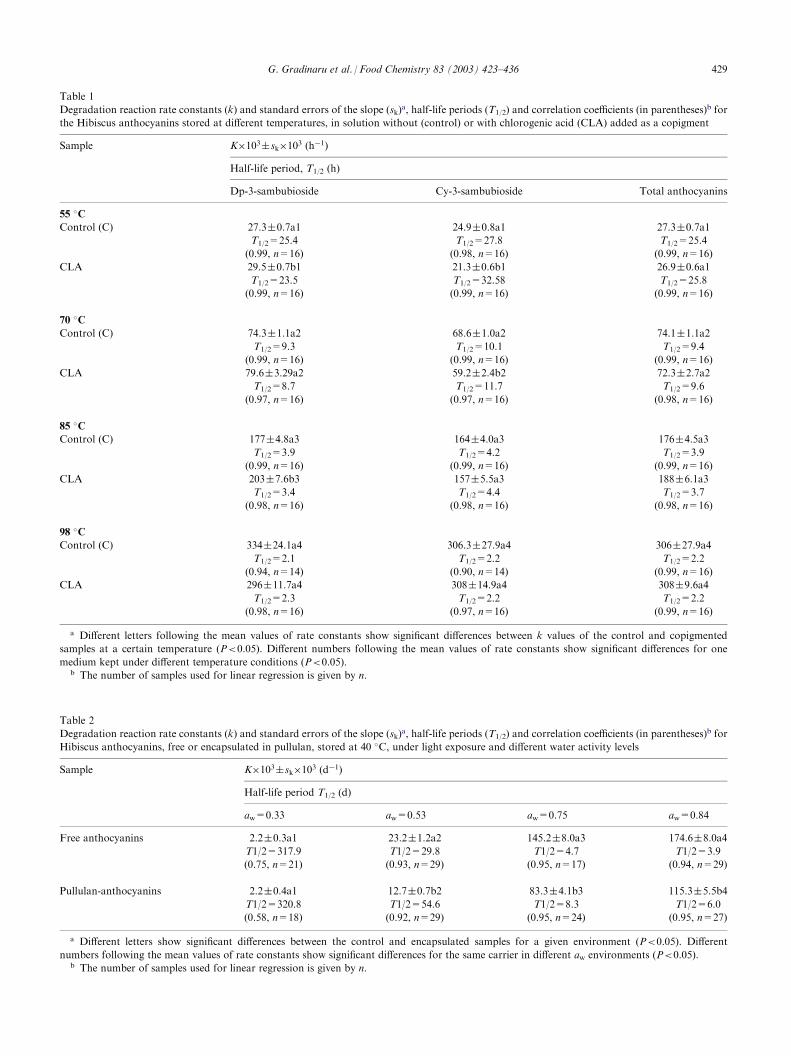
Table 1
Degradation reaction rate constants (k) and standard errors of the slope (sk)a, half-life periods (T1/2) and correlation coefficients (in parentheses)
b for
the Hibiscus anthocyanins stored at different temperatures, in solution without (control) or with chlorogenic acid (CLA) added as a copigment
Sample
K�103 sk�103 (h�1)Half-life period, T1/2 (h)
Dp-3-sambubioside
Cy-3-sambubioside Total anthocyanins55 �C
Control (C)
27.3 0.7a1 24.9 0.8a1 27.3 0.7a1T1/2=25.4
T1/2=27.8 T1/2=25.4(0.99, n=16)
(0.98, n=16) (0.99, n=16)CLA
29.5 0.7b1 21.3 0.6b1 26.9 0.6a1T1/2=23.5
T1/2=32.58 T1/2=25.8(0.99, n=16)
(0.99, n=16) (0.99, n=16)70 �C
Control (C)
74.3 1.1a2 68.6 1.0a2 74.1 1.1a2T1/2=9.3
T1/2=10.1 T1/2=9.4(0.99, n=16)
(0.99, n=16) (0.99, n=16)CLA
79.6 3.29a2 59.2 2.4b2 72.3 2.7a2T1/2=8.7
T1/2=11.7 T1/2=9.6(0.97, n=16)
(0.97, n=16) (0.98, n=16)85 �C
Control (C)
177 4.8a3 164 4.0a3 176 4.5a3T1/2=3.9
T1/2=4.2 T1/2=3.9(0.99, n=16)
(0.99, n=16) (0.99, n=16)CLA
203 7.6b3 157 5.5a3 188 6.1a3T1/2=3.4
T1/2=4.4 T1/2=3.7(0.98, n=16)
(0.98, n=16) (0.98, n=16)98 �C
Control (C)
334 24.1a4 306.3 27.9a4 306 27.9a4T1/2=2.1
T1/2=2.2 T1/2=2.2(0.94, n=14)
(0.90, n=14) (0.99, n=16)CLA
296 11.7a4 308 14.9a4 308 9.6a4T1/2=2.3
T1/2=2.2 T1/2=2.2(0.98, n=16)
(0.97, n=16) (0.99, n=16)a Different letters following the mean values of rate constants show significant differences between k values of the control and copigmented
samples at a certain temperature (P<0.05). Different numbers following the mean values of rate constants show significant differences for one
medium kept under different temperature conditions (P<0.05).b The number of samples used for linear regression is given by n.
Table 2
Degradation reaction rate constants (k) and standard errors of the slope (sk)a, half-life periods (T1/2) and correlation coefficients (in parentheses)
b for
Hibiscus anthocyanins, free or encapsulated in pullulan, stored at 40 �C, under light exposure and different water activity levels
Sample
K�103 sk�103 (d�1)Half-life period T1/2 (d)
aw=0.33
aw=0.53 aw=0.75 aw=0.84Free anthocyanins
2.2 0.3a1 23.2 1.2a2 145.2 8.0a3 174.6 8.0a4T1/2=317.9
T1/2=29.8 T1/2=4.7 T1/2=3.9(0.75, n=21)
(0.93, n=29) (0.95, n=17) (0.94, n=29)Pullulan-anthocyanins
2.2 0.4a1 12.7 0.7b2 83.3 4.1b3 115.3 5.5b4T1/2=320.8
T1/2=54.6 T1/2=8.3 T1/2=6.0(0.58, n=18)
(0.92, n=29) (0.95, n=24) (0.95, n=27)a Different letters show significant differences between the control and encapsulated samples for a given environment (P<0.05). Different
numbers following the mean values of rate constants show significant differences for the same carrier in different aw environments (P<0.05).b The number of samples used for linear regression is given by n.
G. Gradinaru et al. / Food Chemistry 83 (2003) 423–436 429
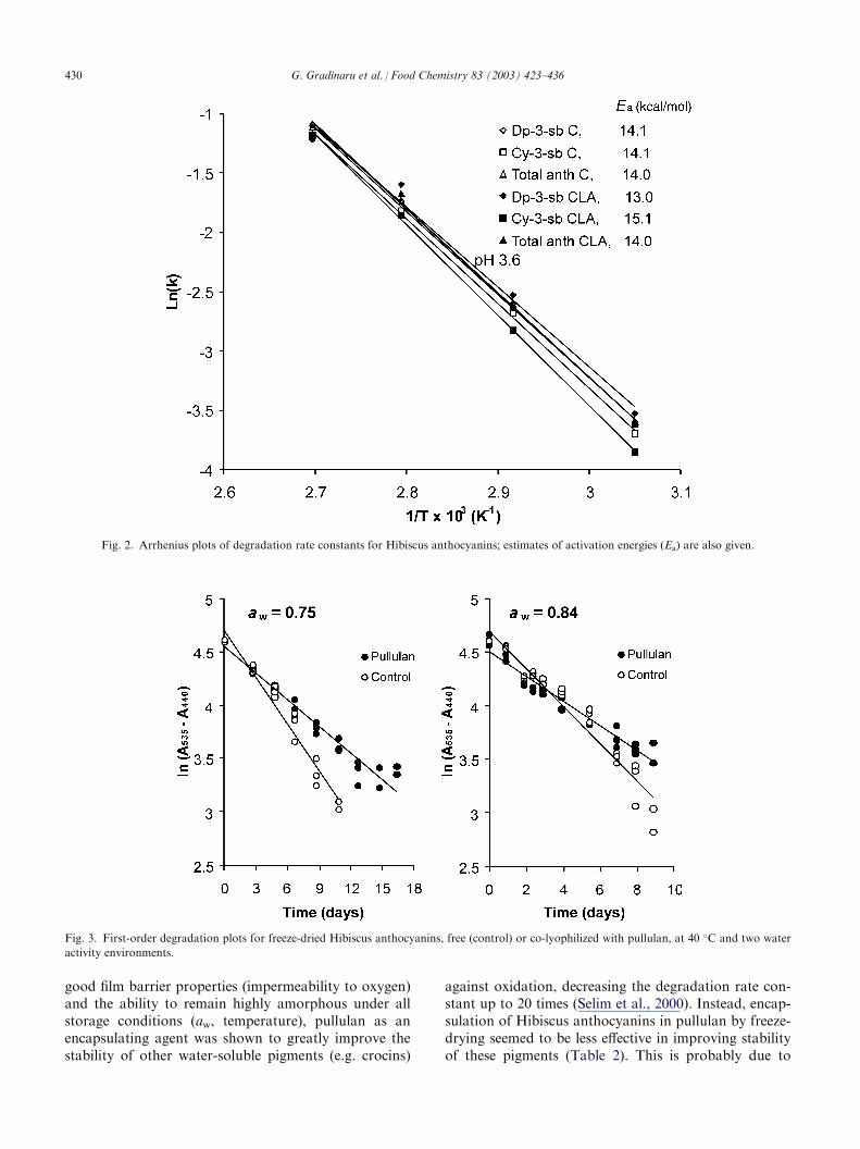
good film barrier properties (impermeability to oxygen)and the ability to remain highly amorphous under allstorage conditions (aw, temperature), pullulan as anencapsulating agent was shown to greatly improve thestability of other water-soluble pigments (e.g. crocins)
against oxidation, decreasing the degradation rate con-stant up to 20 times (Selim et al., 2000). Instead, encap-sulation of Hibiscus anthocyanins in pullulan by freeze-drying seemed to be less effective in improving stabilityof these pigments (Table 2). This is probably due to
Fig. 2. Arrhenius plots of degradation rate constants for Hibiscus anthocyanins; estimates of activation energies (Ea) are also given.
Fig. 3. First-order degradation plots for freeze-dried Hibiscus anthocyanins, free (control) or co-lyophilized with pullulan, at 40 �C and two water
activity environments.
430 G. Gradinaru et al. / Food Chemistry 83 (2003) 423–436
differences in the degradation mechanism betweenanthocyanins (sensitivity to non-covalent and covalenthydration) and other type of pigments. According toRosenberg and co-workers (Moreau & Rosenberg,1998, 1999; Sheu & Rosenberg, 1998), the nature of thepigment (core) as well as the chemistry and physicalproperties of the wall material (pullulan), are importantstability determinants of encapsulated systems.
3.3. The glass–rubber transition and degradationkinetics of Hibiscus anthocyanins
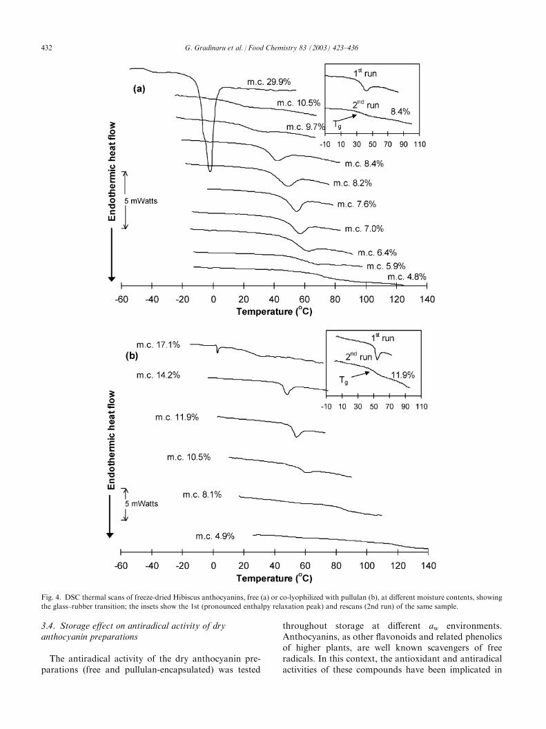
In recent years, mainly due to the pioneering work ofSlade and Levine (Levine & Slade 1986, 1992; Slade &Levine, 1988a, 1988b, 1991; Slade, Levine, & Finley,1989), the concept of glass–rubber transition and itsimplication for stability and properties of low-moisturefoods and biomaterials has been largely advanced.Although the chemical changes in dry products are veryslow, there is evidence that various deteriorative pro-cesses are accelerated if the dried products are stored attemperatures above their Tg; in contrast, translationalmobility and diffusion are essentially restricted in aglassy solid (Slade & Levine, 1991). As the Tg is highlysensitive to water content, the moisture-dependence offood product stability has been attributed to changes inmolecular mobility, as controlled by the glass transition.Therefore, the Tg is often considered as a referencetemperature: below Tg, the food is expected to be stable,whereas above this temperature, the difference betweenstorage temperature and glass transition temperature(T�Tg) is assumed to control the rate of physical, che-mical and enzymic changes in the product. Moreover,processes that are diffusion-limited are expected to con-form to WLF kinetics rather than to the Arrheniusformalism; in this respect, the WLF equation specifies amuch stronger temperature-dependence of reactionrates than the Arrhenius relationship. However, con-flicts exist as to whether only diffusion-limited processes(typically those of low activation energies, 2–6 kcal/mol)are controlled by the glass transition and whethertranslational diffusion coefficients, especially of smallmolecules, remain at quite a high level in a glassy matrixthat is relatively ‘non-dense’, permitting molecular col-lisions between reactants and the reaction to occur evenat sub-Tg temperatures (Bell & Hageman, 1994;Comyns, 1985; Fennema, 1996; Karel, 1993; Orlien,Andersen, Sinkko, & Skibsted, 2000; Schebor, Buera,Karel, & Chirife, 1999).Fig. 4 shows the DSC traces of the freeze-dried
Hibiscus anthocyanins, free or co-lyophilized with pull-ulan, at different water contents. The observed endo-thermal baseline shifts, typical of the glass transition,indicate the progressive lowering of Tg with increasingamounts of water. The Tg-water content relationships(Gordon–Taylor plots) presented in Fig. 5 more clearly
reveal the sensitivity of the amorphous materials towater-plasticization. The Tg curve of the free Hibiscusanthocyanins is shifted to lower temperatures (�40–50 �C) than the pullulan-anthocyanin samples, reflectingthe average molecular size differences between the twosystems. Estimated values of Tg1 for the dried productsare included in the inset of Fig. 5 along with the k valuesof the G-T model. The freeze-dried extracts of Hibiscusanthocyanins, because of their lower Tg than theencapsulated product, would be expected to enter therubbery domain at a much lower water content assum-ing a constant storage temperature. Fig. 6 presents thedependence of anthocyanin degradation rate as a func-tion of T=T�Tg. It is clear from this plot thatanthocyanin degradation greatly increases above thetemperature vicinity of the glass transition for bothmaterials. However, it is interesting to note that pig-ment degradation does occur, even at temperaturesbelow the glass transition. These findings concur withdata from recent studies which have shown that somechemical reactions (ascorbic acid oxidation, aspartamehydrolysis, non-enzymic browning, lipid oxidation) stillproceed at substantial rates in the glassy state (Bell &Hageman, 1994; Karmas, Buera, & Karel, 1992; Nelson,1993; Orlien et al., 2000; Schebor et al., 1999), failing toshow the stability of the ‘glassy system’ as often claimedin the early literature on glass–rubber transitions offood materials (Levine & Slade, 1992; Slade & Levine,1991; Slade, Levine, & Finley, 1989). The occurrence ofanthocyanin degradation at temperatures far below theTg for both free and encapsulated systems (Fig. 6)strongly points to the possibility of some sort of reac-tant mobility in the glassy state and further indicates theinsufficiency of using the calorimetrically-determined Tg
as an absolute threshold of stability. Macroscopic hetero-geneities in the glassy matrix, non-homogeneous distribu-tion of water, and phase separation phenomena (demixingof reactants and inert matrix) are also likely to influencethe apparent reaction rates and further explain why reac-tions do not cease below the measured Tg.A plot of experimental values of 1/k against T�Tg
showed a linear response with all data falling into asingle line. This trend complies with the WLF relation-ship for the temperature dependence of reaction rates inthe rubbery domain. Interestingly, there was no majorchange in the temperature-dependence of the degra-dation rate constants for all samples in the vicinity ofthe glass transition zone. It is worth noting here thatin similar kinetic studies on the applicability of theWLF equation to nonenzymic browning (Roos &Himberg, 1994), oxidation of crocin carotenoids (Tsi-midou & Biliaderis, 1997) and degradation of encap-sulated beetroot pigment (Serris & Biliaderis, 2001),using several storage temperatures, the reaction ratesdid not seem to fit into a common line based on theWLF formalism.
G. Gradinaru et al. / Food Chemistry 83 (2003) 423–436 431
3.4. Storage effect on antiradical activity of dryanthocyanin preparations
The antiradical activity of the dry anthocyanin pre-parations (free and pullulan-encapsulated) was tested
throughout storage at different aw environments.Anthocyanins, as other flavonoids and related phenolicsof higher plants, are well known scavengers of freeradicals. In this context, the antioxidant and antiradicalactivities of these compounds have been implicated in
Fig. 4. DSC thermal scans of freeze-dried Hibiscus anthocyanins, free (a) or co-lyophilized with pullulan (b), at different moisture contents, showing
the glass–rubber transition; the insets show the 1st (pronounced enthalpy relaxation peak) and rescans (2nd run) of the same sample.
432 G. Gradinaru et al. / Food Chemistry 83 (2003) 423–436
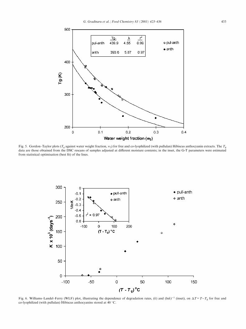
Fig. 5. Gordon–Taylor plots (Tg against water weight fraction, w2) for free and co-lyophilized (with pullulan) Hibiscus anthocyanin extracts. The Tg
data are those obtained from the DSC rescans of samples adjusted at different moisture contents; in the inset, the G-T parameters were estimated
from statistical optimisation (best fit) of the lines.
Fig. 6. Williams–Landel–Ferry (WLF) plot, illustrating the dependence of degradation rates, (k) and (lnk)�1 (inset), on T=T�Tg for free and
co-lyophilized (with pullulan) Hibiscus anthocyanins stored at 40 �C.
G. Gradinaru et al. / Food Chemistry 83 (2003) 423–436 433

the protective effect of vegetable-/fruit-rich diets againstcoronary diseases (Hertog, Feskens, Hollman, Katan, &Kromhout, 1993). The results of Fig. 7, as well as thesummarized data of the initial and final (after a specifiedstorage period) antiradical capacity values presented inTable 3, clearly indicate that, despite colour fading, thebreakdown products of anthocyanins still exhibit sig-nificant antiradical power. These results suggest thatanthocyanins present in food products may continue toprovide their beneficial health effects (antioxidants) evenafter some colour loss has occurred during processingand storage.
4. Conclusions
In this study, the thermal stability of isolated antho-cyanins from dry calyces of H. sabdariffa was examinedin aqueous solutions, with or without the presence ofchlorogenic acid as a copigment, and in the dry stateduring storage at different relative humidity environ-
ments. Copigmentation did not seem to alter the degra-dation kinetics of individual anthocyanin componentsor total anthocyanins of the Hibiscus pigment extractsin the temperature range 55–98 �C. Instead, the degra-dation kinetics of dry anthocyanin preparations waslargely affected by hydration, showing a gradualincrease in the rate with increasing water activity. Theco-lyophilized pullulan-anthocyanins showed a slightlyimproved stability at all relative humidity environmentscompared with the free anthocyanin extract. The tem-perature-dependence of the anthocyanin degradationrates in all freeze-dried materials seemed to follow theWLF kinetic model. However, the calorimetrically-determined Tg of the preparations cannot be consideredas an absolute threshold temperature for pigment stabi-lity. Degradation of anthocyanins, occurred even atsub-Tg temperatures, for all samples, implying sig-nificant reactant mobility in the glassy state. Based onthe findings of this work, further studies would benecessary for the determination of appropriate proces-sing and formulation protocols that could lead to amore efficient utilization of these pigments in actualfood products.
Acknowledgements
G.G. acknowledges the receipt of a fellowship fromMAICh during her MSc studies. This project has beenpartially supported from the Greek Ministry of Indus-try, Energy and Technology and the Spanish Ministeriode Asuntos Exteriores through a grant of bilateralcooperation between Greece and Spain.
References
Al-Kahtani, H. A., & Hassan, B. H. (1990). Spray-drying of roselle
(Hibiscus sabdariffa L.) extract. Journal of Food Science, 55, 1073–
1076.
Beatus, Y., Raziel, A., Rosenberg, M., & Kopelman, I. J. (1985).
Spray-drying microencapsulation of paprika oleoresin. Lebensmit-
tel-Wissenschaft und- Technologie, 18, 28–34.
Bell, L. N., & Hageman, M. J. (1994). Differentiating between the
effects of water activity and glass transition-dependent mobility on a
solid state chemical reaction: aspartame degradation. Journal of
Agricultural and Food Chemistry, 42, 2398–2401.
Biliaderis, C. G., Lazaridou, A., & Arvanitoyannis, I. (1999). Glass
transition and physical properties of polyol-plasticized pullulan-
starch blends at low moisture. Carbohydrate Polymers, 40, 29–47.
Brand-Williams, W., Cuvelier, M. E., & Berset, C. (1995). Use of free
radical method to evaluate antioxidant activity. Lebensmittel-Wis-
senschaft und- Technologie, 28, 25–30.
Brouillard, R., & Dangles, O. (1994). Anthocyanin molecular interac-
tions: the first step in the formation of new pigments during wine
aging? Food Chemistry, 51, 365–371.
Clifford, M. N. (2000). Chlorogenic acids and other cinnamates—nat-
ure, occurrence, dietary burden, absorption and metabolism. Jour-
nal of the Science of Food and Agriculture, 80, 1033–1043.
Clydesdale, F. M., Main, J. H., & Francis, F. J. (1979). Roselle
Fig. 7. Antiradical activity of freeze-dried Hibiscus anthocyanins (free
and co-lyophilized with pullulan) during storage at aw=0.75.
Table 3
Average antiradical capacity (ARP) of free and encapsulated antho-
cyanins in a pullulan matrix at the beginning and at the end of the
specified (in parenthesis) storage period
aw
ARPa, mg DPPH/mg of Hibiscus extractAnthocyanins
Pullulan-anthocyaninsInitial
Final (days) Initial Final (days)0.33
3.5 0.2 4.2 0.3 (210) 3.1 0.2 3.7 0.2 (210)0.53
3.4 0.2 3.5 0.2 (75) 3.0 0.3 2.9 0.3 (75)0.75
3.7 0.1 3.3 0.1 (17) 3.1 0. 1 2.5 0.1 (17)0.84
3.8 0.1 3.6 0.1 (9) 3.2 0.2 2.9 0.3 (9)a Values are given as average standard deviation.
434 G. Gradinaru et al. / Food Chemistry 83 (2003) 423–436

(Hibiscus sabdariffa L.) anthocyanins as food colorants for bev-
erages and gelatin desserts. Journal of Food Protection, 42, 204–
207.
Clydesdale, F. M., Main, J. H., Francis, F. J., & Hayes, K. M. (1979).
Effect of anthocyanins preparations as colorants on hygroscopicity
of dry-pack foods. Journal of Food Protection, 42, 225–227.
Comyns, J. (1985). Polymer permeability. London: Elsevier Applied
Science.
Daravingas, G., & Cain, R. F. (1968). Thermal degradation of black
raspberry anthocyanin pigments in model systems. Journal of Food
Science, 33, 138–142.
Desobry, A. S., Netto, M., & Labuza, T. P. (1997). Comparison of
spray-drying, drum-drying and freeze-drying for b-carotene encap-sulation and preservation. Journal of Food Science, 62, 1158–1162.
Du, C. T., & Francis, F. J. (1973). Anthocyanins of roselle (Hibiscus
sabdariffa). Journal of Food Science, 38, 810–812.
Erlandson, J. A., & Wrolstad, R. E. (1972). Degradation of antho-
cyanins at limited water concentration. Journal of Food Science, 37,
592–595.
Espin, J. C., Soler-Rivas, C., Wichers, H., & Garcia-Viguera, C.
(2000). Anthocyanin-based natural colorants: a new source of anti-
radical activity for foodstuff. Journal of Agricultural and Food
Chemistry, 48, 1588–1592.
Esselen, W. B., & Sammy, G. M. (1973). Roselle—a natural red col-
orant for foods? Food Product Development, 7(2), 80–82, 86.
Esselen, W. B., & Sammy, G. M. (1975). Application for roselle as a
red food colorant. Food Product Development, 9(10), 37–38, 40.
Fennema, O. R. (1996). Water and ice. In O. R. Fennema (Ed.), Food
chemistry (3rd ed.) (pp. 55–93). New York: Marcel Dekker.
Francis, F. J. (1975). Anthocyanins as food colors. Food Technology,
29(5), 52–54.
Francis, F. J. (1989). Food colorants: anthocyanins. Critical Reviews
in Food Science and Nutrition, 28, 273–314.
Gordon, M., & Taylor, J. S. (1952). Ideal copolymers and the second-
order transition of synthetic rubbers. I. Non-crystalline copolymers.
Journal of Applied Chemistry, 2, 493–500.
Hertog, M. G. L., Feskens, E. J. M., Hollman, P. C. H., Katan, M. B.,
& Kromhout, D. (1993). Dietary antioxidant flavonoids and risk of
coronary heart disease: the Zutphen elderly study. Lancet, 342,
1007–1011.
Jackman, R. L., & Smith, J. L. (1992). Anthocyanins and betalains. In
G. A. F. Hendry, & J. D. Houghton (Eds.), Natural food colorants
(pp. 182–241). Glasgow: Blackie.
Jackman, R. L., Yada, R. Y., Tung, M. A., & Speers, R. A. (1987).
Anthocyanins as food colorants—a review. Journal of Food Bio-
chemistry, 11, 201–247.
Karel, M. (1993). Temperature-dependence of food deterioration pro-
cesses. Journal of Food Science, 58(6), ii.
Karmas, R., Buera, M. P., & Karel, M. (1992). Effect of glass transi-
tions on rates of non-enzymatic browning in food systems. Journal
of Agricultural and Food Chemistry, 40, 873–879.
Labuza, T. P. (1984). Moisture sorption. In Practical aspects of iso-
therm measurements and use. Minneapolis, MN: The American
Association of Cereal Chemists.
Levine, H., & Slade, L. (1986). A polymer physicochemical approach
to the study of commercial starch hydrolysis products (SPHs). Car-
bohydrate Polymers, 6, 213–244.
Levine, H., & Slade, L. (1992). Glass transitions in foods. In
H. G. Schwartzberg, & R. W. Hartel (Eds.), Physical chemistry of
foods (pp. 83–221). New York: Marcel Dekker.
Lund, D. B. (1975). Heat processing. In O. R. Fennema (Ed.), Princi-
ples of food science. Part II. Physical principles of food preservation
(pp. 31–92). New York: Marcel Dekker.
Maccarone, E., Maccarone, A., & Rapisarda, P. (1985). Stabilization
of anthocyanins in blood orange fruit juice. Journal of Food Science,
50, 901–904.
Maccarone, E., Maccarone, A., & Rapisarda, P. (1987). Technical
note: color stabilization of orange fruit juice by tannic acid. Inter-
national Journal of Food Science and Technology, 22, 153–160.
Markakis, P., Livingstone, G. E., & Fellers, R. C. (1957). Quantitative
aspects of strawberry-pigment degradation. Food Research, 22, 117–
130.
Mazza, G., & Brouillard, R. (1990). The mechanism of co-pigmenta-
tion of anthocyanins in aqueous solutions. Phytochemistry, 29,
1097–1102.
Mazza, G., & Miniati, E. (1993). Anthocyanins in fruits, vegetables and
grains. Boca Raton, FL: CRC Press.
Meschter, E. E. (1953). Effects of carbohydrates and other factors on
strawberry products. Journal of Agricultural and Food Chemistry, 1,
574–579.
Miniati, E., Damiani, P., & Mazza, G. (1992). Copigmentation and
self-association of anthocyanins in food model systems. Italian
Journal of Food Science, 2, 109–116.
Moreau, D. L., & Rosenberg, M. (1998). Porosity of whey protein-
based microcapsules containing anhydrous milkfat measured by gas
displacement pycnometry. Journal of Food Science, 63, 819–823.
Moreau, D. L., & Rosenberg, M. (1999). Porocity of microcapsules
with wall systems consisting of whey proteins and lactose measured
by gas displacement pycnometry. Journal of Food Science, 64, 405–
409.
Nelson, K. (1993). Reaction kinetics of food stability: comparison of
glass transition and classical models for temperature and moisture
dependence. Doctor of Philosophy Thesis, University of Minnesota.
Orlien, V., Andersen, A. B., Sinkko, T., & Skibsted, L. H. (2000).
Hydroperoxide formation in rapeseed oil encapsulated in a glassy
food model as influenced by hydrophilic and lipophilic radicals.
Food Chemistry, 68, 191–199.
Pouget, M. P., Lejeune, B., Vennat, B., & Pourrat, A. (1990). Extrac-
tion, analysis and study of the stability of Hibiscus anthocyanins.
Lebensmittel-Wissenschaft und- Technologie, 23, 103–105.
Roos, H. Y., & Himberg, M. J. (1994). Nonenzymatic browning
behavior, as related to glass transition, of a food model at chilling
temperatures. Journal of Agricultural and Food Chemistry, 42, 893–
898.
Roos, H. Y., & Karel, M. (1991). Plasticizing effect of water on ther-
mal behavior and crystallization of amorphous food models. Jour-
nal of Food Science, 56, 38–43.
Schebor, C., Buera, M. P., Karel, M., & Chirife, J. (1999). Color for-
mation due to non-enzymatic browning in amorphous, glassy,
anhydrous, model systems. Food Chemistry, 65, 427–432.
Segal, B., & Negutz, G. (1969). Thermal destruction of keracyanin.
Nahrung, 13, 531–535.
Selim, K., Tsimidou, M., & Biliaderis, C. G. (2000). Kinetic studies of
degradation of saffron carrotenoids encapsulated in amorphous
polymer matrices. Food Chemistry, 71, 199–206.
Serris, G. S., & Biliaderis, C. G. (2001). Degradation kinetics of beet-
root pigment encapsulated in polymeric matrices. Journal of the
Science of Food and Agriculture, 81, 1–10.
Slade, L., & Levine, H. (1988a). Structural stability of intermediate
moisture foods—a new understanding?. In J. R. Mitchell, &
J. M. V. Blanshard (Eds.), Food structure—its creation and eval-
uation (pp. 115–147). London: Butterworths.
Slade, L., & Levine, H. (1988b). Non-equilibrium behavior of small
carbohydrate-water systems. Pure and Applied Chemistry, 60, 1841–
1864.
Slade, L., & Levine, H. (1991). Beyond water activity: recent advances
based on an alternative approach to the assessment of food quality
and safety. CRC Critical Reviews in Food Science and Nutrition, 30,
115–360.
Slade, L., Levine, H., & Finley, J. W. (1989). Protein–water interac-
tions: water as a plasticizer of gluten and other protein polymers. In
R. Dixon Phillips, & J. W. Finley (Eds.), Protein quality and the
effects of processing (pp. 9–124). New York: Marcel Dekker.
Sheu, T.-Y., & Rosenberg, M. (1993). Microstructure of microcapsules
G. Gradinaru et al. / Food Chemistry 83 (2003) 423–436 435

consisting of whey proteins and carbohydrates. Journal of Food
Science, 63, 491–494.
Steel, D. G. R., & Torrie, H. J. (1980). Principles and procedures of
statistics. A biometrical approach (2nd ed.). New York: McGraw-
Hill.
Strack, D., & Wray, V. (1993). The anthocyanins. In J. B. Harborne
(Ed.), The flavonoids: advances in research since 1986 (pp. 1–22).
London: Chapman & Hall.
Sugisaki, M., Suga, H., & Seki, S. (1968). Calorimetric study of the
glassy state. IV. Heat capacities of glassy water and cubic ice. Bul-
letin of Japanese Chemical Society, 41, 2591–2599.
Tanchev, S. (1983). Kinetics of thermal degradation of anthocyanins.
In J. V. McLoughlin, & B. M. McKenna (Eds.), Basic Studies in
Food Science, Vol. 2. Proceedings of the 6th International Congress of
Food Science and Technology (pp. 96). Dublin, Ireland: Boole Press.
Teh, L. S., & Francis, F. J. (1988). Stability of anthocyanins from
Zebrina pendula and Ipomoea tricolor in model beverage. Journal of
Food Science, 53, 1580–1581.
Tsimidou, M., & Biliaderis, C. G. (1997). Kinetic studies of Saffron
(Crocus sativus L.) quality deterioration. Journal of Agricultural and
Food Chemistry, 45, 2890–2898.
Wagner, A. D., & Warthesen, J. J. (1995). Stability of spray-dried
encapsulated carrot carotenes. Journal of Food Science, 60, 1048–1052.
Wang, H., Cao, G., & Prior, R. (1997). Oxygen radical absorbing
capacity of anthocyanins. Journal of Agricultural and Food Chem-
istry, 45, 304–309.
Williams, M. L., Landel, R. F., & Ferry, J. D. (1955). Temperature
dependence of relaxation mechanisms in amorphous polymers and
other glass-forming liquids. Journal of the American Chemical
Society, 77, 3701–3707.
436 G. Gradinaru et al. / Food Chemistry 83 (2003) 423–436