Therapeutic Outcome of MR-Guided High-Intensity Focused ...
9
Citation: Erber, B.; Schwarze, V.; Strobl, F.; Burges, A.; Mahner, S.; Goller, S.S.; Rudolph, J.; Ricke, J.; Sabel, B.O. Therapeutic Outcome of MR-Guided High-Intensity Focused Ultrasound (MR-HIFU) in Solitary versus Multiple Uterine Fibroids. Healthcare 2022, 10, 1471. https:// doi.org/10.3390/healthcare10081471 Academic Editor: Andrzej Skr˛ et Received: 6 July 2022 Accepted: 31 July 2022 Published: 4 August 2022 Publisher’s Note: MDPI stays neutral with regard to jurisdictional claims in published maps and institutional affil- iations. Copyright: © 2022 by the authors. Licensee MDPI, Basel, Switzerland. This article is an open access article distributed under the terms and conditions of the Creative Commons Attribution (CC BY) license (https:// creativecommons.org/licenses/by/ 4.0/). healthcare Article Therapeutic Outcome of MR-Guided High-Intensity Focused Ultrasound (MR-HIFU) in Solitary versus Multiple Uterine Fibroids Bernd Erber 1, * , Vincent Schwarze 1 , Frederik Strobl 1,2 , Alexander Burges 3 , Sven Mahner 3 , Sophia Samira Goller 1 , Jan Rudolph 1 , Jens Ricke 1 and Bastian Oliver Sabel 1 1 Department of Radiology, University Hospital, LMU Munich, Marchioninistr. 15, 81377 Munich, Germany 2 Die Radiologie am Isarklinikum, Sonnenstr. 24-26, 80331 Munich, Germany 3 Department of Obstetrics and Gynecology, University Hospital, LMU Munich, Marchioninistr. 15, 81377 Munich, Germany * Correspondence: [email protected]; Tel.: +49-89-4400-73636 Abstract: MR-guided high-intensity focused ultrasound (MR-HIFU) is an effective method for treating symptomatic uterine fibroids, especially solitary lesions. The aim of our study was to compare the clinical and morphological outcomes of patients who underwent MR-HIFU due to solitary fibroid (SF) or multiple fibroids (MFs) in a prospective clinical trial. We prospectively included 21 consecutive patients with SF (10) and MF (11) eligible for MR-guided HIFU. The morphological data were assessed using mint Lesion™ for MRI. The clinical data were determined using the Uterine Fibroid Symptom and Quality of Life (UFS-QOL) questionnaire before and 6 months after treatment. Unpaired and paired Wilcoxon-test and t-tests were applied, and Pearson’s coefficient was used for correlation analysis. A p-value of 0.05 was considered statistically significant. The volume of treated fibroids significantly decreased in both the SF (mean baseline: 118.6 cm 3 ; mean 6-month follow-up: 64.6 cm 3 ) and MF (107.2 cm 3 ; 55.1 cm 3 ) groups. The UFS-QOL showed clinical symptoms significantly improved for patients in both the SF and MF groups regarding concern, activities, energy/mood, and control. The short-term outcome for the treatment of symptomatic fibroids in myomatous uterus by MR-guided HIFU is clinically similar to that of solitary fibroids. Keywords: MR-guided high-intensity focused ultrasound; MR-HIFU; solitary uterine fibroids; multiple uterine fibroids; uterine fibroid symptom and quality of life 1. Introduction Uterine fibroids are the most common benign neoplasms of the uterus in women of reproductive age, with a prevalence of 20–40% and the most common indication for a hysterectomy [1]. These estrogen-dependent tumors originate from the smooth muscle cells [2] and can occur in solitary or multiple forms in different locations in the uterus [3]. About 20–25% of uterine leiomyomata cause symptoms, and menstrual abnormalities are the most frequent [2]. The severity of menorrhagia seems to be increased for submucous lesions, although this symptom can also occur when fibroids are intramurally or sub- serously located [2]. Different reasons are found in the literature for menorrhagia. Some studies suggested that the decreased uterine contractility due to leiomyomata leads to menorrhagia [4], while others claimed that the dysregulation of local growth factors and aberrant angiogenesis might be relevant [5]. Furthermore, increased severity of urogenital symptoms was found for both anterior and intramural fibroids [6]. At present, myomectomy is the preferred treatment option. However, new interven- tional treatment options have emerged that offer benefits in terms of relieving symptoms while avoiding the risks of surgery: uterine artery embolization (UAE) and high-intensity focused ultrasound (HIFU) [7]. HIFU ablation can lead to comparable long-term outcomes Healthcare 2022, 10, 1471. https://doi.org/10.3390/healthcare10081471 https://www.mdpi.com/journal/healthcare
-
Upload
khangminh22 -
Category
Documents
-
view
0 -
download
0
Transcript of Therapeutic Outcome of MR-Guided High-Intensity Focused ...
Citation: Erber, B.; Schwarze, V.;
Strobl, F.; Burges, A.; Mahner, S.;
Goller, S.S.; Rudolph, J.; Ricke, J.;
Sabel, B.O. Therapeutic Outcome of
MR-Guided High-Intensity Focused
Ultrasound (MR-HIFU) in Solitary
versus Multiple Uterine Fibroids.
Healthcare 2022, 10, 1471. https://
doi.org/10.3390/healthcare10081471
Academic Editor: Andrzej Skret
Received: 6 July 2022
Accepted: 31 July 2022
Published: 4 August 2022
Publisher’s Note: MDPI stays neutral
with regard to jurisdictional claims in
published maps and institutional affil-
iations.
Copyright: © 2022 by the authors.
Licensee MDPI, Basel, Switzerland.
This article is an open access article
distributed under the terms and
conditions of the Creative Commons
Attribution (CC BY) license (https://
creativecommons.org/licenses/by/
4.0/).
healthcare
Article
Therapeutic Outcome of MR-Guided High-Intensity FocusedUltrasound (MR-HIFU) in Solitary versus MultipleUterine FibroidsBernd Erber 1,* , Vincent Schwarze 1, Frederik Strobl 1,2, Alexander Burges 3 , Sven Mahner 3,Sophia Samira Goller 1 , Jan Rudolph 1 , Jens Ricke 1 and Bastian Oliver Sabel 1
1 Department of Radiology, University Hospital, LMU Munich, Marchioninistr. 15, 81377 Munich, Germany2 Die Radiologie am Isarklinikum, Sonnenstr. 24-26, 80331 Munich, Germany3 Department of Obstetrics and Gynecology, University Hospital, LMU Munich, Marchioninistr. 15,
81377 Munich, Germany* Correspondence: [email protected]; Tel.: +49-89-4400-73636
Abstract: MR-guided high-intensity focused ultrasound (MR-HIFU) is an effective method fortreating symptomatic uterine fibroids, especially solitary lesions. The aim of our study was tocompare the clinical and morphological outcomes of patients who underwent MR-HIFU due tosolitary fibroid (SF) or multiple fibroids (MFs) in a prospective clinical trial. We prospectively included21 consecutive patients with SF (10) and MF (11) eligible for MR-guided HIFU. The morphologicaldata were assessed using mint Lesion™ for MRI. The clinical data were determined using the UterineFibroid Symptom and Quality of Life (UFS-QOL) questionnaire before and 6 months after treatment.Unpaired and paired Wilcoxon-test and t-tests were applied, and Pearson’s coefficient was used forcorrelation analysis. A p-value of 0.05 was considered statistically significant. The volume of treatedfibroids significantly decreased in both the SF (mean baseline: 118.6 cm3; mean 6-month follow-up:64.6 cm3) and MF (107.2 cm3; 55.1 cm3) groups. The UFS-QOL showed clinical symptoms significantlyimproved for patients in both the SF and MF groups regarding concern, activities, energy/mood, andcontrol. The short-term outcome for the treatment of symptomatic fibroids in myomatous uterus byMR-guided HIFU is clinically similar to that of solitary fibroids.
Keywords: MR-guided high-intensity focused ultrasound; MR-HIFU; solitary uterine fibroids;multiple uterine fibroids; uterine fibroid symptom and quality of life
1. Introduction
Uterine fibroids are the most common benign neoplasms of the uterus in womenof reproductive age, with a prevalence of 20–40% and the most common indication fora hysterectomy [1]. These estrogen-dependent tumors originate from the smooth musclecells [2] and can occur in solitary or multiple forms in different locations in the uterus [3].About 20–25% of uterine leiomyomata cause symptoms, and menstrual abnormalities arethe most frequent [2]. The severity of menorrhagia seems to be increased for submucouslesions, although this symptom can also occur when fibroids are intramurally or sub-serously located [2]. Different reasons are found in the literature for menorrhagia. Somestudies suggested that the decreased uterine contractility due to leiomyomata leads tomenorrhagia [4], while others claimed that the dysregulation of local growth factors andaberrant angiogenesis might be relevant [5]. Furthermore, increased severity of urogenitalsymptoms was found for both anterior and intramural fibroids [6].
At present, myomectomy is the preferred treatment option. However, new interven-tional treatment options have emerged that offer benefits in terms of relieving symptomswhile avoiding the risks of surgery: uterine artery embolization (UAE) and high-intensityfocused ultrasound (HIFU) [7]. HIFU ablation can lead to comparable long-term outcomes
Healthcare 2022, 10, 1471. https://doi.org/10.3390/healthcare10081471 https://www.mdpi.com/journal/healthcare
Healthcare 2022, 10, 1471 2 of 9
with longer time intervals until re-intervention compared with myomectomy [8]. Further-more, several studies compared the outcomes of HIFU and UAE. Reviews by Liu et al. [7]and Yan et al. [9] found the benefits of UAE regarding symptom relief and re-interventionrate. In their meta-analysis, Gao et al. found the lowest incidence of major complicationswith HIFU compared with UAE and myomectomy [10]. The technical effectiveness andsafety of HIFU were reported in the literature for this relatively new treatment option [11].Especially in solitary lesions, HIFU has been proven to be a valid method to improve clini-cal outcomes [12,13]. Therefore, solitary fibroids (SFs) are preferentially treated at manycenters, although there is—to the best of our knowledge—no evidence in the literatureon whether and to what extent patients with multiple fibroids (MFs) can benefit fromMR-HIFU. Therefore, the aim of this study was to compare the clinical and morpholog-ical outcomes of patients with SF to those with MFs undergoing MR-HIFU treatment ina prospective clinical trial.
2. Materials and Methods2.1. Patients
All participants underwent a contrast-enhanced MRI of the uterus in the prone posi-tion for treatment evaluation and were seen by a board-certified specialist in gynecologywith more than 15 years’ experience and a board-certified specialist in radiology withmore than 10 years’ experience in MR imaging as well as 5 years’ experience in MR-guided HIFU. The indication for HIFU ablation was interdisciplinarily made. The inclusioncriteria were as follows: (1) the presence of solitary or multiple symptomatic uterine fi-broids; (2) the patient’s written informed consent; (3) women aged over 18-years; (4) accessto over 50% of the total fibroid volume for both solitary and multiple uterine fibroids.The exclusion criteria were as follows: (1) any contraindication of MRI contrast agent;(2) general contraindications regarding MRI safety; (3) any clinical or MR-morphologicalsigns of malignancy such as leiomyosarcoma; (4) a positive pregnancy test; (5) bowelin the direct path of the HIFU beam in the pre-interventional MRI. Patients receivedcontrast-enhanced imaging prior to treatment, immediately after treatment and at the6-month follow-up.
2.2. MR-Guided HIFU Ablation
All patients were treated with the same commercially available MR-HIFU system(Sonalleve™, Profound Medical Corp., Mississauga, ON, Canada) integrated into a 3.0TMR scanner (Ingenia 3.0T, Philips, Amsterdam, The Netherlands). Details of the MR-HIFU system and respective treatment procedures have been described in the literaturebefore [14,15]. Apart from those showing contraindications such as allergies, all patientsintravenously received 1000 mg metamizole and 500 mg paracetamol as an analgesic and1 mg lorazepam. Patient immobilization was limited to 3 h. In most cases of MFs, notall the fibroids were accessible for HIFU treatment, e.g., when the distance between theskin and lesion was too large. In these patients, the aim was to treat fibroids that wereconsidered to be symptomatic, e.g., larger and anteriorly or intramurally located lesionsthat might cause urogenital symptoms such as frequent urination [6]; or lesions that weresubmucosally located, thus posing a higher risk of menorrhagia [2].
2.3. MRI Data Acquisition and Postprocessing2.3.1. Data Acquisition
The MRI studies were performed with a 1.5 T magnet (Avanto; Siemens Healthineers;Erlangen; Germany or Ingenia 3.0T, Philips, Amsterdam, The Netherlands). Immediate pre-interventional MRI was performed on the day of intervention for planning HIFU ablation,and consisted of T2-weighted (T2w) and T1-weighted sequences without contrast. Theimmediate post-intervention MRI consisted of contrast-enhanced T1-weighted fat-saturated(T1w fs CE) sequences. The pre-intervention baseline imaging for the evaluation of HIFU

Healthcare 2022, 10, 1471 3 of 9
indication and the imaging for the 3-month follow-up consisted of T1- and T2-weightedmultiplanar sequences as well as contrast-enhanced T1-weighted fat-saturated sequences.
2.3.2. MRI Postprocessing
All the MRI data were transferred and post-processed using mint Lesion™ (MintMedical GmbH, Heidelberg, Germany). A resident with 3 years’ experience in MR imagingmanually segmented the fibroid margins in T2w and T1w fs CE sequences and a nonper-fused volume (NPV) using T1w fs CE sequences with volumetric measures using mintLesion™. The signal intensity (SI) of the fibroids was measured in both T2w and T1w fs CEsequences and was set in relation to the SI of the healthy areas of the uterus and skeletalmuscles. An assessment of the fibroid localization (intramural, submucous, subserous) wasmade by the consensus of a board-certified specialist with more than 10 years’ experienceand a resident with more than 3 years’ experience in MR imaging.
2.4. Disease-Specific Symptom and Health-Related Quality of Life Questionnaire for Leiomyomata
The Disease-Specific Symptom and Health-Related Quality of Life Questionnairefor Leiomyomata (UFS-QOL) was given to patients before and 6 months after the HIFUablation; this is a validated instrument consisting of 8 symptom- and 29 health-relatedquality of life items [16]. The questionnaire was designed to assess symptom severity andsymptom impact on the quality of life for women with leiomyomata. It was shown todifferentiate leiomyomata patients with varying degrees of symptom severity [16]. TheUFS-QOL has been applied several times in various studies [17–19]. Items are labeled asconcern (5 items; minimal score: 5; maximal score: 25), activities (7; 7; 35), energy/mood(7; 7; 35), control (5; 5; 25), self-consciousness (3; 3; 15), and sexual function (2; 2; 10). Wecombined self-consciousness and sexual function because these two groups consisted ofa smaller number of items in comparison with the other groups.
2.5. Statistical Analysis
A statistical analysis was performed using the open-source programming languageR (Version 4.0.4, RStudio Inc., Boston, MA, USA) [20]. The differences in continuousmorphologic variables were compared using paired and unpaired t-tests. Categorialvariables regarding UFS-QOL were compared using paired and unpaired Wilcoxon tests fornonparametric data. For the correlation analysis of numeric data, the Pearson correlationcoefficient was used. A p-value of 0.05 was set as the limit of statistical significance.
3. Results3.1. Patients
The mean age for patients with SF was 43.9 years (min 35.0; max 54.0) and 44.8 years(33.0; 52.0) for patients with MFs.
The SFs were submucous in four cases and intramural in six cases, while in theeleven patients with MF, treated lesions were submucosally located in seven cases, sub-serosally in one case and intramurally in nine cases.
Regarding Funaki’s classification (21), three fibroids were assessed to be grade 1,five were grade 2 and two were grade 3 in the SF group. Among the patients witha myomatous uterus, seven fibroids were assessed with Funaki grade 1, eight fibroids asgrade 2, and one fibroid as grade 3.
The relative SI of the SF and MF groups showed the same mean value of 2.0.In Figures 1 and 2, examples of an MRI of both groups are shown.
3.2. Morphological Assessment
The mean volume of treated SFs was 118.6 cm3 (min 2.3; max 261.8; SD 98.4) and107.2 cm3 (19.3; 304.5; 101.3) for treated lesions in patients with MFs. For patients with MFs,the whole fibroid mass (volume of treated and untreated fibroids) was 152.7 cm3 (22.4; 460;153.5). In the 6-month follow-up, the volume of treated fibroids significantly decreased in
Healthcare 2022, 10, 1471 4 of 9
both groups: for SFs, the mean volume in the 6-month follow-up was 64.6 cm3 (0.5; 247.2;77.4), while for treated MFs, it was 55.1 cm3 (10.0; 183.7; 56.3). The data are visualizedin Figure 3.
Healthcare 2022, 10, x FOR PEER REVIEW 4 of 10
Regarding Funaki’s classification (21), three fibroids were assessed to be grade 1, five
were grade 2 and two were grade 3 in the SF group. Among the patients with a myoma‐
tous uterus, seven fibroids were assessed with Funaki grade 1, eight fibroids as grade 2,
and one fibroid as grade 3.
The relative SI of the SF and MF groups showed the same mean value of 2.0.
In Figures 1 and 2, examples of an MRI of both groups are shown.
Figure 1. Images of a 51‐year‐old patient with solitary intramural uterine fibroid in the posterior
wall, Funaki grade 2. (A) Contrast‐enhanced T1‐weighted and (B) T2‐weighted images 2 weeks be‐
fore HIFU treatment, sagittal reformation. Pre‐interventional maximum diameter was 7.5 cm. (C)
Immediate post‐interventional T1 contrast‐enhanced image with a volumetric assessed NPV of 61%,
sagittal reformation. (D) T2‐weighted image 6 months after intervention with a maximum diameter
of 4.1 cm, sagittal reformation.
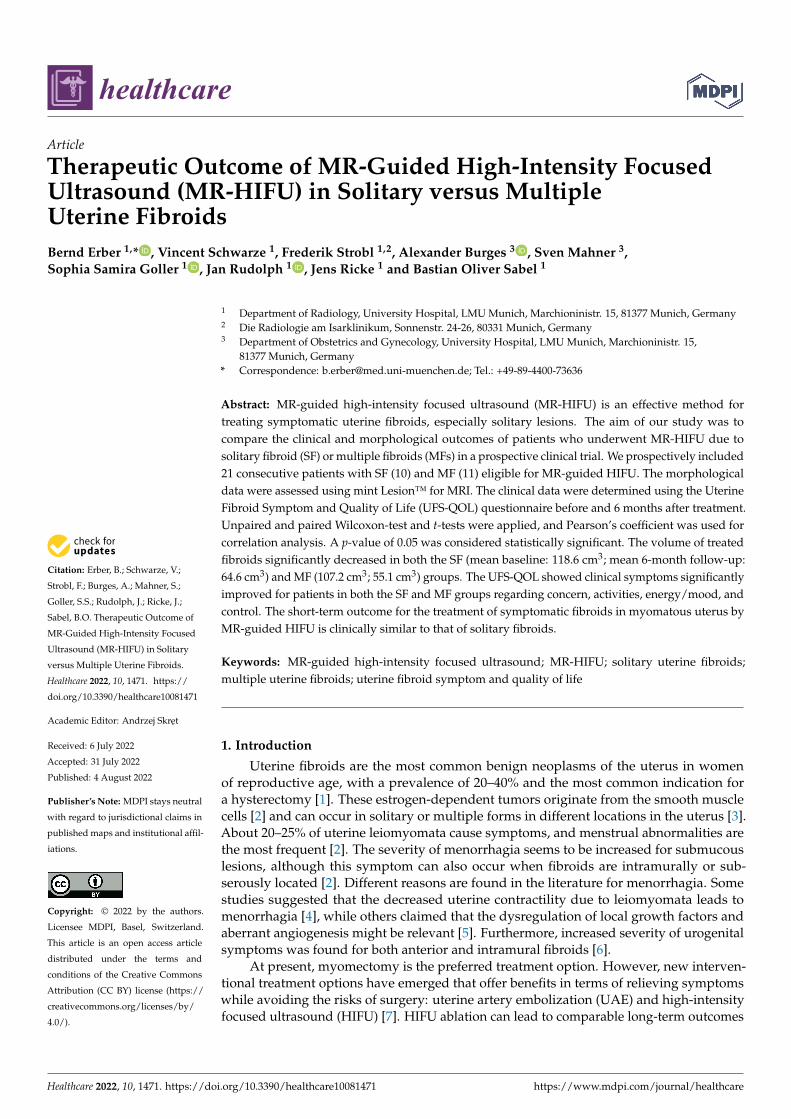
Figure 2. Images of a 39‐year‐old patient with multiple uterine fibroids in the anterior wall and
posterior wall, Funaki grade 2. (A) Contrast‐enhanced T1‐weighted and (B) T2‐weighted images 3
weeks before HIFU treatment of the fibroid in the anterior wall, sagittal reformation. Pre‐interven‐
tional maximum diameter was 3.3 cm. A smaller subserous fibroid is depicted in the posterior wall
Figure 1. Images of a 51-year-old patient with solitary intramural uterine fibroid in the posteriorwall, Funaki grade 2. (A) Contrast-enhanced T1-weighted and (B) T2-weighted images 2 weeksbefore HIFU treatment, sagittal reformation. Pre-interventional maximum diameter was 7.5 cm.(C) Immediate post-interventional T1 contrast-enhanced image with a volumetric assessed NPV of61%, sagittal reformation. (D) T2-weighted image 6 months after intervention with a maximumdiameter of 4.1 cm, sagittal reformation.
Healthcare 2022, 10, x FOR PEER REVIEW 4 of 10
Regarding Funaki’s classification (21), three fibroids were assessed to be grade 1, five
were grade 2 and two were grade 3 in the SF group. Among the patients with a myoma‐
tous uterus, seven fibroids were assessed with Funaki grade 1, eight fibroids as grade 2,
and one fibroid as grade 3.
The relative SI of the SF and MF groups showed the same mean value of 2.0.
In Figures 1 and 2, examples of an MRI of both groups are shown.
Figure 1. Images of a 51‐year‐old patient with solitary intramural uterine fibroid in the posterior
wall, Funaki grade 2. (A) Contrast‐enhanced T1‐weighted and (B) T2‐weighted images 2 weeks be‐
fore HIFU treatment, sagittal reformation. Pre‐interventional maximum diameter was 7.5 cm. (C)
Immediate post‐interventional T1 contrast‐enhanced image with a volumetric assessed NPV of 61%,
sagittal reformation. (D) T2‐weighted image 6 months after intervention with a maximum diameter
of 4.1 cm, sagittal reformation.
Figure 2. Images of a 39‐year‐old patient with multiple uterine fibroids in the anterior wall and
posterior wall, Funaki grade 2. (A) Contrast‐enhanced T1‐weighted and (B) T2‐weighted images 3
weeks before HIFU treatment of the fibroid in the anterior wall, sagittal reformation. Pre‐interven‐
tional maximum diameter was 3.3 cm. A smaller subserous fibroid is depicted in the posterior wall
Figure 2. Images of a 39-year-old patient with multiple uterine fibroids in the anterior wall and poste-rior wall, Funaki grade 2. (A) Contrast-enhanced T1-weighted and (B) T2-weighted images 3 weeksbefore HIFU treatment of the fibroid in the anterior wall, sagittal reformation. Pre-interventionalmaximum diameter was 3.3 cm. A smaller subserous fibroid is depicted in the posterior wall (ar-row), which could not be reached by HIFU due to bowel loops in the beam path. (C) Immediatepost-interventional T1 contrast-enhanced image with a volumetric assessed NPV of 64%, sagittalreformation. (D) T2-weighted image 6 months after intervention with a maximum diameter of2.0 cm, sagittal reformation.
Healthcare 2022, 10, 1471 5 of 9
Healthcare 2022, 10, x FOR PEER REVIEW 5 of 10
(arrow), which could not be reached by HIFU due to bowel loops in the beam path. (C) Immediate
post‐interventional T1 contrast‐enhanced image with a volumetric assessed NPV of 64%, sagittal
reformation. (D) T2‐weighted image 6 months after intervention with a maximum diameter of 2.0
cm, sagittal reformation.
3.2. Morphological Assessment
The mean volume of treated SFs was 118.6 cm3 (min 2.3; max 261.8; SD 98.4) and 107.2
cm3 (19.3; 304.5; 101.3) for treated lesions in patients with MFs. For patients with MFs, the
whole fibroid mass (volume of treated and untreated fibroids) was 152.7 cm3 (22.4; 460;
153.5). In the 6‐month follow‐up, the volume of treated fibroids significantly decreased in
both groups: for SFs, the mean volume in the 6‐month follow‐up was 64.6 cm3 (0.5; 247.2;
77.4), while for treated MFs, it was 55.1 cm3 (10.0; 183.7; 56.3). The data are visualized in
Figure 3.
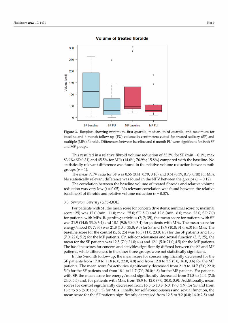
Figure 3. Boxplots showing minimum, first quartile, median, third quartile, and maximum for base‐
line and 6‐month follow‐up (FU) volume in centimeters cubed for treated solitary (SF) and multiple
(MFs) fibroids. Differences between baseline and 6‐month FU were significant for both SF and MF
groups.
This resulted in a relative fibroid volume reduction of 52.2% for SF (min −0.1%; max
83.9%; SD 0.31) and 45.5% for MFs (14.6%; 76.9%; 15.8%) compared with the baseline. No
statistically relevant difference was found in the relative volume reduction between both
groups (p = 1).
The mean NPV ratio for SF was 0.56 (0.41; 0.79; 0.10) and 0.64 (0.39; 0.73; 0.10) for MFs.
No statistically relevant difference was found in the NPV between the groups (p = 0.12).
The correlation between the baseline volume of treated fibroids and relative volume
reduction was very low (r = 0.05). No relevant correlation was found between the relative
baseline SI of fibroids and relative volume reduction (r = 0.07).
3.3. Symptom Severity (UFS‐QOL)
For patients with SF, the mean score for concern (five items; minimal score: 5; maxi‐
mal score: 25) was 17.0 (min. 11.0; max. 25.0; SD 5.2) and 12.8 (min. 6.0; max. 23.0; SD 7.0)
for patients with MFs. Regarding activities (7; 7; 35), the mean score for patients with SF
was 21.9 (14.0; 33.0; 6.4) and 18.1 (9.0; 30.0; 7.4) for patients with MFs. The mean score for
Figure 3. Boxplots showing minimum, first quartile, median, third quartile, and maximum forbaseline and 6-month follow-up (FU) volume in centimeters cubed for treated solitary (SF) andmultiple (MFs) fibroids. Differences between baseline and 6-month FU were significant for both SFand MF groups.
This resulted in a relative fibroid volume reduction of 52.2% for SF (min −0.1%; max83.9%; SD 0.31) and 45.5% for MFs (14.6%; 76.9%; 15.8%) compared with the baseline. Nostatistically relevant difference was found in the relative volume reduction between bothgroups (p = 1).
The mean NPV ratio for SF was 0.56 (0.41; 0.79; 0.10) and 0.64 (0.39; 0.73; 0.10) for MFs.No statistically relevant difference was found in the NPV between the groups (p = 0.12).
The correlation between the baseline volume of treated fibroids and relative volumereduction was very low (r = 0.05). No relevant correlation was found between the relativebaseline SI of fibroids and relative volume reduction (r = 0.07).
3.3. Symptom Severity (UFS-QOL)
For patients with SF, the mean score for concern (five items; minimal score: 5; maximalscore: 25) was 17.0 (min. 11.0; max. 25.0; SD 5.2) and 12.8 (min. 6.0; max. 23.0; SD 7.0)for patients with MFs. Regarding activities (7; 7; 35), the mean score for patients with SFwas 21.9 (14.0; 33.0; 6.4) and 18.1 (9.0; 30.0; 7.4) for patients with MFs. The mean score forenergy/mood (7; 7; 35) was 21.8 (10.0; 35.0; 9.0) for SF and 18.9 (10.0; 31.0; 6.3) for MFs. Thebaseline score for the control (5; 5; 25) was 16.5 (11.0; 23.0; 4.3) for the SF patients and 13.5(7.0; 22.0; 5.2) for the MF patients. On self-consciousness and sexual function (5; 5; 25), themean for the SF patients was 12.5 (7.0; 21.0; 4.4) and 12.1 (5.0; 21:0; 4.5) for the MF patients.The baseline scores for concern and activities significantly differed between the SF and MFpatients, while differences in the other three groups were not statistically significant.
In the 6-month follow-up, the mean score for concern significantly decreased for theSF patients from 17.0 to 11.8 (6.0; 22.0; 4.8) and from 12.8 to 7.5 (5.0; 16.0; 3.6) for the MFpatients. The mean score for activities significantly decreased from 21.9 to 14.7 (7.0; 22.0;5.0) for the SF patients and from 18.1 to 11.7 (7.0; 20.0; 4.8) for the MF patients. For patientswith SF, the mean score for energy/mood significantly decreased from 21.8 to 14.4 (7.0;24.0; 5.5) and, for patients with MFs, from 18.9 to 12.0 (7.0; 20.0; 3.9). Additionally, meanscores for control significantly decreased from 16.5 to 10.8 (6.0; 19.0; 3.9) for SF and from13.5 to 8.6 (5.0; 15.0; 3.3) for MFs. Finally, for self-consciousness and sexual function, themean score for the SF patients significantly decreased from 12.5 to 9.2 (6.0; 14.0; 2.5) and

Healthcare 2022, 10, 1471 6 of 9
from 12.1 to 8.3 (5.0; 19.0; 4.7) for the MF patients. The decrease for the MF patients inself-consciousness and sexual function was not significant (p = 0.10).
Compared with the baseline scores, we found a relative decrease in symptom severityin the 6-month FU regarding concern to 70.8% for patients with SF and 68.0% for patientswith MFs; regarding activities, to 69.1% for SF patients and 69.4% for MF patients; regardingenergy, to 69.5% for SF patients and 68.2% for MF patients; regarding control, to 66.6% forSF patients and 69.3% for MF patients; regarding self-consciousness and sexual function, to78.3% for SF patients and 75.8% for MF patients.
The data are visualized in Figure 4.
Healthcare 2022, 10, x FOR PEER REVIEW 6 of 10
energy/mood (7; 7; 35) was 21.8 (10.0; 35.0; 9.0) for SF and 18.9 (10.0; 31.0; 6.3) for MFs. The
baseline score for the control (5; 5; 25) was 16.5 (11.0; 23.0; 4.3) for the SF patients and 13.5
(7.0; 22.0; 5.2) for the MF patients. On self‐consciousness and sexual function (5; 5; 25), the
mean for the SF patients was 12.5 (7.0; 21.0; 4.4) and 12.1 (5.0; 21:0; 4.5) for the MF patients.
The baseline scores for concern and activities significantly differed between the SF and
MF patients, while differences in the other three groups were not statistically significant.
In the 6‐month follow‐up, the mean score for concern significantly decreased for the
SF patients from 17.0 to 11.8 (6.0; 22.0; 4.8) and from 12.8 to 7.5 (5.0; 16.0; 3.6) for the MF
patients. The mean score for activities significantly decreased from 21.9 to 14.7 (7.0; 22.0;
5.0) for the SF patients and from 18.1 to 11.7 (7.0; 20.0; 4.8) for the MF patients. For patients
with SF, the mean score for energy/mood significantly decreased from 21.8 to 14.4 (7.0;
24.0; 5.5) and, for patients with MFs, from 18.9 to 12.0 (7.0; 20.0; 3.9). Additionally, mean
scores for control significantly decreased from 16.5 to 10.8 (6.0; 19.0; 3.9) for SF and from
13.5 to 8.6 (5.0; 15.0; 3.3) for MFs. Finally, for self‐consciousness and sexual function, the
mean score for the SF patients significantly decreased from 12.5 to 9.2 (6.0; 14.0; 2.5) and
from 12.1 to 8.3 (5.0; 19.0; 4.7) for the MF patients. The decrease for the MF patients in self‐
consciousness and sexual function was not significant (p = 0.10).
Compared with the baseline scores, we found a relative decrease in symptom sever‐
ity in the 6‐month FU regarding concern to 70.8% for patients with SF and 68.0% for pa‐
tients with MFs; regarding activities, to 69.1% for SF patients and 69.4% for MF patients;
regarding energy, to 69.5% for SF patients and 68.2% for MF patients; regarding control,
to 66.6% for SF patients and 69.3% for MF patients; regarding self‐consciousness and sex‐
ual function, to 78.3% for SF patients and 75.8% for MF patients.
The data are visualized in Figure 4.
Figure 4. Boxplots showing minimum, first quartile, median, third quartile and maximum for scores
of UFS‐QOL items: concern, activities, energy/mood, control and combined self‐consciousness and
sexual function. Both scores of 6‐month FU were normalized to baseline scores. Differences between
baseline and 6‐month FU were significant for both SF and MF groups regarding concern, activities,
Figure 4. Boxplots showing minimum, first quartile, median, third quartile and maximum for scoresof UFS-QOL items: concern, activities, energy/mood, control and combined self-consciousnessand sexual function. Both scores of 6-month FU were normalized to baseline scores. Differencesbetween baseline and 6-month FU were significant for both SF and MF groups regarding concern,activities, energy/mood, and control. No significant difference was found for the MF group inself-consciousness and sexual function.
4. Discussion
The aim of our study was to compare the clinical and morphological outcomes ofpatients with solitary fibroids with those of patients with multiple fibroids who under-went MR-HIFU treatment in a prospective clinical trial. The volume of treated fibroidssignificantly decreased in both the SF and MF patients after treatment. Clinical symptomssignificantly improved for most UFS-QOL items. No significant difference was foundbetween groups regarding the improvement in symptoms.
Our findings agree with those in the current literature showing that HIFU is an effectivemethod to treat symptomatic uterine fibroids. The relative volume reduction of treatedfibroids for all 21 patients was between 46% and 52% after 6 months. This corresponds tothe findings of Chang et al. [12], who found a relative volume reduction of 32.8–44.1% forsolitary fibroids after 6 months. In our study, the relative volume reduction was slightlyhigher for SF (52.2%) than for treated MFs (45.5%); however, the differences were notstatistically significant. Nevertheless, this is an interesting result, as the relative SI of treatedfibroids nearly was the same in both groups. According to Funaki’s classification [21],

Healthcare 2022, 10, 1471 7 of 9
fibroids with a lower SI are more suitable for HIFU than fibroids with a higher SI, asa higher SI is associated with hypervascularity and, therefore, a lower response to HIFU.Additionally, we did not find a relevant correlation between the relative SI of the fibroidsand relative volume reduction. However, the discrepancy in the relative volume reductioncould not be explained by different treatment characteristics, as the NPV—which wasshown to be a relevant factor for the outcome of HIFU therapy [22]—was similar in bothgroups. In our study, the mean baseline volume of SF was slightly higher than that ofthe treated fibroids for patients with MF. Chang et al. [12] found a higher impact of HIFUtherapy on patients’ symptoms as well as a reduction in fibroid volume for lesions thatwere smaller than 10 cm in diameter. Cheng et al. [23] found significantly higher ablationrates for fibroids with diameters greater than 7 cm compared to smaller lesions (<3 cmor 5–7 cm). However, these results may not translate to a comparison between solitaryand multiple fibroids. As described in the Results section, the correlation between thebaseline fibroid volume and relative volume reduction was very low. In summary, SF seemto respond slightly better to HIFU therapy than MFs regarding relative volume reduction.However, the differences between the groups were not statistically significant.
More relevant for clinical efficiency and patient sufficiency is the post-interventionalsymptom severity. Interestingly, the baseline scores for all items were slightly higher for SFthan for MFs, although differences were not significant. This might be explained by the dif-ference in baseline fibroid volume, which was higher for SF than for MFs. The improvementin symptom severity was, except for self-consciousness and sexual function, significantfor all items for patients with either solitary or multiple fibroids. This is remarkable asimprovement in symptom severity is the most relevant outcome parameter for patientssuffering from this benign uterine neoplasm.
As described earlier, for patients with MFs, not all lesions were feasible for HIFUtherapy, e.g., because they were not accessible for the HIFU beam due to a large distance tothe skin, as described in the Methods section. However, the finding that patients with MFhad a similar symptomatic benefit to those with SF supports the hypothesis that treatmentof symptomatic fibroids in patients with MF is essential for clinical outcomes. It is expectedthat submucous fibroids frequently cause menorrhagia, although this symptom is also seenwith fibroids in other locations. However, the severity of bleeding seems to be increased bythe presence of submucous fibroids [2]. For the severity of dysuria, an increase was foundfor anterior myomas compared with other locations [6,24]; additionally, an intramurallocation seemed to be associated with increased urinary symptoms [2,25].
Symptom severity (UFS-QOL) was assessed in a short-term follow-up after 6 months.In both the SF and MF groups, a distinct and statistically significant improvement insymptom severity was found, which was accompanied by significant fibroid volumereduction. This is in line with the findings in the literature, for example, Kim et al., whofound significant improvement in UFS-QOL scores after 3 months, which was sustainedsustainability for a period of 3 years [26], or Chang et al. [12], who described a significantbenefit after 6 months.
An interesting question for further study is a comparison between HIFU and UAE forthe outcomes of patients with MFs. UAE may have promising results for lesions in patientswith MFs that are not accessible for HIFU and may be addressed by this method [27].
Limitations
The inclusion of follow-up examinations only after 6 months is a limitation of the study.For this reason, further studies are needed to compare HIFU treatment for SF and MFs atlater follow-up, especially because, in most cases of MFs, not all lesions are accessible orfeasible for HIFU treatment and, therefore, might be prone to further growth. Therefore,a longer follow-up is needed to analyze the possible recurrence of symptoms.
Another limitation is the limited number of patients in both groups, as MR-guidedHIFU is a novel treatment option and the study was designed as a single-center trial.
Healthcare 2022, 10, 1471 8 of 9
Hence, we were unable to obtain more patient data. However, the results show a clear andpromising benefit of MR-HIFU therapy for patients with multiple fibroids.
5. Conclusions
The short-term outcome for the treatment of symptomatic fibroids in myomatousuterus with MR-guided HIFU is clinically and morphologically similar to that for solitaryfibroids. Therefore, MR-guided HIFU therapy should be considered for patients withmultiple lesions. Further long-term follow-up is needed to analyze the sustainability ofsymptom relief.
Author Contributions: Conceptualization, B.E., V.S., F.S., A.B., S.M., J.R. (Jens Ricke) and B.O.S.;methodology, B.E., V.S., F.S., J.R. (Jens Ricke) and B.O.S.; validation, B.E., V.S., F.S., J.R. (Jens Ricke)and B.O.S.; formal analysis, B.E., S.S.G., J.R. (Jan Rudolph) and B.O.S.; investigation, B.E., V.S., S.S.G.,J.R. (Jan Rudolph) and B.O.S.; resources, A.B., S.M., J.R. (Jens Ricke) and B.O.S.; data curation, B.E.and B.O.S.; writing, B.E. and B.O.S.; writing—review and editing, V.S., F.S., A.B., S.M., S.S.G., J.R. (JanRudolph), J.R. (Jens Ricke) and B.O.S.; visualization, B.E., J.R. (Jan Rudolph) and B.O.S.; supervision,J.R. (Jens Ricke) and B.O.S.; project administration, J.R. (Jens Ricke) and B.O.S.; funding acquisition,V.S., F.S., J.R. (Jens Ricke) and B.O.S. All authors have read and agreed to the published version ofthe manuscript.
Funding: This research was funded by the Deutsche Forschungsgemeinschaft (DFG), grantnumber 260098876.
Institutional Review Board Statement: The study was conducted in accordance with the Declarationof Helsinki, and approved by the Institutional Review Board of LMU Munich (18-043, April 2018).
Informed Consent Statement: Informed consent was obtained from all subjects involved inthe study.
Data Availability Statement: The data presented in this study are available on request from thecorresponding author.
Acknowledgments: We thank Julia Schwangler as head of the study center for assistance with thisclinical trial.
Conflicts of Interest: The authors declare no conflict of interest. The funders had no role in the designof the study; in the collection, analysis, or interpretation of data; in the writing of the manuscript; orin the decision to publish the results.
References1. Wallach, E.E.; Vlahos, N.F. Uterine myomas: An overview of development, clinical features, and management. Obstet. Gynecol.
2004, 104, 393–406. [CrossRef] [PubMed]2. Buttram, V.C., Jr.; Reiter, R.C. Uterine leiomyomata: Etiology, symptomatology, and management. Fertil. Steril. 1981, 36, 433–445.
[CrossRef] [PubMed]3. Cramer, S.F.; Patel, A. The frequency of uterine leiomyomas. Am. J. Clin. Pathol. 1990, 94, 435–438. [CrossRef] [PubMed]4. Farrer-Brown, G.; Beilby, J.O.; Tarbit, M.H. The vascular patterns in myomatous uteri. J. Obstet. Gynaecol. Br. Commonw. 1970,
77, 967–975. [CrossRef]5. Stewart, E.A.; Nowak, R.A. Leiomyoma-related bleeding: A classic hypothesis updated for the molecular era. Hum. Reprod. Update
1996, 2, 295–306. [CrossRef]6. Mourgues, J.; Villot, A.; Thubert, T.; Fauvet, R.; Pizzoferrato, A.C. Uterine myomas and lower urinary tract dysfunctions:
A literature review. J. Gynecol. Obstet. Hum. Reprod. 2019, 48, 771–774. [CrossRef]7. Liu, L.; Wang, T.; Lei, B. Uterine Artery Embolization Compared with High-intensity Focused Ultrasound Ablation for the
Treatment of Symptomatic Uterine Myomas: A Systematic Review and Meta-analysis. J. Minim. Invasive Gynecol. 2021,28, 218–227. [CrossRef]
8. Liu, X.; Tang, J.; Luo, Y.; Wang, Y.; Song, L.; Wang, W. Comparison of high-intensity focused ultrasound ablation and sec-ondary myomectomy for recurrent symptomatic uterine fibroids following myomectomy: A retrospective study. BJOG—Int. J.Obstet. Gynaecol. 2020, 127, 1422–1428. [CrossRef]
9. Yan, L.; Huang, H.; Lin, J.; Yu, R. High-intensity focused ultrasound treatment for symptomatic uterine fibroids: A systematicreview and meta-analysis. Int. J. Hyperth. 2022, 39, 230–238. [CrossRef]
10. Gao, H.; Li, T.; Fu, D.; Wei, J. Uterine artery embolization, surgery and high intensity focused ultrasound in the treatment ofuterine fibroids: A network meta-analysis. Quant. Imaging Med. Surg. 2021, 11, 4125–4136. [CrossRef]

Healthcare 2022, 10, 1471 9 of 9
11. Chen, R.; Keserci, B.; Bi, H.; Han, X.; Wang, X.; Bai, W.; Wang, Y.; Yang, X.; Yang, J.; Wei, J.; et al. The safety and effectivenessof volumetric magnetic resonance-guided high-intensity focused ultrasound treatment of symptomatic uterine fibroids: Earlyclinical experience in China. J. Ther. Ultrasound 2016, 4, 27. [CrossRef]
12. Chang, C.T.; Jeng, C.J.; Long, C.Y.; Chuang, L.T.; Shen, J. High-intensity focused ultrasound treatment for large and small solitaryuterine fibroids. Int. J. Hyperth. 2022, 39, 485–489. [CrossRef]
13. Wang, Y.J.; Zhang, P.H.; Zhang, R.; An, P.L. Predictive Value of Quantitative Uterine Fibroid Perfusion Parameters From Contrast-Enhanced Ultrasound for the Therapeutic Effect of High-Intensity Focused Ultrasound Ablation. J. Ultrasound Med. 2019,38, 1511–1517. [CrossRef]
14. Dorenberg, E.J.; Courivaud, F.; Ring, E.; Hald, K.; Jakobsen, J.A.; Fosse, E.; Hol, P.K. Volumetric ablation of uterine fibroids usingSonalleve high-intensity focused ultrasound in a 3 Tesla scanner—First clinical assessment. Minim. Invasive Ther. Allied Technol.2013, 22, 73–79. [CrossRef]
15. Hindley, J.; Gedroyc, W.M.; Regan, L.; Stewart, E.; Tempany, C.; Hynyen, K.; McDannold, N.; Inbar, Y.; Itzchak, Y.;Rabinovici, J.; et al. MRI guidance of focused ultrasound therapy of uterine fibroids: Early results. AJR Am. J. Roentgenol. 2004,183, 1713–1719. [CrossRef]
16. Spies, J.B.; Coyne, K.; Guaou Guaou, N.; Boyle, D.; Skyrnarz-Murphy, K.; Gonzalves, S.M. The UFS-QOL, a new disease-specificsymptom and health-related quality of life questionnaire for leiomyomata. Obstet. Gynecol. 2002, 99, 290–300. [CrossRef]
17. Taran, F.A.; Tempany, C.M.; Regan, L.; Inbar, Y.; Revel, A.; Stewart, E.A.; Group, M.R. Magnetic resonance-guided focusedultrasound (MRgFUS) compared with abdominal hysterectomy for treatment of uterine leiomyomas. Ultrasound Obstet. Gynecol.2009, 34, 572–578. [CrossRef]
18. Lyon, P.C.; Rai, V.; Price, N.; Shah, A.; Wu, F.; Cranston, D. Ultrasound-Guided High Intensity Focused Ultrasound Ablation forSymptomatic Uterine Fibroids: Preliminary Clinical Experience. Ultraschall Med.-Eur. J. Ultrasound 2020, 41, 550–556. [CrossRef]
19. Mohr-Sasson, A.; Machtinger, R.; Mashiach, R.; Nir, O.; Inbar, Y.; Maliyanker, N.; Goldenberg, M.; Rabinovici, J. Long-termoutcome of MR-guided focused ultrasound treatment and laparoscopic myomectomy for symptomatic uterine fibroid tumors.Am. J. Obstet. Gynecol. 2018, 219, 375.e371–375.e377. [CrossRef]
20. R Core Team. R: A Language and Environment for Statistical Computing; R Foundation for Statistical Computing: Vienna, Austria,2021. Available online: https://www.R-project.org/ (accessed on 1 March 2021).
21. Funaki, K.; Fukunishi, H.; Funaki, T.; Sawada, K.; Kaji, Y.; Maruo, T. Magnetic resonance-guided focused ultrasound surgery foruterine fibroids: Relationship between the therapeutic effects and signal intensity of preexisting T2-weighted magnetic resonanceimages. Am. J. Obstet. Gynecol. 2007, 196, 184.e181–184.e186. [CrossRef]
22. Liu, Y.; Wu, X.; Wu, A.; Gong, C.; Wang, Z.; Zhang, L. Ultrasound-guided high intensity focused ultrasound ablation for uterinefibroids: Long-term outcomes and factors affecting local recurrence. Int. J. Hyperth. 2021, 38, 1341–1348. [CrossRef]
23. Cheng, H.; Wang, C.; Tian, J. Correlation between uterine fibroids with various magnetic resonance imaging features andtherapeutic effects of high-intensity focused ultrasound ablation. Pak. J. Med. Sci. 2015, 31, 869–873. [CrossRef]
24. Parker-Autry, C.; Harvie, H.; Arya, L.A.; Northington, G.M. Lower urinary tract symptoms in patients with uterine fibroids:Association with fibroid location and uterine volume. Female Pelvic Med. Reconstr. Surg. 2011, 17, 91–96. [CrossRef]
25. Ekin, M.; Cengiz, H.; Ozturk, E.; Kaya, C.; Yasar, L.; Savan, K. Genitourinary symptoms and their effects on quality of life inwomen with uterine myomas. Int. Urogynecol. J. 2014, 25, 807–810. [CrossRef]
26. Kim, H.S.; Baik, J.H.; Pham, L.D.; Jacobs, M.A. MR-guided high-intensity focused ultrasound treatment for symptomatic uterineleiomyomata: Long-term outcomes. Acad. Radiol. 2011, 18, 970–976. [CrossRef]
27. Keung, J.J.; Spies, J.B.; Caridi, T.M. Uterine artery embolization: A review of current concepts. Best Pract. Res. Clin. Obstet. Gynaecol.2018, 46, 66–73. [CrossRef]