Magnetic resonance-guided thermal surgery
9
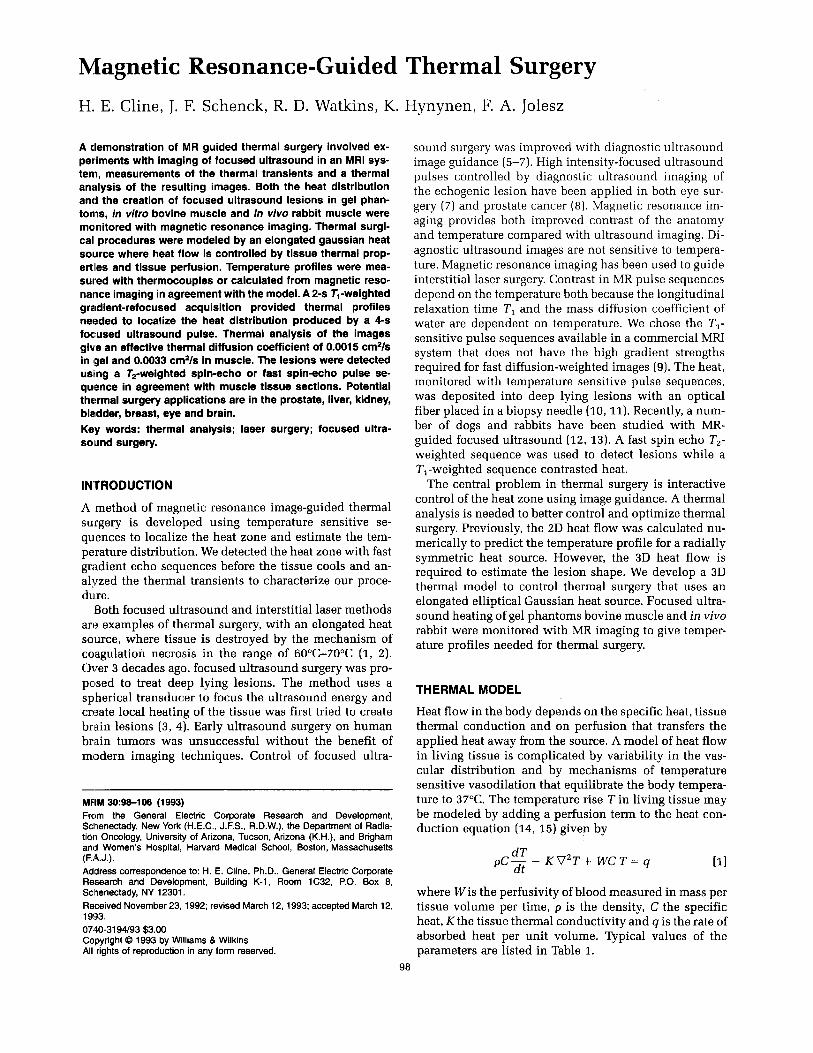
Magnetic Resonance-Guided Thermal Surgery H. E. Cline, J. F. Schenck, R. D. Watkins, K A clemonstration of MR guided thermal surgery involved ex- periments with imaging of focused ultrasound in an MRI sys- tem, measurements of the thermal transients and a thermal analysis of the resulting images. Both the heat distribution and the creation of focused ultrasound lesions in gel phan- toms, in virro bovine muscle and in vivo rabbit muscle were monitored with magnetic resonance imaging. Thermal surgi- cal procedures were modeled by an elongated gaussian heat source where heat flow is controlled by tissue thermal prop- ertiies and tissue perfusion. Temperature profiles were mea- sured with thermocouples or calculated from magnetic reso- nance imaging in agreementwith the model. A 2-9 T,-weighted gradient-refocused acquisition provided thermal profiles needed to localize the heat distribution produced by a 4-s foc:used ultrasound pulse. Thermal analysis of the images give an effective thermal diffusion coefficient of 0.0015 cm2/s in (geland 0.0033 cm2/s in muscle. The lesions were detected usnng a T2-weighted spin-echo or fast spin-echo pulse se- quefice in agreement with muscle tissue sections. Potential thermal surgery applications are in the prostate, liver, kidney, bladder, breast, eye and brain. Key words: thermal analysis; laser surgery; focused ultra- soiund surgery. INTRODUCTION A method of magnetic resonance image-guided thermal surgery is developed using temperature sensitive se- quences to localize the heat zone and estimate the tem- perature distribution. We detected the heat zone with fast gradient echo sequences before the tissue cools and an- alyzed the thermal transients to characterize our proce- dure. Both focused ultrasound and interstitial laser methods are examples of thermal surgery, with an elongated heat source, where tissue is destroyed by the mechanism of coagulation necrosis in the range of 6O0C-7O0C (1, 2). Over 3 decades ago, focused ultrasound surgery was pro- posed to treat deep lying lesions. The method uses a spherical transducer to focus the ultrasound energy and create local heating of the tissue was first tried to create brain lesions (3, 4). Early ultrasound surgery on human brain tumors was unsuccessful without the benefit of modern imaging techniques. Control of focused ultra- MHM 30:98-106 (1993) From the General Electric Corporate Research and Development, Sclhenectady, New York (H.E.C., J.F.S., R.D.W.), the Department of Radia- tion Oncology, University of Arizona, Tucson, Arizona (K.H.), and Brigham and Women's Hospital, Harvard Medical School, Boston, Massachusetts Address correspondence to: H. E. Cline, Ph.D., General Electric Corporate Research and Development, Building K-1, Room 1C32, P.O. Box 8, Sclhenectady, NY 12301. Received November 23, 1992; revised March 12, 1993; accepted March 12, 19!33. 0740-31 94/93 $3.00 Copyright 0 1993 by Williams 8 Wilkins All rights of reproduction in any form reserved. (F.A.J.). . Hynynen, F. A. Jolesz sound surgery was improved with diagnostic ultrasound image guidance (5-7). High intensity-focused ultrasound pulses controlled by diagnostic ultrasound imaging of the echogenic lesion have been applied in both eye sur- gery (7) and prostate cancer (8). Magnetic resonance im- aging provides both improved contrast of the anatomy and temperature compared with ultrasound imaging. Di- agnostic ultrasound images are not sensitive to tempera- ture. Magnetic resonance imaging has been used to guide interstitial laser surgery. Contrast in MR pulse sequences depend on the temperature both because the longitudinal relaxation time TI and the mass diffusion coefficient of water are dependent on temperature. We chose the Tl- sensitive pulse sequences available in a commercial MRI system that does not have the high gradient strengths required for fast diffusion-weighted images (9).The heat, monitored with temperature sensitive pulse sequences, was deposited into deep lying lesions with an optical fiber placed in a biopsy needle (10,11). Recently, a num- ber of dogs and rabbits have been studied with MR- guided focused ultrasound (12, 13). A fast spin echo Tz- weighted sequence was used to detect lesions while a Tl-weighted sequence contrasted heat. The central problem in thermal surgery is interactive control of the heat zone using image guidance. A thermal analysis is needed to better control and optimize thermal surgery. Previously, the 2D heat flow was calculated nu- merically to predict the temperature profile for a radially symmetric heat source. However, the 3D heat flow is required to estimate the lesion shape. We develop a 3D thermal model to control thermal surgery that uses an elongated elliptical Gaussian heat source. Focused ultra- sound heating of gel phantoms bovine muscle and in vivo rabbit were monitored with MR imaging to give temper- ature profiles needed for thermal surgery. THERMAL MODEL Heat flow in the body depends on the specific heat, tissue thermal conduction and on perfusion that transfers the applied heat away from the source. A model of heat flow in living tissue is complicated by variability in the vas- cular distribution and by mechanisms of temperature sensitive vasodilation that equilibrate the body tempera- ture to 37°C. The temperature rise Tin living tissue may be modeled by adding a perfusion term to the heat con- duction equation (14, 15) given by dT dt pC- - KV2T+ WCT= q where W is the perfusivity of blood measured in mass per tissue volume per time, p is the density, C the specific heat, K the tissue thermal conductivity and q is the rate of absorbed heat per unit volume. Typical values of the parameters are listed in Table 1. 98
Transcript of Magnetic resonance-guided thermal surgery
Magnetic Resonance-Guided Thermal Surgery H. E. Cline, J. F. Schenck, R. D. Watkins, K
A clemonstration of MR guided thermal surgery involved ex- periments with imaging of focused ultrasound in an MRI sys- tem, measurements of the thermal transients and a thermal analysis of the resulting images. Both the heat distribution and the creation of focused ultrasound lesions in gel phan- toms, in virro bovine muscle and in vivo rabbit muscle were monitored with magnetic resonance imaging. Thermal surgi- cal procedures were modeled by an elongated gaussian heat source where heat flow is controlled by tissue thermal prop- ertiies and tissue perfusion. Temperature profiles were mea- sured with thermocouples or calculated from magnetic reso- nance imaging in agreement with the model. A 2-9 T,-weighted gradient-refocused acquisition provided thermal profiles needed to localize the heat distribution produced by a 4-s foc:used ultrasound pulse. Thermal analysis of the images give an effective thermal diffusion coefficient of 0.0015 cm2/s in (gel and 0.0033 cm2/s in muscle. The lesions were detected usnng a T2-weighted spin-echo or fast spin-echo pulse se- quefice in agreement with muscle tissue sections. Potential thermal surgery applications are in the prostate, liver, kidney, bladder, breast, eye and brain. Key words: thermal analysis; laser surgery; focused ultra- soiund surgery.
INTRODUCTION
A method of magnetic resonance image-guided thermal surgery is developed using temperature sensitive se- quences to localize the heat zone and estimate the tem- perature distribution. We detected the heat zone with fast gradient echo sequences before the tissue cools and an- alyzed the thermal transients to characterize our proce- dure.
Both focused ultrasound and interstitial laser methods are examples of thermal surgery, with an elongated heat source, where tissue is destroyed by the mechanism of coagulation necrosis in the range of 6O0C-7O0C (1, 2). Over 3 decades ago, focused ultrasound surgery was pro- posed to treat deep lying lesions. The method uses a spherical transducer to focus the ultrasound energy and create local heating of the tissue was first tried to create brain lesions (3, 4). Early ultrasound surgery on human brain tumors was unsuccessful without the benefit of modern imaging techniques. Control of focused ultra-
MHM 30:98-106 (1993) From the General Electric Corporate Research and Development, Sclhenectady, New York (H.E.C., J.F.S., R.D.W.), the Department of Radia- tion Oncology, University of Arizona, Tucson, Arizona (K.H.), and Brigham and Women's Hospital, Harvard Medical School, Boston, Massachusetts
Address correspondence to: H. E. Cline, Ph.D., General Electric Corporate Research and Development, Building K-1, Room 1C32, P.O. Box 8, Sclhenectady, NY 12301. Received November 23, 1992; revised March 12, 1993; accepted March 12, 19!33. 0740-31 94/93 $3.00 Copyright 0 1993 by Williams 8 Wilkins All rights of reproduction in any form reserved.
(F.A.J.).
. Hynynen, F. A. Jolesz
sound surgery was improved with diagnostic ultrasound image guidance (5-7). High intensity-focused ultrasound pulses controlled by diagnostic ultrasound imaging of the echogenic lesion have been applied in both eye sur- gery (7) and prostate cancer (8). Magnetic resonance im- aging provides both improved contrast of the anatomy and temperature compared with ultrasound imaging. Di- agnostic ultrasound images are not sensitive to tempera- ture. Magnetic resonance imaging has been used to guide interstitial laser surgery. Contrast in MR pulse sequences depend on the temperature both because the longitudinal relaxation time TI and the mass diffusion coefficient of water are dependent on temperature. We chose the Tl- sensitive pulse sequences available in a commercial MRI system that does not have the high gradient strengths required for fast diffusion-weighted images (9). The heat, monitored with temperature sensitive pulse sequences, was deposited into deep lying lesions with an optical fiber placed in a biopsy needle (10,11). Recently, a num- ber of dogs and rabbits have been studied with MR- guided focused ultrasound (12, 13). A fast spin echo Tz- weighted sequence was used to detect lesions while a Tl-weighted sequence contrasted heat.
The central problem in thermal surgery is interactive control of the heat zone using image guidance. A thermal analysis is needed to better control and optimize thermal surgery. Previously, the 2D heat flow was calculated nu- merically to predict the temperature profile for a radially symmetric heat source. However, the 3D heat flow is required to estimate the lesion shape. We develop a 3D thermal model to control thermal surgery that uses an elongated elliptical Gaussian heat source. Focused ultra- sound heating of gel phantoms bovine muscle and in vivo rabbit were monitored with MR imaging to give temper- ature profiles needed for thermal surgery.
THERMAL MODEL
Heat flow in the body depends on the specific heat, tissue thermal conduction and on perfusion that transfers the applied heat away from the source. A model of heat flow in living tissue is complicated by variability in the vas- cular distribution and by mechanisms of temperature sensitive vasodilation that equilibrate the body tempera- ture to 37°C. The temperature rise T i n living tissue may be modeled by adding a perfusion term to the heat con- duction equation (14, 15) given by
dT dt
pC- - K V 2 T + W C T = q
where W is the perfusivity of blood measured in mass per tissue volume per time, p is the density, C the specific heat, K the tissue thermal conductivity and q is the rate of absorbed heat per unit volume. Typical values of the parameters are listed in Table 1.
98

LMagnetic Resonance-Guided Thermal Surgery 99
Table 1 Thermal Properties of Tissue and Typical Values of the Pamrneters
13ensity P 1 gmicc !Specific heat C 4.2 joulesiccPC Thermal conductivity K 0.006 w/cmI"C Perfusion w 0.01 gmiccls
IPerfusion time constant Tw = piW 100 s Thermal diffusivity D = WC 0.0015 cm2/s
The values of thermal conductivity and diffusivity are that of water or gel phantoms.
r I I I
PULSEIWAVEFORM GENE RAT0 R
MAGNETIC RESONANCE SYSTEM
I RFAMPLIFIER 1 OPERATIONAL AMPLIFIER
T I \ T H E R M O C ~ U P L E
3 AXIS WATER HYDRAULIC -
MOTION HYDRAULIC CONTROL
TRANSDUCER I
FIG. 1. Schematic of MR compatible focused ultrasound appara- tus. Consisting of electronics to provide RF power to the trans- ducer, a hydraulic x, y, z positioner and a means to measure temperature at the focus. The apparatus is placed in a 1.5 T MR scariner to image the focal lesion and measure the heat effected zone.
The solution of the heat flow Eq. [l] for an instanta- neous point source is a Gaussian form that spreads with time. Fortunately, the heat source q for both focused ul- trasound and interstitial laser therapy may be approxi- mated by an elongated elliptical gaussian heat source. Previously, we have measured the acoustic energy distri- bution of focused ultrasound (13) and have found the side lobes to be small compared with the central focal spot, while in laser therapy we assume the heat from thermal measurements in the literature and the elliptical shape of the lesions (10,11). In the case of focused ultra- sound, the heat source size is determined by the diffrac- tion of ultrasound from a spherical transducer and the acoustic attenuation of tissue. The radius of the half in- tensity focal spot in the focal plane Ro and along the axis Zo depends on the aperture, transducer radius and fre- quency. The maximum power density absorbed qo at the focus depends on the product of the acoustic intensity attenuation and the maximum acoustic intensity. For interstitial laser radiation, optical absorption and scatter- ing determines the heat distribution. The heat source
distribution is elongated along the axis and can be de- scribed by a radius R, in the focal plane and Z , along the axis. We model both cases with a heat source of the form
q(r,z) = q,Exp [ -- 2;; - -1 2;; (21
The side lobes of the diffraction limited intensity from a spherical transducer are small and the Bessel Function expression may be approximated by a gaussian.
The thermal transient T( t ) of a heat pulse of maximum intensity qo( t ) is given by superimposing gaussian instan- taneous sources G(t) sources (16)
T(t) = qo(t')G(t-t')dt' [31
The form of the source is derived by distributing a point source over a gaussian using the fact that the convolution of a gaussian is also a gaussian (16). An instantaneous gaussian source evolves by both thermal conduction and
I:
Table 2 The Time for the Temperature to Rise from 30°C to 60°C at Different Applied Power Levels, Average Heating Rate and Calculated Heat Absorbed in a Gel Phantom with D = 0.0015 cm2/s, K = 0.006 w/cm/s, and c = 4.2 joules/gm/C
Rise time Applied Acoustic Heat Intensity 30°C to 60°C power intensity absorbed attenuation
(S) (W) (W/SQCM) (W/CC) (l/CM)
4 38 685 67 0.049 1.5 100 1800 124 0.034 0.5 200 3600 295 0.041 0.2 300 5040 684 0.063 0.16 450 81 00 832 0.051
I I I I I 0 1 2 3 4
TIME (SECONDS)
FIG. 2. Temperature profile through the focus in a gel phantom heated with focused ultrasound for 0.5 s at 300 watts of applied power corresponding to an acoustic intensity of 5400 watts/cm2. The points were calculated from the gaussian source thermal model using an acoustic intensity attenuation coefficient of 0.063 cm-' and a radial conduction time constant of 1.4 s.

100 Cline et al.
FIG. 3. (a) A 3-mm thick axial slice through two layers gel showing the molten focal lesion indicated by an arrow (dark) 2 mm in diameter and 9 mm long the focus of the sonic heat beam (light), TRITE = 800/20. Notice reflections at the upper surface and the heat loss at the inBerface between the gel layers. The SE image was acquired in 112 s while the ultrasound was applied with a peak intensity of 600 w/cm2. (b) Coronal image through the focal plane shows the dark molten gel indicated by an arrow surrounded by light heated material. (c) The power was turned off and another image taken showing the diffusion of heat and the resolidified gel. (d) The next image taken shows further spread of the heat during cooling. After 336 s the heat radius is 1 cm.
perfusion in a way satisfying the bioheat flow equation by
This fact has been used to monitor the acoustic intensity by using a small thermocouple embedded in a bead of material of known acoustic absorption and measuring the temperature rise after a short pulse. For elongated heat sources the axial heat flow is less than the radial heat flow and only the radial time constant is important in calculating the temperature transient because ‘ T ~ > > T ~ . As the pulse duration increases, to > ‘TR, the radial con- duction becomes important and the temperature tran- sient at the focus is found by setting r = 0 and z = 0 in Eq.
zo2 P [41, neglecting the term containing t/rZ, substitution of Eq. [4] in Eq. [3] and integrating from 0 to t to yield
r2 - z2 - I] ..P[ - 4D(t + TR) 4D(t + rz) T~
cp( 1 + ;) (1 + k ) ( 1 / 2 )
G(t) = i41
where D is the thermal diffusivity 2
rZ=-, r w = - TR - 20’ 2 0 W - Ro
At very short pulse durations, to < TR, compared to the time constants, the temperature rise is proportional to the heat absorbed times the pulse duration, (Q(r,z)to)/(Cp).

Magnetic Resonance-Guided Thermal Surgery 101
25 I 1 I I
0 1 2 3 4
TIME (SECONDS)
FIG. 4. Temperature profile through the focus in a bovine muscle phantom heated with focused ultrasound for 0.5 s at 450 watts of appliied power corresponding to an acoustic intensity of 8100 watts/ cm'. Using the gaussian source thermal model with an acoustic intensity attenuation coefficient of 0.028 cm-I and a radial time constant of 0.62 s.
TIME (SECONDS)
FIG. 5. Calculated temperature transient for a 4-s pulse in bovine muscle using a radial time constant of 0.62 s, attenuation of 0.028 cm-I, and acoustic intensity of 81 00 watts/cmz.
Band after the pulse duration, t > to, the integration limits in Eq. [3] become t and &to giving a decreasing tempera- ture
The effect of perfusion becomes important at times greater than T ~ . Including the effect of perfusion, the temperature at the focus neglecting axial heat flow be- comes
T(t) = - t71
1 + -
which approaches a steady state value of (q,rBICp)ln('rrRl rW) at long pulse durations. The temperature distribu-
tion for a constant continuous source of duration to is given by setting qO(t) to a constant in Eq. [3]. The heat zone is initially described by a gaussian with radii R, and Z,; however, during the heat pulse the heat effected zone increases in size by thermal diffusion, R2( t ) = Ro2 + 2Dt andZ2(t) = Zo2 + 2Dt.
METHOD
The above thermal model was tested with a series of MR-guided focused ultrasound experiments and inde- pendent temperature measurements. An acoustic gel phantom (3M) with a melting point of 57OC and thermal properties similar to water provided an ideal test mate- rial. Bovine muscle specimens were used to test the effect of focused ultrasound on tissue without perfusion. A 12- week old male rabbit was used to demonstrate lesions in live muscle tissue with perfusion. The rabbit was exam- ined again after 10 weeks.
The experimental apparatus is shown schematically, Fig. 1. A nonmagnetic hydraulic positioner (13) was used to move a transducer in a 1.5 T magnetic resonance sys- tem (GE Medical Systems, Milwaukee, WI) while imaging the focal lesions and the heat effected zone. Heat was applied with a 1.54 MHz, 10-cm diameter aperture; 10. 3-cm radius, air-backed, spherical, PZT transducer. The transducer and three piston assembly were contained in a degassed deionized water bath covered with a 75-mi- cron thick Mylar membrane. A pulseIwaveform genera- tor (Wavetek) created a constant amplitude sinusoidal 1.54 MHz pulse of different durations in the range be- tween 0.1 to 600s. The signal was amplified using a 300- watt power amplifier (ENI) to give powers in the range of 10 to 450 watts, monitored with a power meter (Byrd), and sent through an impedance matching circuit to the transducer. The air-backed PZT transducer transforms 57% of the power into a focused acoustic wave (calcu- lated from the impedance of the transducer in air and water). At the focus, the acoustic intensity is given by this gain times the applied power per unit area. The measured power gain of the transducer is estimated to be 1400 using the diffractinn spot size and the 26% efficiency of
1 4 0 L 120
100 t = 4 Seconds a -
80
60
F 40
20
0 -1 0 0.5 1
DISTANCE (CM)
FIG. 6. Calculated radial temperature of the 4-s pulse with time after 2, 4, 6, and 8 s.

102 Cline et al.
FIG. 7. A series of 2-s fast gradient refocused images ( TRJTHangle = 11/3.3/60) in the focal plane of a muscle specimen showing the displacement of a hot spot by moving the focus of a 4-s sonic pulse with a maximum acoustic intensity of 8100 w/cm2.
the transducer measured with a hydrophone at the focus. The focal spot size was measured with a scanning hydro- phone (12) to give the half power radius in the focal plane R, = 0.065 cm and along the axis 2, = 0.45 cm. The ternperature in the specimens was monitored with a K- type thermocouple made from a 125-micron wire in a ceramic insulator tube with the welded bead exposed to the tissue. The thermocouple was inserted into the spec- imen at the focus where the temperature was a maxi- mum, the signal amplified and recorded with an oscillo- scope triggered by the start of the RF pulse to provide a plot of the temperature transient.
19 magnetic resonance image was acquired immedi- ately after the heat pulse to record the heat effected zone and the lesion. Fast gradient refocused echo (FGR) and fast spin echo (FSE) pulse sequences were used in these
experiments to minimize the imaging time. A square sur- face coil receiver, 20 cm on a side, gave increased signal- to-noise compared with the body coil. The longitudinal relaxation time TI of the specimen was measured from a sequence of spin-echo (SE) images at different repetition times and the transverse relaxation time Tz was mea- sured using multiple echo times. Gel phantoms were used to set up the apparatus because the focal spot is easily imaged in this homogenous material which be- comes bright upon heating until it melts and becomes dark. An axial image through the specimen axis and the transducer was acquired with a 112-s SE sequence to locate the focal spot at a distance of 103 mm from the transducer surface while 30 watts of ultrasonic power was applied to the transducer. Refrigerated bovine muscle specimens were then placed on the hydraulic

Magnetic Resonance-Guided Thermal Surgery 103
1 DISTANCE (CM)
2
FIG. 8. Signal across focal lesion acquired in 2 s with fast gradient pulse sequence just after application of a 4-s ultrasound pulse.
0
0 1
DISTANCE (CM)
2
FIG. 9. Calculated temperature profile points assuming a 1% in- crease in TI with a 1" increase in temperature plotted with the temperature profile (solid curve) estimated using the gaussian source thermal model and a thermal diffusivity of 0.0038 cm2/s calculated 5.5 s after the start of a 4-s long heat pulse.
positioner (13). A pulse with an amplitude of 450 watts and duration of 4 s was applied to the transducer to create lesions. A single fast gradient echo image of the focal plane was acquired in 2 s starting about 0.5 s after
each 4-s long sonic pulse using a 20-cm field of view, (TEITRlangle = 3.4 msIll .1 ms/60°), and 192 by 256 resolution. The focal spot was moved during the 30-s wait between heat pulses. A series of eight images were acquired to show the heat spots displaced along a line in the focal plane. After the row of lesions were created in the muscle they were detected with a 9-s T2 weighted FSE image with (TRITE = 500 ms, 85 ms). The 1-mm size lesions appear somewhat brighter than the background in the T,-weighted image. A section of the muscle along the focal plane revealed the lesion. The in vivo rabbit muscle experiments were done to confirm that lesions could be created in perfused tissue with the same apparatus. The rabbit hind thigh was shaved, the rabbit was anesthetized and taped to the positioner.
EXPERIMENTAL RESULTS
First we imaged homogeneous gel specimens to test our thermal model. Second, muscle specimens are used to determine the effects of MR imaging and creating lesion with tissue. Finally, in vivo rabbit muscle was used to determine the effect of perfusion and the response of live tissue.
In the case of the gel, the thermal properties are as- sumed to be equal to those of water, Table 1. We estimate the time constants T~ = 1.4 s and T~ = 53 s from the half power radii of the focus. The radial heat conduction dominates the heat flow in the gel experiments with pulse times less than 4 s. The time, to, for the power to rise 30°C in the gel phantom was measured at different applied power levels, Table 2. A temperature transient for the gel phantom is shown for a 0.5 second pulse, Fig. 2. During the pulse the temperature rises rapidly and then cools more slowly as heat is conducted radially into the gel. The thermal behavior is in agreement with the gaussian source model shown by the calculated points plotted on the curve, Fig. 2. Assuming radial heat flow in the gel phantom Eq. [5], the maximum power density at the focus is given by
The ratio of the energy density absorbed at the focus to that of the acoustic intensity is the intensity attenuation coefficient, 0.1 cm-l at 1.54 MHz. As expected from the model, the ratio of the heat absorbed to the measured acoustic intensity is nearly constant.
Focal lesions were detected with magnetic resonance imaging. At room temperature, the acoustic gel was found to have a short 235 ms Tl relaxation time. The Tl-weighted images of the gel show increased brightness with increasing temperature until the gel melts at 57°C wherein the lesion is shown as a dark elongated spot, Fig. 3. An axial MR image acquired in 112 s while applying 30 watts of power to the transducer shows the focal lesion and the conical beam of heat, Fig. 3a. A coronal slice

104 Cline et al.
through the focal plane shows the dark molten region surrounded by heat, Fig. 3b. A sequence of images were taken after the heat was turned off. The heat diffused and the gel solidified, Fig. 3c. As expected the heated zone spreads in the next image, Fig. 3d. The temperature pro- file after 336 s has a radius of 1 cm which corresponds to the gel thermal diffusivity of 0.0015 cm/s.
In the case of the muscle specimens, more heat is re- quired to create the same temperature rise, Fig. 4. The radial conduction time constant T~ derived from the mea- sured cooling rate curve is 0.62 s compared with 1.4 s for the gel. Since the applied heat source spot size is the same for the gel as the muscle, this difference may be explained by a larger effective thermal conductivity for muscle than gel by a factor of 2.2. The calculated thermal transient for a 4 s pulse in muscle with a 0.13 watts/m/s effective thermal conductivity is derived from the mea- sured radial time constant, T~ = 0.62 s, Fig. 5. Using the ganssian source model, the thermal distribution is calcu- lated at different times during and after the sonic pulse,
FIG. 10. (a) A &weighted image of bovine muscle 10 mm thick through the focal plane reveals a line of focal lesions, indicated by an arrow. (b) A section through the focal plane showing a row of small 1-mm diameter focal lesions, indicated by an arrow.
Fig. 6. The temperature profiles, Fig. 4, have the same shape as those measured in the gel phantom.
A fast gradient echo sequence is optimized to locate the heat zone and estimate the temperature profile. In the muscle, the heat zone appears dark, Fig. 7. A sonic pulse is applied at different locations along a line in the focal plane. An image is acquired in 2 s following the 4-s sonic pulse, the transducer is moved and the procedure re- peated after 30 s to allow the muscle to cool.
A calculation of the temperature profile is made from the signal profile, Fig. 8. Since the longitudinal relax- ation time TI (800 ms) is much larger than the repetition time TR (11 ms), the signal (1 - Exp[-TR/T,]) is inversely proportional to Tl. We calculate the temperature profile across the heat zone from the MRI signal profile, Fig. 9, using the 1% change in Tl with each centigrade degree rise in temperature (17, 18). The MRI derived tempera- ture profile calculated from the change in TI agrees with the profile derived from the measured thermal transient in the muscle.

Magnetic Resonance-Guided Thermal Surgery 105
FIG. 11. An image of the focal plane of a live rabbit posterior showing three focal lesions indicated by arrows in a T2-weighted image taken after 15 min after the treatment.
The lesions in the muscle are detected by the change in the transverse relaxation T,. The 1-mm diameter lesions created by 8 sonic pulses were detected with a Tz- weighted FSE acquisition. The line of lesions in the MR image corresponds to those seen in an optical section of the muscle specimen at the focal plane, Fig. 10.
In the in vivo rabbit muscle it took 3 s to create a lesion at an acoustic intensity of 9000 w/crn2. However, the acoustic coupling is not as good in the case of the animal as in the homogeneous gel or muscle specimen. A lesion in i n vivo rabbit hind muscle is shown by a T,-weighted sequence. After 15 min the lesion is outlined by a bright ring as the body reacts to the lesion, Fig. 11. After the sonic therapy the skin surface had a superficial burn which recovered and the rabbit appeared normal without showing signs of injury. Three lesions were created in the each rabbit thigh. As observed previously (12) in T2- weighted images, a bright ring about the lesion develops in live tissue after several minutes. The same rabbit was examined 10 weeks later and all but one lesion had been healed.
DISCUSSION
Both focused ultrasound and interstitial laser surgery show qualitative agreement with the gaussian source thermal model. The temperature transients measured during focused ultrasound therapy in in vivo man (8) are similar to our experiments in in vitro muscle. However, it is difficult to monitor the therapy with diagnostic ultra- sound imaging because echogenic changes occur in only 60% of the cases studied (8). There is evidence for cavi- tation in the tissue which would complicate the analysis because the small bubble scatter the acoustic beam. In recent laser heating experiments (19) on human cadaver
brain specimens the heat zone was imaged with gradient echo sequences TRITElangle = 200/13/70. Measure- ments of the temperature profile with a fiber optic tem- perature sensor resemble those in our model; however, the laser surgery was performed at lower power levels and longer times than in our ultrasound experiments. Assuming a gaussian heat source, the radial conduction time for the laser experiments is 30 s and the absorption radius Ro becomes 0.3 cm, which agrees with the optical absorption length of the brain to NdYAG radiation. Ul- trasound may be more sharply focused to give a much shorter radial conduction time constant than with NdYAG laser brain surgery. Interstitial laser surgery (20) of a lung tumor was monitored with a fast inversion recovery pulse sequence calibrated with a fiber optic temperature sensor to contrast the 44C contour. As the tumor was heated the boundary of the heat zone was shown by MRI to control the therapy. The problem in interstitial laser surgery is that a maximum lesion size may be created in times equal to the perfusion time con- stant where the thermal distribution approaches a steady state (21). Multiple fibers may extend the lesion size. In focused ultrasound surgery, the lesion can be shaped by moving the focal spot and applying multiple pulses.
Potential clinical applications of focused ultrasound surgery include prostate, kidney, bladder, liver, breast, brain, and eye tumors. Prostate therapy using a transrec- tal probe guided by ultrasound imaging has shown im- provement in the first 15 patients (22). If MR guided methods are applied to the prostate, a transrectal surface coil would provide excellent images of the prostate anat- omy. Prostatic cancer has been treated with appropriate suprapubic transducers (8). MR imaging would be help- ful in monitoring the therapy that is difficult to observe with ultrasound imaging. Breast tumors are readily ac- cessible, but current surgical methods are highly success- ful and it may be difficult for ultrasound to compete. Breast surgery without any incision is an advantage over more invasive techniques. The pendant breast may be placed in close proximity to a larger spherical transducer. MR imaging of the breast (23) gives a better detection of cancer than ultrasound because of aberrations produced by a variation in the velocity of sound (24). Brain surgery by focused ultrasound requires opening a window in the skull. Although this is a common surgical procedure with little risk, it makes ultrasound therapy more complicated than interstitial laser therapy that only requires a small hole in the skull to place an optical fiber. Eye surgery using high frequency ultrasound is a current area of re- search. The thermal transients in the eye (7) have the same shape as those measured in muscle even though a higher frequency transducer is used to create a smaller focal spot needed in the eye surgery. Over 1000 glaucoma patients have been treated by creating lesions to relieve the eye pressure (25). However, lasers that are more readily available for eye surgery may also be used for this purpose with similar results. Tumors of the spinal cord may be treated if a clear path to the lesion is found, Unresectable cancer of the musculoskeletal system may be treated. Metastatic liver tumors in nodular form, kid
108 Cline et al.
ne:y and bladder tumors have also been treated with fo- cuised ultrasound (8). In the above applications MR guid- ance would give improved control of therapy.
MRI imaging with a fast gradient echo sequence pro- vides a means to monitor the heat zone created by fo- cused ultrasound before the tissue cools. The tempera- ture profile may be estimated from the image to control surgery. Temperature transients at the focus from pulses of a few seconds in duration are explained by thermal diffusion. Potential surgical procedures require an acous- tic path free of air or bone to treat deep lying lesions. Considerable work is required before these techniques can be used clinically; however, the development of non- invasive MR guided therapy may have a major impact in future patient treatments.
ACKNOWLEDGMENTS
The authors thank G Vallancien (Centre Medica-Chirugical de la Porte de Choisy) and F Lizzi (Cornel Medical College, New York) for stimulating discussions on the clinical applications of ultra- sound guided focused ultrasound surgery. The authors thank R Ettinger of our laboratory for design and construction of the hydraulic positioner.
REFERENCES 1. 2. 3. 4. 5. 6.
7.
S. Thompsen, Photochem. Photobiaol. 51, 1 (1990). A. L. McKenzie, Phys. Med. B id . 35, 1175 (1990). P. P. Lele, J. Physiol. 160, 494 (1962). F. J. Fry, F. Dunn, Ultrasound Med. Biol. 4, 337 (1978). K. Hynynen, Ultrasound Med. B id . 17, 157 (1991). G. ter Harr, D. Sinnett, I. Rivens, Phys. Med. Biol. 34, 1743 (1989). F. L. Lizzy, D. J. Coleman, J. Driller, M. Ostromogilsky, S. Chang, P. Greenall, IEEE Transsonics and Ultrasonics SU- 31, 473 (1984).
8. G. Vallancien, M. Harouni, B. Veillon, A. Mombet, D. Pra- potnich, J. M. Brisset, J. BougaranJ. Endourol. 6,173 (1992).
9. D. Le Bihan, D. Delannoy, R. L. Levin, Radiology 171, 853 (1989).
10. F. A. Jolesz, A. R. Bleire, P. Jakob, P. W. Ruenzel, K. Huttl, G. J. Jako, Radiology 168, 249 (1988).
11. A. R. Bleier, F. A. Jolesz, M. S. Cohen, R. M. Wiesskoff, J. J. Dalcanton, N. Higuchi, D. A. Feinburg, B. R. Rosen, R. C. Mckinstry, S. G. Hushek, Magn. Reson.
12. K. Hynynen, A. Darkazanli, E. Unger, J. F. Schenck, Med. Phys. 20, 107 (1993).
13. H. E. Cline, J. F. Schenck, K. Hynynen, R. D. Watkins, S. P. Souza, J. Jolesz, Comput. Assist. Tomogr. 16, 956 (1992).
14. H. H. Pennes, J. Appl. Physiol. 1, 93 (1948). 15. B. E. Billard, K. Hynynen, R. B. Roemer, Ultrasound Med.
16. H. S. Carslaw, J. C. Jaeger, in Conduction of Heat in Solids,
17. P. A. Bottomley, T. H. Forster, R. E. Argersinger, et al. Med.
18. R. Matsumoto, 0. Koichi, F. A. Jolesz, Radiology, in press. 19. F. Ebner, M. Fan, R. Stollberger, P. W. Ascher, R. Kleiner, R.
H. German, in “Book of Abstracts, 11th Annual Meeting, Society of Magnetic Resonance in Medicine, 1992,” p. 864.
20. B. Gewiese, F. Beuthan, D. Fobbe, et al. in “Book of Ab- stracts, 11th Annual Meeting, Society of Magnetic Reso- nance in Medicine, 1992,” p. 794.
21. T. Kahn, F. Ulrich, M. Betting, et al. in “Book of Abstracts, 11th Annual Meeting, Society of Magnetic Resonance in Medicine, 1992,” p. 731.
22. R. Bihrle, R. S. Foster, F. J. Fry, N. Sanghvi, S. Raderssbacher, J. P. Donohue, M. Marberger, SOC. Minimally Invasive Surg. 1 (Suppl G) 135 (1992).
Biol. 16, 409 (1990).
p. 510, Oxford Press, 1980.
Phys. 11, 425 (1984).
23. S. E. Harms, et al., Radiology 181P (Suppl) 134 (1991). 24. G. E. Tkahey, P. D. Freiburger, L. F. Nock, D. C. Sullivan,
25. R. H. Silverman, B. Vogelsang, M. J. Rondeau, D. J. Coleman, Ultrasonic Imaging 13, 71 (1990).
Am. I. Ophthalmol. 111, 327 (1991).