Theoretical study on Curcumin: A comparison of calculated spectroscopic properties with NMR,...
9
Theoretical study on Curcumin: A comparison of calculated spectroscopic properties with NMR, UV–vis and IR experimental data Rois Benassi * , Erika Ferrari, Sandra Lazzari, Ferdinando Spagnolo, Monica Saladini Department of Chemistry, University of Modena and Reggio Emilia, via Campi 183, 41100 Modena, Italy article info Article history: Received 19 March 2008 Received in revised form 7 May 2008 Accepted 11 May 2008 Available online 21 May 2008 Keywords: Curcumin DFT calculations NMR Spectroscopic properties abstract The main target of this study is a high-level computational analysis of Curcumin, employing DFT approach with two different sets of basis functions (B3LYP/6-31G * and B3LYP/6-311G ** ). Accurate quan- tum mechanical studies, both in vacuum and in methanol medium, are carried out with the aim to ana- lyze the conformational equilibria, to find the most stable equilibrium structure and to define the nature of the molecular orbitals, fundamental to explain Curcumin binding characteristic. Our theoretical calcu- lations, performed at B3LYP/6-31G * and B3LYP/6-311G ** levels both in vacuum and in methanol medium, confirm that the keto-enolic forms are more stable than the di-keto one, whose extremely low population suggests that this structure should not influence Curcumin properties. Keto-enolic form C results the most stable, independently on calculation level and solvent (methanol) effect. HOMO and LUMO molec- ular orbitals are calculated for all the structures with the two sets of basis with very similar results. MEPs show that the negative charge is localized on the oxygen atoms, which, in the keto-enolic forms, point in the same direction enabling metal coordination. NMR, UV–vis and FT-IR experimental data are employed in the comparison with electronic and confor- mational properties of Curcumin resulting from theoretical calculations. The two different calculation levels (B3LYP/6-31G * and B3LYP/6-311G ** ) give very similar results. Good linear correlations between the experimental 1 H and 13 C NMR chemical shifts (d exp ), in methanol- d 4 (MeOD) and DMSO-d 6 (DMSO), and calculated magnetic isotropic shielding tensors (r calc ) are found (d exp = a r calc + b). A good prediction of UV–vis experimental maximum absorption (k max ) on the basis of conformer populations is obtained. A linear relation with a good correlation coefficient is observed plotting the FT-IR experimental wavenumbers vs. the calculated ones, allowing to predict FT-IR spectra. Ó 2008 Elsevier B.V. All rights reserved. 1. Introduction Curcumin (Fig. 1) the active yellow pigment obtained from the dried rhizomes of Curcuma longa L., is a popular coloring spice and ingredient of many cosmetics and pharmaceuticals [1–3]. Curcumin has been generally associated with a large number of bio- logical and cellular activities, including antioxidant, anti-inflamma- tory, anticarcinogenic, and hypocholesterolemic properties [2–4]. In addition it is reported that Curcumin is able to induce apoptosis in human cancer cells of different tissue origin, including B and T cells, colon, epidermis, prostate, breast and head [5–7]. A photolysis study by Jovanovic et al. attributes the antioxidant mechanism of Curcumin to intermolecular H-atom transfer in the keto-enolic group [8]. A recent theoretical study by Balasubramanian suggests that the keto-enolic form of Curcumin may be responsible for the inhibition of b-amyloid aggregation [9]. Curcumin has also shown to be able to act as chelating agent towards Ga 3+ and Fe 3+ [10], and it is therefore under investigation as potential Ga 3+ /Fe 3+ phar- maceutical carrier in anti-cancer therapy and in the treatment of iron-deficiency pathologies. However, the nature of many biologi- cal properties of Curcumin remains unclear; therefore, it is impor- tant to investigate the structure and reactivity of this prolific medicinal agent. The main target of this study is a high-level computational anal- ysis of Curcumin employing DFT approach with two different sets of basis functions (B3LYP/6-31G * and B3LYP/6-311G ** ). Accurate quantum mechanical studies on Curcumin, both in vacuum and in methanol medium, are here carried out with the aim to analyze the conformational equilibria, to find the most stable equilibrium structure and to define the nature of the molecular orbitals, partic- ularly the highest occupied and lowest unoccupied ones that are important to explain binding characteristic. NMR, UV–vis and FT- IR experimental data are employed in the comparison with elec- tronic and conformational properties of Curcumin resulting from theoretical calculations. 0022-2860/$ - see front matter Ó 2008 Elsevier B.V. All rights reserved. doi:10.1016/j.molstruc.2008.05.024 * Corresponding author. Tel.: +39 0592055046; fax: +39 059373543. E-mail address: [email protected] (R. Benassi). Journal of Molecular Structure 892 (2008) 168–176 Contents lists available at ScienceDirect Journal of Molecular Structure journal homepage: www.elsevier.com/locate/molstruc
-
Upload
independent -
Category
Documents
-
view
9 -
download
0
Transcript of Theoretical study on Curcumin: A comparison of calculated spectroscopic properties with NMR,...
Journal of Molecular Structure 892 (2008) 168–176
Contents lists available at ScienceDirect
Journal of Molecular Structure
journal homepage: www.elsevier .com/locate /molstruc
Theoretical study on Curcumin: A comparison of calculated spectroscopicproperties with NMR, UV–vis and IR experimental data
Rois Benassi *, Erika Ferrari, Sandra Lazzari, Ferdinando Spagnolo, Monica SaladiniDepartment of Chemistry, University of Modena and Reggio Emilia, via Campi 183, 41100 Modena, Italy
a r t i c l e i n f o a b s t r a c t
Article history:Received 19 March 2008Received in revised form 7 May 2008Accepted 11 May 2008Available online 21 May 2008
Keywords:CurcuminDFT calculationsNMRSpectroscopic properties
0022-2860/$ - see front matter � 2008 Elsevier B.V. Adoi:10.1016/j.molstruc.2008.05.024
* Corresponding author. Tel.: +39 0592055046; faxE-mail address: [email protected] (R. Benass
The main target of this study is a high-level computational analysis of Curcumin, employing DFTapproach with two different sets of basis functions (B3LYP/6-31G* and B3LYP/6-311G**). Accurate quan-tum mechanical studies, both in vacuum and in methanol medium, are carried out with the aim to ana-lyze the conformational equilibria, to find the most stable equilibrium structure and to define the natureof the molecular orbitals, fundamental to explain Curcumin binding characteristic. Our theoretical calcu-lations, performed at B3LYP/6-31G* and B3LYP/6-311G** levels both in vacuum and in methanol medium,confirm that the keto-enolic forms are more stable than the di-keto one, whose extremely low populationsuggests that this structure should not influence Curcumin properties. Keto-enolic form C results themost stable, independently on calculation level and solvent (methanol) effect. HOMO and LUMO molec-ular orbitals are calculated for all the structures with the two sets of basis with very similar results. MEPsshow that the negative charge is localized on the oxygen atoms, which, in the keto-enolic forms, point inthe same direction enabling metal coordination.NMR, UV–vis and FT-IR experimental data are employed in the comparison with electronic and confor-
mational properties of Curcumin resulting from theoretical calculations. The two different calculationlevels (B3LYP/6-31G* and B3LYP/6-311G**) give very similar results.Good linear correlations between the experimental 1H and 13C NMR chemical shifts (dexp), in methanol-
d4 (MeOD) and DMSO-d6 (DMSO), and calculated magnetic isotropic shielding tensors (rcalc) are found(dexp = a � rcalc + b). A good prediction of UV–vis experimental maximum absorption (kmax) on the basisof conformer populations is obtained. A linear relation with a good correlation coefficient is observedplotting the FT-IR experimental wavenumbers vs. the calculated ones, allowing to predict FT-IR spectra.
� 2008 Elsevier B.V. All rights reserved.
1. Introduction
Curcumin (Fig. 1) the active yellow pigment obtained from thedried rhizomes of Curcuma longa L., is a popular coloring spiceand ingredient of many cosmetics and pharmaceuticals [1–3].Curcumin has been generally associated with a large number of bio-logical and cellular activities, including antioxidant, anti-inflamma-tory, anticarcinogenic, and hypocholesterolemic properties [2–4].In addition it is reported that Curcumin is able to induce apoptosisin human cancer cells of different tissue origin, including B and Tcells, colon, epidermis, prostate, breast and head [5–7]. A photolysisstudy by Jovanovic et al. attributes the antioxidant mechanism ofCurcumin to intermolecular H-atom transfer in the keto-enolicgroup [8]. A recent theoretical study by Balasubramanian suggeststhat the keto-enolic form of Curcumin may be responsible for theinhibition of b-amyloid aggregation [9]. Curcumin has also shown
ll rights reserved.
: +39 059373543.i).
to be able to act as chelating agent towards Ga3+ and Fe3+ [10],and it is therefore under investigation as potential Ga3+/Fe3+ phar-maceutical carrier in anti-cancer therapy and in the treatment ofiron-deficiency pathologies. However, the nature of many biologi-cal properties of Curcumin remains unclear; therefore, it is impor-tant to investigate the structure and reactivity of this prolificmedicinal agent.
The main target of this study is a high-level computational anal-ysis of Curcumin employing DFT approach with two different setsof basis functions (B3LYP/6-31G* and B3LYP/6-311G**). Accuratequantum mechanical studies on Curcumin, both in vacuum and inmethanol medium, are here carried out with the aim to analyze theconformational equilibria, to find the most stable equilibriumstructure and to define the nature of the molecular orbitals, partic-ularly the highest occupied and lowest unoccupied ones that areimportant to explain binding characteristic. NMR, UV–vis and FT-IR experimental data are employed in the comparison with elec-tronic and conformational properties of Curcumin resulting fromtheoretical calculations.
1914
12
34
56
78
1312
1516
18
17 1110
9
OH
O
HO
OCH3 OCH3
OH
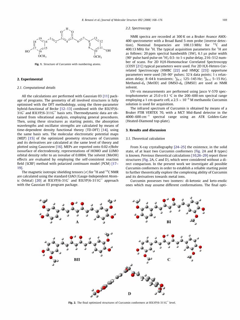
Fig. 1. Structure of Curcumin with numbering atoms.
R. Benassi et al. / Journal of Molecular Structure 892 (2008) 168–176 169
2. Experimental
2.1. Computational details
All the calculations are performed with Gaussian 03 [11] pack-age of programs. The geometry of all involved structures is fullyoptimised with the DFT methodology, using the three-parameterhybrid-functional of Becke [12–13] combined with the B3LYP/6-31G* and B3LYP/6-311G** basis sets. Thermodynamic data are ob-tained from vibrational analysis, employing general procedures.Then, using these structures as starting points, the absorptionwavelengths and oscillator strengths are calculated by means oftime-dependent density functional theory (TD-DFT) [14], usingthe same basis sets. The molecular electrostatic potential maps(MEP) [15] of the optimized geometry structures of Curcuminand its derivatives are calculated at the same level of theory andplotted using Gaussview [16]. MEPs are reported onto 0.02 e/Bohrisosurface of electrodensity, representations of HOMO and LUMOorbital density refer to an isovalue of 0.0004. The solvent (MeOH)effects are evaluated by employing the self-consistent reactionfield (SCRF) method with polarized continuum model (PCM) [17–19].
The magnetic isotropic shielding tensors (r) for 1H and 13C NMRare calculated using the standard GIAO (Gauge-Independent Atom-ic Orbital) [20] at B3LYP/6-31G* and B3LYP/6-311G** approachwith the Gaussian 03 program package.
Fig. 2. The final optimized structures of Curcu
2.2. Spectroscopy
NMR spectra are recorded at 300 K on a Bruker Avance AMX-400 spectrometer with a Broad Band 5-mm probe (inverse detec-tion). Nominal frequencies are 100.13 MHz for 13C and400.13 MHz for 1H. The typical acquisition parameters for 1H areas follows: 20 ppm spectral bandwidth (SW), 6.1 ls pulse width(90� pulse hard pulse on 1H), 0.5- to 1-s pulse delay, 216–512 num-ber of scans. For 2D H,H-Homonuclear Correlated Spectroscopy(COSY [21]) typical parameters were used. For 2D H,X-Hetero Cor-related Spectroscopy (HMBC [22] and HMQC [23]) opportuneparameters were used (50–90� pulses; 32 k data points; 1 s relax-ation delay; 8–64 k transients; 1JH–C 125–145 Hz; 3JH–C 5–15 Hz).Methanol-d4 (MeOD) and DMSO-d6 (DMSO) are used as NMRsolvent.
UV–vis measurements are performed using Jasco V-570 spec-trophotometer at 25.0 ± 0.1 �C in the 200–600 nm spectral rangeemploying a 1 cm quartz cell, a 2.5 � 10�5 M methanolic Curcuminsolution is used for acquisition.
The infrared spectrum of Curcumin is obtained by means of aBruker FTIR VERTEX 70, with a MCT Mid-Band detector in the4000–600 cm�1 spectral range using an ATR Golden-Gate(Heated-Diamond top-plate).
3. Results and discussion
3.1. Theoretical calculation
From X-ray crystallography [24–25] the existence, in the solidstate, of at least two Curcumin conformers (Fig. 2A and B types)is known. Previous theoretical calculations [10,26–29] report threestructures (Fig. 2A, C and D), which were considered without a di-rect comparison. In the present work we investigate all possibleCurcumin conformers in order to establish a reliable starting pointto further theoretically explore the complexing ability of Curcuminand its derivatives towards metal ions.
Curcumin possesses two isomers: di-ketonic and keto-enolicones which may assume different conformations. The final opti-
min conformers at B3LYP/6-311G** level.
170 R. Benassi et al. / Journal of Molecular Structure 892 (2008) 168–176
mized structures for B3LYP/6-311G** are shown in Fig. 2. Calcu-lated values for keto-enolic and di-ketonic structures are reportedin Table 1.
DE, D(E + ZPE), DG and DH values are calculated with two dif-ferent atomic sets of basis (B3LYP/6-31G* and B3LYP/6-311G**)and the results are very similar, generating an equivalent orderof stability among the different species. The same equivalence be-tween the two basis is maintained in molecular geometry calcula-tions; Curcumin keto-enolic structures (A, BI, BII and C),independently on the used set of basis, are completely planar:bond length deviations are within ±0.003 Å, while bond angleand dihedral angle deviations are within ±0.09� and ±0.01�,respectively.
Our theoretical calculation confirms that the keto-enolic formsare more stable than the di-keto one (D) as previously observed[9,26–29]. The extremely low value of D population, calculated invacuum as well as in solvent medium, suggests that this formshould not influence Curcumin properties.
The most stable keto-enolic species depends on the energyterms taken into account: if DE, DE + ZPE) and DH are consideredBII outcomes the most stable, otherwise, if DG term is considered,C results the most stable. The energy gap between these two spe-cies is anyway very small.
The crystal structure reports the enolic hydrogen equally sharedby the two oxygen atoms of the keto-enolic moiety, while calcula-tions show two distinct energetically equivalent keto-enolic formsfor both A and C, since the two possible keto-enolic resonancestructures are equivalent. The tautomeric equilibrium betweenthe two resonance forms occurs through a transition state with aDG* of 0.44 kcal/mol for B3LYP/6-31G* and 0.17 kcal/mol forB3LYP/6-311G**. The extremely low energetic requisite gives anequilibrium in fast exchange in solution, and the active state corre-sponds to the crystal structure with the hydrogen atom equidistantfrom the two oxygen atoms. B structure is swapped in the two con-formers BI and BII, not symmetrically equivalent, but with a verylow energetic gap (D (DG) 0.01 kcal/mol for B3LYP/6-31G* and0.0 kcal/mol for B3LYP/6-311G**). All the structures show twointramolecular hydrogen bonds involving phenolic hydrogen andmethoxylic oxygen (H���O 2.07 Å). The difference in stability be-tween the four keto-enolic conformers (A, BI, BII and C) is verylow as regarding D(E + ZPE) and slightly higher concerning DG.We assume that all the keto-enolic structures can coexist in stan-dard conditions (298.75 K, 1 atm) as a consequence of the possiblefree rotation around the single bond connecting the aromatic ringwith the dienic structure; in fact the calculated rotational energybarrier is rather low (DG* 7.70 and 8.01 kcal/mol, respectively, for
Table 1The total electronic energies (E), zero point vibrational energies (E + ZPE), thermodynamicsfree energy differences in solution (DGMeOH) and relative conformational population in va
Ea E + ZPEa Ga Ha Sb
B3LYP/6-31G*
A �1263.573127 �1263.201114 �1263.260810 �1263.174611 181.421BI �1263.573300 �1263.201363 �1263.260548 �1263.174888 180.289BII �1263.573348 �1263.201384 �1263.260562 �1263.174909 180.272C �1263.573277 �1263.201358 �1263.261439 �1263.174869 182.202D �1263.566078 �1263.194527 �1263.255945 �1263.167534 186.078
B3LYP/6-311G**
A �1263.917090 �1263.547413 �1263.606960 �1263.520864 181.204BI �1263.917292 �1263.547693 �1263.606806 �1263.521174 180.226BII �1263.917346 �1263.547715 �1263.606806 �1263.521190 180.193C �1263.917300 �1263.547707 �1263.607391 �1263.521188 181.430D �1263.905684 �1263.536591 �1263.597850 �1263.509566 185.809
a a.u.b cal mol�1 K�1.c kcal mol�1.
B3LYP/6-31G* and B3LYP/6-311G** for the rotation of a phenyl ringin conformer C). These results justify the presence of the two dif-ferent X-ray structures reported in literature [24–25].
MeOH medium is chosen for calculations as it is the solventused to perform solution study of Curcumin, in particular to collectNMR and UV–vis experimental data.
Solvent effect is irrelevant in relation to stability and conforma-tion equilibria of Curcumin, as it is highlighted by the comparisonof population data (Table 1).
HOMO and LUMO molecular orbitals are calculated for all thestructures with the two set of basis, giving rise to very similar re-sults. A, BI, BII and C show a great similarity (Fig. 3); in these struc-tures HOMO and LUMO are delocalized on the entire molecule,being the electronic density distributed both in the dienic part ofthe molecule and in benzenic one. For the di-ketonic structure(D) the last two occupied molecular orbitals (HOMO and HOMO-1) are almost degener (�0.209 and 0.204 eV and �0.218 and0.213 eV, respectively, for B3LYP/6-31G* and B3LYP/6-311G**),each is delocalized on half molecule giving rise to two separatedfragments. This accounts for the higher stability of A, BI, BII andC conformers with respect to D, suggesting their ability to chelatemetal ions as previously observed [10].
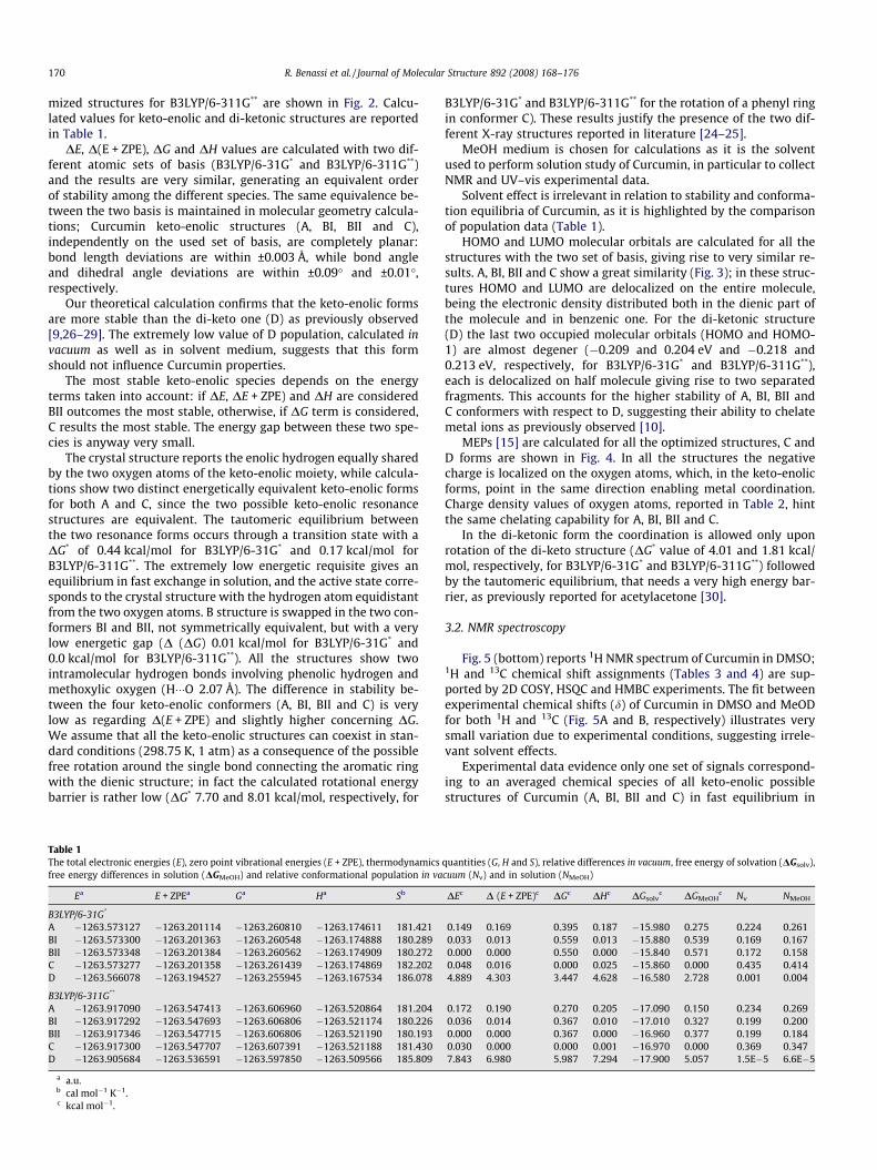
MEPs [15] are calculated for all the optimized structures, C andD forms are shown in Fig. 4. In all the structures the negativecharge is localized on the oxygen atoms, which, in the keto-enolicforms, point in the same direction enabling metal coordination.Charge density values of oxygen atoms, reported in Table 2, hintthe same chelating capability for A, BI, BII and C.
In the di-ketonic form the coordination is allowed only uponrotation of the di-keto structure (DG* value of 4.01 and 1.81 kcal/mol, respectively, for B3LYP/6-31G* and B3LYP/6-311G**) followedby the tautomeric equilibrium, that needs a very high energy bar-rier, as previously reported for acetylacetone [30].
3.2. NMR spectroscopy
Fig. 5 (bottom) reports 1H NMR spectrum of Curcumin in DMSO;1H and 13C chemical shift assignments (Tables 3 and 4) are sup-ported by 2D COSY, HSQC and HMBC experiments. The fit betweenexperimental chemical shifts (d) of Curcumin in DMSO and MeODfor both 1H and 13C (Fig. 5A and B, respectively) illustrates verysmall variation due to experimental conditions, suggesting irrele-vant solvent effects.
Experimental data evidence only one set of signals correspond-ing to an averaged chemical species of all keto-enolic possiblestructures of Curcumin (A, BI, BII and C) in fast equilibrium in
quantities (G, H and S), relative differences in vacuum, free energy of solvation (DGsolv),cuum (Nv) and in solution (NMeOH)
DEc D (E + ZPE)c DGc DHc DGsolvc DGMeOH
c Nv NMeOH
0.149 0.169 0.395 0.187 �15.980 0.275 0.224 0.2610.033 0.013 0.559 0.013 �15.880 0.539 0.169 0.1670.000 0.000 0.550 0.000 �15.840 0.571 0.172 0.1580.048 0.016 0.000 0.025 �15.860 0.000 0.435 0.4144.889 4.303 3.447 4.628 �16.580 2.728 0.001 0.004
0.172 0.190 0.270 0.205 �17.090 0.150 0.234 0.2690.036 0.014 0.367 0.010 �17.010 0.327 0.199 0.2000.000 0.000 0.367 0.000 �16.960 0.377 0.199 0.1840.030 0.000 0.000 0.001 �16.970 0.000 0.369 0.3477.843 6.980 5.987 7.294 �17.900 5.057 1.5E�5 6.6E�5

Fig. 3. Representations of HOMO and LUMO orbital density of Curcumin conformers at B3LYP/6-311G** level.
R. Benassi et al. / Journal of Molecular Structure 892 (2008) 168–176 171
NMR time scale; resonances show a completely symmetric species(C2v): (a) the enolic proton is equally shared between the two oxy-gen atoms of keto-enolic structure and strongly deshielded due toits involvement in a hydrogen bond; (b) ketonic carbon and enoliccarbon are undistinguishable and equivalent, showing a 13C d valueof 183.6 ppm; (c) the two aromatic rings and the double bonds arecompletely equivalent.
Tables 3 and 4 show the GIAO (Gauge-Independent-Atomic-Orbitals) isotropic magnetic shielding tensors (rcalc) for all refinedketo-enolic structures (A, BI, BII and C) for both calculation levels.
�rcalc are obtained on the basis of the relative speciespopulations:
�rcalc ¼ rcalcðAÞ � nA þ rcalcðBIÞ � nBI þ rcalcðBIIÞ � nBII þ rcalcðCÞ � nC
nA, nBI, nBII and nC population are calculated according to the DG val-ues excluding the di-keto form; rcalc(A) and rcalc(C) are the aver-aged values of isotropic shielding tensors of the two resonanceforms of A and C, respectively.
Plotting the experimental 13C and 1H chemical shifts (dexp) inMeOD and DMSO vs. the rcalc for all species (or �rcalc), a linearregression is obtained: dexp = a � rcalc + b (a, b and R2 parametersare give in Tables 3 and 4). This relationship is used to predictthe chemical shifts (dpred). As already observed [31], the correlationbetween experimental chemical shifts and calculated isotropicscreening constants for 13C is better than for 1H (Fig. 6), in factan homogeneous behaviour is observed for 13C independently onthe calculated species with a correlation coefficient R2 rangingfrom 0.9691 to 0.9917. Fig. 6A shows 13C dexp vs. �rcalc while
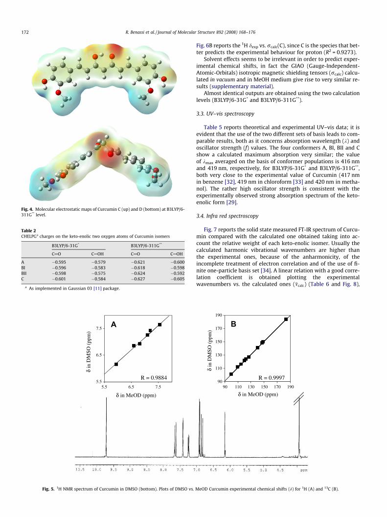
Fig. 4. Molecular electrostatic maps of Curcumin C (up) and D (bottom) at B3LYP/6-311G** level.
Table 2CHELPGa charges on the keto-enolic two oxygen atoms of Curcumin isomers
B3LYP/6-31G* B3LYP/6-311G**
C@O CAOH C@O CAOH
A �0.595 �0.579 �0.621 �0.600BI �0.596 �0.583 �0.618 �0.598BII �0.598 �0.575 �0.624 �0.592C �0.601 �0.584 �0.627 �0.605
a As implemented in Gaussian 03 [11] package.
δ in MeOD (ppm)
δ in
DM
SO (
ppm
)
5.5
6.5
7.5
5.5 6.5 7.5
A
R = 0.9884
Fig. 5. 1H NMR spectrum of Curcumin in DMSO (bottom). Plots of DMSO vs.
172 R. Benassi et al. / Journal of Molecular Structure 892 (2008) 168–176
Fig. 6B reports the 1H dexp vs. rcalc(C), since C is the species that bet-ter predicts the experimental behaviour for proton (R2 = 0.9273).
Solvent effects seems to be irrelevant in order to predict exper-imental chemical shifts, in fact the GIAO (Gauge-Independent-Atomic-Orbitals) isotropic magnetic shielding tensors (rcalc) calcu-lated in vacuum and in MeOH medium give rise to very similar re-sults (supplementary material).
Almost identical outputs are obtained using the two calculationlevels (B3LYP/6-31G* and B3LYP/6-311G**).
3.3. UV–vis spectroscopy
Table 5 reports theoretical and experimental UV–vis data; it isevident that the use of the two different sets of basis leads to com-parable results, both as it concerns absorption wavelength (k) andoscillator strength (f) values. The four conformers A, BI, BII and Cshow a calculated maximum absorption very similar; the valueof kmax averaged on the basis of conformer populations is 416 nmand 419 nm, respectively, for B3LYP/6-31G* and B3LYP/6-311G**,both very close to the experimental value of Curcumin (417 nmin benzene [32], 419 nm in chloroform [33] and 420 nm in metha-nol). The rather high oscillator strength is consistent with theexperimentally observed strong absorption spectrum of the keto-enolic form [29].
3.4. Infra red spectroscopy
Fig. 7 reports the solid state measured FT-IR spectrum of Curcu-min compared with the calculated one obtained taking into ac-count the relative weight of each keto-enolic isomer. Usually thecalculated harmonic vibrational wavenumbers are higher thanthe experimental ones, because of the anharmonicity, of theincomplete treatment of electron correlation and of the use of fi-nite one-particle basis set [34]. A linear relation with a good corre-lation coefficient is obtained plotting the experimentalwavenumbers vs. the calculated ones (�mcalc) (Table 6 and Fig. 8),
90
110
130
150
170
190
90 110 130 150 170 190
B
R = 0.9997
δ in
DM
SO (
ppm
)
δ in MeOD (ppm)
MeOD Curcumin experimental chemical shifts (d) for 1H (A) and 13C (B).

Table 3Experimental (MeOD and DMSO) 13C chemical shifts (dexp
, ppm), predicted 13C chemical shifts (dpred = a � �rcalc þ b, ppm) and calculated GIAO magnetic isotropic shielding tensors (rcalc and �rcalc) for Curcumin
13C MeOD DMSO rcalc* �rcalc
* rcalc** �rcalc
**
dexp dpred* dpred
** dexp dpred* dpred
** A BI BII C A BI BII C
1 140.4 142.2 142.0 141.1 142.7 142.6 55.37 56.55 54.24 54.98 55.17 36.26 39.14 36.38 37.05 37.152 120.6 120.0 120.1 121.6 121.0 121.0 74.73 76.44 73.09 75.66 75.22 58.82 60.34 57.37 60.08 59.313 183.7 182.8 181.0 183.6 182.7 180.9 18.35 25.73 11.19 18.42 18.40 �2.25 4.17 �8.49 �2.23 �2.204 100.2 104.6 101.6 101.2 105.8 102.9 88.89 89.01 89.07 89.30 89.14 77.69 77.83 77.85 78.21 77.955 183.7 182.8 181.0 183.6 182.7 180.9 18.35 11.11 25.53 18.42 18.39 �2.25 �8.63 4.03 �2.23 �2.266 120.6 119.8 119.6 121.6 120.8 120.6 74.73 73.88 77.26 75.66 75.39 58.82 58.50 61.47 60.08 59.757 140.4 142.3 141.9 141.1 142.8 142.5 55.37 53.81 56.07 54.98 55.08 37.68 35.76 38.48 37.05 37.238 127.3 126.9 128.2 126.8 127.7 129.0 68.94 69.01 68.96 68.97 68.96 51.07 51.11 51.09 51.14 51.119 110.1 106.3 104.8 111.9 107.5 106.0 80.91 89.96 90.35 90.13 87.64 67.21 76.93 77.32 76.96 74.7510 147.8 146.2 147.6 148.4 146.8 148.0 51.82 51.40 51.23 51.35 51.47 32.00 31.50 31.32 31.40 31.5511 149.0 150.0 151.6 149.8 150.5 152.0 48.46 47.79 47.99 47.86 48.03 27.93 27.20 27.42 27.36 27.4812 115.0 113.3 114.1 116.2 114.4 115.1 80.81 81.46 81.41 81.49 81.30 64.72 65.62 65.56 65.60 65.4013 122.6 126.6 126.7 123.5 127.5 127.6 76.08 66.32 67.08 66.66 69.22 60.12 49.97 50.82 50.22 52.6114 127.3 126.9 128.2 126.8 127.8 129.0 68.94 68.83 68.87 68.97 68.94 51.07 51.02 51.03 51.14 51.0815 110.1 108.3 108.7 111.9 109.5 109.8 80.91 81.39 80.70 90.13 85.77 67.21 67.68 66.92 76.96 70.8516 147.8 146.1 147.3 148.4 146.6 147.8 51.82 51.80 51.84 51.35 51.57 32.00 32.01 32.05 31.40 31.7917 149.0 149.9 151.4 149.8 150.4 151.8 48.46 48.43 48.26 47.86 48.12 27.93 27.87 27.69 27.36 27.6618 115.0 113.4 114.4 116.2 114.5 115.5 80.81 80.79 80.94 81.49 81.18 64.72 64.58 64.75 65.60 65.0319 122.6 124.5 122.9 123.5 125.4 123.8 76.08 76.33 75.93 66.66 71.16 60.12 60.38 59.98 50.22 56.50
MeODa �1.1306 �1.0852 �1.0847 �1.0740 �1.1050 �1.0134 �0.9762 �0.9767 �0.9592 �0.9897b 204.725 201.867 201.835 201.177 203.124 179.774 178.177 178.203 177.418 178.784R2 0.9910 0.9691 0.9697 0.9772 0.9917 0.9934 0.9749 0.9754 0.9759 0.9910
DMSOa �1.1126 �1.0670 �1.0666 �1.0553 �1.0864 �0.9969 �0.9596 �0.9601 �0.9423 �0.9728b 204.312 201.447 201.417 200.723 202.672 179.744 178.140 178.167 177.367 178.734R2 0.9915 0.9680 0.9687 0.9748 0.9903 0.9932 0.9732 0.9738 0.9730 0.9892
The numbering of atoms refers to Fig. 1.* Calculation performed at B3LYP/6-31G* level.
** Calculation performed at B3LYP/6-311G** level.
R.Benassi
etal./Journal
ofM
olecularStructure
892(2008)
168–176
173

Table 4Experimental (MeOD and DMSO) 1H chemical shifts (dexp, ppm), predicted 1H chemical shifts ðdpred ¼ a�rcalc þ b;ppmÞ and calculated GIAO magnetic isotropic shielding tensors(rcalc and �rcalc) for Curcumin
1H MeOD DMSO rcalc* �rcalc
* rcalc** �rcalc
**
dexp dpred* dpred
** dexp dpred* dpred
** A BI BII C A BI BII C
1 7.56 7.42 7.48 7.64 7.54 7.59 24.95 25.04 24.83 24.87 24.91 24.25 24.35 24.14 24.19 24.232 6.61 6.64 6.64 6.85 6.83 6.84 26.10 26.25 26.00 26.27 26.18 25.48 25.63 25.38 25.65 25.554 5.95 5.94 5.98 6.15 6.20 6.23 27.28 27.32 27.32 27.36 27.33 26.57 26.60 26.60 26.63 26.616 6.61 6.61 6.61 6.85 6.81 6.81 26.10 26.12 26.38 26.27 26.23 25.48 25.49 25.75 25.65 25.607 7.56 7.42 7.49 7.64 7.54 7.60 24.95 24.80 25.01 24.87 24.90 24.25 24.10 24.31 24.19 24.219 7.20 7.02 7.00 7.42 7.18 7.16 25.95 25.40 25.47 25.45 25.56 25.35 24.83 24.90 24.89 24.9912 6.82 7.04 7.00 6.92 7.20 7.16 25.49 25.54 25.55 25.52 25.52 24.93 25.00 25.01 24.99 24.9813 7.09 7.17 7.15 7.25 7.32 7.30 24.89 25.38 25.47 25.43 25.31 24.35 24.83 24.92 24.86 24.7515 7.20 6.91 6.87 7.42 7.08 7.04 25.95 26.00 25.90 25.45 25.73 25.35 25.42 25.34 24.89 25.2018 6.82 7.05 7.02 6.92 7.21 7.17 25.49 25.50 25.51 25.52 25.50 24.93 24.94 24.95 24.99 24.9619 7.09 7.28 7.28 7.25 7.42 7.41 24.89 24.93 24.86 25.43 25.13 24.35 24.41 24.34 24.86 24.55
MeODa �0.5161 �0.5711 �0.5685 �0.6258 �0.6115 �0.5432 �0.595 �0.5927 �0.6548 �0.6334b 20.189 21.611 21.546 23.023 22.648 20.549 21.862 21.807 23.372 22.827R2 0.6678 0.7886 0.7881 0.9273 0.8598 0.7005 0.8115 0.8105 0.9515 0.8743
DMSOa �0.4599 �0.5161 �0.5142 �0.5532 �0.5563 �0.4833 �0.5359 �0.5344 �0.5959 �0.5714b 18.911 20.362 20.316 21.317 21.397 19.214 20.546 20.508 22.059 21.437R2 0.5997 0.7284 0.7292 0.7961 0.8033 0.6272 0.7448 0.7451 0.8914 0.8048
The numbering of atoms refers to Fig. 1.* Calculation performed at B3LYP/6-31G* level.
** Calculation performed at B3LYP/6-311G** level.
Table 5Absorption wavelengths (k) and oscillator strengths (f) for all Curcumin keto-enolic forms
* ** * ** * ** * ** * **
A E (ev) 2.9626 2.9428 3.3314 3.305 3.4377 3.4627 3.646 3.6418 3.6957 3.6891k (nm) 418.49 421.31 372.17 375.14 360.66 358.06 340.06 340.45 335.49 336.08f 1.5218 1.5164 0.0321 0.0316 0 0 0.1259 0.1366 0.0282 0.0288
BI E (ev) 2.9749 2.9565 3.3603 3.3373 3.4344 3.4592 3.6173 3.606 3.6809 3.6691k (nm) 416.77 419.36 368.97 371.51 361.01 358.42 342.76 343.83 336.83 337.92f 1.5198 1.5183 0.031 0.0311 0 0 0.1007 0.106 0.0082 0.0081
BII E (ev) 2.9734 2.9555 3.3497 3.3257 3.4366 3.4618 3.6368 3.6309 3.6575 3.6911k (nm) 416.97 419.5 370.13 372.8 360.77 358.15 340.92 341.47 338.99 335.9f 1.5181 1.5149 0.0399 0.0418 0 0 0.0841 0.0892 0.0301 0.0317
C E (ev) 2.9882 2.9717 3.3809 3.3604 3.435 3.4606 3.609 3.6023 3.6657 3.6531k (nm) 414.91 417.21 366.72 368.96 360.94 358.27 343.54 344.18 338.23 339.4f 1.5221 1.5259 0.0293 0.0283 0 0 0.0611 0.0621 0.0114 0.0119
* Stands for calculation performed at B3LYP/6-31G* level.** Stands for calculation performed at B3LYP/6-311G** level.
80
100
120
140
160
180
200
10 30 50 70 90
calcσ
exp
(ppm
)
)C(calcσ
exp
(ppm
)
A B
5.5
6.0
6.5
7.0
7.5
8.0
24 25 26 27 28
δ δ
Fig. 6. Experimental chemical shifts of Curcumin vs. isotropic magnetic tensors from GIAO/B3LYP/6-311G** calculations for 13C (A) and 1H (B) in DMSO (s) and MeOD (D).
174 R. Benassi et al. / Journal of Molecular Structure 892 (2008) 168–176
which are determined on the basis of the relative species popula-tions according to the following equation:
�mcalc ¼ �mcalcðAÞ �nA þ �mcalcðBIÞ �nBI þ �mcalcðBIIÞ �nBII þ �mcalcðCÞ �nC
nA, nBI, nBII and nC are species populations determined by means ofDG values with the exclusion of negligible di-keto form.
This result suggests that the over-estimation of calculatedwavenumbers is quite systematic and allows to predict FT-IR spec-tra. Overlapping outcomes are obtained using the two different cal-culation levels B3LYP/6-31G* and B3LYP/6-311G**.
In conclusion, the comparison between experimental and theo-retical data advises that calculated predictions are reliable even

A
Wavenumbers (cm-1)
1000200030004000
Inte
nsity
0
200
400
600
800B
Abs
orba
nce
0.7
0.6
0.5
0.4
0.3
0.2
0.1
0.0
Fig. 7. Experimental spectrum of Curcumin: (A) FT-IR spectrum. (B) Calculatedspectrum at B3LYP/6-311G** level of theory. Calculated wavelengths and intensityare estimated as averaged values on the basis of relative populations of keto-enolicspecies.
Table 6More relevant IR vibration frequencies (cm�1) and relative intensity – experimentaland calculated (�mcalc) with B3LYP/6-311G** basis set
Calculated Experimental
�mcalc (cm�1) Intensity (km/mol) m (cm�1) A
860.49 50.030 808.04 0.483981.90 110.354 960.39 0.486
1059.40 52.151 1025.96 0.4431147.33 429.491 1112.74 0.4961179.93 58.140 1135.88 0.4501209.45 100.366 1151.31 0.6131231.68 290.468 1182.17 0.4561262.26 112.022 1203.38 0.4861312.32 811.020 1232.71 0.3811325.99 177.591 1274.74 0.4431474.56 685.852 1427.09 0.4761544.87 660.324 1506.16 0.4901625.33 393.85 1587.15 0.2141639.75 729.055 1600.65 0.2471673.97 644.472 1625.73 0.2432935.38 474.413 2846.46 0.0473766.63 240.171 3507.94 0.088
Calculated IR wavenumbers (cm-1)500 1000 1500 2000 2500 3000 3500 4000
Exp
erim
enta
l IR
wav
enum
bers
(cm
-1)
500
1000
1500
2000
2500
3000
3500
4000
Fig. 8. Correlation between experimental and B3LYP/6-311G** in vacuum calculatedwavenumbers for Curcumin. Equation: �mexp ¼ a � �mcalc þ b; a = 0.9379, b = 34.449,R2 = 0.9983.
R. Benassi et al. / Journal of Molecular Structure 892 (2008) 168–176 175
using relatively small basis set, such as B3LYP/6-31G*, which canbe satisfactory implied to predict spectroscopic properties for com-plex organic structures, reducing computational efforts.
Acknowledgments
We are thankful to the ‘‘Consorzio Interuniversitario per il Cal-colo Automatico dell’Italia Nord Orientale – CINECA” and to the
‘‘Laboratorio di Calcolo Scientifico Avanzato Interdipartimentaledell’Università degli Studi di Modena e Reggio Emilia” for comput-ing facilities.
We are grateful to the ‘‘Centro Interdipartimentale GrandiStrumenti – C.I.G.S.” of the University of Modena and Reggio Emiliaand to the ‘‘Fondazione Cassa di Risparmio di Modena” for supply-ing NMR spectrometer.
Appendix A. Supplementary data
Supplementary data associated with this article can be found, inthe online version, at doi:10.1016/j.molstruc.2008.05.024.
References
[1] R.A. Sharma, A.J. Gescher, W.P. Steward, Eur. J. Cancer 41 (2005) 1955.[2] B.B. Aggarwal, S. Shishodia, Biochem. Pharmacol. 71 (2006) 397.[3] R.K. Maheshwari, A.K. Singh, J. Gaddipati, R.C. Srimal, Life Sci. 78 (2006) 2081.[4] S. Gafner, S.K. Lee, M. Cuendet, S. Barthelemy, L. Vergnes, S. Labidalle, R.G.
Mehta, C.W. Boone, J.M. Pezzuto, Phytochemistry 65 (2004) 2849.[5] B.K. Adams, J. Cai, J. Armstrong, M. Herold, Y.J. Lu, A. Sun, J.P. Snyder, D.C. Liotta,
D.P. Jones, M. Shoji, Anticancer Drugs 16 (2005) 263.[6] B.B. Aggarwal, A. Kumar, A.C. Bharti, Anticancer Res. 23 (2003) 363.[7] M.M. LoTempio, M.S. Veena, H.L. Steele, B. Ramamurthy, T.S. Ramalingam, A.N.
Cohen, R. Chakrabarti, E.S. Srivatsan, M.B. Wang, Clin. Cancer Res. 11 (2005)6994.
[8] S. Jovanovic, S. Steenken, C. Boone, M. Simic, J. Am. Chem. Soc. 121 (1999)9677.
[9] K. Balasubramanian, J. Agric. Food Chem. 54 (2006) 3512.[10] M. Borsari, E. Ferrari, R. Grandi, M. Saladini, Inorg. Chim. Acta 328 (2002) 61.[11] M.J. Frisch, G.W. Trucks, H.B. Schlegel, G.E. Scuseria, M.A. Robb, J.R. Cheeseman,
V.G. Zakrzewski, J.A.jr Montgomery, R.E. Stratmann, J.C. Burant, S. Dapprich,J.M. Illam, A.D. Daniels, K.N. Kudin, M.C. Strain, O. Farkas, J. Tomasi, V. Barone,M. Cossi, R. Cammi, B. Mennucci, C. Pomelli, C. Adamo, S. Clifford, J. Ochterski,G.A. Petersson, P.Y. Ayala, Q. Cui, K. Morokuma, D.K. Malick, A.D. Rabuck, K.Raghavachari, J.B. Foresman, J. Cioslowski, J.V. Ortiz, B.B. Stefanov, G. Liu, A.Liashenko, P. Piskorz, I. Komaromi, R. Gomperts, R.L. Martin, D.J. Fox, T. Keith,M.A. Al-Laham, C.Y. Peng, A. Nanayakkara, C. Gonzalez, M. Challacombe,P.M.W. Gill, B.G. Johnson, W. Chen, M.W. Wong, J.L. Andres, M. Head-Gordon,E.S. Replogle, J.A. Pople, Gaussian 03; Gaussian, Inc.: Pittsburgh, PA, USA, 2003.
[12] A.D. Becke, Phys. Rev. A 38 (1988) 3098.[13] C. Lee, W. Yang, R.G. Parr, Phys. Rev. B 37 (1988) 785.[14] R.E. Stratmann, G.E. Scuseria, M.J. Frish, J. Chem. Phys. 109 (1998) 8218.[15] J.S. Murray, K.D. San (Eds.), Molecular Electrostatic Potentials: Concepts and
Applications; Theoretical and Computational Chemistry Book Series, vol. 3,Elsevier, Amsterdam, 1996.
[16] R. Dennington II, T. Keyth, J. Millam, K. Eppinnett, W.L. Hovell, R. Gilliand,GaussView, Version 3.0, Semichem. Inc., Shawnee Mission, KS, 2003.
[17] S. Miertus, E. Scrocco, J. Tomasi, Chem. Phys. 55 (1981) 117.[18] S. Miertus, J. Tomasi, Chem. Phys. 65 (1982) 239.[19] M. Cossi, V. Barone, J. Cammi, Chem. Phys. Lett. 255 (1996) 327.[20] K. Wolinski, J.F. Hinton, P.J. Pulay, J. Am. Chem. Soc. 112 (1990) 8251.

176 R. Benassi et al. / Journal of Molecular Structure 892 (2008) 168–176
[21] K. Nagayama, A. Kumar, K. Wuethrich, R.R. Ernst, J. Magn. Res. 40 (1980) 321.[22] A. Bax, M.F. Summers, J. Am. Chem. Soc. 108 (1986) 2093.[23] A. Bax, R.H. Griffey, B.L. Hawkins, J. Magn. Res. 55 (1983) 301.[24] H.H. Tonnesen, J. Karlsen, Acta Chem. Scand. B 36 (1982) 475.[25] S.P. Paramiti, Y.V. Ramshanker, S. Suresh, T.N. Guru Row, Acta Cryst. E63
(2007) o860–o862.[26] Y. M Sun, R.X. Wang, S.L. Yuan, X.J. Lin, C.B. Liu, Chin. J. Chem. 22 (2004) 827.[27] K. Ling, K.I. Priyadarsini, H.Y. Zhang, J. Mol. Struct. (THEOCHEM) 684 (2002)
111.[28] L. Shen, H.F. Ji, H. Y Zhang, Chem. Phys. Lett. 409 (2005) 300.
[29] L. Shen, H.F. Ji, Spectrochim. Acta A 67 (2007) 619.[30] C. Ghio, G. Alagona, Int. J. Quant. Chem. 108 (2008) 1840.[31] M. Szafran, E. Bartoszak-Adamska, J. Koput, Z. Dega-Szafran, J. Mol. Struct.
844–845 (2007) 140.[32] A.A. Gorman, V.S. Hamblett, V.S. Srinivasan, P.D. Wood, Phochem. Photobiol.
59 (1994) 389.[33] K.I. Priyadarsini, D.K. Marty, G.H. Naik, M.S. Kumar, M.K. Unnikrishnan, J.G.
Satav, H. Mohan, Free Radic. Biol. Med. 35 (2003) 475.[34] M. Szafran, A. Katrusiak, J. Koput, Z. Dega- Szafran, J. Mol. Struct. 846 (2007)
140.