Intestinal Blood Flow in Patients With Chronic Heart Failure
Upload
khangminh22Category
view
0download
0
European Journal of Heart Failure (2019) 21, 137–155 POSITION PAPERdoi:10.1002/ejhf.1369
The use of diuretics in heart failure withcongestion — a position statement from theHeart Failure Association of the EuropeanSociety of CardiologyWilfried Mullens1,2*, Kevin Damman3, Veli-Pekka Harjola4, Alexandre Mebazaa5,Hans-Peter Brunner-La Rocca6, Pieter Martens1,2, Jeffrey M. Testani7,W.H. Wilson Tang8, Francesco Orso9, Patrick Rossignol10, Marco Metra11,Gerasimos Filippatos12,13, Petar M. Seferovic14, Frank Ruschitzka15,and Andrew J. Coats16
1Ziekenhuis Oost Limburg, Genk, Belgium; 2University of Hasselt, Hasselt, Belgium; 3University of Groningen, University Medical Center Groningen, Groningen,The Netherlands; 4Emergency Medicine, University of Helsinki, Helsinki University Hospital, Helsinki, Finland; 5University of Paris Diderot, Hôpitaux Universitaires Saint LouisLariboisière, APHP, U 942 Inserm, F-CRIN INI-CRCT, Paris, France; 6Maastricht University Medical Center, Maastricht, The Netherlands; 7Yale University, New Haven, CT, USA;8Cleveland Clinic, Cleveland, OH, USA; 9University of Florence, Florence, Italy; 10Université de Lorraine, Inserm, Centre d’Investigations Clinique 1433 and Inserm U1116;CHRU Nancy; F-CRIN INI-CRCT, Nancy, France; 11University of Brescia, Brescia, Italy; 12National and Kapodistrian University of Athens, Athens, Greece; 13University of Cyprus,Nicosia, Cyprus; 14University of Belgrade, Faculty of Medicine, Belgrade, Serbia; 15UniversitätsSpital Zürich, Zürich, Switzerland; and 16IRCCS, San Raffaele Pisana, Rome, Italy
Received 19 July 2018; revised 14 October 2018; accepted 27 October 2018 ; online publish-ahead-of-print 1 January 2019
The vast majority of acute heart failure episodes are characterized by increasing symptoms and signs of congestion with volume overload.The goal of therapy in those patients is the relief of congestion through achieving a state of euvolaemia, mainly through the use of diuretictherapy. The appropriate use of diuretics however remains challenging, especially when worsening renal function, diuretic resistance andelectrolyte disturbances occur. This position paper focuses on the use of diuretics in heart failure with congestion. The manuscript addressesfrequently encountered challenges, such as (i) evaluation of congestion and clinical euvolaemia, (ii) assessment of diuretic response/resistancein the treatment of acute heart failure, (iii) an approach towards stepped pharmacologic diuretic strategies, based upon diuretic response,and (iv) management of common electrolyte disturbances. Recommendations are made in line with available guidelines, evidence and expertopinion.. . . . . . . . . . . . . . . . . . . . . . . . . . . . . . . . . . . . . . . . . . . . . . . . . . . . . . . . . . . . . . . . . . . . . . . . . . . . . . . . . . . . . . . . . . . . . . . . . . . . . . . . . .
Keywords Diuretics • Heart failure • Acute heart failure • Pharmacotherapy • Loop diuretics
IntroductionThe natural history of heart failure is characterized by acutedecompensation episodes, which are associated with increasedmorbidity and mortality and pose an economic burden on oursociety .1,2 Increasing signs and symptoms of congestion are themain reasons why patients with acute heart failure seek urgentmedical care.3–5 Even though congestion often develops over anextended period of time before acute presentation, the remainder
*Corresponding author. Department of Cardiology, Ziekenhuis Oost-Limburg, Schiepse Bos 6, 3600 Genk, Belgium. Tel: +32 89 327160, Fax: +32 89 327918, Email:[email protected]
....
....
....
....
....
....
... of this manuscript will refer to this setting as acute heart failure.
Only a minority of patients with acute heart failure present acutelywith signs and symptoms of low perfusion.4 Given the pivotal roleof congestion in heart failure, diuretics are a cornerstone of therapyin heart failure.6 Guidelines strongly recommend the use of loopdiuretics to alleviate signs and symptoms of fluid overload (classI, level of evidence B).7 This paper discusses the practical use ofdiuretics in patients with acute and chronic heart failure, based oncontemporary evidence and expert opinion.
© 2018 The AuthorsEuropean Journal of Heart Failure © 2018 European Society of Cardiology

138 W. Mullens et al.
Congestion in heart failureDefinition and mechanisms of congestionCongestion in heart failure is defined as signs and symptomsof extracellular fluid accumulation that result in increased car-diac filling pressures.8 Filling pressures are the integrated resultof the cardiac systolic and diastolic function, plasma volume, andvenous capacitance/compliance.9–11 Heart failure with increasedneurohumoral activation induces a state of increased renal sodiumand water avidity resulting in an increased plasma volume.11,12
Also, increased sympathetic output leads to splanchnic arterialand venous constriction resulting in blood redistribution from thesplanchnic capacitance vasculature to the circulatory volume. Thisincreases the effective circulating volume by redistribution, in astate where volume expansion is already present.13 As a result,venous return and cardiac filling pressures increase.11 Indeed, thevenous capacitance function becomes compromised during statesof longstanding venous congestion and/or increased sympatheticactivation in acute heart failure.11,14,15 Importantly, the term vol-ume overload and congestion are often used interchangeably. How-ever, it has been demonstrated that 54% of patients hospitalized foracute heart failure gain ≤ 1 kg during the month prior to admission,suggesting that volume overload incompletely characterizes thepathophysiology of acute heart failure and redistribution of volumemay also contribute to the development of signs and symptomsof congestion.16,17 Furthermore, heart failure is often associatedwith cachexia which makes the interpretation of weight changesdifficult. Additionally, cachexia might result in a loss of plasma pro-teins, reducing plasma oncotic pressure, hampering plasma refillingfrom the interstitium.18,19 Additionally, weight loss during hospi-talization is not necessarily associated with improved in-hospitalor post-discharge morbidity or mortality, however weight gain hasbeen associated with poor outcome.20,21 Therefore, the EuropeanSociety of Cardiology (ESC) guidelines for the diagnosis and treat-ment of acute and chronic heart failure recommend to distinguishacute fluid redistribution from true volume overload in patientspresenting with congestion (no class recommendation).7 As diuret-ics are mainly used to relieve excessive volume, the remainder ofthis manuscript will focus on congestion with excessive volumeoverload.
Detecting congestion in heart failureAlthough the intravascular pressure–volume relationship mayvary across individuals and clinical conditions, the gold standardfor diagnosing congestion in heart failure is cardiac catheterizationwith direct measurement of right atrial pressure and pulmonarycapillary wedge pressure (PCWP).22 However, the invasivenature of this technique limits its routine use in clinical practice.Furthermore, the use of pulmonary artery catheterization toguide decongestive therapy did not improve outcome in theEvaluation Study of Congestive Heart Failure and PulmonaryArtery Catheterization Effectiveness (ESCAPE) study in compar-ison to serial clinical assessment, despite significantly improving
....
....
....
....
....
....
....
....
....
....
....
....
....
....
....
....
....
....
....
....
....
....
....
....
....
....
....
....
....
....
....
....
....
....
....
....
....
....
....
....
....
.... haemodynamics.23 The diagnostic accuracy of non-invasive clinical
and technical assessments of congestion has been validated againstinvasive haemodynamic evaluation and shown a variable sensitivityand specificity (Table 1).22–28 Physical signs and symptoms of con-gestion are based on detecting increased filling pressures and/orthe extravascular fluid build-up secondary to the increased fillingpressures. As such, the jugular venous pulse is the most usefulphysical finding for determining a patient’s volume status. Not onlydoes an elevated jugular venous pulsation (JVP) detect systemiccongestion, but there is good sensitivity (70%) and specificity (79%)between high JVP and elevated left-sided filling pressure. Changes inJVP with therapy usually parallel changes in left-sided filling pressurealthough significant inter-observer variability regarding the extentof JVP elevation exist.29–31 However, in a series of 50 patients withchronic heart failure it was shown that physical signs of congestion(rales, oedema and JVP elevation) were absent in 42% of patientswith a PCWP ≥ 22 mmHg.32 Additionally, there is a waning ofskills in performing physical examination in current practice.33
Also, while a chest X-ray can show signs of lung congestion andpleural fluid, 20% of patients with congestion exhibit a normalchest X-ray.34 In comparison to chest X-ray, lung ultrasound isbetter in ruling out interstitial oedema and pleural effusions. Lungultrasound detects B-lines originating from extravasated fluid intothe interstitium and alveoli.35,36 More than three B-lines in morethan two intercostal spaces bilaterally are considered diagnosticfor the detection of interstitial and alveolar oedema in acute heartfailure. Echocardiographic parameters (Table 1) can be used toestimate right- and left-sided filling pressures, although with lesscertainty in acute heart failure.25 Estimation of right atrial pres-sures can be performed by assessing the collapsibility and width ofthe vena cava. Doppler imaging and tissue Doppler can be used toassess left-sided filling pressures. With rising filling pressures, anincrease in early diastolic mitral inflow velocities (E wave) occurs.This is indicative of increased filling pressures in the presence of alow e’, especially if E-wave deceleration time is short and A-wavevelocities are low.28 Nevertheless, the use of e’ might be limited inadvanced heart failure.25 Guidelines suggest the measurement ofnatriuretic peptides (NPs) in all patients with acute heart failure,especially to distinguish from non-cardiac causes of dyspnoea(class I recommendation, level of evidence A).7 NPs have a highnegative predictive value for ruling out acute heart failure withcongestion [thresholds for excluding acute heart failure; B-typenatriuretic peptide (BNP) < 100 pg/mL, N-terminal pro BNP(NT-proBNP) < 300 ng/mL and mid-regional pro atrial natriureticpeptide < 120 pg/mL].7,37 In patients with history of heart failureor cardiac disease, the combination of signs and symptoms ofcongestion, an indicative chest X-ray and the measurement ofelevated NPs allows for the diagnosis of congestion.22,38 Accordingto local availability, these tests can be supplemented with transtho-racic echocardiography or lung ultrasound. In line with the ESCguidelines, direct haemodynamic evaluation should be reserved forpatients with cardiogenic shock, refractory pulmonary oedema orsuspected mismatch between left and right-sided filling pressures(class IIb recommendation, level of evidence C) or in cases ofuncertainty of the haemodynamic status.7
© 2018 The AuthorsEuropean Journal of Heart Failure © 2018 European Society of Cardiology
Diuretics in heart failure 139
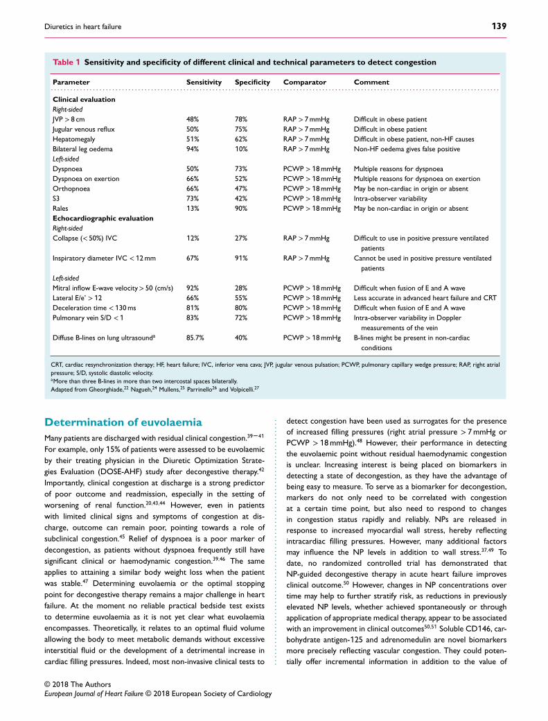
Table 1 Sensitivity and specificity of different clinical and technical parameters to detect congestion
Parameter Sensitivity Specificity Comparator Comment. . . . . . . . . . . . . . . . . . . . . . . . . . . . . . . . . . . . . . . . . . . . . . . . . . . . . . . . . . . . . . . . . . . . . . . . . . . . . . . . . . . . . . . . . . . . . . . . . . . . . . . . . . . . . . . . . . . . . . . . . . . . . . . . . . . . . . . . . . .
Clinical evaluationRight-sidedJVP > 8 cm 48% 78% RAP > 7 mmHg Difficult in obese patientJugular venous reflux 50% 75% RAP > 7 mmHg Difficult in obese patientHepatomegaly 51% 62% RAP > 7 mmHg Difficult in obese patient, non-HF causesBilateral leg oedema 94% 10% RAP > 7 mmHg Non-HF oedema gives false positiveLeft-sidedDyspnoea 50% 73% PCWP >18 mmHg Multiple reasons for dyspnoeaDyspnoea on exertion 66% 52% PCWP >18 mmHg Multiple reasons for dyspnoea on exertionOrthopnoea 66% 47% PCWP >18 mmHg May be non-cardiac in origin or absentS3 73% 42% PCWP >18 mmHg Intra-observer variabilityRales 13% 90% PCWP >18 mmHg May be non-cardiac in origin or absentEchocardiographic evaluationRight-sidedCollapse (< 50%) IVC 12% 27% RAP > 7 mmHg Difficult to use in positive pressure ventilated
patientsInspiratory diameter IVC < 12 mm 67% 91% RAP > 7 mmHg Cannot be used in positive pressure ventilated
patientsLeft-sidedMitral inflow E-wave velocity> 50 (cm/s) 92% 28% PCWP >18 mmHg Difficult when fusion of E and A waveLateral E/e’ >12 66% 55% PCWP >18 mmHg Less accurate in advanced heart failure and CRTDeceleration time <130 ms 81% 80% PCWP >18 mmHg Difficult when fusion of E and A wavePulmonary vein S/D <1 83% 72% PCWP >18 mmHg Intra-observer variability in Doppler
measurements of the veinDiffuse B-lines on lung ultrasounda 85.7% 40% PCWP >18 mmHg B-lines might be present in non-cardiac
conditions
CRT, cardiac resynchronization therapy; HF, heart failure; IVC, inferior vena cava; JVP, jugular venous pulsation; PCWP, pulmonary capillary wedge pressure; RAP, right atrialpressure; S/D, systolic diastolic velocity.aMore than three B-lines in more than two intercostal spaces bilaterally.Adapted from Gheorghiade,22 Nagueh,24 Mullens,25 Parrinello26 and Volpicelli.27
Determination of euvolaemiaMany patients are discharged with residual clinical congestion.39–41
For example, only 15% of patients were assessed to be euvolaemicby their treating physician in the Diuretic Optimization Strate-gies Evaluation (DOSE-AHF) study after decongestive therapy.42
Importantly, clinical congestion at discharge is a strong predictorof poor outcome and readmission, especially in the setting ofworsening of renal function.20,43,44 However, even in patientswith limited clinical signs and symptoms of congestion at dis-charge, outcome can remain poor, pointing towards a role ofsubclinical congestion.45 Relief of dyspnoea is a poor marker ofdecongestion, as patients without dyspnoea frequently still havesignificant clinical or haemodynamic congestion.39,46 The sameapplies to attaining a similar body weight loss when the patientwas stable.47 Determining euvolaemia or the optimal stoppingpoint for decongestive therapy remains a major challenge in heartfailure. At the moment no reliable practical bedside test existsto determine euvolaemia as it is not yet clear what euvolaemiaencompasses. Theoretically, it relates to an optimal fluid volumeallowing the body to meet metabolic demands without excessiveinterstitial fluid or the development of a detrimental increase incardiac filling pressures. Indeed, most non-invasive clinical tests to ..
....
....
....
....
....
....
....
....
....
....
....
....
....
....
....
....
.. detect congestion have been used as surrogates for the presenceof increased filling pressures (right atrial pressure > 7 mmHg orPCWP >18 mmHg).48 However, their performance in detectingthe euvolaemic point without residual haemodynamic congestionis unclear. Increasing interest is being placed on biomarkers indetecting a state of decongestion, as they have the advantage ofbeing easy to measure. To serve as a biomarker for decongestion,markers do not only need to be correlated with congestionat a certain time point, but also need to respond to changesin congestion status rapidly and reliably. NPs are released inresponse to increased myocardial wall stress, hereby reflectingintracardiac filling pressures. However, many additional factorsmay influence the NP levels in addition to wall stress.37,49 Todate, no randomized controlled trial has demonstrated thatNP-guided decongestive therapy in acute heart failure improvesclinical outcome.50 However, changes in NP concentrations overtime may help to further stratify risk, as reductions in previouslyelevated NP levels, whether achieved spontaneously or throughapplication of appropriate medical therapy, appear to be associatedwith an improvement in clinical outcomes50,51 Soluble CD146, car-bohydrate antigen-125 and adrenomedulin are novel biomarkersmore precisely reflecting vascular congestion. They could poten-tially offer incremental information in addition to the value of
© 2018 The AuthorsEuropean Journal of Heart Failure © 2018 European Society of Cardiology

140 W. Mullens et al.
NPs reflecting cardiac congestion. However, their use is currentlyrestricted to the field of research and less embedded in clinicalpractice.52–54 An increase in haemoglobin (haemoconcentration)after decongestion has been proposed as a marker of the reductionof intravascular volume.55–57 However, haemoconcentration onlyprovides a surrogate for a relative reduction in plasma volumebetween two time points and it therefore does not provide an indi-cation of the absolute plasma volume (which might be the target).58
Only late haemoconcentration (e.g. during the last days of hospital-ization) was associated with improved outcome, making it a poorcandidate to guide decongestive therapy.56 In addition, changes inhaematocrit are small, and can also relate to bleeding, phlebotomy,splenic pooling of blood and postural changes. Importantly, anincrease in plasma creatinine is frequently interpreted in clinicalpractice as a decrease in effective circulating volume, promptingphysicians to reduce decongestive therapy, based on the often falseassumption that further decongestion might result in renal tubulardamage. Indeed, during decongestion, an increase in creatinine ..
....
....
....
....
....
....
....
....
....
....
....
....
....
.. should not automatically stop further decongestive therapy, espe-cially if congestion persists. Additionally, an increase in creatinineduring decongestion is not associated with intrinsic renal tubulardamage.59,60 Clinical outcomes are extremely poor if patients aredischarged with ongoing congestion in the face of worsening ofrenal function.20 In addition, an overemphasis on serial biomarkerlevel assessment as a surrogate for changes in volume statusmight lead to inappropriate dose escalation of loop diureticsamong patients without significant residual congestion, potentiallyincreasing the rate of hypotension, renal dysfunction, and otheradverse events. In contrast, improved biomarker levels may pro-vide false reassurance that decongestion has been achieved. In linewith a previous position paper, the use of a multi-parameter-basedevaluation of congestion pre-discharge, using clinical assessmentat rest and during dynamic manoeuvres as well as biomarkers,supplemented with technical assessments according to localexpertise, is probably the best contemporary strategy (Figure 1),but has never been prospectively evaluated.22,61,62
Figure 1 Integrative euvolaemia/congestion evaluation at discharge. 6MWT, 6-minute walk test; BNP, B-type natriuretic peptide; HJR,hepato-jugular reflux; HR, heart rate; JVP, jugular venous pulsation; NP, natriuretic peptide; NT-proBNP, N-terminal pro B-type natriureticpeptide; SBP, systolic blood pressure. ∘The cut-off for NT-proBNP to exclude congestion as endorsed by the Heart Failure Associationposition paper on grading congestion is higher than the cut-off endorsed by the European Society of Cardiology guidelines to exclude acuteheart failure. *Chest X-ray can be clear but presence of abnormalities suggests higher degree of congestion. Partially adapted from the HeartFailure Association position paper on assessing and grading congestion in acute heart failure.22
© 2018 The AuthorsEuropean Journal of Heart Failure © 2018 European Society of Cardiology

Diuretics in heart failure 141
Figure 2 Sites and mode of action and effects on sodium reabsorption in the nephron of different diuretics. AQP2, aquaporin-2; AVP,arginine vasopressin; cAMP, cyclic adenosine monophosphate; eNaC, epithelial sodium channel; HF, heart failure; PKA, protein kinase A; SGLT2,sodium–glucose linked transporter-2.
Mechanisms of action of diureticsin heart failureIn the case of congestion with volume overload, chronic retentionof sodium and water further expands intravascular volume, result-ing in excessive extravascular fluid build-up. Other than ultrafiltra-tion, the only pathway to get rid of sodium and water is throughincreased renal natriuresis and diuresis. Diuretics increase renalsodium and water output. Thorough knowledge of their pharma-cokinetics and pharmacodynamics are mandatory for their suc-cessful employment.63 The site of action of cellular mechanismsof different diuretics are listed in Figure 2 and a synopsis of theirpharmacologic properties is presented in Table 2.64
Diuretic response and resistancein heart failureIn achieving euvolaemia, the degree of volume overload and diureticresponse will determine the success of therapy.65 The capacity ofinducing natriuresis or diuresis following diuretic administration isdefined as diuretic response. Diuretic resistance is defined as animpaired sensitivity to diuretics resulting in reduced natriuresis anddiuresis limiting the possibility to achieve euvolaemia.66 Diureticresponse should always be interpreted in light of the dose andtype of the diuretic agent administered and the degree of volumeoverload, body composition and kidney function. As loop diuretics ..
....
....
....
....
....
....
....
....
....
....
....
....
....
....
....
....
....
....
....
.... form the mainstay of diuretic therapy in heart failure, the terms
diuretic resistance and loop diuretic resistance are often usedinterchangeably.65,67–69 To assess the response to an initiateddiuretic regimen, physicians need an indicator of the diureticresponse. Currently, net fluid output and changes in body weightare frequently used. While assessment of weight might appear to bea simple measurement, it is technically challenging and fluctuationsin weight might not represent changes in volume redistribution.47
Furthermore, there is a poor correlation between weight loss andfluid output.47
As the objective of diuretic therapy is to get rid of excessivesodium (and accompanying water), the measurement of urinarysodium content has recently experienced a renewed interest asan indicator for diuretic response.70–73 In addition to measuringsodium in a continuous urinary collection, a spot urine sample1–2 h following loop diuretic administration has recently demon-strated an excellent correlation with total urine sodium output ina 6 h urine collection.73 This strategy might allow the clinician todetermine loop diuretic response in a systematic and timely fash-ion, potentially allowing for more timely adjustments in therapy.However, during consecutive days of loop diuretic therapy in acuteheart failure, urinary sodium composition changes significantly.74
Despite persistent increased urinary volume output (diuresis),renal sodium output (natriuresis) diminishes over time. There-fore, increasingly hypotonic urine is produced during consecutivedays of loop diuretic therapy, which might relate to numerous fac-tors including altered renal haemodynamics, differential substrate
© 2018 The AuthorsEuropean Journal of Heart Failure © 2018 European Society of Cardiology

142 W. Mullens et al.
Tabl
e2
Pha
rmac
olo
gyo
fdiu
reti
cs
Ace
tazo
lam
ide
Lo
op
diur
etic
sT
hiaz
ide-
like
diur
etic
sM
RA
aA
milo
ride
....
....
....
....
....
....
....
....
....
....
....
....
....
....
....
....
....
....
....
....
....
....
....
....
....
....
....
....
....
....
....
....
....
....
....
....
....
....
....
....
....
....
....
....
....
....
.
Site
ofac
tion
Prox
imal
neph
ron
Asc
endi
nglo
opof
Hen
leEa
rly
dist
alco
nvol
uted
tubu
leLa
tedi
stal
tubu
ieLa
tedi
stal
tubu
leSt
artin
gdo
se/u
sual
chro
nic
dose
Ora
l:25
0–
375
mg
Intr
aven
ous:
500
mg
Furo
sem
ide:
20–
40/4
0–
240
mgb
Bum
etan
ide:
0.5
–1.0
/1–
5m
gb
Tors
emid
e:5
–10/
10
–20
mgb
HC
TZ
:25/
12.
5–
100
mgc
Met
olaz
one:
2.5/
2.5
–10
mgc
Chl
orth
alid
one:
25/2
5–
200
mgc
Chl
orot
hiaz
ide:
500
–100
0m
g(IV
form
ulat
ion
avai
labl
e)
Spir
onol
acto
ne:2
5/25
–50
mg
Eple
reno
ne:2
5/25
–50
mg
Pota
ssiu
mca
nren
oate
:25
–20
0m
g/no
tfo
rch
roni
cus
e
5/10
mg
Max
imum
reco
mm
ende
dto
tald
aily
dose
Ora
l:50
0m
g3x
/day
Intr
aven
ous:
500
mg
3x/d
ayFu
rose
mid
e:40
0–
600
mg
Bum
etan
ide:
10
–15
mg
Tors
emid
e:20
0–
300
mg
HC
TZ
:200
mg
Met
olaz
one:
20m
gC
hlor
thal
idon
e:100
mg
Chl
orot
hiaz
ide:
100
0m
g
50–
100
mg
(dos
esup
to40
0m
gar
eus
edin
hepa
tolo
gy)
20m
g
Hal
f-life
2.4
–5.
4h
Furo
sem
ide:
1.5
–3.
0h
Bum
etan
ide:
1–
1.5
hTo
rsem
ide:
3–
6h
HC
TZ
:6–
15
hM
etol
azon
e:6
–20
hC
hlor
thal
idon
e:45
–60
h
Can
reno
ne:1
6.5
hd
Eple
reno
ne:3
–6
hN
orm
alG
FR:6
–9
hG
FR<
50m
L/m
in:2
1–
144
h
Ons
etPO
:1h
IV:1
5–
60m
inPO
:0.5
–1
he
IV:5
–10
min
e
SC:0
.5he
PO:1
–2.
5h
IV:C
hlor
othi
azid
eis
IVav
aila
ble,
onse
tac
tion:
30m
in
PO:4
8–
72hd
IV:p
otas
sium
canr
enoa
te;2
.5h
PO:2
hIV
:not
avai
labl
e
Ora
lbio
avai
labi
lity
Abs
orpt
ion
isdo
se-d
epen
dent
,do
se>
10
mg/
kgex
hibi
tva
riab
leup
take
Furo
sem
ide:
10
–100
%Bu
met
anid
e:80
–100
%To
rsem
ide:
80–
100
%
HC
TZ
:65
–75
%M
etol
azon
e:60
–65
%f
Chl
orth
alid
one:
unkn
own
Chl
orot
hiaz
ide:
9–
56%
Spir
onol
acto
ne:∼
90%
Eple
reno
ne:6
9%30
–90
%
Ente
rala
bsor
ptio
naf
fect
edby
food
May
beta
ken
with
food
.Foo
dde
crea
ses
sym
ptom
sof
GI
upse
t.
Furo
sem
ide:
yes
(slo
wed
)Bu
met
anid
e:ye
s(s
low
ed)
Tors
emid
e:no
HC
TZ
:unk
now
nM
etol
azon
e:un
know
nC
hlor
thal
idon
e:un
know
n
Spir
onol
acto
ne:b
ioav
aila
bilit
yin
crea
sew
ithhi
ghfa
tfo
odEp
lere
none
:unk
now
n
Unk
now
n
Pote
ncy
(FEN
a%)g
4%20
–25
%e
5–
8%2%
2%
FEN
a,fr
actio
nale
xcre
tion
ofso
dium
;GFR
,glo
mer
ular
filtr
atio
nra
te;G
I,ga
stro
inte
stin
al;H
CT
Z,h
ydro
chlo
roth
iazi
de;H
F,he
art
failu
re;I
V,in
trav
enou
s;M
RA
,min
eral
ocor
ticoi
dre
cept
oran
tago
nist
;PO
,per
oral
;SC
,sub
cuta
neou
s.D
iure
ticag
ents
are
refle
cted
from
the
site
ofac
tion;
from
prox
imal
neph
ron
todi
stal
neph
ron.
a Min
imal
diur
etic
effe
ct.
bD
ose
ofin
trav
enou
san
dor
allo
opdi
uret
ics
are
sim
ilar.
c Onl
yPO
use
inac
ute
HF,
thia
zide
sar
eno
tre
com
men
ded
for
daily
ambu
lato
ryus
ein
chro
nic
stab
leH
F.dC
anre
none
isth
eac
tive
met
abol
iteof
spir
onol
acto
ne.I
ntra
veno
uspo
tass
ium
canr
enoa
teis
the
intr
aven
ous
form
ulat
ion
and
ism
etab
oliz
edto
canr
enon
ere
sulti
ngin
sign
ifica
ntpl
asm
ale
vels
afte
r2.
5h
ofad
min
istr
atio
n.e G
ener
ally
sim
ilar
for
diffe
rent
loop
diur
etic
s.f V
aria
tions
betw
een
phar
mac
eutic
albr
ands
ofm
etol
azon
eex
ist.
g Tes
ted
inno
n-H
Fpa
tient
s.FE
Na
isth
epe
rcen
tage
ofth
eso
dium
filte
red
byth
eki
dney
,whi
chis
ultim
atel
yex
cret
edin
the
urin
e.It
ism
easu
red
base
don
plas
ma
and
urin
ary
sodi
um.I
ncl
inic
alus
e,FE
Na
can
beca
lcul
ated
aspa
rtof
the
eval
uatio
nof
diur
etic
effe
ctiv
enes
s.T
heno
rmal
valu
ede
pend
spr
imar
ilyon
the
GFR
ofth
epa
tient
but
isco
mm
only<
2%in
patie
nts
with
rela
tivel
yin
tact
rena
lfun
ctio
n.D
iure
ticag
ents
incr
ease
FeN
aw
ithlo
opdi
uret
icag
ents
tobe
the
mos
tpo
tent
ones
.FEN
a=
100
×(N
aur
ine×
crea
tinin
epl
asm
a)/(
Na
plas
ma×
crea
tinin
eur
ine)
.
© 2018 The AuthorsEuropean Journal of Heart Failure © 2018 European Society of Cardiology

Diuretics in heart failure 143
delivery (sodium and/or diuretics), neurohormonal factors andstructural kidney alterations. Although several studies have illus-trated the prognostic value of urinary sodium following a firstadministration of a loop diuretic, its prognostic value during con-secutive days remains unstudied.
The pathophysiology of diuretic resistance is multi-factorialand involves sympathetic nervous system activation, renin–angiotensin–aldosterone system (RAAS) activation, nephronremodelling, pre-existing renal function alterations, disruptedpharmacokinetics and dynamics of diuretics and intravascularfluid depletion due to slow plasma refilling.65,75,76 Therefore, astepped pharmacologic approach focused on achieving successfuldecongestion with alterations in diuretic therapy based on earlyand repetitive treatment assessment is suggested to be superiorto standard high-dose loop diuretics in patients with worseningof renal function (serum creatinine increase of > 0.3 mg/dL withinprevious 12 weeks before decompensation), as assessed in apost-hoc analysis of the DOSE-AHF and the Renal OptimizationStrategies Evaluation (ROSE-AHF) trials.77,78
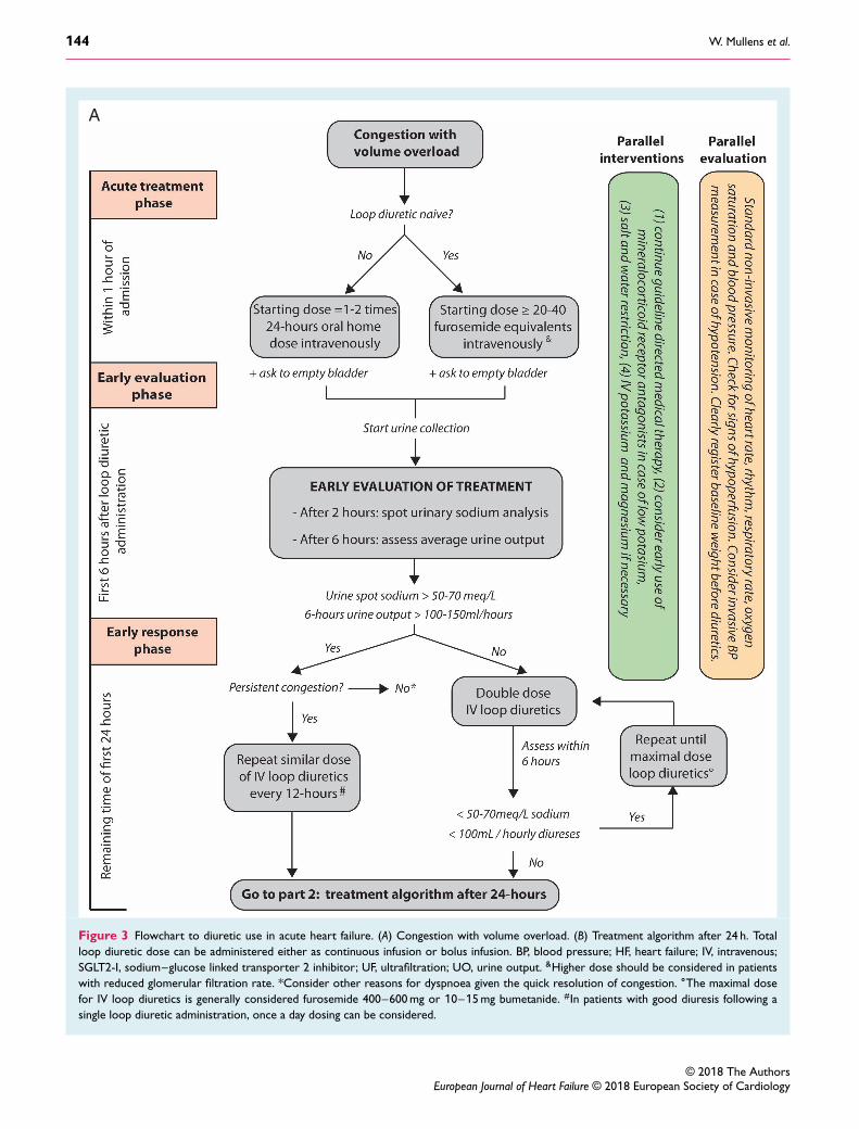
Practical use of diuretics in acuteheart failureGoals of therapy in acutedecompensated heart failureBefore initiating decongestive therapies in acutely decompensatedpatients, the distinction should be made if volume overload or vol-ume redistribution is contributing to congestion.79 The goals oftherapy in patients presenting with congestion and volume over-load consists of (i) achieving thorough decongestion without resid-ual volume overload. Nevertheless, the optimal stopping point ofdecongestive therapy is often difficult to determine, as alludedto above. (ii) Ensuring adequate perfusion pressures to guaranteeorgan perfusion. (iii) Maintaining guideline-directed medical ther-apies as these medications may also increase diuretic responseand improve long-term survival.80,81 When patients with heart fail-ure with reduced (HFrEF) or preserved ejection fraction (HFpEF)decompensate, they often can present with a similar profile ofcongestion.82,83 Therefore, the goal of decongestive therapy is sim-ilar in terms of diuretic use in patients with HFrEF and HFpEF.7 Apractical stepped approach to diuretic treatment and assessmentin acute heart failure is reflected in Figure 3. Once euvolaemiahas been achieved, loop diuretic therapy should be continuedat the lowest dose that can maintain euvolaemia.7,8 Additionally,enrolment of patients in a detailed multi-disciplinary heart fail-ure care management programme, promoting medication adher-ence, up-titration of disease-modifying therapy, cardiac rehabilita-tion, treatment of underlying co-morbidities, timely follow-up withthe health care team, and screening for additive device-based andmedical interventions therapies is essential.7
Loop diureticsLoop diuretics form the backbone of diuretic therapy in acuteheart failure, being used in over 90% of patients.3 Loop diuretics ..
....
....
....
....
....
....
....
....
....
....
....
....
....
....
....
....
....
....
....
....
....
....
....
....
....
....
....
....
....
....
....
....
....
....
....
....
....
....
....
....
....
.. are heavily protein-bound (> 90%) and need to be secreted intothe proximal convoluted tubule through several organic aniontransporters. Therefore, adequate dosing with sufficient plasmalevels is pivotal as renal perfusion is often reduced in heart failure,resulting in diminished secretion of loop diuretics. Additionally,decreased plasma protein content can result in reduced secretionof loop diuretics. Loop diuretics inhibit the Na-K-2Cl symporterat the ascending loop of Henle, and have the most potent diureticeffect, promoting excretion of sodium and chloride (and potassium,albeit to a lesser extent than thiazides).64 The pharmacologicalproperties of the different loop diuretics are presented in Table 2.The bioavailability of orally administered furosemide is highlyvariable (10–90%), and is determined by absorption from thegastrointestinal tract into the bloodstream.6 The oral bioavail-ability for torsemide and bumetanide are consistently higher than80–90%. In addition, torsemide has a longer half-life in heartfailure patients when compared to furosemide or bumetanide.84
Although some smaller studies suggested superior diuretic effectof torsemide, no large randomized studies have compared thedifference between different loop diuretics.85 The TorsemideComparison with Furosemide for Management of Heart Failure(TRANSFORM-HF) trial (NCT03296813) is planned to randomize6000 heart failure patients who are hospitalized. While heartfailure need not to be the reason for hospitalization, its objectiveis to detect a difference between furosemide vs. torsemide forthe primary endpoint all-cause mortality. Given the wide range ofbioavailability of oral furosemide, variance exists in the conversioncalculation. Therefore an oral dose of 40 mg of furosemide isgenerally equivalent to 10–20 mg of torsemide and 0.5–1 mg ofbumetanide. Importantly, loop diuretics may also lead to reninrelease by the macula densa by blocking chloride uptake, furtherstimulating RAAS. Furthermore, chronic use of loop diureticsinduces compensatory distal tubular sodium reabsorption throughhypertrophy of tubular cells, leading to reduced natriuresis.8
Guidelines recommend the use of intravenous loop diuretics inacute heart failure, as the uptake of oral diuretics (particularlyfurosemide) can be diminished in the face of congestion due tobowel oedema (class I, level of evidence B).7 Optimal dosing andtiming of intravenous loop diuretics are pertinent. Loop diureticsexhibit a threshold concentration to invoke natriuresis, necessi-tating a minimal drug dose prior to exceeding the baseline rate ofsodium excretion.6,86 Afterwards a log-linear increase in the doseis necessary to achieve a ceiling in natriuretic response. Furtherincreasing the loop diuretic dose beyond this ceiling will not resultin a greater rate of peak natriuresis, however it will lead to alonger period of loop diuretic over the threshold level and thusincreases total natriuresis. Similarly, multiple administrations cancause additional natriuresis, as it increases the duration of timeabove a natriuretic threshold. These pharmacologic characteristicslead to the following recommendation in acute heart failure: (i)diuretic naïve patients with acute heart failure should receive adose of intravenous furosemide of at least 20–40 mg furosemideequivalent. The higher dose should be considered in patients withpre-existing kidney dysfunction as it is associated with a rightwardshift in the dose–response curve.6,86 (ii) Patients on an ambulatorydiuretic regimen should receive at least the pre-existing oral dose
© 2018 The AuthorsEuropean Journal of Heart Failure © 2018 European Society of Cardiology
144 W. Mullens et al.
A
Figure 3 Flowchart to diuretic use in acute heart failure. (A) Congestion with volume overload. (B) Treatment algorithm after 24 h. Totalloop diuretic dose can be administered either as continuous infusion or bolus infusion. BP, blood pressure; HF, heart failure; IV, intravenous;SGLT2-I, sodium–glucose linked transporter 2 inhibitor; UF, ultrafiltration; UO, urine output. &Higher dose should be considered in patientswith reduced glomerular filtration rate. *Consider other reasons for dyspnoea given the quick resolution of congestion. ∘The maximal dosefor IV loop diuretics is generally considered furosemide 400–600 mg or 10–15 mg bumetanide. #In patients with good diuresis following asingle loop diuretic administration, once a day dosing can be considered.
© 2018 The AuthorsEuropean Journal of Heart Failure © 2018 European Society of Cardiology
Diuretics in heart failure 145
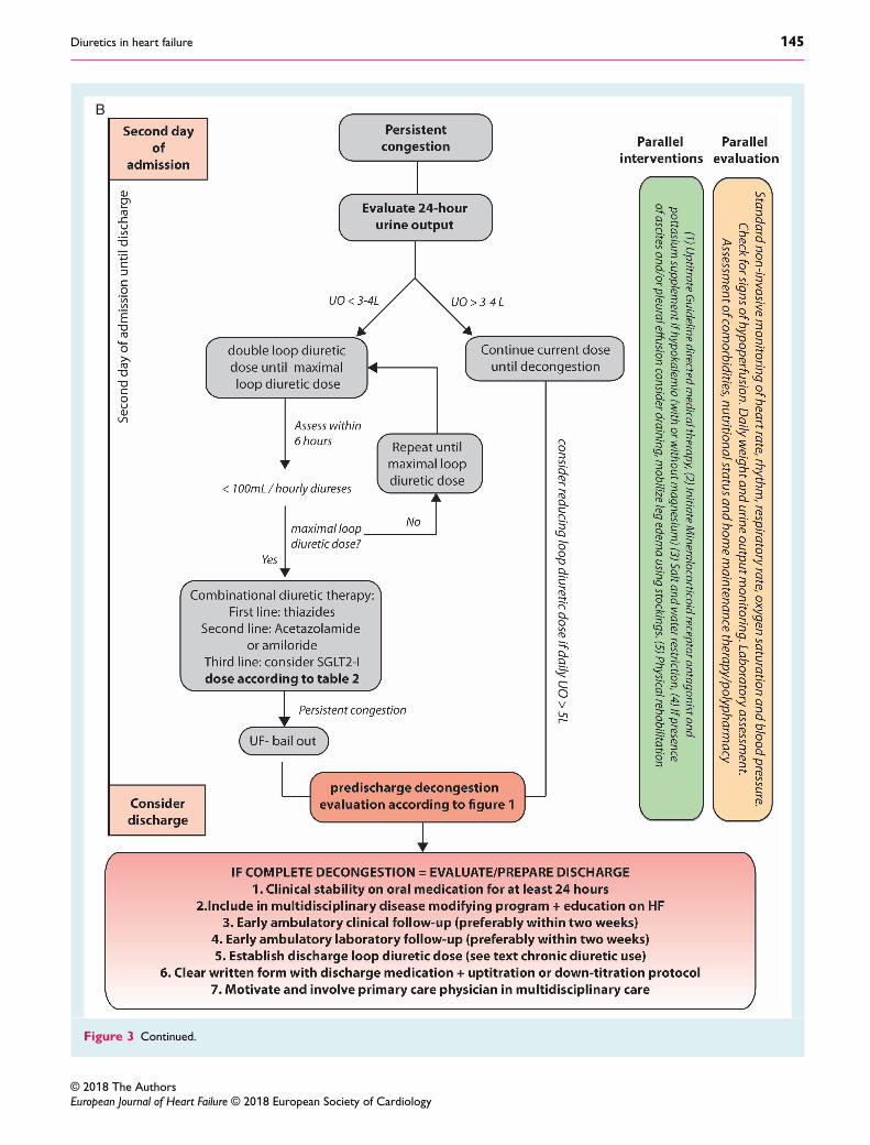
B
Figure 3 Continued.
© 2018 The AuthorsEuropean Journal of Heart Failure © 2018 European Society of Cardiology

146 W. Mullens et al.
administered intravenously. The DOSE-AHF trial demonstratedthat high loop diuretic dose (2.5 times the usual home dose, with atleast 80 mg/day furosemide equivalents) in comparison to low dose(equal to home dose) resulted in a favourable effect on secondaryendpoints of dyspnoea relief, change in weight and net fluid loss.42
Worsening of renal function (defined as an increase in creatinineby more than 0.3 mg/dL) occurred more in the high-dose group.However, a post-hoc analysis of the DOSE-AHF trial illustratedthat this increase in creatinine did not portend a worse outcome.87
In addition, the high-dose group was associated with better out-comes when adjusted for the total amount of loop diureticsreceived, suggesting that adequacy of loop diuretic dosing to reachthe ‘ceiling’ threshold is key.88 Determining the individual ceilingdose in a patient is difficult and is influenced by numerous factors,including previous treatment with loop diuretics, body composi-tion, degree of volume overload and kidney function. However,an intravenous dose ranging between 400–600 mg furosemide vs.10–15 mg bumetanide is generally considered as the maximal totaldaily dose above which limited additional natriuresis should beexpected but side effects will continue to increase. Generally, loopdiuretics are given in multiple doses (twice to three times daily).Intravenous loop diuretics should be administered as early aspossible, since early loop diuretic administration is associated withlower in-hospital mortality.89 In the DOSE-AHF trial, no differencewas seen in the primary endpoint between continuous or bolusinfusion. However, continuous infusion was not preceded by abolus loading dose which might have resulted in not reaching thethreshold dose in the continuous infusion group. If bolus infusion isgiven, doses should be split-up into doses with at least 6 h intervals,to maximize the time above the natriuretic threshold and to avoidrebound sodium retention.90 Continuous infusion should be pre-ceded by a loading dose, which assures the prompt achievementof a steady-state of plasma loop diuretic concentration.6
Stepped pharmacologic careEarly evaluation and loop diuretic intensification
The majority of the diuretic effect of intravenous loop diureticsoccurs within the first couple of hours with a return to base-line sodium excretion by 6–8 h. Early evaluation of the diureticresponse is therefore warranted and will allow for the identificationof patients with a poor diuretic response.67,69,73,74 This will permitearly intensification of loop diuretic dose and/or using a strategyof sequential nephron blockade (combining diuretics with a differ-ent mode of action). Although this concept has yet to be formallytested in prospective trials, such a strategy is important in severalaspects. Firstly, persisting congestion further compromises organfunction.91 Secondly, the plasma refill rate (the rate at which fluidis mobilized from the interstitium into the plasma compartment)might drop during decongestion.92,93 Thirdly, patients are oftenhospitalized in acute care units for the first days, where intensiveadaptation of therapy is more likely to occur than in a regular ward.Additionally, faster decongestion might be especially valuable inhealth care systems where length of hospital stay needs to be short.
In addition to the evaluation of vital signs, daily weights, andsigns/symptoms of congestion as endorsed by ESC guidelines (class ..
....
....
....
....
....
....
....
....
....
....
....
....
....
....
....
....
....
....
....
....
....
....
....
....
....
....
....
....
....
....
....
....
....
....
....
....
....
....
....
....
....
.. I recommendation, level of evidence C), this Cardio-Renal Dys-function Study Group proposes the active evaluation of diureticresponse early after start of therapy. Diuretic response may be eval-uated using urinary volume output and post-diuretic (spot) urinarysodium content as outlined in Figure 3. To allow for standardiza-tion and reliable results, patients presenting with congestion needto empty their bladder before the administration of diuretics. Thedegree of bladder emptying could potentially be checked using abladder scan. Afterwards, determination of urinary spot sodiumcontent allows the clinician to interpret diuretic response, therebygenerating the opportunity to intervene if sodium content is low. Inthe face of congestion with volume overload, a spot urine sodiumcontent of < 50–70 mEq/L after 2 h, and/or an hourly urine output< 100–150 mL during the first 6 h, generally identifies a patientwith an insufficient diuretic response.72,73,94 In patients who pro-duce sufficient urinary volumes following a first intravenous loopdiuretic administration, urinary sodium is almost universally high.However, more recent data indicate that in patients with a lowto medium volume output, spot urinary sodium content offersindependent prognostic information on heart failure admissions ontop of urinary volume output.71 Prompt doubling of loop diureticdose might allow the attainment of a loop diuretic ceiling dose ear-lier (as alluded to in the loop diuretic section). After these dosesare achieved, addition of another diuretic agent should be consid-ered, as increasing the loop diuretic dose any further does notinduces incremental diuresis/natriuresis. In the Cardiorenal Res-cue Study in Acute Decompensated Heart Failure (CARRESS-HF),a strategy of stepped pharmacologic therapy was compared withultrafiltration in acute decompensated heart failure patients withworsened renal function and persistent congestion (online supple-mentary Figure S1). The pharmacologic care approach using earlyassessment of urinary output with adjustment of loop diuretic dos-ing and the addition of a thiazide-like diuretic, resulted in equaldecongestion compared to ultrafiltration, however with fewer seri-ous adverse events.95 Post-hoc comparisons with the DOSE-AHFand ROSE-AHF trials indicates that a stepped pharmacologic careapproach was also associated with greater net fluid and weight loss,without compromising renal function.77,78 As urine sodium contentrarely changes discordantly to urine output during the first day ofdecongestive therapy (in the absence of excessive fluid intake bythe patient), it seems reasonable to assess urinary sodium con-tent always together with urine volume to adjust diuretic intensityduring the first day. Insufficient data are available to support theuse of urinary sodium during consecutive days of decongestion. Inthe CARRESS-HF trial, a urinary output of > 5 L per day allowedthe physicians to reduce diuretic intensity, however continuation ofthe diuretic regimen may be acceptable if renal function and bloodpressure remain stable.
Thiazide or thiazide-like co-administration
Thiazide and thiazide-like diuretics encompass a large classof agents that block the sodium–chloride co-transporter (NCC)in the distal convoluted tubule.96 Therefore, from a theoreti-cal point of view, they may partially overcome distal increasedsodium avidity accompanied with chronic loop diuretic use. Large
© 2018 The AuthorsEuropean Journal of Heart Failure © 2018 European Society of Cardiology
Diuretics in heart failure 147
geographical differences exist in the use of thiazide-like diureticswith metolazone being the most used thiazide-like diuretic in theUnited States.97 The different molecules have a similar blockingeffect of NCC, however they differ in terms of half-lives andoff-target effects (Table 2). In contrast with loop diuretics, meto-lazone and chlorthalidone have a slow gastrointestinal absorption(time to peak up to 8 h) and a very long half-life, therefore if loworal doses are started, they should be given hours before theintravenous loop diuretic is administered as it will take a longtime until a steady state is achieved. However, chlorothiazide hasa short half-life so it should be given closer to the loop diuretic.In healthy individuals, the maximal diuretic effect of a thiazideis limited, generating a diuretic response of maximum 30–40%of a loop diuretic when used in monotherapy.96 Thiazides arealso protein bound requiring adequate renal blood flow to besecreted into the tubules. Furthermore, thiazides can inducesignificant kaliuresis, as per sodium ion lost 2–3 ions of potas-sium are excreted.98 This potassium losing effect is especiallypronounced in high aldosterone states, such as heart failure.99
The rationale for using thiazides in acute heart failure is basedon the finding of increased distal nephron sodium avidity in thecase of (prolonged) loop diuretic administration.100 Indeed, animaldata indicate that distal nephron hypertrophy occurs followingchronic loop diuretic administration, which might explain loopdiuretic resistance to an extent.101 In contrast to conventionalteaching, more recent evidence does support the effectivenessof thiazides in patients with a reduced glomerular filtration rate(< 30 mL/min).102 There are no randomized controlled trialspublished in heart failure testing the use of thiazide diuretics.Currently, there is a study ongoing comparing Metolazone VersusChlorothiazide for Acute Decompensated Heart Failure WithDiuretic Resistance (NCT03574857). A meta-analysis of exist-ing observational data underscores the frequent occurrence ofhypokalaemia. In a propensity-matched analysis of real-worlduse of thiazides (combined with lower-dose loop diuretics) andhigh-dose loop diuretics in heart failure patients, thiazides, butnot high-dose loop diuretics, were independent predictors of theoccurrence of hyponatraemia and hypokalaemia with an indica-tion towards a higher risk for all-cause mortality.103 Given therelative safety of high-dose loop diuretics in the DOSE-AHF trial,a preference might be given to initial intensification of the loopdiuretic dose before adding a thiazide diuretic.42 However, in theCARRESS-HF-trial, the addition of metolazone was an intrinsicpart of the stepped pharmacologic algorithm, resulting in therecommendation of thiazides as a second-line agent in the HeartFailure Society of America practical guidelines.90
Mineralocorticoid receptor antagonists
Mineralocorticoid receptor antagonists (MRAs) exhibitpleiotropic effects, but their renal effects consist of modulat-ing the expression/activity of sodium and potassium channelsin the distal nephron. MRAs have a class I recommendation as adisease-modifying therapeutic agent in symptomatic chronic HFrEF,counteracting the aldosterone escape generated by neurohor-monal over-activation.104,105 Recently in acute heart failure, the ..
....
....
....
....
....
....
....
....
....
....
....
....
....
....
....
....
....
....
....
....
....
....
....
....
....
....
....
....
....
....
....
....
....
....
....
....
....
....
....
....
....
.. incremental diuretic effect of high-dose MRA therapy in additionto standard loop diuretic therapy has been tested in the Aldos-terone Targeted Neurohormonal Combined with NatriuresisTherapy in Heart Failure (ATHENA-HF) trial.106 Therapy with100 mg of spironolactone per day was not superior to 25 mg perday in reducing NT-proBNP or increase urine output after 96 h.However, as illustrated in Table 2, spironolactone is a pro-drugwith onset of action only 48–72 h after oral intake, which couldaccount for the observed nil-effect. However, high-dose MRAwas safe, as it did not result in hyperkalaemia or worsening ofrenal function. Furthermore, MRA therapy might be useful inoffsetting the hypokalaemic effect of potassium-wasting loopand thiazide diuretics.106–108 Importantly, data indicate markedunder-utilization of MRAs as a disease-modifying drug class inHFrEF.109 It is the opinion of the expert panel that early initiationof a MRA, in a regular dose (25 mg), might be useful in reducingtreatment-induced hypokalaemia and may lead to higher chance ofHFrEF patients being discharged on an optimized disease-modifyingtherapy regimen. However, the use of MRA in the acute settingsneeds to be individualized with temporarily discontinuation in caseof the development of hyperkalaemia.
Acetazolamide
Due to haemodynamic alterations in heart failure with a reductionin renal blood flow with a correspondingly increased filtrationfraction, important increases in proximal nephron sodium avidityoccur.9,63 From a pathophysiological point of view, targeting sodiumreabsorption in the proximal tubules has several potential benefitsin heart failure. First, most sodium is reabsorbed in the proxi-mal nephron, especially in decompensated heart failure. Second,greater delivery of chloride to the macula densa cells decreasesrenin production, reducing neurohumoral activation.9 Third,endogenous natriuretic peptides (acting in the distal nephron) willpossibly regain their effects.110 The carbonic anhydrase inhibitoracetazolamide inhibits sodium reabsorption in the proximaltubules. An observational study in patients with decompensatedheart failure and marked volume overload indicated that theaddition of acetazolamide (500 mg intravenous bolus on top ofloop diuretic) improved loop diuretic response with ∼100 mmolNa+ excreted per 40 mg of furosemide dose equivalents.69 Addi-tionally, acetazolamide efficiently boosts the diuretic responsein combination with loop diuretics, as illustrated by one smallrandomized trial including 24 patients with acute volume overloadrefractory to loop diuretic therapy.110 A multicentre, randomized,double-blind, phase IV clinical trial of the diuretic effects of Aceta-zolamide in Decompensated heart failure with Volume OveRload(ADVOR, NCT03505788) will investigate if combination therapywith acetazolamide improves loop diuretic response to increasediuresis in decompensated heart failure patients.111 Observationalstudies have only assessed the role of intravenous acetazolamide,and no data are available supporting the role of oral acetazolamide.
Other potential agents
In addition, the new diabetic drug class of sodium–glucose linkedtransporter-2 (SGLT2 inhibitors) also inhibit proximal sodium
© 2018 The AuthorsEuropean Journal of Heart Failure © 2018 European Society of Cardiology

148 W. Mullens et al.
absorption (Figure 2).9,112,113 Two trials in diabetic patients withmostly established cardiovascular disease, illustrated that SGLT2inhibitors reduced heart failure hospitalizations and resultedin a less steep slope of glomerular filtration rate decline overtime.114,115 However, the potential of SGLT2 inhibitors in heartfailure with or without diabetes remains unknown. Several trialsare ongoing in testing the disease-modifying effect of SGLT2inhibitors in the setting of both chronic and acute heart failure.Amiloride inhibits distal epithelial sodium channels (ENaC), andanecdotal evidence suggests that ENaC inhibition can result indecongestion with a lowering of filling pressures.116 Furthermore,chronic over-expression of ENaC has been implicated in thethiazolidinedione-mediated volume retention witnessed in dia-betics. Finally, vasopressin antagonists limit distal nephron freewater re-uptake by counteracting arginine vasopressin, whichresults in a limited availability of luminal aquaporin water channelsin the renal collecting ducts. This results in increased aquaresiswithout significantly impacting natriuretic response. The selectiveV2- receptor antagonist tolvaptan did not result in a reduction ofmorbidity or mortality in the Efficacy of Vasopressin Antagonismin Heart Failure Outcome Study With Tolvaptan (EVEREST)study in acute heart failure patients when added to standardtherapy.117 This limits its use in heart failure with congestion,as extracellular volume expansion is mainly driven by sodiumretention. However, in more advanced stages of heart failureinappropriately high levels of arginine vasopressin contribute toplasma expansion and dilutional hyponatraemia. More recently,early use of tolvaptan and use in patients with diuretic resistance,renal dysfunction or hyponatraemia, did result in more weight loss,but no significant improvement in dyspnoea relief.118,119 Currently,vasopressin antagonists are only indicated in patients with severehyponatraemia, and their widespread use might be limited by thehigh drug costs. In Europe, tolvaptan is available but not officiallyapproved for heart failure by the European Medicines Agency.
UltrafiltrationUltrafiltration removes plasma water across a semipermeablemembrane driven by a machine generated transmembrane pres-sure gradient. There is limited compelling evidence to supportultrafiltration as first-line therapy over loop diuretics in patientswith acute heart failure.95,120 Therefore, in most centres, ultra-filtration is reserved as a bail-out therapy to relieve congestionif stepped pharmacologic care fails.121 Of note, the PeripheralUltrafiltration for the RElief From Congestion in Heart Failure(PURE-HF) trial (NCT03161158) is evaluating whether tailored,peripheral veno–venous ultrafiltration (CHIARA System) comple-mentary to low-dose diuretics is associated with a reduction incardiovascular mortality and heart failure hospitalization in 90 daysafter randomization compared to usual care including steppedintravenous diuretics in acutely decompensated chronic heart fail-ure with fluid overload (not fully responsive to diuretic therapy).Renal replacement therapy allows for management of metaboliccomplications of anuria/oliguria such as hyperkalaemia, acidosis anduraemia,95,121 although in a large proportion of cases such usehas poor long-term prognosis, especially when systemic perfusion ..
....
....
....
....
....
....
....
....
....
....
....
....
....
....
....
....
....
....
....
....
....
....
....
....
....
....
....
....
....
....
....
....
....
....
....
....
....
....
....
....
....
.. pressures are low.122 Additionally, in the CARRESS-HF trial, theproportion of patients with catheter-related access site bleedingand infection was numerically higher in the ultrafiltration group.
Diuretic use and electrolyteabnormalitiesElectrolyte abnormalities resulting from neurohormonal activation,kidney dysfunction, or iatrogenic due to the employed diuretic regi-men occur frequently during episodes of acute heart failure, mostlyaffecting sodium and potassium handling.121,123,124 Recently, alsoalterations in chloride metabolism have been recognized to inde-pendently predict adverse outcomes.125 Hyponatraemia, defined asa plasma sodium concentration < 135 mEq/L, is the main abnormal-ity of sodium homeostasis occurring in acute heart failure whereashypernatraemia rarely occurs. A sub-analysis of the Organized Pro-gram to Initiate Lifesaving Treatment in Hospitalized Patients withHeart Failure (OPTIMIZE-HF) illustrated that 20% of patients hadhyponatraemia at the time of admission.126 The incidence of hospi-tal acquired hyponatraemia during decongestive therapy for acuteheart failure ranges between 15–25%.127 The pathophysiology ofhyponatraemia in heart failure is either due to the inability toexcrete free water (dilution hyponatraemia) or either due to adepletion of sodium (depletion hyponatraemia),123 or a combina-tion of these factors. A practical approach to hyponatraemia isreflected in Table 3. After confirmation of a low serum osmo-lality, the differentiation between dilution and depletion is madeon the basis of the clinical picture and urinary analysis. Abnor-malities in the potassium homeostasis are typically the result ofthe employed pharmacologic therapy in heart failure in combi-nation with pre-existing renal impairment. Hypokalaemia (plasmaK < 3.5 mEq/L) occurs typically in acute heart failure secondaryto diuretic-induced diuresis with potassium wasting.110 In clin-ical practice, loop diuretic use is the most common reasonfor hypokalaemia, however thiazide diuretics do exhibit an evenstronger kaliuretic effect.110 Treatment consists of adding upfrontMRA therapy during decongestion, increasing RAAS blockade andsupplementation of potassium (Table 3). In addition to potassiumwasting, diuretics often induce the loss of magnesium, poten-tially resulting in therapy-refractory hypokalaemia. Although notsupported by strong evidence, magnesium supplementation couldbe considered during diuretic treatment. Although less commonthan hypokalaemia during acute heart failure, hyperkalaemia (K> 5.0 mEq/L) can occur in patients on RAAS blockade, especiallyin the case of pre-existing renal impairment.128 A clinical approachto hyperkalaemia is reflected in Table 3.
Diuretics in chronic heart failureThe ambulatory loop diuretic dose isvariableLoop diuretics are recommended in chronic heart failure to pre-vent signs and symptoms of congestion.7 This recommendationis valid across the entire spectrum of left ventricular ejection
© 2018 The AuthorsEuropean Journal of Heart Failure © 2018 European Society of Cardiology

Diuretics in heart failure 149
Tabl
e3
App
roac
hto
elec
tro
lyte
dist
urba
nces
inac
ute
hear
tfa
ilure
Hyp
ona
trae
mia
Hyp
oka
laem
iaH
yper
kala
emia
....
....
....
....
....
....
....
....
....
....
....
....
....
....
....
....
....
....
....
....
....
....
....
....
....
....
....
....
....
....
....
....
....
....
....
....
....
....
....
....
....
....
....
....
....
....
.
Defi
nitio
nN
a+<
135
mEq
/LK+<
3.5
mEq
/LK+>
5m
Eq/L
Dia
gnos
ticte
sts
•P
osm
:sho
uld
be<
285
mO
sm/L
(els
eps
eudo
-hyp
onat
raem
ia)
•P
hysi
cale
xam
inat
ion:
todi
ffere
ntia
tebe
twee
nvo
lum
eov
erlo
ador
volu
me
depl
etio
n•
Uri
nary
anal
ysis
:Uos
man
dU
Na
•A
BG
:con
firm
onA
BG,c
heck
pHst
atus
•E
CG
:che
ckpo
tent
iala
bnor
mal
ities
•P
hysi
cale
xam
inat
ion:
usua
llyno
rmal
,ho
wev
erm
uscl
ew
eakn
ess
orpa
raly
sis
pres
ent
inse
vere
case
s•
Lab
:che
ckfo
rM
gde
ficit
•A
BG
:con
firm
onA
BG,c
heck
pHst
atus
•E
CG
:che
ckpo
tent
iala
bnor
mal
ities
•L
ab:c
heck
rena
lfun
ctio
n,ex
clud
eha
emol
ysis
asca
use
ofps
eudo
-hyp
erka
laem
ia
Path
ophy
siol
ogy
•D
iluti
on:
impa
ired
free
wat
erex
cret
ion.
Clin
ical
pict
ure
ofvo
lum
eov
erlo
adw
ithin
appr
opri
ate
high
Uos
m
(≥100
mO
sm/L
).Ty
pica
lin
the
sett
ing
ofA
DH
F•
Dep
leti
on:
true
body
defic
itof
Na+
.Typ
ical
inth
ese
ttin
gof
chro
nic
exce
ssiv
edi
uret
icus
e(a
ndst
rict
Na+
inta
ke).
Clin
ical
pict
ure
ofvo
lum
ede
plet
ion
with
low
Uos
m
(<100
mO
sm/L
)an
dU
Na
(<50
mEq
/L)
•D
iure
tic
use
resu
ltsin
hypo
kala
emia
•P
redi
spo
sing
fact
ors
inH
Fca
npl
aya
role
,for
inst
ance
:cac
hexi
aw
ithlo
wK+
inta
kean
dch
roni
chy
pom
agne
saem
ia
Mos
tlik
ely
due
toco
mbi
natio
nof
RA
AS
bloc
ker
agen
tan
dpo
orre
nalf
unct
ion
with
dim
inis
hed
rena
lpot
assi
umex
cret
ion
capa
city
Trea
tmen
t•
Dilu
tio
n:te
mpo
rari
lyst
opdi
stal
actin
gdi
uret
icsa ,
limit
wat
erin
take
,pro
mot
edi
stal
neph
ron
flow
(loop
diur
etic
s,hy
pert
onic
salin
e,ac
etaz
olam
ide/
SGLT
2in
hibi
tor)
orva
ptan
s,co
rrec
tion
ofK+
and
Mg2+
defic
ienc
ies
•D
eple
tio
n:st
opdi
stal
actin
gdi
uret
icsa ,
calc
ulat
eN
ade
ficit
and
adm
inis
ter
IVN
a+co
rrec
tion
ofK+
and
Mg2+
defic
ienc
ies
•C
ons
ider
disc
ontin
uatio
nof
thia
zide
diur
etic
s•
Upf
ront
use
ofM
RA
duri
ngde
cong
estio
n•
Incr
ease
dose
ofR
AA
Sbl
ocki
ngag
ent
•IV
subs
titut
ion
ofK+
and
Mg2+
:per
iphe
ralo
rce
ntra
ldep
endi
ngon
seve
rity
ofK+
defic
it.
•A
cute
hype
rkal
aem
ia:i
fEC
Gab
norm
aliti
espr
esen
t,th
enpr
even
tar
rhyt
hmia
sby
IVca
lciu
m.I
nter
med
iate
stra
tegi
esin
clud
e:in
sulin
/alb
uter
ol/s
odiu
mH
CO
3IV
.U
ltim
atel
ypo
tass
ium
mus
tbe
rem
oved
from
the
body
with
eith
erdi
uret
ics,
pota
ssiu
mbi
ndin
gre
sins
,or
RRT
.•
Chr
oni
chy
perk
alae
mia
:red
uces
dose
RA
AS
bloc
ker,
incr
ease
loop
diur
etic
,pot
assi
umbi
nder
s
ABG
,art
eria
lblo
odga
san
alys
is;A
DH
F,ac
ute
deco
mpe
nsat
edhe
artf
ailu
re;A
VP,
argi
nine
vaso
pres
sin;
ECG
,ele
ctro
card
iogr
am;H
CO
3,b
icar
bona
te;H
F,he
artf
ailu
re;I
V,in
trav
enou
s;M
RA
,min
eral
ocor
ticoi
dre
cept
oran
tago
nist
;Pos
m,
plas
ma
osm
olal
ity;R
AA
S,re
nin
–an
giot
ensi
n–
aldo
ster
one
syst
em;R
RT,r
enal
repl
acem
ent
ther
apy;
SGLT
2,so
dium
–gl
ucos
elin
ked
tran
spor
ter-
2;U
osm
,uri
neos
mol
ality
;UN
a,ur
ine
sodi
um.
a Dis
tala
ctin
gdi
uret
ics
incl
uded
thia
zide
-like
diur
etic
s,M
RA
and
amilo
ride
.
© 2018 The AuthorsEuropean Journal of Heart Failure © 2018 European Society of Cardiology

150 W. Mullens et al.
fraction. Indeed, diuretics are the only group of drugs with a classI recommendation in patients with heart failure with reduced,mid-range, or preserved ejection fraction.7 However, the effectsof diuretics in chronic heart failure on morbidity and mortalityhave not been studied in large prospective randomized controlledtrials. Several observational studies have linked loop diuretic useto increased mortality, even after multivariate adjustment orpropensity matching.129 However, potential bias remains as sickerpatients are generally prescribed (higher doses of) loop diuretics.A Cochrane meta-analysis has shown that in patients with chronicheart failure, loop diuretics and thiazides might reduce the riskof death and worsening of heart failure in comparison to placeboand could lead to improved exercise capacity.86 However, thismeta-analysis included only small studies with limited follow-up,showing unrealistically large reductions in events. Moreover, thisanalysis was not updated in 2016 as requested by the CochraneInstitute and subsequently withdrawn. Therefore, the prognosticeffect of diuretic therapy is still unknown. Clearly, patients at riskfor congestion would benefit from maintenance therapy with aloop diuretic. However, in patients at low risk for developing wors-ening of congestion, the use of loop diuretics might indeed result inelectrolyte disturbances, further neurohormonal activation, accel-erated kidney function decline, and symptomatic hypotension.130
The latter might especially be relevant in patients with HFrEF asit could result in treatment with lower doses of neurohormonalblockers.43 Therefore, it is generally advised to use to the lowestpossible dose of diuretics and the dose of the loop diuretic oftenneeds to be adjusted to the individual need.131,132 Importantly, theindividual diuretic need significantly changes over time. This wasclearly illustrated by a post-hoc analysis of the CardioMEMS HeartSensor Allows Monitoring of Pressure to Improve Outcomes inClass III Heart Failure (CHAMPION) trial, which indicated thatmainly increases but also decreases in loop diuretic dose werethe most common therapy changes made by treating physicians.133
Nevertheless, uncertainty exists about the optimal dose of loopdiuretics following discharge. For patients who developed an acuteheart failure episode while previously taking a loop diuretic beforeadmission, a higher dose following discharge might need to beused. Additionally, in case that this previous loop diuretic wasfurosemide, a switch to either bumetanide or torsemide might beconsidered, as they have a more predictable absorption patternand bioavailability, especially in the face of subclinical congestion.However, defining the most appropriate outpatient dose of diureticcan be difficult and requires careful follow-up, particularly earlyin the post-discharge period. The chronic use of thiazides in thestable ambulatory setting (sequential nephron blocking) should beavoided, if possible, as this practice often induces severe electrolytedisturbances that could go undetected in the ambulatory setting.Additional research is needed to evaluate ambulatory metrics (inaddition to pulmonary pressures) of volume status, which mightallow easier adaptation of loop diuretic therapy. Registry dataindicate that mildly symptomatic heart failure patients [New YorkHeart Association (NYHA) class I and II] are generally treatedwith similar doses of loop diuretics as more symptomatic heartfailure patients (NYHA class III and IV).134 This underscores theimportance to re-assess loop diuretic need following the initiation ..
....
....
....
....
....
....
....
....
....
....
....
....
....
....
....
....
....
....
....
....
....
....
....
....
....
....
....
....
....
....
....
....
....
....
....
....
....
....
....
....
....
.. of therapies that improve cardiac status (such as cardiac resyn-chronization therapy or sacubitril/valsartan).112,135 A recent pilotstudy illustrated the potential of self-measuring urine chloridecontent after loop diuretic intake using a dipstick to determine theneed for maintenance loop diuretics in stable ambulatory heartfailure patients.136 Despite the guideline recommendation to usethe lowest possible dose of diuretics and discontinue loop diuret-ics if possible, little information is available on discontinuing loopdiuretics in contemporary treated heart failure patients.137,138
A prospective interventional study in 50 stable ambulatoryheart failure patients assessed the feasibility of loop diureticdown-titration and discontinuation.138 At 30 days, down-titrationremained successful in 62% of patients, however baseline investi-gations including physical examination, echocardiography and NPmeasurement were not capable to predict in which patients loopdiuretic down-titration would be successful or not.138
Heart failure disease managementstrategyThe goals of heart failure care are dynamic and vary accordingto the stage of heart failure. In the ambulatory patient, care shouldfocus on up-titrating disease-modifying drugs, evaluating the needfor device-based therapies, enrolling patients in multidisciplinarydisease-modifying programmes, focusing on self-management,physical activity, and dietary interventions.7 Furthermore, effortsshould be made to reduce readmission and improve quality andlongevity of life. With average salt intake in the western worldreaching up to 6–8 g, it has been recommended by the ESC guide-lines to avoid excessive high salt intake (>6 g NaCl= 2.4 g Na perday) and excessive fluid intake (no class recommendation).7 Saltand fluid restriction are often underscored in disease-modifyingprogrammes. Yet, animal and epidemiologic data suggest that anexcessively low sodium intake (<2 g Na+ per day) is associatedwith cardiac remodelling and worse clinical outcome.139 Currentlyfour trials are evaluating the benefit of sodium restriction, includingone trial assessing a hard clinical endpoint.140 A meta-analysis onfluid restriction did not indicate benefit or harm when performedin heart failure patients.141 Therefore, dietary restrictions shouldbe adapted according to the clinical context. In the case of acuteheart failure with dilution hyponatraemia, more stringent fluidrestriction is necessary.
Gaps in knowledge and futuredirectionsEvidence-based medicine with respect to diuretic treatmentin heart failure remains difficult as only a limited number of smallprospective studies have been performed. Ongoing research isnecessary to determine the ideal diuretic strategy and to optimallyevaluate full decongestion (euvolaemia) in heart failure. The roleof urinary sodium to assess the adequacy of diuretic therapyin acute heart failure should be further assessed prospectively. Therole of hypertonic NaCl infusion in conjunction with high-doseloop diuretics in hyponatraemic volume overloaded patients needs
© 2018 The AuthorsEuropean Journal of Heart Failure © 2018 European Society of Cardiology

Diuretics in heart failure 151
to be studied as this concept is supported by several analyses,however suffering from methodologic restrains.142 Random-ized controlled trials are necessary assessing the decongestiveproperties of diuretics other than loop diuretics or MRAs. Noveleffective and safe pharmacologic or mechanical methods to achievedecongestion without inducing end-organ damage are needed. Fur-thermore, several upcoming studies will investigate the optimal useof current diuretic treatment options. The TRANSFORM-HF willassess the superiority of torsemide in comparison to furosemidein reducing all-cause mortality. Furthermore, ongoing studies aretesting the effect of subcutaneous furosemide in comparison tooral furosemide.143
Supplementary InformationAdditional supporting information may be found online in theSupporting Information section at the end of the article.Figure S1. Summary overview of the CARRESS-HF trial.
Conflict of interest: W.M. has received research grants fromNovartis, Vifor, Medtronic, Biotronik, Abbott and Boston Scien-tific. V.P.H. has received research grants from Abbott Laborato-ries, and personal fees from Bayer, Boehringer-Ingelheim, MSD,Orion Pharma, Pfizer, Roche Diagnostics, Thermo Fisher and Vifor.A.M. received speaker’s honoraria from Abbott, Orion, Roche andServier and fee as member of advisory board and/or SteeringCommittee and/or research grant from BMS, Adrenomed, Neu-rotronik, Roche, Sanofi and Sphyngotec. H.P.B.L.R. has receivedresearch grants from Roche Diagnostics, Novartis, Servier andVifor pharma, and advisory board fees from Novartis, Roche Diag-nostics, Vifor Pharma and Servier. P.M. has received a researchgrant from Vifor pharma and Fonds Wetenschappelijk Onder-zoek (grant number: 1127917N) and consultancy fees fromAstraZeneca, Boehringer-Ingelheim, Novartis and Vifor pharma.J.M.T. has received research grants from the NIH, FDA, BoehringerIngelheim, Sanofi, Abbott, FIRE1, Sequana Medical, Otsuka andconsulting fees from Sanofi, Boehringer Ingelheim, Novartis, BMS,AstraZeneca, FIRE1, Sequana Medical, Cardionomic, and Renal-Guard. W.H.W.T. has been supported by grants from the NIH(R01HL103931) and has served as a consultant for the Advi-sory Board Company, MyoKardia Inc, and Sequana Medical Inc.P.R. reports personal fees (consulting) from Fresenius, Grünen-thal, Idorsia, Novartis, Novo-Nordisk, Relypsa, Stealth Peptides,Vifor Fresenius Medical Care Renal Pharma, Vifor, as well as lec-ture fees from Bayer and CVRx; he is a cofounder of CardioRenal.M.M. has received consulting honoraria from Bayer, Novartis, andServier, and speaker’s fees from Abbott Vascular and Novartis. G.F.has served on the committees of trials supported by Bayer, Novar-tis, Servier, Vifor, BI and Medtronic. P.S. reports grants/researchsupports from Ministry of education, science and technologicaldevelopment of Republic of Serbia; and honoraria or consultationfees from Servier, Boehringher Ingelheim, Hemofarm, Novartis,AstraZeneca; he participated in a company sponsored speaker’sbureau: Fondazione Internationale Menarini. F.R. reports grants andpersonal fees from SJM/Abbott, Novartis, Bayer, Servier; personal ..
....
....
....
....
....
....
....
....
....
....
....
....
....
....
....
....
....
....
....
....
....
....
....
....
....
....
....
....
....
....
....
....
....
....
....
....
....
....
....
....
....
.. fees from Zoll, AstraZeneca, Sanofi, Amgen, BMS, Pfizer, Frese-nius, Vifor, Roche, Cardiorentis, Boehringer Ingelheim, other fromHeartware, grants from Mars, during the conduct of the study;since 1st January 2018: no personal payments/all payments directlyto the University of Zurich. A.J.C. reports personal fees fromRespicardia, Vifor and Servier. The other authors have no conflictsof interest to declare.
References1. Filippatos G, Zannad F. An introduction to acute heart failure syndromes:
definition and classification. Heart Fail Rev 2007;12:87–90.2. Mosterd A, Hoes AW. Clinical epidemiology of heart failure. Heart
2007;93:1137–1146.3. Chioncel O, Mebazaa A, Harjola VP, Coats AJ, Piepoli MF, Crespo-Leiro MG,
Laroche C, Seferovic PM, Anker SD, Ferrari R, Ruschitzka F, Lopez-Fernandez S,Miani D, Filippatos G, Maggioni AP; ESC Heart Failure Long-Term RegistryInvestigators. Clinical phenotypes and outcome of patients hospitalized foracute heart failure: the ESC Heart Failure Long-Term Registry. Eur J Heart Fail2017;19:1242–1254.
4. Gheorghiade M, Pang PS. Acute heart failure syndromes. J Am Coll Cardiol2009;53:557–573.
5. Nunez J, Nunez E, Fonarow GC, Sanchis J, Bodi V, Bertomeu-Gonzalez V,Minana G, Merlos P, Bertomeu-Martinez V, Redon J, Chorro FJ, Llacer A.Differential prognostic effect of systolic blood pressure on mortality accordingto left-ventricular function in patients with acute heart failure. Eur J Heart Fail2010;12:38–44.
6. Ellison DH, Felker GM. Diuretic treatment in heart failure. N Engl J Med2017;377:1964–1975.
7. Ponikowski P, Voors AA, Anker SD, Bueno H, Cleland JG, Coats AJ, Falk V,Gonzalez-Juanatey JR, Harjola VP, Jankowska EA, Jessup M, Linde C, Nihoy-annopoulos P, Parissis JT, Pieske B, Riley JP, Rosano GM, Ruilope LM, Rus-chitzka F, Rutten FH, van der Meer P. 2016 ESC Guidelines for the diagnosisand treatment of acute and chronic heart failure: the Task Force for the diagno-sis and treatment of acute and chronic heart failure of the European Society ofCardiology (ESC). Developed with the special contribution of the Heart FailureAssociation (HFA) of the ESC. Eur J Heart Fail 2016;18:891–975.
8. Martens P, Nijst P, Mullens W. Current approach to decongestive therapy inacute heart failure. Curr Heart Fail Rep 2015;12:367–378.
9. Mullens W, Verbrugge FH, Nijst P, Tang WH. Renal sodium avidity inheart failure: from pathophysiology to treatment strategies. Eur Heart J2017;38:1872–1882.
10. Nijst P, Verbrugge FH, Grieten L, Dupont M, Steels P, Tang WH, Mullens W.The pathophysiological role of interstitial sodium in heart failure. J Am Coll Cardiol2015;65:378–388.
11. Verbrugge FH, Dupont M, Steels P, Grieten L, Malbrain M, Tang WH, Mullens W.Abdominal contributions to cardiorenal dysfunction in congestive heart failure.J Am Coll Cardiol 2013;62:485–495.
12. Miller WL. Fluid volume overload and congestion in heart failure: timeto reconsider pathophysiology and how volume is assessed. Circ Heart Fail2016;9:e002922.
13. Fudim M, Jones WS, Boortz-Marx RL, Ganesh A, Green CL, HernandezAF, Patel MR. Splanchnic nerve block for acute heart failure. Circulation2018;138:951–953.
14. Damman K, van Deursen VM, Navis G, Voors AA, van Veldhuisen DJ, HillegeHL. Increased central venous pressure is associated with impaired renal functionand mortality in a broad spectrum of patients with cardiovascular disease. J AmColl Cardiol 2009;53:582–588.
15. Mullens W, Abrahams Z, Francis GS, Sokos G, Taylor DO, Starling RC, YoungJB, Tang WH. Importance of venous congestion for worsening of renal functionin advanced decompensated heart failure. J Am Coll Cardiol 2009;53:589–596.
16. Dovancescu S, Pellicori P, Mabote T, Torabi A, Clark AL, Cleland JG. The effectsof short-term omission of daily medication on the pathophysiology of heartfailure. Eur J Heart Fail 2017;19:643–649.
17. Chaudhry SI, Wang Y, Concato J, Gill TM, Krumholz HM. Patternsof weight change preceding hospitalization for heart failure. Circulation2007;116:1549–1554.
18. Clark AL, Coats AJ, Krum H, Katus HA, Mohacsi P, Salekin D, Schultz MK,Packer M, Anker SD. Effect of beta-adrenergic blockade with carvedilol oncachexia in severe chronic heart failure: results from the COPERNICUS trial.J Cachexia Sarcopenia Muscle 2017;8:549–556.
© 2018 The AuthorsEuropean Journal of Heart Failure © 2018 European Society of Cardiology

152 W. Mullens et al.
19. Loncar G, Springer J, Anker M, Doehner W, Lainscak M. Cardiac cachexia: hicet nunc: "hic et nunc" - here and now. Int J Cardiol 2015;201:e1–e12.
20. Metra M, Davison B, Bettari L, Sun H, Edwards C, Lazzarini V, Piovanelli B,Carubelli V, Bugatti S, Lombardi C, Cotter G, Dei Cas L. Is worsening renalfunction an ominous prognostic sign in patients with acute heart failure?The role of congestion and its interaction with renal function. Circ Heart Fail2012;5:54–62.
21. Ambrosy AP, Cerbin LP, Armstrong PW, Butler J, Coles A, DeVore AD, DunlapME, Ezekowitz JA, Felker GM, Fudim M, Greene SJ, Hernandez AF, O’ConnorCM, Schulte P, Starling RC, Teerlink JR, Voors AA, Mentz RJ. Body weight changeduring and after hospitalization for acute heart failure: patient characteristics,markers of congestion, and outcomes: findings from the ASCEND-HF trial. JACCHeart Fail 2017;5:1–13.
22. Gheorghiade M, Follath F, Ponikowski P, Barsuk JH, Blair JE, Cleland JG,Dickstein K, Drazner MH, Fonarow GC, Jaarsma T, Jondeau G, Sendon JL,Mebazaa A, Metra M, Nieminen M, Pang PS, Seferovic P, Stevenson LW, vanVeldhuisen DJ, Zannad F, Anker SD, Rhodes A, McMurray JJ, Filippatos G.Assessing and grading congestion in acute heart failure: a scientific statementfrom the Acute Heart Failure Committee of the Heart Failure Association ofthe European Society of Cardiology and endorsed by the European Society ofIntensive Care Medicine. Eur J Heart Fail 2010;12:423–433.
23. Binanay C, Califf RM, Hasselblad V, O’Connor CM, Shah MR, Sopko G,Stevenson LW, Francis GS, Leier CV, Miller LW; ESCAPE Investigators andESCAPE Study Coordinators. Evaluation study of congestive heart failureand pulmonary artery catheterization effectiveness: the ESCAPE trial. JAMA2005;294:1625–1633.
24. Nagueh SF. Non-invasive assessment of left ventricular filling pressure. EurJ Heart Fail 2018;20:38–48.
25. Mullens W, Borowski AG, Curtin RJ, Thomas JD, Tang WH. Tissue Dopplerimaging in the estimation of intracardiac filling pressure in decompensatedpatients with advanced systolic heart failure. Circulation 2009;119:62–70.
26. Parrinello G, Greene SJ, Torres D, Alderman M, Bonventre JV, Di Pasquale P,Gargani L, Nohria A, Fonarow GC, Vaduganathan M, Butler J, Paterna S,Stevenson LW, Gheorghiade M. Water and sodium in heart failure: a spotlighton congestion. Heart Fail Rev 2015;20:13–24.
27. Volpicelli G, Skurzak S, Boero E, Carpinteri G, Tengattini M, Stefanone V,Luberto L, Anile A, Cerutti E, Radeschi G, Frascisco MF. Lung ultrasoundpredicts well extravascular lung water but is of limited usefulness in theprediction of wedge pressure. Anesthesiology 2014;121:320–327.
28. Nagueh SF, Smiseth OA, Appleton CP, Byrd BF 3rd, Dokainish H, Edvardsen T,Flachskampf FA, Gillebert TC, Klein AL, Lancellotti P, Marino P, Oh JK, PopescuBA, Waggoner AD. Recommendations for the evaluation of left ventriculardiastolic function by echocardiography: an update from the American Societyof Echocardiography and the European Association of Cardiovascular Imaging.J Am Soc Echocardiogr 2016;29:277–314.
29. Caldentey G, Khairy P, Roy D, Leduc H, Talajic M, Racine N, White M,O’Meara E, Guertin MC, Rouleau JL, Ducharme A. Prognostic value of thephysical examination in patients with heart failure and atrial fibrillation: insightsfrom the AF-CHF trial (Atrial Fibrillation and Chronic Heart Failure). JACC HeartFail 2014;2:15–23.
30. Chakko S, Woska D, Martinez H, de Marchena E, Futterman L, Kessler KM,Myerberg RJ. Clinical, radiographic, and hemodynamic correlations in chroniccongestive heart failure: conflicting results may lead to inappropriate care. AmJ Med 1991;90:353–359.
31. McGee SR. Physical examination of venous pressure: a critical review. Am Heart J1998;136:10–18.
32. Stevenson LW, Perloff JK. The limited reliability of physical signs for estimatinghemodynamics in chronic heart failure. JAMA 1989;261:884–888.
33. Thibodeau JT, Drazner MH. The role of the clinical examination in patients withheart failure. JACC Heart Fail 2018;6:543–551.
34. Collins SP, Lindsell CJ, Storrow AB, Abraham WT. Prevalence of negative chestradiography results in the emergency department patient with decompensatedheart failure. Ann Emerg Med 2006;47:13–18.
35. Platz E, Merz AA, Jhund PS, Vazir A, Campbell R, McMurray JJ. Dynamic changesand prognostic value of pulmonary congestion by lung ultrasound in acute andchronic heart failure: a systematic review. Eur J Heart Fail 2017;19:1154–1163.
36. Al Deeb M, Barbic S, Featherstone R, Dankoff J, Barbic D. Point-of-careultrasonography for the diagnosis of acute cardiogenic pulmonary edema inpatients presenting with acute dyspnea: a systematic review and meta-analysis.Acad Emerg Med 2014;21:843–852.
37. Maeder MT, Mueller C, Pfisterer ME, Buser PT, Brunner-la Rocca HP. Use ofB-type natriuretic peptide outside of the emergency department. Int J Cardiol2008;127:5–16. ..
....
....
....
....
....
....
....
....
....
....
....
....
....
....
....
....
....
....
....
....
....
....
....
....
....
....
....
....
....
....
....
....
....
....
....
....
....
....
....
....
....
.. 38. Philipson H, Ekman I, Forslund HB, Swedberg K, Schaufelberger M. Salt andfluid restriction is effective in patients with chronic heart failure. Eur J Heart Fail2013;15:1304–1310.
39. Gheorghiade M, De Luca L, Fonarow GC, Filippatos G, Metra M, Francis GS.Pathophysiologic targets in the early phase of acute heart failure syndromes. AmJ Cardiol 2005;96:11G–17G.
40. O’Connor CM, Stough WG, Gallup DS, Hasselblad V, Gheorghiade M. Demo-graphics, clinical characteristics, and outcomes of patients hospitalized fordecompensated heart failure: observations from the IMPACT-HF registry. J CardFail 2005;11:200–205.
41. Vaduganathan M, Greene SJ, Fonarow GC, Voors AA, Butler J, Gheo-rghiade M. Hemoconcentration-guided diuresis in heart failure. Am J Med2014;127:1154–1159.
42. Felker GM, Lee KL, Bull DA, Redfield MM, Stevenson LW, Goldsmith SR,LeWinter MM, Deswal A, Rouleau JL, Ofili EO, Anstrom KJ, Hernandez AF,McNulty SE, Velazquez EJ, Kfoury AG, Chen HH, Givertz MM, Semigran MJ, BartBA, Mascette AM, Braunwald E, O’Connor CM; NHLBI Heart Failure ClinicalResearch Network. Diuretic strategies in patients with acute decompensatedheart failure. N Engl J Med 2011;364:797–805.
43. Maggioni AP, Dahlstrom U, Filippatos G, Chioncel O, Crespo LM, Drozdz J,Fruhwald F, Gullestad L, Logeart D, Fabbri G, Urso R, Metra M, Parissis J,Persson H, Ponikowski P, Rauchhaus M, Voors AA, Nielsen OW, Zannad F,Tavazzi L; Heart Failure Association of the European Society of Cardiology(HFA). EURObservational Research Programme: regional differences and 1-yearfollow-up results of the Heart Failure Pilot Survey (ESC-HF Pilot). Eur J HeartFail 2013;15:808–817.
44. Rubio-Gracia J, Demissei BG, Ter Maaten JM, Cleland JG, O’Connor CM,Metra M, Ponikowski P, Teerlink JR, Cotter G, Davison BA, Givertz MM,Bloomfield DM, Dittrich H, Damman K, Perez-Calvo JI, Voors AA. Prevalence,predictors and clinical outcome of residual congestion in acute decompensatedheart failure. Int J Cardiol 2018;258:185–191.
45. Ambrosy AP, Pang PS, Khan S, Konstam MA, Fonarow GC, Traver B, MaggioniAP, Cook T, Swedberg K, Burnett JC Jr, Grinfeld L, Udelson JE, Zannad F,Gheorghiade M; EVEREST Trial Investigators. Clinical course and predictivevalue of congestion during hospitalization in patients admitted for worseningsigns and symptoms of heart failure with reduced ejection fraction: findings fromthe EVEREST trial. Eur Heart J 2013;34:835–843.
46. Lala A, McNulty SE, Mentz RJ, Dunlay SM, Vader JM, AbouEzzeddine OF,DeVore AD, Khazanie P, Redfield MM, Goldsmith SR, Bart BA, AnstromKJ, Felker GM, Hernandez AF, Stevenson LW. Relief and recurrence of con-gestion during and after hospitalization for acute heart failure: insights fromDiuretic Optimization Strategy Evaluation in Acute Decompensated Heart Fail-ure (DOSE-AHF) and Cardiorenal Rescue Study in Acute DecompensatedHeart Failure (CARESS-HF). Circ Heart Fail 2015;8:741–748.
47. Testani JM, Brisco MA, Kociol RD, Jacoby D, Bellumkonda L, Parikh CR, CocaSG, Tang WH. Substantial discrepancy between fluid and weight loss duringacute decompensated heart failure treatment. Am J Med 2015;128:776–783.
48. Drazner MH, Rame JE, Stevenson LW, Dries DL. Prognostic importance ofelevated jugular venous pressure and a third heart sound in patients with heartfailure. N Engl J Med 2001;345:574–581.
49. Letsas KP, Filippatos GS, Pappas LK, Mihas CC, Markou V, Alexanian IP,Efremidis M, Sideris A, Maisel AS, Kardaras F. Determinants of plasmaNT-pro-BNP levels in patients with atrial fibrillation and preserved left ven-tricular ejection fraction. Clin Res Cardiol 2009;98:101–106.
50. Stienen S, Salah K, Moons AH, Bakx AL, van Pol P, Kortz M, Ferreira JP,Marques I, Schroeder-Tanka JM, Keijer JT, Bayes-Genis A, Tijssen JG, PintoYM, Kok WE. NT-proBNP (N-terminal pro-B-type natriuretic peptide)-guidedtherapy in acute decompensated heart failure: PRIMA II randomized controlledtrial (Can NT-ProBNP-Guided Therapy During Hospital Admission for AcuteDecompensated Heart Failure Reduce Mortality and Readmissions?). Circulation2018;137:1671–1683.
51. Desai AS. Are serial BNP measurements useful in heart failure management?Serial natriuretic peptide measurements are not useful in heart failure manage-ment: the art of medicine remains long. Circulation 2013;127:509–516.
52. Nunez J, Llacer P, Bertomeu-Gonzalez V, Bosch MJ, Merlos P, Garcia-Blas S,Montagud V, Bodi V, Bertomeu-Martinez V, Pedrosa V, Mendizabal A,Cordero A, Gallego J, Palau P, Minana G, Santas E, Morell S, Llacer A,Chorro FJ, Sanchis J, Facila L; CHANCE-HF Investigators. Carbohydrateantigen-125-guided therapy in acute heart failure: CHANCE-HF: a randomizedstudy. JACC Heart Fail 2016;4:833–843.
53. Kremer D, Ter Maaten JM, Voors AA. Bio-adrenomedullin as a potential quick,reliable, and objective marker of congestion in heart failure. Eur J Heart Fail2018;20:1363–1365.
54. Gayat E, Caillard A, Laribi S, Mueller C, Sadoune M, Seronde MF, Maisel A,Bartunek J, Vanderheyden M, Desutter J, Dendale P, Thomas G, Tavares M,
© 2018 The AuthorsEuropean Journal of Heart Failure © 2018 European Society of Cardiology

Diuretics in heart failure 153
Cohen-Solal A, Samuel JL, Mebazaa A. Soluble CD146, a new endothe-lial biomarker of acutely decompensated heart failure. Int J Cardiol2015;199:241–247.
55. Testani JM, Chen J, McCauley BD, Kimmel SE, Shannon RP. Potential effects ofaggressive decongestion during the treatment of decompensated heart failureon renal function and survival. Circulation 2010;122:265–272.
56. Testani JM, Brisco MA, Chen J, McCauley BD, Parikh CR, Tang WH. Timingof hemoconcentration during treatment of acute decompensated heart failureand subsequent survival: importance of sustained decongestion. J Am Coll Cardiol2013;62:516–524.
57. Greene SJ, Gheorghiade M, Vaduganathan M, Ambrosy AP, Mentz RJ,Subacius H, Maggioni AP, Nodari S, Konstam MA, Butler J, Filippatos G; EVER-EST Trial Investigators. Haemoconcentration, renal function, and post-dischargeoutcomes among patients hospitalized for heart failure with reduced ejectionfraction: insights from the EVEREST trial. Eur J Heart Fail 2013;15:1401–1411.
58. Breidthardt T, Weidmann ZM, Twerenbold R, Gantenbein C, Stallone F,Rentsch K, Rubini GM, Kozhuharov N, Sabti Z, Breitenbucher D, Wildi K,Puelacher C, Honegger U, Wagener M, Schumacher C, Hillinger P, Osswald S,Mueller C. Impact of haemoconcentration during acute heart failure therapyon mortality and its relationship with worsening renal function. Eur J Heart Fail2017;19:226–236.
59. Ahmad T, Jackson K, Rao VS, Tang WH, Brisco-Bacik MA, Chen HH, Felker GM,Hernandez AF, O’Connor CM, Sabbisetti VS, Bonventre JV, Wilson FP, CocaSG, Testani JM. Worsening renal function in patients with acute heart failurepatients undergoing aggressive diuresis is not associated with tubular injury.Circulation 2018;137:2016–2028.
60. Maisel AS, Wettersten N, van Veldhuisen DJ, Mueller C, Filippatos G, Nowak R,Hogan C, Kontos MC, Cannon CM, Muller GA, Birkhahn R, Clopton P, Taub P,Vilke GM, McDonald K, Mahon N, Nunez J, Briguori C, Passino C, MurrayPT. Neutrophil gelatinase-associated lipocalin for acute kidney injury duringacute heart failure hospitalizations: the AKINESIS study. J Am Coll Cardiol2016;68:1420–1431.
61. Coiro S, Rossignol P, Ambrosio G, Carluccio E, Alunni G, Murrone A, Tritto I,Zannad F, Girerd N. Prognostic value of residual pulmonary congestion atdischarge assessed by lung ultrasound imaging in heart failure. Eur J Heart Fail2015;17:1172–1181.
62. Thavendiranathan P, Yingchoncharoen T, Grant A, Seicean S, Landers SH,Gorodeski EZ, Marwick TH. Prediction of 30-day heart failure-specific read-mission risk by echocardiographic parameters. Am J Cardiol 2014;113:335–341.
63. Verbrugge FH, Dupont M, Steels P, Grieten L, Swennen Q, Tang WH, Mul-lens W. The kidney in congestive heart failure: ‘are natriuresis, sodium,and diuretics really the good, the bad and the ugly?’. Eur J Heart Fail2014;16:133–142.
64. Brater DC. Pharmacokinetics of loop diuretics in congestive heart failure. BrHeart J 1994;72:S40–S43.
65. Ter Maaten JM, Valente MA, Damman K, Hillege HL, Navis G, Voors AA. Diureticresponse in acute heart failure – pathophysiology, evaluation, and therapy. NatRev Cardiol 2015;12:184–192.
66. Ellison DH. Diuretic therapy and resistance in congestive heart failure. Cardiology2001;96:132–143.
67. Testani JM, Brisco MA, Turner JM, Spatz ES, Bellumkonda L, Parikh CR,Tang WH. Loop diuretic efficiency: a metric of diuretic responsiveness withprognostic importance in acute decompensated heart failure. Circ Heart Fail2014;7:261–270.
68. Verbrugge FH, Nijst P, Dupont M, Reynders C, Penders J, Tang WH, Mul-lens W. Prognostic value of glomerular filtration changes versus natriureticresponse in decompensated heart failure with reduced ejection. J Card Fail2014;20:817–824.
69. Verbrugge FH, Dupont M, Bertrand PB, Nijst P, Penders J, Dens J, Verhaert D,Vandervoort P, Tang WH, Mullens W. Determinants and impact of the natri-uretic response to diuretic therapy in heart failure with reduced ejection fractionand volume overload. Acta Cardiol 2015;70:265–273.
70. Ferreira JP, Girerd N, Bettencourt MP, Bento RM, Almeida T, Rola A, Zannad F,Rossignol P, Aragao I. Lack of diuretic efficiency (but not low diuresis) early inan acutely decompensated heart failure episode is associated with increased180-day mortality. Cardiorenal Med 2017;7:137–149.
71. Brinkley DM Jr, Burpee LJ, Chaudhry SP, Smallwood JA, Lindenfeld J, LakdawalaNK, Desai AS, Stevenson LW. Spot urine sodium as triage for effective diureticinfusion in an ambulatory heart failure unit. J Card Fail 2018;24:349–354.
72. Singh D, Shrestha K, Testani JM, Verbrugge FH, Dupont M, Mullens W, TangWH. Insufficient natriuretic response to continuous intravenous furosemide isassociated with poor long-term outcomes in acute decompensated heart failure.J Card Fail 2014;20:392–399.
73. Testani JM, Hanberg JS, Cheng S, Rao V, Onyebeke C, Laur O, Kula A, Chen M,Wilson FP, Darlington A, Bellumkonda L, Jacoby D, Tang WH, Parikh CR. Rapid ....
....
....
....
....
....
....
....
....
....
....
....
....
....
....
....
....
....
....
....
....
....
....
....
....
....
....
....
....
....
....
....
....
....
....
....
....
....
....
....
....
.... and highly accurate prediction of poor loop diuretic natriuretic response in
patients with heart failure. Circ Heart Fail 2016;9:e002370.74. Verbrugge FH, Nijst P, Dupont M, Penders J, Tang WH, Mullens W. Urinary
composition during decongestive treatment in heart failure with reducedejection fraction. Circ Heart Fail 2014;7:766–772.
75. Damman K, Testani JM. The kidney in heart failure: an update. Eur Heart J2015;36:1437–1444.
76. Filippatos G, Farmakis D, Parissis J. Renal dysfunction and heart failure: thingsare seldom what they seem. Eur Heart J 2014;35:416–418.
77. Grodin JL, Stevens SR, de Las FL, Kiernan M, Birati EY, Gupta D, Bart BA,Felker GM, Chen HH, Butler J, Davila-Roman VG, Margulies KB, HernandezAF, Anstrom KJ, Tang WH. Intensification of medication therapy for cardiorenalsyndrome in acute decompensated heart failure. J Card Fail 2016;22:26–32.
78. Grodin JL, Carter S, Bart BA, Goldsmith SR, Drazner MH, Tang WH. Directcomparison of ultrafiltration to pharmacological decongestion in heart failure:a per-protocol analysis of CARRESS-HF. Eur J Heart Fail 2018;20:1148–1156.
79. Martens P, Mullens W. How to tackle congestion in acute heart failure. KoreanJ Intern Med 2018;33:462–473.
80. Butler J, Gheorghiade M, Metra M. Moving away from symptoms-based heartfailure treatment: misperceptions and real risks for patients with heart failure.Eur J Heart Fail 2016;18:350–352.
81. Kula AJ, Hanberg JS, Wilson FP, Brisco MA, Bellumkonda L, Jacoby D, CocaSG, Parikh CR, Tang WH, Testani JM. Influence of titration of neurohormonalantagonists and blood pressure reduction on renal function and decongestionin decompensated heart failure. Circ Heart Fail 2016;9:e002333.
82. Van Aelst LN, Arrigo M, Placido R, Akiyama E, Girerd N, Zannad F, Manivet P,Rossignol P, Badoz M, Sadoune M, Launay JM, Gayat E, Lam CS, Cohen-Solal A,Mebazaa A, Seronde MF. Acutely decompensated heart failure with preservedand reduced ejection fraction present with comparable haemodynamic conges-tion. Eur J Heart Fail 2018;20:738–747.
83. Ambrosy AP, Bhatt AS, Gallup D, Anstrom KJ, Butler J, DeVore AD, Felker GM,Fudim M, Greene SJ, Hernandez AF, Kelly JP, Samsky MD, Mentz RJ. Trajectoryof congestion metrics by ejection fraction in patients with acute heart failure(from the Heart Failure Network). Am J Cardiol 2017;120:98–105.
84. Felker GM, Mentz RJ. Diuretics and ultrafiltration in acute decompensated heartfailure. J Am Coll Cardiol 2012;59:2145–2153.
85. Bikdeli B, Strait KM, Dharmarajan K, Partovian C, Coca SG, Kim N, Li SX,Testani JM, Khan U, Krumholz HM. Dominance of furosemide for loop diuretictherapy in heart failure: time to revisit the alternatives? J Am Coll Cardiol2013;61:1549–1550.
86. Faris RF, Flather M, Purcell H, Poole-Wilson PA, Coats AJ. Diuretics for heartfailure. Cochrane Database Syst Rev 2012;2:CD003838.
87. Brisco MA, Zile MR, Hanberg JS, Wilson FP, Parikh CR, Coca SG, TangWH, Testani JM. Relevance of changes in serum creatinine during a heartfailure trial of decongestive strategies: insights from the DOSE trial. J Card Fail2016;22:753–760.
88. Hanberg JS, Tang WH, Wilson FP, Coca SG, Ahmad T, Brisco MA, TestaniJM. An exploratory analysis of the competing effects of aggressive decon-gestion and high-dose loop diuretic therapy in the DOSE trial. Int J Cardiol2017;241:277–282.
89. Matsue Y, Damman K, Voors AA, Kagiyama N, Yamaguchi T, Kuroda S,Okumura T, Kida K, Mizuno A, Oishi S, Inuzuka Y, Akiyama E, Matsukawa R,Kato K, Suzuki S, Naruke T, Yoshioka K, Miyoshi T, Baba Y, Yamamoto M,Murai K, Mizutani K, Yoshida K, Kitai T. Time-to-furosemide treatment andmortality in patients hospitalized with acute heart failure. J Am Coll Cardiol2017;69:3042–3051.
90. Lindenfeld J, Albert NM, Boehmer JP, Collins SP, Ezekowitz JA, Givertz MM,Katz SD, Klapholz M, Moser DK, Rogers JG, Starling RC, Stevenson WG, TangWH, Teerlink JR, Walsh MN. HFSA 2010 comprehensive heart failure practiceguideline. J Card Fail 2010;16:e1–e194.
91. Harjola VP, Mullens W, Banaszewski M, Bauersachs J, Brunner-la Rocca HP,Chioncel O, Collins SP, Doehner W, Filippatos GS, Flammer AJ, Fuhrmann V,Lainscak M, Lassus J, Legrand M, Masip J, Mueller C, Papp Z, Parissis J,Platz E, Rudiger A, Ruschitzka F, Schafer A, Seferovic PM, Skouri H, YilmazMB, Mebazaa A. Organ dysfunction, injury and failure in acute heart failure:from pathophysiology to diagnosis and management. A review on behalf of theAcute Heart Failure Committee of the Heart Failure Association (HFA) of theEuropean Society of Cardiology (ESC). Eur J Heart Fail 2017;19:821–836.
92. Iimura O, Tabei K, Nagashima H, Asano Y. A study on regulating factors ofplasma refilling during hemodialysis. Nephron 1996;74:19–25.
93. Pietribiasi M, Katzarski K, Galach M, Stachowska-Pietka J, Schneditz D, Lind-holm B, Waniewski J. Kinetics of plasma refilling during hemodialysis sessionswith different initial fluid status. ASAIO J 2015;61:350–356.
94. Buckley LF, Carter DM, Matta L, Cheng JW, Stevens C, Belenkiy RM, BurpeeLJ, Young MA, Weiffenbach CS, Smallwood JA, Stevenson LW, Desai AS.
© 2018 The AuthorsEuropean Journal of Heart Failure © 2018 European Society of Cardiology

154 W. Mullens et al.
Intravenous diuretic therapy for the management of heart failure and volumeoverload in a multidisciplinary outpatient unit. JACC Heart Fail 2016;4:1–8.
95. Bart BA, Goldsmith SR, Lee KL, Givertz MM, O’Connor CM, Bull DA, RedfieldMM, Deswal A, Rouleau JL, LeWinter MM, Ofili EO, Stevenson LW, SemigranMJ, Felker GM, Chen HH, Hernandez AF, Anstrom KJ, McNulty SE, Velazquez EJ,Ibarra JC, Mascette AM, Braunwald E; Heart Failure Clinical Research Network.Ultrafiltration in decompensated heart failure with cardiorenal syndrome. N EnglJ Med 2012;367:2296–2304.
96. Knauf H, Mutschler E. Pharmacodynamic and kinetic considerations on diureticsas a basis for differential therapy. Klin Wochenschr 1991;69:239–250.
97. Jentzer JC, DeWald TA, Hernandez AF. Combination of loop diuretics withthiazide-type diuretics in heart failure. J Am Coll Cardiol 2010;56:1527–1534.
98. Sorensen MV, Grossmann S, Roesinger M, Gresko N, Todkar AP, Barmettler G,Ziegler U, Odermatt A, Loffing-Cueni D, Loffing J. Rapid dephosphorylation ofthe renal sodium chloride cotransporter in response to oral potassium intakein mice. Kidney Int 2013;83:811–824.
99. Hoorn EJ, Nelson JH, McCormick JA, Ellison DH. The WNK kinase net-work regulating sodium, potassium, and blood pressure. J Am Soc Nephrol2011;22:605–614.
100. Rao VS, Planavsky N, Hanberg JS, Ahmad T, Brisco-Bacik MA, Wilson FP,Jacoby D, Chen M, Tang WH, Cherney DZI, Ellison DH, Testani JM. Compen-satory distal reabsorption drives diuretic resistance in human heart failure. J AmSoc Nephrol 2017;28:3414–3424.
101. Kaissling B, Bachmann S, Kriz W. Structural adaptation of the distal convo-luted tubule to prolonged furosemide treatment. Am J Physiol 1985;248(3 Pt2):F374–F381.
102. Agarwal R, Sinha AD. Thiazide diuretics in advanced chronic kidney disease. J AmSoc Hypertens 2012;6:299–308.
103. Brisco-Bacik MA, Ter Maaten JM, Houser SR, Vedage NA, Rao V, Ahmad T,Wilson FP, Testani JM. Outcomes Associated With a Strategy of AdjuvantMetolazone or High-Dose Loop Diuretics in Acute Decompensated HeartFailure: A Propensity Analysis. J Am Heart Assoc 2018;7(18):e009149.
104. Pitt B, Zannad F, Remme WJ, Cody R, Castaigne A, Perez A, Palensky J, Wittes J.The effect of spironolactone on morbidity and mortality in patients with severeheart failure. Randomized Aldactone Evaluation Study Investigators. N Engl J Med1999;341:709–717.
105. Zannad F, McMurray JJ, Krum H, van Veldhuisen DJ, Swedberg K, Shi H, Vincent J,Pocock SJ, Pitt B; EMPHASIS-HF Study Group. Eplerenone in patients withsystolic heart failure and mild symptoms. N Engl J Med 2011;364:11–21.
106. Butler J, Anstrom KJ, Felker GM, Givertz MM, Kalogeropoulos AP, Konstam MA,Mann DL, Margulies KB, McNulty SE, Mentz RJ, Redfield MM, Tang WH, WhellanDJ, Shah M, Desvigne-Nickens P, Hernandez AF, Braunwald E; National HeartLung and Blood Institute Heart Failure Clinical Research Network. Efficacy andsafety of spironolactone in acute heart failure: the ATHENA-HF randomizedclinical trial. JAMA Cardiol 2017;2:950–958.
107. Ferreira JP, Santos M, Almeida S, Marques I, Bettencourt P, Carvalho H.Mineralocorticoid receptor antagonism in acutely decompensated chronic heartfailure. Eur J Intern Med 2014;25:67–72.
108. Verbrugge FH, Martens P, Ameloot K, Haemels V, Penders J, Dupont M, TangWHW, Droogne W, Mullens W. Spironolactone to increase natriuresis incongestive heart failure with cardiorenal syndrome. Acta Cardiol 2018;1–8.
109. Ferreira JP, Rossignol P, Machu JL, Sharma A, Girerd N, Anker SD, ClelandJG, Dickstein K, Filippatos G, Hillege HL, Lang CC, Ter Maaten JM, Metra M,Ng L, Ponikowski P, Samani NJ, van Veldhuisen DJ, Zwinderman AH, Voors A,Zannad F. Mineralocorticoid receptor antagonist pattern of use in heart failurewith reduced ejection fraction: findings from BIOSTAT-CHF. Eur J Heart Fail2017;19:1284–1293.
110. Knauf H, Mutschler E. Sequential nephron blockade breaks resistance todiuretics in edematous states. J Cardiovasc Pharmacol 1997;29:367–372.
111. Mullens W, Verbrugge FH, Nijst P, Martens P, Tartaglia K, Theunissen E,Bruckers L, Droogne W, Troisfontaines P, Damman K, Lassus J, Mebazaa A,Filippatos G, Ruschitzka F, Dupont M. Rationale and design of the ADVOR(Acetazolamide in Decompensated Heart Failure with Volume Overload) trial.Eur J Heart Fail 2018;20:1591–1600.
112. Martens P, Mathieu C, Verbrugge FH. Promise of SGLT2 inhibitors in heartfailure: diabetes and beyond. Curr Treat Options Cardiovasc Med 2017;19:23.
113. Butler J, Hamo CE, Filippatos G, Pocock SJ, Bernstein RA, Brueckmann M,Cheung AK, George JT, Green JB, Januzzi JL, Kaul S, Lam CS, Lip GY, Marx N,McCullough PA, Mehta CR, Ponikowski P, Rosenstock J, Sattar N, Salsali A,Scirica BM, Shah SJ, Tsutsui H, Verma S, Wanner C, Woerle HJ, Zannad F, AnkerSD; EMPEROR Trials Program. The potential role and rationale for treatmentof heart failure with sodium-glucose co-transporter 2 inhibitors. Eur J Heart Fail2017;19:1390–1400.
114. Neal B, Perkovic V, Mahaffey KW, de ZD, Fulcher G, Erondu N, Shaw W,Law G, Desai M, Matthews DR; CANVAS Program Collaborative Group. ..
....
....
....
....
....
....
....
....
....
....
....
....
....
....
....
....
....
....
....
....
....
....
....
....
....
....
....
....
....
....
....
....
....
....
....
....
....
....
....
....
....
.. Canagliflozin and cardiovascular and renal events in type 2 diabetes. N Engl J Med2017;377:644–657.
115. Zinman B, Wanner C, Lachin JM, Fitchett D, Bluhmki E, Hantel S, Mattheus M,Devins T, Johansen OE, Woerle HJ, Broedl UC, Inzucchi SE; EMPA-REG OUT-COME Investigators. Empagliflozin, cardiovascular outcomes, and mortality intype 2 diabetes. N Engl J Med 2015;373:2117–2128.
116. Cheitlin MD, Byrd R, Benowitz N, Liu E, Modin G. Amiloride improveshemodynamics in patients with chronic congestive heart failure treated withchronic digoxin and diuretics. Cardiovasc Drugs Ther 1991;5:719–725.
117. Konstam MA, Gheorghiade M, Burnett JC Jr, Grinfeld L, Maggioni AP, Swed-berg K, Udelson JE, Zannad F, Cook T, Ouyang J, Zimmer C, Orlandi C;Efficacy of Vasopressin Antagonism in Heart Failure Outcome Study WithTolvaptan (EVEREST) Investigators. Effects of oral tolvaptan in patients hos-pitalized for worsening heart failure: the EVEREST Outcome Trial. JAMA2007;297:1319–1331.
118. Felker GM, Mentz RJ, Cole RT, Adams KF, Egnaczyk GF, Fiuzat M, Patel CB,Echols M, Khouri MG, Tauras JM, Gupta D, Monds P, Roberts R, O’ConnorCM. Efficacy and safety of tolvaptan in patients hospitalized with acute heartfailure. J Am Coll Cardiol 2017;69:1399–1406.
119. Konstam MA, Kiernan M, Chandler A, Dhingra R, Mody FV, Eisen H, HaughtWH, Wagoner L, Gupta D, Patten R, Gordon P, Korr K, Fileccia R, PresslerSJ, Gregory D, Wedge P, Dowling D, Romeling M, Konstam JM, MassaroJM, Udelson JE; SECRET of CHF Investigators, Coordinators, and CommitteeMembers. Short-term effects of tolvaptan in patients with acute heart failureand volume overload. J Am Coll Cardiol 2017;69:1409–1419.
120. Costanzo MR, Negoianu D, Jaski BE, Bart BA, Heywood JT, Anand IS, SmelserJM, Kaneshige AM, Chomsky DB, Adler ED, Haas GJ, Watts JA, Nabut JL,Schollmeyer MP, Fonarow GC. Aquapheresis versus intravenous diuretics andhospitalizations for heart failure. JACC Heart Fail 2016;4:95–105.
121. Costanzo MR, Ronco C, Abraham WT, Agostoni P, Barasch J, Fonarow GC,Gottlieb SS, Jaski BE, Kazory A, Levin AP, Levin HR, Marenzi G, Mullens W,Negoianu D, Redfield MM, Tang WH, Testani JM, Voors AA. Extracorporealultrafiltration for fluid overload in heart failure: current ststus and prospectsfor future research. J Am Coll Cardiol 2017;69:2428–2445.
122. Patarroyo M, Wehbe E, Hanna M, Taylor DO, Starling RC, Demirjian S, TangWH. Cardiorenal outcomes after slow continuous ultrafiltration therapy inrefractory patients with advanced decompensated heart failure. J Am Coll Cardiol2012;60:1906–1912.
123. Verbrugge FH, Steels P, Grieten L, Nijst P, Tang WH, Mullens W. Hyponatremiain acute decompensated heart failure: depletion versus dilution. J Am Coll Cardiol2015;65:480–492.
124. Verbrugge FH, Grodin JL, Mullens W, Taylor DO, Starling RC, Tang WH.Transient hyponatremia during hospitalization for acute heart failure. Am J Med2016;129:620–627.
125. Ter Maaten JM, Damman K, Hanberg JS, Givertz MM, Metra M, O’Connor CM,Teerlink JR, Ponikowski P, Cotter G, Davison B, Cleland JG, Bloomfield DM,Hillege HL, van Veldhuisen DJ, Voors AA, Testani JM. Hypochloremia, diureticresistance, and outcome in patients with acute heart failure. Circ Heart Fail2016;9:e003109.
126. Gheorghiade M, Abraham WT, Albert NM, Gattis SW, Greenberg BH,O’Connor CM, She L, Yancy CW, Young J, Fonarow GC; OPTIMIZE-HFInvestigators and Coordinators. Relationship between admission serum sodiumconcentration and clinical outcomes in patients hospitalized for heart failure: ananalysis from the OPTIMIZE-HF registry. Eur Heart J 2007;28:980–988.
127. Konishi M, Haraguchi G, Ohigashi H, Sasaoka T, Yoshikawa S, Inagaki H,Ashikaga T, Isobe M. Progression of hyponatremia is associated with increasedcardiac mortality in patients hospitalized for acute decompensated heart failure.J Card Fail 2012;18:620–625.
128. de DS, Tardif JC, White M, Bourassa MG, Racine N, Levesque S, Ducharme A.Quantification of the risk and predictors of hyperkalemia in patients with leftventricular dysfunction: a retrospective analysis of the Studies of Left VentricularDysfunction (SOLVD) trials. Am Heart J 2006;152:705–712.
129. Damman K, Kjekshus J, Wikstrand J, Cleland JG, Komajda M, Wedel H,Waagstein F, McMurray JJ. Loop diuretics, renal function and clinical outcomein patients with heart failure and reduced ejection fraction. Eur J Heart Fail2016;18:328–336.
130. Martens P, Verbrugge FH, Nijst P, Dupont M, Mullens W. Changes in loopdiuretic dose and outcome after cardiac resynchronization therapy in patientswith heart failure and reduced left ventricular ejection fractions. Am J Cardiol2017;120:267–273.
131. Galve E, Mallol A, Catalan R, Palet J, Mendez S, Nieto E, Diaz A, Soler-Soler J.Clinical and neurohumoral consequences of diuretic withdrawal in patientswith chronic, stabilized heart failure and systolic dysfunction. Eur J Heart Fail2005;7:892–898.
© 2018 The AuthorsEuropean Journal of Heart Failure © 2018 European Society of Cardiology

Diuretics in heart failure 155
132. Grinstead WC, Francis MJ, Marks GF, Tawa CB, Zoghbi WA, Young JB.Discontinuation of chronic diuretic therapy in stable congestive heart failuresecondary to coronary artery disease or to idiopathic dilated cardiomyopathy.Am J Cardiol 1994;73:881–886.
133. Costanzo MR, Stevenson LW, Adamson PB, Desai AS, Heywood JT, BourgeRC, Bauman J, Abraham WT. Interventions linked to decreased heart failurehospitalizations during ambulatory pulmonary artery pressure monitoring. JACCHeart Fail 2016;4:333–344.
134. Beygui F, Anguita M, Tebbe U, Comin-Colet J, Galinier M, Bramlage P, Turgonyi E,Lins K, Imekraz L, de Frutos T, Bohm M. A real-world perspective on theprevalence and treatment of heart failure with a reduced ejection fraction butno specific or only mild symptoms. Heart Fail Rev 2015;20:545–552.
135. Martens P, Belien H, Dupont M, Mullens W. Insights into implementation ofsacubitril/valsartan into clinical practice. ESC Heart Fail 2018;5:275–283.
136. Verbrugge FH, Martens P, Boonen L, Nijst P, Verhaert D, Noyens P, De Vusser P,Dupont M, Tang WH, Mullens W. Loop diuretic down-titration in stable chronicheart failure is often achievable, especially when urinary chloride concentrationis low. Acta Cardiol 2017;73:335–341.
137. Kapelios CJ, Kaldara E, Ntalianis A, Nana E, Pantsios C, Repasos E, Margari Z,Sousonis V, Malliaras K, Nanas JN. Lowering furosemide dose in stable chronicheart failure patients with reduced ejection fraction is not accompanied bydecompensation: a randomized study. Int J Cardiol 2014;177:690–692. ..
....
....
....
....
....
....
....
....
....
....
....
... 138. Martens P, Verbrugge FH, Boonen L, Nijst P, Dupont M, Mullens W. Value of
routine investigations to predict loop diuretic down-titration success in stableheart failure. Int J Cardiol 2018;250:171–175.
139. Gupta D, Georgiopoulou VV, Kalogeropoulos AP, Dunbar SB, Reilly CM, SandsJM, Fonarow GC, Jessup M, Gheorghiade M, Yancy C, Butler J. Dietary sodiumintake in heart failure. Circulation 2012;126:479–485.
140. Colin-Ramirez E, Ezekowitz JA. Salt in the diet in patients with heart failure:what to recommend. Curr Opin Cardiol 2016;31:196–203.
141. Li Y, Fu B, Qian X. Liberal versus restricted fluid administration in heart failurepatients. A systematic review and meta-analysis of randomized trials. Int Heart J2015;56:192–195.
142. Paterna S, Fasullo S, Parrinello G, Cannizzaro S, Basile I, Vitrano G, Terrazzino G,Maringhini G, Ganci F, Scalzo S, Sarullo FM, Cice G, Di Pasquale P. Short-termeffects of hypertonic saline solution in acute heart failure and long-term effectsof a moderate sodium restriction in patients with compensated heart failurewith New York Heart Association class III (Class C) (SMAC-HF Study). AmJ Med Sci 2011;342:27–37.
143. Gilotra NA, Princewill O, Marino B, Okwuosa IS, Chasler J, Almansa J, Cum-mings A, Rhodes P, Chambers J, Cuomo K, Russell SD. Efficacy of intravenousfurosemide versus a novel, pH-neutral furosemide formulation administeredsubcutaneously in outpatients with worsening heart failure. JACC Heart Fail2018;6:65–70.
© 2018 The AuthorsEuropean Journal of Heart Failure © 2018 European Society of Cardiology
Copyright © 2022 FDOKUMEN