Lower Urinary tract Contrast Studies in the Male Dog & Case ...
Upload
khangminh22Category
view
0download
0
Copyright © 2003 Pearson Education, Inc. publishing as Benjamin Cummings
The Urinary System

Functions of the Urinary System
Copyright © 2003 Pearson Education, Inc. publishing as Benjamin Cummings
Elimination of waste productsNitrogenous wastesToxinsDrugs
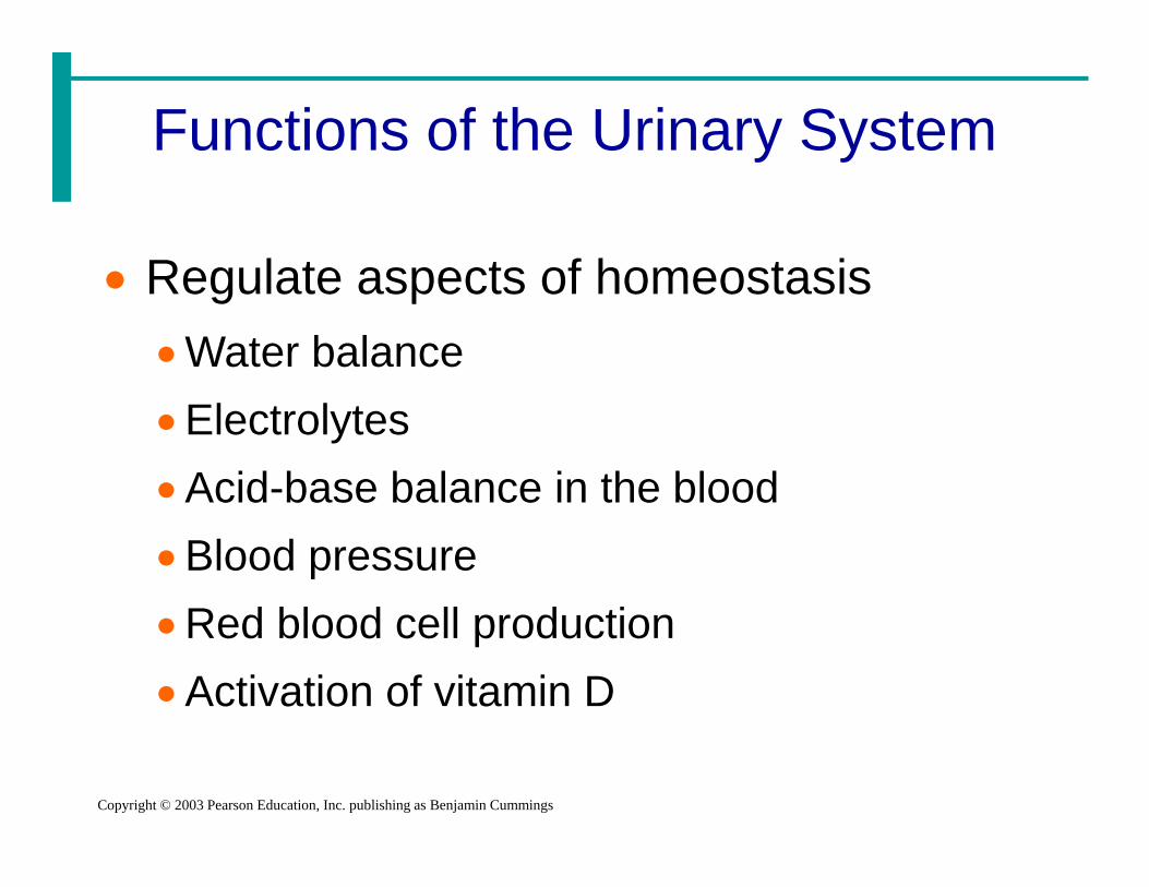
Functions of the Urinary System
Copyright © 2003 Pearson Education, Inc. publishing as Benjamin Cummings
Regulate aspects of homeostasisWater balanceElectrolytesAcid-base balance in the bloodBlood pressureRed blood cell productionActivation of vitamin D

Organs of the Urinary system
Copyright © 2003 Pearson Education, Inc. publishing as Benjamin Cummings
Kidneys Ureters Urinary bladder Urethra
Figure 15.1a

Location of the Kidneys
Copyright © 2003 Pearson Education, Inc. publishing as Benjamin Cummings
Against the dorsal body wall
At the level of T12 to L3
The right kidney is slightly lower than the left
Attached to ureters, renal blood vessels, and nerves at renal hilus
Atop each kidney is an adrenal gland

Regions of the Kidney
Copyright © 2003 Pearson Education, Inc. publishing as Benjamin Cummings
Renal cortex –outer region
Renal medulla –inside the cortex
Renal pelvis –inner collecting tube
Figure 15.2b
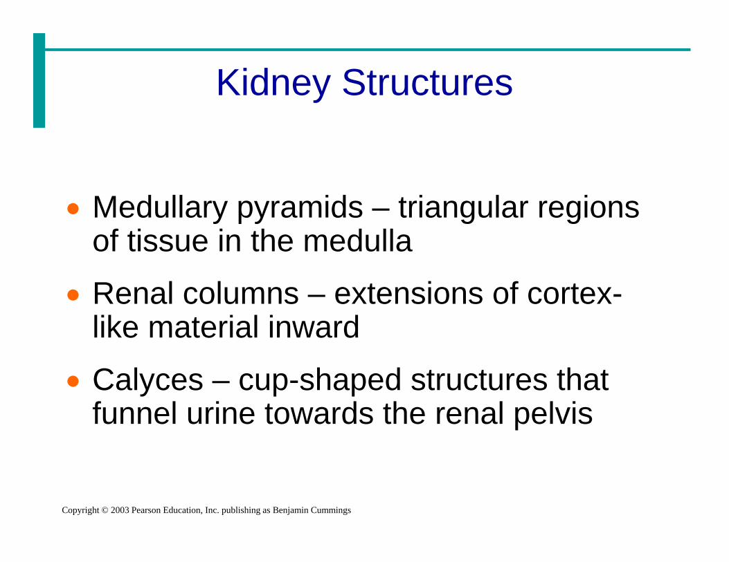
Kidney Structures
Copyright © 2003 Pearson Education, Inc. publishing as Benjamin Cummings
Medullary pyramids – triangular regions of tissue in the medulla
Renal columns – extensions of cortex-like material inward
Calyces – cup-shaped structures that funnel urine towards the renal pelvis

Blood Flow in the Kidneys
Copyright © 2003 Pearson Education, Inc. publishing as Benjamin Cummings
Figure 15.2c

Nephrons
Copyright © 2003 Pearson Education, Inc. publishing as Benjamin Cummings
The structural and functional units of the kidneys
Responsible for forming urine
Main structures of the nephronsGlomerulus
Renal tubule

Glomerulus
Copyright © 2003 Pearson Education, Inc. publishing as Benjamin Cummings
A specialized capillary bed
Attached to arterioles on both sides (maintains high pressure)Large afferent
arterioleNarrow efferent
arterioleFigure 15.3c

Glomerulus
Copyright © 2003 Pearson Education, Inc. publishing as Benjamin Cummings
The glomerulus sits within a glomerular capsule (the first part of the renal tubule)
Figure 15.3c

Renal Tubule
Copyright © 2003 Pearson Education, Inc. publishing as Benjamin Cummings
Glomerular (Bowman’s) capsule
Proximal convoluted tubule
Loop of Henle Distal
convoluted tubule
Figure 15.3b

Types of Nephrons
Copyright © 2003 Pearson Education, Inc. publishing as Benjamin Cummings
Cortical nephronsLocated entirely in the cortex Includes most nephrons
Figure 15.3a

Types of Nephrons
Slide 15 11b
Copyright © 2003 Pearson Education, Inc. publishing as Benjamin Cummings
Juxtamedullary nephronsFound at the boundary of the cortex and
medulla
Figure 15.3a

Peritubular Capillaries
Slide 15 12
Copyright © 2003 Pearson Education, Inc. publishing as Benjamin Cummings
Arise from efferent arteriole of the glomerulus
Normal, low pressure capillaries
Attached to a venule
Cling close to the renal tubule
Reabsorb (reclaim) some substances from collecting tubes
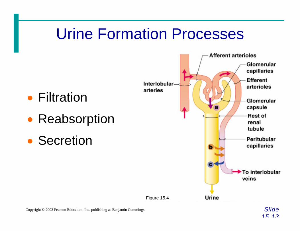
Urine Formation Processes
Slide 15 13
Copyright © 2003 Pearson Education, Inc. publishing as Benjamin Cummings
Filtration
Reabsorption
Secretion
Figure 15.4

Filtration
Slide 15 14
Copyright © 2003 Pearson Education, Inc. publishing as Benjamin Cummings
Nonselective passive process
Water and solutes smaller than proteins are forced through capillary walls
Blood cells cannot pass out to the capillaries
Filtrate is collected in the glomerular capsule and leaves via the renal tubule

Reabsorption
Slide 15 15
Copyright © 2003 Pearson Education, Inc. publishing as Benjamin Cummings
The peritubular capillaries reabsorb several materialsSome waterGlucoseAmino acids Ions
Some reabsorption is passive, most is active Most reabsorption occurs in the proximal
convoluted tubule
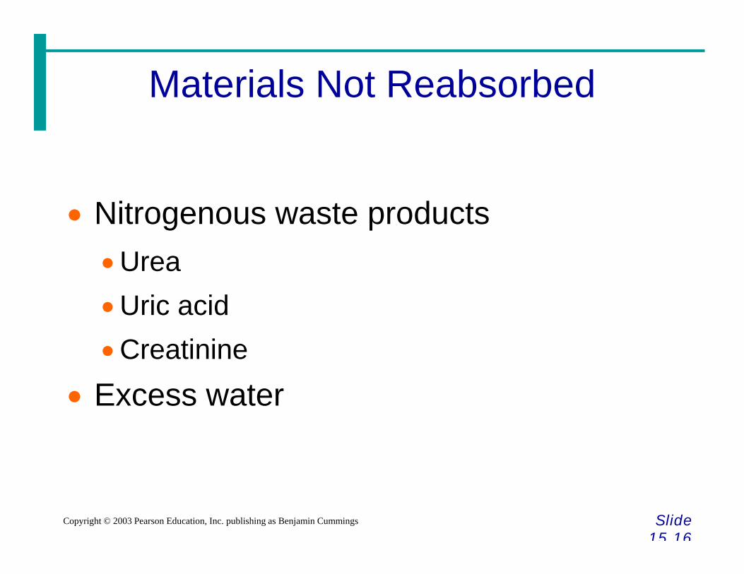
Materials Not Reabsorbed
Slide 15 16
Copyright © 2003 Pearson Education, Inc. publishing as Benjamin Cummings
Nitrogenous waste productsUreaUric acidCreatinine
Excess water

Secretion – Reabsorption in Reverse
Slide 15 17
Copyright © 2003 Pearson Education, Inc. publishing as Benjamin Cummings
Some materials move from the peritubular capillaries into the renal tubulesHydrogen and potassium ions
Creatinine
Materials left in the renal tubule move toward the ureter

Formation of Urine
Slide 15 18
Copyright © 2003 Pearson Education, Inc. publishing as Benjamin Cummings
Figure 15.5

Characteristics of Urine Used for Medical Diagnosis
Slide 15 19
Copyright © 2003 Pearson Education, Inc. publishing as Benjamin Cummings
Colored somewhat yellow due to the pigment urochrome (from the destruction of hemoglobin) and solutes
Sterile
Slightly aromatic
Normal pH of around 6 (varies 4.5-8)
Specific gravity of 1.001 to 1.035

Ureters
Slide 15 20
Copyright © 2003 Pearson Education, Inc. publishing as Benjamin Cummings
Slender tubes attaching the kidney to the bladderContinuous with the renal pelvis
Enter the posterior aspect of the bladder
Runs behind the peritoneum
Peristalsis aids gravity in urine transport

Urinary Bladder
Slide 15 21a
Copyright © 2003 Pearson Education, Inc. publishing as Benjamin Cummings
Smooth, collapsible, muscular sac
Temporarily stores urine
Figure 15.6

Urinary Bladder
Slide 15 21b
Copyright © 2003 Pearson Education, Inc. publishing as Benjamin Cummings
Trigone – three openingsTwo from the ureters
One to the urethrea
Figure 15.6

Urinary Bladder Wall
Slide 15 22
Copyright © 2003 Pearson Education, Inc. publishing as Benjamin Cummings
Three layers of smooth muscle (detrusor muscle)
Mucosa made of transitional epithelium
Walls are thick and folded in an empty bladder
Bladder can expand significantly without increasing internal pressure

Urethra
Slide 15 23
Copyright © 2003 Pearson Education, Inc. publishing as Benjamin Cummings
Thin-walled tube that carries urine from the bladder to the outside of the body by peristalsis
Release of urine is controlled by two sphincters Internal urethral sphincter (involuntary)
External urethral sphincter (voluntary)

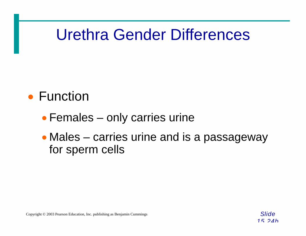
Urethra Gender Differences
Slide 15 24a
Copyright © 2003 Pearson Education, Inc. publishing as Benjamin Cummings
LengthFemales – 3–4 cm (1 inch)
Males – 20 cm (8 inches)
LocationFemales – along wall of the vagina
Males – through the prostate and penis
Urethra Gender Differences
Slide 15 24b
Copyright © 2003 Pearson Education, Inc. publishing as Benjamin Cummings
FunctionFemales – only carries urine
Males – carries urine and is a passageway for sperm cells

Micturition (Voiding)
Slide 15 25
Copyright © 2003 Pearson Education, Inc. publishing as Benjamin Cummings
Both sphincter muscles must open to allow voidingThe internal urethral sphincter is relaxed
after stretching of the bladder
Activation is from an impulse sent to the spinal cord and then back via the pelvic splanchnic nerves
The external urethral sphincter must be voluntarily relaxed

Maintaining Water Balance
Slide 15 26
Copyright © 2003 Pearson Education, Inc. publishing as Benjamin Cummings
Normal amount of water in the human bodyYoung adult females – 50%
Young adult males – 60%
Babies – 75%
Old age – 45%
Water is necessary for many body functions and levels must be maintained

Distribution of Body Fluid
Slide 15 27
Copyright © 2003 Pearson Education, Inc. publishing as Benjamin Cummings
Intracellular fluid (inside cells)
Extracellular fluid (outside cells) Interstitial fluid
Blood plasmaFigure 15.7

The Link Between Water and Salt
Slide 15 28
Copyright © 2003 Pearson Education, Inc. publishing as Benjamin Cummings
Changes in electrolyte balance causes water to move from one compartment to anotherAlters blood volume and blood pressure
Can impair the activity of cells

Maintaining Water Balance
Slide 15 29
Copyright © 2003 Pearson Education, Inc. publishing as Benjamin Cummings
Water intake must equal water output Sources for water intake
Ingested foods and fluidsWater produced from metabolic processes
Sources for water outputVaporization out of the lungs Lost in perspiration Leaves the body in the fecesUrine production

Maintaining Water Balance
Slide 15 30
Copyright © 2003 Pearson Education, Inc. publishing as Benjamin Cummings
Dilute urine is produced if water intake is excessive
Less urine (concentrated) is produced if large amounts of water are lost
Proper concentrations of various electrolytes must be present

Regulation of Water and Electrolyte Reabsorption
Slide 15 31
Copyright © 2003 Pearson Education, Inc. publishing as Benjamin Cummings
Regulation is primarily by hormonesAntidiuretic hormone (ADH) prevents
excessive water loss in urineAldosterone regulates sodium ion content of
extracellular fluid Triggered by the rennin-angiotensin
mechanism Cells in the kidneys and hypothalamus
are active monitors
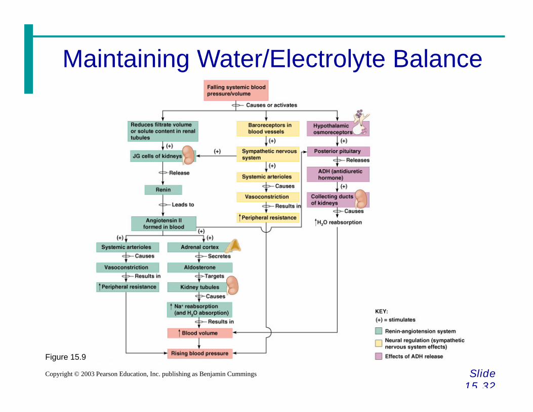
Maintaining Water/Electrolyte Balance
Slide 15 32
Copyright © 2003 Pearson Education, Inc. publishing as Benjamin Cummings
Figure 15.9

Maintaining Acid-Base Balance in Blood
Slide 15 33a
Copyright © 2003 Pearson Education, Inc. publishing as Benjamin Cummings
Blood pH must remain between 7.35 and 7.45 to maintain homeostasisAlkalosis – pH above 7.45
Acidosis – pH below 7.35
Most ions originate as byproducts of cellular metabolism

Maintaining Acid-Base Balance in Blood
Slide 15 33b
Copyright © 2003 Pearson Education, Inc. publishing as Benjamin Cummings
Most acid-base balance is maintained by the kidneys
Other acid-base controlling systemsBlood buffers
Respiration

Developmental Aspects of the Urinary System
Slide 15 38a
Copyright © 2003 Pearson Education, Inc. publishing as Benjamin Cummings
Functional kidneys are developed by the third month
Urinary system of a newbornBladder is small
Urine cannot be concentrated

Developmental Aspects of the Urinary System
Slide 15 38b
Copyright © 2003 Pearson Education, Inc. publishing as Benjamin Cummings
Control of the voluntary urethral sphincter does not start until age 18 months
Urinary infections are the only common problems before old age

Aging and the Urinary System
Slide 15 39
Copyright © 2003 Pearson Education, Inc. publishing as Benjamin Cummings
There is a progressive decline in urinary function
The bladder shrinks with aging
Urinary retention is common in males
Copyright © 2022 FDOKUMEN