A geometric description of the urinary bladder of Turkish Shepherd dog (Karaba
6
Ann Anat 187 (2005) 179—184 A geometric description of the urinary bladder of Turkish Shepherd dog (Karabas - ) Rıdvan Ezentas - a , Kadri Arslan a, , Hu ¨seyin Yıldız b , Bahri Yıldız b , Cengizhan Murathan a a Department of Mathematics, Faculty of Art and Science, Uludag University, 16059 Bursa, Turkey b Department of Anatomy, Faculty of Veterinary Medicine, Uludag University, 16059 Bursa, Turkey Received 13 February 2004; accepted 9 June 2004 Summary In this study, a geometric and experimental work of the urinary bladder of a dog is presented. Experimentally, the diameters on the neck (collum vesicae region), the body (corpus vesicae region), the bottom (apex region) and the longitudinal length of the urinary bladder were measured. Geometrically it was shown that the urinary bladder is comparable with a surface of revolution of a planar (profile) curve. On the same regions of the modelled surface the diameters of the horizontal sections have been measured. It was found that the geometric and the experimental values were closely related. Further the Gaussian curvature K at certain points on the same regions of the modelled surface was calculated. The quadratic approximations of the urinary bladder surface was described with the sign of K at those points. & 2005 Elsevier GmbH. All rights reserved. Introduction The urinary bladder of the dog is a flexible storage organ. When the bladder is empty, it recedes into the pelvic cavity to varying degrees. When filled, it becomes pear shaped. The bladder has a neck which is a cranial blind end and a body in the middle. The apex is a mass of scar tissue especially in young animals. The wall of the bladder is composed of a covering of perito- neum, a muscular coat and a mucous membrane. The muscular coat consists of three layers of coarse, smooth muscle bundles. In the outer layer, the bundles have a longitudinal or oblique course. In the middle layer, the fibers run transversely, and in the inner layer they run longitudinally. Muscle bundles of both inner and outer layers enter and interweave in the middle layer. The apex and neck of the bladder are surrounded by loops which are derived from both inner and outer longitudinal ARTICLE IN PRESS www.elsevier.de/aanat KEYWORDS Turkish Shepherd Dog; Urinary bladder; Surface of revolution 0940-9602/$ - see front matter & 2005 Elsevier GmbH. All rights reserved. doi:10.1016/j.aanat.2004.06.003 Corresponding author. E-mail address: [email protected] (K. Arslan).
Transcript of A geometric description of the urinary bladder of Turkish Shepherd dog (Karaba
ARTICLE IN PRESS
Ann Anat 187 (2005) 179—184
KEYWORDTurkish ShDog;Urinary blSurface of
0940-9602/$ - sdoi:10.1016/j.
�CorrespondE-mail addr
www.elsevier.de/aanat
A geometric description of the urinary bladder ofTurkish Shepherd dog (Karabas-)
Rıdvan Ezentas-a, Kadri Arslana,�, Huseyin Yıldızb, Bahri Yıldızb,Cengizhan Murathana
aDepartment of Mathematics, Faculty of Art and Science, Uludag University, 16059 Bursa, TurkeybDepartment of Anatomy, Faculty of Veterinary Medicine, Uludag University, 16059 Bursa, Turkey
Received 13 February 2004; accepted 9 June 2004
Sepherd
adder;revolution
ee front matter & 200aanat.2004.06.003
ing author.ess: [email protected]
SummaryIn this study, a geometric and experimental work of the urinary bladder of a dog ispresented. Experimentally, the diameters on the neck (collum vesicae region), thebody (corpus vesicae region), the bottom (apex region) and the longitudinal length ofthe urinary bladder were measured. Geometrically it was shown that the urinarybladder is comparable with a surface of revolution of a planar (profile) curve. On thesame regions of the modelled surface the diameters of the horizontal sections havebeen measured. It was found that the geometric and the experimental values wereclosely related. Further the Gaussian curvature K at certain points on the same regionsof the modelled surface was calculated. The quadratic approximations of the urinarybladder surface was described with the sign of K at those points.& 2005 Elsevier GmbH. All rights reserved.
Introduction
The urinary bladder of the dog is a flexiblestorage organ. When the bladder is empty, itrecedes into the pelvic cavity to varying degrees.When filled, it becomes pear shaped.
The bladder has a neck which is a cranial blindend and a body in the middle. The apex is a mass ofscar tissue especially in young animals. The wall ofthe bladder is composed of a covering of perito-
5 Elsevier GmbH. All rights rese
u.tr (K. Arslan).
neum, a muscular coat and a mucous membrane.The muscular coat consists of three layers ofcoarse, smooth muscle bundles. In the outer layer,the bundles have a longitudinal or oblique course.In the middle layer, the fibers run transversely, andin the inner layer they run longitudinally. Musclebundles of both inner and outer layers enter andinterweave in the middle layer. The apex and neckof the bladder are surrounded by loops which arederived from both inner and outer longitudinal
rved.
ARTICLE IN PRESS
Table 1. Experimental measurements of the urinarybladder
Parts Experimental values (a), mm
Collum vesicae 4.31Corpus vesicae 84.96Apex vesicae 10.11Longitudinal length 121.1
R. Ezentas- et al.180
layers and may act in the closure of the internalorifice (Getty, 1975; Nickel et al., 1979; Dyceet al., 1987).
Smith et al. (1999) identified the inner surfacebladder and digitized it from spiral computedtomography (CT) images. The data were centeredto a cylindrical coordinate system and mapped tothe y; z plane. This produced a spatially homo-genous data distribution for surface fitting andreduced the necessary number of fit coordinatesrequired to reproduce the bladder surface to one:the radial coordinate.
In Yıldız et al. (2001a, b) we presented ageometric recognition of the ascending colon ofsome domestic animals (i.e. pig, ruminant, anddog) and a mathematical model of the ascendingcolon of the horse.
The aim of this study is to compare theexperimental work with the geometry of theurinary bladder of the dog.
Materials and methods
Experimental work
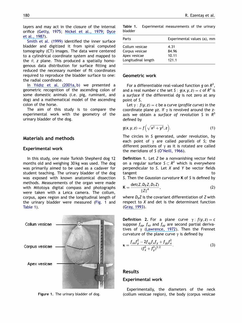
In this study, one male Turkish Shepherd dog 12months old and weighing 30 kg was used. The dogwas primarily aimed to be used as a cadaver forstudent teaching. The urinary bladder of the dogwas exposed with known anatomical dissectionmethods. Measurements of the organ were madewith Mitotoya digital compass and photographswere taken with a Leica camera. The collum,corpus, apex region and the longitudinal length ofthe urinary bladder were measured (Fig. 1 andTable 1).
Figure 1. The urinary bladder of dog.
Geometric work
For a differentiable real-valued function g on R3,and a real number c the set S : gðx; y; zÞ ¼ c of R3 isa surface if the differential dg is not zero at anypoint of S.
Let c : fðy; zÞ ¼ c be a curve (profile curve) in thecoordinate plane yz. If c is revolved around the z-axis we obtain a surface of revolution S in R3
defined by
gðx; y; zÞ ¼ fffiffiffiffiffiffiffiffiffiffiffiffiffiffiffix2 þ y2
p; z
� �: (1)
The circles in S generated, under revolution, byeach point of c are called parallels of S; thedifferent positions of c as it is rotated are calledthe meridians of S (O’Neill, 1966).
Definition 1. Let Z be a nonvanishing vector fieldon a regular surface S � R3 which is everywhereperpendicular to S. Let X and Y be vector fieldstangent toS. Then the Gaussian curvature K of S is defined by
K ¼detðZ;DXZ;DYZÞ
Zk k4
; (2)
where DXZ is the covariant differentiation of Z withrespect to X and det is the determinant function(Gray, 1993).
Definition 2. For a plane curve c : fðy; zÞ ¼ csuppose fyy, fxz and fyy are second partial deriva-tives of c (Lawrence, 1972). Then the Frennetcurvature of the plane curve c is defined by
j ¼fxxf
2y � 2fxyfxfy þ fyyf
2x
ðf2x þ f2yÞ3=2
: (3)
Results
Experimental work
Experimentally, the diameters of the neck(collum vesicae region), the body (corpus vesicae
ARTICLE IN PRESS
A geometric description of the urinary bladder of Turkish Shepherd dog (Karabas-) 181
region), the bottom (apex region) and the long-itudinal length of urinary bladder were measured(Table 1).
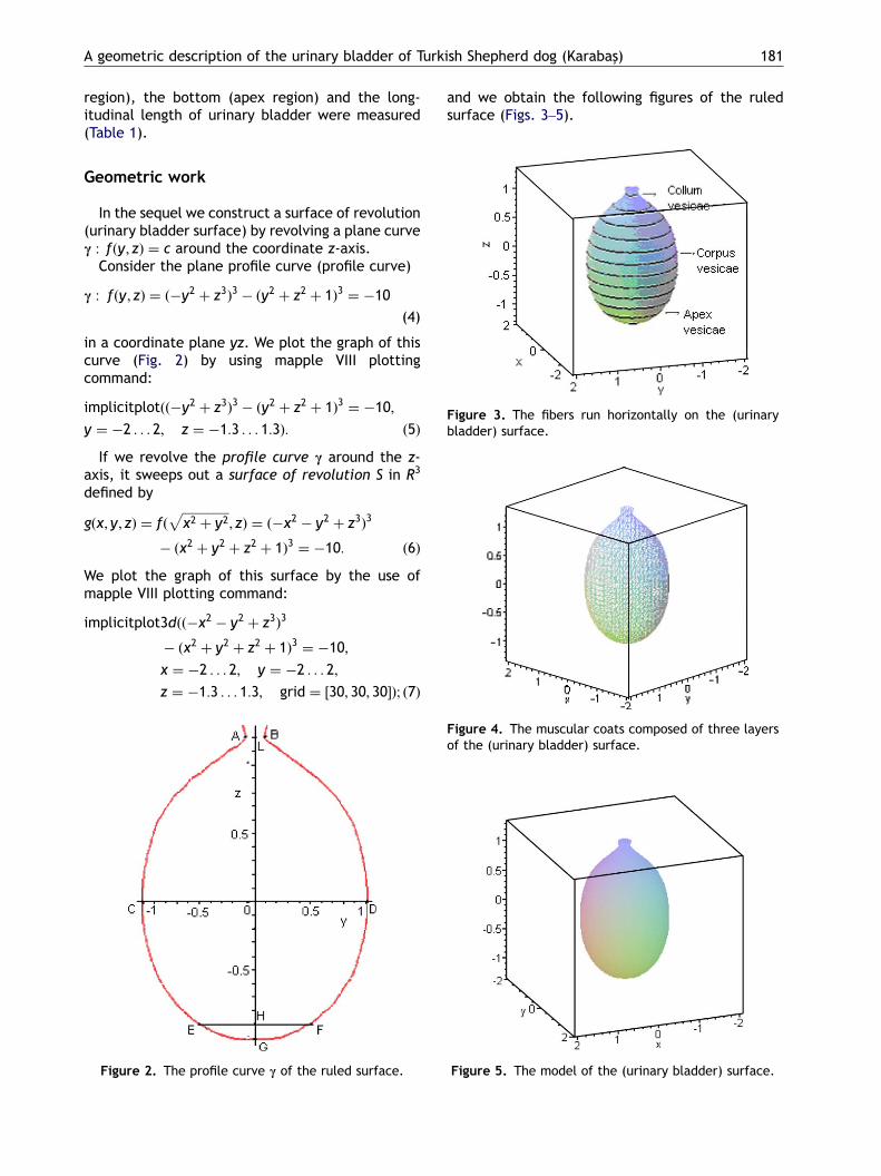
Figure 3. The fibers run horizontally on the (urinarybladder) surface.
Geometric work
In the sequel we construct a surface of revolution(urinary bladder surface) by revolving a plane curvec : fðy; zÞ ¼ c around the coordinate z-axis.
Consider the plane profile curve (profile curve)
c : fðy; zÞ ¼ ð�y2 þ z3Þ3 � ðy2 þ z2 þ 1Þ3 ¼ �10
(4)
in a coordinate plane yz. We plot the graph of thiscurve (Fig. 2) by using mapple VIII plottingcommand:
implicitplotðð�y2 þ z3Þ3 � ðy2 þ z2 þ 1Þ3 ¼ �10;
y ¼ �2 . . . 2; z ¼ �1:3 . . . 1:3Þ: ð5Þ
If we revolve the profile curve c around the z-axis, it sweeps out a surface of revolution S in R3
defined by
gðx; y; zÞ ¼ fðffiffiffiffiffiffiffiffiffiffiffiffiffiffiffix2 þ y2
p; zÞ ¼ ð�x2 � y2 þ z3Þ3
� ðx2 þ y2 þ z2 þ 1Þ3 ¼ �10: ð6Þ
We plot the graph of this surface by the use ofmapple VIII plotting command:
implicitplot3dðð�x2 � y2 þ z3Þ3
� ðx2 þ y2 þ z2 þ 1Þ3 ¼ �10;
x ¼ �2 . . . 2; y ¼ �2 . . . 2;
z ¼ �1:3 . . . 1:3; grid ¼ ½30; 30; 30Þ; ð7Þ
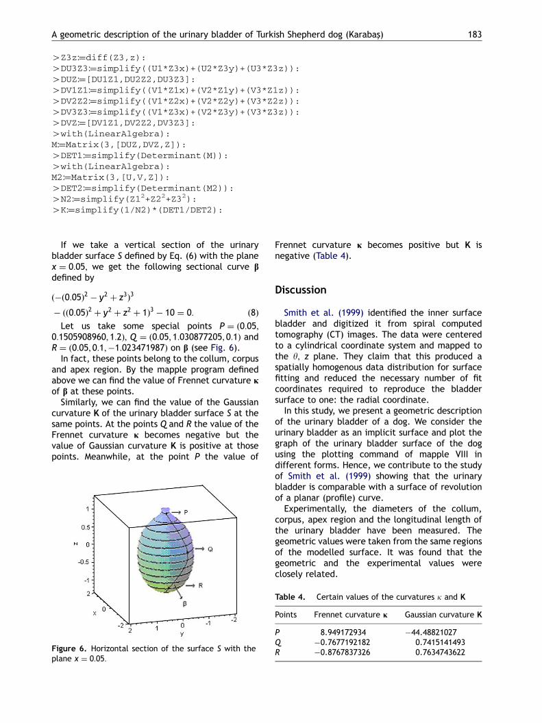
Figure 2. The profile curve c of the ruled surface.
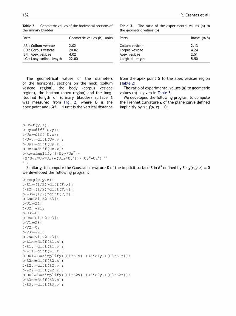
and we obtain the following figures of the ruledsurface (Figs. 3–5).
Figure 4. The muscular coats composed of three layersof the (urinary bladder) surface.
Figure 5. The model of the (urinary bladder) surface.
ARTICLE IN PRESS
Table 2. Geometric values of the horizontal sections ofthe urinary bladder
Parts Geometric values (b), units
jABj: Collum vesicae 2.02jCDj: Corpus vesicae 20.02jEFj: Apex vesicae 4.02jLGj: Longitudinal length 22.00
Table 3. The ratio of the experimental values (a) tothe geometric values (b)
Parts Ratio: (a/b)
Collum vesicae 2.13Corpus vesicae 4.24Apex vesicae 2.51Longitial length 5.50
R. Ezentas- et al.182
The geometrical values of the diametersof the horizontal sections on the neck (collumvesicae region), the body (corpus vesicaeregion), the bottom (apex region) and the long-itudinal length of (urinary bladder) surface Swas measured from Fig. 2, where G is theapex point and jGHj ¼ 1 unit is the vertical distance
from the apex point G to the apex vesicae region(Table 2).
The ratio of experimental values (a) to geometricvalues (b) is given in Table 3.
We developed the following program to computethe Frennet curvature j of the plane curve definedimplicitly by c : fðy; zÞ ¼ 0:
4U:¼f(y,z):4Uy:¼diff(U,y):4Uz:¼diff(U,z):4Uyy:¼diff(Uy,y):4Uyz:¼diff(Uy,z):4Uzz:¼diff(Uz,z):4k:¼simplify(((Uyy*Uz2)-(2*Uyz*Uy*Uz)+(Uzz*Uy2))/(Uy2+Uz2)(3/2)):
Similarly, to compute the Gaussian curvature K of the implicit surface S in R3 defined by S : gðx; y; zÞ ¼ 0
we developed the following program:4F:¼g(x,y,z):4Z1:¼(1/2)*diff(F,x):4Z2:¼(1/2)*diff(F,y):4Z3:¼(1/2)*diff(F,z):4Z:¼[Z1,Z2,Z3]:4U1:¼Z2:4U2:¼-Z1:4U3:¼0:4U:¼[U1,U2,U3]:4V1:¼Z3:4V2:¼0:4V3:¼-Z1:4V:¼[V1,V2,V3]:4Z1x:¼diff(Z1,x):4Z1y:¼diff(Z1,y):4Z1z:¼diff(Z1,z):4DU1Z1:¼simplify((U1*Z1x)+(U2*Z1y)+(U3*Z1z)):4Z2x:¼diff(Z2,x):4Z2y:¼diff(Z2,y):4Z2z:¼diff(Z2,z):4DU2Z2:¼simplify((U1*Z2x)+(U2*Z2y)+(U3*Z2z)):4Z3x:¼diff(Z3,x):4Z3y:¼diff(Z3,y):
ARTICLE IN PRESS
A geometric description of the urinary bladder of Turkish Shepherd dog (Karabas-) 183
4Z3z:¼diff(Z3,z):4DU3Z3:¼simplify((U1*Z3x)+(U2*Z3y)+(U3*Z3z)):4DUZ:¼[DU1Z1,DU2Z2,DU3Z3]:4DV1Z1:¼simplify((V1*Z1x)+(V2*Z1y)+(V3*Z1z)):4DV2Z2:¼simplify((V1*Z2x)+(V2*Z2y)+(V3*Z2z)):4DV3Z3:¼simplify((V1*Z3x)+(V2*Z3y)+(V3*Z3z)):4DVZ:¼[DV1Z1,DV2Z2,DV3Z3]:4with(LinearAlgebra):M:¼Matrix(3,[DUZ,DVZ,Z]):4DET1:¼simplify(Determinant(M)):4with(LinearAlgebra):M2:¼Matrix(3,[U,V,Z]):4DET2:¼simplify(Determinant(M2)):4N2:¼simplify(Z12+Z22+Z32):4K:¼simplify(1/N2)*(DET1/DET2):
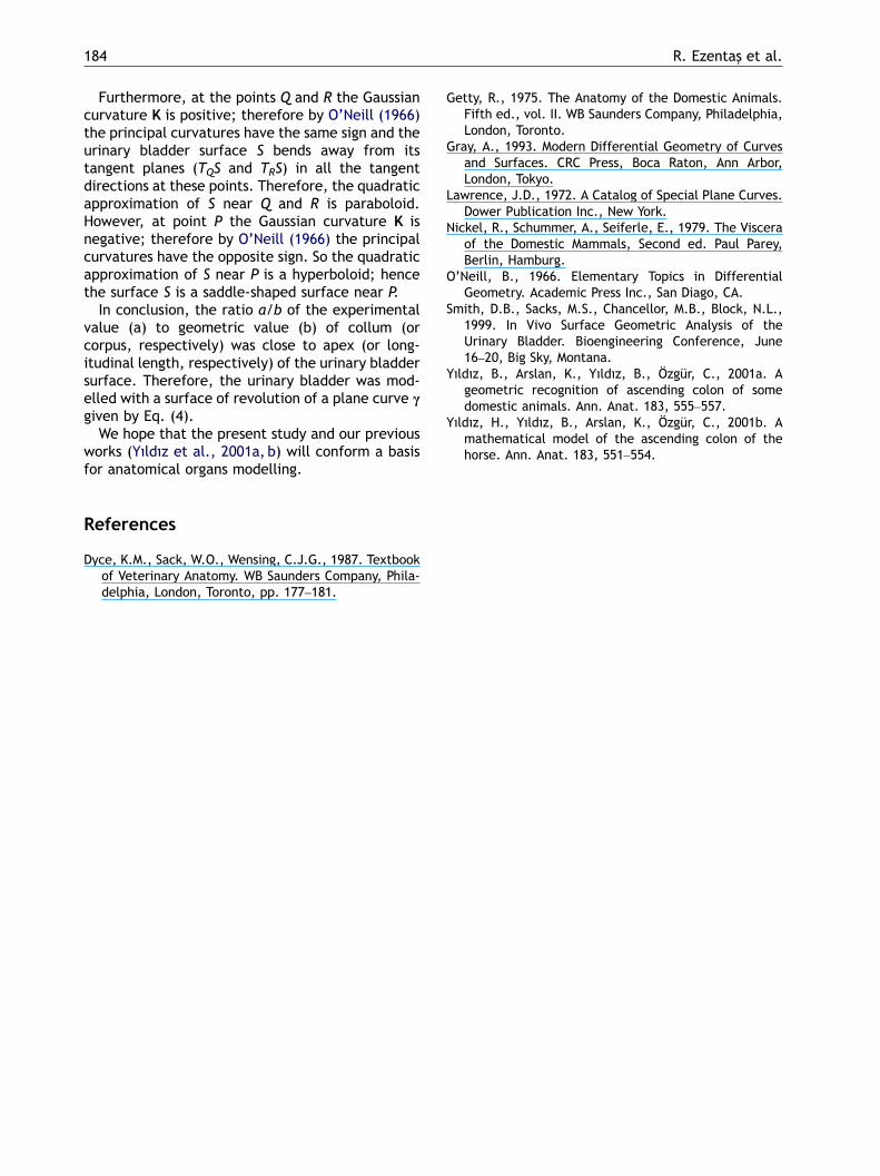
If we take a vertical section of the urinarybladder surface S defined by Eq. (6) with the planex ¼ 0:05; we get the following sectional curve bdefined by
ð�ð0:05Þ2 � y2 þ z3Þ3
� ðð0:05Þ2 þ y2 þ z2 þ 1Þ3 � 10 ¼ 0: ð8Þ
Let us take some special points P ¼ ð0:05;0:1505908960; 1:2Þ; Q ¼ ð0:05; 1:030877205; 0:1Þ andR ¼ ð0:05; 0:1;�1:023471987Þ on b (see Fig. 6).
In fact, these points belong to the collum, corpusand apex region. By the mapple program definedabove we can find the value of Frennet curvature jof b at these points.
Similarly, we can find the value of the Gaussiancurvature K of the urinary bladder surface S at thesame points. At the points Q and R the value of theFrennet curvature j becomes negative but thevalue of Gaussian curvature K is positive at thosepoints. Meanwhile, at the point P the value of
Figure 6. Horizontal section of the surface S with theplane x ¼ 0:05:
Frennet curvature j becomes positive but K isnegative (Table 4).
Discussion
Smith et al. (1999) identified the inner surfacebladder and digitized it from spiral computedtomography (CT) images. The data were centeredto a cylindrical coordinate system and mapped tothe y; z plane. They claim that this produced aspatially homogenous data distribution for surfacefitting and reduced the necessary number of fitcoordinates required to reproduce the bladdersurface to one: the radial coordinate.
In this study, we present a geometric descriptionof the urinary bladder of a dog. We consider theurinary bladder as an implicit surface and plot thegraph of the urinary bladder surface of the dogusing the plotting command of mapple VIII indifferent forms. Hence, we contribute to the studyof Smith et al. (1999) showing that the urinarybladder is comparable with a surface of revolutionof a planar (profile) curve.
Experimentally, the diameters of the collum,corpus, apex region and the longitudinal length ofthe urinary bladder have been measured. Thegeometric values were taken from the same regionsof the modelled surface. It was found that thegeometric and the experimental values wereclosely related.
Table 4. Certain values of the curvatures k and K
Points Frennet curvature j Gaussian curvature K
P 8.949172934 �44.48821027Q �0.7677192182 0.7415141493R �0.8767837326 0.7634743622
ARTICLE IN PRESS
R. Ezentas- et al.184
Furthermore, at the points Q and R the Gaussiancurvature K is positive; therefore by O’Neill (1966)the principal curvatures have the same sign and theurinary bladder surface S bends away from itstangent planes (TQS and TRS) in all the tangentdirections at these points. Therefore, the quadraticapproximation of S near Q and R is paraboloid.However, at point P the Gaussian curvature K isnegative; therefore by O’Neill (1966) the principalcurvatures have the opposite sign. So the quadraticapproximation of S near P is a hyperboloid; hencethe surface S is a saddle-shaped surface near P.
In conclusion, the ratio a/b of the experimentalvalue (a) to geometric value (b) of collum (orcorpus, respectively) was close to apex (or long-itudinal length, respectively) of the urinary bladdersurface. Therefore, the urinary bladder was mod-elled with a surface of revolution of a plane curve cgiven by Eq. (4).
We hope that the present study and our previousworks (Yıldız et al., 2001a, b) will conform a basisfor anatomical organs modelling.
References
Dyce, K.M., Sack, W.O., Wensing, C.J.G., 1987. Textbookof Veterinary Anatomy. WB Saunders Company, Phila-delphia, London, Toronto, pp. 177–181.
Getty, R., 1975. The Anatomy of the Domestic Animals.Fifth ed., vol. II. WB Saunders Company, Philadelphia,London, Toronto.
Gray, A., 1993. Modern Differential Geometry of Curvesand Surfaces. CRC Press, Boca Raton, Ann Arbor,London, Tokyo.
Lawrence, J.D., 1972. A Catalog of Special Plane Curves.Dower Publication Inc., New York.
Nickel, R., Schummer, A., Seiferle, E., 1979. The Visceraof the Domestic Mammals, Second ed. Paul Parey,Berlin, Hamburg.
O’Neill, B., 1966. Elementary Topics in DifferentialGeometry. Academic Press Inc., San Diago, CA.
Smith, D.B., Sacks, M.S., Chancellor, M.B., Block, N.L.,1999. In Vivo Surface Geometric Analysis of theUrinary Bladder. Bioengineering Conference, June16–20, Big Sky, Montana.
Yıldız, B., Arslan, K., Yıldız, B., Ozgur, C., 2001a. Ageometric recognition of ascending colon of somedomestic animals. Ann. Anat. 183, 555–557.
Yıldız, H., Yıldız, B., Arslan, K., Ozgur, C., 2001b. Amathematical model of the ascending colon of thehorse. Ann. Anat. 183, 551–554.