The SWI/SNF ATPase Brm Is a Gatekeeper of Proliferative Control in Prostate Cancer
10
2008;68:10154-10162. Cancer Res Hui Shen, Nathan Powers, Nitin Saini, et al. Control in Prostate Cancer The SWI/SNF ATPase Brm Is a Gatekeeper of Proliferative Updated version http://cancerres.aacrjournals.org/content/68/24/10154 Access the most recent version of this article at: Material Supplementary http://cancerres.aacrjournals.org/content/suppl/2008/12/10/68.24.10154.DC1.html Access the most recent supplemental material at: Cited Articles http://cancerres.aacrjournals.org/content/68/24/10154.full.html#ref-list-1 This article cites by 49 articles, 24 of which you can access for free at: Citing articles http://cancerres.aacrjournals.org/content/68/24/10154.full.html#related-urls This article has been cited by 12 HighWire-hosted articles. Access the articles at: E-mail alerts related to this article or journal. Sign up to receive free email-alerts Subscriptions Reprints and . [email protected] Department at To order reprints of this article or to subscribe to the journal, contact the AACR Publications Permissions . [email protected] Department at To request permission to re-use all or part of this article, contact the AACR Publications Research. on June 13, 2014. © 2008 American Association for Cancer cancerres.aacrjournals.org Downloaded from Research. on June 13, 2014. © 2008 American Association for Cancer cancerres.aacrjournals.org Downloaded from
-
Upload
independent -
Category
Documents
-
view
1 -
download
0
Transcript of The SWI/SNF ATPase Brm Is a Gatekeeper of Proliferative Control in Prostate Cancer

2008;68:10154-10162. Cancer Res Hui Shen, Nathan Powers, Nitin Saini, et al. Control in Prostate CancerThe SWI/SNF ATPase Brm Is a Gatekeeper of Proliferative
Updated version
http://cancerres.aacrjournals.org/content/68/24/10154
Access the most recent version of this article at:
Material
Supplementary
http://cancerres.aacrjournals.org/content/suppl/2008/12/10/68.24.10154.DC1.html
Access the most recent supplemental material at:
Cited Articles
http://cancerres.aacrjournals.org/content/68/24/10154.full.html#ref-list-1
This article cites by 49 articles, 24 of which you can access for free at:
Citing articles
http://cancerres.aacrjournals.org/content/68/24/10154.full.html#related-urls
This article has been cited by 12 HighWire-hosted articles. Access the articles at:
E-mail alerts related to this article or journal.Sign up to receive free email-alerts
Subscriptions
Reprints and
To order reprints of this article or to subscribe to the journal, contact the AACR Publications
Permissions
To request permission to re-use all or part of this article, contact the AACR Publications
Research. on June 13, 2014. © 2008 American Association for Cancercancerres.aacrjournals.org Downloaded from
Research. on June 13, 2014. © 2008 American Association for Cancercancerres.aacrjournals.org Downloaded from

The SWI/SNF ATPase Brm Is a Gatekeeper of Proliferative Control in
Prostate Cancer
Hui Shen,1Nathan Powers,
1Nitin Saini,
7Clay E.S. Comstock,
7Ankur Sharma,
7Katherine Weaver,
1
Monica P. Revelo,3William Gerald,
4Erin Williams,
1Walter J. Jessen,
2Bruce J. Aronow,
2
Gary Rosson,5Bernard Weissman,
5Christian Muchardt,
6Moshe Yaniv,
6and Karen E. Knudsen
7,8
1Department of Cell and Cancer Biology, University of Cincinnati, 2Cincinnati Children’s Hospital, Cincinnati, Ohio; 3Department ofPathology, University of Utah, Salt Lake City, Utah; 4Department of Pathology, Memorial Sloan-Kettering Cancer Center, New York,New York; 5Department of Pathology and Laboratory Medicine, University of North Carolina, Chapel Hill, North Carolina;6Department of Developmental Biology, Institut Pasteur, Paris, France; and Departments of 7Cancer Biology and8Urology, Thomas Jefferson University/Kimmel Cancer Center, Philadelphia, Pennsylvania
Abstract
Factors that drive prostate cancer progression remain poorlydefined, thus hindering the development of new therapeuticstrategies. Disseminated tumors are treated through regimensthat ablate androgen signaling, as prostate cancer cellsrequire androgen for growth and survival. However, recurrent,incurable tumors that have bypassed the androgen require-ment ultimately arise. This study reveals that the Brm ATPase,a component of selected SWI/SNF complexes, has significantantiproliferative functions in the prostate that protect againstthese transitions. First, we show that targeted ablation ofBrm is causative for the development of prostatic hyperplasiain mice. Second, in vivo challenge revealed that Brm�/�epithelia acquire the capacity for lobe-specific, castration-resistant cellular proliferation. Third, investigation of humanspecimens revealed that Brm mRNA and protein levels areattenuated in prostate cancer. Fourth, Brm down-regulationwas associated with an increased proliferative index, consis-tent with the mouse model. Lastly, gene expression profilingshowed that Brm loss alters factors upstream of E2F1; this wasconfirmed in murine models, wherein Brm loss induced E2F1deregulation in a tissue-specific manner. Combined, thesedata identify Brm as a major effector of serum androgen–induced proliferation in the prostate that is disrupted inhuman disease, and indicate that loss of Brm confers aproliferative advantage in prostate cancer. [Cancer Res2008;68(24):10154–62]
Introduction
SWI/SNF chromatin remodeling complexes are critical media-tors of transcriptional control, as manifest through their ability tomobilize nucleosomes (1, 2). SWI/SNF complexes are recruited tochromatin by sequence-specific transcription factors, and therein,regulate gene expression by either promoting nucleosome dis-persion or condensation (3, 4). To control this process, it isincreasingly apparent that the specificity of SWI/SNF function isdictated by biochemical diversity in subunit composition, and that
imbalances in subunit expression can induce significant, oftentissue-specific biological consequence (3, 5–8).
SWI/SNF complexes consist of a central ATPase (Brg1 or Brm),the function of which is required for nucleosome repositioning,a cohort of ‘‘core’’ subunits that are required to reconstitute SWI/SNF function in vitro (BAF47/INI1, BAF155, and BAF170), andcombinatorial assembly of six to eight additional BAF (Brg1-associated factor) subunits that confer specificity of function toindividual SWI/SNF complexes (3–5). Despite the general require-ment of the central ATPase for SWI/SNF function, genetic analysesrevealed remarkable functional heterogeneity between Brm andBrg1. For example, loss of Brg1 is an embryonic lethal event (9),whereas Brm�/� mice are viable and develop normally (10).Subsequent studies using conditional deletion of Brg1 revealeddivergent roles of this ATPase for skin development, limbmorphogenesis, erythropoiesis, vasculature, gliogenesis, neuralstem/progenitor cells, and T-cell development (9, 11–15). Althoughfew phenotypes have been detected in Brm�/� mice, theseanimals are up to 15% heavier than their wild-type littermates, andshow the enlargement of selected organs (e.g., liver, kidney, heart)but reductions in others (spleen, testes; ref. 16). Thus, despite theshared capacity of Brg1 and Brm to confer SWI/SNF activity, theATPases seem to serve distinct biological functions.
In cells of prostatic origin, SWI/SNF proved capable of regulatingthe androgen receptor (AR), a ligand-dependent transcriptionfactor that is a major effector of prostate cancer growth andprogression. AR is the first-line therapeutic target for disseminatedprostate cancer, as prostate cancer cells are exquisitely dependenton AR signaling (17, 18). Ligand (androgen) activation of ARinduces rapid dimerization and nuclear translocation of theprotein, which binds to DNA at androgen-responsive elementsand induces a program of gene transcription in prostate cancercells that results in cell survival and proliferation (19). In addition,AR directs the expression of secretory products that are usedclinically to monitor prostate cancer growth and progression (i.e.,prostate-specific antigen; refs. 20, 21). Using in vitro models, itwas previously shown that AR requires SWI/SNF to induce geneexpression on prostate-specific target genes, and that the receptorshows a preference in transient assays for Brm-induced SWI/SNFaction (22, 23). Similarly, it was shown in cell-based assays thatBrm is the predominant ATPase recruited to sites of AR actionafter androgen stimulation (22, 23). Through these actions, it washypothesized that Brm-mediated AR signaling supports androgen-dependent transcriptional activation in the context of the prostate.In prostate cancer therapy, engagement of AR signaling is targetedthrough the use of regimens that either deprive the receptor of
Note: Supplementary data for this article are available at Cancer Research Online(http://cancerres.aacrjournals.org/).
Requests for reprints: Karen E. Knudsen, Thomas Jefferson University/KimmelCancer Center, Bluemle Building, Room 1008, Philadelphia, PA 19107. Phone: 215-503-8574; Fax: 215-503-8574; E-mail: [email protected].
I2008 American Association for Cancer Research.doi:10.1158/0008-5472.CAN-08-1794
Cancer Res 2008; 68: (24). December 15, 2008 10154 www.aacrjournals.org
Research Article
Research. on June 13, 2014. © 2008 American Association for Cancercancerres.aacrjournals.org Downloaded from

ligand (androgen ablation) or through the use of direct ARantagonists (17, 19, 24). Although these strategies result in tumorremission, recurrent tumors arise within 2 to 3 years in which ARaction is restored despite the absence of the ligand (25). Transitionto this therapy-resistant phase of tumor progression represents theincurable stage of disease; thus, it is imperative to discern thesignaling pathways that influence AR signaling in the prostate.Given the importance of SWI/SNF in controlling AR action in vitro ,the present study interrogated the consequence of the Brm ATPasein the prostate using genetically defined in vivo models andanalyses of human prostate cancer tumors.
Materials and Methods
Mice, xenografts, and human specimens. Targeted deletion of Brm
was previously described (16). Animals were bred and maintained at the
Laboratory Animal Medical Services of University of Cincinnati, and trackedby the Institutional Animal Care and Use Committee. Mice were obtained
from (Brm+/� � Brm+/�) offspring. Genotypes were determined as
previously described (16). Prostates were resected after sacrifice using
standard methods, and immediately dissected into the anterior, ventral, anddorsolateral lobes. Dissected tissues were snap-frozen (for protein or RNA
collection) or were suspended in 10% neutral buffered formalin and paraffin
embedded. For acquisition of human tissue, unidentified specimens fromradical prostatectomy were obtained from the University of Cincinnati,
Department of Pathology tissue bank, in accordance with Institutional
Review Board approval. Tissue microarrays containing benign and
malignant tissue cores were obtained from U.S. Biomax (PR801).Castration/re-supplementation studies. Castration/re-supplementa-
tion studies were performed as indicated using standard procedures.
Briefly, 12-week-old male mice were castrated under anesthesia, recovered
for 14 days, then randomized into cohorts that received a daily
subcutaneous injection of testosterone propionate (3 mg/mL, 100 AL)dissolved in sesame oil or vehicle control. On the third day, mice received
two i.p. injections of 5-bromo-2-deoxyuridine [150 mg/kg bromodeoxyur-
idine (BrdUrd); Sigma]. Mice were sacrificed after 7 days of injection and
prostates immediately resected.Histology and immunohistochemistry. H&E staining was performed
using standard methodologies. For Brm immunohistochemistry, tissue
samples were deparaffinized, antigens retrieved using antigen unmasking
solution (Vector Laboratories), incubated in peroxidase blocking reagent(DakoCytomation) and stained with antisera for Brm (AbCam). Visualiza-
tion was performed using the Vectastain Elite ABC system and 3,3¶-diaminobenzidine (Vector Laboratories) and hematoxylin. Specimens were
evaluated by a clinical pathologist according to established guidelines, andthe immunoreactivity of Brm was scored according to intensity [0 (none),
+ (low), ++ (moderate), +++ (high)] and extent of tumor staining [0 (none),
1 (<25%), 2 (>25%, <50%), 3 (>50%)]. The final Brm score is displayed as acomposite (intensity + extent). For proliferative indices, antisera against
Ki67 (DakoCytomation) or BrdUrd (Accurate Chemical) was used according
to the guidelines of the manufacturer. The DeadEnd Colorimeteric TUNEL
System (Promega) was employed to detect apoptotic cells. For each variable,indices were quantified by counting the total positive and negative nuclei in
the epithelial layer. A minimum of 1,000 cells were scored per mouse for the
anterior and ventral prostate lobes, whereas at least 500 cells per mouse
(due to small size) were tallied for the dorsolateral prostate lobes. Data arepresented as the mean F SD. Statistical analyses were performed using
ANOVA. Sample size per condition and P values are as indicated.
Immunoblots and mRNA analyses. Immunoblots were performedusing standard methods that have been previously described (26). Briefly,
equal protein was separated by SDS-PAGE, transferred to Immobilon
PVDF, and protein detected using antisera directed against h-actin(sc-1616), probasin (sc-17126), E2F1 (sc-193), and E2F4 (sc-1082). Proteinwas visualized using the Odyssey IR system (LiCoR). For mRNA analyses,
total RNA was isolated from mouse tissues and fibroblast cells using
TRIZOL reagent (Invitrogen) and reverse transcribed into cDNA. Amplifi-
cations were performed with primers directed against E2F1 (sense,5¶-GACCCTGCAGAACAGATGG-3¶; antisense, 5¶-GATGTCTCCTGGCAT-GAGG-3¶) and 18S RNA.
Bioinformatics. Analyses of Brm (SMARCA2) expression were per-formed with gene expression array data from a cohort of nonneoplastic and
malignant prostates and the HG-U133A Affymetrix platform, as previously
described (27). Samples were normalized using the Robust Multichip
analysis algorithm as implemented in Bioconductor/R (28). Normalizedgene expression values for each transcript in each sample were set to its
ratio relative to the expression of that transcript’s measurement in
nonneoplastic tissue. Genes that correlated (Pearson coefficients, 0.6–1.0)
or anticorrelated (Pearson coefficients, �1.0 to �0.6) with the averageexpression of four probesets that map to SMARCA2 in benign tissue,
primary prostate cancer, and metastatic deposits were determined using
GeneSpring GX v7.3.1 (Agilent Technologies). Statistically overrepresented
(P < 0.05) gene ontologies in both KEGG and BioCarta pathways wereidentified using the Database for Annotation, Visualization, and Integrated
Discovery (DAVID) 2007 (29).
Results
Brm loss causes lobe-specific prostatic hyperplasia. As SWI/SNF-dependent Brm function has been shown in cultured prostatecancer cells to potentially modulate AR activity (22, 23), theimportance of Brm in the murine prostate was examined.Immunohistochemistry analyses showed that Brm is expressed inall three prostate lobes (dorsolateral, DLP; ventral, VP, and anterior,AP), with the highest levels of expression observed in the luminalepithelia (Supplementary Fig. S1). To determine the effect of Brmstatus on the prostatic epithelia, mice harboring targeted deletionof the Brm (SMARCA2) locus were used. Consistent with previousreports, Brm�/� mice generated offspring with approximate
Figure 1. Brm loss causes lobe-specific disruption of glandular structures inthe murine prostate. H&E sections of the DLP, VP, and AP from 6-month-oldlittermates.
Brm Controls Prostate Cancer Growth
www.aacrjournals.org 10155 Cancer Res 2008; 68: (24). December 15, 2008
Research. on June 13, 2014. © 2008 American Association for Cancercancerres.aacrjournals.org Downloaded from

Mendelian frequency (16), and the colony was maintained throughbreeding of heterozygous animals. First, 6-month-old littermateswere examined for histoarchitecture of the prostate after resectionof discrete lobes and H&E staining (Fig. 1). As shown, no visibledistinctions were noted in the DLP, irrespective of genotype (top).Minimal alteration was noted in the VP, although Brm�/� lobesshowed sporadic regions of epithelial cell tufting (middle). Bycontrast, marked hypercellularity and loss of organized glandularstructure with cribriform appearance was observed in the AP(bottom). These morphologic changes were compatible withhyperplasia, but no nuclear atypia was observed (SupplementaryFig. S2). Thus, Brm loss is sufficient to induce lobe-specificexpansion of the luminal epithelia.
Given these observations, the underpinning cause of luminal cellalterations was investigated by assessing proliferative and apop-totic indices (Fig. 2). As shown in A , the proliferative (Ki67) indicesof the epithelial compartment within the AP and VP were signifi-cantly enhanced in glands obtained from Brm�/� mice as
compared with Brm+/+ littermates (inductions of 2.4-fold and2.0-fold, respectively). A trend of enhanced proliferation wasobserved in the DLP, but these data did not reach statisticalsignificance, consistent with the absence of a consistent pathologicphenotype in this lobe. Together, these data suggest that theobserved hypercellularity in the AP and modest alterations in VPepithelial cell compartments are likely the result of a hyperplasticphenotype. This phenotype did not consistently progress toneoplasia over the time period of monitoring (up to 12 monthsof age), as only 1 of more than 20 mice analyzed developed aneoplastic lesion (data not shown). Because genetic alterationswhich induce epithelial cell hyperplasia in the murine prostate areoften accompanied by an induction in the apoptotic index (30, 31),terminal nucleotidyl transferase–mediated nick end labeling(TUNEL) positivity was also quantified (Fig. 2B). In these studies,the apoptotic index was elevated in both the AP (2.3-fold) and VP(5.7-fold) lobes, whereas no alterations were observed in the DLP.Together, these data indicate that Brm loss induces lobe-specific
Figure 2. Brm loss induces hyperplasia of the prostatic epithelia. A, the proliferative indices of Brm+/+ and Brm�/� littermates were determined in unchallenged6-month-old animals by immunostaining for Ki67. At least 1,000 cells were tallied per animal by a blinded observer for each AP and VP. In the DLP, at least500 cells were scored per animal, due to the small size of this lobe. Averages, SD, and n numbers are as indicated. A representative example of the AP isprovided (bottom ). B, lobes in A were processed to detect apoptotic indices by TUNEL staining.
Cancer Research
Cancer Res 2008; 68: (24). December 15, 2008 10156 www.aacrjournals.org
Research. on June 13, 2014. © 2008 American Association for Cancercancerres.aacrjournals.org Downloaded from

hyperplasia that does not consistently progress to neoplasia, likelyas a result of a concomitant induction in the apoptotic rate.Brm�/� prostatic epithelia remain androgen-responsive
but acquire androgen independence. It has been previouslyshown that androgen ablation causes cell death and/or quiescenceof the murine prostatic luminal epithelia (32). Cell death inducedby androgen ablation is complete within 14 days, but repopulationof the luminal epithelia can be induced by re-supplementation withtestosterone (33). Therefore, the effect of Brm disruption on serumandrogen dependence and androgen responsiveness was examinedusing the schema outlined in Supplementary Fig. S3. Briefly,3-month-old littermates of each genotype were castrated (to induceandrogen ablation), and after 14 days, mice were randomized intotwo different cohorts. The first cohort received daily injections ofvehicle (to monitor serum androgen dependence) for 7 days pre-ceding sacrifice, whereas the second cohort received testosteroneinjections (to monitor androgen responsiveness). In both, BrdUrdwas administered on the 3rd day of injection, so as to capture theproliferative index of cells after castration or re-supplementation.As expected, little BrdUrd incorporation was observed in theepithelia of Brm+/+ animals treated with vehicle alone (Fig. 3A,left bars for each lobe), consistent with the serum androgendependence of these cells. By contrast, there was a trend ofenhanced BrdUrd incorporation in the Brm�/� epithelia of theAP and DLP, which reached statistical significance in the VP(Fig. 3A, right bars), wherein Brm�/� epithelia incurred a 9.1-foldinduction of BrdUrd incorporation over Brm+/+ controls. Thesedata indicate that Brm loss can unexpectedly induce castration-resistant proliferation in a subset of epithelia.
Because the transition to castration resistance is frequentlyassociated with a heightened sensitivity to androgen, the effect ofBrm loss on androgen responsiveness was challenged through re-
supplementation studies (Fig. 3B). After testosterone re-supple-mentation, BrdUrd indices in each lobe of the Brm�/� mousewere indistinguishable from that observed in Brm+/+ littermates.These data indicate that androgen signaling remains intact in theabsence of Brm, and that the proliferative response observed inunchallenged animals is not a result of heightened androgensensitivity. The concept that Brm disruption is sufficient to supportevidence of castration resistance in vivo was surprising, as studiesin cultured cells have suggested that AR activity primarily usesBrm-dependent SWI/SNF chromatin remodeling activity to supportits transactivation function (22, 23), and cells of prostatic originrequire AR activity. Therefore, to further test this conclusion, ARactivity was monitored in Brm�/� prostates by assessing levelsof a well-defined, prostate-specific AR target gene in the mouse,probasin. As shown, no reduction in probasin expression wasobserved by immunoblot (Fig. 3C) or immunohistochemistry (datanot shown) in Brm�/� prostates from unchallenged mice.Together, these data indicate that Brm loss does not compromisethe AR response to testosterone, but can induce castration-resistant proliferation in the murine prostate.Brm expression is attenuated in prostate cancer. Because
targeted disruption of Brm resulted in hyperplasia and castration-resistance in a subset of prostatic epithelia, the status of Brmexpression in human prostate cancer was initially determinedby gene expression profiling of tumor samples that have beenpreviously characterized (27). Using four probe sets that map tothe 3¶-end of the SMARCA2 transcript, Brm expression wasevaluated relative to nonneoplastic tissue (Fig. 4A). As shown, asignificant decrease in Brm levels was observed in tumorspecimens, with the lowest expression observed in advanced(metastatic) disease. These findings were validated in a secondavailable data set (34), wherein Brm expression was significantly
Figure 3. AR signaling is retained in Brm�/� epithelia, and is accompanied by castration-resistant proliferation. A, androgen-independent proliferative indiceswere determined by quantifying BrdUrd incorporation rates after castration (oil cohort). At least 500 epithelial cells per AP and VP or 150 per DLP were scoredper animal. One DLP was excluded from analyses due to its inability to obtain sufficient cell numbers for counting. Columns, mean; bars, SE. B, androgen dependent(AR dependent) proliferation indices were determined in the testosterone cohort, as described in A . C, probasin (AR target gene) expression was monitored byimmunoblot from the prostates of 6-month-old or 10-month-old intact animals, as indicated. Actin was included as a loading control.
Brm Controls Prostate Cancer Growth
www.aacrjournals.org 10157 Cancer Res 2008; 68: (24). December 15, 2008
Research. on June 13, 2014. © 2008 American Association for Cancercancerres.aacrjournals.org Downloaded from

reduced in tumor versus nonneoplastic tissue (Supplementary Fig.S4). Analyses revealed that the levels of AR target gene expression(KLK3, KLK2, and Nkx3.1) remained unchanged, irrespective ofBrm levels (Fig. 4B), consistent with observations in the prostatesof Brm�/� mice. Together, these data indicate that reduced Brmexpression is not sufficient to alter AR activity in vivo , but is anunexpectedly frequent event in prostate cancer.
To validate these findings, immunohistochemical analyses werefirst performed on radical prostatectomy specimens, wherein
matched tumor and nonneoplastic tissues were available (n = 34).Although Brm expression levels were variable in nonneoplastictissue, the protein was readily detected in all specimens (Fig. 4C).In matched tumor tissue, attenuation of Brm expression wasobserved in 47% (16 of 34) of the specimens, wherein Brm levelswere reduced as compared with matched tissue. Representativeimmunostaining is provided (top), and quantification revealed thattumor-specific reductions in Brm expression occurred irrespectiveof the relative Brm levels in paired nonneoplastic tissue. Finally,
Figure 4. Tumor-specific loss of Brm mRNA and protein is observed in human prostate cancers. A, Brm (SMARCA2 ) expression was analyzed in benign prostatichyperplasia (n = 6), primary prostatic adenocarcinomas (n = 79), and metastatic samples of prostate cancer (n = 8; bottom ). The relative expression levels ascompared with nonneoplastic controls are shown, as determined using all SMARCA2 probe sets that map to the 3¶-end of the gene. B, using gene expressionanalyses for tumors in A , the relative association between SMARCA2 status (using four probe sets, green bar ) and AR target gene expression (Nkx3.1, KLK2 , andKLK3 ) was assessed. C, analyses of Brm protein levels in matched nonneoplastic and primary prostate cancer specimens. Top, representative immunostaining.Bottom, quantification of expression from 34 matched samples using a 0 to 3 scale based on the intensity of immunohistochemistry. Relative expression and n for eachcategory. Red dotted lines, the cohorts in which Brm expression was unchanged relative to nonneoplastic; black lines, tumor specimens for which Brm levelswere reduced. D, relative Brm expression as a function of Gleason grade, as obtained by tissue microarray. Bars, mean.
Cancer Research
Cancer Res 2008; 68: (24). December 15, 2008 10158 www.aacrjournals.org
Research. on June 13, 2014. © 2008 American Association for Cancercancerres.aacrjournals.org Downloaded from
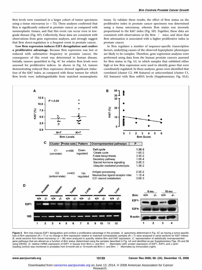
Brm levels were examined in a larger cohort of tumor specimensusing a tissue microarray (n = 72). These analyses confirmed thatBrm is significantly reduced in prostate cancer as compared withnonneoplastic tissues, and that this event can occur even in low-grade disease (Fig. 4D). Collectively, these data are consistent withobservations from gene expression analyses, and strongly suggestthat Brm down-regulation is a frequent event in prostate cancer.Low Brm expression induces E2F1 deregulation and confers
a proliferative advantage. Because Brm expression was lost orreduced with substantive frequency in prostate cancer, theconsequence of this event was determined in human disease.Initially, tumors quantified in Fig. 4C for relative Brm levels wereassessed for proliferative indices. As shown in Fig. 5A , tumorsdemonstrating reduced Brm expression showed significant induc-tion of the Ki67 index, as compared with those tumors for whichBrm levels were indistinguishable from matched nonneoplastic
tissue. To validate these results, the effect of Brm status on theproliferative index in prostate cancer specimens was determinedusing a tissue microarray, wherein Brm status was inverselyproportional to the Ki67 index (Fig. 5B). Together, these data areconsistent with observations in the Brm�/� mice, and show thatBrm attenuation is associated with a higher proliferative index inprostate cancer.
As Brm regulates a number of sequence-specific transcriptionfactors, underlying causes of the observed hyperplastic phenotypesare likely to be complex. Therefore, gene expression analyses wereperformed using data from the human prostate cancers assessedfor Brm status in Fig. 4A , in which samples that exhibited eitherhigh or low Brm expression were used to identify genes that werecoordinately regulated. In these analyses, genes were identified thatcorrelated (cluster C2, 498 features) or anticorrelated (cluster C1,422 features) with Brm mRNA levels (Supplementary Fig. S5A).
Figure 5. Brm loss induces E2F1 deregulation and confers a proliferative advantage in the prostate. A, specimens determined in Fig. 4C as having a tumor-specificloss of Brm expression (N > T) or no change in Brm expression relative to matched nonneoplastic samples (N = T) were analyzed in serial sections for Ki67 indices.B, serial sections from tissue microarray (n = 36) were analyzed to quantify relative Brm and Ki67 expression. C, representation of statistically overrepresentedgene pathways that are altered as a function of Brm status (determined using the samples described in Fig. 4A and identified as per Supplementary Figs. S5 and S6using DAVID). D, relative mRNA expression of E2F1 in tissues from Brm+/+ and Brm�/� littermates (left); protein expression of E2F1, E2F4, and h-actin(loading control) was monitored in prostates from 6-month-old or 10-month-old Brm+/+ and Brm�/� littermates by immunoblot (right ).
Brm Controls Prostate Cancer Growth
www.aacrjournals.org 10159 Cancer Res 2008; 68: (24). December 15, 2008
Research. on June 13, 2014. © 2008 American Association for Cancercancerres.aacrjournals.org Downloaded from

Functional enrichment analyses of each group were performed,and a summary of overrepresented biological pathway alterationsand gene lists are provided in Supplementary Fig. S5B . Gene listsare provided in Supplementary Fig. S6. Given the alterations ofproliferative rate observed in Brm�/� mice, it was noteworthythat alterations in cell cycle control were observed in tumors withlow Brm�/� expression. Analyses of corresponding alterationsrevealed a significant pattern of gene regulation that would bepredicted to confer a proliferative advantage (summarized inSupplementary Fig. S5C) associated with E2F1 deregulation. Forexample, activation of transforming growth factor-h suppressescell cycle control, mediated in part through the ability of Smad2/3and Smad4 transcriptional complexes to induce the p15ink4b andp21cip1 CDK inhibitors (35, 36). Notably, reductions in Smad2 andSmad4 expression were associated with Brm loss; conversely,induction of tumor necrosis factor-a (which can suppress Smadsignaling) also correlated with low Brm expression. These collectivedata indicate that loss of Brm likely relieves the suppression of thecell cycle by facilitating G1 CDK activity, and it would be predictedthat this event would induce excessive RB phosphorylation andpromote G1 progression. More proximal to this pathway, it was alsoobserved that Brm down-regulation significantly correlated withreduced expression of RB itself, and down-regulation of the p27kip1tumor suppressor. Despite these observations, cross-comparisonwith published data sets of RB target genes revealed that only asubset of RB targets were deregulated upon Brm loss (data notshown). Paramount among these was E2F1, a critical mediator ofboth cell cycle progression and cell death, which was induced intumors with low Brm mRNA levels. Indeed, E2F1 anticorrelatedwith Brm expression (two independent probe sets with Pearsoncorrelations of �0.628 and �0.625); in the second data set(Supplementary Fig. S4), Pearson coefficients were �0.931 and�0.844. As these data potentially implicated Brm as an effector ofE2F regulation, E2F1 levels were assessed in the murine prostate.
Consistent with expression analyses in human tumors, E2F1 wasenhanced in the prostates of Brm�/� animals at both the mRNA(Fig. 5D, left) and protein levels (right). Alteration in E2F1 activitywas specific in this tissue, as no alterations were observed withE2F4. Notably, no E2F1 deregulation was observed in tissuesreported to be enlarged in Brm�/� mice (liver and heart). Inaddition, no change was observed in tissues not reported tobe hyperplastic (lung), in tissues that are slightly reduced in sizeupon Brm loss (testes), or in derived murine embryonic fibroblasts(Fig. 5D ; Supplementary Fig. S7). Unexpectedly, a modest inductionof E2F1 was observed in the spleen, which is slightly reduced insize in Brm�/� animals. However, the most striking alterationobserved was increased E2F1 expression in the prostate. Thus,E2F1 deregulation in response to Brm loss seems to be highlycontext-specific, and is not required in other tissue types for ahyperproliferative phenotype. Together, these data indicate thatloss of Brm expression is sufficient to induce E2F1 deregulationand hyperproliferation in cells of prostatic origin. In summary, thestudies herein implicate Brm as a novel suppressor of androgen-dependent proliferation in the prostate, and show that Brm loss issufficient to induce a tissue-specific and E2F1-associated prolifer-ative advantage in this tissue type.
Discussion
The present study shows that loss of the Brm ATPase is sufficientto induce a hyperplastic phenotype in murine models of the
prostate and that Brm down-regulation is observed with highfrequency in prostate cancer. Although, Brm function has beenproposed to influence AR function in vitro , functional studies inmurine models show that AR signaling is retained in Brm�/�animals. By contrast, and consistent with a role in prostate cancerprogression, Brm loss is associated with the transition to castrationresistance. These outcomes are underpinned by observations inboth the Brm�/� prostates and analyses of gene expressionprofiles in human prostate cancer, wherein attenuation of Brm wasassociated with a high proliferative index and E2F1 deregulation.Combined, these data identify Brm as a critical modulator of E2F1expression and growth control in this tissue, and suggest that lossof Brm in prostate cancer confers a proliferative advantage.
The demonstration that Brm down-regulation occurs with highfrequency in prostate cancer was not expected, as Brm�/� micehave not been reported to develop tumors in tissues examinedpreviously (16). However, challenge of Brm�/� mice with lungcarcinogens was reported to increase adenoma formation (37), andloss of Brm expression has been decisively demonstrated in humanlung cancer (38). This latter event was shown to occur throughepigenetic regulation and silencing of Brm mRNA expression. Bycontrast, recently reported Brm loss in gastric cancers occurs as aresult of posttranscriptional Brm mRNA regulation (39). Althoughthe mechanism by which Brm is down-regulated in human prostatecancer remains unexplored, gene expression arrays are suggestiveof mRNA regulation. While this study was in preparation, a reportemerged which suggested that Brg1 mRNA levels are elevated inprostate cancer, especially under conditions wherein Brm expres-sion levels were low (40). Ectopic expression of Brg1 (but not Brm)in cultured cell models increased the invasive capacity of AR-negative PC3 cells, therefore further suggesting that Brg1 and Brmserve differential functions in cells of prostatic origin. In thepresent study, no significant alteration in Brg1 mRNA was observedin Brm-deficient prostate tissues, either in the murine epitheliaor in human prostate cancer (data not shown). Thus, it is notanticipated that the proliferative phenotypes observed result fromBrg1 deregulation.
The result of Brm down-regulation in both human disease andtargeted deletion in murine prostates seems to be the induction ofa hyperplastic phenotype. Although observed only in the VP andAP, lobe-specific hyperplasia is a frequently observed event afteroncogenic insult in the murine prostate (30, 31). Although theprocess that contributes to lobe-specific outcomes are of interest,our data showing that Brm expression is inversely correlated tothe proliferative index in prostate cancer and the murine prostateprovide compelling evidence that Brm loss confers a growthadvantage to this tissue. This outcome seems to be tissue-specific,as previous analyses of Brm�/� animals revealed both organ-specific increases and decreases in weight (16). More detailedanalyses of proliferative defects in Brm�/� have been previouslyinvestigated in MEFs, which retain serum dependence, show littlechange in cell cycle kinetics after serum stimulation, and do notform multilayers, but are partially compromised in the cellularresponse to contact inhibition or UV-induced DNA damage (16).Whether these phenotypes contribute to the proliferative advantageobserved in the prostate, and the cause of observed tissue-specificeffects in Brm�/� animals, will be the focus of future studies.
Particular to prostate cancer, it was surprising to observe thatAR signaling was refractory to Brm loss, both in the murineprostates and as correlated with Brm down-regulation in humandisease. Based on studies performed in vitro (22, 23), it was
Cancer Research
Cancer Res 2008; 68: (24). December 15, 2008 10160 www.aacrjournals.org
Research. on June 13, 2014. © 2008 American Association for Cancercancerres.aacrjournals.org Downloaded from

expected that AR activity might be compromised upon perturba-tion of this SWI/SNF subunit. Whether retained AR signaling in theabsence of Brm is attributed to developmental plasticity remainsan unexplored but clinically relevant question becauseAR target gene expression was unaltered in tumors with reducedBrm expression. Although Brg1 expression was not enhanced inBrm�/� epithelia, it is possible that Brg1-containing complexesare sufficient to sustain AR activity, and/or that the chromatinremodeling needs of AR are satisfied by other chromatinremodeling complexes. Interestingly, down-regulation ofSMARCA3 (HLTF) and SMARCA5 (ISWI/SNF2H) was significantlyobserved in conjunction with Brm down-regulation in cancerspecimens (Supplementary Fig. S6), thus indicating that lossof Brm likely induces the perturbation of other chromatinremodeling pathways that may facilitate AR signaling.
Although AR activity was retained in Brm�/� epithelia(evidenced by sustained probasin expression in unchallengedanimals and uncompromised response to androgen re-supplemen-tation), a major implication of the present study is that Brmloss was sufficient to induce castration-resistant proliferation in asubset of prostatic epithelia. This may be of clinical importance,as the transition to serum androgen independence typicallyrepresents the development of incurable disease. Based on geneexpression profiling of Brm-deficient tumors, at least oneunderlying mechanism is hypothesized to occur through theregulation of the retinoblastoma tumor suppressor (RB)/E2F1 axis.Prior studies showed that RB directly requires SWI/SNF functionto exert negative control over E2F target genes that are critical forS phase progression (41–43); indeed, E2F1 itself is a known E2Ftarget gene. Moreover, pathway analyses from human specimensrevealed that several upstream alterations that would result in RBinactivation (down-regulation of Smad2/3, Smad4, p27kip1, and RBitself) likely contribute to the observed induction of E2F1. In theprostate, loss of RB function has been shown to convey resistanceto androgen ablation and AR antagonist therapies in multiplein vitro systems (44), and targeted deletion of RB in prostaticepithelia provides a proliferative advantage in vivo (45). Thus, Brmdown-regulation may represent a mechanism to partially suppressRB function in this tissue. Because elevated E2F1 is associatedwith both the induction of cellular proliferation and apoptosis (46),these data likely explain the observed outcomes in Brm�/�prostatic epithelia. Based on these findings, it is hypothesized thatthese downstream effects of E2F1 deregulation may actually hinderthe progression of the hyperplastic phenotype, and the relevance ofthis supposition for human disease is being explored.
One additional mechanism that could contribute to thecastration-resistant phenotype is alterations in local hormone
synthesis. Although there are multiple mechanisms that contributeto resurgent AR activity in therapy-resistant tumors (25), it is nowapparent that a substantive percentage of castration-resistantprostate cancers may arise from the activation of intracrinede novo androgen synthesis within the tumor microenvironment(47–49). Indeed, analysis of patients with recurrent tumors afterhormone therapy supports this contention, and the recentobservation that an irreversible Cyp17 inhibitor, abiraterone, canreduce prostate-specific antigen in patients that failed hormonetherapy highlights the clinical relevance of this supposition (50). It isof interest that four effectors of C21 steroid hormone metabolism(Cyp11A1 , which generates pregnenolone from cholesterol; Cyp11B2 ,which converts corticosterone to aldosterone; Cyp17A1 , whichassists pregnenolone and DHEA production, but has also beenrecently implicated in a backdoor pathway for steroid production;and Cyp21A7 , which hydroxylates progesterone) were anticorrelatedwith Brm levels in gene expression arrays from human tumors. Thus,alterations in Brm may affect steroid hormone synthesis. However,serum testosterone levels were indistinguishable from Brm-positiveanimals in castration/re-supplementation studies (SupplementaryFig. S3B). As such, the current data do not support the contentionthat Brm loss increases serum testosterone levels; however, the localeffect within the prostate cannot be excluded and is the focus ofongoing study.
In summary, the data presented herein show that Brm lossresults in prostate-specific hyperplasia, the transition to androgenindependence, and deregulated E2F1 expression after targetedablation in murine tissues. These events hold consequence inhuman disease, wherein down-regulation of Brm is associated witha high proliferative index. Together, these data reveal a putativetumor suppressor function for Brm in the prostate, and suggestthat loss of this protein is sufficient to confer a proliferativeadvantage in this tissue.
Disclosure of Potential Conflicts of Interest
No potential conflicts of interest were disclosed.
Acknowledgments
Received 5/13/2008; revised 9/19/2008; accepted 10/10/2008.Grant support: R01 CA116777 and R21 CA121402 (K.E. Knudsen), CA91048 and
CA102848 (B. Weissman), and Canceropole Ile-de-France (C. Muchardt).The costs of publication of this article were defrayed in part by the payment of page
charges. This article must therefore be hereby marked advertisement in accordancewith 18 U.S.C. Section 1734 solely to indicate this fact.
We thank Drs. R. Matusik and S. Hayward for instruction on prostate resection andpathology. We also recognize the contributions of M. Schiewer and Dr. E. Knudsen forcritical commentary and ongoing discussions.
Brm Controls Prostate Cancer Growth
www.aacrjournals.org 10161 Cancer Res 2008; 68: (24). December 15, 2008
References
1. Muchardt C, Yaniv M. ATP-dependent chromatinremodelling: SWI/SNF and Co. are on the job. J MolBiol 1999;293:187–98.2. Vignali M, Hassan AH, Neely KE, Workman JL. ATP-dependent chromatin-remodeling complexes. Mol CellBiol 2000;20:1899–910.3. Wang W. The SWI/SNF family of ATP-dependentchromatin remodelers: similar mechanisms for diversefunctions. Curr Top Microbiol Immunol 2003;274:143–69.4. Trotter KW, Archer TK. Nuclear receptors andchromatin remodeling machinery. Mol Cell Endocrinol2007;265–266:162–7.
5. Trotter KW, Archer TK. The BRG1 transcriptionalcoregulator. Nucl Recept Signal 2008;6:e004.6. Kadam S, Emerson BM. Transcriptional specificity ofhuman SWI/SNF BRG1 and BRM chromatin remodelingcomplexes. Mol Cell 2003;11:377–89.7. Wang W, Xue Y, Zhou S, Kuo A, Cairns BR, CrabtreeGR. Diversity and specialization of mammalian SWI/SNF complexes. Genes Dev 1996;10:2117–30.8. Moshkin YM, Mohrmann L, van Ijcken WF, VerrijzerCP. Functional differentiation of SWI/SNF remodelers intranscription and cell cycle control. Mol Cell Biol 2007;27:651–61.9. Bultman SJ, Gebuhr TC, Magnuson T. A Brg1 mutationthat uncouples ATPase activity from chromatin remod-
eling reveals an essential role for SWI/SNF-relatedcomplexes in h-globin expression and erythroid deve-lopment. Genes Dev 2005;19:2849–61.10. Sumi-Ichinose C, Ichinose H, Metzger D, ChambonP. SNF2h-BRG1 is essential for the viability of F9murine embryonal carcinoma cells. Mol Cell Biol1997;17:5976–86.11. Lessard J, Wu JI, Ranish JA, et al. An essential switchin subunit composition of a chromatin remodeling com-plex during neural development. Neuron 2007;55:201–15.12. Griffin CT, Brennan J, Magnuson T. The chromatin-remodeling enzyme BRG1 plays an essential role inprimitive erythropoiesis and vascular development.Development 2008;135:493–500.
Research. on June 13, 2014. © 2008 American Association for Cancercancerres.aacrjournals.org Downloaded from

Cancer Research
Cancer Res 2008; 68: (24). December 15, 2008 10162 www.aacrjournals.org
13. Gebuhr TC, Kovalev GI, Bultman S, Godfrey V, Su L,Magnuson T. The role of Brg1, a catalytic subunit ofmammalian chromatin-remodeling complexes, in T celldevelopment. J Exp Med 2003;198:1937–49.14. Indra AK, Dupe V, Bornert JM, et al. Temporallycontrolled targeted somatic mutagenesis in embryonicsurface ectoderm and fetal epidermal keratinocytesunveils two distinct developmental functions ofBRG1 in limb morphogenesis and skin barrier forma-tion. Development 2005;132:4533–44.15. Matsumoto S, Banine F, Struve J, et al. Brg1 isrequired for murine neural stem cell maintenance andgliogenesis. Dev Biol 2006;289:372–83.16. Reyes JC, Barra J, Muchardt C, Camus A, Babinet C,Yaniv M. Altered control of cellular proliferation in theabsence of mammalian brahma (SNF2a). EMBO J 1998;17:6979–91.17. Sowery RD, So AI, Gleave ME. Therapeutic options inadvanced prostate cancer: present and future. Curr UrolRep 2007;8:53–9.18. Klotz L. Hormone therapy for patients with prostatecarcinoma. Cancer 2000;88:3009–14.19. Balk SP. Androgen receptor as a target in androgen-independent prostate cancer [discussion 8–9]. Urology2002;60:132–8.20. Kim J, Coetzee GA. Prostate specific antigen generegulation by androgen receptor. J Cell Biochem 2004;93:233–41.21. Hernandez J, Thompson IM. Prostate-specific anti-gen: a review of the validation of the most commonlyused cancer biomarker. Cancer 2004;101:894–904.22. Marshall TW, Link KA, Petre-Draviam CE, KnudsenKE. Differential requirement of SWI/SNF for androgenreceptor activity. J Biol Chem 2003;278:30605–13.23. Klokk TI, Kurys P, Elbi C, et al. Ligand-specificdynamics of the androgen receptor at its responseelement in living cells. Mol Cell Biol 2007;27:1823–43.24. Culig Z, Klocker H, Bartsch G, Hobisch A. Androgenreceptors in prostate cancer. Endocr Relat Cancer 2002;9:155–70.25. Feldman BJ, Feldman D. The development ofandrogen-independent prostate cancer. Nat Rev Cancer2001;1:34–45.26. Knudsen KE, Arden KC, Cavenee WK. Multiple G1regulatory elements control the androgen-dependentproliferation of prostatic carcinoma cells. J Biol Chem1998;273:20213–22.
27. Glinsky GV, Glinskii AB, Stephenson AJ, HoffmanRM, Gerald WL. Gene expression profiling predictsclinical outcome of prostate cancer. J Clin Invest 2004;113:913–23.28. Irizarry RA, Hobbs B, Collin F, et al. Exploration,normalization, and summaries of high density oligo-nucleotide array probe level data. Biostatistics 2003;4:249–64.29. Huang da W, Sherman BT, Tan Q, et al. The DAVIDgene functional classification tool: a novel biologicalmodule-centric algorithm to functionally analyze largegene lists. Genome Biol 2007;8:R183.30. Stanbrough M, Leav I, Kwan PW, Bubley GJ, Balk SP.Prostatic intraepithelial neoplasia in mice expressing anandrogen receptor transgene in prostate epithelium.Proc Natl Acad Sci U S A 2001;98:10823–8.31. Ellwood-Yen K, Graeber TG, Wongvipat J, et al.Myc-driven murine prostate cancer shares molecularfeatures with human prostate tumors. Cancer Cell 2003;4:223–38.32. Denmeade SR, Lin XS, Isaacs JT. Role of programmed(apoptotic) cell death during the progression andtherapy for prostate cancer. Prostate 1996;28:251–65.33. Mirosevich J, Bentel JM, Zeps N, Redmond SL,D’Antuono MF, Dawkins HJ. Androgen receptor expres-sion of proliferating basal and luminal cells in adultmurine ventral prostate. J Endocrinol 1999;162:341–50.34. Varambally S, Yu J, Laxman B, et al. Integrativegenomic and proteomic analysis of prostate cancerreveals signatures of metastatic progression. Cancer Cell2005;8:393–406.35. Seoane J, Le HV, Shen L, Anderson SA, Massague J.Integration of Smad and forkhead pathways in thecontrol of neuroepithelial and glioblastoma cell pro-liferation. Cell 2004;117:211–23.36. Seoane J, Pouponnot C, Staller P, Schader M, Eilers M,Massague J. TGFh influences Myc, Miz-1 and Smad tocontrol the CDK inhibitor p15INK4b. Nat Cell Biol 2001;3:400–8.37. Glaros S, Cirrincione GM, Muchardt C, Kleer CG,Michael CW, Reisman D. The reversible epigeneticsilencing of BRM: implications for clinical targetedtherapy. Oncogene 2007;26:7058–66.38. Reisman DN, Sciarrotta J, Wang W, Funkhouser WK,Weissman BE. Loss of BRG1/BRM in human lung cancercell lines and primary lung cancers: correlation withpoor prognosis. Cancer Res 2003;63:560–6.
39. Yamamichi N, Inada K, Ichinose M, et al. Frequentloss of Brm expression in gastric cancer correlates withhistologic features and differentiation state. Cancer Res2007;67:10727–35.40. Sun A, Tawfik O, Gayed B, et al. Aberrant expressionof SWI/SNF catalytic subunits BRG1/BRM is associatedwith tumor development and increased invasiveness inprostate cancers. Prostate 2007;67:203–13.41. Strobeck MW, Knudsen KE, Fribourg AF, et al. BRG-1is required for RB-mediated cell cycle arrest. Proc NatlAcad Sci U S A 2000;97:7748–53.42. Strobeck MW, Reisman DN, Gunawardena RW,et al. Compensation of BRG-1 function by Brm: insightinto the role of the core SWI-SNF subunits in retino-blastoma tumor suppressor signaling. J Biol Chem 2002;277:4782–9.43. Zhang HS, Gavin M, Dahiya A, et al. Exit from G1 andS phase of the cell cycle is regulated by repressorcomplexes containing HDAC-Rb-hSWI/SNF and Rb-hSWI/SNF. Cell 2000;101:79–89.44. Sharma A, Comstock CE, Knudsen ES, et al.Retinoblastoma tumor suppressor status is a criticaldeterminant of therapeutic response in prostate cancercells. Cancer Res 2007;67:6192–203.45. Maddison LA, Sutherland BW, Barrios RJ, GreenbergNM. Conditional deletion of Rb causes early stageprostate cancer. Cancer Res 2004;64:6018–25.46. DeGregori J, Johnson DG. Distinct and overlappingroles for E2F family members in transcription,proliferation and apoptosis. Curr Mol Med 2006;6:739–48.47. Singh P, Coe J, Hong W. A role for retinoblastomaprotein in potentiating transcriptional activation bythe glucocorticoid receptor. Nature 1995;374:562–5.48. Locke JA, Guns ES, Lubik AA, et al. Androgen levelsincrease by intratumoral de novo steroidogenesis duringprogression of castration-resistant prostate cancer.Cancer Res 2008;68:6407–15.49. Montgomery RB, Mostaghel EA, Vessella R, et al.Maintenance of intratumoral androgens in metastaticprostate cancer: a mechanism for castration-resistanttumor growth. Cancer Res 2008;68:4447–54.50. Attard G, Reid AH, Yap TA, et al. Phase I clinicaltrial of a selective inhibitor of CYP17, abirateroneacetate, confirms that castration-resistant prostatecancer commonly remains hormone driven. J ClinOncol 2008;26:4563–71.
Research. on June 13, 2014. © 2008 American Association for Cancercancerres.aacrjournals.org Downloaded from