Characterisation of Eucalyptus grandis SWEET and SWI/SNF ...
122
Characterisation of Eucalyptus grandis SWEET and SWI/SNF proteins during symbiosis with Pisolithus microcarpus Christian Benedict Aguirre Masters of Research Thesis Masters of Research Western Sydney University April 2017
-
Upload
khangminh22 -
Category
Documents
-
view
1 -
download
0
Transcript of Characterisation of Eucalyptus grandis SWEET and SWI/SNF ...
Characterisation of Eucalyptus grandis SWEET
and SWI/SNF proteins during symbiosis with
Pisolithus microcarpus
Christian Benedict Aguirre
Masters of Research Thesis
Masters of Research
Western Sydney University
April 2017

Acknowledgements
I would like to take this opportunity to thank all who had been helping me for
the past two years. This work is deeply indebted to my supervisors Professor
Jonathan Plett, Krista Plett, Ian Anderson and all other staff and students
with whom I have been privileged to work with over the last two years.
Without their guidance, critics and encouragements, I would not have
achieved as much in plant-microbe research.
I am truly thankful for the precious opportunities Dr Plett had provided me
throughout my years at the Hawkesbury Institute for the Environment (HIE). I
would also like to thank my thesis committee, Professor Markus Riegler,
Professor Elise Pendall and Professor Jennifer Morrow for reviewing my
thesis proposal and arranging time for my oral presentation. My special
thanks also goes out to Dr Johanna Wong, Professor Jeff Powel, Professor
Jenn Walker, Professor Suzanne Donn, Professor Barbara Drigo, Professor
Sara Hortal Botifoll and Professor Jenn Wollemi Pine Researcher for their
expert guidance throughout all my time at HIE.
I humbly acknowledge the assistance of fellow members at HIE who had
consistently provided me a lot of supports, encouragements and laughter.
Special thanks to Jonathan Plett, Krista Plett and Johanna Wong who had
given technical support and solutions for the Yeast I and II analyses, cross
sectioning of roots, dsiRNA production and carbon capture analyses. Finally,
special thanks to my family for all the love and support they give me. Their
support at every stage in this master degree was very important to me.
AGUIRRE, Christian Benedict

Statement of Authentication
The work presented in this thesis is, to the best of my knowledge and belief,
original except as acknowledged in the text. I hereby declare that I have not
submitted this material, either in full or in part, for a degree at this or any
other institution. All experimental work reported in this thesis was performed
by the author, unless stated otherwise.
AGUIRRE, Christian Benedict
i
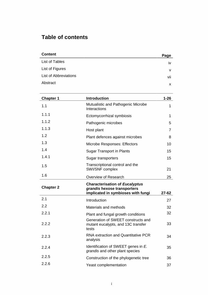
Table of contents
Content Page
List of Tables iv
List of Figures v
List of Abbreviations vii
Abstract x
Chapter 1 Introduction 1-26
1.1 Mutualistic and Pathogenic Microbe Interactions
1
1.1.1 Ectomycorrhizal symbiosis 1
1.1.2 Pathogenic microbes 5
1.1.3 Host plant 7
1.2 Plant defences against microbes 8
1.3 Microbe Responses: Effectors 10
1.4 Sugar Transport in Plants 15
1.4.1 Sugar transporters 15
1.5 Transcriptional control and the SWI/SNF complex 21
1.6 Overview of Research 25
Chapter 2 Characterisation of Eucalyptus grandis hexose transporters implicated in symbioses with fungi 27-62
2.1 Introduction 27
2.2 Materials and methods 32
2.2.1 Plant and fungal growth conditions 32
2.2.2 Generation of SWEET constructs and mutant eucalypts, and 13C transfer tests
33
2.2.3 RNA extraction and Quantitative PCR analysis
34
2.2.4 Identification of SWEET genes in E. grandis and other plant species
35
2.2.5 Construction of the phylogenetic tree 36
2.2.6 Yeast complementation 37

ii
2.2.7 Glucose efflux test 38
2.2.8 GFP localisation 39
2.3 Results 41
2.3.1 Phylogenetic relationships of E. grandis SWEET-like transporters
41
2.3.2 SWEET gene expression in E. grandis tissues
43
2.3.3 E. grandis roots exhibit different morphologies when in contact with different fungal lifestyles
45
2.3.4 SWEET-like genes exhibit differential expression in E. grandis roots during challenge by different fungi
47
2.3.5 E. grandis SWEET-like genes encodes STPs that localise to the plasma membrane of plant cells
49
2.3.6 E. grandis SWEET-like proteins can act as sugar symporters
51
2.3.7 Altered expression of EgSWEET-like genes in E. grandis roots affects carbon export from E. grandis roots
54
2.4 Discussion 59
Chapter 3 Characterisation of Pisolithus albus effector MiSSP9.7 and its interactant, Eucalyptus grandis SWI3D 63-86
3.1 Introduction 63
3.2 Materials and methods 67
3.2.1 MiSSP9.7-GFP production and absorption by eucalypt root cells 67
3.2.2 Yeast One- and Yeast Two-Hybrid analyses 68
3.2.3 BiFC testing in E. grandis 69
3.2.4 Construction of the phylogenetic tree 70
3.2.5 Plant and fungal growth conditions 70
3.2.6 RNA extraction and Real Time Quantitative PCR (RT-QPCR) 71
3.2.7 Generation of SWI3D constructs and transgenic eucalypts 71
3.2.8
Preparation of double stranded interfering RNAs (dsiRNAs) and treatment of roots undergoing colonization by P. albus.
73

iii
3.2.9 Microscopy of transgenic/dsiRNA treated eucalypt roots and Hartig net measurements
74
3.3 Results 75
3.3.1 MiSSP9.7-GFP encodes an effector protein that enters plant root cells
75
3.3.2 MiSSP9.7 interacts with a chromatin remodelling complex (CRC) subunit of E. grandis
77
3.3.3 Phylogenetic relationships of E. grandis SWI3 proteins
80
3.3.4 SWI3 gene expression in E. grandis roots over a two-week time course of colonization by P. albus
82
3.3.5 Altered expression of MiSSP9.7 significantly affects P. albus Hartig net development during colonisation
84
3.4 Discussion 87
Chapter 4 Conclusion and Future Perspective 90-91
References 92-108

iv
List of tables
Table Title Page
Table 1.
Forward (F) and Reverse (R) primers used for overexpression of Euc.grK02694 and Eucgr.B00363 using 35s promoter, and repression of Eucgr.K02688 and Eucgr.L02615 using RNAi knockdown.
58

v
List of figures
Figure Title Page
Figure 1 Representational diagram of a transverse cross-section of a root undergoing colonisation by an ECM fungus
4
Figure 2 Representational diagram of the intracellular distribution of plant sugar transporter proteins 16
Figure 3 The subunits of Arabidopsis thaliana SWI/SNF complex 22
Figure 4 Phylogenetic relationships between SWEET-like proteins collected from different species. 42
Figure 5 Expression profile of 52 SWEET-like gene throughout different tissue in E. grandis: Shoot apex, stem, leaves and root.
44
Figure 6 Images taken of 2 weeks old and E. grandis under different conditions.
46
Figure 7
Regulation of 52 E. grandis SWEET genes when E. grandis associates with: parasites (Phytophthora, Armillaria and S. granulatus), a non-Eucalyptus coloniser (F. oxysporum and S. luteus), or a mutualistic fungus (P. microcarpus strains).
48
Figure 8
Localisation of SWEET-like proteins Eucgr.K02694, Eucgr.K02688, Eucgr.B00363, Eucgr.L02615-GFP fusions in yeast (VW4000).
50
Figure 9
Transport activity of EgSWEET K02694, K02688, B00363, L02615 in yeast. These SWEET proteins complemented VW4000 S. cerevisiae mutants (which lacked 18 hexose transporter genes).
52
Figure 10
Glucose export activity of heterologously expressed Eucgr.K02694, Eucgr.K02688, Eucgr.B00363, Eucgr.L02615 SWEET-like sugar transporter proteins in EBY.VW4000 yeast.
53
vi
Figure 11 Amount of 13C obtained by different types of fungi when associating with wildtype E. grandis relative to each respective control over a time period of 2 weeks.
56
Figure 12 Amount of 13C obtained by SI-12 when associating with mutant eucalypt relative to control SI-12 over a time period of 2 weeks.
57
Figure 13 Images of MiSSP9.7-GFP (i.e. Pa683008-GFP) and Propidium Iodide (PI) stain taken using confocal microscopy.
76
Figure 14 MiSSP9.7 (i.e. Pa683008) interacts with E. grandis nuclear protein SWI3D (Eucgr.I01260).
78
Figure 15 In vivo BiFC proof of the interaction between 683008 and SWI3D.
79
Figure 16 Phylogenetic relationships between SWI3 proteins collected from different species.
81
Figure 17 RT-QPCR of SWI3 genes in E. grandis over a time course of 2 weeks.
83
Figure 18
Altered expression of SWI3D (A) or repression of MiSSP9.7 (Pa683008)by dsiRNA (B) and its effect on Hartig net development.
85
Figure 19 Percent mycorrhization of SWI3D (i.e. Eucgr.I01261) mutant eucalypts.
86

vii
List of abbreviations
∆d13C Change in amount of 13C
13C Carbon isotope 13
13CO2 Carbon (isotope 13) dioxide
ABA Abscisic acid
AM Arbuscular mycorrhizal
Avr Avirulence
AzA Azelaic acid
BiFC Bimolecular fluorescence complementation
BR Brassinosteroids
BzSA Benzoylsalicylic acid
cDNA Copy deoxyribonucleic acid
CK Cytokinins
CO2 Carbon dioxide
CSIRO Commonwealth Scientific and Industrial Research
Organisation
DA Dehydroabietinal
DNA Deoxyribonucleic acid
dsiRNA Double stranded interfering ribonucleic acid
ECM Ectomycorrhizal
ESL ERD six-like transporters
ET Ethylene
ETI Effector-triggered immunity
Fru Fructose
GA Gibberellin
viii
GFP Green fluorescent protein
Glu Glucose
H2O2 Hydrogen peroxide
HR Hypersensitive response
IAA Indole-3-acetic acid
INT Inositol transporter
JA Jasmonic acid
LiAC Lithium acetate
MiSSP Mycorrhizal induced small secreted protein
MMN Modified Mylin Norkin
MS medium Murashige and Skoog medium
MST Monosaccharide transporters
N Nitrogen
NLP Nep1-like proteins
P Phosphorus
PAMPs/MAMPs Pathogen/Microbe associated molecular patterns
PBS Phosphate-buffered saline
PCR Polymerase chain reaction
PEG Polyethylene glycol
pGlcT Plastidic glucose transporter
pH Salinity
PI Propidium Iodide
PiP Pipecolic acid
PMT Polyol monosaccharide transporter
PR Pathogenesis-related

ix
PRR Pattern recognition receptors
PTI/MTI PAMP/MAMP-triggered immunity
QPCR Quantitative polymerase chain reaction
RFP Red fluorescent protein
RNA Ribonucleic acid
RNAi Ribonucleic acid interference
ROS Reactive oxygen species
RT-QPCR Real time-quantitative polymerase chain reaction
SA Salicyclic acid
SAR Systemic acquired resistance
SDS Sodium dodecyl sulphate
SLs Strigolactones
STPs Sugar transporter proteins
Suc Sucrose
SUT Sucrose transporters
SWEET Sugars Will Eventually be Exported Transported
SWI/SNF Switch/Sucrose Non-Fermenting
TAL Transcription activator-like
TMT Tonoplast membrane transporter
VGT Vacuolar glucose transporter
WT Wild-type
YFP Yellow fluorescent protein
YIIH Yeast two-hybrid
YPM Yeast extract peptone with maltose (growth medium)
WGA-FITC Wheat germ agglutinin-Lectin

x
Abstract
Eucalyptus grandis, an economically important bioenergy tree, is constantly
bombarded by different fungal lifestyles seeking to acquire photosynthetically
fixed sugar. How the plant immune system filters beneficial fungi from
pathogenic is poorly understood. This thesis investigates two aspects of
plant immunity: shuttling of sugar and interference by fungal effectors. Plant
sugars are known to play a dual role in plant-microbe interactions: they can
either feed the microbe with growth-limiting carbon or they can act as fuel for
plant secondary metabolism and, subsequently, plant defence. In my first
study I consider how hexose SWEET transporters respond at the
transcriptomic level in E. grandis roots during challenge by different microbes
covering the fungal lifestyles from pathogenic through mutualistic. Further, I
characterise four E. grandis SWEET proteins that share sequence homology
to previously identified SWEET proteins and determine their cellular
localization, their sugar transport capabilities and their role in shuttling carbon
during plant-microbe interactions. In the second part of my thesis, I
investigate how a mutualistic fungus attempts to manipulate plant defences
through the use of effector like proteins. Specifically, I characterise the role
of Pisolithus albus MiSSP9.7, a highly induced secreted fungal protein of
unknown function. I demonstrate that it interacts with a member of the
SWI/SNF protein complex previously identified as being responsible for the
regulation of plant hormone signalling pathways used in immune responses
against microbes. Increased expression of SWI3D in E. grandis roots is tied
to the colonisation process and may regulate a key aspect of plant immunity
towards mutualistic fungi. Taken together, my work provides a better

xi
understanding of the controls used by plants to modulate plant-microbe
interactions and the counter-measures utilized by fungi to overcome host
immunity.
1
Chapter 1 Introduction
1.1 Mutualistic and Pathogenic Microbe Interactions
In their natural environment, plants are constantly confronted with many
different types of soil-borne microbes. Microbes is a broad term that
describes all microscopic organisms, such as bacteria, fungi, nematodes,
oomycetes, archaea, protists, microscopic animals, and microscopic plants
(Genetic Science Learning Center 2017). Plant interactions with these
microbes can be classified in three main categories: parasitic, mutualistic or
commensal. These classifications are an oversimplification as these plant-
microbe associations are dynamic and can range from mutualistic to parasitic
depending on the abiotic factors affecting the ecosystem (Francis and Read
1995). Parasitic plant-microbe interactions involve microbes colonising host
plants and hindering plant growth by feeding on plant tissues and or sugar
storages. Thus, one organism benefits at the others expense. In contrast,
mutualistic pant-microbe interactions involve microbes providing host plants
with scarce nutrients (such as nitrogen (N) and phosphorus (P)) in exchange
for (up to 30% of) the plant’s photosynthetically-derived sugars (Pellegrin et
al. 2015). Thus, both the plant and microbe benefit from this association.
Commensal plant-microbe interactions describe microbes who do not harm
or benefit plants, instead commensal microbes only decompose dead plant
matter (for example (e.g.) decomposing plant litter fall).
1.1.1 Ectomycorrhizal symbiosis
One major type of mutualistic plant-microbe interaction found in forest
ecosystems is the relationship between soilborne ectomycorrhizal (ECM)
2
fungi and trees. This symbiosis involves the transfer of growth limiting soil
nutrients from the fungus to the host plant and photosynthetically derived
sugars from the host to the fungus. ECM fungi play a further role in plant
survival as they support host adaptation to changing environmental
conditions such as climate extremes, drought and soil pollution (Redman et
al. 2009). ECM fungi are commonly used in nurseries to inoculate trees used
for re-forestation because the presence of ECM fungi increases the
establishment success of trees by enhancing tree growth (Brundrett et al.
2005).
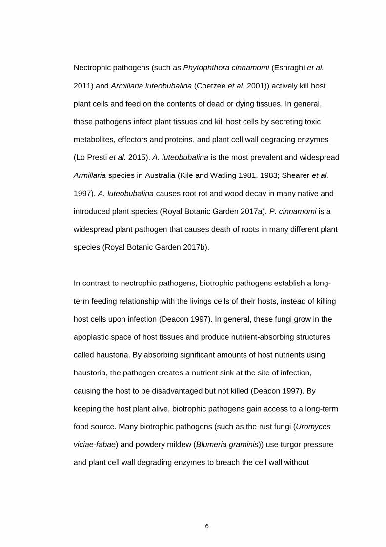
To establish mutualistic associations with plants and begin nutrient
exchange, ECM fungi must first form two essential ectomycorrhizal
structures: the Hartig net (formed within the root) and fungal sheath/mantle
(formed outside the root by surrounding the entire root tip with extrametrical
hyphae). Upon initial contact with host roots, ECM fungi attach to the root
surface and hyphae surrounds the outside of the root, forming the fungal
mantle (Fig. 1). During this contact, ECM fungi secrete effectors (i.e. proteins
and signaling molecules), metabolites and phytohormones (e.g. auxin) that
cause physiological changes within host roots cells to allow fungal hyphae to
penetrate into the root apoplast (i.e. spaces in between root cells) (Fig. 1).
Within the apoplast, the extensive network of fungal hyphae form the Hartig
net completing establishment of the mutualism. The Hartig net is the interface
in which nutrient exchange between the fungus and plant occurs and the
colonised root is called a mycorrhizal root tip (Smith and Read 1997).

3
Although ECM fungi are free-living they are inefficient decomposers when
compared to saprotrophs, thus they form mutualisms with the roots of trees
to gain access to sugars as a carbon source, improving their survivability
(Smith and Read 2008). ECM fungi are free-living because they originate
from saprotrophic ancestors (Hibbett et al. 2000), but have evolved multiple
times to be mutualistic with many plants (Hibbet and Matheny 2009).
Phylogenically, ECM fungi belong to the phyla Ascomycota and
Basidiomycota together with saprotrophic fungi (Plett and Martin 2011, 2015).
Ascomycota is a division of fungi that whose spores are contained in sac-like
structures called an ascus (Plett and Martin 2011). Basidiomycota is a
division of fungi who produce spores using a specialised spore producing
organ (called basidium) (Plett and Martin 2011).

4
Figure 1. Representational diagram of a transverse cross-section of a root undergoing colonisation by an ECM
fungus (adapted from Plett and Martin 2011). (A) Representation of a transverse cross-section of a plant lateral root
before ECM fungal colonisation. (B) Initial contact between host plant root (green cells) and ECM fungal hyphae
(brown cells). The fungus attaches to root surface and secretes effectors that cause physiological changes within
root cells, which then allow fungal hyphae to penetrate into the root apoplast. (C) Representation of a transverse
cross-section of a mature ectomycorrhizal root tip. At this stage of colonisation fungal hyphae has covered the entire
root surface forming a thick fungal mantle. Other hyphae have penetrated into the apoplastic space, forming the
Hartig net where nutrient exchange occurs.

5
Saprotrophic fungi produce enzymes (e.g. cellulases and hemicellulases)
that deconstruct and hydrolyse plant cell wall materials (Plett and Martin
2011). Biotrophic, hemi-biotrophic and nectrophic fungal pathogens (e.g.
Armillaria and Phytophthora) produce toxins, harmful effectors and
carbohydrate-cleaving enzymes that digest or rot plant tissues (Lo Presti et
al. 2015). These features make it hard, if not impossible, for saprotrophic and
pathogenic fungi to form a mutualistic relationship with plants because these
enzymes would damage the host and elicit plant defence responses (Plett
and Martin 2011). However, over the course of their evolution, ECM fungi
have lost a large majority of genes encoding plant cell wall degrading
enzymes (Martin et al. 2008). While the genomes of ECM fungi (e.g. Laccaria
bicolor) still encode a small group of plant cell wall degrading enzymes, these
genes are only expressed when the fungus acts as a weak decomposer in
soil litter and is not in symbiosis with a plant (Martin et al. 2008; Plett and
Martin 2011). The loss of plant cell wall degrading enzymes makes ECM
fungi more dependent on utilising photosynthetically derived sugars as
carbon source received from host plants, but in turn allows ECM fungi to
colonise roots without threatening the integrity of the plant root cells (Plett
and Martin 2011).
1.1.2 Pathogenic microbes
Pathogenic microbes syphon plant nutrients and or directly feed on plant
tissues (or plant sugar storages) for their own growth and development.
Pathogenic fungi are subdivided into three groups based on the way they
parasitise plants, these are: necrotrophs, biotrophs and hemi-biotrophs.
6
Nectrophic pathogens (such as Phytophthora cinnamomi (Eshraghi et al.
2011) and Armillaria luteobubalina (Coetzee et al. 2001)) actively kill host
plant cells and feed on the contents of dead or dying tissues. In general,
these pathogens infect plant tissues and kill host cells by secreting toxic
metabolites, effectors and proteins, and plant cell wall degrading enzymes
(Lo Presti et al. 2015). A. luteobubalina is the most prevalent and widespread
Armillaria species in Australia (Kile and Watling 1981, 1983; Shearer et al.
1997). A. luteobubalina causes root rot and wood decay in many native and
introduced plant species (Royal Botanic Garden 2017a). P. cinnamomi is a
widespread plant pathogen that causes death of roots in many different plant
species (Royal Botanic Garden 2017b).
In contrast to nectrophic pathogens, biotrophic pathogens establish a long-
term feeding relationship with the livings cells of their hosts, instead of killing
host cells upon infection (Deacon 1997). In general, these fungi grow in the
apoplastic space of host tissues and produce nutrient-absorbing structures
called haustoria. By absorbing significant amounts of host nutrients using
haustoria, the pathogen creates a nutrient sink at the site of infection,
causing the host to be disadvantaged but not killed (Deacon 1997). By
keeping the host plant alive, biotrophic pathogens gain access to a long-term
food source. Many biotrophic pathogens (such as the rust fungi (Uromyces
viciae-fabae) and powdery mildew (Blumeria graminis)) use turgor pressure
and plant cell wall degrading enzymes to breach the cell wall without
7
affecting host viability, after which they develop haustoria (O’Connell and
Panstruga 2006; Lo Presti et al. 2015).
Hemi-biotrophs use both biotrophic and necrotrophic methods of acquiring
host plant nutrients depending on the stages of their life cycle (Lee and Rose
2010). During initial infection, hemi-biotrophs establish a biotrophic
relationship with the host, but as they develop, they then later kill host cells
and feed on the contents of dead or dying tissues. Hemibiotrophic fungi such
as Phytophthora infestans and Magnaporthe oryzae initially develop bulged
biotrophic invasive hyphae that later change into thin necrotrophic hyphae
(O’Connell and Panstruga 2006).
1.1.3 Host plant
Eucalyptus grandis is an important forest tree that interacts with a variety of
microbes, including both mutualistic ECM and parasitic fungi. E. grandis is
the most widely planted hardwood forest tree because of its many industrial
and environmental uses (Myburg et al. 2014). The tree’s easy maintenance,
high adaptability and rapid growth has led to the adoption of Eucalypt
plantations in over 100 countries worldwide (Myburg et al. 2014). Planted
Eucalypts provide key renewable sources for the production of solid timber,
pulp, paper, bioenergy and biomaterials, while reducing human impacts on
native forests (Bauhus et al. 2010). Furthermore, Eucalypts provide many
environmental services including sequestering atmospheric CO2 to reduce
global warming, providing habitats for native Australian animals and soil,
water and forest conservation. Eucalypts also have a large diversity and high

8
concentration of essential oils that are key ingredients in commercial
products, as well as having medicinal and ecological functions (Myburg et al.
2014).
1.2 Plant defences against microbes
To survive, plants must effectively defend against pathogenic microbes.
Unlike animals, plants do not have mobile immune cells, a somatic adaptive
immune system, or circulatory system, instead plants rely on multifaceted
innate immune defences (Jones and Dangl 2006). The first line of defence
against most microbes consists of physical barriers, such as the waxy cuticle
on the surface of leaves, the cell wall and plasma membrane (Fu and Dong
2013). Furthermore, some plants produce chemicals, such as glycosylated
triterpenoids, saponins and reactive oxygen species (ROS), that can disrupt
the plasma membranes of most fungal pathogens (Bednarek and Osbourn
2009; Hemetsberger et al. 2012). Plant cells express pattern recognition
receptors (PRRs) on their surface that recognise invariant molecular patterns
found on invading microbes, called pathogen/microbe-associated molecular
patterns (PAMPs/MAMPs) (Jones and Dangl 2006; Fu and Dong 2013).
PAMPs/MAMPs are essential components found on microbes including,
short peptides, peptidoglycans, chitin, bacterial flagellin and
lipopolysaccharides. For example, the receptor kinase FLS2 acts as a PRR
by detecting bacterial flagellin (Yoon et al. 2012). When PAMPs/MAMPs are
recognised by PRRs, it leads to PAMP/MAMP-triggered immunity (PTI/MTI)
(Boller & Felix, 2009).
9
In addition, initial pathogen attacks induce the production and transfer of
signalling molecules (such as salicylic acid (SA), glycerol-3-phosphate (G3P),
diterpenoid dehydroabietinal (DA), benzoylsalicylic acid (BzSA), pipecolic
acid (Pip) and azelaic acid (AzA)) all throughout the plant (Anand et al. 2008;
Chanda et al. 2011; Chaturvedi et al. 2012; Kamatham et al. 2016; Reimer-
Michalski and Conrath 2016; Jung et al. 2009). These molecules stimulate
the expression of antimicrobial genes resulting in broad-spectrum resistance
against future infections in distal, uninfected plant tissues (Conrath 2006;
Durrant and Dong 2004; Fu and Dong 2013). This phenomenon, called
systemic acquired resistance (SAR), is conserved throughout many plant
species. SAR results in extended periods of resistance (ranging from several
weeks to months) against many different types of pathogens (Gao et al.
2015; Kuc 1987). Further, SAR establishes transgenerational immune
memory within plants (a process referred to as priming) (Mauch-Mani and
Mauch 2005; Luna et al. 2012; Rasmann et al. 2012; Slaughter et al. 2012).
This immune memory enables stronger and faster defence responses
against future pathogen attacks. Although some plant immune responses are
associated with cell death at the site of infection, SAR promotes cell survival
in uninfected plant tissues. However, immune responses and systemic
synthesis of SA are known to trigger SAR. SAR can also be induced by
invading fungi, bacteria, fungi, oomycetes and viruses (Conrath 2006;
Durrant and Dong 2004; Fu and Dong 2013; Ryals et al. 1996).
Plants produce a wide range of hormones, including salicylic acid (SA),
jasmonic acid (JA), ethylene (ET), auxin, abscisic acid (ABA), gibberellin
10
(GA), brassinosteroids (BR), cytokinins (CK), and strigolactones (SLs) (Bari
and Jones 2009; Gomez-Roldan et al. 2008; Umehara et al. 2008). These
hormones play essential roles in growth, responses to biotic and abiotic
stresses and in immune responses against pathogens (Robert-Seilaniantz et
al. 2007; Adie et al. 2007). SA is involved in the activation of defence
responses against biotrophic and hemi-biotrophic pathogens, and is a key
contributor to SAR (Grant and Lamb 2006). SA levels increase in infected
plant tissues and high levels activate PR genes, enhancing resistance to a
wide spectrum of pathogens (Denance et al. 2013). JA and ET are generally
involved in defence against necrotrophic pathogens and herbivorous insects
(Bari and Jones 2009). Auxin and ABA contribute to plant play important
roles as signaling molecules in plant defence responses (Zhang et al. 2007;
Mauch-Mani and Mauch 2005; Navarro et al 2008;). GA and BR enhances
resistance to biotrophic pathogens (Bari and Jones 2009). Cytokinins act as
signaling molecules regulating plant defence responses against some
pathogens (Bari and Jones 2009). SLs are involved in establishing resistance
to specific bacterial and fungal pathogens (Marzec 2016).
1.3 Microbe Responses: Effectors
To overcome plant defences and establish either infection or mutualistic
associations, microbes have evolved various mechanisms. One mechanism
utilised by both pathogenic and mutualistic fungi is the use of small secreted
molecules called effectors. Effectors suppress the host plant’s immune
responses and modulates host cell physiology (Giraldo and Valent 2013;
Plett et al. 2011, 2014a, b). Thus, pathogenic fungi secrete effectors to

11
establish infection, whereas mutualistic fungi secrete effectors to form
mutualisms and mycorrhizal structures (e.g. Hartig net of ectomycorrhizal
(ECM) fungi and arbuscules of arbuscular mycorrhizal (AM) fungi). Effectors
secreted by fungi are classified as either apoplastic or cytoplasmic effectors
(Kamoun 2006). Apoplastic effectors target surface receptors within the
apoplast and cytoplasmic effectors directly enter inside the plant cell (Dong et
al. 2011, Djamei et al. 2011; Park et al. 2012).
Effectors can be used to avoid or suppress PTI/MTI and successfully
establish infection. For instance, Pseudomonas syringae establishes
infection by secreting an effector called AvrPtoB that promotes the
degradation of FLS2 in Arabidopsis (Göhre et al. 2008). Effectors that
supress PTI/MTI are commonly used by pathogens to infect plants, and thus
have been termed avirulence (avr) factors. Each respective gene that
encodes avr proteins is called an avr genes. However, it has recently been
discovered that mutualistic microbes also produce effectors to suppress host
immunity and form mutualistic associations (Klopphoiz et al. 2011; Plett et al.
2011; 2014a, b). Thus, the term effectors is not specific to pathogens, but
denotes a broad range of secreted molecules that suppress plant immune
responses to allow either pathogens to infect plants, or allow beneficial
microbes to colonise plants and form mutualistic associations.
However, plants have evolved to recognise effectors and respond using
intracellular immune receptors, such as resistance proteins (R proteins), that
directly detect effectors, or indirectly detect their activity (Fu and Dong 2013).

12
Detected effectors then lead to effector triggered immunity (ETI) (Jones and
Dangl 2006). For example, tomato plants (Lycopersicon esculentum 76R
lines) produce an R protein called Prf, that detects the AvrPtoB effector, thus
triggering ETI (Salmeron et al. 1996). ETI prevents further pathogen growth
and spread, and normally causes apoptosis (programmed cell death) at the
site of infection, a phenomenon known as the hypersensitive response (HR)
(Caplan et al. 2008; Holliday et al. 1981).
Pathogenic fungi have evolved different lifestyles and each promote virulence
via effectors in different ways. Biotrophic and hemibiotrophic fungal
pathogens feed on living host cells, and secrete many effectors to suppress
immune responses. The fungal pathogen U. maydis secretes the Pep1
(protein essential for penetration 1) effector that accumulates in the host’s
apoplast (Doehlemann et al. 2009). Pep1 binds and inhibits the activity of the
plant peroxidase protein 12 (POX12) (Hemetsberger et al. 2012). POX12
activity is essential for producing ROS (such as H2O2) that are key
components of PTI (Jermy 2012). This suppression of PTI components
allows U. maydis to grow and feed on host cells within the apoplast. Further,
U. maydis also secretes the enzyme chorismate mutase (Cmu1) during
infection to reduce the levels of chorismate within host cells. Chorismate
serves as a precursor for the production of SA, thus virulence is enhanced.
Unlike biotrophs and hemibiotrophs, nectrophic fungi feed on dead plant
tissues and secrete effectors to induce host plant death. These include
polyketide toxins, secondary metabolites, non-ribosomal peptide toxins and

13
necrosis-inducing proteins (Lo Presti et al. 2015; Stergiopoulos et al. 2013;
Qutob et al. 2006). For example, the pathogen Phytophthora produces Nep1-
like proteins (NLPs) that directly cause plant cell death in many NLP sensitive
dicotyledonous plants (Feng et al. 2014; Glazebrook 2005; Bailey et al.
2005). Also the wheat pathogens P. tritici-repentis and S. nodorum produce
ToxA effectors that targets host chloroplasts and binds to ToxABP1 (Lo
Presti et al. 2015). ToxABP1 is a protein involves in thylakoid formation and
thus ToxA-ToxABP1 binding hinders photosynthesis resulting in cell death
(Lo Presti et al. 2015; Manning et al. 2007).
Although effectors are commonly used by pathogens to induce virulence,
beneficial microbes use their own unique effectors to form mutualistic
associations with plants. For instance, the SP7 effector secreted by the AM
fungus Glomus intraradices, binds with the transcription factor ERF19 in
Medicago truncatula (Kloppholz et al. 2011). ERF19 regulates the expression
of several defence genes in M. truncatula (Kloppholz et al. 2011). When
constitutively expressed in roots, SP7 results in increased mycorrhization
while decreasing the levels of defence responses within the host plant
(Kloppholz et al. 2011). Klopphoiz (et al. 2011) further showed that
overexpressing ERF19 within M. truncatula significantly impaired mycorrhizal
colonisation, whereas repressing ERF19 accelerated mycorrhizal
colonisation (Kloppholz et al. 2011). These findings indicate that the SP7
effector modulates the activity of the ERF19 transcription factor to suppress
PTI and allow formation of mutualistic associations.
14
Like SP7, the MiSSP7 effector of Laccaria bicolor is essential for the
establishment of mycorrhizal root tips with Populus trichocarpa (Plett et al.
2011). MiSSP7 is secreted upon receiving diffusible signals from P.
trichocarpa roots (Plett et al. 2011). Repression of MiSSP7 in L. bicolor
mutants (via RNAi knockdown) were unable to form mycorrhizal structures
and enter into symbiosis with host plant roots (Plett et al. 2011). Plett et al.
(2014a, b) later discovered that MiSSP7 enters the plant cell nucleus and
interacts with PtJAZ6, a negative regulator of JA–induced gene regulation
(Plett et al. 2014a). MiSSP7 reduces JA–induced degradation of PtJAZ6,
resulting in the repression of JA–induced genes (Plett et al. 2014a). Most
these repressed JA-induced genes have functions relating to cell wall
modification (Plett et al. 2014a). Thus repression of these genes enables
hyphal penetration into the root and formation of the Hartig net (Plett et al.
2014a, b). Interestingly, ECM fungi secrete effectors to repress the
expression of JA-induced genes in their host, in contrast to AM fungi and
biotrophic pathogens that induce jasmonic acid responses during host
colonization (Doehlemann et al. 2008, Lopez-Raez et al. 2010). Thus, ECM
fungi are thought to have evolved unique colonisation strategies (Lo Presti et
al. 2015).
In addition, to promote virulence pathogenic microbes induce the expression
of sugar transporter proteins (STPs) in host plants. The rice pathogen
Xanthomonas oryzae secretes the effector PthXo1, a transcriptional
activator-like (TAL) protein that binds directly to the OsSWEET11 promoter to
increase its expression (Yang et al. 2006). Reducing the levels of
15
OsSWEET11 via RNA interference (or when mutations are present in the
OsSWEET11 promoter) slows the growth of the pathogen (Yang et al. 2006).
Further, studies in Arabidopsis have shown that bacterial and fungal
pathogens (e.g. Golovinomyces cichoraceacam and Botrytis cinerea) induce
the expression of host cell SWEET genes to successfully obtain sugars
(Chen et al. 2010).
1.4 Sugar Transport in Plants
1.4.1 Sugar transporters
In terrestrial ecosystems, plants and other photosynthetic organisms fix
atmospheric CO2 via photosynthesis to produce sugars for energy, as well as
the organic compounds of which they are composed of (Raven et al. 2011).
The coordination of these photosynthetically produced sugars is essential for
plant development, adapting to environmental stresses and cell to cell
communication (Doidy et al. 2012). Not only do sugars provide the energy to
drive cellular machinery, they also serve as key signalling molecules that can
travel all throughout the plant (Rolland et al. 2006). In plants, transport of
sugars from photosynthetic source leaves to sink organs (or sink organisms,
e.g. associating mycorrhizal fungi) comprises several different steps
depending on plant species and organ type (Doidy et al. 2012). Sugars
produced in source tissues (e.g. mesophyll) are transported throughout
plants via phloem (vascular tube-like tissues that run throughout plants) in
conjunction with sugar transporter proteins. Currently, there are three known
major families of sugar transporter proteins: sucrose transporters (SUTs),
16
monosaccharide transporters (MSTs) and SWEETs (Sugars Will Eventually
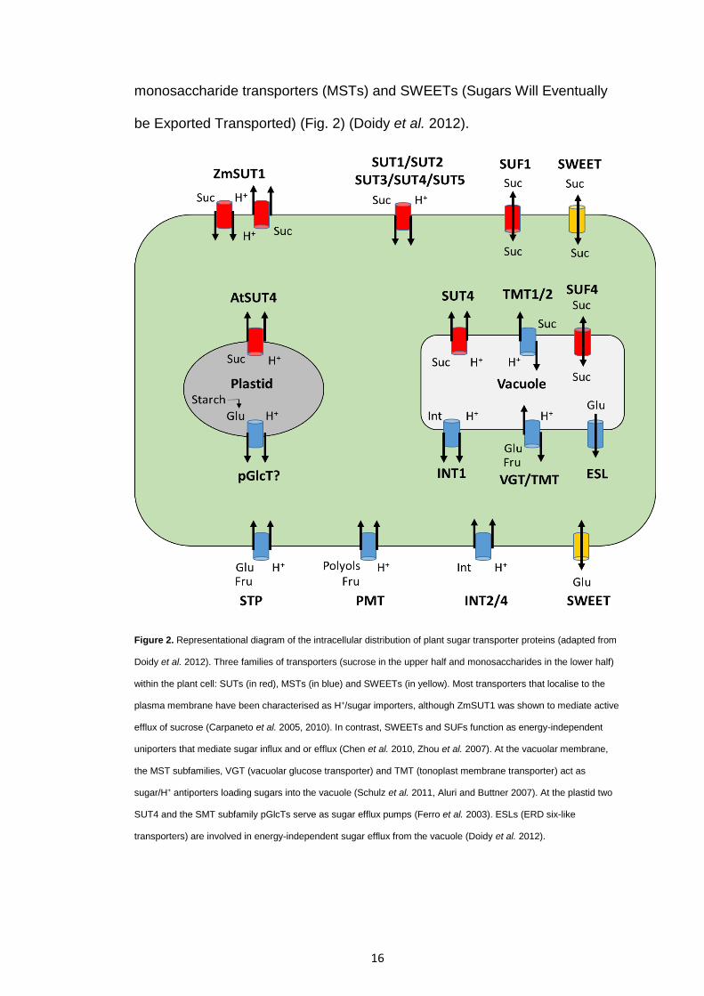
be Exported Transported) (Fig. 2) (Doidy et al. 2012).
Figure 2. Representational diagram of the intracellular distribution of plant sugar transporter proteins (adapted from
Doidy et al. 2012). Three families of transporters (sucrose in the upper half and monosaccharides in the lower half)
within the plant cell: SUTs (in red), MSTs (in blue) and SWEETs (in yellow). Most transporters that localise to the
plasma membrane have been characterised as H+/sugar importers, although ZmSUT1 was shown to mediate active
efflux of sucrose (Carpaneto et al. 2005, 2010). In contrast, SWEETs and SUFs function as energy-independent
uniporters that mediate sugar influx and or efflux (Chen et al. 2010, Zhou et al. 2007). At the vacuolar membrane,
the MST subfamilies, VGT (vacuolar glucose transporter) and TMT (tonoplast membrane transporter) act as
sugar/H+ antiporters loading sugars into the vacuole (Schulz et al. 2011, Aluri and Buttner 2007). At the plastid two
SUT4 and the SMT subfamily pGlcTs serve as sugar efflux pumps (Ferro et al. 2003). ESLs (ERD six-like
transporters) are involved in energy-independent sugar efflux from the vacuole (Doidy et al. 2012).

17
Sucrose is the main form of sugar used in long distance transport, and all
plants possess a family of SUTs (Doidy et al. 2012). There are five classes of
SUTs: SUT1-5 (Kuhn et al. 2010; Braun and Slewinski et al. 2009). SUT1
transporters are only found in dicot plants and are responsible for sucrose
phloem loading (Zhang et al. 2016) and sucrose partitioning in sink organs
(Buttner 2007). SUT2 act as sugar sensors as well as transporters (Barth et
al. 2003). SUT 3 transporters function identically to SUT1 transporters, but
are only found in monocot plants (Doidy et al. 2012). SUT4 regulate
intracellular sucrose partitioning, sucrose efflux from source leaves and the
utilisation of sucrose in lateral and terminal sink organs (Payyavula et al.
2011; Eom et al. 2011). Finally, SUT5 is the least studied transporter, but is
thought to play a role in sucrose phloem loading in source tissues (Zhang et
al. 2016)
In addition to long-distance transport, sugars (such as monosaccharides) are
also distributed within cells, i.e. partitioned into different organelles
depending on requirements, as well as between cells (Buttner 2007). For
example, during the day many plant species temporarily store sugars in the
form of starch in the chloroplasts of source leaves (Weise et al. 2006). At
night, that starch is catalysed to release monosaccharides (such as glucose)
(Weise et al. 2006; Weber et al. 2000), which is then exported from the
chloroplast (Buttner 2007). Furthermore, in sink tissues sucrose is hydrolysed
by invertases which yields glucose and fructose which are transported via
sugar transporter proteins (STPs) (Fig. 2) (Doidy et al. 2012). The STPs
responsible for monosaccharide transport are MSTs and SWEETs (Doidy et

18
al. 2012). The plant MST gene family is large, containing 53 MSTs in
Arabidopsis, 65 in rice, 58 in Medicago truncatula, and 59 in grapevine
(Vitrus Vinifera) (Doidy et al. 2012). Monosaccharides are further subdivided
into several subfamilies based on their substrate specificity, these are: polyol
monosaccharide transporter (PMT), inositol transporter (INT), vacuolar
glucose transporter (VGT), tonoplast membrane transporter (TMT), and
plastidic glucose transporter (pGlcT) (Doidy et al. 2012).
SWEETs belong to a distinct transporter family that contain a novel structure
consisting of a tandem repeat of three transmembrane domains connected
by a linker-inversion transmembrane domain (Chen et al. 2010). There are
17 SWEET genes in Arabidopsis, 21 in rice, 15 in M. truncatula and
approximately 47 in Eucalyptus grandis (Chen et al. 2010; Eom et al. 2015).
In Arabidopsis, SWEETs are divided into four phylogenetic clades, clade I
(AtSWEET1-3 homologues, typically monosaccharide transporters), clade II
(AtSWEET4-8 homologues, typically monosaccharide transporters), clade III
(AtSWEET9-15, sucrose transporters) and clade IV (AtSWEET16, 17,
fructose transporters) (Eom et al, 2015). Of note, SWEET clades do not
determine which physiological process the protein is involved in, for example
AtSWEET5, AtSWEET8 and AtSWEET13 are involved in pollen nutrition, yet
they are found in either clades II or III (Eom et al. 2015).
SWEETs play important roles in many plant processes, including nectar
secretion, phloem loading, sugar filling in seeds, regulating pollen nutrition,
vacuolar hexose transport, carbon reallocation in leaves during stress or
19
senescence and during plant-microbe interactions (both pathogenic and
mutualistic) (Eom et al. 2015; Guo et al. 2014). Pathogens are known to use
effectors to manipulate host plant SWEET expression to increase the amount
of sugars at the site of infection (Chen et al. 2010; Streubel et al. 2013). For
example, the rice pathogen Xanthomonas oryzae grows in the apoplasm and
xylem of the host and secretes the transcription activator-like (TAL) effectors
PthXo1 and AvrXa7 to induce the expression of host OsSWEET11 and
OsSWEET14 respectively, which increases the amount of sugar released
into the apoplasm for the pathogen to utilise (Chen et al. 2010). Mutant X.
oryzae lacking the PthXo1 effector was less virulent and repressing the
expression of OsSWEET11 (via RNA interference) resulted in decreases in
pathogen growth (Chen et al. 2010). In addition, adding mutations in the
promoter of OsSWEET11 provided protection from X. oryzae infection (Chen
et al. 2010). Bacterial and fungal pathogens induce the expression of
different sets of SWEETs (Chen et al. 2010). For example, the bacterial
pathogen Pseudomonas syringae highly induces the expression of
AtSWEET4, AtSWEET5, AtSWEET7, AtSWEET8, AtSWEET10, AtSWEET12
and AtSWEET15 in Arabidopsis (Chen et al. 2010). However, infection with
the fungal pathogen Golovinomyces cichoracearum induces AtSWEET12.
Infection with a different fungal pathogen, Botrytis cinereal, induced the
expression of AtSWEET4, AtSWEET15 and AtSWEET17 (Ferrari et al.
2007). Interestingly almost all SWEETs targeted by pathogen effectors are
clade III SWEETs and have been shown to export sucrose (Eom et al. 2015).
Furthermore, in grapevine (Vinus Vinifera) the glucose transporter
VvSWEET4 is highly induced by necrotrophic pathogens, but not biotrophic
20
pathogens (Chong et al 2014). VvSWEET4 is upregulated ROS production
and necrotrophic pathogen virulence factors (Chong et al 2014). Additionally,
AtSWEET4 Arabidopsis mutants were less susceptible to B. cinereal
(necrotrophic pathogen) infection (Chong et al 2014).
While the role of SWEETs in plant-pathogen interactions has been (and is
still being) widely researched (Chen et al. 2010, 2012, 2013, 2015a, 2015b;
Chong et al. 2014; Cohn et al. 2014; Liu et al. 2011; Perotto et al. 2014), the
role of SWEETs in mutualistic plant-microbe interactions is mostly unknown
(Casieri et al. 2013; Tarkka et al. 2013). An early study found that the MtN3
SWEET in Medicago truncatula is highly upregulated after exposure to
Rhizobium meliloti (Gamas et al, 1996). Therefore, MtN3 SWEET was
thought to play a role in nodulation (Gamas et al. 1996), perhaps by providing
the associating bacteria with hexoses in exchange for nitrogen thus
stabilising the mutualism.
Much of what is known about the role of SWEETs in parasitic interactions
and plant development has been studied in Arabidopsis and rice plants
(Chen et al. 2012; Zhou et al. 2014). Although SWEET genes have been
identified in most sequenced plant genomes, their individual roles in plant
development and pathogen nutrition has yet to be explored. In addition, since
Arabidopsis cannot form mutualistic interactions with fungi, the role of each
individual SWEET protein in mutualistic plant-microbe interactions has yet to
be determined.

21
1.5 Transcriptional control and the SWI/SNF complex
Extensive gene regulation occurs within plant root cells during plant-fungal
interactions. Eucalypts can form mutualistic relationships with ECM fungi, but
are also the target of many soil borne pathogens (e.g. Armillaria spp.).
However very little is known about the mechanisms that control gene
activation or repression in plants during interactions between long lived
perennial trees and their mycorrhizal associates. Chromatin modifications are
thought to be one way in which these gene activation or repression pathways
are controlled. While many different nuclear protein complexes regulate this
process, the most studied class of Chromatin Remodelling Complexes
(CRCs) is the SWI/SNF (Switch/Sucrose Non-Fermenting) complex
(Sarnowska et al. 2016).
SWI/SNF genes were first identified in the yeast Saccharomyces cerevisiae
(Abrams et al. 1986) and later in Arabidopsis thaliana (Brzeski et al. 1999),
Drosophila and mammals (Mohrmann and Verrijzer 2005). The original yeast
SWI/SNF complex consists of 12 subunits. The core of the complex is made
up of one SWI2/SNF-2type ATPase, one SNF5, and two copies of SWI3
subunits (Narlikar et al. 2002). This core is adequate for nucleosome sliding
but it is normally associated with other subunits, which act as receptors for
the SWI/SNF complex to interact with other proteins that affect chromatin
remodelling (Phelan et al. 1999). The core subunits of yeast SWI/SNF
complexes are similar to the SWI/SNF complex found in Arabidopsis
thaliana. In A. thaliana there are four putative SWI/SNF-type SNF2-ATPases
(SYD, BRM, CHR12/MINU1, CHR23/MINU2), and one SNF5 (BSH) subunit
22
(Farrona et al. 2004). Because of their sequence similarity, SYD, BRM and
MINU1/2 are thought to have chromatin remodelling activity, but to date, only
MINU2 has been shown to do this (Han et al. 2015). In addition, there are
four homologs of SWI3 genes in A. thaliana (SWI3A, B, C and D) (Sarnowski
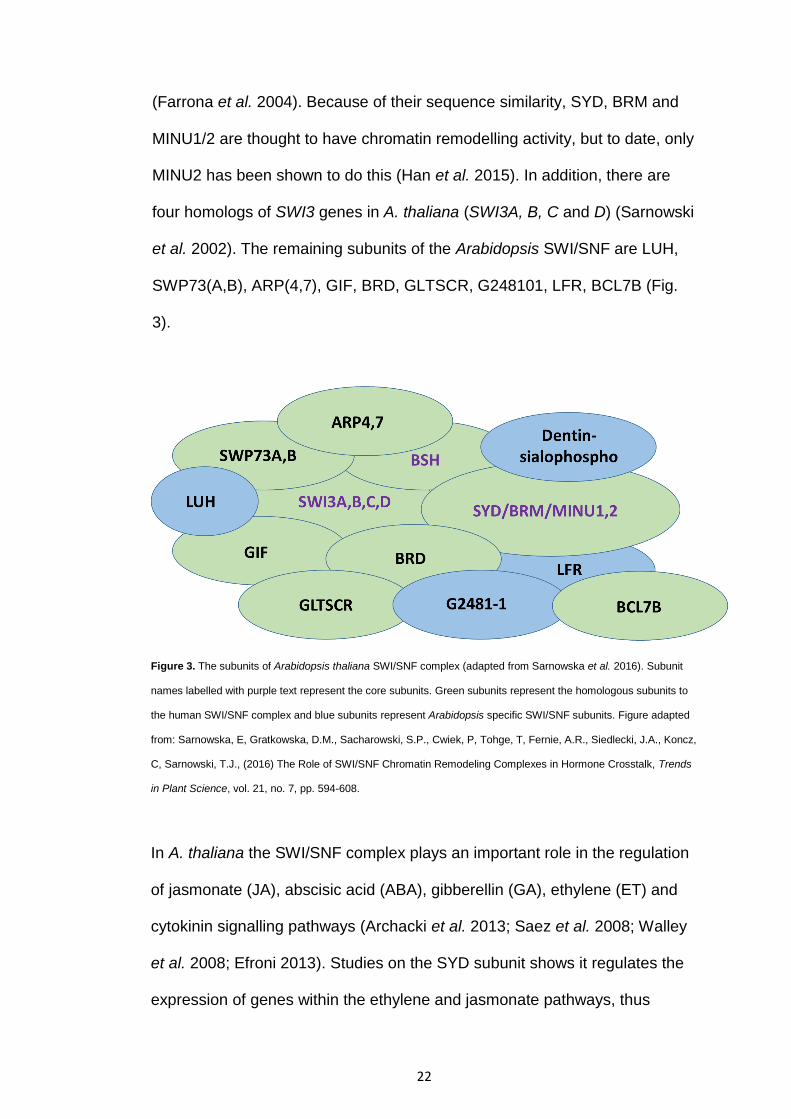
et al. 2002). The remaining subunits of the Arabidopsis SWI/SNF are LUH,
SWP73(A,B), ARP(4,7), GIF, BRD, GLTSCR, G248101, LFR, BCL7B (Fig.
3).
Figure 3. The subunits of Arabidopsis thaliana SWI/SNF complex (adapted from Sarnowska et al. 2016). Subunit
names labelled with purple text represent the core subunits. Green subunits represent the homologous subunits to
the human SWI/SNF complex and blue subunits represent Arabidopsis specific SWI/SNF subunits. Figure adapted
from: Sarnowska, E, Gratkowska, D.M., Sacharowski, S.P., Cwiek, P, Tohge, T, Fernie, A.R., Siedlecki, J.A., Koncz,
C, Sarnowski, T.J., (2016) The Role of SWI/SNF Chromatin Remodeling Complexes in Hormone Crosstalk, Trends
in Plant Science, vol. 21, no. 7, pp. 594-608.
In A. thaliana the SWI/SNF complex plays an important role in the regulation
of jasmonate (JA), abscisic acid (ABA), gibberellin (GA), ethylene (ET) and
cytokinin signalling pathways (Archacki et al. 2013; Saez et al. 2008; Walley
et al. 2008; Efroni 2013). Studies on the SYD subunit shows it regulates the
expression of genes within the ethylene and jasmonate pathways, thus

23
contributing to the plant’s immune response against fungal pathogens
(Walley et al. 2008). In addition, studies have shown that the syd and brm
mutations change the expression of genes controlled by the ABA and GA
hormone signalling pathways (Bezhani et al. 2007). Numerous experiments
have indicated a link between the germination of seedlings on exogenous
sugar and ABA/ethylene activity (Gazzarrini and McCourt 2001). Gazzarrini
and McCourt (2001) found that low sugar levels interfere with the inhibitory
effects of ABA on germination, whereas prevention of seedling development
post-germination by high sugar concentrations is dependent on ABA
synthesis.
A series of signalling events are involved in the interaction between fungal
and root cells, necessary for forming functional symbiotic structure. This
appears to be caused by activating and deactivating of genes in both fungus
and host plant. Certain elicitors are produced by the root cells that regulate
the expression of fungal genes to establish symbioses (Burgess et al. 1995).
Certain genes are activated that are responsible for the development of a
Hartig net and hyphal mantle (Salzer et al. 1997) and the deactivating of
certain fungal genes encoding factors for host plant defence reactions.
Certain elicitors present in ECM fungi are deactivated by chitinases of the
root cortex without harming the fungus, thus establishing the formation of
ectomycorrhizas (Salzer and Boller 2000). Plants must regulate their defence
pathways (e.g. regulate hormone pathways ABA, JA, SA, auxin, CK) and
activate sugar-related genes to form (or stabilise) mutualistic associations,
but still defend against pathogens who attempt to avoid plant defences by
24
secreting effectors that suppress plant immunity (Lo Presti et al. 2015) or
upregulate the transcription of sugar-related genes in order to syphon sugar
(Chen et al. 2010). We hypothesize that the SWI/SNF complex regulates the
expression of sugar-related genes and hormone pathways within the plant
during these interactions.
While the role of the SWI/SNF complex during plant-ECM fungal
relationships is largely unknown, production of defence and growth hormones
(such as auxin (IAA), ABA, JA, ET, SA and SK) are regulated by the
SWI/SNF complex, and these hormones are also produced by ECM fungi
(Ma et al. 2009). For example, Auxin regulates the development of embryo
and fruit, vascular bundle and root growth (Parvaiz 2011). It is synthesized in
the stem tip and young leaf and is then translocated to the required location.
There are different soil microbes that are able to produce auxin.
Ectomycorrhizal fungi produce cytokinin and indole acetic acid (IAA part of
the auxin class of plant hormones) to stimulate host plant root growth. In
addition, plant produce auxins and the expression of auxin genes are
controlled by the SWI/SNF complex.
Extensive gene regulation occurs within plant root cells during plant-fungal
interactions. Eucalypts can form mutualistic relationships with ECM fungi, but
are also the target of many soil borne pathogens (e.g. Armilarria spp.).
However very little is known about the mechanisms that control gene
activation or repression in plants during interactions between long lived
perennial trees and their mycorrhizal associates. Chromatin modifications are
25
thought to be one way in which these gene activation or repression pathways
are controlled. While many different nuclear protein complexes regulate this
process, one of the main complexes identified is the ‘SWI/SNF’ protein
complex. The SWI/SNF complex controls many plant hormone signalling
pathways. The role of the SWI/SNF complex in interacting with fungal
effectors, in perennial trees during their interaction with mycorrhizal
associates has yet to be explored.
1.6 Overview of Research
To determine the role of SWEET proteins in plant-microbe interactions, we
identified and categorised the SWEET-like genes of E. grandis. Further, we
determined if there was tissue-specific expression profiles of the identified E.
grandis SWEET-like genes throughout seedling tissues. We also compared
and contrasted the expression of these genes in roots when in contact with a
pathogenic, saprotrophic and mutualistic fungi. Finally we determined the
effects of overexpressing and repressing 4 eucalypt SWEET genes.
To understand transcriptional regulation in mutualistic plant-microbe
mutualisms, we performed qPCR over a time course of 2 weeks on four E.
grandis SWI/SNF complex subunits (i.e. SWI3A, B, C, and D). A Pisolithus
microcarpus MiSSP9.7 effector was found to interact with the SWI3D subunit
using yeast two-hybrid experiments between MiSSP9.7 and nuclear proteins.
Bimolecular fluorescence complementation (BiFC) experiments further
proved those protein interactions. Further, we identified where MiSSP9.7
26
localises within host plant cells. Lastly, we determined whether
overexpressing or repressing MiSSP9.7 and SWI3D affects mycorrhization.
The information gleaned from this study will further our understanding about
the controls and mechanisms involved in different types of plant-microbe
interactions. The belowground microbes play a significant role in plants’
growth and health (Artursson et al. 2006; Richardson et al. 2009). One of the
major constraints in eucalypts is soilborne pathogens. Thus, this study could
potentially find ways to improve Eucalyptus growth and health that will result
in economic and environmental benefits.
27
Chapter 2 Characterisation of Eucalyptus grandis
hexose transporters implicated in symbioses with
fungi
2.1 Introduction
Carbon, in the form of simple sugars, is essential for the development of all
living organisms. In terrestrial ecosystems plants, animals and microbes
interact with one another and the environment to obtain, utilize and
eventually recycle carbon. Fixation of light energy and atmospheric carbon
dioxide (CO2) by plant photosynthesis produce organic compounds such as
sugars utilized by plants for maintenance and growth. Sucrose is the main
product of photosynthesis and is transported from source to sink tissues via
the phloem (Koch 2004). Sucrose plays a key role in many regulatory
mechanisms, including growth and development, differential gene
expression, stress-related responses and plant innate immunity (Gomez-
Ariza et al. 2007; Tognetti et al. 2013; Tauzin and Giardina 2014). Sucrose
cleavage products, glucose and fructose, also act as signaling molecules. Of
the two hexoses, glucose has been better described in relation with the
hexokinase signaling pathway (Moore et al. 2003; Cho et al. 2009) while for
fructose a specific pathway has been proposed involving the ABA- and
ethylene-signaling pathway (Cho and Yoo 2011; Li et al. 2011).

28
Herbivorous animals and insects obtain energy from these plant organic
compounds through ingestion while microbes obtain photosynthate from
plants by three main mechanisms: parasitism, mutualism or via
decomposition. While these mechanism classifications are an
oversimplification, with plant-microbe associations being able to dynamically
change from mutualism to parasitism depending on both biotic and abiotic
factors (Francis and Read 1995; Johnson et al. 1997; Saikkonen et al. 1998;
Jones and Smith 2004), they serve as a useful framework for understanding
how plant immune system response differs based on the benefit of the
microbe to the plant. Pathogenic microbes may exploit photosynthetically
derived sugars through manipulation of host plants sugar transporter proteins
(STPs or SWEETs) (Chen et al. 2010; Cohn et al. 2014). Chen et al. (2010)
first identified and characterised SWEETs in Arabidopsis, highlighting the fact
that pathogens hijack sugars by sending TAL effectors to induce expression
of specific SWEETs. Similar to Chen et al. (2010), Cohn et al. (2014) found
that the bacterial pathogen Xanothomonas axonopodis syphons sugar from
cassava plants by using TAL effectors to induce MeSWEET10a. Similar to
pathogens, virus infection can lead to increases in sugar levels within plant
tissues, although the benefit of this to virus replication is unknown (Shalitin
and Wold 2000). As a means to combat sugar leakage, plants interacting
with pathogenic microbes have been found to uptake/retrieve sugars from the
apoplast through the increased expression of specific STPs (Chen et al.
2015b). This SWEET2 limits the amount of sugar obtained by the pathogen;
thus restricting the pathogen’s spread and growth throughout the rest of the
plant (Chen et al. 2015b).

29
In contrast to pathogens, beneficial microbes obtain sugars from plants by
forming mutualistic symbioses. Plants form mutualistic symbioses to improve
acquisition of growth limiting nutrients, and mutualistic microbes associate
with plants to gain carbon (Smith and Read 2008). Over 80% of trees
associate with ectomycorrhizal fungi (Pellegrin et al. 2015; Wang and Qui
2006; Smith and Read 2008). Ectomycorrhizal fungi (e.g. Pisolithus) utilise
hyphal networks to efficiently explore soil, acquiring nutrients (such as N and
P) to provide to the host plant in exchange for carbon (in the form of sugars)
(Nehls 2008; Smith and Read 2008). Therefore, this mutualistic symbiosis is
constituted by a constant exchange in nutrients between the two partners,
resulting in better growth for both symbionts. ECM fungi further aid plant
survival by supporting host adaptation to changing environmental conditions
such as climate extremes, drought and soil pollution (Redman et al. 2011;
Kipfer et al. 2012).
One aspect of sugar transport and accumulation in plant tissues during
microbial challenge that is often overlooked is the use of certain sugars as
substrates for the synthesis of defensive metabolites and as priming agents.
It is thought that plants modulate their sugar pools to act either as a source of
carbon and energy, or to use as signals and priming molecules to enhance
defence responses (Gomez-Ariza et al. 2007). These conclusions come
based on observations that sugars are able to regulate pattern recognition
genes used in plant innate immunity (Jones and Dangl 2006; Mohammad et
30
al. 2012) and because increases in sucrose and myo-inositol concentrations
are often observed under biotic stresses (Valluru and Van den Ende 2011).
Gomez-Ariza et al. (2007) demonstrated that the external application of
sucrose in Maize plants increased plant expression of pathogenesis-related
(PR) genes and overall resistance to a wide range of microbial pathogens.
Morkunas et al. (2014) showed that soluble sugars contribute to immune
responses against pathogens by stimulating isoflavone production in plants.
Phloem mobile oligosaccharides have also been found to induce defence
responses within plants. These include the: 1-ketose (a fructosyl
oligosaccharide), raffinose (a galactosyl oligosaccharide:), trehalose (a
disaccharide of glucose) and galactinol (galactosyl-myo-inositol; Hofmann et
al. 2015; Mohammad et al. 2012). Kim et al. 2008 showed galactinol
activates defence genes (NtACS1, PR1a and PR1b) in response to fungal
pathogen attacks. Trehelose, meanwhile, can partly induce resistance
against powdery mildew (Blumeria graminis) in wheat by activation of
phenylalanine ammonia-lyase and peroxidase genes (Reignault et al. 2001;
Muchembled et al. 2006) while the Trehalose Phosphate Synthase11
(TPS11) gene regulates defence responses in Arabidopsis against aphids
(Singh et al. 2011). Therefore, sugars function as priming molecules and as
signalling molecules, that lead to effective immune responses (Morkunas and
Ratajczak 2014).
Given the complex roles of sugars in plant-microbe interactions, it is
important that we characterise the mechanism by which these compounds
are transported in plant tissues during microbial challenge. Compared to
31
annual model plants (Büttner 2010; Yamada et al. 2016; Chen et al. 2010,
2012, 2013, 2015a, 2015b), less is known about how sugar transport
systems activated during interactions between long lived perennial trees and
various types of fungi. The interaction between Eucalyptus grandis with
different types of fungi (i.e. pathogenic, saprotrophic and mutualistic fungi)
offers a good model for studying this topic as the E. grandis genome has
been sequenced (Myburg et al. 2014), because E. grandis is a tractable
system for genetic modification and because a number of pathogens and
symbionts of E. grandis are culturable. The aims of this study were to identify
the SWEET-like genes of E. grandis and characterize a number of these
proteins that are differentially regulated during plant-microbe interaction.

32
2.2 Materials and methods
2.2.1 Plant and fungal growth conditions
Growth of E. grandis seedlings was performed following the methods outlined
in Plett et al. (2014a). E. grandis seeds were obtained from the
Commonwealth Scientific and Industrial Research Organisation (CSIRO,
Clayton, Vic., Australia) tree seed centre (Seedlot 21068) and sterilised in
30% hydrogen peroxide for 10 min followed by five washes with sterile water
for 5 min each. Seeds were then transferred onto 1% agar water medium and
allowed to germinate at 25oC with a 16/8 hour light/dark cycle. Germinated
seedlings were then transferred to ½ Modified Mylin Norkin (MMN) medium
on top of a sterile cellophane membrane to prevent root growth into the
medium.
One oomycete eucalypt pathogen (Phytophthora cinnamomi) and 3 genera of
fungi were used in this study: Suillus granulatus (non-Eucalyptus colonising
ECM which acts parasitically in our experimental set-up), Fusarium
oxysporum (non-Eucalyptus specific parasite), Suillus luteus (non-Eucalyptus
colonising ECM), and Pisolithus microcarpus isolate SI12 (Eucalyptus
colonising ECM). All fungal cultures used in this study were propagated 1
month on 1x MMN before subculturing hyphae from the growing edge of the
colony onto ½ MMN medium covered in a sterile cellophane membrane and
grown in the dark at 25°C for 2 weeks.
For plant colonisation experiments, plant seedlings were transferred directly
onto each fungal colony. The contact plates were then placed in a growth
cabinet under a 16/8 h light/dark cycle at 25°C for 2 weeks after which they
were harvested and frozen directly in liquid nitrogen. E. grandis control plants

33
were grown axenically and treated identically for the same length of time and
under the same conditions.
2.2.2 Generation of SWEET constructs and mutant eucalypts, and 13C
transfer tests
35S:Eucgr.K02678, 35S:Eucgr.B00363, RNAi:Eucgr.L02615 and
RNAi:Eucgr.K02688 were amplified from cDNA synthesized using KAPA HiFi
polymerase (KAPA Biosystems) following the manufacturer’s instructions
(see Table 1. for primers used). The amplified fragments were gel purified
and ligated into pDONR222, PCR verified and sequence verified. Positive
inserts were then ligated into pH2GW7 (35S:) or to pH7GWIWG2(II) (RNAi)
plasmids using Gateway Gene Cloning (Life Technologies) and transformed
into Rhizobium rhizogenes (formerly known as Agrobacteria rhizogenes)
isolate K599. E. grandis seedlings were grown from seed to one month old
on 1% agar media.
To generate mutant roots, E. grandis seedling roots were severed from the
stem using sterile scalpels. The remaining wounded part of the stem was
dipped into growing colonies of mutant R. rhizogenes containing SWEET
constructs, and grown upside down on ½ MS media for 1 week in a growth
cabinet with a constant temperature of 25oC and a 16 hour photoperiod. To
prevent R. rhizogenes from killing the seedlings, E. grandis stems were then
transferred to ½ MS Timentin (conc. 150 µg/mL) media. Once per week the
stems were transferred to new ½ MS Timentin media and grown under the
same conditions. Mutant roots usually emerged within 1-2 weeks, but took a
34
total of 3-4 weeks to grow long enough for fungal contact. After 4 weeks of
growth, transgenic plants were transferred onto ½ MMN media covered with
a sterile cellophane membrane and colonies of 2-week-old Pisolithus
microcarpus isolates (SI-12) placed on top, making direct contact with the
roots of the plant. These contacts were left for a total of 14 days in a cabinet
with a daytime high temperature of 30oC and low of 22 oC with a 16-hour
photoperiod. To prepare for 13C transfer tests, on the 8th day two holes were
burnt into the lids of each plate (using a soldering iron), and covered with
micropore tape. On the morning 9th day all contacts were placed into a plastic
tank (which had a rubber septum on one side and a fan for circulation on the
inside) and the lid was sealed down using clamps so no air could escape.
That same morning 12ml of 13CO2 gas (99% atom enrichment) was injected
into the tank by using a syringe that penetrated through the rubber septum on
the side of the tank, and left for 5 hours to allow for gas uptake. Afterwards,
the lid of the tank was opened and aerated. Contacts were then placed back
into the tank and left in the cabinet above. This 13CO2 pulse was repeated on
the 12th day. On the 14th day extra-radical fungal hyphae harvested for 13C
analysis. 13C labelling of eucalypts in contact with different fungal types was
carried out identically.
2.2.3 RNA extraction and Quantitative PCR analysis
RNA was extracted from four tissue types: photosynthate source tissues
(mature leaves), transport tissues (stem) and photosynthate sink tissues
(shoot apex including the 2 youngest developing leaves and roots) using the
Qiagen RNeasy Plant Kit with the RLC lysis buffer supplemented with 25 mg

35
ml-1 PEG 8000 and following the manufacturer’s instructions thereafter. The
cDNA was synthetized using the iScript Select cDNA Synthesis Kit (BioRAD).
The Quantitative PCR (QPCR) reactions were performed using Sensifast
Sybr Low-ROX Mastermix (Bioline) and QPCR machine (C1000 Touch TC,
CFX96 RTsystem (BioRAD)), where the cycle parameters were as follows: 1.
95oC 3 mins, 2. 95oC 30 seconds, 3. 55oC 30 seconds, 4. 72oC 30 seconds
(steps 2-4 was repeated x39), 5. Melt curve analysis. Tissue wide SWEET-
like gene expression was normalized to the expression of the housekeeping
genes Eucgr.C00350.1 and Eucgr.K02046.1 (Plett et al. 2014a). The primers
used in this study have been tested for their efficiency and their specificity.
To visualize tissue wide and root-fungi SWEET-like expression profiles, we
used two programs: ‘Cluster 3.0’
(http://bonsai.hgc.jp/~mdehoon/software/cluster/software.htm last accessed
16/3/17) and ‘Java TreeView’ (http://jtreeview.sourceforge.net/ last accessed
16/3/17).
2.2.4 Identification of SWEET genes in E. grandis and other plant
species
Using the Phytozome database, we identified SWEET-like genes by
examining the E. grandis 2.0 genome (Eucalyptus grandis v2.0;
http://www.phytozome.net/ last accessed: 2/7/2016) and identifying
sequences that share homology (based on the 3-transmembrane-helix-
domain polypeptide) to previously identified SWEETs. Sequences were
aligned to E. grandis transcripts taken from Phytozome v9.1 and
corresponding to the E. grandis genome v2.0 using the Phytozome database

36
(https://phytozome.jgi.doe.gov/pz/portal.html#!info?alias=Org_Egrandis last
accessed 16/3/17). This led to the identification of 52 genes with high
homology (Fig. 4). The SWEET genes identified in our study were designated
as AtSWEET1-like to AtSWEET17-like based on their homology to each
Arabidopsis SWEET gene respectively (Fig. 4). Likewise SWEET genes in
other species were obtained by identifying sequences in their genome that
share homology to previously identified SWEETs, using Phytozome
(https://phytozome.jgi.doe.gov/pz/portal.html last accessed: 16/3/17). All
species used: Arabidopsis thaliana (At) TAIR10, Amborella trichopoda v1.0,
Citrus clementina (Cc) v1.0, Eucalyptus grandis (Eg) v2.0, Metacargo
truncatula (Mt) Mt4.0v1, Populus trichocarpa (Pt) v3.0.
2.2.5 Construction of the phylogenetic tree
To determine the phylogenetic relationships of E. grandis SWEET-like
proteins, we constructed a phylogenetic tree using the online tool
‘Phylogeny.fr’ (Dereeper et al. 2008). SWEET-like gene sequences of plants
(Arabidopsis thaliana (26), Amborella trichopoda (9), Citrus clementina (18),
Eucalyptus grandis (52), Medicago truncatula (25), Populus trichocarpa (28))
were downloaded from the Phytozome database. “One click” phylogenetic
analysis was used, with a concatenated MUSCLE alignment adjusted by
Gblocks. The tree was rooted with Human SWEET Transporter 1 as the
outgroup.

37
2.2.6 Yeast complementation
To test whether four putative eucalypt SWEET proteins (AtSWEET11, 12, 13,
14-like [Eucgr.K02694], AtSWEET4, 5-like [Eucgr.K02688, Eucgr.B00363,
Eucgr.L02615]) could transport sugars we performed yeast complementation
assays. We used S. cerevisiae (strain EBY.VW4000) as a model system to
test the sugar capabilities of these 4 putative SWEETs. EBY.VW4000 was
chosen because the sugar transporter genes in this strain has been knocked-
out (i.e. has mutations in its sugar transporter genes), except for the genes
that encode maltose transporters (Wieczorke et al. 1999). Therefore
EBY.VW4000 cannot grow on media with a carbon source other than
maltose. However, when EBY.VW4000 is transformed with the vector
containing the putative SWEET genes, the yeast will begin to transcribe and
express those SWEET genes. Thus growth on other types of sugar media
will be restored if the SWEET protein product is able to transport sugars.
The open reading frames (ORF) of Eucgr.K02678, Eucgr.B00363,
Eucgr.L02615 and Eucgr.K02688 were cloned into pYES2 vector using In-
Fusion ligation kit (Clonetech) using BamH1 and HindiIII ligases, and
transformed into E. coli (strain Top10). Constructs were selected for using
antibiotic resistance and PCR verification followed by sequencing. These
constructs were then transformed into Saccharomyces cerevisiae strain
EBY.VW4000 following Easy TRAFO protocol (Gietz and Woods 2002). In
brief, EBY.VW4000 was grown in YPM liquid media at 300C overnight with
shaking (at 255 rpm) (i.e. pre-grown to the log phase). The next morning
these yeast were used for transformation of four SWEET constructs (i.e.
38
Eucgr.K02678, Eucgr.B00363, Eucgr.L02615 and Eucgr.K02688 all in pYES-
GFP vector). 1.5 ml of Yeast were harvested into 1.5ml Eppendorf tubes via
centrifugation (3000 x g for 30 seconds) and media removed, re-suspended
in sterile water, centrifuged again and sterile water removed. The pellets
were re-suspended while adding each of the following chemicals: 240 ml of
PEG (50% w/v), 36 ml of LiAC (pH7), 50 µl of pre-boiled Salmon Sperm
DNA, 29 µl of sterile water, and 5 µl of each construct (concentration 0.1 – 1
µg of DNA). Yeast transformants were then incubated at 420C for 3 hours.
During the 3 hour incubation, the cells were re-suspended every 15 mins by
vigorously shaking the tube until the transformation solution looked
homogenous. Afterwards yeast transformants were centrifuged at max speed
for 30 seconds and resuspended in 1 ml of sterile water. Cells were then
plated on SC media lacking uracil where maltose was the carbon source,
incubated at 300C for 3-4 days and the resulting colonies PCR screened.
Verified colonies were plated on media supplemented with different sugar
sources (fructose, galactose, glucose, sucrose and maltose) at dilutions 10-1,
10-2 and 10-3 and were incubated at 30oC for 3 days and photographed.
Control yeast were transformed with pYES-GFP vector without SWEET gene
inserts and treated identically for the same length of time and under the same
conditions.
2.2.7 Glucose efflux test
Glucose efflux tests were performed following methods described in Jansen
et al. (2002) study, with minor adjustments. Eucgr.K02678, Eucgr.B00363,
Eucgr.L02615 and Eucgr.K02688 cloned into pYES2 vector were tested. All
39
yeast strains (i.e. S. cerevisiae mutants) used for testing glucose efflux were
grown to the stationary phase (i.e. 3-4 days incubation at 30oC) in liquid SC
medium lacking uracil with a maltose concentration of 7.5 grams per litre.
Samples were then harvested by centrifugation (5,000 x g, 3 mins), media
removed and samples weighed (to obtain wet weight). Yeast samples were
then resuspended in five-fold liquid SC medium lacking both uracil and a
sugar source. After 10 mins of incubation, maltose solution (100 grams per
litre) equivalent to 1/5 of total volume was added, and samples (150 μl per
sample) were taken at 15 min time intervals for 90 mins. Sugar
concentrations were determined using the Sigma-Aldrich Glucose Assay Kit
and a CLARIOstar® spectrophotometer.
2.2.8 GFP localisation
All 4 SWEET constructs were made as previously described above (see
Materials and methods, yeast complementation). The ORF of Eucgr.K02678,
Eucgr.B00363, Eucgr.L02615 and Eucgr.K02688 were cloned into pYES2
vector using In-Fusion ligation kit (Clonetech) using BamH1 and HindiIII
ligases, and transformed into E. coli (strain Top10). Constructs were selected
for using antibiotic resistance, PCR verified and sequenced. These
constructs were then transformed into Saccharomyces cerevisiae strain
EBY.VW4000 following Easy TRAFO protocol (Gietz and Woods 2002). S.
cerevisiae (strain EBY.VW4000) were transformed with each of the 4
constructs following the quick and easy TRAFO protocol (Gietz and Woods
2002). After 3 days, colonies were observed using confocal scanning
40
microscopy. The GFP was excited at 488nm excitation and emission
captured between 520-540nm.

41
2.3 Results
2.3.1 Phylogenetic relationships of E. grandis SWEET-like transporters
A total of 52 SWEET-like genes were identified in the E. grandis genome
using the Phytozome database (available at
http://phytozome.jgi.doe.gov/pz/portal.html last accessed: 16/3/17) based on
homology to Arabidopsis SWEET transporters with proven hexose transfer
capability (Chen et al. 2010) as a template. Compared to annual plants and
other trees genomes (A. thaliana (17 SWEETs), A. trichopoda (9), C.
clementina (18), E. grandis (52), M. truncatula (25), P. trichocarpa (28)), a
significantly larger number of SWEET-like genes were found in the E. grandis
genome (52 potential SWEET-like genes; Fig. 4). This large number of
SWEET-like genes was the result of expansions and duplications that have
occurred within the E. grandis genome rather than the occurrence of novel
SWEET-like gene families (Fig. 4). The genes SWEET1-14, 16 and 17 show
expansion within the E. grandis genome (Fig. 4). These same SWEET genes
show expansion in A. trichopoda, C. clementina, M. truncatula and P.
trichocarpa genomes.

42
Figure 4. Phylogenetic relationships between SWEET-like proteins collected from different species. A.
thaliana, A. trichopoda, C. clementina, E. grandis, M. truncatula, P. trichocarpa. The tree was rooted
with Human SWEET Transporter 1 as outgroup.

43
2.3.2 SWEET gene expression in E. grandis tissues
Quantitative PCR of 52 SWEET-like genes in 4 major tissue types of E.
grandis seedlings showed a variety of expression patterns (Fig. 5B). We
were able to identify three groupings of genes based on expression patterns
(Fig. 5B). Group 1 was highly expressed throughout the all tissues tested;
with Eucgr.C01371 being the most highly expressed gene at 12 fold above
the reference genes. Group 2 genes expression profiles were consistently
similar to the reference genes and Group 3 genes exhibited lower expression
levels as compared to the reference genes with Eucgr.F01609 being the
lowest expressed gene across all four tissues (Fig. 5B). As with Groups 1
and 2, group 3 genes showed nearly identical expression levels in all tissues
tested.

44
Figure 5. Expression profile of 52 SWEET-like gene throughout different
tissue in E. grandis: Shoot apex, stem, leaves and root. (A) Image of E.
grandis control plant grown axenically; labels show the different tissue and
where RNA was extracted. (B) A total of 52 E. grandis SWEET-like genes
show similar levels of expression throughout the four tissues. The heat map
is annotated on the right-hand side, with genes grouped based on amount of
expression: highly expressed genes (group 1), averagely expressed (group
2), and lowly expressed (group 3).

45
2.3.3 E. grandis roots exhibit different morphologies when in contact
with different fungal lifestyles
The effect of different fungi and oomycetes on E. grandis root morphology
was lifestyle dependent. In all cases roots were surrounded by fungal hyphae
(excluding control plant which were grown axenically) (Fig. 6). S. granulatus,
Phytophthora cinnamomi, and Armillaria luteobubalina led to hallmarks of
parasitic interactions, i.e. discoloration of leaves (reddish appearance
compared to controls) and blackening of roots tips. A. luteobubalina is a well-
known eucalypt pathogen that causes death of eucalypt roots (Kile 1981,
1983) and P. cinnamomi is a common plant pathogen that causes death of
roots in many different plant species (Royal Botanic Garden 2017a, b). S.
granulatus is mycorrhizal on pines, however in our experimental set-up the
fungus acted parasitically on the plant as the interaction resulted in short and
unhealthy roots, discoloration of leaves and root death (blackened roots)
(Fig. 6B and G), which was a similar result to P. cinnamomi and A.
luteobubalina contacts (Fig. 6F and H). When E. grandis was grown with
commensal fungi (i.e. Suillus luteus) plants exhibited no evidence of
parasitism or of pathogenesis (Fig. 6C and I). E. grandis was grown in
contact with the mutualistic ECM fungal isolates of P. microcarpus, we
observed significant hyphal growth around the roots and successful
colonisation of roots as denoted by shortening of lateral roots and the
formation of a thick fungal mantle around the root surface (Fig. 6D and J).
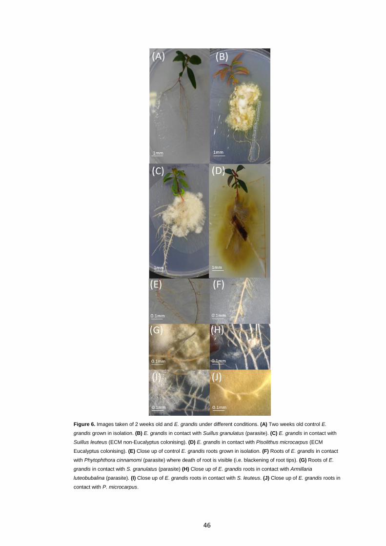
46
Figure 6. Images taken of 2 weeks old and E. grandis under different conditions. (A) Two weeks old control E.
grandis grown in isolation. (B) E. grandis in contact with Suillus granulatus (parasite). (C) E. grandis in contact with
Suillus leuteus (ECM non-Eucalyptus colonising). (D) E. grandis in contact with Pisolithus microcarpus (ECM
Eucalyptus colonising). (E) Close up of control E. grandis roots grown in isolation. (F) Roots of E. grandis in contact
with Phytophthora cinnamomi (parasite) where death of root is visible (i.e. blackening of root tips). (G) Roots of E.
grandis in contact with S. granulatus (parasite) (H) Close up of E. grandis roots in contact with Armillaria
luteobubalina (parasite). (I) Close up of E. grandis roots in contact with S. leuteus. (J) Close up of E. grandis roots in
contact with P. microcarpus.
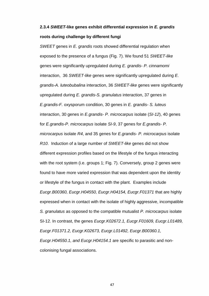
47
2.3.4 SWEET-like genes exhibit differential expression in E. grandis
roots during challenge by different fungi
SWEET genes in E. grandis roots showed differential regulation when
exposed to the presence of a fungus (Fig. 7). We found 51 SWEET-like
genes were significantly upregulated during E. grandis- P. cinnamomi
interaction, 36 SWEET-like genes were significantly upregulated during E.
grandis-A. luteobubalina interaction, 36 SWEET-like genes were significantly
upregulated during E. grandis-S. granulatus interaction, 37 genes in
E.grandis-F. oxysporum condition, 30 genes in E. grandis- S. luteus
interaction, 30 genes in E.grandis- P. microcarpus isolate (SI-12), 40 genes
for E.grandis-P. microcarpus isolate SI-9, 37 genes for E.grandis- P.
microcarpus isolate R4, and 35 genes for E.grandis- P. microcarpus isolate
R10. Induction of a large number of SWEET-like genes did not show
different expression profiles based on the lifestyle of the fungus interacting
with the root system (i.e. groups 1; Fig. 7). Conversely, group 2 genes were
found to have more varied expression that was dependent upon the identity
or lifestyle of the fungus in contact with the plant. Examples include
Eucgr.B00360, Eucgr.H04550, Eucgr.H04154, Eucgr.F01371 that are highly
expressed when in contact with the isolate of highly aggressive, incompatible
S. granulatus as opposed to the compatible mutualist P. microcarpus isolate
SI-12. In contrast, the genes Eucgr.K02672.1, Eucgr.F01609, Eucgr.L01489,
Eucgr.F01371.2, Eucgr.K02673, Eucgr.L01492, Eucgr.B00360.1,
Eucgr.H04550.1, and Eucgr.H04154.1 are specific to parasitic and non-
colonising fungal associations.

48
Figure 7. Regulation of 52 E. grandis SWEET genes when E. grandis associates
with: parasites (Phytophthora, Armillaria and S. granulatus), a non-Eucalyptus
coloniser (F. oxysporum and S. luteus), or a mutualistic fungus (P. microcarpus
strains). The heat map is annotated on the right-hand side, groups formed based on
amount of expression within the roots during these associations: highly regulated in
all conditions (group 1) and isolate dependent expression (group 2). The position of
Eucgr.B000363, Eucgr.L02615, Eucgr.K02694 and Eucgr.K02688 were highlighted
using red text for the Eucgr.number.

49
2.3.5 E. grandis SWEET-like genes encodes STPs that localise to the
plasma membrane of plant cells
If EgSWEET-like proteins facilitate the uptake and or export of sugars
between plant cells and the apoplast, then these proteins must localise to the
plasma membrane of plant cells. To test this, we used S. cerevisiae
(EBY.VW4000) as a model organism. S. cerevisiae (EBY.VW4000) was
transformed with four SWEET constructs (Eucgr.K02694, Eucgr.K02688,
Eucgr.B00363, Eucgr.L02615-GFP tagged genes), including a positive
control (AtSWEET1). We examined GFP fluorescence within those cells
using confocal microscopy (Fig. 8). As expected AtSWEET1 localised to the
plasma membrane (Fig. 8). Interestingly, only Eucgr.K02694 localised to the
plasma membrane, whereas the rest of the EgSWEETs localised to the
cytoplasm (Fig. 8)
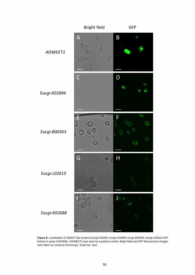
50
Figure 8. Localisation of SWEET-like proteins Eucgr.K02694, Eucgr.K02688, Eucgr.B00363, Eucgr.L02615-GFP
fusions in yeast (VW4000). AtSWEET1 was used as a positive control. Bright field and GFP fluorescence images
were taken by confocal microscopy. Scale bar: 4μm
51
2.3.6 E. grandis SWEET-like proteins can act as sugar symporters
To determine the sugar transport capability of 4 putative E. grandis SWEET-
like proteins in vivo, we tested the ability of the 4 EgSWEET-like genes to
complement VW4000 S. cerevisiae (Fig. 9). As explained previously,
EBY.VW4000 cannot grow on media with a carbon source other than
maltose. The negative control (VW4000 only) grew on maltose media and
was unable to grow on other sugar media. All other yeast heterologously
expressing EgSWEET genes complemented the VW4000 strain when grown
on galactose. AtSWEET1 constructs (i.e. positive control) complemented
VW4000 allowing growth and utilisation of all four sugar sources. Most
notably Eucgr.K02694 had stronger growth compared to the other SWEETs.
Both Eucgr.L02615 and Eucgr.K02694 were able to grow on all sugar media,
while Eucgr.B00363 and Eucgr.K02688 had weak growth on fructose,
sucrose and glucose. Therefore, the 4 EgSWEET genes tested had different
affinities and abilities to transport sugar.
To determine if these same SWEET-like proteins were able to export
glucose, we performed glucose efflux tests as per (Jansen et al. 2002). A
maltose solution was added to mutant EBY.VW4000 colonies containing the
4 EgSWEET constructs (Eucgr.K02678, Eucgr.B00363, Eucgr.L02615 and
Eucgr.K02688 in pYES2) and extracellular glucose concentration was
measured over a total time period of 90 mins. All EgSWEETs exhibit glucose
exporting capabilities at varying levels (Fig. 10). Most notably Eucgr.K02694
exhibited the highest rate of glucose export, even higher than AtSWEET1.

52
Figure 9. Transport activity of EgSWEET K02694, K02688, B00363, L02615 in yeast. These SWEET
proteins complemented VW4000 S. cerevisiae mutants (which lacked 18 hexose transporter genes).
Yeast VW4000 was transformed with pYES-GFP vector which each had EgSWEET gene inserts
respectively. Ten-fold serial dilutions of the transformants were transferred to plates with minimal
media lacking uracil and containing either (A) fructose, (B) sucrose, (C) glucose, (D) galactose or (E)
maltose. SWEET1 was used as a positive control. VW4000 S. cerevisiae negative control was
transformed with an empty pYES. Ten-fold serial dilutions of the transformants were transferred to
plates with minimal media lacking uracil and containing either maltose, glucose, sucrose, fructose and
galactose. All plates were incubated for 3 days at 30oC and then photographed.
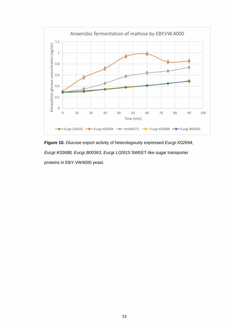
53
Figure 10. Glucose export activity of heterologously expressed Eucgr.K02694,
Eucgr.K02688, Eucgr.B00363, Eucgr.L02615 SWEET-like sugar transporter
proteins in EBY.VW4000 yeast.
0
0.2
0.4
0.6
0.8
1
1.2
0 10 20 30 40 50 60 70 80 90 100
Extr
acel
lula
r gl
uco
se c
on
cen
trat
ion
(u
g/m
L)
Time (min)
Anaerobic fermentation of maltose by EBY.VW.4000
Eucgr.L02615 Eucgr.K02694 AtSWEET1 Eucgr.K02688 Eucgr.B00363
54
2.3.7 Altered expression of EgSWEET-like genes in E. grandis roots
affects carbon export from E. grandis roots
We determined whether the amount of carbon transferred from eucalypt to
fungi differs depending on fungal lifestyle. 13CO2 carbon transfer experiments
were performed on E. grandis replicates in contact with different fungi
(ranging from parasitic, saprotrophic and mutualistic) over a time period of 2
weeks (Fig. 11). Overall the amount of 13C obtained by the fungi showed little
difference relative to control fungi that had been grown axenically, with the
exception of P. cinnamomi. P. cinnamomi obtained large amounts of 13C
relative to control P. cinnamomi. Apart from the eucalypt-Phytophthora
contact, changes in 13C levels for all fungi were not statistically significant.
However there was insufficient time to run all eucalypt-fungal controls, thus
these are preliminary results only.
We used stable isotope carbon tracing to determine whether overexpression
or repression of EgSWEET genes affects carbon transfer from host E.
grandis to fungal symbionts (Fig. 12). Overexpression of Eucgr.L02615
showed little effect on carbon transfer, however overexpression of
Eucgr.K02688 greatly increased the amount of 13C transferred from E.
grandis roots to P. microcarpus SI-12 relative to empty-vector control (Fig.
12). Repression of Eucgr.K02694 and Eucgr.B00363 had no significant effect
of carbon transfer. Our results show that increased expression of certain
EgSWEETs in the roots of E. grandis affects the amount of carbon
transferred from the host eucalypt to the associating Pisolithus. Although no

55
changes were significant, these results suggest that the efficiency of sugar
transport may differ between families and individual EgSWEETs.

56
Figure 11. Amount of 13C obtained by different types of fungi when associating with
wildtype E. grandis relative to each respective control over a time period of 2 weeks.
No changes were statistically significant.
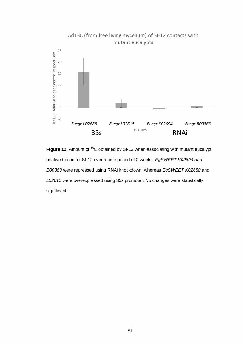
57
Figure 12. Amount of 13C obtained by SI-12 when associating with mutant eucalypt
relative to control SI-12 over a time period of 2 weeks. EgSWEET K02694 and
B00363 were repressed using RNAi knockdown, whereas EgSWEET K02688 and
L02615 were overexpressed using 35s promoter. No changes were statistically
significant.
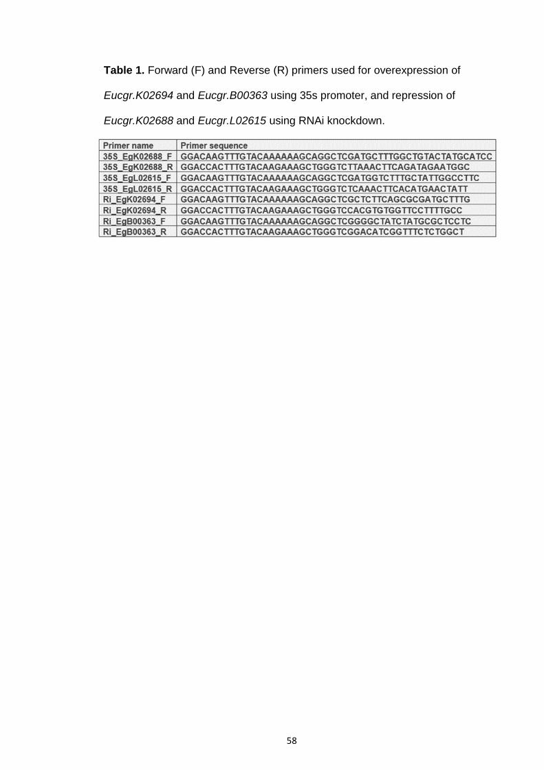
58
Table 1. Forward (F) and Reverse (R) primers used for overexpression of
Eucgr.K02694 and Eucgr.B00363 using 35s promoter, and repression of
Eucgr.K02688 and Eucgr.L02615 using RNAi knockdown.

59
2.4 Discussion
Eucalyptus trees have provided human society with many environmental and
economic benefits throughout the world. Like all trees, the growth and health
of eucalypts depend upon the types of soil microbes (e.g. parasitic,
saprotrophic and mutualistic microbes) present within the rhizosphere (i.e.
soil area surrounding the root) (Aggangan et al. 2013). Despite society’s
heavy reliance on these trees’ growth (e.g. for industrial and medicinal oils,
renewable energy source, paper and pulp production, CO2 sequestration),
little is known about the underlying controls and mechanisms used by these
trees when interacting with soil microbes. In this study we annotated 52 E.
grandis SWEET-like genes, we determine the tissue-specific profiles of their
expression and compared and contrasted the expression of these genes in
roots when in contact with a parasitic, saprotrophic and mutualistic fungi.
We found that there is no tissue specificity of SWEET-like gene expression
between the different tissues of E. grandis, with the exception of
Eucgr.L01492 (Fig. 5B). Similar to our findings, studies on G. max found
similar, non-specific expression of SWEET genes throughout the plants’
tissue (Patil et al. 2015). In contrast, one study on Arabidopsis found that
AtSWEET11 and AtSWEET12 are specific and highly expressed in phloem
tissue (Chen et al. 2014). However, this difference in specificity may be due
to the greater number of SWEET genes found in E. grandis and G. max
compared to Arabidopsis.

60
While interaction with fungi induced differential expression of SWEET-like
genes in E. grandis, very few of these genes showed expression patterns
specific to a particular plant-fungal combination. It was interesting to note
that the fewest number of significantly up-regulated SWEET-like genes were
found in the mutualistic plant-fungal interaction and the largest number of
significantly up-regulated SWEET-like was in the parasitic plant-fungal
interaction. Bacterial and fungal pathogens are known to modulate the
expression of plant SWEET genes (Chen et al. 2010; Chen et al. 2014; Liu et
al. 2011), i.e. SWEETs are differentially expressed in root cells colonised by
pathogenic bacteria or fungi. Previous studies on annual plants (e.g.
Arabidopsis) found high expression of AtSWEET12, AtSWEET4,
AtSWEET15 and AtSWEET17 during pathogen infection (Ferrari et al. 2007;
Chen et al. 2010; Chen et al. 2014; Xuan et al. 2013). We found that the
following genes are up-regulated only in plant-parasitic interactions:
Eucgr.B00360 (AtSWEET4 homologue), H04550 (AtSWEET17 homologue),
H04154, K02672 and K02673 (AtSWEET12 homologue), L01492
(AtSWEET1 homologue ); in addition to other SWEET homologues F01371
(AtSWEET2 homologue), L01489 (AtSWEET3 homologue), F01609
(AtSWEET9 homologue). Other similar findings are the up-regulation of
AtSWEET12, AtSWEET4, AtSWEET15, and AtSWEET17 homologues in E.
grandis roots under all fungal conditions. Also studies on M. truncatula shows
up-regulation of STP genes, containing a conserved MtN3/saliva (SWEET-
like) domain, in roots when associating with AM fungi and Rhizobia bacteria
(Liu et al. 2011; Perotto et al. 2014). Arabidopsis AtSWEET11 and
AtSWEET12, both have two MtN3/saliva domains, and function as sucrose

61
exporters which efflux sucrose from phloem parenchyma cells out into the
apoplast (Chen et al. 2012). atsweet11:atsweet12 double mutants showed
slower growth, chlorosis, and large amounts levels of sugar and starch
accumulation in leaves relative to wild-type (WT) plants (Chen et al. 2012).
With large reserves of sugars, plants are commonly targeted by microbes.
Bacteria, fungi and oomycetes have all evolved various strategies to
proliferate by tapping into the nutritional reserves of plants. Previous
research showed that pathogens increase the flow of sugars (such as
sucrose and glucose) towards the apoplast, where they grow, by
manipulating plant plasma membrane sugar transporters (Chen et al. 2010;
Cohn et al. 2014). However our results suggest that some SWEET proteins
(for e.g. Eucgr.K02694) may be used by cells to uptake sugars from the
external environment (Fig. 9). Interestingly, Eucgr.K02694 is repressed in
mutualistic interactions (at least for P. microcarpus SI-12) but highly
upregulated during parasitic interactions (at least for P. cinnamomi),
suggesting that eucalypts may upregulate Eucgr.K02694 to restrict sugar
uptake by pathogens. This result has also been described by Yamada et
al. (2016) who found that plants retaliate to pathogen infections by
reabsorbing the sugars inside their cells, using a select few STPs (i.e.
STP13), which results in less sugar in the apoplast and ultimately starves
the pathogen.
Our study lays the foundation to further our understanding of the controls
surrounding sugar transport in perennial trees during plant-microbe
62
interactions. In terms of perennial trees, the induction of SWEET-like genes
by TAL effectors used by pathogenic, saprotrophic and mutualistic fungi has
yet to be explored. Furthermore the effects of elevated CO2 on the
expression of SWEET-like genes under different plant-microbe interactions,
the efficiency of sugar transport of each SWEET-like protein and
characterisation of these SWEET-like sugar transporter proteins still needs to
be done. Understanding these mechanisms could prove useful for increasing
E. grandis growth and promote stress tolerance, and this warrants further
investigation.
63
Chapter 3 Characterisation of the Pisolithus albus
effector MiSSP9.7 and its interactant, Eucalyptus
grandis SWI3D
3.1 Introduction
Plants constantly interact with a wide range of soilborne microbes. Microbes
associate with plants to obtain carbon (in the form of sugars) for their own
growth and development. These microbes do this by either: forced syphoning
host plant sugars (i.e. parasitic/pathogenic interactions), or by exchanging
growth limiting nutrients (such as N and P) for the plant’s sugars (i.e.
mutualistic interactions). Plants are constantly attacked by pathogens, but
they commonly associate with mutualistic microbes (Partida-Martinez and
Heil 2011; Smith and Read 1995, 1997, 2008). Over 80% of terrestrial plants
form mutualistic associations with mycorrhizal fungi (Pellegrin et al. 2015) as
well as associating with many different species of beneficial bacteria
(Franche et al. 2009). Thus, plants have evolved signaling pathways and an
adaptable immune system that is thought to distinguish and facilitate the
establishment of microbes within roots (Jayaraman et al. 2012). Exactly how
signaling pathways and the plant immune system distinguish between
pathogens and mutualists is not fully understood.
To form intimate symbioses with plants, fungi must first overcome or
manipulate their hosts’ immune system. The plant innate immune system
consists of physical barriers (such as the plant cell wall and waxy cuticle
64
layer protecting the epidermis of plant organs) and chemical defence
mechanisms (such as the hypersensitive cell death response (HR)). To deal
with host immune responses, pathogenic microbes have evolved a plethora
of mechanisms to detoxify host metabolites, hijack host sugar transporters,
hide their presence from host immune receptors or to kill host tissues (Lo
Presti et al. 2015; Chen et al. 2010; Yang et al. 2006; Brown and Tellier
2011; Schmidt and Panstruga 2011; Ciuffetti et al. 1997).
Another means by which microbes, in particular pathogenic fungi, directly
manipulate host immunity is through the production of small secreted
proteins called effectors. Effectors broadly describe secreted proteins that
either: kill the host plant (in necrotrophic and hemibiotrophic fungi),
manipulate host cell physiology or suppress the host’s immune response (de
Jonge et al. 2011; Rafiqi et al. 2012; Stassen et al. 2011; Bozkurt et al. 2012;
Chuma et al. 2011; Angot et al. 2006; Giraldo et al. 2013a, 2013b; Okmen et
al. 2014; Stergiopoulos et al. 2009, 2013; Zuccaro et al. 2014). For example,
necrotrophic fungi (e.g. wheat pathogens Stagonospora nodorum and
Pyrenophora tritici-repentis) produce nectrophic effectors, such as secondary
metabolites, toxic polyketides and non-ribosomal peptides, to kill host plant
cells (Horbach et al. 2011; Qutob et al. 2006; Stergiopoulos et al. 2009, 2013;
Oliver et al. 2012). Other pathogens initially secrete effectors that suppress
host immune response and allow the pathogen to thrive and eventually kill
the host. This is seen in the plant pathogen Magnaporthe oryzae which
secretes SLp1 and AvrPiz-t effectors which suppresses chitin-activated
pathogen-associated molecular pattern (PAMP)-triggered immunity (PTI) and

65
cytoplasmic plant immunity (Lo Presti et al. 2015). In addition, some small
RNAs have been described as pathogen effectors. B. cinerea produce small
RNAs that disrupt A. thaliana RNA interference machinery by binding to
AtARGONAUTE1 which selectively silences immunity related genes that are
complementary to these small RNAs (Weiberg et al. 2013, 2014).
While effectors have been studied extensively in plant pathogens, mutualistic
fungi also use effectors to suppress the host’s immune response to form
mycorrhizal associations and structures (e.g. Hartig net and arbuscules).
Ectomycorrhizal (ECM) fungi secrete many different effectors (Pellegrin et al.
2015), called Mycorrhizae induced Small Secreted Proteins (MiSSPs), which
are thought to play a key role for establishing plant-ECM mutualisms (Plett
and Martin 2015; Plett et al. 2017). The ECM fungus L. bicolor produces the
effector MiSSP7 that interacts with the jasmonic acid co-receptor PtJAZ6 (a
negative regulator of jasmonic acid-induced gene transcription) (Plett et al.
2011, 2014a, b). This interaction results in the suppression of jasmonic acid-
induced genes, allowing L. bicolor to develop the apoplastic Hartig net and
establish symbiosis (Plett et al. 2014a, b). In compatible Suillus-plant
pairings, SSP genes were found to be significantly upregulated when
compared to controls (Liao et al. 2016). Similarly, to ECM fungi, the use of
effectors to establish mutualisms have been identified in arbuscular
mycorrhizal (AM) fungi. Rhizofagus irregularis (formerly Glomus intaradices)
secretes the SP7 effector that targets the pathogenesis-related transcription
factor ERF19 in the plant nucleus, which temporarily suppresses the plants
immune response (Klopphoiz et al. 2011). ERF19 is highly upregulated

66
during infection by the fungal pathogen Colletotrichum trifolii, but only
temporarily during AM colonisation (Klopphoiz et al. 2011). Taken together,
these findings suggest that effectors are essential components for the
establishment of plant-fungal mutualisms.
The recent release of new ECM fungal genomes has highlighted the
presence of dozens more effector like proteins encoded by a range of
mycorrhizal fungi (Kohler et al. 2015). One of these fungi, Pisolithus albus, is
of special interest to Australia as it makes up a critical component of forest
biota through its symbiotic interaction with the roots of eucalypt trees. The
aims of this study were to characterise one of the effector-like genes of the
closely related Pisolithus albus, called PaMiSSP9.7 (homologous to
Pismi.63008) and the potential role it plays during host-fungal interaction. We
show that it interacts with SWI3D, a subunit of the SWI/SNF complex
implicated in the transcriptional regulation of sugar and hormone related
genes.

67
3.2 Materials and methods
3.2.1 MiSSP9.7-GFP production and absorption by eucalypt root cells
The open reading frame for PaMiSSP9.7 was amplified from cDNA
synthesized using iScript (BioRad) following the manufacturer’s instructions.
The amplified fragments were gel purified and ligated into a pET22(b)-GFP
plasmid using In-Fusion clonase (Clontech), and plasmids transformed in
Escherichia coli (strain BL21) where PaMiSSP9.7 was expressed.
PaMiSSP9.7 protein expression and purification was performed following
methods described by Shen et al. (2009) using GE Healthcare His SpinTrap
columns. The eluted protein was purified via dialysis using 1x phosphate
buffered saline (PBS pH 7.4) and running on SDS-Page gel to validate the
purity of PaMiSSP9.7.
To test the up-take of PaMiSSP9.7 into E. grandis root cells, roots of E.
grandis seedlings were submerged into a MiSSP9.7-GFP solution, and
incubated in a growth chamber at 25oC with light for 4 hours. Negative
controls were made by submerging roots in either pure GFP without
MiSSP9.7 diluted in PBS or in only PBS solution, and treated identically from
there onwards. Roots treated with MiSSP9.7-GFP or GFP alone were then
rinsed in PBS, severed from the stem of the seedling, fixed in 4%
paraformaldehyde and left at 4oC overnight. Afterwards, roots were washed
with new PBS and then stained with 1% propidium iodide for 10 mins. Roots
were then examined under a Leica SP6 confocal microscope.
68
3.2.2 Yeast One- and Yeast Two-Hybrid analyses
Yeast I and II hybrid screens were carried out as per Plett et al. (2011).
PaMiSSP9.7 was amplified from cDNA synthesized using iScript (BioRad)
following the manufacturer’s instructions. The amplified fragments were gel
purified and ligated into pDONR222 and PCR and sequence verified.
Positive inserts were then ligated into pDEST22 plasmids using Gateway
Gene Cloning (Life Technologies) and transformed into S. cerevisiae strain
MAV203 using a library scale transformation procedure as per the ProQuest
yeast two-hybrid system protocol (ProQuest catalog number PQ10001-01
and PQ10002-01). One yeast II hybrid analysis was performed and plated
on selective medium (-L-W-H + 25 mM 3-amino-triazol) which tests for
putative interacting protein(s). A further test of protein-protein interaction was
performed using a β-Gal activity assay as described by Walout and Vidal
(2001). After the blue colour was developed for 24 hours at 37oCelsius,
photos were taken of each colony using a Zeiss stereomicroscope with
attached colour camera.
For Yeast I Hybrid assay, bait sequences were constructed by cloning
MiSSP9.7 into pDEST32 plasmid in frame with Gal4-DBD and transformed
into MaV103 cells (mating type a). Self-activation tests for the DBD bait strain
were then performed using the interaction controls described above (Yeast I
and II analyses done by J. Plett).
69
3.2.3 BiFC testing in E. grandis
SWI3D (i.e. Eucgr.I01261) and MiSSP9.7 (i.e. Pa683008) genes used in
BiFC cloning were inserted directly into the N-labelled version of the pBiBCt-
2in1 vector using GATEWAY cloning techniques (Grefen and Blatt, 2012).
Firstly, SWI3D and MiSSP9.7 were amplified from E. grandis cDNA via PCR
and inserted into pDONR221 using BP ligation. These vectors with inserts
were then transformed into E. coli for cloning. E. coli colonies containing the
correct vector and insert were identified by growing transformants on LB
media supplemented with appropriate antibiotics and then PCR screening
colonies to verify colonies containing the correct gene insert based on size.
Verified E. coli colonies were grown at 37oC overnight and purified using
Zippy plasmid purification kit (Zymo). Purified SWI3D and MiSSP9.7 genes in
pDONR221 were then ligated into the N-labelled version of the pBiBCt-2in1
vector using LR ligase. The pBiBCt-2in1 vector produced a red fluorescent
protein (RFP) signal that enabled identification of successfully transformed
cells based on the presence of RFP. Genes inserts were sequence verified
and the final construct was transformed into Agrobacterium
tumefaciense clone GV3101. One positive colony was grown overnight at
28oC in LB supplemented with appropriate antibiotics and then used for
transformation. After 48 hours, leaf discs were excised and observed using
confocal scanning microscopy. The settings used to observe the yellow
fluorescent protein (YFP) was 20% argon power to excite at 488nm excitation
followed by emission capture between 520-540nm. For red fluorescent
protein (RFP) internal transformation control, we excited the samples at
561nm followed by emission capture between 580-650 nm.
70
3.2.4 Construction of the phylogenetic tree
To determine the phylogenetic relationships of E. grandis SWI3 proteins, we
constructed a phylogenetic tree using the online tool ‘Phylogeny.fr’ (Dereeper
et al. 2008). SWI3 gene sequences of plants (Arabidopsis thaliana (4), Citrus
clementina (4), Eucalyptus grandis (4), Medicago truncatula (4), Populus
trichocarpa (6)) were downloaded from the Phytozome database. “One click”
phylogenetic analysis was used, with a concatenated MUSCLE alignment
adjusted by Gblocks. The tree was rooted with Yeast SWI3 as the outgroup.
3.2.5 Plant and fungal growth conditions
Growth of E. grandis seedlings was performed similarly to chapter 2,
following the methods outlined in Plett et al. (2014a). E. grandis seeds
obtained from the Commonwealth Scientific and Industrial Research
Organisation (CSIRO, Clayton, Vic., Australia) tree seed centre (Seedlot
21068) were sterilised in 30% hydrogen peroxide for 10 mins followed by 5
washes with sterile water for 5 mins each. Seeds were then transferred onto
1% agar water medium and allowed to germinate at 25oC with a 16/8 hour
light/dark cycle. Once germinated, seedlings were transferred to ½ Modified
Mylin Norkin (MMN) medium on top of a sterile cellophane membrane to
prevent root growth into the medium. Pisolithus albus cultures used in this
study were propagated at least 4 weeks on 1x MMN before subculturing the
outer growing hyphae onto ½ MMN medium covered in a sterile cellophane
membrane and grown under a 16/8 h light/dark cycle at 25°C for 2 weeks.
71
All plant colonisation experiments were carried out by placing eucalypt
seedlings directly onto each fungal colony. The contact plates were grouped
and labelled as 0 hour (pre-symbiosis contact), 24 hour contacts, 48 hour
contacts and 2 week contacts (at least 3 replicates per time group), then all
placed in a growth cabinet under a 16/8 h light/dark cycle at 25°C. Eucalypt
roots were harvested and frozen directly in liquid nitrogen at each respective
time period (i.e. 0 hour, 24 hour, 48 hour and 2 week contact periods).
3.2.6 RNA extraction and Real Time Quantitative PCR (RT-QPCR)
RNA extraction was performed on all contact groups (minimum of 3
replicates per group) using the RNeasy Mini Kit (Qiagen) following the
manufacturer’s instructions. A total of 1 μg of RNA for each sample was used
to synthesize cDNA using the iScript cDNA synthesis kit (Bio-Rad) following
the manufacturer’s instructions. This cDNA was used as a template for RT-
QPCR using the Bio-Line SensifastTM reaction mix and the Bio Rad CFX96
Touch Real-Time PCR Detection System. The reference genes
Eucgr.C00350.1 and Eucgr.K02046.1, were utilized to normalize the results.
3.2.7 Generation of SWI3D constructs and transgenic eucalypts
Generation of EgSWI3D (i.e. Eucgr.I01261) constructs was performed
following methods described in chapter 2. 35S:EgSWI3D and
RNAi:EgSWI3D were amplified from cDNA synthesized using iScript
(BioRad) following the manufacturer’s instructions. The amplified DNA
fragments were gel purified, ligated into pDONR222 and PCR verified.
Positive inserts were then ligated into pH2GW7 (35S:) or pH7GWIWG2(II)
72
(RNAi) vectors using Gateway Gene Cloning (Life Technologies) forming the
SWI3D constructs. These SWI3D constructs were then transformed into
Rhizobia rhizogenes (formerly known as Agrobacteria rhizogenes) isolate
K599. E. grandis seedlings were grown from seed to one month old on 1%
agar media. These seedlings were used to generate transgenic roots that
either overexpressed or repressed Eucgr.I01261.
To generate mutant roots, E. grandis seedling roots were cut and removed
from the stem using sterile scalpels. The remaining wounded part of the stem
was dipped into growing colonies of mutant R. rhizogenes containing SWI3D,
and grown upside down on ½ MS media for 1 week in a growth cabinet with
a constant temperature of 25oC and a 16 hour photoperiod. To prevent death
of seedlings by R. rhizogenes, E. grandis stems were then transferred to ½
MS Timentin (conc. 150 µg/mL) media. Once per week the stems were
transferred to new ½ MS Timentin media and grown under the same
conditions. Mutant roots became visible usually within 1-2 weeks, but took a
total of 3-4 weeks to grow long enough for fungal contact. After 4 weeks of
growth, transgenic eucalypts were transferred onto ½ MMN media on top a
sterile cellophane membrane and colonies of 2-week-old Pisolithus albus
isolates (SI-12) placed on top, making direct contact with the roots. These
contacts were left for 2 weeks at 25oC with a 16/8 hour light/dark cycle.
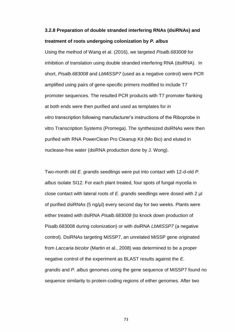
73
3.2.8 Preparation of double stranded interfering RNAs (dsiRNAs) and
treatment of roots undergoing colonization by P. albus
Using the method of Wang et al. (2016), we targeted Pisalb.683008 for
inhibition of translation using double stranded interfering RNA (dsiRNA). In
short, Pisalb.683008 and LbMiSSP7 (used as a negative control) were PCR
amplified using pairs of gene-specific primers modified to include T7
promoter sequences. The resulted PCR products with T7 promoter flanking
at both ends were then purified and used as templates for in
vitro transcription following manufacturer’s instructions of the Riboprobe in
vitro Transcription Systems (Promega). The synthesized dsiRNAs were then
purified with RNA PowerClean Pro Cleanup Kit (Mo Bio) and eluted in
nuclease-free water (dsiRNA production done by J. Wong).
Two-month old E. grandis seedlings were put into contact with 12-d-old P.
albus isolate SI12. For each plant treated, four spots of fungal mycelia in
close contact with lateral roots of E. grandis seedlings were dosed with 2 µl
of purified dsiRNAs (5 ng/µl) every second day for two weeks. Plants were
either treated with dsiRNA Pisalb.683008 (to knock down production of
Pisalb.683008 during colonization) or with dsiRNA LbMiSSP7 (a negative
control). DsiRNAs targeting MiSSP7, an unrelated MiSSP gene originated
from Laccaria bicolor (Martin et al., 2008) was determined to be a proper
negative control of the experiment as BLAST results against the E.
grandis and P. albus genomes using the gene sequence of MiSSP7 found no
sequence similarity to protein-coding regions of either genomes. After two
74
weeks of treatment, percent root colonization was assessed and samples
were taken for microscopy and performed RNA extraction.
3.2.9 Microscopy of transgenic/dsiRNA treated eucalypt roots and
Hartig net measurements
Transgenic or dsiRNA treated E. grandis-P. albus contacts described above
were assessed for percent mycorrhization before severing the roots from
stems, where they were fixed in 4% paraformaldehyde and left at 4oC
overnight. Roots were then embedded in 6% agarose and mycorrhizal root
tips were sectioned on a Campden Instruments vibratome into 30μm cross
sections. Cross sections were stained with 1% propidium iodide for 10 mins,
washed with PBS and stained again with WGA-FITC and rinsed in PBS
(Embedding and cross sectioning done by K. Plett). Roots were then
examined under a Leica SP6 confocal microscope and photos taken. Hartig
net depths were calculated using ImageJ software. Controls were fixed,
stained and examined identically to the test samples.

75
3.3 Results
3.3.1 MiSSP9.7 encodes an effector protein that enters plant root cells
We tested the up-take of Pa683008-GFP (MiSSP9.7-GFP) by E. grandis
roots in the absence of P. albus. We observed fluorescence within the
nucleus of host cells, indicating MiSSP9.7-GFP localised to the nucleus (Fig.
13B). The merge of the two signals with bright-field confirms this. The
negative control (i.e. GFP with no attached protein) was not visibly taken up
into host cells under our experimental conditions (Fig. 13F). Since PI
localised to the nucleus and the GFP signal co-localized with the PI signal
(Fig. 13E), we can conclude that the MiSSP9.7 enters the nucleus.
76
Figure 13. Images of MiSSP9.7-GFP (i.e. Pa683008-GFP) and Propidium Iodide (PI) stain taken using confocal
microscopy. The first panel shows auto-fluorescence (Fig. 13A) and the nucleus (N) is shown in each panel. PI (blue
fluorescence) (Fig. 13B) and MiSSP9.7-GFP (green fluorescence) (Fig. 13C) co-localised within the nucleus of cells
(Fig. 13E). Negative controls show GFP only without any attached protein (Fig. 13F), PI only (Fig. 13G) and a
merge of the two negative control images (Fig. 1H). GFP alone (i.e. without any attached protein) was not able to
enter into root cells (Fig. 13F, H).
77
3.3.2 MiSSP9.7 interacts with a chromatin remodelling complex (CRC)
subunit of E. grandis
Because the MiSSP9.7 localises to the nucleus in host plant cells, we
determined if there were any nuclear proteins of E. grandis that interact with
this effector (Fig. 14). Using a yeast II hybrid we determined that MiSSP9.7
interacts with SWI3D (Eucgr.I01261), a subunit of the SWI/SNF CRC. This
interaction was compared to three controls: a strong positive interaction
(Krev1/RalGDS-wt), a weak positive interaction (Krev1/RalGDS-m1) and two
proteins that do not interact (Krev1/RalGDS-m2). Yeast colonies under 3AT
selection and X-Gal tests showed that MiSSP7.9: Eucgr.I01261 closely
resembled a weak positive interaction (Fig. 14).
This interaction was further confirmed using Bi-Fluorescent Complementation
(BiFC) (Fig. 15). Nicotiana benthamiana leaf cells were transformed with
pBiFCt-2in1-NN (containing MiSSP9.7 and SWI3D) in which positive
transformants are identified by constitutive expression of red fluorescent
protein (RFP) (Fig. 15 third pane from left). Examination of these cells using
confocal microscopy showed a strong YFP signal, showing that 683008 and
SWI3D interacted causing the two halves of YFP to join and fluoresce (Fig.
15). A merge of the two signals with bright-field shows that the interaction is
occurring in the nucleus of the cell (right most pane).
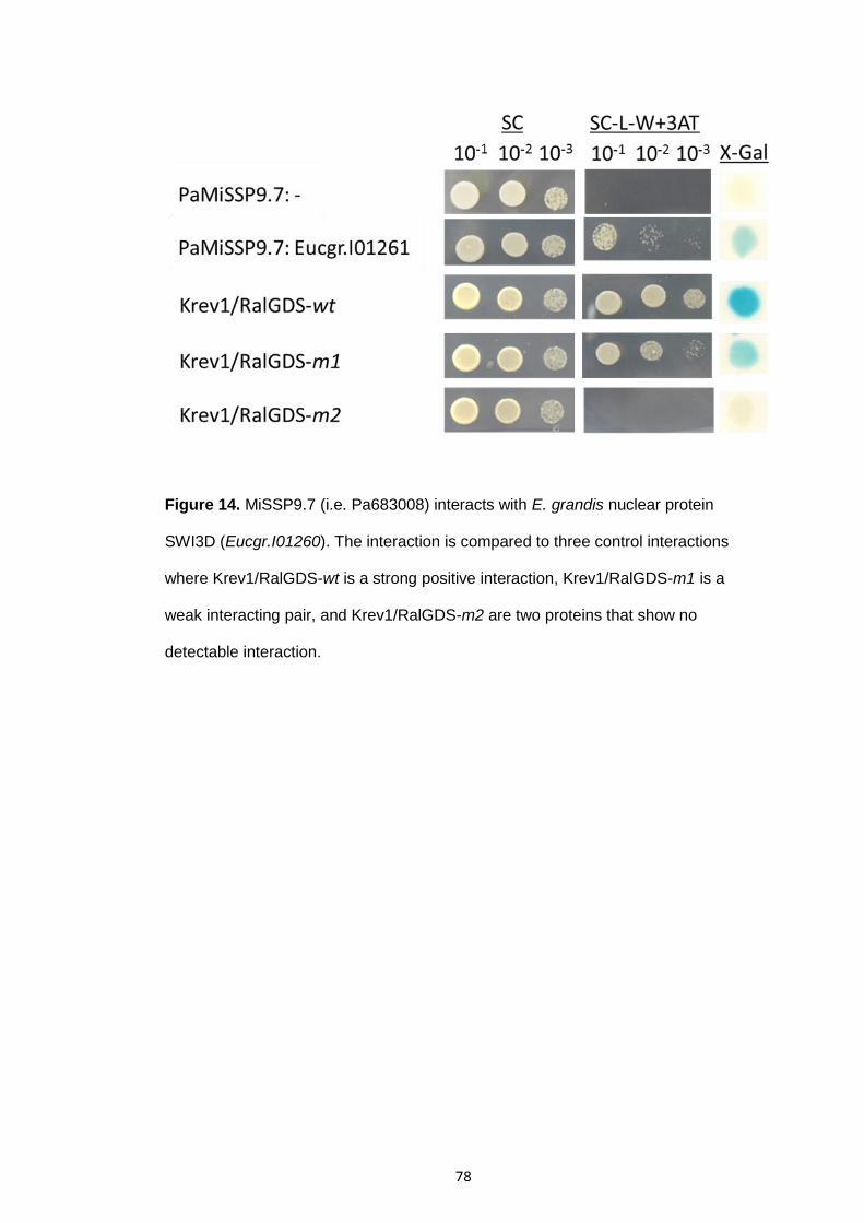
78
Figure 14. MiSSP9.7 (i.e. Pa683008) interacts with E. grandis nuclear protein
SWI3D (Eucgr.I01260). The interaction is compared to three control interactions
where Krev1/RalGDS-wt is a strong positive interaction, Krev1/RalGDS-m1 is a
weak interacting pair, and Krev1/RalGDS-m2 are two proteins that show no
detectable interaction.

79
Figure 15. In vivo BiFC proof of the interaction between 683008 and SWI3D. N.
benthamiana leaf cells transformed with vector containing both genes, where
transformed cells are denoted by constitutive expression of RFP (third panel),
interaction between 683008 and SWI3D is shown by the reconstitution of the YFP
signal (second panel) and the merge of the two signals with bright-field (right most
panel).
80
3.3.3 Phylogenetic relationships of E. grandis SWI3 proteins
A total of 4 SWI3 genes were identified in the E. grandis genome using the
Phytozome database (available at http://phytozome.jgi.doe.gov/pz/portal.html
last accessed: 28/3/17) based on homology to Arabidopsis SWI3 subunits,
which have been proven form part of the SWI/SNF complex (Sarnowska et
al. 2016), as a template, in addition to keyword searches (Fig. 16). Similar
numbers of SWI3 genes were found to be encoded by the E. grandis genome
when compared to annual plants and other trees genomes (Eucalyptus
grandis (4), Arabidopsis thaliana (4), Citrus clementina (4), Medicago
truncatula (4), Populus trichocarpa (6)) (Fig. 16).

81
Figure 16. Phylogenetic relationships between SWI3 proteins collected from
different species. A. thaliana, C. clementina, E. grandis, M. truncatula, P.
trichocarpa. The tree was rooted with S. cerevisiae Yeast SWI3 subunit as the
outgroup.
82
3.3.4 SWI3 gene expression in E. grandis roots over a two-week time
course of colonization by P. albus
Since all four SWI3 subunits play a role in the SWI/SNF complex, we
determined the gene expression of all four eucalypt SWI3 genes (SWI3A, B,
C and D) using quantitative PCR (QPCR), during pre- and post-colonisation
of E. grandis roots by P. albus over a 2 week time period. The time points
were 24 hour in-direct contact (i.e. pre-symbiosis signaling), 24 hour direct
contact, 48 hour direct contact and 2 week colonized root tissues. We found
that SWI3D was significantly upregulated in roots during pre-symbiosis
signaling and 48 hours after contact (Fig. 17). SWI3A was significantly up-
regulated during pre-symbiotic signaling and then later was significantly
repressed. SWI3B was significantly repressed at during the early stages of
physical interaction between E. grandis and P. albus. SWI3C transcription
was not significantly altered.
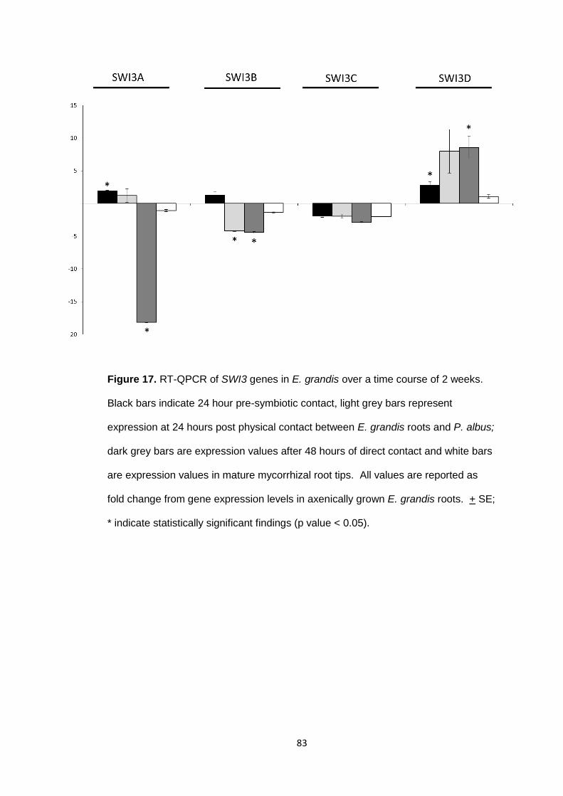
83
Figure 17. RT-QPCR of SWI3 genes in E. grandis over a time course of 2 weeks.
Black bars indicate 24 hour pre-symbiotic contact, light grey bars represent
expression at 24 hours post physical contact between E. grandis roots and P. albus;
dark grey bars are expression values after 48 hours of direct contact and white bars
are expression values in mature mycorrhizal root tips. All values are reported as
fold change from gene expression levels in axenically grown E. grandis roots. + SE;
* indicate statistically significant findings (p value < 0.05).
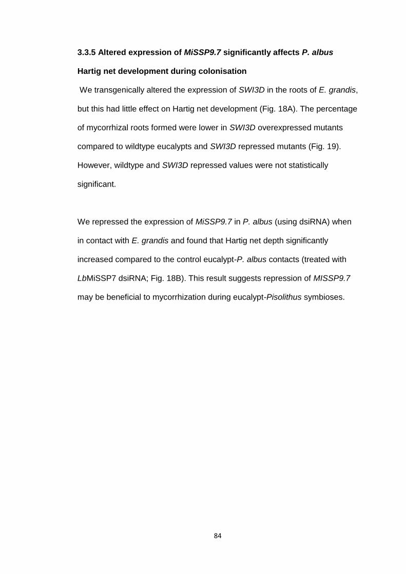
84
3.3.5 Altered expression of MiSSP9.7 significantly affects P. albus
Hartig net development during colonisation
We transgenically altered the expression of SWI3D in the roots of E. grandis,
but this had little effect on Hartig net development (Fig. 18A). The percentage
of mycorrhizal roots formed were lower in SWI3D overexpressed mutants
compared to wildtype eucalypts and SWI3D repressed mutants (Fig. 19).
However, wildtype and SWI3D repressed values were not statistically
significant.
We repressed the expression of MiSSP9.7 in P. albus (using dsiRNA) when
in contact with E. grandis and found that Hartig net depth significantly
increased compared to the control eucalypt-P. albus contacts (treated with
LbMiSSP7 dsiRNA; Fig. 18B). This result suggests repression of MISSP9.7
may be beneficial to mycorrhization during eucalypt-Pisolithus symbioses.
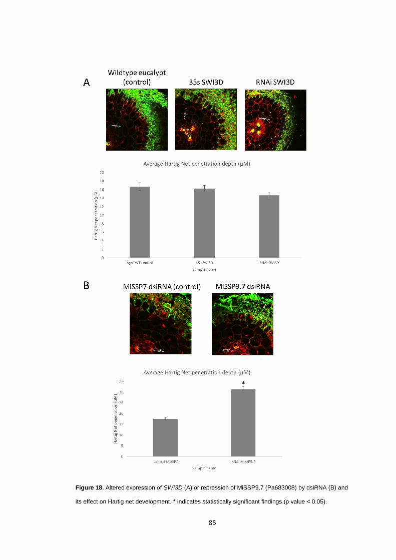
85
Figure 18. Altered expression of SWI3D (A) or repression of MiSSP9.7 (Pa683008) by dsiRNA (B) and
its effect on Hartig net development. * indicates statistically significant findings (p value < 0.05).
86
Figure 19. Percent mycorrhization of SWI3D (i.e. Eucgr.I01261) transgenic
eucalypts. Eucgr.I01261 was overexpressed (using 35s promoter) in 3 E. grandis
replicates and repressed (using RNAi knockdown) in another 3 replicates.
Percentage of mycorrhized roots were counted after 2 weeks of contact with
Pisolithus albus cultures. Wild type control eucalypts were treated identically to the
mutants, except for altering SWI3D expression. * indicates statistically significant
findings (p value < 0.05).

87
3.4 Discussion
Plants are constantly confronted by a range of different soil microbes (in
particular parasitic and mutualistic microbes) present within the rhizosphere.
Plants must associate with mutualistic fungi to improve their survivability,
while defending against pathogens. The mechanisms used by plants to do
this is still currently being researched (Rey et al. 2015; Plett et al. 2015).
Effectors are signaling molecules secreted by fungi that are used to
communicate to plants (Lo Presti et al. 2015; Kloppholz et al. 2011; Plett et
al. 2011, 2014a, 2014b, 2015). Plants in turn, respond in many different ways
depending on the types of effectors secreted. Indeed, plants respond
differently to effectors secreted by parasitic fungi, compared to plants
responding to effectors secreted by mutualistic fungi (Lo Presti et al. 2015;
Rafiqi et al. 2012; Plett et al. 2011, 2014a, 2014b, 2015; Klopphoiz et al.
2011). Our research describes how the MiSSP9.7 effector modulates ECM-
plant fungal interactions and formation of mycorrhizal structures. We
determined that MiSSP9.7 enters host root cells and localizes to the nucleus.
We further show that MiSSP9.7 interacts with a CRC subunit (SWI3D) and
we characterise the expression of SWI3 genes in eucalypt roots in contact
with P. albus. Finally, we show that differential expression of either SWI3D or
MiSSP9.7 can alter the outcome of mycorrhization.
The SWI/SNF complex has been characterized in other plant systems where
it directly regulates multiple plant hormone pathways, including auxin (IAA),
Abscisic Acid (ABA), Jasmonic Acid (JA), Ethylene (ET), Salicylic acid (SA)
and cytokinins (Sarnowska et al. 2016). Plants hormones such as SA, JA,

88
and ethylene (ET) play major roles in regulating plant defence responses. SA
is involved in the reaction against biotrophic and hemi-biotrophic pathogens,
while JA and ET are associated with defence against necrotrophic pathogens
and herbivorous insects. Increased (SA) accumulation and signaling repress
biotrophic pathogens but encourage the colonization of plant tissues by ECM
fungi (Plett et al. 2014a, b; Tschaplinski et al. 2014; Lebeis et al. 2015).
Therefore, P. albus may be able to indirectly modulate host immune
responses by using certain effectors that bind to essential DNA regulating
complexes, such as the SWI/SNF CRC.
We found that there is no difference in Hartig net formation when
transgenically over-expressing or repressing SWI3D in E. grandis roots (Fig.
18). Overexpression of SWI3D, however, lowers the percentage of root tips
colonized. Reduced expression of MiSSP9.7 however caused significant
increases in the Hartig net root penetration depth into host root cells (Fig.
18). This result is counter-intuitive as all previously characterized mutualistic
effectors (Plett et al. 2011; Kloppholz et al. 2011; Plett et al. 2014a, b) have
been found to repress host immunity and increase Hartig net formation.
These results would suggest that mycorrhizal fungi also encode effector
proteins that curb the colonization success of the fungus. This may be a
mechanism by which the ECM fungus is able to remain non-pathogenic and
still benefit the host. This result, however, will require further experimentation
to substantiate.
89
This study provides further insight for understanding the controls used during
plant-fungal interactions. Our results suggest that pre-symbiotic signaling
ECM fungi, such as Pisolithus, release effectors from hyphae that bind to
nuclear proteins within plant root cells, which then regulate transcription
Understanding this mechanism could prove useful for increasing E. grandis
colonisation by beneficial microbes and prevent disease and deserves further
investigation.

90
Chapter 4 Conclusion and Future Perspective
In this study, we identified and categorised 52 SWEET-like genes of E.
grandis. We further determined that there was no tissue-specific expression
profiles of the identified E. grandis SWEET-like genes throughout seedling
tissues. Additionally, we also characterised the expression of these genes in
roots when in contact with pathogenic, saprotrophic and mutualistic fungi. We
identified 9 eucalypt SWEET-like genes that were only upregulated in plant-
parasitic interactions. Finally, we characterised further 4 SWEET-like
proteins.
This study expanded our understanding of the role of SWEETs in plant-
microbe interactions. In contrast with previous SWEET studies, this study
determined the role of SWEETs in a large, long-lived perennial tree species
(i.e. eucalypts) that has the largest number of SWEET genes known to date.
Previous studies have only studied SWEETs in small annual plants, such as
Arabidopsis, rice and legumes. While other studies have provided evidence
for SWEET involvements in parasitic plant-microbe interactions, the role of
SWEETs in mutualistic plant-microbe interactions was largely unknown. This
study revealed that only a few SWEET genes were upregulated in mutualistic
plant-microbe interactions compared to parasitic plant-microbe interactions.
In addition to eucalypt SWEET characterisation, we also characterised
MiSSP9.7 and the SWI3D subunit of the eucalypt SWI/SNF complex. We
identified that MiSSP9.7 localises within the nucleus of host plant cells. We
determined that the MiSSP9.7 effector interacts with the eucalypt SWI3D

91
subunit belonging to the SWI/SNF CRC complex. We further performed
qPCR over a time course of 2 weeks on four E. grandis SWI/SNF complex
subunits (i.e. SWI3A, B, C, and D). Lastly, we found that repressing
MiSSP9.7 significantly affects Hartig net penetration depth during
mycorrhization. Although a number of studies have shown that the SWI/SNF
complex plays a role in plant defence and plant-microbe interactions, to our
knowledge no other study has determined the role of SWI3 subunits in plant-
microbe interactions, let alone mutualistic plant-microbe interactions.
Moreover, to our knowledge no other study has determined the role the
MiSSP9.7 effector during plant-microbe interactions. This study expanded
our understanding of the effectors ECM fungi use to communicate with
plants, as well as transcriptional regulation in mutualistic plant-microbe
interactions.
92
References
Adie, BA, Perez-Perez, J, Perez-Perez, M, Godoy, M (2007), ABA is an essential signal for plant resistance to pathogens affecting JA biosynthesis and the activation of defences in Arabidopsis, The Plant Cell, vol. 19, no. 5, pp. 1665-1681.
Aggangan, N and Moon, HK (2013), The effects of soil sterilization, mycorrhizal inoculation, and rates of phosphorus on growth and survival of Kalopanax septemlobus microplants during the acclimatization period, Plant Biotechnology Reports, vol. 7, no. 1, pp. 71-82.
Anand, A, Uppalapati, S, Ryu, CM, Allen, S, Kang, L, Tang, Y, Mysore, K (2008), Salicylic Acid and Systemic Acquired Resistance Play a Role in Attenuating Crown Gall Disease Caused by Agrobacterium tumefaciens, Plant Physiology, vol. 146, no. 2, pp. 703-715.
Angot, A, Peeters, N, Lechner, E, Vailleau, F, Baud, C, Gentzbittel, L, Sartorel, E, Genschik, P, Boucher, C, Genin, S, Aurelie, A, Nemo, P, Esther, L, Fabienne, V, Catherine, B, Laurent, G, Elodie, S, Pascal, G, Christian, B, Stephane, G (2006), Ralstonia solanacearum requires F-box-like domain-containing type III effectors to promote disease on several host plants, Proceedings of the National Academy of Sciences of the United States of America, vol. 103, no. 39, pp. 14620-14625.
Archacki, R, Buszewicz, D, Sarnowski, T, Sarnowska, E, Rolicka, A, Tohge, T, Fernie, A, Jikumaru, Y, Kotlinski, M, Iwanicka-Nowicka, R, Kalisiak, K, Patryn, J, Halibart-Puzio, J, Kamiya, Y, Davis, S, Koblowska, M, Jerzmanowski, A (2013), BRAHMA ATPase of the SWI/SNF chromatin remodeling complex acts as a positive regulator of gibberellin-mediated responses in Arabidopsis, PLoS ONE, vol. 8, no. 3, pp.1-12.
Artursson, V, Finlay, RD, and Jansson, JK (2006), Interactions between arbuscular mycorrhizal fungi and bacteria and their potential for stimulating plant growth, Environmental Microbiology, vol. 8, no. 1, pp. 1-10.
Bailey, BA, Bae, H, Strem, MD, Antunes de Mayolo, G, Guiltinan, MJ, Verica, JA, MAximova, SN, Bowers, JH (2005), Developmental expression of stress response genes in Theobroma cacao leaves and their response to Nep1 treatment and a compatible infection by Phytophthora megakarya, Plant Physiology Biochemistry, vol. 43, pp. 611-622.
Bari, R, and Jones, J (2009), Role of plant hormones in place defence responses, Plant Molecular Biology, vol. 69, no. 4, pp. 473-488.
93
Barth, I, Meyer, S, and Sauer, N (2003), PmSUC3: Characterization of a SUT2/SUC3-type sucrose transporter from Plantago major, The Plant Cell, vol. 15, no. 6, pp. 1375-1385.
Bauhus, J, van der Meer, PJ, Kanninen, M (2010), Ecosystem Goods and Services from Plantation Forests, London, Earthscan.
Bednarek P, Osbourn A (2009), Plant-microbe interactions: chemical diversity in plant defense. Science, vol. 324, no. 5928, pp. 746-748.
Bezhani, S, Winter, C, Hershman, S, Wagner, JD, Kennedy, JF, Kwon, CS, Pfluger, J, Su, Y, Wagner, D (2007), Unique, shared, and redundant roles for the Arabidopsis SWI/SNF chromatin remodeling ATPases BRAHMA and SPLAYED, The Plant Cell, vol. 19, no. 2, pp.403-416.
Boller, T and Felix, G (2009), A renaissance of elicitors: perception of microbe-associated molecular patterns and danger signals by pattern-recognition receptors, Annual Review of Plant Biology, vol. 60, pp. 379-406.
Bozkurt, TO, Schornack, S, Banfield, MJ, Kamoun, S (2012), Oomycetes, effectors, and all that jazz, Current Opinion in Plant Biology, vol. 15, no. 4, pp. 483-492.
Braun, DM and Slewinski, TL (2009). Genetic control of carbon partitioning in grasses: roles of sucrose transporters and Tie-dyed loci in phloem loading, Plant Physiology, vol. 149, no. 1, pp. 71-81.
Brown, JKM and Tellier, A (2011), Plant-Parasite Coevolution: Bridging the Gap between Genetics and Ecology, Annual Review of Phytopathology, vol. 49, pp. 345-367.
Brundrett, M, Malajczuk, N, Mingqin, G, Daping, X, Snelling, S & Dell, B 2005, Nursery inoculation of Eucalyptus seedlings in Western Australia and Southern China using spores and mycelial inoculum of diverse ectomycorrhizal fungi from different climatic regions (Report), Forest Ecology and Management, vol. 209, no. 3, p. 193.
Brzeski, J, Podstolski, W, Olczak, K, Jerzmanowski, A, Identification and analysis of the Arabidopsis thaliana BSH gene, a member of the SNF5 gene family, Nucleic Acids Research, vol. 27, no. 11, pp. 2393-2399.
Burgess, T, Laurent, P, Dell, B, Malajczuck, N, Martin, F (1995), Effect of the fungal isolate aggressivity on the biosynthesis of symbiosis-related polypeptides in differentiating eucalypt ectomycorrhiza, Planta, vol. 195, no. 3, pp. 408-417.
Buttner, M (2007), The monosaccharide transporter(-like) gene family in Arabidopsis, FEBS Letters, vol. 581, no. 12, pp. 2318-2324.

94
Buttner, M (2010), The Arabidopsis sugar transporter (AtSTP) family: an update, Plant Biology, vol. 12, no. 1, pp. 35-41.
Caplan, J, Padmanabhan, M, Dinesh-Kumar, SP (2008), Plant NB-LRR immune receptors: from recognition to transcriptional reprogramming, Cell Host & Microbe, vol. 3, no. 3, pp. 126-135.
Casieri, L, Ait Lahmidi, N, Doidy, J, Veneault-Fourrey, C, Migeon, A, Bonneau, L, Courty, PE, Garcia, K, Charbonnier, M, Delteil, A, Brun, A, Zimmermann, S, Plassard, C, Wipf, D (2013), Biotrophic transportome in mutualistic plant-fungal interactions, Mycorrhiza, vol. 23, no. 8, pp. 597-625.
Chanda, B, Xia, Y, Mandal, MK, Yu, K, Sekine, KT, Gao, QM, Selote, D, Hu, Y, Stromberg, A, Navarre, D, Kachroo, A, Kachroo, P (2011), Glycerol-3-phosphate is a critical mobile inducer of systemic immunity in plants, Nature Genetics, vol. 43, no. 5, pp. 421.
Chaturvedi R, Venables B, Petros RA, Nalam V, Li M, Wang, X, Takemoto, LJ, Shah, J (2012), An abietane diterpenoid is a potent activator of systemic acquired resistance, The Plant Journal, vol. 71, no. 1, pp. 161-172.
Chen, HY, Huh, JH, Yu, YC, Ho, LH, Chen, LQ, Tholl, D, Frommer, WB, Guo, WJ (2015b), The Arabidopsis vacuolar sugar transporter SWEET2 limits carbon sequestration from roots and restricts Pythium infection, The Plant Journal, vol. 83, no. 6, pp. 1046-1058.
Chen, LQ (2013), SWEET sugar transporters for phloem transport and pathogen nutrition, New Phytologist, vol. 201, pp. 1150-1155.
Chen, LQ, Bi-Huei, H, Davidem S, Sonia, O, Alisdair, F, Wolf, F (2012), Sucrose Efflux Mediated by SWEET Proteins as a Key Step for Phloem Transport, Science, vol. 335, no. 6065, pp. 207-211.
Chen, LQ, Hou, BH, Lalonde, S, Takanaga, H, Hartung, ML, Qu, XQ, Guo, WJ, Kim, JG, Underwood, W, Chaudhuri, B, Chermak, D, Antony, G, White, FF, Somerville, SC, Mudgett, MB, and Frommer, WB (2010), Sugar transporters for intercellular exchange and nutrition of pathogens, Nature, vol. 468, no. 7323, pp. 527.
Chen, LQ, Lin, IW, Qu, XQ, Sosso, D, McFarlane, HE, Londono, A, Samuels, AL, Frommer WB (2015a), A Cascade of Sequentially Expressed Sucrose Transpoters in the Seed Coat and Endosperm Provides Nutrition for the Arabidopsis Embryo, The Plant Cell, vol. 27, no. 3, pp. 607-619.
Cho, JI, Ryoo, N, Eom, JS, Lee, DW, Kim, HB, Jeong, SW, Lee, YH, Kwon, YK, Cho, MH, Bhoo, S, Hahn, TR, Park, YI, Hwang, I, Sheen, J, Jeon, JS (2009), Role of the Rice Hexokinases OsHXK5 and
95
OsHXK6 as Glucose Sensors, Plant Physiology, vol. 149, no. 2, pp. 745-759.
Cho, YH and Yoo, SD (2011), Signaling Role of Fructose Mediated by FINS1/FBP in Arabidopsis thaliana, PLoS Genetics, vol. 7, no. 1, pp. 1-10.
Chong, J, Piron, MC, Meyer, S, Merdinoglu, D, Bertsch, C, Mestre, P (2014), The SWEET family of sugar transporters in grapevine: VvSWEET4 is involved in the interaction with Botrytis cinerea, Journal of Experimental Botany, vol. 65, no. 22, pp. 6589-6601.
Chuma, I, Isobe, C, Hotta, Y, Ibaragi, K, Futamata, N, Kusaba, M, Yoshida, K, Terauchi, R, Fujita, Y, Nakayashiki, H, Valent, B, Tosa, Y (2011), Multiple translocation of the AVR-Pita effector gene among chromosomes of the rice blast fungus Magnaporthe oryzae and related species, PLoS Pathogens, vol. 7, no. 7, pp. 1-21.
Ciuffetti, LM, Tuori, RP, Gaventa, JM, (1997), A single gene encodes a selective toxin causal to the development of tan spot of wheat, The Plant Cell, vol. 9, no. 2, pp. 135-144.
Coetzee, MPA, Wingfield, BC, Bloomer, P, Ridley, GS, Kile, GA, Wingfield, MJ (2001), Phylogenetic relationships of Australian and New Zealand Armillaria species, Mycologia, vol. 93, no. 5, pp. 887-896.
Cohn, M, Bart, RS, Shybut, M, Dahlbeck, D, Gomez, M, Morbitzer, R, Hou, BH, Frommer, WB, Lahaye, T, Staskawicz, BJ (2014), Xanthomonas axonopodis Virulence Is Promoted by a Transcription Activator-Like Effector-Mediated Induction of a SWEET Sugar Transporter in Cassava, Molecular Plant-Microbe Interactions, vol. 27, no. 11, pp. 1186-1196.
Conrath, U (2006), Systemic acquired resistance, Plant Signaling & Behavior, vol. 1, no. 4, pp. 179-184.
de Jonge, R, Bolton, MD, Thomma, BP (2011), How filamentous pathogens co-opt plants: the ins and outs of fungal effectors, Current Opinion in Plant Biology, vol. 14, no. 4, pp. 400-406.
Deacon, JW (1997), Modern Mycology, Blackwell Scientific, Oxford.
Denance, N, Sanchez-Vallet, A, Goffner, D, Molina, A (2013) Disease resistance or growth: the role of plant hormones in balancing immune responses and fitness costs, Frontiers in Plant Science, vol. 4, pp. 155.
Djamei, A, Schipper, K, Rabe, F, Ghosh, A, Vincon, V, Kahnt, J, Osorio, S, Tohge, T, Fernie, AR, Feussner, I, Fuessner, K, Meinicke, P, Stierhof, YD, Schwarz, H, Macek, B, Mann, M, Kahmann, R (2011),
96
Metabolic priming by a secreted fungal effector, Nature, vol. 478, no. 7369, pp. 395.
Doehlemann, G, van der Linde, K, Assmann, D, Schwammbach, D, Hof, A, Mohanty, A, Jackson, D, Kahmann, R (2009), Pep1, a secreted effector protein of Ustilago maydis, is required for successful invasion of plant cells, PLoS Pathogens, vol. 5, no. 2, pp. 1-17.
Doehlemann, G, Wahl, R, Vranes, M, de Vries, RP, Kamper, J, Kahmann, R (2008), Establishment of compatibility in the Ustilago maydis/maize pathosystem, Journal of Plant Physiology, vol. 165, no. 1, pp. 29-40.
Doidy, J, Grace, E, Kuhn, C, Simon-Plas, F, Casieri, L, Wipf, D (2012), Sugar transporters in plants and in their interactions with fungi, Trends in Plant Science, vol. 17, no. 7, pp. 413-422.
Dong, S, Yin, W, Kong, G, Yang, X, Qutob, D, Chen, Q, Kale, S, Sui, Y, Zhang, Z, Dou, D, Zheng, X, Gijzen, M, Tyler, B, Wang, Y (2011), Phytophthora sojae avirulence effector Avr3b is a secreted NADH and ADPribose pyrophosphorylase that modulates plant immunity, PLoS Pathogens, vol. 7, no. 11, pp. 1-19.
Durrant, WE and Dong, X (2004), Systemic Acquired resistance, Annual Review of Phytopathology, vol. 42, pp. 185-209.
Efroni, I, (2013) Regulation of leaf maturation by chromatin mediated modulation of cytokinin responses, Developmental Cell, vol. 24, no. 4, pp. 438.
Eom, JS, Cho, JI, Reinders, A, Lee, SW, Yoo, Y, Tuan, P, Choi, SB, Bang, G, Park, YI, Cho, MH, Bhoo, S, an, G, Hahn, TR, Ward, J, Jeon, JS (2011), Impaired function of the tonoplast-localized sucrose transporter in rice (Oryza sativa), OsSUT2, limits the transport of vacuolar reserve sucrose and affects plant growth, Plant Physiology, vol. 157, no. 1, pp. 109-119.
Eshraghi, L, Aryamanesh, N, Anderson, J, Shearer, B, McComb, J, Hardy, G, O’Brien, P (2011), A quantitative PCR assay for accurate in planta quantification of the necrotrophic pathogen Phytophthora cinnamomi, European Journal of Plant Pathology, vol. 13, no. 3, pp. 419-430.
Farrona, S, Hurtado, L, Bowman, J, Reyes, J (2004), The Arabidopsis thaliana SNF2 homolog AtBRM controls shoot development and flowering, Development, vol. 131, no. 20, pp. 4965-4975.
Feng, BZ, Zhu, XP, Fu, L, Lv, RF, Storey, D, Tooley, P, Zhang, XG (2014), Characterization of necrosis-inducing NLP proteins in Phytophthora capsica, BMC Plant Biology, vol. 14, pp. 126.
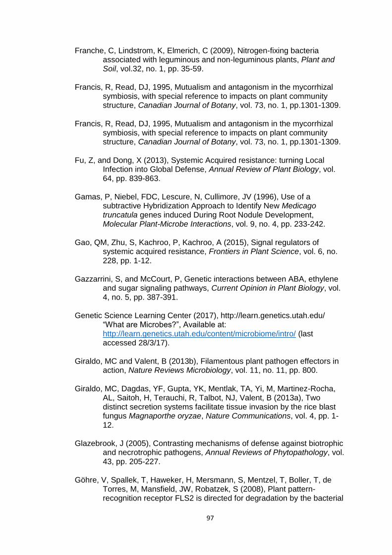
97
Franche, C, Lindstrom, K, Elmerich, C (2009), Nitrogen-fixing bacteria associated with leguminous and non-leguminous plants, Plant and Soil, vol.32, no. 1, pp. 35-59.
Francis, R, Read, DJ, 1995, Mutualism and antagonism in the mycorrhizal symbiosis, with special reference to impacts on plant community structure, Canadian Journal of Botany, vol. 73, no. 1, pp.1301-1309.
Francis, R, Read, DJ, 1995, Mutualism and antagonism in the mycorrhizal symbiosis, with special reference to impacts on plant community structure, Canadian Journal of Botany, vol. 73, no. 1, pp.1301-1309.
Fu, Z, and Dong, X (2013), Systemic Acquired resistance: turning Local Infection into Global Defense, Annual Review of Plant Biology, vol. 64, pp. 839-863.
Gamas, P, Niebel, FDC, Lescure, N, Cullimore, JV (1996), Use of a subtractive Hybridization Approach to Identify New Medicago truncatula genes induced During Root Nodule Development, Molecular Plant-Microbe Interactions, vol. 9, no. 4, pp. 233-242.
Gao, QM, Zhu, S, Kachroo, P, Kachroo, A (2015), Signal regulators of systemic acquired resistance, Frontiers in Plant Science, vol. 6, no. 228, pp. 1-12.
Gazzarrini, S, and McCourt, P, Genetic interactions between ABA, ethylene and sugar signaling pathways, Current Opinion in Plant Biology, vol. 4, no. 5, pp. 387-391.
Genetic Science Learning Center (2017), http://learn.genetics.utah.edu/ “What are Microbes?”, Available at: http://learn.genetics.utah.edu/content/microbiome/intro/ (last accessed 28/3/17).
Giraldo, MC and Valent, B (2013b), Filamentous plant pathogen effectors in action, Nature Reviews Microbiology, vol. 11, no. 11, pp. 800.
Giraldo, MC, Dagdas, YF, Gupta, YK, Mentlak, TA, Yi, M, Martinez-Rocha, AL, Saitoh, H, Terauchi, R, Talbot, NJ, Valent, B (2013a), Two distinct secretion systems facilitate tissue invasion by the rice blast fungus Magnaporthe oryzae, Nature Communications, vol. 4, pp. 1-12.
Glazebrook, J (2005), Contrasting mechanisms of defense against biotrophic and necrotrophic pathogens, Annual Reviews of Phytopathology, vol. 43, pp. 205-227.
Göhre, V, Spallek, T, Haweker, H, Mersmann, S, Mentzel, T, Boller, T, de Torres, M, Mansfield, JW, Robatzek, S (2008), Plant pattern-recognition receptor FLS2 is directed for degradation by the bacterial

98
ubiquitin ligase AvrPtoB, Current Biology, vol. 18, no. 23, pp. 1824-1832.
Gomez-Ariza, J, Campo, S, Rufat, M, Messeguer, J, San Segundo, B, Coca, M (2007), Sucrose-mediated priming of plant defence responses and broad-spectrum disease resistance by overexpression of the maize pathogenesis-related PRms protein in rice plants, Molecular Plant-Microbe Interactions, vol. 20, no. 7, pp. 832-842.
Gomez-Roldan, V, Fermas, S, Bewer, PB, Puech-Pges, V, Dun, EA, Pillot, JP, Letisse, F, Matusova, R, Danoun, S, Portais, JC, Bouwmeester, H, Becard, G, Beveridge, CA, Rameau, C, Rochange, SF (2008), Strigolactone inhibition of shoot branching, Nature, vol. 455, no. 7210, pp. 189.
Grant, M and Lamb, C (2006), Systemic immunity, Current Opinion in Plant Biology, vol. 9, no. 4, pp. 414-420.
Grefen, C and Blatt, M.R (2012), A 2in1 cloning system enables ratiometric bimolecular fluorescence complementation (rBiFC), Biotechniques, vol. 53, no. 5, pp. 311–314.
Hemetsberger C, Herrberger C, Zechmann B, Hillmer M, Doehlemann G (2012), The Ustilago maydis effector Pep1 suppresses plant immunity by inhibition of host peroxidase activity, PLoS Pathogens, vol. 8, no. 5, pp. 1-15.
Hemetsberger, C, Herrberger, C, Zechmann, B, Hillmer, M, Doehlemann, G (2012), The Ustilago maydis effector Pep1 suppresses plant immunity by inhibition of host peroxidase activity, PLoS Pathogens, vol. 8, no. 5, pp. 1-15.
Herbers, K, Meuwly, P, Frommer, WB, Metraux, JP, Sonnewald, U (1996), Systemic Acquired Resistance Mediated by the Ectopic expression of invertase: possible hexose sensing in the secretory pathway, The Plant Cell, vol. 8, no. 5, pp. 793-803.
Hibbet, DS, Gilbert, LB, Donoghue, MJ (2000), Evolutionary instability of ectomycorrhizal symbioses in basidiomycetes, Nature, vol. 407, no. 6803, pp. 506.
Hibbett, D.S. and Matheny, P.B. (2009), The relative ages of ectomycorrhizal mushrooms and their plant hosts estimated using Bayesian relaxed molecular clock analyses, BMC Biology, vol. 7, no. 1, pp. 13.
Hofmann, J, Hahm, HS, Seeberger, PH, Pagel, K (2015), Identification of carbohydrate anomers using ion mobility-mass spectrometry, Nature, vol. 526, no. 7572, pp. 241-244.
99
Holliday, MJ, Keen, NT, Long, M (1981), Cell death patterns and accumulation of fluorescent material in the hypersensitive response of soybean leaves to Pseudomonas syringae pv. glycinea, Physiological Plant Pathology, vol. 18, no. 3, pp. 279.
Horbach, R, Navarro-Quesada, AR, Knogge, W, Deising, HB (2011), When and how to kill a plant cell: infection strategies of plant pathogenic fungi, Journal of Plant Physiology, vol. 168, no. 1, pp. 51-62.
Jayaraman D, Forshey KL, Grimsrud PA, Ane´, JM (2012), Leveraging proteomics to understand plant–microbe interactions, Frontiers in Plant Science, vol. 3, no. 4, pp. 1-6.
Jermy, A (2012), Fungal pathogenesis: Ustilago gives plants a Pep talk, Nature Reviews Microbiology, vol. 10, no. 7, pp. 444.
Johnson, NC, Graham, JH, Smith, FA (1997), Functioning of mycorrhizal associations along the mutualism-parasitism continuum, New Phytologist, vol. 135, no. 4, pp. 575-585.
Jones, DL, Hodge, A, Kuzyakov, Y (2004), Plant and mycorrhizal regulation of rhizodeposition, New Phytologist, vol. 163, no. 3, pp. 459-480.
Jones, JDG and Dangl, JL (2006), The Plant immune system, Nature, vol. 444, no. 7117, pp. 323.
Jung, HW, Tschaplinski, TJ, Wang, L, Glazebrook, J, Greenberg, JT (2009), Priming in systemic plant immunity, Science, vol. 324, no. 5923, pp. 89-91.
Kamatham, S, Neela, KB, Pasupulati, AK, Pallu, R, Singh, SS, Gudipalli, P (2016), benzoylsalicylic acid isolated from seed coats of Givotia rottleriformis induces systemic acquired resistance in tobacco and Arabidopsis, Phytochemistry, vol. 126, pp. 11-22.
Kamoun, S (2006), A catalogue of the effector secretome of plant pathogenic oomycetes, Annual Review of Phytopathology, vol. 44, pp. 41-60.
Kile, GA and Watling, R (1981), An expanded concept of Armillaria luteobubalina, Transactions of the British Mycological Society, vol. 77, no. 1, pp. 75-83.
Kile, GA and Watling, R (1983), Armillaria species from south-eastern Australia, Transactions of the British Mycological Society, vol. 81, no. 1, pp. 129-140.
Kipfer, T, Wholgemuth, T, Heikden, A, Ghazoul, J Egli, S (2012), Growth response of drought-stressed Pinus sylvestris seedlings to single- and multi-species inoculation with Ectoymycorrhizal Fungi, PLoS One, vol. 7, no. 4, pp. 1-7.
100
Kloppholz, S, Kuhn, H, Requena, N (2011), A Secreted Fungal Effector of Glomus intradices Promotes Symbiotic Biotrophy, Current Biology, vol. 21, no. 14, pp. 1204-1209.
Koch, K (2004), Sucrose metabolism: regulatory mechanisms and pivotal roles in sugar sensing and plant development, Current Opinions in Plant Biology, vol. 7, no. 3, pp. 235-246.
Kuc, J (1987), Translocated signals for plant immunization. Annals of the New York Academy of Sciences, vol. 494, no. 1, pp. 221-223.
Kuhn, C, and Grof, C (2010), Sucrose transporters of higher plants, Current Opinion in Plant Biology, vol. 13, no. 3, pp. 287-297.
Lee, SJ and Rose, JKC (2010), Mediation of the transition from biotrophy to necrotrophy in hemibiotrophic plant pathogens by secreted effector proteins, Plant signaling & behaviour, vol. 5, no. 6, pp. 769-772.
Li, J, Li, G, Wang, H, Wang Deng, X (2011), Phytochrome Signaling Mechanisms, The Arabidopsis Book, vol. 9, pp. 1-26.
Liao, HL, Chen, Y, Vilgalys, R (2016), Metatranscriptomic Study of Common and Host-Specific Patterns of Gene Expression between Pines and Their Symbiotic Ectomycorrhizal Fungi in the Genus Suillus, PLoS Genetics, vol. 12, no. 10, pp. 1-24.
Liu, Q, Yuan, M, Zhou, Y, Li, X, Xiao, J, Wang, S (2011), A paralog of the MtN3/saliva family recessively confers race-specific resistance to Xanthomonas oryzae in rice, Plant, Cell & Environment, vol. 34, no. 11, pp. 1958-1969.
Lo Presti, L, Lanver, D, Schweizer, G, Tanaka, S, Liang, L, Tollot, M, Zuccaro, A, Reissmann, S, Kahmann, R (2015), Fungal effectors and plant susceptibility, Annual Review of Plant Biology, vol. 66, pp. 513-545.
Lopez-Raez, JA, Verhage, A, Fernandez, I, Garcia, JM, Azcon-Aguilar, C, Flors, V, Pozo, MJ (2010), Hormonal and transcriptional profiles highlight common and differential host responses to arbuscular mycorrhizal fungi and the regulation of the oxylipin pathway, Journal of Experimental Botany, vol. 61, no. 10, pp. 2589-2601.
Luna, E, Bruce, TJ, Roberts, MR, Flors, V, Ton, J (2012), Next-generation systemic acquired resistance. Plant Physiology, vol. 158, no. 2, pp.844-853.
Ma, L, Wu, X, Zheng, L (2009), Relationship between plant hormone level excreted by ectomycorrhizal fungi and growth of poplar NL-895, Frontiers of Forestry in China, vol. 4, no. 2, pp. 236-241.
101
Manning, VA, Hardison, LK, Ciuffetti, LM (2007), Ptr ToxA interacts with a chloroplast-localized protein, Molecular Plant-Microbe Interactions, vol. 20, no. 2, pp. 168-177.
Martin, F, Aerts, A, Ahren, D, Brun, A, Danchin, EGJ, Duchaussoy, F, Gibon, J, Kohler, A, Lindquist, E, Pereda, V, Salamov, A, Shapiro, HJ, Wuyts, J, Dlaudez, D, Buee, M, Brokstein, P, Canback, B, Cohen, D, Courty, PE, Coutinho, PM, Delaruelle, C, Detter, JC, Deveau, A, Difazio, S, Duplessis, S, Fraissinet-Tachet, L, Lucic, E, Frey-Klett, P, Fourrey, C, Feussner, I, Gay, G, Grimwood, J, Hoegger, PJ, Jain, P, Kilaru, S, Labbe, J, Lin, YC, Leguem Vm Tacon, FL, Marmeisse, R, Melayah, D, Montanini, B, Muratet, M, Nehls, U, Niculita-Hirzel, H, Oudot-Le Secq, MP, Peter, M, Quesneville, H, Rajashekar, B, Reich, M, Rouhier, N, Schmutz, J, Yin, T, Chalot, M, Henrissat, B, Kues, U, Lucas, S, Van De Peer, Y, Podila, GK, Polle, A, Pukkila, PJ, Richardson, PM, Rouze, P, Sanders, IR, Stajich, GE, Tunlid, A, Tuskan, G, Grigoriev, IV (2008), The genome sequence of the basidiomycete fungus Laccaria bicolor provides insights into the mycorrhizal symbiosis, Nature, vol. 452, no. 7183, pp. 88.
Marzec, M (2016), Strigolactones as Part of the Plant Defence System, Trends in Plant Science, vol. 21, no. 11, pp. 900-903.
Mauch-Mani, B and Mauch, F (2005), The role of abscisic acid in plant pathogen interactions, Current Opinion in Plant Biology, vol. 8, no. 4, pp. 409-414.
Mohammad, RBM and Van den Ende, W (2012), Sugars and plant innate immunity, Journal of Experimental Botany, vol. 63, no. 11, pp. 3989-3998.
Mohrmann, L and Verrijzer, CP (2005), Composition and functional specificity of SWI2/SNF2 class chromatin remodelling complexes, Biochimica et Biophysica Acta, vol. 11, no. 2-3, pp. 59-73.
Moore, B, Zhou, L, Rolland, F, Hall, Q, Cheng, WH, Liu, YX, Hwang, I, Jones, T, Sheen, J (2003), Role of the Arabidopsis glucose sensor JXK1 in nutrients, light, and hormonal signaling, Science, vol. 300, no. 5617, pp. 332-336.
Morkunas, I and Ratajczak, L (2014), The role of sugar signaling in plant defense resposnes against fungal pathogens, Acta Physiologiae Plantarum, vol. 36, no. 7, pp. 1607-1619.
Muchembled, J, Sahraoui, ALH, Grandmougin-Ferjani, A, Sancholle, M (2006), Changes in lipid composition of Blumeria graminis f.sp. tritici conidia produced on wheat leaves treated with heptanoyl salicylic acid, Phytochemistry, vol. 67, no. 11, pp. 1104-1109.
102
Myburg, AM, Grattapaglia, D, Tuskan, GA, Hellsten, U, Hayes, RD, Grimwood, J, Jenkins, J, Lindquist, E, Tice, H, Bauer, D, Goodstein, DM, Dubchak, I, Poliakov, A, Mizrachi, E, Kullan, ARK, Hussey, SG, Pinard, D, Merwe, K, Singh, P, Jaarsveld, I, Silva-Junior, O, Togawa, RC, Pappas, MR, Faria, DA, Sansaloni, CP, Petroli, CD, Yang, X, Ranjan, P, Tschaplinski, TJ, Ye, CY, Li, T, Sterck, L, Vanneste, K, Murat, F, Soler, M, Clemente, HS, Saidi, N, Cassan-Wang, H, Dunand, C, Hefer, CA, Bornberg-Bauer, E, Kersting, AR, Vining, K, Amarashinghe, V, Ranik, M, Naithani, S, Elser, J, Boyd, AE, Liston, A, Spatafora, JW, Dharmwardhana, P, Raja, R, Sullivan, C, Romanel, E, Alves-Ferreira, M, Kulheim, C, Foley, W, Carocha, V, Paiva, J, Kudrna, D, Brommonschenkel, SH, Pasquali, G, Byrne, M, Rigault, P, Ribbits, J, Spokevicius, A, Jones, RC, Steane, DA, Vaillancourt, RE, Potts, BM, Joubert, F, Barry, K, Jr, GJP, Strauss, SH, Jaiswal, P, Grima-Pettenati, J, Salse, J, Peer, YVD, Roksar, DS, Schmutz J (2014), The genome of Eucalyptus grandis, Nature, vol. 510, no.7505, pp. 356-368.
Narlikar, GJ, Fan, HY, Kingston, RE (2002), Cooperation between complexes that regulate chromatin structure and transcription, Cell, vol. 108, no. 4, pp. 475-487.
Navarro, L, Bari, R, Achard, P, Lison, P, Nemri, A, Halberd, NP, Jones, JDG (2008), DELLAs Control Plant Immune Responses by Modulating the Balance of Jasmonic Acid and Salicylic Acid Signaling, Current Biology, vol. 18, no. 9, pp. 650-655.
Nehls, U (2008), Mastering ectomycorrhizal symbiosis: the impact of carbohydrates, Journal of Experimental Botany, vol. 59, no. 5, pp. 1097-1108.
O’connell, RJ and Panstruga, R (2006), Tete a tete inside a plant cell: establishing compatibility between plants and biotrophic fungi and oomycetes, New Phytologist, vol. 171, no. 4, pp. 699-718.
Okmen, B and Doehlemann, G (2014), Inside plant: biotrophic strategies to modulate host immunity and metabolism, Current Opinion in Plant Biology, vol. 20, pp. 19-25.
Oliver, RP, Friesen, TL, Faris, JD, Solomon, PS (2012), Stagonospora nodorum: from pathology to genomics and host resistance, Annual Review of Phytopathology, vol. 50, pp. 23-43.
Park, CH, Chen, S, Shirsekar, G, Zhou, B, Khang, CH, Songkumarn, P, Afzal, AJ, Ning, Y, Wang, R, Bellizzi, M, Valent, B, Wang, GL (2012), The Magnaporthe oryzae effector AvrPiz-t targets the RING E3 ubiquitin ligase APIP6 to suppress pathogen-associated molecular pattern-triggered immunity in rice, The Plant Cell, vol. 24, no. 11, pp. 4748-4762.

103
Partida-Martinez, LP and Heil, M (2011), The microbe-free plant: fact or artifact?, Frontiers in Plant Science, vol. 2, no. 100, pp. 1-16.
Parvaiz, A (2011), Environmental Adaptations and Stress Tolerance of Plants in the Era of Climate Change, 1st edn, New York: Springer New York.
Patil, G, Vlliyofdan, B, Deshmukh, R, Prince, S, Nicander, B, Zhao, M, Sonah, H, Song, L, Lin, L, Chaudhary, J, Liu, Y, Joshi, T, Xu, D, Nguyen, HT (2015), Soybean (Glycine max) SWEET gene family: insights through comparative genomics, transcriptome profiling and whole genome re-sequence analysis, BMC Genomics, vol. 16, no. 520, pp. 1-16.
Payyavula, RS, Tay, KHC, Tsai, CJ, Harding, SA (2011), The sucrose transporter family in Populus: the importance of a tonoplast PtaSUT4 to biomass and carbon partitioning, The Plant Journal, vol. 65, no. 5, pp. 575-770.
Pellegrin, C, Morin, E, Martin, FM, Veneault-Fourrey, C (2015), Comparative Analysis of Secretomes from Ectomycorrhizal Fungi with an Emphasis on Small-Secreted Proteins, Frontiers in Microbiology, vol. 6, no. 1278, pp. 1-15.
Perotto, S, Rodda, M, Benetti, A, Sillo, F, Ercole, E, Rodda, M, Girlanda, M, Murat, C, Balestrini, R (2014), Gene expression in mycorrhizal orchid protocorms suggest a friendly plant-fungus relationship, Planta, vol. 239, no. 6, pp. 1337-1349.
Plett, JM and Martin, F (2011), Blurred boudnaries: lifestyle lessons from ectomycorrhizal fungal genomes, Trends in Genetics, vol. 27, no. 1, pp. 14-22.
Plett, JM and Martin, F (2015), Reconsidering mutualistic plant–fungal interactions through the lens of effector biology, Current Opinion in Plant Biology, vol. 26, pp. 45–50.
Plett, JM, Daguerre, Y, Wittulsky, S, Vayssieres, A, Deveau, A, Melton, SJ, Kohler, A, Morrell-Falvery, Jennifer, L, Brun, A, Veneault-Fourrey, C, Martin, F (2014a), Effector MiSSP7 of the mutualistic fungus Laccaria bicolor stabilizes the Populus JAZ6 protein and represses jasmonic acid (JA) responsive genes, Proceedings of the National Academy of Sciences of the United States of America, vol. 111, no. 22, pp. 8299-8304.
Plett, JM, Kemppainen, M, Kale, SD, Kohler, A, Legue, V, Brun, A, Tyler, BM, Pardo, AG, Martin, F (2011), A secreted effector protein of Laccaria bicolor is required for symbiosis development, Current Biology, vol. 21, no. 14, pp. 1197-1203.

104
Plett, JM, Khachane, A, Ouassou, M, Sundberg, B, Kohler, A, Martin, F (2014b), Ethylene and jasmonic acid act as negative modulators during mutualistic symbiosis between Laccaria bicolor and Populus roots, New Phytologist, vol. 202, no. 1, pp. 270-286.
Plett, JM, Yin, H, Mewalal, R, Hu, R, Li, T, Ranjan, P, Jawdy, S, Paoli, HCD, Butler, G, Burch-Smith, TM, Guo, HB, Chen, CJ, Kohler, A, Anderson, IC, Labbe, JL, Martin, F, Tuskan, GA, Yang, X (2017), Populus trichocarpa encodes small, effector-like secreted proteins that are highly induced during mutualistic symbiosis, Scientific Reports, vol. 7, no. 382, pp. 1-13.
Qutob, D, Kemmerling, B, Brunner, F, Kufner, I (2006), Phytotoxicity and innate immune responses induced by Nep1-like proteins, The Plant Cell, vol. 18, no. 12, pp. 3721-3744.
Qutob, D, Kemmerling, B, Brunner, F, Kufner, I (2006), Phytotoxicity and innate immune responses induced by Nep1-like proteins, The Plant Cell, vol. 18, no. 12, pp. 3721-3744.
Rafiqi, M, Ellis, JG, Ludowici, VA, Hardham, AR, Dodds, PN (2012), Challenges and progress towards understanding the role of effectors in plant-fungal interactions, Current Opinion in Plant Biology, vol. 15, no. 4, pp. 477-483.
Rasmann, S, De Vos, M, Casteel, C, Tian, D, Halitschke, R, Sun, J, Agrawal, A, Felton, G, Jander, G (2012), Herbivory in the previous generation primes plants for enhanced insect resistance, Plant Physiology, vol. 158, no. 2, pp. 854-863.
Raven, P, Johnson, GB, Mason, KA (2011), Biology, 9th edn., New York, McGraw Hill.
Redman, R, Kim, Y, Woodward, J, Greer, C, Espino, L, Dota, S, Rodriguez, R, Increased Fitness of Rice Plants to Abiotic Stress Via Habitat Adapted Symbiosis: A Strategy for Mitigating Impacts of Climate Change, PLoS One, vol. 6, no. 7, pp. 1-10.
Redman, R, Kim, Y, Woodward, J, Greer, C, Espino, L, Doty, S, Rodriguez, R (2011), Increased fitness of rice plants to abiotic stress via habitat adapted symbiosis: a strategy for mitigating impacts of climate change, PLoS One, vol. 6, no. 7, pp. 1-10.
Reignault, PH, Cogan, A, Muchembled, J, Lounes-Hadj Sahraoui, A, Durand, R, Sancholle, M (2001), Trehalose induces resistance to powdery mildew in wheat, New Phytologist, vol. 149, no. 3, pp. 519-529.
Reimer-Michalski, EM and Conrath, U (2016), Innate immune memory in plants, Seminars in Immunology, vol. 28, no. 4, pp. 319-327.
105
Rey, T, Chatterjee, A, Buttay, M, Toulotte, J, Schornack, S (2015), Medicago truncatula symbiosis mutants affected in the interaction with a biotrophic root pathogen, New Phytologist, vol. 206, no. 2, pp. 497-500.
Richardson, AE, Barea, JM, McNeill, AM, Prigent-Combaret, C (2009), Acquisition of phosphorus and nitrogen in the rhizosphere and plant growth promotion by microorganisms, Plant Soil, vol. 321, no. 1, pp. 305-339.
Robert-Seilaniantz, A, Navarro, L, Bari, R, Jones, JDG (2007), Pathological hormone imbalances, Current Opinion in Plant Biology, vol. 10, no. 4, pp. 372-379.
Rolland, F, Baena-Gonzalez, E, Sheen, J (2006), Sugar sensing and signaling in plants: conserved and novel mechanisms, Annual Review of Plant Biology, vol. 57, pp. 675.
Royal Botanic Garden (2017a), www.rbgsyd.nsw.gov.au “Armillaria Root Rot”, Available at: https://www.rbgsyd.nsw.gov.au/Plants/Pests-Diseases/Armillaria-Root-Rot (last accessed 28/3/17).
Royal Botanic Garden (2017b), www.rbgsyd.nsw.gov.au “Phytophthora Dieback”, Available at: https://www.rbgsyd.nsw.gov.au/Plants/Pests-Diseases/Phytophthora-Dieback (last accessed 28/3/17).
Ryals, Ja, Neuenschwander, UH, Willits, MG, Molina, A, Steiner, HY and Hunt, MD (1996), System Acquired Resistance, The Plant Cell, vol. 8, no. 10, pp. 1809-1819.
Saez, A, Rodrigues, A, Santiago, J, Rubio, S, Rodriguez, PL, (2008), HAB1–SWI3B interaction reveals a link between abscisic acid signaling and putative SWI/SNF chromatin-remodeling complexes in Arabidopsis, The Plant Cell, vol. 20, no. 11, pp. 2972-2988.
Saikkonen, K, Faeth, SH, Helander, M, Sullivan, TJ (1998), Fungal endophytes: a continuum of Interactions with Host Plants, Annual Review of Ecology and Systematics, vol. 291, no. 1, pp. 319-343.
Salmeron, JM, Oldroyd, GE, Rommens, CM, Scofield, SR, Kim, HS, Lavelle, DT, Dahlbeck, D, Staskawicz, BJ (1996), Tomato Prf is a member of the leucine-rich repeat class of plant disease resistance genes and lies embedded within the Pto kinase gene cluster, Cell, vol. 86, no. 1, pp. 123-133.
Salzer, P and Boller, T (2000), Elicitor-induced reactions in mycorrhizae and their suppression, In: Podila GK, Douds DD Jr (eds), Current advances in mycorrhizae research, APS Press, St. Paul, Minnesota

106
Salzer, P, Sirrenberg, A and Hager, A (1997), Differential Effect of Purified Spruce Chitinases and β-1,3-Glucanases on the Activity of Elicitors from Ectomycorrhizal fungi, Plant Physiology, vol. 114, no. 3, pp. 957-968.
Sarnowska, E, Gratkowska, D.M., Sacharowski, S.P., Cwiek, P, Tohge, T, Fernie, A.R., Siedlecki, J.A., Koncz, C, Sarnowski, T.J. (2016), The Role of SWI/SNF Chromatin Remodeling Complexes in Hormone Crosstalk, Trends in Plant Science, vol. 21, no. 7, pp. 594-608.
Sarnowski, T, Pawlikowska, K, Kaczanowski, S, Herzmanowski, A (2002), AtSWI3B, an Arabidopsis homolog of SWI3, a core subunit of yeast Swi/Snf chromatin remodeling complex, interacts with FCA, a regulator of flowering time, Nucleic Acids Research, vol. 30, no. 15, pp. 3412-3421.
Schmidt, SM, and Panstruga, R (2011), Pathogenomics of fungal plant parasites: what have we learnt about pathogenesis?, Current Opinion in Plant Biology, vol. 14, no. 4, pp. 392-399.
Shalitin, D and Wolf, S (2000), Cucumber mosaic virus infection affects sugar transport in melon plants, Plant Physiology, vol. 123, no. 2, pp. 597-604.
Shearer, BL, Crane, CE, Fairman, RG, Grant, MJ (1997), Occurrence of Armillaria luteobubalina and Pathogen-mediated Changes in Coastal Dune Vegetation of South-western Australia, Australian Journal of Botany, vol. 45, no. 5, pp. 905-917.
Singh, V, Louis, J, Ayre, BG, Reese, JC, Shah, J, Trehalose phosphate synthase11-dependent trehalose metabolism promotes Arabidopsis thaliana defense against the phloem-feeding insect Myzus persicae, The Plant Journal, vol. 67, no. 1, pp. 94-104.
Slaughter, A, Daniel, X, Flors, V, Luna, E, Hohn, B, Mauch-Mani, B (2012), Descendants of primed Arabidopsis plants exhibit resistance to biotic stress, Plant Physiology, vol. 158, no. 2, pp. 835-843.
Smith SE, Read DJ (1997), Mycorrhizal Symbiosis, Academic Press Inc., San Diego.
Smith, S.E. and Read, D.J. (2008) Mycorrhizal Symbiosis, (3rd edn), Academic Press, San Diego.
Stassen, JHM, and den Ackerveken, GV (2011), How do oomycete effectors interfere with plant life?, Current Opinion in Plant Biology, vol.14, no. 4, pp. 407-414.
Stergiopoulos, I and de Wit, PJGM (2009), Fungal Effector Proteins, Annual Review of Phytopathology, vol. 47, pp. 233-273.
107
Stergiopoulos, I, Collemare, J, Mehrabi, R, Wit, PJGM (2013), Phytotoxic secondary metabolites and peptides produced by plant pathogenic Dothideomycete fungi, FEMS Microbiology Reviews, vol. 37, no. 1, pp. 67-93.
Streubel, J, Pesce, C, Hutin, M, Koebnik, R, Boch, J, Szurek, B (2013), Five phylogenetically close rice SWEET genes confer TAL effector-mediated susceptibility to Xanthomonas oryzae pv. oryzae, New Phytologist, vol. 200, no. 3, pp. 808-819.
Tarkka, M, Herrmann, S, Wubet, T, Feldhahn, L, Recht, S, Kurth, F, Mailander, S, Bonn, M, Neef, M, Angay, O, Bacht, M, Graf, M, Maboreke, H, Fleischmann, F, Grams, TEE, Ruess, Lm Schadler, M, Brandl, R, Scheu, S, Schrey, SD, Grosse, I, Buscot, F (2013), OakContigDF159.1, a reference library for studying differential gene expression in Quercus robur during controlled biotic interactions: use for quantitative transcriptomic profiling of oak roots in ectomycorrhizal symbiosis, New Phytologist, vol. 199, no. 2, pp. 529-540.
Tauzin, AS and Giardina, T (2014), Sucrose and invertases, a part of the plant defense response to the biotic stresses, Frontiers in Plant Science, vol. 5, no. 293, pp. 1-8.
Tognetti, JA, Pontis, HG, Martinez-Noel, GMA (2013), Sucrose signaling in plants, Plant Signaling & Behavior, vol. 8, no. 3, pp. 1-10.
Tschaplinski, TJ, Plett, JM, Engle, NL, Deveau, A, Cushman, KC, Martin, MZ, Doktycz, MJ, Tuskan, GA, Brun, A, Kohler, A, Martin, F (2014), Populus trichocarpa and Populus deltoids exhibit different metabolomic responses to colonization by the symbiotic fungus Laccaria bicolor, Molecular Plant-Microbe Interactions, vol. 27, no. 6, pp. 546-556.
Umehara, M, Hanada, A, Yoshida, S, Akiyama, K, Arite, T, Takeda-Kamiya, N, Magome, H, Kamiya, Y, Shirasu, K, Yoneyama, K, Kyozuka, J, Yamaguchi, S (2008), Inhibition of shoot branching by new terpenoid plant hormones, Nature, vol. 455, no. 7210, pp. 195.
Valluru, R and Van den Ende, W (2011), Myo-inositol and beyond – Emerging networks under stress, Plant Science, vol. 18, no. 4, pp. 387-400.
Walley, J.W. et al. (2008), The chromatin remodeler SPLAYED regulates specific stress signaling pathways, PLoS Pathogens, vol. 4, no. 12, pp. 1-8.
Wang, B and Qiu, YL (2006), Phylogenetic distribution and evolution of mycorrhizas in land plants, Mycorrhiza, vol. 16, no. 5, pp. 299-363.

108
Weber, A, Servaites, JC, Geiger, DR, Kofler, H, Hille. D. Groner, F, Hebbeker, U, Flugge, UI (2000), Identification, purification, and molecular cloning of a putative plastidic glucose translocator, The Plant Cell, vol. 12, no. 5, pp. 787-801.
Weiberg, A, Wang, M, Bellinger, M, Jin, H (2014), Small RNAs: a new paradigm in plant-microbe interactions, Annual Review of Phytopathology, vol. 52, pp. 495-516.
Weiberg, A, Wang, M, Lin, FM, Zhao, H, Zhang, Z, Kaloshian, I, Huang, HD, Jin, H (2013), Fungal small RNAs suppress plant immunity by hijacking host RNA interference pathways, Science, vol. 342, no. 6154, pp. 118-123.
Weise, S, Schrader, S, Kleinbeck, K, Sharkey, T, Carbon balance and Circadian Regulation of Hydrolytic and Phosphorolytic Breakdown of transitory Starch1, Plan Physiology, vol. 141, no. 3, pp. 879-886.
Yang, B, Sugio, A, White, FF (2006), Os8N3 is a host disease-susceptibility gene for bacterial blight of rice, Proceedings of the National Academy of Sciences of the United States of America, vol. 103, no. 27, pp. 10503-10508.
Yang, B, Sugio. A, White, FF, Bing, Y, Akiko, S, Frank, FW (2006), Os8N3 is a host disease-susceptibility gene for bacterial blight of rice, Proceedings of the National Academy of Sciences of the United States of America, vol. 103, no. 27, pp. 10503-10508.
Yoon SI, Kurnasov O, Natarajan V, Hong M, Gudkov AV, Osterman, AL and Wilson, IA (2012), Structural basis of TLR5-flagellin recognition and signaling, Science, vol. 335, no. 6070, pp. 859-864.
Zhang, Q, Hu, W, Zhu, F, Wang, L, Yu, Q, Ming, R, Zhang, J (2016), Structure, phylogeny, allelic haplotypes and expression of sucrose transporter gene families in Saccharum, BMC Genomics, vol. 17, no. 88, pp. 1-18.
Zhang, Z, Li, Q, Li, Z, Staswick, P, Wang, M, Zhu, Y, He, Z (2007), Dual regulation role of GH3.5 in salicylic acid and auxin signaling during Arabidopsis-Pseudomonas syringae interaction, Plant Physiology, vol. 145, no. 2, pp. 450-464.
Zhou, Y, Liu, L, Huang, W, Yuan, M, Zhou, F, Li, X, Lin, Y (2014), Overexpression of OsSWEET5 in Rice Causes Growth Retardation and Precocious Senescence, PLOS one, vol. 9, no. 4, pp. 1-9.
Zuccaro, A, Lahrmann, U, Langen, G (2014), Broad compatibility in fungal root symbioses, Current Opinion in Plant Biology, vol. 20, pp. 135-145.