Light microscopic analysis of Tectona grandis L.f. wood inoculated with Irpex lacteus and...
8
ORIGINALS ORIGINALARBEITEN Light microscopic analysis of Tectona grandis L.f. wood inoculated with Irpex lacteus and Phanerochaete chrysosporium Rina D. Koyani • Kishore S. Rajput Received: 18 April 2013 / Published online: 23 November 2013 Ó Springer-Verlag Berlin Heidelberg 2013 Abstract Felling of immature teak (Tectona grandis L.f.) trees or delay in transport of wood logs from felling sites provide platform to microbial attack. Among them, white rot fungi are central driving force that degrades wood and causes severe economic loss. In contrast, Phanerochaete chrysosporium and Irpex lacteus are more extensively studied for their ability to degrade synthetic dyes and poly- aromatic compounds. Therefore, in the present study, both the fungi collected from the Gujarat forest were utilised for in vitro decay test to assess their potential in lignin deg- radation, extent of cell wall damage and pattern of wood decay in sound blocks of teak. In the early stage of fungal inoculation, there was a negligible amount of weight loss; after 1 month it became rapid and highest weight loss (30.05 % by P. chrysosporium and 27.97 % by I. lacteus) was observed at the end of 120 days. Mycelial invasion occurred through vessels, from vessels to axial and ray parenchyma and subsequently into xylem fibres. Both the strains showed selective delignification and the first symptom of degradation was defibration, separation of rays, and formation of boreholes on ray cell walls at an advanced stage. Xylem fibres and parenchyma cells lost their integrity and collapsed completely. Among all the cell types, parenchyma cells and fibres were more vulnerable to fungal attack, while vessels were resistant to the activity of lignolytic enzymes. Lichtmikroskopische Untersuchung von mit Irpex lacteus und Phanerochaete chrysosporium befallenem Teakholz (Tectona grandis L.f.) Zusammenfassung Das Fa ¨llen junger Teakba ¨ume (Tectona grandis L.f.) oder Verzo ¨gerungen beim Ab- transport der gefa ¨llten Holzsta ¨mme bietet eine ideale Plattform fu ¨r mikrobiellen Befall. Vorwiegend Weiß- fa ¨ulepilze sind verantwortlich fu ¨r die Holzzersto ¨rung, die schwerwiegende wirtschaftliche Scha ¨den verursacht. Von den Weißfa ¨ulepilzen Irpex lacteus und Phanero- chaete chrysosporium wurde bisher vorwiegend der Abbau von synthetischen Farbstoffen und polyaromatis- chen Bestandteilen untersucht. Aus diesem Grunde wurden hier die beiden aus dem Gujarat Forest stam- menden Pilze fu ¨r In-vitro Pilzabbauversuche verwendet, um zu untersuchen, inwieweit diese Lignin abbauen und die Zellwand bescha ¨digen und wie die Holzzersto ¨rung von nicht befallenen Teakholzpru ¨fko ¨rpern abla ¨uft. Zu Beginn der Pilzbeimpfung war der Masseverlust uner- heblich; nach einem Monat beschleunigte sich dieser und nach 120 Tagen wurde der ho ¨chste Masseverlust (30,05 % bei Phanerochaete chrysosporium, 27,97 % Irpex lacteus) verzeichnet. Das Myzel befiel erst die Gefa ¨ße, dann das axiale und Markstrahl-Parenchym und schließlich die Fasern des Xylems. Beide Sta ¨mme zeig- ten selektive Delignifizierung und die ersten Symptome der Zersto ¨rung waren Zerfaserung, Trennung der Markstrahlen und Durchdringung der Markstrahl- zellwa ¨nde im fortgeschrittenen Stadium. Xylemfasern und Parenchymzellen wurden bescha ¨digt und kollabier- ten vollsta ¨ndig. Unter all den Zellarten waren Parenc- hymzellen und Fasern fu ¨r Pilzbefall mehr anfa ¨llig, wohingegen die Gefa ¨ße gegenu ¨ber lignolytischen Enzy- men resistent waren. R. D. Koyani Á K. S. Rajput (&) Department of Botany, Faculty of Science, The Maharaja Sayajirao University of Baroda, Vadodara 390 002, India e-mail: [email protected] 123 Eur. J. Wood Prod. (2014) 72:157–164 DOI 10.1007/s00107-013-0763-7
Transcript of Light microscopic analysis of Tectona grandis L.f. wood inoculated with Irpex lacteus and...

ORIGINALS ORIGINALARBEITEN
Light microscopic analysis of Tectona grandis L.f. wood inoculatedwith Irpex lacteus and Phanerochaete chrysosporium
Rina D. Koyani • Kishore S. Rajput
Received: 18 April 2013 / Published online: 23 November 2013
� Springer-Verlag Berlin Heidelberg 2013
Abstract Felling of immature teak (Tectona grandis L.f.)
trees or delay in transport of wood logs from felling sites
provide platform to microbial attack. Among them, white
rot fungi are central driving force that degrades wood and
causes severe economic loss. In contrast, Phanerochaete
chrysosporium and Irpex lacteus are more extensively
studied for their ability to degrade synthetic dyes and poly-
aromatic compounds. Therefore, in the present study, both
the fungi collected from the Gujarat forest were utilised for
in vitro decay test to assess their potential in lignin deg-
radation, extent of cell wall damage and pattern of wood
decay in sound blocks of teak. In the early stage of fungal
inoculation, there was a negligible amount of weight loss;
after 1 month it became rapid and highest weight loss
(30.05 % by P. chrysosporium and 27.97 % by I. lacteus)
was observed at the end of 120 days. Mycelial invasion
occurred through vessels, from vessels to axial and ray
parenchyma and subsequently into xylem fibres. Both the
strains showed selective delignification and the first
symptom of degradation was defibration, separation of
rays, and formation of boreholes on ray cell walls at an
advanced stage. Xylem fibres and parenchyma cells lost
their integrity and collapsed completely. Among all the cell
types, parenchyma cells and fibres were more vulnerable to
fungal attack, while vessels were resistant to the activity of
lignolytic enzymes.
Lichtmikroskopische Untersuchung von mit Irpex
lacteus und Phanerochaete chrysosporium befallenem
Teakholz (Tectona grandis L.f.)
Zusammenfassung Das Fallen junger Teakbaume
(Tectona grandis L.f.) oder Verzogerungen beim Ab-
transport der gefallten Holzstamme bietet eine ideale
Plattform fur mikrobiellen Befall. Vorwiegend Weiß-
faulepilze sind verantwortlich fur die Holzzerstorung, die
schwerwiegende wirtschaftliche Schaden verursacht.
Von den Weißfaulepilzen Irpex lacteus und Phanero-
chaete chrysosporium wurde bisher vorwiegend der
Abbau von synthetischen Farbstoffen und polyaromatis-
chen Bestandteilen untersucht. Aus diesem Grunde
wurden hier die beiden aus dem Gujarat Forest stam-
menden Pilze fur In-vitro Pilzabbauversuche verwendet,
um zu untersuchen, inwieweit diese Lignin abbauen und
die Zellwand beschadigen und wie die Holzzerstorung
von nicht befallenen Teakholzprufkorpern ablauft. Zu
Beginn der Pilzbeimpfung war der Masseverlust uner-
heblich; nach einem Monat beschleunigte sich dieser und
nach 120 Tagen wurde der hochste Masseverlust
(30,05 % bei Phanerochaete chrysosporium, 27,97 %
Irpex lacteus) verzeichnet. Das Myzel befiel erst die
Gefaße, dann das axiale und Markstrahl-Parenchym und
schließlich die Fasern des Xylems. Beide Stamme zeig-
ten selektive Delignifizierung und die ersten Symptome
der Zerstorung waren Zerfaserung, Trennung der
Markstrahlen und Durchdringung der Markstrahl-
zellwande im fortgeschrittenen Stadium. Xylemfasern
und Parenchymzellen wurden beschadigt und kollabier-
ten vollstandig. Unter all den Zellarten waren Parenc-
hymzellen und Fasern fur Pilzbefall mehr anfallig,
wohingegen die Gefaße gegenuber lignolytischen Enzy-
men resistent waren.
R. D. Koyani � K. S. Rajput (&)
Department of Botany, Faculty of Science, The Maharaja
Sayajirao University of Baroda, Vadodara 390 002, India
e-mail: [email protected]
123
Eur. J. Wood Prod. (2014) 72:157–164
DOI 10.1007/s00107-013-0763-7

1 Introduction
Wood is the natural source of renewable energy and plays
an important role in the national economy. The main role
of wood is not only to provide energy but it also provides
building material and furniture for commodities and as a
raw material for paper, paper products, boards and wood
products (Plomion et al. 2001). It is also one of the
imperative sinks for atmospheric carbon dioxide stored in
the form of cell wall material. However, many times living
trees as well as wood logs are invaded by microorganisms
causing severe economic loss due to structural damage in
the wood components of forest trees and butt and root rot
diseases of fruit trees (Nicolotti et al. 2010). Among them,
white rot fungi are central driving forces that have the
ability to degrade lignin selectively or simultaneously
along with other cell wall polysaccharides (Blanchette
1984a, b; Maloy and Murray 2001; Schwarze 2007; Koyani
et al. 2010; Sanghvi et al. 2013). In contrast, members of
ascomycetes and deuteromycetes also attack wood but
most of them are unable to degrade lignin, a major and
complex biopolymer which is an important constituent of
wood cell wall (Blanchette 1984a, b; Adaskaveg et al.
1995; Maloy and Murray 2001; Schwarze 2007; Sanghvi
et al. 2013).
Owing to lignin degradation ability, white rot fungi are
extensively studied due to their higher potential in the
production of ligninolytic enzymes that are used in biore-
mediation process. Among them, Irpex lacteus and Phan-
erochaete chrysosporium are extensively studied by several
workers (Datta et al. 1991; Baborova et al. 2006; Gassara
et al. 2010; Erkurt 2010; Koyani et al. 2013) but similar
information is lacking on their ability to degrade wood, its
pattern of wood decay and extent of damage caused. In the
forest areas, both fungi are observed frequently on wood
logs of different trees including teak. In the present study,
several wood rot fungi were collected from naturally
infected wood samples from Gujarat Forests. Since, Irpex
lacteus and Phanerochaete chrysosporium are known in
the literature for high lignolytic activity, it was expected to
damage the wood within a short period.
Tectona wood is prized for its valuable timber industry
throughout the world and widely used for heavy construc-
tion and fine quality furniture, doors and windows (Nocetti
et al. 2011). It is therefore under cultivation in different
parts of the tropical countries other than its natural habitat.
Various aspects, such as seasonality of vascular cambium
(Rao and Dave 1981; Rao and Rajput 1999; Priya and Bhat
1997), wood properties and geographical variations in wood
density (Bhat and Priya 2004; Varghese et al. 2000; Mac-
chioni et al. 2007), properties of juvenile wood (Priya and
Bhat 1997), province effect on growth ring structure
(Nocetti et al. 2011) and effect of insect defoliation and
false ring formation (Priya and Bhat 1998; Rajput et al.
2005; Kokutse et al. 2009, 2010) have been studied in
detail. Many times teak wood logs stored in the forest depot,
saw mills and window frames in old buildings exposed to
open environment show fungal fruiting bodies on them. In
spite of its high timber value, damage caused by these fungi
on teak wood has not been investigated. Since, fruiting
bodies of both the fungi are mostly observed on the surface,
i.e. sapwood of wood logs (which is less resistant to fungal
attack), sapwood is utilised in the present study to investi-
gate the ability of Irpex lacteus and Phanerochaete chry-
sosporium in degrading teak wood by in vitro laboratory
decay test and to characterise wood decay pattern by ana-
tomical methods using light microscope.
2 Materials and methods
2.1 Source of fungi
Irpex lacteus and Phanerochaete chrysosporium used in
this study were isolated from the infected teak wood
samples collected from Junagarh (Saurashtra region) forest
of Gujarat State. Small pieces of infected wood samples
were surface sterilized by 0.1 % HgCl2 for 40–45 s with
intermediate washing by sterile distilled water. Subse-
quently, these wood pieces were treated with 70 % ethanol
for few seconds and inoculated on 2.5 % Malt Extract Agar
(MEA) media. Pure cultures were established by serial
transfer and cultures were maintained at 4 �C. Fungal DNA
was extracted according to the method described by Moller
et al. (1992) and sent to Chromous Biotech Pvt. Ltd.,
Banglore for PCR sequencing and molecular identification.
2.2 Wood decay test
Cubic wood blocks (2 9 2 9 2 cm3) from sound sapwood
portion of Tectona grandis L.f. were prepared from the
stem disc free from knots. Prior to rewetting, some of the
wood blocks were marked and weighed prior to and after
fungal inoculation to study the weight loss. Marked and
unmarked wood blocks were rewetted in water for 24 h to
obtain optimum moisture level to facilitate fungal action.
Subsequently, these blocks were autoclaved at 120 �C for
30 min; surface sterilized with routine method and kept in
an autoclaved Petri dish containing Malt Extract Agar
Media. Thereafter, these Petri dishes were inoculated with
15-day-old pure culture of Irpex lacteus and Phanerocha-
ete chrysosporium. Each Petri dish containing four blocks
was incubated for 30, 60, 90 and 120 days at 27 ± 1 �C
and 70 % relative humidity. For each incubation period,
four wood blocks without fungal inoculation were main-
tained as control. At the time of harvesting, three treated
158 Eur. J. Wood Prod. (2014) 72:157–164
123

blocks and one control block were removed and cleaned
with a brush to take out mycelia superficially growing on
the external surface of the blocks. This experiment was
performed in triplicates for every incubation period.
Unmarked wood blocks were fixed in formaldehyde
acetic acid alcohol (Berlyn and Miksche 1976) for histo-
logical study while marked wood blocks were oven dried
and percent weight loss was calculated as: weight of oven
dried wood block after fungal incubation/weight of oven
dried original wood block) 9 100. After 24 h of fixation
these blocks were transferred to 70 % ethanol for further
processing and storage.
2.3 Sample processing
Suitably trimmed samples were dehydrated with Tertiary
Butyl Alcohol series and processed by routine method of
paraffin embedding as described by Berlyn and Miksche
(1976). Transverse, radial and longitudinal sections of
12–15 lm thickness were obtained using a Leica Rotary
Microtome (RM 2035). Some of the samples were also
sectioned directly on the sliding microtome and stained
with Safranin-Astra blue combination (Srebotnik and
Messener 1994). After dehydration in ethanol-xylene ser-
ies, the sections were mounted in dibutyl phthalate xylene
(DPX). Important results were micro-photographed with a
Leica DM 2000 research microscope.
3 Results
3.1 Wood structure
Secondary xylem of Tectona grandis L.f. was ring porous
with distinct growth rings. Sapwood was white or pale
yellow while heartwood was light golden brown in fresh
wood and brown to dark brown in dry wood, often with
darker streaks. The secondary xylem was composed of
vessels, fibres, axial and ray parenchyma cells. Parenchyma
formed thin sheaths around the vessels, which were distinct
in early wood but visibility disappeared in latewood and
may be seen only with a hand lens. Rays were uni-mul-
tiseriate with oval to polygonal cluster of ray cells. They
were 351–521 (±11.27) lm in height and 56–74
(±2.37) lm wide. Vessels were mostly solitary, oval to
oblong in outline with simple perforation plates on the
transverse to slightly oblique terminal ends. Vessel ele-
ments were measured about 184–278 (±7.70) lm and
50–135 (± 6.86) lm in length and width, respectively.
Length of the xylem fibres ranged from 1,075 to 1,364
(±9.59) lm. Axial and ray parenchyma possessed oval to
circular simple pits.
3.2 Wood colonisation and weight loss
Both, Irpex lacteus and Phanerochaete chrysoporium
completely ramified over the wood blocks within
10–12 days of their inoculation. Hyphae of both the strains
entered into the wood cells through the vessel lumen and
vessel associated axial parenchyma. From the vessels,
hyphae traversed into the neighbouring rays and gradually
extended to all directions including xylem fibres (Fig. 1a)
and adjacent axial and ray parenchyma cells. Hyphae tra-
versed from one cell to the next through the pits present on
their walls and formed a mycelial mat in the ray cells.
When the fungal mycelia moved from one cell to the next
through the pits, the mycelia adjust their diameter to the
relatively narrow pit diameter. No visual damage was
observed in the cell walls within 15 days of incubation.
After 30 days of inoculation, no appreciable weight loss
was observed in the wood blocks inoculated with both the
strains. Thereafter, weight loss became rapid in the suc-
ceeding days and showed 27.97 % by Irpex lacteus and
30.05 % by Phanerochaete chrysosporium after 120 days
(Table 1). However, negligible weight loss of control wood
samples was observed after 120 days, which might be
associated with the preparation procedure.
3.3 Degradation of cell components
Wood blocks inoculated with I. lacteus showed not much
appreciable alterations in the cell wall after 30 days except
that some of the fibres and ray cells located at the periphery
of the block showed separation from the middle lamellae
(Fig. 1b). As a result, the portion of secondary wall
exposed to ligninolytic enzymes produced by the fungi was
stained blue with Astra blue instead of red by safranin
(Fig. 1b). Separation of fibres and ray cells was observed
only adjacent to the rays and vessel elements. Occurrence
of fungal mycelia in the fibre lumen was observed fre-
quently in wood blocks inoculated with both the strains
(Fig. 1c). However, wood cells located in the central part
of the blocks were intact and not much alteration was
observed in cell walls. In contrast, wood blocks inoculated
with P. chrysosporium showed no visible signs of fungal
attack on the cell walls of xylem rays (Fig. 1d) and vessels
but fibres showed the first sign of structural alterations,
which were typical of selective delignification, i.e. sepa-
ration of fibres from the middle lamella (Fig. 1e). Similar
to former fungal strain, separation of xylem elements
adjacent to the rays (Fig. 1d) and vessels was more com-
mon in all the treated wood blocks. At this stage, deligni-
fication was mostly restricted to the cells located at the
external (peripheral) part of the wooden blocks.
After 60 days, there was no pronounced cell wall
changes in different cell types of xylem but separation of
Eur. J. Wood Prod. (2014) 72:157–164 159
123
these elements scattered uniformly throughout the blocks.
In transverse view, many of the fibres and ray cells showed
concentric delignification starting from middle lamellae
towards lumen (Fig. 2a, b). As a result, the secondary wall
adjacent to the middle lamellae was stained blue with Astra
blue instead of red by safranin. Effect of fungal enzymes
was predominantly more pronounced at cell corners of ray
cells and showed distinct intercellular spaces (Fig. 2a),
which consequently separated the ray cells (Fig. 2b). At
this stage, ray cell corners became thinner and discoloured
due to the absence of lignin and stained blue with Astra
blue. Compared to I. lacteus (Fig. 2a), blocks inoculated
with P. chrysosporium, were relatively more delignified
and showed cell wall thinning and complete separation of
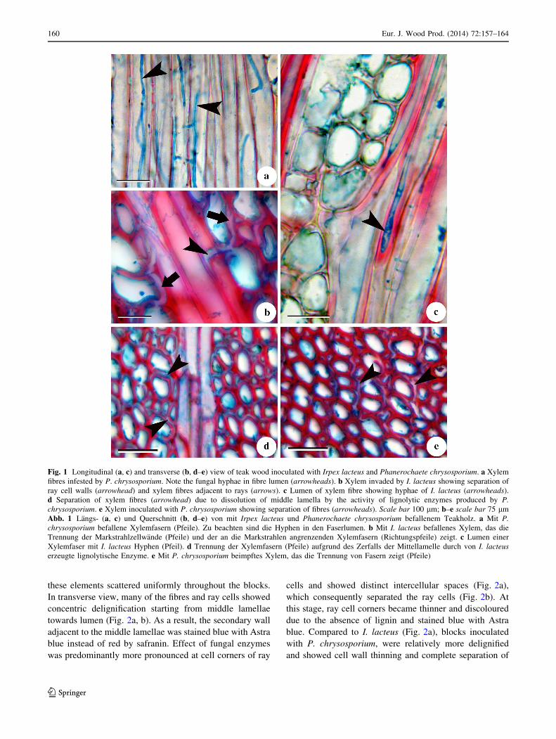
Fig. 1 Longitudinal (a, c) and transverse (b, d–e) view of teak wood inoculated with Irpex lacteus and Phanerochaete chrysosporium. a Xylem
fibres infested by P. chrysosporium. Note the fungal hyphae in fibre lumen (arrowheads). b Xylem invaded by I. lacteus showing separation of
ray cell walls (arrowhead) and xylem fibres adjacent to rays (arrows). c Lumen of xylem fibre showing hyphae of I. lacteus (arrowheads).
d Separation of xylem fibres (arrowhead) due to dissolution of middle lamella by the activity of lignolytic enzymes produced by P.
chrysosporium. e Xylem inoculated with P. chrysosporium showing separation of fibres (arrowheads). Scale bar 100 lm; b–e scale bar 75 lm
Abb. 1 Langs- (a, c) und Querschnitt (b, d–e) von mit Irpex lacteus und Phanerochaete chrysosporium befallenem Teakholz. a Mit P.
chrysosporium befallene Xylemfasern (Pfeile). Zu beachten sind die Hyphen in den Faserlumen. b Mit I. lacteus befallenes Xylem, das die
Trennung der Markstrahlzellwande (Pfeile) und der an die Markstrahlen angrenzenden Xylemfasern (Richtungspfeile) zeigt. c Lumen einer
Xylemfaser mit I. lacteus Hyphen (Pfeil). d Trennung der Xylemfasern (Pfeile) aufgrund des Zerfalls der Mittellamelle durch von I. lacteus
erzeugte lignolytische Enzyme. e Mit P. chrysosporium beimpftes Xylem, das die Trennung von Fasern zeigt (Pfeile)
160 Eur. J. Wood Prod. (2014) 72:157–164
123

ray cells (Fig. 2b). Except for cell wall thinning and sep-
aration of ray cells, no noticeable sign of cell wall change
was observed in the morphology of xylem elements in the
transverse and longitudinal plane.
Separation of vessel elements without any significant
effect on their wall was recorded only after 90 days of
fungal inoculation. Narrow vessels and tracheids also
showed separation due to dissolution of middle lamella
(Fig. 2c). At this stage, fibres were separated consis-
tently throughout the wood blocks. When compared
with other xylem elements, rays became more suscep-
tible to fungal invasion. Pits on the ray and axial
parenchyma walls became larger in size and irregular
in shape (Fig. 2d). Subsequently, several additional
boreholes were formed on the lateral walls (Fig. 2e)
due to the activity of fungal enzymes. Though disso-
lution of middle lamella and separation of cells were
common in axial elements, early wood axial paren-
chyma cells were more vulnerable to enzyme activity
as compared to fibres and axial parenchyma of the late
wood (Fig. 2f).
Wood blocks exposed to both the fungi for 120 days
were severely damaged due to ligninolytic enzymes
secreted by the fungal mycelia. Cell walls were com-
pletely bleached out and not only stained blue coloured
with Astra blue due to loss of lignin but also showed
complete disintegration of cells (Fig. 2f). At this stage,
xylem fibres were not only separated from each other but
also lost their rigidity due to removal of lignin from the
walls. Formation of several boreholes on the lateral walls
of fibres and extensive thinning of many ray cells
resulted in partial or complete disintegration of rays. In
case of xylem fibres and axial parenchyma, pits present
on their lateral wall also became larger, irregular in size,
which measured about 4–6 lm in diameter, sometimes
2–3 such boreholes fused together and formed relatively
larger boreholes. Vessels were more resistant in contrast
to fibres and axial parenchyma, while rays were the most
vulnerable cell types to fungal enzymes. At this stage,
vessel walls also showed uneven thinning; therefore,
most often vessel walls were broken at the time of
sectioning.
4 Discussion
Longevity or natural durability of timber depends upon
various factors such as genetic makeup of the species,
surrounding environment, moisture content in the atmo-
sphere, growth rate and age of that specific species
(Suprapti 2010). Therefore, fortitude of any wood is also
determined by its natural durability, they are always sus-
ceptible to deterioration due to wood borers, beetles or
wood rot fungi. Tectona is one of the important timbers
known for natural durability and strength. However, rainy
season provides ideal situation for different fungi to invade
wood logs under natural conditions. During monsoon, high
moisture content and relatively low temperature of the
surrounding environment/atmosphere favors fungal growth
while wood surface provides a suitable platform to estab-
lish fungal infection. In the present study, Irpex lacteus and
Phanerochaete chrysosporium isolated from naturally
infected wood samples were utilized for the in vitro decay
test. Both the fungi belong to basidiomycota and show
selective delignification pattern, which was manifested by
separation of xylem cells. Anagnost (1998) and Lujan Luna
et al. (2004) considered this feature as the most reliable
indicator of selective delignification. Selective delignifi-
cation by both fungal isolates may be supported by the
staining technique described by Srebotnik and Messener
(1994), which contributes to discriminate between simul-
taneous and selective delignification (Lujan Luna et al.
2004). Other signs of simultaneous rot are cell wall thin-
ning, borehole formation, rounded pit erosion and forma-
tion of erosion troughs (Anagnost 1998; Lujan Luna et al.
2004). All these features are characteristic to both the
fungal isolates except for the formation of erosion troughs
which was not observed in this study.
Mycelia of both fungal isolates although totally ramified
over the wood blocks and all the cells types of secondary
xylem were invaded, no appreciable weight loss was
observed even after 1 month. It appears that in the early
stage of fungal invasion, mycelia survive on the reserved
photosynthate (if any) available in the wood cells particu-
larly ray and axial parenchyma. Similar behaviour of wood
rot fungi has been correlated with the presence of several
low molecular weight compounds present in the wood that
might have been consumed first at the initial stage of
invasion without damaging cell wall polymers (Fengel and
Wegener 1989; Worall et al. 1997). Percent weight loss of
wood blocks increased rapidly after 30 days, and showed
27.97 % loss by Irpex lacteus and 30.05 % by Phanero-
chaete chrysosporium after 120 days. Rapid increase in
rate of weight loss seems to be associated with complete
consumption of low molecular weight compounds present
in the wood and the degradation potential of fungi. Lujan
Luna et al. (2004) reported about 50–60 % weight loss of
Table 1 Average percent weight loss of Tectona sapwood inoculated
with Irpex lacteus and Phanerochaete chrysosporium
Tab. 1 Mittlerer prozentualer Masseverlust von mit Irpex lacteus
und Phanerochaete chrysosporium beimpftem Teaksplintholz
Decay fungi % weight loss
(60 days)
% weight loss
(90 days)
% weight loss
(120 days)
Irpex lacteus 9.75 (± 5.23) 15.06 (± 4.84) 27.97 (± 4.97)
Phanerochaete
chrysosporium
10.61 (± 3.41) 17.92 (± 4.94) 30.05 (± 5.89)
Eur. J. Wood Prod. (2014) 72:157–164 161
123

poplar wood within 2–5 months by Pycnoporus sanguineus
and Gonoderma lucidum. Therefore, it appears that weight
loss differs from fungal species to species or within species
and natural durability of the wood.
In the early stages of fungal attack, hyphae enter through
the vessel lumen and further invasion is facilitated by ray
cells and vessel associated parenchyma cells for wide
spread distribution of mycelia. Similar mechanism of
Fig. 2 Tangential longitudinal (a, b) and transverse (c–f) view of teak wood inoculated with Irpex lacteus and Phanerochaete chrysosporium.
a Separation of rays cells at cell corners (arrowheads) in wood blocks inoculated by I. lacteus. b Separation of ray cells (arrowhead) in wood
blocks inoculated by P. chrysosporium. Note that separation of cells and wall thinning is relatively more as compared to a. c Dissolution of
middle lamella and separation of vessels (arrowheads) in wood inoculated with P. chrysosporium after 90 days. d Separation of fibres adjacent to
ray (arrows). Arrowheads indicate development of additional bore holes on the ray cell walls by I. lacteus. e Separation of early wood
parenchyma (arrow) and late wood fibres (arrowhead) by enzymatic activity of P. Chrysosporium. f Severely affected secondary xylem at the
end of 120 days of incubation showing loss of cell integrity and deformed xylem cells. a–e scale bar 75 lm; f scale bar 100 lm
Abb. 2 Langsschnitt tangential (a, b) und Querschnitt (c–f) von mit Irpex lacteus und Phanerochaete chrysosporium beimpftem Teakholz.
a Trennung der Markstrahlzellen an den Zellecken (Pfeile) in mit I. lacteus beimpften Prufkorpern. b Trennung der Markstrahlzellen (Pfeile) in
mit P. chrysosporium beimpften Prufkorpern. Zu beachten ist, dass die Zelltrennung und die Wandverdunnung verglichen mit a großer sind.
c Zerfall der Mittellamelle und Trennung der Gefaße (Pfeile) in mit P. chrysosporium beimpften Prufkorpern nach 90 Tagen. d Trennung der an
die Markstrahlen angrenzenden Fasern (Richtungspfeile). Die Pfeile zeigen die Entwicklung von zusatzlicher Durchdringung der
Markstrahlzellwande durch I. lacteus. e Trennung des Parenchyms von Fruhholz- (Richtungspfeil) und Spatholzfasern (Pfeil) durch
Enzymaktivitat von P. chrysosporium. f Stark befallenes sekundares Xylem nach 120-tagigem Befall, das den Verlust der Zellstruktur und
verformte Xylemzellen aufweist
162 Eur. J. Wood Prod. (2014) 72:157–164
123

fungal invasion has also been reported in Ailanthus (Koy-
ani et al. 2010) and other trees (Rayner and Boddy 1988;
Schwarze 2007; Schwarze et al. 2004; Sanghvi et al. 2013).
The presence of fungal hyphae in all the cell types coin-
cides with the observations by Lujan Luna et al. (2004).
Access to adjacent cells is reported to occur through the
simple or bordered pits present on the walls which subse-
quently become larger in diameter. It may also penetrate
directly by forming boreholes with the help of specialised
cell wall degrading hyphae (Schwarze et al. 2004;
Schwarze 2007; Koyani et al. 2010; Sanghvi et al. 2013). In
this study, formation of several boreholes is also observed
on the walls of xylem fibres, axial and ray parenchyma
cells. According to Anagnost (1998) and Lujan Luna et al.
(2004), fungi that selectively delignify wood may also form
boreholes that are similar to simultaneous rot.
Among the different cell types of xylem, ray cells are
highly vulnerable and showed significant damage due to
fungal activity, while vessels are relatively resistant, except
the bleaching effect on the vessel wall pits. The observa-
tions here are in agreement with Lujan Luna et al. (2004)
who reported similar feature in case of populous wood
treated with Pycnoporus sanguineus and Gonoderma lu-
cidum. Similar feature has also been observed in an earlier
study on Ailanthus excelsa (Koyani et al. 2010) and Aza-
dirachta indica (Sanghvi et al. 2013). Blanchette (1988)
named various reasons for the persistence of vessels in
wood degraded by white rot fungi. Iiyama and Pant (1988)
reported higher content of syringyl monomer in fibres and
ray parenchyma, while fibre tracheids contain higher gua-
iacyl monomer content (Faix et al. 1985; Blanchette 1988).
Therefore, hardwood is degraded rapidly by white rot fungi
due to higher content of syringyl units of lignin than the
guaiacyl (Faix et al. 1985; Lujan Luna et al. 2004; Koyani
et al. 2010). However, further chemical studies are required
to confirm this hypothesis of increasing concentration of
guaiacyl monomer with persistence of vessels in Tectona.
At an advanced stage of decay, completely delignified
tissue stained blue with safranin-astra blue combination.
Complete removal of lignin from the cell wall results in
separation of fibres and ray cell which leads to loss of cell
integrity and consequently leads to collapse. Though, ves-
sels are resistant to fungal action; separation of vessels at
this stage may be associated with the dissolution of com-
pound middle lamella. Severance of cells in response to
complete degradation of middle lamella in Populous treated
with G. lucidum is also reported by Lujan Luna et al. (2004).
5 Conclusion
Irpex lacteus and Phanerochaete chrysosporium show sim-
ilar white rot pattern resulting in 27–30 % weight loss after
120 days. As expected, both the fungal strains have higher
potential of lignin degradation as is reported for textile dye
and polyaromatic compound degradation and may serve as
potential species to be utilised in biopulping process. How-
ever, further studies on the wood that is used in the paper
industry are needed. Fibres and parenchyma cells are more
vulnerable to ligninolytic activity of the fungal enzymes and
at an advanced stage of wood decay; fibres show complete
separation and collapse, while vessels are the most resistant
cell type. Rapid weight loss of wood blocks after 30 days
may be associated with complete utilization of low molec-
ular weight compounds present in the wood.
Acknowledgments The authors would like to thank the Council of
Scientific and Industrial Research (CSIR), New Delhi for providing
financial support to carry out the work. Thanks are also due to Dr.
Gerd Wegener, Editor-in-Chief and anonymous reviewers for their
critical suggestions on the manuscript.
References
Adaskaveg JE, Gilbertson RL, Dunlap MR (1995) Effects of
incubation time and temperature on in vitro selective delignifi-
cation of Silver leaf oak by Ganoderma colossum. Appl Environ
Microbiol 61(6):139–144
Anagnost SE (1998) Light microscopic diagnosis of wood decay.
IAWA J 19(2):141–167
Baborova P, Moder M, Baldrian P, Cajthamlova K, Cajthaml T
(2006) Purification of a new manganese peroxidase of the white-
rot fungus Irpex lacteus, and degradation of polycyclic aromatic
hydrocarbons by the enzyme. Res Microbiol 157(3):248–253
Berlyn GP, Miksche JP (1976) Botanical microtechnique and
cytochemistry. The Iowa State University Press, Ames, Iowa,
p 326
Bhat KM, Priya PB (2004) Influence of provenance variation on wood
properties of teak from the Western Ghat Region in India. IAWA
J 25(3):273–282
Blanchette RA (1984a) Screening wood decayed white rot fungi for
preferential lignin degradation. Appl Environ Microbiol
48(3):647–653
Blanchette RA (1984b) Selective delignification of eastern hemlock
by Ganoderma tsugae. Phytopathology 74(2):153–160
Blanchette RA (1988) Resistance of hardwood vessels to degradation
by white rot Basidiomycetes. Can J Bot 66(9):1841–1847
Datta A, Bettermann A, Kirk K (1991) Identification of a specific
manganese peroxidase among ligninolytic enzymes secreted by
Phanerochaete chrysosporium during wood decay. Appl Environ
Microbiol 57(5):1453–1460
Erkurt HA (2010) Biodegradation of azo dyes, vol 9. In: Barcelo D,
Kostianoy AG (eds) The handbook of environmental chemistry.
Springer, Verlag Berlin Heidelberg
Faix O, Mozuch MD, Kirk TK (1985) Degradation of gymnosperm
(guaiacyl) vs. angiosperm (syringyl/guaiacyl) lignins by Phan-
erochaete chrysosporium. Holzforschung 39(4):203–208
Fengel D, Wegener G (1989) Wood: chemistry, ultrastructure,
reactions, 2nd edn. Walter de Gruyter, Berlin, p 613
Gassara F, Brar SK, Tyagi RD, Verma M, Surampalli RY (2010)
Screening of agro-industrial wastes to produce ligninolytic
enzymes by Phanerochaete chrysosporium. Biochem Eng J
49(3):388–394
Eur. J. Wood Prod. (2014) 72:157–164 163
123

Iiyama K, Pant R (1988) The mechanism of the Maule colour
reaction. Introduction of methylated syringyl nuclei in softwood
lignin. Wood Sci Technol 22(2):167–175
Kokutse AD, Adjonou K, Kokou K (2009) Relationship between
ecological indicators and teak wood characteristics in Tchorogo
plantation (Togo). Int J Biol Chem Sci 3(3):483–491
Kokutse AD, Stokes A, Kokutse NK, Kokou K (2010) Which factors
most influence heartwood distribution and radial growth in
plantation teak? Ann For Sci 67:407
Koyani RD, Sanghvi GV, Bhatt IM, Rajput KS (2010) Pattern of
delignification in Ailanthus excelsa Roxb., wood by Inonotus
hispidus. Mycology 1(3):204–211
Koyani RD, Sanghvi GV, Sharma RK, Rajput KS (2013) Contribution
of lignin degrading enzymes in decolourisation and degradation
of reactive textile dyes. Intl Biodete and Biodegrad 77:1–9
Lujan Luna ML, Murace MA, Keil GD, Otano ME (2004) Patterns of
decay caused by Pycnoporus sanguineus and Ganoderma
lucidum (Aphyllophorales) in poplar wood. IAWA J 25(4):425–
433
Macchioni N, Nocetti M, Rozenberg P (2007) Early detection of
surface quality of Teak (Tectona grandis L.f.) through X-ray
microdensitometry. Test on West African plantations. In:
Blanchet P (ed) Proc International Scientific Conference on
Hardwood Processing (ISCHP), September 24–26, Quebec City,
Canada, pp 311–316
Maloy OC, Murray TD (2001) Encyclopedia of plant pathology.
Wiley, London, pp 1201–1203
Moller EM, Bahnweg G, Sandermann H, Geiger HH (1992) A simple
and efficient protocol for high molecular weight DNA from
filamentous fungi, fruit bodies and infected plant tissue. Nucl
Acid Res 20:6115–6116
Nicolotti G, Gonthier P, Guglielmo F (2010) Advances in detection
and identification of wood rotting fungi in timber and standing
trees. In: Gherbawy Y, Voigt K (eds) Molecular identification of
fungi. Springer Verlag, Berlin Heidelberg, pp 251–276
Nocetti M, Rozenberg P, Chaix G, Macchioni N (2011) Provenance
effect on the ring structure of teak (Tectona grandis L.f.) wood
by X-ray microdensitometry. Ann For Sci 68(8):1375–1383
Plomion C, Leprovost G, Stokes A (2001) Wood formation in trees.
Plant Physiol 127:1513–1523
Priya PB, Bhat KM (1997) Wood anatomical changes in juvenile teak
due to insect defoliation. IAWA J 18(3):307–313
Priya PB, Bhat KM (1998) False ring formation in teak (Tectona
grandis L.f) and the influence of environmental factors. For Ecol
Manag 108(3):215–222
Rajput KS, Rao KS, Patil UG (2005) Cambial anatomy, development
and structural changes in the wood of teak (Tectona grandis L.f.)
associated with insect defoliation. J Sustain For 20(4):51–63
Rao KS, Dave YS (1981) Seasonal variations in the cambial anatomy
of Tectona grandis L.f. (Verbenaceae). Nord J Bot 1(4):535–542
Rao KS, Rajput KS (1999) Seasonal behaviour of vascular cambium
in teak (Tectona grandis L.f.) growing moist deciduous and dry
deciduous forests. IAWA J 20(1):85–93
Rayner ADM, Boddy L (1988) Fungal decomposition of wood: its
biology and ecology. Wiley, Chichester, p 587
Sanghvi GV, Koyani RD, Rajput KS (2013) Anatomical character-
ization of Teak wood (Tectona grandis L.f.) decayed by fungus
Chrysosporium asperatum. J Trop For Sci 25(4):547–553
Schwarze FMWR (2007) Wood decay under the microscope. Fungal
Biol Rev 21:133–170
Schwarze FWMR, Mattheck C, Engels J (2004) Fungal strategies of
wood decay in trees. Springer Verlag, Heidelberg
Srebotnik E, Messener K (1994) A simple method that uses
differential staining and light microscopy to assess the selectivity
of wood delignification by white rot fungi. Appl Environ
Microbiol 60(4):1383–1386
Suprapti S (2010) Decay resistance of 84 Indonesian wood species
against fungi. J Trop For Sci 22(1):81–87
Varghese M, Nicodemus A, Ramteke PK, Anbazhagi G, Bennet SSR,
Subramanian K (2000) Variation in growth and wood traits
among nine populations of teak in Peninsular India. Silvae Genet
49(4–5):201–205
Worall JJ, Anagnost SE, Zabel RA (1997) Comparison of wood decay
among diverse lignicolus fungi. Mycologia 89(2):199–219
164 Eur. J. Wood Prod. (2014) 72:157–164
123