The Stoichiometry of Host PrP C Glycoforms Modulates the Efficiency of PrP Sc Formation in Vitro
11
The Stoichiometry of Host PrP C Glycoforms Modulates the Efficiency of PrP Sc Formation in Vitro ² Koren A. Nishina, ‡ Nathan R. Deleault, ‡ Sukhvir P. Mahal, § Ilia Baskakov, | Thorsten Luhrs, ⊥ Roland Riek, ⊥ and Surachai Supattapone* ,‡ Department of Biochemistry, Dartmouth Medical School, HanoVer, New Hampshire 03755, Department of Infectology, Scripps Florida, 5353 Parkside DriVe, Jupiter, Florida 33458, Medical Biotechnology Center, UniVersity of Maryland Biotechnology Institute, Baltimore, Maryland 21201, and The Salk Institute for Biological Studies, 10010 North Torrey Pines Road, La Jolla, California 92037 ReceiVed July 28, 2006; ReVised Manuscript ReceiVed September 10, 2006 ABSTRACT: A central event in the formation of infectious prions is the conformational change of a host- encoded glycoprotein, PrP C , into a pathogenic isoform, PrP Sc . However, the molecular requirements for efficient PrP conversion remain unknown. In this study, we employed the recently developed protein misfolding cyclic amplification (PMCA) and scrapie cell assay (SCA) techniques to study the role of N-linked glycosylation on prion formation in vitro. The results show that unglycosylated PrP C molecules are required to propagate mouse RML prions, whereas diglycosylated PrP C molecules are required to propagate hamster Sc237 prions. Furthermore, the formation of Sc237 prions is inhibited by substoichio- metric levels of hamster unglycosylated PrP C molecules. Thus, interactions between different PrP C glycoforms appear to control the efficiency of prion formation in a species-specific manner. Much evidence supports the “protein-only” hypothesis that conversion of a normal, host glycoprotein (PrP C ) 1 into a protease-resistant, infectious isoform (PrP Sc ) is the central pathogenic event in transmissible prion diseases such as Creutzfeldt-Jakob disease (CJD) in humans, bovine spongi- form encephalopathy (BSE) in cattle, chronic wasting disease (CWD) in cervid species, and scrapie in ungulates (1, 2). Although PrP C is highly conserved among mammals, small differences in the PrP C amino acid sequence appear to cause “species barriers”, which manifest as reduced rates of disease transmission between different animal species (3, 4). Inter- estingly, novel self-propagating “strains” of infectious prions with distinctive clinical and neuropathological features can arise when prions are inefficiently transmitted between different animal species and can then be faithfully maintained upon serial passage within the same animal species (5, 6). Several studies have shown that prion strains are associated with PrP Sc molecules that possess distinctive biochemical characteristics (7-11) and that some of these chemical properties can be propagated in vitro (12). Species-dependent transmission barriers and strain variation appear to be related phenomena (13). Recent biophysical studies of model proteins suggest that the formation of prion strains may be governed by differences in the potential folding pathways available to different protein substrates (14-17). Mature PrP molecules possess glycans linked to asparagine residues 180 and 196 and a C-terminal glycophosphatidyli- nositol (GPI) anchor (18-20). The relative distribution of di-, mono-, and unglycosylated PrP C glycoforms varies between different animal species, and regional variations in the stoichiometry of PrP C glycoforms have also been identified within the brains of individual animals (21-23). The ability of different prion strains to maintain distinctive distributions of PrP Sc glycoforms upon serial passage suggests that the N-linked glycans may play an important role in the specificity of PrP conversion (9). Therefore, we decided to investigate whether the glycosylation profile of PrP C mol- ecules influences the species and/or strain specificity of prion formation. Other investigators previously studied this question by either (1) inhibiting glycosylation in cells using tunicamycin (24-26) or (2) expressing PrP molecules with mutant ² This work was supported by the National Institutes of Health (R21 AI058979, R01 NS046478, and T32 AI007519), the Burroughs Wellcome Fund, Pew Charitable Trusts, and the U.S. Army. * To whom correspondence should be addressed: Department of Biochemistry, 7200 Vail Building, Dartmouth Medical School, Hanover, NH 03755. Phone: (603) 650-1192. Fax: (603) 650-1193. E-mail: [email protected]. ‡ Dartmouth Medical School. § Scripps Florida. | University of Maryland Biotechnology Institute. ⊥ The Salk Institute for Biological Studies. 1 Abbreviations: PrP C , cellular isoform of the prion protein; PrP Sc , scrapie isoform of the prion protein; PMCA, protein misfolding cyclic amplification; SCA, scrapie cell assay; CJD, Creutzfeldt-Jakob disease; BSE, bovine spongiform encephalopathy; CWD, chronic wasting disease; GPI, glycophosphatidylinositol; PrP27-30, purified proteinase K-digested PrP Sc ; UN, preparation enriched for unglycosylated PrP C ; DI, preparation enriched for diglycosylated PrP C ; DI f UN, DI preparation subjected to enzymatic deglycosylation; ALL, preparation of PrP C containing di-, mono-, and unglycosylated PrP C ; MOPS, 3-(N- morpholino)propanesulfonic acid; EDTA, ethylenediaminetetraacetic acid; IMAC, immobilized metal affinity chromatography; PNGase F, peptide N-glycosidase F; MWCO, molecular weight cutoff; WGA, wheat germ agglutinin; SP, sulfonylpropyl; Abl, 10% (w/v) ablated (Prnp 0/0 ) brain homogenate; PK, proteinase K; PBS, phosphate-buffered saline without calcium or magnesium; PVDF, polyvinylide difluoride; TBST, Tris-buffered saline with Tween; HRP, horseradish peroxidase; Ha, hamster; Mo, mouse; REC, E. coli-expressed, R-helical, recombi- nant PrP molecules; CFC, cell-free conversion; Tg, transgenic. 14129 Biochemistry 2006, 45, 14129-14139 10.1021/bi061526k CCC: $33.50 © 2006 American Chemical Society Published on Web 11/03/2006
-
Upload
independent -
Category
Documents
-
view
2 -
download
0
Transcript of The Stoichiometry of Host PrP C Glycoforms Modulates the Efficiency of PrP Sc Formation in Vitro
The Stoichiometry of Host PrPC Glycoforms Modulates the Efficiencyof PrPSc Formation in Vitro†
Koren A. Nishina,‡ Nathan R. Deleault,‡ Sukhvir P. Mahal,§ Ilia Baskakov,| Thorsten Luhrs,⊥ Roland Riek,⊥ andSurachai Supattapone*,‡
Department of Biochemistry, Dartmouth Medical School, HanoVer, New Hampshire 03755, Department of Infectology,Scripps Florida, 5353 Parkside DriVe, Jupiter, Florida 33458, Medical Biotechnology Center, UniVersity of Maryland
Biotechnology Institute, Baltimore, Maryland 21201, and The Salk Institute for Biological Studies,10010 North Torrey Pines Road, La Jolla, California 92037
ReceiVed July 28, 2006; ReVised Manuscript ReceiVed September 10, 2006
ABSTRACT: A central event in the formation of infectious prions is the conformational change of a host-encoded glycoprotein, PrPC, into a pathogenic isoform, PrPSc. However, the molecular requirements forefficient PrP conversion remain unknown. In this study, we employed the recently developed proteinmisfolding cyclic amplification (PMCA) and scrapie cell assay (SCA) techniques to study the role ofN-linked glycosylation on prion formation in vitro. The results show that unglycosylated PrPC moleculesare required to propagate mouse RML prions, whereas diglycosylated PrPC molecules are required topropagate hamster Sc237 prions. Furthermore, the formation of Sc237 prions is inhibited by substoichio-metric levels of hamster unglycosylated PrPC molecules. Thus, interactions between different PrPC
glycoforms appear to control the efficiency of prion formation in a species-specific manner.
Much evidence supports the “protein-only” hypothesis thatconversion of a normal, host glycoprotein (PrPC)1 into aprotease-resistant, infectious isoform (PrPSc) is the centralpathogenic event in transmissible prion diseases such asCreutzfeldt-Jakob disease (CJD) in humans, bovine spongi-form encephalopathy (BSE) in cattle, chronic wasting disease(CWD) in cervid species, and scrapie in ungulates (1, 2).Although PrPC is highly conserved among mammals, smalldifferences in the PrPC amino acid sequence appear to cause
“species barriers”, which manifest as reduced rates of diseasetransmission between different animal species (3, 4). Inter-estingly, novel self-propagating “strains” of infectious prionswith distinctive clinical and neuropathological features canarise when prions are inefficiently transmitted betweendifferent animal species and can then be faithfully maintainedupon serial passage within the same animal species (5, 6).Several studies have shown that prion strains are associatedwith PrPSc molecules that possess distinctive biochemicalcharacteristics (7-11) and that some of these chemicalproperties can be propagated in vitro (12). Species-dependenttransmission barriers and strain variation appear to be relatedphenomena (13). Recent biophysical studies of modelproteins suggest that the formation of prion strains may begoverned by differences in the potential folding pathwaysavailable to different protein substrates (14-17).
Mature PrP molecules possess glycans linked to asparagineresidues 180 and 196 and a C-terminal glycophosphatidyli-nositol (GPI) anchor (18-20). The relative distribution ofdi-, mono-, and unglycosylated PrPC glycoforms variesbetween different animal species, and regional variations inthe stoichiometry of PrPC glycoforms have also beenidentified within the brains of individual animals (21-23).The ability of different prion strains to maintain distinctivedistributions of PrPScglycoforms upon serial passage suggeststhat the N-linked glycans may play an important role in thespecificity of PrP conversion (9). Therefore, we decided toinvestigate whether the glycosylation profile of PrPC mol-ecules influences the species and/or strain specificity of prionformation.
Other investigators previously studied this question byeither (1) inhibiting glycosylation in cells using tunicamycin(24-26) or (2) expressing PrP molecules with mutant
† This work was supported by the National Institutes of Health (R21AI058979, R01 NS046478, and T32 AI007519), the BurroughsWellcome Fund, Pew Charitable Trusts, and the U.S. Army.
* To whom correspondence should be addressed: Department ofBiochemistry, 7200 Vail Building, Dartmouth Medical School, Hanover,NH 03755. Phone: (603) 650-1192. Fax: (603) 650-1193. E-mail:[email protected].
‡ Dartmouth Medical School.§ Scripps Florida.| University of Maryland Biotechnology Institute.⊥ The Salk Institute for Biological Studies.1 Abbreviations: PrPC, cellular isoform of the prion protein; PrPSc,
scrapie isoform of the prion protein; PMCA, protein misfolding cyclicamplification; SCA, scrapie cell assay; CJD, Creutzfeldt-Jakob disease;BSE, bovine spongiform encephalopathy; CWD, chronic wastingdisease; GPI, glycophosphatidylinositol; PrP27-30, purified proteinaseK-digested PrPSc; UN, preparation enriched for unglycosylated PrPC;DI, preparation enriched for diglycosylated PrPC; DI f UN, DIpreparation subjected to enzymatic deglycosylation; ALL, preparationof PrPC containing di-, mono-, and unglycosylated PrPC; MOPS, 3-(N-morpholino)propanesulfonic acid; EDTA, ethylenediaminetetraaceticacid; IMAC, immobilized metal affinity chromatography; PNGase F,peptideN-glycosidase F; MWCO, molecular weight cutoff; WGA,wheat germ agglutinin; SP, sulfonylpropyl; Abl, 10% (w/v) ablated(Prnp0/0) brain homogenate; PK, proteinase K; PBS, phosphate-bufferedsaline without calcium or magnesium; PVDF, polyvinylide difluoride;TBST, Tris-buffered saline with Tween; HRP, horseradish peroxidase;Ha, hamster; Mo, mouse; REC,E. coli-expressed,R-helical, recombi-nant PrP molecules; CFC, cell-free conversion; Tg, transgenic.
14129Biochemistry2006,45, 14129-14139
10.1021/bi061526k CCC: $33.50 © 2006 American Chemical SocietyPublished on Web 11/03/2006

glycosylation sites in transgenic mice (27, 28). However,such manipulations could potentially cause abnormal foldingand/or trafficking of PrP molecules during biosynthesis (27,29-31), and polypeptide sequence dissimilarities betweenmutant and wild-type PrPC molecules could also affecttransmission barriers and strain recognition in a mannerindependent of glycosylation. Furthermore, pharmacologicand genetic approaches cannot be used to study possiblefunctional interactions between different PrPC glycoforms.Therefore, we used a direct biochemical approach to produceand isolate natively processed, wild-type PrPC glycoformsfrom hamsters and mice, two related species with differentnative PrPC glycoform distributions. We then used the proteinmisfolding cyclic amplification (PMCA) technique (32) totest the ability of isolated PrPC glycoforms, alone and incombination, to amplify and propagate purified proteinaseK-digested PrPSc (PrP27-30) molecules. The results of theseexperiments revealed unexpected functional interactionsbetween PrPC glycoforms, which regulate prion formationin a species-specific manner.
MATERIALS AND METHODS
Preparation of Crude Brain Homogenates.Either onehamster brain or two mouse brains (Harlan Sprague Dawley,Inc., Indianapolis, IN) were Potter homogenized in 10 mLof ice-cold PBS (phosphate-buffered saline without calciumor magnesium) containing Complete protease inhibitors(Roche, Indianapolis, IN). The homogenate was centrifugedat 200g for 30 s, and the postnuclear supernatant wascollected.
Preparation of NatiVe and Unglycosylated PrPC (UN, DIf UN). We used a modified protocol to partially purify PrPC
based on the method described by Pan et al. (33). All stepswere performed at 4°C. Either six hamster or 12 mousebrains were Potter homogenized in 40 mL of ice-cold bufferA [20 mM MOPS (pH 7.0) and 150 mM NaCl] containingComplete EDTA-free protease inhibitors. The homogenatewas initially centrifuged at 200g for 30 s; the postnuclearsupernatant was then removed and centrifuged at 3200g for20 min. The resulting pellet was resuspended and Douncehomogenized in 30 mL of buffer A and Complete EDTA-free protease inhibitors. Four milliliters of a 10% DOC/TritonX-100 detergent mixture was added to the homogenate, andthe mixture was incubated on ice for 30 min and centrifugedat 100000g for 30 min. The solubilized supernatant wasapplied to a pre-equilibrated 2 mL IMAC-CuSO4 column(GE Healthcare, Piscataway, NJ). The column was washedwith 10 mL of IMAC-CuSO4-W [buffer A, 10 mM imidazolein buffer A (pH 7.0), and 1% Triton X-100] and eluted with10 mL of IMAC-CuSO4-E1 [20 mM MES (pH 6.4), 150mM imidazole in buffer A (pH 7.0), 150 mM NaCl, and 1%Triton X-100]. The IMAC-CuSO4 eluate was applied to apre-equilibrated 2 mL SP Sepharose cation exchange column(Sigma, St. Louis, MO). The column was washed with 10mL of SPW [20 mM MOPS (pH 7.0), 250 mM NaCl, and1% Triton X-100] and eluted with 8 mL of SPE [20 mMMOPS (pH 7.5), 500 mM NaCl, and 1% Triton X-100].
The N-linked glycans were removed from PrPC usingglycerol-free PNGase F (peptideN-glycosidase F) (NewEngland Biolabs, Beverly, MA) under nondenaturing condi-tions; 50µL of glycerol-free PNGase F (∼25000 units) was
added to 500µL of SP eluate, and the mixture was incubatedfor 24 h at 37°C. After the initial incubation, an additional50 µL of PNGase F was added, and the mixture wasincubated for an additional 24 h at 37°C. The sample wasrepurified using a pre-equilibrated 200µL IMAC-CuSO4
resin; all subsequent steps were performed at 4°C. Initially,the sample was incubated on an end-over-end rotator withthe IMAC-CuSO4 slurry for 30 min in a capped columnsupport (Bio-Rad, Hercules, CA). The column was thenwashed with 20 mL of IMAC-CuSO4-W and eluted with 500µL of IMAC-CuSO4-E1. The eluate was loaded into a 3500MWCO Slide-A-Lyser (Pierce, Rockford, IL) and dialyzedovernight into buffer C [20 mM MOPS (pH 7.5), 150 mMNaCl, and 0.5% Triton X-100] to yield the deglycosylatedPrPC product “UN”. The “Native” sample used in Figure1A was incubated with PBS instead of PNGase F butotherwise processed identically.
To generate “DIf UN”, 50 µL of “DI” (prepared asdescribed below) was incubated with 5µL of PNGase F(2500 units) for 24 h at 37°C.
Preparation of Glycosylated PrPC (ALL, DI). A solubilizedsupernatant from either mouse or hamster brain homogenatewas obtained as described in the preceding section. Thissolubilized supernatant was first applied to a pre-equilibrated2 mL IMAC-CuSO4 column. The column was washed with10 mL of IMAC-CuSO4-W and eluted with 4 mL of IMAC-CuSO4-E2 [20 mM MOPS (pH 7.5), 750 mM NaCl, 150mM imidazole in buffer A (pH 7.0), and 1% Triton X-100].This eluate was then applied to a pre-equilibrated 1 mL WGA(wheat germ agglutinin) (Vector Laboratories, Burlingame,CA) column. The flow-through fraction was collected anddialyzed overnight into buffer C to yield a preparation ofPrPC containing all three glycoforms in an approximatelyequal distribution (ALL).
Meanwhile, the WGA column was washed with 20 mLof WGAW1 [20 mM MOPS (pH 7.5), 750 mM NaCl, and1% Triton X-100], followed by 5 mL of WGAW2 [20 mMMOPS (pH 7.5), 150 mM NaCl, and 1% Triton X-100]. Thesample was eluted with 4 mL of WGAE (buffer B, 50 mMN-acetylglucosamine, and 1% Triton X-100) and dialyzedovernight into buffer C to yield the diglycosylated PrPC
substrate (DI).Preparation of PrP27-30.PrP27-30 was prepared sepa-
rately from eight different scrapie strains [five mouse strains(RML, 22A, 139A, C506, and Me7) and three hamster strains(Sc237, 139H, and Drowsy)] kindly provided by S. B.Prusiner (San Francisco, CA). Each scrapie-infected brainwas homogenized in 10 volumes (w/v) of PBS with adisposable plastic homogenizer (Kendall, Mansfield, MA)and centrifuged at 200g for 30 s. One hundred microlitersof postnuclear supernatant was diluted into 900µL of bufferD (PBS and 1% Triton X-100) and then incubated with 10µg/mL PK (proteinase K, specific activity of 30 units/mL)(Roche Applied Science, Mannheim, Germany) for 30 minat 20°C. Protease digestion was terminated by the additionof 5 mM PMSF (phenylmethanesulfonyl fluoride, from a 0.3M stock solution in methanol). The digested sample wascentrifuged at 4°C for 1 h at100000g, and the pellet wasresuspended in 200µL of ice-cold buffer D by two 30 spulses of an indirect sonicator set at output 6.5 [Misonix(Farmingdale, NY) 3000-MPD sonicator with a microplatehorn]. An additional 800µL of ice-cold buffer D was added
14130 Biochemistry, Vol. 45, No. 47, 2006 Nishina et al.
to the suspension, and the sample was then centrifuged for30 min at 100000g and 4°C. The resulting pellets (with theexception of 22A) were resuspended in 1 mL of buffer D asdescribed above. RML, 139A, Sc237, 139H, and Drowsywere used in the amplification reaction at a 1:10 dilution;Me7 and C506 were used in the amplification reaction at a1:20 dilution. 22A was resuspended in 100µL of buffer Dby sonication and was used in the amplification reaction withno dilution.
Protein Misfolding Cyclic Amplification (PMCA).PMCAwas performed by the method of Castilla et al. (34) usingthe various PrPC substrate preparations described in precedingsections. The total 111µL amplification reaction volume wascomposed of 50µL of PrPC substrate, 25µL of Abl [10%(w/v) Prnp0/0 brain homogenate], 1µL of 500 mM EDTA(pH 8.0), 10µL of 500 mM imidazole in buffer A (pH 7.0),0.75% Triton X-100, and 25µL of PrP27-30 (diluted asdescribed above). When present, recombinant PrP23-321
FIGURE 1: Strain-dependent conversion of various PrP substrates. (A) Western blot of PMCA reactions. Lanes 1-3 contained input substratesnot treated with protease; in lanes 4-12, each of the indicated substrates was subjected to PMCA in the absence (lanes 4-6) or presence(lanes 7-12) of PrP27-30 template. Labels: Native, native PrPC; UN, unglycosylated PrPC; REC, recombinant PrP23-231; Abl, Prnp0/0
brain homogenate. (B) Western blot of PMCA reactions. Lanes 1-4 contained input samples not treated with protease; in lanes 5-9, eachof the indicated substrates was subjected to PMCA with PrP27-30 template in the presence of 2.5% Abl brain homogenate. Labels: ALL,flow-though fraction of lectin column; DIfUN, unglycosylated PrPC prepared by deglycosylation of the DI preparation;+PK, digestedwith proteinase K; Sc, negative control reaction mixture lacking PrPC substrate; Mouse, mouse substrates and RML template; Hamster,hamster substrates and Sc237 template. The apparent molecular mass markers on all Western blots are at 40, 33, 24, and 17 kDa. Similarresults were obtained in three independent experiments.
Prion Glycoforms Biochemistry, Vol. 45, No. 47, 200614131

was expressed and refolded by two independent methods aspreviously described (35, 36) and used at a final concentra-tion of 4µg/mL, similar to the concentration of brain-derivedPrPC substrates used in the reactions. Reactions withoutablated brain homogenate had PBS substituted for Abl. Themixtures were subjected to PMCA for 24 h using a Misonixprogrammable sonicator equipped with a microplate horncontaining 350 mL of water, and set for 30 s bursts every30 min at output 6.5. The temperature was maintained bycirculating heated water through a section of aluminum coilto heat the air inside the acoustic chamber (Misonix), andthe microplate horn was covered with a sheet of plastic filmto minimize evaporation. Before each burst, the bath tem-perature measured 41°C, and after each burst, the temper-ature measured 42°C. Samples were mounted in a holderthat prevented lid opening and held the bottom of the PCRtubes∼3 mm from the horn surface. In Figure 2, a smallaliquot of each sample was removed following incubationand boiled for 10 min in an equal volume of 2× SDS sample
buffer, and 10µL of the resulting minus (-) PK sampleswas loaded for electrophoresis. In Figures 1 and 3A,B andTable S1 (Supporting Information), input substrate prepara-tions were diluted 1:10 into PBS and boiled for 10 min inan equal volume of 2× SDS sample buffer, and 20µL ofeach resulting minus (-) PK sample was loaded forelectrophoresis. Meanwhile, the plus (+) PK samples weretreated with 25µg/mL PK and incubated for 30 min at 37°C. The digested samples were then boiled in 2× SDSsample buffer, and 80µL of each sample was loaded forelectrophoresis.
Serial in Vitro PrPSc Propagation Reactions and CellInfection. Serial in vitro PrPSc propagation reactions wereperformed by the method of Castilla et al. (34) using thePrPC preparations described above. The initial (day 1)reaction mixture was composed of 100µL of PrPC, 50 µLof 10% (w/v) Abl, 2µL of 500 mM EDTA (pH 8.0), 20µLof 500 mM imidazole in buffer A (pH 7.0), 0.75% TritonX-100, and 50µL of PrP27-30 (diluted as described above).
FIGURE 2: Functional interactions between PrPC glycoforms. (A) PMCA reactions using mixtures of PrPC substrates. Each of the indicatedsubstrates was subjected to PMCA with PrP27-30 template in the presence of 2.5% Abl brain homogenate. Abbreviations: Sc, no PrPC
substrate added; ALL, flow-though fraction of the lectin column; 3UN:1DI, 37.5µL of UN and 12.5µL of DI added to the PMCA reactionmixture; 2UN:2DI, 25µL of UN and 25µL of DI; 1UN:3DI, 12.5 µL of UN and 37.5µL of DI. (B) PMCA reactions using a fixedconcentration of diglycosylated hamster PrPC and varying concentrations of unglycosylated hamster PrPC. Each of the indicated substrateswas subjected to PMCA with Sc237 PrP27-30 template in the presence of 2.5% Abl brain homogenate. All samples contained 20µL ofDI HaPrPC and varying amounts of UN HaPrPC. Labels: UN:DI, molar ratio of UN HaPrPC to DI HaPrPC in the substrate mixture;+PK,digested with proteinase K; Sc, negative control reaction mixture lacking PrPC substrate. Similar results were obtained in three independentexperiments. Using Adobe Photoshop to quantitate signal intensities, and assuming molecular masses of 27 and 36 kDa for UN and DIHaPrPC, respectively, we estimate that the 2UN:2DI samples have molar ratios of∼2.3 for mouse and∼0.8 for hamster.
14132 Biochemistry, Vol. 45, No. 47, 2006 Nishina et al.
The mixtures were subjected to one round of PMCA for 24h as described above. Half of the reaction mixture (day 1)was PK digested as described above. Twenty microliters from
the completed reaction (day 1) was used to seed the next200µL reaction mixture (day 2) containing a fresh substratemixture [100µL of PrPC, 50 µL of 10% (w/v) Abl, 2 µL of
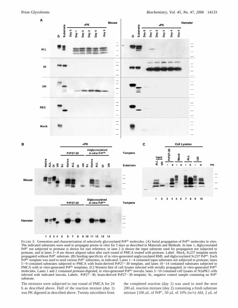
FIGURE 3: Generation and characterization of selectively glycosylated PrPSc molecules. (A) Serial propagation of PrPSc molecules in vitro.The indicated substrates were used to propagate prions in vitro for 5 days as described in Materials and Methods. In lane 1, diglycosylatedPrPC not subjected to protease is shown for size reference; in lane 2 is shown the input substrate used for propagation not subjected toprotease, and in lanes 3-8 are shown aliquots taken after each round of PMCA treated with protease. Label: Mock, Sc237 template mockpropagated without PrPC substrate. (B) Seeding specificity of in vitro-generated unglycosylated RML and diglycosylated Sc237 PrPSc. EachPrPSc template was used to seed various PrPC substrates, as indicated. Lanes 1-4 contained input substrates not subjected to protease; lanes5-9 contained substrates subjected to PMCA with brain-derived PrP27-30 template, and lanes 10-14 contained substrates subjected toPMCA with in vitro-generated PrPSc templates. (C) Western blot of cell lysates infected with serially propagated, in vitro-generated PrPSc
molecules. Lanes 1 and 2 contained protease-digested, in vitro-generated PrPSc inocula; lanes 3-10 contained cell lysates of N2aPK1 cellsinfected with indicated inocula. Labels: PrP27-30, brain-derived PrP27-30 template; Sc, negative control sample containing no PrPC
substrate.
Prion Glycoforms Biochemistry, Vol. 45, No. 47, 200614133
500 mM EDTA (pH 8.0), 20µL of 500 mM imidazole inbuffer A (pH 7.0), 8µL of PBS, and 0.75% Triton X-100].This process was repeated for a total of 6 days to completesix propagation rounds. Every 2 days, the bath water waschanged to prevent accumulation of residue in the sonicatorhorn.
The final (day 6) samples were titrated between 10-4 and10-6 using OptiMeM (Invitrogen, Carlsbad, CA), 10%bovine growth serum (Hyclone, Logan, UT), and 1%penicillin/streptomycin as a diluent and applied to highlyinfectable PK1 neuroblastoma cells (37). Following 5× 1:8splits, cell infectivity was measured by assaying 20 000 cells/sample, as previously described using the scrapie cell assayin end-point titration format (37). Wells were consideredpositive if the “spot” count was greater than the averagebackground (three spots) value plus 5 times the standarddeviation (six spots), i.e.,g32 spots. The average numberof infectious particles (m) in each sample was calculatedusing the Poisson formulaP(o) ) e - m, whereP(o) is theprobability that a well is not infected, i.e.,P(o) ) noninfectedwells/total wells. Therefore,m ) ln(total wells/noninfectedwells). In parallel, infected cells were assayed for PrPC andPrPSc content as follows. For each sample, a confluent 10cm plate was washed with 2× 5 mL of PBS, lysed with 2mL of ice-cold lysis buffer [50 mM Tris (pH 8.0), 150 mMNaCl, 0.5% sodium deoxycholate, and 0.5% Triton X-100],and centrifuged briefly (<5 s) to remove debris. SDS sampleloading buffer (2×) was added to a 100µL aliquot of thesupernatant and the mixture was boiled for 10 min, and 2µL of each sample was loaded for electrophoresis of theminus (-) PK sample. Meanwhile, for plus (+) PK samples,900 µL of the supernatant was treated with 20µg/mL PKfor 30 min at 37°C. PK digestion was stopped with theaddition of 5 mM Pefabloc (Roche), and the mixture wasconcentrated by centrifugation for 1 h at100000g and 4°C.The resulting pellet was resuspended in 100µL of lysisbuffer, and 2× SDS sample loading buffer was added. Thesample was boiled for 10 min, and 50µL of each samplewas loaded for electrophoresis.
Electrophoresis and Immunoblotting.SDS-PAGE wasperformed on 1.5 mm 12% acrylamide gels with an acryla-mide:bisacrylamide ratio of 29:1. Following electrophoresis,the proteins were transferred to a methanol-charged, buffer-equilibrated polyvinylide difluoride (PVDF) membrane (Mil-lipore Corp., Bedford, MA) using a Trans-blot SD Semi-Dry Transfer Cell (Bio-Rad) set at 2 mA/cm2 for 35 min.The membrane was treated with 3 M GdnSCN (Roche) for30 min. Then, the membrane was rinsed with TBST [10 mMTris (pH 7.2), 150 mM NaCl, and 0.1% Tween 20] andblocked for 1 h in skim milk (Hood, Chelsea, MA) withTBST. The blocked membrane was incubated overnight at4 °C with 3F4 mAb diluted 1:5000 in TBST (SignetLaboratories, Inc., Dedham, MA) for detection of hamsterPrP or D13-humanized Fab diluted 1:3000 in TBST (kindlyprovided by S. B. Prusiner) for detection of mouse PrP.Following this incubation, the membrane was washed for 3× 10 min in TBST and then incubated for 1 h at 4°C witheither a HRP (horseradish peroxidase)-labeled anti-mouseIgG secondary antibody conjugate (diluted 1:5000 in TBST)(GE Healthcare) for 3F4 or a HRP-labeled anti-human Fab2conjugate (diluted 1:30000 in TBST) (Pierce) for D13. Themembrane was washed again for 4× 10 min with TBST;
the blot was developed with ECL reagent (Pierce), sealed inplastic covers, and exposed to Super RX film (Fujifilm,Tokyo, Japan). Exposed films were developed automaticallyin a Kodak M35A X-Omat film processor. Relative molec-ular masses of 40, 33, 24, and 17 kDa are based on migrationof prestained standards (Fermentas, Hanover, MA) and areindicated at the right of each panel. Where indicated,quantitative densitometry was performed on nonsaturatedfilm exposures using Adobe Photoshop.
RESULTS
ConVersion of Specific PrPC Glycoforms into PrPSc inVitro. We performed reconstituted PMCA reactions bymixing various PrPC-containing preparations, PrPSc, andcrude Prnp0/0 (Abl) brain homogenate, which providesessential cellular cofactors for the formation of both mouseand hamster PrPSc molecules in vitro (38) and in vivo (3).Using Cu2+ affinity and ion exchange chromatography, wepartially purified PrPC ∼50-fold from either mouse (Mo) orhamster (Ha) brains to produce a native preparation of thenormal distribution of PrPC glycoforms in whole brain foreach species (Figure 1A, lane 1). Because the reconstitutedreaction mixtures contain crude Abl brain homogenate, wereasoned that it was not necessary for the purposes of thisstudy to purify PrPC to homogeneity. As expected, in thepresence of Abl brain homogenate, the native preparationof MoPrPC successfully amplified PrPSc molecules derivedfrom RML ∼4-fold, and the native preparation of HaPrPC,which contains little mono- or unglycosylated PrPC, amplifiedPrPSc molecules from Sc237∼12-fold (Figure 1A, lanes7-9). We also produced an unglycosylated PrPC substrate,designated UN, by enzymatically deglycosylating the puri-fied, native PrPC preparation withN-glycosidase F (PNGaseF) under nondenaturing conditions and then repurifying thedigested product (to remove the PNGase F prior to additionof Abl brain homogenate) (Figure 1A, lane 2). In PMCAreactions, the unglycosylated MoPrPC substrate successfullyamplified the RML PrPSc template in the presence of Ablbrain homogenate (Figure 1A, top panel, lane 11), indicatingthat N-linked glycosylation of PrPC is not required for RMLprion conversion. In contrast, the unglycosylated HaPrPC
substrate did not amplify Sc237 PrPSc template under similarconditions (Figure 1A, bottom panel, lane 11). This unex-pected result indicates that the ability of unglycosylated PrPC
molecules to undergo prion conversion is either species- orstrain-dependent.Escherichia coli-expressed,R-helical, re-combinant PrP molecules (REC) lacking post-translationalmodifications did not form protease-resistant PrPSc productswhen mixed with either RML or Sc237 prions and subjectedto PMCA in the presence of Abl brain homogenate (Figure1A, lanes 12 and 13). It is unclear why bacterially expressedPrP is not an efficient PMCA substrate, but its inefficiencymay be due to either the lack of a GPI anchor or a subtledifference in protein structure compared to native PrPC.
Next, we sought to test the ability of other PrPC glyco-forms, either alone or in combination, to convert into PrPSc
by PMCA in the presence of Abl brain homogenate. Toprepare PrPC substrates with various glycoform profiles, weapplied the native PrPC preparation of each species to animmobilized lectin column in the presence of 0.75 M NaCl.The flow-through fraction of the lectin column, designated“ALL”, was relatively depleted of diglycosylated and
14134 Biochemistry, Vol. 45, No. 47, 2006 Nishina et al.

enriched in unglycosylated PrPC molecules, compared to thenative PrPC preparations (Figure 1B, lane 2). Subsequentelution of the column withN-acetylglucosamine yielded apreparation of predominantly diglycosylated PrPC molecules,which we termed DI (Figure 1B, lane 3). We then usedPNGase F to deglycosylate the DI sample, producing anunglycosylated PrPC preparation directly derived from DI,which we termed “DIfUN” (Figure 1B, lane 4). Remark-ably, isolated DI MoPrPC substrate failed to amplify the RMLPrPSc template (Figure 1B, top panel, lane 8), but deglyco-sylation of this preparation rescued the ability of DIfUNMoPrPC substrate to convert into RML PrPSc, which indicatesthat the procedure used to prepare DI MoPrPC did notgenerate or isolate an intrinsically inactive substrate (Figure1B, top panel, lane 9). The inability of the DI MoPrPC
preparation to amplify RML PrPSc template indicates thatthe presence of unglycosylated MoPrPC molecules within thenative MoPrPC preparation is required for conversion of theglycosylated isoforms. This surprising conclusion was con-firmed by the observation that the ALL substrate containingall three MoPrPC glycoforms successfully formed RMLPrPSc, including an isoform corresponding to diglycosylatedPrPSc (Figure 1B, top panel, lane 7). In marked contrast,isolated diglycosylated HaPrPC substrate successfully ampli-fied Sc237 PrPSc template, showing that the presence ofunglycosylated HaPrPC molecules is not required for Sc237PrPSc amplification (Figure 1B, bottom panel, lane 8).Furthermore, the ALL substrate containing all three HaPrPC
glycoforms, including unglycosylated HaPrPC, did not am-plify the Sc237 PrPSc template efficiently, suggesting thatunglycosylated HaPrPC molecules might competitively inhibitSc237-induced conversion of glycosylated HaPrPC molecules(Figure 1B, bottom panel, lane 7).
Functional Interactions between PrPC Glycoforms Regu-late PrPSc Formation.Our results led us to hypothesize thatthe stoichiometric composition of PrPC glycoforms controlsthe conversion efficiency of prion conversion and thatspecies- and/or strain-specific interactions between differentPrPC glycoforms could modulate PrPSc formation. To testthis hypothesis directly, we mixed isolated diglycosylatedand unglycosylated PrPC molecules together at variousstoichiometric ratios and then tested the ability of thesemixtures to serve as substrates in PMCA reactions. Thesereconstitution experiments confirmed that unglycosylatedMoPrPC molecules are required for amplification of RMLPrPSc template; even a 1:3 UN:DI molar ratio was sufficientto drive formation of both PrPSc glycoforms (Figure 2A, toppanel, compare lanes 9 and 13). This result also providesadditional evidence that our protocol for preparing DIMoPrPC did not create or isolate an intrinsically inactivesubstrate. In contrast, diglycosylated HaPrPC molecules wererequired for the amplification of Sc237 PrPSc template. Anapproximately equimolar ratio of UN to DI HaPrPC mol-ecules was required for formation of Sc237 PrPSc in vitro(Figure 2A, bottom panel, lane 12). Paradoxically, increasinglevels of unglycosylated HaPrPSc could be detected as theUN:DI ratio was decreased, indicating that diglycosylatedHaPrPC molecules are capable of recruiting small amountsof unglycosylated HaPrPC molecules into Sc237 prions(Figure 2A, bottom panel, lanes 12 and 13). At the sametime, the presence of substoichiometric levels of unglyco-sylated HaPrPC appeared to inhibit the formation of Sc237
HaPrPSc (Figure 2A, bottom panel, compare lanes 9 and 13).The inhibition of HaPrPSc formation by unglycosylatedHaPrPC in reconstituted reaction mixtures containing digly-cosylated HaPrPC (Figure 2A, bottom panel, lanes 11-13vs lane 9) supports our hypothesis that the poor conversionof the hamster ALL substrate (Figure 2A, bottom panel,compare lanes 8 and 9) is most likely caused by unglyco-sylated HaPrPC in the ALL substrate acting as a competitiveinhibitor. To investigate the isolated effect of unglycosylatedHaPrPC molecules on the formation of Sc237 PrPSc mol-ecules, we performed an experiment in which a fixedconcentration of diglycosylated HaPrPC substrate was mixedwith increasing concentrations of unglycosylated HaPrPC.These results confirmed that unglycosylated HaPrPC mol-ecules potently inhibited the formation of diglycosylatedSc237 PrPSc molecules in a dose-dependent manner, evenat substoichiometric ratios (Figure 2B). Furthermore, thisreconstitution experiment suggests that the inability of thehamster ALL substrate to form Sc237 PrPSc molecules cannotsimply be attributed to selective enrichment of an incom-petent diglycosylated HaPrPC subspecies during lectin chro-matography, since even competent DI HaPrPC substrate canbe inhibited by addition of unglycosylated HaPrPC.
The PrPScGlycoform Profile Does Not Affect ConVersionSpecificity.Having established that the stoichiometry of PrPC
glycoforms in the substrate mixture regulates the efficiencyof PrPSc formation in a species- and/or strain-specific manner,we next sought to examine whether the glycosylation stateof PrPSc molecules in the template might also influence thespecificity of PrPSc formation. To perform such experiments,it was first necessary to obtain isolated PrPSc glycoforms.To do this, we used a serial propagation protocol describedby Castilla et al. (34) to generate isolated RML and Sc237PrPSc glycoforms in vitro. Briefly, specific PrPC substrateswere initially mixed with PrP27-30 PrPSc template and Ablbrain homogenates and subjected to PMCA for 24 h.Following this incubation, one-tenth of the reaction volumewas then used to seed a new sample containing fresh PrPC
substrate and Abl brain homogenate. This process wasrepeated for five rounds, and samples from each round wereassayed for PrPSc. The results show that unglycosylated RMLPrPSc and diglycosylated Sc237 PrPSc molecules were suc-cessfully generated and propagated in vitro (Figure 3A). Asexpected, the ALL preparation of HaPrPC [which contains ahigher proportion of the inhibitory unglycosylated glycoformcompared to native brain homogenate and which does notamplify Sc237 PrPSc (Figure 1B, bottom panel, lane 7)] didnot serially propagate Sc237 prions, whereas the ALLpreparation of MoPrPC successfully propagated RML prions(Figure 3A, top panels). An additional sample, “Mock”, wasprepared in which input PrP27-30 was mock propagatedwithout substrate (Figure 3A, bottom panels) for use insubsequent PMCA and cell culture infection assays as anegative control to confirm that six rounds of serial propaga-tion sufficiently diluted out the original PrP27-30 inputtemplate.
We then used these novel PrPSc molecules formed by serialpropagation to test whether isolated PrPScglycoforms retainedthe templating characteristics of the native prion strains fromwhich they were derived. To do so, we performed PMCAreactions using various PrPC preparations as substrates witheither brain-derived PrP27-30 or specific PrPSc glycoforms
Prion Glycoforms Biochemistry, Vol. 45, No. 47, 200614135
as templates. The results of this comparison show that bothunglycosylated RML PrPSc and diglycosylated Sc237 PrPSc
templates induced the prion conversion of various PrPC
substrates in a manner indistinguishable from that of thenative RML and Sc237 PrP27-30 templates, respectively(Figure 3B). Parallel reactions seeded with the negativecontrol sample Mock showed no PrPSc formation, confirmingthat our in vitro-generated PrPSc templates were not con-taminated with the original input PrP27-30 (data not shown).To confirm these in vitro findings, we infected cultured N2aPK1 cells with samples of in vitro-propagated RML prionscontaining either all three glycoforms (propagated ALL) oronly unglycosylated (propagated UN) PrPSc molecules (37).Using the scrapie cell assay (SCA) in end-point titrationformat, we first confirmed that the propagated ALL and UNsamples were infectious, while the Mock propagation controlsample contained no detectable infectivity at the dilutionsthat were assayed. Specifically, these measurements indicatedthat the propagated ALL sample contained 4.6× 10⁶
infectious particles/mL, the UN sample contained 6× 10⁵
infectious particles/mL, and the Mock sample contained<103
infectious particles/mL (Table S1 of the Supporting Informa-tion). These results confirmed that (1) 5× 10-fold serialdilutions were adequate to reduce input infectivity to a levelbelow detection by SCA and (2) unglycosylated MoPrPC isa competent substrate for forming infectious prions as wellas PrPSc. When analyzed by Western blot, lysates of N2acells infected with only unglycosylated PrPSc molecules werefound to contain all three PrPSc glycoforms (Figure 3C, lane10), confirming the results of the PMCA assays (Figure 3B,top panel, lane 11). As expected, no PrPSc was detected incells infected with the Mock propagation control sample
(Figure 3C, lane 6). Taken together, the results of ourexperiments with isolated PrPSc glycoforms indicate that theminimal and essential PrP entity of RML prions is ungly-cosylated MoPrP, which has the ability to recruit mono- anddiglycosylated MoPrP molecules (Figure 3B, top panel, lanes11 and 12, and Figure 3C, lane 10). In contrast, Sc237 prionsconsist mainly of diglycosylated HaPrP molecules, whichare unable to recruit un- or monoglycosylated HaPrPisoforms efficiently (Figure 3B, bottom panel, lane 12).
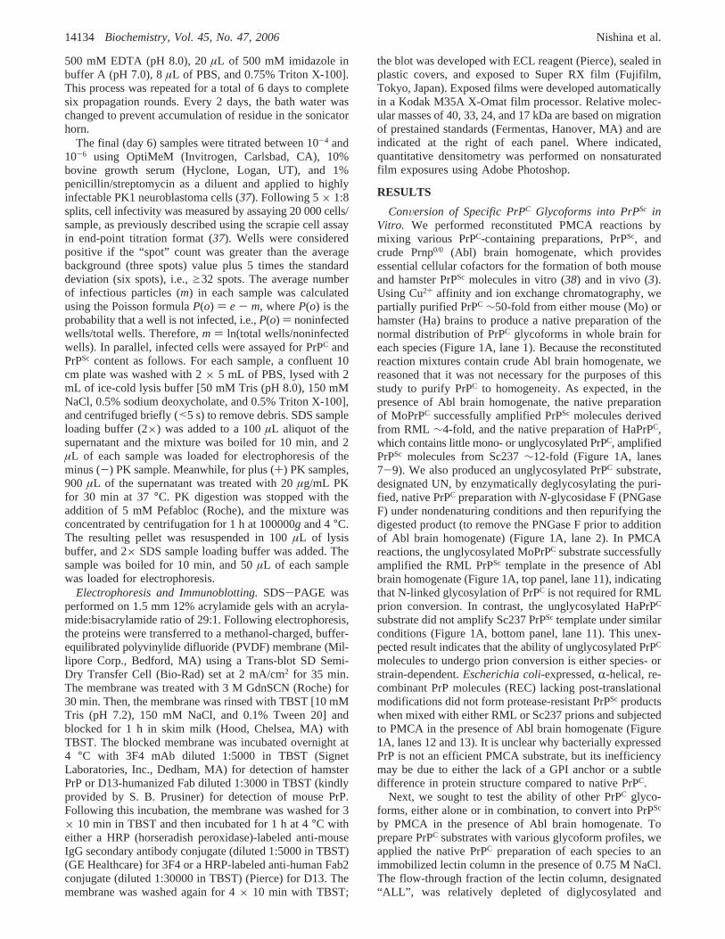
Effects of PrPC Sequence on the Formation of IsolatedPrPSc Glycoforms. The different glycoform preferencesobserved in hamster versus mouse PMCA reactions couldtheoretically be due to either (1) differences in amino acidsequence between HaPrPC and MoPrPC molecules or (2) thedifferent strain-specific templating properties of Sc237 versusRML prions. To distinguish between these two possibilities,we assessed the conversion of specific HaPrPC and MoPrPC
glycoforms in PMCA reactions driven by other hamster andmouse prion strains. The results show that two additionalstrains of hamster prions successfully converted DI HaPrPC,but not UN HaPrPC (Figure 4, top panel). Four additionalstrains of mouse prions did not convert DI MoPrPC, and twoof these strains successfully converted UN PrPC (Figure 4,bottom panel). Taken together, these results suggest that thePrP sequence is the dominant factor in determining thepreferred PrPC glycoform substrate. However, the relativelypoor ability of mouse prion strains C506 and 22A to convertUN PrPC substrate suggests that strain properties of the PrPSc
template may also influence PrPC glycoform preference(Figure 4, bottom panel, compare lanes 10 and 15 to lane5).
FIGURE 4: Seeding specificity of various scrapie strains. PrP27-30 template prepared from two hamster strains (139H and Drowsy) andfour mouse strains (139A, C506, 22A, and Me7) were separately incubated with different preparations of PrPC (Native, ALL, DI, and UN)and subjected to PMCA in the presence of 2.5% Abl brain homogenate, as described in Materials and Methods. All samples were subjectedto proteinase K digestion. Label: Sc, negative control sample containing no PrPC substrate. Note that the characteristic difference inelectrophoretic mobility between proteinase K-digested 139H and Drowsy PrPSc molecules cannot be easily appreciated in this blot becausea short gel was used, and the samples were not deglycosylated prior to electrophoresis.
14136 Biochemistry, Vol. 45, No. 47, 2006 Nishina et al.
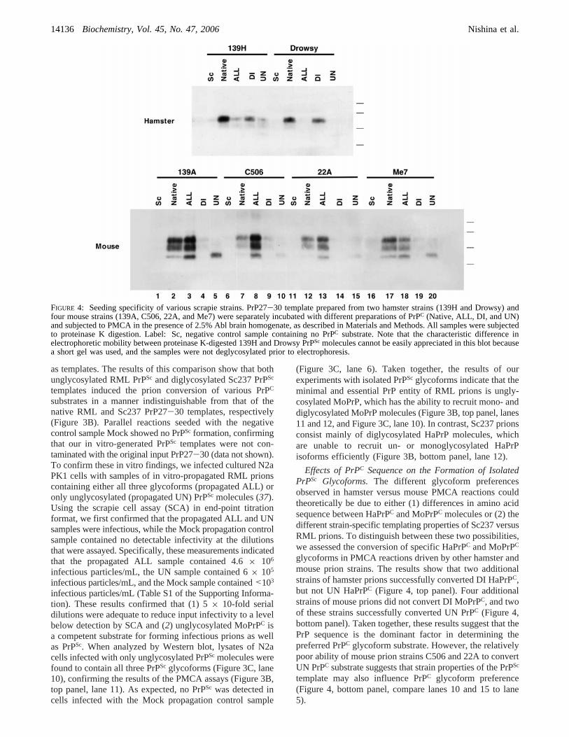
The transmission of infectious prions between mice andhamsters is inefficient, and this species barrier is recapitulatedin cell-free PrPSc formation assays (39). Previous transgenicmouse studies have established that amino acid sequencesimilarity between PrPC and PrPSc molecules is the mostimportant factor in determining species-dependent transmis-sion barriers (3, 4). However, since the glycoform profilesof PrPC molecules differ significantly between hamsters andmice (native HaPrPC is relatively enriched with diglycosy-lated PrPC), we investigated whether differential PrP glyco-sylation might also contribute to the transmission barrierbetween these two species. Using PMCA, we tested whetherDI MoPrPC and UN HaPrPC substrates could propagate DISc237 PrPSc and UN RML PrPSc templates, respectively. Theresults showed that no PrPSc propagation was detected witheither of these cross-species substrate-template combina-tions, whereas intraspecies control samples produced suc-cessful PrPSc propagation (Figure 5). Thus, PrPC glycosyla-tion does not appear to contribute significantly to speciesbarriers in vitro.
DISCUSSION
In summary, our biochemical and cell culture studiesindicate that subtle changes in the stoichiometry of host PrPC
glycoforms can significantly impact prion conversion in aspecies- and strain-specific manner. In particular, our experi-ments show that a substoichiometric ratio of unglycosylatedMoPrPC molecules within a substrate mixture is required forthe formation of RML prions. In contrast, diglycosylatedHaPrPC molecules are required for, while unglycosylatedHaPrPC molecules inhibit, the formation of Sc237 prions.The following hypothetical model could account for theseresults. Assuming that prions consist of a multimeric complex
of PrPSc molecules (40), the intermolecular interactionbetween HaPrP molecules in the assembly of Sc237 prionsmay be stabilized by N-linked glycans. The assembly processwould therefore require the presence of glycosylated HaPrPmolecules, and binding of unglycosylated HaPrP moleculeswould interfere with this process (Figure 6). In contrast,N-linked glycans may cause steric hindrance for the assemblyof PrPSc molecules in RML prions, and an orderly arrange-ment of different MoPrP glycoforms may be required toassemble this prion strain (Figure 6). Potentially, our datacould also be explained by a model in which the substratefor prion propagation is a multimeric PrPC complex com-posed of different glycoforms, and the conversion of theentire complex into PrPSc is initiated by the conversion of aspecific glycoform. This alternative model would provide asimple explanation for the interesting observation thatalteration of the PrPC glycoform profile changes the glyco-form ratio of the PrPSc molecules produced by prion-infected,cultured cells treated with tunicamycin (26). However, noheterogeneous, multimeric PrPC complexes have yet beenidentified.
Because the PrPC amino acid sequence is highly conservedbetween mice and hamsters, it is somewhat surprising thatPMCA reactions using PrP molecules isolated from thesetwo animals species should exhibit different glycoformrequirements. However, recent folding studies show thatrecombinant HaPrP and MoPrP form amyloid fibers withdifferent morphologies in vitro, apparently via differentmechanisms of lateral assembly (41).
Previous studies have clearly demonstrated that radiola-beled unglycosylated HaPrPC substrate could convert into aprotease-resistant conformation when incubated with a sto-ichiometric excess of Sc237 PrPSc molecules in the cell-free
FIGURE 5: Sequence-dependent propagation barriers between isolated hamster and mouse PrP glycoforms. In vitro-generated PrPSc moleculeswere serially propagated for 5 days using isolated PrPC glycoform substrates. For the top panel, unglycosylated RML was used as the initialseed for either unglycosylated mouse or hamster PrPC substrate, as indicated. In the bottom panel, diglycosylated Sc237 was used as theinitial seed for either diglycosylated mouse or hamster PrPC substrate, as indicated.
Prion Glycoforms Biochemistry, Vol. 45, No. 47, 200614137

conversion (CFC) assay (24, 42). The model shown in Figure6 also provides a reasonable explanation for those findings.Although addition of unglycosylated HaPrP to Sc237 prionsmay inhibit elongation by “capping”, this process would notnecessarily prevent conversion of substoichiometric amountsof unglycosylated HaPrP caps into a protease-resistantconformation. Protease-resistant HaPrP molecules formed inthis way could be specifically detected in the CFC assaybecause only newly converted radioactive PrPSc moleculesand not template PrPSc molecules would produce autorad-iographic signals. In contrast, no higher-magnitude Westernblot PrPSc signals would be expected in PMCA reactionsusing unglycosylated HaPrPC, since capping would inhibitPrPSc amplification.
The conclusions drawn from our biochemical experimentsare consistent with infectivity studies using transgenic miceexpressing selectively glycosylated MoPrPC molecules thatappear to traffic properly to the cell surface. Those studiesshowed that diglycosylated MoPrPC is not essential for theformation of infectious prions in vivo (28). Our conclusionsare also consistent with studies showing that Tg(HaPrP)transgenic mice coexpressing endogenous MoPrPC and wild-type HaPrPC are susceptible to Sc237 prions, because the13A5 mAb-reactive HaPrPC glycoform profile of Tg(HaPrP)mice is enriched for diglycosylated HaPrPC in a mannersimilar to that of hamster brain (3). Unfortunately, noinfectivity studies using transgenic mice expressing selec-tively glycosylated HaPrPC molecules have yet been reported.
While the data reported here provide evidence that thecomposition of di-, mono-, and unglycosylated PrPC mol-ecules within substrate mixtures regulates the efficiency ofprion formation, an even greater level of substrate diversitycould conceivably be generated by subtle variations in thearchitecture of the branching oligosaccharide moieties at-
tached to individual PrPC molecules (43). The proposedmodel, which includes the process of molecular selection(i.e., distinct PrP glycoforms), and the data presented alsorationalize the species- and strain-specific patterns of PrPSc
glycosylation (9), which are maintained upon serial passage.Finally, neurons in different brain regions express different,characteristic ratios of PrPC glycoforms (21-23); therefore,it is reasonable to hypothesize that individual prion strainspreferentially infect cells from specific brain regions becauseonly those cells contain the appropriate PrPC glycoformstoichiometry required for assembly of specifically organizedaggregates of PrPSc molecules.
ACKNOWLEDGMENT
We thank Dr. Judy Rees for help editing the manuscriptand Dr. Charles Weissmann for helpful discussions.
SUPPORTING INFORMATION AVAILABLE
End-point titration of prion infectivity in samples subjectedto serial PMCA propagation. This material is available freeof charge via the Internet at http://pubs.acs.org.
REFERENCES
1. Prusiner, S. B. (1982) Novel proteinaceous infectious particlescause scrapie,Science 216, 136-44.
2. Prusiner, S. B. (2000)Prion biology and diseases, Cold SpringHarbor Laboratory Press, Plainview, NY.
3. Scott, M., Foster, D., Mirenda, C., Serban, D., Coufal, F., Walchli,M., Torchia, M., Groth, D., Carlson, G., DeArmond, S. J., et al.(1989) Transgenic mice expressing hamster prion protein producespecies-specific scrapie infectivity and amyloid plaques,Cell 59,847-57.
4. Scott, M., Groth, D., Foster, D., Torchia, M., Yang, S. L.,DeArmond, S. J., and Prusiner, S. B. (1993) Propagation of prionswith artificial properties in transgenic mice expressing chimericPrP genes,Cell 73, 979-88.
FIGURE 6: Hypothetical model of prion propagation. N-Linked glycans form crucial intermolecular contact sites between PrP monomersduring Sc237 assembly but cause steric hindrance during RML assembly.
14138 Biochemistry, Vol. 45, No. 47, 2006 Nishina et al.
5. Bruce, M. E. (1993) Scrapie strain variation and mutation,Br.Med. Bull. 49, 822-38.
6. Carlson, G. A. (1996) Prion strains,Curr. Top. Microbiol.Immunol. 207, 35-47.
7. Bessen, R. A., and Marsh, R. F. (1992) Biochemical and physicalproperties of the prion protein from two strains of the transmissiblemink encephalopathy agent,J. Virol. 66, 2096-101.
8. Telling, G. C., Parchi, P., DeArmond, S. J., Cortelli, P., Montagna,P., Gabizon, R., Mastrianni, J., Lugaresi, E., Gambetti, P., andPrusiner, S. B. (1996) Evidence for the conformation of thepathologic isoform of the prion protein enciphering and propagat-ing prion diversity,Science 274, 2079-82.
9. Collinge, J., Sidle, K. C., Meads, J., Ironside, J., and Hill, A. F.(1996) Molecular analysis of prion strain variation and theaetiology of ‘new variant’ CJD,Nature 383, 685-90.
10. Peretz, D., Scott, M. R., Groth, D., Williamson, R. A., Burton, D.R., Cohen, F. E., and Prusiner, S. B. (2001) Strain-specifiedrelative conformational stability of the scrapie prion protein,Protein Sci. 10, 854-63.
11. Safar, J., Wille, H., Itri, V., Groth, D., Serban, H., Torchia, M.,Cohen, F. E., and Prusiner, S. B. (1998) Eight prion strains havePrP(Sc) molecules with different conformations,Nat. Med. 4,1157-65.
12. Bessen, R. A., Kocisko, D. A., Raymond, G. J., Nandan, S.,Lansbury, P. T., and Caughey, B. (1995) Non-genetic propagationof strain-specific properties of scrapie prion protein,Nature 375,698-700.
13. Ridley, R. M., and Baker, H. F. (1996) To what extent is strainvariation evidence for an independent genome in the agent of thetransmissible spongiform encephalopathies?Neurodegeneration5, 219-31.
14. Tanaka, M., Chien, P., Naber, N., Cooke, R., and Weissman, J.S. (2004) Conformational variations in an infectious proteindetermine prion strain differences,Nature 428, 323-8 (seecomment).
15. Tanaka, M., Chien, P., Yonekura, K., and Weissman, J. S. (2005)Mechanism of cross-species prion transmission: An infectiousconformation compatible with two highly divergent yeast prionproteins,Cell 121, 49-62.
16. King, C. Y., and Diaz-Avalos, R. (2004) Protein-only transmissionof three yeast prion strains,Nature 428, 319-23.
17. Jones, E. M., and Surewicz, W. K. (2005) Fibril conformation asthe basis of species- and strain-dependent seeding specificity ofmammalian prion amyloids,Cell 121, 63-72.
18. Endo, T., Groth, D., Prusiner, S. B., and Kobata, A. (1989)Diversity of oligosaccharide structures linked to asparagines ofthe scrapie prion protein,Biochemistry 28, 8380-8.
19. Locht, C., Chesebro, B., Race, R., and Keith, J. M. (1986)Molecular cloning and complete sequence of prion protein cDNAfrom mouse brain infected with the scrapie agent,Proc. Natl. Acad.Sci. U.S.A. 83, 6372-6.
20. Stahl, N., Borchelt, D. R., Hsiao, K., and Prusiner, S. B. (1987)Scrapie prion protein contains a phosphatidylinositol glycolipid,Cell 51, 229-40.
21. DeArmond, S. J., Qiu, Y., Sanchez, H., Spilman, P. R., Ninchak-Casey, A., Alonso, D., and Daggett, V. (1999) PrPc glycoformheterogeneity as a function of brain region: Implications forselective targeting of neurons by prion strains,J. Neuropathol.Exp. Neurol. 58, 1000-9.
22. Beringue, V., Mallinson, G., Kaisar, M., Tayebi, M., Sattar, Z.,Jackson, G., Anstee, D., Collinge, J., and Hawke, S. (2003)Regional heterogeneity of cellular prion protein isoforms in themouse brain,Brain 126, 2065-73.
23. Somerville, R. A., Hamilton, S., and Fernie, K. (2005) Transmis-sible spongiform encephalopathy strain, PrP genotype and brainregion all affect the degree of glycosylation of PrPSc,J. Gen.Virol. 86, 241-6.
24. Kocisko, D. A., Come, J. H., Priola, S. A., Chesebro, B., Raymond,G. J., Lansbury, P. T., and Caughey, B. (1994) Cell-free formationof protease-resistant prion protein,Nature 370, 471-4.
25. Taraboulos, A., Rogers, M., Borchelt, D. R., McKinley, M. P.,Scott, M., Serban, D., and Prusiner, S. B. (1990) Acquisition ofprotease resistance by prion proteins in scrapie-infected cells doesnot require asparagine-linked glycosylation,Proc. Natl. Acad. Sci.U.S.A. 87, 8262-6.
26. Vorberg, I., and Priola, S. A. (2002) Molecular basis of scrapiestrain glycoform variation,J. Biol. Chem. 277, 36775-81.
27. DeArmond, S. J., Sanchez, H., Yehiely, F., Qiu, Y., Ninchak-Casey, A., Daggett, V., Camerino, A. P., Cayetano, J., Rogers,M., Groth, D., Torchia, M., Tremblay, P., Scott, M. R., Cohen,F. E., and Prusiner, S. B. (1997) Selective neuronal targeting inprion disease,Neuron 19, 1337-48.
28. Neuendorf, E., Weber, A., Saalmueller, A., Schatzl, H., Reifenberg,K., Pfaff, E., and Groschup, M. H. (2004) Glycosylation deficiencyat either one of the two glycan attachment sites of cellular prionprotein preserves susceptibility to bovine spongiform encephal-opathy and scrapie infections,J. Biol. Chem. 279, 53306-16.
29. Rogers, M., Taraboulos, A., Scott, M., Groth, D., and Prusiner,S. B. (1990) Intracellular accumulation of the cellular prion proteinafter mutagenesis of its Asn-linked glycosylation sites,Glycobi-ology 1, 101-9.
30. Lehmann, S., and Harris, D. A. (1997) Blockade of glycosylationpromotes acquisition of scrapie-like properties by the prion proteinin cultured cells,J. Biol. Chem. 272, 21479-87.
31. Muramoto, T., DeArmond, S. J., Scott, M., Telling, G. C., Cohen,F. E., and Prusiner, S. B. (1997) Heritable disorder resemblingneuronal storage disease in mice expressing prion protein withdeletion of anR-helix, Nat. Med. 3, 750-5.
32. Saborio, G. P., Permanne, B., and Soto, C. (2001) Sensitivedetection of pathological prion protein by cyclic amplification ofprotein misfolding,Nature 411, 810-3.
33. Pan, K. M., Baldwin, M., Nguyen, J., Gasset, M., Serban, A.,Groth, D., Mehlhorn, I., Huang, Z., Fletterick, R. J., Cohen, F.E., and Prusiner, S. B. (1993) Conversion ofR-helices intoâ-sheets features in the formation of the scrapie prion proteins,Proc. Natl. Acad. Sci. U.S.A. 90, 10962-6.
34. Castilla, J., Saa, P., Hetz, C., and Soto, C. (2005) In vitrogeneration of infectious scrapie prions,Cell 121, 195-206.
35. Bocharova, O. V., Breydo, L., Parfenov, A. S., Salnikov, V. V.,and Baskakov, I. V. (2005) In vitro conversion of full-lengthmammalian prion protein produces amyloid form with physicalproperties of PrP(Sc),J. Mol. Biol. 346, 645-59.
36. Riek, R., Hornemann, S., Wider, G., Glockshuber, R., andWuthrich, K. (1997) NMR characterization of the full-lengthrecombinant murine prion protein, mPrP(23-231),FEBS Lett. 413,282-8.
37. Klohn, P. C., Stoltze, L., Flechsig, E., Enari, M., and Weissmann,C. (2003) A quantitative, highly sensitive cell-based infectivityassay for mouse scrapie prions,Proc. Natl. Acad. Sci. U.S.A. 100,11666-71.
38. Deleault, N. R., Geoghegan, J. C., Nishina, K., Kascsak, R.,Williamson, R. A., and Supattapone, S. (2005) Protease-resistantPrion Protein Amplification Reconstituted with Partially PurifiedSubstrates and Synthetic Polyanions,J. Biol. Chem. 280, 26873-9.
39. Kocisko, D. A., Priola, S. A., Raymond, G. J., Chesebro, B.,Lansbury, P. T., Jr., and Caughey, B. (1995) Species specificityin the cell-free conversion of prion protein to protease-resistantforms: A model for the scrapie species barrier,Proc. Natl. Acad.Sci. U.S.A. 92, 3923-7.
40. Silveira, J. R., Raymond, G. J., Hughson, A. G., Race, R. E., Sim,V. L., Hayes, S. F., and Caughey, B. (2005) The most infectiousprion protein particles,Nature 437, 257-61.
41. Makarava, N., Bocharova, O. V., Salnikov, V. V., Breydo, L.,Anderson, M., and Baskakov, I. V. (2006) Dichotomous versuspalm-type mechanisms of lateral assembly of amyloid fibrils,Protein Sci. 15, 1334-41.
42. Priola, S. A., and Lawson, V. A. (2001) Glycosylation influencescross-species formation of protease-resistant prion protein,EMBOJ. 20, 6692-9.
43. Rudd, P. M., Wormald, M. R., Wing, D. R., Prusiner, S. B., andDwek, R. A. (2001) Prion glycoprotein: Structure, dynamics, androles for the sugars,Biochemistry 40, 3759-66.
BI061526K
Prion Glycoforms Biochemistry, Vol. 45, No. 47, 200614139