The soybean sucrose binding protein gene family: genomic organization, gene copy number and...
11
DOI: 10.1093/jxb/erg301 RESEARCH PAPER The soybean sucrose binding protein gene family: genomic organization, gene copy number and tissue- speci®c expression of the SBP2 promoter Luis Anto à nio S. Contim 1 , Alessandro J. Waclawovsky 2 , Nelson Delu  -Filho 2 , Carlos P. Pirovani 2 , Wellington R. Clarindo 3 , Marcelo E. Loureiro 2 , Carlos R. Carvalho 3 and Elizabeth P. B. Fontes 1,2, * 1 Departamento de Bioquõ Âmica e Biologia Molecular, Universidade Federal de Vic Ëosa 36571.000, Vic Ëosa, MG, Brazil 2 BIOAGRO, Universidade Federal de Vic Ëosa 36571.000, Vic Ëosa, MG, Brazil 3 Departamento de Biologia Geral, Universidade Federal de Vic Ëosa 36571.000, Vic Ëosa, MG, Brazil Received 28 February 2003; Accepted 6 August 2002 Abstract The sucrose binding protein (SBP) from soybean has been implicated as an important component of the sucrose uptake system. Two SBP genomic clones, gsS641.1 and gsS641.2, which correspond to allelic forms of the GmSBP2/S64 gene, have been isolated and characterized. As a member of the seed storage protein superfamily, it has been shown that the SBP gene structure is similar to vicilin genes with intron/ exon boundaries at conserved positions. Fluores- cence in situ hybridization (FISH) suggested that the soybean SBP gene family is represented by at least two non-allelic genes corresponding to the pre- viously isolated GmSBP1 and GmSBP2/S64 cDNAs. These two cDNAs share extensive sequence simil- arity but are located at different loci in the soybean genome. To investigate transcriptional activation of the GmSBP2 gene, 2 kb 5¢-¯anking sequences of gsS641.1 and gsS641.2 were fused to the b-glucu- ronidase (GUS) reporter gene and to the green ¯uor- escent protein (GFP) reporter gene and inde- pendently introduced into Nicotiana tabacum by Agrobacterium tumefaciens-mediated transformation. The SBP2 promoter directed expression of both GUS and GFP reporter genes with high speci®city to the phloem of leaves, stems and roots. Thus, the overall pattern of SBP±GUS or SBP±GFP expression is consistent with the involvement of SBP in sucrose translocation-dependent physiological processes. Key words: FISH, gene family, Glycine max, phloem-speci®c expression, promoter activity, sucrose binding protein. Introduction A central characteristic of plants is the capacity to convert light energy through photosynthesis into carbohydrate. These molecules can be stored, directly consumed as an energy source or utilized as structural components of the cells. Because of that plants are considered as basic producers of the energy ¯ux of the planet trophy chain. Although carbon autotrophy is a typical feature of plant cells, whole plants behave as physiological mosaics in which photosynthetically active tissues, such as mesophyll cells from mature leaves (source tissues), export carbo- hydrate to photosynthetically less active or inactive tissues, such as stems, ¯owers and roots (sink tissues) (Fro Èmmer and Sonnewald, 1995; Stitt, 1996). The processes that regulate carbon allocation to the various sink organs directly impact plant development. Sucrose is the main carbohydrate that is transported cell-to-cell and used for long-distance transport in the vascular system of a large number of higher plants. Sucrose not only functions as a transport metabolite, but also contributes to the osmotic driving force for phloem translocation (mass ¯ow) and serves as a signal to activate or repress speci®c genes in a variety of different tissues (Lalonde et al., 1999). The cell- to-cell sucrose transport occurs either directly via plasmo- desmata (symplastic transport) or across plasma mem- branes mediated by protein carriers (apoplastic transport) * To whom correspondence should be addressed. Fax: +55 31 38992864. E-mail: [email protected] Journal of Experimental Botany, ã Society for Experimental Biology 2003; all rights reserved Journal of Experimental Botany, Page 1 of 11 Advance Access published October 29, 2003 by guest on June 11, 2013 http://jxb.oxfordjournals.org/ Downloaded from
Transcript of The soybean sucrose binding protein gene family: genomic organization, gene copy number and...

DOI: 10.1093/jxb/erg301
RESEARCH PAPER
The soybean sucrose binding protein gene family:
genomic organization, gene copy number and tissue-
speci®c expression of the SBP2 promoter
Luis AntoÃnio S. Contim1, Alessandro J. Waclawovsky2, Nelson Delu -Filho2, Carlos P. Pirovani2,
Wellington R. Clarindo3, Marcelo E. Loureiro2, Carlos R. Carvalho3 and Elizabeth P. B. Fontes1,2,*
1 Departamento de BioquõÂmica e Biologia Molecular, Universidade Federal de VicËosa 36571.000, VicËosa, MG,Brazil2 BIOAGRO, Universidade Federal de VicËosa 36571.000, VicËosa, MG, Brazil3 Departamento de Biologia Geral, Universidade Federal de VicËosa 36571.000, VicËosa, MG, Brazil
Received 28 February 2003; Accepted 6 August 2002
Abstract
The sucrose binding protein (SBP) from soybean has
been implicated as an important component of the
sucrose uptake system. Two SBP genomic clones,
gsS641.1 and gsS641.2, which correspond to allelic
forms of the GmSBP2/S64 gene, have been isolated
and characterized. As a member of the seed storage
protein superfamily, it has been shown that the SBP
gene structure is similar to vicilin genes with intron/
exon boundaries at conserved positions. Fluores-
cence in situ hybridization (FISH) suggested that the
soybean SBP gene family is represented by at least
two non-allelic genes corresponding to the pre-
viously isolated GmSBP1 and GmSBP2/S64 cDNAs.
These two cDNAs share extensive sequence simil-
arity but are located at different loci in the soybean
genome. To investigate transcriptional activation of
the GmSBP2 gene, 2 kb 5¢-¯anking sequences of
gsS641.1 and gsS641.2 were fused to the b-glucu-
ronidase (GUS) reporter gene and to the green ¯uor-
escent protein (GFP) reporter gene and inde-
pendently introduced into Nicotiana tabacum by
Agrobacterium tumefaciens-mediated transformation.
The SBP2 promoter directed expression of both GUS
and GFP reporter genes with high speci®city to the
phloem of leaves, stems and roots. Thus, the overall
pattern of SBP±GUS or SBP±GFP expression is
consistent with the involvement of SBP in sucrose
translocation-dependent physiological processes.
Key words: FISH, gene family, Glycine max, phloem-speci®c
expression, promoter activity, sucrose binding protein.
Introduction
A central characteristic of plants is the capacity to convert
light energy through photosynthesis into carbohydrate.
These molecules can be stored, directly consumed as an
energy source or utilized as structural components of the
cells. Because of that plants are considered as basic
producers of the energy ¯ux of the planet trophy chain.
Although carbon autotrophy is a typical feature of plant
cells, whole plants behave as physiological mosaics in
which photosynthetically active tissues, such as mesophyll
cells from mature leaves (source tissues), export carbo-
hydrate to photosynthetically less active or inactive tissues,
such as stems, ¯owers and roots (sink tissues) (FroÈmmer
and Sonnewald, 1995; Stitt, 1996). The processes that
regulate carbon allocation to the various sink organs
directly impact plant development. Sucrose is the main
carbohydrate that is transported cell-to-cell and used for
long-distance transport in the vascular system of a large
number of higher plants. Sucrose not only functions as a
transport metabolite, but also contributes to the osmotic
driving force for phloem translocation (mass ¯ow) and
serves as a signal to activate or repress speci®c genes in a
variety of different tissues (Lalonde et al., 1999). The cell-
to-cell sucrose transport occurs either directly via plasmo-
desmata (symplastic transport) or across plasma mem-
branes mediated by protein carriers (apoplastic transport)
* To whom correspondence should be addressed. Fax: +55 31 38992864. E-mail: [email protected]
Journal of Experimental Botany, ãSociety for Experimental Biology 2003; all rights reserved
Journal of Experimental Botany, Page 1 of 11
Advance Access published October 29, 2003 by guest on June 11, 2013
http://jxb.oxfordjournals.org/D
ownloaded from
(Lemoine, 2000; Williams et al., 2000). Both symplastic
and apoplastic transports can contribute for phloem
loading and unloading processes, although in some plants
one route of sucrose loading may predominate over the
other (FroÈmmer and Sonnewald, 1995). In fact, in
several species, sucrose transporters have been identi-
®ed as essential for phloem loading into sieve elements,
the phloem-speci®c photoassimilate-transporting cells
(Riesmeier et al., 1994; BuÈrkle et al., 1998).
Complementation assays using an invertase-de®cient
yeast mutant have been used to identify and isolate the
sucrose transporter SoSUT1 cDNA from spinach leaves
(Riesmeier et al., 1992) and StSUT1 from potato leaves
(Riesmeier et al., 1993). The subsequent identi®cation of
sucrose transporters in other species has been accom-
plished by hybridization screening using heterologous
probes or PCR-ampli®cation from these initial sequences
(Lemoine, 2000). The members of the SUT family encode
highly hydrophobic disaccharide transporters with two sets
of six membrane-spanning domain structures, separated by
a large cytoplasmic loop (Williams et al., 2000). Many of
these transporters have been shown to mediate sucrose
transport through a proton-coupled transport mechanism.
The SUT1 protein has been described as the proton-
motive-force-driven sucrose symporter that mediates
phloem loading and long-distance transport, the key
transport step in assimilate partitioning for many plants
(Riesmeier et al., 1994; BuÈrkle et al., 1998). SUT1 serves
as a high-af®nity transporter, whereas SUT4, a second
member of this sucrose transporter family, corresponds to
the low-af®nity/high capacity saturable component of
sucrose uptake found in leaves (Weise et al., 2000). A third
structurally related-member of the family has been iden-
ti®ed and designated SUT2 (Barker et al., 2000). Although
the whole family of sucrose transporter genes of a given
species has not been identi®ed, the sucrose transporters
make a large gene family, as at least seven distinct
sequences that encode putative sucrose transporters are
present in the Arabidopsis database (Williams et al., 2000).
A sucrose binding protein (SBP), structurally unrelated
to the members of the SUT family, was ®rst identi®ed in
soybean cotyledons and has been demonstrated to be
involved in sucrose translocation-dependent physiological
processes in plants (Ripp et al., 1988). SBP repression
studies in tobacco have indeed shown some of the typical
phenotypes caused by impairment of sucrose translocation
(Riesmeier et al., 1994; KuÈhn et al., 1996), such as the
accumulation of carbohydrates within source leaves, the
inhibition of photosynthesis and stunted growth (Pedra
et al., 2000). Furthermore, manipulation of SBP levels in
transgenic cell lines correlated with the ef®ciency of
radiolabelled sucrose uptake by the cells and altered
sucrose-cleaving activities in a metabolic compensatory
manner (DeluÂ-Filho et al., 2000). Direct evidence impli-
cating SBP in sucrose transport has been obtained with
complementation studies using a secreted invertase-de®-
cient mutant yeast strain, incapable of growth on medium
containing sucrose as the only carbon source (Overvoorde
et al., 1996, 1997; Pirovani et al., 2002). The SBP-
mediated speci®c sucrose uptake in yeast displays linear,
non-saturable kinetics up to 30 mM external sucrose, being
relatively insensitive to the pH gradient across the
membrane (Grimes and Overvoorde, 1996; Overvoorde
et al., 1996). These biochemical features closely resemble
the kinetic properties of the previously characterized linear
component of sucrose uptake in higher plants (Maynard
and Lucas, 1982; Lin et al., 1984). Nevertheless, these
relevant data do not allow the apparent inconsistency
between the absence of typical membrane transporter
structural motifs on SBP and an SBP-mediated sucrose
transport mechanism to be reconciled and, as a conse-
quence, a scenario for SBP function remains elusive.
Homologous SBP genes have been isolated from pea
(GeneBankÔ accession number Y11207) and Vicia faba
(GeneBankÔ accession number VFA292221). In spinach,
an SBP homologue was immunolocalized in the plasma
membrane of sieve elements in fully expanded leaves,
shoots and roots (Warmbrodt et al., 1989, 1991) and, in
tobacco, it was detected in the microsomal fraction of
young leaves from Nicotiana tabacum (Pedra et al., 2000).
In soybean, two SBP genes have been identi®ed (Grimes
et al., 1992; Pirovani et al., 2002), but an extensive
analysis has not been performed to provide a global view
of the SBP gene content. In this investigation, the isolation
of SBP2 genomic clones is reported and in situ hybridiza-
tion was performed on isolated nuclei to characterize the
soybean SBP gene family. Data are also presented on the
tissue-speci®c expression of the SBP2 promoter.
Materials and methods
Screening of soybean genomic libraries and DNA sequenceanalysis
A size-selected genomic library propagated in lZAPII (9.753109
pfu) was screened by plaque hybridization, as described bySambrook et al. (1989), using the complete SBP2/S64 cDNA(GeneBankÔ accession number AF191299) as probe. The hybridiz-ation probe was radiolabelled with [a-32P]dCTP by random primedlabelling (Amersham Pharmacia Biotech). Two positive clones,gsS641.1 and gsS641.2, were plaque puri®ed, excised andsequenced. Sequencing was carried out with a combination of insertsubcloning and primer walking. The identity of these clones wasobtained by sequence comparison analysis using the BLASTprogram (Altschul et al., 1990). The computer program ClustalWwas used for sequence alignment.
Genomic DNA gel blot analysis
DNA was extracted from young leaves, digested overnight withBamHI or EcoRI, precipitated with ethanol 70% (v/v) and separatedon a 1% (w/v) agarose gel. The gel was washed with 250 mM HClfollowed by alkaline denaturation (Sambrook et al., 1989). Afterneutralization, the DNA was transferred to nylon membranes andUV ®xed (Stratalinker, Stratagene). The SBP2 cDNA was labelled
2 of 11 Contim et al.
by guest on June 11, 2013http://jxb.oxfordjournals.org/
Dow
nloaded from

using the Primer-It Fluor Fluorescence Labelling Kit (Stratagene)according to the instructions of the supplier. Hybridization and
washing of the blots were performed using standard procedures(Sambrook et al., 1989). The hybridization signals were revealed by
the IluminatorÔ Nonradioactive Detection System (Stratagene)according to the instructions of the supplier.
Probe preparation for in situ hybridization
Two DNA fragments were used as probe for in situ hybridization.The ®rst one corresponds to the 4 kb EcoRI insert from pgsS641.2.The second one corresponds to a 1.5 kb DNA fragment extending
from position 313 to 1695 of the GmSBP1 cDNA (GeneBankÔaccession number L06038). This DNA fragment corresponds to theinsert of the soy25Z12 cDNA clone, previously isolated in the
laboratory through the screening of a soybean seed cDNA libraryusing the SBP2/S64 cDNA (GeneBankÔ accession number
AF191299) as probe. Both DNA fragments, used as FISHprobes, were released from the vector by EcoRI digestion, gel-puri®ed and labelled using the Primer-It Fluor Fluorescence
Labelling Kit (Stratagene) according to the recommendations ofthe supplier.
Cytological preparations and in situ hybridization
Soybean seeds were germinated in Petri dishes containing a ®lm ofdistilled water, and incubated at 29 °C in the dark. Seedlings with0.5±1 cm long roots were ®xed in a fresh ice-cold methanol:acetic
acid solution (3:1, v:v) and kept at ±20 °C for 24 h. Then, the rootswere excised at 0.1 cm from the root tip and macerated with a freshly
prepared Flaxzyme (NOVO) enzymatic solution (1/10), and incu-bated at 35 °C for 90 min. The macerated cells were dissociated in aclean slide with a fresh ®xative solution, air-dried and stained with a
2%Giemsa solution in phosphate buffer, pH 6.8, for 5 min (Carvalhoand Saraiva, 1997).The slides containing soybean interphase nuclei were treated with
100 mg ml±1 RNase-A in 23 SSC (150 mM NaCl, 15 mM Na3citrate, pH 7.0) at 37 °C for 1 h, washed with the same buffer, thenwith PBS (130mMNaCl, 7 mMNa2HPO4, 3 mMNaH2PO4, pH 7.0),dehydrated sequentially in 70%, 80% and 100% ethanol (2 min each)
at room temperature and ®nally air-dried. The probe (2 ng ml±1) andsalmon sperm DNA (200 ng ml±1) were denatured in the hybridiz-
ation solution [50% (v/v) deionized formamide, 10% (w/v) dextransulphate, 50 mM sodium phosphate, pH 7.0, and 23 SSC] for 75 °Cfor 15 min and, then, further co-denatured with the nuclei for 4 min
at 75 °C. The denatured nuclei were hybridized for 12 h at 37 °C inHYBAID OmiSlide termal cycler. After hybridization, the slideswere washed twice in 23 SSC containing 50% (v/v) formamide for
2 min each at 45 °C; four times for 2 min in 23 SSC at roomtemperature and then rinsed in PBS for 2 min. The slides were air-
dried and mounted in 15 ml of detection buffer (12.5 mg ml±1
triethylenediamine in glycerol, 1/23 PBS and 200 ng ml±1
propidium iodide).
Microscopy and analysis of the hybridization signal
The images of isolated nuclei were captured with a CCD videocamera and digitalized by an image analysis system attached to the
Olympus BX 60 re¯ected-light ¯uorescence microscope withobjective 1003 and WU excitation (cube U-MWU) ¯uorescence®lter, BA 420 barrier ®lter and DM400 dichroic mirror. Image
analyses were conducted using the public domain Image SXM 1.68software (Rasband, 1997). The original colour TIFF images were
converted to a grey scale up to a 255 grey value for the relativedensity plot analysis. Using the colour table tool of the software, the®nal plot images were digitally pseudo-coloured with the ®lter
colour spectrum.
Construction of SBP2 promoter±reporter geneconstructs
A SBP2 promoter±GUS fusion gene was constructing by cloning a2.0 kb EcoRI/NcoI fragment from pgsS641.1 into the EcoRI/NcoIsites of pCAMBIA 1381Z (Roberts et al., 1996) to give pUFV335,also referred to as ±2000pSPB2±GUS. To construct an SBP2
promoter±GFP fusion gene, a Klenow-repaired KpnI/SpeI GFPcDNA fragment was transferred from pCAMBIA 1302 (Robertset al., 1996) to the Klenow-repaired NcoI site of pUFV335. Theresulting clone, pUFV419 (±2000pSBP2±GFP), contains a GFP
cDNA under the control of 2.0 kb 5¢-¯anking sequence ofgsS641.1.
Generation of transgenic plants
The pCAMBIA-derived recombinant plasmids or pCAMBIA 1381Zbinary vector alone were used to transform Nicotiana tabacum L. cv.Havana plants by Agrobacterium tumefaciens-mediated leaf disctransformation (Alvim et al., 2001) and the transformed plants wereregenerated on medium containing hygromycin (50 mg l±1) (Buzeliet al., 2002). For the ±2000pSBP2±GUS constructs, plantlets wereassayed for GUS activity and primary transformants were eithermaintained in vitro or transferred into soil and grown in standardizedgreenhouse conditions (T0 plants) to generate seeds. Most of therooted plants were tested for the incorporation of the hygromycingene and SBP2 promoter±reporter gene fusions by PCR analysis.The intensity of GUS staining among the selected lines was similarand the expression pattern was consistent between independenttransgenic lines. Detailed sectional analyses for tissue-speci®cexpression were carried out on eleven independent SBP2±GUS
transgenic lines and ®ve independent SBP2±GFP transgenic lines.One hygromycin-resistant plant for the pCAMBIA 1381Zincorporated binary vector was used as control.
Analysis of expression patterns
Protein extraction and ¯uorometric assay for GUS activity wereperformed essentially as described by Jefferson et al. (1987) withmethylumbelliferone (MU) as a standard. Extracts were preparedfrom the tissue that had been frozen in liquid nitrogen and kept at±80 °C until processing. For the standard assay, plant tissues wereground in 0.5 ml of GUS assay buffer [100 mM NaH2PO4.H2O(pH 7.0), 10 mM EDTA, 0.1% (w/v) sarcosyl, 0.1% (v/v) TritonX-100] and 50 ml of this extract were mixed with 50 ml of GUS assaybuffer containing 2 mM of 4-methylumbelliferyl-b-D glucuronide(MUG) as a substrate. The mixture was incubated at 37 °C for 15 minand GUS activity was measured using a DYNA Quant 200Fluorometer (Amersham Pharmacia Biotech).For the transgenic lines carrying SBP2±GUS fusion, histo-
chemical analysis of b-glucuronidase activity was performed aspreviously described (McCabe et al., 1988). The tissues (roots, stemsand leaves) were sampled and sectioned using a hand microtome.Tissue sections were embedded in the GUS assay buffer [100 mMNaH2PO4.H20 (pH 7.0), 0.5 mM K4Fe(CN)6.3H2O, 10 mMNa2EDTA.2H2O, 0.1% (v/v) Triton X-100] containing 5 bromo-4-chloro-3-indolyl-b-D glucuronide (X-Gluc) (McCabe et al., 1988)and incubated at 37 °C in the dark for 4 h. Pigments were extractedfrom stained tissues with methanol:acetone (3:1, v:v). Afterextensive washing, the clari®ed tissues were stored in 50% (v/v)glycerol until photodocumentation. The micrographies were takenunder an Olympus AX-70 microscope.Transformed plants containing SBP2±GFP construct were har-
vested and mounted. An Olympus BX 60 ¯uorescence microscopewith objectives 103, 203, and 403 and B excitation (WB cubeU-MWU) ¯uorescence ®lters were used to view plants. Images werecaptured with a CCD video camera and analysed using the publicdomain Image SXM 1.68 software (Rasband, 1997). The original
The soybean SBP gene family 3 of 11
by guest on June 11, 2013http://jxb.oxfordjournals.org/
Dow
nloaded from
colour TIFF images were processed as previously described. Theimages were digitally pseudocoloured using the colour ®lter number5 (cyan, yellow and green).
Results
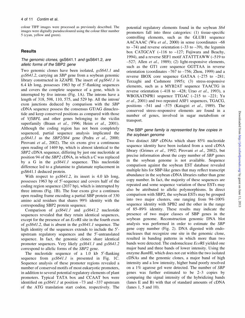
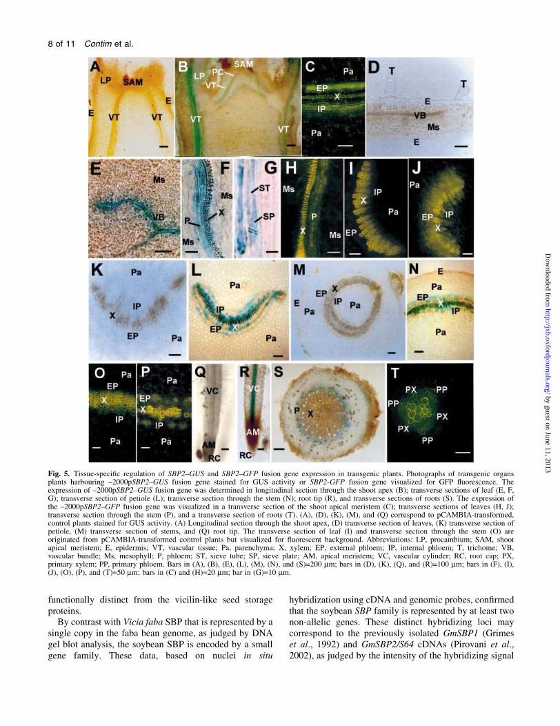
The genomic clones, gsS641.1 and gsS641.2, areallelic forms of the SBP2 gene
Two genomic clones have been isolated, gsS641.1 and
gsS641.2, carrying an SBP gene from a soybean genomic
library constructed in lZAPII. The insert of pgS641.1 is
6.4 kb long, possesses 1963 bp of 5¢-¯anking sequences
and covers the complete sequence of a gene, which is
interrupted by ®ve introns (Fig. 1A). The introns have a
length of 318, 598, 110, 575, and 529 bp. All the intron/
exon junctions deduced by comparison with the SBP
cDNA sequence possess the consensus GT/AG dinucleo-
tide and keep conserved positions as compared with those
of VfSBPL and other genes belonging to the vicilin
superfamily (Braun et al., 1996; Heim et al., 2001).
Although the coding region has not been completely
sequenced, partial sequence analysis implicated the
gsS641.1 as the SBP2/S64 gene (Pedra et al., 2000;
Pirovani et al., 2002). The six exons give a continuous
open reading of 1469 bp, which is almost identical to the
SBP2 cDNA sequence, differing by just one nucleotide at
position 94 of the SBP2 cDNA, in which a C was replaced
by a G in the gsS641.1 sequence. This nucleotide
difference led to a glutamine to glutamate replacement in
gsS641.1 deduced protein.
With respect to gsS641.2, its insert is 4.0 kb long,
possesses 1963 bp of 5¢-sequence and covers half of the
coding region sequence (2037 bp), which is interrupted by
three introns (Fig. 1B). The four exons give a continuos
open reading frame encoding a partial SBP protein of 330
amino acid residues that shares 99% identity with the
corresponding SBP2 protein sequence.
Comparison of gsS641.1 and gsS641.2 nucleotide
sequences revealed that they retain identical sequences,
except for the presence of an EcoRI site in the fourth exon
of gsS641.2, that is absent in the gsS641.1 sequence. The
high identity of the sequences extends to include the 5¢-upstream regulatory sequences and the 5¢-untranslatedsequence. In fact, the genomic clones share identical
promoter sequences. Very likely gsS641.1 and gsS641.2
correspond to allelic forms of the SBP2 gene.
The nucleotide sequence of a 1.0 kb 5¢-¯ankingsequence from gsS641.1 is presented in Fig. 1C.
Sequence analysis of these promoter regions revealed a
number of conserved motifs of most eukaryotic promoters,
in addition to several potential regulatory elements of plant
promoters. Typical TATA box and CCAAT box were
identi®ed on gsS641.1 at position ±73 and ±337 upstream
of the ATG translation start codon, respectively. The
potential regulatory elements found in the soybean S64
promoters fall into three categories: (1) tissue-speci®c
controlling elements, such as the GLUB1 sequence
AACAAAC (Wu et al., 2000) in sense (coordinates ±68
to ±74) and reverse orientation (±33 to ±39), the legumin
box CATGCAY (±116 to ±127; Fujiwara and Beachy,
1994), and a reverse SEF1 motif ATATTTAWW (±519 to
±527; Allen et al., 1989); (2) light-responsive elements,
such as the GT1 core sequence GGTTAA in reverse
orientation (coordinates ±767 to ±756; Zhou, 1999) and a
reverse IBOX core sequence GATAA (±275 to ±281;
Terzaghi and Cashmore 1995); (3) stress-responsive
elements, such as a MYB2AT sequence TAACTG in
reverse orientation (±418 to ±428; Urao et al., 1993), a
WBOXATNPR1 sequence TTGAC (±215 to ±221; Yu
et al., 2001) and two repeated ASF1 sequences, TGACG,
positions ±541 and ±575 (Katagiri et al., 1989). The
conserved stress±responsive elements are found in a
number of genes, involved in sugar metabolism or
transport.
The SBP gene family is represented by few copies inthe soybean genome
Two distinct SBP cDNAs which share 85% nucleotide
sequence identity have been isolated from a seed cDNA
library (Grimes et al., 1992; Pirovani et al., 2002), but
precise information about the copy number of SBP genes
in the soybean genome is not available. Sequence
comparison against the soybean EST database revealed
multiple hits for SBP-like genes that may re¯ect transcript
abundance in the soybean cDNA libraries rather than gene
copy number. In fact, the majority of these sequences are
repeated and some sequence variation of these ESTs may
also be attributed to allelic polymorphisms. In direct
comparison with SBP2, the soybean ESTs may be grouped
into two major clusters, one ranging from 94±100%
sequence identity with SPB2 and the other in the range
of 85±89% identity. These results may indicate the
presence of two major classes of SBP genes in the
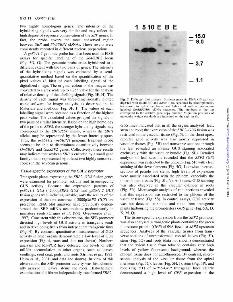
soybean genome. Reconstruction genomic DNA blot
analysis was performed in order to estimate the SBP
gene copy number (Fig. 2). DNA digested with endo-
nucleases that recognize one site in the genomic clone,
resulted in banding patterns in which more than two
bands were detected. The endonuclease EcoRI yielded one
major band and three bands of lower intensity. Using the
enzyme BamHI, which does not cut within the two isolated
cDNAs and the genomic clones, a major band of high
intensity and a low intensity, higher band poorly resolved
on a 1% agarose gel were detected. The number of SBP
genes was further estimated to be 2±3 copies by
comparing the signal intensity of the hybridizing bands
(lanes E and B) with that of standard amounts of cDNA
(lanes 1, 5 and 10).
4 of 11 Contim et al.
by guest on June 11, 2013http://jxb.oxfordjournals.org/
Dow
nloaded from
The S64/SBP2 and SBP genes are located in distinctregions on the soybean genome
To estimate the SBP gene copy number in the soybean
genome further, ¯uorescence in situ hybridization was
performed on interphase nuclei using a fragment of the
SBP cDNA as probe (Fig. 3A, B, C). At moderate
stringency, four hybridizing spots on the genome were
detected, suggesting the presence of two distinct loci or
Fig. 1. Gene strucuture of soybean SBP2. (A) Schematic diagram of the intron±exon organization of GmSBP2 in the soybean genome (gsS641.1).The position of the translation start codon (ATG) in the ®rst exon and the translation stop codon within exon 6 are indicated. The exons (blackboxes) are numbered from 1 to 6. Promoter region and 3¢-¯anking genomic sequence are indicated by white boxes and introns as hatched boxes.The positions of some restriction enzyme sites are indicated. (B) Schematic representation of the gsS641.2 allele. An extension of 4.0 kbsequences representing the genomic clone gsS641.2 is presented in which the promoter sequences, intron and exons are indicated as in (A). Thepositions of some restriction enzyme sites are indicated. (C) Putative cis-regulatory elements on gsS641.1 promoter regions. The sequencespresented extend until the ATG translational initiation codon of gsS641.1. Numbers indicate the position relative to the translation start codon. Theputative transcriptional start site +1 is indicated, followed by the putative TATA box. Several putative cis-regulatory elements are underlined andindicated by their appropriate names.
The soybean SBP gene family 5 of 11
by guest on June 11, 2013http://jxb.oxfordjournals.org/
Dow
nloaded from
two highly homologous genes. The intensity of the
hybridizing signals was very similar and may re¯ect the
high degree of sequence conservation of the SBP genes. In
fact, the probe covers the most conserved region
between SBP and S64/SBP2 cDNAs. These results were
consistently repeated in different nucleus preparations.
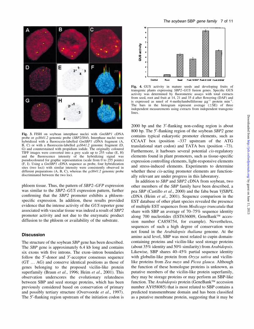
A gsS641.2 genomic probe has also been used in FISH
assays for speci®c labelling of the S64/SBP2 locus
(Fig. 3D, G). The genomic probe cross-hybridized to a
different extent with the two pairs of genes. The intensity
of the hybridizing signals was estimated by a semi-
quantitative method based on the quanti®cation of the
pixel values (8 bits) of each labelling signal of the
digitalized image. The original colour of the images was
converted to a grey scale up to a 255 value for the analysis
of relative density of the labelling signals (Fig. 3E, H). The
density of each signal was three-dimensionally plotted
using software for image analysis, as described in the
Materials and methods (Fig. 3F, I). The values of each
labelling signal were obtained as a function of the highest
peak value. The calculated values grouped the signals in
two pairs of similar intensity. Based on the high homology
of the probe to SBP2, the stronger hybridizing signals may
correspond to the SBP2/S64 alleles, whereas the SBP1
alleles may be represented by the lower intensity spots.
Thus, the gsS641.2 (pgSBP2) genomic fragment probe
seems to be able to discriminate quantitatively between
GmSBP1 and GmSBP2 genes. Collectively, these results
may indicate that soybean SBP is encoded by a small gene
family that is represented by at least two highly conserved
copies in the soybean genome.
Tissue-speci®c expression of the SBP2 promoter
Transgenic plants expressing the SBP2±GUS fusion genes
were examined for promoter activity and tissue-speci®c
GUS activity. Because the expression patterns of
gsS641.1±GUS (±2000pSBP2±GUS) and gsS641.2±GUS
fusion genes were indistinguishable, only the results of the
expression of the ®rst construct (-2000pSBP2±GUS) are
presented. RNA blot analyses have previously demon-
strated that SBP mRNA accumulates predominantly in
immature seeds (Grimes et al, 1992; Overvoorde et al.,
1997). Consistent with this observation, the SPB promoter
directed high levels of GUS activity in transgenic seeds
and in developing fruits from independent transgenic lines
(Fig. 4). By contrast, quantitative measurements of GUS
activity in other organs demonstrated a low level of GUS
expression (Fig. 4, roots and data not shown). Northern
analysis and RT-PCR have detected low levels of SBP
mRNA accumulation in other organs, such as leaves,
seedlings, seed coat, pods, and roots (Grimes et al., 1992;
Heim et al., 2001; and data not shown). In view of this
observation, the SBP promoter activity was histochemic-
ally assayed in leaves, stems and roots. Histochemical
examination of different independently transformed SBP2±
GUS lines indicated that in all the organs analysed (leaf,
stem and root) the expression of the SBP2±GUS fusion was
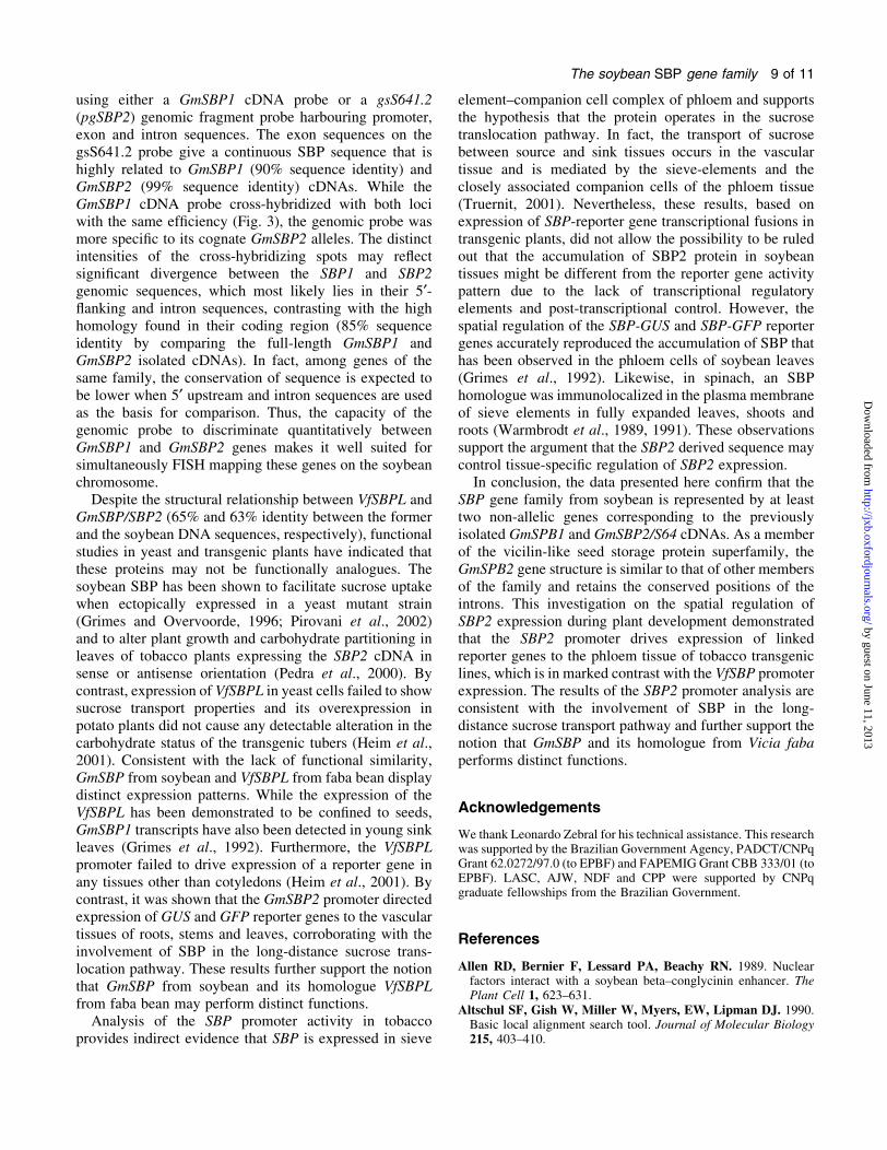
restricted to the vascular tissue (Fig. 5). In the shoot apex,
reporter gene activity was also mostly expressed in
vascular tissues (Fig. 5B) and transverse sections through
the leaf revealed an intense GUS staining associated
exclusively with the vascular bundle (Fig. 5E). Detailed
analysis of leaf sections revealed that the SBP2±GUS
expression was restricted to the phloem (Fig. 5F) with clear
staining of the sieve elements (Fig. 5G). Likewise, in cross-
sections of petiole and stems, high levels of expression
were mostly associated with the phloem, especially the
inner phloem tissues (Fig. 5L, N). An intense GUS staining
was also observed in the vascular cylinder in roots
(Fig. 5R). Microscopic analysis of root sections revealed
that this expression was restricted to the phloem of the
vascular tissue (Fig. 5S). In control assays, GUS activity
was not detected in shoots and roots from transgenic
plants harbouring the promoterless GUS gene (Fig. 5A, D,
K, M, Q).
The tissue-speci®c expression from the SBP2 promoter
was also analysed in transgenic plants containing the green
¯uorescent protein (GFP) cDNA fused to SBP2 upstream
sequences. Analyses of the vascular tissues from trans-
verse sections of untransformed, control leaves (Fig. 5I),
stem (Fig. 5O) and roots (data not shown) demonstrated
that the xylem tissue from tobacco contains very high
levels of yellow ¯uorescent background, whereas the
phloem tissue does not auto¯uoresce. By contrast, micro-
scopic analysis of the vascular tissue from the apical
meristem (Fig. 5C), leaves (Fig. 5H, J), stem (Fig. 5P), and
root (Fig. 5T) of SBP2±GFP transgenic lines clearly
demonstrated a high level of GFP expression in the
Fig. 2. DNA gel blot analysis. Soybean genomic DNA (10 mg) wasdigested with EcoRI (E) and BamHI (B), separated by electrophoresis,transferred to nylon membrane and hybridized with a ¯uorescein-labelled GmSBP2/S64 cDNA sequence. The numbers at the topcorrespond to the relative gene copy number. Migration positions ofmolecular weight standards are indicated on the right in kb.
6 of 11 Contim et al.
by guest on June 11, 2013http://jxb.oxfordjournals.org/
Dow
nloaded from
phloem tissue. Thus, the pattern of SBP2±GFP expression
was similar to the SBP2±GUS expression pattern, further
con®rming that the SBP2 promoter exhibits a phloem-
speci®c expression. In addition, these results provided
evidence that the intense activity of the GUS reporter gene
associated with vascular tissue was indeed a result of SBP2
promoter activity and not due to the enzymatic product
diffusion to the phloem or availability of the substrate.
Discussion
The structure of the soybean SBP gene has been described.
The SBP gene is approximately 6.4 kb long and contains
six exons with ®ve introns. The exon±intron boundaries
follow the 5¢-donor and 3¢-acceptor consensus sequence
(GT ¼ AG) and conserve identical positions as those of
genes belonging to the proposed vicilin-like protein
superfamily (Braun et al., 1996; Heim et al., 2001). This
observation underscores the evolutionary relatedness
between SBP and seed storage proteins, which has been
previously considered based on conservation of primary
and possibly tertiary structure (Overvoorde et al., 1997).
The 5¢-¯anking region upstream of the initiation codon is
2000 bp and the 3¢-¯anking non-coding region is about
800 bp. The 5¢-¯anking region of the soybean SBP2 gene
contains typical eukaryotic promoter elements, such as
CCAAT box (position ±337 upstream of the ATG
translational start codon) and TATA box (position ±73).
Furthermore, it harbours several potential cis-regulatory
elements found in plant promoters, such as tissue-speci®c
expression controlling elements, light-responsive elements
and stress-induced elements. Experiments to determine
whether these cis-acting promoter elements are function-
ally relevant are under progress in this laboratory.
In addition to SBP and SBP2 cDNA from soybean, two
other members of the SBP family have been described, a
pea SBP (Castillo et al., 2000) and the faba bean VfSBPL
cDNA (Heim et al., 2001). Sequence comparison in the
EST database of other plant species revealed the presence
of multiple EST sequences from Medicago truncatula that
share with SBP an average of 70±75% sequence identity
along 700 nucleotides (EST636009, GeneBankÔ acces-
sion number CA858754, for example). Nevertheless,
sequences of such a high degree of conservation were
not found in the Arabidopsis thaliana genome. At the
amino acid level, SBP was most related to cupin domain-
containing proteins and vicilin-like seed storage proteins
(about 35% identity and 50% similarity) from Arabidopsis.
Likewise, SBP shares 40±45% partial sequence identity
with globulin-like protein from Oryza sativa and vicilin-
like proteins from Zea mays and Picea glauca. Although
the function of these homologue proteins is unknown, as
putative members of the vicilin-like protein superfamily,
they may be storage proteins or may perform an SBP-like
function. The Arabidopsis protein (GeneBankÔ accession
number AY058085) that is most related to SBP contains a
predicted transmembrane domain and has been classi®ed
as a putative membrane protein, suggesting that it may be
Fig. 3. FISH on soybean interphase nuclei with GmSBP1 cDNAprobe or gsS641.2 genomic probe (SBP2/S64). Interphase nuclei werehybridized with a ¯uorescein-labelled GmSBP1 cDNA fragment (A,B, C) or with a ¯uorescein-labelled gsS641.2 genomic fragment (D,G) and counterstained with propidium iodide. The originally colouredTIFF images were converted into a grey scale up to 255 value (E, H)and the ¯uorescence intensity of the hybridizing signal waspseudocoloured for graphic representation (scale from 0 to 255 points)(F, I). Using a GmSBP1 cDNA sequence as probe, four hybridizationsites (two loci) with similar intensity were consistently observed indifferent preparations (A, B, C), whereas the gsS641.2 genomic probediscriminated between the two loci.
Fig. 4. GUS activity in mature seeds and developing fruits oftransgenic plants expressing SBP2±GUS fusion genes. Speci®c GUSactivity was determined by ¯uorometric assays with total extractsfrom seed, root and fruit at 14, 21 and 35 d after ¯owering (DAF) andis expressed as nmol of 4-methylumbelliferone mg±1 protein min±1.The bars in the histogram represent average (6SE) of threeindependent measurements using extracts from independent transgeniclines.
The soybean SBP gene family 7 of 11
by guest on June 11, 2013http://jxb.oxfordjournals.org/
Dow
nloaded from
functionally distinct from the vicilin-like seed storage
proteins.
By contrast with Vicia faba SBP that is represented by a
single copy in the faba bean genome, as judged by DNA
gel blot analysis, the soybean SBP is encoded by a small
gene family. These data, based on nuclei in situ
hybridization using cDNA and genomic probes, con®rmed
that the soybean SBP family is represented by at least two
non-allelic genes. These distinct hybridizing loci may
correspond to the previously isolated GmSBP1 (Grimes
et al., 1992) and GmSBP2/S64 cDNAs (Pirovani et al.,
2002), as judged by the intensity of the hybridizing signal
Fig. 5. Tissue-speci®c regulation of SBP2±GUS and SBP2±GFP fusion gene expression in transgenic plants. Photographs of transgenic organsplants harbouring ±2000pSBP2±GUS fusion gene stained for GUS activity or SBP2-GFP fusion gene visualized for GFP ¯uorescence. Theexpression of ±2000pSBP2±GUS fusion gene was determined in longitudinal section through the shoot apex (B); transverse sections of leaf (E, F,G); transverse section of petiole (L); transverse section through the stem (N); root tip (R), and transverse sections of roots (S). The expression ofthe ±2000pSBP2±GFP fusion gene was visualized in a transverse section of the shoot apical meristem (C); transverse sections of leaves (H, J);transverse section through the stem (P), and a transverse section of roots (T). (A), (D), (K), (M), and (Q) correspond to pCAMBIA-transformed,control plants stained for GUS activity. (A) Longitudinal section through the shoot apex, (D) transverse section of leaves, (K) transverse section ofpetiole, (M) transverse section of stems, and (Q) root tip. The transverse section of leaf (I) and transverse section through the stem (O) areoriginated from pCAMBIA-transformed control plants but visualized for ¯uorescent background. Abbreviations: LP, procambium; SAM, shootapical meristem; E, epidermis; VT, vascular tissue; Pa, parenchyma; X, xylem; EP, external phloem; IP, internal phloem; T, trichome; VB,vascular bundle; Ms, mesophyll; P, phloem; ST, sieve tube; SP, sieve plate; AM, apical meristem; VC, vascular cylinder; RC, root cap; PX,primary xylem; PP, primary phloem. Bars in (A), (B), (E), (L), (M), (N), and (S)=200 mm; bars in (D), (K), (Q), and (R)=100 mm; bars in (F), (I),(J), (O), (P), and (T)=50 mm; bars in (C) and (H)=20 mm; bar in (G)=10 mm.
8 of 11 Contim et al.
by guest on June 11, 2013http://jxb.oxfordjournals.org/
Dow
nloaded from
using either a GmSBP1 cDNA probe or a gsS641.2
(pgSBP2) genomic fragment probe harbouring promoter,
exon and intron sequences. The exon sequences on the
gsS641.2 probe give a continuous SBP sequence that is
highly related to GmSBP1 (90% sequence identity) and
GmSBP2 (99% sequence identity) cDNAs. While the
GmSBP1 cDNA probe cross-hybridized with both loci
with the same ef®ciency (Fig. 3), the genomic probe was
more speci®c to its cognate GmSBP2 alleles. The distinct
intensities of the cross-hybridizing spots may re¯ect
signi®cant divergence between the SBP1 and SBP2
genomic sequences, which most likely lies in their 5¢-¯anking and intron sequences, contrasting with the high
homology found in their coding region (85% sequence
identity by comparing the full-length GmSBP1 and
GmSBP2 isolated cDNAs). In fact, among genes of the
same family, the conservation of sequence is expected to
be lower when 5¢ upstream and intron sequences are used
as the basis for comparison. Thus, the capacity of the
genomic probe to discriminate quantitatively between
GmSBP1 and GmSBP2 genes makes it well suited for
simultaneously FISH mapping these genes on the soybean
chromosome.
Despite the structural relationship between VfSBPL and
GmSBP/SBP2 (65% and 63% identity between the former
and the soybean DNA sequences, respectively), functional
studies in yeast and transgenic plants have indicated that
these proteins may not be functionally analogues. The
soybean SBP has been shown to facilitate sucrose uptake
when ectopically expressed in a yeast mutant strain
(Grimes and Overvoorde, 1996; Pirovani et al., 2002)
and to alter plant growth and carbohydrate partitioning in
leaves of tobacco plants expressing the SBP2 cDNA in
sense or antisense orientation (Pedra et al., 2000). By
contrast, expression of VfSBPL in yeast cells failed to show
sucrose transport properties and its overexpression in
potato plants did not cause any detectable alteration in the
carbohydrate status of the transgenic tubers (Heim et al.,
2001). Consistent with the lack of functional similarity,
GmSBP from soybean and VfSBPL from faba bean display
distinct expression patterns. While the expression of the
VfSBPL has been demonstrated to be con®ned to seeds,
GmSBP1 transcripts have also been detected in young sink
leaves (Grimes et al., 1992). Furthermore, the VfSBPL
promoter failed to drive expression of a reporter gene in
any tissues other than cotyledons (Heim et al., 2001). By
contrast, it was shown that the GmSBP2 promoter directed
expression of GUS and GFP reporter genes to the vascular
tissues of roots, stems and leaves, corroborating with the
involvement of SBP in the long-distance sucrose trans-
location pathway. These results further support the notion
that GmSBP from soybean and its homologue VfSBPL
from faba bean may perform distinct functions.
Analysis of the SBP promoter activity in tobacco
provides indirect evidence that SBP is expressed in sieve
element±companion cell complex of phloem and supports
the hypothesis that the protein operates in the sucrose
translocation pathway. In fact, the transport of sucrose
between source and sink tissues occurs in the vascular
tissue and is mediated by the sieve-elements and the
closely associated companion cells of the phloem tissue
(Truernit, 2001). Nevertheless, these results, based on
expression of SBP-reporter gene transcriptional fusions in
transgenic plants, did not allow the possibility to be ruled
out that the accumulation of SBP2 protein in soybean
tissues might be different from the reporter gene activity
pattern due to the lack of transcriptional regulatory
elements and post-transcriptional control. However, the
spatial regulation of the SBP-GUS and SBP-GFP reporter
genes accurately reproduced the accumulation of SBP that
has been observed in the phloem cells of soybean leaves
(Grimes et al., 1992). Likewise, in spinach, an SBP
homologue was immunolocalized in the plasma membrane
of sieve elements in fully expanded leaves, shoots and
roots (Warmbrodt et al., 1989, 1991). These observations
support the argument that the SBP2 derived sequence may
control tissue-speci®c regulation of SBP2 expression.
In conclusion, the data presented here con®rm that the
SBP gene family from soybean is represented by at least
two non-allelic genes corresponding to the previously
isolated GmSPB1 and GmSBP2/S64 cDNAs. As a member
of the vicilin-like seed storage protein superfamily, the
GmSPB2 gene structure is similar to that of other members
of the family and retains the conserved positions of the
introns. This investigation on the spatial regulation of
SBP2 expression during plant development demonstrated
that the SBP2 promoter drives expression of linked
reporter genes to the phloem tissue of tobacco transgenic
lines, which is in marked contrast with the VfSBP promoter
expression. The results of the SBP2 promoter analysis are
consistent with the involvement of SBP in the long-
distance sucrose transport pathway and further support the
notion that GmSBP and its homologue from Vicia faba
performs distinct functions.
Acknowledgements
We thank Leonardo Zebral for his technical assistance. This researchwas supported by the Brazilian Government Agency, PADCT/CNPqGrant 62.0272/97.0 (to EPBF) and FAPEMIG Grant CBB 333/01 (toEPBF). LASC, AJW, NDF and CPP were supported by CNPqgraduate fellowships from the Brazilian Government.
References
Allen RD, Bernier F, Lessard PA, Beachy RN. 1989. Nuclearfactors interact with a soybean beta±conglycinin enhancer. ThePlant Cell 1, 623±631.
Altschul SF, Gish W, Miller W, Myers, EW, Lipman DJ. 1990.Basic local alignment search tool. Journal of Molecular Biology
215, 403±410.
The soybean SBP gene family 9 of 11
by guest on June 11, 2013http://jxb.oxfordjournals.org/
Dow
nloaded from
Alvim FC, Carolino SMB, Cascardo JCM, Nunes CC, MartinezCA, Otoni WC, Fontes EPB. 2001. Enhanced accumulation ofBiP in transgenic plants confers tolerance to water stress. PlantPhysiology 126, 1042±1054.
Barker L, KuÈhn C, Weise A, Schulz A, Gebhardt C, Hirner B,Hellmann H, Schulze W, Ward JM, Frommer WB. 2000.SUT2, a putative sucrose sensor in sieve elements. The Plant Cell12, 1153±1164.
Braun H, Czihal A, Shutov AD, BaÈumlein H. 1996. A vicilin-likeseed protein of cycads: similarity to sucrose binding proteins.Plant Molecular Biology 31, 35±44.
BuÈrkle L, Hibberd JM, Quick WP, KuÈhn C, Hirner B,Frommer WB. 1998. The H+-sucrose cotransporter NtSUT1 isessential for sugar export from tobacco leaves. Plant Physiology118, 59±68.
Buzeli RAA, Cascardo JCM, Rodrigues LAZ, Andrade MO,Loureiro ME, Otoni WC, Fontes EPB. 2002. Tissue-speci®cregulation of Bip/Grp78 genes: a cis-acting regulatory domain isrequired for BiP promoter activity in plant meristems. Plant
Molecular Biology 50, 757±771.Carvalho CR, Saraiva LS. 1997. High-resolution HKG-banding inmaize mitotic chromosomes. Journal of Plant Research 110,417±420.
Castillo J, Rodrigo MI, MaÂrques JA, ZuÂnigat AÂ , Franco L. 2000.A pea nuclear protein that is induced by dehydration belongs tothe vicilin superfamily. European Journal of Biochemistry 267,2156±2165.
DeluÂ-Filho N, Pirovani CP, Pedra JHF, Matrangolo FSV,MaceÃdo JNA, Otoni WC, Fontes EPB. 2000. A sucrose bindingprotein homologue from soybean affects sucrose uptake intransgenic tobacco suspension-cultured cells. Plant Physiology
and Biochemistry 38, 353±361.FroÈmmer WB, Sonnewald U. 1995. Molecular analysis of carbonpartitioning in solanaceous species. Journal of Experimental
Botany 46, 587±607.Fujiwara T, Beachy RN. 1994. Tissue-speci®c and temporalregulation of a beta-conglycinin gene: roles of the RY repeat andother cis-acting elements. Plant Molecular Biology 24, 261±272.
Grimes HD, Overvoorde PJ, Hipp K. 1992. A 62 kD sucrosebinding protein is expressed and localized in tissues activelyengaged in sucrose transport. The Plant Cell 4, 1561±1574.
Grimes HD, Overvoorde PJ. 1996. Functional characterization ofsucrose binding protein-mediated sucrose uptake in yeast. Journalof Experimental Botany 47, 1217±1222.
Heim U, Wang Q, Kurz T, et al. 2001. Expression patterns andsubcelular localization of a 52 kDa sucrose-binding proteinhomologue of Vicia faba (VfSBPL) suggest different functionsduring development. Plant Molecular Biology 47, 461±474.
Jefferson RA, Kavanagh TA, Bevan MW. 1987. Gus fusions: b-glucuronidase as a sensitive and versatile gene fusion marker inhigher plants. EMBO Journal 6, 3901±3907.
Katagiri F, Lam E, Chua NH. 1989. Two tobacco DNA-bindingproteins with homology to the nuclear factor CREB. Nature 340,727±730.
KuÈhn C, Quick WP, Schulz A, Sonnewald U, Frommer WB.1996. Companion cell-speci®c inhibition of the potato sucrosetransporter SUT1. Plant, Cell and Environment 19, 1115±1123.
Lalonde SL, Boles E, Hellmann H, Barker L, Patrick JW,Frommer WB, Ward JM. 1999. The dual function of sugarcarriers: transport and sugar sensing. The Plant Cell 11, 707±726.
Lemoine R. 2000. Sucrose transporters in plants: update onfunction and structure. Biochimica et Biophysica Acta 1, 246±262.
Lin W, Schmitt MR, Hitz WD, Giaquinta RT. 1984. Sugartransport into protoplasts isolated from developing soybeancotyledons. Plant Physiology 75, 936±940.
Maynard JW, Lucas WJ. 1982. A re-analysis of the two-component phloem loading system in Beta vulgaris. Plant
Physiology 69, 734±739.McCabe DE, Swain WF, Martinell BJ, Christou P. 1988. Stabletransformation of soybean (Glycine max) by particlebombardment. Biotechnology 6, 923±926.
Overvoorde PJ, Chao WS, Grimes HD. 1997. A plasmamembrane sucrose-binding protein that mediates sucrose uptakeshares structural and sequence similarity with seed storageproteins but remains functionally distinct. Journal of BiologicalChemistry 272, 15898±15904.
Overvoorde PJ, Frommer WB, Spencer D. 1996. A soybeansucrose binding protein independently mediates non-saturablesucrose uptake in yeast. The Plant Cell 8, 271±280.
Pedra JHF, DeluÂ-Filho N, Pirovani CP, Contim LAS, DeweyRE, Otoni WC, Fontes EPB. 2000. Antisense and senseexpression of a sucrose binding protein homologue gene fromsoybean in transgenic tobacco affects plant growth andcarbohydrate partitioning in leaves. Plant Science 152, 87±98.
Pirovani CP, MaceÃdo JNA, Contim LAS, Matrangolo FSV,Loureiro, ME, Fontes EPB. 2002. A sucrose binding proteinhomologue from soybean exhibits GTP-binding activity thatfunctions independently of sucrose transport activity. EuropeanJournal of Biochemistry 269, 3998±4008.
Rasband W. 1997. Image SXM 1.61. A public domain software forimage analysis, written at the US National Institute of Health,extensions by Steve Barret and available from the Internet byanonymous ftp from: zippy.nimh.nih.gov.
Riesmeier JW, Willmitzer L, Frommer WB. 1992. Isolation andcharacterization of a sucrose carrier cDNA from spinach byfuntional expression in yeast. EMBO Journal 11, 4705±4713.
Riesmeier JW, Hirner B, Frommer WB. 1993. Expression of thesucrose transporter from potato correlates with the sink-to-sourcetransition in leaves. The Plant Cell 5, 1591±1598.
Riesmeier JW, Willmitzer L, Frommer WB. 1994. Evidence foran essential role of the sucrose transporter in phloem loading andassimilate partitioning. EMBO Journal 13, 1±7.
Ripp KG, Viitanen PV, Hitz WD, Franceschi VR. 1988.Identi®cation of a membrane protein associated with sucrosetransport into cells of developing soybean cotyledons. PlantPhysiology 88, 1435±1445.
Roberts CS, Rajagopal S, Smith LA, et al. 1996. A comprehensive
set of modular vectors for advanced manipulations and ef®cient
transformation of plants by both Agrobacterium and direct DNAuptake methods. Canberra, Australia: Center for the Applicationof Molecular Biology to International Agriculture (CAMBIA).
Sambrook J, Fritsch EF, Maniatis EFT. 1989. MolecularcloningÐa laboratory manual, 2nd edn. New York: ColdSpring Harbor Laboratory Press.
Stitt M. 1996. Plasmodesmata play an essential role in sucroseexport from leaves: a step toward an integration of metabolicbiochemistry and cell biology. The Plant Cell 8, 565±571.
Terzaghi WB, Cashmore AR. 1995. Light-regulated transcription.Annual Review of Plant Physiology and Plant Molecular Biology
46, 445±474.Truernit E. 2001. Plant physiology: the importance of sucrosetransporters. Current Biology 11, 169±194.
Urao T, Yamaguchi-Shinozaki K, Urao S, Shinosaki K. 1993. AnArabidopsis myb homologue is induced by dehydration stress andits gene product binds to the conserved MYB recognitionsequence. The Plant Cell 5, 1529±1539.
Warmbrodt RD, Buckhout TJ, Hitz WD. 1989. Localization of aprotein, immunologically similar to a sucrose-binding proteinfrom developing soybean cotyledons, on the plasma membrane ofsieve-tube members of spinach leaves. Planta 180, 105±115.
Warmbrodt RD, Vanderwoude WJ, Hitz WD. 1991. Studies on
10 of 11 Contim et al.
by guest on June 11, 2013http://jxb.oxfordjournals.org/
Dow
nloaded from
the localization of a protein, immunologically similar to a 62-kilodalton sucrose-binding protein isolated from developingsoybean cotyledons, in the shoot and root of spinach. NewPhytologist 118, 501±511.
Weise A, Barker L, KuÈhn C, Lalonde S, Buschmann H,Frommer WB, Ward JM. 2000. A new subfamily of sucrosetransporters, SUT4, with low af®nity/high capacity localized inenucleate sieve elements of plants. The Plant Cell 12, 1345±1355.
Williams LE, Lemoine R, Sauer N. 2000. Sugar transporters inhigher plantsÐa diversity of roles and complex regulation.Trends in Plant Science 5, 283±290.
Wu C, Washida H, Onodera Y, Harada K, Takaiwa F. 2000.Quantitative nature of the prolamin-box, ACGT and AACA
motifs in a rice glutelin gene promoter: minimal cis-element
requirements for endosperm-speci®c gene expression. The Plant
Journal 23, 425±421.Yu D, Chen C, Chen Z. 2001. Evidence for an important role of
WRKY DNA binding proteins in the regulation of NPR1 gene
expression. The Plant Cell 13, 1527±1540.Zhou DX. 1999. Regulatory mechanism of plant gene transcription
by GT-elements and GT-factors. Plant Science 4, 210±214.
The soybean SBP gene family 11 of 11
by guest on June 11, 2013http://jxb.oxfordjournals.org/
Dow
nloaded from