The Smallest Active Carbamoyl Phosphate Synthetase Was Identified in the Human Gut Archaeon...
13
Fax +41 61 306 12 34 E-Mail [email protected] www.karger.com Research Article J Mol Microbiol Biotechnol 2012;22:287–299 DOI: 10.1159/000342520 The Smallest Active Carbamoyl Phosphate Synthetase Was Identified in the Human Gut Archaeon Methanobrevibacter smithii Elena Popa a Nirosha Perera b Csaba Z. Kibédi-Szabó a Hedeel Guy-Evans b David R. Evans c Cristina Purcarea a a Department of Microbiology, Institute of Biology Bucharest, Romanian Academy, Bucharest, Romania; b Department of Chemistry, Eastern Michigan University, Ypsilanti, Mich., and c Department of Biochemistry and Molecular Biology, Wayne State University School of Medicine, Detroit, Mich., USA sponds to one of the two synthetase domains of the full- length CPSase that catalyze the ATP-dependent phosphory- lations involved in the three-step synthesis of carbamoyl phosphate. This protein represents the smallest naturally oc- curring active CPSase characterized thus far. The small M. smithii CPSase appears to be specialized for carbamoyl phos- phate metabolism in methanogens. Copyright © 2012 S. Karger AG, Basel Introduction In recent years, many studies [Dridi et al., 2011; Horz and Conrads, 2010; Nava et al., 2011; Plottel et al., 2011] have focused on the involvement of the human microbi- ome in human health and disease. While archaea colonize several different anaerobic niches within the human body [Belay et al., 1988, 1990], there is a single predominant spe- cies, Methanobrevibacter smithii, present in the human gut [Miller et al., 1982]. First isolated from feces, this metha- nogenic organism is a strict anaerobe that grows on a mix- ture of hydrogen and carbon dioxide [Balch et al., 1979]. Samuel and Gordon [2006] made the remarkable dis- covery that a synergic interaction in the colon between M. smithii and Bacteroides thetaiotaomicron, the major gut prokaryote, appears to have an important role in control- ling host obesity [Samuel and Gordon, 2006]. Complex di- Key Words Carbamoyl phosphate synthetase Ancestral form Methanobrevibacter smithii Intestinal archaea Evolution Obesity Abstract The genome of the major intestinal archaeon Methanobrevi- bacter smithii contains a complex gene system coding for carbamoyl phosphate synthetase (CPSase) composed of both full-length and reduced-size synthetase subunits. These ammonia-metabolizing enzymes could play a key role in controlling ammonia assimilation in M. smithii, affecting the metabolism of gut bacterial microbiota, with an impact on host obesity. In this study, we isolated and characterized the small (41 kDa) CPSase homolog from M. smithii. The gene was cloned and overexpressed in Escherichia coli, and the re- combinant enzyme was purified in one step. Chemical cross- linking and size exclusion chromatography indicated a ho- modimeric/tetrameric structure, in accordance with a dimer- based CPSase activity and reaction mechanism. This small enzyme, MS-s, synthesized carbamoyl phosphate from ATP, bicarbonate, and ammonia and catalyzed the same ATP-de- pendent partial reactions observed for full-length CPSases. Steady-state kinetics revealed a high apparent affinity for ATP and ammonia. Sequence comparisons, molecular mod- eling, and kinetic studies suggest that this enzyme corre- Published online: October 27, 2012 Dr. Cristina Purcarea Department of Microbiology Institute of Biology Bucharest, Romanian Academy 296 Splaiul Independentei, RO–060031 Bucharest (Romania) E-Mail cristina.purcarea @ ibiol.ro © 2012 S. Karger AG, Basel 1464–1801/12/0225–0287$38.00/0 Accessible online at: www.karger.com/mmb Downloaded by: Max Planck Society 134.76.223.30 - 1/23/2014 9:54:49 AM
-
Upload
independent -
Category
Documents
-
view
2 -
download
0
Transcript of The Smallest Active Carbamoyl Phosphate Synthetase Was Identified in the Human Gut Archaeon...

Fax +41 61 306 12 34E-Mail [email protected]
Research Article
J Mol Microbiol Biotechnol 2012;22:287–299 DOI: 10.1159/000342520
The Smallest Active Carbamoyl Phosphate Synthetase Was Identified in the Human Gut Archaeon Methanobrevibacter smithii
Elena Popa a Nirosha Perera b Csaba Z. Kibédi-Szabó a Hedeel Guy-Evans b
David R. Evans c Cristina Purcarea a
a Department of Microbiology, Institute of Biology Bucharest, Romanian Academy, Bucharest , Romania; b Department of Chemistry, Eastern Michigan University, Ypsilanti, Mich. , and c Department of Biochemistry and Molecular Biology, Wayne State University School of Medicine, Detroit, Mich. , USA
sponds to one of the two synthetase domains of the full-length CPSase that catalyze the ATP-dependent phosphory-lations involved in the three-step synthesis of carbamoyl phosphate. This protein represents the smallest naturally oc-curring active CPSase characterized thus far. The small M. smithii CPSase appears to be specialized for carbamoyl phos-phate metabolism in methanogens.
Copyright © 2012 S. Karger AG, Basel
Introduction
In recent years, many studies [Dridi et al., 2011; Horz and Conrads, 2010; Nava et al., 2011; Plottel et al., 2011] have focused on the involvement of the human microbi-ome in human health and disease. While archaea colonize several different anaerobic niches within the human body [Belay et al., 1988, 1990], there is a single predominant spe-cies, Methanobrevibacter smithii, present in the human gut [Miller et al., 1982]. First isolated from feces, this metha-nogenic organism is a strict anaerobe that grows on a mix-ture of hydrogen and carbon dioxide [Balch et al., 1979].
Samuel and Gordon [2006] made the remarkable dis-covery that a synergic interaction in the colon between M. smithii and Bacteroides thetaiotaomicron, the major gut prokaryote, appears to have an important role in control-ling host obesity [Samuel and Gordon, 2006]. Complex di-
Key Words
Carbamoyl phosphate synthetase � Ancestral form � Methanobrevibacter smithii � Intestinal archaea � Evolution � Obesity
Abstract
The genome of the major intestinal archaeon Methanobrevi-bacter smithii contains a complex gene system coding for carbamoyl phosphate synthetase (CPSase) composed of both full-length and reduced-size synthetase subunits. These ammonia-metabolizing enzymes could play a key role in controlling ammonia assimilation in M. smithii, affecting the metabolism of gut bacterial microbiota, with an impact on host obesity. In this study, we isolated and characterized the small (41 kDa) CPSase homolog from M. smithii. The gene was cloned and overexpressed in Escherichia coli , and the re-combinant enzyme was purified in one step. Chemical cross-linking and size exclusion chromatography indicated a ho-modimeric/tetrameric structure, in accordance with a dimer-based CPSase activity and reaction mechanism. This small enzyme, MS-s, synthesized carbamoyl phosphate from ATP, bicarbonate, and ammonia and catalyzed the same ATP-de-pendent partial reactions observed for full-length CPSases. Steady-state kinetics revealed a high apparent affinity for ATP and ammonia. Sequence comparisons, molecular mod-eling, and kinetic studies suggest that this enzyme corre-
Published online: October 27, 2012
Dr. Cristina Purcarea Department of Microbiology Institute of Biology Bucharest, Romanian Academy 296 Splaiul Independentei, RO–060031 Bucharest (Romania) E-Mail cristina.purcarea @ ibiol.ro
© 2012 S. Karger AG, Basel1464–1801/12/0225–0287$38.00/0
Accessible online at:www.karger.com/mmb
Dow
nloa
ded
by:
Max
Pla
nck
Soc
iety
13
4.76
.223
.30
- 1/
23/2
014
9:54
:49
AM

Popa /Perera /Kibédi-Szabó /Guy-Evans /Evans /Purcarea
J Mol Microbiol Biotechnol 2012;22:287–299 288
etary polysaccharides that cannot be metabolized by the human host are digested by enzymes in the gut microbi-ome [Stams, 1994]. Bacterial fermentation of polysaccha-rides yields short-chain fatty acids that provide as much as 10% of the daily caloric intake of the host. Archaeal meth-anogenesis facilitates bacterial fermentation by preventing the accumulation of H 2 gas and other end products that would otherwise inhibit the process. Analysis of the cecal content of gnotobiotic mice colonized with M. smithii alone or together with B. thetaiotaomicron revealed that many genes that participate in the metabolism of these end products are upregulated when both organisms are pres-ent in the gut [Samuel et al., 2007]. Several lines of evi-dence suggest that obesity is associated with elevated levels of intestinal methanogenic archaea [Million et al., 2011]. Consequently, M. smithii is considered to be a potential therapeutic target in the treatment of obesity [Buck and Hansen, 2007]. The principle source of nitrogen in M. smithii is ammonia, resulting from the degradation of amino acids by the host or intestinal bacteria. M. smithii competes with B. thetaiotaomicron for the available am-monia, and when both organisms populate the gut, the transcription of several enzymes involved in ammonia transport and assimilation is upregulated in M. smithii . Although there have been numerous microbiological and physiological studies of human gut microbiota, little is known about the metabolism and key enzymes of the ma-jor archaeon in the gastrointestinal tract, M. smithii.
Carbamoyl phosphate synthetase (CPSase; EC 6.3.5.5) plays a major role in ammonia assimilation in all organ-isms. The enzyme catalyzes the synthesis of carbamoyl phosphate (CP), a common precursor of pyrimidine nu-cleotides, arginine, and, in terrestrial vertebrates, urea, where it represents the major means of eliminating am-monia. CPSases are multisubunit or multidomain pro-teins. Escherichia coli CPSase is composed of a 40-kDa glutaminase (CarA, GLN) subunit which hydrolyzes glu-tamine and transfers ammonia to a 120-kDa synthetase (CarB, SYN) subunit [Trotta et al., 1971]. The SYN sub-unit consists of two homologous domains having a near-ly identical tertiary fold and active site residues that were thought to have evolved by ancestral duplication and fu-sion [Nyunoya and Lusty, 1983]. The amino half of the subunit, designated CPS.A, catalyzes the synthesis of car-bamate from ATP, bicarbonate, and ammonia, while the carboxyl half, CPS.B, catalyzes CP synthesis from carba-mate and a second ATP [Alonso and Rubio, 1995; Guy and Evans, 1996; Post et al., 1990]. E. coli CPSase GLN and SYN subunits associate to form heterodimers and tetramers [Anderson, 1986]. The partial reactions cata-
lyzed by CPSase, first demonstrated for the E. coli enzyme [Anderson and Meister, 1965; Meister, 1989], are thought to occur universally in nearly all organisms:
CPS.A: (1) ATP + bicarbonate ] carboxy phosphate + ADP; (2) carboxy phosphate + NH 3 ] carbamate + Pi CPS.B: (3) ATP + carbamate ] CP + ADP
Each of the major CPSase domains is comprised of three subdomains designated A1, A2, A3 and B1, B2, B3 [Guillou et al., 1989; Guy and Evans, 1996; Post et al., 1990; Rubio et al., 1991]. The X-ray structure of E. coli CPSase [Thoden et al., 1997] showed that the A1-A2 and B1-B2 subdomains have nearly identical tertiary folds. A2 and B2 are catalytic subdomains, catalyzing reactions 1–2 and 3, respectively [Guy et al., 1997; Post et al., 1990; Thoden et al., 1997]. The A3 and B3 subdomains have distinctly different tertiary structures and functions. B3 is a regulatory subdomain that has been adapted to bind allosteric ligands [Rubio et al., 1991]. Remarkably, the ac-tive sites on the GLN, CPS.A and CPS.B domain are con-nected by a 96-Å-long intramolecular tunnel that seques-ters the labile intermediates within the E. coli enzyme complex [Thoden et al., 1997].
Although the structural organization of CPSases is very diverse, all are composed of homologous domains and subdomains and catalyze the same series of reac-tions. Generally, bacteria possess a single CPSase that cat-alyzes the formation of CP used for both arginine and pyrimidine nucleotide biosyntheses, while enteric and gram-positive bacteria contain two CPSases, each spe-cific for one of these two metabolic pathways [Paulus and Switzer, 1979]. These enzymes are comprised of homolo-gous SYN subunits of comparable size [Yang et al., 1997]. Most CPSases have a full-length SYN subunit consisting of fused CPS.A-CPS.B domains, but there are several or-ganisms, including the thermophilic archaeon Methano-caldococcus jannaschii [Bult et al., 1996] and the hyper-thermophilic eubacterium Aquifex aeolicus [Ahuja et al., 2001], that have a split carB gene encoding separate poly-peptides corresponding to CPS.A and CPS.B domains. The only known exception is a 34-kDa carbamate kinase-like CPSase of an entirely different sequence and tertiary structure found in the hyperthermophilic archaeon Py-rococcus furiosus [Durbecq et al., 1997; Legrain et al., 1995; Marina et al., 1998; Uriarte et al., 2001], and in Py-rococcus abyssi [Purcarea et al., 1996, 2001].
The genome of M. smithii contains carA and carB genes coding for a 40-kDa GLN subunit and a 118-kDa SYN subunit (MS-l, SYN1), respectively, that are similar to the CPSase subunits found in most bacteria [Popa et
Dow
nloa
ded
by:
Max
Pla
nck
Soc
iety
13
4.76
.223
.30
- 1/
23/2
014
9:54
:49
AM

CPSase from M. smithii J Mol Microbiol Biotechnol 2012;22:287–299 289
al., 2009]. Surprisingly, the genome also contains two genes coding for much smaller proteins that are homolo-gous to the CPSase synthetase domain. One of these pro-teins (SYN2) consists of 391 residues (45 kDa) with a se-quence that resembles a truncated CPS.B domain, but it lacks active site residues that are known to participate in substrate binding and catalysis. Therefore, it is likely to be encoded by an inactive pseudogene. The second small CPSase homolog (MS-s, SYN3) is also homologous to a part of CPS.B and consists of 367 residues (41 kDa) in-cluding the residues implicated in catalysis [Popa et al., 2009]. Prior to this report it was not known whether this protein has catalytic activity or is an inactive remnant of an unproductive gene duplication event.
We have isolated and characterized the small M. smithii CPSase MS-s, showing that it is catalytically ac-tive, synthesizing CP from NH 3 , 2 ATP, and bicarbonate by the same mechanism utilized by the large CPSases. This is the smallest functional CPSase discovered thus far and may more closely resemble the putative ancestral ki-nase that gave rise to this diverse family of enzymes.
Results and Discussion
Sequence Analysis The large SYN subunit of the CPSase from the metha-
nogens M. smithii (MS-l), Methanothermobacter thermo-autotrophicus (MT-l), and Methanosphaera stadtmanae (MST-l) are 70–73% identical to one another ( fig. 1 b). The alignment of the E. coli CPSase SYN subunit with the
large CPSases MS-l, MT-l, and MST-l gave sequence iden-tities of 50–53%, indicating that the CPSases from E. coli and methanogens are closely related to each other and to the SYN subunit of most prokaryotic and eukaryotic or-ganisms.
The small CPSase from M. smithii, MS-s, is homolo-gous to the CPS.B subdomain of E. coli but is significant-ly smaller, i.e. 367 versus 513 residues, respectively ( fig. 1 a), representing about a third of the full-length sub-unit (1,073 residues). Based on the deduced amino acid sequence, the protein has a calculated molecular mass of 41,403 Da and an isoelectric point of 4.62. MS-s aligns with residues 560–947 of the E. coli SYN subunit corre-sponding to the B1 and B2 subdomains of CPS.B [Goillou et al., 1989] ( fig. 1 a) with a score of 20.1% sequence iden-tity ( fig. 1 b) and therefore lacks the B3 subdomain at the carboxyl end of the polypeptide.
MS-s is also homologous to the E. coli CPS.A do-main, as expected, since the CPS.A and CPS.B domains of the E. coli enzyme are quite similar to one another (19.3% identity, 34.5% similarity). However, the align-ment score of MS-s is somewhat higher for CPS.B (20.1% identity, 40.1% similarity) than for CPS.A, and the mod-eling program selected CPS.B as the best structur-al template. By analogy to the function of B3 in other CPSases, which have binding sites for both allosteric ac-tivators and inhibitors [Fresquet et al., 2000; Goillou et al., 1989; Guy and Evans, 1996; Rubio et al., 1991], the lack of this domain in MS-s suggests that this enzyme is unregulated.
B1 B2
EC (MS-l , MT-l, MST-l)
MS-s (MT-s, MST-s)
GLN SYN
B3A1 A2 A3 B1 B2
CPS.A CPS.B
a
MST-s MT-s MS-l MST-l MT-l EC PA % identity
48.1 47.6 23.5 24.2 19.1 20.1 11.5 MS-s
46.9 24.3 25.7 14.5 25.3 13.3 MST-s
27.0 25.9 21.2 21.8 14.5 MT-s
73.5 73.1 52.0 5.5 MS-l
70.7 50.3 6.2 MST-l
52.8 8.3 MT-l
5.8 ECb
Fig. 1. MS-s sequence identity and domain structure. a Domain and subdomain orga-nization of MS-s and the full-length SYN subunits. The full-length SYN domain consists of homologous CPS.A and CPS.B domains, each of which are comprised of subdomains designated A1, A2, A3 andB1, B2, B3, respectively, of the E. coli SYN subunit. b Protein sequence identity (%) was calculated by pair-wise alignment of CPSase synthetase subunits, as indicated in Experimental Procedures. The proteins analyzed in both panels are: M. smithii small (MS-s) and large (MS-l) SYN; M. thermoautotrophicus small (MT-s) and large (MT-l) SYN, M. stadtmanae small (MST-s) and large (MST-l) SYN; E. coli (EC) SYN, and P. abyssi (PA) CK-likeCPSase.
Dow
nloa
ded
by:
Max
Pla
nck
Soc
iety
13
4.76
.223
.30
- 1/
23/2
014
9:54
:49
AM

Popa /Perera /Kibédi-Szabó /Guy-Evans /Evans /Purcarea
J Mol Microbiol Biotechnol 2012;22:287–299 290
Open reading frames encoding large and small puta-tive CPSases found in M. smithii are also present in the genomes of the thermophilic archaeon M. thermoauto-trophicus and in the human intestinal archaeon M. stadt-manae ; therefore, this unusual complement of CPSase genes may represent a common feature of methanogens. The overall sequences of the small CPSases from these three methanogens are well conserved (46.9–48.1% iden-tity) and are much less similar to the synthetase domain of E. coli CPSase (20.1–25.3% identity).
Moreover, the deduced amino acid sequence of the small M. smithii CPSase has a limited overall identity score (19.1–24.2%) with the corresponding SYN subunits of full-length CPSases from E. coli and methanogens, but an appreciably higher identity score (47.6–48.1%) with the small CPSases from methanogens ( fig. 1 b). The large SYN subunit of the CPSase from the methanogens M. smithii (MS-l), M. thermoautotrophicus (MT-l), and M. stadtmanae (MST-l) are 70–73% identical to one another ( fig. 1 b). The alignment of the E. coli CPSase SYN subunit with the large CPSases MS-l, MT-l, and MST-l gave se-quence identities of 50–53%, indicating that the full-length CPSases from E. coli and methanogens are closely related to each other and to the SYN subunit of most pro-karyotic and eukaryotic organisms.
Since both MS-s and the CK-like CPSases are small CP-synthesizing enzymes having a molecular mass of 41 kDa and 33 kDa, respectively, we investigated the possi-bility that MS-s may be more closely related to the CK-like CPSases than to the larger forms of the SYN CPSase subunits found in most bacteria. However, while the per-cent identity between MS-s and E. coli CPSase is relative-
ly low (20.1%), the percent identity between the MS-s and P. abyssi CK-CPSase is only 11.5%. This difference is highly significant at these low levels of sequence similar-ity. Four randomized versions of the P. abyssi CK-CPSase sequence were generated of identical in size and amino acid composition but completely unrelated amino acid sequences. Sequence alignments yielded a percent iden-tity between MS-s and the four randomized P. abyssi pro-teins of 7.4, 13.0, 14.1, and 15.1%. This analysis suggests that MS-s and P. abyssi CPSase matches are coincidental and that the proteins are not homologs. This interpreta-tion was reinforced by the conservation of E. coli CPSase active site residues and the kinetic studies describedbelow.
The calculated phylogenetic tree ( fig. 2 ) illustrates the relationship between the CPSase SYN subunit from methanogens, E. coli and other organisms belonging to all three domains of organisms, Bacteria, Archaea, and Eukarya. The small CPSases from the methanogens were the first to diverge, forming a separate branch. These pro-teins are more distantly related to the other bacterial, ar-chaeal, and eukaryotic CPSases. Distinct branches clus-ter the eukaryotic CPSases from yeast (SC) and mammals (CAD), the archaeal large CPSases, and the bacterial full-length CPSases. One unanticipated result is that CPSase from A. aeolicus (AA), an organism of ancient lineage which has a split gene encoding separate CPS-A and CPS-B polypeptides [Ahuja et al., 2001], is a close relative of E. coli CPSase and other bacterial CPSases. Interestingly, the large methanogenic CPSases cluster together forming a separate lineage distinct from other bacterial and ar-chaeal enzymes. The carbamate kinase-like CPSase from
MethanogensShort CPSases
CK-like CPSase
ArchaeaLarge CPSases
EukaryotesLarge multifunctional CPSases
MethanogensLarge CPSases
BacteriaLarge CPSases
PF
PT
SA
SC
CAD
MJ
MST-l
MT-l
MS-l
AA
HP
ECMT-s
MST-s
MS-s
PA
Fig. 2. Phylogenetic tree of CPS SYN sub-units. The phylogenetic tree was con-structed as indicated in Experimental Pro-cedures, using the amino acid sequences of CPS SYN subunits or domains from the methanogens M. smithii small (MS-s) and large (MS-l), M. thermoautotrophicus small (MT-s) and large (MT-l), M. stadt-manae small (MST-s) and large (MST-l), M. jannaschii (MJ), A. aeolicus (AA), E. coli (EC), Helicobacter pylori (HP), the ar-chaea Picrophilus torridus (PT), Sulfolobus acidocaldarius (SA), P. furiosus (PF), P.abyssi (PA) CK-like CPSase, Saccharomy-ces cerevisiae (SC) pyrimidine-specific en-zyme, and the hamster pyrimidine biosyn-thetic multifunctional protein (CAD).
Dow
nloa
ded
by:
Max
Pla
nck
Soc
iety
13
4.76
.223
.30
- 1/
23/2
014
9:54
:49
AM

CPSase from M. smithii J Mol Microbiol Biotechnol 2012;22:287–299 291
P. abyssi (PA) [Purcarea et al., 2001] is located on a distant branch of the phylogenetic tree, consistent with its dis-tinctive sequence and structure.
The residues involved in substrate binding and cataly-sis in MS-s were identified based on X-ray structural analysis of E. coli CPSase with a nonhydrolyzable ATP analog [Thoden et al., 1999]. The alignment of M. smithii MS-s with the E. coli CPS.B and CPS.A domains ( fig. 3 ) showed that all of the putative active site residues from one or the other domain have been conserved. The only exception is Arg848 in E. coli CPS.B that has been re-placed in the archaeal enzyme by Gly274.
All of the active site residues identified in the X-ray structure of E. coli CPSase with a nonhydrolyzable ATP analog bond [Thoden et al., 1999] are located between residues 129–306 of the A2 subdomain and 677–848 of the B2 subdomain ( fig. 3 ). Sequence alignment showed that of the 12 active site residues in CPS.A, which catalyze carbamate formation (reactions 1 and 2), all are con-served in the corresponding positions in CPS.B, which catalyzes CP synthesis (reaction 3). This high degree of conservation is consistent with the similarity of the reac-tions catalyzed by the two domains: ATP-dependent phosphorylations of bicarbonate and carbamate. The
121 DAIDKAEDRRRFDVAMKKIGLETARSGIAHTMEEALAVAADVGFPCIIRPSFTMGGSGGGIAYN 129 169 175/6
97 RAVRLTSDKIKTKEFYNEIGVPTPQYQIL--AKDDFESKLKMEFPVVLKQGQGQGGKDIKVAES 107 145 151/2 667 DAIDRAEDRERFQHAVERLKLKQPANATVTTIEMAVEKAKEIGYPLVVRPSYVLGGRAMEIVYD 677 715 721/2 185 REEFEEICARGLDLSPTKELLIDES-LIGWKEYEMEVVRDKNDNCIIVCSIENFDAM-GIHTGD 208 210 215 241 243 161 LDDVKEYFEE-------FDHALCEKFIEGS-EISIEVLGYNGEYVPLSPIYKGETTLEGIHPLN 177/8 180 184 211 213 731 EADLRRYFQTAVSVSNDAPVLL-DHFLDDAVEVDVDAI-CDGEMVLIGGIMEHIEQA-GVHSGD 753/4 756 761 786 788 248 SITVAPAQTLTDKEYQIMRNASMAVLREIGVETGGSNVQFAVNPKNGRLIVIEMNPRVSRSSAL 285 299 301 306 218 KIKTAPCLVEGLDNNLVQRTAYKVAKNLGSD--GIFEMDFMFSKDEQQLYAIEVNTRPNGTRYL
253 267 269 274
793 SACSLPAYTLSQEIQDVMRQQVQKLAFELQVR-GLMNVQFAV--KNNEVYLIEVNPRAARTVPF 829 841 843 848
E. coli M. smithii Functional Interactions CPS.A CPS.B MS-s
Arg129 (Arg677) Lys107 and phosphates AMP-PNP Arg169 Arg715 Lys145 phosphate of AMP-PNP Gly175 Gly721 Gly151 phosphate of AMP-PNP Gly176 Gly722 Gly152 phosphate of AMP-PNP Glu208 Asp753 Glu177 amino group of adenine ring (Ser209 ) His754 Lys178 amino group of adenine ring Leu210 Leu756 Ile180 purine ring Glu215 Glu761 Glu184 2’ and 3’ hydroxyl of ribose Gly241 Gly786 Gly211 2’ hydroxyl of ribose Gln285 Gln829 Asp253* phosphate of AMP-PNP/Mg 2+
Glu299 Glu841 Glu267 binds Mg2+ Asn301 Asn843 Asn269 binds Mg2+ Arg306 Arg848 Gly274* phosphate of AMP-PNP
CPS.A
MS-s
CPS.B
CPS.A
MS-s
CPS.B
CPS.A
MS-s
CPS.B
��
�
�
�
�
�
Fig. 3. Active site conservation of MS-s. The alignment of MS-s with E. coli CPS.A and CPS.B domains was carried out as indi-cated in Experimental Procedures. The residues that interact with the bound substrates AMPPNP and Mg 2+ and those that are part of the loop closing the active site identified in the X-ray structure of E. coli CPSase [Thoden et al., 1999] are indicated in both the
alignment and the table. The interacting residues in each E. coli subdomain are shaded grey and their position (residue number) is indicated. Residues in MS-s that are not conserved are under-lined in the alignment and indicated by an asterisk in the table. The corresponding active site residues that are not interacting in CPS.A or CPS.B are enclosed in parentheses.
Dow
nloa
ded
by:
Max
Pla
nck
Soc
iety
13
4.76
.223
.30
- 1/
23/2
014
9:54
:49
AM

Popa /Perera /Kibédi-Szabó /Guy-Evans /Evans /Purcarea
J Mol Microbiol Biotechnol 2012;22:287–299 292
substrates are isosteric; the only difference is that one of the oxygens in bicarbonate is replaced by an N-H in car-bamate. When MS-s was included in the sequence align-ment, 10 of these same 12 residues were found to be iden-tical or highly conservative substitutions (K/R or L/I) with residues in CPS.A and/or CPS.B. Two MS-s residues, Asp253 and Gly274, are not conserved ( fig. 3 ), but these are interactions with the backbone amide, not the side chain. As will be subsequently discussed, the alignments are consistent with the observation that MS-s can cata-lyze both carbamate and CP formation (reactions 1–3). It is significant that the putative active site residues are nearly perfectly conserved while the overall sequence identity to the E. coli subdomains is only 20%.
Molecular Modeling Modeling of the three-dimensional structure of MS-s
was attempted using Modeller 9v9. The three-dimen-sional model was computed using the X-ray structure of the E. coli CPSase SYN subunit (pdb: 1BXR) as a template. The program fit ( fig. 4 ) MS-s to the B1-B2 subdomains of the carbamate phosphorylation domain (CPS.B). The Ra-
machandran plot of the model structure indicated that 95.6% of the residues are in most favored or allowed re-gions, 2.2% in generously allowed regions, and 2.2% in disallowed regions (data not shown). Due to the low se-quence identity between MS-s and E. coli B1-B2 (20%), the model statistics were poor [ERRAT2 score of 74 and Z score (ProSa) of –5.78], precluding an accurate posi-tioning of MS-s active site residues. However, the super-position of the model with E. coli CPSase gives a root mean square deviation of 1.66 Å for 144 � -carbons and 1.66 Å for backbone atoms, suggesting that the overall tertiary fold is likely to be approximately correct.
Cloning, Expression, and Purification of Recombinant MS-s M. smithii ATCC 35061 (DSM 861) cells were grown an-
aerobically at 37 ° C using an H 2 :CO 2 gas mixture, as de-scribed in Experimental Procedures. Under these condi-tions, the culture reached the stationary phase after 200 h (data not shown). Chromosomal DNA was isolated in the presence of mutanolysin, as described in Experimental Procedures, yielding 86.5 mg/ml archaeal DNA. The MS-s
E. coli
MS-s
Fig. 4. Molecular modeling of MS-s. Su-perposition of the modeled three-dimen-sional structure of MS-s (red) with the E. coli CPSase 1BXR [Thoden et al., 1999] X-ray structure (grey). The position of the ATP analog AMPPNP [Thoden et al., 1999] is shown (blue) bound to both CPS.A and CPS.B domains of the E. coli enzyme; the box indicates the region of the carba-mate phosphorylation domain in E. coli CPSase, corresponding to B1-B2 subdo-mains.
Dow
nloa
ded
by:
Max
Pla
nck
Soc
iety
13
4.76
.223
.30
- 1/
23/2
014
9:54
:49
AM

CPSase from M. smithii J Mol Microbiol Biotechnol 2012;22:287–299 293
carB gene (YP_001273061) coding for the M. smithii 367-residue CPSase was amplified by PCR, and the result-ing DNA fragment was inserted into the pRSETC expres-sion vector, using Bam HI- Nco I restriction sites. This vector allows the expression of the recombinant CPSase with a 3-kDa His-tag fused to the amino end of the protein. The resulting plasmid (pMS-s) was analyzed by double diges-tion with the cloning restriction enzymes Bam HI- Nco I, and both strands were sequenced. The construct pMS-s en-coding MS-s was expressed in E. coli BL21(DE3) by isopro-pyl- � - D -thiogalactopyranoside (IPTG) induction at 20 ° C, and the recombinant MS-s was purified from the soluble fraction in a single step, by Ni 2+ affinity chromatography ( fig. 5 a), as indicated in Experimental Procedures. The yield of the purified recombinant MS-s obtained was 14 mg/l of culture. SDS-PAGE analysis of the purified protein gave the expected molecular mass of 44 kDa, taking into consideration the fused 3-kDa His-tag polypeptide.
Oligomeric Structure The oligomeric structure of MS-s was assessed by
chemical cross-linking with dimethyl suberimidate [Da-vies et al., 1970]. The time course of the cross-linking re-action ( fig. 5 b) shows that monomeric MS-s gradually disappeared as the reaction proceeded, and was convert-ed into two discrete high molecular species, possibly a tetramer and a higher oligomeric species. The formation of sharp bands in the gels argues against nonspecific as-sociation of the monomers which would tend to give a broad, highly heterogeneous species forming a smear.
E. coli CPSase monomers associate to form dimers and tetramers [Kim and Raushel, 2001]. Since MS-s is homol-ogous to the truncated CPS.B domain, the dimer would correspond to a full-length CPS.A-CPS.B synthetase monomer without the A3 and B3 subdomains. The MS-s tetramer would be equivalent to a dimer of the E. coli syn-thetase domain again without A3 and B3. The expected molecular mass of the MS-s dimer and tetramer would be 88 and 176 kDa, respectively, taking into account the ap-pended 3-kDa His-tag polypeptide. Failure to observe the dimeric species is not unusual during chemical cross-linking and can occur if the residues involved in forming the cross-links are not optimally positioned so that the rate of cross-linking of the tetramer and higher oligomers occurs much more rapidly than the dimer.
The oligomeric structure of MS-s was further investi-gated by size exclusion chromatography on a calibrated Hiload 16/60 superdex 200 column ( fig. 5 c). Two protein peaks were observed by measuring the absorbance at 280 nm and SDS-PAGE analysis. Calibration of the column
with known proteins gave a molecular mass of 91 kDa and 185 kDa for these two peaks, in good agreement with the expected size of the MS-s dimer (88 kDa) and tetra-mer (176 kDa). No monomeric MS-s was present, indicat-ing that the protein associates to form homodimers and dimers of dimers.
Catalytic Activity of MS-s The ammonia-dependent CPSase activity of the re-
combinant M. smithii CPSase MS-s was measured in the coupled reaction with ATCase, as indicated in Experi-mental Procedures. The enzyme catalyzes CP synthesis from ATP, ammonium chloride, and bicarbonate, with a specific activity at 37 ° C of 77 8 5.9 nmol/min/mg. Tem-perature had a very limited effect on the activity of MS-s, as the specific activity only decreased to 58.3 8 4.2 nmol/min/mg when the assays were conducted at 22 ° C.
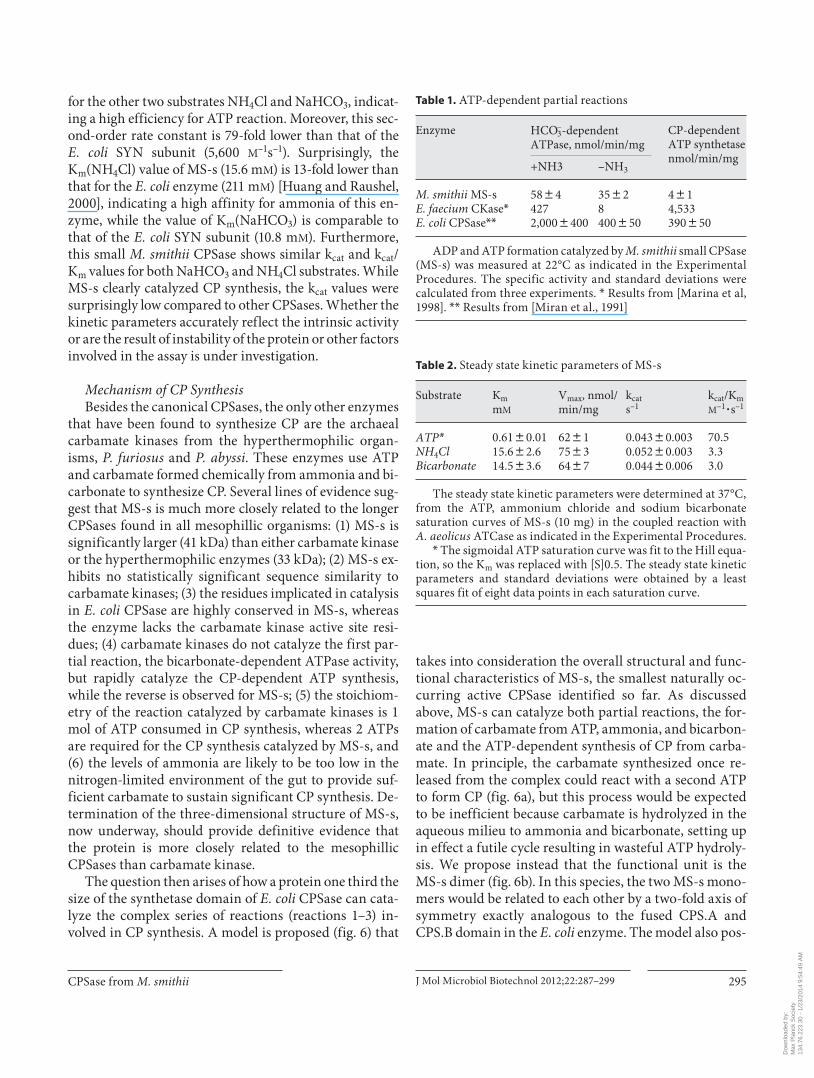
This small M. smithii CPSase also catalyzed the ATP-dependent partial reactions in accordance with the three-step mechanism demonstrated for E. coli CPSase [Kothe et al., 2005; Meister, 1989] ( table 1 ). The bicarbonate-de-pendent ATPase reaction in the presence (reactions 1 and 2) and absence (reaction 1 only) of ammonia measured at 22 ° C showed a ratio of 1.7: 1, indicating that 2 ATP mol-ecules are consumed in the CP synthesis when all of the substrates are present. Moreover, the CP-dependent ATP synthetase reverse reaction rate is very low (4.52 nmol/min/mg) relative to the ATP hydrolysis, representing 8% of the forward ATPase reaction in the presence of ammo-nia, as in the case of A. aeolicus CPSase [Ahuja et al., 2001].
Enterococcus faecium carbamate kinase [Marina et al., 1998] does not catalyze the first partial reaction, i.e. the bicarbonate-dependent ATP hydrolysis in the absence of ammonia, whereas the second partial reaction measured in the reverse direction as a CP-dependent ATP synthesis occurs at a very high rate ( table 1 ). In contrast, for MS-s, like E. coli CPSase [Rubio et al., 1987], the rate of the bi-carbonate-dependent ATP hydrolysis is high relative to the CP-dependent ATP synthesis activity. The kinetic studies provided additional strong evidence that MS-s is a canonical CPSase, not a carbamate kinase.
Steady-State Kinetic Parameters Substrate saturation curves for ATP, NH 4 Cl, and
NaHCO 3 of the recombinant MS-s were measured at 37 ° C (data not shown), and the kinetic parameters were cal-culated from the least squares fit to either the Michae-lis-Menten or the Hill equation. While both NH 4 Cl and NaHCO 3 saturation curves follow Michaelis-Menten ki-netics, the ATP saturation curve is slightly sigmoidal, with
Dow
nloa
ded
by:
Max
Pla
nck
Soc
iety
13
4.76
.223
.30
- 1/
23/2
014
9:54
:49
AM

Popa /Perera /Kibédi-Szabó /Guy-Evans /Evans /Purcarea
J Mol Microbiol Biotechnol 2012;22:287–299 294
a Hill coefficient of n H = 1.4 8 0.1. The steady-state ki-netic parameters ( table 2 ) indicated a relatively high appar-ent affinity for ATP (K m = 0.61 m M ), 2- and 12-fold higher than the K m (ATP) of the E. coli SYN subunit (1.3 m M ) [Ru-bio et al., 1987] and A. aeolicus CPS.A-CPS.B complex
(7.43 m M ) [Ahuja et al., 2001], respectively. The k cat value for this substrate (0.043 s –1 ) is 170-fold lower than the cor-responding value (7.3 s –1 ) for the E. coli SYN subunit [Ru-bio et al., 1987]. The apparent second-order rate constant k cat /K m for ATP (70.5 M –1 s –1 ) is 21–23-fold higher than that
kDa
11890
50
34
26
MS-s
Std 1 2 3 4 5 6 7 8 9
a
220120
50
(MS-s)8/n(MS-s)4
MS-s
kDa Std 1 2 3 4
b
–2290 300 310 320 330 340 350 360 370
0
2
4
6
8
10
12
14
16
18
20
22
24
Ab
sorb
ance
(mA
U)
Elution volume (ml)
^ ^ ^ ^ ^ ^ ^ ^ ^ ^ ^ ^ ^ ^ ^ ^ ^ ^ ^ ^
1 2 3 4 5 6 7 8 9 10 12 14 16 18
15 16 17 18
20 22 24 26 28 30 32 34 36
35 36
38
3837 39
40
40 41
42 44 46 48
MS-sUV
c
Fig. 5. MS-s purification and oligomeric structure determination. a Purification of the recombinant MS-s was carried out by af -finity chromatography on Ni 2+ -Probond resin as described in Experimental Procedures, and the elution fractions were ana-lyzed by 12% SDS-PAGE. The lanes correspond to the molecular weight marker (Std), fractions 1–3 eluted with TN buffer (50 m M TrisHCl, pH 8, 200 m M NaCl), fractions 4–6 eluted with TN buf-fer containing 50 m M imidazole, and fractions 7–9 eluted with TN buffer containing 100 m M imidazole. b Chemical cross-linking
was carried out at 22 ° C for 0, 5, 10, and 15 min (lanes 1–4) using 20 � g recombinant MS-s in 100 m M TEA buffer, pH 8.5, 100 m M NaCl, and 10 m M dimethylsuberimidate. The reaction mixture was quenched with 100 m M TrisHCl, pH 8, and analyzed by 10% SDS-PAGE. c Size exclusion chromatography was carried out as indicated in Experimental Procedures. The elution profile was obtained by measuring the absorbance at 280 nm. The peak frac-tions (fractions 15–18 and 35–41) were analyzed by 10% SDS-PAGE and found to contain MS-s.
Co
lor v
ersi
on
avai
lab
le o
nlin
e
Dow
nloa
ded
by:
Max
Pla
nck
Soc
iety
13
4.76
.223
.30
- 1/
23/2
014
9:54
:49
AM
CPSase from M. smithii J Mol Microbiol Biotechnol 2012;22:287–299 295
for the other two substrates NH 4 Cl and NaHCO 3 , indicat-ing a high efficiency for ATP reaction. Moreover, this sec-ond-order rate constant is 79-fold lower than that of the E. coli SYN subunit (5,600 M –1 s –1 ). Surprisingly, the K m (NH 4 Cl) value of MS-s (15.6 m M ) is 13-fold lower than that for the E. coli enzyme (211 m M ) [Huang and Raushel, 2000], indicating a high affinity for ammonia of this en-zyme, while the value of K m (NaHCO 3 ) is comparable to that of the E. coli SYN subunit (10.8 m M ). Furthermore, this small M. smithii CPSase shows similar k cat and k cat /K m values for both NaHCO 3 and NH 4 Cl substrates. While MS-s clearly catalyzed CP synthesis, the k cat values were surprisingly low compared to other CPSases. Whether the kinetic parameters accurately reflect the intrinsic activity or are the result of instability of the protein or other factors involved in the assay is under investigation.
Mechanism of CP Synthesis Besides the canonical CPSases, the only other enzymes
that have been found to synthesize CP are the archaeal carbamate kinases from the hyperthermophilic organ-isms, P. furiosus and P. abyssi . These enzymes use ATP and carbamate formed chemically from ammonia and bi-carbonate to synthesize CP. Several lines of evidence sug-gest that MS-s is much more closely related to the longer CPSases found in all mesophillic organisms: (1) MS-s is significantly larger (41 kDa) than either carbamate kinase or the hyperthermophilic enzymes (33 kDa); (2) MS-s ex-hibits no statistically significant sequence similarity to carbamate kinases; (3) the residues implicated in catalysis in E. coli CPSase are highly conserved in MS-s, whereas the enzyme lacks the carbamate kinase active site resi-dues; (4) carbamate kinases do not catalyze the first par-tial reaction, the bicarbonate-dependent ATPase activity, but rapidly catalyze the CP-dependent ATP synthesis, while the reverse is observed for MS-s; (5) the stoichiom-etry of the reaction catalyzed by carbamate kinases is 1 mol of ATP consumed in CP synthesis, whereas 2 ATPs are required for the CP synthesis catalyzed by MS-s, and (6) the levels of ammonia are likely to be too low in the nitrogen-limited environment of the gut to provide suf-ficient carbamate to sustain significant CP synthesis. De-termination of the three-dimensional structure of MS-s, now underway, should provide definitive evidence that the protein is more closely related to the mesophillicCPSases than carbamate kinase.
The question then arises of how a protein one third the size of the synthetase domain of E. coli CPSase can cata-lyze the complex series of reactions (reactions 1–3) in-volved in CP synthesis. A model is proposed ( fig. 6 ) that
takes into consideration the overall structural and func-tional characteristics of MS-s, the smallest naturally oc-curring active CPSase identified so far. As discussed above, MS-s can catalyze both partial reactions, the for-mation of carbamate from ATP, ammonia, and bicarbon-ate and the ATP-dependent synthesis of CP from carba-mate. In principle, the carbamate synthesized once re-leased from the complex could react with a second ATP to form CP ( fig. 6 a), but this process would be expected to be inefficient because carbamate is hydrolyzed in the aqueous milieu to ammonia and bicarbonate, setting up in effect a futile cycle resulting in wasteful ATP hydroly-sis. We propose instead that the functional unit is the MS-s dimer ( fig. 6 b). In this species, the two MS-s mono-mers would be related to each other by a two-fold axis of symmetry exactly analogous to the fused CPS.A and CPS.B domain in the E. coli enzyme. The model also pos-
Table 1. A TP-dependent partial reactions
Enzyme H CO3–-dependent
ATPase, nmol/min/ mgCP-dependent ATP synthetasenmol/min/mg
+N H3 –NH3
M. smithii MS-sE. faecium CKase*E. coli CPSase**
58844272,0008400
35828400850
4814,533390850
ADP and ATP formation catalyzed by M. smithii small CPSase (MS-s) was measured at 22°C as indicated in the Experimental Procedures. The specific activity and standard deviations were calculated from three experiments. * Results from [Marina et al, 1998]. ** Results from [Miran et al., 1991]
Table 2. S teady state kinetic parameters of MS-s
Substrate KmmM
Vmax, nmol/min/mg
kcats–1
kcat/KmM–1
� s–1
ATP* 0.6180.01 6281 0.04380.003 70.5NH4Cl 15.682.6 7583 0.05280.003 3.3Bicarbonate 14.583.6 6487 0.04480.006 3.0
T he steady state kinetic parameters were determined at 37°C, from the ATP, ammonium chloride and sodium bicarbonate saturation curves of MS-s (10 mg) in the coupled reaction with A. aeolicus ATCase as indicated in the Experimental Procedures.
* The sigmoidal ATP saturation curve was fit to the Hill equa-tion, so the Km was replaced with [S]0.5. The steady state kinetic parameters and standard deviations were obtained by a least squares fit of eight data points in each saturation curve.
Dow
nloa
ded
by:
Max
Pla
nck
Soc
iety
13
4.76
.223
.30
- 1/
23/2
014
9:54
:49
AM

Popa /Perera /Kibédi-Szabó /Guy-Evans /Evans /Purcarea
J Mol Microbiol Biotechnol 2012;22:287–299 296
tulates that, as in E. coli CPSase, the two active sites are connected by an intramolecular tunnel. When ATP, am-monia, and bicarbonate bind to monomer (a in fig. 6 b) it becomes the site of synthesis of carbamate. Carbamate is then transferred to monomer b which now becomes the CP synthesis domain by default. In a subsequent cata-lytic cycle the roles may be reversed such that subunit b catalyzes carbamate synthesis and subunit a converts car-bamate to CP. In this scheme, carbamate remains seques-tered within the complex and protected from hydrolysis.
While further structural and biochemical studies are required to validate this model, there is precedence for this mechanism in mammalian CPSase. It was shown that both CPS.A and CPS.B domains of mammalian CAD separately cloned and expressed in E. coli could each dimerize and catalyze both ATP-dependent reac-tions and the overall synthesis of CP [Guy et al., 1997]. Moreover, the dissociation of CPS.A or CPS.B homodi-mers by high hydrostatic pressure abolished CP synthe-
sis, while the dissociated subunits could still catalyze the CPSase partial reactions [Guy et al., 1998].
Putative Physiological Function of MS-s In addition to MS-s, M. smithii possesses an active,
full-length glutamine-dependent CPSase, MS-l (unpubl. data), so the question arises of what selective advantage the unusual CPSase MS-s provides for the organism. Am-monia is the major source of nitrogen in M. smithii for the biosynthesis of nucleotides, amino acids, and many other metabolites. The organism has a specific ammonia trans-porter and two mechanisms for assimilation of ammonia, the glutamine synthetase-glutamate synthase and the glu-tamate dehydrogenase pathways [Samuel et al., 2007]. However, the availability of ammonia is strictly limited in the intestine and there is a competition for this scare re-source between M. smithii and B. thetaiotaomicron . This conclusion is supported by several lines of evidence. For example, analytical studies [Samuel et al., 2007] demon-strated that, in M. smithii, the ratio of glutamine to 2-oxo-glutarate, a good indication of the nitrogen status of the organism, was reduced 32-fold when M. smithii was co-colonized with B. thetaiotaomicron . Moreover, when both organisms are present in the intestine, there is an appre-ciable upregulation of the expression of enzymes involved in nitrogen assimilation in M. smithii [Samuel et al., 2007].
MS-s is a streamlined CPSase that is ideally suited to the task of capturing ammonia and converting it to py-rimidines and arginine. The kinetic studies showed that it has an appreciably higher affinity for ammonia than other CPSases. Moreover, the enzyme is also probably unregulated since the B3 or allosteric subdomain, the lo-cus of regulation of all known CPSases, is not present in MS-s. While eukaryotic mitochondrial CPSase I, the en-zyme that initiates the urea cycle, can be allosterically activated, there are no known allosteric inhibitors [Rubio and Cervera, 1995] presumably because it is essential in ureotylic animals to continuously and rapidly dispose of any ammonia formed via urea biosynthesis. Similarly, MS-s would be expected to be constitutively active if one of its major functions is to sequester ammonia, so that al-losteric regulation would confer no advantage.
Conclusions
MS-s is a unique enzyme, the smallest functionalCPSase discovered thus far and one that is only distantly related to other bacterial and archaeal CPSases. It consists of a catalytic domain but lacks the module involved in al-
ATP + NH3 + HCO3–
ADP + carbamate ATP + carbamate
ADP + carbamoyl phosphate
NH3 + HCO3–
a
ATP + NH3 + HCO3–
Carbamoylphosphate
ATP + NH3 + HCO3–
Carbamoylphosphate
a a
b b
ATP
ATP
b
Fig. 6. Functional model of MS-s. Schematic representation of CP synthesis by MS-s. a Carbamate is synthesized from bicarbonate, ammonia, and ATP, released from the enzyme, and then binds once again where it is phosphorylated by a second ATP molecule forming CP. This process would be expected to be inefficient since carbamate once released from the complex would be degraded to bicarbonate and ammonia. b The synthesis of carbamate is cata-lyzed by one of the monomers in the MS-s dimer (a) which binds bicarbonate, ammonia, and ATP; the carbamate is sequestered and transferred to the second monomer (b) which catalyzes the phosphorylation of carbamate to form CP. In a subsequent cata-lytic cycle, the roles of subunits a and b may be reversed.
Co
lor v
ersi
on
avai
lab
le o
nlin
e
Dow
nloa
ded
by:
Max
Pla
nck
Soc
iety
13
4.76
.223
.30
- 1/
23/2
014
9:54
:49
AM
CPSase from M. smithii J Mol Microbiol Biotechnol 2012;22:287–299 297
losteric regulation. The catalytically active species is an MS-s homodimer or larger species and the mechanism of CP synthesis appears to occur in three steps by the same mechanism utilized by the full-length prokaryotic and eukaryotic CPSases. CPSase are postulated to be derived from an ancestral kinase resembling MS-s. Thus, the characterization of this enzyme may provide a glimpse of the evolution of CPSases, which are thought to have aris-en by gene duplication, fusion, and the recruitment of domains that confer regulatory functions. In addition to supplementing the function of the full-length CPSase MS-l by bolstering the supply of pyrimidine nucleotides and arginine, MS-s may provide an additional mecha-nism for harvesting ammonia, a scarce nutrient in the intestine. These minimal CPSases may be a characteristic of archaeal methanogens, since they are also present in M. thermoautotrophicus and M. stadtmanae . Consider-ing that there is apparently no mammalian counterpart of this unusual protein, MS-s may be an attractive drug target in the treatment of human obesity.
Experimental Procedures
Materials Pfu DNA polymerase, nucleotides, restriction enzymes, Bam-
HI and Nco I, thermosensitive alkaline phosphatase (FastAP), and T4 DNA ligase were from Fermentas Life Sciences; Ni 2+ -ProBond resin was purchased from Invitrogen; aspartate, carbamoyl aspar-tate, antipyrine, diacetyl monoxime, triethoanolamine (TEA), phosphoenol pyruvate, dimethyl suberimidate, and the enzymes pyruvate kinase, lactate dehydrogenase, hexokinase, and glucose 6-phosphate dehydrogenase were from Sigma-Aldrich; A. aeoli-cus aspartate transcarbamoylase (ATCase) recombinant enzyme was expressed in E. coli and purified as previously described [Pur-carea et al., 2003].
Strains and Plasmids The M. smithii ATCC35061 strain (DSM 861) was from the
DSMZ Bacteria Collection [Samuel et al., 2007]. The E. coli DH5 � and BL21(DE3) strains and the pRSETC expression vector were from Invitrogen.
M. smithii Growth The methanogen M. smithii ATCC35061 was cultivated an-
aerobically at 37 ° C in DSMZ 119 medium containing fatty acids and supplemented with DSMZ 320 salt solutions, in the presence of an 80% H 2 :20% CO 2 gas mixture [Samuel et al., 2007]. Cultures of 20 ml were inoculated with 1-ml cultures under an N 2 atmo-sphere. The flasks were flushed every 24 h with fresh H 2 :CO 2 mix-ture, and cell viability was analyzed by microscopy.
Cloning and Expression M. smithii chromosomal DNA was isolated from a 20-ml cul-
ture using a DNeasy Blood and Tissue Kit for genomic DNA ex-traction (Qiagen), with an additional cell lysis step using 50 units
of mutanolysin (Sigma). The carB1 gene (YP_001272934) from M. smithii was amplified by PCR using 2.5 units of Pfu DNA poly-merase, 170 ng of M. smithii chromosomal DNA as a template, and 100 pmol of 5MS-s (5 � -GGAGATTGGATCCAAATTTTA-TTTAT-CGGTTCAAG-3 � ) and 3MS-s (5 � -GTGTTTAAGCCAT-GG TTA-AATCAAATCTTCAACATATC-3 � ) primers defining the 5 � and 3 � ends of the M. smithii carB1 gene, respectively. The DNA fragment was amplified after 30 cycles of 95 ° C for 45 s, 52 ° C for 1 min, and 72 ° C for 3 min each cycle, and purified with a DNA Clean & Concentrator Kit (Zymo Research). After digestion with Bam HI/ Nco I, the gene (750 ng) was inserted into the pRSETC ex-pression vector (130 ng) digested with the same restriction en-zymes, and dephosphorylated with thermosensitive alkaline phosphatase. Both strands of the resulting construct were se-quenced using a Genetic Analyzer 3500 (Applied Biosystems). The M. smithii CPS gene was expressed in E. coli BL21(DE3) after a 5-hour induction at 20 ° C with 1 m M IPTG.
Purification The recombinant M. smithii enzyme expressed in E. coli as an
N-terminal fused protein with a 3-kDa His-tag polypeptide was purified in one step at 4 ° C by affinity chromatography. Cells from a 100-ml culture were resuspended in 3 ml of 50 m M TrisHCl, pH 8, containing 2 m M 2-mercaptoethanol and 1: 100 protease inhib-itor cocktail (Sigma), and disrupted by sonication 6 times for 30 s using a Sonifier Cell disruptor W-350 (Bronson Sonic Power). The cell extract was centrifuged at 20,000 g for 30 min, and the super-natant was applied to a 1-ml Ni 2+ -ProBond column equilibrated with 50 m M TrisHCl, pH 8, and 200 m M NaCl (TN buffer). After 20-ml washes with the same buffer, the enzyme was eluted with 1-ml fractions of TN buffer containing increasing (50, 100, 200, and 500 m M ) imidazole concentrations. The elution fractions were analyzed by electrophoresis on 12% SDS-PAGE gels. Frac-tions containing pure recombinant CPSase were collected and the buffer was changed to 50 m M TrisHCl, pH 8, and 5 m M 2-mercap-toethanol, using PD-10 desalting columns (GE Healthcare), to eliminate imidazole and NaCl.
Chemical Cross-Linking Fractions containing 20 � g M. smithii CPSase in 100 m M tri-
ethanolamine, pH 8.5, and 100 m M NaCl were incubated with 10 m M dimethyl suberimidate [Davies et al., 1970] for 0, 5, 10, and 15 min at room temperature in a total volume of 45 � l. The reaction was stopped by addition of 5 � l of 1 M TrisHCl, pH 8, and the cross-linked proteins were visualized by SDS-PAGE (10% gels).
Size Exclusion Chromatography Size exclusion chromatography was carried out at 4 ° C using a
Hiload 16/60 Superdex 200 prep grade column equilibrated with 50 m M TrisHCl, pH 8, containing 5 m M 2-mercaptoethanol. The column was loaded with 500- � l (5 mg) of the purified MS-s, and the protein was eluted at 1 ml/min with the equilibration buffer, monitoring the absorbance at 280 nm. The fractions containing the protein peaks were analyzed by SDS-PAGE.
CPSase Assay Ammonia-dependent CPSase activity was measured using the
radioactive assay in which the CPSase reaction is coupled with A. aeolicus aspartate transcarbamoylase (ATCase) [Ahuja et al., 2001]. Expression and purification of the A. aeolicus ATCase were
Dow
nloa
ded
by:
Max
Pla
nck
Soc
iety
13
4.76
.223
.30
- 1/
23/2
014
9:54
:49
AM

Popa /Perera /Kibédi-Szabó /Guy-Evans /Evans /Purcarea
J Mol Microbiol Biotechnol 2012;22:287–299 298
carried out as previously described [Purcarea et al., 2003]. The reaction mixture contained 100 m M sodium [ 14 C] bicarbonate (100 dpm/ � mol), 20 m M ATP, 22 m M MgCl 2 and 200 m M ammo-nium chloride, 50 m M TrisHCl, pH 8.0, 100 m M KCl, in the pres-ence of 6 m M aspartate, pH 7, and 1 � g of purified A. aeoli-cus ATCase and various concentrations of recombinant MS-s CPSase, in a total volume of 0.3 ml. After a 20-min incubation at 37 ° C, the reaction was stopped by addition of 0.7 ml 10% trichlor-acetic acid. After evaporation of the liquid phase at 95 ° C to de-compose the unreacted bicarbonate, the radioactivity incor-porated in the stable reaction product carbamoyl aspartate was measured after addition of 8 ml Aquasol scintillation liquid. Al-ternatively, the carbamoyl aspartate formed in the coupled reac-tion was measured by a colorimetric assay previously described [Purcarea et al., 2001].
Partial Reactions The bicarbonate-dependent ATPase reaction was measured
using a pyruvate kinase/lactate dehydrogenase coupled assay [Post et al., 1990] by monitoring the rate of ADP synthesis in the presence and absence of ammonium chloride. The reaction was performed in a final volume of 0.5 ml containing 100 m M sodium bicarbonate, 20 m M ATP, 22 m M MgCl 2 , 50 m M TrisHCl, pH 8.0, 100 m M KCl, 1 m M phosphoenolpyruvate, 0.2 m M NADH, 10 units of pyruvate kinase, and 20 units of lactate dehydrogenase, and, optionally, 200 m M ammonium chloride. The reaction rate was measured spectrophotometrically at 340 nm, and the concen-tration was calculated using � NADH = 6.22 M –1 � cm –1 . The CP-de-pendent ATP synthetase activity was assayed by measuring the rate of ATP formation from ADP and CP in the coupled reaction with hexokinase/glucose-6-phosphate dehydrogenase, following a similar procedure [Miran et al., 1991]. The reaction mixture contained 50 m M TrisHCl, pH 8.0, 100 m M KCl, 22 m M MgCl 2 ,1 m M NAD, 20 m M ADP, 2 m M CP, 5 m M glucose, 20 units of hexokinase, and 10 units of glucose-6-phosphate dehydrogenase in a final volume of 0.5 ml. Both partial reactions were assayed at 22 ° C and the initial rate was calculated from the time course up to a 5-min time course.
Structure Analysis and Molecular Modeling The genes encoding the CPSase subunits in the M. smithii
ATCC35061 genome sequence were identified by BLAST-NCBI genome database screening. Sequence analysis was performed us-ing the genomic and proteomic software package ExPASy SIB Bioinformatics Resource Portal. Protein pair alignment was car-ried out with EMBOSS needle alignment (EMBL-EBI) and mul-tiple alignment was performed using ClustalW. Amino acid se-quence randomization was performed using random protein se-quence generator RandSeq software (ExPaSy). Modeling of the three-dimensional structure was carried out using Modeller 9v9 [Sali and Blundell, 1993], based on the 1BXR X-ray structure of E. coli CPSase in complex with the ATP analog AMPPNP [Thoden et al., 1999]. Fifty models were generated and the best candidate was chosen based on DOPE (discrete optimized protein energy), zDOPE (normalized discrete optimized protein energy), and GA341 scores. The energy minimization was performed with Gromos 96 implemented in Swiss PDB Viewer. The model was further refined by loop optimization using ModLoop [Fiser and Sali, 2003] followed by energy minimization. In every round of optimization, at least 10 models where generated, followed by en-ergy minimization. The quality of the model was assessed by the Ramachandran plot obtained with PROCHEK [Laskowski et al., 1993], ERRAT2 [Colovos and Yeates, 1993], and ProSa [Sippl, 1993] programs. Superposition of the MS-s model with 1BXR and calculation of the root mean square deviation were obtained using Swiss PDB Viewer [Guex and Peitsch, 1997]. The structures where visualized using the PyMol Molecular Graphics System 0.99rc6 (Schrodinger, LLC).
Acknowledgments
We thank Asmita Vaishnav for technical expertise and assis-tance. This work was supported by ANCS-UEFISCDI Romania, PNII ID_1034 (contract No. 1023/2009), NIH R01 grant GM/CA60371, and the Wayne State University Provost’s Research Stimulation Award.
References
Ahuja A, Purcarea C, Guy HI, Evans DR: A nov-el carbamoyl phosphate synthetase from Aquifex aeolicus. J Biol Chem 2001; 276: 45694–45703.
Alonso E, Rubio V: Affinity cleavage of carbam-oyl-phosphate synthetase I localizes regions of the enzyme interacting with the molecule of ATP that phosphorylates carbamate. Eur J Biochem 1995; 229: 377–384.
Anderson PM: Carbamoyl-phosphate synthe-tase: an example of effects on enzyme prop-erties of shifting an equilibrium between ac-tive monomer and active oligomer. Bio-chemistry 1986; 25: 5576–5582.
Anderson PM, Meister A: Evidence for an acti-vated form of carbon dioxide in the reaction catalyzed by Escherichia coli carbamyl phos-phate synthetase. Biochemistry 1965; 4: 2803–2809.
Balch WE, Fox GE, Magrum LJ, Woese CL, Wolf RS: Methanogens: reevaluation of a unique biological group. Microbiol Rev 1979; 43: 260–296.
Belay N, Johnson R, Rajagopal BS, De Macario ES, Daniels L: Methanogenic bacteria from human dental plaque. Appl Environ Micro-biol 1988; 54: 600–603.
Belay N, Mukhopadhyay B, De Macario EC, Galask R, Daniels L: Methanogenic bacteria in human vaginal samples. J Clin Microbiol 1990; 28: 1666–1668.
Buck S, Hansen EE: Genomic and metabolic ad-aptations of Methanobrevibacter smithii to the human gut. Proc Natl Acad Sci USA 2007; 104: 10643–10648.
Bult CJ, White O, Olsen GJ, Zhou L, Fleischmann RD, Sutton GG, Blake JA, FitzGerald LM, Clayton RA, Gocayne JD, Kerlavage AR,
Dougherty BA, Tomb FJ, Adams MD, Reich CI, Overbeek R, Kirkness EF, Weinstock KG, Merrick JM, Glodek A, Scott JL, Geoghan-gen NS, Venter JC: Complete genome se-quence of the methanogenic archaeon, Methanococcus jannaschii. Science 1996; 273: 1058–1073.
Colovos C, Yeates TO: Verification of protein structures: patterns of nonbonded atomic in-teractions. Protein Sci 1993; 2: 1511–1519.
Davies GE, Stark GR: Dimethyl suberimidate, a cross-linking reagent, in studying the sub-unit structure of oligomeric proteins. Proc Natl Acad Sci USA 1970; 66: 651–656.
Dridi B, Raoult D, Drancourt M: Archaea as emerging organisms in complex human mi-crobiomes. Anaerobe 2011; 17: 56–63.
Durbecq V, Legrain C, Roovers M, Piérard A, Glansdorff N: The carbamate kinase-like
Dow
nloa
ded
by:
Max
Pla
nck
Soc
iety
13
4.76
.223
.30
- 1/
23/2
014
9:54
:49
AM

CPSase from M. smithii J Mol Microbiol Biotechnol 2012;22:287–299 299
carbamoyl phosphate synthetase of the hy-perthermophilic archaeon Pyrococcus furio-sus, a missing link in the evolution of car-bamoyl phosphate biosynthesis. Proc Natl Acad Sci USA. 1997; 94: 12803–12808.
Fiser A, Sali A: ModLoop: automated modeling of loops in protein structures. Bioinformat-ics 2003; 19: 2500–2501.
Fresquet V, Mora P, Rochera L, Ramón-Maiques S, Rubio V, Cervera J: Site-directed mutagen-esis of the regulatory domain of Escherichia coli carbamoyl phosphate synthetase identi-fies crucial residues for allosteric regulation and for transduction of the regulatory sig-nals. J Mol Biol 2000: 299: 979–991.
Guex N, Peitsch MC: SWISS-MODEL and the Swiss-PdbViewer: an environment for com-parative protein modeling. Electrophoresis 1997; 18: 2714–2723.
Guillou F, Rubino SD, Markovitz RS, Kinney DM, Lusty CJ: Escherichia coli carbamoyl-phosphate synthetase: domains of glutamin-ase and synthetase subunit interaction. Proc Natl Acad Sci USA 1989; 86: 8304–8308.
Guy HI, Bouvier A, Evans DR: The smallest car-bamoyl-phosphate synthetase. J Biol Chem 1997; 272: 29255–29262.
Guy HI, Evans DR: Function of the major syn-thetase subdomains of carbamyl-phosphate synthetase. J Biol Chem 1996; 271: 13762–13769.
Guy HI, Schmitt B, Hervé G, Evans DR: Pres-sure-induced dissociation of carbamoyl-phosphate synthetase domains: the catalyti-cally active form is dimeric. J Biol Chem 1998; 273: 14172–14178.
Horz HP, Conrads G: The discussion goes on: what is the role of Euryarchaeota in humans. Archaea DOI: 10.1155/2010/967271.
Huang X, Raushel FM: An engineered blockage within the ammonia tunnel of carbamoyl phosphate synthetase prevents the use of glu-tamine as a substrate but not ammonia. Bio-chemistry 2000; 39: 3240–3247.
Kim J, Raushel FM: Allosteric control of the oligomerization of carbamoyl phosphate synthetase from Escherichia coli. Biochemis-try 2001; 40: 11030–11036.
Kothe M, Purcarea C, Guy HI, Evans DR, Pow-ers-Lee SG: A Novel carbamoyl-phosphate synthetase from Aquifex aeolicus. J Biol Chem 2005; 14: 37–44.
Laskowski RA, MacArthur MW, Moss DS, Thornton JM: PROCHECK: a program to check the stereochemical quality of protein structures. J Appl Crystallogr 1993; 26: 283–291.
Legrain C, Demarez M, Glansdorff N, Pierard A: Ammonia-dependent synthesis and meta-bolic channelling of carbamoyl phosphate in the hyperthermophilic archaeon Pyrococcus furiosus. Microbiology 1995; 141: 1093–1099.
Marina A, Uriarte M, Barcelona B, Fresquet V, Cervera J, Rubio V: Carbamate kinase from Enterococcus faecalis and Enterococcus fae-cium: cloning of the genes, studies on the en-zyme expressed in Escherichia coli, and se-quence similarity with N -acetyl- L -glutamate kinase. Eur J Biochem 1998; 253: 280–291.
Meister A: Mechanism and regulation of the glu-tamine-dependent carbayl phosphate syn-thetase of Escherichia coli. Adv Enzymol Relat Areas Mol Biol 1989; 62: 315–374.
Miller TL, Wolin MJ, De Macario EC, Macario AJ: Isolation of Methanobrevibacter smithii from human feces. Appl Environ Microbiol 1982; 43: 227–232.
Million M, Maraninchi M, Henry M., Armou-gom F, Richet H, Carrieri P, Valero R, Raccah D, Vialettes B, Raoult D: Obesity-associated gut microbiota is enriched in Lactobacillus reuteri and depleted in Bifidobacterium ani-malis and Methanobrevibacter smithii. Int J Obes DOI: 10.1038/ijo.2011.153.
Miran SG, Chang SH, Raushel FM: Role of the four conserved histidine residues in the ami-dotransferase domain of carbamoyl phos-phate synthetase. Biochemistry 1991; 30: 7901–7909.
Nava GM, Caronero F, Croix JA, Greenberg E, Gaskins HR: Abundance and diversity of mucosa-associated hydrogenotrophic mi-crobes in the healthy human colon. ISME J DOI: 10.1038/ismej.2011.90.
Nyunoya H, Lusty CJ: The carB gene of Esche-richia coli: a duplicated gene coding for the large subunit of carbamoyl-phosphate syn-thetase. Proc Natl Acad Sci USA 1983; 80: 4629–4633.
Paulus TJ, Switzer RL: Caracterization of pyrim-idine-repressible and arginine-repressible carbamyl phosphate synthetase from Bacil-lus subtilis. J Bacteriol 1979; 137: 82–91.
Plottel CS, Blaser MJ: Microbiome and malig-nancy. Cell Host Microb 2011; 10: 324–335.
Popa E, Rusu A, Zamfir M, Dumitru L, Purcarea C: An ammonia-metabolizing enzyme from the human archaeon Methanobrevibacter smithii might represent a missing link in the evolution of carbamoyl phosphate synthe-tases. Biotechnol Biotechnol Equip 2009; 23: 533–537.
Post LE, Post DJ, Raushel FM: Dissection of the functional domains of Escherichia coli car-bamoyl phosphate synthetase by site-direct-ed mutagenesis. J Biol Chem 1990; 265: 7742–7747.
Purcarea C, Ahuja A, Lu T, Kovari L, Guy HI, Evans DR: Aquifex aeolicus aspartate trans-carbamoylase, an enzyme specialized for the efficient utilization of unstable carbamoyl phosphate at elevated temperature. J Biol Chem 2003; 278: 52924–52934.
Purcarea C, Hervé G, Cunin R, Evans DR: Clon-ing, expression and structure analysis of car-bamat kinase-like carbamoyl phosphate syn-thetase from Pyrococcus abyssi. Extremo-philes 2001; 5: 229–239.
Purcarea C, Simon V, Prieur D, Hervé G: Purifica-tion and characterization of carbamylphos-phate synthetase from the deep-sea hyper-thermophilic archaebacterium Pyrococcus ab-yssi . Eur J Biochem 1996; 236: 189–199.
Rubio V, Cervera J: The carbamoyl-phosphat synthetase family and carbamate kinase: structure-function studies. Biochem Soc Trans 1995; 23: 879–883.
Rubio V, Cervera J, Lusty CJ, Bendala E, Britton HG. Domain structure of the large subunit of Escherichia coli carbamoyl phosphate syn-thetase: location of the binding site for the allosteric inhibitor UMP in the COOH-ter-minal domain. Biochemistry 1991 30: 1068–1075.
Rubio SD, Nyunoya H, Lusty CJ: In vivo synthe-sis of carbamyl phosphate from NH3 by the large subunit of Escherichia coli carbamyl phosphate synthetase. J Biol Chem 1987; 262: 4382–4386.
Šali A, Blundell TL: Comparative protein model-ling by satisfaction of spatial restraints. J Mol Biol 1993; 234: 779–815.
Samuel BS, Gordon JI: A humanized gnotobiotic mouse model of host-archaeal-bacterial mu-tualism. Proc Natl Acad Sci USA 2006; 103: 10011–10016.
Samuel BS, Hansen EE, Manchester JK, Coutin-ho PM, Henrissat B, Fulton R, Latreille P, Kim K, Wilson RK, Gordon JI: Genomic and metabolic adaptations of Methanobrevi-bacter smithii to the human gut. Proc Natl Acad Sci USA 2007; 104: 10643–10648.
Sippl MJ: Recognition of errors in three-dimen-sional structures of proteins. Proteins 1993; 17: 355–362.
Stams AJ: Metabolic interactions between anaer-obic bacteria in methanogenic environ-ments. Antonie van Leeuwenhoek 1994; 66: 271–294.
Thoden JB, Holden HM, Wesenberg G, Raushel FM, Rayment I: The structure of carbamoyl phosphate synthetase: a journey of 96 Å from substrate to product. Biochemistry 1997; 36: 6305–6316.
Thoden JB, Wesenberg G, Raushel FM, Holden HM: Carbamoyl phosphate synthetase: clo-sure of the B-domain as a result of nucleotide binding. Biochemistry 1999; 38: 2347–2357.
Trotta PP, Burt ME, Haschemeyer RH, Meister A: Reversible dissociation of carbamyl phos-phate synthetase into a regulated synthesis subunit and a subunit required for glutamine utilization. Proc Natl Acad Sci USA 1971; 68: 2599–2603.
Uriarte M, Marina A, Ramon-Maiques S, Rubio V, Durbecq V, Legrain C, N. Glansdorff N: Carbamoyl phosphate synthesis: carbamate kinase from Pyrococcus furiosus . Methods Enzymol 2001; 331: 236–247.
Yang H, Park SM, Nolan WG, Lu CD, Abdelal AT: Cloning and characterization of the ar-ginine-specific carbamoyl-phosphate syn-thetase from Bacillus stearothermophilus. Eur J Biochem 1997; 249: 443–449.
Dow
nloa
ded
by:
Max
Pla
nck
Soc
iety
13
4.76
.223
.30
- 1/
23/2
014
9:54
:49
AM