Elastomeric Polymers for Retrofitting of Reinforced Concrete ...
Upload
independentCategory
view
5download
0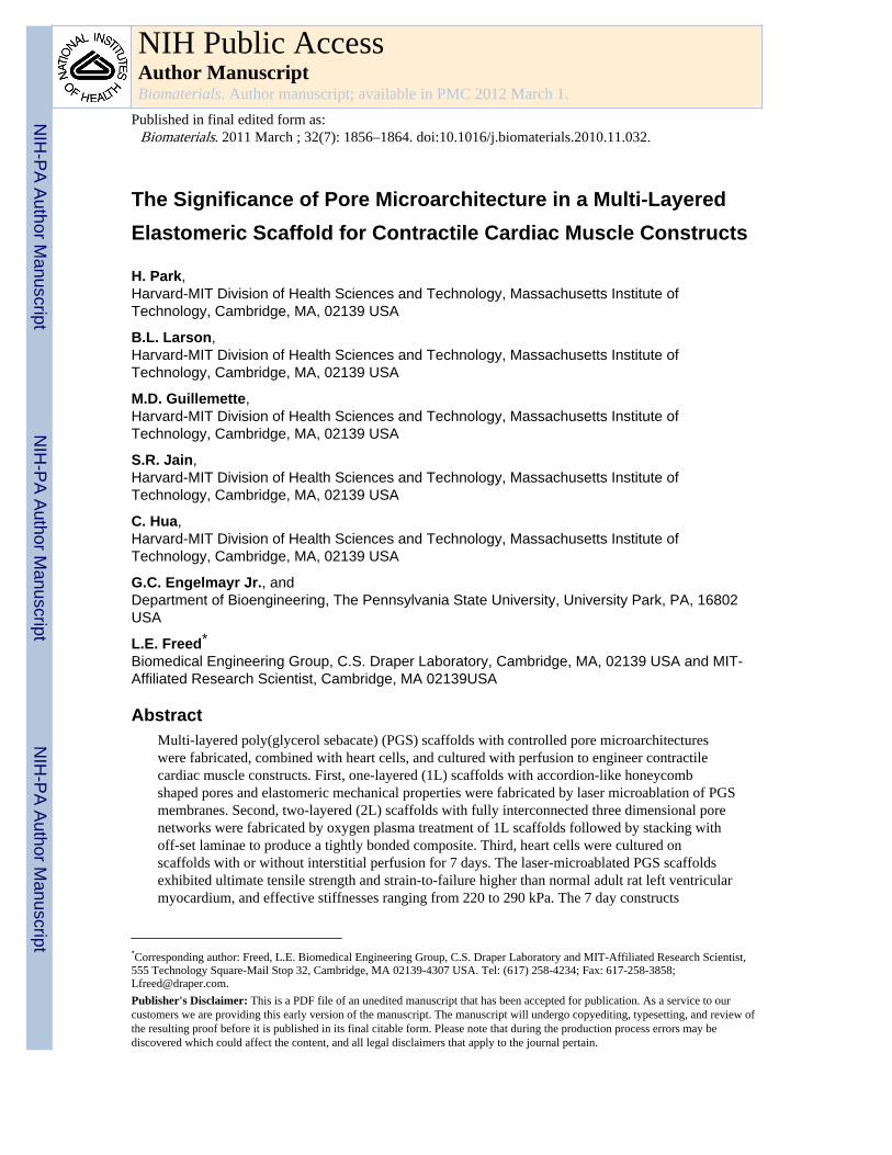
The Significance of Pore Microarchitecture in a Multi-LayeredElastomeric Scaffold for Contractile Cardiac Muscle Constructs
H. Park,Harvard-MIT Division of Health Sciences and Technology, Massachusetts Institute ofTechnology, Cambridge, MA, 02139 USA
B.L. Larson,Harvard-MIT Division of Health Sciences and Technology, Massachusetts Institute ofTechnology, Cambridge, MA, 02139 USA
M.D. Guillemette,Harvard-MIT Division of Health Sciences and Technology, Massachusetts Institute ofTechnology, Cambridge, MA, 02139 USA
S.R. Jain,Harvard-MIT Division of Health Sciences and Technology, Massachusetts Institute ofTechnology, Cambridge, MA, 02139 USA
C. Hua,Harvard-MIT Division of Health Sciences and Technology, Massachusetts Institute ofTechnology, Cambridge, MA, 02139 USA
G.C. Engelmayr Jr., andDepartment of Bioengineering, The Pennsylvania State University, University Park, PA, 16802USA
L.E. Freed*Biomedical Engineering Group, C.S. Draper Laboratory, Cambridge, MA, 02139 USA and MIT-Affiliated Research Scientist, Cambridge, MA 02139USA
AbstractMulti-layered poly(glycerol sebacate) (PGS) scaffolds with controlled pore microarchitectureswere fabricated, combined with heart cells, and cultured with perfusion to engineer contractilecardiac muscle constructs. First, one-layered (1L) scaffolds with accordion-like honeycombshaped pores and elastomeric mechanical properties were fabricated by laser microablation of PGSmembranes. Second, two-layered (2L) scaffolds with fully interconnected three dimensional porenetworks were fabricated by oxygen plasma treatment of 1L scaffolds followed by stacking withoff-set laminae to produce a tightly bonded composite. Third, heart cells were cultured onscaffolds with or without interstitial perfusion for 7 days. The laser-microablated PGS scaffoldsexhibited ultimate tensile strength and strain-to-failure higher than normal adult rat left ventricularmyocardium, and effective stiffnesses ranging from 220 to 290 kPa. The 7 day constructs
*Corresponding author: Freed, L.E. Biomedical Engineering Group, C.S. Draper Laboratory and MIT-Affiliated Research Scientist,555 Technology Square-Mail Stop 32, Cambridge, MA 02139-4307 USA. Tel: (617) 258-4234; Fax: 617-258-3858;[email protected]'s Disclaimer: This is a PDF file of an unedited manuscript that has been accepted for publication. As a service to ourcustomers we are providing this early version of the manuscript. The manuscript will undergo copyediting, typesetting, and review ofthe resulting proof before it is published in its final citable form. Please note that during the production process errors may bediscovered which could affect the content, and all legal disclaimers that apply to the journal pertain.
NIH Public AccessAuthor ManuscriptBiomaterials. Author manuscript; available in PMC 2012 March 1.
Published in final edited form as:Biomaterials. 2011 March ; 32(7): 1856–1864. doi:10.1016/j.biomaterials.2010.11.032.
NIH
-PA Author Manuscript
NIH
-PA Author Manuscript
NIH
-PA Author Manuscript
contracted in response to electrical field stimulation. Excitation thresholds were unaffected byscaffold scale up from 1L to 2L. The 2L constructs exhibited reduced apoptosis, increasedexpression of connexin-43 (Cx-43) and matrix metalloprotease-2 (MMP-2) genes, and increasedCx-43 and cardiac troponin-I proteins when cultured with perfusion as compared to static controls.Together, these findings suggest that multi-layered, microfabricated PGS scaffolds may beapplicable to myocardial repair applications requiring mechanical support, cell delivery and activeimplant contractility.
IntroductionCardiovascular disease is the leading cause of death in developed countries [1] andcongenital heart disease, which affects approximately one percent of newborns world-wide,is associated with high morbidity [2]. The functional consequences of myocardial infarction(MI) and other heart defects in which muscle fibers and collagen networks are disrupted areloss of myocardial elasticity, compliance and pumping action [3]. Current myocardialregeneration strategies, while promising [4], are unable to recreate the robust mechanicaland contractile properties of normal heart muscle. In particular, an effective graft formyocardial repair is a critical unmet need, where combining elasticity and strength withoutcompromising heart cell viability and contractility have proved challenging [5–7].
In the prototypical tissue engineering approach, three-dimensional (3D) scaffolds providethe delivery vehicle for transplanting large numbers of viable cells toward a goal of tissueregeneration [8,9]. Numerous 3D biomaterials have been explored as cardiac tissueengineering scaffolds, including non-woven poly(glycolic acid) (PGA) mesh [10-12],collagen gel [13,14], collagen foam [15–19], alginate foam [20,21], chitosan foam [22],knitted poly(lactic acid) [23], knitted hyaluronan ester [24], poly-4-hydroxybutyrate foam[25], poly(lactic acid)/poly(glycolic-co-lactic acid) (PLLA/PLGA) foam [26], andcomposites of natural and synthetic polymers [27]. However, these scaffolds are eitherthermoplastic polymers, which tend to be stiffer than normal soft tissues, degrade by bulkhydrolysis, and fail under long-term cyclic loading [28], or naturally occurring materialswith intrinsic variability, immunogenicity, and mechanical strength concerns [29]. Langerand colleagues [30] developed a tough bioresorbable elastomer, poly(glycerol-sebacate)(PGS), that degraded predominately by surface hydrolysis [31] and has been tested invarious tissue engineering applications [32–34] including myocardial repair. The mechanicalproperties of the PGS elastomer, both in the context of non-porous membranes [7,35,36] andporous scaffolds [37,38], could be tailored to match those of normal heart muscle. Recently,one-layered (1L) PGS scaffolds with in-plane pore anisotropy, i.e., rectangular andaccordion-like honeycomb pores produced by laser microablation of ~250 μm thick PGSmembranes [37], were shown to guide the alignment of cultured neonatal rat heart cells [37]and C2C12 myoblasts [39].
Alternatives to the cell-scaffold paradigm include “scaffold-free” approaches based ontransplanting cell-cell or cell-ECM grafts. As examples, engraftment and vascularizationwere demonstrated for heart cell patches comprised of human embryonic stem cell-derivedcardiomyocytes, endothelial cells, and fibroblasts [40] and electrical and vascular integrationwere demonstrated after implantation of thin (~100 μm) heart cell sheets comprised ofinterconnected cardiomyocytes [41]. However, scalability remains a major limitation ofscaffold-free approaches [9,13,42,43]. Other alternative approaches include “cell-free”biomaterials for myocardial repair. However, biomaterials used for congenital heart defectrepair in pediatric patients are limited by lack of potential for growth and remodeling[44,45], and although cell-free, non-porous PGS membranes were recently shown to reduce
Park et al. Page 2
Biomaterials. Author manuscript; available in PMC 2012 March 1.
NIH
-PA Author Manuscript
NIH
-PA Author Manuscript
NIH
-PA Author Manuscript
postinfarction myocardial hypertrophy in rodents, these implants could not assist contractilefunction, suggesting a role for cell-PGS implants in future approaches [35].
In the present study, multi-layered elastomeric PGS scaffolds with controlled poremicroarchitectures were fabricated and combined with heart cells to engineer contractilecardiac muscle constructs in vitro. Excitation threshold, gene expression, and cardiacspecific marker proteins were assessed under different conditions of cell seeding andcultivation, in particular scaffold coating with laminin (LN) to promote heart cell attachment[11,38,46] and interstitial perfusion to promote heart cell viability [12,18–20,47].
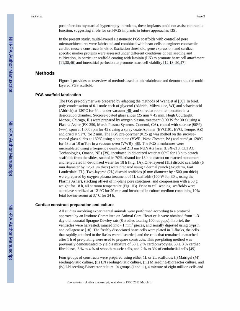
MethodsFigure 1 provides an overview of methods used to microfabricate and demonstrate the multi-layered PGS scaffold.
PGS scaffold fabricationThe PGS pre-polymer was prepared by adapting the methods of Wang et al [30]. In brief,poly-condensation of 0.1 mole each of glycerol (Aldrich, Milwaukee, WI) and sebacic acid(Aldrich) at 120ºC for 64 h under vacuum [48] and stored at room temperature in adesiccation chamber. Sucrose-coated glass slides (25 mm × 45 mm, Hugh Courtright,Monee, Chicago, IL) were prepared by oxygen plasma treatment (100 W for 30 s) using aPlasma Asher (PX-250, March Plasma Systems, Concord, CA), coated with sucrose (90%)(w/v), spun at 1,000 rpm for 45 s using a spray coater/spinner (EVG101, EVG, Tempe, AZ)and dried at 92°C for 2 min. The PGS pre-polymer (0.25 g) was melted on the sucrose-coated glass slides at 160ºC using a hot plate (VWR, West Chester, PA) and cured at 120ºCfor 48 h at 10 mTorr in a vacuum oven (VWR) [48]. The PGS membranes weremicroablated using a frequency quintupled 213 nm Nd:YAG laser (LSX-213, CETACTechnologies, Omaha, NE) [39], incubated in deionized water at 60ºC for 18 h to detachscaffolds from the slides, soaked in 70% ethanol for 18 h to extract un-reacted monomersand rehydrated in de-ionized water for 18 h (Fig. 1A). One-layered (1L) discoid scaffolds (6mm diameter by ~250 μm thick) were prepared using a dermal punch (Acuderm, FortLauderdale, FL). Two-layered (2L) discoid scaffolds (6 mm diameter by ~500 μm thick)were prepared by oxygen plasma treatment of 1L scaffolds (100 W for 30 s, using thePlasma Asher), stacking off-set of in-plane pore structures, and compression with a 50 gweight for 18 h, all at room temperature (Fig. 1B). Prior to cell seeding, scaffolds wereautoclave sterilized at 121ºC for 20 min and incubated in culture medium containing 10%fetal bovine serum at 37ºC for 24 h.
Cardiac construct preparation and cultureAll studies involving experimental animals were performed according to a protocolapproved by an Institute Committee on Animal Care. Heart cells were obtained from 1–3day old neonatal Sprague Dawley rats (8 studies totaling 100 rat pups). In brief, theventricles were harvested, minced into ~1 mm3 pieces, and serially digested using trypsinand collagenase [10]. The freshly dissociated heart cells were plated in T-flasks, the cellsthat rapidly attached to the flasks were discarded, and the cells that remained unattachedafter 1 h of pre-plating were used to prepare constructs. This pre-plating method waspreviously demonstrated to yield a mixture of 63 ± 2 % cardiomyocytes, 33 ± 3 % cardiacfibroblasts, 3 % to 4 % of smooth muscle cells, and 2 % to 3% of endothelial cells [49].
Four groups of constructs were prepared using either 1L or 2L scaffolds: (i) Matrigel (M)seeding-Static culture, (ii) LN seeding-Static culture, (iii) M seeding-Bioreactor culture, and(iv) LN seeding-Bioreactor culture. In groups (i and iii), a mixture of eight million cells and
Park et al. Page 3
Biomaterials. Author manuscript; available in PMC 2012 March 1.
NIH
-PA Author Manuscript
NIH
-PA Author Manuscript
NIH
-PA Author Manuscript
20 μL of M (BD Biosciences, catalog number 354230, San Jose, CA) was seeded onto eachscaffold while working at 4ºC in a 12-well plate, and constructs were incubated at 37ºC topromote M gelation [17]. In groups (ii and iv), scaffolds were first LN-coated by incubationin 50 μg/mL of LN (BD Biosciences) at room temperature for 4 h. Eight million cellssuspended in 20 μL of culture medium were then seeded onto each scaffold in a 24-wellinsert transwell (BD Falcon, catalog number 353096, Bedford, MA), 0.5 mL of mediumwere added to the bottom well, and constructs were incubated at 37ºC to promote cellattachment. In groups (i and ii), constructs were cultured statically in 12-well plates. Ingroups (iii and iv) constructs were cultured with bidirectional interstitial perfusion using anoscillating bioreactor [19,50]. In brief, each discoid specimen was fixed in a cylindricalspecimen chamber (8 mm long, 6 mm diameter) within a loop of silicone rubber tubing(Tygon 3350, 1/4 inch diameter × 1/32 inch wall, Cole Parmer, Vernon Hills, IL). Up to 12loops, each containing one construct and 10 mL of media, were mounted on an incubator–compatible base that slowly oscillated the chamber about its central axes at 0.05 revolutionsper minute, which corresponded to a linear interstitial flow velocity of 0.2 mm/s (Fig. 1C)[19]. Culture medium was completely replaced on culture day 4, and constructs wereharvested on culture day 7. A phase contrast micrograph of a representative construct, 6 mmdiameter × 0.5 mm thick, is shown at low magnification in Fig. 1D.
Mechanical testingPGS scaffolds and specimens of adult rat left ventricular myocardium (LV) were tested aspreviously described [37]. In brief, the PGS scaffolds, which were dry when tested, wereglued to a manila paper frame that was fixed between stainless steel grips (n=4 per testdirection, 5 mm gauge length, 3 mm width), while the LV specimens, which were wet whentested, were fixed directly between steel grips (n=9 per test direction, 5 mm gauge length, 4mm width). Specimen thicknesses were measured using a dial gauge (accuracy 0.01 mm;L.S. Starrett Co., Athol, MA). Specimens were mounted on an Electroforce ELF 3200mechanical tester (Bose-Enduratec, Framingham, MA) controlled by WinTest software andfitted with a 250 g load cell (model 31-1435-03; Sensotech, Inc., Columbus, OH).Specimens were strained to failure at a rate of approximately 1 percent per second (0.1 V/s).Independent specimens were tested in two different directions: a preferred direction (PD),where stretch was applied along the long pore axis of the scaffold or the circumferential axisof the heart in the LV group, and an orthogonal cross-preferred direction (XD), wherestretch was applied along the orthogonal scaffold pore axis or the longitudinal (apex-to-base) axis of the heart in the LV group [37]. Effective stiffness (E) was determined by alinear regression within the initial linear region of the curve up to a strain of 0.1. Ultimatetensile strength (UTS) and strain-to-failure (εf) were taken as the maximum stress and strainmeasured prior to the onset of failure, respectively.
ElectrophysiologyConstruct response to electrical field stimulation was assessed as previously described [19].In brief, specimens (4 to 7 specimens per group) were placed in environmentally controlledtest chambers fitted with two ¼-inch diameter carbon rod electrodes (Ladd Research,Williston, VT) separated by 1.5 cm and connected to platinum wire leads. An electricalpulse generator (Astro-Med Inc. West Warwick, RI) was used to apply monophasic squarepulses at 1 Hz, and excitation threshold (ET) was determined by incrementally increasingthe voltage until each stimulus was followed by a synchronous contraction of the construct,as observed at 10X magnification using a Nikon Diaphot microscope.
Scanning electron microscopy (SEM)Representative scaffolds and constructs were gradually dehydrated in a series of ethanolsolutions (35, 50, 70, 80, 90 and 100%) and then completely dried with
Park et al. Page 4
Biomaterials. Author manuscript; available in PMC 2012 March 1.
NIH
-PA Author Manuscript
NIH
-PA Author Manuscript
NIH
-PA Author Manuscript
hexamethyldisilazane solution. The dehydrated specimens were sputter-coated with Au-Pdalloy using a 108auto Sputter Coater (Cressington Scientific Instruments, Watford, UK) andexamined using a Hitachi S3500 SEM (Hitachi High Technologies America, Pleasanton,CA). Cellular dehydration during processing for SEM made it difficult to evaluate cellmorphology.
Confocal and light microscopySpecimens (2 or 3 specimens per group) were fixed in 10% neutral buffered formalin for 24h. Specimens for confocal microscopy were permeabilized using 0.1% Triton X-100,blocked with 0.1% bovine serum albumin, stained with phalloidin-fluorescein isothiocyanateconjugate (Sigma, St. Louis, MO), counterstained with DRAQ5 nuclear stain (Axxora LLC,San Diego, CA), and examined using a Zeiss LSM 510 laser scanning confocal microscopeas previously described [37]. Other specimens for histological analyses were paraffin-embedded and sectioned to 5 μm thickness, stained, and examined using a Zeiss Axiovert200M microscope [19]. Apoptosis was assessed by the terminal deoxynucleotidyltransferase biotin 2’-deoxyuridine 5’-triphosphate nick end labeling (TUNEL) assay [19].Cardiac troponin-I (Tn-I) and connexin-43 (Cx-43) were assessed by incubation with anappropriate primary antibody (AB1627 for Tn-I; 05-763 for Cx-43, Millipore, Billerica,MA) and secondary antibody, stained with a Standard Elite ABC kit (Vector, Peterborough,UK), and counterstained with hematoxylin [19].
Real time polymerase chain reaction (PCR) and deoxyribonucleic acid (DNA) assaysTo quantify construct gene expression levels, real time PCR analyses were performed usingTaqman® Universal PCR Master Mix (Applied Biosystems, Foster City, CA). In brief, totalRNA was isolated (10 specimens per group) by homogenization in Trizol (Invitrogen,Carlsbad, CA) followed by extraction in chloroform and centrifugation (20,800 g, 4°C, 20min). The RNA was precipitated using a RNeasy Mini kit (Qiagen, Valencia, CA). ThecDNA was synthesized by reverse transcription using the Superscript First-Strand SynthesisSystem (Invitrogen) with a PCR Sprint Thermal cycler (Thermo Electron Co., Waltham,MA). The gene-specific primers for Cx-43, matrix metalloprotease-2 (MMP-2), andglyceraldehyde 3-phosphate dehydrogenase (GAPDH) were respectively Rn 01433957_s1,Rn 02532334_s1, and Rn99999916_s1 (Assays-on demand ™ products, AppliedBiosystems). Gene expression levels were normalized to GAPDH and then relativeexpression was calculated. Construct DNA content was assessed using the Quant-iTPicogreen dsDNA assay kit (Invitrogen, Carlsbad, CA).
Statistical analysisData were calculated as mean ± standard error and analyzed by analysis of variance inconjunction with Tukey’s post hoc test using Statistica (Tulsa, OK). Statistical significancewas established at P values < 0.05.
ResultsScaffold microfabrication
To produce 1L scaffolds with accordion-like honeycomb pores in PGS membranes, weadapted our previously described method [37] for use with a frequency quintupled 213 nmNd:YAG laser. A program was written in Visual Basic for Applications to generatesequences of coordinates and laser parameters, and suitably formatted for uploading into thesoftware controlling this laser (DigiLaz II, v.3.1.0; CETAC Technologies) such that aspecified in-plane pore microarchitecture could be automatically created in a specifiednumber of rows and columns. By using this laser’s standard square aperture and
Park et al. Page 5
Biomaterials. Author manuscript; available in PMC 2012 March 1.
NIH
-PA Author Manuscript
NIH
-PA Author Manuscript
NIH
-PA Author Manuscript
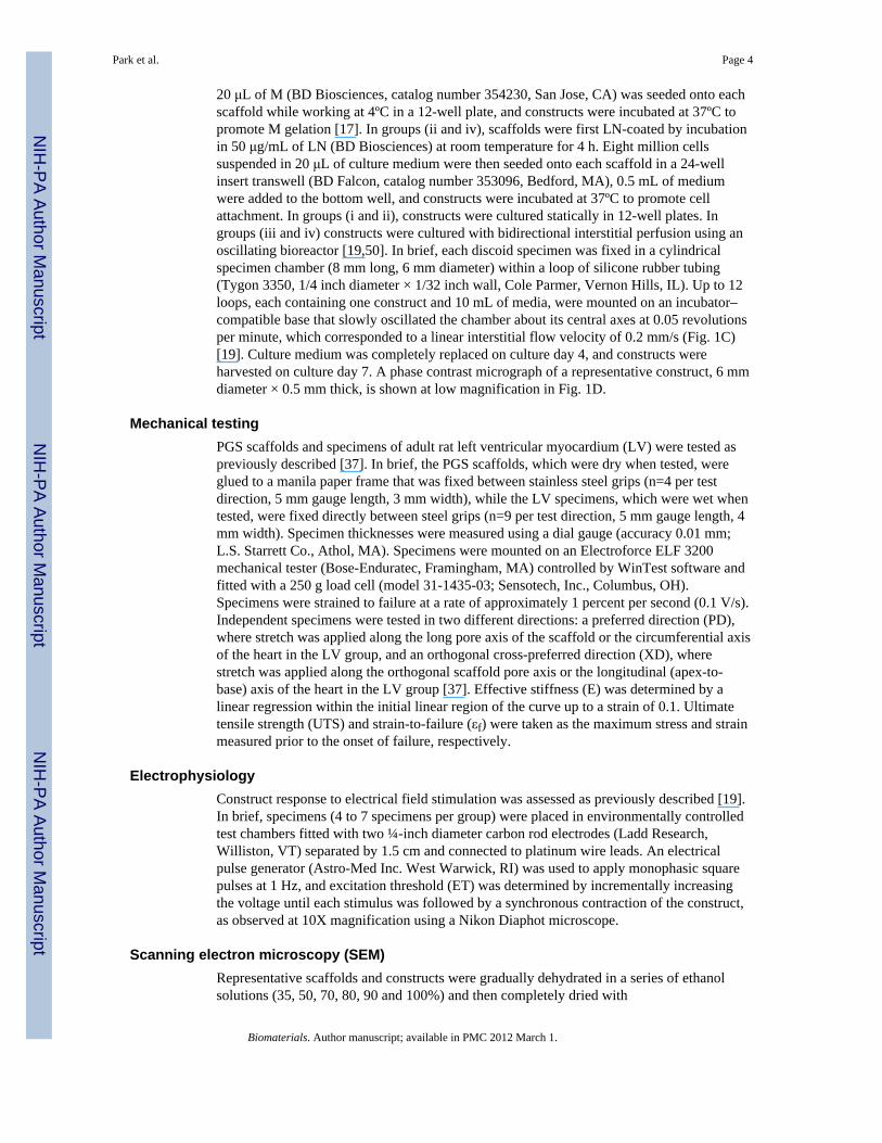
demagnification objective lens and focusing on the top surface of the PGS membrane, thespot size was 150 μm × 150 μm. Operating parameters selected for laser microablation of250 μm thick PGS membranes were 100% energy (4.3 mJ), 20 Hz, and 150 shots per hole.By overlapping two square pores oriented at 45 degrees, 1L scaffolds were produced withaccordion-like pores that extended from the top to the bottom of the PGS membrane (Fig.1A). The pores were on average ~250 μm long × 150 μm wide and the intervening structuralelements (i.e., struts) were on average ~40 μm wide (Fig. 2A).
To produce two-layered (2L) scaffolds, we adapted a technique previously used for adhesivebonding of non-degradable materials [51,52] to the biodegradable PGS elastomer.Prefabricated 1L scaffolds were oxygen plasma treated (100 W for 30 s), stacked such thatthe pores and struts in each layer were off-set, and laminated with compression, all at roomtemperature. The resulting 2L scaffolds had in-plane accordion-like honeycomb pores andoff-set, interconnected pores extending from the top to the bottom surface of the compositescaffold (Fig. 1B and Fig. 2B). Oxygen plasma-mediated lamination yielded a tight interfacebetween the two bonded PGS layers that could not be distinguished by SEM (Fig. 2C,D) andwas not abolished by subsequent processing steps including ethanol washing.
Effects of scaffold pore microarchitecture on heart cell seeding and cultureThe main differences in pore structure between 1L scaffolds (~250 μm thick) and 2Lscaffolds (~500 μm thick) was the presence of a fully interconnected 3D pore network withlateral off-set between lamina in the 2L scaffolds. This 3D pore microarchitecture allowedcells to be readily seeded throughout its full thickness (Fig. 2C) and also allowed masstransport to and from centrally located cells by interstitial perfusion (Fig. 1C). The 1L and2L scaffolds were mechanically stable over 7 days of culture with heart cells under staticand perfusion conditions. Harvested constructs exhibited good handling properties and lightmicroscopy showed no evidence of delamination or PGS degradation over the relativelyshort (i.e. 7 day) culture period.
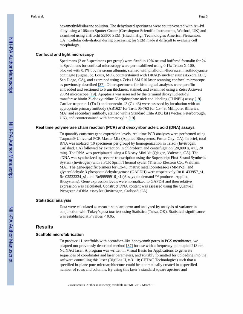
Scaffold mechanical characterizationBaseline mechanical properties of 1L PGS scaffolds and specimens of normal adult rat LVare shown in Fig. 3. Scaffolds exhibited values for E ranging from 220 to 290 kPa, UTSranging from 200 to 225 kPa, and εf ranging from 1.5 to 1.8 (Fig. 3A, B, C, respectively).Differences in mechanical properties due to specimen type (i.e., scaffold versus LV) werestatistically significant for E (p<0.0001), UTS (p<0.0001) and εf (p<0.0001), such thatscaffolds were stiffer than normal LV and failed at stress values and strain values higherthan normal LV. Differences in mechanical properties due to test direction (i.e., PD versusXD) were statistically significant for E (p<0.01) and UTS (p<0.05), indicating a trendtoward scaffold anisotropy.
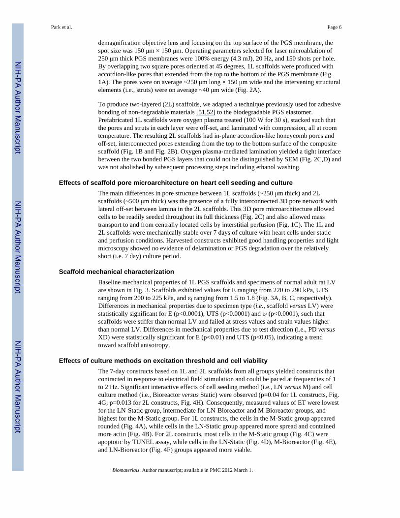
Effects of culture methods on excitation threshold and cell viabilityThe 7-day constructs based on 1L and 2L scaffolds from all groups yielded constructs thatcontracted in response to electrical field stimulation and could be paced at frequencies of 1to 2 Hz. Significant interactive effects of cell seeding method (i.e., LN versus M) and cellculture method (i.e., Bioreactor versus Static) were observed (p=0.04 for 1L constructs, Fig.4G; p=0.013 for 2L constructs, Fig. 4H). Consequently, measured values of ET were lowestfor the LN-Static group, intermediate for LN-Bioreactor and M-Bioreactor groups, andhighest for the M-Static group. For 1L constructs, the cells in the M-Static group appearedrounded (Fig. 4A), while cells in the LN-Static group appeared more spread and containedmore actin (Fig. 4B). For 2L constructs, most cells in the M-Static group (Fig. 4C) wereapoptotic by TUNEL assay, while cells in the LN-Static (Fig. 4D), M-Bioreactor (Fig. 4E),and LN-Bioreactor (Fig. 4F) groups appeared more viable.
Park et al. Page 6
Biomaterials. Author manuscript; available in PMC 2012 March 1.
NIH
-PA Author Manuscript
NIH
-PA Author Manuscript
NIH
-PA Author Manuscript
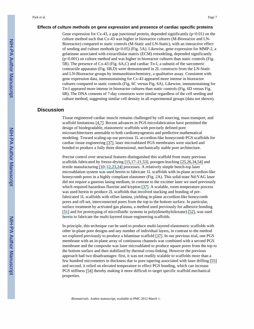
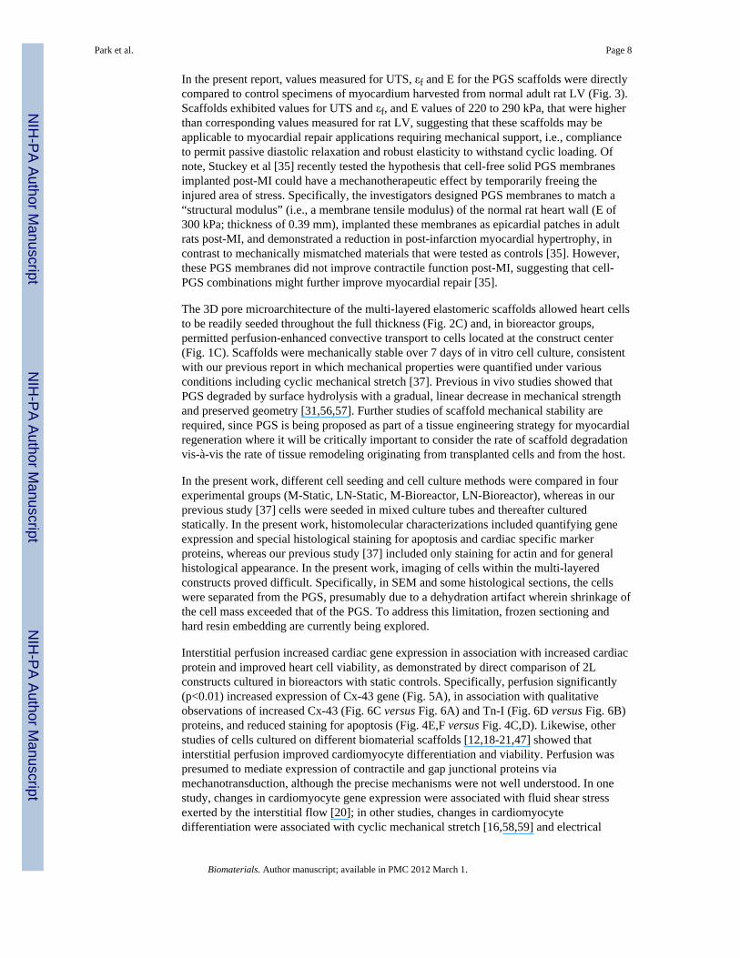
Effects of culture methods on gene expression and presence of cardiac specific proteinsGene expression for Cx-43, a gap junctional protein, depended significantly (p<0.01) on theculture method such that Cx-43 was higher in bioreactor cultures (M-Bioreactor and LN-Bioreactor) compared to static controls (M-Static and LN-Static), with an interactive effectof seeding and culture methods (p<0.05) (Fig. 5A). Likewise, gene expression for MMP-2, agelatinase associated with extracellular matrix (ECM) remodeling, depended significantly(p<0.001) on culture method and was higher in bioreactor cultures than static controls (Fig.5B). The presence of Cx-43 (Fig. 6A,C) and cardiac Tn-I, a subunit of the sarcomericcontractile apparatus (Fig. 6B,D) were demonstrated in 2L constructs from the LN-Staticand LN-Bioreactor groups by immunohistochemistry, a qualitative assay. Consistent withgene expression data, immunostaining for Cx-43 appeared more intense in bioreactorcultures compared to static controls (Fig. 6C versus Fig. 6A). Likewise, immunostaining forTn-I appeared more intense in bioreactor cultures than static controls (Fig. 6D versus Fig.6B). The DNA contents of 7-day constructs were similar regardless of the cell seeding andculture method, suggesting similar cell density in all experimental groups (data not shown).
DiscussionTissue engineered cardiac muscle remains challenged by cell sourcing, mass transport, andscaffold limitations [4,7]. Recent advances in PGS microfabrication have permitted thedesign of biodegradable, elastomeric scaffolds with precisely defined poremicroarchitectures amenable to both cardiomyogenesis and predictive mathematicalmodeling. Toward scaling-up our previous 1L accordion-like honeycomb PGS scaffolds forcardiac tissue engineering [37], laser microablated PGS membranes were stacked andbonded to produce a fully three dimensional, mechanically stable pore architecture.
Precise control over structural features distinguished this scaffold from many previousscaffolds fabricated by freeze-drying [15,17–21,53], porogen-leaching [25,26,34,54] andtextile manufacturing [10–12,23,24] processes. A relatively simple bench-top lasermicroablation system was used herein to fabricate 1L scaffolds with in-plane accordion-likehoneycomb pores in a highly compliant elastomer (Fig. 2A). This solid-state Nd:YAG laserdid not require a gaseous lasing medium, in contrast to the excimer laser we used previouslywhich required hazardous fluorine and krypton [37]. A scalable, room temperature processwas used herein to produce 2L scaffolds that involved stacking and bonding of pre-fabricated 1L scaffolds with offset lamina, yielding in-plane accordion-like honeycombpores and off-set, interconnected pores from the top to the bottom surface. In particular,surface treatment by activated gas plasma, a method used previously for adhesive bonding[51] and for prototyping of microfluidic systems in poly(dimethylsiloxane) [52], was usedherein to fabricate the multi-layered tissue engineering scaffolds.
In principle, this technique can be used to produce multi-layered elastomeric scaffolds withother in-plane pore designs and any number of individual layers, in contrast to the methodwe explored previously to produce a bilaminar scaffold [37]. In our previous trial, one PGSmembrane with an in-plane array of continuous channels was combined with a second PGSmembrane and the composite was laser microablated to produce square pores from the top tothe bottom surface and then stabilized by thermal cross-linking. However the previousapproach had two disadvantages: first, it was not readily scalable to scaffolds more than afew hundred micrometers in thickness due to pore tapering associated with laser drilling [55]and second, it relied on elevated temperature to effect PGS bonding, which can increasePGS stiffness [54] thereby making it more difficult to target specific scaffold mechanicalproperties.
Park et al. Page 7
Biomaterials. Author manuscript; available in PMC 2012 March 1.
NIH
-PA Author Manuscript
NIH
-PA Author Manuscript
NIH
-PA Author Manuscript
In the present report, values measured for UTS, εf and E for the PGS scaffolds were directlycompared to control specimens of myocardium harvested from normal adult rat LV (Fig. 3).Scaffolds exhibited values for UTS and εf, and E values of 220 to 290 kPa, that were higherthan corresponding values measured for rat LV, suggesting that these scaffolds may beapplicable to myocardial repair applications requiring mechanical support, i.e., complianceto permit passive diastolic relaxation and robust elasticity to withstand cyclic loading. Ofnote, Stuckey et al [35] recently tested the hypothesis that cell-free solid PGS membranesimplanted post-MI could have a mechanotherapeutic effect by temporarily freeing theinjured area of stress. Specifically, the investigators designed PGS membranes to match a“structural modulus” (i.e., a membrane tensile modulus) of the normal rat heart wall (E of300 kPa; thickness of 0.39 mm), implanted these membranes as epicardial patches in adultrats post-MI, and demonstrated a reduction in post-infarction myocardial hypertrophy, incontrast to mechanically mismatched materials that were tested as controls [35]. However,these PGS membranes did not improve contractile function post-MI, suggesting that cell-PGS combinations might further improve myocardial repair [35].
The 3D pore microarchitecture of the multi-layered elastomeric scaffolds allowed heart cellsto be readily seeded throughout the full thickness (Fig. 2C) and, in bioreactor groups,permitted perfusion-enhanced convective transport to cells located at the construct center(Fig. 1C). Scaffolds were mechanically stable over 7 days of in vitro cell culture, consistentwith our previous report in which mechanical properties were quantified under variousconditions including cyclic mechanical stretch [37]. Previous in vivo studies showed thatPGS degraded by surface hydrolysis with a gradual, linear decrease in mechanical strengthand preserved geometry [31,56,57]. Further studies of scaffold mechanical stability arerequired, since PGS is being proposed as part of a tissue engineering strategy for myocardialregeneration where it will be critically important to consider the rate of scaffold degradationvis-à-vis the rate of tissue remodeling originating from transplanted cells and from the host.
In the present work, different cell seeding and cell culture methods were compared in fourexperimental groups (M-Static, LN-Static, M-Bioreactor, LN-Bioreactor), whereas in ourprevious study [37] cells were seeded in mixed culture tubes and thereafter culturedstatically. In the present work, histomolecular characterizations included quantifying geneexpression and special histological staining for apoptosis and cardiac specific markerproteins, whereas our previous study [37] included only staining for actin and for generalhistological appearance. In the present work, imaging of cells within the multi-layeredconstructs proved difficult. Specifically, in SEM and some histological sections, the cellswere separated from the PGS, presumably due to a dehydration artifact wherein shrinkage ofthe cell mass exceeded that of the PGS. To address this limitation, frozen sectioning andhard resin embedding are currently being explored.
Interstitial perfusion increased cardiac gene expression in association with increased cardiacprotein and improved heart cell viability, as demonstrated by direct comparison of 2Lconstructs cultured in bioreactors with static controls. Specifically, perfusion significantly(p<0.01) increased expression of Cx-43 gene (Fig. 5A), in association with qualitativeobservations of increased Cx-43 (Fig. 6C versus Fig. 6A) and Tn-I (Fig. 6D versus Fig. 6B)proteins, and reduced staining for apoptosis (Fig. 4E,F versus Fig. 4C,D). Likewise, otherstudies of cells cultured on different biomaterial scaffolds [12,18-21,47] showed thatinterstitial perfusion improved cardiomyocyte differentiation and viability. Perfusion waspresumed to mediate expression of contractile and gap junctional proteins viamechanotransduction, although the precise mechanisms were not well understood. In onestudy, changes in cardiomyocyte gene expression were associated with fluid shear stressexerted by the interstitial flow [20]; in other studies, changes in cardiomyocytedifferentiation were associated with cyclic mechanical stretch [16,58,59] and electrical
Park et al. Page 8
Biomaterials. Author manuscript; available in PMC 2012 March 1.
NIH
-PA Author Manuscript
NIH
-PA Author Manuscript
NIH
-PA Author Manuscript
stimulation [53,60]. In the present study, perfusion significantly (p<0.001) increasedMMP-2 gene expression as compared to statically cultured controls. Likewise, otherspreviously related increased MMP expression with fluid shear stress and interstitial flow[61,62], and we reported increased cell-to-cell network formation in association withincreased MMP-2 expression in engineered cardiac tissue [63]. Perfusion was presumed toimprove cell survival by enhanced convective transport of oxygen, since hypoxia inducesapoptosis in cardiomyocytes [64]; of note the bioreactor used in the present study wasentirely made of gas permeable silicone rubber such that entire device served as anoxygenator [19].
There was an interactive effect of cell seeding method and cell culture method on ET and,unexpectedly, the lowest threshold was measured for the LN-Static group (Fig. 4G and Fig.4H). This finding may be rationalized by biophysical cues introduced by either the presenceof perfusion or hydrogel and acting in such a manner as to attenuate mechanical andmicrostructural cues provided by the PGS scaffold. Specifically, cell embedding in Matrigel,a hydrogel too soft to promote heart cell spreading [65,66] or myogenesis by stem cells [67],may have attenuated mechanical cues provided by the PGS in the M-Bioreactor and M-Static groups. Moreover, fluid shear stresses caused by interstitial cross-perfusion may haveattenuated in-plane microstructural cues provided by the accordion-like honeycomb pores inthe LN-Bioreactor group. Hence, this study provided new insight into why previous studiesthat used Matrigel or cross-perfusion [12,18–21,38,47] may not have achieved the desiredheart cell differentiation. To further explore this hypothesis will require adapted in vitromodels in which scaffold-based cell guidance features and biophysical cues are betterintegrated such that effects of these two stimuli on cell differentiation can act in concertinstead of in opposition.
Ongoing work involves further optimization of laser microablation conditions, including theuse of different apertures, since the dimensions (and associated volume fraction) of the PGSstruts are critical determinants of scaffold structural and mechanical properties and the in-plane anisotropy recently shown to be associated with heart cell guidance [37,39]. Otherefforts involve mathematical modeling and simulation of the elastomeric mechanicalproperties of the multi-layered elastomeric scaffolds, based on the in-plane tessellation ofaccordion-like honeycomb pores [68].
ConclusionMulti-layered elastomeric PGS scaffolds with controlled pore microarchitectures werefabricated by laser ablation and oxygen plasma-mediated lamination, seeded with heart cells,and cultured with interstitial perfusion. The laser-microablated PGS exhibited UTS and εfhigher than normal rat left ventricular myocardium and stiffnesses ranging from 220 to 290kPa. Heart cell culture on these scaffolds yielded cardiac muscle constructs. Excitationthresholds were unaffected by scaffold scale up from 1L to 2L. The 2L constructs exhibitedreduced apoptosis, increased expression of Cx-43 and MMP-2 genes, and qualitativeincreases in Cx-43 and Tn-I proteins when cultured with perfusion as compared to staticcontrols. Together, these findings suggest that multi-layered microfabricated PGS scaffoldsmay be applicable to myocardial repair applications requiring mechanical support, celldelivery and active implant contractility.
AcknowledgmentsThis work was funded by the American Recovery and Reinvestment Act (ARRA), Award 1-R01-HL086521-01A2(to LEF) from the National Heart, Lung and Blood Institute (NHLBI). The content is solely the responsibility of theauthors and does not necessarily represent the official views of the NHLBI or NIH. We are indebted to R. Langerfor general advice, J. Wang and J. Hsiao for help with polymer synthesis, processing, and SEM, N. Watson for help
Park et al. Page 9
Biomaterials. Author manuscript; available in PMC 2012 March 1.
NIH
-PA Author Manuscript
NIH
-PA Author Manuscript
NIH
-PA Author Manuscript
with microscopy, M.G. Moretti and G. Talo and E. Kim for help with the bioreactor, and A. Jean for helpfuldiscussions regarding scaffold mechanics and modeling.
References1. Lloyd-Jones D, Adams RJ, Brown TM, Carnethon M, Dai S, De Simone G, et al. Heart disease and
stroke statistics--2010 update: a report from the American Heart Association. Circulation2010;121:e46–e215. [PubMed: 20019324]
2. Martin JA, Hamilton BE, Sutton PD, Ventura SJ, Menacker F, Munson ML. Births: final data for2003. Natl Vital Stat Rep 2005;54:1–116.
3. Fomovsky GM, Holmes JW. Evolution of scar structure, mechanics, and ventricular function aftermyocardial infarction in the rat. Am J Physiol Heart Circ Physiol 2010;298:H221–8. [PubMed:19897714]
4. Chien KR, Domian IJ, Parker KK. Cardiogenesis and the complex biology of regenerativecardiovascular medicine. Science 2008;322:1494–7. [PubMed: 19056974]
5. Boethig D, Thies WR, Hecker H, Breymann T. Mid term course after pediatric right ventricularoutflow tract reconstruction: a comparison of homografts, porcine xenografts and Contegras. Eur JCardio-Thorac Surg 2005;27:58–66.
6. Bautista-Hernandez V, Marx GR, Gauvreau K, Pigula FA, Bacha EA, Mayer JE Jr, et al.Coarctectomy reduces neoaortic arch obstruction in hypoplastic left heart syndrome. J ThoracCardiovasc Surg 2007;133:1540–6. [PubMed: 17532953]
7. Chen QZ, Harding SE, Ali NN, Lyon AR, Boccaccini AR. Biomaterials in cardiac tissueengineering: ten years of research survey. Mater Sci Eng R-Reports 2008;59:1–37.
8. Langer R, Vacanti JP. Tissue engineering. Science 1993;260:920–6. [PubMed: 8493529]9. Mooney DJ, Vandenburgh H. Cell delivery mechanisms for tissue repair. Cell Stem Cell
2008;2:205–13. [PubMed: 18371446]10. Bursac N, Papadaki M, Cohen RJ, Schoen FJ, Eisenberg SR, Carrier R, et al. Cardiac muscle tissue
engineering: toward an in vitro model for electrophysiological studies. Am J Physiol1999;277:H433–44. [PubMed: 10444466]
11. Papadaki M, Bursac N, Langer R, Merok J, Vunjak-Novakovic G, Freed LE. Tissue engineering offunctional cardiac muscle: molecular, structural, and electrophysiological studies. Am J PhysiolHeart Circ Physiol 2001;280:H168–78. [PubMed: 11123231]
12. Carrier RL, Rupnick M, Langer R, Schoen FJ, Freed LE, Vunjak-Novakovic G. Perfusionimproves tissue architecture of engineered cardiac muscle. Tissue Eng 2002;8:175–88. [PubMed:12031108]
13. Eschenhagen T, Zimmermann WH. Engineering myocardial tissue. Circ Res 2005;97:1220–31.[PubMed: 16339494]
14. Zimmermann WH, Melnychenko I, Wasmeier G, Didie M, Naito H, Nixdorff U, et al. Engineeredheart tissue grafts improve systolic and diastolic function in infarcted rat hearts. Nat Med2006;12:452–8. [PubMed: 16582915]
15. Li RK, Jia ZQ, Weisel RD, Mickle DA, Choi A, Yau TM. Survival and function of bioengineeredcardiac grafts. Circulation 1999;100:II63–9. [PubMed: 10567280]
16. Akhyari P, Fedak PW, Weisel RD, Lee TY, Verma S, Mickle DA, et al. Mechanical stretchregimen enhances the formation of bioengineered autologous cardiac muscle grafts. Circulation2002;106:I137–42. [PubMed: 12354723]
17. Radisic M, Euloth M, Yang L, Langer R, Freed LE, Vunjak-Novakovic G. High-density seeding ofmyocyte cells for cardiac tissue engineering. Biotechnol Bioeng 2003;82:403–14. [PubMed:12632397]
18. Radisic M, Yang L, Boublik J, Cohen RJ, Langer R, Freed LE, et al. Medium perfusion enablesengineering of compact and contractile cardiac tissue. Am J Physiol Heart Circ Physiol2004;286:H507–16. [PubMed: 14551059]
19. Cheng M, Moretti M, Engelmayr GC, Freed LE. Insulin-like growth factor-I and slow, bi-directional perfusion enhance the formation of tissue-engineered cardiac grafts. Tissue Eng Part A2009;15:645–53. [PubMed: 18759675]
Park et al. Page 10
Biomaterials. Author manuscript; available in PMC 2012 March 1.
NIH
-PA Author Manuscript
NIH
-PA Author Manuscript
NIH
-PA Author Manuscript
20. Dvir T, Levy O, Shachar M, Granot Y, Cohen S. Activation of the ERK1/2 cascade via pulsatileinterstitial fluid flow promotes cardiac tissue assembly. Tissue Eng 2007;13:2185–93. [PubMed:17518740]
21. Dvir T, Benishti N, Shachar M, Cohen S. A novel perfusion bioreactor providing a homogenousmilieu for tissue regeneration. Tissue Eng 2006;12:2843–52. [PubMed: 17518653]
22. Blan NR, Birla RK. Design and fabrication of heart muscle using scaffold-based tissueengineering. J Biomed Mater Res A 2008;86:195–208. [PubMed: 17972281]
23. Matsubayashi K, Fedak PW, Mickle DA, Weisel RD, Ozawa T, Li RK. Improved left ventricularaneurysm repair with bioengineered vascular smooth muscle grafts. Circulation 2003;108(Suppl1):II219–25. [PubMed: 12970236]
24. Boublik J, Park H, Radisic M, Tognana E, Chen F, Pei M, et al. Mechanical properties andremodeling of hybrid cardiac constructs made from heart cells, fibrin, and biodegradable,elastomeric knitted fabric. Tissue Eng 2005;11:1122–32. [PubMed: 16144448]
25. Yang C, Sodian R, Fu P, Luders C, Lemke T, Du J, et al. In vitro fabrication of a tissue engineeredhuman cardiovascular patch for future use in cardiovascular surgery. Ann Thorac Surg2006;81:57–63. [PubMed: 16368335]
26. Lesman A, Habib M, Caspi O, Gepstein A, Arbel G, Levenberg S, et al. Transplantation of atissue-engineered human vascularized cardiac muscle. Tissue Eng Part A 2010;16:115–25.[PubMed: 19642856]
27. Park H, Radisic M, Lim JO, Chang BH, Vunjak-Novakovic G. A novel composite scaffold forcardiac tissue engineering. In Vitro Cell Dev Biol Anim 2005;41:188–96. [PubMed: 16223333]
28. Engelmayr GC Jr, Hildebrand DK, Sutherland FW, Mayer JE Jr, Sacks MS. A novel bioreactor forthe dynamic flexural stimulation of tissue engineered heart valve biomaterials. Biomaterials2003;24:2523–32. [PubMed: 12695079]
29. Feng Z, Matsumoto T, Nakamura T. Measurements of the mechanical properties of contractedcollagen gels populated with rat fibroblasts or cardiomyocytes. J Artif Organs 2003;6:192–6.[PubMed: 14598103]
30. Wang Y, Ameer GA, Sheppard BJ, Langer R. A tough biodegradable elastomer. Nat Biotechnol2002;20:602–6. [PubMed: 12042865]
31. Wang Y, Kim YM, Langer R. In vivo degradation characteristics of poly(glycerol sebacate). JBiomed Mater Res A 2003;66:192–7. [PubMed: 12833446]
32. Pritchard CD, Arner KM, Neal RA, Neeley WL, Bojo P, Bachelder E, et al. The use of surfacemodified poly(glycerol-co-sebacic acid) in retinal transplantation. Biomaterials 2009;31:2153–62.[PubMed: 19962754]
33. Kemppainen JM, Hollister SJ. Tailoring the mechanical properties of 3D-designed poly(glycerolsebacate) scaffolds for cartilage applications. J Biomed Mater Res A 2010;94:9–18. [PubMed:20091702]
34. Crapo PM, Gao J, Wang Y. Seamless tubular poly(glycerol sebacate) scaffolds: high-yieldfabrication and potential applications. J Biomed Mater Res 2008;86:354–63.
35. Stuckey DJ, Ishii H, Chen QZ, Boccaccini AR, Hansen U, Carr CA, et al. Magnetic resonanceimaging evaluation of remodeling by cardiac elastomeric tissue scaffold biomaterials in a ratmodel of myocardial infarction. Tissue Eng Part A 2010;16:3395–402. [PubMed: 20528670]
36. Chen QZ, Bismarck A, Hansen U, Junaid S, Tran MQ, Harding SE, et al. Characterisation of a softelastomer poly(glycerol sebacate) designed to match the mechanical properties of myocardialtissue. Biomaterials 2008;29:47–57. [PubMed: 17915309]
37. Engelmayr GC Jr, Cheng M, Bettinger CJ, Borenstein JT, Langer R, Freed LE. Accordion-likehoneycombs for tissue engineering of cardiac anisotropy. Nat Mater 2008;7:1003–10. [PubMed:18978786]
38. Marsano A, Maidhof R, Wan LQ, Wang Y, Gao J, Tandon N, et al. Scaffold stiffness affects thecontractile function of three-dimensional engineered cardiac constructs. Biotechnol Progress2010;26:1382–90.
39. Guillemette MD, Park H, Hsiao JC, Jain SR, Larson BL, Langer R, et al. Combined technologiesfor microfabricating elastomeric cardiac tissue engineering scaffolds. Macromol Biosci2010;10:1330–7. [PubMed: 20718054]
Park et al. Page 11
Biomaterials. Author manuscript; available in PMC 2012 March 1.
NIH
-PA Author Manuscript
NIH
-PA Author Manuscript
NIH
-PA Author Manuscript
40. Stevens KR, Kreutziger KL, Dupras SK, Korte FS, Regnier M, Muskheli V, et al. Physiologicalfunction and transplantation of scaffold-free and vascularized human cardiac muscle tissue. ProcNatl Acad Sci U S A 2009;106:16568–73. [PubMed: 19805339]
41. Furuta A, Miyoshi S, Itabashi Y, Shimizu T, Kira S, Hayakawa K, et al. Pulsatile cardiac tissuegrafts using a novel three-dimensional cell sheet manipulation technique functionally integrateswith the host heart, in vivo. Circ Res 2006;98:705–12. [PubMed: 16469955]
42. Laflamme MA, Murry CE. Regenerating the heart. Nat Biotechnol 2005;23:845–56. [PubMed:16003373]
43. Shimizu T, Sekine H, Isoi Y, Yamato M, Kikuchi A, Okano T. Long-term survival and growth ofpulsatile myocardial tissue grafts engineered by the layering of cardiomyocyte sheets. Tissue Eng2006;12:499–507. [PubMed: 16579683]
44. Shin'oka T, Matsumura G, Hibino N, Naito Y, Watanabe M, Konuma T, et al. Midterm clinicalresult of tissue-engineered vascular autografts seeded with autologous bone marrow cells. J ThoracCardiovasc Surg 2005;129:1330–8. [PubMed: 15942574]
45. Delius RE, Walters HL 3rd. Re-operative surgery in pediatric patients. Semin Thorac CardiovascSurg Pediatr Card Surg Annu 2003;6:108–15. [PubMed: 12740777]
46. Boateng SY, Lateef SS, Mosley W, Hartman TJ, Hanley L, Russell B. RGD and YIGSR syntheticpeptides facilitate cellular adhesion identical to that of laminin and fibronectin but alter thephysiology of neonatal cardiac myocytes. Am J Physiol Cell Physiol 2005;288:C30–8. [PubMed:15371257]
47. Maidhof R, Marsano A, Lee EJ, Vunjak-Novakovic G. Perfusion seeding of channeled elastomericscaffolds with myocytes and endothelial cells for cardiac tissue engineering. Biotechnol Prog2010;26:565–72. [PubMed: 20052737]
48. Neeley WL, Redenti S, Klassen H, Tao S, Desai T, Young MJ, et al. A microfabricated scaffold forretinal progenitor cell grafting. Biomaterials 2008;29:418–26. [PubMed: 17961646]
49. Naito H, Melnychenko I, Didie M, Schneiderbanger K, Schubert P, Rosenkranz S, et al.Optimizing engineered heart tissue for therapeutic applications as surrogate heart muscle.Circulation 2006;114:I77–8.
50. Valonen PK, Moutos FT, Kusanagi A, Moretti MG, Diekman BO, Welter JF, et al. In vitrogeneration of mechanically functional cartilage grafts based on adult human stem cells and 3D-woven poly(epsilon-caprolactone) scaffolds. Biomaterials 2010;31:2193–200. [PubMed:20034665]
51. Hall JR, Westerdahl CAL, Devine AT, Bodnar MJ. Activated gas plasma surface treatment ofpolymers for adhesive bonding. J Appl Polym Sci 1969;13:2085–96.
52. Duffy DC, McDonald JC, Schueller OJA, Whitesides GM. Rapid prototyping of microfluidicsystems in poly(dimethylsiloxane). Anal Chem 1998;70:4974–84.
53. Radisic M, Park H, Shing H, Consi T, Schoen FJ, Langer R, et al. Functional assembly ofengineered myocardium by electrical stimulation of cardiac myocytes cultured on scaffolds. ProcNatl Acad Sci U S A 2004;101:18129–34. [PubMed: 15604141]
54. Gao J, Crapo PM, Wang Y. Macroporous elastomeric scaffolds with extensive micropores for softtissue engineering. Tissue Eng 2006;12:917–25. [PubMed: 16674303]
55. Dahotre, NB.; Harimkar, SP. Laser fabrication and machining of materials. New York: SpringerScience Business Media LLC; 2008.
56. Sundback CA, Shyu JY, Wang Y, Faquin WC, Langer RS, Vacanti JP, et al. Biocompatibilityanalysis of poly(glycerol sebacate) as a nerve guide material. Biomaterials 2005;26:5454–64.[PubMed: 15860202]
57. Pomerantseva I, Krebs N, Hart A, Neville CM, Huang AY, Sundback CA. Degradation behavior ofpoly(glycerol sebacate). J Biomed Mater Res A 2009;91:1038–47. [PubMed: 19107788]
58. Fink C, Ergun S, Kralisch D, Remmers U, Weil J, Eschenhagen T. Chronic stretch of engineeredheart tissue induces hypertrophy and functional improvement. FASEB J 2000;14:669–79.[PubMed: 10744624]
59. Birla RK, Huang YC, Dennis RG. Development of a novel bioreactor for the mechanical loadingof tissue-engineered heart muscle. Tissue Eng 2007;13:2239–48. [PubMed: 17590151]
Park et al. Page 12
Biomaterials. Author manuscript; available in PMC 2012 March 1.
NIH
-PA Author Manuscript
NIH
-PA Author Manuscript
NIH
-PA Author Manuscript
60. Tandon N, Cannizzaro C, Chao PH, Maidhof R, Marsano A, Au HT, et al. Electrical stimulationsystems for cardiac tissue engineering. Nat Protoc 2009;4:155–73. [PubMed: 19180087]
61. Kang H, Bayless KJ, Kaunas R. Fluid shear stress modulates endothelial cell invasion into three-dimensional collagen matrices. Am J Physiol Heart Circ Physiol 2008;295:H2087–97. [PubMed:18805898]
62. Shi ZD, Ji XY, Qazi H, Tarbell JM. Interstitial flow promotes vascular fibroblast, myofibroblast,and smooth muscle cell motility in 3-D collagen I via upregulation of MMP-1. Am J Physiol HeartCirc Physiol 2009;297:H1225–34. [PubMed: 19465549]
63. Nichol JW, Engelmayr GC Jr, Cheng M, Freed LE. Co-culture induces alignment in engineeredcardiac constructs via MMP-2 expression. Biochem Biophys Res Commun 2008;373:360–5.[PubMed: 18559256]
64. Kang PM, Haunstetter A, Aoki H, Usheva A, Izumo S. Morphological and molecularcharacterization of adult cardiomyocyte apoptosis during hypoxia and reoxygenation. Circ Res2000;87:118–25. [PubMed: 10903995]
65. Bhana B, Iyer RK, Chen WL, Zhao R, Sider KL, Likhitpanichkul M, et al. Influence of substratestiffness on the phenotype of heart cells. Biotechnol Bioeng 2010;105:1148–60. [PubMed:20014437]
66. Jacot JG, McCulloch AD, Omens JH. Substrate stiffness affects the functional maturation ofneonatal rat ventricular myocytes. Biophys J 2008;95:3479–87. [PubMed: 18586852]
67. Gilbert PM, Havenstrite KL, Magnusson KE, Sacco A, Leonardi NA, Kraft P, et al. Substrateelasticity regulates skeletal muscle stem cell self-renewal in culture. Science 2010;329:1078–81.[PubMed: 20647425]
68. Jean A, Engelmayr GC Jr. Finite element analysis of an accordion-like honeycomb scaffold forcardiac tissue engineering. J Biomech. 201010.1016/j.jbiomech.2010.06.032
Park et al. Page 13
Biomaterials. Author manuscript; available in PMC 2012 March 1.
NIH
-PA Author Manuscript
NIH
-PA Author Manuscript
NIH
-PA Author Manuscript
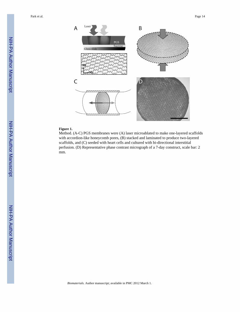
Figure 1.Method. (A-C) PGS membranes were (A) laser microablated to make one-layered scaffoldswith accordion-like honeycomb pores, (B) stacked and laminated to produce two-layeredscaffolds, and (C) seeded with heart cells and cultured with bi-directional interstitialperfusion. (D) Representative phase contrast micrograph of a 7-day construct, scale bar: 2mm.
Park et al. Page 14
Biomaterials. Author manuscript; available in PMC 2012 March 1.
NIH
-PA Author Manuscript
NIH
-PA Author Manuscript
NIH
-PA Author Manuscript
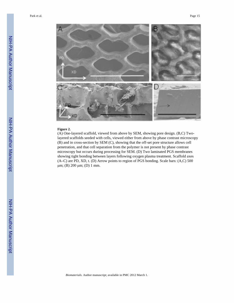
Figure 2.(A) One-layered scaffold, viewed from above by SEM, showing pore design. (B,C) Two-layered scaffolds seeded with cells, viewed either from above by phase contrast microscopy(B) and in cross-section by SEM (C), showing that the off-set pore structure allows cellpenetration, and that cell separation from the polymer is not present by phase contrastmicroscopy but occurs during processing for SEM. (D) Two laminated PGS membranesshowing tight bonding between layers following oxygen plasma treatment. Scaffold axes(A–C) are PD, XD, t. (D) Arrow points to region of PGS bonding. Scale bars: (A,C) 500μm; (B) 200 μm; (D) 1 mm.
Park et al. Page 15
Biomaterials. Author manuscript; available in PMC 2012 March 1.
NIH
-PA Author Manuscript
NIH
-PA Author Manuscript
NIH
-PA Author Manuscript
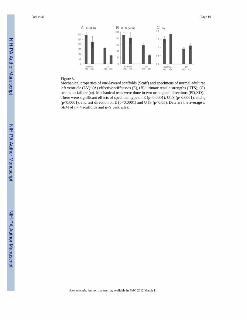
Figure 3.Mechanical properties of one-layered scaffolds (Scaff) and specimens of normal adult ratleft ventricle (LV): (A) effective stiffnesses (E), (B) ultimate tensile strengths (UTS); (C)strains-to-failure (εf). Mechanical tests were done in two orthogonal directions (PD,XD).There were significant effects of specimen type on E (p<0.0001), UTS (p<0.0001), and εf(p<0.0001), and test direction on E (p<0.0001) and UTS (p<0.05). Data are the average ±SEM of n= 4 scaffolds and n=9 ventricles.
Park et al. Page 16
Biomaterials. Author manuscript; available in PMC 2012 March 1.
NIH
-PA Author Manuscript
NIH
-PA Author Manuscript
NIH
-PA Author Manuscript
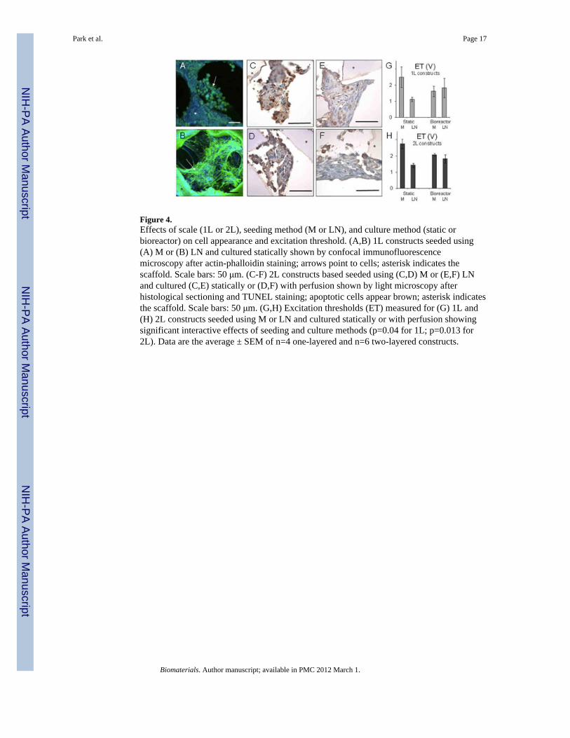
Figure 4.Effects of scale (1L or 2L), seeding method (M or LN), and culture method (static orbioreactor) on cell appearance and excitation threshold. (A,B) 1L constructs seeded using(A) M or (B) LN and cultured statically shown by confocal immunofluorescencemicroscopy after actin-phalloidin staining; arrows point to cells; asterisk indicates thescaffold. Scale bars: 50 μm. (C-F) 2L constructs based seeded using (C,D) M or (E,F) LNand cultured (C,E) statically or (D,F) with perfusion shown by light microscopy afterhistological sectioning and TUNEL staining; apoptotic cells appear brown; asterisk indicatesthe scaffold. Scale bars: 50 μm. (G,H) Excitation thresholds (ET) measured for (G) 1L and(H) 2L constructs seeded using M or LN and cultured statically or with perfusion showingsignificant interactive effects of seeding and culture methods (p=0.04 for 1L; p=0.013 for2L). Data are the average ± SEM of n=4 one-layered and n=6 two-layered constructs.
Park et al. Page 17
Biomaterials. Author manuscript; available in PMC 2012 March 1.
NIH
-PA Author Manuscript
NIH
-PA Author Manuscript
NIH
-PA Author Manuscript
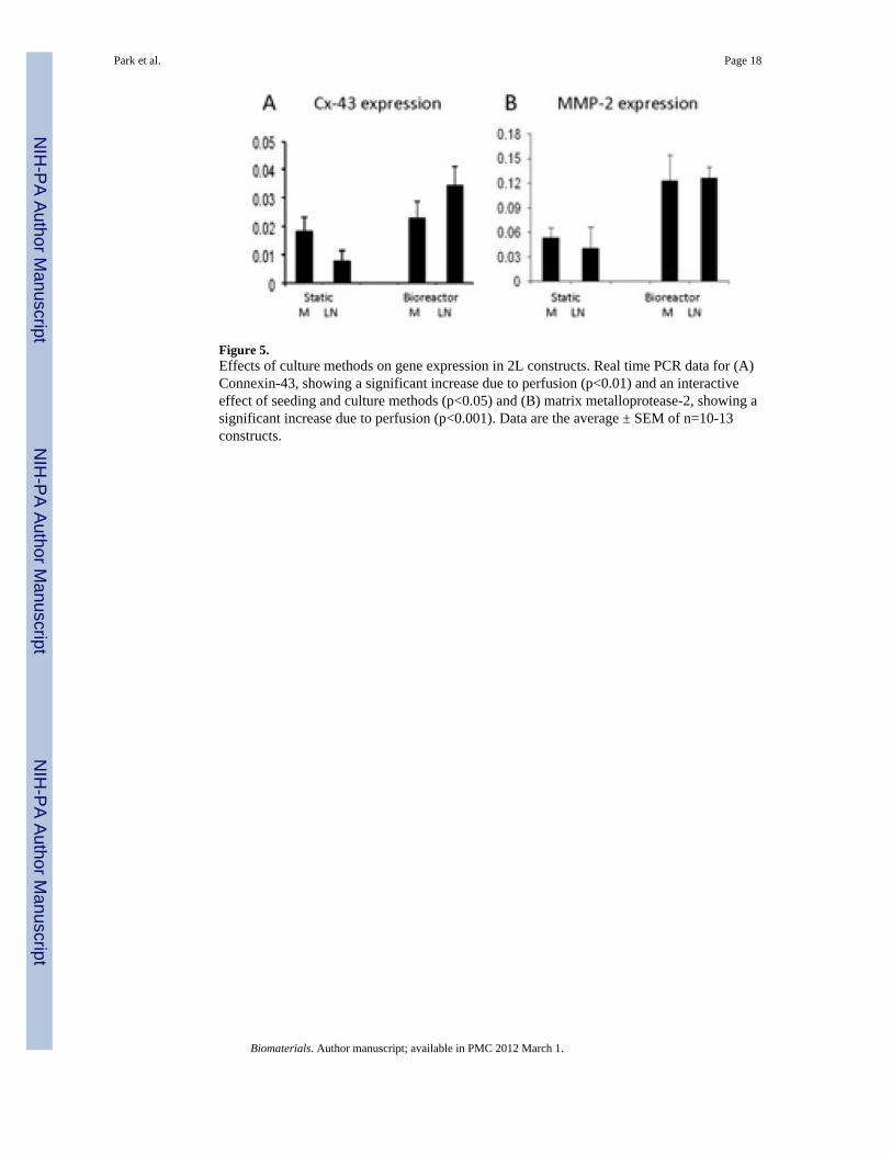
Figure 5.Effects of culture methods on gene expression in 2L constructs. Real time PCR data for (A)Connexin-43, showing a significant increase due to perfusion (p<0.01) and an interactiveeffect of seeding and culture methods (p<0.05) and (B) matrix metalloprotease-2, showing asignificant increase due to perfusion (p<0.001). Data are the average ± SEM of n=10-13constructs.
Park et al. Page 18
Biomaterials. Author manuscript; available in PMC 2012 March 1.
NIH
-PA Author Manuscript
NIH
-PA Author Manuscript
NIH
-PA Author Manuscript
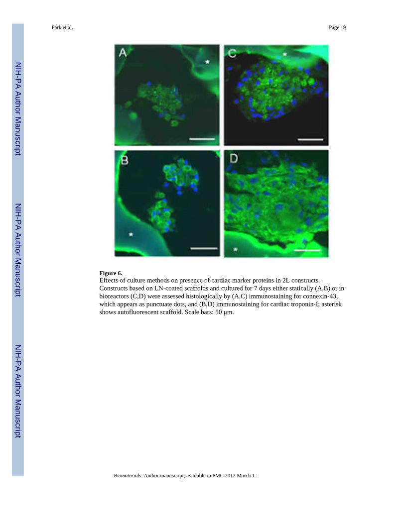
Figure 6.Effects of culture methods on presence of cardiac marker proteins in 2L constructs.Constructs based on LN-coated scaffolds and cultured for 7 days either statically (A,B) or inbioreactors (C,D) were assessed histologically by (A,C) immunostaining for connexin-43,which appears as punctuate dots, and (B,D) immunostaining for cardiac troponin-I; asteriskshows autofluorescent scaffold. Scale bars: 50 μm.
Park et al. Page 19
Biomaterials. Author manuscript; available in PMC 2012 March 1.
NIH
-PA Author Manuscript
NIH
-PA Author Manuscript
NIH
-PA Author Manuscript
Copyright © 2022 FDOKUMEN