Effect of Rot-, Fire-, and Water-Retardant Treatments on Jute ...
Upload
khangminh22Category
view
0download
0
West Virginia Agricultural and Forestry ExperimentStation Bulletins
Davis College of Agriculture, Natural ResourcesAnd Design
1-1-1906
The ripe rot or mummy disease of guavasJohn L. Sheldon
Follow this and additional works at: https://researchrepository.wvu.edu/wv_agricultural_and_forestry_experiment_station_bulletins
This Bulletin is brought to you for free and open access by the Davis College of Agriculture, Natural Resources And Design at The Research Repository@ WVU. It has been accepted for inclusion in West Virginia Agricultural and Forestry Experiment Station Bulletins by an authorized administrator ofThe Research Repository @ WVU. For more information, please contact [email protected].
Digital Commons CitationSheldon, John L., "The ripe rot or mummy disease of guavas" (1906). West Virginia Agricultural and Forestry Experiment StationBulletins. 104.https://researchrepository.wvu.edu/wv_agricultural_and_forestry_experiment_station_bulletins/104
brought to you by COREView metadata, citation and similar papers at core.ac.uk
provided by The Research Repository @ WVU (West Virginia University)


WEST VIRGINIA UNIVERSITY
AGRICULTURAL EXPERIMENT STATION
MORGANTOWN, W. VA.
Bulletin 104. April 1, 1906.
The Ripe Rot or
Mummy Disease of Guavas.
By JOHN L. SHELDON.
[The Bulletins and Reports of this Station will be mailed free to any
citizen of West Virginia upon written application. Address Director of
Agricultural Experiment Station, Morgantown, W. Va.]

THE REGENTS OF THE WEST VIRGINIA UNIVERSITY
Name of Regent. P. 0. Address.
Hon. C. M. Babb Falls, W. Va.
Hon. J. B. FinlEy Parkersburg, W. Va.
Hon. D. C. GallahER Charleston, W. Va.
Hon. E. M. Grant Morgantown, W. Va.
Hon. C. E. Haworth Huntington, W. Va.
Hon. C. P. McNell Wheeling, W. Va.
Hon. L. J. Williams Lewisburg, W. Va.
Hon. T. P. Jacobs New Martinsville, W. Va.
Hon. J. R. Trotter Buckhannon, W. Va.
President of the Board of Regents J. R. Trotter
President of the University D. B. Purinton
Treasurer A. R. WhitehillAuditor W. J. White
STATION STAFF
James H. Stewart, A. M Director and Agriculturist
Bert H. Hite, M. S Vice Director and Chemist
John L. Sheldon, Ph. D Bacteriologist
Horace Atwood, M. S. Agr Assistant Agriculturist
W. E. RumsEy, B. S. Agr Entomologist in Charge
T. C. Johnson, A. M Associate Horticulturist
Frank B. Kunst Assistant Chemist
C. S. Forkum, B. S Assistant Chemist
Leicester Patton Assistant Chemist
Fred E. Brooks Special Agent
W. J. White Librarian
M. A. Stewart Librarian
Alice EnglE Stenographer

The Ripe Rot or Mummy Disease
of Guavas.
By John L. Sheldon.
Introduction.
While walking through the greenhouses of the United
States Department of Agriculture at Washington, D. C, in com-
pany with Mr. A. F. Woods, Assistant Chief of the Bureau of
Plant Industry, the writer noticed among some guavas that were
ripening at the time (December 20, 1903) a number that were
brown and shriveled. Upon closer examination it was found
that the shriveled fruits had not naturally dried and remained
on the trees after ripening, but that they had been attacked by
some fungus of the anthracnose type. The disease was new to
the writer, and with Mr. Woods' permission, specimens of the
infected fruits were collected for future examination and study.
Importance and Distribution.
It was not on account of the economic importance of the
ripe-rot or mummy disease of the guava that a study of the life-
history of the fungus causing it was begun, for at that time the
disease was not thought to be of enough economic importance
to warrant a careful investigation. It was studied, rather, as a
side line along with the anthracnose of cucurbits, and more
especially on account of its similarity to the bitter-rot disease of
apples.
Judging from the effects of the fungus on the guavas in the
greenhouse, it would seem to be as destructive to the guava as
the bitter-rot is to the apple. To what extent it damages the
guava crop under natural conditions, the writer can not say
from personal observations, never having had the privilege of

300 west Virginia experiment station.
seeing guavas growing under natural conditions, either wild or
cultivated.
In order to obtain some data as to the amount of damage
caused by the disease, and specimens of the infected fruits from
different localities, letters of inquiry were sent to Porto Rico,
Florida, California, and Australia.
Mr. O. W. Barrett, botanist at the Porto Rico experiment
station, replied as follows : "In regard to the occurrence of the
'mummy' disease of the guava in Porto Rico, will say that it is
quite prevalent throughout the island on both forms of Psidium
guaiava. It also occurs occasionally on the semi-cultivated
guavas here, but does practically no damage, the trees always
cropping heavily, and occasionally twice in a year." Later he
wrote, "Can you suggest any possible treatment for this mummydisease? It is really a serious matter and I have not recom-
mended the planting of guavas as a crop here thus far. Several
canning companies would probably consider the planting of
guavas seriously if it were not for this disease."
Mr. N. A. Cobb, of the Department of Agriculture of NewSouth Wales, Australia, wrote, "I notice that the disease does
more harm to the guavas here than any other disease of this
fruit with which I am acquainted. Of course the guava does
not cut much of a figure in our fruit industry, so I cannot say
that the loss amounts to much from a monetary point of view.
In particular instances the loss is one that requires attention, but
these instances are necessarily few, as few grow the fruit."
Correspondents in California and Florida wrote that the
disease occurs there, but that it is of no special importance as
yet ; while others wrote that the disease has not been observed.
The disease also occurs in Mexico, but no data have been obtained
with respect to its prevalence and the damage it causes.
Appearance oe the Ineected Fruit.
There was considerable difference in the appearance of the
infected fruits from different localities*. Thosfe collected at
Washington, D. C, had circular, brown, decayed areas on them,
the diameter of the decayed area depending upon the progress of

RIPE ROT OR MUMMY DISEASE. 30I
the disease. In the more advanced stages, there were numerous
masses of salmon colored conidia scattered over the decayed
areas, resembling the masses of conidia of a Collctotrichum or a
Gloeosporium. A microscopical examination showed that the
fungus corresponded more nearly to a Gloeosporium, no setae
being present in and around the acervuli. The entire fruit
finally became decayed and considerably wrinkled and shrunken.
Plate I, fig. 1.
The specimens from Porto Rico and Florida, collected by
Mr. O. W. Barrett and Mr. P. H. Rolfs, respectively, were very
hard and smooth, and more or less spotted with masses of con-
idia. Many of the conidia had evidently been washed off by the
rains. Plate I, figs. 2 and 3.
History and Description of the Disease.
It was thought when the specimens were collected by the
writer in Washington that the disease must be a comparatively
new or rare one, at least one that had not received a great amount
of attention. No record of it could be found in any of the litera-
ture on plant diseases that was available in the libraries of the
West Virginia University and Experiment Station. There was
no reference to a Gloeosporium on guavas in Saccardo's Sylloge
Fungorum, neither were there any mounted specimens of the
infected fruits in the pathological herbarium of the United States
Department of Agriculture until some were furnished by the
writer.
Mrs. Flora W. Patterson, mycologist of the United States
Department of Agriculture, very kindly offered to examine the
publications on file in the different libraries of the Department,
and reported that there was an article by Delacroix on a disease
of guavas caused by a Gloeosporium. This article was sent to
the writer by the Libarian of the Department. Tt is the onlv one
that the writer has seen which gives a general description of the
disease and the fungus causing it. The article was published
about eight months before the specimens of guavas were col-
lected in Washington.
Delacroix's description was made from specimens of guavas
302 west Virginia experiment station.
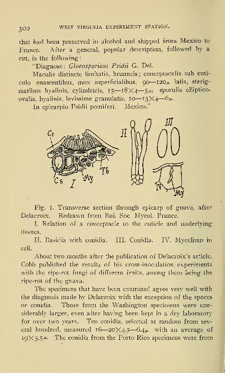
that had been preserved in alcohol and shipped from Mexico to
France. After a general, popular description, followed by a
cut, is the following:
"Diagnose: Gloeosporium Psidii G. Del.
Maculis distincte limitatis, brunneis ; conceptaculis sub cuti-
culo enascentibus, mox superficialibus, 90—120> latis, sterig-
matibus hyalinis, cylindricis, 15—18X4
—
5m: sporulis elliptico-
ovalis, hyalinis, levissime granulatis, 10—13X4—°VIn epicarpio Psidii pomiferi. Mexico."
Fig. 1. Transverse section through epicarp of guava, after
Delacroix. Redrawn from Bui. Soc. Mycol. France.
I. Relation of a conceptacle to the cuticle and underlying
tissues.
II. Basidia with conidia. III. Conidia. IV. Mycelium in
cell.
About two months after the publication of Delacroix's article,
Cobb published the results of his cross-inoculation experiments
with the ripe-rot fungi of different fruits, among them being the
ripe-rot of the guava.
The specimens that have been examined agree very well with
the diagnosis made by Delacroix with the exception of the spores
or conidia. Those from the Washington specimens were con-
siderably larger, even- after having been kept in a dry laboratory
for over two years. Ten conidia, selected at random from sev-
eral hundred, measured 16—20X4-5—6.4^, with an average of
19X5-5 ^- The conidia from the Porto Rico specimens were from

RIPE ROT OR MUMMY DISEASE. 3O3
I to 2p. shorter than those from Washington. It will he
seen that the length of these conidia is approximately 5 ^
more than those measured by Delacroix. However, this differ-
ence may be accounted for in part from the fact that the conidia
which he measured had possibly been shrunken by the alcohol in
which the guavas were preserved, or they may have been imma-
ture. Evidently Delacroix did not notice the round spot that is
normally present near the center of the conidia and which is not
granular like the remainder of the cell contents. Some of the
conidia were slightly curved and a few were narrowed at one end.
LlEE-HlSTORY OE THE FuNGUS.
Inoculations.
Apples.-—Upon returning from Washington, inoculations
were made directly from guavas into apples. Pure cultures of
the fungus were used for much of the later work. The writer
did not know then that Cobb had made the same kind of inocula-
tions and thought the anthracnoses, or ripe-rots, of the guava and
other fruits were possibly caused by the same fungus.
A few of the apples were washed with water, then with a
solution of corrosive sublimate (5 :iooo), rinsed with sterile wrater,
and finally wiped with a sterile towel before they were inoculated,
but nearly as good results were obtained without this extra
trouble.
The inoculations were made by taking a few conidia directly
from a guava, or a pure culture, and inserting them under the
skin of an apple with a sterile needle. Baldwins, Bellflowers and
an unknown variety of a yellow sweet apple were used. After
the apples were inoculated, they were placed on a glass plate and
covered with hand-glasses in order to exclude insects. Whenthere were indications that there was too much moisture under
the hand-glasses, the apples were removed and placed in open
jars, the insects being kept out by tying a piece of fine muslin
over the opening.
After two days the effects of the fungus could usually be
seen as a circular brown spot around the place where the inocula-
tion had been made. The size of the spot increased more rapidly

304 west Virginia experiment station.
on some apples than on others, even of the same variety, especially
when the inoculation was made in a bruise, and more rapidly on
the sweet variety and the Bellflower than on the Baldwin. Dif-
ferences in temperature, ripeness, texture, and acidity probably
made a difference in the rate of growth of the fungus, and a like
difference in the size of the infected area. There were no indica-
tions of the presence of the fungus except where the inoculations
were made.
In about five or six days, longer in some instances, small
black specks began to appear in the brown and decaying areas.
These specks increased in size until they broke open the epider-
mis of the apple, when masses of conidia began to ooze out
through the openings. Plate II, Figs. I and 2. The color of
the masses of conidia varied from a dirty white to a salmon.
Occasionally the conidia oozed out in a straight thread or curled
to one side. The newly formed conidia were 12—13.5X4-5
—
6 m,
about the same size as those taken from the guava by Delacroix
(10—13X4—6ji), but considerably shorter than those taken
from the Porto Rico and Washington specimens by the writer.
The black specks which appeared under the epidermis of
the apples a few days after they had been inoculated were what
Delacroix called conceptacles. They were composed of rows of
cells, with a more or less parallel arrangement, terminating in
hyphae (basidia), which bore the conidia. Plate III, fig. 1.
The gradual development of the conceptacles finally causes a
rujpturing of the epidermis, followed by a liberation of the
conidia.
Unless the apples happened to be infected with other fungi,
like the blue molds, the rot extended from the point of inocula-
tion over the entire surface. After a few days the rotted area
became somewhat shrunken, wrinkled and blackened from the
center outward. When there was sufficient moisture under the
hand-glasses, the masses of conidia changed from pink to black.
A microscopical examination showed that they had germinated
and anastomosed so that they formed a firm body resembling a
pycnidium, from the inside of which conidia were still being
formed and given off. The apples were sometimes covered by a

RIPE ROT OR MUMMY DISEASE. 305
felted mycelium when they were exposed to an excess of mois-
ture. Plate II, fig. 3. They finally shriveled and shrunk to
about half their normal size.
The fungus spread from the point of inoculation to the inside
of the apple in the form of a cone with its apex at the core and
its base on the surface of the apple. Plate II, fig. 4. This cone-
shaped portion was brown and bitter, what may be called a
"nasty bitter" for lack of a better descriptive term.
Inoculations made in young winter apples, about an inch in
diameter, and still on the tree, were unsuccessful. Inoculations in
young Sweet Boughs and other sweet apples just beginning to
show signs of ripening were successful in the laboratory. Goodresults were obtained from the sweet apples when inoculated on
the trees a few weeks later. The rot developed very rapidly and
conidia were produced in abundance. Most of the apples fell off
after the rot had begun to develop. What apples did not fall off
were shaken off by boys so that the rot did not have an oppor-
tunity to spread. None was observed on any of the other
apples.
Damson Plums.—Several inoculations made from pure cul-
tures into Damson plums were successful, the pink masses of
conidia appearing in a few days.
Apple Trees.—No results were obtained from the use of
conidia from guavas or pure cultures when inoculated into the
young twigs or branches of apple trees. There is a possibility
that the inoculations were not made at the right stage of growth.
From inoculations made with the conidia into apples, it will
be seen that the macroscopic characteristics of the bitter-rot of
the apple were reproduced in detail. The rotted portions of the
apples were bitter, the same as they are when they have the bitter-
rot.
Artificial Cultures.
Apple-agar was used as a culture medium for most of the
artificial cultures. It was prepared by heating ground apples
in three times their weight of distilled water at a temperature
below boiling until. the pieces of apple were soft. The decoction

306 west Virginia Experiment station.
was then filtered and normal NaOH added to the filtrate to re-
duce the acidity, not enough to neutralize it, but to leave it
slightly acid to litmus paper. One per cent, of agar-agar was
then added. After the agar-agar had been dissolved by heating,
the medium was filtered and sterilized.
Plate Cultures.—Plate cultures were made after the usual
manner by the addition of conidia to tubes of melted apple-agar.
Blank plates were also made and conidia taken from guavas
planted in them. This latter method gave nearly as good results
as the usual poured plate method, the pure cultures being removed
from the plates in each case. Sub-cultures were made from the
plates into other plates and test tubes where the fungus developed
very rapidly.
The mycelium was at first white, gradually changing to a
dark gray. Pink masses of conidia began to appear in the
plates and test tubes after a few days ; they were occasionally
arranged concentrically but more often scattered about, being
abundant at the edge of the plate where the mycelium extended
up the side of the Petri dish. Some of the newly formed con-
idia germinated after a few days, and the plates became covered
with a dense, felted mycelium.
A few plates were coated wholly or in part with a thin layer
of sterile paraffin. Conidia were then introduced through the
layer of paraffin into the apple-agar beneath. The fungus madea good growth beneath the paraffin layer, but no conidia were
produced until the mycelium reached the edge of the layer, or
holes that were pricked through the layer with a needle, then
they were produced in the same way that thy were in the ordinary
plates. The paraffin was used in order to determine whether
conceptacles would be formed beneath it and rupture it in order
to liberate the conidia, the same as was dome when it grew in the
guavas and apples, but none were formed. A few conidia were
formed on the surface of the paraffin after a number of weeks,
but they were the result of filaments penetrating the paraffin and
then producing conidia on the surface.
The conidial masses, which were usually pink at first, germi-
nated and formed black bodies resembling pycnidia, the same as

RIPS ROT OR MUMMY DISEASE. 3O7
those previously described that were found on the inoculated
apples. Conidia were given off from these bodies in much the
same way that they are from the pycnidia of certain of the
Spliaeropsideae. The pycnidia-like bodies became more and
more chimney-like from the addition of the germinating and
anastomosing conidia which were produced at the base and sides
of the cavity. Plate III, fig. 3.
Test-tube Cultures.—Besides the test-tube cultures on apple-
agar referred to above, others were made on muskmelon-agar
and lactose-agar, but not with very satisfactory results. The
cultures in test tubes were not in general as satisfactory as those
made in Petri dishes and flasks, except for preserving stock
cultures.
Drop Cultures.—Conidia from the guavas germinated in
drop cultures of water in from four to six hours. The conidia
did not lose their capacity for germination for several months if
they were kept dry. In germinating, a germtube appears at one
or both ends of the conidium, occasionally at the side. Many of
the conidia became from one to three septate during germination.
Plate IV, figs. 3 and 8.
There was a marked change in the size and distribution of
the granules in the conidia from the time they were placed in the
water until after the branched mycelium had begun to develop.
The difference in the size of the granules was considerable as one
generation of spores followed another in drop cultures of apple-
agar, especially when they could be traced from one spore. In
the younger conidia they were very small and uniformly dis-
tributed, while in the older and those that were germinating, they
were large and unevenly distributed. Plate IV, fig. 22.
The drop cultures proved to be a very satisfactory means
for watching the development of the fungus from the time a
conidium began to show signs of germinating until one genera-
tion after another of conidia had been produced and in turn had
germinated. The drop cultures made from apple-agar were
exceptionally good for this work.
The conidia were formed on the ends of hyphae by a gradual
enlargement of the hypha, the conidium finally breaking off,

•?o8 WEST VIRGINIA EXPERIMENT STATION.
other conidia being formed in the same manner. Plate IV, figs.
18 and 21. In the way. the conidia were produced, this fungus
does not differ from many of the Hyphomycetes. The conidia
collected on the surface of the plates as small pink masses (Plate
IV, figs. 18 and 19), and when a hypha extended out into the air
the conidia formed a small ball. Plate IV, fig. 20. The conidia
are somewhat gelatinous, and this causes them to hang together
when moist.
Conidia were produced sparingly in drop cultures of water,
Instead of conidia, there were brown, spore-like bodies, which for
convenience were called chlamydospores, although they may not
have been real chlamydospores. These were borne on the ends
of hyphae and also in the filaments. In shape they were spherical
to ovate and oblong and contained one or more vacuoles. Plate
IV, figs. 9-17. No attempt was made to germinate them in order
to determine if they were similar to spores. The chlamydospore-
like bodies were formed in water cultures and in nutrient media
after the food had probably been exhausted. None were found
in guavas nor in the inoculated apples. One or more of the cells
in the filament adjacent to the spore-like bodies were often colored
brown.
By means of the drop cultures and a camera lucida, it was
possible to draw in outline the development of the fungus
through three successive generations of conidia from a conidium
taken from a guava. The difference in the size of the conidia
as the food became exhausted being very noticeable. Plate IV,
fig. 22. A difference of five microns between the conidia from
the guavas and those from the inoculated apples may have been
due to the food supply—that furnished by the guava being more
suitable to the development of the fungus than that furnished by
the apple.
The germinating conidia and mycelium anastomosed freely
with one another. Plate IV, fig. 10. Some of the conidia which
did not germinate became brown, others becoming so after ger-
mination.
The conidia that developed in the apple-agar cultures agreed
fairly well in shape and size with those obtained from the inocu-

RIPE ROT OR MUMMY DISEASE. 309
latecl apples, averaging from 13.5—i8X4-5—6/*. the longest
ones being rare, and usually narrow in proportion to their
length.
Flask Cultures.—Besides the cultures made in plates, test
tubes and hanging drops, others were made in flasks. Corn meal
was put in some of the flasks to the depth of about half an inch
and enough hot apple-agar added to wet the meal. The flasks
with their contents were then sterilized and finally inoculated
from pure cultures of the fungus. The other flasks were filled
to about the same depth with corn meal mush made by cooking
the corn meal in eight times its weight of distilled water. These
flasks were then sterilized and inoculated from pure cultures.
There was a rapid development of the mycelium, showing
that corn meal alone or combined with apple-agar made a suitable
medium for the growth of the fungus. Conidia were formed to
some extent, the pink masses being visible round the sides of the
flasks.
Small black bodies, from a mere speck to five millimeters in
diameter, were produced in the felt-like mycelium that covered
the medium, but they could not be seen except where they were
lying near the sides of the flasks. They were also scattered
through the medium, for the most part near the surface. Amicroscopical examination of some of these bodies showed that
the outside was composed of a layer of brown filaments, the inside
being composed of colorless ones ; others contained conidia borne
on basidia in the same way that the spores are in an immature
pycnidium. Similar pycnidia-like bodies were found in the guavas,
and they have also been observed by the writer in artificial
cultures of Colletotrichum lagenarium (Pass.) Ell. & Hal. They
were evidently what Delacroix called the conceptacles. Struc-
turally they were very much like the bodies that formed beneath
the epidermis of the inoculated apples and which produced the
masses of conidia. Plate III, fig. 2.
Formation of Ascospores.
An abstract in Science refers to the finding of the ascigerous
stage of the fungus in some old cultures by the writer. In these

-?Io WEST VIRGINIA EXPERIMENT STATION.
cultures, which were originally obtained from a guava received
from Porto Rico, it was noticed that there were papillae scattered
through the felted mycelium, caused by hard black bodies lifting
the mycelium from the medium. These bodies were about a milli-
meter in diameter and were composed of a number of smaller
bodies, like pycnidia or perithecia, growing together. After a
careful search it was found that there were colorless, one-celled
spores in a few of them, but for the most part they were empty
or sterile. Occasionally asci in different stages of development
were found, showing that the bodies were perithecia and not
pycnidia.
In general, the perithecia were simple, or now and then with
their walls united, or a large one contained one or more small
ones. There were some from 200—300^ in diameter and manysmaller ones, spherical, except when crowded against one another,
and only an occasional one with a distinct beak and ostiolum.
Plate IV, figs. 23 and 24. The walls of the young perithecia were
membranaceous and composed of polygonal cells ; the older ones
were hard, approaching carbonaceous.
Combined with the asci were masses of filaments correspond-
ing to paraphyses. As soon as water came in contact with the con-
tent of the perithecia, the mature asci, and what was taken for
paraphyses, gradually disappeared, apparently dissolving in the
water. On account of a scarcity of material and the ephemeral
nature of the contents of the perithecia, it was impossible to get
as clear a conception of the asci and paraphyses as could have been
obtained otherwise. In general, the asci were cylindrical to
broadly clavate, rather blunt at the apex and with a short pedicel.
They varied in length, as most asci do even in the same perithe-
cium, but were about 45-55 x 9-10^ Some were possibly a little
longer. The usual number of spores was eight, one ascus having
only six, and the spores were sub-biseriate in the asci. Plate IV,
figs. 26-29. The spores resembled the conidia of the Gloeo-
sporium stage and could scarcely be distinguished from the curved
conidia ; they were mostly curved, continuous, hyaline, granular,
and usually contained a distinct spot near the center without
granules, or one or two vacuoles. Plate IV, figs. 25 and 28.

RIPE ROT OR MUMMY DISEASE.3 J i
Average spores, taken from the growing cultures, measured 13-15
x5~6^; ten from the microtome sections were 12-17.5x3.5-5^,
being somewhat less than the former.
The Name of the Fungus.
This disease of the guavas is one that corresponds to the
anthracnoses of a number of fruits and vegetables. Macro-
scopically the acervuli are almost identical with those of the
anthracnose of the watermelon, the bean, the apple, the eggplant,
the pepper, and of the fruits of other plants, but the conidia
differ from the conidia of some of these, and no setae have been
found in connection with the acervuli in either the guavas, inocu-
lated apples, or the artificial cultures. The conidial stage is
characteristic of the genus Gloeosporium, and the fungus as it
was found on the guavas by Delacroix was called Gloeosporium
Psidii. It has been shown by the writer that the fungus has an
ascigerous stage, corresponding in nearly every particular to the
genus Glomerella. Such being the case, and following the cus-
tom of other investigators of this group of fungi, the name of
this fungus would be changed from Gloeosporium Psidii G. Del.
to Glomerella Psidii (G. Del.)
Summary and Recommendations.
The ripe-rot or mummy disease of guavas has been reported
from Mexico, Porto Rico, Florida, and i\ustralia. In Porto Rico
and Australia it is often very destructive to the crop of ripening
guavas, being of considerable economic importance on account
of the value of the fruit for preserving purposes.
Brown spots appear on the ripening fruits, the size of the
spots gradually increasing until the entire fruit becomes affected,
the decayed fruits finally falling off or the "mummies" remaining
on the trees.
The fungus causing the disease was first described as Gloeo-
sporium Psidii by Delacroix in 1903. It has been successfully in-
oculated into a number of fruits directly from guavas, and grownin various artificial culture media.
Two distinct conidial stages have been observed ; one, with the
condiia borne on hyphae after the usual manner of many of the

•2 12 WEST VIRGINIA EXPERIMENT STATION.
Hyphomycetes ; the other, with conidia on basidia after the man-ner of the Melanconiae. An ascigerous stage, very similar to
that of the genus Glomerella, was obtained in artificial cultures.
The writer has not been able to find any great amount of dif-
ference between this fungus and the descriptions of the one
causing the bitter-rot of the apple. The difference has not been
considered sufficient to warrant making it another species. Their
effects on apples were practically identical ; their growth in
artificial cultures was much the same ; and their conidia, perithecia
and ascospores were produced in much the same way and showed
about the same variations in size and shape. While the asci of the
guava fungus obtained by the writer were not of the same general
shape as those figured by Clinton for the bitter-rot fungus (his
being more pointed at the apex), they agreed very nearly with
those figured by von Schrenk & Spaulding.
The one point of difference, and one that has been made of
considerable importance in the classification of the Pyrenomycetes,.
was the presence of paraphyses. Clinton says "There were no
signs of paraphyses" in his cultures of the bitter-rot fungus, and
von Schrenk and Spaulding, who also made a rather exhaustive
study of the same fungus, do not mention the finding of para-
physes, in fact, they described the genus Glomerella as "apara-
physate."
The writer has demonstrated to his own satisfaction a number
of times that there were paraphyses present in the ascigerous
stage obtained from species of Gloeosporium and Colletotrichum.
The paraphyses were seen when the contents of the perithecia
were teased out, and serial sections of the perithecia showed the
asci imbedded in a matrix, such as would be present if the asci
were surrounded by paraphyses. The writer has made a
recent study of the ascigerous stage of Glomerella rufomaculans
(Berk.) Sp. & von Sch., by means of pure cultures isolated from
a Baldwin apple, and has found that it also has paraphyses.
There is a possibility that further investigation will show that
paraphyses are present in most, if not all, species of Glomerella.
It is, therefore, recommended that the phrase paraphyses some-
times present be included in future descriptions of the genus.
Glomerella, or an equivalent.

RIPE ROT OR MUMMY DISEASE. 313
INDEX TO LITERATURE.
(The following have been cited in the preparation of this
bulletin.)
1902. Clinton, George P. Apple Rots in Illinois. Bui. Univ.
111. Agr. Exp. Sta. 69 : 193-21 1. F 1902.
1903. Delacroix, G. De la tavelure des Goyaves produite par le
Gloeosporium Psidii nov. sp. G. Del. Bui. Soc. Mycol.
France. 19 : 2. 143-145. 30 Ap 1903.
Cobb, N. A. Letters on the Diseases of Plants. Agr.
Gaz. N. S. W. 14 : 7. 627-652. Jl 1903.
Schrenk, Herman von, and Perley Spaulding. The Bitter
Rot of Apples. Bui. U. S. Dept. Agr. Bur. PI. Ind.
44 :9-45- 15 Jl *903-
1004. Sheldon, John L. Diseases of Melons and Cucumbers
during 1903 and 1904. Bui. W. Va. Agr. Exp. Sta. 94: 121-137. D 1904.
The Ascigerous Stage of Gloeosporium Psidii. Science,
n. s. 21 : 526. 143. Ja 1905.
1906. Saccardo, P. A. Gloeosporium Psidii Delacr. Sylloge Fun-
gorum 18 :45i. 1906.

3H west Virginia Experiment station.
DESCRIPTION OF PLATES.
(Halftones from photographs and photomicrographs by Mr.
W. E. Rumsey.)
Plate I. Guavas affected with the ripe-rot or mummy disease.
Natural size. The white spots on the fruits represent masses
of conidia. Fig. I.—From greenhouse of the United States
Department of Agriculture, Washington, D. C. Fig. 2.
—
From Mayaguez, Porto Rico. Fig. 3.—From Eldred, Florida.
Plate II. Apples inoculated with conidia of the ripe-rot fungus.
Fig. 1.—The white spots on the apple show the masses of
conidia just beginning to collect. Fig. 2.—A more advanced
stage of the rot than -shown by fig. 1. Fig. 3.—A still more
advanced stage of the rot, the apple being completely covered
by the mycelium. The black specks near the stem-end of
the apple are masses of conidia that have germinated and
anastomosed. Fig. 4.—Section through an inoculated apple,
showing the decayed, cone-shaped portion at the left, caused
by the fungus.
Plate III. Fig. 1.—Section through the skin of an inoculated
apple, showing a conceptacle with basidia bearing conidia that
has broken through the epidermis. The color of the conidia
is the result of using Flemming's solution as a fixing reagent.
Fig. 2.—Section of one of the bodies referred to in the text
that formed in the cultures of corn meal mush. Fig. 3.
—
Section of a chimney-like body, composed of conidia that
have germinated and anastomosed, with conidia in different
stages of maturity inside. Fig. 4.—Section through two
perithecia joined together. The larger shows the beak and
ostiolum and rows of ascospores.
RIPE ROT OR MUMMY DISEASE. 3*5
Plate IV. Microscopic characters of the ripe-rot fungus in various
stages. Figs. 1 and 2.—Conidia from guava. Figs 3-7.
—
Germination of conidia and the development of mycelium.
Figs. 6-8.—Septa formed in conidia during germination.
Figs. 9-17.—Chlamydospore-like bodies. Fig. 10.—Conidia
anastomosing. Figs. 18-22.—Conidia in different stages of
development. Figs. 18 and 19.—Masses of conidia collecting
on poured plate. Fig. 20.—Masses of conidia on end of
hypha which has extended out into the air. Fig. 22.
—
First and second generations of conidia from a conidium.
Figs. 23 and 24.—Outlines, showing sections of rostrate
perithecia. The walls of the perithecia appear thicker than
they really are on account of the thickness of the sections.
Fig. 25.—Ascospores. Fig. 26.—An immature ascus. Fig.
27.—An ascus containing six ascospores. Figs. 28 and 29.
—
Broken asci with ascospores.


PLATE I


PLATE I I
M<>^^h9 ^^.
:
''.: $a
n*^2>*:.\: .':'^s^«*,- ^


PLATE III
j*4k&*&itr3mmYmEHV
<JmNEjGI>\hki J^» v^^H
^«»^^ iV* »*
1BLrBate*
JH ^BmB
mm wW -' ri<gfl B
L.«W ^^^Hk.
4 P 15 P^« • '• Iv^^O^^k^
fA-

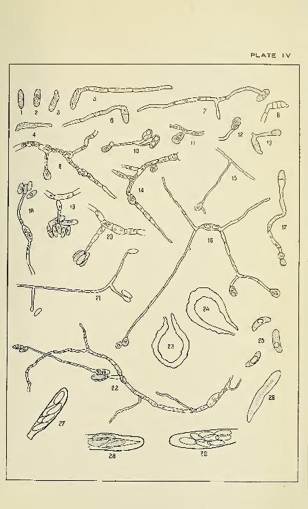
PLATE IV

Copyright © 2022 FDOKUMEN


![Konzervatorsko-restauratorski radovi na Zagrebačkoj mumiji. [Conservation of the Zagreb Mummy.]](https://static.fdokumen.com/doc/165x107/6333d4d8b94d6238420264d6/konzervatorsko-restauratorski-radovi-na-zagrebackoj-mumiji-conservation-of-the.jpg)