Elias, J 2011 The Mummy of Pa-di-heru-pa-khered, Milwaukee Public Museum 10265. Akhmim Mummy Studies...
16
Radiological Studies of Egyptian Mummies in Miscellaneous Collections: AMSC-7 Pa-di-heru-pa-khered The Mummy of Pa-di-heru-pa-khered, Milwaukee Public Museum 10265 By Jonathan Elias, Ph.D. Akhmim Mummy Studies Consortium Research Paper 11-1 (RSEM-MC AMSC-7) ©2011 AMSC Research, LLC, Carlisle, Pennsylvania 1
-
Upload
independent -
Category
Documents
-
view
1 -
download
0
Transcript of Elias, J 2011 The Mummy of Pa-di-heru-pa-khered, Milwaukee Public Museum 10265. Akhmim Mummy Studies...

Radiological Studies of Egyptian Mummies in Miscellaneous Collections: AMSC-7 Pa-di-heru-pa-khered
The Mummy of Pa-di-heru-pa-khered,
Milwaukee Public Museum 10265
By
Jonathan Elias, Ph.D.
Akhmim Mummy Studies Consortium Research Paper
11-1
(RSEM-MC AMSC-7)
©2011 AMSC Research, LLC, Carlisle, Pennsylvania
1

Radiological Studies of Egyptian Mummies in Miscellaneous Collections: AMSC-7 Pa-di-heru-pa-khered
Catalog No. AMSC-7
The Mummy of an Adult Male named Pa-di-heru-pa-khered (MPM 10265)
Location: Milwaukee Public Museum, Milwaukee, WI
Provenience and Early History
The mummy and its wooden coffin were originally acquired by Adolph Meinecke, a trustee of the
Milwaukee Public Museum, and he in turn sold it to the museum on December 20th 1887 (Price $74.68).
This sale occurred three months after the donation to the Milwaukee Public Museum of another
mummy (Djedhor, MPM 10264), by Adolph’s son Ferdinand. Final registry of MPM 10265 occurred on
February 25, 1888. Precise details of Adolph Meinecke’s acquisition are unknown, but a 1962 letter
written by his grandson, Ferdinand Meinecke, Jr., conveys his impression that the mummy was likely to
have been purchased during one of Adolph’s “many business trips in Europe”. There is a reference to a
guide named “Stangen of Berlin” in connection with the acquisition of MPM 10264. This it seems is Carl
Stangen, the proprietor (with his brother Louis) of a well-regarded “tourists’ bureau” established in the
1860’s1 and headquartered in Berlin (10 Mohrenstrasse). 2 This establishment was later absorbed into
the Hapag Line in 1905.3 Stangen’s importance in the tourist trade is comparable to that of Thomas
Cook. He appears to have been particularly interested in arranging large group movements in
connection with popular world fairs and expositions).4 The Stangen firm ran its first tour to Egypt before
1870 and would after 20 years activity there, have had the connections necessary to obtain artifacts
recently excavated, to ship them in a scheduled way and then re-sell them fairly easily to wealthy
business clients. Carl Stangen may have imported artifactual material directly from Egypt for sale in
Germany and it is known that he himself traveled there. The likelihood that Adolph Meinecke acquired
his mummies in Europe (rather than in Egypt) is supported by a notice in The New York Times (dated
September 27, 1885) relating to Meinecke’s recent trip there and his intention to eventually donate the
material to the public.5
“Adolph Meinecke, of Milwaukee, has brought from a tour of Europe, an odd jumble of curios, which he intends to give to the public museum. A mask made of the human face, stuffed wolves, chamois, and other beasts, armour, stone implements, and other antiquarian lumber are in the collection.”
2

Radiological Studies of Egyptian Mummies in Miscellaneous Collections: AMSC-7 Pa-di-heru-pa-khered
Radiological Examinations and Other Projects
1st CT scan May 16, 1986 by MPM (Carter Lupton) at the facilities of GE Medical Systems, New Berlin WI.
2nd CT Scan September 17, 1999, GE Medical Systems; 3rd CT Scan June 23, 2006 by MPM and Akhmim
Mummy Studies Consortium at GE Healthcare, Waukesha, WI, using a GE Lightspeed VCT scanner. A 4th
more detailed CT scan (at .0625 mm slice thickness) was performed at GE Healthcare/Medical Systems
Waukesha, WI on April 5, 2011, by MPM . Once again the work was performed in collaboration with the
Akhmim Mummy Studies Consortium, Carlisle, Pennsylvania.
Subject and Persona
Mummy is of a young male. The original MPM catalog (dated 1889) lists the owner as “Peteharpochrat,
son of Min” and describes him as a priest of “Nes-hor”. A proper reading of the texts on the coffin
confirm the identity of the mummy as the stolist priest (sm3ty) Pa-di-heru-pa-khered, son of the stolist
priest (sm3ty) Neshor. The mother’s name is not included. The name Pa-di-heru-pa-khered roughly
translates: “the one whom Horus-the-child has given”. Horus-the-child is the offspring of Isis, and Min,
the fertility god standing at the top of the divine hierarchy of Akhmim.
Dating/Burial Modality
Pa-di-heru’s coffin is of Akhmimic design and its style is consistent with the work of a Ptolemaic Period
workshop active around 250 BC. It is a simple design (classic for its era and of high quality) with strong
lines and vivid color zones. It is extraordinarily well-preserved. The mummy appears to be of Ptolemaic
date and would almost certainly have been equipped with cartonnage plaques. During the manufacture
period in question, the cartonnage assemblage would have been a four-piece cover: (1) a head cover
with lappet wig (in blue-gray) and an attached mask modeled with idealized features of the deceased as
an Osirian entity (probably gilded with eyes painted white with black irises and edge details); (2) a
collar/torso/abdomen plaque; (3)leg cover extending to the upper ankle; and (4) the so-called “boot”
element extending over the feet.
3

Radiological Studies of Egyptian Mummies in Miscellaneous Collections: AMSC-7 Pa-di-heru-pa-khered
Fig. 1: Head of Pa-di-heru’s coffin showing its gilded face and collar design conforming to the Akhmimic style
Condition
The condition of the mummy is excellent overall. The original mummy bundle is intact with no visible
evidence of unwrapping. The cartonnage elements are missing; and in their place lies a faience amulet
of a baboon (Hapi) flanked by two wing pieces in faience. These three elements undoubtedly come from
a different source and are not integral to the mummy. Eye tissue is preserved within both orbits (the
rectus muscles are still evident behind each ocular bulb) and the ear cartilage is intact. The right clavicle
appears to be depressed unnaturally, in the direction of the spinal column. Lower down on the right
side, there appears to be a displacement of at least one rib.
4

Radiological Studies of Egyptian Mummies in Miscellaneous Collections: AMSC-7 Pa-di-heru-pa-khered
Fig. 2: General view of Pa-di-heru (3D volumetric reconstruction)
Fig. 3: Pa-di-heru Rib displacement right side
5
Radiological Studies of Egyptian Mummies in Miscellaneous Collections: AMSC-7 Pa-di-heru-pa-khered
Wrapping Structure, Amulets and Inclusions
Prior to bandaging, Pa-di-heru’s body was coated with resin. This was thickly painted onto the body
while it was on its back and accumulated posteriorly. The mummy bundle is built up in four major
bandaging horizons each divided by a coating of resin. The density of wrapping is moderate, meaning
that there is perceptible air pocketing between pasted layers. The amount of pasting is relatively high.
The first innermost bundling horizon progressed around the body and passed between the body wall
and the right arm, but appears to have passed over the left arm due to that arm’s close contact with the
body wall. The second horizon was built up to a point where the two arms were incorporated into a
general bundle. Within this horizon, a small, extremely dense (visually opaque) object (Inclusion A) was
placed anterior to the upper abdomen along the body axis. The central location of this object indicates
that its placement was deliberate.
Fig. 4: Inclusion A, level of embalming incision
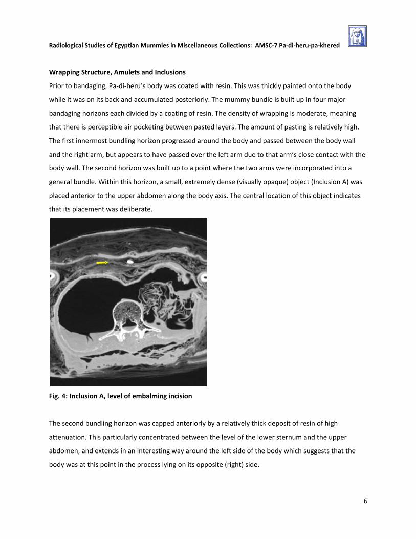
The second bundling horizon was capped anteriorly by a relatively thick deposit of resin of high
attenuation. This particularly concentrated between the level of the lower sternum and the upper
abdomen, and extends in an interesting way around the left side of the body which suggests that the
body was at this point in the process lying on its opposite (right) side.
6
Radiological Studies of Egyptian Mummies in Miscellaneous Collections: AMSC-7 Pa-di-heru-pa-khered
Fig. 5: Character of wrapping: stratification and resin applications between Layers with focus on the anterior and left side
The third horizon is composed of wrapping of moderate density in which the amount of pasting is less
than was seen in the two innermost horizons. Axial views of the wrapping shows that it is undulating or
bunched as a result of tension applied when the fourth (outermost) horizon of the bundle was created.
The exterior surface of the fourth horizon is coated with resin of high attenuation, and may represent a
ritual resin pouring that occurred prior to attachment of the outer shroud. This pouring extends up to
the crest of the shoulder, but did not include the neck. A visually opaque object (Inclusion B), smaller
than Inclusion A is noted above the right shoulder.
Fig. 6: Inclusion B, above the right shoulder
7
Radiological Studies of Egyptian Mummies in Miscellaneous Collections: AMSC-7 Pa-di-heru-pa-khered
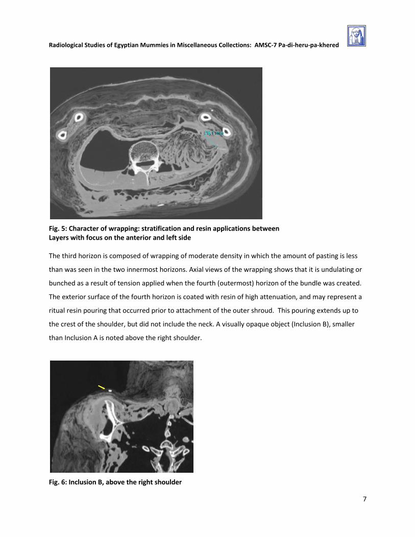
Excerebration
The embalmers achieved excerebration with complete removal of brain tissue and dura mater. This
operation was bi-lateral and resulted in significant disruption of the nares and destruction of the nasal
septum. The opening created in the ethmoid bone for brain evacuation is 41.6 mm. Following brain
removal , resin was injected into the cranium and solidified against the occipital facet of the brain case.
Its depth varies from 23.0 mm to 31.9 mm. The infill index is 27.7 (just over 1/4 of total cranial volume).
The resin is stratified, with greater density noted to the posterior of the injected mass, probably the
result of gravity settling of heavier particles following a single injection episode. As the upper or anterior
layer of the resin hardened, the attenuated resin film collapsed into an underlying air cavity. Resin also
fills abundantly the sphenoidal sinus the upper reaches of the spinal canal and the throat. The amount
of resin deposition suggests a significant in-pouring that speaks to the relatively high status of Pa-di-heru
within A thick resin coating or “mask” covers the nasal orifice. This is a feature identified relatively
recently and noted in an ever increasing number of Akhmim mummies. There is a heavier accumulation
of this resin on the right side of the face.
Fig. 7: Resin deposits within the brain case and the sphenoid sinus, and a resin “mask” on the exterior of the nasal orifice
8

Radiological Studies of Egyptian Mummies in Miscellaneous Collections: AMSC-7 Pa-di-heru-pa-khered
A relic of the excerebration process appears to exist in the form of a linear structure which projects from
the nasal aperture into the right orbit. This object is of medium attenuation (-140 to 120 HU). It can be
interpreted as a reed or stick (right side 27.2 mm long; left side 36.0 mm long), bent or folded as a result
of significant impact against the right wall of the right orbit, after penetrating through the right side of
the nasal passage. It is bent at an angle of 103.3 degrees.
Fig. 8: A reed (excerebration tool?) abandoned in the nasal passage and right orbit
Evisceration
Embalmers removed Pa-di-heru’s internal organs via an incision (28.0 to 36.2 mm wide) in the left flank
of his body, above the level of the iliac crest. The sides of the incision were not tightly drawn back
together, but are connected by a flap of adhering resin that extends below the forearm. The forearm is
tucked tightly against the embalmer’s opening. A liberal amount of resin was injected into the body
cavity following organ removal, and a considerable amount of this material has leaked into the posterior
part of the mummy bundle. The wadding used to plug the incision is loosely arranged (77.8 mm across
by 66.5 mm A-P) and fills the abdominal zone between the incision and the spinal column. A resin cap
closes the incision and the left forearm is embedded onto to this feature. Most of Pa-di-heru’s cardiac
tissue was removed during evisceration. This is an unusual situation at odds with what is seen in with
other Akhmimic mummies of Ptolemaic date, and indeed those of other origin and date. The heart
9

Radiological Studies of Egyptian Mummies in Miscellaneous Collections: AMSC-7 Pa-di-heru-pa-khered
should have been left within the thoracic cavity, and its absence appears to have been an inadvertent
error in Pa-di-heru’s embalming.
Fig. 9: Pa-di-heru’s abdomen, showing the embalming incision, theleft forearm positioned against it; the abundant resin has leaked into the underlying bandages.
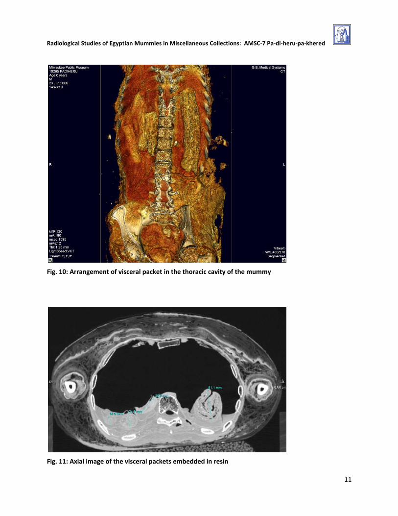
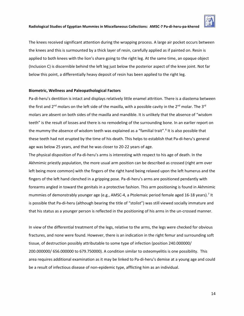
Visceral Packets (VP)
Pa-di-heru’s internal organs (or representative portions of them) were returned to the body in visceral
packets. Four such packets were found within his thorax. The largest of the group is placed by itself in
the left side of the thoracic cavity, oriented longitudinally and parallel to the spinal column. It is partially
submerged in the body resin which reaches a depth of 30.3 mm in this area. Three other packets, all
considerably smaller are nestled in a group on the right side of the spine and relatively close to it. The
packet closest to the spinal column is flat, the remaining packets are cylindrical. All packets are mired in
resin within the body cavities. In addition, a structure of linen wadding (loosely folded) is located in the
pelvic cavity. It too became entirely saturated once resin was injected into the body interior.
10
Radiological Studies of Egyptian Mummies in Miscellaneous Collections: AMSC-7 Pa-di-heru-pa-khered
Fig. 10: Arrangement of visceral packet in the thoracic cavity of the mummy
Fig. 11: Axial image of the visceral packets embedded in resin
11

Radiological Studies of Egyptian Mummies in Miscellaneous Collections: AMSC-7 Pa-di-heru-pa-khered
Disposition and Wrapping of Arms
Pa-di-heru’s arms hang downward at their respective sides and the forearms angle inward toward the
pubic axis. The fingers of the left hand curl inward over the level of the penis and testicles, in a gesture
of protection. Note however that the fingers do not actually cup the genitals. The fingers of the right
hand are relaxed, and the hand itself lies in the space between the inner aspect of the right thigh and
the genitals. The wrapping of the forearms is minimal, perhaps amounting to no more than two bandage
layers. The upper arms are more heavily wrapped (ca. 6-8 layers) and a cylindrical bandage structure is
noted between the bandaged right humerus and the body wall.
Fig. 12: Pa-di-heru: disposition of arms (3D volumetric rendering)
Disposition and Wrapping of Legs
There is potentially meaningful attention lavished on Pa-di-heru’s legs. In comparison with the arms,
which are thinly (even negligibly) wrapped compared with other mummies in the study, Pa-di-heru’s legs
are wrapped in a broadly, padded way. Thickness of wrapping of the left leg varies from 11.6 to 23.1
mm; Thickness of wrapping of the right leg is greater than the left and varies from 26.5 to 40.4 mm. This
difference seems significant in relation to the perimortal condition of the right leg (see Biometric and
Paleopathological Factors). The space between the legs is largely filled with textile wadding.
12

Radiological Studies of Egyptian Mummies in Miscellaneous Collections: AMSC-7 Pa-di-heru-pa-khered
Fig. 13a: Legs at level of femur; suspicious shadow (right)
Fig. 13b: Legs at level of knees showing resin distribution
Fig. 13c: Legs just below knee level (Inclusion C at right)
Fig. 13d: Lower legs showing resin focused on the right leg
13
Radiological Studies of Egyptian Mummies in Miscellaneous Collections: AMSC-7 Pa-di-heru-pa-khered
The knees received significant attention during the wrapping process. A large air pocket occurs between
the knees and this is surmounted by a thick layer of resin, carefully applied as if painted on. Resin is
applied to both knees with the lion’s share going to the right leg. At the same time, an opaque object
(Inclusion C) is discernible behind the left leg just below the posterior aspect of the knee joint. Not far
below this point, a differentially heavy deposit of resin has been applied to the right leg.
Biometric, Wellness and Paleopathological Factors
Pa-di-heru’s dentition is intact and displays relatively little enamel attrition. There is a diastema between
the first and 2nd molars on the left side of the maxilla, with a possible cavity in the 2nd molar. The 3rd
molars are absent on both sides of the maxilla and mandible. It is unlikely that the absence of “wisdom
teeth” is the result of losses and there is no remodeling of the surrounding bone. In an earlier report on
the mummy the absence of wisdom teeth was explained as a “familial trait”.6 It is also possible that
these teeth had not erupted by the time of his death. This helps to establish that Pa-di-heru’s general
age was below 25 years, and that he was closer to 20-22 years of age.
The physical disposition of Pa-di-heru’s arms is interesting with respect to his age of death. In the
Akhmimic priestly population, the more usual arm position can be described as crossed (right arm over
left being more common) with the fingers of the right hand being relaxed upon the left humerus and the
fingers of the left hand clenched in a gripping pose. Pa-di-heru’s arms are positioned pendantly with
forearms angled in toward the genitals in a protective fashion. This arm positioning is found in Akhmimic
mummies of demonstrably younger age (e.g., AMSC-4, a Ptolemaic period female aged 16-18 years).7 It
is possible that Pa-di-heru (although bearing the title of “stolist”) was still viewed socially immature and
that his status as a younger person is reflected in the positioning of his arms in the un-crossed manner.
In view of the differential treatment of the legs, relative to the arms, the legs were checked for obvious
fractures, and none were found. However, there is an indication in the right femur and surrounding soft
tissue, of destruction possibly attributable to some type of infection (position 240.000000/
200.000000/ 656.000000 to 679.750000). A condition similar to osteomyelitis is one possibility. This
area requires additional examination as it may be linked to Pa-di-heru’s demise at a young age and could
be a result of infectious disease of non-epidemic type, afflicting him as an individual.
14

Radiological Studies of Egyptian Mummies in Miscellaneous Collections: AMSC-7 Pa-di-heru-pa-khered
Fig. 14: Area of shadows passing through the soft tissue and bone of Pa-di-heru’s Right thigh
Pa-di-heru’s stature in life can be determined on the basis of on-screen measurements of his
femurs,tibias, and humeri using coronal CT images. This produces a range of 153.635-160.142 cm
(60-63 inches). This is far less than the original height estimate of 68 inches (174 cm) produced by Dr.
Norman Sullivan in 1999.
Notes
1Founding date put variously: 1863 by Enzensberger, H. M. (2001) A Theory of Tourism, p. 128 Originally published as Vergebliche Brandung der Ferne: Eine theorie des Tourismus” Merkur 126 1958, 701-720. 1868 by RIPM Consortium Ltd, (2006) Schlesische Theater-Zeitung (1863) Breslauer Theater-Zeitung (1864), xvii, note 1.
2 New York Times April 21, 1895, “Salon of Champ de Mars” col. 4.
3 Hapag operates the former Stangen business as the "Hamburg-Amerika Linie Reisebüro" beginning around 1904.http://www.hapag-lloyd.com/en/about_us/history_between_1886_1918.html
4 New York Times, April 21, 1895, “Salon of Champ de Mars” col. 4.
5 New York Times, September 27, 1885, “Art Notes” col. 1 6 Lupton, C. An Historical Study of Two Egyptian Mummies (2001), p. 222. 7 Chan et al, CT of a Ptolemaic Mummy (2008).
15

Radiological Studies of Egyptian Mummies in Miscellaneous Collections: AMSC-7 Pa-di-heru-pa-khered
Bibliography,
Chan, S. Elias, J. Hysell, M. and Hallowell, M. (2008) CT of a Ptolemaic Mummy from the Ancient Egyptian City of Akhmim. RadioGraphics 28: 2023-2032.
Lupton, C. (1987) “Egypt and Milwaukee, the Best of Both Worlds”, Wisconsin Academy Review, March, 6-9.
Lupton, C. (1988) “Mummies are Forever,”Lore 38(2) Summer, 3-8.
Lupton, C. (2001) “An Historical Study of Two Egyptian Mummies in the Milwaukee Public Museum”, in Emily Williams, ed. Human Remains, Conservation retrieval and analysis. Proceedings of a conference held in Williamsburg, VA, Nov. 7-11th, 1999. BAR International Series 934, 215-225.
16