Synthetic osteochondral grafting of ankle osteochondral lesions.
Upload
independentCategory
view
1download
0
The reverse sural fasciocutaneous flapfor the treatment of traumatic, infectiousor diabetic foot and ankle wounds: aretrospective review of 16 patients
Ioannis A. Ignatiadis, MD, PhD1,*, Vassiliki A. Tsiampa, MD1,Spyridon P. Galanakos, MD1, Georgios D. Georgakopoulos,MD1, Nicolaos E. Gerostathopoulos, MD, PhD1, Mihai Ionac,MD, PhD3, Lucian P. Jiga, MD, PhD3 and Vasilios D. Polyzois,MD, PhD2
1Hand Surgery-Upper Limb and Microsurgery Department, KAT General Hospital, Athens, Greece; 24thOrthopedic Department, KAT General Hospital, Athens, Greece; 3Vascular Surgery Department, TimisoaraUniversity Hospital, Timisoara, Romania
The authors present their experience with the use of sural fasciocutaneous flaps for the treatment of various
soft tissue defects in the lower limb. This paper is a review of these flaps carried out between 2003 and 2008.
The series consists of 16 patients, 12 men and 4 women with an average age of 40 years (17�81) and with a
follow-up period between 1 and 8 years. The etiology was major velocity accident in six cases, diabetes
mellitus with osteomyelitis after ORIF for fractures (3), work accident in five, and another two cases with
complications of lower limb injuries. The defect areas were located on calcaneus, malleolar area, tarsal area
and lower tibia. Associated risk factors in the patients for the flap performance were diabetes (five patients)
and cigarette smoking (eleven patients).
The technique is based on the use of a reverse-flow island sural flap combined with other flaps in three cases
(medial plantar, peroneal, gastrocnemius). The anatomical structures which constituted the pedicle were the
superficial and deep fascia, the sural nerve, the lesser saphenous vein and skin.
The flap was viable in all 16 patients. Five cases had a superficial necrosis that were skin grafted, 2 cases
experienced a partial skin necrosis which were treated with a secondary flap (posterior tibial perforator and
medial plantar artery) and another case demonstrated a delayed skin healing.
The sural fasciocutaneous flap is useful for the treatment of severe and complex injuries and their
complications in diabetic and non diabetic lower limbs. Its technical advantages are easy dissection,
preservation of more important vascular structures in the limb and complete coverage of the soft tissue
defects in just one operation without the need of microsurgical anastomosis. Thus this flap offers excellent
donor sites for repairing soft tissue defects in foot and ankle.
Keywords: foot-ankle; necrosis; defects; sural flap; diabetic foot necrosis; neuropathy, wounds
Received: 20 September 2010; Revised: 1 November 2010; Accepted: 16 November 2010; Published: 12 January 2011
Introduction (1, 2)
The management of soft-tissue defects in the lower
limbs due to an increase of high energy trauma
currently has become a very frequent procedure.
The reconstructive surgeon should be capable of carrying
out an integral treatment of these wounds, including
not only open reduction and internal or external fixation
of the fracture fragments, but also the management
of possible complications such as local skin necrosis. In
1992, Masquelet et al. described the use of the neurocu-
taneous flap for reconstruction of soft-tissue defects of
the distal third of the leg (1). Among the main indications
for a sural fasciocutaneous flap are soft-tissue defects of
the heel and the lateral or medial perimalleolar regions
(2). This flap can also be combined with other flaps
in cases with some special soft-tissue defects (e.g. cases
with simultaneous osteomyelitis, bone defect, and avas-
cular necrosis).
(page number not for citation purpose)
�CLINICAL RESEARCH ARTICLE
Diabetic Foot & Ankle 2011. # 2011 Ioannis A. Ignatiadis et al. This is an Open Access article distributed under the terms of the Creative CommonsAttribution-Noncommercial 3.0 Unported License (http://creativecommons.org/licenses/by-nc/3.0/), permitting all non-commercial use, distribution, andreproduction in any medium, provided the original work is properly cited.
1
Citation: Diabetic Foot & Ankle 2011, 2: 5653 - DOI: 10.3402/dfa.v2i0.5653

Materials and methodsThis study is a retrospective review of the sural fasciocu-
taneous and neurocutaneous flaps carried out in 16
patients (Table 1): 12 men and 4 women with an average
age of 40 years (ages 17�81) and a postoperative follow-up
between 1 and 8 years. The soft-tissue defect in the lower
limb in this study was located on the heel and calcaneal
area in five cases, on the malleolar and paramalleolar area
(Fig. 1a, b) in three cases, on the distal tibia in another
three cases, dorsal tarsal area in four cases, and medial
plantar area in one case. Five patients were diabetic
and suffered from osteomyelitis (four pressure sores and
one post-traumatic osteomyelitis). Eleven patients were
smokers and only five were non-smokers.
In two cases where the fracture was treated with
external fixation, the sural flap was performed to treat:
1. The soft-tissue defect in combination with the
external fixation.
2. The post-traumatic infection due to pin track
infection.
In another case, the flap was performed in order to treat
the remaining soft-tissue defect due to residual osteo-
myelitis from post-traumatic infection of a distal tibial
fracture, which was treated with surgical debridement and
distraction osteogenesis with the Ilizarov technique. In the
cases where the sural flap was used to cover defects due to
osteomyelitis infection, the patients were diabetic except
one case where the infection was a complication from an
ankle arthroplasty.
One sural flap in a case with large local hematoma was
preceded by a cross-leg emergency flap. The majority of
flaps were fasciocutaneous except two (patients with
calcaneal osteomyelitis) that were neurofasciocutaneous.
During surgery all patients were positioned prone and
were anaesthetized with spinal anesthesia. A tourniquet
was placed in the proximal lower limb.
For the three diabetic patients with calcaneal osteomye-
litis, the osteomyelitis was diagnosed with clinical and
radiographic examinations and typical Radiographies.
Intraoperative cultures and/or histological examinations
confirmed the diagnosis for each of them. The dorsalis
pedis and posterior tibial artery pulses were palpable in all
patients. Broad spectrum antimicrobial therapy was ad-
ministered to all patients. Postoperative antimicrobial
treatments were adjusted according to the results of culture
antibiograms for the microorganisms isolated from the
deep-tissue specimens. The average therapy consisted of
3 to 4 weeks of intravenous treatment followed by 2�3
weeks of oral treatment in moderate infections and longer
protocols of treatment in case of osteomyelitis. The
duration of antibiotic therapy was determined on the basis
of culture results and clinical assessments of the wound and
soft tissues. The patients’ follow-up was between 1 and 8
years.
Surgical techniqueThe sural fasciocutaneous flap is made up of skin and
subcutaneous fat, the superficial and deep fascia of the
posterior part of the leg, sural nerve, the sural vein, and
the superficial sural artery. The flap is based initially on
the vascularization that runs with the sural nerve. Flap
vascularization is accomplished by distal reverse flow of
the superficial sural artery dependent on perforators of
the peroneal arterial system. There are numerous anasto-
mosis between the peroneal artery and the vascular axis
of the flap. The most distal is usually found at a distance
of 4�5 cm above the tip of lateral malleolus; thus, it
consists of the pivot point of the flap. Flap harvesting
techniques have been well described in the literature
(3�6).
The flap was outlined according the size of the defect on
a zigzag line drawn from the popliteal fossa to the Achilles
tendon along the posterior middle one-third of the leg. The
pedicle consisted of subcutaneous tissue, deep fascia, the
small saphenous vein, the sural nerve (in 2 cases), and
accompanying arteries. The small saphenous vein and
the sural nerve were ligated (proximally and distally) at
the proximal border of the flap and then the lateral and
medial border of the flap and pedicle were incised down
to the muscle. Then the flap was rotated over its pedicle
up to 1808 (Fig. 2a, b) and adapted to the defect (Fig. 3a, b).
In two cases the pedicle of the flap included the distal
peroneal perforator of the peroneal artery. Skin closure
was achieved with sutures on the reapproximated tips of
each triangle of the z incision, and free skin grafts covered
the created defects. At the end of the procedure the
tourniquet was deflated and adequate circulation of the
flap verified. Numerous punctures of the flap using a 25 G
needle were made to allow bleeding thus minimizing
hyperemia and venous congestion. The limb was dressed
with cotton and elastic bandages.
ResultsDemographic information, complications, and results are
presented in Table 1. All flaps survived completely (Fig. 4a,
b). In one case a slight venous congestion was managed
with the vasodilating agent Buflomedil Hydrochloride
(600 mg�1/24 h) and oral micronized purified flavonoid
fraction (MPFF) (Daflon) �avasoprotector and venotonic
agent. In four cases, a small superficial necrosis was
observed that was managed with minor revision and the
use of split thickness skin graft. In another two complex
cases, the sural flap was combined with the posterior tibial
artery perforator flap for complete defect closure and with
a medial plantar neurovascular flap in order to provide
sensation and ankle arthrodesis (case with calcaneal bone
defect and Achilles tendon rupture).
Ioannis A. Ignatiadis et al.
2(page number not for citation purpose)
Citation: Diabetic Foot & Ankle 2011, 2: 5653 - DOI: 10.3402/dfa.v2i0.5653

Table 1. Patients treated with the reverse flow sural flap
Case
Age, gender and
history of smoking
Type and location
of defect
Type of flap and
dimensions Fractures and/or infection Mechanism of Injury
Timing of defect prior
to sural flap surgery
Complications and
treatment Last follow-up
1 49, Male
Smoking History
Calcaneus Sural flap
5�8 cm
Calcaneal
osteomyelitis
Diabetic neuropathy
and Infection
12 months None 8 years
2 26, Female
Smoking History
Malleolus Sural flap
5�7 cm
Soft tissue necrosis Motor Vehicle
Accident (MVA)
2 weeks None 7 years
3 56, Male
Smoking History
Lower Distal Tibia Sural flap
4.5�8 cm
Lower distal tibia
posttraumatic
osteomyelitis
MVA and History Of
Diabetes mellitus
2 months Delayed Skin healing
(4 weeks)
1 year
4 26, Male Tarsal dorsal area Sural flap
6�9 cm
Second and third
metatarsal fractures
Work related injury 1 month Superficial necrosis
treated with split
thickness skin graft
(STSG) 10 days
postoperatively
4 years
5 31, Male
Smoking History
Dorsum of the
ankle
Sural flap
6.5�10 cm
Ankle Fracture treated
with arthrodesis and
postsurgical osteomyelitis
Work related injury 12 months Superficial necrosis
treated with STSG
5 days postoperatively
3 years
6 51, Female
Smoking History
Diabetic decubitus
calcaneus
Sural flap
6.5�16 cm
Calcaneal
osteomuelitis
Diabetic neuropathy
and Infection
2 months Superficial necrosis
treated with STSG
2 weeks postoperatively
4.5 years
7 17, Male Medial plantar and
tarsal area
Cross leg 7�11 cm
followed by Sural
flap
6�10 cm
Calcaneal avulsion
fracture
Work related injury 3.5 weeks for the
cross-leg flap
followed by a sural
flap 4 weeks later
None 4 years
8 35, Male Lower tibia
anteromedial
aspect
Gastrocnemius
4�15 cm followed
by Sural flap
4�6 cm
Open tibia comminuted
fracture
MVA 3 weeks None 3 years
9 33, Male Calcaneus and
Achilles area
Combined Sural &
Peroneal perforator
flaps
7�10 cm
Failed open reduction
internal fixation of a
calcaneal fracture
Work related injury 1 month Partial necrosis treated
with a tibial posterior
artery perforator flap
10 days postoperatively
3.5 years
Reve
rsesu
ralfa
scio
cuta
neous
flap
sfo
rtre
atin
gin
fectio
ns
Cita
tion:
Dia
betic
Foot
&A
nkle
2011,
2:
5653
-D
OI:
10.3
402/d
fa.v2
i0.5
653
3(p
ag
en
um
ber
no
tfo
rcita
tion
pu
rpo
se)

Table 1 (Continued)
Case
Age, gender and
history of smoking
Type and location
of defect
Type of flap and
dimensions Fractures and/or infection Mechanism of Injury
Timing of defect prior
to sural flap surgery
Complications and
treatment Last follow-up
10 29, Female
Smoking History
Dorsun of the
ankle
Combined-Sural &
Peroneal perforator
flaps
7�9 cm
Subluxation of Midfoot
and Rearfoot joints and
extensor tendon
lacerations
MVA Upon initial
presentation
Superficial necrosis
treated with STSG
1 week postoperatively
3 years
11 81, Male Diabetic decubitus
calcaneus
Sural flap
6�8 cm
Calcaneal
osteomyelitis
Diabetic neuropathy
and Infection
3 months None 3 years
12 40, Male
Smoking History
External
paramalleolar
areas
Sural flap
5�7 cm
Tibiocalcaneal
arthrodesis with severe
soft tissue necrosis
MVA and History Of
Diabetes mellitus
1 month None 4 years
13 42, Male
Smoking History
Open Tibia
Fracture Midshaft
Sural flap
5�7.5 cm
Posteromedial tibial area MVA 1 month Superficial necrosis
treated with STSG
2 weeks postoperatively
1.5 years
14 35, Male
Smoking History
Malleolar soft
tissue necrosis
Sural flap
4.5�7 cm
Previously treated tibial
fracture and
postoperative
osteomyelitis
Postraumatic 2 months None 2 years
15 65, Female
Smoking History
Dorsal Tarsal
aspect
Sural flap
5�6 cm
Previous total ankle
arthroplasty
Postraumatic 2 months None 4 years
16 24, Male
Smoking History
Calcaneus and
Achilles area
Sural flap
6�10 cm
Severe calcaneal and
Achilles tendon defects
from injury
Work related injury 2 months Secondary medial
plantar flap 3�5 cm
5 weeks after the initial
surgery followed by a
3rd operation of a
tibiocalcaneal arthrodesis
3 months later
1 year
Ioannis
A.
Ignatia
dis
et
al.
4(pag
en
um
ber
no
tfo
rcita
tion
pu
rpo
se)
Cita
tion:
Dia
betic
Foot
&A
nkle
2011,
2:
5653
-D
OI:
10.3
402/d
fa.v2
i0.5
653

The average surgery time was 60�100 min (including
flap dissection) and the average size of the flap 5.6�8.7 cm. The flaps provided a successful coverage in all
cases. Between 1.5 months and 3 months postoperatively,
all patients walked without crutches (Fig. 4a, b) or with
the utilization of external fixation devices.
DiscussionThere is a large variety of pedicled or muscular flaps for
the reconstruction of post-traumatic soft-tissue defects of
the lower limb. These techniques are not all familiar to
reconstructive surgeons. The potential for flap failure and
donor side morbidity are common risk factors.
The main alternatives to sural flaps are free flaps, lateral
supramalleolar skin flaps, and posterior tibial perforator
flaps. Free flap reconstruction of defects requires lengthy
and costly hospitalization, microsurgery training and
experience, special instruments, and a two-team approach.
The long operative time and functional donor-site
morbidity are the major disadvantages of this method.
Free flaps are advised for extensive skin defects (1).
The lateral supramalleolar skin flap offers a range
of coverage similar to that of the sural flap, but the
dissection is more difficult than for a sural flap and offers
no advantages and the remaining non-sensitive area in
sural flaps is smaller than the one left after the transec-
tion of the superficial peroneal nerve. Theoretically, the
sural flap doesn’t cover as distal as the supramalleolar
flap, but some authors stated that the distally based sural
flap is more reliable than the lateral supramalleolar flap,
especially regarding the venous congestion and have
shown the usefulness of the sural flap for weight-bearing
areas even when resensibilization is not performed.
A lateral supramalleolar skin flap is not recommended
for coverage in this areas. The global proportion of
failures is almost four times as great for the supramal-
leolar skin flap (7). The posterior tibial perforator flap is
another option (8). It is a very reliable lap and indicated
in lower limb defects, especially in reconstruction of
chronic Achilles tendon defects. A more difficult dissec-
tion and a larger learning curve are the main disadvan-
tages. A medial distal septocutaneous flap (9) based on
the intramuscular posterior tibial perforators is an
alternative.
The advantages of the sural flap compared to the above
mentioned flaps are the simplicity of the design and
dissection of the pedicle that can be carried out with a
loupe magnification and without the need of microsurgi-
cal instrumentation or anastomosis, the preservation of
the principal vascularization of the lower limb, and the
need for only one operation. It can also be combined with
various osteosynthesis techniques as external fixation of
comminuted fractures and Ilizarov, and also with other
fasciocutaneous flaps (tibial, medial plantar), muscle
flaps (gastrocnemius), or distant flaps (cross-leg).
Fig. 1. (a) Case 1 (female) and (b) case 2 (male) young patients with external malleolar and lateral tarsal area necrosis.
Fig. 2. (a and b) Cases 1 and 2, sural flap harvesting.
Reverse sural fasciocutaneous flaps for treating infections
Citation: Diabetic Foot & Ankle 2011, 2: 5653 - DOI: 10.3402/dfa.v2i0.5653 5(page number not for citation purpose)
According to the treatment of calcaneal osteomyelitis
(except the sural flap), various surgical methods have
been used like debridement and coverage with local
muscle or free muscle flaps (10�13), partial calcaneal
resection (14�17), and total calcanectomy (18). Below-
the-knee amputation has been recommended for cases
with extensive and/or progressive involvement of the os
calcis (19).
Useful local muscle flaps for calcaneal osteomyelitis
treatment are the abductor digiti minimi and abductor
hallucis muscles. The major disadvantage of these flaps is
their small size especially when there is an extensive tissue
loss. When local viable tissue is inadequate, free muscle
transfers allow coverage. The rectus abdominis and gracilis
muscle flaps are usually preferred for the treatment of
calcaneal osteomyelitis (20). Coverage with the above
mentioned flaps has demonstrated good functional results
and adequate weight-bearing surfaces (11�13).
Muscle flaps have good vascularity for the treatment
of infection, a positive effect on bone healing, and an
obliteration effect because of their bulky tissue. However,
free muscle flap reconstruction of calcaneal osteomyelitis
associated defects requires lengthy and costly hospitalization,
microsurgical training and experience, special instru-
ments, and a two-team approach. The major disadvan-
tages of this method are the long operative time and
functional donor-site morbidity.
The fasciocutaneous sural flaps that were successfully
used in our series (Table 1) have many advantages for
the treatment of calcaneal osteomyelitis including easy
dissection, short operative time, wide rotation arc, and
acceptable donor-site morbidity. There is no need to
sacrifice the major artery of the leg. Unlike free flaps,
there is no need for a vascular anastomosis; thus, there is
no risk for the blood supply of the foot. They can be
elevated in relatively large sizes, so that part of the
flap can be deepithelialized and used for obliteration
of a bone cavity. Diabetes mellitus alone does not usually
compromise the vascularity of these flaps if they present
with adequate blood supply. In addition, other reports,
on the literature confirm that they can be used safely for
soft-tissue reconstruction of diabetic patients (21).
In conclusion, the reverse flow sural flap constitutes a
reliable and versatile technique that may be a part of the
reconstructive surgeon’s armamentarium and thus facil-
itating the integral treatment of complex lower limb
injuries with large defects in diabetic or non-diabetic
patients.
Conflict of interest and fundingThe authors have not received any funding or benefits
from industry or elsewhere to conduct this study.
Fig. 4. (a and b) Cases 1 and 2: final results.
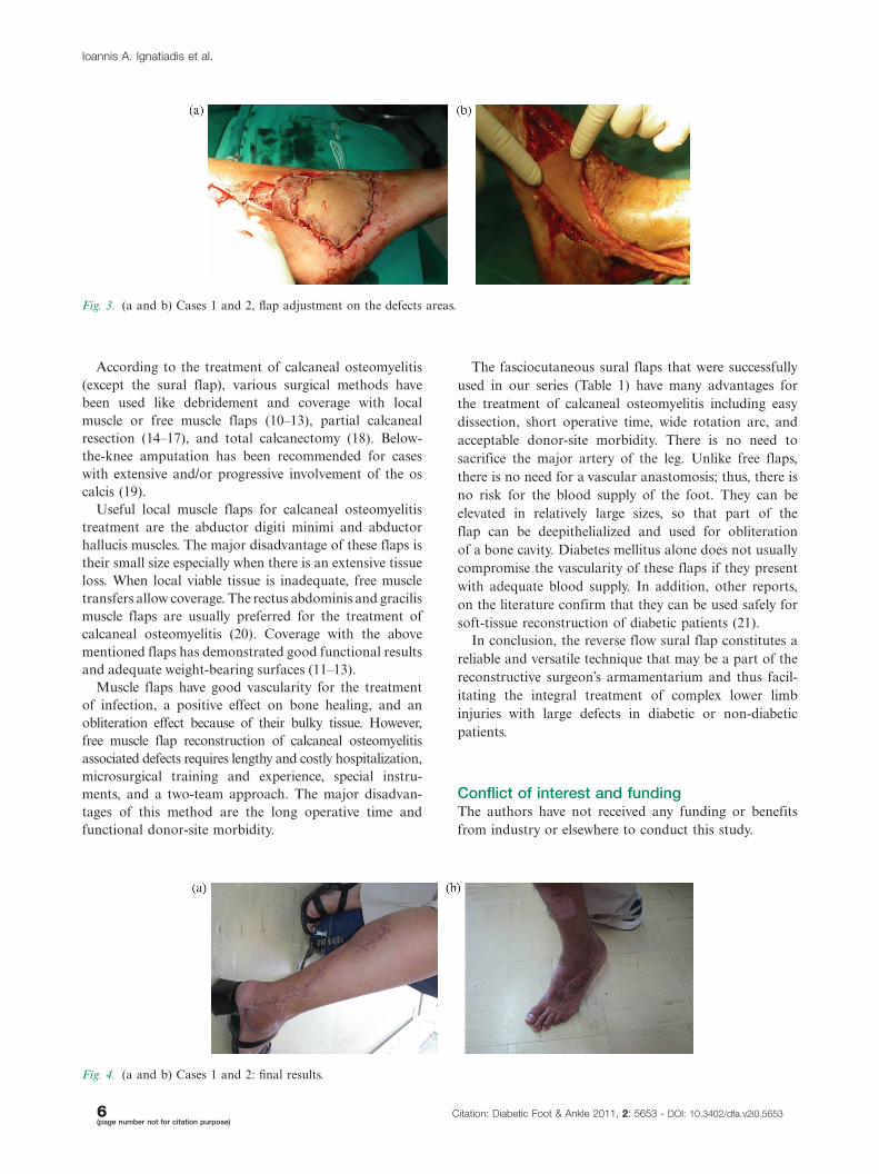
Fig. 3. (a and b) Cases 1 and 2, flap adjustment on the defects areas.
Ioannis A. Ignatiadis et al.
6(page number not for citation purpose)
Citation: Diabetic Foot & Ankle 2011, 2: 5653 - DOI: 10.3402/dfa.v2i0.5653

References
1. Baumeister SP, Spierer R, Erdman R, Sweis R, Lewin LS,
German GK, et al. A realistic complication analysis of 70 sural
artery flaps in a multimorbid patients group. Plast Reconstr
Surg 2003; 112: 129�40.
2. Rios-Luna A, Villanueva-Martinez M, Fahandezh-Saddi H,
Villanueva-Lopez F, del Cerro-Gutierrez M. Versatility of the
sural fasciocutaneous flap in coverage defects of the lower limb.
Injury 2007; 38: 824�31.
3. Hasegawa M, Torii S, Katoh H, Esaki S. The distally based
superficial sural artery flap. Plast Reconstruct Surg 1994; 93:
1012�20.
4. Conskunfirat OK, Velidedeoglu HV, Sahin U, Demir Z. Reverse
neurofasciocutaneous flaps for soft-tissue coverage of the lower
leg. Ann Plast Surg 1999; 43: 14.
5. Jeng SF, Wei FC, Kuo YR. Salvage of the distal foot using
distally based sural island flap. Ann Plast Surg 1999; 43: 499.
6. Yilmaz M, Karatas O, Barutcu A. The distally based superficial
artery island: clinical experience and modifications. Plast
Reconstr Surg 1998; 102: 2358.
7. Price MF, Capizzi PJ, Watterson PA, Lettieri S. Reverse sural
artery flap: caveats for success. Ann Plast Surg 2002; 48: 496�504.
8. Cavadas PC, Landin L. Reconstruction of chronic Achilles
tendon defects with posterior tibial perforator flap and soleus
tendon graft: clinical series. Plast Reconstruct Surg 2006; 117:
266�71.
9. Costa Ferreira A, Reis J, Amarante J. Reconstruction of soft
tissue defects of the heel with local fasciocutaneous flaps. Ann
Plast Surg 2005; 54: 580�1.
10. Fitzerald RH Jr, Ruttle PE, Arnold PG, Kelly PJ, Irons, GB.
Local muscle flaps in the treatment of chronic osteomyelitis.
J Bone Joint Surg (Am) 1985; 67: 175.
11. Wellisz T, Rechnic M, Douherty W, Sherman R. Coverage of
bilateral lower extremity calcaneal fractures with osteomyelitis
using a single split free gracilis muscle transfer. Plast Reconstr
Surg 1990; 85: 457.
12. Anderson RB, Foster MD, Gould JS, Hanel DP. Free tissue
transfer and calcanectomy as treatment of chronic osteomyelitis
of the os calcis: a case report. Foot Ankle 1990; 11: 168�71.
13. Attinger C, Cooper P. Soft tissue reconstruction for calcaneal
fractures or osteomyelitis. Orthop Clin North Am 2001; 32:
135�70.
14. Woll TS, Beals RK. Partial calcanectomy for the treatment of
osteomyelitis of the calcaneus. Foot Ankle 1991; 12: 31�3.
15. Perez ML, Wagner SS, Yun J. Subtotal calcanectomy for chronic
heel ulceration. J Foot Ankle Surg 1994; 33: 572.
16. Baravarian B, Menendez MM, Weinheimer DJ, Lowery C,
Kosanovich R, Vidt L. Subtotal calcanectomy for the treatment
of large heel ulceration and calcaneal osteomyelitis in the
diabetic patient. J Foot Ankle Surg 1999; 38: 194.
17. Smith DG, Stuck RM, Ketner L, Sage RM, Pinzur MS. Partial
calcanectomy for the treatment of large ulcerations of the heel
and calcaneal osteomyelitis. J Bone Joint Surg (Am.) 1992; 74:
571�6.
18. Martini M, Martini-Benkeddache Y, Bekkechi T, Daoud A.
Treatment of chronic osteomyelitis of the calcaneus by resection
of the calcaneus: a report of twenty cases. J Bone Joint Surg
(Am) 1974; 56: 542�8.
19. Crandall RC, Wagner FW Jr. Partial and total calcanectomy: a
review of thirty-one consecutive cases over a ten-year period.
J Bone Joint Surg (Am) 1981; 63: 152�5.
20. May JW Jr, Rohrich RJ. Foot reconstruction using free micro
vascular muscle flaps with skin grafts. Clinics in Plastic Surgery
1986; 13: 681.
21. Yildirim S, Akan M, Akoz T. Soft tissue reconstruction of the
foot with distally based neurocutaneous flaps in diabetic
patients. Ann Plast Surg 2002; 48: 258�64.
*Ioannis A. IgnatiadisHand Surgery-Upper Limb and Microsurgery DepartmentKAT General HospitalAthens, GreeceEmail: [email protected]
Reverse sural fasciocutaneous flaps for treating infections
Citation: Diabetic Foot & Ankle 2011, 2: 5653 - DOI: 10.3402/dfa.v2i0.5653 7(page number not for citation purpose)
Copyright © 2022 FDOKUMEN