The Replication Focus Targeting Sequence (RFTS) Domain Is a DNA-competitive Inhibitor of Dnmt1
24
1 The RFTS domain is a DNA-competitive inhibitor of Dnmt1* Farisa Syeda 1 , Rebecca L. Fagan 2 , Matthew Wean 2 , George V. Avvakumov 1 , John R. Walker 1 , Sheng Xue 1 , Sirano Dhe-Paganon 1,3 and Charles Brenner 2, 3 From the 1 Structural Genomics Consortium and Department of Physiology, University of Toronto, 101 College Street, Toronto, Ontario, M5G 1L7, Canada and the 2 Department of Biochemistry, Carver College of Medicine, University of Iowa, Iowa City, IA 52242, USA Running Head: RFTS domain auto-inhibits Dnmt1 catalysis DNA methyltransferase 1 (Dnmt1) is the principal enzyme responsible for maintenance of cytosine methylation at CpG dinucleotides in the mammalian genome. The N-terminal Replication Foci Targeting Sequence (RFTS) domain of Dnmt1 has been implicated in subcellular localization, protein association and catalytic function. However, progress in understanding its function has been limited by the lack of assays for and a structure of this domain. Here we show that the naked DNA-binding and polynucleosome- binding activities of Dnmt1 are inhibited by the RFTS domain, which functions by virtue of binding the catalytic domain to the exclusion of DNA. Kinetic analysis with a fluorigenic DNA substrate establishes the RFTS domain as a 600-fold inhibitor of Dnmt1 enzymatic activity. The crystal structure of the RFTS domain reveals a novel fold and supports a mechanism in which an RFTS-targeted Dnmt1-binding protein, such as Uhrf1, may activate Dnmt1 for DNA-binding. DNA cytosine methylation, an epigenetic mark that occurs predominantly *Use of the Advanced Photon Source was supported by the U.S. Department of Energy Office of Basic Energy Sciences, under Contract DE- AC02-06CH11357. The Structural Genomics Consortium is a registered charity (#1097737) supported by the Canadian Institutes for Health Research, the Canadian Foundation for Innovation, and Genome Canada through the Ontario Genomics Institute. Work at University of Iowa at CpG dinucleotides, is the principal eukaryotic DNA modification (1). DNA methylation is a key component of gene silencing, which makes significant contributions to cell phenotype. Establishment and maintenance of DNA methylation patterns is governed by three catalytically active DNA methyltransferases: Dnmt3a, Dnmt3b and Dnmt1. The Dnmt3 isoforms are responsible for de novo methylation during germ cell and embryonic development, and bind with high-affinity to unmethylated DNA sequences (2). CpG methylation patterns are maintained in mammals by Dnmt1 with hemi-methylated CpG dinucleotides serving as preferred substrates. Human Dnmt1 (1616 aa) consists of a conserved C-terminal catalytic core (1140-1616) and a large N-terminal region (1-1139) harboring multiple globular conserved domains, including the DNA methyltransferase-associated protein 1 (DMAP1)-binding domain (3), the proliferating cell nuclear antigen (PCNA)- binding domain (4), the Replication Foci Targeting Sequence (RFTS) domain (5), the CXXC domain (6) and two Bromo- Adjacent Homology (BAH) domains (7) (Fig 1). was supported by grant CA075954 and contract HHSN261200433000C from the National Cancer Institute, and a generous gift from the Roy J. Carver Foundation. 3 To whom correspondence should be addressed. E- mail: [email protected] or [email protected] http://www.jbc.org/cgi/doi/10.1074/jbc.M110.209882 The latest version is at JBC Papers in Press. Published on March 9, 2011 as Manuscript M110.209882 Copyright 2011 by The American Society for Biochemistry and Molecular Biology, Inc. by guest on May 17, 2016 http://www.jbc.org/ Downloaded from by guest on May 17, 2016 http://www.jbc.org/ Downloaded from by guest on May 17, 2016 http://www.jbc.org/ Downloaded from by guest on May 17, 2016 http://www.jbc.org/ Downloaded from by guest on May 17, 2016 http://www.jbc.org/ Downloaded from by guest on May 17, 2016 http://www.jbc.org/ Downloaded from by guest on May 17, 2016 http://www.jbc.org/ Downloaded from
Transcript of The Replication Focus Targeting Sequence (RFTS) Domain Is a DNA-competitive Inhibitor of Dnmt1

1
The RFTS domain is a DNA-competitive inhibitor of Dnmt1* Farisa Syeda1, Rebecca L. Fagan2, Matthew Wean2, George V. Avvakumov1, John R. Walker1,
Sheng Xue1, Sirano Dhe-Paganon1,3 and Charles Brenner2, 3 From the 1Structural Genomics Consortium and Department of Physiology, University of Toronto, 101 College Street, Toronto, Ontario, M5G 1L7, Canada and the 2Department of Biochemistry, Carver College of Medicine, University of Iowa, Iowa City, IA 52242, USA
Running Head: RFTS domain auto-inhibits Dnmt1 catalysis
DNA methyltransferase 1 (Dnmt1) is the principal enzyme responsible for maintenance of cytosine methylation at CpG dinucleotides in the mammalian genome. The N-terminal Replication Foci Targeting Sequence (RFTS) domain of Dnmt1 has been implicated in subcellular localization, protein association and catalytic function. However, progress in understanding its function has been limited by the lack of assays for and a structure of this domain. Here we show that the naked DNA-binding and polynucleosome-binding activities of Dnmt1 are inhibited by the RFTS domain, which functions by virtue of binding the catalytic domain to the exclusion of DNA. Kinetic analysis with a fluorigenic DNA substrate establishes the RFTS domain as a 600-fold inhibitor of Dnmt1 enzymatic activity. The crystal structure of the RFTS domain reveals a novel fold and supports a mechanism in which an RFTS-targeted Dnmt1-binding protein, such as Uhrf1, may activate Dnmt1 for DNA-binding. DNA cytosine methylation, an epigenetic mark that occurs predominantly
*Use of the Advanced Photon Source was supported by the U.S. Department of Energy Office of Basic Energy Sciences, under Contract DE-AC02-06CH11357. The Structural Genomics Consortium is a registered charity (#1097737) supported by the Canadian Institutes for Health Research, the Canadian Foundation for Innovation, and Genome Canada through the Ontario Genomics Institute. Work at University of Iowa
at CpG dinucleotides, is the principal eukaryotic DNA modification (1). DNA methylation is a key component of gene silencing, which makes significant contributions to cell phenotype. Establishment and maintenance of DNA methylation patterns is governed by three catalytically active DNA methyltransferases: Dnmt3a, Dnmt3b and Dnmt1. The Dnmt3 isoforms are responsible for de novo methylation during germ cell and embryonic development, and bind with high-affinity to unmethylated DNA sequences (2). CpG methylation patterns are maintained in mammals by Dnmt1 with hemi-methylated CpG dinucleotides serving as preferred substrates.
Human Dnmt1 (1616 aa) consists of a conserved C-terminal catalytic core (1140-1616) and a large N-terminal region (1-1139) harboring multiple globular conserved domains, including the DNA methyltransferase-associated protein 1 (DMAP1)-binding domain (3), the proliferating cell nuclear antigen (PCNA)-binding domain (4), the Replication Foci Targeting Sequence (RFTS) domain (5), the CXXC domain (6) and two Bromo-Adjacent Homology (BAH) domains (7) (Fig 1). was supported by grant CA075954 and contract HHSN261200433000C from the National Cancer Institute, and a generous gift from the Roy J. Carver Foundation. 3To whom correspondence should be addressed. E-mail: [email protected] or [email protected]
http://www.jbc.org/cgi/doi/10.1074/jbc.M110.209882The latest version is at JBC Papers in Press. Published on March 9, 2011 as Manuscript M110.209882
Copyright 2011 by The American Society for Biochemistry and Molecular Biology, Inc.
by guest on May 17, 2016
http://ww
w.jbc.org/
Dow
nloaded from
by guest on May 17, 2016
http://ww
w.jbc.org/
Dow
nloaded from
by guest on May 17, 2016
http://ww
w.jbc.org/
Dow
nloaded from
by guest on May 17, 2016
http://ww
w.jbc.org/
Dow
nloaded from
by guest on May 17, 2016
http://ww
w.jbc.org/
Dow
nloaded from
by guest on May 17, 2016
http://ww
w.jbc.org/
Dow
nloaded from
by guest on May 17, 2016
http://ww
w.jbc.org/
Dow
nloaded from

2
The CXXC domain is understood to contribute to catalytic activity by interacting with unmethylated CpG DNA substrates (6,8). This was observed in the recently solved crystal structures of Dnmt1, which encompass sequences from the CXXC domain to the C-terminus (9). In the structures, the CXXC domain binds and holds unmethylated duplex CpG-containing DNA away from the active site while the acidic linker between the CXXC and BAH1 domains is bound in the active site between the DNA segment and the S-adenosyl homocysteine product (9). This observation helps to explain the relationship between the CXXC and catalytic domains. However, it does not address the role of domains N-terminal to the CXXC, which are not present in the structures.
We set out to clarify the structure and function of the RFTS domain. The RFTS domain is conserved by sequence (Fig S1) and contains the binding site for Uhrf1 (10), a Dnmt1-associated protein that recruits Dnmt1 to hemimethylated DNA (11-15). Despite the significance of the RFTS domain, progress in understanding its function has been limited by the availability of stable, soluble protein fragments and robust DNA methyltransferase assays. Here, we generate soluble protein fragments of Dnmt1 and establish activities for them. Strikingly, the binding of Dnmt1 to naked DNA oligonucleotides and native polynucleosomes is inhibited by the RFTS domain. Kinetic analysis establishes that Dnmt1 without the RFTS domain functions with a 1 nM Km for an internally quenched oligonucleotide substrate. This represents a >100-fold binding advantage with respect to recent assays with hemimethylated oligonucleotide substrates (9,16). By comparison of kcat/Km terms between RFTS-containing and RFTS-
lacking forms of Dnmt1, the RFTS domain is a 600-fold inhibitor of DNA methylation. Moreover, by titrating the RFTS domain into reactions with the RFTS-lacking Dnmt1 construct, we show that RFTS is a 100 nM inhibitor that is strictly competitive with DNA-binding. Finally, the crystal structure of the RFTS domain reveals features that may allow it to occlude DNA substrate-binding by the catalytic domain in a manner that could be relieved by a Dnmt1 activator, such as Uhrf1. EXPERIMENTAL PROCEDURES
Cloning, Expression, and Purification—Human Dnmt1 constructs schematized in Fig 1 were cloned and purified with similar methods (17). Briefly, cDNA templates (MHS1768-98980929.pCR-XL-TOPO) from OpenBiosystems were cloned into the pET28MHL or pNICCH vectors using the In-Fusion CF Dry-Down PCR Cloning Kit (Clontech, 639605), transformed into E. coli BL21 (DE3) cells and grown in Terrific Broth in the presence of 50 µg/ml of Kanamycin at 37 ºC. Selenomethionyl derivatives of the RFTS domain were expressed in M9 medium supplemented with glycerol using an M9 SeMet High-Yield growth media kit package (M2D045004-50L, Medicilon) according
The abbreviations used are: Dnmt1, DNA methyltransferase 1; RFTS, Replication Foci Targeting Sequence (residues 351-600 of DNMT1); DMAP1, DNA methyltransferase-associated protein 1; PCNA, proliferating cell nuclear antigen; BAH, Bromo-Adjacent Homology; EMSA, Electrophoretic Mobility Shift Assay; CpG, cytosine-phosphate-guanosine dinucleotide; SAM, S-adenosyl methionine; DTT, Dithiothreitol; TCEP, tris(2-carboxyethyl)phosphine; EDTA, ethylenediaminetetraacetic acid; PMSF, phenyl-methyl sulfonyl fluoride; BSA, bovine serum
by guest on May 17, 2016
http://ww
w.jbc.org/
Dow
nloaded from
3
to manufacturer’s instructions. After lysis, cell supernatants were subjected to metal-affinity chromatography using TALON columns (BD Biosciences). Protein was further purified by gel filtration (HighLoad 16/60 Superdex 200 column, GE Healthcare) equilibrated with buffer A (20 mM Tris-HCl, pH 8.0, 0.5 M NaCl, 5% glycerol, 2 mM DTT) and by ion-exchange chromatography on a 5 ml HiTrapQ column using a gradient of buffer A to 50 % buffer B (20 mM Tris-HCL, pH 8.0, 1 M NaCl, 5% glycerol, 2 mM DTT). Full length Dnmt1 was obtained from New England Biolabs.
Nucleosome purification—Polynucleosomes from chicken erythrocytes were obtained by micrococcal nuclease digestion as described (18). Briefly, cells were lysed and vortexed thoroughly in buffer containing 10 mM Tris, pH 8, 6 mM MgCl2, 0.1 mM PMSF, 0.05% NP-40, 80 mg/ml sucrose. Nuclei were extracted and washed by a four-step sequential process and further digested by microccocal nuclease (Sigma) for 10 min at 37 ºC in 10 mM Tris, pH 8, 0.2 mM CaCl2, 0.1 mM PMSF and 10 mg/ml sucrose. Purified native chromatin was extracted in 10 mM Tris, pH 8, 0.25 mM EDTA buffer and analyzed on agarose gels.
Electrophoretic mobility-shift assays—DNA binding was analyzed using duplex DNA oligonucleotides of varying lengths, sequences and methylation states (see supplemental Table 1). Duplex DNA (100 ng per lane) was incubated overnight at 4 ºC with an appropriate concentration of protein (as indicated in the figure legend) in 10 mM Tris-HCl, pH 7.5, 150 mM NaCl, 0.5 mM DTT and 0.1 mg/ml BSA and subjected to polyacrylamide gel electrophoresis (10% Tris-borate-EDTA albumin; TBE, Tris-borate EDTA; WHB, winged helix B; RING, really interesting new gene.
(TBE) gels (BioRad) run in 0.5x TBE buffer at 100 V for 1.5 h on ice). Gels were stained with 10,000-fold diluted SYBR Green-I (Invitrogen) for 5 min and were washed twice with 0.5x TBE buffer and imaged using an excitation wavelength of 633 nm.
Dnmt1 activity assay—Dnmt1 activity was measured using an assay related to that developed by Wood (16) in which a hemimethylated, internally quenched, fluorescent hairpin DNA substrate is incubated with Dnmt1 and S-adenosyl methionine (SAM) in the presence of Gla I, which cleaves the fully methylated product. Assays (0.1 ml) were performed in triplicate in 96-well half-area black plates at 37 °C. Triplicate reactions were prepared by making a solution of 10 mM Tris-HCl, pH 7.5, 5 mM MgCl2, 1 mM DTT, 100 mM K Glu, 0.105 mg/mL BSA, 1.05 mM SAM (Sigma-Aldrich, St. Louis, HPLC purified) and 1.05x the desired concentration of oligonucleotide. For inhibition studies, 1.05x the desired concentration of the RFTS domain (351-600) was added to the solution. SAM was added freshly to limit conversion to S-adenosyl homocysteine, which inhibits the Dnmt1 reaction. Ninety-five microliters of this solution were added per well and brought to 37 °C. A second solution was made containing Dnmt1 and Gla I (SibEnzyme, West Roxbury, MA) in 10 mM Tris-HCl, pH 7.5, 5 mM MgCl2, 1 mM DTT, 100 mM K Glu such that 0.8 U of Gla I would be added to each well. Coupled reactions were initiated by adding 5 µL of enzyme solution, and the wells were covered with 5 µL of mineral oil. Data were collected in a Wallac Victor2 using excitation and emission wavelengths of 485 nm and 535 nm, respectively. A negative control containing Gla I without Dnmt1 was subtracted from each assay condition to correct for both substrate
by guest on May 17, 2016
http://ww
w.jbc.org/
Dow
nloaded from
4
fluorescence and fluorescence from slow Gla I cleavage of the hemimethylated substrate. A standard curve was created using the fully cleaved oligonucleotide products to convert arbitrary fluorescence units to concentration. The oligonucleotide, custom synthesized at IDT (Coralville, IA), was 5’FAM-CCTATGCGmCATCAGTTTTCTGATGmCGmCATAGG-3’ Iowa Black in which mC denotes 5-methyl deoxycytidylate residues and underlined residues form the Gla I restriction site. Initial rate data were fit either to the Michaelis-Menten equation or to the quadratic velocity equation for tight-binding substrates (19,20) to determine Vmax and Km. Data were fitted using either Kaleidagraph (Synergy, Inc) or Prism (Graph Pad Software, Inc). The quadratic velocity equation is:
Crystallization—Selenomethionyl
derivatives of the RFTS domain (351-600) were mixed with detergent Anapoe80 solution (10%, v/v) in a 5:1 ratio (final protein and detergent concentrations were 16.5 mg/ml and 1.7%, respectively) and crystals were grown at 298 K using the hanging drop method by mixing 1 volume of this solution with 1 volume of well solution consisting of 23% polyethylene glycol 3350, 0.1 M bis-Tris, pH 6.0, 0.3 M Na acetate and 5 mM TCEP. The crystals were cryoprotected by immersion in the well solution mixed 1:1 with 20% (w/v) sucrose, 4% (w/v) glucose, 18% (v/v) glycerol and 18% (v/v) ethylene glycol, and placed in liquid nitrogen.
Structure determination—Diffraction data from a crystal of the selenomethionyl derivative of the Dnmt1
RFTS domain was collected at beamline 19-ID at the Advanced Photon Source. The data set, collected at the selenium peak wavelength, was integrated and scaled using the HKL2000 program suite (21). The structure was solved by single wavelength anomalous diffraction techniques using the program SOLVE/RESOLVE (22). A second data set collected at the beamline 23-ID-B was used for refinement of the structure to 2.3 Å resolution. Iterative model building using the graphics program Coot (23) and maximum-likelihood and translation/liberation/screw refinement with the program REFMAC5 (24) led to the final model. REFMAC5 parameters were generated using the TLSMD web server. Statistics of data collection, processing, and refinement are provided in Table 1. RESULTS AND DISCUSSION
The RFTS domain inhibits DNA-binding independently of DNA sequence, length, methylation status or CpG content—It has been proposed that the RFTS domain of Dnmt1 tunes the catalytic activity of the conserved DNA methyltransferase domain toward specific DNA substrates (6). To test this hypothesis, we titrated constructs containing the RFTS domain (351-1616) or lacking the RFTS domain (621-1616) against 12bp hemi-methylated DNA oligonucleotides and analyzed DNA-binding using an electrophoretic mobility shift assay (EMSA) (Fig 2A). Surprisingly, at a range of Dnmt1 concentrations from 0.5 to 10 µM, the RFTS-lacking 621-1616 construct bound the DNA but the RFTS-containing 351-1616 construct showed no detectable binding even at high protein concentrations (Fig 2A). Moreover, DNA binding to the RFTS domain-lacking 621-
by guest on May 17, 2016
http://ww
w.jbc.org/
Dow
nloaded from
5
1616 construct was inhibited by the RFTS domain (351-600) added in trans (Fig 2B). These data suggested that the RFTS domain might inhibit the ability of the Dnmt1 catalytic domain to bind particular naked DNA sequences. To determine whether the RFTS domain might be responsible for substrate selectivity and permit the catalytic domain to bind some but not all DNA sequences, we surveyed a series of different DNA substrates of various lengths, methylation states, CpG content and sequences. As shown in Fig S2, the 621-1616 construct had its electrophoretic mobility shifted by all DNA constructs, regardless of methylation status, length, or sequence, while the 351-1616 construct, containing the RFTS domain, showed impaired binding of all oligonucleotides investigated. Even non-CpG oligonucleotides were bound by the 621-1616 Dnmt1 construct in a manner that was inhibited in trans by addition of the RFTS domain (Fig S3).
Dnmt1 binding to polynucleosomes is inhibited by the RFTS domain—The CXXC and catalytic domains of Dnmt1 are both known to possess DNA-binding activities (8,25,26). To determine whether the RFTS domain interferes with either the CXXC, the catalytic, or both domain functions, we generated protein constructs containing only the CXXC domain (621-698 and 621-740) or the RFTS with the CXXC domain (351-698). Extensive attempts to express and purify fragments of Dnmt1, which begin C-terminal to the CXXC domain, were unsuccessful, suggesting that CXXC sequences may be required for folding and/or stability of the catalytic domain. Because naked DNA bound very poorly to CXXC domain constructs, we purified native polynucleosomes, which consist of epigenetically modified histone octamers and segments of DNA in various
methylation states. EMSAs indicate that the 351-1616 construct is inhibited with respect to the polynucleosome-binding ability of the 621-1616 construct (Fig 3A). In addition, the CXXC domain possesses a polynucleosomal binding capacity (Fig 3B, lanes 6-7), which is inhibited by the RFTS domain both in cis (Fig 3B, lane 4 and Fig 3C, lane 2) and in trans (Fig 3C, lanes 4-6). However, the binding to polynucleosomes by the 621-1616 construct, containing both the CXXC and the catalytic domains, was not disrupted in trans by addition of the RFTS-only domain (Fig 3D). These data suggest that the polynucleosome-binding activity of the CXXC domain is additive with the DNA-binding activity of the catalytic domain and that simultaneous engagement of polynucleosomes by both domains renders the complex relatively resistant to exclusion of DNA by the RFTS.
RFTS domain is a potent inhibitor of Dnmt1 enzyme activity— Data from EMSAs suggested that the RFTS domain is an intrinsic inhibitor of DNA-binding by Dnmt1 sequences C-terminal to the RFTS. To test this hypothesis, we synthesized an internally quenched, fluorescent hairpin DNA substrate (16). In this restriction enzyme-coupled assay, methylation of a hemimethylated CpG site forms a Gla I site and generates fluorescence in real time (Fig 4A). Analysis of the RFTS-lacking protein construct (621-1616) with 20 nM DNA substrate indicated striking fluorescence-generating activity (Fig 4B). Initial attempts to determine steady-state kinetic parameters with this enzyme and oligonucleotide indicated that the Km was in the low nM range. Since assays were conducted with 2 nM Dnmt1, there was a need to test for the complication that the enzyme may titrate a significant proportion of substrate from the free form to an E-S complex at low substrate
by guest on May 17, 2016
http://ww
w.jbc.org/
Dow
nloaded from
6
concentrations (19). To test for this behavior, substrate-dependent initial rates were measured at enzyme concentrations of 1 to 5 nM. As the enzyme concentration increased, data deviated significantly from the hyperbolic behavior of Michealis-Menten enzyme kinetics. However, at all enzyme concentrations examined, the data fit well to the tight-binding equation (19,20), giving essentially the same Km (1 nM) and kcat (~0.1 min-1) values. Remarkably, Dnmt1 activity could not be detected at 2 nM of the RFTS domain-containing construct (351-1616) and 20 nM DNA substrate (Fig 4C). This led us to assay the RFTS domain-containing construct at higher enzyme and DNA substrate concentrations to test the hypothesis that the RFTS domain inhibits DNA-binding and not some other step such as binding SAM. As shown in Fig 5A, the second order rate constant with respect to the DNA substrate was 1.87 x 10⁶ M-1s-1 for the RFTS domain-lacking construct. Inclusion of the RFTS domain increased the Km for DNA by ~15-fold while depressing kcat by ~40-fold (Fig 5A and Fig S4). Similar kinetic constants were determined for the full-length protein. Thus, the RFTS domain functions as a ~600-fold inhibitor of binding and methyl group transfer to the DNA substrate. This finding is strikingly different than recently published results, which monitored transfer of a tritiated methyl group from SAM to DNA. In those studies, the form of Dnmt1 with the first 645 amino acids deleted was only 2-fold more active than full length Dnmt1 in kcat/Km (9). The greater activity in this study may be attributed to an improved assay and/or the greater activity of the Dnmt1 construct beginning at residue 621. Because full length Dnmt1 has been assayed using an internally quenched fluorigenic substrate and found not to be
saturated with the DNA substrate at 240 nM (16), we argue that the amino acid 621-1616 Dnmt1 construct is strongly derepressed for DNA-binding and catalysis. To further examine the inhibitory function of the RFTS domain, we titrated the RFTS polypeptide in trans to the RFTS-lacking Dnmt1 construct (621-1616) and determined kinetic parameters. Plotting the data according to the method of Lineweaver and Burk (Fig 5B) clearly indicated that inhibition is competitive with the DNA substrate. As the concentration of RFTS domain was increased, the Km(apparent) was increased without alteration of Vmax. By nonlinear regression, the RFTS domain-dependent effects on Km(apparent) indicated a Ki of 103 ± 11 nM. Thus, the RFTS domain is a previously unrecognized endogenous inhibitor of DNA binding within Dnmt1.
Crystal structure of the Dnmt1 RFTS domain and a model of auto-inhibition—Having established discrete assays for the function of the RFTS, we aimed to determine whether the physical characteristics of the domain might account for inhibition of DNA-binding. A selenomethionyl form of the RFTS domain crystallized in space group P21 and diffracted to 2.3 Å resolution. A refined model of the crystal structure is depicted in Fig 6 (see Table 1 for refinement statistics). The RFTS domain consists of a zinc-binding motif, followed by a β-barrel and a helical bundle, which are closely associated. Using the Secondary Structure Matching algorithm (27), the closest match to the β-barrel is the structure of the Src family kinase HCK Src homology 3 domain (Q score 0.25, rmsd 2.36 Å over 46 aligned Cα positions). The six-stranded β -barrel lobe of RFTS extends from residues 401 to 502, capped on one end by an α -helix (residues 496-499, α2)
by guest on May 17, 2016
http://ww
w.jbc.org/
Dow
nloaded from

7
and the other end by a helical bundle (residues 495-600). The nearest structural homolog of the helical bundle, as calculated by the DALI server (28), is the winged helix B (WHB) lobe of the Cullin protein, Cdc53 (RMSD of 1.7 Å across 68 backbone atoms). The role of the WHB lobe in these E3 ubiquitin ligases, which has recently been elucidated, involves regulating the enzyme via an autoinhibitory mechanism (29). The WHB lobe in the inhibited state binds to the really interesting new gene (RING) domain of the ligase, preventing the RING from reaching the substrate. Neddylation of the WHB relieves its interaction with the RING, allowing the RING to access substrate (29).
In recent crystal structures, the acidic linker between the CXXC and BAH1 domains associates with the DNA-binding site of the methyltransferase domain and prevents productive access of DNA to the active site, where S-adenosyl homocysteine is bound (Fig 7A) (9). Deletion of the CXXC domain and linker region increased kcat/Km by 10-fold on nonmethylated DNA substrates and increased activity on hemimethylated substrates by 20% (9). However, the 600-fold effect of the RFTS domain suggests that, were it present, the RFTS inhibitory activity would predominate over the mild effect of the CXXC-BAH1 linker in excluding DNA from the active site.
We sought to develop a model for Dnmt1 autoinhibition that would account
for the 600-fold inhibition by the RFTS domain and account for how a positive regulator of Dnmt1 might relieve this inhibition. As shown in Fig 7B, RFTS has a size and electrostatic properties compatible with occlusion of the DNA substrate-binding site of Dnmt1. Three acidic loops, each disordered in our crystal structure of RFTS, are positioned on the side of the RFTS domain we predict to associate with three basic patches of the methyltransferase domain of Dnmt1. We postulate that Uhrf1, a multidomain E3 ubiquitin ligase, which is required for maintenance DNA methylation and Dnmt1 localization in embryonic stem cells (11,12), is the key mediator of Dnmt1 derepression. According to our model, Uhrf1 binding to RFTS would release the inhibitory domain from the active site and allow DNA to bind productively. This model is attractive for two reasons. First, Uhrf1 has been reported to associate with Dnmt1 within the RFTS (10). Second, Uhrf1 is thought to function by binding hemimethylated DNA and recruiting Dnmt1 for methyl transfer to the nonmodified cytosine of a hemimethylated site (13) or to nearby CpG dinucleotides (14,15). If Uhrf1 binding to the RFTS is required to relieve inhibition of DNA-binding, a mechanism is provided to activate Dnmt1 for processive methylation of hemimethylated regions of DNA identified by the essential activator, Uhrf1.
Acknowledgements: The authors thank Yanjun Li and Peter Loppnau for plasmid constructions, Juan Pizzaro and Abdellah Allali-Hassani for technical assistance, Peter Lewis for assistance with chromatin preparation, and Bryce Plapp for conversations on enzyme inhibition.
by guest on May 17, 2016
http://ww
w.jbc.org/
Dow
nloaded from
8
REFERENCES 1. Suzuki, M. M., and Bird, A. (2008) Nat Rev Genet 9, 465-476 2. Yokochi, T., and Robertson, K. D. (2002) J Biol Chem 277, 11735-11745 3. Rountree, M. R., Bachman, K. E., and Baylin, S. B. (2000) Nat Genet 25, 269-277 4. Chuang, L. S., Ian, H. I., Koh, T. W., Ng, H. H., Xu, G., and Li, B. F. (1997) Science
277, 1996-2000 5. Leonhardt, H., Page, A. W., Weier, H. U., and Bestor, T. H. (1992) Cell 71, 865-873 6. Bestor, T. H. (1992) Embo J 11, 2611-2617 7. Callebaut, I., Courvalin, J. C., and Mornon, J. P. (1999) FEBS Lett 446, 189-193 8. Pradhan, M., Esteve, P. O., Chin, H. G., Samaranayke, M., Kim, G. D., and Pradhan, S.
(2008) Biochemistry 47, 10000-10009 9. Song, J., Rechkoblit, O., Bestor, T. H., and Patel, D. J. (2011) Science 331, 1036-1040 10. Achour, M., Jacq, X., Ronde, P., Alhosin, M., Charlot, C., Chataigneau, T., Jeanblanc,
M., Macaluso, M., Giordano, A., Hughes, A. D., Schini-Kerth, V. B., and Bronner, C. (2008) Oncogene 27, 2187-2197
11. Bostick, M., Kim, J. K., Esteve, P. O., Clark, A., Pradhan, S., and Jacobsen, S. E. (2007) Science 317, 1760-1764
12. Sharif, J., Muto, M., Takebayashi, S., Suetake, I., Iwamatsu, A., Endo, T. A., Shinga, J., Mizutani-Koseki, Y., Toyoda, T., Okamura, K., Tajima, S., Mitsuya, K., Okano, M., and Koseki, H. (2007) Nature 450, 908-912
13. Arita, K., Ariyoshi, M., Tochio, H., Nakamura, Y., and Shirakawa, M. (2008) Nature 455, 818-821
14. Avvakumov, G. V., Walker, J. R., Xue, S., Li, Y., Duan, S., Bronner, C., Arrowsmith, C. H., and Dhe-Paganon, S. (2008) Nature 455, 822-825
15. Hashimoto, H., Horton, J. R., Zhang, X., Bostick, M., Jacobsen, S. E., and Cheng, X. (2008) Nature 455, 826-829
16. Wood, R. J., McKelvie, J. C., Maynard-Smith, M. D., and Roach, P. L. (2010) Nucleic Acids Res 38, e107
17. Bacik, J. P., Walker, J. R., Ali, M., Schimmer, A. D., and Dhe-Paganon, S. (2010) J Biol Chem 285, 20273-20280
18. Chan, S., Attisano, L., and Lewis, P. N. (1988) J Biol Chem 263, 15643-15651 19. Cha, S. (1970) J Biol Chem 245, 4814-4818 20. Goldstein, A. (1944) J Gen Physiol 27, 529-580 21. Otwinowski, Z., and Minor, W. (1997) Methods in Enzymology 276, 307-326 22. Terwilliger, T. C. (2000) Acta Crystallogr D Biol Crystallogr 56, 965-972 23. Emsley, P., Lohkamp, B., Scott, W. G., and Cowtan, K. (2010) Acta Crystallogr D Biol
Crystallogr 66, 486-501 24. Vagin, A. A., Steiner, R. A., Lebedev, A. A., Potterton, L., McNicholas, S., Long, F., and
Murshudov, G. N. (2004) Acta Crystallogr D Biol Crystallogr 60, 2184-2195 25. Svedruzic, Z. M., and Reich, N. O. (2005) Biochemistry 44, 9472-9485 26. Jeltsch, A. (2006) Epigenetics 1, 63-66 27. Krissinel, E., and Henrick, K. (2004) Acta Crystallogr D Biol Crystallogr 60, 2256-2268 28. Holm, L., Kaariainen, S., Rosenstrom, P., and Schenkel, A. (2008) Bioinformatics 24,
2780-2781 29. Duda, D. M., Borg, L. A., Scott, D. C., Hunt, H. W., Hammel, M., and Schulman, B. A.
(2008) Cell 134, 995-1006
by guest on May 17, 2016
http://ww
w.jbc.org/
Dow
nloaded from
9
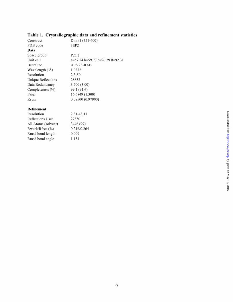
Table 1. Crystallographic data and refinement statistics
Construct Dnmt1 (351-600) PDB code 3EPZ Data Space group P2(1) Unit cell a=57.54 b=59.77 c=96.29 ß=92.31 Beamline APS 23-ID-B Wavelength ( Å) 1.0332 Resolution 2.3-50 Unique Reflections 28832 Data Redundancy 3.700 (3.00) Completeness (%) 99.1 (91.6) I/sigI 16.6849 (1.300) Rsym 0.08500 (0.97900) Refinement Resolution 2.31-48.11 Reflections Used 27330 All Atoms (solvent) 3446 (99) Rwork/Rfree (%) 0.216/0.264 Rmsd bond length 0.009 Rmsd bond angle 1.154
by guest on May 17, 2016
http://ww
w.jbc.org/
Dow
nloaded from

10
FIGURE LEGENDS
FIGURE 1. Dnmt1 protein domains. Full-length and truncated Dnmt1 constructs are shown with conserved domains colored as follows: DMAP1, red; RFTS domain, yellow; CXXC domain, teal; two BAH domains, blue; methyltransferase domain, magenta. FIGURE 2. The RFTS domain is an inhibitor of naked DNA-binding. A) RFTS-containing (351-1616) and RFTS-lacking (621-1616) Dnmt1 (0.5 - 10 µM) were incubated with 100 ng of 12bp hemimethylated oligonucleotide and analyzed for DNA binding via an EMSA. DNA is detected with Sybr Green (upper panel) and protein is detected with Coomassie (lower panel). The RFTS-lacking protein exhibits superior DNA-binding to the RFTS-containing protein as indicated by the shift in the electrophoretic mobility of the DNA observed in lanes 4 - 7. B) The RFTS domain (0.5 - 5.0 µM) was added in trans to assays containing 5 µM of RFTS-lacking Dnmt1 and 100 ng of hemimethylated oligonucleotide. The RFTS domain inhibits DNA binding to the RFTS-lacking protein, which allows the DNA to migrate rapidly. The sequences of all DNA oligonucleotides are presented in supplemental Table 1. FIGURE 3. Dnmt1 binding to polynucleosomes is inhibited by the RFTS domain. A) RFTS-containing (351-1616) and RFTS-lacking (621-1616) Dnmt1 (0.5 - 5.0 µM) were incubated with polynucleosomes (150 ng DNA) and analyzed for binding via an EMSA. The Sybr Green stained gel shows that polynucleosomal DNA runs as a typical smeary ladder (lane 1). The RFTS-lacking protein exhibits enhanced polynucleosome-binding ability when compared to the RFTS-containing protein as indicated by the shift in mobility of the DNA bands, which are retained in the wells upon protein interaction. B) The polynucleosome-binding ability of all protein constructs was investigated via EMSA. 5 µM protein was incubated with 150 ng of polynucleosomal DNA. Constructs containing the RFTS domain (lanes 3 - 5) show impaired polynucleosome-binding. The CXXC-only constructs exhibit tight binding to polynucleosomes (lanes 6 - 7). C) The RFTS domain inhibits polynucleosome-binding to the CXXC domain. The RFTS domain (0.5 - 5 µM) was added in trans to assays containing 5 µM of the CXXC domain (621-698) and 150 ng of polynucleosomes. The RFTS domain inhibits DNA-binding to the CXXC domain as indicated by increased mobility of the DNA. D) The RFTS domain (0.5 - 5.0 µM) was added in trans to assays containing 5 µM of the RFTS-lacking protein and 150 ng of polynucleosomes. The isolated RFTS domain does not compete polynucleosomes from the 621-1616 form of Dnmt1. FIGURE 4. The RFTS domain is an endogenous inhibitor of Dnmt1 activity. A) Schematic of the Gla I-coupled Dnmt1 assay. B) Time- and Dnmt1-dependence of the Gla I-coupled assay with 20 nM DNA substrate. With the RFTS-lacking (621-1616) protein, robust increases in fluorescence are observed that are dependent on the concentration of Dnmt1. C) Autoinhibition of RFTS-containing Dnmt1. At 20 nM DNA substrate, 2 nM of the RFTS-lacking (621-1616) Dnmt1 nears completion in 30 min, whereas no reaction progress is observed with 2 nM of the RFTS-containing (351-1616) Dnmt1 construct. FIGURE 5. The RFTS domain is a DNA-competitive inhibitor of methyltransferase activity. A) DNA substrate-dependent methyltransfer rates for the RFTS-lacking (621-1616) (●) and RFTS-containing (351-1616) (■) proteins were measured at 1 mM SAM. The RFTS domain confers a ~40-fold
by guest on May 17, 2016
http://ww
w.jbc.org/
Dow
nloaded from
11
reduction in kcat and a ~15-fold increase in Km for the DNA substrate. B) The RFTS domain (351-600) was used in trans as an inhibitor of the methyltransferase activity of the RFTS-lacking (621-1616) protein. The double reciprocal plot of the data with no RFTS domain (●), 100 nM RFTS domain (■), 250 nM RFTS domain (▲), and 500 nM RFTS domain (!) clearly shows that the RFTS domain is competitive with respect to the DNA substrate. Using nonlinear regression to fit the alteration in Km(apparent), a Ki of 103 ± 11 nM was determined. FIGURE 6. Structure of the Dnmt1 RFTS domain. The RFTS domain is shown in a stereo ribbon representation with lobes color-coded: yellow, β -barrel; orange, zinc-ribbon; and green, α -helical bundle. The zinc atom is depicted as a grey sphere with interacting residues in stick format. Dashed lines represent disordered regions. FIGURE 7. Two models of autoinhibition in Dnmt1. Shown are electrostatic surface representations, generated with a gradient from -10 (red) to 10 (blue) kT/e, of Dnmt1 from BAH1 to the C-terminus. In A), derived from PDB 3PT6, a structure not including the RFTS domain, the acidic linker (magenta) between the BAH1 and the CXXC (green) domains blocks productive DNA access to the active site (9). In B), consistent with our observations (Fig. 2B and Fig. 5B) that the RFTS domain competes for DNA-binding with CXXC-containing constructs such as 621-1616, Dnmt1 is displayed with the CXXC domain and acidic linker excluded as in PDB 3PTA. The RFTS domain is depicted in an orientation in which three acidic loops may descend on three basic patches in the methyltransferase domain. The DNA-competitive nature of RFTS domain inhibition suggests that RFTS binds to the exclusion of DNA and may be released by an RFTS-targeted Dnmt1 activator, such as Uhrf1.
by guest on May 17, 2016
http://ww
w.jbc.org/
Dow
nloaded from

Supplemental Information
Table 1. Oligonucleotide Sequences and Nomenclature
12bp-hm-Ic GGGCCXGCAGGG
CCCGGGCGTCCC
16bp-hm-Ic GTGGGCCXGCAGGGTG
CACCCGGGCGTCCCAG
20bp-hm-Ic GGATGGGCCXGCAGGGTTGG
CCTACCCGGGCGTCCCAACC
24bp-hm-Ic GGTAATGGGCCXGCAGGGTATTGG
CCATTACCCGGGCGTCCCATAAGG
16bp-hm-Ia GTGGGCCCGCAGGXTG
CACCCGGGCGTCCGAG
24bp-nm AAATTGAGCCCGAGCCTCCCGTTC
TTTAACTCGGGCTCGGAGGGCAAG
24bp-hm-1 AAATTGAGCCXGAGCCTCCCGTTC
TTTAACTCGGGCTCGGAGGGCAAG
24bp-hm-2 AAATTGAGCCXGAGCCTCCXGTTC
TTTAACTCGGGCTCGGAGGGCAAG
24bp'-hm-1 GAGCXCGTAAGCCCGTTCAGGTCG
CTCGGGCATTCGGGCAAGTCCAGC
24bp'-hm-2 GAGCXCGTAAGCCCGTTXAGGTCG
CTCGGGCATTCGGGCAAGTCCAGC
24bp-nonCpG TCCAGGACTTCTCTCAGGTTAACT
AGGTCCTGAAGAGAGTCCAATTGA
X, 5-‐methylcytosine
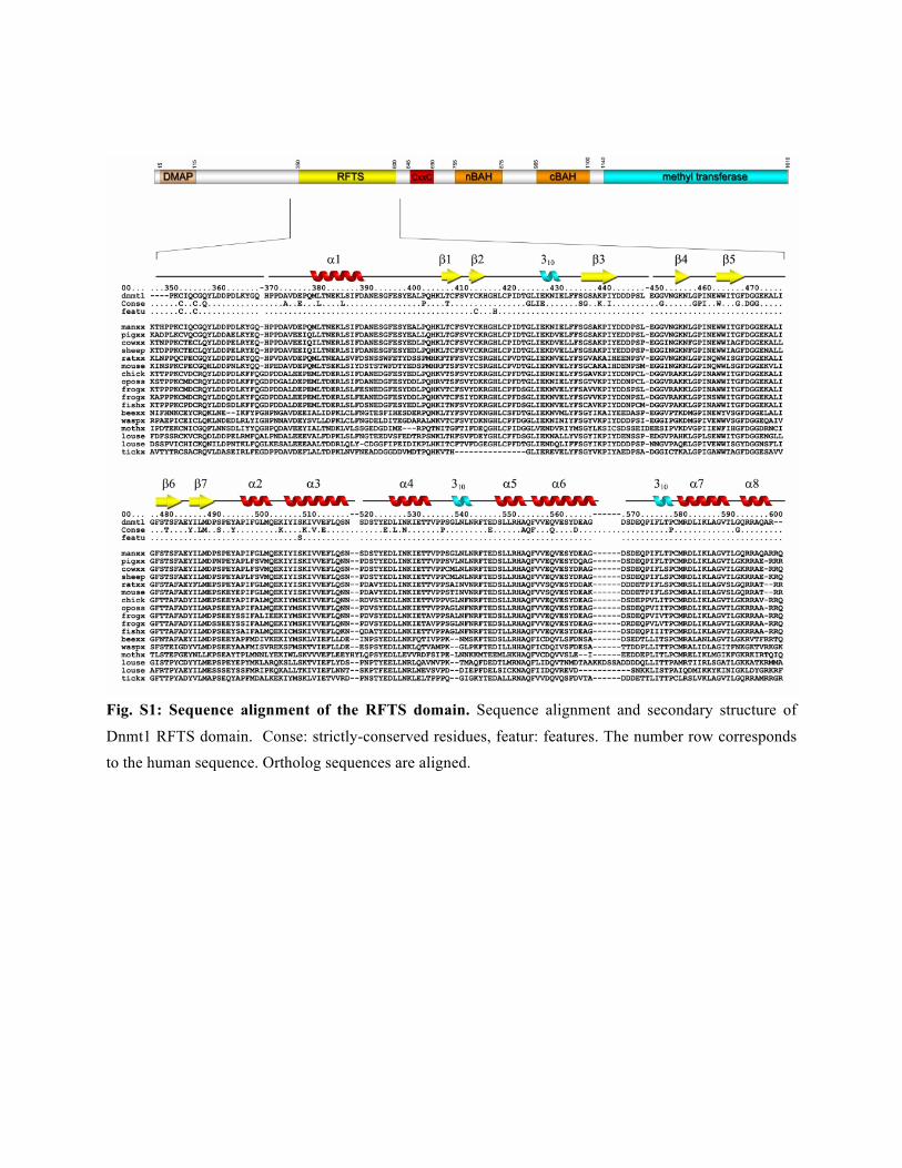
Fig. S1: Sequence alignment of the RFTS domain. Sequence alignment and secondary structure of
Dnmt1 RFTS domain. Conse: strictly-conserved residues, featur: features. The number row corresponds
to the human sequence. Ortholog sequences are aligned.
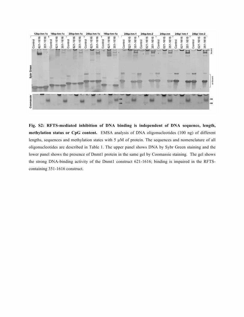
Fig. S2: RFTS-mediated inhibition of DNA binding is independent of DNA sequence, length,
methylation status or CpG content. EMSA analysis of DNA oligonucleotides (100 ng) of different
lengths, sequences and methylation states with 5 µM of protein. The sequences and nomenclature of all
oligonucleotides are described in Table 1. The upper panel shows DNA by Sybr Green staining and the
lower panel shows the presence of Dnmt1 protein in the same gel by Coomassie staining. The gel shows
the strong DNA-binding activity of the Dnmt1 construct 621-1616; binding is impaired in the RFTS-
containing 351-1616 construct.

Fig. S3: In trans RFTS inhibition of binding of non-CpG containing DNA. The RFTS domain (2.5 –
5 µM) was added in trans to assays containing 5 µM of RFTS-lacking (621-1616) or RFTS-containing
(351-1616) Dnmt1 and 100 ng of non-CpG oligonucleotide. The upper panel shows DNA by Sybr Green
staining and the lower panel shows the presence of Dnmt1 protein in the same gel by Coomassie staining.
The RFTS domain inhibits DNA binding to the RFTS-lacking protein, which allows the DNA to migrate
rapidly. No binding was detected with the 351-1616 protein construct.
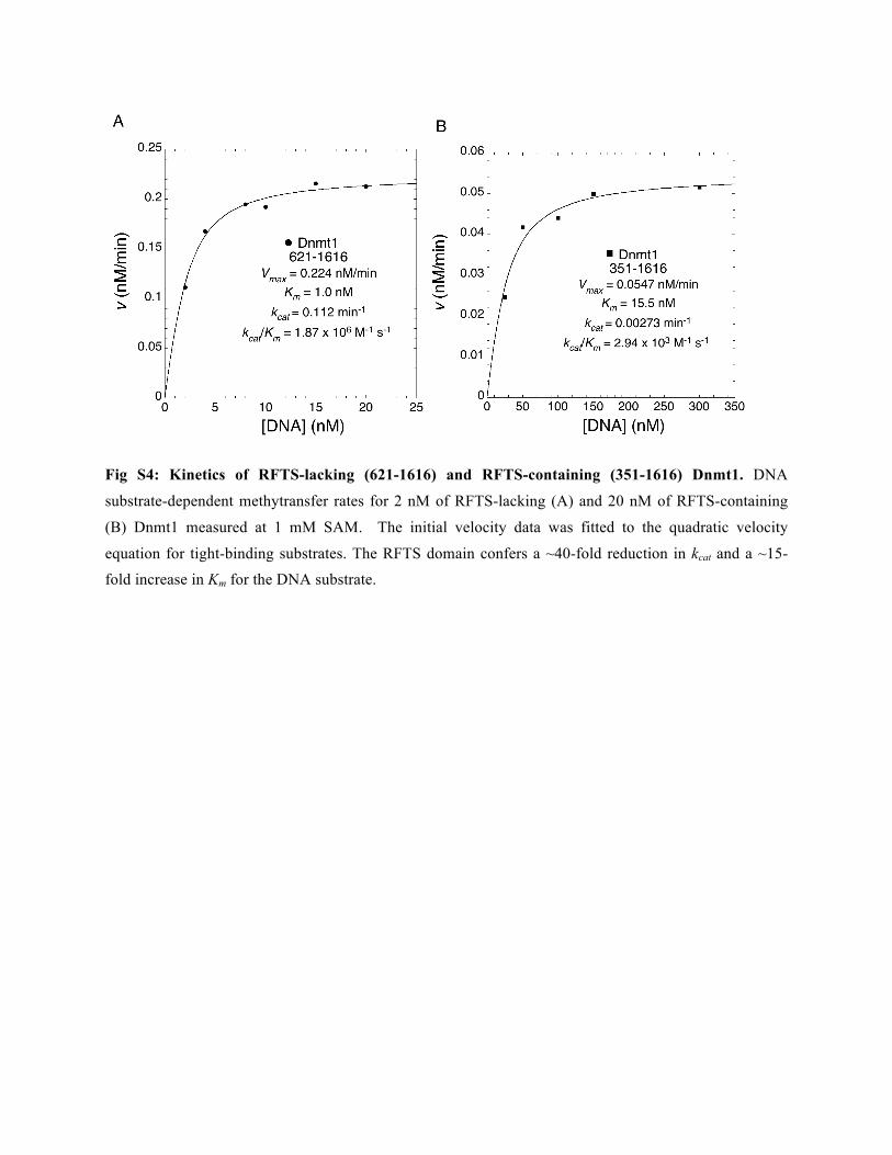
Fig S4: Kinetics of RFTS-lacking (621-1616) and RFTS-containing (351-1616) Dnmt1. DNA
substrate-dependent methytransfer rates for 2 nM of RFTS-lacking (A) and 20 nM of RFTS-containing
(B) Dnmt1 measured at 1 mM SAM. The initial velocity data was fitted to the quadratic velocity
equation for tight-binding substrates. The RFTS domain confers a ~40-fold reduction in kcat and a ~15-
fold increase in Km for the DNA substrate.

Sheng Xue, Sirano Dhe-Paganon and Charles BrennerFarisa Syeda, Rebecca L. Fagan, Matthew Wean, George V. Avvakumov, John R. Walker,
The RFTS domain is a DNA-competitive inhibitor of Dnmt1
published online March 9, 2011J. Biol. Chem.
10.1074/jbc.M110.209882Access the most updated version of this article at doi:
Alerts:
When a correction for this article is posted•
When this article is cited•
to choose from all of JBC's e-mail alertsClick here
Supplemental material:
http://www.jbc.org/content/suppl/2011/03/09/M110.209882.DC1.html
http://www.jbc.org/content/early/2011/03/09/jbc.M110.209882.full.html#ref-list-1
This article cites 0 references, 0 of which can be accessed free at
by guest on May 17, 2016
http://ww
w.jbc.org/
Dow
nloaded from